
Structure of a superoxide dismutase and implications for copper-ion chelation
Upload
khangminh22Category
view
0download
0
MERCURY(II) THIOLATE AND SELENOLATE INTERACTIONS,
AND CHELATION THERAPY
BY
ALAN PETER ARNOLD B.Sc. Melb.), B.Sc. (Hons.), ARACI
A thesis submitted in fulfilment of the requirements for
the degree of
Doctor of Philosophy
Chemistry Department University of Tasmania Hobart Tasmania Australia
1982
This thesis contains no material which has been accepted for the award
of any other degree or diploma in any University, and to the best cf my
knowledge, contains no copy or paraphrase of material previously presented
by another person, except where due reference is made in the text.
Alan P. Arnold.

FOR MY PARENTS

ACKNOWLEDGEMENTS
It is a pleasure to respectfully acknowledge the continuous
patience and guidance given by Dr. A.J. Canty throughout this study.
I am grateful to Dr. G.B. Deacon and Mr. M. Hughes (Mbnash
University) for their assistance with the measurement of far infrared
spectra, to Dr. A.H. White and Dr. B.W. Skelton (University of
Western Australia) for the X-ray crystallographic studies, and to
Dr. R.N. Sylva (Australian Atomic Energy Commission) for supplying a
listing of his new version of the program MINIQUAD.
My sincere thanks are due to Mr. J.C. Bignall of the Central
Science Laboratory (University of Tasmania) for his expert tuition
and assistance with the intricacies of Laser-Raman spectroscopy of
intractable samples, his colleagues Mr. N.W. Davies and Mr. M. Power
for the measurement of mass spectra and to Mr. R.R. Thomas for the 1 H
nmr spectra.
Mr. R. Ford of the Geology Department (University of Tasmania) is
gratefully acknowledged for his assistance with the determination of
the X-ray powder diffraction patterns of several malodorous mercury(II)
selenolates.
I wholeheartedly thank Mrs. B. Dix and Mrs. M. Stafford for their
immaculate typing and Mrs. H. Hen for several of the diagrams in this
thesis. Any virtues in presentation are due to these ladies, all
deficiencies to me.
To the academic and technical staff of this Department, I express
my appreciation for their always cheerful assistance, and humbly

apologise to them for the indiscretions of an amateur organoselenium
chemist.
Last, but by no means least, I thank my wife, Julie, for her
understanding and encouragement during this work. Her patient
assistance with many tedious potentiometric titrations and transcription
of my writing into readable text has been invaluable.
Financial support from the National Health and Medical Research
Council and the Commonwealth of Australia for a Postgraduate Research
Award is gratefully acknowledged.

ABSTRACT
This thesis is an account of a study of some aspects of the
biological chemistry of mercury. The interactions of mercury
compounds with both simple and naturally occurring thiols, some
selenols, and several antidotes for mercury poisoning have been
investigated.
The dithiol antidotes 2,3-dimercaptosuccinic acid (DISH 4 ) and
the sodium salt of 2,3-dimercaptopropane-l-sulfonate (Unithiol,
Na[UH 2 ]) form isolable complexes of stoichiometry (MeHg) 2 ENSH 2 and
Na[(MeHg) 2U]. The mercury(II) complexes Hg(DMSH 2 ).2H 20 and Na[HgU]
have polymeric structures, (-Hg-S'S-), similar to that reported for
the Hg(II) complex of the classic heavy metal antidote, British Anti-
Lewisite (BALH 2 ).
The stability constants for the interaction of MeHg(II) with
several monothiols in aqueous solution have been determined potentio-
metrically. High stability of complexes with a-mercaptocarboxylic
acids and a-mercaptoamines (log 3 110 (1`15-17) necessitated the use of a
titration method involving iodide competition. 2,3-Dimercaptosuccinic
acid forms a MeHg(II) complex with log = similar to that
expected for interaction of MeHg(II) with a monothiol; but BALH 2 and
Unithiol form complexes with log fi lo 2-3 orders of magnitude higher,
suggesting the presence of chelation.
Implications of the results obtained from both synthetic and
solution studies for the use of dithiols as antidotes for MeHg(II)
poisoning are discussed.
A computer program for the potentiometric evaluation of ligand
hydrolysis constants has been written. The algorithm uses a rigorous

least-squares procedure and can be applied to mixtures of multiprotic
acids or bases. Any titration parameter can be refined.
Complexes of 400 with selenols have been prepared and the first
structural studies of Hg(II) selenolates obtained. Comparison of
vibrational spectra and X-ray powder diffraction patterns for these
complexes and their thiol analogs allows assignment of structures for
some complexes and suggests that, for Hg(SeR) 2 , polymeric structures
may be more common than for Hg(SR) 2 . Thus., Hg(SeR) 2 (R.Me, Et, Bu t )
are polymeric and Hg(SeCH 2CO2H) 2 is linear,'but for a large number Of
analogous thiolate complexes, only Hg(SBU )2 is known to be polymeric.
Single crystal X-ray diffraction studies show a polymeric structure
for Hg(SeMe) 2 based on .distorted tetrahedral geometry for mercury with
bridging selenolate groups . .
The structure of [Bu tSeHgCl(py)m ]4:is similar to those previously
reported for the sulfur analogs with pyridine.and 47methylpyridine, and
is isomorphous with the latter. The structure is based on an eight-
Membered ring of alternating Hg and Se atoms (-Hg-SeBut7 )4 having a
centre of symmetry and two mercury environments, 'Hg(u 7SeBu t ) 2 (u-C1) 2 . 1
and 'Hg(u-SeBu ) 2Cl(py)', with a dichloro bridge linking the former
mercury atoms. The complex [EtSeHgC1(py)] 4 has a similar structure but
a dichloro bridge is absent and all mercury atoms have the environment
'Hg(U .-SeEt) 2C1(py) . .
The first valid comparison between Hg-S and Hg-Se bond lengths
for analogous thiolate and selenolate complexes of Hg(II) indicates
that the Hg-Se bond lengths are slightly shorter than expected from
comparisons of sulfur and selenium covalent radii.
Possible synthetic routes to selenium analogs of antidotal dithiois
are discussed. Although selenium analogs of BALH 2 and 1 .,3-dimercapto-2-
propanol (DMPH ) could not be isolated the new compounds selenetan-3-ol,
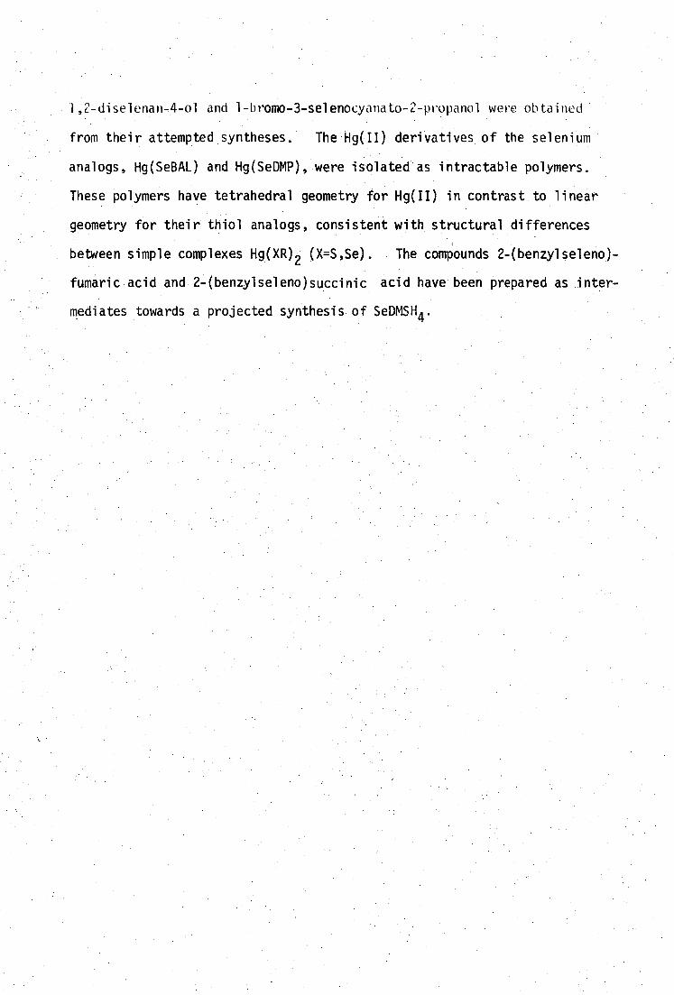
1a-diselenan-4-ol and 1-bromo-3-selenocyanato-2-propanol were obtained
from their attempted syntheses. The'Hg(II) derivatives of the selenium
analogs, Hg(SeBAL) and Hg(SeDMP), were isolated as intractable polymers.
These polymers have tetrahedral geometry for Hg(II) in contrast to linear
geometry for their thiol analogs, consistent with structural differences
between simple complexes Hg(XR) (X=S,Se). The compounds 2 7 (benzylseleno)-
fumaric.acid and 2-(benzylseleno)succinic acid have been prepared as inter-
mediates towards a projected synthesis of SeDMSH4.
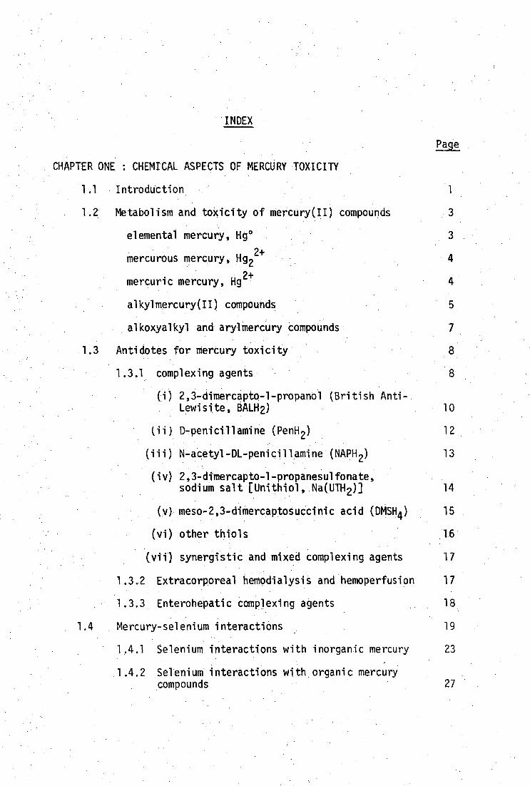
INDEX
Pate
CHAPTER ONE : CHEMICAL ASPECTS OF MERCURY TOXICITY
1.1 Introduction 1
1.2 Metabolism and toxicity of mercury(II) compounds 3
elemental mercury, Hg° 3
mercurous mercury, Hg 22+
4
mercuric mercury, Hg2+ 4
alkylmercury(II) compounds 5
alkoxyalkyl and arylmercury compounds 7
1.3 Antidotes for mercury toxicity 8
1.3.1 complexing agents 8
2,3-dimercapto-l-propanol (British Anti- Lewisite, BALH2) 10
(ii) D-penicillamine (PenH 2 ) 12
(iii)N-acetyl-DL-penicillamine (NAPH 2 ) 13
(iv) 2,3-dimercapto-1-propanesulfonate, sodium salt [Unithiol, Na(UTH2)] 14
(v)meso-2,3-dimercaptosuccinic acid (DMSH ) 15
(vi)other thiols 16
(vii)synergistic and mixed complexing agents 17
1.3.2 Extracorporeal hemodialysis and hemoperfusion 17
1.3.3 Enterohepatic complexing agents 18
1.4 Mercury-selenium interactions 19
1.4.1 Selenium interactions with inorganic mercury 23
1.4.2 Selenium interactions with organic mercury compounds 27

CHAPTER TWO : STRUCTURAL CHEMISTRY OF MERCURY(II) THIOLATES
2.1 Introduction 30
2.2 Structural features of MeHg(II) thiolates 32
2.3 Structural features of complexes of the type, Hg(SR) 2 43
2.4 Structural features of Hg(II) dithiolate complexes 53
2.5 Structural features of RSHgX species 55
2.6 Conclusions 59
CHAPTER THREE SOLUTION CHEMISTRY OF METHYLMERCURY(II) THIOLATES
AND SELENOLATES
3.1 Introduction 61
3.2 The aqueous solution chemistry of.methylmercury(II)
3.2.1 Coordination of MeHg(II) in aqueous solution 62
3,2.2 NMR investigations and MeHg(II) interactions in solution 75
3.2.3 NMR evaluation of MeHg(II)-thiolate formation constants 77
3:3 Methylmercury(II) complexation.with thiolate ligands 80
3:3:1 MethylmercurAII) monothiolate-equilibria 80
3.3.2- Potentiometric determination of formation constants of MeHg(II) thiolates 87
analysis of HI - content.. 93.
analysis of MeHg content 94
(i) 2-mercaptoethanol 97
(ii)mercaptoacetic acid 103
(iii)mercaptoacetic acid, 0-methylester 105
(iv)mercaptosuccinic acid 105
(v)L-cysteine 108

(vi) DL-homocysteine 116
(vii)DL-penicillamine 117
(viii)N-acetyl-DL-penicillamine 120
(ix) glutathione 123
(x) thiocholine perchloate 126
(xi)4-mercapto-N-methylpiperidine 129
3.3.3 Methylmercury(II) formation constants with vicinal dithiols 136
(i) 2,3-dimercapto-1-propanol, BALH 2 136
(ii) 2,3-dimercapto-l-propanesulfonate(sodium salt), Unithiol 144
(iii)meso-2,3-dimercaptosuccinic acid, DMSH 4 149
3.4 Interactions of antidotal thiols with MeHg(II) in vivo 155
3.4.1 Physiological pH 156
3.4.2 Competition of antidotal thiols with endogenous ligands in vivo 157
3.5 Methylmercury(II) interactions with selenium donors in aqueous solution 162
3.5.1 MeHg(II)-selenolate equilibria 162
3.5.2 MeHg(II)-diselenide interactions 169
3.6 Conclusions 171
CHAPTER FOUR : SYNTHESIS, VIBRATIONAL SPECTROSCOPY AND STRUCTURE OF
MERCURY(II) SELENOLATES
4.1 Preparation of Mercury(II) selenolates
173
4.1.1 Preparation of Hg(SeR) 2 complexes
173
4.1.2 Preparation of RSeHgX complexes
176
4.2 X-ray diffraction characterisation of mercury(II)- selenolates
178
4.2.1 Hg(SeR) 2 178
4.2.2 RSeHgX 190
4.2.3 Comparison of Hg-S and Hg-Se bonding distances 213
4.3 Vibrational spectroscopy of mercury(II) selenolates 217
4.3.1 Bis(selenolato)mercury(II) complexes, Hg(SeR) 2 217
• 4.3.2 Mercury(II) diselenolates 222
4.4 Conclusions 224
CHAPTER FIVE : SELENIUM ANALOGS OF ANTIDOTAL DITHIOLS
5.1 Introduction 225
5.2 Possible routes for synthesis of dithiols and 227 diselenols
5.2.1 Direct introduction to SeH 228
5.2.2 Use of protected thiols and selenols 229
5.2.3 Introduction of protected selenol groups 236
(i) alkylation of selenolates 236
(ii) addition of selenols to alkenes and alkynes 237
(iii)lactone and epoxide ring opening 237
5.2.4 Choice of protective groups for SeBALH 2 and SeDMSH4 syntheses 243
5.2.5 Selenols from selenocyanate hydrolysis 244
5.2.6 Other methods 246
(i) reduction of trithiocarbonates and xanthates to dithiols 246
(ii)conversion of alcohols to selenoethers 247
(iii)reduction of episulfides
247
(iv)addition of diselenides to alkenes and alkynes
248

5.3 Attempted syntheses Of.SeDMSH 2 and SeBALH 2 248
5.3.1 SeDMSH 4 248
benzoyl protection
249
(ii) benzyl protection
250
5.3.2 SeBALH 2 and related SeDMPH 2
262
(i) SeDMPH 2
262
(ii)SeBALH 2 281
5.4 Conclusions
282
CHAPTER SIX : POTENTIOMETRIC DETERMINATION OF AQUEOUS FORMATION CONSTANTS
OF VERY STABLE COMPLEXES
6.1 Introduction 284
6.2 Evaluation of ligand hydrolysis equilibria 286
6.2.1 Definition of 'Equilibrium Constants' used 286 in this work
6.2.2 Calibration of glass-electrodes as [e] probes 291
6.2.3 Titration assembly 297
titrant solutions 302
titration procedure
302
pH(s) buffers
305
6.2.4 Available computer programs for evaluation o ligand-hydrolysis equilibria
306
6.2.5 TITRAT and related programs for treatment of titrations of mixtures of multiprotic acids and bases
308
6.3 Evaluation of formation constants of stable complexes 317
6.3.1 1-1 4-- competition method for obtaining aqueous stability constants 317
6.3.2 Evaluation of high stability constants for MeHg-thiolate complexes 323

6.3.3 Computer programs for the evaluation of metal-ligand stability constants 326
6.4 Display of titration data and species distributions 330
6.4.1 COMIX 331
6.4.2 COMIXH 332
6.5 Appendix: A simple device for monitoring attainment of pH equilibrium 333
• CHAPTER SEVEN : EXPERIMENTAL 335
7.1 General reagents 336
7.2 General procedures used in organoselenium preparations 337
7.3 Preparation of complexes [MeHg(II) and Hg 2+] of • dithiols and model thiols • 340
7.4 Preparation of selenium compounds and complexes (and S-analogs)
• REFERENCES
APPENDIX 1
APPENDIX 2
PUBLICATIONS •
347
367
401
402
403

CHAPTER ONE
CHEMICAL ASPECTS OF MERCURY TOXICITY
1.1 Introduction
Mercury and its compounds are not rare in nature, yet if has not
been demonstrated that mercury has any essential role in any metabolic
pathway of any known species. Today's concern with mercury is due to
its environmental impact, and its deleterious effects on human health.
Contamination of the natural environment by mercury compounds, particularly
methylmercury(II), has become a serious problem in many parts of the
world. Major outbreaks of human methylmercury(II) poisoning at Minamata
Bay (1953) and Niigata (1965) in Japan have generated international aware-
ness of Minamata Disease. The history and general background to these
events and the clinical and pathological features of Minamata Disease,
which include severe, irreversible central-nervous system damage and
congenital abnormalities, have been recently reviewed.1
'2
Organo-
mercurials have been responsible for further accidental poisoning episodes
at Guatemala (1963), 3 Alamogordo New Mexico (1969) 4 and in Iraq (1973), 5 ' 6
due to grain treated with methyl- and phenylmercurial fungicides.
Over 400 deaths resulted from the Iraqi poisonings alone.4
The Japanese Outbreaks directly attributed to industrial discharges,1,2
were instrumental in promoting the sudden upsurge of interest in the
biological and environmental behaviour of mercury (and of heavy metals in
general) seen in the early 1960's. Although organomercurials had been
known to be lethally toxic since 1865, 7 until the large-scale outbreaks
most toxicological research concerned with mercury had been restricted
to elemental mercury, particularly the vapour, inorganic mercury(II)
compounds and mercurial diuretics. Biological methylation of inorganic

mercury by methylcorrinoid derivatives, e.g. methylcobalamin, is well
9 established.
8, Thus, potentially more dangerous methylmercury(II)
results from inorganic mercury(II)-contaminated environments.
There is much evidence indicating the relevance of mercury(II) complexes
with sulfhydryl ligands in the biotransport, metabolism, toxicity and
microbiological transformations of mercury(II). For example, the toxic
agent in the Minamata episodes seems to have been MeHgSMe, isolated from
shellfish. 1 Biosynthesis of this compound may involve the complex of
MeHg(II) with cysteine. Some cobalamin-independent organisms, e.g.
Neurospom Crassa can methylate Hg2+ . Homocysteine and cysteine
complexes are implicated in the mechanism. 10 Several antidotes for
mercury toxicity will be discussed in this Introduction, all of which are
thiols.
This thesis considers some of the chemical aspects in the solid
state and in aqueous solution of mercury(II) coordination with thiolate
ligands, including several with reported antidotal activity toward•
inorganic and methylmercury(II). Recent developments in the biological
interactions between mercury and selenium are discussed, and some model
mercury(II)-selenolates have been investigated in the solid state.

1.2 Metabolism and toxicity of mercury(II) compounds
The chemical features of the three oxidation states (0, 1+, 2+)
of mercury differ markedly. For this reason it is convenient to
consider them separately. The experimental toxicology and pharma-
cology of mercury has been extensively reviewed11-14
and so
only the salient features which distinguish the types of mercury
compounds, are noted here. The mode of intoxication by mercury
compounds is usually oral, or by inhalation in the case of mercury
vapor.
Elemental mercury, Hg
In the liquid metallic form, mercury is not significantly toxic,
passing through the gastrointestinal tract unabsorbed, and is fecally
excreted.13
The saturation vapor pressure of mercury under normal
3 ambient conditions is sufficiently high (10-15 mg Hg/m ),
13 to pose
a severe health threat in poorly ventilated areas. Due to its rela-
tively high lipophilicity mercury vapor is efficiently (80%)14
transferred from alveolar air into the bloodstream. In the blood-
2+ stream, Hg
os rapidly oxidised to Hg and although the half life of
Hg° is only 30s, this is sufficiently long to allow a significant
proportion of Hg° to traverse the blood-brain barrier (or the placental
barrier) leading to a tenfold higher accumulation in the brain than for
inorganic mercury poisoning.15,16
Long-term exposure to low levels
of mercury vapor is therefore of some concern.
Several minutes after inhalation, tissue distribution approaches
that of Hg2+
, except for the significantly higher proportion retained
in the brain, particularly in the cerebral and cerebellar cortex and
14 certain brain nucleii.
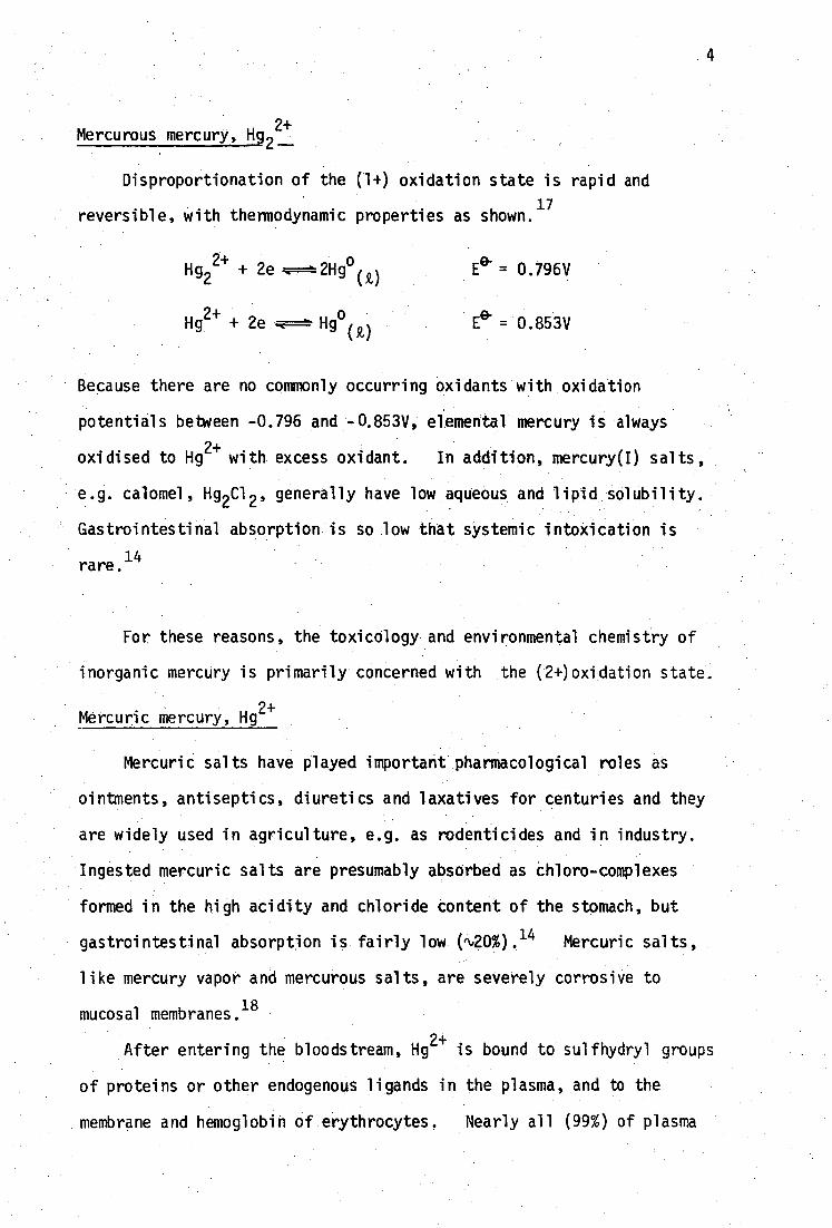
Mercurous mercury, Hg 2 2+
Disproportionation of the (1+) oxidation state is rapid and
reversible, with thermodynamic properties as shown.17
Hg22+ + 2e
Hg2+ + 2e Hg0 (,)
= 0.796V
e- E = 0.853V
Because there are no commonly occurring oxidants with oxidation
potentials between -0.796 and -0.853V, elemental mercury is always
oxidised to Hg2+ with excess oxidant. In addition, mercury(I) salts,
e.g. calomel, Hg 2C1 2 , generally have low aqueous and lipid solubility.
Gastrointestinal absorption is so low that systemic intoxication is
rare.14
For these reasons, the toxicology and environmental chemistry of
inorganic mercury is primarily concerned with the (2+)oxidation state.
Mercuric mercury, Hg2+
Mercuric salts have played important pharmacological roles as
ointments, antiseptics, diuretics and laxatives for centuries and they
are widely used in agriculture, e.g. as rodenticides and in industry.
Ingested mercuric salts are presumably absorbed as chloro-complexes
formed in the high acidity and chloride content of the stomach, but
gastrointestinal absorption is fairly low ( 20%). 14 Mercuric salts,
like mercury vapor and mercurous salts, are severely corrosive to
mucosal membranes 18
After entering the bloodstream, Hg2+
is bound to sulfhydryl groups
of proteins or other endogenous ligands in the plasma, and to the
membrane and hemoglobin of erythrocytes. Nearly all (99%) of plasma

2+ is bound to non-filtrable protei Hg n thiols, e.g. albumin and
globulin.13
The concentrations of mercuric ion in the plasma and
erythrocytes are approximately equal14
and the target organs for Hg2+
are the kidneys. 13 Mercury is bound to 2 or 3 cysteinyl groups in
19 kidney metallothionein. Mercuric ion is gradually removed from the
blood by passive glomerular filtration and active tubular transport
,in the kidneys, as well as by fecal excretion from gastrointestinal
mucosa.14
These two routes are of comparable importance and result
in a half-life for Hg2+ of 1-2 months.
1 4
AlkylmerCury(II) compounds
Alkylmercurials are used widely as fungicides, and methylmercury(II)
is the result of biomethylation of inorganic mercury residues in
sediments.20 Short chain monoalkyl mercurials, particularly MeHg(II),
are considerably more resistant to metabolic dealkylation (to form Hg)
than than long chain analogs, although significant biotransformation of
MeHg(II) occurs in the kidneys and the liver.21-23
The compounds of
methyl - and ethylmercury(II) have been most closely studied. Because
of its biological and environmental importance only MeHg(II) will be
discussed here.
The extremely high translational diffusion characteristics of
MeHgC1 seem to be responsible for its ready absorption from the stomach
24 into the bloodstream, rather than lipid solubility. The distribution
ratio from water to lipid has been recently determined to be only q,2,
thus, MeHgC1 diffuses rapidly but does not partition well in lipids
.24
Methylmercury(II) distribution in the body differs markedly from
that of Hg2+.25,26
Preferential accumulation of MeHg(II) occurs in
the erythrocytes, producing a characteristic erythrocyte:plasma mercury
concentration ratio of 10:1. 14 Methylmercury(II) readily passes through
the blood-brain and placental barriers. Up to 10% of the whole-body
13 burden of MeHg(II) may be found in the brain, which is the target
organ for methylmercury(II) poisoning. Congenital Minamata disease
tragically results from foetal accumulation of acutely toxic doses of
MeHg(II).1,2
The neurotoxicity of MeHg(II) does not seem to be due
solely to biotransformation to Hg2+ in brain tissue.
27
There is little doubt that the toxic influence of MeHg(II) is due
to inactivation of specific sulfhydryl sites of cysteinyl residues of
proteins and enzymes. 28 Although the 1:1 MeHg(II)-sulfhydryl inter-
action serves as a selective biochemical probe for SH groupS29
the
active cellular sites of MeHg(II) toxicity have not thus far been
firmly established. Significant amounts of radioactively labelled
MeHg(II) are found in mitochondria 30 and MeHg compounds directly affect
rat liver mitochondria in vitro. 31-33 The ability of some thiols to
reactivate MeHg(II)-inhibited enzymes of rat liver mitochondria in
34,35 vivo has recently been used to evaluate possible antidotal
thiols. 36
MeHgC1 also interacts with unsaturated phospholipids and
disrupts lipid membrane permeability even at mercury concentrations of
-7 37 10 M, consequently,lipid biomembranes have been suggested to be
the primary targets for MeHg(II).37-8
Several workers have characterised intracellular MeHg(II)-sulfhydryl
species. A glutathione complex has been identified from rat liver
Cytosol, with 24% of mercury in the small molecular weight fraction,39
and metallothionein-bound MeHg(II) has been reported to account for up
to 40% of total protein-bound MeHg(II) in the liver cytosol of rainbow
trout.40 Attempts to relate these findings to the nature of MeHg(II)
species in vivo must be limited by the rapid redistribution of mercurial
among the mixture of sulfhydryl ligands resulting from cell-wall
41 disruption. Such ligand exchange is fast on the nmr timescale and
7
is discussed later in this thesis (page 75 ). The methylmercury-
thiolate species in the bile of rats has a molecular weight intermediate
between that of the MeHg(II) complexes of cysteine and glutathione and
is not the complex of N-acetylcysteine or homocysteine. 42
The toxicology of methylmercury is dominated by its long biological
half-life (3 months) and hence, its propensity for accumulation with
continued sub-acute exposure.13 High concentrations of methylmercury(II)
are found in the liver and kidneys but normal urinary excretion is low,
accounting for only 10% of excreted mercury.14 As a result, the
predominant pathway for excretion is via MeHg(II) species in the bile.
Unfortunately, the high rate of gastrointestinal reabsorption of
biliary MeHg(II) species provides an efficient route for enterohepatic
recirculation and contributes to the long half-life in the body.
Attempts to increase either urinary or fecal excretion of
MeHg(II) form the basis of two possible courses for antidotal therapy
for MeHg(II) intoxication and will be discussed in the next section.
Dialkylmercurials such as Me 2Hg may pose higher health risks than
monoalkylmercurials for acute exposure, as they are generally more
volatile and lipophilic and hence, more easily absorbed. However,
these compounds do not separately form a high environmental or bio-
logical hazard because they are readily degraded to monoalkylmercurials
and/or mercuric ion.13
Alkoxyalkyl and Arylmercury(II) compounds
Alkoxyalkylmercurials are used in agriculture and industry as
fungicides. These compounds are rapidly degraded to Hg2+ w
hich
determines their toxicity and pharmacology.43
Arylmercurials, particularly phenylmercury(II), are widely used as
fungicides and mould inhibitors in paints and as wood preservatives.
• Large scale poisoning episodes e.g. in Pakistan44
have occurred as
a result of ingesting treated grain. Phenylmercury(II) is rapidly
degraded to inorganic mercury(II), eventually showing the body distri-
bution of Hg2+
.25
Disproportionation of mercaptoamino acid and
dithiolate complexes of PhHg(II) to diphenylmercury has been shown in
this laboratory to occur in vitro.45 Metabolic conversion of PhHg(II)
to diphenylmercury46
and to elemental mercury47,8
has also been
demonstrated in some microorganisms.
1.3 Antidotes for Mercury toxicity
The term antidote used throughout this thesis defines a chemo-
therapeutic agent which aids the removal of toxic metal from the body
after intoxication. Many forms of metal intoxication may be alleviated
with therapeutic 'chelating' agents which enhance urinary excretion
of metal. Alternatively, antidotes which enhance fecal excretion of
metal released in the bile may be used.
These methods will be separately discussed below in relation to
removal of inorganic Hg2+ and methylmercury(II).
1.3.1 Complexing agents
The use of complexing agents for therapeutic manipulation of
metals in the human body is now well established, and has been reviewed
elsewhere
.49
The term 1 chelatings agent seems to have been loosely
used in the toxicological literature. In many situations there is
little or no evidence that the reagent is bound entirely to one metal
atom, in the chelating sense. 17 Evidence from this laboratory indicates
that complexes formed between mercuric ion and several antidotal dithiols
are not chelating (Section 2.41. • Similarly, the methylmercury(l+)
cation forms unidentate complexes particularly with thiolate ligands.

The term, complexing agent, will be used throughout this thesis, except •
• in those situations where chelation can be established.
Because mercury will inevitably be bound to sulfhydryl groups of
proteins, etc. after intoxication, an effective antidotal complexing
agent must
(1) be of low toxicity,
(ii)be able to bind mercury strongly enough to compete with a
multitude of biological ligands, e.g. chloride, nitrogenous bases,
sulfhydryl-containing proteins, etc.,
(iii)discriminate against abundant endogenous metals such as zinc
and calcium,
(iv) be sufficiently lipophilic to penetrate cell membranes to
reach intracellular sites of metal deposition and
(v) form a lipophilic complex in the body compartment where mercury
is accumulated.
Highly charged species are inefficient in this regard e.g. polyamine
carboxylic acids are largely retained in the extracellular space, and rely
on endogenous ligands to remove metals from inside the ce11. 50 If
enhanced urinary excretion is desired, this complex should change to a
new hydrophilic complex in the plasma, so that metal can be eliminated
by the kidneys. While high mobility is required of the metal-antidote
complex in this last step, redistribution into more susceptible organs,
e.g. MeHg(II) into the brain, is undesirable.
Mercury(II) is a typical Class B 51 or isoft 152 metal and consequ-
ently has a high affinity, for sulfur donor ligands and particularly for
thiolates. Many compounds containing sulfhydryl groups have been
evaluated as antidotes for various forms of mercury poisoning, and
these will be discussed here.
1 0
(i) 2,3-dimercapto-l-propanol (British Anti-Lewisite, BALH ,*
Dimercaprol)
The efficacy of BALH 2 as a general heavy metal antidote was
recognised during World War II. The historical development of its
preparation and use has been recently reviewed.
54-6 Treatment for acute and chronic
57,8 poisoning by metallic mercury
has been reported but the therapeutic effect of BALH 2 is uncertain. 26
The life-saving effect of BALH 2 treatment for acute inorganic mercury(II)
poisoning is well-established and has been reviewed.26 There are
conflicting reports as to the effect of BALH 2 on urinary excretion of
mercury after inorganic mercury(II) poisoning, e.g. some workers report
increased excretion,59-61
n contrast to decreased excretion by others.
62,3
Fecal elimination of mercury is the major route for inorganic mercury
poisoning, therefore, renal failure does not preclude metal decorpora-
. 14 tion. Considerable redistribution of inorganic mercury between
organs occurs with BALH 2 treatment, which may cause an increase in the
amount of mercury in the brain.61-3 This latter effect has been
attributed to timing of BALH 2 treatment.61
Although BALH 2 increases biliary excretion of MeHg(II)64-5
in rats, fecal elimination is not enhanced 5 9 ' 66 due to a corresponding
increase in gastrointestinal reabsorption.26 Administration of BALH 2
increases the brain uptake rate of MeHg(II) in mice ° and increases the
level of MeHg(II) in the brain of rats. "
The use of BALH2
is lethal in cases of methoxyethylmercury(II)
*Throughout this work, antidotal thiols will be abbreviated to indicate loss of thiol protons on complex formation, e.g. BALH2, PenH2, DMSH4, etc. Although BALH2 exists in stereoisomeric forms, only the racemic material has been used pharmacologically, although the optically active forms have been synthesised."
poisoning, 26 but is the most effective antidote for phenylmercury(II)
poisoned animalS. 26
The major disadvantages of BALH 2 are related to its limited
aqueous solubility 15 and relatively high nephrotoxicity" (Table
1.1). A typical course of BALH 2 treatment for acute inorganic
mercury(II) poisoning requires four-hourly deep intramuscular injections
of BAL (3-5 mg/kg) for 2 days followed by six-hourly injections of a
lower dose (2.5-3 mg/kg) for up to seven days.18 Many patients . suffer
side effects from such treatment 70 and the mortality is N2%, mainly from
secondary infections.14 Consequently, more water-soluble, less toxic
thiol antidotes have been sought.
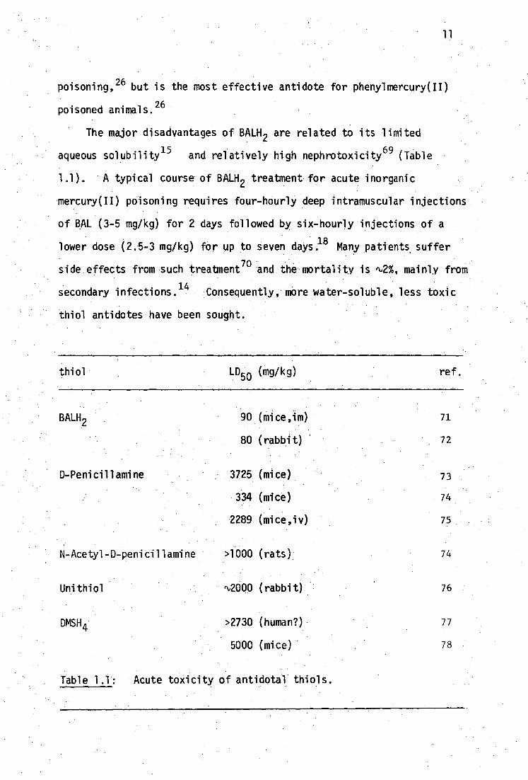
thiol
LD50 (mg/kg)
ref.
BALH2 90 (mice,im) 71
80 (rabbit) 72
D-Penicillamine 3725 (mice) 73
334 (mice) 74
2289 (mice,iv) 75
N-Acetyl -D-peni ci 1 1 amine >1000 (rats) 74
Unithiol r‘,2000 ( rabbi t) 76
DMSH 4 >2730 (human?) 77
5000 (mice) 78
Table 1.1: Acute toxicity of antidotal thiols.

12
(ii) D-penicillaminet (PenH 2 )
In 1950 it was noted that treatment of inorganic mercury(II)-
poisoned patients with penicillin produced increased urinary excretion
79 of mercury. The antidotal effect is due to the metabolite, D-
penicillamine. However, this compound was not available commercially
until its beneficial action in alleviating the symptoms of Wilson's
disease (due to Cu accumulation) had been demonstrated.80
PenH2 is
water-soluble and has low toxicity (Table 1.1). Although it is
commonly administered nowadays (100 mg/kg orally) 18 subsequent to
BALH2 treatment of inorganic mercury poisoning, early reports of its
efficacy in man were not encouraging81 although marked reductions in
mortality (better than BALH 2 ) among HgC1 2 and HgNO3-intoxicated rats
was observed, 74 but only at high doses (65 mg/kg).26 PenH2 is also
used to treat sub-acute mercurialism. 18
D-Penicillamine does not easily penetrate erythrocyte membranes
in vitro,82
however, Magos et al. have recently reported decreased
levels of mercury in the brain following PenH 2 administration to
83 MeHg(II) intoxicated rats. Earlier contradictory reports by other
workers26
may have been due to low doses of antidote. High doses
(220 mg/kg) removed MeHg(II) from all organs except the kidneys in
rats but lower doses (80 mg/kg) were ineffective. 84 PenH2 accelerates
the urinary excretion of MeHg(II)5,85
but unlike BALH 2 , does not
84,86 redistribute MeHg(II) into the brain. Biliary excretion of
MeHg(II) is increased in rats treated with PenH 2 65,83
but fecal
• • The nomenclature of this stereoisomer may be confused in the literature, being variously denoted D- and DL-penicillamine. The racemic isomer (DL-) is commercially available for therapeutic use as Metcaptase(R).
The Merck Index describes the D- isomer as naturally occurring.75
13
excretion is not, due to rapid reabsorption of the MeHg-pen complex
from the gut.26
High doses of PenH 2 (1 g/kg) to pregnant rats
prevented fetal morphological changes caused by MeHgCl. 87
• In cases of rats poisoned by methoxyethylmercury, immediate
injection of PenH 2 has a lifesaving effect increasing urinary
excretion of Hg, but is only marginally effective against phenyl-
mercury.26,66
(iii) N-Acetyl-DL-penicillamine (NAPH 21
Acetylation of_DL-penicillamine (or cysteine or homocysteine
increases the lipophilic character of the antidote. Thus NAPH 2
readily penetrates erythrocyte membranes, unlike PenH 2 . 82 NAPH2 is
even less toxic than Pe11H2
(Table 1.1) and may be administered orally. .
N-acetylation of the amino group of PenH2 and similar compounds,
protects the molecule against the action of catabolic enzymes and •
reduces tubular reabsorption in the kidney glomeruli.88
NAPH2 is
more effective than PenH 2 against acute-HgC1 2 intoxication in mice
. 14 and increases urinaryHg excretion. Its use in sub-acute mercuri-
alism has been recorded.18
Failures ofNAPH 2 to remove MeHg(II) from
the brain in mice14 and as an antidote for human MeHg(II) poisoning
" (Iraq)
5 were due to low doses of antidote.
82 Thus high doses (3-4
mmol/kg) remove up to '50% of mercury in the brain 86 ' 89 and are therefore•
much more effective than PenH 2 in this regard, whereas lower doses
(0J-0.2 mmol/kg) have no effect.5,88-9
•Oral NAPH2 treatment of
MeHgC1 intoxicated micemobilised mercury from all organs and reduced
fetal and maternal mercury levels. 5 Urinary and fecal Hg-excretion
• were increased.
5 NAPH
2 is more effective than PenH 2' glutathione,
cysteine, N .-atetylcysteine or N-acetyl-homOcysteine in the removal of
MeHg(II) from an albumin complex in

14
2,3-dimercapto-l-propanesulfonate, sodium salt [Unithiol,t
Na(UTH2 )] —
Because of the relatively high toxicity and low aqueous solubility .
of BALH2' its water-soluble sulfonate analog, Unithiol, was prepared
in 1955. 90 ' 91 The toxicity of Unithiol is very low (Table 1.1) and
it may be administered orally with no long term effects. Chronic
treatment (0.6 mmol/kg) produced transitory reductions in the copper
concentration of some orgas92
and increased renal excretion of copper'
and zinc.93
Although only recently available commercially in Western
Europe and America, it has been used successfully in the U.S.S.R. to
treat inorganic mercury(II) toxicity in man.94 The high water solu-
bility contributes to its relatively low gastrointestinal absorption
rate (30%/24 hrs95 in comparison with PenH2 60%96) and its rapid renal
clearance (70-80%/24 hrs).97
Thus Unithiol facilitates excretion of
mercuric mercury via the kidneys. 98
Despite its low lipophilicity and hence its low membrane permea-
bility, high doses (180 mg/kg) of Unithiol seem to be more effective
than PenH 2 (and N-acetyl-homocysteine thiolactone) in reducing the
brain mercury level after MeHgC1 intoxication.82 This effect has
been attributed to a reduced brain uptake rather than increased removal
of mercury. 99
Treatment with lower doses (114 mg/kg) did not
mobilise significant amounts of mercury from the brain in mice.100
Urinary MeHg(II) excretion is substantially enhanced upon Unithiol
treatment.99
Fecal MeHg(II) excretion is also slightly enhanced,99
although this may be due to the increased flow of bile and bile-salts
101 caused by Unithiol. Unithiol does not remove Cd from metallothionen
tUnithiol is marketed for therapeutic use under the name Dimaval(R)
by Heyl & Co. Chem.-Pharm. Fabrik, Berlin. The stereoisomeric nature of Unithiol does not seem to have been reported. The commercial product is presumably a racemic form, like BALH2.

15
in the bile, but BALH 2 does. 102
(v) meso-2,3-dimercaptosuccinic acid (DMSH IX
Introduction of carboxylate groups into a dithiol molecule
increases its aqueous solubility. Thus the disodium salt and the
1:1 mercury(II) complex of DMSH 4 are water-soluble. The toxicity
of DMSH4 is very low (Table 1.1) and like Unithiol, it may be
administered orally with no long-term effects103 although its metabolism
DO is not well understood. DMSH
4 was first used therapeutically in
1954 as the trypanocidal antimony complex.104 The arsenic complex has
105 106- 7 similar activity. DMSH 2 has been used as an antidote for lead
and arsenic103 poisoning for which it is as effective as BALH 2 , and is
better than PenH2 at increasing urinary excretion of gold after
Au-mercaptosuccinate(Myocrisin) treatment.108
DMSH4 is an effective antidote to acute HgC1 2 poisoninr and is
more effective than PenH 2 at removal of Hg from the kidneys, liver and
7 brain of mice.
78,107,110,111
DMSH4 seems to be a most promising antidote for MeHg(II) toxicity.
It is five times as effective as PenH 2 for Hg excretion in mice after
MeHgBr administration and decreases the Hg content of kidneys, liver
11D- 1 and particularly the brain.
78, Doses of 90 mg/kg/day removed two
thirds of mercury from the brain when given to MeHgC1 intoxicated mice.
For this purpose DMSH4 is better than PenH 2 , mercaptosuccinic acid100
and BALH2 112
. Postexposure preventative treatment with DMSH 4 to rats
after MeHgC1 injection prevents the increase of mercury levels in the
Only this stereoisomer has been studied extensively, however the racemic form was reported to be better than the meso- form at accelerating urinary mercury elimination from rats after HgC1 administration. 112
2
16
brain.113
The MeHg(II) level in the brains of neonatal rats is significantly
114 lower than that of controls after administration of DMS to the dams.
(vi) Other thiols
Several N-acetylated mercaptoamino acids (other than PenH 2 ) have been
tested as antidotes in animal experiments, particularly against MeHg(II)
toxicity.
N-Acetylhomocysteine is nearly as effective as PenH 2 (and better than
homocysteine) for decorporation of MeHg(II) from mice, and is less toxic. 88
The corresponding thiolactone produces larger increases in urinary excretion
of mercury,88 and mobilises more than MeHg(II) from the brain,
115 but very
high doses are required. In contrast, the non-acetylated thiolactone does
not mobilise mercury from the brain in mice or monkeys, and is toxic to
monkeys.86
Diethyldithiocarbamate and disulfuran decrease urinary and biliary
MeHg(II) excretion and increase the mercury content of the brain in mice.116
Oral doses of 8-mercaptopropionylglycine (600 mg/day) produced 3- to
6-fold increases in urinary mercury excretion for Minamata patients, and
was better than PenH 2 in this regard. 117
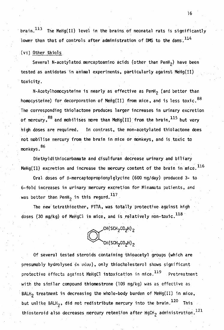
The new tetrathioether, PTTA, was totally protective against high
doses (30 mg/kg) of MeHgC1 in mice and is relatively non-toxic. 118
CH(SCH CO2H)
CH(SCH 2CO2H) 2
Of several tested steroids containing thioacetyl groups (which are
presumably hydrolysed in vivo), only thiocholesterol shows significant
protective effects against MeHgC1 intoxication in mice
.119
Pretreatment
with the similar compound thiomestrone (109 mg/kg) was as effective as
BALH2 treatment in decreasing the whole-body burden of MeHg(II) in mice,
but unlike BALH2' did not redistribute mercury into the brain.
120 This
thiosteroid also decreases mercury retention after HgCH 2 administration.121
17
(vii) Synergistic and mixed complexing agents
Whereas treatment with one complexing agent may not significantly
alter the rate of metal decorporation, simultaneous addition of a
second agent may prove effective. The second agent may alter a
metabolic process or function which enables the complex formed between
the metal and first agent to be more efficienty excreted, e.g. pheno-
barbitone enhances biliary excretion but is ineffective alone as a
mercury antidote. Fecal elimination of mercury from MeHgC1 intoxicated
mice treated with a polythiol resin is thus enhanced by simultaneous
122 treatment with phenobarbitone.
The formation of ternary or mixed complexes between the metal and
both complexing agents has been postulated to account for enhanced
123t elimination of plutonium with salicylate and DTPA treatment, although
some of the experimental evidence for the mixed species has been
questioned.124
Mixed ligand treatment (BAL/CaEDTA) has been used
125 for lead and copper
126 toxicity but mixed complexes have not been
identified.
This form of treatment does not seem to have been widely used for
treatment of mercury(II) toxicity.
1.3.2 Extracorporeal Hemodialysis and Hemoperfusion
Antidotal thiols such as penicillamine and N-acetylpenicillamine
increase the fraction of non-protein bound MeHg(II) in the blood. In
vitro studies have shown 55-60 fold increases in the low M.Wt. MeHg(II)
- fraction after 10
2 M cysteine treatment, 44% of which is dialysable in
a single pass.127
In vivo, extracorporeal hemodialysis of blood from
dogs intoxicated with MeHgC1, resulted in a 100 fold increase in MeHg
removal rate after treatment with cysteine.128 Significantly, mercury
was removed from the brain. In contrast, clearance of Hg2+
was not
tdiethylenetriaminepentacetic acid.
CH 3 (CH 2 ) n CH\ SCH CO H 2 2
/SCH
2CO
2H
n = 10,16 131
18
significantly increased after treatment with BALH 2 and hemodialysis. 129
The use of thiolpolymers to remove mercury from the dialysate has been
suggested.129
Perfusion of human plasma through a bed of thiolpolymer
microspheres has recently been reported to decrease mercury levels, in
vitro, and is cheaper than dialysis. 130
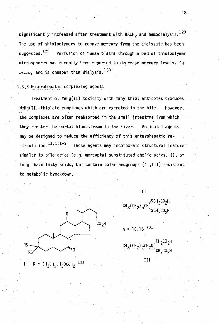
1.3,3 Enterohepatic complexing agents
Treatment of MeHg(II) toxicity with many thiol antidotes produces
MeHg(II)-thiolate complexes which are excreted in the bile. However,
the complexes are often reabsorbed in the small intestine from which
they reenter the portal bloodstream to the liver. Antidotal agents
may be designed to reduce the efficiency of this enterohepatic re-
circulation.11,131-2
These agents may incorporate structural features
similar to bile acids (e.g. mercaptal substituted cholic acids, I), or
long chain fatty acids, but contain polar endgroups (II,III) resistant
to metabolic breakdown.
II
CH 2CO 2H RS CH (CH ) 2CH 2N(
RS CH2CO2H
III I. R = CH 3CH 2 ,H 2OCCH 2 . 131

19
An alternative principle which disrupts enterohepatic recirculation
employs insoluble polymers which contain many groups capable of MeHg(II)
binding. The polymer is ingested and traps MeHg(II) in the gut from
which it is fecally excreted without reabsorption. Powdered human
hair treated with mercaptoacetate, 133 polystyrene polymers containing
sulfhydryl groups,5,122,134
polyterephthalate polymers containing
thioether groups,118,135-6
and mercaptostarch137
have been used for this
purpose. All of these agents increase the fecal elimination of mercury
and several reduce brain mercury levels. Phenobarbitone has been used
to increase bile blow and hence promote fecal MeHg(II) excretion from
mice treated with polythiol resin.122,138 Attempts to use the macro-
molecular polythiol, mercaptodextran, to disrupt enterohepatic recircula-
tion have failed, presumably due to metabolic breakdown into its component,
N-acetyl-homocysteine. 115
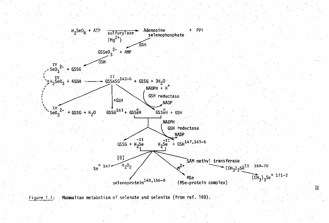
1.4 Mercury-Selenium interactions
Selenium was shown to be an essential trace element by Schwarz in
1957139 and the biotransformations and metabolism of selenium are now
fairly well established and have been summarised in Figure 1.1. The
physical properties, chemistry and roles of selenium in trace-element
metabolism have been elucidated in two recent comprehensive texts.0-1
The most important process in the mammalian metabolism of selenite,
Se032-
, appears to be the formation of selenotrisulphides, RSSeSR142-6
particularly with glutathione (Table 1.1). The major selenium-binding
components in the blood plasma of rats appears to be albumin when Se0 32-
erythrocytes and plasma are incubated in vitro. 147 selenium is
concentrated in the albumin fraction of rat plasma after the administration
of small doses of Se032- , primarily in association with proteins with a
molecular weight of 77,000.148-9 In mice, selenite is metabolised and
H S 0 + ATP sulfurylase 26. (mg2+ )
GSSe032- + AMP
Adenosine + PPi selenophosphate
GSH
GSH 2_ -Se03 GSSG
iv % ,H 2Se03 4GSH
+ GSSG + H 20
GSSeSG142-6
+ GSSG + 3H 20
NADPH +
GSH reductase
NADP
GSSeH + GSH• GSSG163 +GSSeH
+GSH
NADPH
GSH reductase
NADP -11
GSSG + H 2Se -11
H Se + GSH147' 165-6
[0]
Se° 167
H 202
selenoprotein148'1 66-8
SAM methyl transferase 2+
(CH )2 sin 169-70
3
MSe (CH)Se+ 171-2 (MSe-protein complex)
Figure 1.1: Mammalian metabolism of selenate and selenite (from ref. 160).
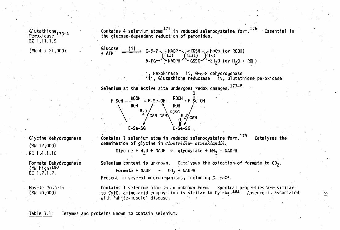
Muscle Protein (MW 10,000)
Contains 4 selenium atoms175
in reduced selenocysteine form.176
Essential in the glucose-dependent reduction of peroxides.
Glucose ILL_ G-6-P NADP 2GSH H202 (or ROOH)
(ii) •iii) iv) 6-PG NADPH GSSG 2H20 (or H20 + ROH)
Hexokinase ii, G-6-P dehydrogenase Glutathione reductase iv, Glutathione peroxidase
Selenium at the active site undergoes redox changes:177-8
0
Glutathione173-4
Peroxidase EC 1.11.1.9
(MW 4 x 21,000)
Glycine dehydrogenase
(MW 12,000)
EC 1.4.1.10
Formate Dehydrogenase (MW high) 180 EC 1.2.1.2.
+ATP
E-SeH ROOH
E Se-OH
E-Se-SG
ROOH • E Se-OH ROH
1117// GSH 2
GSSG
0
E-Se -SG
Contains 1 selenium atom in reduced selenocysteine form. 179 Catalyses the deamination of glycine in Clostridium stricklandii.
Glycine + H20 + NADP -+ glyoxylate + NH 3 + NADPH
Selenium content is unknown. Catalyses the oxidation of formate to CO 2 .
Formate + NADP CO2 + NADPH
Present in several microorganisms, including E. coli.
Contains 1 selenium atom in an unknown form. Spectral properties are similar to CytC, amino-acid composition is similar to Cyt-b5. 181 Absence is associated with 'white-muscle' disease.
Table 1.1: Enzymes and proteins known to contain selenium.
22
and bound to albumin -lipoprotein and an unidentified protein in
the plasma. 150
Since the initial observation (1960) •that selenite is preventative
against the injurious effects of cadmium salts on testes in rats, 151
and against renal and intestinal necrosis caused by inorganic mercury(II)
salts152 there has been much interest in the role of selenium as
'Nature's antidote to heavy metal toxicities'. 153 In an even earlier
study with BALH 2 , Tobias et al. noted that selenite protected mice
against the otherwise lethal effects of inhaled CdC1 2 ,154 but twenty
years elapsed until the pioneering work by Kar and co-workers. 151
There is evidence that selenium may be involved in the detoxification
155- 7 2+ 158 of Tl, and of Ag , Pb (and perhaps Cu ). Particularly
relevant to this work are the extensive reviews of the interactions of
2+ 152-3,8-9 selenium with Cd
2+ and Hg , including two published very
recently (1980).160-1
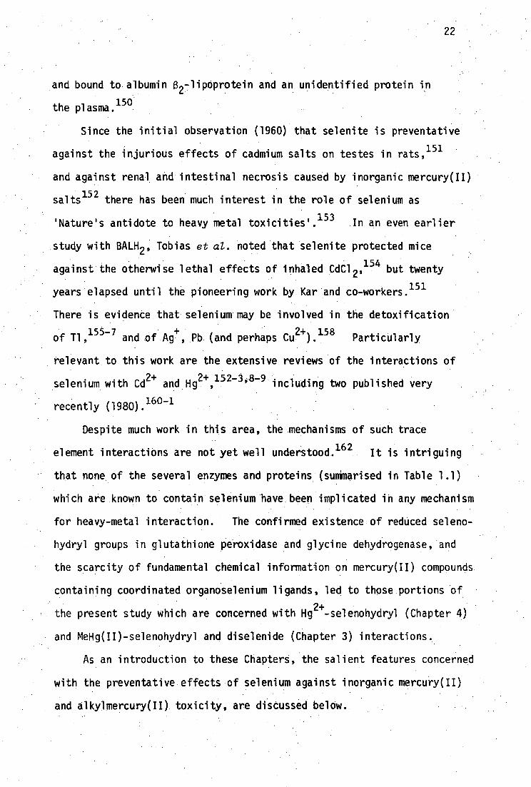
Despite much work in this area, the mechanisms of such trace
element interactions are not yet well understood. 162 It is intriguing
that none of the several enzymes and proteins (summarised in Table 1.1)
which are known to contain selenium have been implicated in any mechanism
for heavy-metal interaction. The confirmed existence of reduced seleno-
hydryl groups in glutathione peroxidase and glycine dehydrogenase, and
the scarcity of fundamental chemical information on mercury(II) compounds
containing coordinated organoselenium ligands, led to those portions of
the present study which are concerned with Hg2+
-selenohydryl (Chapter 4)
and MeHg(II)-selenohydryl and diselenide (Chapter 3) interactions.
As an introduction to these Chapters, the salient features concerned
with the preventative effects of selenium against inorganic mercury(II)
and alkylmercury(II) toxicity, are discussed below.
23
1.4.1 Selenium interactions with inorganic mercury
Interactions between selenium and inorganic mercury(II) compounds
182-3162, have recently been reviewed by several authors
159, but some
of the more important findings are recorded here.
There do not appear to be many reports regarding the effects of
selenium on intoxication by elemental mercury, other than that low
levels of dietary selenite (3.1 mg Se/kg food) do not seem to increase
the bodily retention of mercury in rats exposed to occupational levels
of mercury vapor. 184
- Parizek has shown that dietary Se03
2 prevents growth depression
due to oral administration of HgC1 2 in rats.185 Groth and co-workers
- report that long term dietary supplementation with selenate, Se0 4
2 ,
1 reduces chronic renal tubular damage in rats administered HgC12
86
Selenate has a protective effect in rats on weight loss and histo-
pathology due to HgC1 2 .187 Selenite and selenomethionine provide
complete protection against kidney and intestinal necrosis in rats
fed lethal doses of HgC1 2 . 152 Remarkably, this effect is not due to
improved elimination of mercury, on the contrary, the whole body
elimination rate is decreased188
as are urinary152,189
and fecal190
excretion rates in rats, although the effect is not marked in the
latter case. Urinary excretion of selenium in mice is also depressed
by administration of HgC1 2 but the mercury excretion rate is dependent
on the administered dose of selenite. 191 In lactating rats, selenite
decreases the mercury content of fetuses, milk and sucklings, but
increases the whole-body mercury content of the dams.159
Selenite152 and selenate188 increase the whole-body retention of
mercury but dramatically change its organ diStribution. 192 Single
doses of selenite, selenate and selenomethionine all decrease Mercury

24
levels in the kidney152,193
and increase the content of the liver.194
In contrast, repeated simultaneous administration of Hg2+
and
selenite is reported to increase the mercury levels in kidneys of rats
and mice.195
Selenite removes inorganic mercury from metallothionein in vitro, 196- 7
but does not stimulate the biosynthesis of the apoprotein. 198
Chmielnicka and co-workers have shown that the selenite-induced trans-
location of inorganic mercury from the kidney (where it is bound
presumably to metallothionein) to the liver,199
is accompanied by an
accumulation of mercury in the liver mitochondria.200 In particular,
combined and separate exposures of rats to HgC1 2 (0.5 mg Hg/kg) and
selenite (0.5 mg Se/kg) for 2 weeks decrease the content of mercury in
intramitochondrial structures of the kidney and both Hg and Se accumulate
in the inner and outer mitochondrial membranes.201 Levander has also
reported that HgC1 2 inhibits the swelling of liver mitochondria, other-
wise induced by selenite. Yamane et al. report nine - fold increases
in mercury content of nucleii, microsomal and mitochondrial fractions
of rat liver homogenates after concurrent administration of HgC1 2 and
Na 2Se04'194 It is likely that these sites, where many important
metabolic processes are catalysed, are the targets for damage by
mercuryll and may also be the sites relevant to molecular interactions
between selenite and inorganic mercury.
Single injections of selenite cause a dramatic redistribution of
inorganic mercury in the blood. Mercury is removed from the
erythrocytes into the plasma149189 "
203-5 where it is bound to high
molecular weight protein species containing equimolar amounts of
mercury and selenium. The magnitude of the effect can be seen in rats
where normally 50% of mercury is bound to low molecular weight protein

25
fractions in plasma, but after administration of selenite, 205 or
selenate,194 this proportion is less than 1%. Selenomethionine
causes a similar redistribution, but higher doses are needed for the
same response and liver enzymes are essential for the effect in
vitro, presumably to metabolise selenomethionine to selenite. 192
Burk et al., 149 have demonstrated that mercury and selenium appear
together in rat plasma as a macromolecular protein complex of the
type ProtSSeHgX. A very similar complex has been identified in
hemosylates of rabbit blood. 204 The specific complex in these cases
has not yet been identified, but appears to be similar to the 130,000
Dalton cadmium and selenium binding component found by Gasiewicz and
148 165 Smith. ' These authors have shown that H 2 Se interacts with
Hg in in plasma to yield a protein complex of molecular weight 330,000
containing equimolar amounts of mercury and seleni um. 147 An analogous
Cd2+
protein complex of molecular weight 130,000 formed under identical
conditions is considered to be colloidal CdSe stabilised by association
with specific but unidentified macromolecular proteins.147
The forma-
tion of complexes of this type may explain changes in the biokinetics
and toxicity of mercury but the biokinetics of Hg2+
salts are also
altered in situations of selenium deficiency.206
The occurrence of an equimolar accumulation of selenium and
mercury in the liver and brain of seals207 and in the liver cells of
Mediterranean cetaceans208
has been reported. Ratios of Hg:Se of
1-3 have been reported in the liver and brain of whales. 209 The
protective effects of selenate on HgC1 2 toxicity has been associated
with the formation of electron-dense particles in the kidney proximal
tubule cells and the reticuloendothelial cell cytoplasm and the extra-
cellular space of Disse in the liver. 187 Carmichael and Fowler have
used energy-dispersive X-ray analysis to obtain a Hg:Se ratio of 1:2 in

26
these particles, which is in contrast to 1:1 ratio found by Groth et
1 aZ. 86 This apparent discrepancy may be due to translocation of Hg
or Se during sample processing or examination. The mercury in these
particles has been shown to be thermally labile when a critical temp-
erature under the X-ray beam is reached. 157 Interestingly, the
- administration of tellurite, Te03
2 , prevents the formation of these
particles and is synergistic with Se032- in the prevention of renal
necrosis caused by chronic exposure to HgC1 2 .149 In contrast to
- - Se03
2 , administration of Te0 32 with HgC1 2 does not affect the organ
distribution of mercury, but it does protect against acute HgC1 2 toxicity
and increases the whole body retention of mercury.188
In man, elevated levels of mercury and selenium have been found in
the brain, thyroid, pituitary and kidneys of mercury miners, 16 years
after the cessation of exposure.210 Although increased retention of
selenium has also been reported elsewhere in persons occupationally
exposed to inorganic mercury, 211 it is questionable whether selenite
mitigated the effects of chronic mercury exposure in these cases.
Parizek has warned that not all the interactions between inorganic
mercury and selenium are beneficial, and that the concomitant presence
of mercury and certain selenium metabolites produces a lethal syndrome
in rats, particularly if selenite is administered before HgC12 • 152
These toxic effects have been shown to be similar to those caused by
large doses of dimethylselenide. 152 Parenteral pretreatment with
HgC1 2 (10 mg/kg) decreases the excretion of volatile selenium compounds
in female rats after administration of Na 2Se03 (0.3 mg/kg ip). 152,192
The effect is more marked than identical pretreatment with CdC1 2 or
ZnC1 2' and selenomethionine acts similarly.212 Curiously, male rats
are much more susceptible to the toxic syndrome than females, but this
sex-linked difference is not related to the sex-linked differences found

27
for dimethylselenide conversion to the excretion product,trimethyl-
selenonium ion (see Figure 1.1). Parizek has postulated that the
toxic effect may be due to a selenium metabolite such as MeSe - , and
that HgC1 2 may either alter the metabolic pathways and/or distribution
of this intermediate or sensitivity of the critical organs to MeSe7 ,
etc.
These unresolved toxic effects must, at the moment, restrict the
use of selenium compounds in the prevention and/or therapy of mercury
intoxication.
1.4.2 Selenium interactions with organic mercury compounds
The interactions of selenium with organomercurials other than
methylmercury(II), have not been well studied. Mengel and Karlog205
report that selenite-induced translocation of mercury to macromolecular
proteins in the plasma of rats treated with methoxyethylmercuric
chloride is not observed, in contrast to previous reports.213
Chmielnicka et al. report that selenite suppresses the induction of rat
kidney metallothionein due to EtHgC1 or to PhHgCl.214
The interactions of selenium with methylmercury(II) have been
recently reviewed.162,215
Concentrations of MeHgC1 in excess of 5 x
-5 10 M completely inhibit the in vitro activity of the selenium-
containing enzyme glutathione peroxidase (Table 1.1) in rat liver
216 homogenates. and selenite suppresses the induction of metallothionein
due to MeHg-cyanoguanidine.214
Dietary selenite has been shown to be
protective against the toxic effects. of MeHg(II) in quail ,2179
chicks , 208,220 rats190,221-4
and pigs. 2256 Contrariwise, MeHg(II)
(10 mg Hg/kg) protected against selenite-induced weight loss in rats. 1. 85
Selenium present in dietary fish may have a protective effect against
),.217,221 AeHg(II but the fairly minor effects may be due to changes in

28
dietary Protein, etc.
Fish meal containing selenium is reported to have delayed the
growth retardation and reversed the neurological degeneration in
weanling rats caused by dietary MeHgCl.221a
As in the case of inorganic mercury, the organ distribution of
MeHg(II) is profoundly affected by selenite, but the changes differ
markedly from those found with Hg2+
.
- Single doses of Se03
2 to MeHgCl-treated rats, produce a BALH 2-
like effect which immediately increases the mercury level of the
brain followed by a later decrease.222-3
In rats administered both
MeHgC1 and Na2Se03 , seven-fold increases in the selenium content of
the brain, liver and kidney have been reported.221
'
227
Ohi et al. 221
report that the total mercury and inorganic mercury contents of the
brain increase markedly when selenite is administered to MleHg(II)
poisoned rats but in contrast, the MeHg(II) content in the brain and
other organs is not significantly altered.
Closer relationships appear to exist between the Se and Hg 24-
contents of various organs than with MeHg(II) content, which may reflect
a selenium-induced change in the biotransformation of MeHg(II) to Hg2+
however dietary selenium has no effect on the rate of C-Hg bond cleavage
of MeHg(II) in rat liver homogenates in vitro. 228
A direct interactive mechanism between selenium and MeHg(II) may
also be possible. Half of the methylmercury(II) incubated in whole
blood with an equivalent quantity of selenite, is benzene extractable
without acidification. 229 Lipid soluble MeHg(II)-selenium complexes
such as (MeHg) 2Se (analogous to the active species in the Minamata
episodes 192 ) may be responsible for the redistribution of MeNg(II) into
the brain after Se0 2 administration.
29
In summary, the interactions of selenium with mercury compounds
(and heavy metals in general) appear to be affected by many variables
such as age, sex and nutritional status of the experimental animals
and much work needs to be done in these areas before clear mechanisms
can be elucidated.

CHAPTER TWO
STRUCTURAL CHEMISTRY OF THIOLATES
2.1 Introduction
Complex formation in vivo between mercury(II) compounds and endogenous
thiols plays a major role in the biological chemistry of mercury (Chapter
1). All of the currently used antidotes for mercury toxicity take
advantage of the higher thermodynamic affinityt of mercury for sulfhydryl
donors than for other possible ligands such as nitrogen-containing bases,
chloride, etc. The historically held rationale for the use of the dithiol
BALH 2 as an antidote for mercury poisoning is based on the ability of this
ligand to form stable chelate complexes with arsenic. 54 Curiously,
despite the long and widespread use of BALH 2 as a heavy-metal antidote, no
definitive evidence for chelation to any metal has been demonstrated; for
example, no single-crystal X-ray structure of any BALH 2 complex (or of any
closely related vicinal dithiol except the recent complex of MeHg(II) with
trans-1,2-dimercaptocyclohexanedithio1 23° , page 42) has been reported.
This situation is almost cetainly due to the intractable nature of BALH 2
complexes which are often very poorly soluble in most solvents, making
crystal growth very difficult. As will be discussed later, it has been
shown by vibrational spectroscopy in this laboratory that the mercury(II)
complex, HgBAL, is not a chelate, but rather has a polymeric structure,
with linear 'SHgS' bonding .231
Although chelating structures for HgBAL
are chemically implausible considering the strong disposition of Hg(II)
for linear sp or tetrahedral sp B coordination , such structures are
tDespite the unquestionable high stability of inorganic mercury(II) thiolates, the formation constants of Hg(SR) 2 complexes are very poorly established.232
30
31
still found in recent toxicological discussions. 233
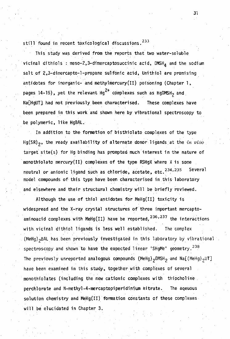
This study was derived from the reports that two water-soluble
vicinal dithiols : meso-2,3-dimercaptosuccinic acid, DMSH 4 and the sodium
salt of 2,3-dimercapto-1-propane sulfonic acid, Unithiol are promising
antidotes for inorganic- and methylmercury(II) poisoning (Chapter 1,
pages 14-15), yet the relevant Hg 2+ complexes such as HgDMSH 2 and
Na[HgUl] had not previously been characterised. These complexes have
been prepared in this work and shown here by vibrational spectroscopy to
be polymeric, like HgBAL.
In addition to the formation of bisthiolato complexes of the type
Hg(SR) 2 , the ready availability of alternate donor ligands at the in vivo
target site(s) for Hg binding has prompted much interest in the nature of
monothiolato mercury(n) complexes of the type RSHgX where X is some
neutral or anionic ligand such as chloride, acetate, etc.234 ,235 Several.
model compounds of this type have been characterised in this laboratory
and elsewhere and their structural chemistry will be briefly reviewed.
Although the use of thiol antidotes for MeHg(II) toxicity is
widespread and the X-ray crystal structures of three important mercapto-
aminoacid complexes with MeHg(II) have be reported, 236 ' 237 the interactions
with vicinal dithiol ligands is less well established. The complex
(MeHg) 2BAL has been previously investigated in this laboratory by vibrational
spectroscopy and shown to have the expected linear 'SHgMe' geometry. 238
The previously unreported analogous compounds (MeHg) 2 DMSH 2 and Na[(MeHg) 2 UT]
have been examined in this study, together with complexes of several
monothiolates (including the new cationic complexes with thiocholine
perchlorate and N-methyl-4-mercaptopiperidinium nitrate. The aqeuous
solution chemistry and MeHg(II) formation constants of these complexes
will be elucidated in Chapter 3.
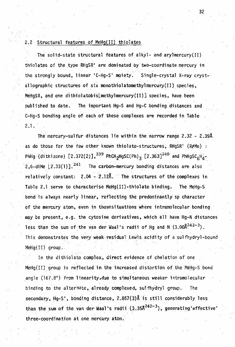
2.2 Structural features of MeHg(II) thiolates
The solid-state structural features of alkyl- and arylmercury(II)
thiolates of the type RHgSR' are dominated by two-coordinate mercury in
the strongly bound, linear 'C-Hg-S' moiety. Single-crystal X-ray cryst-
allographic structures of six monothiolatomethylmercury(II) species,
MeHgSR, and one dithiolatobis[methylmercury(II)] species, have been
published to date. The important Hg-S and Hg-C bonding distances and
C-Hg-S bonding angle of each of these complexes are recorded in Table
2.1.
The mercury-sulfur distances lie within the narrow range 2.32 - 2.39A
as do those for the few other known thiolato-structures, RHgSR' (RtMe)
PhHg (dithizone) [2.372(2)], 239 PhCH2HgSC(Ph) 3 [2.363] 240 and PhHgSC6H4-
2,6-diMe L2.33(1)].241 The carbon-mercury bonding distances are also
relatively constant: 2.04 - 2.12A. The structures of the complexes in
Table 2.1 serve to characterise MeHg(II)-thiolate binding. The MeHg-S
bond is always nearly linear, reflecting the predominantly sp character
of the mercury atom even in thosesituations where intramolecular bonding
may be present, e.g. the cytosine derivatives, which all have Hg-N distances
less than the sum of the van der Waal's radii of Hg and N (3.00A 242-3 ).
This demonstrates the very weak residual Lewis acidity of a sulfhydryl-bound
MeHg(II) group.
In the dithiolato complex, direct evidence of chelation of one
MeHg(II) group is reflected in the increased distortion of the MeHg-S bond
angle (167.8°) from linearity,due to simultaneous weaker intramolecular
binding to the alternate, already complexed, sulfhydryl group. The
secondary, Hg-S', bonding distance, 2.857(3)A is still considerably less
than the sum of the van der Waal's radii (3.35A 242-3 ), generating'effective'
three-coordination at one mercury atom.
32

Complex
[MeNgSCH2CH(AH3 )C 2-].H 20 b
LMeHgSC(Me) 2CH(41-1 3)CO2- ] 2 .H 20
[MeHgSC(Me) 2CH(AH 2HgMe)CO2- J a
NH 2
,N )1, unmo h
Oe N • JL, HgMe
S--
N
Me
SHgMe
.Hg-S/A C-Hg/A C-Hg-S/ ° Ref
• 2.352(1) 2.10(4) 177.6(9) 236
2.38(1),2.36(1) 2.07(6),2.09(5) 175(2),175(2) 237
2.35(1) 2.04(4),2.07(4) 176(1) • 237
2.39(2) 2.13(6) 174 244
2.393(4) 2.09(1) 178.6(4) 245
2.390(6) 2.09(2) 178.6(9) 245
2.363(4),2.367(4) 2.08(2),2.12(2) 167.8(5),117.1(5) 230
f
Table 2.1: Mercury bonding distances and angles from the published crystal structures of MeHgSR complexes.

0( H
Footnotes to Table 2.1
the mercaptoamino acid complexes have been reviewed by Carty 246
Figure 2.1
Figure 2.2. Two independent molecules in the unit cell
preliminary structrue in ref. 247
Figure 2.3. Bonding distance Hg-N 2.13(3)A.
preliminary structure in ref. 248
weak bonding distance Hg...N 2.83(3)A
weak bonding distance Hg...N 2.80(2)A
0 weak bonding distance Hg...N 2.95(2)A
weak bonding distance Hg...S' 2.857(3)
34
Figure 2. : Molecular structure of Vethyl-L-cysteinatomercury(II)
from reference 236.
C( b)
(a)
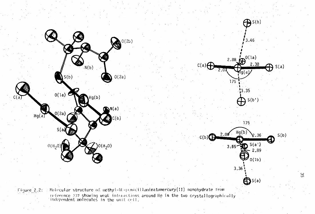
Finure 2.2: . Molecular structure of. methyl T HL-peniCii . laminatomertury(II) monohydrate from
rci'erence 237 showing weak inti:roctinns . around Hg in the two crystallographically independent nnlecules in the unit cell:
s
3.36 1 s
C( 2.88 Ash
v4P
I g
175 Is
"3.35
(9 s(b.)
2.33 S(a)
176
0( 1 )
• 2.85
170
C(2)
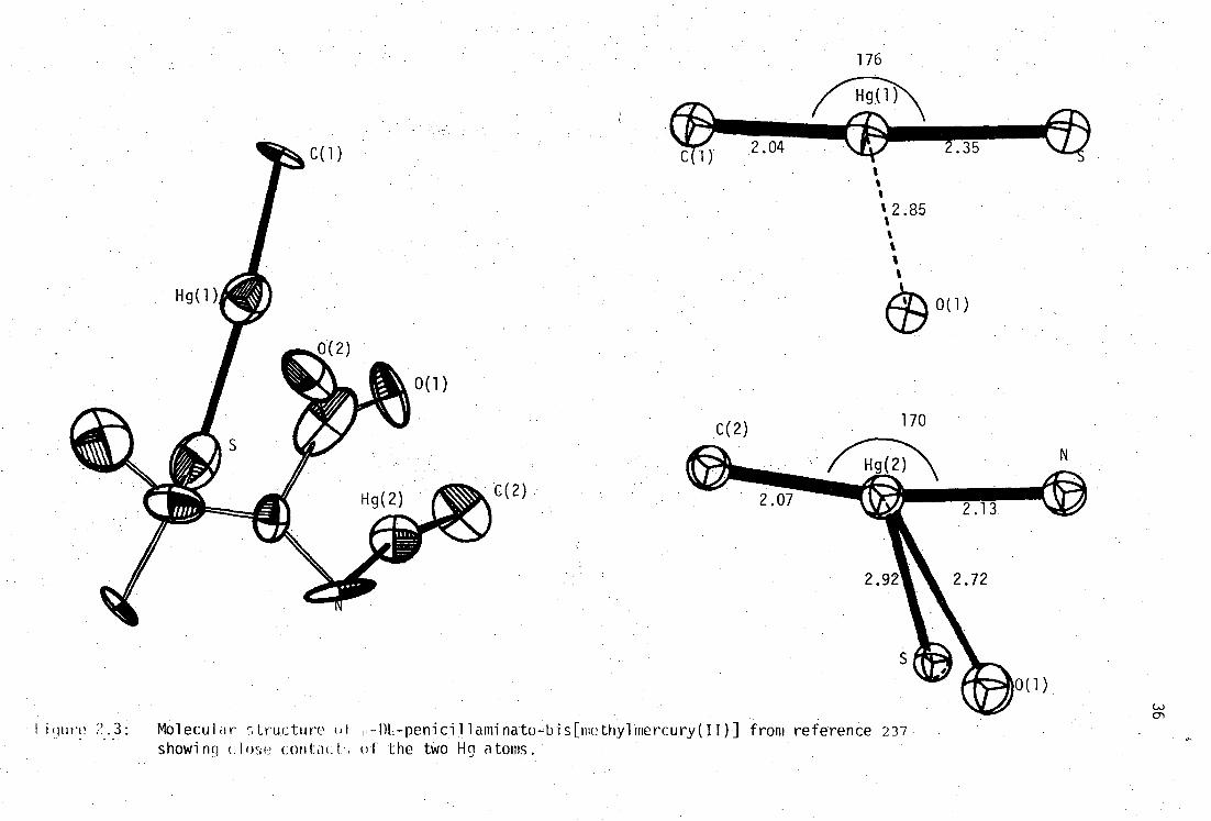
I i (lure Mol ecul a r s tructure of H - DL - peflitil1aminato - bis[methyl mercury( )] from reference 237 showing clw-ie con. tacL of the two Hg atoms.
37
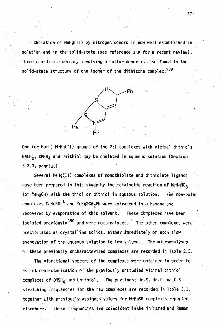
Chelation of MeHg(II) by nitrogen donors is now well established in
solution and in the solid-state (see reference 249 for a recent review).
Three coordinate mercury involving a sulfur donor is also found in the
solid-state structure of one isomer of the dithizone complex: 239
One (or both) MeHg(JI) groups of the 2:1 complexes with vicinal dithiols
BALH 2' DMSH4 and Unithiol may be chelated in aqueous solution (Section
3.3.3, page136).
Several MeHg(II) complexes of mOnothiolate and dithiolate ligands
have been prepared in this study by the metathetic reaction of MeHgNO 3 .
(or MeHg0H) with the thiol or dithiol in aqueous solution. The non-polar
complexes MeHgBu t and MeHgSCH2Ph were extracted into hexane and
recovered by evaporation of this solvent. These complexes have been
isolated previOusly250.and were not analysed. The other complexes were
precipitated as crystalline solids, either immediately or upon slow
evaporation of the aqueous solution to low volume. The microanalyses
of these previously uncharacterised complexes are recorded in Table 2.2.
The vibrational spectra of the complexes were obtained in order to
assist characterisation of the previously unstudied vicinal dithiol
complexes of DMSH 4 and UnithioL The pertinent Hg-S, Hg-C and C-S
stretching frequencies for the new complexes are recorded in Table 2.3,
together with previously assigned values for MeHgSR complexes reported
elsewhere. These frequencies are coincident inthe infrared and Raman

Complex
MeHgSCH 2CO2H
[MeHgSCH CH 2NMe 3]C104
[MeHgSC 5H 10NHMe]NO3 a
MeHgSCH(CO2H)CH 2CO 2H
MeHgSCHCO2H 1
MeHgSCHCO 2H
MeNgSCH 2
1 MeHgSCHCH SO Na
38
required % found %
Ng H. Hg
11.8 1.97 65.4 10.5 11.6 2.01 65.1 10.1
16.6 3.71 46.2 7.4 16.7 3.76 45.9 7.6
20.6 3.94 49.1 7.8 20.6 3.91 49.0 7.6
16.5 2.21 55.0 8.8 16.8 2.43 55.1 8.7
11.8 1.65 65.6 10.5 12.0 1.74 65.5 10.2
9.4 1.73 62.7 15.0 9.4 1.87 1.48 62.4
Table 2.2: Previously uncharacterised MeNg(II) thiolates and dithiolates
a monothiol is 4-mercapto-N-methylpiperidine
spectra, consistent with the absence of a centre-of-symmetry in these
molecular solids. The most readily assigned vibrational band is due to
v(HgC) and is very sharp and intense in the Raman spectra, and almost
invariant in frequency for these complexes (528-555 cm -1 ) and other
methylmercury(II) complexest , e.g. Me 2Ng (v s 515, vas550) 255 ; MeHgX,
X=F (561-573), 256 Cl (539-558) 257-63 , Br(538-545)257-8 ,262-4,
1(526-
538 )257-8,260-3 ; MeHgC(SiMe3 ) 3 (523-528) 265 ; MeHgCN(559-565) 266 and
MeHgSCN(543-562) 267-8 .
The expected mercury-sulfur coordination is confirmed by the absence
of vSH near 2500 cm. This vibration is weak in the infrared spectra
but very intense and characteristic in the Raman spectra of the thiol
The values in parenteses represent the range reported by various authors in both i.r. and Raman spectra and are often solvent and phase dependent.

Complex rm-1 •
vHgC/' vcsicm-1 Ref.
monothiolate ligands
MeHgSMe 333m [329vs] 533 [537] 692 [700] 251
329m [327w] 522 [533] 698 [697]. 252
MeHgSBu t 383w [390m] 534m. [536vs] 582m [586s]
MeHgSPh 382m [382m] 536m[547vs] 692 [698] 253
MeHgSCH 2P 346m[342t] 538m [540vs] / 700s [702w]
.'680W 1684m]
MeHgSCH 2 C H (334m '318msh [329s]
1 550vw,br '529w [538vs]
654m [676m]. 627m .
[MeHgSCH 2CH(h13 )CO2-]H20 325m [326vs] 538m [538vs] not reported 236
[MeHgSC(Me) 2CH(hH 3)CO2- ] 2H 20 322vw,sh [322m] 555m [555m] not reported 254
[MeHgSCH 2CH 2 iMe3]C104- 329s [331s] 542m [542s] 621vs [621w]
MeHgSCH(CO2H)CH262H f 361m [362m) 539m [550vs] 673m [677w]
371 [373m] 543w 719w1718W]
[MeHgSC 5 11 10hHMe]NO3 348 [353m] 533m [539s]
748w [750sj
(Table 2.3 continued over..

dithiolate ligands
3,4-di(MeHgS)C6 H 3-Me
MeNgSCH 2
MeHgSCHCH OH, (MeHg) 2BAL
MeHgSCHCO 2H
MeHgSCH 2CO2H, (MeHg) DMSH 2
MeNgSCH 2
MeHgSCHCH SO3Na
MeHgSCH,
I CH(OH)
MeNgS-CH 2
354w [370&357m]g
549w [553vw,sh] 335w [338m] 528w 1.526vsj
328m.br [328s,br] 530m [537vs]
537m [535vw,sh] 352s [358m] 550w,sh [550s]
526m [531s]
341s t342vs] 543s [537vs]
685w.[690m
662vW [656vw,sh] 238
686m 1675m]
660w 1655vw] 586m [585vw]
684m [687w] 238 MeHg) 2DMP
, Na[(M0Hg)2UT] 333w,br [329m,br]
Table 2.3: Hg-S, Hg-C and C-S stretching frequencies for MONg(II) thiolates and dithiolatesa
athis work unless otherwise stated. Raman values are shown in parentheses. btoluene-3,4-dithiol
cN-methy1-4-mercaptopiperidine d1,3-dimercapto-2-propanol e in D20 (PD5.6)vHgS [342m], v cHg[546s]
fin 020 pD=5.6 vHgs [365m] vHgc[547vs] gligand has 364w hpoor quality far infrared spectrum.
41
ligands. The mercury-sulfur stretching frequencies of the monothiolato
complexes shown in :Fable 2.3 also fall within a fairly narrow range,
329-390 cm-1 . The band due to this vibration is absent in the spectra
of the thiol ligands but is usually of medium to strong intensity in
both i.r. and Raman spectra of the complexes. The centrosymmetric
sulfide compex (MeHg) 2S also has1 vHgs near this range [v as 344, vs 300]. 269
The carbon-sulfur stretching frequency is more difficult to assign
in these complexes, as there are usually several weak to medium intensity
ligand vibrations in the region 600-700 cm -1 . The values of "es reported
in Table 2.2 have been assigned by comparison with previously published
values and are probably moderately coupled to other ligand vibrations,
giving rise to variable intensities.
The vibrational features of the. 2:1 MeRg(II) complexes with
•dithiolate ligands are also shown in Table 2.3 and can be readily
compared with those of monothiolato complexes. In the previously
reported 2:1 complexes of vicinal BALH 2 and its non-vicinal analog DMPH 2 ,
values for vHgc , vHgs and vcs are all consistent with simple linear
'C-HgS' coordination. 238 The 2:1 complex with toluene-3,4-dithiol has
• been previously prepared 230 in conjunction with that of the chelating
dithiol, trans-1,2-dimercaptocyclohexane (Figure 2.4), but no spectroscopic
details are available. The vibrational features of the toluene-3,4-dithiol
complex and those of the complexes with antidotal DMSH4 and Unithiol are
also consistent with simple linear 'C-Hg-S' coordination to form structures
of the type:
H 3C
H 3C
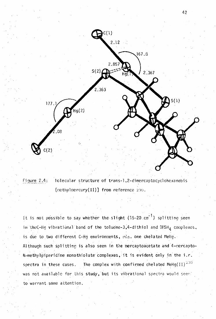
42
Finure 2. _ holecular structure of trans-1,2-dimercaptoCyclohexanebjs
[methylmertury(II)] from reference 2 -30,
It is not possible to say whether the slight (15-20 cm -1 ) splitting seen
in Ihec-Hg vibrational band of the toluene-3,4-dithiol and DrISH 4 complexes,
is due to two different C4in environments, vin, one chelated MeH(L
Although Such Splittinc is also seen in the Mercaptoacetate and 4-mercapto-
N-methylpiperidine monothiolate complexes, it is evident only in the i.r.
spectra in these cases. The complex with confirmed chelated MeHg(II)
was not available for this study, but its vibrational spectra would seem
to warrant some attention.
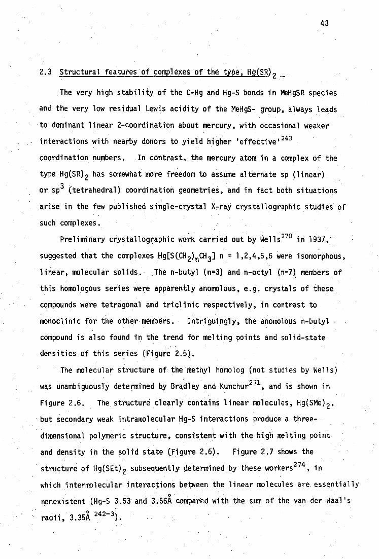
43
2.3 Structural features of complexes of the type, Hg(SR) 2
The very high stability of the C-Hg and Hg-S bonds in MeHgSR species
and the very low residual Lewis acidity of the MeHgS- group, always leads
•to dominant linear 2-coordination about mercury, with occasional weaker
interactions with nearby donors to yield higher 'effective' 243
coordination numbers. In contrast, the mercury atom in a complex of the
type Hg(SR) 2 has somewhat more freedom to assume alternate sp (linear)
or sp3 (tetrahedral) coordination geometries, and in fact both situations
arise in the few published single-crystal X-ray crystallographic studies of
such complexes.
Preliminary crystallographic work carried out by Wells 270 in 1937,
suggested that the complexes Hg[S(CH 2 ) nCH 3] n = 1,2,4,5,6 were isomorphous,
linear, molecular solids. The n-butyl (n=3) and n-octyl (n=7) members of
this homologous series were apparently anomolous, e.g. crystals of these
compounds were tetragonal and triclinic respectively, in contrast to
monoclinic for the other members. Intriguingly, the anomolous n-butyl
compound is also found in the trend for melting points and solid-state
densities of this series (Figure 2.5).
The molecular structure of the methyl homolog (not studies by Wells)
was unambiguously determined by Bradley and Kunchur 271 , and is shown in
Figure 2.6. The structure clearly contains linear molecules, Hg(SMe) 2 ,
but secondary weak intramolecular Hg-S interactions produce a three-
dimensional polymeric structure, consistent with the high melting point
• and density in the solid state (Figure 2.6). Figure 2.7 shows the
structure of Hg(SEt) 2 subsequently determined by these workers 274 , in
which intermolecular interactions between the linear molecules are essentially
nonexistent (Hg-S 3.53 and 3.56R compared with the sum of the van der Waal's
radii, 3.354 242-3)•
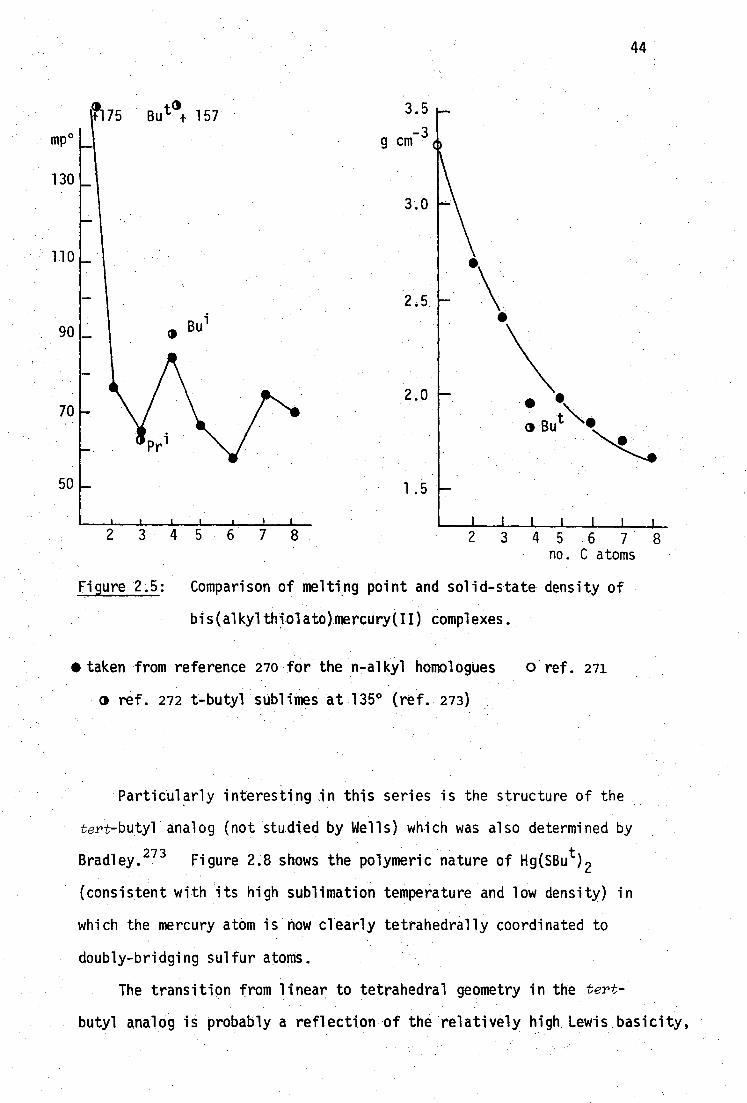
11'175Bu tt) 4. 157 3.5
-3 g cm
mp o
130
110
90
70
50
5 6 7 no. C atoms
44
234
Figure 2.5: Comparison of melting point and solid-state density of
bis(alkylthiolato)mercury(II) complexes.
*taken from reference 270 for the n-alkyl homologues 0 ref. 271
0 ref. 272 t-butyl sublimes at 135° (ref. 273)
Particularly interesting in this series is the structure of the
tert-butyl analog (not studied by Wells) which was also determined by
Bradley. 273 Figure 2.8 shows the polymeric nature of Hg(SBu t ) 2
(consistent with its high sublimation temperature and low density) in
which the mercury atom is now clearly tetrahedrally coordinated to
doubly-bridging sulfur atoms.
The transition from linear to tetrahedral geometry in the tert-
butyl analog is probably a reflection of the relatively high Lewis basicity,

45
0 c Os • Hg
Figure 2.6: Molecular structure of Hg(SMe) 2 from reference 271
Figure 2.7: Molecular structure of Hg(SEt) 2 from reference 274
Figure 2.8: Molecular structure of Hg(SBu t ) 2 from reference 273

163 S r":0.32
S'<33 N \
46
hence high bridging ability, of the tert-butyl sulfur, but may also be
influenced by steric effects. The complex of Hg2+
with 8-mercapto-
quinoline (Figure 2.9) apparently has nearly linear 'HgS 2 ' geometry
with strong Hg-N interactions. 275
Figure 2.9: Molecular structure of bis(8-mercaptoquinolinato)mercury(II)
Details of the structure are unavailable in Tasmania and one abstracted
distance, 275 Hg-N 1.36-1.50A is surely incorrect (the covalent radii of
digonal and tetrahedral Hg(II) are 1.30A and 1.48A. respectively243 ).
Dichlorobis(6-mercaptopurine)mercury(II) consists of discrete
monomeric molecules with distorted tetrahedral 'HgS 2C1 2 geometry about
mercury276
(Figure 2.10).
Interestingly, chelation of Hg by S and N 7 is not observed due to
the "preference (of Hg) for chlorine over nitrogen donors." 276 Although
formally a thiol, the ligand has appreciable thione character which must
assist in increasing the residual Lewis acidity of the 'HgS 2 ' moiety and
thus favour tetrahedral over linear coordination.

• Figure 2.10: Molecular structure of dichlorobis 6-mercaptopurine)
mercury(II), from reference 276.
The C-S bond retains an appreciable double-bond character in the
complex but this is not directly reflected in the Hg-S bond distance 0
(2.48A), e.g. Hg(SMe) 2 has Hg-S 2.36A but Hg(SEt) 2 has Hg-S 2.454.
The only other crystallographically studied bisthiolato[mercury(II)]
complexes, of which this author is aware, are those formed between HgC1 2
and the biologically important thiols, L-cysteine 277 '246 a
nd DL-penicill-
amine278 ' 246. The complex Hg[SCH 2CH(NH3 )CO23[SCH 2CH(NH 3 )CO2H]Cl.k.H 20 has
nearly linear 'HgS 2 ' geometry and ionic chloride (Figure 2.11). As Carty
points out,246
this species has only one extra proton than "mercury
cysteinate", Hg[SCH 2CN(NH3 ) 02] 2 , which is a metabolic product of certain
mercurial diuretics. 234 •
The closely related penicillamine complex Hg{[SC(Me) 2CH(N11 3 )CO 2H] 2C1}
Cl.H 2 0 contains one additional proton (and hence one additional chloride for
electroneutrality) and has a similar structure, but the linear molecules are
47

Figure 2.11: Molecular structure of Hg[SCH 2CH(NH 2 )CO2][SCH 2CH(NH3 )CO 2H]
ClAH20, from reference 277. The penicillamine complex
is similar, but the chloride shown here bridges two
molecules (Hg-C1 2.85A) 278
linked into spirals by the new (bridging) chloride.
The complex Hg(SCH 2CO 2H) 2 , I, was precipitated as glistening plates
by the addition of HgC1 2 or Hg(CN) 2 to two equivalents of mercaptoacetic
acid (thioglycolic acid) in water.
HgX 2 + 2HSCH2CO2H Hg(SCH 2CO2H) 2 2HX
Although this compound has been known for many years, 278 it does not seem
to have been characterised in the solid state, and so its microanalysis
is recorded below (Table 2.4). It is of interest in this study as a
model for the complex with DMSH 4 , and also for the bis(selenolato)mercury(II)
analog ( page 186).
Dissolution of I in two equivalents of aqueous KOH and evaportion of
the solution under nitrogen, produced K2[Hg(5CH2CO2 ) 2] as a white powder.
48
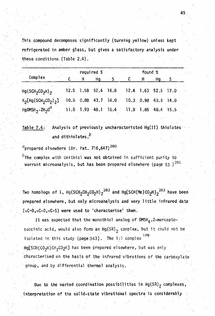
This compound decomposes significantly (turning yellow) unless kept
refrigerated in amber glass, but gives a satisfactory analysis under
these conditions (Table 2.4).
Complex C
required %
H Hg S C
found %
H Hg S
Hg(SCH 2CO 2H) 2 12.5 1.58 52.4 16.8 12.4 1.63 52.5 17.0
K2 [Hg(SCH 2CO2 ) 2] 10.5 0.88 43.7 14.0 10.3 0.98 43.9 14.0
HgDMSH 2 .2H 20a
11.5 1.93 48.1 15.4 11.9 1.85 48.4 15.5
Table 2.4: Analysis of previously uncharacteristed Hg(II) thiolates
and dithiolates.b
aprepared elsewhere (Br. Pat. 716,647) 280
bThe complex with Unithiol was not obtained in sufficient purity to
warrant microanalysis, but has been prepared elsewhere (page 53 ) 281
Two homologs of I, Hg(SCH 2CH2CO2H) 2 282 and Hg[SCH(Me)CO2H] 2283 have been
prepared elsewhere, but only microanalysis and very little infrared data
(vC=0,vC-0,vC-S) were used to 'characterise' them.
It was expected that the monothiol analog of DMSH 4 ,2-mercapto-
succinic acid, would also form an Hg(SR) 2 complex, but it could not be
isolated in this study (page343). The 1:1 complex
Hg[SCH(CO 2H)CH 2CO2H] has been prepared elsewhere, but was only
characterised on the basis of the infrared vibrations of the carboxylate
group, and by differential thermal analysis.
49
Due to the varied coordination possibilities in Hg(SR) 2 complexes,
interpretation of the solid-state vibrational spectra is considerably
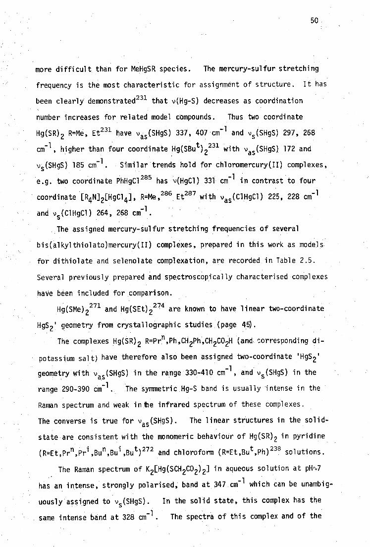
50
more difficult than for MeHgSR species. The mercury-sulfur stretching
frequency is the most characteristic for assignment of structure. It has
been clearly demonstrated231 that v(Hg-S) decreases as coordination
number increases for related model compounds. Thus two coordinate
Hg(SR) 2 R=Me, Et231 have v as (SHgS) 337, 407 cm v s (SHgS) 297, 268
cm-1 , higher than four coordinate Hg(SBu t ) 2231 with vas (SHgS) 172 and
vs(SHgS) 186 Cm-1 . Similar trends hold for chloromercury(II) complexes,
e.g. two coordinate PhHgC1 285 has v(HgC1) 331 cm contrast to four
coordinate [R4N] 2 [HgC1 4], R=Me, 286 Et287 With Vas (C1HgC1) 225, 228 cm71
and v 5 (C1HgC1) 264, 268 cm.
The assigned mercury-sulfur stretching frequencies of several
bis(alkylthiolato)mercury(II) Complexes, prepared in this work as models
for dithiolate and selenolate complexation, are recorded in Table 2.5.
Several previously prepared and spectroscopically characterised complexes
have been included for comparison.
Hg(SMe) .2 2 7 1 and Hg(SE02 274 are known to have linear two-coordinate
HgS 2 ' geometry from crystallographic studies (page 45).
The complexes Hg(SR) 2 R=Prn,Ph,CH2Ph,CH 2CO2H (and corresponding di-
potassium salt) have therefore also been assigned two-coordinate 'HgS 2 . 1
-1 geometry with v as (SHgS) in the range 330-410 cm, and vs (SHgS) in the
range 290-390 cm -1 . the 'symmetric Hg-S band is usually intense in the
Raman spectrum and weak in lhe infrared spectrum of these complexes.
The converse is true for vas (SHgS). The linear structures in the solid-
state are consistent with the monomeric behaviour of Hg(SR) 2 in pyridine
(R=Et,Prn ,Pr i ,Bu n ,Bu i ,Bu t ) 272 and chloroform (R=Et,Bu t ,Ph) 238 solutions.
The Raman spectrum of K2[Hg(SCH 2CO2) 2] in aqueous solution at pli ,k,7
has an intense, strongly polarised, band at 347 cm can be unambig-
uously assigned to v s (SHgS). In the solid state, this complex has the
sate intense band at 328 cm - . The Spectra of this complex and of the

51
Complex
v s (SHgS)
vs (SHgS)
Ref .b
monothiolates
Hg(SMe) 2
Hg(SEt) 2 d
Hg(SPrn ) 2
Hg(SPr 1 ) 2 f
Hg(SBu r1 ) 2 g
Hg(SBu 1 ) 2 h
Hg(SBu t. ) 2
Hg(SPh) 2 ,
'Hg(SCH 2Ph) 2
Hg(SCH 2CO 2H) 2
K 2 [Hg(SCH 2CO2 ) 2]
338vs [338w] 298w [298m] 272 337vs [338vw] 296m [297vs] 231
405s,b [394vs] 231
408vs [412w] 392sh [395vs] 272
assignment uncertain 272
assignment uncertain 272
assignment uncertain 272
172vs,b [188vs] 231
365s,br [344s]
238
381s [385vw,sh] 352w [356s]
379s [370w,sh] 351w L350s] k
355s [360vv] 321w [328vs]
dithiolates
HgBAL
HgDMP
HgDMSH 2 .2H20
Na[HglIT]
,1,348m,vbr
353m,b
389m
,v360w,sh
[298s,vb]
[325vs]
306w
[316s,bj
238
238
Table 2.5: Mercury-sulfur stretching frequenciesa for mercury(II)
thiolate and dithiolate complexes.
Footnotes to Table 2.5
aRaman bands are shown in parentheses [ ]. Abbreviations: v, very; s, strong; m, medium; w, weak; b, broad; sh, shoulder; p, polarised.
this work unless otherwise indicated. Assignments for R=Prn, Pr
i,
Bun, Bui have been made from the spectral bands recorded in ref. 272 and often differ from those made by Biscarini et al.
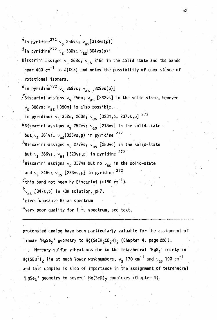
cin pyridinju v s 355vs; vas
[ 318vs(P)]
din pyridine 272 v s 330s; v as [304vs(p)]
Biscarini assigns v s 268s; vas 246s in the solid state and the bands
-1 near 400 cm to “CCS) and notes the possibility of coexistence of
rotattonal isomers.
e i-
n pyridine272 v
s 359vs; v
as 1.329vs(P).1
41scarini assigns v s 256m; vas [232vs] in the solid-state, however
vs
388vs; vas
[380m] is also possible.
in pyridine: v s 352m, 260m; v
as [323m,p, 237vs,p]
272
gBiscarini assigns v s 252vs; v as [218vs] in the solid-state
but vs 361vs, vas (325vs,p) in pyridine 272
Biscarini •assigns vs 277vs; vas
[250vs] in the solid-state
but vs 366vs; vas [323vs,p] in pyridine 272
iBiscarini assigns v 33•vs but no vas
in the solid-state
and vs 246s; vas [233vs,p] in pyridine 272
° this band not been by Biscarini (>180 am-1
kvas
[347s,p] in KOH solution, pH7.
gives unusable Raman spectrum
mvery poor quality for i.r. spectrum see text.
protonated analog have been particularly valuable for the assignment of
linear 'HgSe2 ' geometry to Hg(SeCH 2CO2H) 2 (Chapter 4, page 220).
Mercury-sulfur vibrations due to the tetrahedral 'HgS 4 ' moiety in
-1 Hg(SBu t ) 2 lie at much lower wavenumbers, v s 170 cm
-1 and vas 190 cm
and this complex is also of importance in the assignment of tetrahedral
'HgSe4 ' geometry to several Hg(SeR) 2 complexes (Chapter 4).
52

53
2.4 Structural features of'Hg(II)Aithiolate complexes
The 1:1 dithiolato complex of DMSH4 is a dihydrate and the infrared
spectrum of this complex shows a broad absorption due to hydrogen bonded
OH (3200-3600 m,br). The complex with Unithiol was isolated as the
sodium salt from acidic solution. This product was consistently
produced as a somewhat sticky solid which could not be dried (even by
high vacuum pumping or storage over P 205 ) to an adequate state to warrant
microanalysis. The near infrared spectrum of this product also showed
broad OH absorption (3200-3600 w) and was generally of poorer quality than
the other complexes studied. Both dithiolate complexes have very broad,
low intensity, far infrared spectra (600-200 cm -1 ). Only two bands are
discernible in the far i.r. spectra of HgDMSH 2 .2H 20 : 389m, 306w and
these absorptions do not appear in the spectrum of the ligand and have
thus been assigned as v as (SHgS) and v s (SHgS) respectively.
All attempts to obtain a usable Raman spectrum from this solid were
fruitless, even under conditions of low energy (red dye) laser excitation
which did not seem to lead to sample decomposition. HgBAL has been
shown to be amorphous and also gives relatively poor quality, diffuse
spectra. 231
The Unithiol complex Na[Hg(UT)].2H 20
gives a poor quality far infrared spectrum, with a very broad medium
intensity absorption [600-250 cm] containing poorly resolved features
at 580m, 540w, 360w,sh [v as (SHgS)]. The Raman spectrum of this complex
is featureless in the range 600-100 cm -1 except for a strong, broad band
at 316 cm-1 assigned to v s (SHgS).
The infrared spectrum of HgDMSH 2 .2H20 and the infrared and Raman
spectra of Na[Hg(UT)] are consistent with the linear polymeric structure
postulated for HgBAL:231
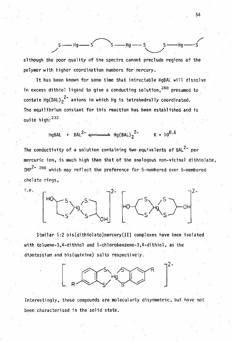
—2
HO OH
S—Hg— S S — Hg — S S—Hg—S
although the poor quality of the spectra cannot preclude regions of the
polymer with higher coordination numbers for mercury.
It has been known for some time that intractable HgBAL will dissolve
in excess dithiol ligand to give a conducting solution ,288 presumed to
contain Hg(BAL) 22-
anions in which Hg is tetrahedrally coordinated.
The equilibrium constant for this reaction has been established and is
quite high: 232
HgBAL + BAL 2- Hg(BAL) 2 2- K = 108 ' 6
The conductivity of a solution containing two equivalents of BAL 2- per
mercuric ion, is much high than that of the analogous non-vicinal dithiolate,
DMP 2- 288 which may reflect the preference for 5-membered over 6-membered
chelate rings,
S /S
/19 S SOH
i .e.
Similar. 1:2 bis(dithiolato)mercury(II) complexes have been isolated
with toluene-3,4-dithiol and 1-chlorobenzene-3,4-dithiol, as the
dipotassium and bis(quinine) salts respectively.
—2- S\
1-19 / •
Interestingly, these compounds are molecularly disymmetric, but have not
been characterised in the solid state.

2.5 Structural features of RSHgX species
Many bis(alkylthiolato)mercury(II) complexes will dissolve in
solutions of HgX 2 , X=C1,Br,I, acetate to form complexes of the type
RSHgX via the equilibria
Hg(SR) 2 + HgX2 , 2RSHgX
The equilibria for R = CF3 ; X=C1,Br,I, have been followed by vibrational
spectroscopy290.
Such species may be of considerable importance in the
-biological context, since many endogenous ligands, e.g. nitrogen containing
bases, chloride etc. will be present at the Hg-sulfhydryl binding site(s)
in vivo. Most 1:1 Hg(II) thiolates which could act as models for the
biological interaction of RSHg(II) with non-thiol ligands, are insoluble
in water and common organic solvents, but several dissolve in coordinating
solvents such as pyridiA9e34 imidazole 291 to form adducts, some of which
have also been crystallographically characterised.
MeSHg02CMe is uniquely water-soluble and has been shown by Raman
spectroscopy to form a diner in solution near pH6 292 .
Although no new RSHgX species have been prepared in this study, the
first RSeHgX analogs to be characterised by single-crystal X-ray diffraction
are described in Chapter 4. For this reason, a brief review of the
structural features of RSHgX complexes is included here.
All RSHg02CMe complexes studied crystallographically to date (R=Me; 293
Pro , Bun. 294
) consist of polymeric (-Hg-SR) n parallel chains which are
linked by acetate groups to form wrinkled sheets. The structure of the
methyl analog is identical to that of isomorphous MeSeHg0 2CMe shown in
Figure 2.12. In the N-base adducts RSHg0 2CMe(py) R=Me293
,Et295 and
MeSHg02CMe(4Mepy)295
, coordination of pyridine or 4-methylpyridtne results
in displacement of bridging acetate groups to give isolated chains with
55

R S
0 // I 0
56
s 'Hg Hg /1
Figure 2.12: Schematic structure of polymeric RSHg0 2CMe;
R=Me 293 , Prn and Bun 291
PY S 00
Hg
\O SR PY
SR
Figure 2.13: Schematic structure of the pyridine adducts of
RSHg02CMe; R=Me293
, Et295
and the 4-methylpyridine
adduct of MeSHg0 2CMe 295 .

57
N-base and bidentate acetate coordination at each mercury atom.
. The complexes MeSHgX (X=C1
, 296Br295) are isostructural and are
based on (Hg-SMe) n chains linked by triply bridging halogen atoms
(coordinated to two mercury atoms in one chain and one mercury atom in
an adjacent chain) to form wrinkled sheets.
I I 1 1 I v 1 S I ' \ : S I
'M H /MeHg' 1 gNle,, Hg 7
_ / :
N X / 1 S I 1 1X
X
N , Hg Me / Hg 1 1 N
Hg Mez 1
x S 1 1 1
Figure 2.14: Schematic structure of MeSHgX (X=C1,296
Br295
) taken
from reference 297.
Other crystallographically characterised RSHgX (X=halide) complexes
which contain (-Hg-SR) n chains are those formed between HgC1 2and cysteine 277
or penicillamine ; 278 and between HgB 2 and the thiosteroid spiroxazone 298 .
The molecular structure of HgC1 2[SCH 2CH(NH3 )CO 2H] is shown in
Figure 2.15a, and consists of chains of -(-Hg-SCys) n groups in which
the Hg atom is tetrahedrally coordinated with 'Hg(p-SCys)C1 2 1 geometry.
The penicillamine complex, (HgC1 2 ) 2 [SC(Me) 2 CH(NH 3 )CO 2H.J.2H 20 has a more
complex structure (Figure 2.15b) in which the sulfhydryl donor asymmetrically
bridges two mercury atoms. The mercury atoms have distorted tetrahedral
geometry of the types 'Hg(u-SPen)C1 3 1 and 'Hg(p-SPen)C1(C1 1 ) 2 1 . In the
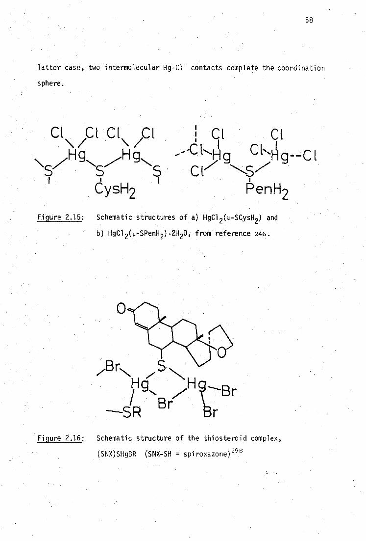
Br
zgrN /S N Hg H
.13.rr° SR
58
latter case, two intermolecular Hg-C1' contacts complete the coordination
sphere.
CE c i c t C E 1 CL Cl \ / \/ ' I CI% I
Hg Hg - -C 1..F.i g -Hg--CI
\ 7 N ,7- CIV 57 S S S
I ysH2 1 l'enH2
Figure 2.15: Schematic structures of a) HgC1 2 (11-SCysH 2 ) and
b) HgC1 2 (p-SPenH 2 ).2H 20, from reference 246.
Figure 2.16: Schematic structure of the thiosteroid complex,
(SNX)SHgBR (SNX-SH = spiroxazone) 298

59
295 The isostructural complexes [Bu tSHgCl(py) 0.51943 and [Bu tSHgC1(4-Mepy) 0.5 ] 4
differ from the other RSHgX complexes reported inasmuch as the tetratheric
molecules are based on ,eight-membered -(-Hg-SBu t ) 4 rings, rather than
infinite Chains_293 There are two mercury environments, 'Hg(p-SBu
t) 2C1N'
and Hg(1.-SBut )(p-C1) 2 '. A new RSeHgX compound, [ButSeHgCl(py) 0. 0 4 has
been shown in this study to be isomorphoUs with the 4 7Mepy adduct of the
sulfur analog, and the crystal structures of these compounds are discussed
in more detail in Chapter 4 of this thesis.
The vibrational spectra of polymeric RSHgX complexes are difficult to
interpret. Canty has recently noted that assignment of structure .based
cn simple point group approaches to the spectra of these molecules, may be
misleading. 297 For example, the spectra of MeSHgC1 were interpreted in -
terms Of a monomeric, dimeric or polymeric structure based on
'Hg(p-SMe) 2Hg' or 'Hg(p-C1) 2Hg' bridging units,2 99 however the previously
described single-crystal X-ray determination 29 (page 57 ) is not
consistent with this interpretation.
Assignment of mercury-ligand vibrational frequencies is not straight-
forward for the RSHg(halides) as mercury-sulfur and mercury-halogen
vibrations occur at similar frequencies in related complexes, e.g. HgC1 2 : 1 300
'as 375 85-368 300 , vs 330-310 cm_ ', linear Hg(SR)2: vas 330-410, v s
, 290-390 cm 51 ), and BU tSHgCl: i.r. 359m, 273s, 236m; Raman 391m,
384m,.359m; 330m, 274vs•, 223vs, 201Vs cm71 .3o1
2.6 Conclusions
The known crystallographic structures of MeHg(II)-thiolates have been
reviewed and several MeNgSR complexes characterised by vibrational
spectroscopy in the solid state. Coincident infrared and Raman bands
in the range 320-390 cm been assigned as Hg-S stretching modes,
60
-1 and bands in the range 520-560 cm to Hg-C modes, consistent with the
expected linear 'MeHgS' geometry. MeHg(II) dithiolates have similar
vibrational spectroscopic and structural features.
Bis(thiolato)mercury(II) compounds, Hg(SR) 2 have vibrational spectra
consistent with normally linear 'HgS 2 ' geometry
[vas(SHgS) 330-410 cm vs (SHgS) 290-390 cm-l j ,
except for Hg(SBu ) 2 which is known to have tetrahedrally coordinate
mercury [v s (SHgS) '070 cm vas (SHgS) r190 cm-1 ].
All Hg(II)-dithiolates studied by vibrational spectroscopy to date
seem to have polymeric, linear structures.
Monothiolatomercury(II) species, MeSHgX, have complex polymeric
structures based on (-Hg-SR) n infinite chains or eight-membered rings.
Interpretation of the vibrational spectra of these polymeric molecules
cannot readily be achieved usfng simple point-group approaches.
61
CHAPTER THREE
SOLUTION CHEMISTRY OF METHYLMERCURY(II) THIOLATES AND SELENOLATES
3.1 Introduction
Methylmercury has a high affinity for 'soft' sulfhydryl ligands,
consistent with the 'soft' acid nature of the metal and it is generally
considered that many of the toxicological properties of MeHg(II) are
12 due to bonding to sulfhydryl groups of peptides and proteins in vivo.
It is therefore somewhat surprising that only a small amount of detailed
stability constant information for methylmercury thiolates is available,
and that this includes some serious discrepancies in the magnitude of the
formation constants for MeHg(II) with several monothiols. 'Since
solution equilibrium constants may be of some significance in the
understanding of the effectiveness of antidotal thiols for removal of
MeHg(II) from body tissues, this study has involved redetermination of
the MeHg(II) formation constants with the biologically important mono-
thiols cysteine and glutathione, and the simple monothiols mercapto-
ethanol, mercaptoacetic acid, mercaptosuccinic acid, thiocholine and
4-mercapto-N-methylpiperidine. In addition, the complexation equilibria
with antidotal thiols penicillamine and N-acetylpenicillamine and vicinal
dithiols BALH 2 , Unithiol and DM51-I 4 have been investigated.
When this work was near completion, Reid and Rabenstein published
the results of their 1 11 nmr evaluations of MeHg(II) formation constants
with many of the monothiols mentioned above. 302 These workers have
apparently determined the formation constants with glutathione, but the
results were unpublished; 3°, 3 however, the formation constant of
MeHg(II) with the biological Monothiol homOcysteine was reported.
Attempts to investigate homocysteine in this study were unfortunately
62
frustrated by low purity of the commercially available product and rapid
decomposition in solution.
A brief report of part of the work described in this Chapter of the
thesis was presented at a conference prior to publication of the work by
Reid and Rabenstein (Appendix 1). Publication of their work produced
a unique opportunity to compare the results obtained from two widely
different techniques and firmly establish stability constants for the
MeHg(II) thiolate interaction.
The important features of the nmr technique, which has been used
elsewhere for studies of MeHg(II) equilibria with many non-thiolate
ligands, will be discussed below. The potentiometric method, used to
evaluate the stability constants in this study, will be described in
detail in Chapter 6 of this thesis, but the stability constants them-
selves will be discussed below.
The reported antagonistic effects of some selenium compounds against
methylmercury toxicity and the existence of reduced selenohydryl groups
in some selenoproteins (Chapter 1) has generated considerable interest
in methylmercury selenolate interactions in solution. Several nmr
studies have indicated that MeHg(II)-selenolate binding may be thermo-
dynamically more favorable than analogous thiolate binding, but no
stability constant data have been recorded. Attempts to study the
methylmercury-selenoacetate system potentiometrically in this work have
been frustrated by failure to obtain the free ligand in solution for
accurate evaluation of its hydrolysis constants.
3.2 The Aqueous Solution Chemistry of Methylmercury(II)
3.2.1 Coordination of MeHg(II) in aqueous solution
The solution chemistry of MeHg(II) has been recently reviewed, 41
and is dominated by the tendency of mercury toward linear two-coordination.

63
However, some residual Lewis acidity is evident from the formation of
complexes with chelating ligands in which higher coordination numbers
for MeHg(II) of three or four for mercury have been found. The
chemistry of MeHg(II) with these ligands has been reviewed elsewhere.249,304
In addition to polydentate ligands, some simple unidentate ligands also
305 305 permit higher coordination, e.g. iodide, cyanide and thi
268 ocyanate.
The possibility of higher coordination with sulfur donors was considered
for the interaction of MeHg(II) with thiols used In this study, parti-
cularly the antidotal vicinal dithiols BALH 2 , Unithiol, and DMSH4 .
Previous work in this laboratory has not indicated any definitive
evidence for chelation in the complex (MeHg) 2BAL in the solid state, but
a recent simple crystal X-ray structure230 of the complex trans-(l,2-
cyclohexanedithiolato)bis[methylmercury(II)] shows S-chelated methyl-
mercury (page 42 ). In solution, Schwarzenbach and Schellenberg305
have shown that mercaptoethanol will interact with MeHg(II) to form a
complex of the type MeHg(SR) 2- , as well as the highly stable monothiolate.
MeHgSCH 2CH 2OH + -SCH 2CH2OH MeHg(SCH 2CH 2OH) 2- K io627
The aqueous solution chemistry of MeHg(II) is also dominated by
the self-association and hydrolysis reactions:
MeHg(OH 2 ) 4- + MeHIPH + H20
MeHig. (OH 2 ) + + MeHg) 20H+ + H 20 K2 - [MeHg(OH 2 ) 41[MeHg0H]
[(MeHg) 0 4.]
[(MeHg) 20H / ]NeHg0H]
The first two hydrolysis equilibria were proposed by Schwarzenbach and
Schellenbere 5 from equilibrium constant measurements, 305 and have been
_ [MeHg0Hj N i
[MeHg(OH)2
+][OH1
[(MeHg) 20H + ]
(MeHg) 20H+ + MeHg0H
H 20

64
-8 supported by Raman
306 and 1 H nmr306 ' 309 spectral data.
Equilibrium constants for these equilibria have been measured here under the
conditions used for methylmercury-thiolate and thiol hydrolysis equilibria
(0.1 M KNO 3 , 25°). Several acidic (HNO 3) solutions of methylmercuric
ion were titrated with 0.1 M KOH, and the composite data (186 titration
points) treated by MINIQUAD81 (page 326) to generate equilibrium
constants P. 10-1
and p. 20-1
for the equilibria:t
a [MeHg0H] MeHg
+ + H 20 .-=---- MeHg0H + H
+ 10-1
[MeHg+][e] -1
[(MeHg) 0H+ ] 2MeHe + H 20c (MeHg) 20H 11 +
_ + +
2 20-1
[MeHg+]2[H+]-1
The actual titration data are shown in Figure 3.1, fitted by curves
calculated using these values of10-1 and - 201 ' A comparison of
P' B
-
these constants from this work, and those obtained elsewhere, is shown
c + in Table 3.1. It should be noted that, unless values for IS 01 = [H ][0H - ]
are accurately known under actual experimental conditions used to
determine 1310-1 and 13,20-1' these constants must be expressed in terms of
[W] and not [OH - ].
The trinuclear cation (MeHg)30
+ has been shown by
1H nmr spectros-
copy to be a very minor component in dilute solutions, since its
equilibrium constant for formation is small, 0.7±0.3.P °6 The trinuclear
cation has been isolated as perchlorate 310 or nitrate 311 salts from
concentrated neutral aqueous solutions, and single crystal X-ray
structures of these salts have been determined. In the dilute solutions
used in this study, [MeHg] l0 3M, (MeHg) 30+ constitutes, at most, 0.01%
of the total MeHg(II) concentration, hence the corresponding equilibrium
constant for its formation, 30 _ 2 , was unobtainable.
tFor brevity MeHg will be used subsequently to represent MeHg(OH 2 ) The computer program MIMQUAD81 will be discussed in Chapter 6.

9.2 -
7.6
6.0
4.4
2.8.-
pH a 16/1/80
initial mmoles
KOH/M
0.1085
vol./mL
59.96
MeHg(II)
0.1540
H+
0.04164
b 27/2/80 0.1549 0.04246 0.1089 59.96
c 8/1/80 0.1537 0.0408 0.1085 59.96
d 8/1/80 0.1542 0.5399 0.1085 64.98
e 11/10/79 0.8653 0.1706 0.09898 49.96
f 11/10/79 0.8656 0.6888 0.09898 54.96
g 12/10/79 0.1722 0.03340 0.09898 59.96
h 12/10/79 0.1728 0.5436 0.09898 64.95
65
-1.5
0 mMoles'e/mmoles MeHg(II)
1.5
Figure 3.1: Methylmercury(II) hydrolysis data.
The solid curves were calculated by COMIXH for the experimental conditions
shown, with log= -4.607 and log B 1310-1 . 20-1 = -
2.234.

[MeHg0H]
[(MeHg) 20H+]
[MeHg 4 ] 2 [H 4 ] -1
[MeHg0H]
[MeHg+][0H - ]
[(MeHg) 20H+ ]
log
1 log log
method conditions ref.
MeHg+][e] -/ [MeHg0H][MeHg
-4.607(9) b -2.234(18)b 9.25(3) d 2.37(4) d gl. h 25°[0.1 KNO 3 ] A513.86(3) this v" work
-2.22d -4.59c -2.536
9.370 2.37c gl.j 20°[0.1 KNO 3 ] p<=13.96 305
9.30c 2.37° -4.70e -2.33e 9.36(5)f 2.87(7)f nmrgij 25°[0.19 MeHg(II)] 309
9.29° 2.31 c nmrg' i 25°[0•3 MeHg(II)] 306
-4.56±0.05 gl.h 25°[0.1 KNO3 ] 312
-4.686(15) c -1.725(30) c 9.624(15)d 2.961(16) d gl. h 25°[0.1 NaNO 3] p<=13.95 313
-4.67(3) c' k gl. 25°[0.1 KNO3] 314
-4.50e gl. 24-5° 315
-4.40(2) c 9.55(7) d di str.h 25°[1.0 NaNO3/C1/PO4] 316
9.33±0.12c 2.78±0.18c nmrg33 25 ° [ 0.1 NaNO3] 317
cconstant actually reported in original reference. . dformation constant (page 63 ) calculated using the values of K w recorded in the original reference.
c e Auoted in references 313,316 'recalculated in reference 317 gl, not given. electrodes. calibrate in .terms of.[H 47
]. - hel
ectrodes calibrated by [11
+] =10
-pH. i
electrode calibration unspecified. iyii+ calculated using the Davies equation 318 and used to correct mixed constants.
Table 3.1: Methylmercury(II) hydrolysis constants. a
aestimated standard deviations of the last digit are recorded in parentheses. bweighted geometric means of eight values calculated by MINIQUAD81 from data shown in Figure 3.1.

[(MeHg) 20H+ ] 630-2 -
[MeH49 4 ]2 [H4 ] -
[MeHg0H]
1 •
[MeHe][0] -1 [(MeHg) 2OH I ][MeHg0H]
[(MeHg) 30 -1 ]
67
[(MeHg) 301 ] 3MeHg
+ + H 20 (MeHg) 30
+ + 2H
+ 630-2 -
[MeHg+ ] 3 [e] -2
This species was neglected in subsequent evaluations of MeHg(II)
equilibrium constants. A distribution diagram for the methylmercury(II)
-3 hydrolysis species is shown in Figure 3.2, calculated by COMIX for 10 M
[MeHg]total
using the constants 10-1
and B .20-1 obtained in this study.
The constant 630 _ 2 was calculated from the published values of K 3 for 306
formation of (MeHg) 30 in concentrated solutions (page 64 ) since
K3 = (320-1
From the logarithmic t concentration scale of Figure 3.2, it can clearly
be seen that the trinuclear species is negligible.
A similar series of complexes, MeHgS - , (MeHg) 2S and (MeHg) 3S+ is
formed from sulfide, S2-
, with formation constants several orders of
magnitude higher than the hydroxo and oxo species. 305 The crystal
structure of [(MeHg) 3S]C104 has been reported. 310
A comprehensive list of MeHg(II) formation constants with ligands
containing various donor atoms (0, N P, S, Se, C and halogens) is
shown in Table 3.2, together with values for the protonation constants
of the ligands and 1 2,1( 1 H- 199Hg)1 (page 75 ) for the MeHg(II) complexes
where available. The clear preference of MeHg(II), compared with H -1. ,
for 'soft' donors (C, S, Se) can be seen from a comparison of stability
constants in Table 3.2.
In a hypothetical system containing MeHg(II) and several competing
ligands, some of which may participate in protolytic equilibria, the
tAll other species distribution diagrams in this thesis have linear concentration scales.

68
ligand log Kfa pKad
0-donors
319
q,12 319
9.25-9.5
6.46 323
320 •■,25
319 q,20
15.7
8.24
02-
CH3Hg0-
OH -
Se032-
(CH34)20 ,1,6 319 mo 319 CO32- 6.10 323 9.57
tropolonate 5•43 319 'm 6.70 319,m phenolate 5.48,319 4.6.5 324 9.62
HPO 42- ,
HP03 4-
5.03, 3° 9 4.74 316
4.67 305
6.79
6.3
4-NO2-phenolate 3.80 319 7.15
(CH3 ) 3C.0O2- 3.50 309 4.95
CH32CO2 - 3 . 39 309 4.80
CH3CO2- 3 . 18 , 309 ,„3 . 5 , 324 3 . 55 , 328 3.193°9 4.65
+NH3 (CH 2 ) 7CO2- 3.15 331 4.56
+NH 3 (CH 2 ) 5CO2 - 3.10 331 4.46
+NH 3 (CH 2 ) 4CO2- 3.0 331 4.33
+NH 3 (CH 2 )3CO
2- 2.7 331 4.12
Se03H - 2.70 323
(CH 3 ) 2CHCH(NH 3 )CO2- 2.7 331 2.29
CH3CONHCH
2CO2 - acetyl- glycine
2.68 309 3.40
HCO2- 313 2.67, 3°9 2.68 3.55
+NH3 (CH 2 ) 2CO2 - 13-alanine 2.52 331 3.61
CH3Hg0H 2.31-2.96f 4.40-4.701
C1CH CO - 2 +2 2.19 309 2.75
(CH3 ) 3NCH 2CO2- 1.71 3 °9 1.87
Cl2CHCO2 - 1.14 309 1.00
Se042-
so42- 1.12 323
0.94 323
1.8
1.36
H 20 -1.7g -1.7g
H 2PO4 -1.74
N-donors
11.79 319 14.3 319 imidazolate
EDTA4-
uridine(N-3)
9.263 333
9.0 334
10.0
9.2 334 tryptophan 8.85 335 9.39 335 8-hydroxyquinoline 8.8 312 9.63
bAstidine(NH2) 8 . 8 324 9.1
tryptamine 8.47 335 10.2 339
4-hydroxyquinoline-2-CO2H 8.4 312 11.0
8-hydroxyquinoline-5-0 3 - 8.3 312 8.37
NH 2CH 2 (CH 2 C6H 5 )CO2- 8.29 331 9.16
L-2-phenylalanine
NH 2CH 2CH 2NH 2 8.25 10.05
CH3H9-imidazole 8.18 319 9.65 319
L-tyrosine(NH2) 8 . 12 337,1 9.11 337 guanosine(N-1) n,8.1 334 9.2 334
7-iodo-8-hydroxyquinoline- 8.1 312 7.10 5-503-
321 158(C6H6 )
203.0, 3°9 204.0(C6H6 ), 214.2(pyr) 322
223.5 323
221.4 323
208.5(CDC1 3), 325 207(pyr) 326-7
206(CHC1 3 ) 327
211.5(CDC13) 325
214.3(C 6H6 ), 220.8(pyr), 233.4(D20), 322
212.4(CDC1 3 ), 329 213.5(CDC1 3 ), 325
232, 220 (diox), 33° 233.3 3°9
230 .4 3 31
230.2 331 290 .031
237.9 3°9
230.5331
232.5309
219.0(C0C1 3 ), 325 217.9(CHC1 3 ) 329
233.0(CDC1 3 ), 329 222.8(CHC1 3 ), 329
245.8 3°9
248.5 323
251.4 323
260, 33° 259.8(C6H6), 322
251.8(CD300) 332
213.1 335
213.6 336
216.5 336
1 2J 1 H.. 199Hol e
Table 3.2:
Formation constants of MeHg( II) a and Protona' b complexes of various ligands, a and 2J( 1 H- 199Hg) for the
MeHg(II) complexes.
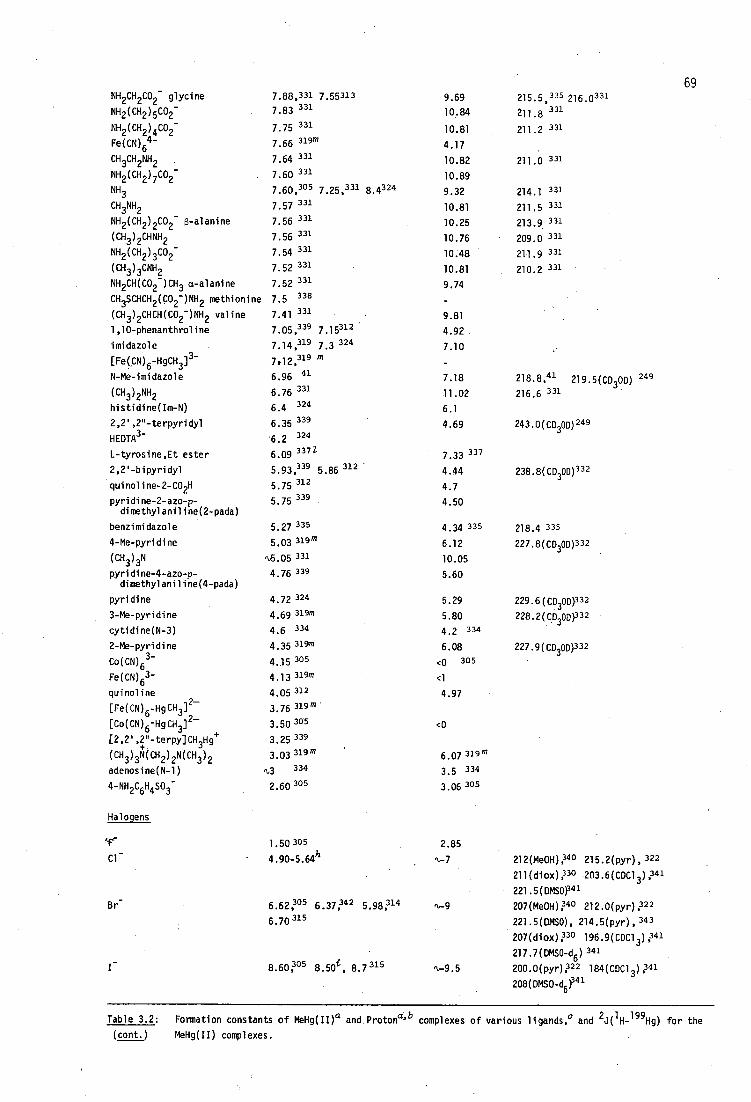
NH2CH 2CO2- glycine
NH2 (CH2 ) 5 CO2 -
NH2 (CH2 ) 4 CO2-
Fe(CN) 64-
CH3
CH2
NH2 •
NH2 (CH 2 ) 7 CO2 -
NH 3 CH3NH2 NH 2 (CH 2 ) 2CO2- 6-alanine
(CH3 ) 2 CHNH 2
NH2 (CH 2 ) 3CO2-
(CH3 ) 3CNH 2
NH2CH(CO2- )CH3 a-alanine
CH3SCHCH 2 (CO2- )NH 2 methionine
(CH3 ) 2 CHCH(CO2- )NH 2 valine
1,10-phenanthroline
imidazole
[Fe(GN) 6 -HgCH3 ]3-
N-Me-imidazole
(CH3 ) 2 NH 2
histidine(Im-N)
2,2',2"-terpyridyl
HEDTA3-
L-tyrosine,Et ester
2,2'-bipyridyl
guinoline-2-CO2H
pyridine-2-azo-p-dimethylaniline(2-pada)
benzimidazole
4-Me-pyridine
(CH3 ) 3N
pyridine-4-azo-p-dimethylaniline(4-pada)
pyridine
3-Me-pyridine
cytidine(N-3)
2-Me-pyridine
Co(CN) 6 3-
Fe(CN) 63-
guinoline
[Fe(CN) 6 -HgCH 3 ] 2-
[Co(CN) 6 -HgCH 3) 2.-
[2,2',.2."-terpACH3Hg+
(CH 3 ) 3N(CH 2 ) 2N(CH3 ) 2
adenosine(N-1)
4-NH 2C6H4S0
3-
Halogens
7.88,331 7,55313 7.83 331
7.75 331
7.66 319m
7.64 331
7.60 331 7 . 50 ,305 7 . 25 , 331 8 . 4324
7.57 331
7.56 331
331 7.56
7.54 331
7.52 331
331 7.52 338 7.5
7.41 331
7.05,339 7 . 15312
324
7.14,319 7.3
7.12,319
6.96 41
6.76 331
6.4 324 6.35 339 6.2 324
6.09 3371
5•93,339 5.86 312
5.75 312
5.75 339
5.27 335
5.03 319m
N.5.05 331
4.76 339
4.72 324
4.69 319m
4.6 334
4.35 319m
4.15 395
4.13 31"
4.05 312
3.76 119m
3.50 3°5
3.25 339
3.03 319m
,3 334
2.60 3°5
1.50 305
4.90-5.64h
6.62 ,305 5 . 2742 6. 98 314
6.70 315
9.69
10.84
10.81
4.17
10.82
10.89
9.32
10.81
10.25
10.76
10.48
10.81
9.74
9.81
4.92
7.10
7.18
11.02
6.1
4.69
7.33 337
4.44
4.7
4.50
4.34 335
6.12
10.05
5.60
5.29
5.80
4.2 334
6.08
<0 305
<1
4.97
<0
6.07 319m
3 . 5 334
3.06 3° 5
2.85 Cl - •
Br-
8.60P5 8.50'1 , 8.7 315
69 2..58 331 216.0 331
211.2 331
211.0 331
214.1 331
211.5 331
213.9 331
209.0 331
211.9 331
210.2 331
218.8,41 219.5(0300) 249 216.6 331
243.0(CO30D) 249
238.8(CD30D) 332
218.4 335
227.8(CO300) 332
229.6(CD300332 228.2(CD300332
227.9(CO300)332
212(Me0H),340 215.2(pyr), 322
); 30 203.6(CDC1 211(diox 3),341
221.5(DMSO)341
207(Me0H );4° 212.0(pyr),322
221.5(DMS0), 214.5(pyr), 343
207(diox );39 196.9(CDC1 3),341
217.7(DMSO-d6 )341
200.0(pyr )322 184(CDC1 3 );41 N-9.5
208(DMSO-d6 pc.
Table 3.2: Formation constants of MeHg( In a and Protona' h complexes of various ligands,c and 2J( 1 H- 199Hg) for the (cont.) MeHg(II) complexes.

P-donors
P(CH 2CH 3 ) 3
(CH 3CH 2 ) 2PCH 2CH 2OH
(C5H 5 ) 2PC5H4-3-S03-
Se-donors
SeCN-
S-donorsi
S2-
CH 3HgS
5203-
1-methyl-quinaldine-thione
Me2
70 15 3os k
8.8 305
14.6 3°5
8.12 3°5
9.15 3° 5 ,x0 305
6.79 323
<1 200.4 323
21.2 305 q,14 146323
16.34 305 156.6(pyr)22 156(pyr) 323
10.90;°5 11.05 178(PY0 26 191.0 309
9.66 319 '0.1 319
8.28 395 2 305
Michler's thioketone
1-methylpyridine-2-thione
.S032-
(CH 3H0 2S
(NH2 ) 2C=S
HOCH 2CH 2SHgCH 3 NCS -
(HOCH 2CH2S) 2 (CH 2 ) 2
S(CH 2CH 2CO2 - ) 2
(HOCH 2CH 2S) 2 (CH 2 ) 5
(HOCH 2CH 2S) 2 (CH 2 ) 3
(HOCH 2CH 2S) 2CH 2
(HOCH 2CH2S) 2 (CH 2 ) 5H9CH 3
Co(NH 3 ) 5NCS +
HOCH 2SH 2SCH2CH 2OH
8.15 319
8.11, 3°5 7.96 323 ,1,7 305
6.92 319
6.27 3° 5
6.06, 305 6.1 324
4.27 319
4.20 319
4.13 319
3.80 319
3.38 319
3.24 319
3.20 305
3.14 319
1-1.6 344
6.79
0
-1.2 318
0
<0 345
<0 319
<0 319
<0 319
<0 319
<0 319
<0 319
<0 305
319
172.4 323
202 323
205.6(Me0H );68 208.0(pyr) 22
203 323
s4L„ 5
(HOCH 2CH 2S)(CH 2 ) 3HgCH 3
(HOCH 2CH 2S) 2 (CH 2 ) 2HgCH 3
CH 3SCH 2CH 2CH(CO2 - )NH 3+ methionine S(CH 2CH 2503- )
CH 3HgSCN
CH 3SCH 3 C-donors
3.07 305k o 305k
2.86 318
2.84 318
1.94 338
' 1.91 319
1.65 305
-1.4 34"
=CH N(CH3 ) 2
<0 <0
223 3 38
0 319
0 305 239.9(Me0H) 250
',-CH3- n.,37 n
C6H 5- 1.21 n
CH- 14.0, 3°5 14.2 324
malononitrile 10.4 349
diethylmalonate <8.6 349
[Cu([13]dieneN4 )] 2+ <6.5 349
acetylacetone 5 . 9 349
,t46 101(CHC1 3 ), 341 101-109 (var.) 347
105.1(DMS0 )1 41 101(neat), 325
104.3(pyr), 322 100.6(C012 ),322
101.5(neat)1 49
1.30 109.0(CDC1 3 ) 9 34° 140(C06 ),321
9.14 177.6(pyr), 343 178.0(pyr), 322
176.0(C5H5 )122 175(di0x), 330
188(D20) 330
11.39
13.3
9.95
9.0 350
Table 3.2:
Formation constants of MeHg(II) a and Protona'b
complexes of various ligands,c 2 and J(
1 H-
199 Hg) for the
(cont•)
MeHg(II) complexes.

[Ni([14]dieneN4 )] 2+
2-acetylcyclopentanone
2-methyl-1,3-indandione
Meldrum's Acid
dimedone .
1,3-dimethylbarbituric acid
1 ,3-cyclopentenedione
CH(CN) 3
5.46 349
<4.3 349
<4 349
3.72 349
3.70 349
3.60 349
2.1 349
<2 349
6.28
7.88
6.18
4 . 83 351
5.25 352
4.45
4.25
-5.1
71
Table 3.2: Formation constants of MeHg(II) a and Protonc6b complexes of various ligands, ° and 2J( 1 H- 199H9) for the
(cont.) MeHg(II) complexes.
afor the equilibrium MeHg + + L - =.*MeHgL.
bexperimental conditions are not included here, but equilibrium constants are in aqueous solution unless otherwise
stated.
°this Table and Tables 3.1 (hydroxide), 3.3 (chloride), 3.4 (monothiolates - previous work), 3.6 (monothiolates - this' work) and 3.11 (dithiolates - this work) form a comprehensive list of MeHg(II) stability constants to date
(1981), and updates the unpublished compilation of Erni (1977). 319
dfor polyprotic ligands. pK
a refers to the MeHg(II)-coordinating group, reference 353, unless otherwise stated.
dfor MeHg(II) complexes in aqueous solution unless otherwise indicated. This Table is not a comprehensive list
of coupling constants. Only values of 2J of complexes for which the MeHg(II) formation constant is known, have
been included.
fsee Table 3.1, page
gwith [H 20] = 55M by definition.
hsee Table 3.3, page 81 .
'this work.
s not including monothiolates (Tables 3.4 and 3.6) and dithiolates (Table 3.11).
k20% Et0H.
l constants of the form MeHg0H + HL-7=MeHgL + H 20 have been converted using log 13 (MeHg0H) = -4.60 and pK a (HL) in
the reference shown.
mtheses by various authors (Em ZUrich) in reference 319.
calculated from thermodynamic data in reference 319.
°with MeHg02CMe in CH2C12.
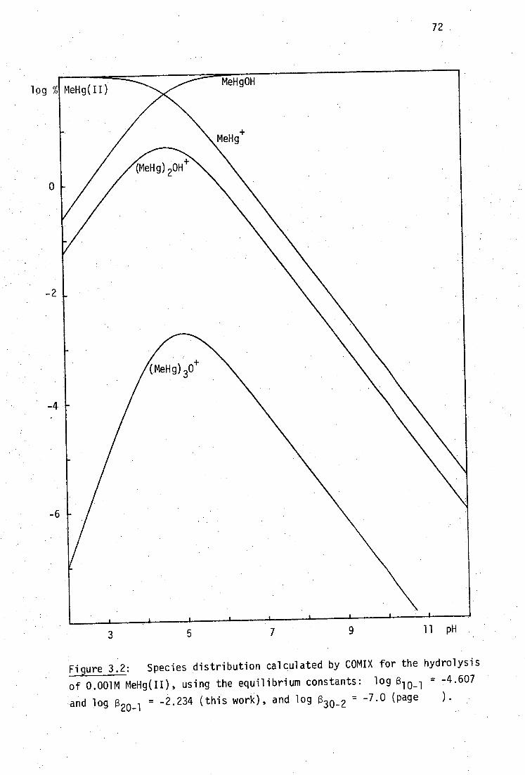
72
Figure 3.2: Species distribution calculated by COMIX for the hydrolysis
of 0.001M MeHg(II), using the equilibrium constants: log (3 10-1 = -4.607
and log B 20-1 30-2 = = -2.234 (this work), and log B 7.0 (page ).
. .

73
nature of the dominant MeHg(II) complex(es) may frequently be postulated
by comparison of the conditional stability constants of MeHg(II) with the
various ligands. The conditional stability constant, Kcond , is defined .
generally by
/MeH gLall complexed species cond 1MeHg(II1
'not bound to Ll[D-not bound to MeHg(II))
Even if the ligand is not involved in protolytic equilibria, since MeHg(II)
is hydrolysable, Kcond is pH dependent. Figure 3.3 shows values of Kcond
for MeHg(II) with a few of the ligands shown in Table 3.2. The condi-
tional formation constant curves were calculated with COMIX for 10 -3 M
solutions of MeHg(II) and ligand. It can be seen that thiolate complexes
predominate, except at either extreme of the pH scale. At low pH, protons
compete more effectively than MeHg(II) for ligand, whereas at high pH,
hydroxide competes more effectively than ligand for MeHg(II). The
extremely stable MeHg(II) thiolate complexes require special techniques
for determination of their formation constants since protons cannot
compete effectively with MeHg(II) for thiolate (page320). It is
generally a simple matter to evaluate the dominant MeHg(II) species in a
mixture of ligands, by comparison of the products, Kcond [L].
If thiolates are involved in the equilibria and are present at more
than trace amounts, they will usually dominate the MeHg(II) species
distribution, but in one important biological situation this is not the
case. If MeHg(II) is ingested orally, comparisons of K cond indicate that
significant concentrations of lipid-soluble MeHgC1 occur at the low pH and
chloride concentration of the stomach, facilitating absorption into the
354 bloodstream. In this work, use has been made of competitive equilibria
between thiolate ligands and iodide, or between two thiolate ligands, to
tPrograms COMIX, COMIXH and TITRAT are described in Chapter 6. A listing of COMIXH is given as Appendix 2.

74
3
5
7
11 pH
Figure 3.3: Conditional stability constants, log K cond , as a
function of pH for MeHg(II) with several ligands. The donor atom is
indicated.
a hydroxide (0), b chloride (Cl), c iodide (I), d acetic acid (0),
e methylamine (N), f mercaptoethanol (S).
Calculated by COMIX for 0.001M MeHg(II) and 0.001M ligand.
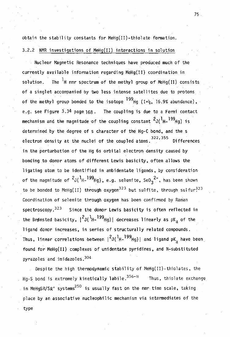
75
obtain the stability constants for MeHg(II)-thiolate formation.
3.2.2 NMR investigations of MeHg(II) interactions in solution
Nuclear Magnetic Resonance techniques have produced much of the
currently available information regarding MeHg(II) coordination in
solution. The 1 H nmr spectrum of the methyl group of MeHg(II) consists
of a singlet accompanied by two less intense satellites due to protons
of the methyl group bonded to the isotope 199
Hg (I=k, 16.9% abundance),
e.g. see Figure 3.34 page 168. The coupling is due to a Fermi contact
mechanism and the magnitude of the coupling constant 2J(
1H-
199Hg) is
determined by the degree of s character of the Hg-C bond, and the s
electron density at the nuclei of the coupled atoms.322,355 Differences
in the perturbation of the Hg 6s orbital electron density caused by
bonding to donor atoms of different Lewis basicity, often allows the
ligating atom to be identified in ambidentate ligands, by consideration
of the magnitude of 2J(
1H-
199Hg), e.g. selenite, Se0 3
2-, has been shown
to be bonded to MeHg(II) through oxygen 323 but sulfite, through su1furq 23
Coordination of selenite through oxygen has been confirmed by Raman
spectroscopy. 323 Since the donor Lewis basicity is often reflected in
the BrOnsted basicity, 12J(
1H-
199Hg)1 decreases linearly as pK a of the
ligand donor increases, in series of structurally related compounds.
Thus, linear correlations between 12J(
1H-
199Hg)1 and ligand pK
a have been
found for MeHg(II) complexes of unidentate pyridines, and N-substituted
pyrazoles and imidazoles. 304
Despite the high thermodynamic stability of MeHg(II)-thiolates, the
Hg-S bond is extremely kinetically labile. 356-8 Thus, thiolate exchange
in MeHgSR/SR' systems250 is usually fast on the nmr time scale, taking
place by an associative nucleophilic mechanism via intermediates of the
type
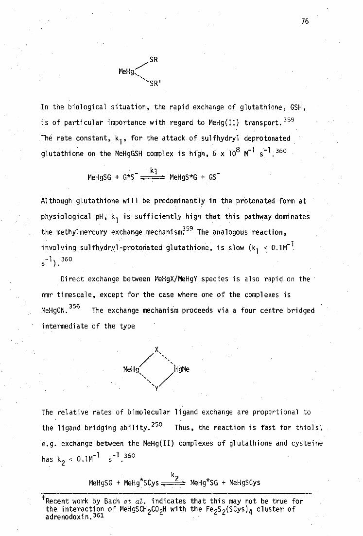
76
SR
MeHg,,
'SR'
In the biological situation, the rapid exchange of glutathione, GSH,
is of particular importance with regard to MeHg(II) transport. 359
The rate constant, kl'
for the attack of sulfhydryl deprotonated
glutathione on the MeHgGSH complex is high, 6 x 10 8 M-1 s-1.360
MeHgSG + G*S_ kl
MeHgS*G + GS
Although glutathione will be predominantly in the protonated form at
physiological pH, 1( 1 is sufficiently high that this pathway dominates
the methylmercury exchange mechanism 59 The analogous reaction,
involving sulfhydryl-protonated glutathione, is slow (k 1 < 0.1M-1
s-1
).360
Direct exchange between MeHgX/MeHgY species is also rapid on the
nmr timescale, except for the case where one of the complexes is
MeHgCN.356
The exchange mechanism proceeds via a four centre bridged
intermediate of the type
MeHg, 110e
The relative rates of bimolecular ligand exchange are proportional to
the ligand bridging ability.250 Thus, the reaction is fast for thiols,
'e.g. exchange between the MeHg(II) complexes of glutathione and cysteine
-1 -1 360 has k
2 < 0.1M s .
MeHgSG + MeHg*SCys MeHg*SG + MeHgSCys
tRecent work by Bach et al. indicates that this may not be true for the interaction of MeHgSCH 2CO 2H with the Fe 2S 2 (SCys) 4 cluster of adrenodoxin.361

77
The bimolecular exchange between Bu tHgSCH and Bu tHgSCH(CH 3 )C6H 5 has
k2
3 x 10⁷
M-1
s-1
in HCF2C1:HCFC1 2 at 25 0 ,
250 which would be four
orders of magnitude faster than diffusion controlled processes if the
exchange mechanism was dependent on uncomplexed thiolate or organo-
250 mercurial.
When exchange is rapid on the nmr timescale, an exchange-averaged
value for the chemical shift of methyl protons of MeHg(II), CH 3 , is
observed. The pH-dependence of 6CH 3 has been used to evaluate
MeHg(II) formation constants with a range of inorganic anions,323
carboxylic acids, 309 amines and aminocarboxylic acids,331 methionine,338
and recently, of several monothiols. 302
3.2.3 NMR evaluation of MeHg(II)-thiolate formation constants
Reid and Rabenstein302 have measured the displacement constants,
Kd
in their terminology, for the exchange equilibria:
[RS - ][MeHgSCH 2 CO2 - ] MeHgSR + -SCH 2CO2 -:F====MeHgSCH2CO2- + RS - Kd -
[MeHgSR][-SCH 2CO2 - ]
The monothiOls, RSH = merCaptoethanol, penicillamine, N-acetylpenicill-
aMine, cysteine, homocysteine and mercaptosuccinic acid, are among the
several whose formation constants with MeHg(II) were evaluated in this
study using the potentiometric titration method.
By monitoring the chemical shift of the methylene protons of
mercaptoacetate, (MA 21 , as the pH of the equilibrium mixture is
altered, Kd can be determined because the relative affinities of RSH
and MAH2 for MeHg(II) are pH-dependent. Since Kd is the quotient of
the individual formation constants of the two thiols for MeHg(II):
[MeHgSR] [MeHgSCHCO2 ] Kd
-
[MeHgl ][RS - ] [MeHe][ -SCH2 CO2 - ]
= Kf(MeHgSR) K (MeHgMA-)
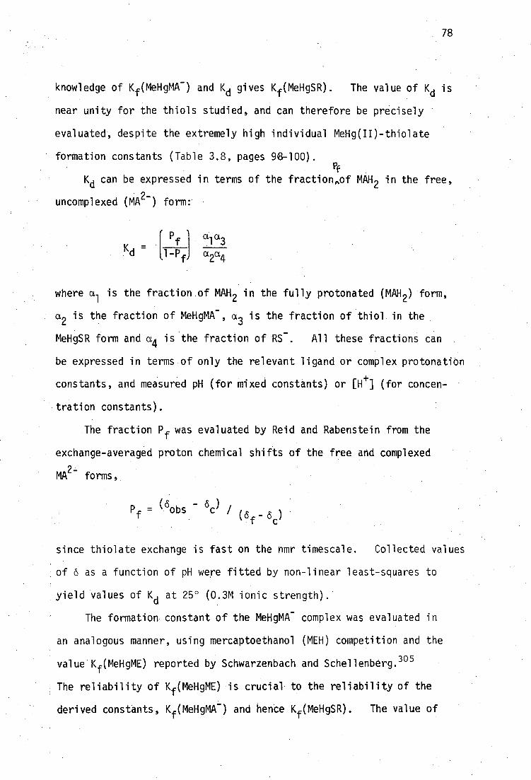
78
knowledge of Kf (MeHgMA- ) and Kd gives Kf(MeHgSR). The value of Kd is
near unity for the thiols studied, and can therefore be precisely
evaluated, despite the extremely high individual MeHg(II)-thiolate
formation constants (Table 3.8, pages 98-100). Pf
Kd can be expressed in terms of the fraction,of MAH in the free,
uncomplexed (MA21 form:
I Pf I a1 a3 ll-Pf
a2a4
where a is the fraction of MAH 2 in the fully protonated (MAH ) form, 1
a2 is the fraction of MeHgMA - , a3 is the fraction of thiol in the
MeHgSR form and a4 is the fraction of RS - . All these fractions can
be expressed in terms of only the relevant ligand or complex protonation
constants, and measured pH (for mixed constants) or [e] (for concen-
tration constants).
The fraction Pf was evaluated by Reid and Rabenstein from the
exchange-averaged proton chemical shifts of the free and complexed
MA2-
forms,
since thiolate exchange is fast on the nmr timescale. Collected values
of 6 as a function of pH were fitted by non-linear least-squares to
yield values of Kd
at 25° (0.3M ionic strength).
The formation constant of the MeHgMA - complex was evaluated in
an analogous manner, using mercaptoethanol (MEH) competition and the
value Kf (MeHgME) reported by Schwarzenbach and Schellenberg.305
The reliability of Kf(MeHgME) is crucial to the reliability of the
derived constants, Kf (MeHgMA- ) and hence Kf (MeHgSR). The value of
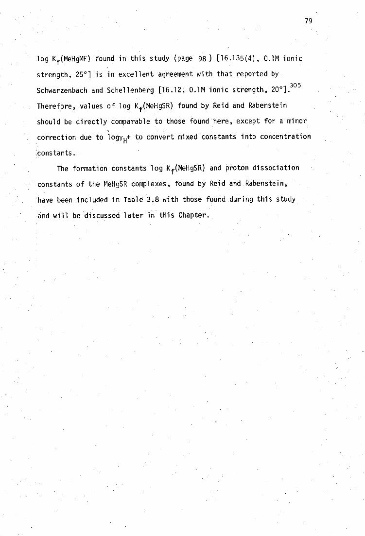
79
log Kf(MeHgME) found in this study (page 98) [16.135(4), 0.1M ionic
strength, 25°] is in excellent agreement with that reported by
Schwarzenbach and Schellenberg [16.12, 0.1M ionic strength, 201. 3°5
Therefore, values of log Kf(MeHgSR) found by Reid and Rabenstein
should be directly comparable to those found here, except for a minor
correction due to logy H+ to convert mixed constants into concentration
!constants.
The formation constants log Kf(MeHgSR) and proton dissociation
constants of the MeHgSR complexes, found by Reid and Rabenstein
have been included in Table 3.8 with those found during this study
and will be discussed later in this Chapter.
80
3.3 Methylmercury(II) Complexation with Thiolate Ligands
3.3.1 Methylmercury(II) monothiolate equilibria
The thermodynamic stability of complexes formed between MeHg(II)
and monothiolate ligands has not been extensively studied. Until the
commencement of this study, and the publication of the work by
Reid and Rabenstein, 302 few reports had been published containing
formation constants of MeHg(II)-thiolate complexes. Some of the earlier
work, by Simpson,324 with cysteine and glutathione and by Schwarzenbach
and Schellenbere 5 with mercaptoethanol recorded formation constants for
the equilibria:
+ - MeHg + RS .7.,==== MeHgSR K -
[MeHgSR]
in the range 15.2 < log K < 16.2, indicating the high stability of these
complexes. A series of substituted thiophenols has been studied by
Schwarzenbach362
indicating that the MeHg(II)-thiolate formation constants
are linearly related to the thiophenolic acid dissociation constants, pK
For 17 substituted thiophenols, the linear relationship is found to be
log K = 0.798 pKa + 877
(which also reproduces log K for cysteine, glutathione and mercaptoethanol
within 0.5 log units).
It was therefore of some concern that Hojo et ai. 363 have more
recently reported equilibrium constants for the interaction of MeHg(II)
with some previously unstudied monothiols which were several orders of
magnitude lower than expedted by consideration of the aliphatic thiol
acidity in the above correlation. Thus, pKa values in the range 8.0 to
10.4 would indicate log K values in the range 15 to 17, but values of
6.7 to 9.0 were recorded by these workers. In addition, reported values
[MeHg 4 ][RS - ]
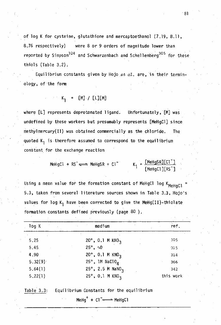
81
of log K for cysteine, glutathione and mercaptoethanol (7.19, 8.11,
8.76 respectively) were 8 or 9 orders of magnitude lower than
reported by Simpson324 and Schwarzenbach and Schellenbere 05 for these
thiols (Table 3.2).
Equilibrium constants given by Hojo et al. are, in their termin-
ology, of the form
= [M] / [L][M]
where [L] represents deprotonated ligand. Unfortunately, [M] was
undefined by these workers but presumably represents [MeHgCl] since
methylmercury(II) was obtained commercially as the chloride. The
quoted K 1 is therefore assumed to correspond to the equilibrium
constant for the exchange reaction
MeHgC1 +RS - r.F.= MeHgSR + Cl - [MeHgSR][C1 - ]
K - 1 [MeHgCl][RS - ]
Using a mean value for the formation constant of MeHgC1 log L e- m HgC1 =
5.3, taken from several literature sources shown in Table 3.3, Hojo's
values for log K 1 have been corrected to give the MeHg(II)-thiolate
formation constants defined previously (page 80 ).
log K medium ref.
5.25 20 0 , 0.1 M KNO 3 305
5.45 25°, rt,0 315
4.90 20 0 , 0.1 M KNO3 314
5.32(9) 25°, 1M NaC104 366
5.64(1) 25°, 2.5 M NaNO 3 342
5.22(1) 25°, 0.1 M KNO 3 this work
Table 3.3: Equilibrium Constants for the equilibrium
MeHg+
+ Cl MeHgC1

82 :
RSH
log K(MeHgSR) ,b
medium ref.
cysteine 15.7 '125°, (‘-'0 324
7.19 (125)C
22°, 'LO 363
glutathione 15.9 r\,25°, ft,0 324
8.11 (13.4) c 22°, r\JO 363
2-mercaptoethanol 16.2 20 0 , 0.1M KI 305
8.76 (14.1) c 22°, rt0 363
penicillamine 6.72 (12•0) c 22°, q0 363
N-acetylpenicillamine 8.87 (14•2) c 22°, A,0 363
2-mercaptopropionic acid 9.03 (14•3) c 22°, q..0 363
2-mercaptosuccinic acid 8.16 (13.5) c 22°, q,0 363
thiophenol 14.67 20°, 0.1M KI 362 in 1:1 Me0H/H 20
2-Me° 15.60 3-Me° 14.48 4-Me0 14.94 4-0H 14.88 11
2,4-diMe 14.82 2-Me 14.71 tt
3-Me 14.62 4-Me 14.69 4-F 14.13 2-C1 13.89 3-C1 13.85 4-C1 14.09 2-Br 13.77 3-Br 13.82 4-Br 13.86 3-I 13.70 2-S03 - ,4-NO2 12.72 20°, 0.1 KNO 3 364' 2,4-diNO2 10.48 11 364 4-NO2 12.3 20°, 0.1 NaC104 365
Table 3.4: Previously reported formation constants of methylmercury(II)-
monothiolate complexes.
+ - afor the equilibrium MeHg + SR 17== MeHgSR
bthe thiolate is considered to be completely deprotonated.
ccorrected for MeHgC1 equilibria, using log K(MeHgC1) = 5.3.
dnot including nmr studies by Reid and Rabenstein,302
discussed on page 77 and recorded in Table 32 (page 98 ).

83
Even with this correction, if appropriate, the data in Table 3.4 show
that serious discrepancies remain in the accepted values for methylmercury-
thiolate formation constants. For this reason, the equilibrium constants
for MeHg(II) complexation with 2-mercaptoethanol, mercaptoacetic acid and
its 0-methylester, mercaptosuccinic acid, cysteine, homocysteine, penicill-
amine, N-acetylpenicillamine, glutathione, thiocholine and N-mercapto-
piperidine (shown in Figure 3.4) were investigated by the potentiometric
method involving iodide competition, described in detail in Chapter 6 of
this thesis. Formation constants of MeHg(II) with cationic thiolates
such as thiocholine have not previously been reported.
Perhaps surprisingly, the earliest reported MeHg(II)-thiolate
stability constants, determined by Hughes, involved the most complex
thiols studied to date. Equilibrium constants for MeNgI exchange with
human and bovine serum albumin, bovine oxyhemoglobin and bovine carbonyl-
hemoglobin were determined by distribution of MeHgI between toluene and
aqueous phases of the thiols.367-8
The equilibrium constant of MeHg(II) with the sulfhydryl protonated
form of human mercaptoalbumin
[MeHgSAlb][H+ ] MeHe + + H K -
[MeHe][AlbSH]
has been calculated by Simpson 324 to be 106 ' 9 using values of the distri-
bution constant for MeHgI between tolueneand water, D = 500 and for MeHgI
formation, 108.7
determined by him; and Hughes 368
value for MeHgI/AlbSH
exchange in toluene,
Thus K - [MeHgSR][e][I - ] [MeHgI(toluene)] • [MeHgn
• [MeHgI(toluene)][AlbSH] [MeHgI] [MeHg+][I-]
- 10 • 500 • io8.7
= 10
6.9

HSCH 2 CH OH -
HS CH CO H — 2 2- HSCH CO CH - 2 2 3
84
HOC CO H
- 2 \CH CH / 2- / - 2 HS
NH -3 HSCH 2 CH / \
CO 2H . 2-
/NH
HSCH CH CH - 2 2 \ CO 2H
CH 3 NH
HSC.CH 2
- - ' NCO H - 2 x
CH 3 2- CH 3 F.".I.
NH
HO2 CCH 2 • NHC CHCH 2 NH • C CH 2 CH 2 CH/ -3
II I - II I II
x
CO H HO C SH NH CO H 0 SH 0 2- - 3- 2- +
+ HS CH H2N(CH3)3a
b Figure 3.4: Fully protonated formd
s' of monothiolate ligands used for complexation of MeHg(II) in this work.
used as the perchlorate sal t. b all thiols were used as neutral ligands, except for thiochol i ne
c this shorthand structure is used by Rabenstein, 369 and be used here for brevity. till
dissociable protons are underlined.
CH 1 /3 NH(02 CCH )
HSC-CH I
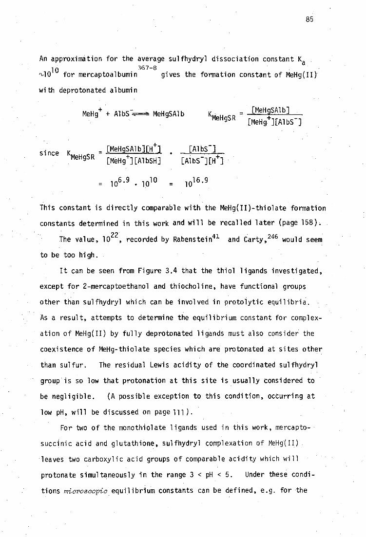
85
An approximation for the average sulfhydryl dissociation constant Ka
367-8 %1010 for mercaptoalbumin gives the formation constant of MeHg(II)
with deprotonated albumin
- MeHg+ + AlbS. MeHgSAlb
[MeHgSAlb] KMeHgSR
[MeHg+][AlbS - ]
_ [MeHgSAlb][e] [AlbS - ] since K
MeHgSR [MeHg 4 ][AlbSH] [AlbS -][e]
= 106.9
1010
= 1016.9
This constant is directly comparable with the MeHg(II)-thiolate formation
constants determined in this work and will be recalled later (page 158).
The value, i0 22 , recorded by Rabenstein 41 and Carty, 246 would seem
to be too high.
It can be seen from Figure 3.4 that the thiol ligands investigated,
except for 2-mercaptoethanol and thiocholine, have functional groups
other than sulfhydryl which can be involved in protolytic equilibria.
As a result, attempts to determine the equilibrium constant for complex-
ation of MeHg(II) by fully deprotonated ligands must also consider the
coexistence of MeHg-thiolate species which are protonated at sites other
than sulfur. The residual Lewis acidity of the coordinated sulfhydryl
group is so low that protonation at this site is usually considered to
be negligible. (A possible exception to this condition, occurring at
low pH, will be discussed on page 111).
For two of the monothiolate ligands used in this work, mercapto-
succinic acid and glutathione, sulfhydryl complexation of MeHg(II)
leaves two carboxylic acid groups of comparable acidity which will
protonate simultaneously in the range 3 < pH < 5. Under these condi-
tions microscopic equilibrium constants can be defined, e.g. for •the
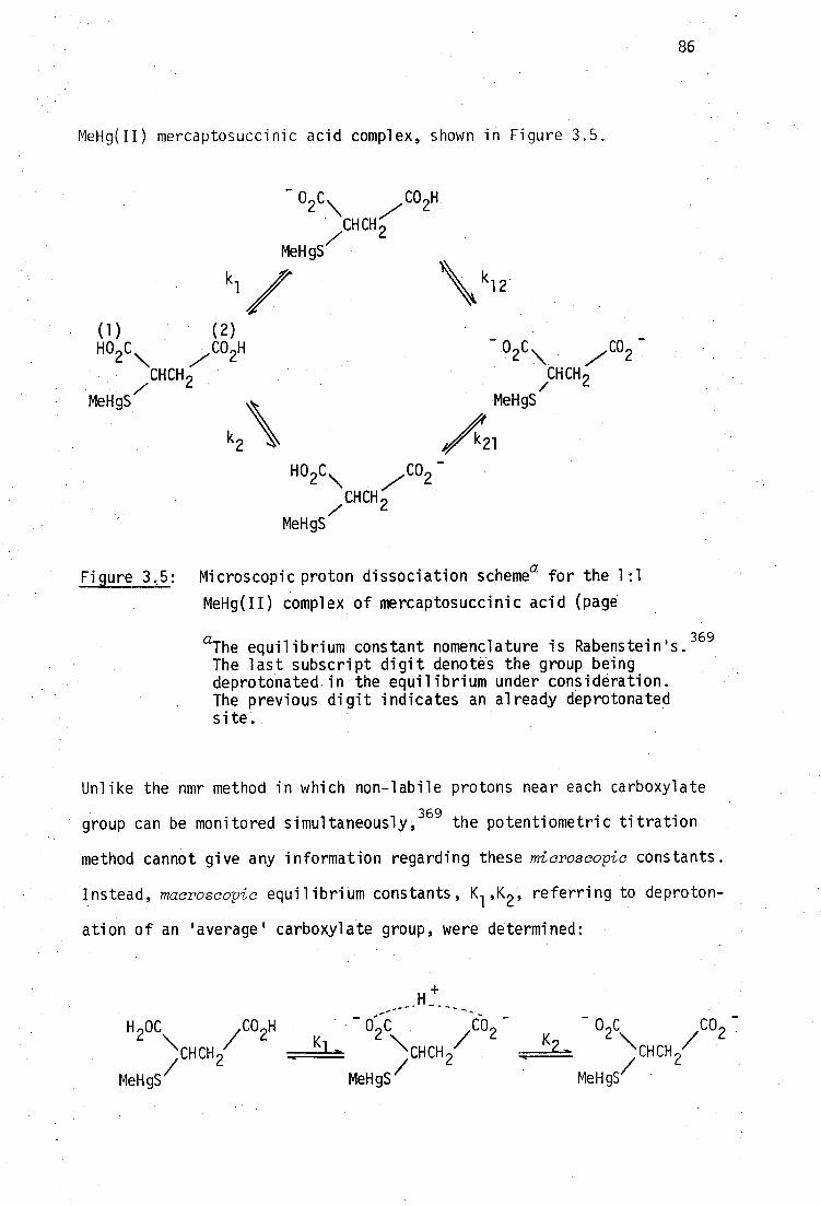
H 2OC C 02H /
‘CHCH // 2 MeHgS
2 / 2 CH CH / 2
- 0 C CO K 2
2\ / 2 ‘CHCH / 2
MeHgS MeHgS
MeHg(II) mercaptosuccinic acid complex, shown in Figure 3.5.
86
0 2 C\ • CHCH
MeHgS
(1) (2) HO C CO H - 0 C
_ 2 \ y 2
2 \ ,CO2
• CHCH 2CHCH 2
MeHgS/ /
/(21MeHgS
k2\
• 02C\
CO -
CH CH /
MeHgS
Figure 3.5: Microscopic proton dissociation scheme a for the 1:1
MeHg(II) complex of mercaptosuccinic acid (page
aThe equilibrium constant nomenclature is Rabenstein's. 369
The last subscript digit denotes the group being deprotonated, in the equilibrium under consideration. The previous digit indicates an already deprotonated S ite.
Unlike the nmr method in which non-labile protons near each carboxylate
group can be monitored simultaneously, 369 the potentiometric titration
method cannot give any information regarding these microscopic constants.
Instead, macroscopic equilibrium constants, K 1 ,K 2 , referring to deproton-
ation of an 'average' carboxylate group, were determined:
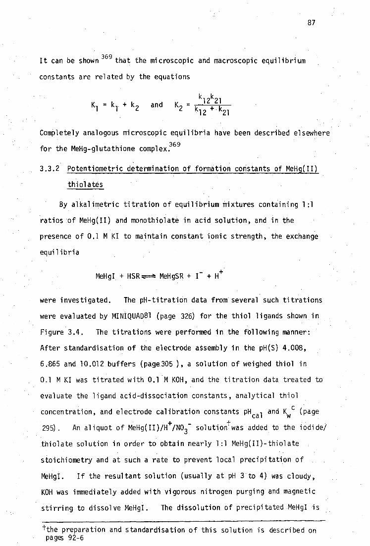
87
It can be shown 369
that the microscopic and macroscopic equilibrium
constants are related by the equations
k12
k21
and K2 -
12 + k21
Completely analogous microscopic equilibria have been described elsewhere
for the MeHg-glutathione complex.369
3.3.2 Potentiometric determination of formation constants of MeHg(II)
thiolates
By alkalimetric titration of equilibrium mixtures containing 1:1
ratios of MeHg(II) and monothiolate in acid solution, and in the
presence of 0.1 M KI to maintain constant ionic strength, the exchange
equilibria
MeHgI + HSR.r.F=2: MeHgSR + I - +
were investigated. The pH-titration data from several such titrations
were evaluated by MINIQUAD81 (page 326) for the thiol ligands shown in
Figure 3.4. The titrations were performed in the following manner:
After standardisation of the electrode assembly in the pH(S) 4.008,
6.865 and 10.012 buffers (page305 ), a solution of weighed thiol in
0.1 M KI was titrated with 0.1 M KOH, and the titration data treated to
evaluate the ligand acid-dissociation constants, analytical thiol
concentration, and electrode calibration constants pH cal and Kwc
(page
295) . An aliquot of MeHg(II)/H+
/NO3- solution was added to the iodide/
thiolate solution in order to obtain nearly 1:1 MeHg(II)-thiolate
stoichiometry and at such a rate to prevent local precipitation of
MeHgI. If the resultant solution (usually at pH 3 to 4) was cloudy,
KOH was immediately added with vigorous nitrogen purging and magnetic
stirring to dissolve MeNgI. The dissolution of precipitated MeNgI is
tthe preparation and standardisation of this solution is described on pages 92-6

88
often slow, taking several hours if the precipitate is allowed to
coagulate. On the few occasions the solution remained cloudy above
pH 5, they were discarded, and the process repeated with more dilute
solutions of thiolate and MeHg(II), or additional KOH was added to the
KOH-titrated thiolate, prior to addition of MeHg(II) so that the final
pH was not less than 4 to 5.
In the discussion below of the MINIQUAD81 treatment of MeHg(II)-
thiolate formation constants, reference will be made to a 'sum-of-squares'
value, U, generated by the program. Values of U < 10 -9 generally
indicate that all titration points are, on average, fitted to better
than 0.005 pH units. Values U < 10-10 indicate fits to better than
0.002 pH units. This degree of agreement is difficult to depict on
figures showing calculated and experimental titration curves over the
range 3 < pH < 10. For such figures the experimental data points are
circled, with circle radii equivalent to 0.02 pH units for clarity.
This does not represent the reliability of pH or volume measurements,
which for individual tirations were considered to be 0.002 pH units and
0,001 mL respectively. Typical agreement between calculated and experi-
mental titrations is shown in Table 3.5 for the MeHg(II)-mercaptoacetate-
iodide system (page 103), calculated by COMIXH using values of equilibrium .
constants used by MINIQUAD81. All MINIQUAD81 treatments were performed
using fixed values of the equilibrium constants for MeHg(II) hydrolysis',
e.g. log 10-1 = -4.607. The binuclear species (MeHg) 20H + , with log
1320-1 = -2.234, needed only to be considered in acidic Solutions
containing excess MeHg(II) in the absence of iodide. Inclusion of this
species otherwise increased the program running time with no decrease in
U or improvement in parameter standard deviations. Constants for ligand
protonation were fixed at the weighted geometric means of values obtained
from preliminary titrations (using TITRAT) and are shown in Table 3.6.
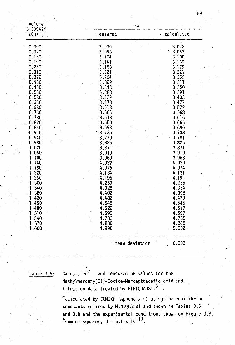
89
volume 0.09947M KOH/mL
pH
measured calculated
0.000 3.030 3.022 0.070 3.068 ' 3.063 0.130 3.104 3.100 0.190 3.141 3.139 0.250 3.180 3.179 0.310 3.221 3.221 0.370 3.264 3.265 0.430 3.309 3.311 0.480 3.348 3.350 0.530 3.388 3.391 0.580 3.429 3.433 0.630 3.473 3.477 0.680 3.518 3.522 0.730 3.565 3.568 0.780 3.613 3.616 0.820 3.653 3.655 0.860 3.693 3.696 0.9-0 3.736 3.738 0.940 3.779 3.781 0.980 3.825 3.825 1.020 3.871 3.871 1.060 3.919 3.919 1.100 3.969 3.968 1.140 4.022 4.020 1.180 4.076 4.074 1.220 4.134 4.131 1.260 4.195 4.191 1.300 4.259 4.255 1.340 4.328 4.324 1.380 4.402 4.398 1.420 4.482 4.479 1.450 4.548 4.545 1.480 4.620 4.617 1.510 4.696 4.697 1.540 4.783 4.785 1.570 4.880 4.886 1.600 4.990 5.002
mean deviation 0.003
Table 3.5: Calculateda and measured pH values for the
Methylmercury(II)-Iodide-Mercaptoacetic acid and
titration data treated by MINIQUAD81. b
acalculated by COMIXH (Appendix 2 ) using the equilibrium
constants refined by MINIQUAD81 and shown in Tables 3.6
and 3.8 and the experimental conditions shown on Figure 3.8. -10
sum-of-squares, U = 5.1 x100.
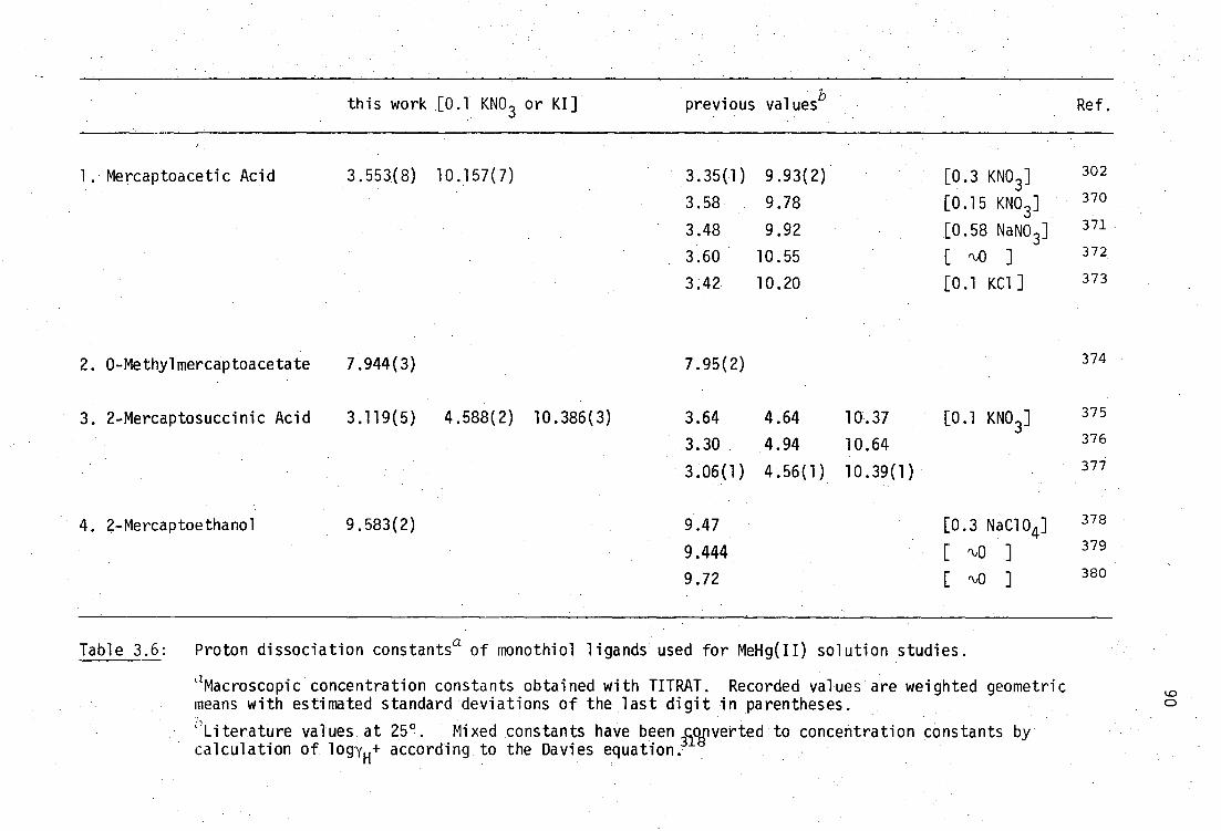
this work [0.1 KNO3 or KI] previous valuesb Ref.
1. Mercaptoacetic Acid
2. O-Methylmercaptoacetate
3. 2-Mercaptosuccinic Acid
4. 2-Mercaptoethanol
3.553(8)
7.944(3)
3.119(5)
9.583(2)
10.157(7)
4.588(2) 10.386(3)
3.35(1)
3.58
3.48
3.60
3.42
7.95(2)
3.64
3.30
3.06(1)
9.47
9.444
9.72
9.93(2)
9.78
9.92
10.55
10.20
4.64
4.94
4.56(1)
10.37
10.64
10.39(1)
[0.3 KNO ] 3
[0.15 KNO3]
[0.58 NaNO 3 ]
[ %0 ]
[0.1 KC1]
0.1 KNO3]
[0.3 NaC104 ]
[ %0 ]
[ rb0 ]
302
370
371
372
373
374
375
376
377
378
379
380
Table 3.6: Proton dissociation constants a of monothiol ligands used for MeHg(II) solution studies.
zMacroscopic concentration constants obtained with TITRAT. Recorded values are weighted geometric means with estimated standard deviations of the last digit in parentheses. 'Literature values at 25 9 . Mixed constants have been
318 converted to concentration constants by
calculation of logyH+ according to the Davies equation.
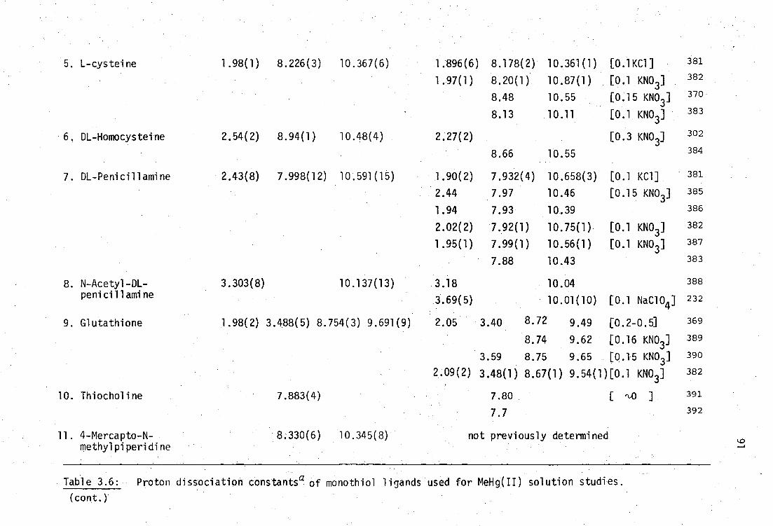
5. L-cysteine 1.98(1) 8.226(3) 10.367(6) 1 .896 (6) 8.178(2) 10.361(1) [0.1KC1] 381
1.97(1) 8.20(1) 10.87(1) [0.1 KNO3 ] 382
8.48 10.55 [0.15 KNO3 ] 370
8.13 10.11 [0.1 KNO3 ] 383
6. DL-Homocysteine 2.54(2) 8.94(1) 10.48(4) 2.27(2) [0.3 KNO3]
302
8.66 10.55 384
7. DL-Penicillamine 2.43(8) 7.998(12) 10.591(15) 1.90(2) 7.932(4) 10.658(3) [0.1 KC1] 381
2.44 7.97 10.46 [0.15 KNO3 ] 385
1.94 7.93 10.39 386
2.02(2) 7.92(1) 10.75(1) [0.1 KNO3 ] 382
1.95(1) 7.99(1) 10.56(1) [0.1 KNO3 ] 387
7.88 10.43 383
8. N-Acetyl-DL- 3.303(8) 10.137(13) 3.18 10.04 388
penicillamine 3.69(5) 10.01(10) [0.1 NaC104 ] 232
9. Glutathione 1.98(2) 3.488(5) 8.754(3) 9.691(9) 2.05 3.40 8.72 9.49 [0.2-0.5] 369
8.74 9.62 [0.16 KNO3 ] 389
3.59 8.75 9.65 [0.15 KNO 3 ] 390
2.09(2) 3.48(1) 8.67(1) 9.54(1)[0.1 KNO3 ] 382
10. Thiocholine 7.883(4) 7.80 ] 391
7.7 392
11. 4-Mercapto-N- 8.330(6) 10.345(8) not previously determined methylpiperidine
Table 3.6: Proton dis sociation consthntsa of monothiol ligands used for MeHg(II) solution studies.
(cont.)
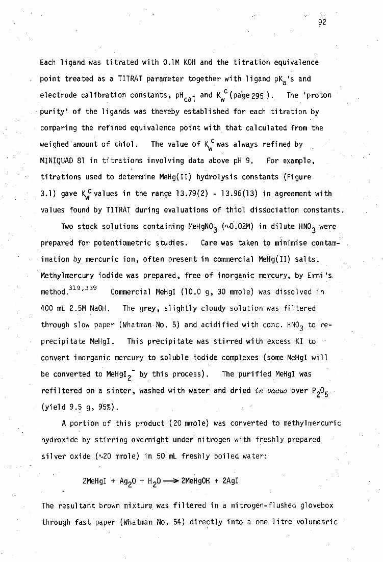
92
Each ligand was titrated with 0.1M KOH and the titration equivalence
point treated as a TITRAT parameter together with ligand pK a 's and
electrode calibration constants, pH cal and Kwc (page 295). The 'proton
purity' of the ligands was thereby established for each titration by
comparing the refined equivalence point with that calculated from the
weighed amount of thiol. The value of Kv(1 : was always refined by
MINIQUAD 81 in titrations involving data above pH 9. For example,
titrations used to determine MeHg(II) hydrolysis constants (Figure
3.1) gave Kv;:values in the range 13.79(2) - 13.96(13) in agreement with
values found by TITRAT during evaluations of thiol dissociation constants.
Two stock solutions containing MeHgNO 3 (01.02M) in dilute HNO 3 were
prepared for potentiometric studies. Care was taken to minimise contam-
ination by mercuric ion, often present in commercial MeHg(II) salts.
Methylmercury iodide was prepared, free of inorganic mercury, by Erni's.
m 319,339 ethod.
Commercial MeHgI (10.0 g, 30 mole) was dissolved in
400 mL 2.5M NaOH. The grey, slightly cloudy solution was filtered
through slow paper (Whatman No. 5) and acidified with conc. HNO 3 to re-
precipitate MeHgI. This precipitate was stirred with excess KI to
convert inorganic mercury to soluble iodide complexes (some MeHgI will
be converted to MeHgI2
- by this process). The purified MeHgI was
refiltered on a sinter, washed with water and dried in vacuo over P 2
(yield 9.5 g, 95%).
A portion of this product (20 mmole) was converted to methylmercuric
hydroxide by stirring overnight under nitrogen with freshly prepared
silver oxide (%20 mmole) in 50 mL freshly boiled water:
2MeHgI + Ag 20 + H 20 ---> 2MeHg0H + 2AgI
The resultant brown mixture was filtered in a nitrogen-flushed glovebox
through fast paper (Whatman No. 54) directly into a one litre volumetric

93
flask containing a slight excess of nitric acid (20 mnole).
After dilution to 1000 mL with freshly deionised water, the MeHg 4-/
1-1 4-/NO3- solution was nominally 0.02M [MeHg +] and 0.005M [e]. When
required, aliquots were withdrawn under nitrogen. This solution and
another prepared similarly, were stored in the dark and the concentra-
tions of MeHg(II) and H + determined periodically by the methods described
below.
Analysis of Fri- content
Unless the MeHg(II) content is known, alkimetric titrations of
+ + - MeHg /H /NO
3 solutions cannot be used to determine the H
+ content
because MeHg0H species are readily formed (page 63 ). The equivalence
point, which corresponds to the composite MeHg+ + H 4- content, of a
titration of 0.005M (MeHg +/e/NO3- ) with 0.1M KOH can be located potentio-
metrically with a glass/double junction calomel (1M KNO 3 , sat. KC1)
electrode pair. Although the equivalence point is not very sharp (e.g.
Figure 3.1) it can be reliably located with a precision of 1-2 parts-per-
thousand (ppt), by employing Gran Type-I 393 or Type-I1 394 calculation
procedures.
In mixtures of 0.005M MeHg(II) and 0.01M chloride (or iodide),
MeHg(II) hydrolysis is suppressed to the extent that MeHg0H comprises
less than 1% of the total MeHg(II) content below pH 7.0 (or 10.3) due
to formation of MeHgC1 (or MeHgI).
+ + - To determine only the H content of MeHg /H /NO 3 solutions,
aliquots were titrated with standardised KOH in the presence of 0.1M
KI. The resultant strong acid-strong base titrations were monitored
potentiometrically and the sharp equivalence points located by Gran
methods395
with a precision limited by the content of weakly acidic
impurities in the titration vessel e.g. carbonate, silicates etc.
For example, 0.1M KOH titration of 10 mL aliquots of MeHg+/0.005M
94
NO 3 diluted to 50 mL with freshly deionised water, gave Gran
plots which were curved near the equivalence point.395 indicating the
presence of 41.2 pmole of weakly acid impurities. This amount corres-
ponds to a titre of <0.002 mL or <4 ppt of the equivalence point.
Similar levels of weakly acidic impurities were found in subsequent
titrations of MeHg(II)-thiolate systems in either 0.1M KI or KNO 3 media.
These titrations have equivalence points rb0.002 mL higher than expected
from consideration of analytical concentrations of the component solu-
tions. The impurities presumably arise from the 50 mL water, 0.5-0.8 g
KI or KNO3'
titration-cell walls etc. (page 304).
The H of the 0.02M MeHg +/0.005M H -1-/NO 3- solution was
monitored periodically by this method over a period of 16 months and
showed an increase from 0.003M to 0.005M during this time
Analysis for MeHg(II) content
Various methods have been reported previously for the analysis of
methylmercury(II) stock solutions. Libich and Rabenstein 309 ' 331 used
an nmr titrimetric method, based on thiosulfate complexation of MeHg(II),
to analyse 0.19M MeHg0H solutions.
MeHg0H + S 20. + OH -
The endpoint of this reaction was determined by 1H nmr with a standard
deviation of 4-5 ppt. These workers confirmed the accuracy of the nmr
method by atomic absorption spectrophotometric analysis of mercury, but
with somewhat lower precision (10 ppt). Jawaid 333,342 and co-workers
have used an atomic absorption method396 for analysis of MeHg +/e/NO 3
solutions. Instrumentation for atomic absorption and nmr methods was
unavailable for this study.
Rabenstein and co-workers have subsequently determined the methyl-

MTK (yellow)
SHgMe
11, H +
MeHgMTK+ (purple)
+ Me 2 N..( C -NMe2
! SH
MeHg+ -N
Me2
Me N Me2
95
mercury(II) content of stock MeHg0H.solutions by chloride titration at
pH 2 in 80% Et0H, with potentiometric endpoint detection.306,360
Pre-
liminary results indicated that the potentiometric chloride method could
not be used to analyse the dilute (0.02M) MeHe/e/NO 3- solutions used in
this work due to a significant chloride leak from the calomel reference
electrode. A double-junction reference electrode was unavailable at
that time. Ingman366
'
397 has used a spectrophotometric titration based
on dithizone to determine MeHg(II) with a precision better than 10 ppt.
Simpson and co-workers324,398
and Hughes 367-8 have used dithizone titri-
metric methods, with visual endpoints.
The most convenient method was found to be that described by
Ern1319,339
in which MeHg(II) is titrated with thiosulfate in 50%
aqueous methanol, using Michler's thioketone* (MTK) as an indicator.
Schwarzenbach and Schellenbere 5 have determined the formation
constant, log K = 8.27, of the 1:1 MeHg(II) - MTK complex:
H-MTK+
(yellow)
and report that protonation of MTK occurs with pK a r‘,2. At pH 4, in the
presence of MeHg(II), all the indicator is in the MeHg-MTK+
form. Titra-
tion with thiosul fate displaces MeHg(II), giving a sharp colour change
Michler's thioketone, bis[4-(dimethylamino)phenyl]methanethione, was obtained from ICN Pharmaceuticals Inc. as bis(dimethylamino)benzophenone and stored at -20°C.
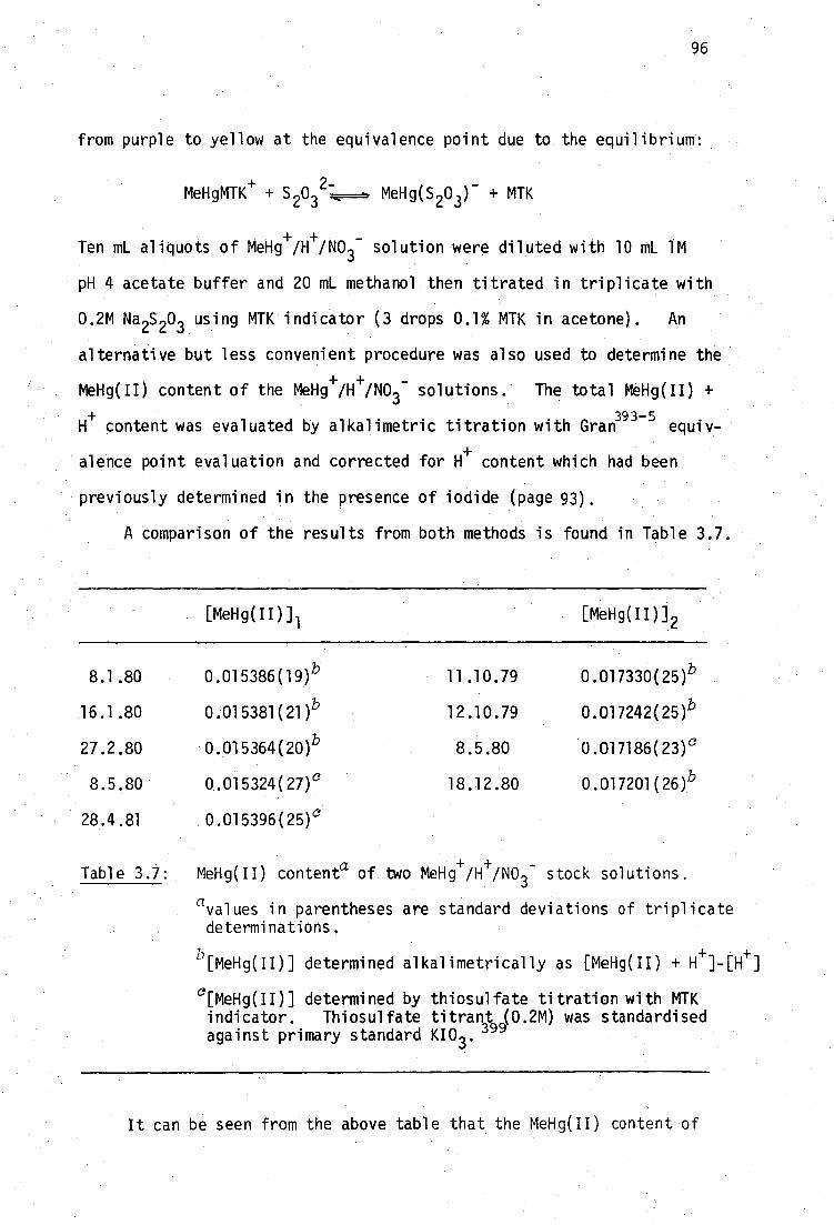
96
from purple to yellow at the equivalence point due to the equilibrium:
MeRgMTK+ + MeHg(S 203 ) - + MTK
Ten mL aliquots of MeHg +/e/NO 3- solution were diluted with 10 mL 1M
pH 4 acetate buffer and 20 mL methanol then titrated in triplicate with
0.2M Na 2S 203 us i ng MTK indicator (3 drops 0.1% MTK in acetone). An
alternative but less convenient procedure was also used to determine the
MeHg(II) content of the MeHg +/e/NO3- solutions. The total MeHg(II) +
H+
content was evaluated by alkalimetric titration with Gran393-5
equiv-
alence point evaluation and corrected for H+ content which had been
previously determined in the presence of iodide (page 93).
A comparison of the results from both methods is found in Table 3.7.
[MeHg(II)] 1 [MeHg(II)] 2
8.1.80 0•015386(19) b 11.10.79 0.017330(25)b
16.1.80 0:015381(21) b 12.10.79 0•017242(25) b
27.2.80 '0.015364(20)b 8.5.80 0.017186(23) c
8.5.80 0.015324 .(27) c 18.12.80 0.017201(26)b
28.4.81 .0.015396(25) c
Table 3.7: MeHg(II) contenta of two Meliiii-/e/NO3 - stock solutions.
avalues in parentheses are standard deviations of triplicate determinations.
+ + [MeHg(11)] determined alkalimetrically as [MeHg(II) +
c [MeHg(II)] determined by thiosulfate titration with MTK indicator. Thiosulfate titrant (0.2M) was standardised against primary standard KI0 3 . 399
It can be seen from the above table that the MeHg(II) content of

97
both solutions remained unchanged, within experimental uncertainty, over
a period of 15 months.
Formation constants of MeHg(II) with the monothiols are discussed
below and are recorded in Table 3.8.
(i) 2-Mercaptoethanol was fractionally distilled in vacuo, bp 63.3-63.5°/
18 mm (lit. 75 157-8° (dec.)/742 mm) and stored under nitrogen at -20 0 .
Titrations with KOH indicated a proton purity of 99.9(2)%. Values for
the proton dissociation constant of mercaptoethanol were identical in
0.1M KNO 3 and 0.1M KI solutions, indicating no iodide-thiolate interaction.
The proton dissociation constant is in good agreement with that determined
elsewhere (Table 3.6). In the presence of MeHg(II) and 0.1M KI, four
sets of titration data were fitted satisfactorily with MINIQUAD 81 by
considering the complex MeHgSCH 2CH 2OH as the only MeHg(II)-thiolate
species present. No evidence of ternary MeHg(II)-iodide-thiolate
species was indicated. In the presence of 0.1M KI, species such as
(MeHg) 2SCH 2CH 2OH can be neglected.
The value found for log f3110 [16.135(4), Table 3.8], agrees with
305 that found by Schwarzenbach and Schellenberg (16.12) but is in dis-
agreement with the value found by Hojo et ca.363
[MeHgSCH 2CH 2OH]
MeHg+ + -SCH2CH2OH=F=hMeHgSCH2CH2OH 13110 = [MeHg+][ -SCH 2CH 2OH]
Calculated and experimental titration curves are shown in Figure 3.6,
and the species distribution in the presence of 0.1M KI, in Figure 3.7.
Because Schwarzenbach and Schellenberg305 have reported the exis-
tence of a complex MeHg(SCH 2CH 2OH) 2- , a titration of MeHg(II) and a ten-
fold excess of mercaptoethanol in the presence of iodide was performed.
Treatment of these data by MINIQUAD 81, using fixed values of the equili-
brium constants for MeHg(II) hydrolysis, ligand protonation and

Thiol
ligand equilibrium constant
1. Mercaptoacetic acid
log filo lll
2. 0-Methylmercapto- log fil0
acetic acid
3. 2-Mercaptosuccinic log 1l0 acid
3111
fil2
4. 2-Mercaptoethanol log filo 5. L-cysteine log
°110
111 0112
0210 13211
212
0310
Equilibrium constants a" of MeHg(II) complexes
this work Rabenstein et al.
with iodide competition
with no competing ligand
16.932(12) ).
20.689(11)
14.98(2)
17.142(7) 1
21.983(5)
25.110(1)}
16.135(4)
16.464(14) 1
25.476(9)
rejected
3.757(6)
4.841(2)
3.127(7)
9.012(6)
25.40(1)
28.06(3)
25.26(2)
29.69(3)
32.13(13)
29.02(3)
3.730(2),
8.936(6) }
11.60(2)
8.800(7) = log K 2f
13.221(13) 1 15.67(12) d
12.56(2) d = log K3f
3.78(2) e
2.66(2)
4.42(2)
2.45(10)
16.92(1)gi
20.57(2)1
17.31(6), 1
22.01(8)
25.39(11)
16.12h
g 16.67(1)}
25.32(3) 1 27.61(11)
3.65(1)
4.70(2) 3 . 38(3)
8.65(2)
2.29(8)
Table 3.8: Methylmercury(II)-monothiolate equilibrium constants refined by MINIQUAD81.

6. DL-Homocysteine
7. DL-Penicillamine
8. N-Acetyl-DL-Penicillamine
9. Glutathione
16.45(1) 8.97 ( 2 )
25.42(3) 2.11(6) 27.53(9)
1 16.596(15),
8.743(7) 16•94(2)g l 7.99(1)
25.339(10) 24.93(3) } 2.18 ( 5 )
rejected 27.11(8)
10 9 13110 16.505(6)
111 19.80(4)
f3
log 13110 15.994(15)
} 9.263(7) 9.11(2) i
13111
25.257(9) 1 3.42(2) 3.41(12)
B112
28.68(2)
3.27(5)
B113 rejected
1
10. Thiocholinef log 13110 14.603(12)
11. 4-Mercapto-N-methyl piperidine
log 16.06(3) B10 Pill 25.33(3) } 268(7)
Table 3.8: Methylmercury(II)-monothiolate equilibrium constants refined by MINIQUAD81.
(cont.
athe : equiTibrta corresponding to the macroscopic constants are described in the text'. Constants Which have been derived (e.g, pKa 's)rather than measured, are italicised.
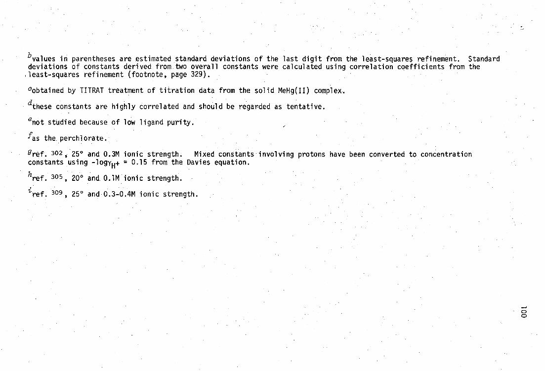
bval
ues in parentheses are estimated standard deviations of the last digit from the least-squares refinement. Standard deviations of constants derived from two overall constants were calculated using correlation coefficients from the ,least-squares refinement (footnote, page 329).
°obtained by TITRAT treatment of titration data from the solid MeNg(II) complex.
dthese constants are highly correlated and should be regarded as tentative.
enot studied because of low ligand purity.
'as the.perchlorate.
gref. 302 , 25° and 0.3M ionic strength. Mixed constants involving protons have been converted to concentration constants using -logye = 0.15 from the Davies equation.
ref. 305 , 20° and 0.1M ionic strength.
• ref. 309 , 25 and 0.3-0.4M ionic strength.
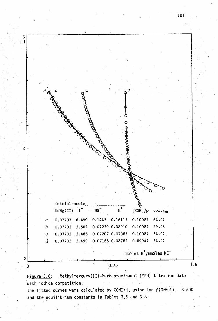
initial mmole
MeHg(II) I -
ME
H+
[KOH]N vol•/mL
6 pH
101
a 0.07703 6.490 0.1445 0.16115 0.10087 64.97
b 0.07703 5.502 0.07229 0.08910 0.10087 59.98
c 0.07703 5.488 0.07207 0.07385 0.10087 54.97
d 0.07703 5.499 0.07168 0.08782 0.09947 54.97
moles H Ambles ME
0
0.75
1.5
Figure 3.6: Methylmercury(II)-Mercaptoethanol (MEH) titration data
with iodide competition.
The fitted curves were calculated by COMIXH, using log 6(MeHgI) = 8.500
and the equilibrium constants in Tables 3.6 and 3.8.

100% MeHg(11)
MeHgI 110 MeHgSCH 2CH 2OH
50
MeHg0H,(MeHg) 20H 4,
102
7
9 11 pH
Figure 3.7: Species distribution calculated by COMIX for 0.0014M
MeHg(II) and 0.0014M 2-Mercaptoethanol in 0.1M KI, using the equilibrium
constants in Tables 3.6 and 3.8.
The heavy line indicates the pH-range of MINIQUAD81 data.

103
MeHgSCH 2CH 2OH formation, could not refine a formation constant for the
1:2 complex. Similar 1:2 formation constants were not sought for the
other thiol ligands described below.
(ii) Mercaptoacetic acid was fractionally distilled under reduced
nitrogen pressure following azeotropic removal of water with benzene,400
bp. 58.5-60.5°/0.5 mm (lit.4 01 95-6°/8 mm). The thiol was stored under
nitrogen as a solid at -20°. Titrations with KOH indicated a proton
purity of 98.0(5)%.
Values for the proton dissociation constants of the ligand are
shown in Table 3.6 and fall within the range of previously reported
values.
In the presence of 0.1M KI and MeHg(II), KOH titrations could be
fitted satisfactorily by consideration of the species MeHgSCH 2CO 2- and
MeHgSCH 2CO2H with coexist with MeHgI below pH 5.
Olin + "" [MeHg ][-SCH 2CO2- ]
NeHgSCH 2 CO2HJ + efiL==, MeHgSCH 2CO2H 13111
[MeHg4I-SCH 2CO2- ][e]
The constant p, 110
agrees with that found by Reid and Rabenstein in 0.3M
KNO3 .302
Their value for the proton dissociation constant, pK al[3.65(1)] of the
MeHg(II) complex is close to that found here as log- log 13110 13 111
[3.76(1)]. In the absence of iodide, the solid complex (page 38 )
titrates as a monoprotic acid with pK; 3.74(1) in good agreement with
the above values. Calculated and experimental titration curves are
shown in Figure 3.8 and the species distribution in the presence of
0.1M KI, in Figure 3.9.
Mixed constants reported by these workers have been converted to concentration constants in 0.3M ionic strength, using logy + = 0.15 from the Davies equation.
MeHg + SCH 2CO 2 -w—m.MeHgSCH 2CO2- [MeHgSCH 2CO 2 - ]
MeHg+
+ -SCH

[KOH]im Vol./MeHg(II) MA 2- H
+
a
b
c
0.07703
0.1895
0.2051
0.08244
0.1895
0.2051
0.17977
0.1895
0.4144
5.482 0.09947
0.09649
0.09837
54.97
100.0
102.0
t.‘
mmoles H+/moles MA
2-
initial mmoles
6 pH
4
104 •
0)
0,
CA'
:"411
0 1.25 2.5
Figure 3.8: Methylmercury(II)-Mercaptoacetic acid (MAH 2 ) titration
data, a with iodide competition; b,c with no competing ligand.
The fitted curves were calculated by COMIXH, using log 8(MeHgI) = 8.500
and equilibrium constants in Tables 3.6 and 3.8.

MeHg(II)
MeNgI 110 MeHgSCH 2CO2,
MeHgSCH C
111
MeHg0H,(MeHg) 20H+
104b
1 0 0%
50
5
9
11 pH
Figure 3.9: Species distribution calculated by COMIX for 0.0014M
MeHg(II) and 0.0014M Mercaptoacetic acid in 0.1M KI, using the
equilibrium constants in Tables 3.6 and 3.8.
The heavy line indicates the pH-range of MINIQUAD81 data.

105
(iii) Mercaptoacetic acid, 0-methyl ester was prepared by acid catalysed
0-alkylation of mercaptoacetic acid in refluxing methanol. Fractional
vacuum distillation gave a colourless oil bp. 46-7.5°/4 mm (lit. 378
42-3°/10 mm). Data from alkalimetric titrationt of this thiol could
not be fitted adequately by TITRAT without assuming the presence of a
moderately acidic impurity, presumably mercaptoacetic kid. Treatment
by TITRAT of this ligand as a mixture of mercaptoacetic acid and the
0-methyl ester gave the relatively imprecise value of acid dissociation
constant for the ester, shown in Table 3.6. The concentrations of
both compounds were treated as parameters with fixed values for mercapto,
acetic acid dissociation constants, indicating (0.4% Of this impurity .
An approximate value for the previously unreported MeHg(II) formation
constant of the 0-methyl ester was obtained by titration of the (impure)
ligand with MeHg(II) in 0..1M KI'and is shown in Table 3.8, The
MeHg(II) complexes of the mercaptoacetic acid *purity were included
in the MINIQUAD 81 treatment', using values for their formation constants ,
recorded previously (page (0)
MeHg+ + MeÔ2CCH
MeHgSCH 9CO9Me [MeHgSCHCO2Me]
110 + [MeHg ][Me02CCH 2S 7 ]
.(iv) Mercaptosuccinic acid was purified by extraction of an alkaline
aqueous solution with ether, followed by acidification of the aqueous
layer.40o The white crystalline thiol was dried over la 205 under
40o nitrogen, mp. 147-50° (lit. 153740, 149-50° 75 ) TitratiOns with
KOH gave a proton purity of 98.5(4)%. Values for the two carboxylate
and thiol proton dissociation constants obtained by TITRAT are shown in '
Table 3.6, and are in agreement with previously published literature
values. In the presence of MeHg(II) and 0.1M KI the titrations could
be fitted satisfactorily with - the three equilibria shown below:
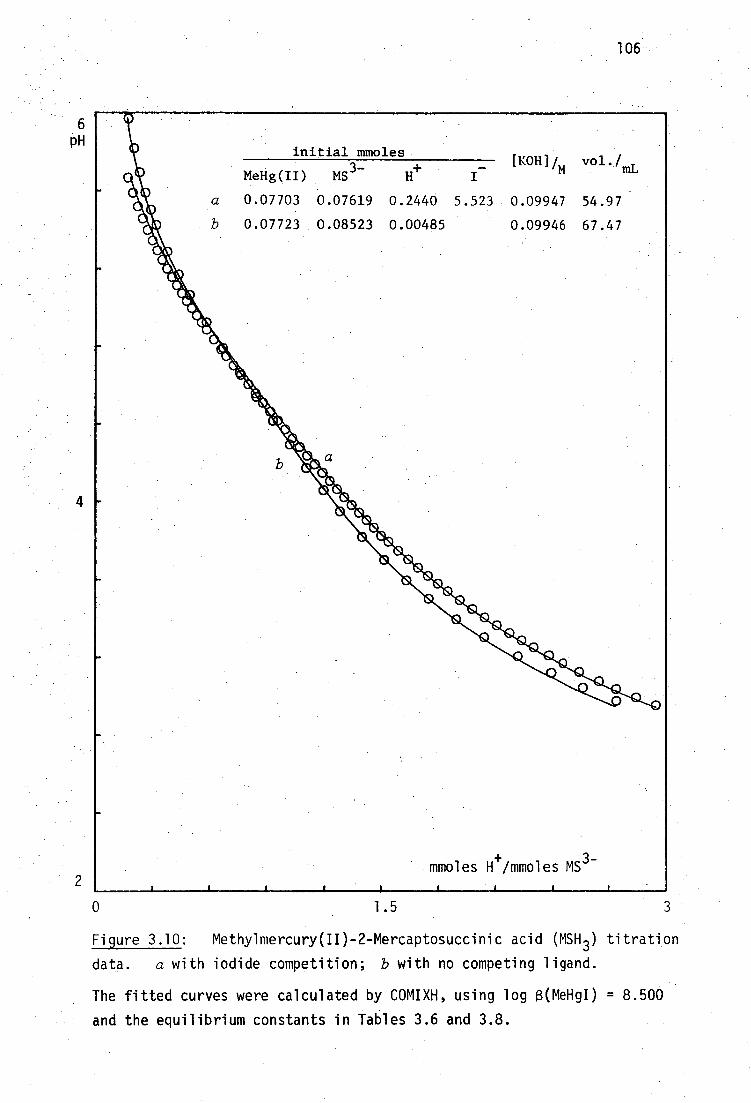
H+ _ [1:0111/m vol./ mL MeHg(II) MS 3- I
a 0.07703 0.07619 0.2440 5.523 0.09947 54.97
0.07723 0.08523 0.00485 0.09946 67.47
initial mmoles
6 pH
106
mmoles H+/mmoles MS
3-
0 1.5 3
Figure 3.10: Methylmercury(II)-2-Mercaptosuccinic acid (MSH 3 ) titration
data. a with iodide competition; b with no competing ligand.
The fitted curves were calculated by COMIXH, using log 8(MeHgI) = 8.500
and the equilibrium constants in Tables 3.6 and 3.8.

50
100%
107
3
5
7
9
11 pH
Figure 3.11: Species distribution calculated by COMIX for 0.0014M
MeHg(II) and 0.0014M 2-Mercaptosuccinic acid in 0.1M KI, using the
equilibrium constants shown in Tables 3.6 and 3.8.
The heavy line indicates the pH-range of MINIQUAD81 data.
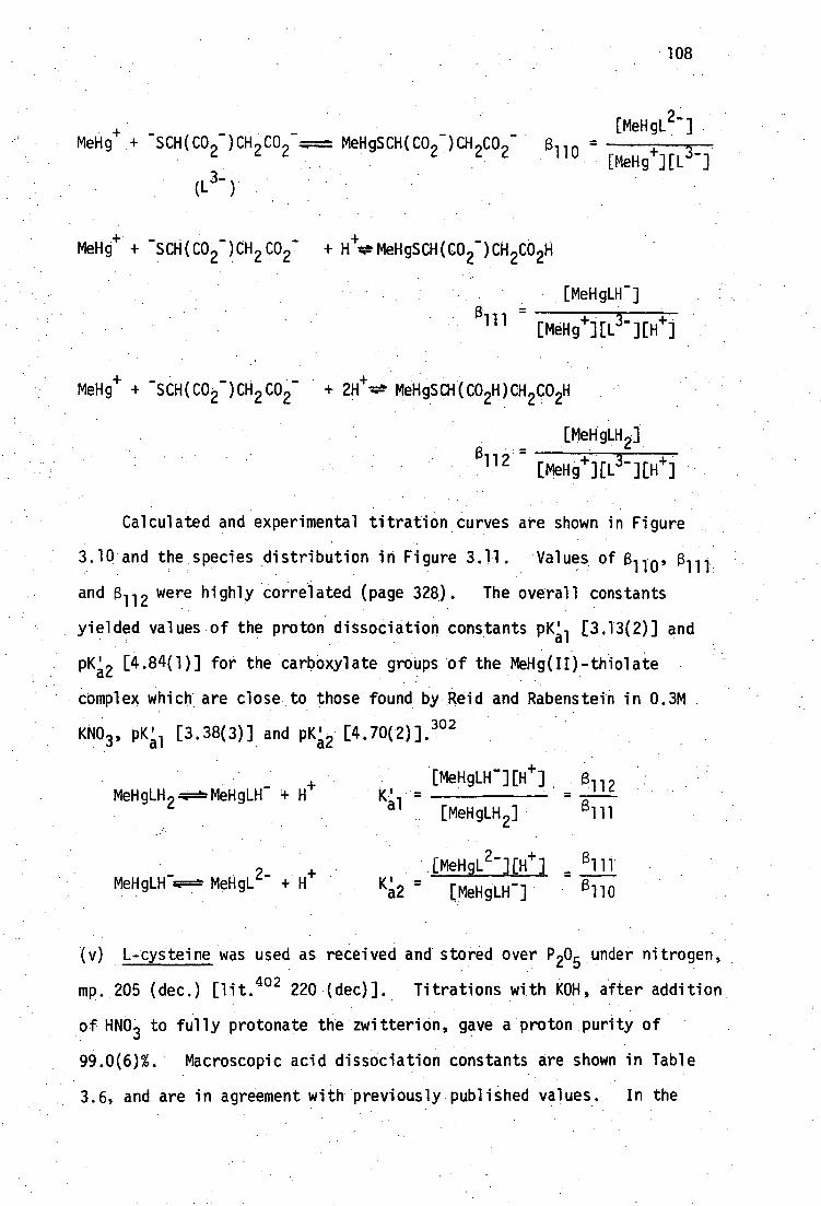
MeHg+ +
•
-SCH(CO21CH 2 CO2 + 2H+=.4b MeHgSCH(CO 2H)CH 2 CO 2H
[MeHgLH 2 ] a 112 - + 3- + [MeHg ][L ][H ]
108
MeHg + +
•
- SCH(CO 21CH
(L3- )
MeHgSCH(CO 2- )CH 2CO 2- 8, 110
[MeHgL2- ]
[MeHgi ][L3- ] .
MeHg+ +
•
-SCH(CO21CH 2 CO 2 7 + erorMeHgSCH(CO )CH CO H
[MeHgLEI - ]
aill =+ 3- + [MeHg ][L ][H ]
Calculated and experimental titration curves are shown in Figure
3.10 and the species distribution in Figure 3.11. Values of f3 110' 12111 and 8 112 were highly correlated (page 328). The overall constants
yielded values of the proton dissociation constants pWal [3.13(2)] and
pK; 2 [4.84(1)] for the carboxylate groups of the MeHg(II)-thiolate
complex which are close to those found by Reid and Rabenstein in 0.3M
KNO 3 , prai [3.38(3)] and piCa2 [4.70(2)].302
MeHgLH Alo=bMeHgLH - +
MeHgLH -.e=== MeHgL 27 + H i-
[MeHgLH - ][e] B -112 a 8 111 [MeHgLH 2 ]
[MeHgL 2-][e] _ E 111' K2 [MeHgLH-]
(v) L-cysteine was used as received and stored over P 205 under nitrogen,
mp. 205 (dec.) [lit. 402 220 (dec)]. Titrations with KOH, after addition
of HNO 3 to fully protonate the zwitterion, gave a proton purity of
99.0(6)%. Macroscopic acid dissociation constants are shown in Table
3.6, and are in agreement with previously-published values. In the

109
presence of MeHg(II) and 0.1M KI, the titration curve, a, shown in
Figure 12, was fitted satisfactorily (U = 8 x 10⁻¹⁰) by assuming the
presence of the complex species shown below. The pq value of the
mono-protonated species [9.01(1)] was obtained from the appropriate
overall formation constants. The constant, a112' could not be
refined precisely from thesedata, and could be evaluated more readily
from data in the absence of iodide, described later.
iNH2 MeHg+ + SCH2C _
CO2
/11H MeHgSCH Cif\ '
CO2-
[MeHgL- ] 110 + 2-
[MeHTHL ]
+ AH ./NH1
MeHg+ -SCH20-1\ 9 `_ MeHgSCH 2CH\ '_ f3111 -
CO2 CO2
+
/ 3
NH, NH [MeHgLH2 ] MeHg
+ +
-SCH e " +2H+-'-,=------'MeHgSCH2CH\ a112
- [MeHg][L2
2 t-][H+]2 \CO - CO2H
The equilibrium constants are recorded in Table 3.8.
Expected sulfhydryl binding is supported by the crystal structure
of the 1:1 complex (page 34 ).236
Reid and Rabenstein "2 report
microscopic constants in 0.3M KNO3 for the equilibrium with deprotonated
ligand (log Kf = 16.69 in their terminology, in close agreement with
(3110 above), and with amino-protonated ligand:
NH /NH1
--. 2 MeHg+ -SCH CH/ 3 MeHgSCH2 H, log Ki = 15.38 'CO2 -
CO2
Addition of cationic MeHg to the sulfhydryl group of the amino-
protonated ligand is seen to be less favorable than to the deprotonated
form. Microscopic proton dissociation constants of the ligand show a
similar trend for addition of a proton instead of MeNg +) to the sulfhydryl
site.
[MeHgLH]
MeHg+1[L2 ][H-1-]

initial mmoles
MeHg(II) Cys2-
H+
I- pCOH]/m ° vol./mL
110
2
a 0:07703 b 0.15358
. c,c'0.00703
0.07721 0.10996 0.03684.
0.17216 0.01309 0.09671
5.500 0.17216 -0.09946 0.10087
54.97 63.66 54.97
moles H/moles Cys • '
-1
Figure 3.12: Methylmercury(II)-L-cysteine (CysH 2 ) titration data.
The fitted curves a,b,c were calculated by COMIXH using log 6(MeHgI) =
8.500 and the equilibrium constants in Tables 3.6 and 3.8. The 310
species was neglected. •Curve c includes the 310 species.
1 0 pH

111
Rabenstein and Fairhurst have nmr evidence that the MeHg(II) -
complexed sulfhydryl group of the 1:1 glutathione and penicillamine
complexes, may be protonated below pH2. 36° This study did not involve
such acidic conditions and this type of species was therefore not
considered for cysteine of the other mercaptoamino acids described
subsequently.
The equilibrium distribution diagram for the 1:1 system in the
presence of iodide is shown in Figure 3.13a.
In addition to the 1:1 MeHg(IF)-cysteine complex, a 2:1 complex
of closely related DL-penicillamine has been isolated by Carty et al.
which has been shown to have deprotonated-amino-bound MeHg(II) as
well as the expected MeHg(U ).248 )-sulfhydryl binding (page 36 The
2:1 MeHg(II).cysteine system has been investigated here by the titration
of cysteine solutions containing more than one equivalent of MeHg(II)
in the absence of iodide. Under these conditions, the sultydryl
group is always coordinated therefore the equilibria were treated
by MINIQUAD81 assuming a 'ligand' of the form MeHgSCH 2CH(NH 2 )CO2- ,
which may form complexes with MeHg(II) as follows:
11119 NH2 HgMe MeHg+ + MeHgSCH CH\ ' q==='
/ MeNgSCH2CH
CO2 CO2-
(L' - )
,NH 2 (MeHg) 2 'SCH CH\ _ ÷÷
C° 2 .
,NH 2 MeHgSCH CFI\
CO 2HgMe
[MeHgC]
2f [MeHe][L'-]
The second stepwise constant, log K2f , for formation of the 2:1 complex
was found to be 8.81(2). This is similar to that for amine complexes

MeHg(II)
/NH
2 MeHgI 111 112 MeHgSCH2CH\ _
CO2
Me SCH CH/NH 3 MeHg0H, NCO
2H
112 (MeHg) 20
112
100°
50
3 5 7
9
11 pH
Figure 3.13a: Species distribution calculated by COMIX for 0.0014M
MeHg(II) and 0.0014M L-cysteine in 0.1M KI, using the equilibrium
constants in Tables 3.6 and 3.8.
The heavy line indicates the pH-range of MINIQUAD81 data.
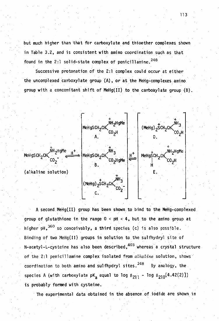
113
but much higher than that for carboxylate and thioether complexes shown
in Table 3.2, and is consistent with amino coordination such as that
found in the 2:1 solid-state complex of penicillamine. 248
Successive protonation of the 2:1 complex could occur at either
the uncomplexed carboxylate group (A), or at the MeHg-complexes amino
group with a concomitant shift of MeHg(II) . to the carbOXylate group .(B).
,NH 2HgMe MeHgSCH2C1c
/MH2HgMe H+ MeHgSCH CH\
CO2-
(alkaline solution)
CO ,H 2 A.
,NH3 MeHgSCH 2CH\
CO2HgMe B.
NH / 3 (MeHg)2SCH H\
CO H 2 D.
/NH 2HgMe MeHgSCH 2CH\
1 CO H 2
• ,/N14
Me0g)2SCH CH\ CO27
C. •wall■
A second MeHg(II) group has been shown to bind to the MeHg-complexed
group of glutathione in the range 0 < pH .< 4, but to the amino group at
higher pH, 38° so conceivably, a third species (c) is also possible.
Binding of two MeHg(II) groups in solution to the sulfhydryl site of
N-acetyl-L-cysteile has also been described, 403 whereas a crystal structure
of the 2:1 penicillamine complex isolated from alkaline solution, shows
coordination to both amino and sulfhydryl sites.248
By analogy, the
species A (with carboxylate pKa equal to log 8 -211 - log 8 210 [4.42(2)])
is probably formed with cysteine.
The experimental data obtained in the absence of iodide are shown in

114
Figure 3.12. The fitted curves shown in Figure 3.12 were calculated
using fixed values of the formation constants for unprotonated and mono-
protonated 1:1 species (page 109) and 2:1 species, together with refined
values shown in Table 3.8 for the diprotonated 1:1 and 2:1 (D or E)
species. The distribution diagram for 0.002M MeHg(II) and 0.001M
L-cysteine is shown in Figure 3.13b.
The titration curves obtained in absence of iodide cannot be
fitted well (U = 7 x 10 the two sets of data) in the range
4 < pH < 5, using the abovementioned set of constants. The MeHg(II):
cysteine ratios used in these titrations were 1.04 and 2.10. Refine-
ment of the formation constant of 3:1 species significantly improved
the fit to these data, which was most marked (U = 5.5 x 10 -9+4 x 10-11 )
for the titration with MeHg(II) = cysteine > 2.
NH 2 2MeHg
+ + MeHgSCH CH ---*" 2 \
CO2
+ + ,NH 2HgMe + ,NH 2HgMe
MeHgSCH 2CH, 4--+ (MeHg) 2 SCH \ 'CO2HgMe CO2C0
F. G.
K - 3 2 -
[MeHg+ ] [L' ]
However, the 3:1 formation constant shown in Table 3.8 was very highly
correlated with that for the twice protonated 2:1 species and both of
these constants should be regarded as tentative. The magnitude of the
third stepwise constant for addition of MeHe [3.76(1)] may be consistent
with weak carboxylate binding near to the cationic complexed-amino group
(F) or to a thioether type of binding (G), similar to that found with
methionine (Table 3.2) and glutathione at low pH. 360 Under these '\,2:1
UMeHg) 2L' + ]
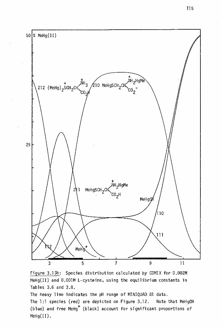
50
25
115
3
7
9
11
Figure 3.13b: Species distribution calculated by COMIX for 0.002M
MeHg(II) and 0.001M L-cysteine, using the equilibrium constants in
Tables 3.6 and 3.8.
The heavy line indicates the pH range of MINIQUAD 81 data.
The 1:1 species (red) are depicted on Figure 3.12. Note that MeHg0H
(blue) and free MeHg+ (black) account for significant proportions of
MeHg(II).

116
conditions, only a small proportion of the 310 species is formed in the
range 4 < pH < 6, at the expense of the 212 species.
In their nmr study of MeHg(II) complexation with N-Acetyl-L-
homocysteine, Simpson et 421. 403 do not preclude the formation of
2+ (MeHg) 3SCH 2CH 2CH(NH0 2CMe)CO 2H at high MeHg(II): thiol ratios. Such
a species is unlikely because of the high positive charge on the sulfur
donor. In that study, the nmr solutions would have been appreciably
acidic (pH < 4) due to the release of the sulfhydryl proton upon MeNg(II)
coordination:
MeHgC1 + HSCH 2CH 2CH(NHCO2Me)CO2H MeHgSCH 2CH 2CH(NHCO2Me)CO 2H + H+ + C1
Formation of a 2:1 species with protonated carboxylate is likely to be
favored over that of the 3:1 species under these conditions. In this
work however, the amino group of cysteine is available for coordination
to form the 3:1 species, F and G above, in which the doubly-positive
charge is delocalised by analogy with the 2:1 protonated species, D and
E.
The refinement problems associated with 2:1 and 3:1 species
(high correlation, large numbers of refinable constants, etc.) were
avoided with the thiols described below by the use of MeHg(II) : RSH
ratios close to unity.
(vi) DL-homocysteine was used as received but stored over P 20 5 under
nitrogen, mp. 225 (dec.) (lit. 400 232-3°). Titrations with KOH, after
addition of HNO 3 to fully protonate the zwitterion, were evaluated with
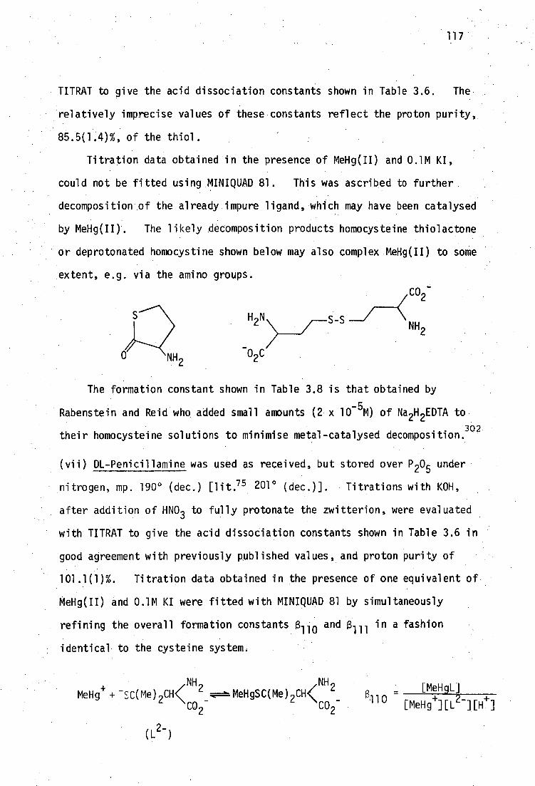
S - S NH2
_ > 02C
117
TITRAT to give the acid dissociation constants shown in Table 3.6. The
relatively imprecise values of these constants reflect the proton purity,
85.5(1.4)%, of the thiol.
Titration data obtained in the presence of MeHg(II) and 0.1M KI,
could not be fitted using MINIQUAD 81. This was ascribed to further
decomposition of the already impure ligand, which may have been catalysed
by MeHg(II). The likely decomposition products homocysteine thiolactone
or deprotonated homocystine shown below may also complex MeHg(II) to some
extent, e.g. via the amino groups.
The formation constant shown in Table 3.8 is that obtained by
Rabenstein and Reid who added small amounts (2 x 10⁻⁵M) of Na2H 2EDTA to
their homocysteine solutions to minimise metal-catalysed decomposition.302
(vii) DL-Penicillamine was used as received, but stored over P 205 under
nitrogen, mp. 190° (dec.) [lit.75 201° (dec.)]. Titrations with KOH,
after addition of HNO 3 to fully protonate the zwitterion, were evaluated
with TITRAT to give the acid dissociation constants shown in Table 3.6 in
good agreement with previously published values, and proton purity of
101.1(1)%. Titration data obtained in the presence of one equivalent of
MeHg(II) and 0.1M KI were fitted with MINIQUAD 81 by simultaneously
refining the overall formation constants f3. 110 and 3111 in a fashion
identical to the cysteine system.
NH <
11
NH2 [MeHgL]
MeHg + + - SC(Me) 2CH‹ 2_...1===MeHgSC(Me) 2 CH
13 0 CO 2 CO2 - [MeHg-I][L2-][H+]

0 3.0
118
6
2
Figure 3.14: Methylmercury(II)-DL-Penicillamine(PenH 2 ) titration data
with iodide competition.
The fitted curve was calculated by COMIXH, using log (MeHgI) = 8.500
and the equilibrium constants in Tables 3.6 and 3.8.
10 pH initial mmoles
MeHg(II) Pen2- +
0.07703 0.07707 0.17368 5.498 0.10087 54.97
[KOH]im vol./
moles H /mmoles Pen2-
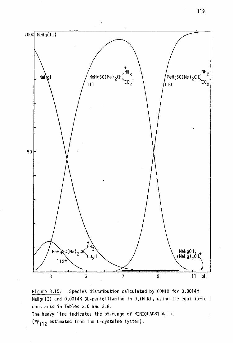
5 7 3
100A MeHg(II)
NH 2 MeHgSC(Me) 2C‹
110 CO2
50
MeHg0H, (MeHg) 20H\
1 11 pH
119
Figure 3.15: Species distribution calculated by COMIX for 0.0014M
MeHg(II) and 0.0014M DL-penicillamine in 0.1M KI, using the equilibrium
constants in Tables 3.6 and 3.8.
The heavy line indicates the pH-range of MINIQUAD81 data.
(*13112 estimated from the L-cysteine system).

120
NH NH3+ - [MeHgL] MeHg + SC(Me) C O + tw=t-MeHgSC(Me) CH‹CO
B
2 - 111 + -
[MeHg ]LL ] C
These are recorded in Table 3.8 and are in good agreement with the
microscopic constants found by Reid and Rabenstein for complexation with
amino deprotonated ligand. 302 Expected sulfhydryl binding is supported
by the crystal structure of the 1:1 complex. 247 Addition of more than
one equivalent of MeHg(II) to penicillamine was expected to produce an
equilibrium mixture of protonated and deprotonated 2:1 complexes with
pH-dependent MeHg(II) binding sites similar to cysteine (page 111) and
was therefore not studied here. A 2:1 complex with sulfhydryl-bound
and amino-bound MeHg(II) groups has been isolated from alkaline solution. 248
Calculated and experimental titration curves are shown in Figure 3.14
and the 1:1 distribution diagram in presence of 0.1M•KI, in Figure 3.15.
The pK; of the doubly protonated complex could not be evaluated in the
pH range used here (pH > 4) but was estimated to be fL3, similar to that
for the 1:1 cysteine complex.
(viii) N-Acetyl-DL-Penicillamine was used as received but stored over
P205 under nitrogen, mp. 180-1
0 (lit. 75 183°). Titration with KOH
and treatment of the data with TITRAT gave the acid dissociation constants
recorded in Table 3.6, in good agreement with previously recorded values,
and indicated a proton purity of 94.4(1)%. Since the amide group is not
protonated in aqueous solution the 1:1 equilibrium system with MeHg(II)
in the presence of 0.1M KI was treated by MINIQUAD 81 by considering.
Only the two equilibria shown:
+ NH(0
2 CMe) . NH(.09CMe)
- MeHg +; SC(Me) 2CH‹ ;----i-MeHgSC(Me) 2 CH‹ `
2 CO2 CO - , 2 (L)
- [MeHgL-J
6 110][L 2 ]
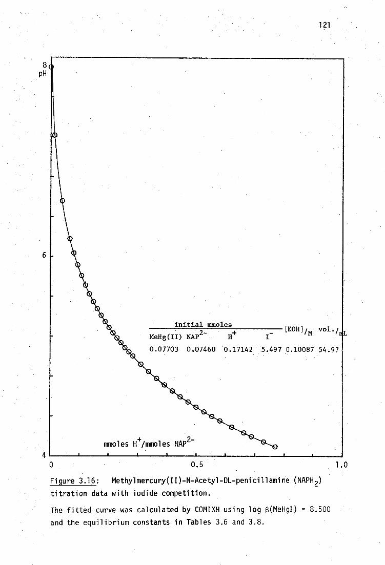
41
initial mmoles 2- +
[KOH]/m vol./ MeHg(II) NAP H
0.07703 0.07460 0.17142 5.497 0.10087 54.97
mmoles H+/mmoles NAP
2-
I
121
0.5 1.0
Figure 3.16: Methylmercury(II)-N-Acetyl-DL-penicillamine (NAPH 2 )
titration data with iodide competition.
0
The fitted curve was calculated by COMIXH using log 13(MeHgI) = 8.500
and the equilibrium constants in Tables 3.6 and 3.8.
8 pH

9 11 pH.
Figure 3.17: Species distribution calculated by COMIX for 0.0014M
MeHg(II) and 0.0014M N-Acetyl-DL-Penicillamine in 0.1M KI, using the
equilibrium constants in Tables 3.6 and 3.8.
The heavy line indicates the pH-range of MINIQUAD81 data.
(*111 estimated from the L-cysteine system).
7 5 3
122
1 00%
50
,NH( CMe) MeHgSC(Me) 2CIT\
CO2H MeHg0H,(MeHg) 20e\ 111
1 4
/NH(02CMe) MeHgI 110 MeHgSC(Me) 2CH\ _
CO2
(II

123
NH(02CMe) NH(02CMe) MeHg+ + -SCH(Me) CH‹
- + H MeHgSC(Me) CH<
CO 2 CO2H
_ [MeHgLH] 111
[MeHe][L2-][H+]
The 1:1 formation constants are recorded in Table 3.8. The value of
6110 is in good agreement with that found by Reid and Rabenstein in
0.3M KNO 3 . 302 The protonated complex is not very soluble below pH 5
and so the value of 6 111 is not precise, being calculated from few data
.
points. The proton dissociation constant, OK; [3.29(5)], of the complex
is close to that found . by the abovementioned workers [2.95(7)].
Calculated and experimental titration curvet are shown in Figure
3.16, and the species distribution with 0.1M KI, in Figure 3.17. The
2:1 system was not studied.
(ix) Glutathione was used as received but stored over P 205 under
nitrogen, mp. 190 (dec.) (lit.75 195°). Protonation constants of
the tripeptide were obtained by KOH titration of acidified solutions.
Treatment of the data with TITRAT gave the results shown in Table 3.6,
in good agreement with those previously reported. The proton purity
was found to be 104.0(2)%.
In the presence of one equivalent of MeHg(II) and 0.1M KI, titra-
tion data over the range 4 < pH < 10 were evaluated with MINIQUAD 81
for the equilibria below:
1 MeHg+ I +ll -0 C S - NH CO -
2 2
(L3- )
I I I-
[MeHgL2-1 C SHgMe H CO2- 6110 [MeHg4 ][L 7 ]
MeHg+
+ S NH 2 CO 2 -0 2 C SHgMe NH 3 CO - I_I I_
2
[MeHgLH - ] 111
-
[MeHg4 ][L 3- ][e]
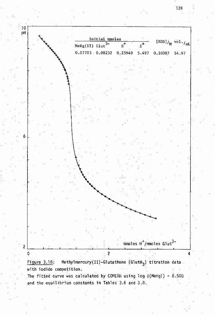
0 2 4
1 0 pH
initial mmoles
MeHg(II) Glut 3 H+-
0.07703 0.08232 0.25949
124
Figure 3.18: Methylmercury(II)-Glutathone (GlutH3 ) titration data
with iodide competition.
The fitted curve was calculated by COMIXH using log 13(MeHgI) = 8.500
and the equilibrium constants in Tables 3.6 and 3.8.

100% MeHg(II)
--CO 2-
-SHgMe
NH 3
CO2 -
--CO 2-
-SHgMe
NH 2
--CO2 -
111 110
--CO-- 2'.
-SHgMe 112:
-NH 3
--0O2-'
--CO 2H
-SHgMe
-NH 3
--CO H 2
112
50
113:
MeHg0H, (MeHg) 20H
MeHgI
125
3
5
7
9
11 pH
Figure 3.19: Species distribution calculated by COMIX for 0.0014M
MeHg(II) and 0.0014M Glutathione in 0.1M KI, using the equilibrium
constants in Tables 3.6 and 3.8.
The heavy lines indicates the pH-range of MINIQUAD81 data.
(*f3 1 .13 estimated from the microscopic constants in ref. 309)
MeHg+ + +
1 I + 2H -7.==== 1I 1 NH CO2 HO C SHgMe NH CO
2 1. 3 2
126
[MeHgLH 2 ]
fi112 . [MeHg
+][1.
3-][H
+ 2
The formation constants are recorded in Table 3.8. Inclusion of a
thrice protonated complex Din) did not improve the fit to the data.
The valueof 1l0 agrees with the previously reported values of
Schwarzenbach and Schellenberg 305 and Simpson 324 Reid and Rabenstein
have not published a value of this constant. Proton dissociation
constants of the 1:1 complex, corresponding to log B . 112 -
log fill
9.26(1)] are in .42(1)] and log fi ll a B ro EP Ki al = 3 - lo K2 = - - 110 -. ;
agreement with the macroscopic constants calculated from microscopic
constants reported by Rabenstein 309 [3.31(12) and 9.11(2) respectively].
The calculated and experimental titration curves for the 1:1 MeHg(II)-
glutathione system are shown in Figure 3.18. The species distribution
over the range 2 < pH < 12 is shown in Figure 3.19, using values plio ,
fill' 112 evaluated in this work, and an estimate of log 113*5 fr°111
the microscopic constants reported by Rabenstein.309 The 2:1 system
has been previously studied360 and was not considered here.
(x) Thiocholine perchlorate was kindly provided by Philip Guerney
(University of New South Wales), and stored at - 20 0 under nitrogen,
mp. 137-9°
Although the MeHg(II) complex of cysteamine has been isolated, 405
the formation constants of MeHg(II) with cationic thiolates such as
thiocholine have not been reported. Titration of the perchlorate salt
of the ligand with KOH, and treatment of the data with TITRAT, gave
the acid dissociation constant in agreement with the previously
reported value, shown in Table 3.6 and proton purity 96.1(2)%.
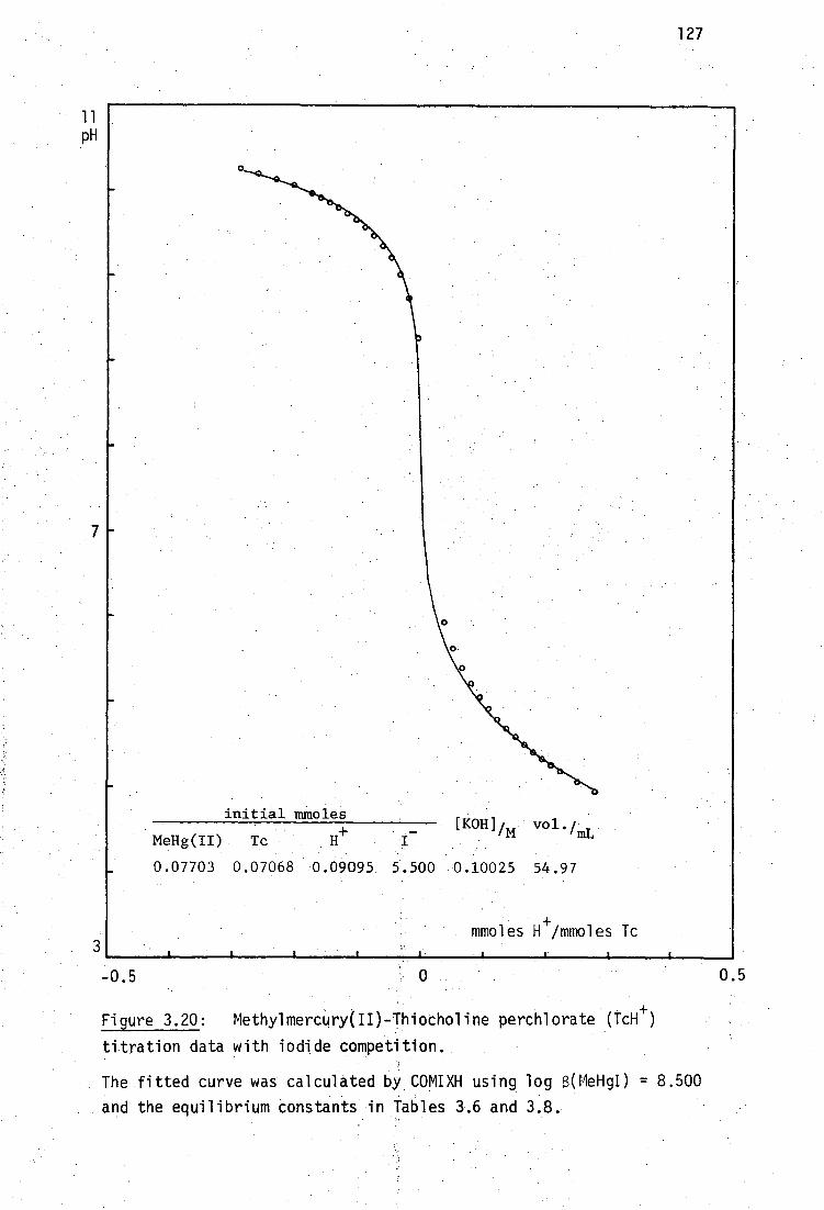
0.5 -0.5
initial mmoles
MeHg(II) Tc H+
0.07703 0.07068 0.09095 5.500 0.10025
127
11 pH
Figure 3.20: Methylmercury(II)-Thiocholine perchlorate IcH + )
titration data with iodide competition.
The fitted curve was calculated by COMIXH using log 13(MeHgI) = 8.500
and the equilibrium constants in Tables 3.6 and 3.8.

128
MeHg(II)
MeHgI 110 MeHgSCH 2CH 2 NMe 3
leINIONNIE111•1111•11■11111•11 4.)
MeHg0H,(MeHg) 20H
100 %
50
7
9 11 pH
Figure 3.21: Species distribution calculated by COMIX for 0.0014M
MeHg(II) and 0.0014M Thiocholine perchlorate in 0.1M KI, using the
equilibrium constants in Tables 3.6 and 3.8.
The heavy line indicates the pH-range of MINIQUAD81 data.
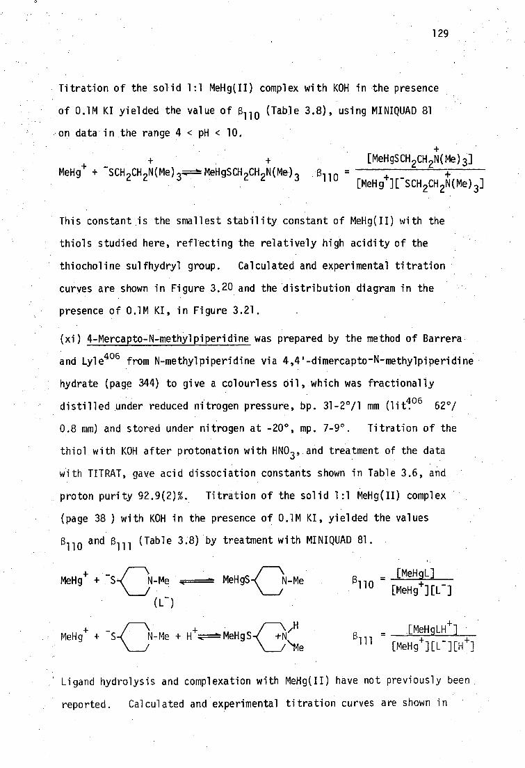
(3. 111 - [MeHig4 ][L - ][H+ ]
IMeHgLe]
129
Titration of the solid 1:1 MeHg(II) complex with KOH in the presence
of 0.1M KI yielded the value of (3 110 (Table 3.8), using MINIQUAD 81
on data in the range 4 < pH < 10.
+ - • MeHg + SCH 2CH 2N(Me) 3 c
MeHgSCH,,CH 2 N(Me) [MeHgSCH 2CH 2N(Me)3]
al 10 = + [MeHg+][ -SCH 2CH 2N(Me) ]
This constant is the smallest stability constant of MeHg(II) with the
thiols studied here, reflecting the relatively high acidity of the
thiocholine sulfhydryl group. Calculated and experimental titration
curves are shown in Figure 3.20 and the distribution diagram in the
presence of 0.1M KI, in Figure 3.21.
(xi) 4-Mercapto-N-methylpiperidine was prepared by the method of Barrera
and Lyle406 from N-methylpiperidine via 4,4'-dimercapto -N-methylpiperidine
hydrate (page 344) to give a colourless oil, which was fractionally
l distilled under reduced nitrogen pressure, bp. 31-2°/1
mm (1it o6 62 ,/
0.8 mm) and stored under nitrogen at -20°, mp. 7-9 0 • Titration of the
thiol with KOH after protonation with HNO 3 , and treatment of the data
with TITRAT, gave acid dissociation constants shown in Table 3.6, and
proton purity 92.9(2)%. Titration of the solid 1:1 MeHg(II) complex
(page 38 ) with KOH in the presence of 0.1M KI, yielded the values
a 110 and f3 111 (Table 3.8) by treatment with MINIQUAD 81.
\ [MeHgL] MeHg
+ + N-Me MeHgS N-Me (3110 -
/ [MeHg4-][L - ] (L - )
MeHg + S N-Me + H .-E===MeHgS +N + - < \
/
+ VI
Ligand hydrolysis and complexation with MeHg(II) have not previously been
reported. Calculated and experimental titration curves are shown in
/'Me
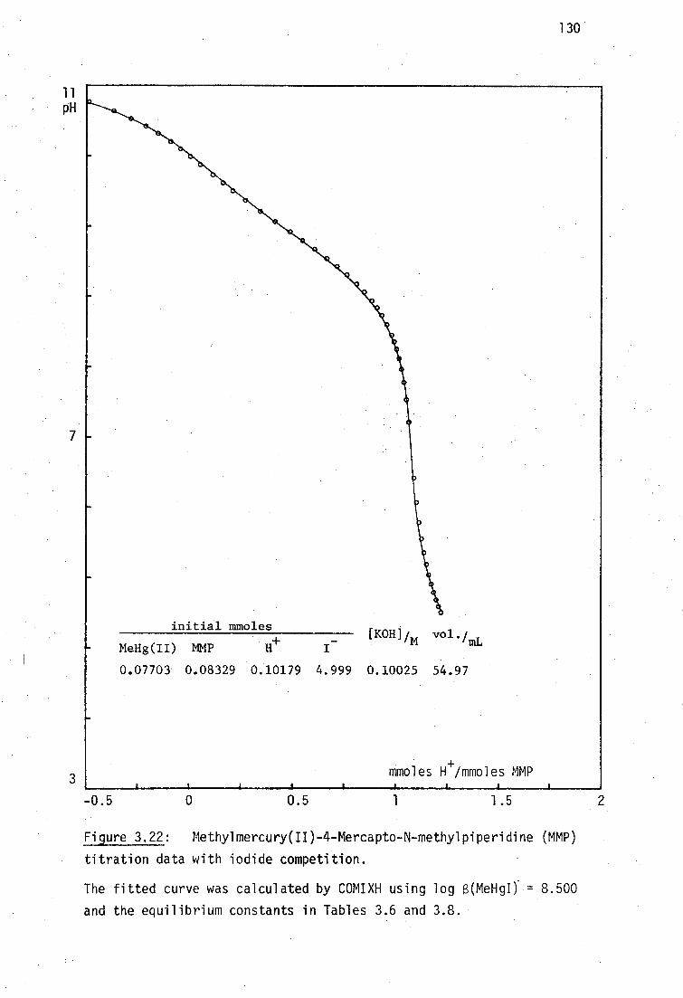
0.5 1 -0.5 1.5 0
11 pH
7
H+ [KOH]im vo1.411,
MeHg(II) MMP
0.07703 0.08329 0.10179 4.999 0.10025 54.97
initial mmoles
130
3
Figure 3.22: Methylmercury(II)-4-Mercapto-N-methylpiperidine (MMP)
titration data with iodide competition.
The fitted curve was calculated by COMIXH using log (MeHgI) . = 8.500
and the equilibrium constants in Tables 3.6 and 3.8.

3
131
1 0 0% MeHg(II)
50
5 7 9 11 pH
I MeHg0HpleHg) 20H+
k ,
Figure 3.23: Species distribution calculated by COMIX for 0.0014M
MeHg(II) and 0.0014M 4-mercapto-N-methylpiperidine in 0.1M KI, using
the equilibrium constants in Tables 3.6 and 3.8.
The heavy line indicates the pH-range of MINIQUAD81 data.

—0O 2
(2) 8.93
SH (3)
—NH 3 (4)
9.13 --0O
2- (1)
13k 124
r--0O 2-
—S-
NH3+
--0O27
pk1234
9.28
s - - NH 2
- CO2 - ---00
2 -
—SH
Pk1243
132
Figure 3.22 and the species distribution in the presence of 0.1M KI,
in Figure 3.23.
The equilibrium constants for MeHg(II)-monothiolate interaction
found in this study, and elsewhere, lie in the range 14.64 to 17.60
and are highly correlated with the BrOnsted basicity of the sulfhydryl
donor groups. This can clearly be seen from the plot of log
versus pKa (SH) in Figure 3.24 which includes values for all monothiols
studied in this work and by Reid and Rabenstein. 3021he values found by
363 . Hojo et al. are in disagreement.
The intrinsic BrOnsted basicity of the sulfhydryl group in the
u-mercaptoamine ligands studied here, cannot adequately be represented
by macroscopic proton dissociation constants because the amine and
thiolate groups protonate simultaneously. Microscopic sulfhydryl
protonation constants for these ligands were estimated by assuming the
same relationships between macroscopic and microscopic constants as were
found in glutathione by Rabenstein. 369 For glutathione, the following
microscopic constants were determined 369
--NH 2
---002-
The amino group is apparently 0.2 log units more basic than the thiolate
group, independent of whether these groups are protonated or deprotonated.

17
16
15
14
133
8
9
10 pKa (SH)
Figure 3.24: Correlation between the MeHg(II)-thiolate formation
constant, log 13110' and BrOnsted basicity of the sulfhydryl donor,
pKa (SH). The monothiol ligand numbering follows that of Tables 3.6
and 3.8.
Log elm from this work4), using estimates of microscopic pK a (SH) for
mercaptoamines (page 134) and microscopic constants reported by Reid and
Rabenstein302
for ligands in which the amino group is protonated,(:),
and unprotonated,(E).

1 34
Similar relationships have been found for the microscopic constants of
cysteine:4"
Macroscopic constants for composite SH + NH 3+ deprotonation, Ka/
and Ka2 are related to the microscopic constants by the relationships '
Kal
= K123
+ k123
Ka2 - k1234
+ k1243
Use of the approximations plc 1234 = Pk1243 + 0.2 and pk124 = Pk123 0 ' 2
enables the microscopic protonation constants of the sulfhydryl (k 1243 )
amine (k1234 ) groups of the mercaptoamines to be separately evaluated
from the macroscopic constants and these are recorded in Table '3.9.
pka (NH 3+ ) pka (SH)
L-cysteine 10.15 9.95
(10.09) (9.74)302
DL-penicillamine 10.38 10.18
(10.29) (9.70)302
glutathione 9.53 9.33
(9.20)4°7
-mercapto-N-methylpiperidine 10.13 9.93
Table 3.9: Estimateda microscopic proton dissociation constants of
amine and sulfhydryl groups of fully depronated a-
mercaptoamines.
aUsing the approximation pk a (NH 3
+) = pk a (SH) + 0.2
described in the text and using macroscopic constants
recorded in Table 3.6.
k1234
• k1243

135
The relationship between 8 110 for log KmeH gsR and pKa(SH) from this
combined data is found to be
logKMeHgSR = 0.893 pK a (SH) + 7.77
The relevance of these formation constants to the interaction of
antidotal thiols in vivo will be discussed in the next Section, after
the formation constants with some vicinal dithiols have been established.

136
3.3.3 Methylmercury(II) Formation Constants with Vicinal Dithiols
The only formation constants in the literature of methylmercury(II)
with a dithiolate ligand seem to be those reported by Hojo et al. for
the interaction with BALH 2' 363 Some serious discrepancies with values
obtained by these workers for the formation constants of MeHg(II)-
monothiolate complexes have previously been discussed (page 80 ).
As several important antidotes for methylmercury(II) toxicity are
vicinal dithiols (BALH 2 , Unithiol and DMSH 4 ) it was of interest to
determine the equilibrium constants for the formation of MeHg(II)
complexes with these ligands.
To completely describe the solution chemistry of MeHg(II) complexes
with dithiols, both 1:1 and 2:1 species (protonated and unprotonated)
need to be considered, which renders evaluation of individual equilibrium
constants much more difficult than for monothiols.
Attempts to purify the non-vicinal dithiol, 1,3-dimercapto-2-
propanol, DMPH 2 , by fractional distillation under reduced nitrogen
pressure, bp. 61.5-3.5°/0.2 mm (lit.400 82°/1.5 mm) apparently caused
decomposition, indicated by low proton-purity of the product (<85% by
alkalimetric titration). Consequently, comparison of MeHg(II) stability
constants with 0MPH 2 and its vicinal dithiol analog, BALH 2 , was not
possible.
2,3-dimercapto-l-propanol, BALH2 , was fractionally distilled under
reduced nitrogen pressure, bp. 74-570.5 mm (lit. 408 74-671 mm), and
stored under nitrogen at -20°. Titration of the dithiol with KOH, and
treatment of the data with TITRAT, gave the two macroscopic proton
dissociation constants recorded in Table 3.10, and indicated a proton
purity of 97.5(6)%. It should be noted that, since the two sulfhydryl
groups are of comparable basicity, they are simultaneously protonated
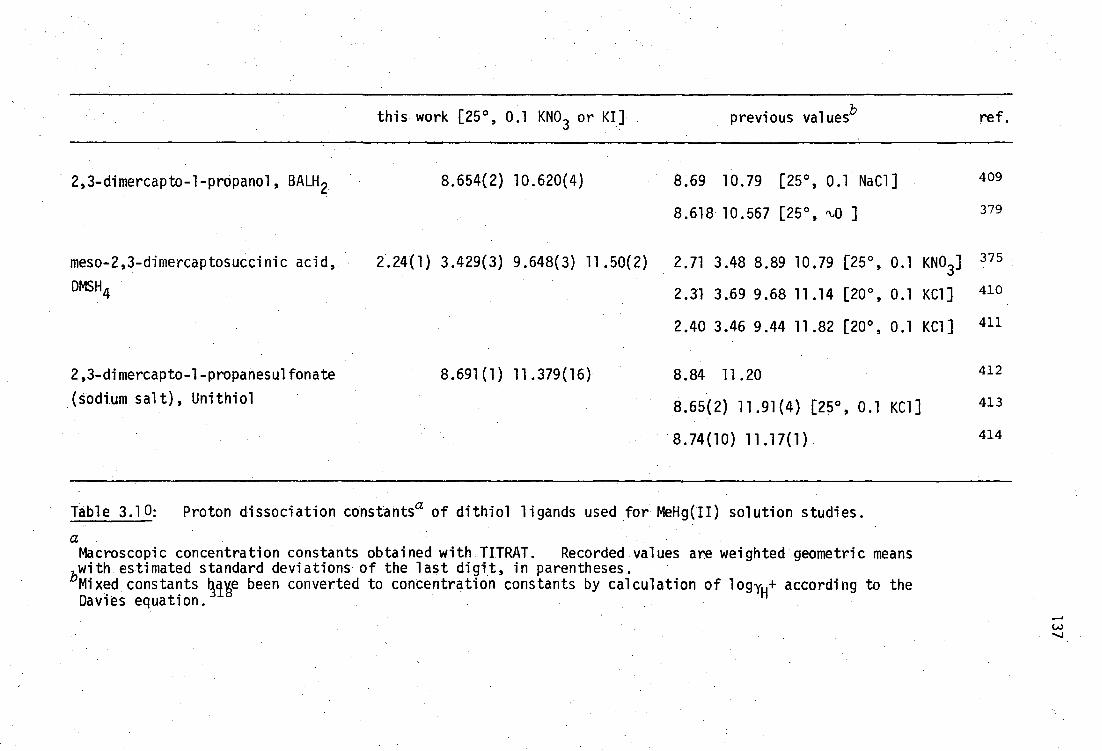
this work [25°, 0.1 KNO3 or K1] previous valuesb ref.
2,3-dimercapto-1-propanol, BALH 2
meso-2,3-dimercaptosuccinic acid,
DMSH4
2,3-dimercapto-1-propanesulfonate
(sodium salt), Unithiol
2.24(1)
8.654(2)
3.429(3)
8.691(1)
10.620(4)
9.648(3) 11.50(2)
11.379(16)
8.69 10.79 [25°, 0.1 NaCl]
8.618 10.567 [25 , fq) ]
2.71 3.48 8.89 10.79 [25°, 0.1 KNO3]
2.31 3.69 9.68 11.14 [20°, 0.1 KG]]
2.40 3.46 9.44 11.82 [20°, 0.1 KG]]
8.84 11.20
8.65(2) 11.91(4) [25°, 0.1 KC1]
8.74(10) 11.17(1)
409
379
375
410
411
412
413
414
Table 3.10: Proton dissociation constantsa of dithiol ligands used for MeHg(II) solution studies.
a Macroscopic concentration constants obtained with TITRAT. Recorded values are weighted geometric means with estimated standard deviations of the last digit, in parentheses. bMixed constants 44te been converted to concentration constants by calculation of logyH+ according to the Davies equation.

138
above pH 9 in a similar fashion to the a-mercaptoamines described
previously. Microscopic proton dissociation constants of vicinal
dithiols do not seem to have been previously recorded.
Addition of one equivalent of MeHg(II) to BALH 2 is expected to
establish the following equilibria: t
HC--CH-CH 2OH MeHg
+ + c
S S
(BAL2- )
HzC7-CtCH 2OH 110
[MeHgBAL2-]
g S , [MeHg4][BAL2-]
ileH‘
HC--CH-CH 0 H 9C--CH-CH 2 OH + 7 \ _ [MeHgBALH] MeHg+. + 7 \ H -
-S S- MeHgS SH 111
[MeHg+][BAL2-][H]
Proton and MeHg(II) exchange in the 111 species is likely to be rapid.
An order-of-magnitude estimate for the equilibrium constant for the
addition of one MeHg(II) group to the deprotonated dithiol ligand was
made using the correlation between sulfhydryl basicity, pK a (SH), and
log KMeHgSR
for monothiolates (page135 ). If the microscopic pKa 's of
the sulfhydryl donors are assumed to be between the macroscopic proton
dissociation constants, log plio should be in the range 15 to 17, and
therefore be amenable to evaluation by iodide competition. However,
when equivalent quantities of MeHg(II) and BALH 2 were titrated with KOH
in the presence of 0.1M KI, it became apparent that.MbHg(II) was not
dissociated from sulfur in the range 4 < pH <-10, indicating log 13110
>17 to 18. Only the proton dissociation constant (Wa = (310110) of
the 1:1 protonated species could be obtained from these data. More
tDotted lines indicate that MeHg may be bound to either donor, possibly to both, and the curved arrow indicates rapid exchange in subsequent diagrams in this chapter.
139
efficient competition for MeHg(II) was achieved by using mercapto-
ethanol instead of iodide. A similar approach was coincidentally
taken by Reid and Rabenstein in their nmr studies of monothiolates,
using mercaptoacetate competition. 309
This monothiol has only a
single proton nmr resonance but mercaptoethanol has the advantage in
this work of contributing only two extra equilibria to the overall
equilibrium scheme, i.e. ligand hydrolysis and MeHg(II) complexation.
Titration of a solution containing equivalent quantities of
mercaptoethanol, BALH 2 , and MeHg(II) over the range 5 < pH < 10,
yielded data which were treated with MINIQUAD81 to give log
19.56(10) for the 1:1 MeHg(II) complexation of BALH 2 and pq = 7.60(1)
for the protonation of the 1:1 complex (Table 3.11). The calculated
and experimental titration curve for this titration is shown in Figure
3.25 and the distribution diagram for the 1:1:1 system is shown in
Figure 3.26A.
The magnitude of 13110 is approximately 100 times higher than
expected for a simple monothiolate interaction, even considering the
availability of two sulfhydryl groups, which is expected to statistically
advantage MeHg(II)-sulfhydryl coordination.
In contrast to the very high stability of the 1:1 complex, addition
of a second MeHg(II) group is less favorable. Addition of a second
equivalent of MeHg(II) to the 1:1 system, in the presence of mercapto-
ethanol gave titration data which could be fitted adequately assuming
only the 1:1 complex and MeHgSCH2CH 2OH. Similarly, with 0.1M iodide
competition, suitable for MeHg(II)-thiolate interactions with log 8 in
in the range 13 to 17, the second MeHg(II) group preferentially formed
insoluble MeNgI. Thus both mercaptoethanol and iodide compete more
effectively for the second MeHg(II) group, than does the remaining
sulfhydryl donor of the 1:1 complex.

Dithiol Equilibrium constantsa of MeHg(II) complexes
ligand with competition with no competing ligand
2,3-dimercapto-l-propanol (BALH 2 )
2,3-dimercapto-l-propanesulfonate, sodium salt (Na[UTH 2 ])
meso-2,3-dimercaptosuccinic acid (DMSH4 )
log
log
log
110 111
112
110 111 112
210 211
110 111 112 113
210 211 212
21.155(6) }
21.01(8) b l 28.57(8) 30.82(8)
17.11(3) C 1 25.61(16) 30.42(18); 34.2
-,,- x 7.00)
7.559(4)
2.250(7) d
8.50(15) 4.81.(8) 3.8(3).
30.02(11)
3.1.1 3 39.6
10.46(10)
10.26(8)
,1,14e
=log K2f
1.90(8)d
}4 565(8)f
Figure 3.11: Methylmercury(II)-dithiolate equilibrium constants refined by MINIQUAD81
avalues in parentheses are estimated standard deviations of the last digit, from the least-squares refinement. Standard deviations of derived constants (italicised) were calculated using correlation coefficients from the least-squares refinement (footnote, page 329). .
bmercaptoethanol competition. cibdide competition. dassuming pKa (SO3H)=2.0. eestimated from behaviour of several titrations.' Complete MINIQUAD81 refinement non-...convergent ftitration of OeHgY2DMSH2, no competition.

initial mmoles 2-
MeHg(II) BAL
a 0.07703 0.06084 b 0.08613 0.08939 c 0.08613 0.08774 d 0.08613 0.04439
+ H
0.22618 0.19655 0.19237 0.10528
ME
0.07986
[KOH]/m
vol./
0.10247 64.97 0.10920 64.97 0.10920 64.97 0.10920109.9
mmoles i+/mmoles BAL
2-
141
-2.0
0
2.0
Figure 3.25: Methylmercury(II)-BALI 2 titration data.
a with mercaptoethanol (MEW competition; b -d with no competing ligand.
The fitted curves were calculated by COMIXH, using the equilibrium
constants in Tables 3.6, 3.8, 3.10 and 3.11.
11 pH
7
3

100%
50
142
3
5
7
9
11 pH
Figure 3.26A: Species distribution calculated by COMIX for 0.0014M
MeHg(II), 0.0014M BALH 2 and 0.0014M Mercaptoethanol, using the
equilibrium constants in Tables 3.6, 3.8, 3.10 and 3.11.
The heavy line indicates the pH-range of MINIQUAD81 data.
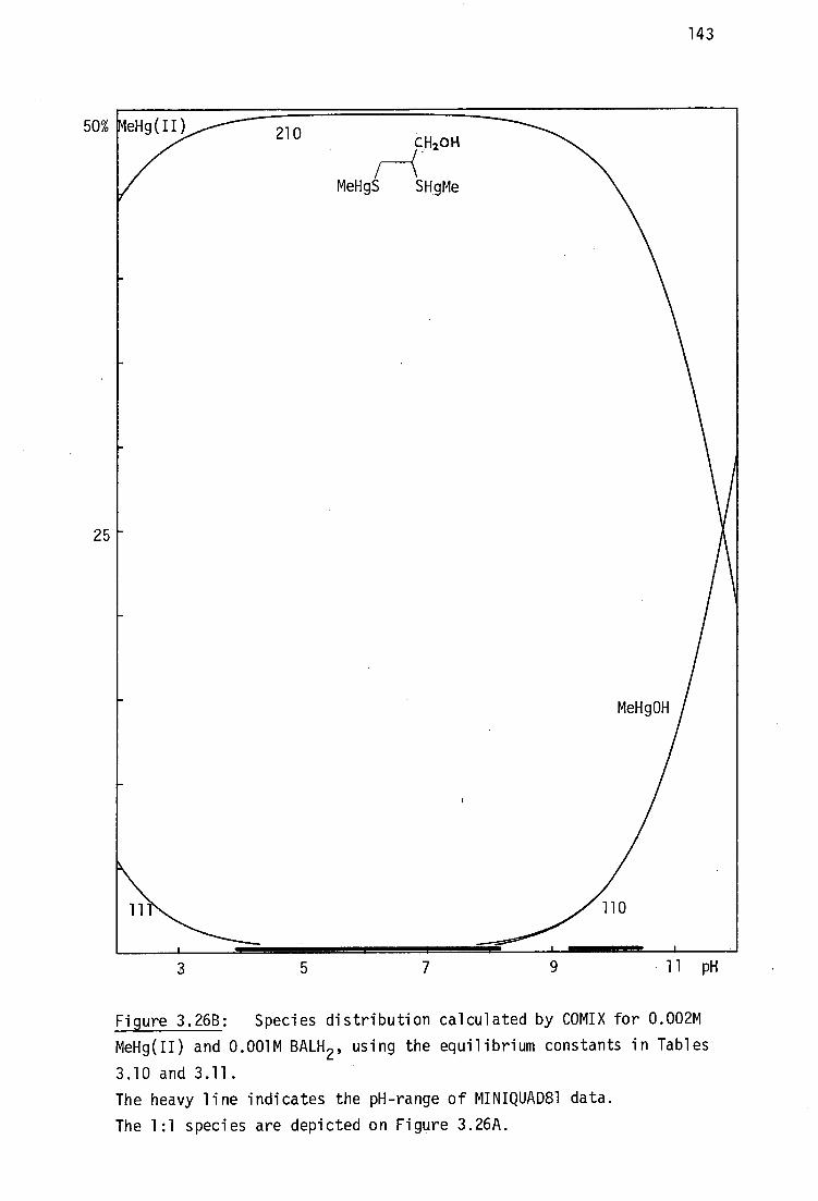
143
MeHg(II) 210
CH2014
MeHgS SHgMe
MeHg0H
50%
25
3
5
7
9
11 pH
Figure 3.26B: Species distribution calculated by COMIX for 0.002M
MeHg(II) and 0.001M BALH 2 , using the equilibrium constants in Tables
3.10 and 3.11.
The heavy line indicates the pH-range of MINIQUAD81 data.
The 1:1 species are depicted on Figure 3.26A.
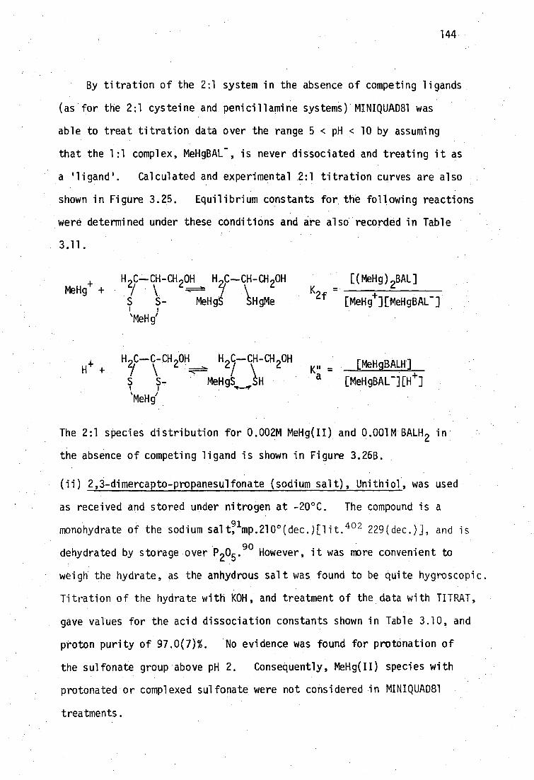
K = 2f + -
[MeHg ][MeHgBAL ]
[(MeHg) 2BAL]
144
By titration of the 2:1 system in the absence of competing ligands
(as for the 2:1 cysteine and penicillamine systems) MINIQUAD81 was
able to treat titration data over the range 5 < pH < 10 by assuming
that the 1:1 complex, MeHgBAL - , is never dissociated and treating it as
a ligand'. Calculated and experimental 2:1 titration curves are also
shown in Figure 3.25. Equilibrium constants for the following reactions
were determined under these conditions and are also recorded in Table
3.11.
MeHg+
+ H2C--CH-CH OH H C--CH-CH 2 OH
( \
S S- MeHgS 1-1gMe
‘MeHg'
H C--C-CH OH H C--CH-CH OH H + \ 2 2/\ 2
MeHgS H
'MeHg/
- [MeHgBALH]
[MeHgBAL -][e]
The 2:1 species distribution for 0.002M MeHg(II) and 0.001M BALH 2 in
the absence of competing ligand is shown in Figure 3.26B.
(ii) 2,3-dimercapto..-propanesulfonate (sodium sal -t), Unithiol, was used
as received and stored under nitrogen at -20°C. The compound is a
monohydrate of the sodium salt? imp.210 0 (dec.)E1it. 402 229(dec.)j, and is
dehydrated by storage over P 205 . 90 However, it was more convenient to
weigh the hydrate, as the anhydrous salt was found to be quite hygroscopic.
Titration of the hydrate with KOH, and treatment of the data with TITRAT,
gave values for the acid dissociation constants shown in Table 3.10, and
proton purity of 97.0(7)7. No evidence was found for protonation of
the sulfonate group above pH 2. Consequently, MeHg(II) species with
protonated or complexed sulfonate were not considered in MINIQUAD81
treatments.

145
Titration of equivalent quantities of Unithiol and MeHg(II) in
the presence of iodide indicated that iodide competition is inapprop-
riate as a method of evaluating log 13110' as was the case for BALH 2 .
Titration of equivalent quantities of mercaptoethanol, Unithiol and
MeHg(II) over the range 4 < pH < 11 and treatment with MINIQUAD81,
gave values of the constants, log110 [21.01(8)] and pq [7.56(1)] 13
for the species in the equilibria below:
+ H C--CH-CH
2 SO
3 H C--CH-CH 2 SO3 - _ [MeHgUT2- 4
MeHg + 3 _ 2/ \
S S S S 110
[MeHg+HUTJ7 ] 1 /
(UT3- )
H 9p,-CH-CH 9S
MeHg' + 7 \ -
-s s- H C--CH-CH SO 2/ \ 3 _ [MeHgUTH 7 ]
' + + MeHgS SH
111 [MeHg HUT
3- ][H ]
The calculated and experimental 1:1:1 titration curve is shown in
Figure 3.27 and the species distribution in Figure 3.28A.
Formation of the 2:1 MeHg(II)-Unithiol complex was evaluated in the
absence of competing thiol or iodide, in an identical fashion to that for
BALH 2' to give the constants (Table 3.11) corresponding to the equilibria:
. /
'
H 2 C--CH-CH 3 2 Hf-- \ CH-CH-eS0
3 v . . [(MeHg)2UT-]
MeHig 2f + 2- MeHgS SHgMe [MeHg ][MeHgUT ]
MeH4
H,C--CH-CH,S0, 7 H oc—CH-CH 2SO3 H+ + y L 7, \ -s S 7 MeHgS SH
'MeH6
K" - [MeHgr]i.
a [MeHgUT 7][H ]
The calculated and experimental titration curve for the 2:1 system is
also shown in Figure 3.27, and the distribution diagram for 0.002M
MeHg(II) with 0.001M Unithiol, in the absence of competing ligand, in
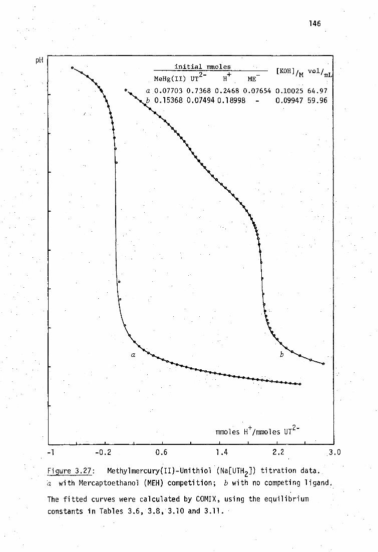
MeHg(II) UT2-
ME [KOH]im vol/mL
initial mmoles
a 0.07703 0.7368 0.2468 0.07654 0.10025 64.97 b 0.15368 0.07494 0.18998 - 0.09947 59.96
a
moles H /mmoles UT2-
146
-0.2
0.6
1.4
2.2
3.0
Figure 3.27: Methylmercury(II)-Unithiol (Na[UTH 2 ]) titration data.
a with Mercaptoethanol (MEH) competition; b with no competing ligand.
The fitted curves were calculated by COMIX, using the equilibrium
constants in Tables 3.6, 3.8, 3.10 and 3.11.
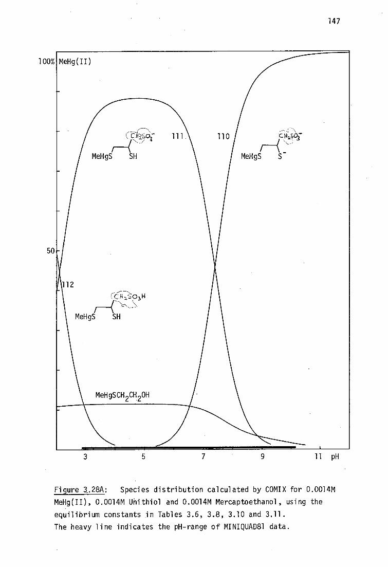
100%
50
147
3
5
7
9
11 pH
Figure 3.28A: Species distribution calculated by COMIX for 0.0014M
MeHg(II), 0.0014M Uhithiol and 0.0014M Mercaptoethanol, using the
equilibrium constants in Tables 3.6, 3.8, 3.10 and 3.11.
The heavy line indicates the pH-range of MINIQUAD81 data.

3 5 7 9 11 pH
148
50
25
Figure 3.28B: Species distribution calculated by COMIX for 0.002M
MeHg(II) and 0.001M Unithiol, using the equilibrium constants in
Tables 3.10 and 3.11.
The heavy line indicates the pH-range of MINIQUAD81 data.
The 1:1 species are depicted on Figure 3.28A.
111 11
110
% MeHg(II) 210 Az.sPi
MeNgS SHgMe
211 ^
‘-C14 2. so344 MeHg0H
MeHgS SHgMe

149
Figure 3.28B.
(iii) meso-2,3-dimercaptosuccinic acid was used as received but stored
415 over P
205
under nitrogen, nip. 185° (dec.) [lit. l82_50 15
(dec.) ]. Titration with 0.1M KOH, after addition of HNO3 to pH 2.8
to 3.0, and treatment of the data with TITRAT provided values of the
two carboxylic acid and two thiol proton dissociation constants recorded
in Table 3.10 and proton purity 97.6(3)%. The value for pKa4 is in
least agreement with previously recorded values. The determination of
pKa 's > 10.5 has been found in this study to be extremely sensitive to
the value used for K. K% was treated as a parameter by TITRAT and
hence contributes to some of the imprecision of pK a4 . The refined
values of Kvci for DMSH4 titrations are entirely consistent with those
values found during refinement of acidity constants for all the other
thiols used in this study, whereas the value for K often not
recorded by other workers or has been taken from previously reported
literature values. It is particularly noticeable (Tables 3.8 and
3.10) that large disparities exist in previously recorded pK a 's above
1 0.
In an equilibrium mixture of DMSH and one equivalent of MeHg(II),
the following equilibria involving 1:1 MeHg(II)-DMSH4 complexes are
expected:
S7 - [MeHgDMS.
3-]
6 HgMe
110 [MeHg
+][DMS
4-]
+,MbHg+
0 SH
- [MeHgDMSH
2-]
111 [MeHg
+][DMS
4-1[
+]
+ MeHg+
+ H
-02
SHgMe

C S -
+ 2MeHg+
• [(MeHg) 2DMS 2- ]
12.210 - +.) 4- [MeHg ] [DMS ]
150
7,0 C [MeHgDMSH
2
H ) [MeHg+][DMS4- ][e] 2
+ + i , -] + MeHg + 2H =z-F= 1 13.112 -
\02C SHgMe
HO 2C
+ MeHg+ + 3H+ ---
HO2C
SH)
SHgMe
[MeHgDMSH 3 ]
[meHg+][Dms4- ]oi+j 3
Unlike the 1:1 systems of MeHg(II)-BALH 2 and MeHg(II)-Unithiol,
titration of MeHg(II) and DMSH 4 in the presence of mercaptoethanol gave
a formation constant for the 1:1 complex' '
of approximately 10 16 , 3110
accompanied by three successive protonation equilibria as shown. And
in contrast to the BALH 2 and Unithiol 1:1 systems, in order to satis-
factorily fit the titration data with MINIQUAD81, a series of 2:1
species needed to be simultaneously included. Unsatisfactory results
were obtained by simultaneous refinement of all 1:1 and 2:1 constants,
since these were highly correlated, but the 2:1 constant 13 210 was
indicated to be in the range 1014
-1016
. Iodide competition was
therefore necessary to obtain the 2:1 formation constants for the
equilibria shown:
O C SHgMe
+ 2MeHg+
+ H+
H ‘ 02 C SHgMe
[(MeHg),DMSH - ] 13211 - + 2 4- +
[MeHg ] [DMS ][H ]
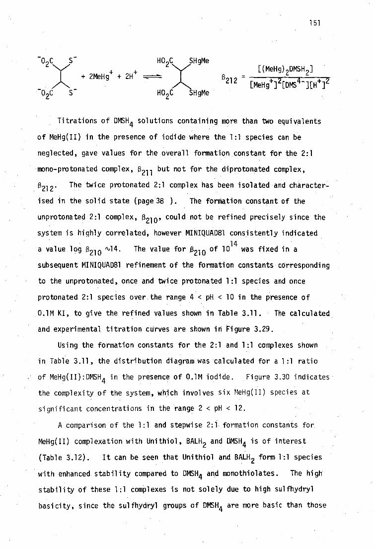
151
HO2C SHgMe
[(MeHg) 2DMSH 2 ]
212 _
[MeHg+] 2[DMS4- ][H+ ] 2 + 2MeHg
+ + 2H+
HO2C SHgMe
Titrations of DMSH 4 solutions containing more than two equivalents
of MeHg(II) in the presence of iodide where the 1:1 species can be
neglected, gave values for the overall formation constant for the 2:1
mono-protonated complex B ' 211 but not for the diprotonated complex,
13212
The twice protonated 2:1 complex has been isolated and character-
ised in the solid state (page 38 ). The formation constant of the
unprotonated 2:1 complex B ' - 210'
system is highly correlated, however MINIQUAD81 consistently indicated ,
a value log B -210
,L14. The value for 13 210 of 1014
was fixed in a
subsequent MINIQUAD81 refinement of the formation constants corresponding
to the unprotonated, once and twice protonated 1 .:1 species and once
protonated 2:1 species over.the range 4 < pH < 10 in the presence of
.0.1M KI, to give the refined values shown in Table 3.11. The calculated
and experimental titration curves are shown in Figure 3.29.
Using the formation constants for the 2:1 and 1:1 complexes shown
in Table 3.11, the distribution diagram was calculated for a 1:1 ratio
of MeHg(II):DMSH 4 in the presence of 0.1M iodide. Figure 3.30 indicates
the complexity of the system, which involves six MeHg(II) species at
significant concentrations in the range 2 < pH < 12.
A comparison of the 1:1 and stepwise 2:1 formation constants for
MeHg(II) complexation with Unithiol, BALH 2 and DMSH4 is of interest
(Table 3.12). It can be seen that Unithiol and BALH 2 form 1:1 species
with enhanced stability compared to DMSH 4 and monothiolates. The high
stability of these 1:1 complexes is not solely due to high sulfhydryl
basicity, since the sulfhydryl groups of DMSH 4 are more basic than those
could not be refined precisely since the

0
0 0
• 0
11
pH
152
initial mmoles [KOH]m
MeHg(II) UT3- H
a 0.07703 0.09267 0.10611 0.07603 -0.09840 68.61 b 0.07703 0.09267 0.46942 0.07603 0.10025 64.97 c 0.15358 0.14993 0.63577 5.9984 0.09943
mmoles eimmoles UT 3-
3 0 1.5
Figure 3.29: Methylmercury-Unithiol (Na[UTH 2]) titration data.
a,b with mercaptoethanol (L=ME - ) competition
c with iodide competition (L=I - )
The fitted curves were calculated by COMIXH using the equilibrium constants in Tables 3.6, 3.8, 3.10 and 3.11 with log (3(MeHgI) = 8.500.

3 5 9 7 11 pH
_0c c0_
2) 2
MeHgS S _-
0,C CO2-
4. ) (
MeHgS SHgMe
153
Figure 3.30: Species distribution calculated by COMIX for 0.0014M
MeHg(II) and 0.0014M DMSH 4 in 0.1 KI, using the equilibrium constants
in Tables 3.10 and 3.11.
Note the existence of a 2:1 species (green) at these concentrations.
The heavy line indicates the pH-range of MINIQUAD81 data.

154
of BALH 2 and Unithiol. The carboxylate groups of DMSH 22-
do not
appear to reduce the BrOnsted basicity of the sulfhydryl donors and
it is unlikely that an electronic effect could produce the marked
reduction in MeHg(II) affinity.
a b ligand log
110 log
13 B . 210-1°m P110 al (SH) pK2 (SH)
Unithiol 21.01(8) 10.26(7) 11.38(2) 8.69(1)
BALH2 '
19.56(9) 10.47(10) 10.62(1) 8.65(1)
DMSH4 17.11(3)
mercaptoacetic
acid° 17.10(1)
'114 11.50(1)
10.16(1)
9.65(1)
Table 3.12: Methylmercury(II)-sulfhydryl association constants for
the dithiolate antidotes.
aindicates the magnitude of binding of one MeHg(II) group to the dithiolate.
bindicates the magnitude of binding of MeHg(II) to the remaining sulfhydryl group of the 1:1 complex.
°typical monothiol included for comparison. Data taken
from Tables 3.6 and 3.8.
The enhanced stability is attributad to a chelate effect due to
bridging MeHg(II) in the 1:1 complexes of Unithiol and BALH 2 as
shown

H 21 C--CH-CH 2OH 1 S S \ Hg
Me
and
H C--CH-CH2SO
37
I I s s /
Hg
Me
155
The absence of such bridging in the 1:1 MeHg(II)-DMSH 4 complex may be
due •to an unfavorable conformation of the meso-dicarboxylic acid,
possibly caused by steric or electrostatic effects of the two bulky
negatively charged carboxylate groups. The 2:1 complexes for the
three vicinal dithiols all have overall formation constants in the
range 1030 to 10
31 Thus it would appear that the structures of the
2:1 complexes may be similar, and may all involve either simple mono-
dentate MeHg-S bonding, or chelated structures similar to that found
by Moore et al. for the 2:1 MeHg(II) complex of trans-1,2-cyclohexane-
dithio1 230 (Figure 2.4, page 42). Such chelating behaviour may also
be present in solution in the 1:1 complexes of Unithiol and BALH 2 .
There does not appear to be definitive vibrational spectroscopic
evidence for MeHg(II) chelation in the solid-state of these 2:1 anti-
dotal dithiol complexes (Chapter 2).
3.4 Interactions of antidotal thiols with MeHg(II) in vivo
The mechanisms of MeHg(II) toxicity will not be discussed here but
it is assumed that interactions with endogenous sulfhydryl groups are
of primary importance (Chapter 1). The modes of action of several mono-
thiols and vicinal dithiols for which stability constant data have been
obtained in this study, will be discussed in terms of their competition
with biological sulfhydryl groups for MeHg(II). It is further assumed
that the antidotal thiols (RSH) which will be protonated at physiological
pH act by removing MeHg(II) from binding sites of a model protein,
mercaptalbumin, AlbSH, according to the rapidly established equilibrium:
156
MeHgSAlb + RSH------ MeHgSR + AlbSH
The physicochemical properties of the complex MeHgSR are assumed to be
primarily responsible for the subsequent pattern of methylmercury
redistribution and hopefully, excretion. Polar and/or charged
complexes are expected to be amenable to rapid renal clearance. In
contrast, relatively non-polar, uncharged species are expected to have
enhanced lipid solubility, thus facilitating possible redistribution
through lipid membranes, in particular, the blood-brain barrier.
3.4.1 Physiological pH
In order to predict the metal-ligand species distribution in a
simple biological model, some estimate must be made of the 'pH'at or
near the site of metal interaction, e.g. in blood plasma, inside
erythrocytes or at the surface of cell membranes, etc. The pH of
many biological environments has been measured by both intrusive methods,
417 e.g. micro pH-electrodes or indirect methods, e.g. monitoring phosphate
equilibria in vivo by 31 P nmr.418
In this study, no attempt has been made to examine actual sites of
MeHg(II) binding in vivo. In discussing the possible existence of
methylmercury(II) thiolate species in extremely simplistic models of
biological systems, it has been assumed that the pH of the interactive
site is near 7.4. t Since no effort has been made to propose a complex
system of competing ligands, e.g. a plasma model such as that used for
2+ 2+ 2+ 3+ 2+ 2+ 2+ 419 Ca , Mg , Mn , Fe , Cu , Zn , Pb speciation, it was felt
that this pH represents a reasonable value for the sake of chemical
rather than toxicological discussion. Further, it will be seen that
tpH 7.40 is the generally accepted value for plasma pH under conditions • of pC0 9 = 5.33gp and constant saturation - fraction'of 02 in arterial blood at 370:
157
in most cases discussed here, variations in pH in the range 6.0 to
8.0 have little effect on conclusions drawn at pH 7.4. In addition,
it should be realised that the site of interaction may not even be in
an environment of constant pH, since many of the multitudinous reactions
in cell metabolism involve protolytic equilibria and which may be
dependent on the partial pressures of oxygen and CO 2 .417
3.4.2 Competition of antidotal thiols with endogenous ligands in vivo
For an antidotal thiol to remove intracellularly bound MeHg(II)
from protein binding sites, it must firstly be capable of reaching the
target cell without undergoing appreciable metabolic breakdown.
Cysteine and glutathione are unsuitable as antidotes since they are
readily incorporated into normal metabolic pathways,354
and in fact,
glutathione has been shown to increase the MeHg(II) content of the
82 brain after administration to mice. The metabolic product of some
antidotes may be more active than the administered form, e.g. the
substituted steroid thiomestrone (below) has some antidotal activity
towards HgC1 2 and MeHgC1 which may be due to the deacetylated (sulfhydryl
form). 120
MeOH
'SAc
Once transported to the MeHg(II) binding area, the thiolate must be
suited to passage through the cell membrane to the presumed intra-
cellular binding site. Here it must compete effectively with endogenous
ligands to form a MeHg(II) complex having the required properties to pass
out of the cell, and which may be sufficiently water soluble to be renally
excreted. Lipophilic MeHg(II) complexes may be excreted with the
AcS
Me
--Me
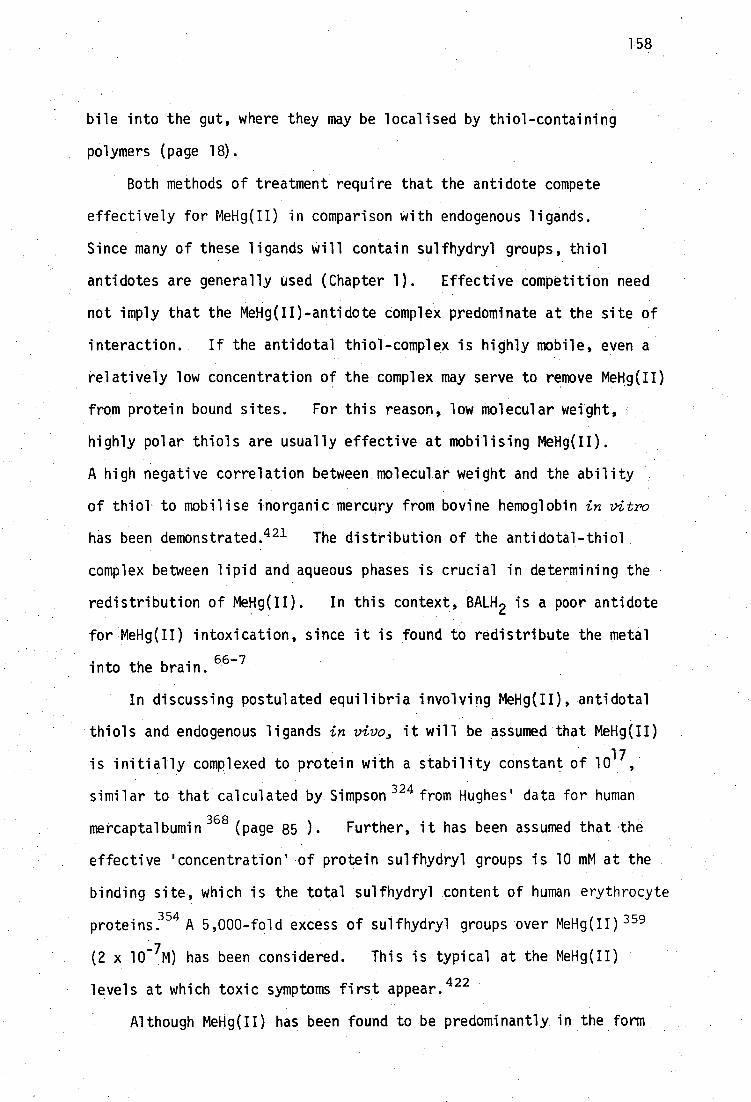
158
bile into the gut, where they may be localised by thiol-containing
polymers (page 18).
Both methods of treatment require that the antidote compete
effectively for MeHg(II) in comparison With endogenous ligands.
Since many of these ligands Will contain sulfhydryl groups, thiol
antidotes are generally used (Chapter 1). Effective competition need
not imply that the MeHg(II)-antidote complex predominate at the site of
interaction. If the antidotal thiol-complex is highly mobile, even a
relatively low concentration of the complex may serve to remove MeHg(II)
from protein bound sites. For this reason low molecular weight,
highly polar thiols are usually effective at Mobilising MeHg(II).
A high negative correlation between molecular weight and the ability
of thiol to mobilise inorganic mercury from bovine hemoglobin in vitro
has been demonstrated. 421 The distribution of the antidotal-thiol
complex between lipid and aqueous phases is crucial in determining the
redistribution of MeHg(II). In this context, BALH 2 is a poor antidote
for .MeHg(II) intoxication, since it is found to redistribute the metal
into the brain. 66-7
In discussing postulated equilibria involving MeHg(II), antidotal
. thiols and endogenous ligands in vivo, it will be assumed that MeHg(II)
is initially complexed to protein with a stability constant of 1017
similar to that calculated by Simpson 324 from Hughes' data for human
mercaptalbumin368 (page 85 ). Further, it has been assumed that the
effective 'concentration' of protein sulfhydryl groups is 10 mM at the
binding site, which is the total sulfhydryl content of human erythrocyte
proteins.354 A 5,000-fold excess of sulfhydryl groups over MeHg(II) 359
-7 (2 x 10 M) has been considered. This is typical at the MeHg(II)
levels at which toxic symptoms first appear.422
Although MeHg(II) has been found to be predominantly in the form

159
of a hemoglobin complex in erythrocytes, formation constants with
hemoglobin are unknown.
It is more difficult to estimate the antidote concentration at
the active . site. Under conditions used for animal studies, typically,
the antidote is given at a level more than lop times greater than the
MeHg(II) burden. If 1 g of antidotal monothiol with M.Wt. 200 was
equally dispersed throughout 5 L of body fluids, its concentration
would be 1 mM. As a comparison, the cellular glutathione cOncen-
tration is 0.1-10Nm.423
A preliminary in vitro distribution of
MeHg(II)-DMSH4 species using this antidote:metal ratio was presented
at a conference in 1981 and is included as Appendix 1 of this thesis.
Figure 3.31 shows conditional stability constants calculated under the
above conditions for the antidotal thiols penicillamine and N-acetYl-
penicillamine, and the dithiols BALH 2 , DMISH4 and Unithiol obtained in
this work. Human mercaptalbumin, described above, is included for
comparison. The MeHg(II) species distribution diagrams for each of
these antidotal thiols in the presence of human mercaptalbumin, are
shown in Figure 3.32. Although a chloride concentration of 0.1M was
included in the COMIX calculation to simulate the plasma chloride
content,424 MeHgC1 is negligible on the scale shown.
The relative proportions of protein-bound and antidote-bound
MeHg(II) species will vary considerably due to errors in the postulated
concentrations of MeHg(II), antidotal, and protein thiols, but the
pH-dependence of the species distribution will not. Therefore to
assess antidotal activity, we may consider the nature of the major
predicted antidotal - thiol MeHg(II) species at pH 7.4, irrespective of
whether it predominates over protein-bound MeHg(II).
For the antidotal thiols shown, except BALH 2 , it can be seen that
all the major MeHg(II) thiolate species are anionic at pH 7.4. It is

160
3 5 7 9 11 pH
Figure 3.31: Conditional stability constants of antidotal thiols and dithiols with MeHg(II).
Calculated using the equilibrium constants in Tables 3.6, 3.8, 3.10 and 3.11 for 2 x 10 -7M MeHg(II) and 0.1M chloride with 0.001M thiols: a DL-penicillamine, b N-acetyl-DL-penicillamine, c BALH 2 , d Unithiol and e DMSH4. f mercaptalbumin (0.010M) is included for comparison.
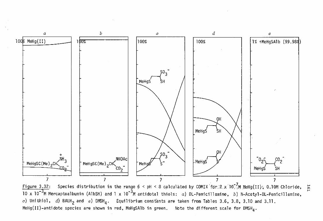
7 7 7 7 7
Figure 3.32: Species distribution in the range 6 < pH < 8 calculated by COMIX'fdr .2 x .1.07 7,M Mel-19( 1 0 -, 0.10M Chloride, _
10 x 10-3
M Mercaptoalbumin (AlbSH) and 1 x 10 3M antidotal thiols: a) DL-Penicillamine, b) N-Acetyl-DL-Penicillamine,
c) Unithiol, d) BALH 2 and e) DMSH4 . Equilibrium constants are taken from Tables 3.6, 3.8, 3.10 and 3.11.
MeHg(II)-antidote species are shown in red, MeHgSAlb in green. Note the different scale for DMSH4.
100 1 MeHg(II)
,NH 1 MeHgSC(Me) 2CHN
UU%
,NHOAc - MeHgSC(Me) 2CH\ _
CO2
1 00%
SO3-
MeHgS SH
100% 1% tMeHgSAlb (99.98%
0 C CO - 22
MeHgS SH
162
expected that these species would have high aqueous and relatively
low lipid solubility. All are 1:1 species, consistent with the low
MeHg:thiolate ratio at the active site.
The situation with BALH 2 is unique in this series of thiols in
that a large proportion of the MeHg(II)-BAL complex is uncharged at
pH 7.4 and probably relatively lipophilic. This presumably is
responsible for the ready passage of BAL-complexed MeHg(II) through
the blood/brain barrier. The species (MeHg) 2BAL is present at a
concentration of 10-17
M under these conditions, too low to affect
MeHg(II) redistribution. The low formation constants of DMISH 4 result
in a very small fraction of antidote-bound MeHg(II) compared with the
other thiols shown. This antidote has been shown to effectively
increase urinary MeHg(II) excretion however (page 15 ),supporting the
hypothesis that only small concentrations of mobile low molecular
weight species are necessary for efficient MeHg(II) transport.
3.5 Methylmercury(II) interactions with selenium donors in aqueous
solution
3.5.1 MeHg(II)-selenolate equilibria
There is indirect evidence from I H nmr studies that selenium in
selenoamino acids 4°4 and selenocyanate 323 has a higher affinity for
MeHg(II) than sulfur in corresponding mercaptoamino acids and thio-
cyanate. The pH-dependence of chemical shift data for MeHgSeCN has
been used to show that the formation constant of this complex (Table
3.2) is 0.72 log units higher than for MeHgSCN. 323 For several
selenoamino acids, the evidence relies on the observation that
2 1 J( H-
199 Hg)I is smaller than for analogous mercaptoamino acids.
Similarly2J(
1H)99
Hg)I is smaller for selenocyanate than for
thiocyanate 323 andselenide than for sulfide.323

163
Formation constants for MeHg(II) with selenohydryl-containing
ligands are of some interest, considering the presence of reduced
selenol groups in glutathione peroxidase176
and glycine reductase,179
and the uncertain method by which selenium reduces the toxicity of
MeHg(II) (page 27 ). Furthermore, selenols are stronger BrOnsted
425 acids than are thiols, e.g. selenocholine has a selenohydryl pK
a = 4.68(5),
while thiocholine has pK a = 7.883(4) (Table 3.6), yet I H nmr evidence
indicates the possibility of higher MeHg(II) stability constants with
selenols than for thiols. Because no MeHg(II)-selenohydryl formation
constants have been reported to date, some consideration was given in
this work to the potentiometric study of these systems.
Most of the monothiols investigated in this work have previously
reported selenium analogs. The ease of oxidation of the selenohydryl
group renders these ligands difficult to prepare and maintain in a
pure state, e.g. Zdansky doubts whether selenocysteine and seleno-
homocysteine have been obtained in pure forms.
426 The fairly lengthy
published synthetic routes to selenocysteine4! 27-8selenopenicillamine429
and selenocholine425 were not attempted. 2-Selenoethanol was prepared
in low yield by reduction of the diselenide, 43° but the oily compound
was difficult to handle and decomposed when stored over molecular
sieves (page 360). Selenoglutathione does not seem to have been
isolated in an unprotected form (Table 5.1).431
2-Selenosuccinic acid
is unknown (but see page 241 for a possible synthesis).
Because the complex MeHgSeCH 2CO2H had been prepared for solid-
state characterisation of MeHg(II)-selenolates (page 220, attempts
were made to study the MeHg-selenoacetate system in solution. The
proton nmr spectrum of this complex (Figure 3.33) has 1 2 J( 1 H- 199Hg)I
166.5 Hz, - which is smaller than that for the analogous thiolato complex
(170.0 Hz, page 359 ),but the difference is less than that for the seleno-
amino acids reported by Sugiura4"

7 6 8
1 2JI = 166.5 HZ
fifaiialv411)1444
164
Figure 3.33: Proton nmr spectrum of MeHgSeCH 2CO2H in D20
(pD = 5.4) showing 1H-
199Hg coupling. The methylene proton reson-
ance (6 = 7.06) is measured relative to dioxane (8=10.00) internal

165
and'shown . in Table 3.13. In order to determine the MeHg-selenoacetate
formation ,constant, the proton dissociation constant of the free ligand
is required.
Attempts to isolate the ligand by borohydride reduction of diseleno-
diacetic acid were unsuccessful. Since the MeHg(II)-selenoacetate
formation constant will be very sensitive to the dissociation constant
used for selenohydryl dissociation, any preparative procedure for the
ligand must ensure the absence of acidic or basic impurities. This
situation is difficult to achieve using commonly used veductants sodium
borohydride, hypophosphorus acid, etc. Although electrochemical
reduction of various diselenides has been reportedNhis technique does
not seem to have been used for the preparation of selenols for proton
dissociation studies, but may be suitable in this regard.
Microscopic hydrolysis constants of selenoacetic acid have been
previously determined spectrophotometricallyr 4 The selenol was prepared
by borohydride reduction of the diselenide and extraction into chloro-
form. From the small difference in 12J(
1H-
199Hg)I between selenolate-
and thiolate-complexes, it was expected that selenohydryl binding to
MeHg(II) would probably be less than one log unit (at most) stronger
than thiol binding. It was considered that the use of macroscopic
ligand hydrolysis constants derived from the microscopic constants
determined elsewhere, under different conditions, may have produced a
misleading comparison between selenoacetate and mercaptoacetate binding.
A valid comparison between MeHg(II) selenolate- and thiolate-
formation constants awaits future accurate determination of selenohydryl
dissociation constants. The MeHg(II) complex of ButSH has the lowest
mercury-sulfur coupling constant of MeHg(II)-thiolates studied to date.250
In fact, only the complexes with sulfide and the carbanions CH 3- and
C6H5- (Table 3.2) have lower values of
2J, reflecting the high basicity
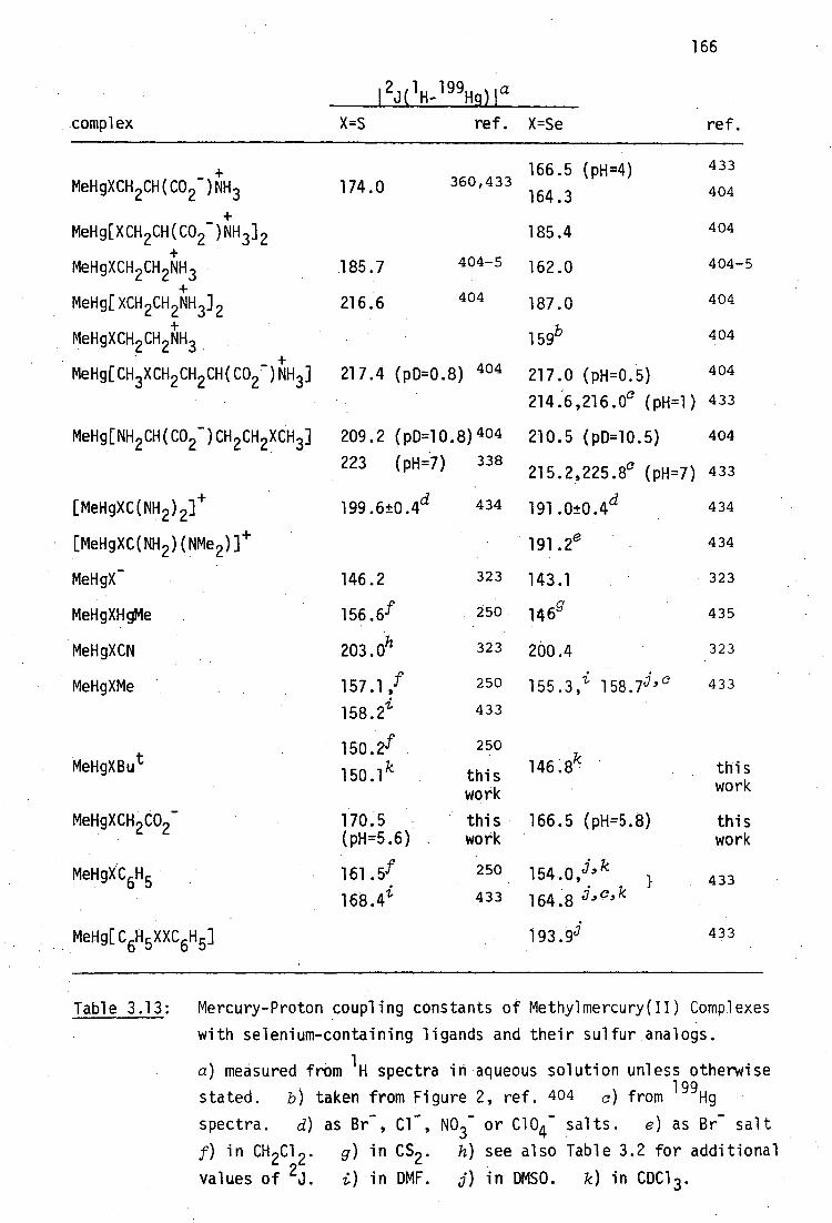
166
1 2J( 1 H_199Ho1 a
complex X=S ref. X=Se ref.
+ 166.5 (pH=4) 433
MeHgXCH 2CH(CO 2- )NH3 174.0 360,433 164.3 404
+ _ MeHg[XCH 2CH(CO 2 )NH 3 ] 2 185.4 404
+ MeHgXCH 2CH 2NH3 185.7 404-5 162.0 404-5
+ MeHg[XCH 2CH 2NH 3 ] 2 216.6 404 187.0 404
+ MeHgXCH2 CH 2NH3 159b 404
+ MeHg[CH3XCH 2 CH2 CH(CO2 )NH3 ] 217.4 (p0=0.8) 404 217.0 (p14=0.5) 404
214:6,216.0c (pH=1) 433
MeHg[NH 2CH(CO2 - )CH 2CH XCH 3 ] 209.2 (p0=10.8) 404 210.5 (p0=10.5) 404
223 (pH=7) 338 215.2,225.8c (p14=7) 433
[MeHgXC(NH 2 ) 2 ] 1- 199.6±0.4d 434 191.0±0.4d 434
[MeHgXC(NH 2 )(NMe )]/- 191.2e 434
MeHgX- 146.2 323 143.1 323
MeHgXHgMe 156.6f 250 146g 435
MeHgXCN 203.0h 323 200.4 323
MeHgXMe 157.1,f 250 155 . 3 , i 158 . 7j, 0 433
158.2i 433
150.2f 250
MeH gXBut .
1501
k this work
1 46.8k this work
MeHgXCH 2CO2 - 170.5 this 166.5 (pH=5.8) this (pH=5.6) work work
MeHgXC6H 161.5f 250 154.0,j' k 1 433
168.4i 433 164.8 j' c' k
MeHg[C6H 5XXC6H 5 ] 193.9i 433
Table 3.13: Mercury-Proton coupling constants of Methylmercury(II) Complexes
with selenium-containing ligands and their sulfur analogs.
a) measured from 1 14 spectra in aqueous solution unless otherwise
stated. b) taken from Figure 2, ref. 404 0) from 199Hg
spectra. d) as Br- , Cl - , NO3- or C104- salts. e) as Br- salt
A in CH 2C1 2 . g) in CS2 . h) see also Table 3.2 for additional
values of 2J. i) in DMF. j) in DMSO. k) in CDC13.

167
of the tert-butyl group. Since Bu tSeH had been prepared in order
to synthesise the new bis mercury(II) selenolate Hg(SeBu t ) 2
(page186 ) it was of interest to prepare MeHgSeBu t and compare
I 2J( 1H-
199Hig)1 for this complex with that of the sulfur analog.
Addition of an ethereal solution of ButSeH (prepared by the
Grignard route, page 349) to an ice-cold solution of MeHg0H
(3.3 mole) in 1:2 aqueous methanol (50 ml) and careful evaporation
of the combined hexane extracts (5 x 25 ml) at 0°C under low
vacuum (25 mm Hg), produced crude MeHgSeBu t . The white solid was
purified by sublimation (50-60°/15 mm), mp. 57-8.5°, and analysed
satisfactorily for carbon, hydrogen and mercury (page 351). The
analogous thiolate was prepared similarly using Bu tSH. The proton
nmr spectrum of MeHgSeBu t in CDC1 3 is shown in Figure 3.34. The
mercury-proton coupling constant (measured from an expanded spectrum)
was found to be 146.8 Hz, lower than that for the sulfur analog under
1 the same conditions. The H nmr resonances of the methyl and
tert-butyl protons are shifted downfield in MeHgSeBu t (cS = 0.85,
1.65 relative to TMS) compared with those in MeHgSBu
t (S = 0.78,
1.51). Similar small downfield shifts have been found in several
selenium-substituted dicarboxylic acids discussed in Chapter 5.
Due to the correlation between 2J and donor basicity,
MeHgSeBut presumably has the highest MeHg(II)-selenohydryl formation
constant of the selenols in Table 3.13.

168
C(CH)
HgCH3
IFJ] = . 146ji Hz
Figure 3.34: Proton nmr spectrum of MeHgSeBut in CDC1 3 showing
1 14- 199Hg coupling. Spinning sidebands are indicated by asterisks.
(Dioxane = 10.06).

169
3.5.2 MeHg(II)-Diselenide interactions
Sugiura et a/. have reported that values of 1 2J( 1 H- 199Hg)I in
mixtures of MeHg(II) and the diselenides, selenocystamine and seleno-
cystine (Table 3.13), are similar to those with sulfhydryl donors,
implying that these diselenides have an affinity for MeHg(II) comparable
to that of thiols.404
As the 'interactions of MeHg(II) with disulfides
are weak, the possibility of strong interaction in solution between
MeHg(II) and diselenodiacetic acid was investigated potentiometrically.
Attempts to obtain solid complexes from mixture of diselenodiacetic
acid and MeHgNO 3 in acetone were unsuccessful, always resulting in
recovery of diselenide. A similar situation resulted with dibenzyl-
diselenide which was unsuited to potentiometric studies here since it
is aprotic and insoluble in water. Elsewhere, attempts to prepare a
MeHg(II) complex with selenocystamine dihydrochloride have failed, and
no cleavage product was obtained. 433
Titrations of MeHg(II) and diselenodiacetic acid in the presence
of competing ligand in the range 2.7 < pH < 10.4, show that MeHg(II)
hydrolysis is suppressed, indicating complexation. These titrations,
shown in Figure 3.34, were treated with MINIQUAD81 but only poor fits
(U > 10-8 ) were obtained above pH 5.- Below this pH, the data can be
fitted as a simple diprotic acid with PKal = 3.04(3) and pK 82 = 4.00(4)
which are similar to values for the diselenide ligand, pKa1 = 3.196(1),
pKa 2 = 4.058(2). This is consistent with very weak interaction (if
any) with the carboxylate groups. It appears that the MeHg(II)-
diselenide complex is stable, with log 12. 10, since the titrations
can be fitted reasonably by assuming that MeHg(II) is never dissociated
from the complex below pH 5.
In the presence of 0.1M KI, insoluble MeHgI is formed below pH 10.5
with [MeHg(II)] = 1.5 x 10 -3M and [RSeSeR] = 3 x 10 -3 . The same MeHg(II)
170
concentration was used in the monothiolate systems described previously,
but MeHgI was absent above pH 4, thus it appears that the diselenide
is not as efficient at competing with iodide for MeHg(II) as the mono-
thiols described previously. Therefore the MeHg(II)-diselenide forma-
tion constant is probably less than 10 16 With more dilute solutions,
(0.05M I - , 0.7 x 10⁻³M MeHg(II), 1.5 x 10 -3M RSeSeR), no precipitation
of MeHgI was observed in the range 3 < pH < 11. This titration was
treated with MINIQUAD81, using several hypothetical systems of equilibria
involving 1:1 and 1:2 MeHg(II)-RSeSeR complexes and their protonated
analogs but the data could not be fitted adequately (U > 10 -8 ) over this
pH-range with any combination of these species.

Conclusions
Equilibrium constants which describe the aqueous solution
behaviour of equivalent quantities of MeHg(II) and various monothiols
over the pH-range 3 < pH < 10, have been determined potentiometrically
using iodide competition. The values obtained here agree well with
those found elsewhere by an nmr method 302
for the monothiol ligands
2-mercaptoehtanol, mercaptoacetic acid, mercaptosuccinic acid,
L-cysteine, DL-penicillamine, N-acetyl-DL-penicillamine and
glutathione. In addition, new formation constants are reported for
the thiocholine cation and 4-mercapto-N-methylpiperidine. Monothiolate-
MeHg(II) formation constants are now firmly established and may be
estimated from the correlation with pK a of the sulfhydryl donor:
log K (MeRgSR) = 0.893 pK (SH) + 7.77
Previously unreported equilibrium constants with the vicinal
dithiol antidotes BALH 2 and Unithiol indicate that significantly more
stable 1:1 complexation occurs for BALH 2 and Unithiol than is
expected from estimates of the basicity of the sulfhydryl donors
in these ligands. Chelation of MeHg(II) by the vicinal 6-donor atoms
is suggested as an explanation for this phenomenon. In contrast,
DMSH4 behaves like a monothiol inthe 1:1 interaction.
Equilibrium constants for the competition of human mercaptalbumin
with the antidotal thiols DL-penicillamine, N-Acetyl-DL-penicillamine,
Unithiol and DMSH4 for MeHg(II), indicate that polar, electrically
charged MeHg(II)-antidote species are formed at physiological pH.
In contrast, a large proportion of the complex formed withBALH 2 is
neutral and non-polar, which may explain the observed redistribution
of MeHg(II) into the brain upon administration of BALH2.66'67
171

CHAPTER FOUR
SYNTHESIS, VIBRATIONAL SPECTROSCOPY AND STRUCTURE OF
MERCURY(II) SELENOLATES
Direct chemical interactions between mercury and selenium have been
recognised for many years. The first characterised organoselenium
compound, 'selenomercaptan', EtSeH, produced the first example of
Hg-Se bonding in 1847 as yellow crystalline Hg(SeEt) 2436 Despite
the preparation of many such iselenomercaptides' as derivatives for
the qualitative identification of selenols, the structural chemistry
of these bis(Selenolato)mercury(II) compounds has not been studied.
It has already been shown (Chapter 1) that interactions between
selenium and mercury(II) may be of some importance in the mechanisms
of the protective effect of Se against inorganic (and perhaps organic
mercury(II) toxicity. In this light, it is therefore somewhat
surprising that no complexes, formed between mercUry(II) and selenohydryl-
containing ligands, have been investigated although Ganther 224 has
reported that the important selenite metabolite, selenodiglutathione
(Figure 1.1, page 20) interacts with Hg 2+ in vitro.
In this study, several complexes of the type Hg(SeR) 2 formed
between Hg2+ and simple monoselenols RSeH (R=Me,Et,Bu t ,CH 2Ph) and
CH 2 CO2 H) have been prepared and structurally characterised in the solid
state by X-ray diffraction and vibrational spectroscopic methods.
It will be demonstrated that several of these complexes are not
isostructural with their sulfur analogs. In addition, 1:1 complexes
of the type RSeHgX have been prepared and characterised either
directly (HgX=Me,0 2CMe) or as pyridine adducts (R,X=Et,C1; But,C1)
172

173
by crystallography.
It is hoped that some of these simple model compounds may be of use
for future characterisation of Hg-Se complexes of biological significance.
4.1 Preparation of mercury(II) selenolates
4.1.1 Preparation of Hg(SeR) 2 complexes
Several complexes Hg(SeR) 2 have previously been prepared as
derivatives for selenol identification, generally by two methods:
(i) reaction of the selenol with a mercuric salt or oxide following
the analogous mercaptide procedure of Wertheim437 or Claesson438
2 RSeH + HgX 2 Hg(SeR) 2 + 2HX R,X = Me, CN439 Et, C1 440
2 RSeH + Hg0 -± Hg(SeR) 2 + H20
R = t441
and (ii) reductive cleavage of diselenides by metallic mercury.
RSeSeR + Hg Hg(SeR) 2
(R = CH3443; CH2-- CO
2 H444 (and other alkyl carboxylic acids
)440; --
c05445-6 tru ru Iru ru , C6H5CH2CH2
; CF ; kw13/2kAl, 442 3447-8, c3F7,
C6 F5449-50 , and the mixed complex C6 F5SHgSeC6F5450 ).
Trifluoromethylselenyl chloride, CF 3SeC1, reacts with metallic
mercury to form Hg(SeCF3 )2447 . Bis(n-butylselenolato)mercury(II) has
been prepared by the reaction of diethylmercury with n-butylseleno1. 451
The compounds Hg(SeR) 2 , R=Me, Et, CH2CO2H have been obtained in this
work by the reductive cleavage of the appropriate diselenide with excess
metallic mercury. The compounds with R=Bu t and CH 2Ph were obtained from
the appropriate selenols and mercuric cyanide. All complexes have been
isolated in analytical purity (Table 4.1).
The complexes, except for the selenoacetate complex, are very poorly
soluble in common solvents.

calc %
found % mpt. Hg(SeR) 2
Method of preparation C H Hg C H Hg this work Hg(SR) 2
R = Me 6.2 1.6 51.6 6.3 1.5 51.8 119(dec) 175 + Hgi---2P-2-3.Hg(SeMe) 2
Et 11.5 2.4 48.2 11.2 2.3 47.7 72(dec) 76437
EtSeSeEt + Hg CHC13
Hg(SeEt) 2
But 20.3 3.8 42.2 20.3 3.9 42.7 145(dec
)b 159-60452 2ButSeH + Hg(CN)2 Et20
Hg(SeBut)2
CH Ph
CH 2CO2H
31 .L 2.6 37.1 30.9 2,6 36.9 111(dec) 117-8d 2PhCH2 SeH + Hg(CN) 2
MOH .
10.1 1.3 42.1 10.3 1.4 42.1 80(dec) c >190(dec) d (HO CCH 2Se) 2 + Hg 2.H SO 1.NaHCO/
>Hg(SeCH CO H) 2 24
Table 4:1: Methods of preparation and analytical data for bis(alkylselenolato)mercury(II) complexes.
aall compounds blacken without melting. bsublimes 165°/0.1 mm. °this compound is photosensitive.
dthis work.

175
The complex Hg(SeMe) 2 can be crystallised as yellow diamond-shaped
plates from a large volume of hot pyridine. These were sufficiently
well-formed to permit single crystal X-ray diffraction studies on the
complex - the first mercury(II) selenolate structure reported.
• The complex Hg(SeEt) 2 was obtained as a yellow crystalline product
by very slow evaporation (several months) of a pyridine solution.
However, the crystals were not of sufficient quality for X-ray structure
determination and give a very poor quality X-ray powder diffraction pattern
- perhaps indicating a highly polymeric structure.
The complex Hg(SeBu t ) 2 seems to be previously unreported and could
be recrystallised from benzene or chloroform as fine, hairlike, colourless
needles, very similar to the sulfur analog, but were unsuitable for X-ray
analysis. The reported single crystal X-ray structure of Hg(SBu t ) 2 used
crystals obtained by vacuum sublimation of the complex. The selenium
analog could be sublimed but the resulting crystals were extensively
twinned and unsuitable for single crystal analysis.
The complex Hg(SeCH 2Ph) 2 , previously reported by Antweiler1 49bwas
crystallised from benzene or chloroform as a yellow microcrystalline
product unsuitable for single crystal analysis. The sulfur analog was
obtained similarly as a white product.
The complex Hg(SeCH 2CO2H) 2 was obtained as a fine yellow powder on
acidification of an aqueous alkaline (bicarbonate) solution. It can be
recrystallised from methanol, but this does not produce high quality
crystals. Fredga has reported that this compound gives large crystals
(1 cm) when crystallised in large quantity from water, 44° but insufficient
material was obtained here for this approach. The sulfur analog was
obtained as small, shining, colourless plates by the reaction of two
equivalents of mercaptoacetic acid with mercuric cyanide in ethanol.
176
Although most of these Hg(SeR) 2 compounds have been reported
previously, they have not previously been structurally, characterised
in any way. The complexes prepared here are generally yellow (except
for Hg(SeBu t ) 2 , which is colourless) and light sensitive, particularly
water-soluble Hg(SeCH 2CO2H) 2 . An abstract of Fredga's early work with
this compound, records it as grey,444 which may have been due to contamination
by metallic mercury in his preparation. All of the compounds appear to
be stable when refrigerated in amber glass at -20 0 .
4.1.2 Prepartion of RSeHgX complexes
Thiolatomercury(II) complexes, RSHgX can be prepared by the reaction
of equimolar amounts of Hg(SR) 2 with mercuric salts,HgX 2 :
Hg(SR) + HgX2 2RSHgX
This reaction is an equilibrium (in Me0H) for CF 3SHgX (X = Cl,Br,I),
with equilibirium constants K = 2.3±0.3 for CF 3SHgBr and 20±10 for CF 3SHgC1
formation 290
Alternatively, reaction of HgX 2 with RSH also produces RSHgX
products:
HgX 2 + RSH RSHgX + HX
Many of these compounds form adducts with pyridine bases, some of which
have been crystallographically studied (Chapter 2, page 55 ).
The only analogous selenolatomercury(II) derivatives reported to
date have been CF 3SeHgX (X = Cl,Br,I,CN,SCN,02CMe).448 [Dr. A.J.
Carty (University of Waterloo) has kindly described the formation of
PhSeHgC1 from Hg(SePh) 2 and HgC1 2 ,433 but this work has not yet been
published.]
Of particular interest inthis study was the preparation of MeSeHg0 CMe,
177
whose sulfur analog has been shown by Raman spectroscopy to be dimeric in
aqueous solution at pH 6.292 Yellow, otherwise insoluble Hg(SeMe) 2
dissolves in an aqueous ethanolic solution of Hg(02CMe) 2 to precipitate
white, crystalline MeSeHg02CMe1' which has been shown by X-ray powder
diffraction (page 190) to be isomorphous with MeSHg02CMe. Unfortunately,
the selenium analog is not sufficiently water-soluble to allow Raman
spectroscopic investigation of its structure in solution. Preliminary
experiments with other Hg(SeR) 2 complexes suggest that preparation of
RSeHg0 2CMe complexes with Hg(0 2CMe) 2 may be a general phenomenon.
In contrast to the water-soluble MeSHg0 2CMe, monothiolatomercury(II)
halides are insoluble (Chapter 2, page 55 ) and so no new RSeHgX (X =
halide) complexes were characterised here, although preliminary experiments
showed that they are readily formed, as expected. HOwever, reaction of
Hg(SeR) 2 (R = Et,Bu t) with HgC1 2 in pyridine produced colourless crystalline
adducts which were recrystallised from ethanolic pyridine to produce
relatively large good-quality crystals. The single-crystal X-ray diffraction
structures of [EtSeHgC1(py)]4 and [Bu SeHgCl(py) 0.5 ] 4 will be described
later in this Chapter.
• MeSeHg02CMe (C3H602SeHg) requires: C 10.2, H 1.71, Hg 56.7%; found:
C 10.2, H 1.75, Hg 56.5%.
178
4.2 X-ray Diffraction Characterisation of Mercury(II)-Selenolates
4.2.1 Hg(SeR) 2
As a preliminary investigation into the structure of the complexes
Hg(SeR) 2 (R=Me, Et, Bu t , CH2Ph, CH 2CO2H), X-ray powder diffraction
patterns were obtained and compared with those of the sulfur analogs.
The complexes Hg(SR) 2 (R=Me,Et,Bu t 231 ' 453 , CH2Ph, CH 2CO2H) were obtained
as white crystalline products. The methanethiolato complex can occur
in two crystalline modifications. 291 The X-ray powder diffraction pattern
of Hg(SMe) 2 prepared by Wertheim's method 437 in this laboratory and re-
crystallised from ethanol can be indexed with unit-cell dimensions reported
by Bradley and Kunchur. 271 Samples prepared by addition of imidazole to
aqueous solutions of MeHgS02CMe291
gave a different powder diffraction
pattern. In historical context, these will be referred to as the 'old'
and 'new' forms respectively. Infrared spectra of the two forms are
similar, indicating linear S-Hg-S geometry in the new form. The structures
271 274 t273 of the compounds with R=Me, (old form), Et, Bu have been determined
by single crystal X-ray diffraction (Section 2.3, page 43 ).
Because the compounds Hg(SeMe) 2 ,Hg(SeEt) 2 , and Hg(SeBu t ) 2 have
unpleasant odours, Debye-Scherrer powder diffraction patterns were
recorded photographically from ground samples contained in sealed Lindemann
glass capillaries. t The analogous sulfur compounds were treated identi-
cally (though they are odourless) facilitating comparison of the patterns.
The seleno- and mercaptoacetate complexes are odourless and although
photographic powder patterns were obtained from samples in Lindemann
capillaries, better results were obtained from automatic chart-recordings
using larger (planar) specimens in a powder diffractometer.
For the qualitative comparison of the powder diffraction-patterns
tThe assistance of Mr. R. Ford (Geology Department) in this task is gratefully acknowledged.
179
of analogous sulfur- and selenium compounds, due care must be taken of
the different scattering powers of S and Se. Differences in atomic
radii of sulfur and selenium were expected to produce relatively small
changes in unit-cell parameters for isomorphous compounds. In
compounds containing large quantities of both selenium and mercury,
the intensity of the diffracted X-rays might be expected to differ
considerably from the sulfur analog, where the Hg-atom scattering
dominates more than for selenolates.
If the powder diffraction patterns of a known isomorphous pair of
Hg-S and Hg-Se compounds could be obtained, the magnitude of this sulfur
versus selenium 'phase' problem could be assessed qualitatively. Such
a pair of compounds was unavailable prior to this work.
It will be shown subsequently (see Figure 4.5), that the compounds
[ButSeHgCl(py) 0. 04 and [Bu tSHgC1(4-Mepy) 0.5]4 are isomorphous and that
the powder diffraction intensities and positions of both compounds are
very similar, and are particularly characteristic at low (2e<20 0 )
scattering angles. Powder diffraction patterns of pairs of compounds
Hg(XR) 2 (X=S,Se) (R=Me, Et, Bu t , CH 2CO2H, CH 2Ph) were examined quali-
tatively in view of this finding.
Hg(XMe) 2 (X=S,Se)
X-ray powder pattern data obtained for bis(methaneselenolato)
mercury(Inweremeasured from a photographic recording and are shown
in Table 4.2, together with powder diffraction data for both forms of
the sulfur analog.
The complex Hg(SeMe) 2 does not seem to be isomorphous with either
crystalline modification of its sulfur analog. Fortunately, crystals
of the selenolate grown from pyridine were available and a single-crystal
X-ray diffraction study was undertaken by Drs. A.H. White and B.W. Skelton

9.86s 10.05, 9.61, s db
8.91, 8.41, s,db
5.90 m
4.28w
4.09 w
3.80 m,br
3.57 w
3.50 w
3.25 w
8.3 s,br
6.63 vw
5.61 w
4.16m
3.90 w
3.63 m
3.31 w
3.17 vw
3.07 m
3.005 w
2.457 m
1.966 w
1.903 vw
1.602 vw
3.02, 2.99 w,db 2.90 w
2.87 w
2.70w
2.66 w 2.66 w
2.59 w 2.56 m
2.45 w 2.46 w
2.37 w 2.42 vw 2.23 w 2.27 m
2.22 vw
2.08, 2.05, w,db 2.05 w
1.99w 1.96 vw
1.96w 1.91 m
1.87w
1.78 vw
180
0 Hg(SMe d
meas/A Hg(SeMe) 2
d dmeas
/A
'old' ford° 'new' forma
Table 4.2: X-ray powder diffraction data for Hg(XMe) 2 (X=S,Se) a
athe finely ground samples were contained in 0.3 mm diameter Lindemann capillaries. bprepared by Wertheims method in ref. 291
aNi-filtered CuKa (12 hrs). dMn-filtered FeK (6.5 hrs.)
a

181
(University of Western Australia). The powder diffraction pattern
could be satisfactorily indexed using cell dimensions obtained from
the single crystal used for data collection.
Crystal and molecular structure of Hg(SeMe) 2
A crystal of this complex was mounted in Tasmania and preliminary
Weissenberg and oscillation photographs taken to ensure its suitability
for single crystal structure refinement. Subsequently, a yellow
prismatic crystal 0.10 x 0.16 x 0.16 mm was used for the crystallo-
graphic work at the University of Western Australia. A unique data
set was gathered at 295 K using a Syntex P2 1 four-circle diffractometer
in the conventional 20/0 mode, within the limits 20<50°. 1058 reflec-
tions were obtained, 818 of these with I>30 - (I) were considered 'observed'
and used in the least-squares refinement after absorption correction.
The crystal did not deteriorate appreciably during data collection.
Crystal data for Hg(SeMe) 2
C2H6HgSe 2 , M=388.6, Monoclinic, space group P21/c (Ch, No. 14).
a = 8.440(4), b = 10,732(3), c = 6.681(3) A
= 96.14(4)°
U = 601.7(4) A3 , Dc (Z=4) = 4.29 g cm -3
F(000) = 664 0 -1
Monochromatic Mo Ka radiation, X = 0.71069 A, p = 339 cm .
The structure was solved by the heavy atom method and refined by
full matrix least squares with anisotropic thermal parameters for the
non-hydrogen atoms. Hydrogen atoms were included with (x, y, z, U)
as invariant estimates. Neutral atom scattering factors were used,
those for non-hydrogen atoms being corrected for anomalous dispersion
454-6 f"). All computations were performed using the X-RAY
76 system 457 implemented by S.R. Hall on a Perkin-Elmer 32/40 computer

182
(University of Western Australia).
The structure was refined to residuals R, R' = 0.044, 0.057, with
reflection weights (a 2 (F0 )+0.0005(F0 ) 2 ) -1 .
The complex Hg(SeMe) 2 is polymeric, with infinite one-dimensional
chains along b. The mercury atoms are pseudo-tetrahedrally coordinated
to pairs of bridging methane-selenolato ligands. The chain is generated
by a succession of inversion centres and twofold (screw) rotations.
The pseudo-tetrahedral mercury geometry is similar to that in Hg(SBu t ) 2
although the thiolate has an inversion centre between each mercury atom.
An ORTEP diagram of the unit-cell contents, projected down c, is shown
in Figure 4.1.
The spacings between successive mercury atoms are different and 0
alternating, being 3.538(1) A for a pair of mercury atoms bridged by a 0
pair of Se(2) ligands (related by an inversion centre), and 4.070(2) A
for a pair separated by two Se(1) ligands (related by a 2-fold axis). 0
Mercury-selenium(1) distances are similar, 2.625(2), 2.659(2) A, and 0
are comparable to one of the Hg-Se(2) distances, 2.614(2) A. The other 0
Hg-Se(2) distance is appreciably longer, 2.764(2) A, reflecting the
distorted mercury geometry. Moreover, whereas the three angles, about
Se(1), are nearly equal, 98.6(7)-100.77(7)°, those about Se(2) are more
irregular, 82.61(6)-100.7(6)°. The smallest angle, 82.61(6)°, is
contained between two mercury atoms and Se(2). This geometrical
distortion about se(2) may be a consequence of methyl-methyl interactions
within the chain about its twofold (screw) axis. 0 0
The distances C(2)-C(2) and C(1)-C(1) are 4.21(3) A and 3.80(4) A
respectively.
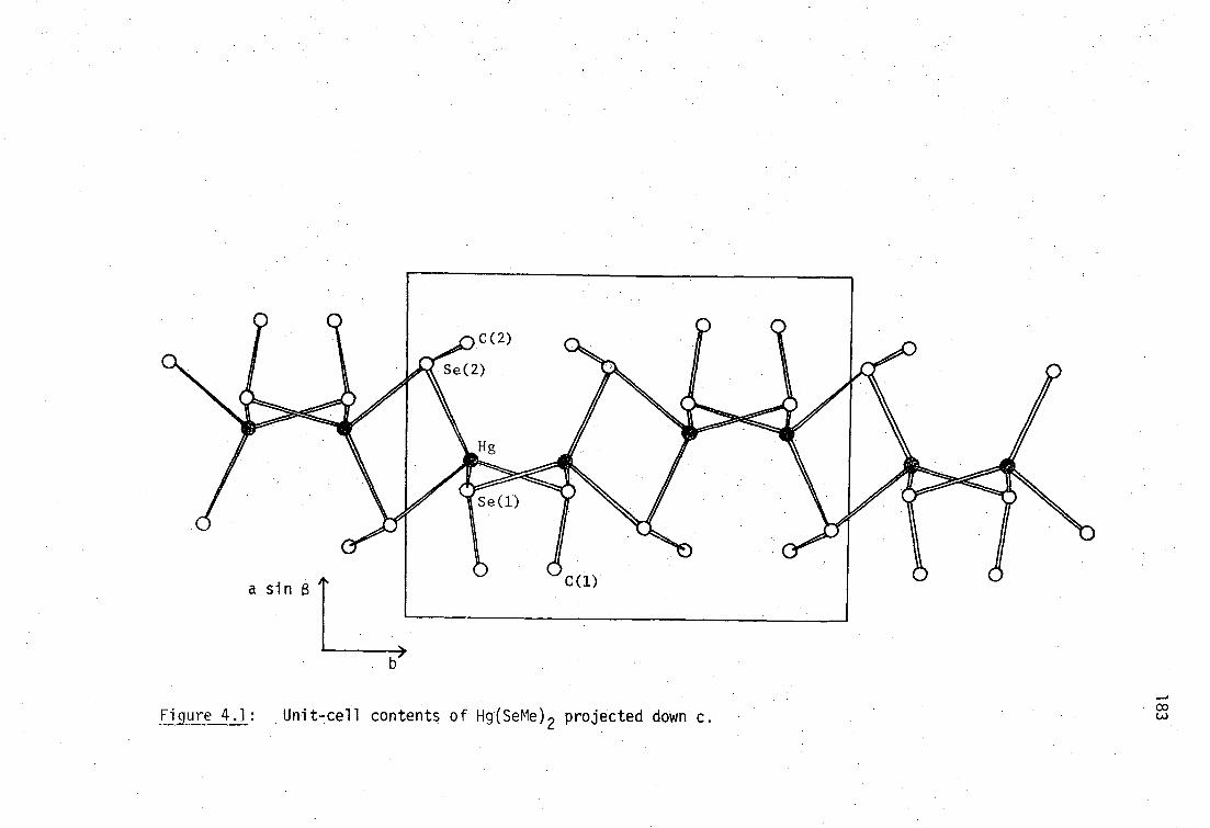
b>
Figure 4.1: Unit-cell contents of Hg(SeMe) 2 projected down c.

184
Atoms Parameters
0* Distances/A
Hg-Se(1) 2.625(2)
Hg-Se(2) 2.614(2)
Hg-Se(2 i ) 2.764(2)
Hg-Se(1 i1 ) 2.659(2)
Se(1)-C(1) 1.96(2)
Se(2)-C(2) 1.97(3)
Angles/deg.
Se(1)-Hg-Se(2) 124.63(8)
Se(1)-Hg-Se(2 1 ) 111.01(6)
Se(1)-Hg-Se(1 i1 ) 103.70(7)
Se(2)-Hg-Se(2 i ) 97.79(7)
Se(2)-Hg-Se(111
) 115.02(7)
102.64(6)
Hg-Se(1)-C(1) 98.6(7)
Hg-Se(1)-Hgiii 100.77(7)
C(1)-Se(1)-Hg' 99.3(8)
Hg-Se(2)-C(2) 100.7(6)
Hg-Se(2)-Hg 1 82.21(6)
C(2)-Se(2)-Hg 1 99.3(7)
Atom transformations: i (1-x,,i)
ii (x,1/2-y,4z)
iii (x,3/4-y,z-k)
*Hg...Hg i , 3.538(1) A, Hg...Hgi i , 4.070(2) A
Table 4.3: Interatomic bonding distances and angles (non-hydrogen atoms) for Hg(SeMe) 2 .
Least-squares estimated standard deviations of the last digit are given in parentheses.

Atom
U11
U22
U33 U12 U13
U23
Hg 0.45780(9) 0.14166(7) 0.11886(12) 38.2(5) 31.7(5) 41.6(6) 1.2(4) 13.3(3) -3.3(3)
Se(1) 0.3715(2) 0.3651(2) -0.0100(3) 36.7(11) 28.5(10) 32.2(10) -4.6(8) 7.3(8) -2.0(8)
C(1) 0.143(3) 0.335(2) -0.072(4) 38(12) 59(15) 71(16) 6(11) 21(11) 5(13)
H(1A) 0.120(-) 0.272(-) -0.176(-) 84(-)
H(1B) 0.092(-) 0.310(-) 0.042(-) 84 (-)
H(1C) 0.110(-) 0.414(-) -0.121(-) 84(-)
Se(2) 0.7335(2) 0.0392(2) 0.0774(3) 26.1(10) 31.1(10) 44.0(11) -5(8) 2.8(8) -3.3(9)
C(2) 0.794(3) 0.131(2) -0.158(4) 44(13) 30(11) 81(17) -14(9) 19(12) 4(11)
H(2A) 0.721(-) 0.118(-) -0.276(-) 78(-)
H(28) 0.805(-) 0.218(-) -0.135(-) 78(-)
H(2C) 0.895(-) 0.094(-) -0.176(-) 78(-)
° Table 4.4: Atomic coordinates (X,y,z), and anisotropic thermal parameters (10
3 A2 ) for Hg(SeMe) 2 .
Least-squares estimated standard deviations of the last digit are given in parentheses.
Anisotropic thermal parameters-are of the form: exp(-2n2(U11h
2a*2
+...2U23k1b*c*)).

186
Hg(XE0 2 , (X=S,Se)
The complex Hg(SeEt) 2 , contained in a 0.3 mm diameter Lindemann
capillary, gave a very diffuse X-ray powder pattern on exposure to
Ni-filtered CuKa (6 hours) or Fe-filtered CoKa (22 hours) radiation.
The sulfur analog, in contrast, diffracts well under the same conditions.
The selenolate is hard and brittle and may be a glass.
Hg(XBu t ) 2 , (X=S,Se)
The complex Hg(SBut
) 2 is of interest as it is the only simple 1:2
complex to show pseudo-tetrahedral 'HgS 4 ' geometry
.273
It is therefore
the model compound for vibrational spectroscopic characterisation of
thiolate-mercury(II) compounds with tetrahedral coordination. 231
Bis(tert-butaneselenolato)mercury(II) was isolated as fine, long,
colorless hairlike needles, identical in nature to the thiol analog..
These crystals were unsuitable for single crystal X-ray studies.
The powder diffraction data for Hg(XBu t ) 2 (X=S,Se) were obtained
photographically and are recorded in Table 4.5, and are very similar to
that obtained for the sulfur analog under identical conditions. The
selenolate data can be indexed satisfactorily using the published 273
orthorhombic unit-cell parameters of the sulfur analog.
The compounds Hg(XBu t ) 2 (X=S,Se) are therefore considered to be
isomorphous. The vibrational spectra are discussed in Section 4.3.1
in terms of tetrahedral ''HgSe 4 1 geometry.
Hg(XCH 2CO2H) 2 (X=S,Se)
The complex Hg(SCH 2CO2H) 2 can be dissolved in aqueous solution by
deprotonation, and vibrational studies in both, solid state and in
solution indicate that it has the expected linear 'HgS 2 ' coordination
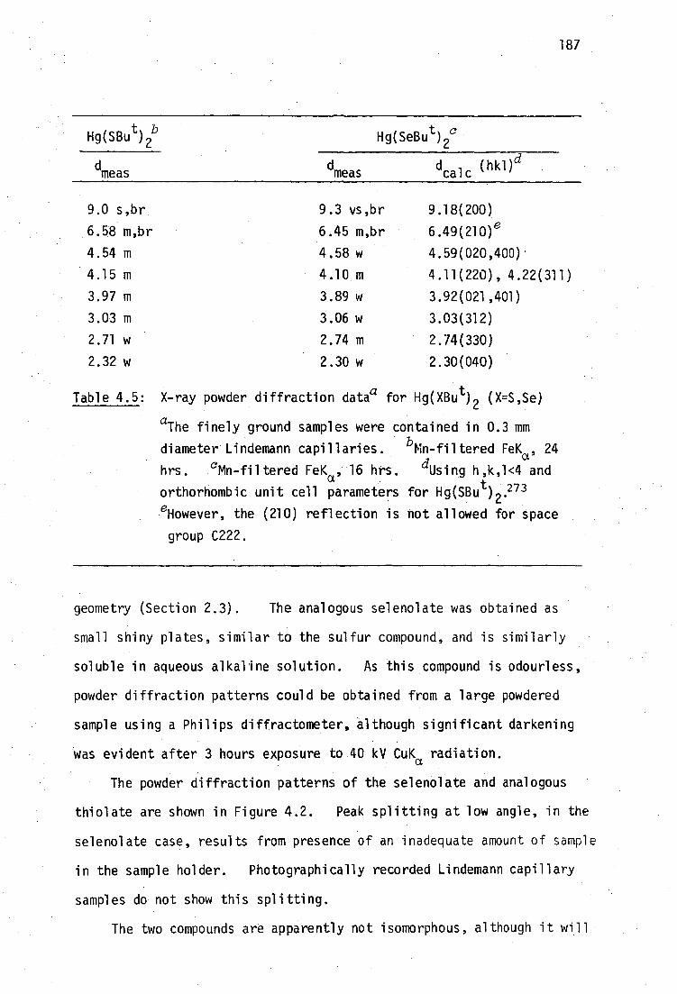
187
Hg(SBu t ) 26 Hg(SeBu t ) 2C
dcalc
(hkl)d
dmeas
dmeas
9.0 s,br
6.58 m,br
4.54 m
4.15 m
3.97 m
3.03 m
2.71 w
2.32 w
9.3 vs,br
6.45 m,br
4.58 w
4.10 m
3.89 w
3.06 w
2.74 m
2.30 w
9.18(200)
6•49(210) 6
4.59(020,400)
4.11(220), 4.22(311)
3.92(021,401)
3.03(312)
2.74(330)
2.30(040)
Table 4.5: X-ray powder diffraction data a for Hg(XBu t) 2 (X=S,Se)
aThe finely ground samples were contained in 0.3 mm
diameter Lindemann capillaries. bMn-filtered FeKa , 24
hrs. Nn-filtered FeKa , 16 hrs. dUsing h,k,1<4 and
orthorhombic unit cell parameters for Hg(SBu t ) 2 .273 eHowever, the (210) reflection is not allowed for space
group C222.
geometry (Section 2.3). The analogous selenolate was obtained as
small shiny plates, similar to the sulfur compound, and is similarly
soluble in aqueous alkaline solution. As this compound is odourless,
powder diffraction patterns could be obtained from a large powdered
sample using a Philips diffractometer, -although significant darkening
was evident after 3 hours exposure to 40 kV CuK a radiation.
The powder diffraction patterns of the selenolate and analogous
thiolate are shown in Figure 4.2. Peak splitting at low angle, in the
selenolate case, results from presence of an inadequate amount of sample
in the sample holder. Photographically recorded Lindemann capillary
samples do not show this splitting.
The two compounds are apparently not isomorphous, although it will
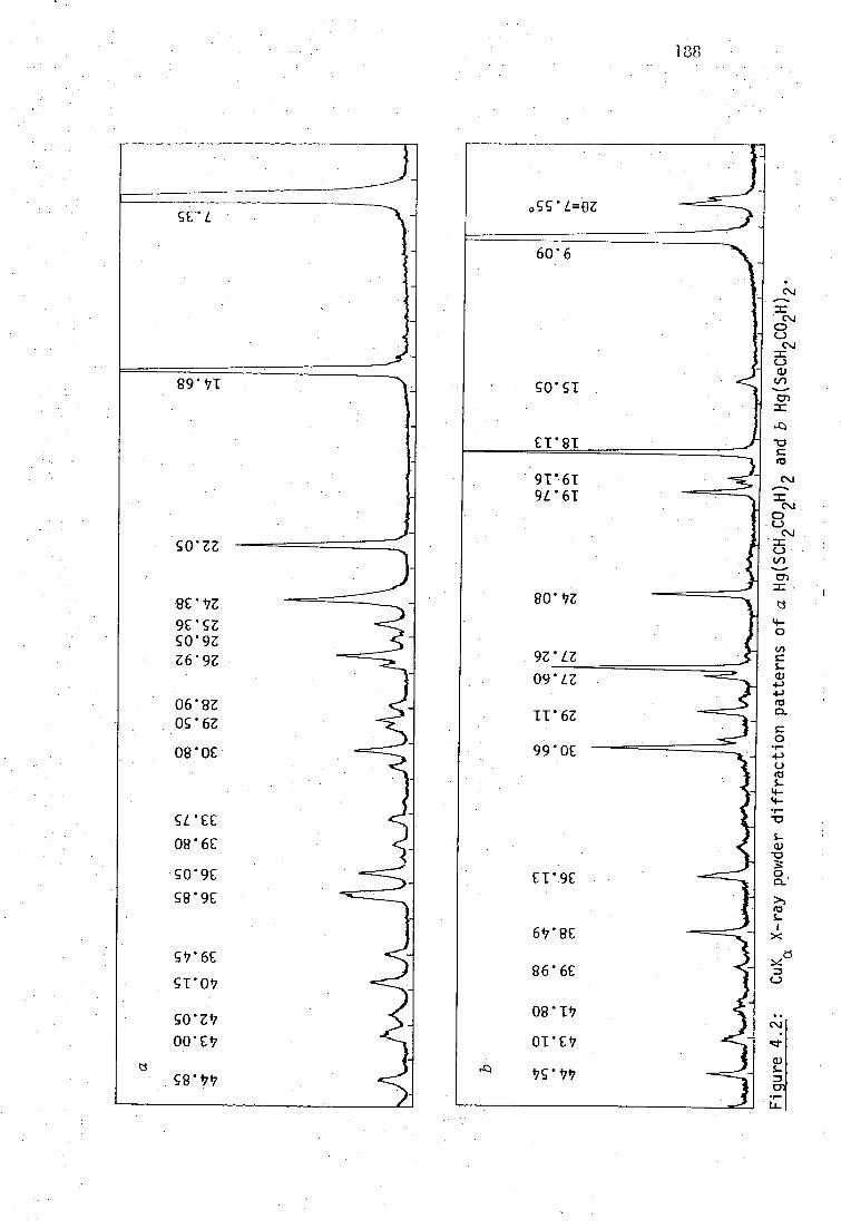
188
cc L
89 .471
2C -17Z 9E'SZ 50'9Z Z6'9Z
068Z Og'6Z
080C
SUCE 086E
- S0'9E S8'9C
5.47'6C
ST'0 17
co 00'CV
g8 .17 17
ogg'L=OZ
6076
gO'ST
ET'ST
91'61 9L61
80 .17Z
9Z'LZ 09'LZ
11 . 6Z
990C ----
Cr9C
6 47'8C
86'6C
08 —P7
175-47 17

8T *9=0Z____
ST*8 ---- -
89'8
9£'91 9C91 L0'8T 89 . 81
WOZ .
LS'EZ CitZ S9"7Z
SZ'9Z
8I'LZ
OT . 8Z
09 . 6Z
L8 . 0£ EVIC
189
6 17'S
8Z . 9 689 SU.L
c6 01
OUTT
00'ET Oc'ET
S9' 47T
_c
V)
-cr
•
_c C \
Cr)
4- 0 In
4-3 co
0 •r-
4- •r-
• -0
S-Q./ "0
0 • 0-
X
=

190
be shown from vibrational spectral studies (Section 4,3) that
Hg(SeCH 2CO 2H) 2 has linear 'HgSe 2 ' in the solid state.
Hg(XCH 2Ph) 2 (X=S,Se)
X-ray powder diffraction patterns obtained from these compounds,
as flat specimens in a diffractometer, show significant differences
in the low-angle scattering region (Figure 4.3). Subsequent vibra-
tional studies (page 220) have shown that, like Hg(XMe) 2 , the sulfur
compound has linear 'HgS 2 1 , but the selenium compound has tetrahedral
'HgSe4 1 geometry.
4.2.2 X-ray diffraction studies of RSeHgX
In a similar manner to Hg(SeR) 2 compounds, X-ray powder diffraction
patterns were obtained from analogous pairs of compounds MeXHg02CMe
(X=S,Se) and the pyridine adducts of Bu tXHgC1 (X=S,Se).
MeXHg02CMe (X=S,Se)
The sulfur compound of the analogous pair has previously been
characterised by single crystal X-ray diffraction. 293 The X-ray
powder diffraction pattern of MeSeHg0 2CMe, obtained photographically,
is identical to that obtained from MeSHg0 2CMe under similar conditions
(Table 4.6). The pattern can be adequately indexed using monoclinic
unit cell parameters obtained from the single-crystal study of
MeSHg0 2CMe.293
The selenium compound was therefore considered to be isomorphous
with MeSHg0 2CMe, and thus has a polymeric structure of (-Hg-SeMe-) n
chains linked into sheets by bridging acetate groups (Figure 4.4).
The small amount of MeSeHg0 2CMe available could not be crystallised
sufficiently well for a single crystal X-ray diffraction confirmation
of its isomorphism with MeSHg02CMe.
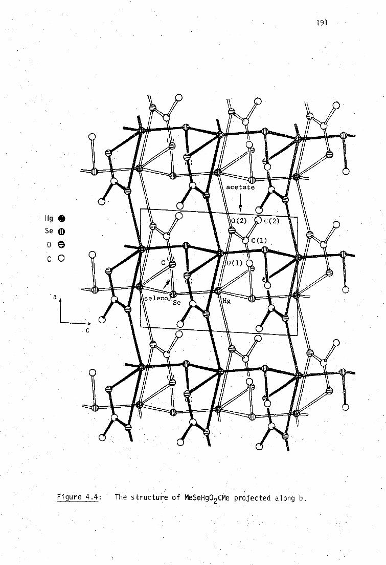
191
Figure 4.4: . The structure of MeSeHg0 CMe projected along
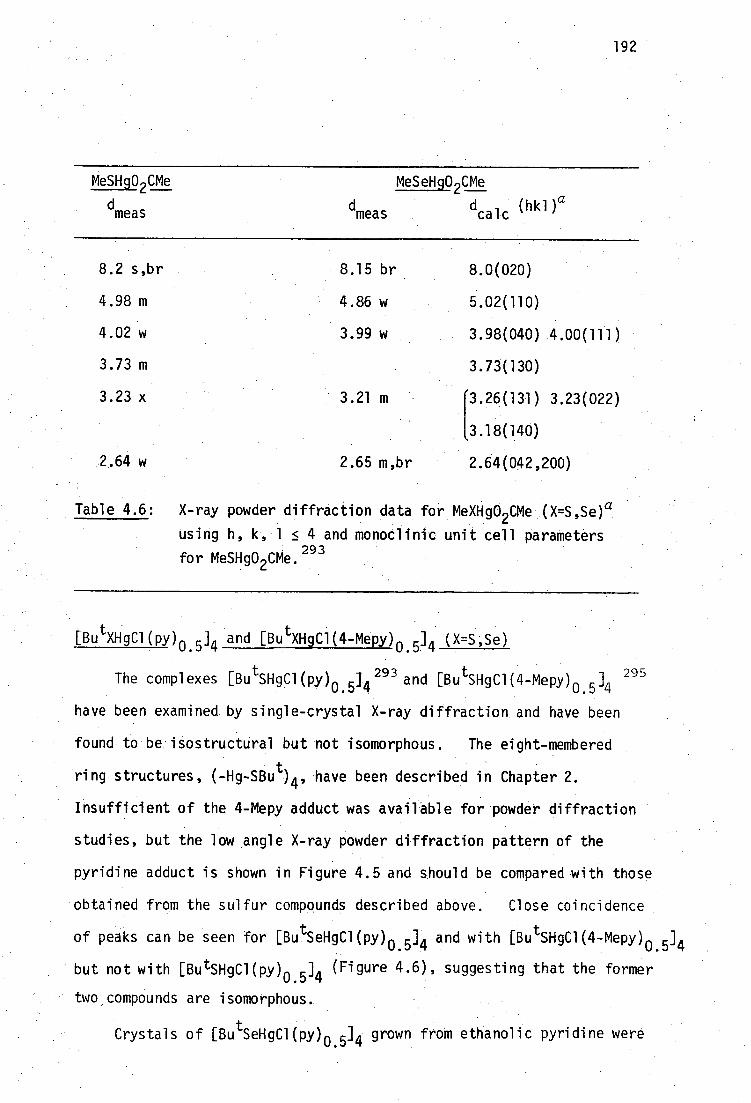
192
MeSHg02CMe MeSeHg0 2CMe
dmeas
dmeas dcalc
(hkl)a
8.2 s,br 8.15 br 8.0(020)
4.98 m 4.86 w 5.02(110)
4.02 w 3.99 w 3.98(040) 4.00(111)
3.73 m 3.73(130)
3.23 x 3.21 m 3.23(022)
[
3.26(131)
3.18(140)
2,64 w 2.65 m,br 2.64(042,200)
Table 4.6: X-ray powder diffraction data for MeXHg0 2CMe (X=S,Se) a
using h, k, 1 < 4 and monoclinic unit cell parameters
for MeSHg0 2CMe. 293
utX11C1(PY) 0. 04 and [Bu tXHgC1(4-Mepy) 0.5] 4 (X=S,Se)
The complexes [Bu tSHgCl(py) 0. 04 293 and [Bu tSHgC1(4-Mepy) 0. 04 295
have been examined by single-crystal X-ray diffraction and have been
found to be - isostructural but notisomorphous. The eight-membered
ring structures, (-Hg-SBu t ) 4 , have been described in Chapter 2.
Insufficient of the 4-Mepy adduct was available for powder diffraction
studies, but the low angle X-ray powder diffraction pattern of the
pyridine adduct is shown in Figure 4.5 and should be compared with those
obtained from the sulfur compounds described above. Close coincidence
of peaks can be seen for [BultSeHgCl(py) 0. 04 and with [Bu tSH9C1(4-Mepy) 0. 04
' but not with [ButSHgC1(py) 0. 04 (Figure 4.6), suggesting that the former
two compounds are isomorphous.
Crystals of [Bu tSeHgCl(py) 0. 04 grown from ethanolic pyridine were
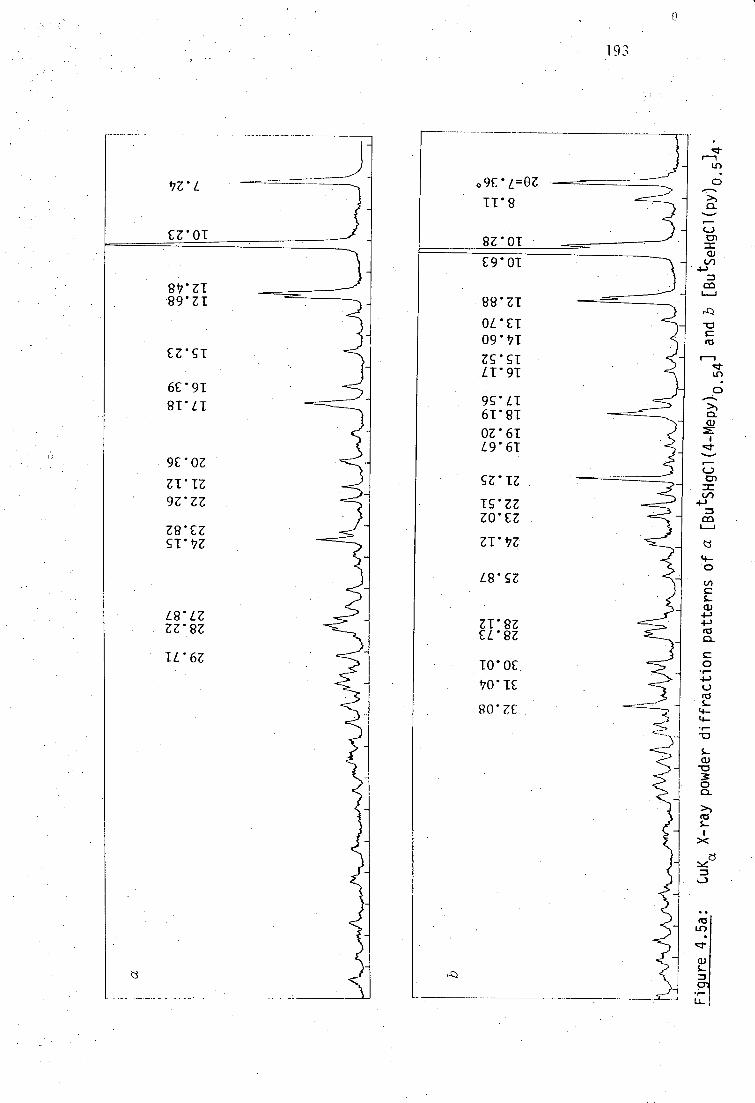
09PL=8Z
TT'S
SZ'OT
E9 . 0 -1
88 . ZT
OCET 09' 17T
Zg'ST L1 91
9S'LT 61 . 81 Or6T L9'61
SZ'TZ
TS'ZZ ZO*EZ
ZT't7Z
LR'SZ
ZI'SZ ECK
TO'0£
170'TE
RO'ZE
^
193
_ 47Z*L
EZ'OI
8 17 . ZT 89 . ZT
CZ'ST
6E91
81'LT
9E'OZ
ZI'TZ
9Z'ZZ
ZS'EZ ST' 47Z
L8'LZ ZZ•SZ
TL'6Z
r-1

co "DC co ■.o C,, -e-sr r-- co-4- N. Lfl o CNN- 0 CO %.0 CO r-I N. CO Lr.) cmo co • • • • • • • • • • • • • • • • 0
CD 01 131 CN1 ,-40 0303 1.11 C1.1 C11 1-6-1 cr) C,ICV C11 CN CN CN1 CN v-i r-I r-4 r-1 1-1 r-11-1 r-I
Figure 4.5b: CuK X-ray powder diffraction pattern of [ButSHgCl(py) ]. (cf. Fig. 4.5a) _ 0.5 Li

195
examined in Tasmania for single-crystal diffraction suitability.
Preliminary oscillation and Weissenberg photographs of one such crystal
mounted about the c-axis gave estimates of unit cell parameters c = 0 0
9.95 A and C(a) = 0.127, (b) = 0.09121 (giving a = 12.14 A, b = 0
16.91 A assuming a = = y sk, 90'). The 0-level Weissenberg photograph
required reflections Ok0, k=2n, consistent with species group P2 1 /c,
and also appeared to have all reflections h10 absent.
A single crystal X-ray diffraction study of this crystal was
undertaken by Drs. A.H. White and B.W. Skelton (University of Western
Australia).
Crystal and Molecular structure of [Bu tSeHgCl(py) 0. 04
A unique data set was gathered at 295 K from a crystal 0.26 x
0.1 x0.55 mm mounted in a capillary containing some pyridine, using
a Syntex P/2 1 four-cycle diffractometer in the conventional 20/0 mode
within the limits of 20<50°. 3209 reflections were obtained, 2016
with 1>30. (I) were considered to be 'observed'. The crystal blackened
badly during data collection, necessitating appropriate scaling of the
reflection intensities.
Crystal data for [Bu tSeHgCl(py) 0. 04
C8 H 50 Cl 4 Hg 2 N2 Se4' M = 836.4, Monoclinic, space group P2 1 /c.
a = 12.151(5), b = 16.738(7), c = 10.138(6) A, = 90.93(4)°
U =2062(2) A3 , Dc (Z=2) = 2.69 g cm-3
F(000) = 1512
Monochromatic MoK radiation, A = 0.71069 A, u mo = 180 cm-1 a
The structure was refined in an identical manner to that for Hg(SeMe) 2
(page 181) to residuals R, R' = 0.058, 0.066.
The tetranuclear complex [ButSeHgCl(py) (0.0 4 is isomorphous with
the 4-methylpyridine complex [Bu SHgC1(4-Mepy) 0.04 (a = 12.334(7),
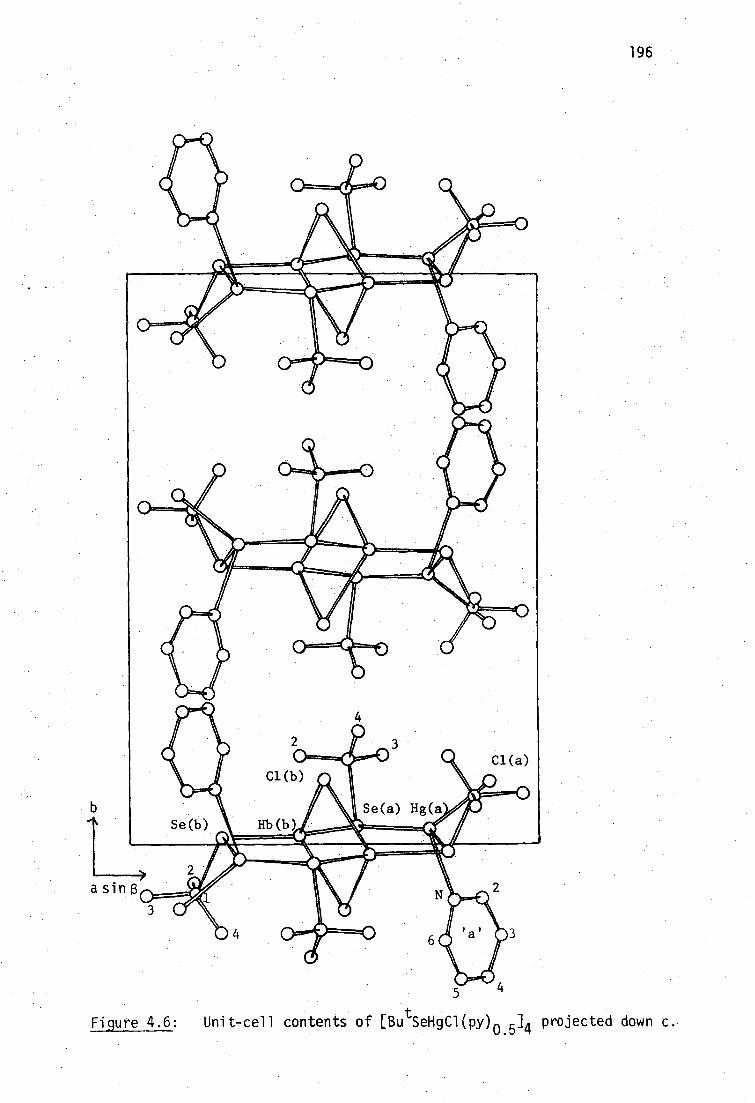
196
5
Unit-cell contents of Du (" ) 05 SeHgC1 4 projected down c. . Figure 4.6:

Atom Distance Atoms Angle Atoms Angle
Mercury a
Hg(a)-C1(a) 2.478(9) Cl(a)-Hg(a)-Se(a) 112.9(2) Se(a)-Hg(a)-Se(b 1 ) 133.7(1)
Hg(a)-Se(a) 2.579(3) Cl(a)-Hg(a)-N . 95.6(5) N-Hg(a)-Se(b 1 ) . 99.1(5)
Hg(a)-N . 2.49(2) Cl(a)-Hg(a)-Se(b 1 ) 110.6(2) Hg(b)...Hg(a)...Hg(b i )
Hg(a)-Se(b 1 ) 2.589(3) Se(a)-Hg(a)-N 91.7(5)
Mercury b
Hg(b)-C1(b) 2.723(6) Cl(b)-Hg(b)-Se(b) 89.4(1) Hg(a)...Hg(b)...Hg(a i ) 123.85(3)
Hg(b)-Se(b) 2.501(3) Cl(b)-Hg(b)-Se(a) 107.1(1) Hg(b i )...Hg(b)...Hg(a i ) 62.10(2)
Hg(b)-Se(a) 2.503(3) C1(b)-Hg(b)-C1(b 1 ) 95.2(2)
Hg(b)-C1(b 1 ) 2.720(6) Se(b)-Hg(b)-Se(a) 155.0(1)
Hg(b)...Hg(a) 3.905(2) Se(b)-Hg(b)-C1(b 1 ) 108.2(1)
Hg(b)...Hg(b 1 ) 3.670(2) Se(a)-Hg(b)-C1(b 1 ) . 89.3(1)
Hg(b)...Hg(a 1 ) 3.892(2) Hg(a)...Hg(b)...Hg(b 1 )
. Chlorine b
Hg(b)-C1(b)-Hg(b 1 ) 84.8(2)
Selenol a
Se(a)-C(1a) 2.03(2) Hg(b)-Se(a)-Hg(a) 100.4(1) Se(a)-C(1a)-C(4a) 111(17)
C(1a)-C(2a) 1.51(3) Hg(b)-Se(a)-C(1a) 102.0(6) C(2a)-C(1a)-C(3a) 111(2)
C(1a)-C(3a) 1.56(3) Hg(a)-Se(a)-C(1a) 105.3(7) C(2a)-C(1a)-C(4a) 114(2)
0 cont... Table 4.7: Interatomic distances (A) and angles (degrees) for [Bu tSeHgCl(py) 0.5]4 .
Transformation of the asymmetric unit: i (1-x, 1-z)
Least-squares estimated standard deviations of the last digit are given in parentheses

C(1 a)-C(4a) 1.42(4) Se(a) 7C(1a)-C(2a) 105(2) C(3 )-C(1a)-C(4a) 113(2)
Se(a)-.C(la)-C(3a) 102(2)
Selenol b
Se(b)-C(1b) 2.02(3) .Hg(b) , -Se(b)-C(1b) 102.3(7) Se(b)-C(1b)-C(4b) 111(2)
C(1b)-C(2b) 1.50(4) H4(a 1 )-Se(b)-C(1b) 106.3(8) C(2b)-C(1b)-C(2b) 106(2)
C(1b)-C(3b) 1.54(4) Hg(b) 7Se(b)-Hg(a 1 ) 99.7(1) C(2b)-C(1b)-C(4b) 115(3)
C(1b)-C(4b) 1.44(4) Se(b)-C(1b)-C(2b) 106(3) C(3b)-C(1b)-C(4b) 113(3)
Se(0)-C(1b)-C(3b) 106(2)
Pyridine
N-C(2) 1.37(3) Hg(a)-N-C(2) 120(2) C(4)-C(5)-C(6) 117(3)
N-C(6) 1.27(5) Hg(a)-N-C(6) 126(2) C(5)-C(6)-N 129(3)
C(2)-C(3) 1.33(5) C(2)-N-C(6) 113(3)
C(3)-C(4) 1.38(6) N-C(2)-C(3) 124(3)
C(4)-C(5) 1.35(6) C(2)-C(3)-C(4) 118(3)
C(5)-C(6) 1.33(5) C(3)-C(4)-C(5) 118(3)
Table 4.7:
Interatomic distances (A) and angles (degrees) for [Bu tSeHgCl(py) 0.0 4
(cont.)
Transformation of the asymmetric unit: i (1-x, je, 1-z)
Least-squares estimated standard deviations of the last digit are given in parentheses.
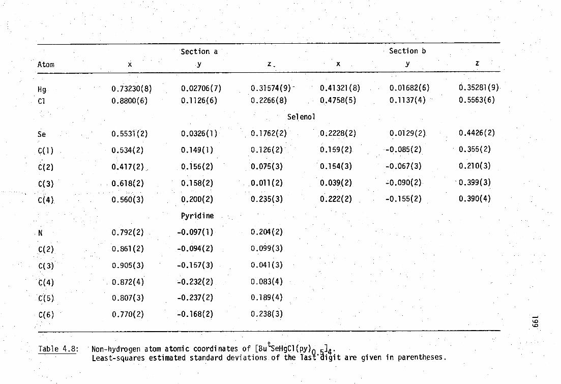
Atom
Section a Section b
Hg 0.73230(8) 0.02706(7) 0.31574(9) - 0.41321(8) 0.01682(6) 0.35281(9)
Cl 0.8800(6) 0.1126(6) 0.2266(8) 0.4758(5) 0.1137(4) 0.5563(6)
Selenol
Se 0.5531(2) 0.0326(1) 0.1762(2) 0.2228(2) 0.0129(2) 0.4426(2)
C(1) 0.534(2) 0.149(1) 0.126(2) 0.159(2) -0.085(2) 0.355(2)
C(2) 0.417(2) 0.156(2) 0.075(3) 0.154(3) -0.067(3) 0.210(3)
C(3) 0.618(2) 0.158(2) 0.011(2) 0.039(2) -0.090(2) '0.399(3)
C(4) 0.560(3) 0.200(2) 0.235(3) 0.222(2) -0.155(2) 0.390(4)
Pyridine
N 0.792(2) -0.097(1) 0.204(2)
C(2) 0.861(2) -0.094(2) 0.099(3)
C(3) 0.905(3) -0.157(3) 0.041(3)
C(4) 0.872(4) -0.232(2) 0.083(4)
C(5) 0.807(3) -0.237(2) 0.189(4)
C(6) 0.770(2) -0.168(2) 0.238(3)
Table 4,8:
'Non-hydrogen atom atomic coordinates of [Bu SeHgCl(py) 04'
Least-squares , estimated standard deviations of the Tas digit are given in parentheses.

200
Atom U 11 U22 U33 U12 U13 U23
Section a
Hg 29.8(6) 47.6(7) 48.6(6) -1.0(5) -0.1(4) 5.3(5)
Cl 42(5) 94(7) 91(6) -19(5) 6(4) 32(5)
Selenol a
Se 32(1) 28(1) 37(1) -1(1) -2(1) 0(1)
C(1) 53(17) 15(12) 31(12) -2(11) 11(10) -1(10)
C(2) 34(17) 46(17) 72(18) 13(13) 5(13) 16(15)
C(3) 54(18) 59(19) 39(14) -21(15) 1(12) 2(13)
C(4) 96(26) 51(20) 56(18) 4(18) -17(16) 17(15)
Pyridine
45(15) 47(15) 45(12) 11(12) -6(10) -1(11)
C(2) 56(20) 60(21) 54(17) 11(16) -1(13) -15(15)
C(3) 63(24) 137(41) 64(21) 0(26) 12(16) -21(24)
C(4) 103(31) 55(24) 104(29) -3(23) 9(22) -32(22)
C(5) 111(33) 46(21) 106(28) 7(21) 45(24) -8(21)
C(6) 57(21) 86(28) 58(18) -1(20) 23(14) -19(19)
Section b
Hg 27.4(6) 40.2(6) 43.9(5) -5.1(5) 1.5(4) 2.8(5)
Cl 45(4) 25(3) 50(3) -1(3) -1(3) -5(3)
Selenol b
Se 26(1) 47(2) 42(1) 2(1)' 1(9) 0.(1)
C(1) 22(15) 94(26) 55(16) -20(16) 7(11) -29(16)
C(2) 77(27) 227(59) 73(24) -62(33) 6(18) -24(29) .
C(3) 23(16) 89(25)• 76(19) -11(16) - 14(12) -23(13)
C(4) 31(21) 71(25). 222(44) -13(18) -35(23) -86(28)
Table 4.9: Anisotropic thermal parameters for [Bu tSeHgCl(py) 0.04 .
Form: exp(-2n2(U11h2a*2+...+2U23k1b*c*))

Atom 103 U A2 r A C-H
Selenol a H(2a) 0.404(-) 0.123(-) -0.002(-) 78(-) 0.96(-) 111(-) 109(-) H(2b) 0.365(-) 0.142(-) 0.140(-) 78(-) 0.95(-) 113(-) 110(-) H(2c) 0.407(-) 0.211(-) 0.050(-) 78(-) 0.96(-) 106(-) 108(-) H(3a) 0.691(-) 0.147(-) 0.036(-) 72(-) 0.94(_) 114(-) 109(-) H(3b) 0.598(-) 0.124(-) -0.063(-) 72(-) 0.96(-) 112(-) 109(-) H(3c) 0.610(-) 0.213(-) -0.015(-) 72(-) 0.97(-) 103(-) 110(-) H(4a) 0.514(-) 0.189(-) 0.307(-) 93(-) 0.95(-) 112(-) 108(-) H(4b) 0.636(-) 0.192(-) 0.264(-) 93(-) 0.98(-) 111(-) 105(-) H(4c) 0.553(-) 0.256(-) 0.213(-) 93(-) 0.97(-) 113(-) 106( - )
Selenol b H(2a) 0.105(-) -0.021(-) 0.191(-) 160(-) 1.00(-) 111(-) 106(-) H(2b) 0.223(-) -0.055(-) 0.173(-) 160(-) 0.94(-) 115 ( - ) 110(-) H(2c) 0.122(-) -0.122(-) 0.164(-) 160(-) 0.96(-) 109(-) 106(-) H(3a) 0.034(-) -0.095(-) 0.490(-) 81(-) 0.93(-) 112(-) 110(-) H(3b) -0.002(-) -0.044(-) 0.370(-) 81(-) 0.96(-) 111(-) 108(-) H(3c) 0.007(-) -0.136(-) 0.356(-) 81(-) 0.97(-) 106(-) 110(-) H(4a) 0.296(-) -0.151(-) 0.368(-) 133(-) 0.92(-) 113(-) 108(-) H(4b) 0.218(-) -0.163(-) 0.486(-) 133( - ) 0.99(-) 108(-) 105(-) H(4c) 0.191(-) -0.203(-) 0.352(-) 133(-) 0.96(-) 112(-) 110(-)
r.) c) _.,
Table 4.10: Methyl hydrogen atom parameters for [Bu SeHgCl(py) 0.04 . C-C-H , = H(n)-C-H(n+1), n = a,b,c.

202
0 b = 17.468(9), c = 9.999(5) A, a= 91.18(4)° ., space group P2 1 /c) but
not with the analogous pyridinate [Bu tSHgC1(py) 0. 04 (a = . 14.399(6),
b = 9.893(6), c = 9.597(5) A, a = 121.82(1), = 102.52(4), y =
102.94(4) , space group P1). These findings are consistent with X-ray
powder diffraction patterns. Consistent With the presence of pyridine
rather than 4-Mepy, the complex has an asymmetric unit Volume of
515.5(5) A3 ,' very similar to 517.8(4) A3 for [Bu tSH9C1(py) 0 .:04 , but
not the 4 .-Mepy analog, 538.5(5) A3 .
Finally, no peaks in the Fourier difference map could be attributed
to presence of a methyl carbon' of 4-Mepy, indicating that a mistake in
sample labelling or preparation had not occurred. In addition, a
- freshly prepared sample of [BU tSH9C1(py) .05]4 had all unit cell parameters
in agreement with those previously published. Molecules of tetrameric
[BUtSeHgCl(py) (L 0 4 are based on an eight-membered ring of alternating
mercury and selenium atoms (-Hg-SeBu )4. The rings have a centre of
symmetry and two mercury environments, Hg(1140 2 (i.17C1)2 and 'Hg(11-Se) 2C1(py)'
with a dichlOro bridge linking the former mercury atoms (Figure 4.6).
The close relationships between the eight Membered ring structures for
[Bu tSeHgCl(py) 0. 04 and the sulfur analogs [Bu tSHgC1(4-Mepy) 0 ..04
(isomorphous) and [ButSHgC1(py) 0. 04 can be clearly seen as projections .
of these structures normal to planes containing the four Se or S atoms
(figure 4.7). All of these compounds have distorted tetrahedral
geometry for mercury, with the largest angles formed by dominant
Se-Hg-Se and S-Hg-S moieties.
[EtSeHgC1(py)] 4
Reaction of equimolar quantities of Hg(SeEt) 2 and HgC1 2 in pyridine

203
produces a colorless crystalline complex which does not readily lose
pyridine and analyses as EtSeHgCl(py). The analogous sulfur compound
is unknown. High quality prismatic crystals were deposited when the
complex was dissolved in pyridine, and ethanol added, to produce a 1:1
Et0H/pyridine ratio.
A single crystal X-ray diffraction study was performed by Drs.
A.H. White and B.W. Skelton (University of Western Australia) on a
crystal of this complex.
Crystal and molecular structure Of [EtSeHgCl(py) . 14
A unique data set was gathered at 295 K from a crystal 0.11 x
0.07 x 0.06 mm mounted in a sealed capillary to minimise pyridine loss.
A Syntex P2 1 four-circle diffractomer was used in the conventional 26/6
mode with 20<60° to give 3794 reflections, 1678 with I>3a(I) were
considered to be 'observed'. The crystal blackened during data
collection, but no data scaling was necessary.
Crystal data for [EtSeHgCl(py)] 4
C 040C1 4Hg4N4Se4 , M = 1692.7 Monoclinic, space group P2 1 /c 0
a = 8.044(5), b = 17,387(14), c = 15.585(21) A, a= 101.75(2)° U = 2134(3) A3 , Dc (2.2) = 2.63 g cm-3
F(000) = 1520 0
Monochromatic MoK radiation, A = 0.71069 A, 1.1 174 cm-1 a Mo
The structure was refined in an identical manner to Hg(SeMe) 2 (page 181)
to residuals R,R = 0.046, 0.049.
Molecules of tetrameric [EtSeHgC1(py)]4 are based on the same eight-
membered ring found in tert-butylthiolato and tert-butylselenolato hemi-
base adducts described previously. The ( - Hg- SeEt- ) 4 rings contain
mercury atoms bridged by ethaneselenolate groups, but contain only one
mercury environment 'Hg(p-SeEt) 2C1(py).' in contrast to the t-butyl

Single molecules of a [EtSeHgCl(py)] 4 , b [ButSeHgC1(py) 0.5 ]4 , c [ButSeHgCl(py) 0.5 ]4
and dtSHgC1(4-Mepy) 0.5 1 4 projected normal to the plane of the four chalcogen atoms.
Tjaire 4.7:

205
Atoms Parameters
0 Distances/A
HgC1 2.543(7), 2.546(6)
Hg-N 2.42(2), 2.48(2)
Hg-Se(A) 2.542(3), 2.541(4)
Hg-Se(B) 2.543(3), 2.551(3)
Angles/degrees
Cl-Hg-N 92.8(5), 95.0(5)
Cl-Hg-Se(A) 111.9(2), 113.6(2)
Cl-Hg-Se(B) 103.7(2), 106.7(2)
N-Hg-Se(A) 99.3(5), 101.3(6)
N-Hg-Se(B) 106.5(5), 99.7(5)
Se(A)-Hg-Se(B) 134.7(1), 132.2(1)
Hg-Se-C(11) 103.6(8), 101.3(8)
Also: Hg(A), C(1B)-S (13)-Hg(A)(R,1-y,l-z), 99.20(8), 100.7(9)°.
Table 4.11 : Mercury/selenium geometries for [EtSeHgCl(py)] 4 .
The two entries in each column are for Hg, Se, A,B
respectively. Italicized entries involve Se(B)
(R,1-y,l-z).
Least-squares estimated standard deviations of the last
digit are given in parentheses.

206
Ligand a Ligand b
(a) ethaneselenolate ligand 0
Distances/A
Se-C(11)
C(11)-C(10)
1.92(3)
1.35(5)
1.99(3)
1.47(5)
Angles/degrees
(b)
Se-C(11)-C(12)
pyridine rings
120(3) 113(2)
0 Distances/A
N(1)-C(2) 1.33(4)
1.31(3)
1.36(4)
•N(1)-C(6) 1.32(4)
C(2)-C(3) 1.36(5) 1.38(4)
C(3)-C(4) 1.34(5) 1.40(4)
C(4)-C(5) 1.33(4) 1.37(5)
C(5)-C(6) 1.34(4) 1.36(5)
Angles/degrees
C(2)-N(1)-C(6) 118(2) 120(2)
N(1)-C(2)-C(3) 120(3) 120(2)
C(2)-C(3)-C(4) 121(3) 120(3)
C(3)-C(4)-C(5) 118(3) 118(3)
C(4)-C(5)-C(6) 119(3) 121(3)
C(5)-C(6)-N(1) 123(2) 122(3)
Hg-N(1)-C(2) 120(2) 114(2)
Hg-N(1)-C(6) 122(2) 125(2)
Table 4.12: Ligand geometries for [EtSeHgCl(py)] 4 .
Least-squares estimated standard deviations of the last digit are given in parentheses.

Atom
Section a Section b
Hg 0.00980(13) 0.47808(6) 0.29104(7) 0.21525(13) 0.43468(6) 0.53369(6)
Cl -0.0344(9) 0.3927(4) 0.1559(4) -0.0868(8) 0.3846(4) 0.5182(4)
Se 0.3016(3) 0.4589(1) 0.3880(2) 0.2883(3) 0.5038(1) 0.6809(2)
C(11) 0.373(4) 0.361(2) 0.351(2) 0.370(4) 0.417(2) 0.762(2)
C(12) 0.266(6) 0.301(2) 0.342(2) 0.421(5) 0.440(3) 0.854(3)
N(1) 0.068(3) 0.590(1) 0.290(1) 0.353(3) 0.309(1) 0.576(1)
C(2) 0.109(3) 0.657(2) 0.252(2) 0.519(4) 0.306(1) 0.571(2)
C(3) 0.153(4) 0.718(2) 0.207(2) 0.605(3) 0.237(2) 0.581(2)
C(4) 0.155(4) 0.713(2) 0.121(2) 0.518(4) 0.169(2) 0.592(2)
C(5) 0.115(4) 0.647(2) 0.081(2) 0.349(4) 0.175(2) 0.595(2)
C(6) 0.077(4) 0.586(1) 0.127(1) 0.270(4) 0.244(2) 0.585(2)
Table 4.13: Non-hydrogen atom coordinates for [EtSeHgC1(py)] 4 .
Least-squares estimated standard deviations of the last digit are given in parentheses.
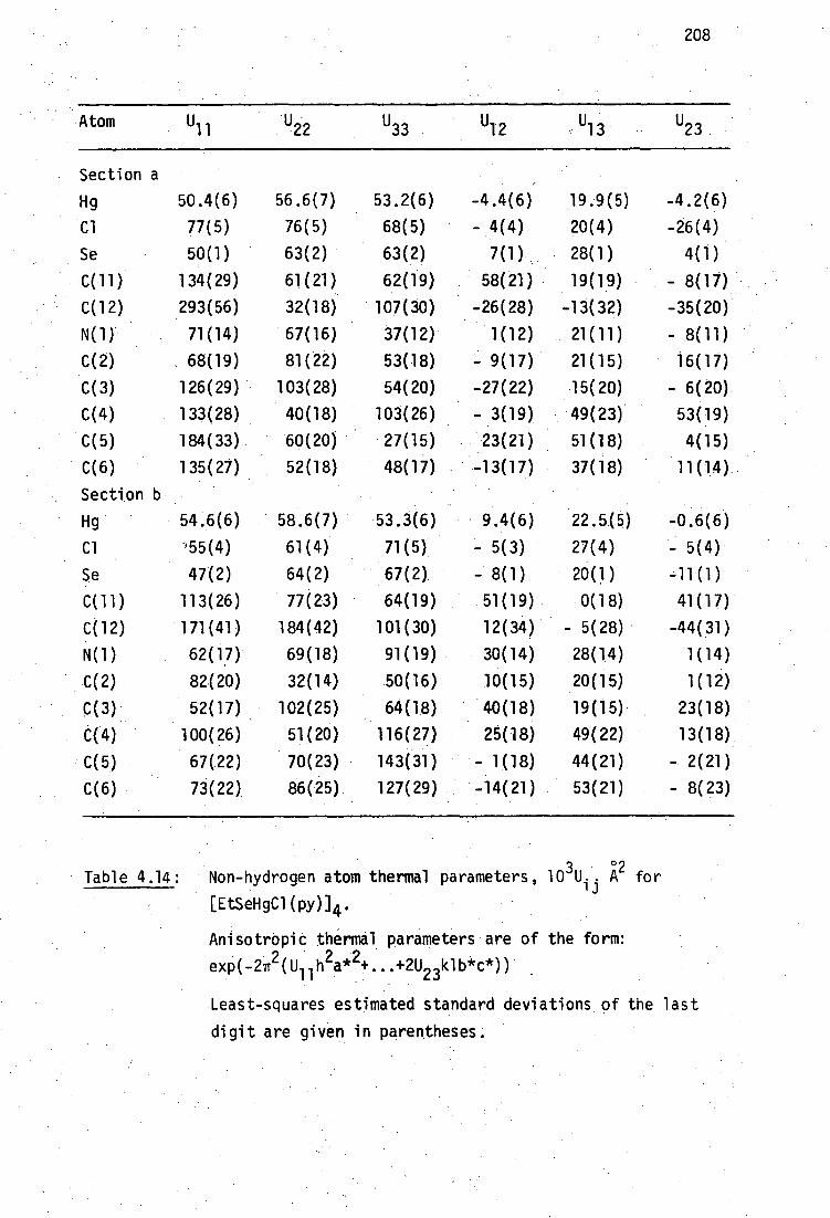
208
Atom U11 U33 P12 - u13 - U23
Section a
Hg 50.4(6) 56.6(7) 53.2(6) -4.4(6) 19.9(5) -4.2(6)
Cl 77(5) 76(5) 68(5) - 4(4) 20(4) -26(4)
Se 50(1) 63(2) 63(2) 7(1) 28(1) 4(1)
C(11) 134(29) 61(21) 62(19) 58(21) 19(19) - 8(17)
C(12) 293(56) 32(18) 107(30) -26(28) -13(32) -35(20)
N(1) 71(14) 67(16) 37(12) 1(12) 21(11) - 8(11)
C(2) 68(19) 81(22) 53(18) - 9(17) 21(15) 16(17)
C(3) 126(29) 103(28) 54(20) -27(22) 15(20) - 6(20)
C(4) 133(28) 40(18) 103(26) - 3(19) 49(23) 53(19)
C(5) 1E4(33) 60(20) 27(15) 23(21) 51(18) 4(15)
C(6) 135(27) 52(18) 48(17) -13(17) 37(18) 11(14)
Section
Hg 54.6(6) 58.6(7) •53.3(6) 9.4(6) 22.5(5) -0.6(6)
Cl -'55(4) 61(4) 71(5) - 5(3) 27(4) - 5(4)
Se 47(2) 64(2) 67(2) - 8(1) 20(1) -11(1)
C(11) 113(26) 77(23) 64(19) 51(19) 0(18) 41(17)
C(12) 171(41) 184(42) 101(30) 12(34) - 5(28) -44(31)
N(1) 62(17) 69(18) 91(19) 30(14) 28(14) 1(14)
C(2) 82(20) 32(14) 50(16) 10(15) 20(15) 1(12)
C(3) 52(17) 102(25) 64(18) 40(18) 19(15) 23(18)
C(4) 100(26) 51(20) 116(27) 25(18) 49(22) 13(18)
C(5) 67(22) 70(23) 143(31) - 1(18) 44(21) - 2(21)
C(6) 73(22) 86(25) 127(29) -14(21) 53(21) - 8(23)
Table 4.14:
3 °
Non-hydrogen atom thermal parameters, 10 - .A2 for ij [EtSeHgCl(py)] 4 .
Anisotropic thermal parameters are of the form:
exp(-27r2( Ull h2a*24. ...+2U 23klb*c*))
Least-squares estimated standard deviations of the last
digit are given in parentheses.

Atom 3 °2
10 U A 0
r A 4) 0
Aromatic hydrogens
Section a
0,0 = N,C(n+1)-C(n)-H(n)
H(2) 0.114(-) 0.661(-) 0.315(-) 81(-) 0.98(-) 120(-) 120(-)
H(3) 0.180(-) 0.768(-) 0.238(-) 115(-) 1.00(-) 120(-) 119(-)
H(4) 0.181(-) 0.758(-) 0.088(-) 97(-) 0.97(-) 122(-) 120(-)
H(5) 0.117(-) 0.641(-) 0.021(-) 104(-) 0.95(-) 122(-) 119(-)
H(6) 0.055(-) 0.536(-) 0.098(-) 97(-) 0.99(-) 120(-) 117(-)
Section b
H(2) 0.578(-) 0.354(-) 0.560(-) 63(-) 0.99(-) 120(-) 120(-)
H(3) 0.728(-) 0.235(-) 0.582(-) 84(-) 0.99(-) 120(-) 120(-)
H(4) 0.579(-) 0.120(-) 0.597(-) 92(-) 0.98(-) 120(-) 122(-)
H(5) 0.287(-) 0.128(-) 0.607(-) 104(-) 1 .00(-) 119(-) 120(-)
H(6) 0.148(-) 0.248(-) 0.583(-) 106(-) 0.97(1) 120(-) 118(-)
Methyl hydrogens 0 = C-C-H, 0= H(n)-C-H(n+1), n = A,B,C
Section a
H(12A) 0.229(-) 0.294(-) 0.398(-) 165(-) 0.98(-) 108(-) 105(-)
H(12B) 0.164(-) 0.317(-) 0.301(-) 165(-) 0.98(-) 106(-) 109(-)
H(12C) 0.300(-) 0.254(-) 0.325(-) 165(-) 0.91(-) 120(-) 108(-)
cont...
• - Table 4.15: Hydrogen atom parameters for [(EtSeHgC1)py] 4 .
Least-squares estimated standard deviations of the last digit are given in parentheses.

Section b H(12A) 0.511(-) 0.476(-) 0.860(-) 155(-) 0.94(-) 110(-) 110(-)
H(128) 0.327(-) 0.464(-) 0.873(-) 155(-) 0.96(-) 110(-) 107(-)
H(12C) 0.458(-) 0.397(-) 0.893(-) 155(-) 0.97(-) 113(-) 109(-)
Methyl hydrogens 0,0 = Se, C-C-H, x = H-C-H
Section a H(11A) 0.412(-) 0.369(-) 0.297(-) 104(-) 0.97(-) 108(-) 109(-) 104(-)
H(118) 0.478(-) 0.347(-) 0.394(-) 104(-) 1.00(-) 107(-) 108(-)
Section b H(11A) 0.277(-) 0.377(-) 0.757(-) 98(-) 1.00(-) 109(-) 109(-) 104(-)
H(11B) 0.461(-) 0.389(-) 0.743(-) 98(-) 0.97(-) 111(-) 110(-)
Table 4.15: Hydrogen atom parameters for [(EtSeHgC1)py4. (cont.)
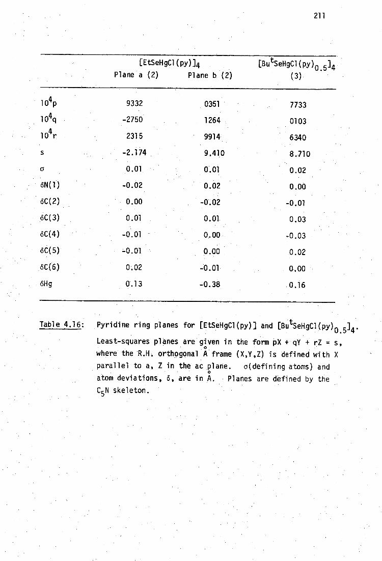
211
[EtSeHgC1(py)]4
Plane a (2) Plane b (2)
[ButSeHgCl(py) 0. 04
(3).
104 p 9332 0351 7733
104q -2750 1264 0103
104r 2315 9914 6340
s -2.174 9.410 8.710
a 0.01 0.01 0.02
6N(1) -0.02 0.02 0.00
dC(2) 0.00 -0.02 -0.01
oC(3) 0.01 0.01 0.03
6C(4) -0.01 0.00 -0.03
6C(5) -0.01 0.00 0.02
oC(6) 0.02 -0.01 0.00
olig 0.13 -0.38 0.16
Table 4.16: Pyridine ring planes for [EtSeHgCl(py)] and [Bu tSeHgCl(py) 0. 04 .
Least-squares planes are given in the form pX + qY + rZ = s, 0
where the R.H. orthogonal A frame (X,Y,Z) is defined with X
parallel to a, Z in the ac plane. a(defining atoms) and
atom deviations, d, are in A. Planes are defined by the
C5 N skeleton.

212
Figure 4.8: Unit-cell contents Of [EtSeHgC1(py)] 4 projected down a.

213
derivatives. A projection of the unit cell contents along c is
shown in. figure 4.8. The molecule is centrosymmetric about a
crystallographic symmetry element, and the two independent mercury
atoms have very similar geometries with bond distances within 2a
and bond angles within 3 0 .(except for angles N-Hg-Se(b) which differ
by 6.8°). The structure lacks 6 dichloro bridge between Hg atoms•
'found in the t,butyl substituted derivatives, but instead has an
extra Pyridine molecule coordinated to these mercury atoms.
A view of a single molecule of [EtSeHgC1(py)] 4, projected normal
to the plane containing the four selenium atoms is shown in Figure
4.7. In the t-butyl derivatives, steric hindrance permits
coordination of only two pyridine base's per tetrameric.unit, in
contrast to foUr for the ethyl derivative, with- concomitant dichloro
bridging to maintain tetrahedral mercury'coOrdination, .The disposition
.of t-butyl methyl groups in [ tBuSeHgCl(py) 0 04.i .s.consistent with this
observation, being such as to allow coordination of pyridine bases to
two of the centrosymmetrically related mercury atoms when the ring is'
chlorine bridged, but not at the other pair.
The pyridine rings in [EtSeHgCl(py)] and [Bu tSeHgCl(py) 0.5 .] 4 are
planar as expected: The maximum deviation from least squares planes, 0
defined by the C 5N skeletons, being 70.03 A in the t-butyl derivative.
Table 4.16 shows that mercury atoms coordinated to pyridine lie close
to the pyridine plane.-
4.2.3 Comparison of Hg-S and Hg-Se bonding distances
This work has produced the first report of Mercury(II)-selenolate
bond distances. It is of interest to compare Hg- S and Hg -Se bond
distances in closely related complexes because it has been noted earlier

214
458 0
that Hg-Se bond lengths in [HgC1 2 (Ph 3 PSe)] 2 (2.53 A) and the
0 selenourea complex of MeHg(II), [MeHgSeC(NH 2
)2]N0
3
434 (2.477(3) A)
are shorter than expected, even though for these complexes suitable
sulfur analogs are not available for a direct comparison. 0
The covalent radii of sulfur and selenium differ by 0.13 A .
242 (Pauling), although recent - determinations:Of the structures of elemental
sulfur and selenium, and of R2X2 (X=S,Se) result in distances 0
{1/21(Se-Se)(S-S)]l covering the range 0.13(2) - 0.182(4) A (Table 4.1 7 ). .
Values Df Hg-S and Hg-Se bond distances in closely related
complexes are given in Table 4.18 for 'Hg(p-SR) 4 1 ; the difference 0
between average values is 0.04 A. For [BUtSeHgCl(py) 0.5 14 which has
bond angles at mercury Within 5 0 of those for the sulfur analogs, the 0
differences are 0.12 A for 'Hg(p-XBu t ) 2 (p-C1)2' and 0.08 A for
'Hg(p-XBut
) 2C1N'.
In general it can be concluded that Hg-Se bond distances are
slightly shorter than expected from a consideration of sulfur and
selenium covalent radii. In the MeHg(II)-selenourea complex, this
has been considered to be a reflection of 'strong' Hg-Se bonding4.34
Sugiura has suggested that this may be due to the enhanced ability of
selenium to participate in metal-ligand backbonding.404

Cl C l
215
Structure 0
S-S/A
monoclinic-X8
K2X3
H02C-K(7),CO2H
2.046(4) 459
2 . 083(1) 461
2.076(5)462
2.069(15)463
2.068(8) 464 0 N-XX-4-7b
2.030(5)466
2.023(1) 467
2.0394 68
F F
- XX 2.059(4) 466
F F
[(H 2N) 2C-XX-C(NH 2 ) 2]C1 2 2.017(2) 47° 2.380(6) °
X-X distances 1.009(1)-1.042(1) a 1.145(5)-1.190(3)b
Table 4.17: Differences between covalent radii in isomorphous sulfur
and selenium structures.
a' b The X - X distances are in good agreement with the Pauling 0 0 242
covalent radii of 1.16 A (Se) and 1.02 A (S).
0 0 Se-Se/A kl(Se-Se)-(S-S)]/A
2.334(6)450
0.144(5)
2.383(2)461
0.150(2)
2.40( 462
0.16(3)
2.321(15) 463 0.126(15)
2.346(1)465
2.290 (10 ) 466
2 .333(15 ) 469
2 . 319 (4 ) 466
0.139(5)
0.134(6)
0.147
0.130(4)
0.182(4)

0 0
Hg-S/A
[(Hg-Se)-(Hg-S) ]/A
f 2.38(1), 2.39(1) 293b 0.12 295°
I 2.379(8), 2.395(8)
2.488(7), 2.509(9)293b 295c 0.08
2.501(8), 2.505(8)
Hg-Se/A
'Hg(p-XR) ( -C1) 2 1 2.501(3), 2.503(3) a
'Hg(p4R) 2C1N 1 2.579(3), 2.589(3) a
2.541(4)-2.511(3) d
'Hg(p-XR) 4 1 2.624(2)-2.764(2) °
433 'MeHgXCH 2CH(NH 3)(C0 ) 2.466(5)
2731 2.59, 2.68
0.04
236 2.352(12) 0;06
Table 4.18: Comparisons of Hg-S and Hg-Se bond lengths in closely related structures
a [ButSeHgCl(py) 0.04 , b[Bu
tSHgCl(py
c [844
t SHgC1(4-Mepy) (m4, [EtSeHgCl(py)4,
eHg(SeMe) 2,(SBut
crl
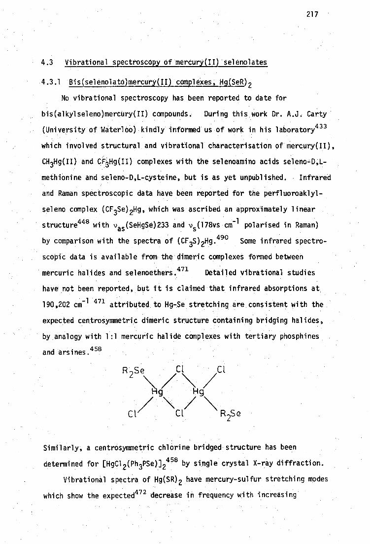
4.3 Vibrational spectroscopy of mercury(II) selenolates
4.3.1 Bis(selenolato)mercury(II) complexes, Hg(SeR) 2
No vibrational spectroscopy has been reported to date for
bis(alkylseleno)mercury(II) compounds. During this work Dr. A.J. Carty
(University of Waterloo) kindly informed us of work in his laboratory 433
which involved structural and vibrational characterisation of mercury(II),
CH3Hg(II) and CFpg(II) complexes with the selenoamino acids seleno-D,L-
methionine and seleno-D,L-cysteine, but is as yet unpublished. • Infrared
and Raman spectroscopic data have been reported for the perfluoroaklyl-
seleno complex (CF3Se) 2Hg, which was ascribed an approximately linear
structure448 with v
s (SeHgSe)233 and v (178vs cm polarised in Raman)
a
by comparison with the spectra of (CF3S) 2Hg.490 Some infrared spectro-
scopic data is available from the dimeric complexes formed between
mercuric halides and selenoethers.471 Detailed vibrational studies
have not been reported, but it is claimed that infrared absorptions at
190,202 cm attributed to Hg-Se stretching are consistent with the
expected centrosymmetric dimeric structure containing bridging halides,
by analogy with 1:1 mercuric halide complexes with tertiary phosphines
and arsines. 458
S e CI Cl
Hg Hg
Cl/ \Cl/ \ R Se 2
Similarly, a centrosymmetric chlorine bridged structure has been
determined for [HgC1 2 (Ph 3PSe)] 2458 by single crystal X-ray diffraction.
Vibrational spectra of Hg(SR) 2 have mercury-sulfur stretching modes
which show the expected472 decrease in frequency with increasing
217

]
ms/ms Hg 112 vHgSe vHgS
SelmSemHg 0 . 7 'Has
218
coordination number, e.g. Hg(SMe) 2 which has linear 'HgS 2 ' geometry 271
1 (Figure 2.6, page 45) has v s (SHgS) 297 cm- and vas (SHgS) 337 cm -1
whereas tetrahedra1 273 Hg(SBut) 2 (Figure 2.8, page 45) has v s (SHgS)
• - 188 cm' and vas (SHgS) 172 cm-1•231 Substitution of sulfur by
• selenium in isostructuiva compounds would be expected 473 to lead to a
decrease in the mercury-chalcogen stretching frequencies, according to•
the ratio of Hg-S and Hg-Se reduced-masses, as follows:
This calculation can only be considered an approximation which assumes
that vibrational force-constants remained unchanged on substitution of
S by Se. Since there is some evidence that Hg-Se bonds may be somewhat
stronger than Hg-S bonds (page 214), calculated values of vHgse will be,
to a first approximation, too low.
Linear 'HgSe' coordination is therefore expected to give rise to
vas (SeHgSe) 1,235 cm-1 and vs (SeHgSe) n,210 cm comparison with
Hg(SMe) 2 . The perfluorinated complex Hg(SeCF 3 ) 2448 has vas 233 cm-1
(solid mull), and v s 178 cm-1 (Me0H solution) which is in good agreement
-1 -1 with the calculated value v s 170 cm (from v s 243 cm for Hg(SCF3 ) 2 in
290 • 435
Me0H solution ). Similarly Se(HgMe) 2 has vas 231, v s 201 cm-1 ,
compared with v as 344, v s 300 cm-1 for the linear sulfur analog. 269
Tetrahedral 'HgSe 4 ' coordination can similarly be expected to give
both vas and v s near 140 cm by consideration of Hg(SBut
) 2 which has
-1 vs 172 (IR) and v as 188 (Raman) cm
219 Tetrahedral [Ph4P]2[Hg(SePh)
has v s 150 cm-1
.474
• Vibrational spectra of the Hg(SeR) 2 complexes prepared in this work
are recorded in Table 4.19 together with the spectra of their sulfur
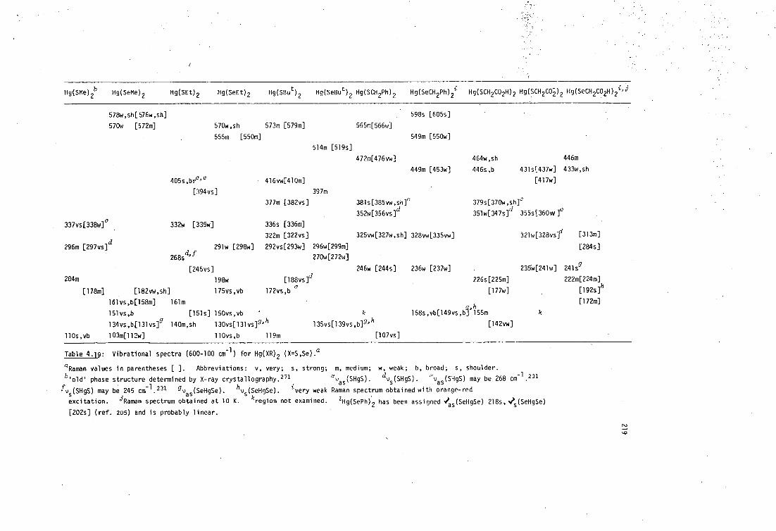
Hg(SMe) 2b Hg(SeMe) 2 Hg(SEt) 2 Hg(SeEt) 2 Hg(SBu t ) 2 Hg(SeBu t ) 2 Hg(SCH2Ph)
2 Hg(SeCH 2Ph) 2i Hg(SCH 2CO2H)2 Hg(SCH 2C0-2 ) 2 Hg(SeCH 2CO2H) 2i'i
578w,sh[576w,sh]
598s [605s]
570w [572m] 570w,sh 573m [579m]
565m[566w]
555m [550m]
549m [550w]
514m [519s]
472m[476vw]
464w,sh 446m
449m [453w] 446s,b 431s[437w] 433w,sh
[417w]
381s[385vw,sh]' 379s[370w,sh]e
352w[356vs]d 351w[347s] 1 355s[360mo]°
405s,brc ' e
416vw[410m]
[394 vs
397m
377m [382vs]
337vs[338w] ° 332w [335w]
296m [297vs]d
268sd' f
[245vs]
204m 198w [188vs]d
[178m] [182vw,sh] 175vs,vb 172vs,b
161vs,b[158m] 161m
151vs,b [151s] 150vs,vb
134vs,b[131vs] g 140m,sh 130vs[131vs]9' h
110s,vb 103m[112w] 110vs,b 119m
135vs[139vs,b]g' h
[107vs]
226s[225m]
[177w]
g,h 158s,vb[149vs,b] 155m
[142vw]
321w[328vsf1 [313m]
[284s]
235w[241w] 2415g
222m[224m]
[192s]h
[172m]
336s [336m]
322m [322vs] 325vw[327w,sh] 328vw[335vw]
291w [298w] 292vs[293w] 296w[299m]
270w[272w]
246w [244s] 236w [237w]
Table 4. g: Vibrational spectra (600-100 cm -1 ) for Hg(XR) 2 (X=S,Se).a
°Raman values in parentheses [ ]. Abbreviations: v, very; s, strong;
b 'old phase structure determined by X-ray crystallography. 271
fvs (SHgS) may be 245 cm -1 . 231 gvas (SeHgSe). hvs (SeHgSe). i very weak
excitation. jRaman spectrum obtained at 10 K. kregion not examined.
[202s] (ref. 205) and is probably linear.
m, medium; w, weak; b, broad; s, shoulder.
avas (SH0). dv s (SENS). '-'vas (SHgS) may be 268 cm-1 .231
Raman spectrum obtained with orange-red
1Hg(SePh) 2 has been assigned 4"as (SeHgSe) 218s, ■?5 (SeHgSe)
220
analogs. The single-crystal X-ray structure of Hg(SeMe) 2 indicates
polymeric 'HgSeil l geometry (Figure 4.1, page 183) and powder diffraction
patterns confirm that Hg(SeBu t ) 2 is isomorphous with the tetrahedral
sulfur analog.
The spectra of Hg(SeM0 .2 and Hg(SeBu t ) 2: haye intense bands at
-1 130-140 cm which have been assigned as Hg-Se stretching modes by
analogy with Hg(SBu t ) 2 . The features 158s,vb [149vt,b) in the
spectrum of . Hg(SeCH 2Ph) 2 have also been assigned as Hg-Se modes due
to the tetrahedral ligSe 4 1 moiety. . X-ray powder diffraction indicates
that this complex is not isomorphous with its sulfur analog (Figure 4.3,.
.page 189)..
In contrast to these tetrahedral complexes, Hg(SeCH 2CO2H) 2 appears
to be linear, although X-ray powder diffraction (Figure 4.2, page 188)
indicates that. it is not isomorphous with its sulfur analog.
The selenium. compound is phototensitive, 444 and very readily
decomposes in the Raman laser-beam. By Using low energy (red) excitation
and cooling thesample to 10K, a weak, but informative Raman spectrum was
obtained which has - a strong band at 192 cm in the infrared)
which has been assigned v s (SeOgSe) i .and no significant bands in the range .
,1 170-100 cm. The strong infrared band at 241 cm is probably v as (SeHgSe).
These values compare well with v s 178(Me0H) and vas 233 cm-1 for
Hg(SeCF3 ) 2448 and vs201, vas231 cm71 for Se(HgMe) 2 435 . which are assumed
to be linear. ,
The methylmercury(II) complex MeNgSeCH2CO 2H, which is almost
certainly linear, is also,somewhat phOtosensitive, but a Raman spectrum
was obtained from a solution which had been. used to measure the proton:
nnIr spectrum (Figure 3.34, page 164). Although rather noisy, the Raman
spectrum shown in Figure 4.9 has Moderate intensity bands which are
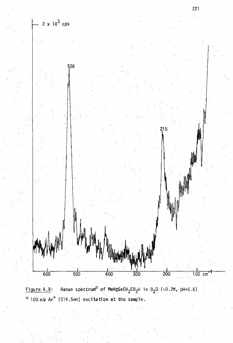
,(0 II
x 103 cps
221
215
i -1 600 500 400 300 200 100 cm
- Figure 4.9: Raman sPectruma of MeHgSeCH2CO 2H in D 20.( 0.2 , H=5.6)
a 100 inW Ar (514.5nm) excitation at the sample.
26

22?
coincident in the infrared: v(HgC) 526s[526s] and v(HgSe) 210s,b[215m].
The spectra of MeHgSeBu t confirm these assignments: v(HgC) 521m L526m]
and v(HgSe) 194s [200s]. The unpublished spectra of a selenocysteine
complex,433 MeHg[SeCH 2CH(NH3 )CO2].H 20, also has v(HgC) 536s [530s] and
v(HgSe) 228m [220s] and is apparently isomorphous433 with the (linear)
sulfur analog (Figure 2.1, page 34).
4.3.2 Mercury(II) diSelenblates
During the attempted preparation Of 1,3-diseleno-2-propanol. (the
selenium analog of the non-vicinal isomer of BALH 2 , DMPH 2 ), an intractable
pale-yellow mercury(II) complex was obtained by the addition of Hg(CN) .2
to a solution containing the diselenOlate (Chapter 5, page274 ). The
product has an analysis consistent with 1.:1 stoichiometry t and is the
selenium analog of HgDMP, previously characterised in this laboratory. 38
HgDMP has the interesting property that it can be obtained as a
crystalline adduct, HgDMP.(py) 1.5 , from pyridine, and is dimeric in this
solvent. 238 Unfortunately, HgSeDMP is insoluble (<100 mg/100 ml) in
pyridine and appears to be insoluble in all common. solvents, like HgBAL.,
Like the dithiolates studied in this laboratory (Chapter2 , page53 ),
HgSeDMP has very, broad, diffuse vibrational spectra, consistent with a
polymeric structure.
The far infrared spectrum of HgSeDMP shows only two weak, extremely
broad features at 400-500 - cm-1 and 100-220 cm -.. 1 and is thus unusable.
In contrast, the Raman spectrum shows an intense band at 167 cm
the only feature below 600 cm-1 . This is probably v s (SeHgSe), due to
tetrahedral 'HgSe4 1 centres in the polymer, and this distinguishes
tHgDMP (HgC 3H 6OSe 2) requires C 8.65 H 1.45, Hg 48.2%; found C 8.88,
H 1.56, Hg 48.2%.

223
HgSeDMP from its sulfur analog which has a linear, polymeric structure
with v(HgS) 333m,b [325vs]. 238
A similar attempted preparation of the vicinal diselenol analog of
BALH 2 , 2,3-diseleno-l-propanol, failed, but a very small quantity of
yellow powder was obtained by the addition of Hg(CN) 2 to the solution
suspected to contain the diselenolate. Like HgSeDMP, this powder was
insoluble in all common solvents. Some of this product was stored
under pyridine for three months (in an attempt to induce some dissolution
and/or crystallisation) but only an insignificant amount had dissolved
in this time.
The near infrared (3000-300 cm -1 ) spectrum of the pyridine-insoluble
residue shows several bands which appear to be due to coordinated pyridine,
e.g. 1607, 1590m, 615m, and 413, 406w (free pyridine has these bands at
1518, 601 and 403 cm-1 475 ), despite prolonged (24 hrs/ 0.1 mm) high
vacuum pumping and subsequent storage over concentrated H 2SO4 . The
Raman spectrum of this product is superimposed on a high intensity
background which gradually decays over several hours to reveal two sharp
bands at 1036s, 1004m cm to the totally symmetric ring mode of
coordinated pyridine (free pyridine has 990 cm and a strong band
at 179 cm-1 (HgSeDMP has 168 cm -1 ) due to v(HgSe). Unfortunately,
insufficient sample was available for microanalysis or far infrared
spectrum, but it would appear that this product may also have a polymeric,
tetrahedral structure like HgSeDMP, and unlike HgBAL, 231 consistent with
the general tendency toward tetrahedral 'HgSe4 1 geometry in mercury(II)
selenolates.
4.4 Conclusions
Evaluation of vibrational spectra for many complexes of the type
Hg(SR) 2 shows a predominance of linear (S-Hg-S) geometry. In contrast,
single-crystal X-ray diffraction studies of Hg(SeMe) 2 and vibrational
studies of Hg(SeR) 2 suggest that polymeric structures with tetrahedral
iNgSe4 1 geometry may be more common for Hg(II) selenolates.
All 1:1 thiolates RSHgX examined crystallographically, are polymeric
and form complexes with pyridine bases which are polymeric or tetrameric.
Analogous 1:1 selenolates RSeHgX behave similarly, e.g. MeXHg0 2CMe (X=
S,Se) are isomorphous, [ButSeHgC1(py) 0. 04 is isomorphous, with
[ButSHgC1(4-Mepy) 0. 04 , and [EtSeHgCl(py)]4 has a similar eight-membered
(-Hg-SeR-) 4 structure, but lacks a dichloro bridge found in the tert-
butyl complexes.
The intractable complexes HgSeBAL and HgSeDMP are probably polymeric
with tetrahedral 'HgSee geometry, in contrast to the linear sulfur
analogs HgBAL and HgDMP.
224

225
CHAPTER FIVE
SELENIUM ANALOGS OF ANTIDOTAL DITHIOLS
5.1 Introduction
Selenium analogs of dithiol antidotes have not been reported.
Attempts to synthesise analogs for studies of their interactions with
Hg(II) and MeHg(II) were commenced for several reasons. Of particular
interest are structural differences between Hg(SR) 2 and Hg(SeR) 2
favouring higher co-ordination numbers for mercury in the selenolates
(Chapter 4), and solution results suggesting that some dithiols act as
bidentate ligands toward MeHg(II) (Chapter 3).
It was hoped that free diselenol ligands could be obtained so that
MeHg-diselenol stability constants could be determined and compared with
those for the analogous dithiols. In view of possible direct in vivo
interactions between mercuric ion and selenium synthetic routes to
analogs of antidotal dithiols BALH 2 and DMSH4 were investigated.* It
was expected that a diselenol analog of Unithiol, III, would probably
form intramolecular mixed S-Se species (e.g. organo-Bunte salts RSSe03 ), 477
and its synthesis was not considered.
H 21C- 0 \ HSe SeH
HO C CO H H 2C-CH-CH 2S0 - Na+ . 2 \ /. 2
/CH--CH\ H5d'SeH
.HSe SeH
I, SeBALH 2 II, SeDMSH4
*For convenience, these will be abbreviated as SeBALH , I, and SeDMSH , II.
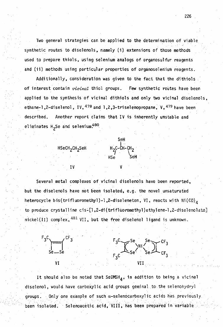
226
Two general strategies can be applied to the determination of viable
synthetic routes to diselenols, namely (i) extensions of those methods
used to prepare thiols, using selenium analogs of organosulfur reagents
and (ii) methods using particular properties of organoselenium reagents.
Additionally, consideration was given to the fact that the dithiols
of interest contain vicina thiol groups. Few synthetic routes have been
applied to the synthesis of vicinal dithiols and only two vicinal diselenols,
ethane-1,2-diselenol, IV, 478 and 1,2,3-triselenopropane, V, 479 have been
described. Another report claims that IV is inherently unstable and
eliminates H 2Se and seleniumP°
SeH i
HSeCH CH SeH H C-CH-CH 2 2
HSe SeH
IV
Several metal complexes of vicinal diselenols have been reported,
but the diselenols have not been isolated, e.g. the novel unsaturated
heterocycle bis(trifluoromethyl)-1,2-diseleneten, VI, reacts with Ni(C0) 4
to produce crystalline cis-[1,2-di(trifluoromethyl)ethylene-1,2-diselenolato]
nickel(II) complex, 481 VII, but the free diselenol ligand is unknown.
Se Se-........CF3
"Ni '
/ Se Se--CF
3
VI
VII
It should also be noted that SeDMSH 4 , in addition to being a vicinal
diselenol, would have carboxylic acid groups geminal to the selenohydryl
groups. Only one example of such a-selenocarboxylic acids has previously
been isolated. Selenoacetic acid, VIII, has been prepared in variable

X
1) alkaline reduction
2)acidification SeH
SeH
227
yield in aqueous alkaline solution by removal of mercury from
Hg(SeCH 2CO2H) 2 with Na2S,482 or by electrolytic reduction of the
diselenide, IX, under a hydrogen atmosphere in 50% yield. 432,482
A spectrophotometric determination of the acidity constants of VIII
was achieved by reduction of IX with sodium borohydride, careful acidi-
fication with deaerated HC1 and extraction into CHC1 3 . 374
HSeCH 2 CO H HO2 CCH
2 SeSeCH 2 CO
2 H
VIII IX
The difficulties involved in obtaining compounds with geminal
carboxylic acid and selenohydryl groups have been noted elsewhere. 483
Fredga has attempted to produce the diselenol, X, by reduction of
1,2-diselenane-3,6-dicarboxylic acid, x1,484 in alkaline solution.
Acidification leads to rapid decomposition and formation of red selenium:
CO 2H 2
Se
\r)e
CO2H
XI Similarly, in this work, it has not been possible to isolate
SeBALH 2 and SeDMSH 4 despite several attempts using various synthetic
routes, but the Hg(II) complex of SeBALH 2 and of the isomeric non-vicinal
diselenol have been prepared.
5.2 Possible routes for synthesis of dithiols and diselenols
Free selenols are very easily oxidised e.g. by atmospheric oxygen
to form diselenides. This is of paramount importance in all reported
selenol syntheses described below.

228
5.2.1 Direct introduction of SeH
Alkylation of NaHSe to form alkaneselenols has often been reported. 485-6
NaSeH + RX•> RSeH + NaX
Common alkylating agents used are alkyl halides and potassium alkyl
sulfates. The sodium hydrogen selenide reagent can be prepared by
dissolving gaseous H 2Se in NaOH, by dissolving Se in methanolic Na0Me
or, more recently by reducing Se with sodium borohydride in aqueous
or ethanolic solution. 487 Many thiols, including several non-vicinal
• dithiols, have been prepared in this fashion, using H 2S, or metal
sulphides etC. 488-9 Vicinal dithiols, e.g. BALH 2 , 490-1 Unithio1 9 re
formed under these conditions but in lower yields. If both halide
groups are secondary, extensive dehalogenation occurs to produce a
trans-olefin. 492-5
1 • R2
NaSH _
Br Br Br
R 2
Owen has commented that halide replacement, to produce dithiols,.is
most favorable when both groups are primary and less favorable when
one is secondary,496 e.g. 2,3-dimercapto-T-butanol and 2,3-dibromo 7 2-
methyl-l-propanol could not be prepared by this method. 497
Although :HSeCH 2CH 2SeH cannot be prepared in this way, 48° the non-
vicinal diselenols HSe-(CH 2 ) 3-SeH 480 and HSeCH 2CH(OH)CH 2SeH479 have been
prepared using this method, but they are often contaminated with cyclic -
diselenides formed by oxidation.
Interestingly, the only reported triselenol, propane 7 1.,2,3-.-
' triselenol,.V, the selenium analog of glycerine, was reputedly formed 479

229
from alkylation of an alkaline solution of sodium hydrogen selenide by
1,2,3-tribromopropane. Although the preparation of the related
compounds 1-seleno-2,3-propanediol and 1,3-diseleno-2-propanol has
been reported by Baroni, 479 the compound of interest here, 2,3-
diseleno-l-propanol, SeBALH 2 , was not prepared. It should be noted
that the parent vicinal dibromide in this case contains one primary and
one secondary bromide group.
Considering the ease of oxidation of the selenohydryl group, it
is more usual to introduce selenium as a protected selenoether during
monoselenol syntheses, especially if subsequent modifications are to
be made to other functional groups. The last stage in a multi-step
synthesis of such compounds is cleavage of the Se- protecting group
(deprotection).
5.2.2 Use of protected Thiols and Selenols
In the multi-step synthesis of complex molecules containing the
-XH (X=S,Se) fUnction, it is generally necessary to introduce X as the
• thio-or selenoether group in order to protect against oxidation. Much
of the literature concerning sulfur and selenium protecting groups is
concerned with the preparation of peptides containing cysteinyl- or
selenocysteinyl residues. " The literature of thiol protecting groups
has been well reviewed. 499-9 Protecting groups which have been used
for selenoaminO.acid and selenopeptide syntheses are summarised in
Table 5.1.
About seventy thiO1 protecting groups have been,described. 499 Many
have been used to protect an already existing -SH function so that
subsequent reactions may be carried out elsewhere in the molecule. For
the current purpose, these are therefore not useful. However, - if the
protective reaction inVolves, generation of a thiol group in situ by
reduction of a disulfide, it was considered for inclusion in this work.

.
selenocystelneb
C1CH 2CH(NH 3 )C02Me
C1CH2CH(NH 2 )C02H
0
230
K2Se2/KOH c,d ;242Tt > 4SeCH2CH(NH 2 )CO2Hh I z 1 >Bz1SeCH2CH(NH 2 )CO2H II
36 hrs, 25°
_1111ja-__> Na/NH,f
Bz1SeCH 2CH(NHR)CO2H -----=> I R = H (36%) [0]
R = COPh (24%) PhSe- PhSeCH 2CH(NHR)C02Hg
1. Ba or Ca(SeH),› IC (11%)
2. [0]
1. C1CH2SeCH 2Ph> 1. HI
2. HC1 2. [0]
Ref.
500
501-3e
501
504
505-6
431
507
428a
,,CO2Me Bz1SeH 1. NaOH 1. Na/NH3f
I 70% Na0Me 2. H
+ 2. [0]
Bz1Se - • HBr/HOAcf NaOH > II TosOCH 2CH(NHZ)C0 2Bzh (60%) (99%) (50%)
1. HSe - 2. Na0H-, [ SeCH 2CH(NHZ)CO2fizh] i ----> > I
1. Na/NH3f > RSeCH2CH(NH2)CO2H
R Me,' Pr, al lyl
2. RI
selenohomocysteineb
1. Bz1Se- BrCH 2CH2 / -13 ---------> Bz1SeCH
2CH (70%) HN, ,eNH x III
0
1. Na0H,155° > Bz1SeCH2CH 2CH(NH 2 )CO2H
2. H+ ,pH 5.5 508
IV (86%)
H 2C=C\ NHC(0)Me
II
Na/NHlf fSeCH 2CH2CH(NH 2 )CO2H]2
[0]
Bz1Se- C1CH 2CH2CH(Naz)C02H > IV
(72%)
+ Bz1Se - IV (79%) BrCH 2CH 2CH(NH 3 )COH >
Bz1Se - 1. Na0H/diox. NH AOH T0sOCH2CH 2CH(NHZ)C02Np > Bz1SeCH 2CH 2CH(NHZ)C02Np > ' > IV (38%)
2. HBr/HOAc pH 5.5
0 Bz1SeH HCN/N11 3> 1. HC1,H0Ac b R1 =R2=R3 =R4=H
R R C=CR -C15 IV (50%) 1 2 3R4 Cat. Me0H 2. OH ,pH5.8
mono Me-substituted IV (25-52%)
R1 =R2 =R3' =H. R4 =Me
R1 =R2 =R4' =H- R3 =Me
R2 =R3 =R4' =H. R4 =Me
Bz1SeH (NH4 ) 2 > CO3 1. Na0H,108° > > IV (41%)m RI =R2=R3 =R4=H Cat. NaCN 2. H
+,pH 5.8
monomethyl derivatives above
diMe IV = R1 =R3=Me; R2=R4=H
triMe IV = RI =R2=R4=Me; R3=h
509
sloe
Na/NH 3> 512
[0]
513
514-5
516
selenomethionine and selenoethionine l
Na/NH,f
RI IV or V "> RSeCH2CH2CH(NH 2)C02H VI = R=Me
R=Me,Et
including mono-Me derivatives (R=Me)
509,512
514-5e
510e,517
Table 5.1: Synthesis of selenoamino acids and selenopeptides. a

RCH2C(Me) 2CH(OH)CONHCH 2CH 2CONHCH 2CH 2SeBz1
Na,NH 3f
[0] IRCH2C(Me)2CH(OH)CONHCH2CH2CONHCH2CH2Se32 531
231
1. ReSe- ,160° > VI R=M0
2. H+,pH 5.8
518
RSe T0sOCH 2CH 2CH ( NH2)C02H
VI R=Me,Et 5118
selenolanthionine and selenocystathioninel'i 519-21
"CO2Et 1. -SeCH 2CH(NH 2 )CO2H1 HO2CCH(NH 2 )CH2x H 1. HC1
2C=C,‘ NHC(0)Me Cat. 2. OH- , pH 6 Se
HO2CCH(NH 2)(CH 2 ) n/
n=1 VII (selenolanthionine) 522
C1CH 2CH(NH 2 )C02H 1. -5e(CH) nCH(NH2)CO2H1
VII
505be
n=2 VIII (selenocystathionine)
5238
-SeCH2 CH(NHZ)C0 2Bzh1 T0sOCH 2CH(NHZ)CO2Bzh > > VII
Se(CH 2 ) 2CH(NHZ)C02Bzhi
TosOCH 2CH(NHZ)C02Np » VIII 5118
selenoglutathione (Glu-SeCys-Gly) b
[ZNH-SeCys] 2 2NH 2CH 2CO2Et OH -
[0]
Cys(SeBz1)-Gly a-Et-(NHZ)-G1u
[Glu-SeCys-Gly} 2
IX
524
[0]
5078
CH2 OH
B0cNHCHCO.NHCH 2CO2Bz1
1. TosCl/pyr 2. Bz1Se - DCC
Glu(ZNH 2OBz1) - Cys(SeBz1) - Gly(0Bz1) 431 3 ' CF3CO2H
CO BZ1 2 ZNHCHCH 2CH 2OH
selenooxytocins (and uncyclised nonapeptides)
1-seleno: ZSeCys(Bz1)-Tyr-Ile-Gln-Asn---Cys(Bz1)-Pro-Leu-Gly-NH 2 X 525
6-seleno: (Bz1)-Tyr-Ile-Gln-Asn-SeCys(Bz1)-Pro-Leu-Gly-NH 2 . XI 525 - 7
1,6-diseleno: ZSeCys(Bz1)-Tyr-Ile-Gln-Asn-SeCys(Bz1)-Pro-Leu-Gly-NH 2 XII 528- 9
deamino -1-seleno: (Bz1)-Tyr-Ile-Gln-Asn---Cys(Bz1)-Pro-Leu-Gly-NH 2 XIII 525
deaminon' -6-seleno: (Bz1)-Tyr-Ile-Gln-Asn-SeCys(Bz1)-Pro-Leu-Gly-NH 2 XIV
,
525 - 7
deamino *-1,6-diseleno: (Bz1)-Tyr-Ile-Gln-Asn-SeCys(Bz1)-Pro-Leu-Gly-NH 2 XIV 530
l' NaNH f 3 x 1-selenooxytocin 525
2. [0], pH 6.8
selenocoenzyme-A, and isoselenocoenzyme-A
R = OH (selenopantethine),-OPO 3H 2
Table 5.1: Synthesis of selenoamino acids and selenopeptides.
(cont.)

N \
N4? R
cH20 f-01-0-
0 6H 6H
R'iR" = OH 2OH; cyclic PO4;
R'0 OR" P03H 2 ,H (SeC0A) 2 XV
H,P03H 2 iso(SeCoA) 2 XVI
232
532
Na/NH 3 XV,XVI Me 2NCH 2XH 2SeC(0)Ph
Se-benzoyl-CoA and iso-Se-benzoyl-CoA
1 Bu3Nf
SeCoA iso-SeCoA
Table 5.1: Synthesis of selenoamino acids and selenopeptides.
(cont.)
aor substituted derivatives, usually DL- or meso- as shown (except see e).
The following abbreviations are used: Bzl = benzyl, BZ = benzoyl, Bzh = diphenylmethyl, Boc = tert-butyloxycarbonyl,
Z = benzyloxycarbonyl, Tos = p-toluene sulfonyl, Mpr = 5-mercaptopropionyl, Np = p-nitrophenyl, DCC = N,N'-
dicyclohexylcarbodiimide.
bthe selenohydryl amino acid has not been isolated in a pure state.
eno protective group was used.
dFischer-Raske cystine synthesis. Inconsistent yield.
eapplied to preparation of optically active products.
'reagent for removal of the selenohydryl-protecting group.
gthe protective group could not be removed without decomposition to PhSeSePh.
hGabriel-SOrensen phthalimidomalonic ester synthesis. Low yields.
iselenolate not isolated but used in situ.
jnaturally occurring selenoamino acid.
kStrecker synthesis.
/selenoethers.
mBucherer-Bergs hydantoin synthesis.
crystallographic data are available.
233
• In addition, many routes use organosulfur reagents for which the organo-
selenium derivatives are either unknown, or hypothetically would require'
reagents which would be difficult to manipulate (H 2Se or CSe 2 ).
The common protecting groups meeting these requirements are described
below. In addition to the preparation of the protected selenol, the
means of deprotection or cleavage of.the Protected group was considered
' (e.g. methods which required anhydrous HF were rejected).
The benzyl (Bzl) group is the earliest Widely used protective group
for cysteinY1 residues, and remains the most widely used for Se-containing
peptides, etc. (Table 5.1). The benzyl group could be introduced using
either benzylselenol (a foul smelling Colourless oil, but readily handled
. under non-oxidising conditions) or its sodium salt (formed in situ by
-reduction of the diselenide under alkaline conditions, e.g. using sodium
borohydride or Rongalite).. Benzylselenol can also be formed from the
" diselenide by in situ reduction with hypophosphorus acid. RemOval of -
• the benzyl group from benzyl protected selenium peptides for example, is
usually accomplished by sodium reduction in liquid ammonia, followed by
acidification of the ammonium salt of the selenol. Alternatively,
benzyl-protected-thiols have been deprotected with strong acids e.g.
HF/anisOle 498br CF 3S03H. 533
Substituted benzyl groups,. notably 4-Me0Bz1 or.4NO 2-Bz1, generally
offer Tittle advantage over the unsubstituted groups for thiol protection,
except that milder deprotective , conditions have been used. The selenols5347
and diselenides are known. 538-9
The S-triphenylmethyl (tritY1) protecting group has been usedin
several thiol-..peptide syntheses. The trityl group can be removed by
formation of the mercury(II)-thio-peptide complex. Mercury is subsequ-
ently removed as the sulfide 51 0- by electrolytic 'reduction at a mercury
cathode: 542 Triphenylmethaneselen01 and 'diselenide do not seem to have
234
been prepared to date.
The But- protecting group is usually added to a pre-existing thiol
group in thio-peptide syntheses, using Bu tOH. It has been cleaved by
anhydrous HF, but a recent report indicates that it can be cleaved under
milder conditions by conversion to the S-(2-nitrophenyl)sulfenyl deriva-
tive or by NaBH4 reduction.543 Since we have used Bu tSeH in the
preparation of Hg(SeBu t ) 2 and MeHgSeBu t (see pages 350-1 ), the formation
of Bu t-selenoethers was considered as a means of selenol protection,
although this has not been previously reported.
Several thiols have been used to convert the cysteinyl moiety into
asymmetric disulfides 544-7 as a means of protection of RSH:
cys-SH + RSH ---> cys-S-S-R
It should be noted that under these conditions, it is an entire cys-S-
group which is the protecting group, i.e. the disulfide bond is cleaved
on deprotection. , Although selenol and diselenidereagents (for genera-
tion of selenol in situ) are available for use as protecting groups in
this way, this technique do6s nOt seem to.. have been used for selenol
protection.
The Acetami .domethyl group has apparently become preferred to the
. Bzl group for thiol protection in peptide synthesis,548 but it does not
appear to have been used as a seleniUm protecting group. The selenol
and diselenide are unknown.
Schotte549 has indicated that,despite several synthetic routes to ,
nOnLvicinal dimercaptodicarboxylit acids, "Special methods" need to be
applied in the case of dimercaptosuccinic acid (DMSH4). The only
'reported preparations of DMSH 4 have utilised the mild hydrolysis of
S-acetyl protecting groups of 2.,3-bis(aCetylthio)succinic . acid, XII.4)6550
The bis(S-acetyl) compound was prepared by 'addition of thiolacetic acid,

235
XIII, to acetylenedicarboxylic acid.
0 HO CCECCO H + 2H C.0
HO22H/
2 2 3 \SH H3COC.S S.COCH
3
. By careful control of reaction temperatures, a predominance of
racemic- or meso-XII could be obtainec0 15-6 Removal of the acetyl
groups can be accomplished in one minute using 3N NaOH at 40° 416
The acetylthio group has been widely used by Owen in the synthesis of .
vicinal dimercapto alcohols, e.g. CH 2SH• .CHSH(CHOH) rICH 2OH [n=0(BALH 2 );. 551
1,4922p2-3”2], CH 2SH.CHSH(CH 2 ) nCH2OH [n=1; 53 2P ] And:propane-
1,20-trithio1 5 .5
Direct utilisation of this technique for SeDMSH 4 synthesis would .
require the use of selenolacetic acid XIV.. This compound has been
mentioned in the early organoselenium literature. Addition of the
Grignard reagent HSeMgBr (from H 2Se with EtMgBr) to acetylchloride in
ether is reported to form XIV, Which decomposed to eo selenium on
•Attempted - isolatiOn. 556 An earlier report showed that XIV Could not be
obtained by acetylatiOn of NaSeH 7 More recently, attempts to
prepare acetyl, propionyl and isobutyryl selenols by the reaction of
the acyl chlorides with H 2Se in pyridine were unsuccessful 58 (A
report of XIV as an oil, bp. 135°, from the reaction of diacetyl-
yohimbine with Se at 300 05,59 is almost certainly incorrect given the
extreme oxidation sensitivity of Acyl selena.fg),.
SeH \`SeH
XIV xv
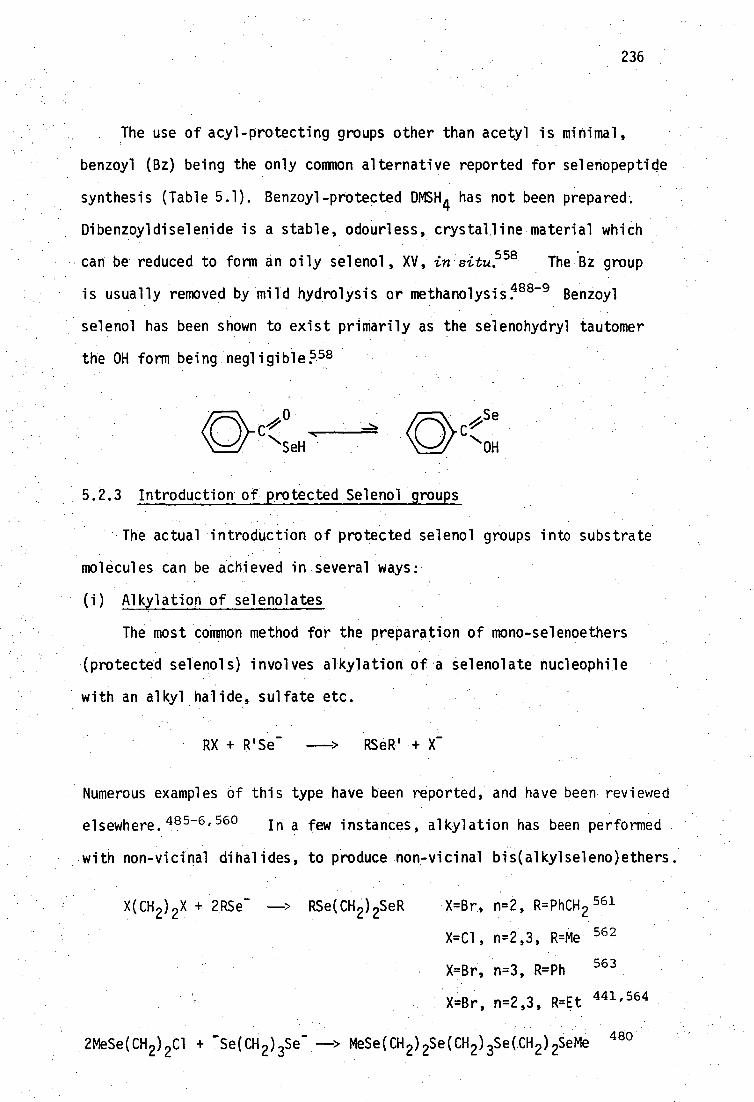
236
The use of acyl-protecting groups other than acetyl is minimal,
benzoyl (Bz) being the only common alternative reported for selenopeptide
synthesis (Table 5.1). Benzoyl-protected DMSH 4 has not been prepared.
Dibenzoyldiselenide is a stable, odourless, crystalline material which
can be reduced to form an oily selenol, XV, in situ.558 The 'Biz group
is usually removed by mild hydrolysis or methanolysis: 488-9 Benzoyl
selenol has been shown to exist primarily as the selenohydryl tautomer
the OH form being negligible 58
Se
OH
5.2.3 Introduction of protected Selenol groups
The actual introduction of protected selenol groups into substrate
molecules can be achieved in several ways:
Alkylation of selenolates
The most common method for the preparation of mono-selenoethers
(protected selenols) involves alkylation of a selenolate nucleophile
with an alkyl halide, sulfate etc.
RX + R'Se -
RS R•+
Numerous examples of this type have been reported, and have been reviewed
elsewhere. 485-6,560 In a few instances, alkylation has been performed
with non-vicinal dihalides, to produce non-vicinal bis(alkylseleno)ethers.
X(CH 2 ) 2 X + 2RSe -
ne(CH 2 ) 2S eR -X=Br, n=2, R=PhCH 561
X=C1, n=2,3, R=Me 562
X=Br, n=3, R=Ph 563
X=Br, n=2,3, R=Et 441,564
2MeSe(CH 2 ) 2C1 + -Se(CH 2 ) 3Se --* MeSe(CH2)2Se(CH2)3Se(CH2)2SeMe 480'

237
Vicinal bis(alkylthio)ethers can be prepared using this route e.g. n=1
above, but usually only when the ‘halide groups are not both secondary,
e.g. 1,4-diacetoxy-2,3-dibromobutane eliminates bromide when treated
with thiolacetic acid/pyridine to form 1,4-diacetoxybut-2-ene 492 (but
10,11-bis(acetylthio)hendecanoic acid is formed from the corresponding
vicinal secondary dibromide under the same conditions 585 ).
Attempts to alkylate selenolates, using reagents with vicinal
•leaving groups, do not seem to have been reported, perhaps because Of
problems faced in the analogous dithiol syntheses (see page 228 for
. preparations Of non vicinal diselenols, using NaSeH).
(ii) Addition of Selenols to alkenes and alkynes
The addition of thiols to activated alkenes and alkynet is well
established and may proceed via either radical 568 or: base-catalysed 567
pathways e.g. the base-catalysed reaction has been usedto prepare
crystalline derivatives of olefins;68 and several new dithioethers
which may be potential heavy-metal antidotes have similarly been
prepared. fromacetylenedicarboxylic acid. 569 Analogous addition of
selenols (mainly aryl) has also received much attention, almost solely
from Russian workers. Reported addition reactions of selenols to
alkenes and alkYnes have been summarised in Tables 5.2 and 5.3 respect- •
ively.
Lactone and epoxide ring opening
The preparation of selenium-containing carboxylic acids is often
carried out by lactone ring opening, And epOxides are similarly opened
to produce alcohols. These reactions have been summarised in Table 5.4..
. Phenyl selenide has recently been reviewed as an effective reagent for.
S2-type cleavages of lactones and esters. 585 To prepare protected
SeDMSH 4 using this method, two selenoether groups would need to be
introduced. A possible route is shown below:

571 Bz1SCH ,CH9SeBz1 2>CH-CHi
HO2C
Et3N 00 >
R1
Bz1SCH 2.
C=CH / 2 HO2C
RR3
\C(0)R4
Bz1Se\ ,C(0)R4 tR CHR' /, 2 3
C=C" Na0Me•
H,C=C" "NH(0)Me
0
1.Na/NH (2.) 3 >
2.Fe3+ ,02
H+ ,PH1
SeCH 2CH\ CO - 2 2
> -> 506
238
alkene
product
ref.
ionic additions (base catalysed
Bz1SeH:
CO2 H Et3 N / Me C=
, 2 \t02H Et2T1H -
, 1.Na/NH3 (2.)b - CH
1 .5 . > --CH-CHJOnH] -CO > 2 . 02 ._ 1 c .c
CH3
2
570
= H,H,H,Me; Me,H,H,H; H4H,Me,H; Me,Me,H,Me 572
H,H,H,H
516
/H
g\C°2
H
pirridine> 5b, 12 hr.
Bz1SeCH CHCO H 573
PhSeH:
R R,
H' 'CO2R
R ,R2 ,R3 =
Na0Me(Et) ›
Ft' Ii PhSeCK. H.00 R
2 3 2
H,H,H; H,Me,H; H,Me,Me
CO2 ' ' H H H .; CO2 " Et H Et
574
Table 5.2: Addition of selenolsa to alkenes.
aunder ionic conditions the selenol is deprotonated.
to cleave the benzyl group.
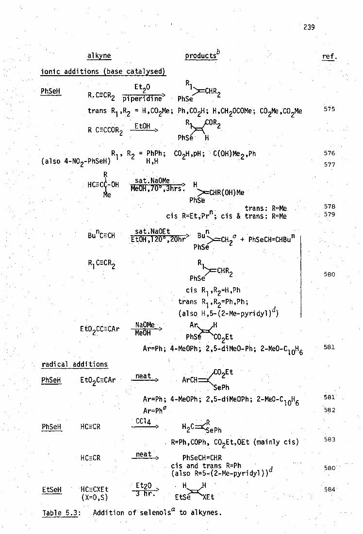
Table 5.3: Addition of selenolsa to alkynes.
ref. productsb
111 >=CHR9
PhSe `
alkyne
ionic additions (base catalysed)
Et 0 PhSeH 2
R.CECR 2 piperidine',
trans R1 ,R2 = H,CO2Me; Ph,CO 2H; H,CH 2OCOMe; CO 2Me,CO 2Me
R OR2
PhSe H
CO2H,pH; C(OH)Me 2 ,Ph
Et0H > R.CECCO R2
R=Me R=Me
• sat.Na0Me u Me0H,70°,3hrs: " >=CHR(OH)Me
PhSe
cis R=Et,Prn ; cis &
580
584 Et20 3 hr.
neat >
EtSeH HCECXEt (X=0,S)
HCECR PhSeCH=CHR cis and trans R=Ph (also R=5-(2-Me-pyridy1))"
H
EtSe XEt
R1 ,
(also 4-NO2-PhSeH)
HCE1-0H
R2 = PhPh; H,H
1 :>=CHR,
PhSe `
cis R1' R2 =H,Ph
trans Rl ' R2 =Ph,Ph;
(also H,5-(2-Me-pyridyl) d)
02Et neat > ArCH
SePh
Ar=Ph; 4-Me0Ph; 2,5-diMe0Ph; 2-Mle0-C 10H 6
Ar=Phe
CC14 > u r R• "2'=(SePh
R=Ph,COPh, CO E OEt (mainly cis)
BunCECH
R1CECR
Et0 CCECAr
radical additions
PhSeH EtO2CECAr
PhSeH HCECR
239
575
576
577
578 579
580
581
581
582
583
satAa0Et n. 'Et0H,120°,20hr> Bu :>=CH 9c + PhSeCH=CHBUn
PhSe `
Na0Me > Ar H Me0H
PhS CO2 Et
Ar=Ph; 4-Me0Ph; 2,5-diMe0-Ph; 2-Me0-C 10H6

240
Table 5.3: Addition of selenolsa to alkynes.
(cont.)
a under ionic addition conditions the selenol is deprotonated.
b stereochemistry is that indicated in the original reference and appears
to be solvent dependent.
cappears to be an exception to 1,2-addition. dstereochemistry not
given under base-catalysed conditions. estereochemistry incorrectly
assigned 581 i n ref. 582

241
0
RSe- HO
SeR
C CO2H
SeR
0 --->
RSe
HO 2 ? N x H02
The starting 3-1actones of a-halomalic acids are unknown (although an
a,t3-dimethy1 substituted analog has been prepaM) and are probably
unstable with respect to the isomers of 2-halobutenedioic acids.
The related selenol of interest, 2-selenosuccinic acid, XVI, may
possibly be obtained using this technique.
HOC 2 CO„H H 2Se
4 or NaHSe SeH XVI
HOC) /CO2H
SeR
In this case the starting B-lactone of malic acid is known. 87 It
may be noted in passing that this route has not been attempted with
a-lactones, which could be expected to generate protected, substituted
a-sel enocarboxyl c acids
HO 2
\o • R'Se
R'SeCR2CO2H
ct-Lactones (R=CH 3 , nBu,-(CH 2 ) n -, n=2,3,4,588 Bu t ;589 CF 359 can be
prepared in high yield photochemically at 77 K, but most polymerise
above this temperature.588 Their stability can be enhanced electronic-
ally e.g. R=CF3 has a half life of 8 hours at 24 C,590 or sterically,
e.g. R=Bu t which is stable below -20°C.589 The usual route to these
a-selenocarboxylic acids would be alkylation of the selenolate HO 2CCR2Se -
formed by in situ reduction of the corresponding diselenides. However,
in most of the cases above except R=Me 591) the highly substituted
diselenides are unknown.

HOCH 2CH 2SeMe 593
HO, R—`C.CH SePr (or Ph) 594 R/
2
MeSeH
PrSeH PhSeH >. or
(CH 30MgSe) 24Me0H
Bz1Se- >
MeSe- >
242
ref.
1 H 2 Se (HOCH 2 2Se*2
592
Pr : R 1 ,R2 = Me,Me; Me,H; C1CH 2 ,H; CH
CH 2=CH,Me; CH 2=CH; H;
CH 2=CC1 ,H
Ph: R1 ,R2 = Me,Me; Me,H; C1CH ,H
EL 1 Na2 Se
2. [OH] (HO CCH CH 2Se+2 595
• Bz1Se ,DM F> Bz1SeCH 2 CH H 596 2. H
1. Bzl Se - ,DMF> 2. H +
PhSeT>
CH 2Se+2
= H,H,H; H,H,Me R4=H Me,H,H; Me,H 2 OH
NH 2 CH—CO 2H
R =R =R =H, R =NH 1 2 3 4 2
CO2H 1
CH 2SeBz1
(HO 2 CCH
,R R
MeSeCH
CH 2 J Se— 2
CO 2H
many examples
597
598:
518
487
599
585
Table 5.4: Opening of lactones and epoxides by organoselenium reagents.
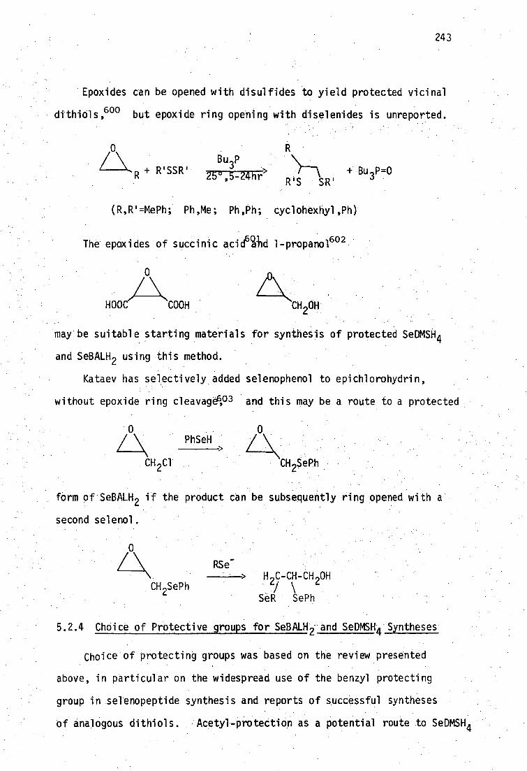
0
■// HOOC COOH CH 2 OH
243
Epoxides can be opened with disulfides to yield protected vicinal
dithiols,600 but epoxide ring opening with diselenides is unreported.
0 R
+ R'SSR' 3 25°,5-24hr"
, -----\
Bu P
R'S SR' + Bu =0
(R,W=MePh; Ph,Me; Ph,Ph; cyclohexhyl,Ph)
The epoxides of succinic aciegild 1-propano1 642
may be suitable starting materials for synthesis of protected SeDMSH 4
and SeBALH 2 using this method.
Kataev has selectively added selenophenol to epichlorohydrin,
without epoxide ring cleavage6,° 3 and this may be a route to a protected
0
CH 2 Cl
PhSeH 0
CH 2SePh
form of SeBALH 2 if the product can be subsequently ring opened with a
second selenol.
CH 2 SePh H C-CH-CH OH 2/ \ 2
SeR SePh
5.2.4 Choice of Protective groups for SeBALH 2 and SeDMSH4 Syntheses
Choice of protecting groups was based on the review presented
above, in particular on the widespread use of the benzyl protecting
group in selenopeptide synthesis and reports of successful syntheses
of analogous dithiols. Acetyl-protection as a potential route to SeDMSH4

XVI I
5.2.5 Selenols from Selenocyanate Hydrolysis
244
synthesis was rejected since selenolacetic acid is not available, but
the benzoyl group was considered as a possible alternative although
2,3-bis(benzoylthio)succinic acid is unknown.
The benzyl group has been successfully used for protection in the
604 synthesis of BALH 2 . Bis-benzylthiolation can be achieved using
vicinal dibromides in which the bromide groups are not both primary, e.g.
2,3-dibromo-2-methyl-l-propanol (cf. page 233 • 497
However, alkylation
of benzylthiolate with 2,3-dibromo-l-propanol produces the 1,3-bis(benzyl-
thio)-2-propanol as the almost exclusive product, due to rearrangement via
- an epoxide. 6045 Removal of benzyl groups from 2,3-bis(benzylthio)-1-
Prbpanol, XVII, and the isomeric 1,3-bis(benzylthio)-2-propanol, XVIII,
can be achieved by hydrogenolysis 605 or by sodium reduction in liquid
490,604 ammonia. 2,3-Bis(benzylthio)succinic acid, XV, has been prepared 606
by Larsson, by addition of benzylthiol to acetylenedicarboxylic acid,
suggesting the similar use of benzylselenol for synthesis of SeDMSH 4 .
OH
1 H 2C-CH-CH OH WC-CH-CH,
21 Bz1S SBz1 Bz1S SBZ1
HO C 2 \
CH-CH
Bz1S
CO2H
SBz1
The hydrolysis of selenocyanates is a well established route to
monoselenols. Hydrolysis may be achieved in alkaline media (e.g.
Zn/NaOH 6°7 or KOH/ROH 6°8) , usually producing diselenides during the
workup. Advantage may be taken of the higher stability of selenols in
acidic solution by reduction with other reagents, e.g. Zn 6°9 or
3 ' H po
2 478,610 Alkylselenocyanates are conveniently prepared by displace-
ment of bromide from alkyl bromides using KSeCN in acetone, and many non-

245
vicinal diselenocyanates have been prepared using this method.611-14
Addition of KSeCN to vicinal dibromides leads to the elimination
of selenium and bromide to produce an olefin%15 except in the case of
1,2-dibromoethane, where 1,2-diselenocyanatoethane can be isolated as a
stable crystalline materi al •6134
A recent report of the addition of thiocyanogen to alkenes (under
hetero-616 and homolytic 617 conditions) and alkynes 618 (homolytic
conditions only) shows that vicinal dithiocyanates are formed
NCS SCN
R-CEC-R'
R ,.CH.CH.R
(SCN) 2
Selenocyanogen would be required to extend this reaction to selenium
analogs. Although reported to be isolable as a very unstable solid,619
it can readily be produced in good yield at -60°C by the action of
bromine on KSeCN.608 It has been used as a selenocyanating agent for
N-heterocycles,608 but addition of (SeCN) 2 to alkenes and alkynes does
not seem to have been reported. Wille has briefly reported on the
addition of HSeCN to propynal and 3-butynone, 620 e.g.
0 0
R.C.CECH + HSeCN ---> R.C.CH=CH.SeCN
(R=CH 3 ,H)
These additions are usually prevented by electron withdrawing groups,
e.g. R.C1, CO2H, CO 2Et, Bu t , and thus the reaction would not seem to be
appropriate for SeDMSH 4 synthesis. Addition of (SCN) 2 to propargyl or
vinyl alcohols has not been attempted and these may be possible routes
to SeBALH 2 as shown:
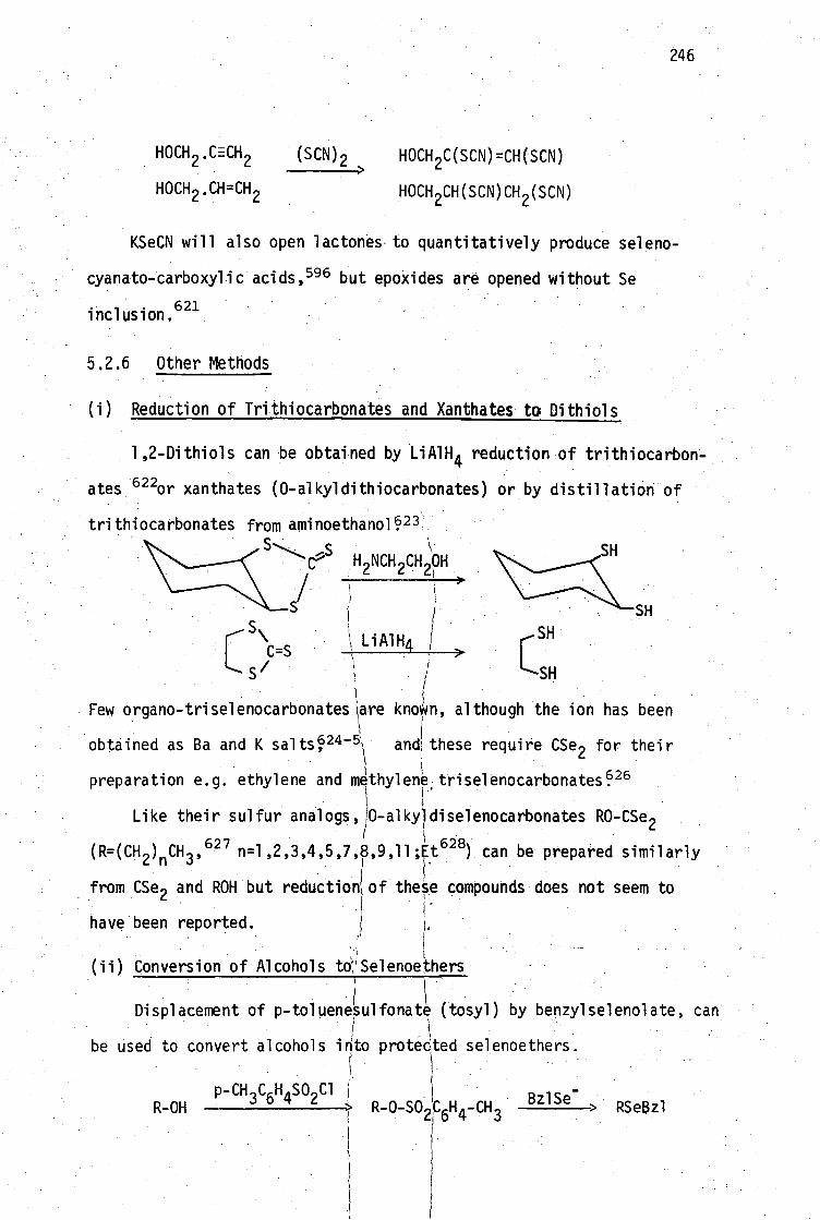
246
HOCH .C=-CH2 (SCN) 2
HOCH 2C(SCN ) =CH(SCN)
HOCH 2* CH=CH 2
HOCH
2CH(SCN)CH 2(SCN)
KSeCN will also open lactones.to quantitatively produce seleno-
cyanato-carboxylic acids, 596 but epoxides are opened without Se
inclusion 621
5.2.6 Other Methods
(i) Reduction of Trithiocarbonates and Xanthates to Dithiols
1,2-Dithiols can be obtained by L1A1H 4 reduction of trithiocarbon-
ates 6220r xanthates (0-alkyldithiocarbonates) or by distillation of
trithiocarbonates
>
C C=S
s/
SH
Few organo-triselenocarbonates are known, although the ion has been
obtained as Ba and K sa1ts 24-5 and these require CSe2 for their
preparation e.g. ethylene and methyleTtriselenocarbonatesP 6
Like their sulfur analogs, 0-alkyldiselenocarbonates RO-CSe 2
(R=(CH2 ) CH 3 627 n=1,2,3,4,5,7,8,9,11,1t ---
AlP ) can be prepared similarly n '
from CSe 2 and ROH but reductionl of these compounds does not seem to
have been reported.
(ii)Conversion of Alcohols tOYSelenoethers
Displacement of p-toluenesulfonate (tosyl) by benzylselenolate, can
be used to convert alcohols into protected telenoethers
p-CH C6 H SO
2Cl I BZ1Se- R-OH > R-O-S02
C6 RSeBz1
from aminoethano19 23 ,s
H2 NCH 2 CH OH
S
LiA1H4
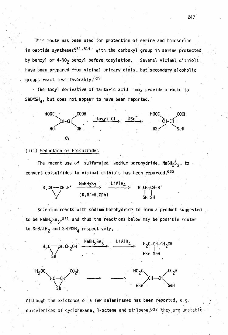
247
•This route has been used for protection of serine and homoserine
in peptide synthesesin, 511 . with the carboxyl group in serine protected
by benzyl or 4-NO 2 benzyl before tosylation. Several vicinal dithiols
have been prepared from vicinal primary diols, but secondary alcoholic
groups react less favorably. 629
The tosyl derivative of tartaric acid . may provide a route to
SeDMSH4' but does not appear to have been reported.
HOOC COOH
CH-CH
HO OH
tosyl Cl
HOOC COON
,/,
RSe SeR
XV
(iii) Reduction of Episulfides
The recent use of 'sulfurated' sodium borohydride, NaBH 2S , to
convert episulfides to vicinal dithiols has been reported. 63°
NaBH2S3 LiA1H4 R.CH—CH.R' > --------> •R.CH-CH-R'
(R,W=H2OPh) I I SH SH
Selenium reacts with sodium borohydride to form a product suggested
to be NaBH2 Se3' 631 and thus the reactions below may be possible routes
to SeBALH 2 and SeDMSH4 respectively,
H C CH .CH OH 2 \ y 2
Se
NaBH 2Se3 L1A1H4 H1C-CH-CH2OH
I HSe SeH
H 2OCN\ //CO2H 2 N\ CO2H
HC—CH > >
Hse/
SeH Se
Although the existence of a few seleniranes has been reported, e.g.
episelenides of cyclohexane, 1-octene and stilbene,632 they are unstable
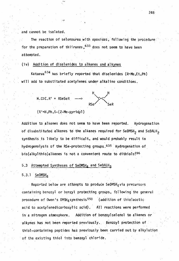
248
and cannot be isolated.
The reaction of selenourea with epoxides, following the procedure
for the preparation of thiiranes, 633 does not seem to have been
attempted.
(iv) Addition of diselenides to alkenes and alkynes
Kataeva 634 has briefly reported that diselenides (R=W,Et,Ph)
will add to substituted acetylenes under alkaline conditions.
H.CEC,R . + RSeSeR
(R'=H,Ph,5-(2-Me-pyridyl)
\\C=C(1
RSe/ , .SeR
Addition to alkenes does not seem to have been reported. Hydrogenation
of disubstituted alkenes to the alkanes required for SeDMSH 2 and SeBALH 2
synthesis is likely to be difficult, and would probably result in
hydrogenolysis of the RSe-protecting groups. 635 Hydrogenation of
bis(alkylthio)alkenes is not a convenient route to dithiols1 96
5.3 Attempted Syntheses of SeDMISH4 and SeBALH
5.3.1 SeDMSH 4
Reported below are attempts to produce SeDMISH4via precursors
containing benzoyl or benzyl protecting groups, following the general
procedure of Owen's DMS14synthesis 550 (addition of thiolacetic
acid to acetylenedicarboxylic acid). All reactions were performed
in a nitrogen atmosphere. Addition of benzoylselenol to alkenes or
alkynes has not been reported previously. Benzoyl protection of
thiol-containing peptides has previously been carried out by alkylation
of the existing thiol into benzoyl chloride.

249
Benzoyl Protection
Dibenzoyldiselenide, XXI, was prepared by alkylation of ethanolic
NaSeH (from Se in NaBH4) and oxidation of the resulting yellow solution
with air
C-SeSe-C
II II 0 0
XXI
The odourless, yellow crystalline material is a convenient source of
benzoyl selenol. Addition of excess sodium borohydride to an ethanolic
suspension of XXI under nitrogen caused the solid to dissolve, yielding
a deep yellow solution. (Reduction of diselenides to selenols usually
causes the disappearance of the yellow colour due to the diselenide
chromophore. However, Jensen reports that oily benzoyl selenol is
yellow in sodium hydroxide solution 558). Filtration of this solution
under nitrogen into a stoichiometric quantity of acetylenedicarboxylic
acid in ethylacetate yielded a deep red solution. Dibenzoyldiselenide
and grey selenium were obtained as the only components of a grey
precipitate which formed overnight. Dilution of the filtrate with
water and extraction with ether produced a small amount of diselenide
from the yellow ether layer. The components of the aqueous phase could
not be separated; red selenium was produced on acidification and
evaporation to low volume.
The expected highly polar dicarboxylic acid product is likely to
be very difficult to separate, without decomposition, from boric acid
formed from borohydride oxidation. The acidity of the carboxylic acid
groups, e.g. DMSH 4 has plc, 2.24(1) and pK2 3.43(1), requires low pH to
ensure solvent extraction from aqueous systems. The products were too
polar for alumina or silica column chromatographic separation. Reduction
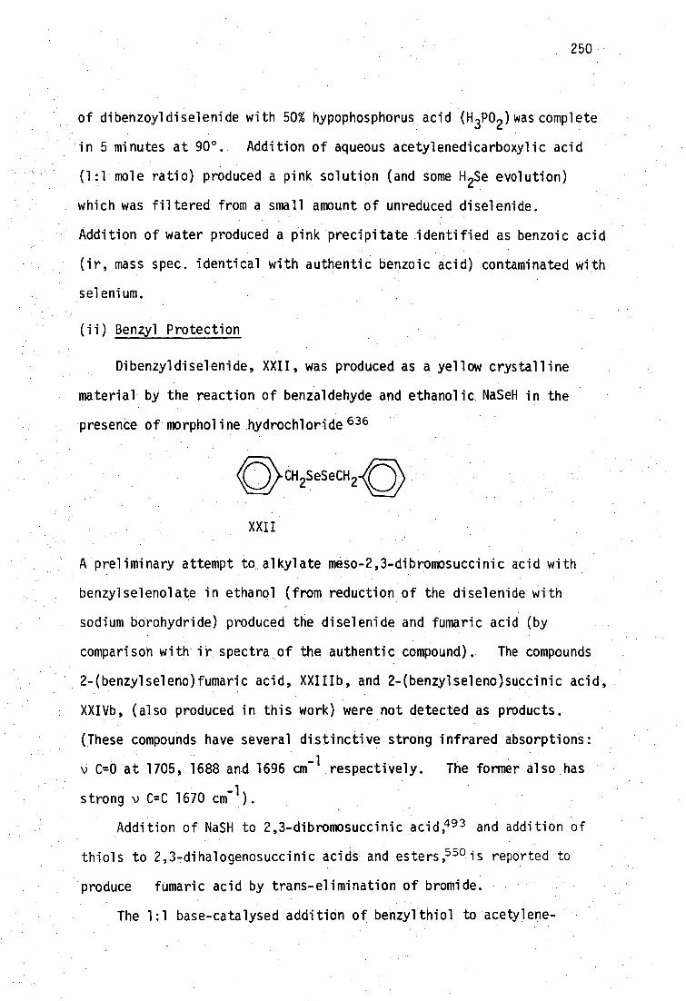
250
of dibenzoyldiselenide with 50% hypophosphorus acid (H 3P02 ) was complete
in 5 minutes at 90 0 . Addition of aqueous acetylenedicarboxylic acid
(1:1 mole ratio) produced a pink solution (and some H 2Se evolution)
which was filtered from a small amount of unreduced diselenide.
Addition of water produced a pink precipitate identified as benzoic acid
(ir, mass spec. identical with authentic benzoic acid) contaminated with
selenium.
(ii) Benzyl Protection
Dibenzyldiselenide, XXII, was produced as a yellow crystalline
material by the reaction of benzaldehyde and ethanolic NaSeH in the
presence of morpholine hydrochloride 636
CH SeSeCH
XXII
A preliminary attempt to alkylate meso-2,3-dibromosuccinic acid with
benzylselenolate in ethanol (from reduction of the diselenide with
sodium borohydride) produced the diselenide and fumaric acid (by
comparison with ir spectra of the authentic compound). The compounds
2-(benzylseleno)fumaric acid, XXIIIb, and 2-(benzylseleno)succinic acid,
XXIVb, (also produced in this work) were not detected as products.
(These compounds have several distinctive strong infrared absorptions:
v C=0 at 1705, 1688 and 1696 cm The former also_has
strong v C=C 1670 cm).
Addition Addition of NaSH to 2,3-dibromosuccinic acid,493 and addition of
thiols to 2,3 -dihalogenosuccinic acids and esters, 550 is reported to
produce fumaric acid by trans-elimination of bromide.
The 1:1 base-catalysed addition of benzylthiol to acetylene-

HO2
Bzl-X
CO2H
251
dicarboxylic acid, ADCA, has been reported to result in the formation
of 2-(benzylthio)fumaric acid, XXIIIa!' 37
XXIII a, X=S
b, X=Se
HOC CO2H
Bzl
XXIV a, X=S
b, X=Se
Addition of two benzylthio groups to ADCA was also achieved, to
produce the previously mentioned protected DMSH 4 derivative meso-2,3-
bis(benzylthio)succinic acid, XIX. Reaction conditions can be varied
to give predominantly the racemic or meso producO 06 The meso-2:1
adduct also results from addition of benzylthio to the trans-1:1
adduct, XXIIIaJ538
Following this approach it was found that the base-catalysed
addition of benzylselenol to ADCA produced only the new unsaturated 1:1
adduct, XXIIIb. Benzylselenol was obtained as a foul smelling, colour-
less oil (bp. 61.0-61.5 ° /8mm) by Painter's Grignard-reagent method. 501
Using Larsson's procedure, neat benzylselenol was added to one
equivalent of ADCA dissolved in sodium bicarbonate solution. The
colourless solution was stirred for 36 hours under nitrogen, then
extracted with ether to remove any unreacted diselenide. Upon acidi-
fication with HC1 (Congo red), the yellow product XXIIIb (55%) was
obtained. Recrystallisation from aqueous ethanol gave XXIIb as bright
yellow needles, m.p. 180-3 ° (dec.).
0C H 1. Bz1Se-/Na0Et 2\ /
HO C.CEC.009H > C=C ` 2. H
+ \
Bz1Se/ CO
2H
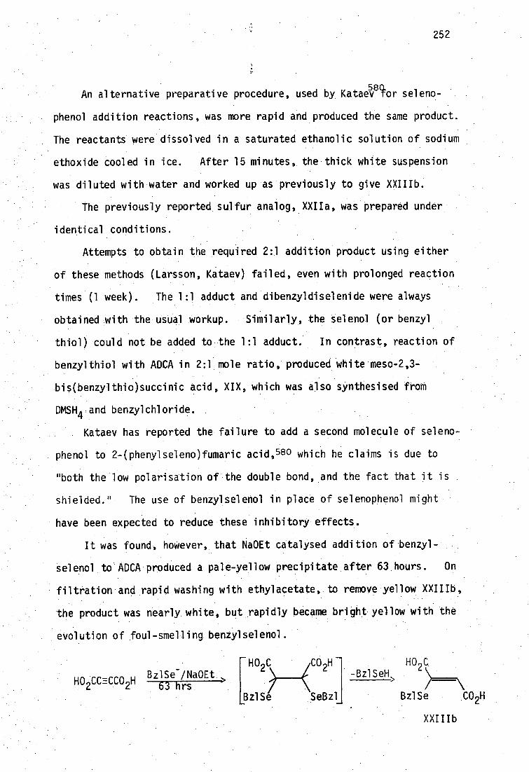
252
580L An alternative preparative procedure, used by Kataev tor seleno-
phenol addition reactions, was more rapid and produced the same product.
The reactants were dissolved in a saturated ethanolic solution of sodium
ethoxide cooled in ice. After 15 minutes, the thick white suspension
was diluted with water and worked up as previously to give XXIIIb.
The previously reported sulfur analog XXIIa, was prepared under
identical conditions.
Attempts to obtain the required 2:1 addition product using either
of these methods (Larsson, Kataev) failed, even with prolonged reaction
times (1 week). The 1:1 adduct and dibenzyldiselenide were always
obtained with the usual Workup. Similarly, the Selenol (or benzyl
thiol) could not be added to the 1:1 adduct. In contrast, reaction of
benzylthiol with ADCA in 2:1 mole ratio, produced white meso-2,3-
bis(benzylthio)succinic acid, XIX, which was also synthesised from
DMSH4 .and benzylchloride.
Kataev has reported the failure to add a second molecule of seleno-
phenol to 2-(phenylseleno)fumaric acid, 580 which he claims is due to
"both the low polarisation oUthe double bond, and the fact that it is
shielded." The use of benzylselenol in place of selenophenol might
have been expected to reduce these inhibitory effects.
It was found, however, that Na0Et catalysed addition of benzyi-
Selenol to ADCA produced a pale-yellow precipitate after 63 hours. On
filtration and rapid washing with ethylacetate, to remove yellow XXIIIb,
the product was nearly white, but rapidly became bright yellow with the
evolution of foul-smelling benZylselenol.
Bz1Se -/Na0Et HO ccEcco 2H 63 hrs
HO2ciCO2
H -
Bz1Se SeBz1
HO 9 C -Bz1SeH
Bz1Se CO 2H 2
XXIIIb

253
The white product may have been the required 2:1 benzyl protected
SeDMISH4'
but its instability precludes its use to conveniently obtain
a deprotected product.
The 1H nmr spectra (in acetone-de, ) of XXIIIa and XXIIIb are
summarised in Table 5.5.
2-(benzylX)fumaric acid C H
7.38
7.39
CH --2
4.25
4.23
4.21
CH=C CO,H a
5.1(br)
5.3(br)
X=S (this work)
(ref. 637 )
X=Se (this work)
6.45
6.50
6.69
Table 5.5: Proton chemical shifts for the acids trans-H0 2CC(XCH CeH e )=
CHCO2 H in acetone-d
6 (6 ±0.02) aresonances due to
carboxylate protons and H 20 impurity in solvent.
The benzylic protons in these compounds are essentially unaffected by
substitution of sulfur by selenium. This result is somewhat unexpected; ,
and may reflect a balance between differences in inductive and mesomeric
effects of the chalcogens in these conjugated Systems. In contrast,
the benzylic protons in dibenzyldiselenide (3.836 An CDC1 3 , this work)
are deshielded with respect to dibenzyldisulfide (3.516 in CC1 4 ) 63.9
and shielded with respect to dibenzylditelluride (4.256 in CDC1 3 , this
work). The methyl protons of dimethyl polysulfides and polyselenides
show analogous behaviour.639 In addition, the olefinic proton of the
selenium analog (.6.456) is deshielded With respect to the sulfur deriv-
ative.(6'.696).. This deshielding effect has also been noted in several
compounds where selenium is part of a conjugated system;640 indicating
that mesomeric influences may be dominant.

254
Larsson has compared the olefinic proton chemical shifts in D 20 of
the disodium salts of 2-(benzylthio)fumaric acid (6.246) and 2-(benzylthio)
maleic acid (5.686) .637 From Table 5.5 it can be seen that the proton
chemical shifts of XXIIIb are close to those of the trans- sulfur analog,
supporting the assignment of trans- stereochemistry for XXIIIb. No
coupling between selenium and the benzylic protons of XXIIIb could be
177 observed. Although one-, two- and three-bond H- Se couplings have
been observed elsewhere641, the
77Se satellites are of low intensity (7.5%
natural abundance) and were not detected in any 1 H nmr spectra recorded
in this work.
The selenium compound, XXIIIb, is bright yellow and the analogous
sulfur compound, XXIII6, also synthesised in this study, is light yellow.
Aqueous alkaline solutions of these compounds are colourless. The near
ultraviolet absorption spectra of XXIIIa and XXIIIb are shown in Figure
5.1, It Can be seen that the yellow Colours of these compounds are
due to tailing of and intense absorption near 300 nm into the visible
region.
The expected position of Amax for the n.4ff* transition of the
unsaturated carboxylic acid chromophore of the sulfur compound can be
calculated. The position of Amax for simple 8-substituted ci,8-unsatur7
• ated carboxylic acids is given at 208±5 nm by Nielsen'.642
\
f3 (1\c02H
It has been reported that carboxyl ate substitution in the s.-.position of
simple enone chromophores has no effect on the position of X .643
max
Similarly, addition of CO2H to c,-unsaturated carboxylic acids does not
alter the position of Amax of the chromophore, e.g. the a,-unsaturated

1.0 Abs
0.5
200 400 nm CJ1 c.n
400 nm 200
Figure 5.1: Ultraviolet absorptiona of 2-(benzylseleno)fumaric acid and 2-(benzylthio)fumaric acid.
a1.0 x 10
-4M solutions at the pH shown, in 1 cm cells.

256
dicarboxylic acids, maleic and fumaric acids, have Amax (6 in water)
of 210(1.3 x 10⁴ ) and 208(1.4 x 10⁴), respectively 44
The degree of bathochromic shift caused by an S-alkyl sUbstituent
can be deduced from the change in Xmax for enones 643 to be ±85 nm.
The sulfur analog XXIIIa is thus expected to have Xmax (in water) at
293 nm (208 + 85). The position of Xmax is found to be 294 nm at pH
3.7, where the compound is 92% protonated (the hydrolysis constants
pical , pKa2 have been Measured to be 2.03(1), 4.49(1) for XXIIIa and
2.01(1), 4.46(1) for XXIIIb at 25° in 0.1M KNO3 ).
By comparing the spectra for protonated forms of XXIIIa and XXIIIb
it is clear that substitution of selenium for sulfur causes a batho-
chromiC shift in the 1147r* transition of +12 nm.
Thus, the contribution to the position of Xmax due to 6-Se-alkyl
substitution would appear to be t98 nm.
Bathochromic shifts are often found to be in the order O<S<Se for
analogous compounds. This may be explained by a hyperconjugative
mechanism. Thus in compounds XXIIIa and XXIIIb the lower energy for
n-nr* transition in the selenium analog may be due to stabilisation of a
charge-separated excited state (in extended conjugation with the a,6 -
unsaturated system) due to the increased ability of Se to accept a
delocalised negative charge.
This possibility has been postulated by Mautner . and co -workers to
explain the bathochromic trends in compounds containing the selenoamide
mOietyf.45-7
Deprotonation Of WIIa and XXII1b causes the 11±7* transition to
move to higher energy (hypsochromic shift) (Table 6.6).

257
r1.4114 transition energy/kJ mo1 -1
XXIIIa X=S XXIIIb X=Se
2(3z1X)C(CO2H)=CH(CO H) 422 407
2(Bz1X)C(CO2- )=CH(CO2 - ) 405 389
Table 5.6: a,f3-unsaturated carbonyl transition energy shifts of
XXIIIa,b on deprotonation of the carboxylate groups.
The new saturated analog of XXIIIb 2-(benzylseleno)-succinic acid,
XXIVb was also prepared. Base-catalysed (Na0Et) addition of benzyl-
selenol to maleic acid, and the usual workup to remove dibenzyldiselenide,
produced a white product on acidification. Recrystallisation from
aqueous ethanol produced XXIVb as fine white needles, mp. 185-7°. The
sulfur analog XXIVa has been previously prepared by Larsson, and was 648
synthesised in this work using the Na0Et catalysed method.
Microanalyses for the new compounds XXIIIb and XXIVb are shown
below.
found % calc. %
C H Se C H Se
2-(benzylseleno)fumaric acid 46.6 3.6 27.8 46.7 3.5 27.7
2-(benzylseleno)succinic acid 46.0 4.4 27.2 46.0 4.2 27.5
The 100 MHz proton nmr spectra (in acetone-d6 ) of XXIVa and XXIVb
are shown in Figures 5.2 and 5.3 respectively. Substitution of Se for
S has caused the expected small downfield shift (see page 253 ) in the
singlet resonance of the benzylic protons (4.036 for XXIVa compared with
4.096 for XXIVb). These values are close to those previously recorded
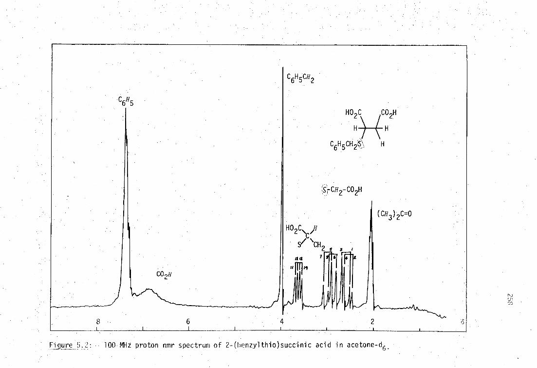
HO2 C 2H
H H
H 2 -
-CO2H
(C113 ) C=0
0 2C\ ,H C
'CH 2 s a
/J /2
SI r— 1
7 f 6 T
CO2H
ii
8 6
Figure 5. 2: 100 MHz proton nmr spectrum of 2- (benzyl tnio)succinic acid in acetone-q.
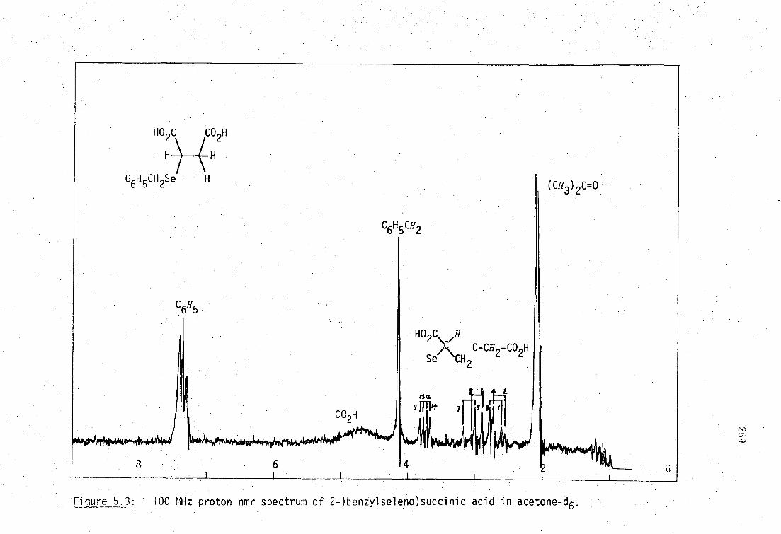
1-IOC CON 2 ( 2
H H
Se H
HO 2C, ,H ‘C- C-CH
Se" "CH, 2 c
CO H
Figure 5.3: 100 MHz proton nmr spectrum of 2-)benzylseleno)succinic acid in acetone-d6.

for the unsaturated analogs XXIIIa and XXIIIb (page 253 ) In the nmr
spectrum of XXIVb, 1 H-
77 Se coupling was not observed.
A feature of interest in these spectra is the well-resolved 12
line-ABX system of methylene (A,B) and methine (X) protons.
HO,C /CO 2H
C-- C
Bz1X/f/ Hb
H x Ha
The spectra were treated by the method of Garbisch 649 to obtain the
considered to be negative 50 The numbering of the ABX transitions
J < O.
coupling constants Jax' bx at) J and J The geminal coupling, J
ab' was
shown in Figures 5.2 and 5.3 is the only combination which satisfies the
intensity and coupling constant relationships required by Garbisch, with
/equilibrium in acetone-d
6 which/ is rapid on the nnr timescale. The
treatment of the spectra are summarised in Table 5.7.
three possible staggered conformations are shown in Figure 5.4.
ab The values for J
ax and J derived from second-order
Both compounds are expected to be in conformational (rotational)
649
\
*
bx
260
XBzl• CO12H •
Hx I II
III
Figure 5.4: Staggered ,conforMations L f XXIVa and XXIVb.
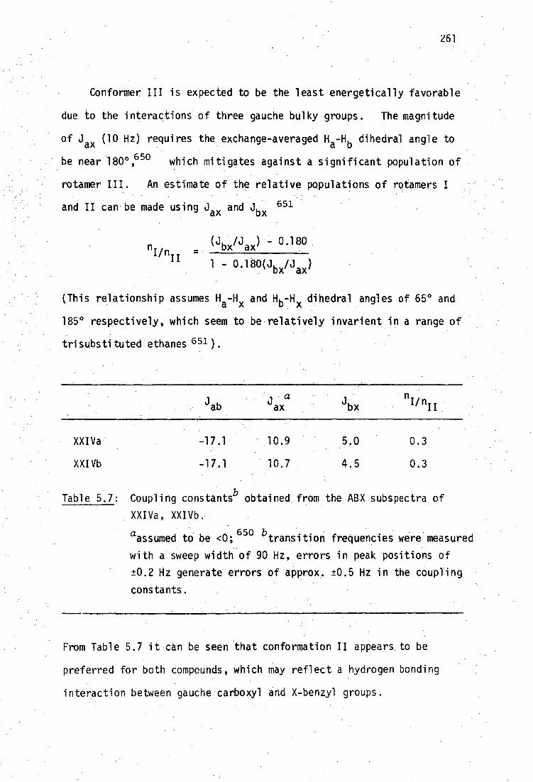
261
Conformer III is expected to be the least energetically favorable
due to the interactions of three gauche bulky groups. The magnitude
of Jax (10 Hz) requires the exchange-averaged H a-Hb dihedral angle to
be near 180 0 ,650 which mitigates against a significant population of
rotamer III. An estimate of the relative populations of rotamers I
and II can be made using Jax and Jbx 651
(J /J ) - 0.180 nun _ bx ax
1 - 0.18 °(Jbx/jax)
(This relationship assumes Ha-Hx and H b -Hx dihedral angles of 65° and
185° respectively, which seem to be relatively invarient in a range of
trisubstituted ethanes 651 ).
nunII X
XXIVa -17.1 10.9 5.0 0.3
XXIVb -17.1 10.7 4.5 0.3
Table 5.7: Coupling constantsb
obtained from the ABX subspectra of
XXIVa, XXIVb.
aassumed to be <0;
650 b transition frequencies were measured
with a sweep width of 90 Hz, errors in peak positions of
±0.2 Hz generate errors of approx. ±0.5 Hz in the coupling
constants.
From Table 5.7 it can be seen that conformation II appears to be
preferred for both compounds, which may reflect a hydrogen bonding
interaction between gauche carboxyl and X-benzyl groups.
II
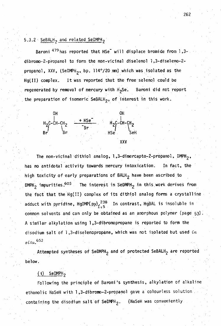
262
5.3.2 SeBALH 2 and related SeDMPH 2
Baroni 479 has reported that HSe - will displace bromide from 1,3-
dibromo-2-propanol to form the non-vicinal diselenol 1,3-diseleno-2-
propanol, XXV, (SeDMPH 2 , bp. 114°/20 mm) which was isolated as the
Hg(II) complex. It was reported that the free selenol could be
regenerated by removal of mercury with H 2Se. Baroni did not report
the preparation of isomeric SeBALH 2 , of interest in this work.
OH OH
+ HSe- H H C-CH-CH
Br 2/ 2
Br Br HSe SeH
XXV
The non-vicinal . dithiol analog, 1,3-dimerCapto 72-propano40MpH 2 ,
has no antidotal activity towards mercury intoxication. In fact, the
high toxicity of early preparations of BALH2 have been ascribed to
,DMPH:2 impurities.605 The interest in SeDMPH 2 in this.work derives from
-
the fact that the Hg(II) complex of its dithidl analog: forms a crystalline
• adduct with pyridine, HgDMP(py). 238 In contrast, HgBAL is insoluble in lt5
common solvents and can only be obtained as an amorphous polymer (page 53)-.
A similar alkylation using 1,3-dibromopropane is reported to form the
disodium salt of 1,3-diselenopropane, which was not isolated but used in
situ.652
Attempted syntheses of SeDMPH 2 and of protected SeBALH 2 are reported
below.
(i) SeDMPH 2 Following the principle of Baroni's synthesis, alkylation of alkaline
ethanolic NaSeH with 1,3-dibromo-2-propanol gave a colourless solution
containing the disodium salt of SeDMPH 2 . (NaSeH was conveniently.

263
prepared by the action of sodium borohydride on selenium, in contrast to
Baroni's use of H 2 Se). Acidification of this solution to pH 4-5
resulted in the evolution of H 2Se . (from Se'2- impurities) which was
removed by purging with nitrogen (under slight vacuum) until the effluent
gas did not darken lead acetate paper (2 hours). Evaporation under
nitrogen and ether extraction gave An orange oil which could be distilled
under reduced pressure. The light orange distillate (bp. 79-85°/1.5 mm)
solidifies at -20 0 , The Raman spectrum of this oil showed no band's in
the region 2000-2700 cm-1
. Free selenols have an intense polarised
Raman band due to ■SeH at 2280-2300 cm-1 which is weak in infrared
spectra. . Additionally, a weak absorption at 286 cm -1 in the Raman
spectrum indicated that diselenide was only a minor component. Ali-
phatic diselenides, RSeSeR, give an intense polarised Raman band at
233-293 cm 654But65 -1 (R=MeP 3 Bun , 5) which is weak in infrared
spectra. • No values of vSeSe have been reported for spectra of 1,2-
diselenocycloalkenes.
_Mass spectrometry .(Figure 5.54) indicated that the product is
predominantly a compound with the formula.C 3H60Se (M4- * 137.9603, calc.
137.9603) with a minor proportion Of 4 less volatile diselenide
C3H6OSe2 (Figure 5 . .5b).
The mixture could not be separated by fractional distillation under
reduced pressure, or chromatographically on alumina, but could be
partially separated by fractional distillation under high vacuum to give
two selenium containing fractions, enriched in the first and second
compounds respectively.
Cyclic selenides have previously .been prepared as major products
from non7vicinal dibromides .(Table 5.8). The proton nmr spectrum
(Figure 5.6) of the mixture, is consistent with the major component
being selenetan-3-ol, XXVI. The minor component is most likely 1,2-

R=11
CO 2H 2
- Se
2
H 2Se
Al2 03' 400 >
selenanes
R R"
Br R=R 1 =R!'=H 660,664
(and others)
seleniranes
264
Ref
/0\ 1. Ph P=S 3
• 2. CF3CO
2H > RAI
R = H, R' = Ph, octyl
632
selenetanes
r
' R=R'=H,Me 656-7
H 2C=CH.CH 2Cy/
0
HO rSe Cl HO2C CO2H
electrolysis > R=H,R'=OH
Cl' /402H
H 2
658
591
selenolanes
tri\/\r-Br
Co
659-60
661
662
• (CH 2=CHCH 2 ) 20
2. tuC1 2/KSeCN/Me0H > CH OMe 663
R'
H 2 Se • :• R=R1=R"=H 662.
Al203'400 0>
H 3CCH=0,H 2Se• Rei,R'=C02tt ' R u =0:
Et0H,-100' 24
H 666
Et0 .2C"\e^CO2Et 0
Al Se /Et0H LiA1H 2 3 4 R=Ph,W=H,R"=0 667
H 3CCO2Na or Al(OPr1)3> R,R 1 various,R"=0H,H 668-9
Table 5.8: Previously reported monoselenoalkanes. a a many of these compounds undergo addition reactions with halogens to produce 1,1'-dihalogeno- and ring-substituted products but these are not considered here. Similarly, many 1,1-dioxides are known.

R" R" >
R"=H
265
ref
diselenolanes
R'
diselenanes
X=SeCN,R=R'=H 614,656,670,672
X=SeCN,R=Me,R'=Me X=Br,R=Me,R'=Ph X=Br,R=H,W=CO 2H
H X=SeBz1,R=H,R'=CO, ) c 672 X=C1,R=RI=CH 2OH
R".=CO 2Et,X=SeCN,R=R 1 =H 872
R=H 659,673-4
R=C0 H 484,591
Table 5.9: Previously reported 1,2-diselenocycloalkenes.
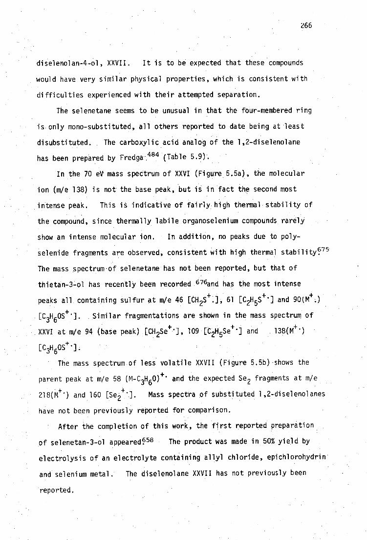
266
diselenolan-4-ol, XXVII. It is to be expected that these compounds
would have very similar physical properties, which is consistent with
difficulties experienced with their attempted separation.
The selenetane seems to be unusual in that the four-membered ring
is only mono-substituted, all others repotted to date being at least
disubttituted. The carboxylic acid analog Of the 1,2-diselenolane
has been prepared by Fredga-. 481.1 (Table 5.9) - .
In the 70 eV mass spectrum of XXVI (Figure 5.5a), the molecular
ion (m/e 138) is not the base peak, but is in fact the second most
intense peak. This is indicative of fairly high thermal stability of
the compound, since thermally labile organoselenium compounds rarely
show an intense molecular ion. In addition, no peaks due to poly-
. selenide fragments are observed, consistent with high thermal stability 75
The mass spectrum-of selenetane has not been reported, but that of
thietan-3-ol has recently been recorded 676and has the most intense
peaks all containing sulfur at m/e 46 [CH 2S+ :], 61 [C 2H 5S+ '] and 90(M4- ..)
.[C3H60S :]. Similar fragmentations are shown in the mass spectrum of
XXVI at m/e 94 (bate peak) [CH 2Se+ ] 109 . [C2H and 13801+ 1
[C3F1 605 4. ]:
The mass spectrUm of less volatile XXVII (Figure 5.5h) shows the
parent peak at M/e 58 (M-C 3H60) -/- * and the expected Se 2 fragments at m/e
218(M *) and 160 [8e ]. Mass spectra of substituted 1,2-diselenolanes
have not been previously reported for comparison.
• After the completion of this work, the first reported preparation
of selenetan-3-ol appearee68 The product was made in 50% yield by
electrolysis of an electrolyte containing allyl chloride, epichlorohydrin
and selenium metal. • The diselenolane XXVII has not previously been
reported.

100
Figure 5.5a: Low resolution 70 eV mass spectrum of XXVI.
94
CH 2 Se+
I % .
OH Se
138
M -CHO
HOSe
92
43
7
90 136
134
1 100 I I
300 m/e
28
200
J
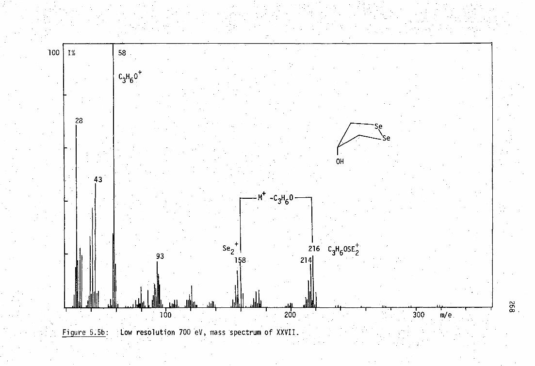
100
Figure 5.5b: Low resolution 700 eV, mass spectrum of XXVII.
I% 58
OH
28
43
Se 2+
18
.111,1„„ itit 1.1111 160
II II
93 216
214
200 .'t .
- 1 "( 1
300 . m/e.

269
The proton nmr spectrum of XXVI in CDC1 3 is shown in Figure 5.6.
Apart from a minor impurity at 1.336 (m, 0.4H), the spectrum can be
interpreted in a first-order fashion as two triplets centred at 3.126
(2H) and 3.376 (2H) respectively, corresponding to two sets of axial
and equatorial protons, each of which is coupled to the proton geminal
to the hydroxyl group, giving rise to a quintet (1H) centred at 4.856.
Selenetane has a similar methylene proton multiplet at 3.20 77 The
D20 exchangeable OH proton appears as a broad signal centred at 1.456
(1H). Satellites due to 2J(
1H-
77Se) cannot be observed.
The proton nmr spectrum of the sulfur analog, thietan-3-ol, has recently
been interpreted 78 and is apparently consistent with the expected
saddle-conformation of the four-membered ring having an equilibrium
saddle angle of 140-150 0 . Details of the nmr spectrum of this compound
are not available, as the original Russian paper is unavailable in
Tasmania.
The closely related compound 3-acetylthietane has been mentioned
in an nmr study of thietene-1,1-dioxides 79 The axial and equatorial
methylene protons are reported as a multiplet 333 Hz downfield from TMS
in CC14
(5.556 on the 60 MHz instrument) which differs somewhat from
the value of 4.356 found here for selenetan-3-ol. The positions of
a-methylene proton resonances were not recorded in the abovementioned
studyP9
A complete description of the proton nmr spectrum of selenetan-3-ol
involves treatment of an A2B2X spin system. The two pairs of axial and
equatorial protons are magnetically non-equivalent due to long range
spin coupling and the occurrence of conformational equilibria shown
below:

270
L.)
4-, C.)
C.)
C.)
4- C 7-7 =4" F.:
C • o
4-0
C•••
LC:)
CC
'Cr —
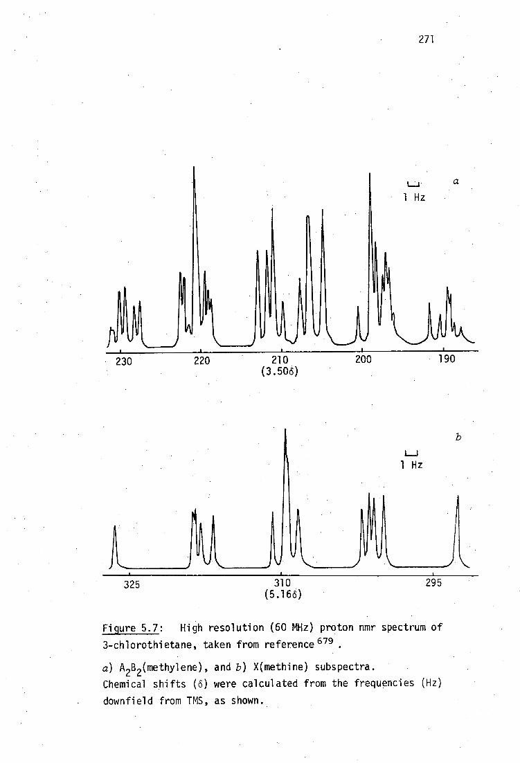
271
1_1
1 Hz
• I
A k.
1
ir
2 100
230 220 210 (3.506)
190
1 Hz
325
310
295 (5.166)
Figure 5.7: High resolution (60 MHz) proton nmr spectrum of
3-chlorothietane, taken from reference 679 .
a) A2B2(methylene), and b) X(methine) subspectra.
Chemical shifts (6) were calculated from the frequencies (Hz)
downfield from TMS, as shown.

HO Ha
HO
> HO
Such a treatment has been carried out for 3-ch1orothietane 8°
and sixty-four lines of the A2B 2X system identified (Figure 5.7).
At lower resolution, such as that under which the spectra were
recorded in this work, the A 2B 2 sub-spectrum can be considered as
two asymmetric closely coupled triplets. The X sub-spectrum is a
quintet. These features appear in the spectrum of selenetan-3-ol
(Figure 5.6).
Details of vibrational assignments of spectra of 3-substituted
thietanes do not seem to have been published, although a small diagram
of the infrared spectrum of thietan-3-ol is available 81 which is
similar to that of XXVI. The vibrational spectra of thietan 82-ghd
of selenetane 68 ave been assigned. The infrared spectrum of XXVI
can be tentatively assigned by comparison with those of 1,3-disubstituted
2-propanolsand selenetane 686and is shown in Table 5.10 below.
272
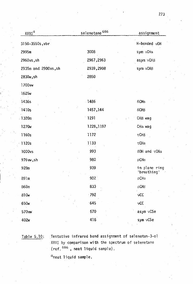
273
XXVI a selenetane 686 assignment
3150-3550s,vbr H-bonded v0H
2995m 3008 sym vCHa
2960vs,sh 2967,2963 asym vCH(3
2935m and 2900vs,sh 2939,2908 sym vCH(3
2830w,sh 2850
1700vw
1625w
1436s 1486 SCHa
1410s 1457,144 (SCHp,
•1320m 1291 CH (3 wag
1270w 1228,1197 CHa wag
1160s 1172 TCHI3
•1120s 1133 TCHa
1000vs 993 60H and TCHa
976vw,sh 980 PCHa
928m 939 in plane ring 'breathing'
891m 902 PCHa
868m 833 pCK
810w 792 vCC
650w • 645 vCC
570vw 570 asym vCSe
402w 418 sym vCSe
Table 5.10:
Tentative infrared band assignment of selenetan-3-ol
XXVI by comparison with the spectrum of selenetane
(ref.686 neat liquid sample).
aneat liquid sample.
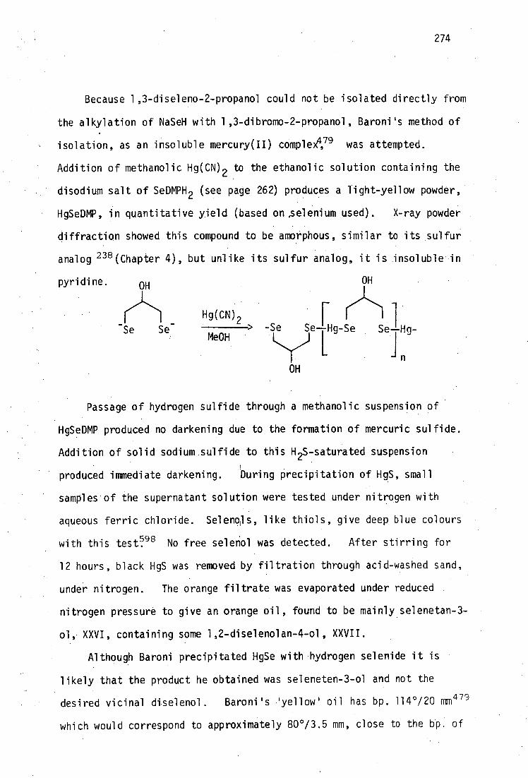
274
Because 1,3-diseleno-2-propanol could not be isolated directly from
the alkylation of NaSeH with 1,3-dibromo-2-propanol, Baroni's method of
isolation, as an insoluble mercury(II) compleX 4,78 was attempted.
Addition of methanolic Hg(CN) 2 to the ethanolic solution containing the
disodium salt of SeDMPH 2 (see page 262) produces a light-yellow powder,
HgSeDMP, in quantitative yield (based on ,selenium used). X-ray powder
diffraction showed this compound to be amorphous, similar to its sulfur
analog 238 (Chapter 4), but unlike its sulfur analog, it is insoluble in
pyridine. OH OH
Hg(CN)2 r > Se Se--Hg-Se S g- Me0H
in
OH
Passage of hydrogen sulfide through a methanolic suspension of
HgSeDMP produced no darkening due to the formation of mercuric sulfide.
Addition of solid sodium sulfide to this H 25-saturated suspension
•1
produced immediate darkening. During precipitation of HgS, small
samples . of the supernatant solution were tested under nitrogen with
aqueous ferric chloride. Selenols, like thiols, give deep blue colours
with this test 98 No free selenol was detected. After stirring for
12 hours, black HgS was removed by filtration through acid-washed sand,
under nitrogen. The orange filtrate was evaporated under reduced
• nitrogen pressure to give an orange oil, found to be mainly selenetan- -
ol, XXVI, containing some 1,2-diselenolan-4-ol, XXVII.
Although Baroni precipitated HgSe with hydrogen selenide it is
likely that the product he obtained was seleneten-3-ol and not the
desired vicinal diselenol. Baroni's 'yellow' oil has bp. 114°/20 MM479
which would correspond to approximately 80 0/3.5 mm, close to the bp. of

275
XXVI (79-85°/3.5 mm) using the usual bp.-pressure nomograms. 687
An alternative synthetic route to SeDMP was attempted. A small
scale attempt to prepare 1,3-diselenocyanato-2-propanol by refluxing
the dibromide with two equivalents of KSeCN in methanol, produced an
orange oil after evaporation and ether extraction under nitrogen. High
vacuum fractional distillation afforded two fractions. The first,
minor, fraction (bp. 50-53°/0.01 mm) showed no infrared absorption in
the region expected for vCEN of selenocyanates e.g. 2150 cm
several a,i3-alkoxyselenocyanates;588 and was discarded.
OH
CH 2 -CH-CH I
1
NCSe SeCN .
XXVIII
The second major fraction (bp. 60-75°/0.01 mm) showed intense infrared
absorption at 2135 cm-1
consistent with the presence of selenocyanate.
In addition, the remainder of the infrared spectrum was very similar to
that of 1,3-dibromopropanol. Extensive hydroxyl hydrogen bonding was
indicated by a strong, broad absorption at 3100-3600 cm -1 . Mass
spectrometry (Figure 5.8) indicated a parent ion containing one atom of
both bromine and selenium in its parent ion m/e 243, consistent with the
formula C4H 6BrN0Se(M+
242.8814, calc. 242.8796). The expected
patterns of isotopic peaks in fragments containing Br 2 , BrSe and
Se 2 are shown in Figure 5.9. • The parent ion in Figure 5.8 clearly •
contains the BrSe.fragment. The product is probably 1-bromo-3-
selenocyanato-2-propanol, XXIX.
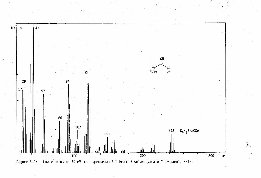
300 m/e
100 43
29
27 57
.111 II
121
Figure 5.8: Low resolution 70 eV mass spectrum of 1-bromo-3-selenocyanato-2-propanol, XXIX.
94
80
11111 Ii I 200
243 C4 H 6 BrNOSe
LI
107
1.11 I I fill 100
150
1 1,

277
163 154 158 162 155 164 Br 2
BrSe Se 2
Figure 5.9: Isotopic patterns for Br 2 , BrSe and Se 2 isotopic fragments.
The presence of a minor component of diselenide was indicated by a very
weak isotopic cluster at m/e 444, containing the Se2 fragment, in the
chemical ionisation mass-spectrum.
The structure, XXIX, was confirmed by the 1 H nmr spectrum in CDC1 3
(Figure 5.10). The main features of the spectrum are:
(i) broad hydroxyl proton resonance at 2.96(IH), which disappears
with D20 exchange. This type of broad resonance is observed in nmr
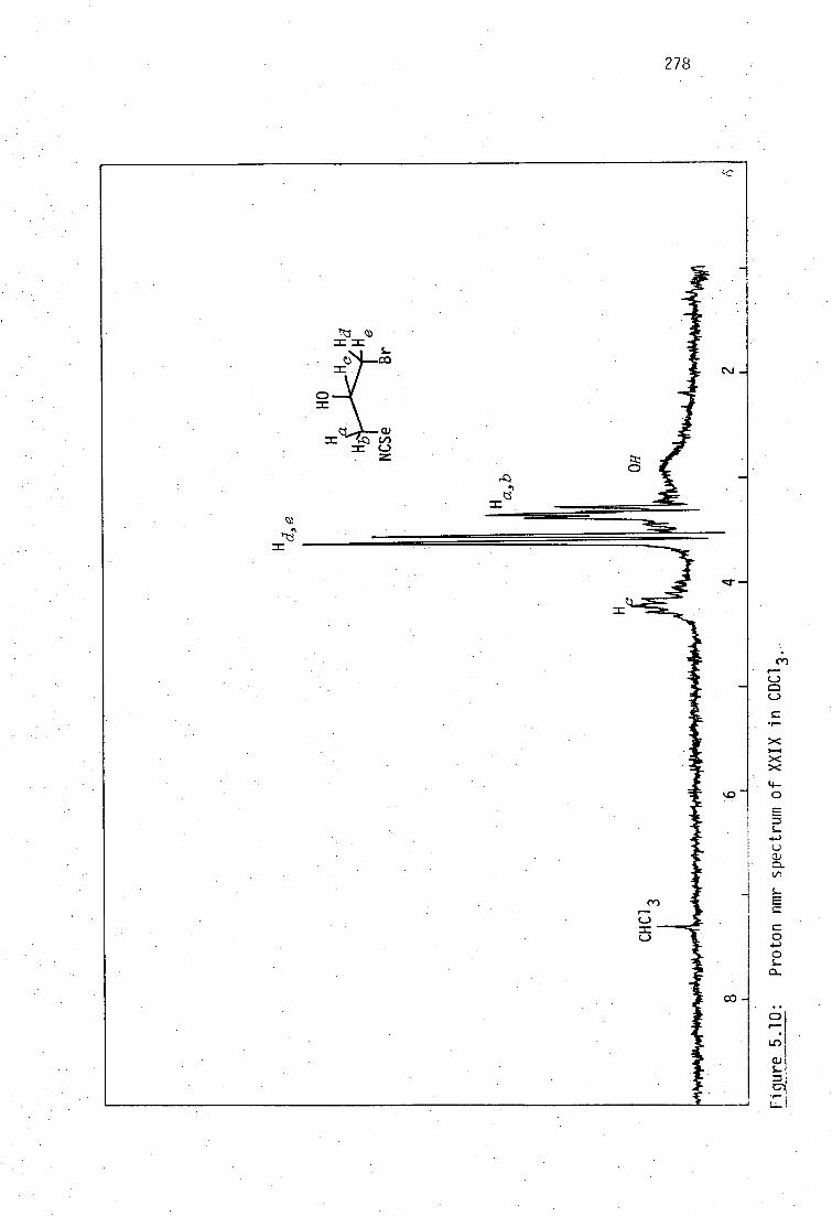
278
Cr
-
czt —
■-••• (=1
1—• >.<
4— 0
s- 4-J
(t)
Prot
on
nmr

279
spectra of 0MPH 2 , BALH 2 and other disubstituted propanols used in this
work, but the position of resonance is quite variable.
(ii)a multiplet centred at 4.236(1H), similar to the multiplet
(quintet, 1H) appearing at 4.036 in the spectrum of 1,3-dibromo-2-
propanol which is due to H c , geminal to the hydroxyl group.
(iii)an asymmetric doublet 3.56,3.626(2H), identical to that in
the spectrum of 1,3-dibromo-2-propanol, due to the protons geminal to
the bromine, Hd,e , which appear to be magnetically equivalent. The
couplingcd
J J ce is 6 Hz. -=
(iv) three singlet resonances at 3.28, 3.35, 3.386(2H) which must
be associated with protons H a,b geminal to the selenocyanate. This
pattern can be rationalised by assigning the central (most intense)
singlet resonance arbitrarily to H a , which requires J ap0. The outer
pair of resonances form an asymmetric doublet which is similar to (iii)
due to Hb , with bd=10 Hz. Satellites due to 2J( 1 H-
77Se) could not be
observed.
Two points arise, namely, the lack ofcoupling of H a with Hd , yet
Jbd=10 Hz and the lack of .geminal coupling between H a and Hb .
The lack of coupling between Ha and Hd suggests that the dihedral
angle between these protons Must be near to 90 0 , from consideration of
the Karplus equation 50 A conformation of XXIX consistent with this
requirement is Shown in the diagram below, which is a Newman projection
of the molecule, viewed along the C(SeCN)-C(OH) bond. This conforma-
tion also accounts for hydrogen bonding between the hydroxyl and one
selenium Tone pair forming a five-membered ring. Evidence of extensive
hydrogen bonding is found in the infrared spectrum (page 365) and is
also consistent with J ad,L6 Hz since the dihedral, angle between H a and
Hd is approximately 30 0 P. 5. 0 Additionally, the geometry of the two
seleniumLlone pairs is suited to donation Of electron density into

280
adjacent H a and H b a-bonds. Such hyperconjugative donation will make
the geminal coupling constant, J ab' more positive6.50 Since unperturbed -
geminal coupling constants are usually negative 650 this accounts for
J NO in this case. An identical situation, depicted below, is found ab
650 in 1,3-dioxolane producing

281
(ii) SeBALH 2
The failure to obtain non-vicinal SeDMPH 2 did not encourage the
preparation of vicinal SeBALH 2 by similar routes. Attempted alkylation
of ethanolic NaSeH by 2,3-dibromo-l-propanol produced a deep red-brown
solution from which allyl alcohol (detected by gc. retention time
comparison with an authentic sample) could be extracted.
- C--CH-CH OH + 2HSe>
H2C=CH-CH
2OH + 2Br- + H2 S + Se
1 1 Br Br
Nucleophilic substitution of vicinal dibromides with thiols has often
been reported to cause elimination to form an olefin.
Addition of ethanolic mercuric acetate to the red-brown solution
precipitated a small quantity of pale-yellow powder, which was insoluble
in hot pyridine and other common solvents. This complex was not
identical to HgSeDMP as might have been expected if 2,3-dibromo-l-
propanol was contaminated with a small quantity of the non-vicinal
isomer. -
•
Selenocyanation of 2,3-dibromo-l-propanol was not attempted in
view of the' failure to obtain a diselenocyanate.of SeDMPH 2 .
Addition of two equivalents of benzylselenol to propargyl alcohol
dissolved in ethanolic sodium ethoxide solution produced a crystalline
product after ak hours. This product (probably a 1:1 adduct similar
to those found during additions to acetylenedicarboxylic acid) was not
isolated but redissolved to give a red solution overnight. Extraction
into petroleum ether removed dibenzyldiselenide to leave a red oil which
decomposed to produce selenium on standing. The products were not
examined - further in view of difficulties experienced with product.

282
separation from attempted SeDMSH 4 synthesis. Addition of excess
thiolacetic acid to pent-3-yn-l-ol is reported to produce only a mono-
adduct, 554 due to an inactivating effect of the hydroxyl group e.g.
hex-l-yne produces both mono- and di- adducts under these conditions 89
No attempt was made to alkylate sodium benzylselenolate with 2,3-
dibromo-l-propanol because the analogous thiolate reaction produces the
rearranged product 1,3-bis(benzylthio)-2-propanol, exclusively604-5
(page 244).
5.4 Conclusions
Possible methods for the preparation of Se-protected diselenols
have been reviewed, and several routes to synthesis of protected
selenium analogs of DMSH 4 and BALH 2 and its isomer DMPH 2 have been
investigated.. The protected SeDMSH 4 compound, 2,3 7bis(benzylseleno)
succinic acid could not be obtained in this study.
Addition of two equivalents of benzylselenolate to acetylene-
dicarboxyliC acid gives only the new 1:1 addition product XXIIIb, in
contrast to the addition of benzylthiolate where 4 2:1 adduct can be
Obtained. Similarly, the new saturated analog of XXIIIb, XXIVb was
obtained by addition of benzylselenolate to maleic acid. Benzylthiolate
reacts identically.
Perturbations of the n-*Tr* electronic transition of an a,87unsaturated
carbonyl chromophote by the benzylselenolate group produces a large batho-
chromic shift which causes XXIVb to appear bright yellow. The analogous
thioether As pale yellow, consistent with the similar but smaller effect
of the benzylthiolate group.
Second-order analysis of the ABX 1 H nmr spectra of XXIVb and its
sulfur analog showsa preference for a conformation in which one carboxyl ,
group is gauche to the chalcogen.

283
Attempted preparation of SOMPH 2 from 1,3-dibromo-2-propanol leads
to the cyclic product selenetan-3-ol, only recently reported in the
literature. The 100 MHz proton nmr spectrum can be interpreted as an incomplere.:iy
- resolved five spin A2B2X coupled system. Reaction of 1,3-dibromo-2- ,
propanol with two equivalents of KSeCN, resulted in the addition of
only one selenocyanate group. The new product 1-bromo-3-selenocyanato-
1 2-propanol has been characterised by H nmr, ms and ir spectroscopy and
shows considerable hydrogen bonding between the hydroxyl and seleno-
cyanate groups.
Attempted preparations of SeBALH 2 by alkylation of NaSeH with
2,3-dibromo-1-propanol, lead to the expected formation of allyl alcohol
by elimination of bromide. No characterisable products other than Se
and dibenzyldiselenide could be isolated from reaction of benzyl-
selenolate with propargyl alcohol.
284
CHAPTER SIX
POTENTIOMETRIC DETERMINATION OF AQUEOUS FORMATION
CONSTANTS OF VERY STABLE COMPLEXES
6.1 Introduction
The aqueous formation constants of MeHg(II)-thiolate complexes
had not been extensively studied Until this work, and the work of
Reid and Rabenstein 302 (Chapter 3). Apparent discrepancies in
previously reported values for the formation constants of MeHg(II)
monothiolates, and the need to evaluate the stability of MeHg(II)
complexes with antidotal vicinal.dithiols led to the determination of
the constants which have been discussed in Chapter 3.
In order to compare the formation constants for MeHg(II) with a
range of monothiols and vicinal dithiols (containing sulfhydryl groups
with acid dissociation constants in the range 9.0< pK a < 11.5),. a
method which is able to determine these constants with some precision
is required. The methods currently available for this requitement
are based on potentiometric, spectrophotometric and nuclear magnetic
resonance techniques. ,(Other methods . , e.g. polarography,-solvent
extraction, ion exchange, etc. are generally less precise and of more
limited applicability 690). All of these methods depend on the ability
• to monitor the concentration(s) of one or more equilibrium components
as the equilibrium position is altered. Each of these techniques has
been reviewed and extensively used to study solution equilibria 90-1
The most widely used and the most precise is the potentiometric
method, which was considered to be best suited to investigation of
equilibria between MeHg(II) and thiolate ligands in this study. • The
spectrophotometric method,although it offers a unique advantage of
285
sometimes determining the number of significant equilibrium species,
was unsuitable because the thiolate ligands and methylmercury(II) ion
do not have chromophores which are sensitive to equilibrium conditions,
e.g. pH, and which are readily accessivle in the near UV/visible spectral
, 360 199. g H 337 and 309,360,369) regions. NMR Methods (e.g. 11
L
have been used extensively to study MeHg(II) and offer the advantage of
being able to determine the concentrations of species involved in
microscopic equilibria, e.g. the protonation or complexation of sulfhydryl
and amino groups of biologically important mercaptoamines can be monitored
individually. (The technique has been discussed in some detail in
Chapter 3 of this thesis).

286
6.2 Evaluation of ligand hydrolysis equilibria
6.2.1 Definition of 'Equilibrium Constants' used in this work
The term equilibrium constant', used in this work, warrants some
clarification.
Consider a simple monoprotic BrOnsted acid dissociation in aqueous
solution, the equilibrium for which can be written
HL (ao + nH 20 ;,====e(ao + (aq)
Although all species, HL (a0, L - (ao , (ao, are probably extensively
solvated, few methods of investigation can distinguish between degrees
of hydration ., and solvent molecules will not be included in subsequent
equilibria although it should not be forgotten that they may be involved ,
in primary coordination. In particular, the methylMercury(II) cation
will be represented by MeHe for brevity, instead of MeHg(OH 2 ) 47 , and
the proton will be considered:As H i'. In addition, the concentration
or activity Of water is considered to be constant, at least for dilute .
solutions. The thermodynamic acid dissociation constant for the above
equilibrium, which can be related directly to free energy changes, is
written as Ke .
{HL}
where the parentheses 0 indicate activities of the relevant species.
To determine Ke-, the activities, or at least the activity coefficients,
y (which relate activity to concentration, e.g. {L - } = y L4L- ] ), of all•
species must be determined. Although yH+ has been extensively studied,
e.g. for the determination of pH standards, this has only been achieved
for a limited range of electrolyte conditions. Estimates of mean
activity coefficients, y±, for electrolyte species other than protons

287
are usually made using empirical relationships between y± and solution
ionic strength. Table 6.1 shows that these relationships may give
widely different values for mean molar activity coefficients, especially
for electrolytes which are not of the 1:1 type, even at the moderate
ionic strength (0.1M) used in many equilibrium constant studies. 692
For highly charged ions, e.g. the anion DMS 4 , such calculations of
activity coefficients are highly tenuous. It is more usual to ignore
activities of species other than protons by defining 'mixed' or
operational equilibrium constants.
{H 4-}[L]
[HL]
Proton activities are determined directly from the relationship pH
-logTH+1 after calibration of a glass indicator electrode in solutions
of known proton activity. The concentrations of other species can be
determined directly, e.g. spectrophotometrically using the Beer-Lambert-
Bouger relationship between absorbance and concentration, or with other
potentiometric electrodes calibrated in terms of concentration (very
rarely available), but are more usually calculated. The mathematical
evaluation of species concentrations assumes the validity of the
equilibrium expression, electroneutrality, and mass-balance equations,
and in this case requires two independent measurements of pH from
different equilibrium solutions. The abovementioned equations can be
solved for [L -] and [HL]. It is usual to over-determine these equations,
i.e. obtain many more experimental observations than there are unknowns,
and use least-squares or other multiparametric curve- fitting.
procedures to obtain [1.-] and [HL] (page 306). It should be noted that
[H ] is required in the mass-balance and electroneutrality expressions,
still necessitating an empirical estimate for yH+. It has been

Model 2 3 4 6 8 9 12 16
Debye-HUckel 0.6890 0.4747 0.3271 0.2254 0.1070 0.0508 0.0350 0.0114 0.0026
Guntelberg 0.7535 0.5678 0.4278 0.3224 0.1830 0.1039 0.0783 0.0335 0.0108
Davies 0.7715 0.5952 0.4592 0.3542 0.2108 0.1255 0.0968 0.0444 0.0157
Scatchard 0.7767 0.6033 0.4686 0.3460 0.2196 0.1325 0.1029 0.0482 0.0176
Bjerrum 0.7623 0.6526 0.5900 0.5498 0.5012 0.4731 0.4630 0.4418 0.4249
Comments
log y± may be extended to take into account ionic 'size' logy± = -z+z_ Air/(1+Bain
log y± = - z+ z_ AM/(1+)7)
may be extended by consider-ing a constant ionic size of 3A°. log y± = -z+z_A47(1+3B4)
log y± = -z+z_A47(1+1/7)
+0.2Az+z_I
log y± = -z+z_Ail/(1+1.54)
may be extended by consider-ing a temperature dependent demoninator log y± = -z+z_AIIT/(1+4.5Bin
log y± = -z+z_Air/(1+Ba B takes into account ion pairing if ion diameter is less than aB .
Table 6.1: Theoretical mean activity coefficients at 25°C and 0.1M Ionic Strength 692
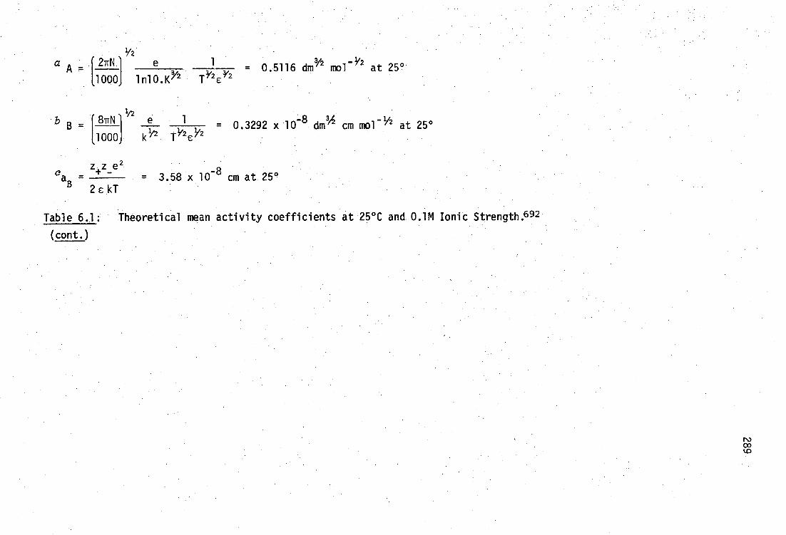
2TrN e 1- - 0.5116 dm Y2 mol - at 25°
1000 1n10.K2 7 2
1/2 8TrN 1 e 1 0.3292 x 10⁻⁸ m
3 cm mo -1/2 at 25°
11000j k ir2 T 1/2d12 *
z z e 2 ea -
+ - = 3.58 x 10 -8 cm at 25° 2 e kT
Table 6.1: Theoretical mean activity coefficients at 25°C and 0.1M Ionic Strength. 692
(cont.)

290
indicated by Bates, 93 that Km has the disadvantage that it reflects
small changes in ionic environment via ail but neglects interionic
effect on ions other than H+
.
The third form of equilibrium constant is used in this work and
101 tabulated extensively elsewhere: --,694
ine concentration or stoichiometric
constant, KC , is defined as
[e][L- ]
[HL]
In this case, it is necessary toevaluate hydrogen ion concentration,
[el instead of activity, e.g. using a glass-electrode calibrated in
terms of proton concentration by the methods described in Section 6.
The concentrations of ligand species can be measured as for Km (rarely)
or calculated by least-squares procedures. Stoichiometric constants
are used throughout this work unless otherwise stated, and can be
written for more complex metal-ligand-proton equilibria in an identical
fashion to those above.
For example, the formation equilibrium of the complex
, (MeHg)m I i (SR)sHilwould be written in terms of components MeHg , n-
and e:
mMeHg+ + i1 + 811Sn- + i (SR) s Hh
with an overall stoichiometric equilibrium constant,
[(MeHgm) I i (SR) sHill
[MeHg+] n [I- i i [Rsn-
]8[H+p
It has been found that mixed iodide-thiolate species are not formed in
this study (page g7 ), and so the iodide subscript has been omitted from
the MeHg(II)-thiolate constants recorded in Chapter 3. (However, for

291
data input,to the computer program MINIQUAD 81 all component subscripts
are required).
In addition, hydroxo complexes will be described with h < 0, e g.
(MeHg) 20H+ will have [(MeHg) 2 )H 4 ]
207 1 [meHg+ ] 2 [H+ ]-2
It should therefore be emphasised that O's of complexes involving
hydroxide, are not formal formation constants, but can be related to
these by the autoprotolysis constant of water, K, since, for example:
8(actual formation)
(MeHg) 20e) 20-1 (‹)-1
I I
This practice is widely used in the stability constant literature, and
is used here for consistency. Equilibrium constants, such as B
can be evaluated from pH titration data in acid solution, without
- recourse to experimental values of Kw whereas the corresponding •actual
formation constant requires accurate values for Kvci in order to evaluate
[OH], contributing additional uncertainty.
6.2.2 Calibration of glass-electrodes as [e] probes
When a glass-electrode assembly is calibrated using a primary
standard buffer, pH(s), and then transferred to a measurement solution,
the pH reading corresponding to H is related to [H] s of the buffer
by the defined relationship 695
(6.1) (E -E') + (E.' - -log
[H+] 1 = pHs s j
2.303 RT/F
js log yHi.,
The measured pH can be related to [H] only if (i) liquid junction
potentials for the buffer, E js , and test solution, Ej ', are either
negligible or identical, and (i ) the activity coefficient y H+' can

joH ‹I [H+]
292
be accurately calculated or measured in both solutions. Since the
test solution and primary buffers are of totally different ionic
composition, e.g. the NBS standard buffers commonly used for calibra-
tion, 0.05m potassium hydrogen phthalate and 0.01m sodium tetraborate,
have ionic strengths 0.053M and 0.02M respectively, it is most unlikely
that either condition can be fulfilled.
It is therefore necessary to calibrate the glass-electrode assembly
as a H -concentration probe, in the ionic medium to be used as measure-
ment solution.696 This is done by noting that the emf of the electrode
varies linearly with log[H ]in acidic solutions as follows:
(6.2A)
E= meter
where the term E° includes the standard potentials of reference and
glass half-cells, the asymmetry potential of the glass electrode, and
activity coefficient terms which are assumed to remain constant during
an experimental titration. The term j H [H ]accounts for the fact that
the liquid junction potential varies with [H ] at low pH (i.e. the
glass-electrode becomes 'non-linear' at low pH).
A similar expression can be written to calibrate the glass-electrode
in alkaline conditions.
(6.2B)
meter = E°
OH ' + RTF-
where E°H accounts for the glass-electrode asymmetry potential (which
O
is observed to differ in acid and alkaline solutions by up to 2 mV)
and j oli accounts for non-linearity at high pH (assuming the electrode
has negligible 'sodium error' which is true for modern high resistance
glass-electrodes).
Since the emf of the electrode assembly can usually be determined
with a precision of 0.1 mV using commercially available pH-meters, the
+ (RTF-1 in 10)log[H ] +

293
pH range with which j H and joH are negligible (i.e. the electrode
response is linear) can be calculated. In titrations of the highest
precision, additional small corrections to the liquid junction potential
of the form 4L- ' EL2- [HL- 3 li L2- have been used.697 Typical values
for jH are shown in Table 6.2. Less information is available for j oH ,
but it is generally considered to be smaller in magnitude than jH , and
of opposite sign.
x/M1 (j /[H / ]/mV pH at which j = 0.1 mV ref.
3.0 16.5 2.2 698
1.0 63 2.8 698
0.1 440 3.6 699
Table 6.2: Acid region non-linear corrections, j H for cells of
the type Nal' (x-h M), H+ (h,M), C104- (xM) 1 NaC104 (xM)
It can be seen from this table that even in the least linear case (0.1M
NaC104 ), the glass-electrode behaves linearly (within 0.1 mV or
0.002 pH) as a concentration probe above pH 3.6 and since the effect
is smaller in alkaline solution, below pH 10.4. From preliminary
titrations of 0.1M KNO3 (or 0.1M KOH) with 0.1M HNO
3 in this study, it
has been found that the value of jjH ]is approximately 200 mV, giving
electrode linearity in the range 3.3< pH < 10.7.
At the concentrations of metal and ligand solutions used in this
work, it has rarely been necessary to use pH values below 3.0, and so
non-linearity corrections are not needed. However, several ligands
of interest, especially vicinal dithiols, have thiol acid dissociation
constants pKA > 10, necessitating titration to pH 11 . , where some non-
linearity correction is necessary. This will be treated in Section 6.

294
The calibration parameters E°A, j H and E°H OH
1" i Kw c are usually
U' -
obtained from preliminary titrations of either standardised strong
acid with strong base, or ionic medium with acid or base, and fitting
the emf versus [e] data manually or by non-linear least-squares
procedures. 7°° The non-linearity terms
•only on ionic medium, whereas E0, ' EoOH l vary somewhat day-to-day. H
Trends in these latter values may be used to monitor the condition of
the glass-electrode assembly. In addition, it should be noted that
values of K the ionic medium of interest can be evaluated since
2.303RTF-1 = pK. In this work, pKtpci has been determined at 25 °
(0.1 KNO3) to be 13.74 ± 0.02 from eight such titrations, using a
variety of glass and reference electrode systems.
Measurements of K: and the quotient of activity coefficients and
water activity, yH YOH RH 2
Oh can be used to calculate values of the
concentration ionic product of water, K. Such values have been
tabulated by Sweeton, Mesmer and Baes 701 (Kis Qw in their terminology)
using the activity quotient obtained from Harned and Owen in alkali
metal halide solutions.702 The quotient does not seem to have been
reported for KNO3 solutions.
KNO3 solutions, at 25°, pKw
density Of water 997.048 kg
Assuming identical behaviour in MC1 and
= 13.781 ± 0.006 ,)7a0n1d the
-3 m at this temperature7,qm the molar scale
used in this work
PK = 13.784 ± 0.006
which is in good agreement with the value found in this study.
An alternative procedure which avoids preliminary titrations,
considers the terms E°'' E° OH ' j
H and j
OH as parameters to be refined
•
H'
simultaneously with the equilibrium constants, etc., in the ligand
hydrolysis constant determination. The extra parameters of course
joH Kw seem to be dependent

295
necessitate the accumulation of additional titration data, but this
approach has been used successfully for high precision potentiometric
evaluation of acid dissociation constants of multiprotic acids and
bases 704-6 The implementation of this method by the program TITRAT
derived for such purposes in this work will be described in Section
6.2.5
The TITRAT program assumes a glass-electrode-reference electrode
response Of the type 695 '707-9
(6.3) - log[H
+] = oHmeter
correction
If activity coefficients are considered to be invariant, as in this
work in 0.1 M KNO3
ionic background, this can be written
(6.4) - log[e] = pH meter
PHcal
where PHcal
incorporates terms for standard electrode potentials of
glass and reference half-cells, glass-electrode asymmetry potential
and proton activity. It can be seen to be analogous to the expression
used previously (equation 6.1 ) for calibration in terms of [H f] using
electrode emf readings
(6.5) meter
RTF- 1n10
+
RTF 1n10
The first term is exactly pH meter • The correction term in this case
accounts for non-linearity of the electrode system in acid solution
(page 292), which normally can be neglected in the range 3 < pH.< 10.
It is of interest that this.relationship is identical to that used by
VanUitert and Haas 710and Agrawal 711 to calibrate glass-electrodes in

296
mixed-solvents, so that TITRAT can be used under these conditions. It
should be noted that neglect of non-linearity is not essential, and an
[H i] dependent term could be included on the right hand side of equation
6. and the coefficient treated as a titration parameter, accessible
from low pH titration data.
The term pH cal simply calibrates a pHmeter in terms of hydrogen ion
concentration, and can be refined from titration data at pH < 4 using
TITRAT. In this work, the pH meter was standardised daily (at least)
with three NBS primary buffers t in order to monitor the linearity of
the electrode, and to make values of pH cal comparable on a day-to-day
basis, although this is not strictly necessary. Values for pH cal
evaluated by TITRAT from many titrations fall within the range 4.04 to
-0.08 pH units. This is in excellent agreement with similar ValUeS 695 f 70779
found elsewhere using electrodes standardised in a similar manner, and
the uncertainty in pH cal , ±0.02 units, is Of the tame Order as the
uncertainty in NBS buffers.
Hydroxide concentration is calculated in TITRAT by defining K = [H
+][0H - ]. Values of in the titration medium can be evaluated from
E° 1 and E 01 H terms as described previously (page 294), but it is
H O
convenient to treat K c as a titration parameter accessible from titration
data at high pH.
It has been found in this work that titration regions above pH 10
cannot be fitted satisfactorily with TITRAT using the value p< = 13.74(2)
found by prior experiment (page 294). This is due to non-linearity of
the glass-electrode system described by jOH </[H+] term (page 293) above
pH 10.7, which has not been taken into account in the pH calibration
t0.025 equimolal phosphate pH(s) 6.865, 0.01m phthalate, pH(s) 4.008 and 0.01m carbonate/bicarbonate, pH(s) 10.012, page 305.
297
equation 6. . It has been found however, that refinement of 1( 11
satisfactorily fits titration data in this region, with refined values
of p< lying in the range 13.82 < pK < 13.90 for many titrations.
Refinement of Kw is tantamount to inclusion of an alkaline
non-linearity coefficient similar to j oH . Since the absolute value of
pK is not of intrinsic value for this work, this calibration procedure
was considered to be totally satisfactory, and has been used to deter-
mine values of pKa > 10 for thiols, in good agreement with previously
• reported values (Section 3.3.2). It should be noted however, that the
value for j oH could be treated as a TITRAT parameter (similar to j H
mentioned previously) if the value of pic i has been determined independ-
ently. (Attempted simultaneous evaluation of jand ‹, as parameters OH
would be unwise since these variables are perfectly correlated
6.2.3 Titration assembly
The titration cell used in this work is based on the water-jacketed
cell described by Perrin7 12 in which the glass and reference electrodes
are thermostatted for their entire lengths to avoid thermal gradients
which cause pH drifts. The double-walled vessel, shown in Figure 6.1,
was maintained at a constant temperature of 25.00±0.02° by circulation
of water through the jacket from a large water bath, which also contained
buffer solutions and volumetric glassware kept at this temperature.
(The water bath Beckman thermometer was calibrated against a second
thermometer which in turn had been calibrated to within ±0.005° using
a Pt-resistance thermometer). The cell could be hermetically sealed
by a Teflon stopper holding the glass and reference electrodes, thermo-
meter, nitrogen-inlet and outlet tubes, two titrant delivery tubes,
and which had provision for an additional two electrodes. The vessel
was mounted atop a magnetic stirrer and thermally insulated from it by
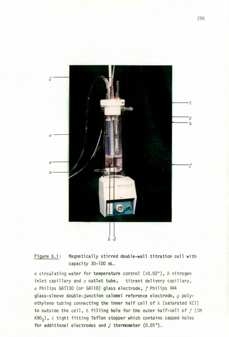
a
298
b d
Figure 6.1: Magnetically stirred double-wall titration cell with
capacity 30-100 mL.
a circulating water for temperature control (±0.02'), 1 nitrogen
inlet capillary and c outlet tube, titrant delivery capillary,
e Philips GAT130 (or GA110) glass electrode, f Philips R44
glass-sleeve double-junction calomel reference electrode, g poly-
ethylene tubing connecting the inner half cell of h (saturated KC1)
to outside the cell, h filling hole for the outer half-cell of f (1M
KNO3 ) ' i tight fitting Teflon stopper which contains capped holes
for additional electrodes and j thermometer (0.010).
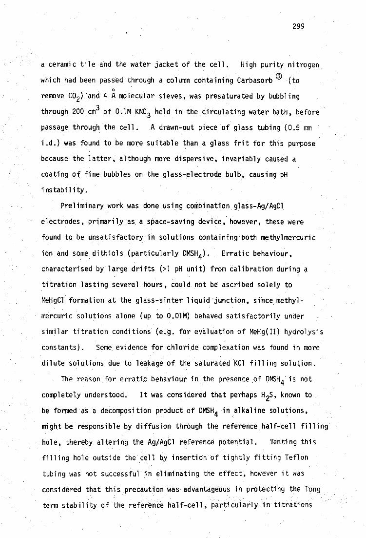
299
a ceramic tile and the water jacket of the cell. High purity nitrogen
qD which had been passed througha column containing Carbasorb (to 0
remove CO2 ) and 4 A molecular sieves, was presaturated by bubbling
through 200 cm3 of 0.1M KNO 3 held in the circulating water bath, before
passage through the cell. A drawn-out piece Of glass tubing (0.5 mm
i.d.) was found to be more suitable than a glass frit for this purpose
because the latter, although more dispersive, invariably caused a
coating of fine bubbles on the glass-electrode bulb causing pH
instability.
Preliminary work was done using combination,glass-Ag/AgCl
electrodes, primarily as. a space-saving device, however, these were
found to be unsatisfactory in solutions containing both methylmercuric
ion and some dithiols (particularly DMSH4 ). Erratic behaviour,
characterised by large drifts (>1 pH unit) from Calibration during a
titration lasting several, hours, could not be ascribed Solely to
MeHgC1 formation at the glass-sinter liquid junction, since methyl-
mercuric solutions alone (up to 0.01M) behaved satisfactorily under
similar titration conditions (e.g. for evaluation of MeHg(II) hydrolysis
constants). Some evidence for chloride complexation was found in more
dilute solutions due to leakage of the saturated Kci filling Solution.
The reason for erratic behaviour in the presence of DMSH 4 is not
completely understood. It was considered that perhaps H 2S, known to
be formed as a ,decomposition product of DMSH4 in alkaline solutions,
might be responsible by diffusion through the reference half-tell filling'
hole, thereby altering the Ag/AgC1 reference potential. Venting this
filling hole outside the cell by insertiOn'Of tightly fitting Teflon
tubing was not successful in eliminating the effect, however it was
considered that this precaution was advantageous in protecting the long
term stability of the reference half-cell, particularly in titrations

/ (ci II -ci I )Pi iz i RI i '
X ( IPI -IPII = F
-c. : 1,I
)p
300
involving volatile thiols, and was maintained throughout all subsequent
titrations (including the eventual use of separate calomel reference
electrodes),. The use of separate glass (or Ag/AgC1) reference elec-
trodes.eliminated hysteresis problems, which may have been due to the
close proximity of the combination electrode glass-bulb and liquid
Junction frit. A double junction (glass-sleeve) calomel reference
electrode with saturated KC1 in the inner cell, and 1M KNO 3 in the
outer junction, was finally used to prevent chloride diffusion into
the.titration solution.
An important consideration when using potentiometric methods is
the stability of liquid junction potentials in the electrode chain.
Potentiometric cells of highest stabilityllust incorporate no liquid
. junction, e.g. use of Ag/AgCl: reference electrodes, or incorporate a
liquid junction whose magnitude is .small (for high precision) and
remains constant. .
The magnitude of the diffusion potential due to the double liquid
junction for the Cell,. C below.
.1 II III
Hg,Hg2 C1 2 ,KC1(sat.4.2M)IIKNO3(1M)iltest-solution,KNO3(0.1M),e(hM)1Ag/AgC1
glass
can be evaluated from potentials tp .1 -ti 11 and.tp 11 - using Henderson's -
solution to the Nernst-Planck equation7D if mobilities, p i , of the
species with charge z 1 and cOncentration c 1 ci,II'
in the two
solutions, are known. For example, the diffusion potential of the
liquid junction qq-
is given approximately by
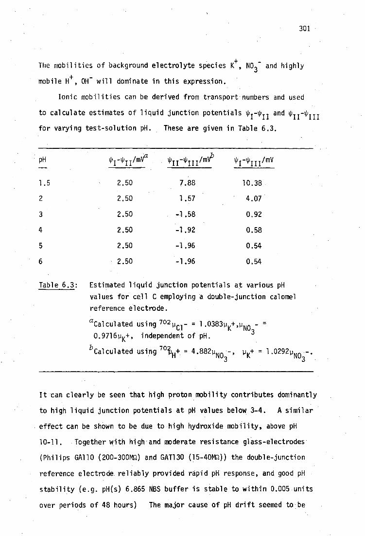
301
The mobilities of background electrolyte species K+, NO
3- and highly
mobile H+ , OH will dominate in this expression.
Ionic mobilities can be derived from transport numbers and used
to calculate estimates of liquid junction potentialsip 1 -p 11 and
for varying varying test-solution pH. These are given in Table 6.3.
pH 114111 /m 1 4 111 /mV
1.5 2.50
2 2.50
3 2.50
4 2.50
5 2.50
6 2.50
7.88
1.57
-1.58
-1.92
-1.96
-1.96
10.38
4.07
0,92
0.58
0.54
0.54
Table 6.3: Estimated liquid junction potentials at various pH
values for cell C employing a double-junction calomel
reference electrode.
aCalculated using 7o2pur 1.0383p_+ U ' . NO3- =
0•971611 K+, independent of pH.
bCalculated using 79t
H+ = 4.882p
NO3 4 p + = 1.029 - . K NO3
It can clearly be seen that high proton mobility contributes dominantly
to high liquid junction potentials at pH values below 3-4. A similar
effect can be shown to be due to high hydroxide mobility, above pH
10-11. Together with high . and moderate resistance glass-electrodes
(Philips GA110 (200-3000 and GAT130 (15-4014)) the double-junction
reference electrode reliably provided rapid pH response, and good pH
stability (e.g. pH(s) 6.865 NBS buffer is stable to within 0.005 units
over periods of 48 hours) The major cause of pH drift seemed' to be
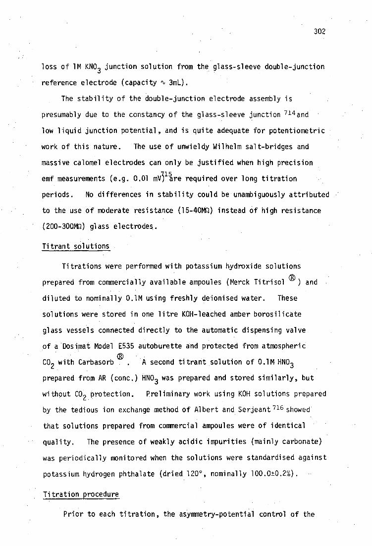
302
loss of 1M KNO3 junction solution from the glass-sleeve double-junction
reference electrode (capacity % 3mL).
The stability of the double-junction electrode assembly is
presumably due to the constancy of the glass-sleeve junction 714 and
low liquid junction potential, and is quite adequate for potentiometric
work of this nature. The use of unwieldy Wilhelm salt-bridges and
massive calomel electrodes can only be justified when high precision
emf measurements (e.g. 0.01 m1/3 15are required over long titration
periods. No differences in stability could be unambiguously attributed
to the use of moderate resistance (15-400 instead of high resistance
(200-3000 glass electrodes.
Titrant solutions
Titrations were performed with potassium hydroxide solutions
prepared from commercially available ampoules (Merck Titrisol and
diluted to nominally 0.1M using freshly deionised water. These
solutions were stored in one litre KOH-leached amber borosilicate
glass vessels connected directly to the automatic dispensing valve
of a Dosimat Model E535 autoburette and protected from atmospheric
CO2 with Carbasorb . A second titrant solution of 0.1M HNO 3
prepared from AR (conc.) HNO 3 was prepared and stored similarly, but
without CO 2 protection. Preliminary work using KOH solutions prepared
by the tedious ion exchange method of Albert and Serjeant 716 showed
that solutions prepared from commercial ampoules were of identical
quality. The presence of weakly acidic impurities (mainly carbonate)
was periodically monitored when the solutions were standardised against
potassium hydrogen phthalate (dried 120 0 , nominally 100.0±0.2%).
Titration procedure
Prior to each titration, the asymmetry-potential control of the
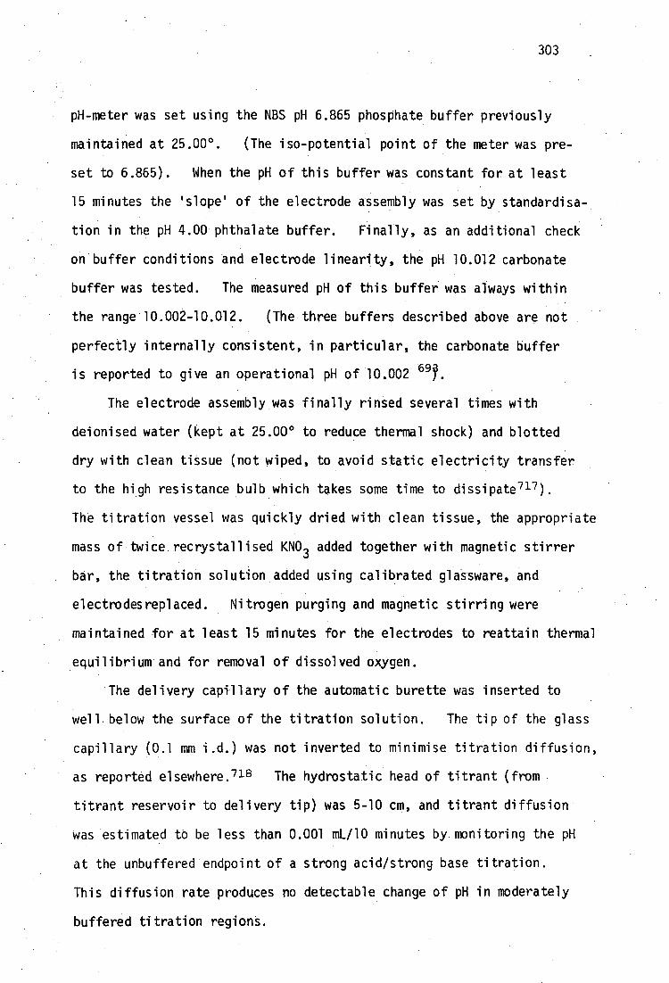
303
pH-meter was set using the NBS pH 6.865 phosphate buffer previously
maintained at 25.00 0 . (The iso-potential point of the meter was pre-
set to 6.865). When the pH of this buffer was constant for at least
15 minutes the 'slope' of the electrode assembly was set by standardisa-
tion in the pH 4.00 phthalate buffer. Finally, as an additional check
on buffer conditions and electrode linearity, the pH 10.012 carbonate
buffer was tested. The measured pH of this buffer was always within
the range 10.002-10.012. (The three buffers described above are not
perfectly internally consistent, in particular, the carbonate buffer
is reported to give an operational pH of 10.002 69 .
The electrode assembly was finally rinsed several times with
deionised water (kept at 25.00° to reduce thermal shock) and blotted
dry with clean tissue (not wiped, to avoid static electricity transfer
to the high resistance bulb which takes some time to dissipate 717 ).
The titration vessel was quickly dried with clean tissue, the appropriate
mass of twice recrystallised KNO 3 added together with magnetic stirrer
bar, the titration solution added using calibrated glassware, and
electrodesreplaced. Nitrogen purging and magnetic stirring were
maintained for at least 15 minutes for the electrodes to reattain thermal
equilibrium and for removal of dissolved oxygen.
The delivery capillary of the automatic burette was inserted to
well below the surface of the titration solution. The tip of the glass
capillary (0.1 mm i.d.) was not inverted to minimise titration diffusion,
as reported elsewhere. 718 The hydrostatic head of titrant (from
titrant reservoir to delivery tip) was 5-10 cm, and titrant diffusion
Was estimated to be less than 0.001 mL/10 minutes by monitoring the pH
at the unbuffered endpoint of a strong acid/strong base titration.
This diffusion rate produces no detectable change of pH in moderately
buffered titration regions.

304
Small volumes of titrant were added using the automatic burette
in the 0.01 mL 'step' mode while the solution was stirred magnetically
and with nitrogen. Stirring was then stopped and nitrogen passed
over the solution surface until the pH showed less than 0.001 units
change per minute. Maintenance of pH equilibrium was at first monitored
with the output of the pH meter connected to a chart recorder (10 mV full
scale) and subsequently with the device described in Appendix 6.1.
Some confusion appears in the literature over the decision to
record equilibrium pH values in stirred or unstirred solutions, even in
work covering high precision pH measurements. In cells containing
liquid junctions the liquid junction potential can show significant
stirring effects. 717 In order to maintain a constant liquid junction
potential throughout a long titration, it is necessary in these cases
to ensure a constant stirring speed. In this work, it was felt that
a 'zero' stirring speed could be maintained most precisely, even though
attainment of equilibrium may be slightly slower in this case.
Titration data in this region invariably required inordinately
long equilibrium times (>30 minutes) even when the titrant delivery
capillary was removed (to eliminate titrant diffusion) and the vessel
maintained under a static nitrogen atmosphere. In addition, it is to
be expected that the pH of this region is determined by the presence
of minor amounts of weakly acidic and basic impurities, e.g. dissolved
• CO2' bo ron and aluminium-containing anions leached from the glass
vessel etc. Ciavatta has shown that very small amounts (<1 mole)
of these products are inevitably present, even under the most rigorous
titration conditions?-9 We have found in this work, by refinement of
the total proton concentration, H tot , in these titrations, that H tot is
always 1-4 pmole greater than expected from analytical 14 + concentrations.
This behaviour is reflected by 0.001-0.004 mL equivalent point systematic

305
errors, consistent with the above impurities (page 302). For these
reasons data in this region, unless buffered by the system under
investigation (rarely), have not been included for refinement of
equilibrium constants.
pH(t) buffers
The choice of pH(s) values for standard buffers varies on a nation-
wide basis, according to national definitions of the pH scale. Composi-
tions of buffer solutions are not universal g. potassium
hydrogen phthalate studies are prepared as 0.05 molal (10.12 g per 1000
mL solution at 25°) with pH(s) at 25° 4.004 (U.S.A.), 4.01 (Hungary),
4.01 (Poland), 4.008 (U.S.S.R.), or 0.05 molar (10.21 g/1000 ml at 25°)
with ph(s) at 25° 4.008 (Germany), 4.01 (Japan), 4.005 (U.K., defines
pH scale) .
Although variations between scales are small, they are not immeasur-
able in the pH-meters capable of 0.001 pH resolution. The recently
recommended IUPAC values for standard pH buffers at 25°C have been used
in this. work. They are pH(s) 4.008 for 0.05 molal potassium hydrogen
phthalate - (10.12 g/1000 mL at 25°), ph(s) 6.865 for 0.025 equimolal
potassium dihydrogen,orthophosphate (3.39g) and disodium hydrogen ortho-
phosphate (3.53g/1000 mL at 25°).
To extend the calibrated pH range of the glass electrode to high
pH, the NBS 0.01 equimolal sodium carbonate (2.640g Na 2CO3 and 2.092g
NaCO3/1000M1 at 25° ) was used. The highest pH buffer recommended by
IUPAC ie. 0.01 molal sodium tetraborate decahydrate, pH(S)=9.180 7. 21
Preliminary work with Na 2B407 .10H 20 as an acidimetric standard indicated
that our product was partially dehydrated. Although the dilution effect
of this buffer is sma11, 693it was found to be more convenient to use the
carbonate-bicarbonate buffer for which high purity reagents were available.

306
6:24 Available computer programs for evaluation of ligand-hydrolysis
equilibria
Acid dissociation constants for mono- or diprotic ligands of known
purity can be obtained from potentiometric data by manual calculation,
e.g. using well-established methods, even under circumstances where two
acidic groups dissociate simultaneously.716 For more complex multi-
protic ligands, the deprotonation equilibria are best evaluated using
least-squares data fitting, and many simple computer programs are
available for this task of calculating only the acid-dissociation
constants from potentiometric data. Such programs require that the
concentrations of ligands and titrant be known precisely, electrode
calibration factors (e.g. E°,' . m
j. PHcal
etc., page 291) be determined '
before the titration and assumed to remain applicable during the
titration, and for weakly basic ligands the value of K be known for
the appropriate conditions under study. It is often inconvenient or
impossible to satisfy all of these requirements simultaneously, parti-
cularly for high precision titrations. Electrode calibrations may
change on transfer from calibrant solution (e.g. standard buffers or
strong acid/base titration, page 292) to measuring solution, the purity
of readily oxidisable ligands (e.g. thiols used in this work) is often
not 100% and the ligands may decompose significantly if kept in solution,
even for short periods before titration etc. An acidic ligand solution
may be dilute or may involve overlapping buffer regions so that equivalent
points cannot readily be detected by the usual means. In these circum-
stances it is convenient to use least-squares procedures to obtain values
for electrode-calibration constants, concentration of ligand, etc., from
the same data used to evaluate the ligand hydrolysis constants, albeit
at necessarily reduced precision since extra parameters are being
extracted from the data. Situations may also arise where mixtures of
307
multiprotic ligands with similar acidity constants are titrated, e.g.
HL in the presence of CO 32-
impurity. Least-squares procedures have
been used to treat such multi-protolyte equilibria. The few
published multiparametric curve-fitting programs which achieve some
or all of these ends, have been reviewed elsewhere. 690,722-5
Although multiparametric curve-fitting procedures are widely used
in chemistry and other fields, the use of correct weighting of data is
often overlooked, giving rise to incorrect estimates of fitted-model
parameters. 726-8
Similarly, conventional least-squares curve-fitting approaches
almost invariably assume that random errors reside with only one of
the experimental variables. In a two variable case, e.g. titration
curves consisting of pairs of (titrant volume, pH) data, the values of
titrant volume are usually considered to be totally reliable whereas the
measured pH values incorporate some random error. In many modern
titration systems, including the system used in this work, this is not
the case. Specifically, the automatic syringe type burette used in this
work of capacity 5 mL is reproducible to within 0.001 mL, hence titres
of 0.5 and 5.0 mL incorporate uncertainties of 0.2 and 0.02%respectively.
The measured pH can often be recorded to a precision, in buffered regions,
of 0.001 pH units over the entire pH range, corresponding to a constant
relative error of 0.1% in [e] (or, if pH is considered to be a variable,
rather than [e], 0.05% and 0.01% at pH 2 and 10 respectively).
Extremely high precision titrations can be performed with precision
of 0.01 mV in glass electrode emf 715 (0.0002 pH units) and l0 in
0.5 mL titres.729 It can clearly be seen that both experimental variables
carry comparable uncertainties, and hence normal least-squares methods
should not be applied. The use of 'rigorous' least-squares techniques
are therefore necessary, and these have been described generally elsewhere,

308
but seem to have been used infrequently in the potentiometric field.
The rigorous least-squares approach has been applied to straight-line
fitting of variables with correction errors, and used to obtain forma-.
tion constants of diprotic acids and mononuclear metal-ligand complexes
ML and ML2'
70° A computer program using this approach, RIGREG, has
been written by this author and used to calculate diprotic acid disso-
ciation constants for evaluation of the general acid-base titration
program TITRAT, described below.
The only computer program for treatment of ligand hydrolysis
constants using rigorous least-squares approach, of which this author
is aware, is TITRA, described by Schwartz and Gelb. 706 A copy
of this program was kindly made available by one of the authors (LMS)
and was used in the initial stages of this study.
The original interactive program has been made more efficient in
terms of convergence properties and running time, and generalised to
treat the titration of any number of multiprotic acids or bases, and to
extract any titration parameter by rigorous least-squares curve-fitting.
6.2.5 TITRAT and related programs for treatment of titrations of
mixtures of multiprotic acids and bases
TITRAT is an interactive computer program which has been written
in OS/8 Basic for implementation on a Digital PDP8E minicomputer with
12K available core memory for array storage etc. The program uses an
iterative non-linear least-squares algorithm to fit experimental data
from an aqueous titration of a mixture of BrOnsted acids or bases by a
strong monofunctional acid or base, and refine simultaneously any number
of titration parameters described in the model below. The procedure
is statistically rigorous, using a weighting scheme which accounts for
statistical uncertainties in both pH and titrant volume. The statis-
tical procedure is based on that in the program TITRA 706 but the

[H 9- qk-j
K4 k
[Fi g -0 1
n -j k k k
][H+
H
(n k-pk+q k -h) 2
1+
(n -p k+q k-j+1) 2
309
refinement algorithm has been generalised to cope with (theoretically)
any number of acids or bases and has much more efficient convergence
properties than TITRA.
The aqueous solution model considers that a number of N protolytic
bases, the kth
of which is
[H B] -nk pk
have been added to an initial solution volume, 1/ 0 mL and the mixture
titrated with strong monofunctional acid or base, of molarity, F. In
the above generalised species, the charge, n, of the ionic species
involved in protonation or deprotonation which was initially added to
the titration system, may be negative, e.g. K-E [H 2PO4 ] - or positive, e.g.
[NHO +Cl or zero, e.g. [H 2 succinate]° . The number of displaceable
protons of the general ampholyte is p. It is assumed further that the
ampholyte can accept a maximum number of protons, q, described as its
functionality.
The concentration of the i th-protonated form of the kth base is
(nk-p
k +
k)
[H. B]
. . .
For the t
j dissociation of the base:
Hq +1 B (n k-p k + q k-j + 1)
H + H B kk - +
cik - J
the stepwise thermodynamic equilibrium constant can be written
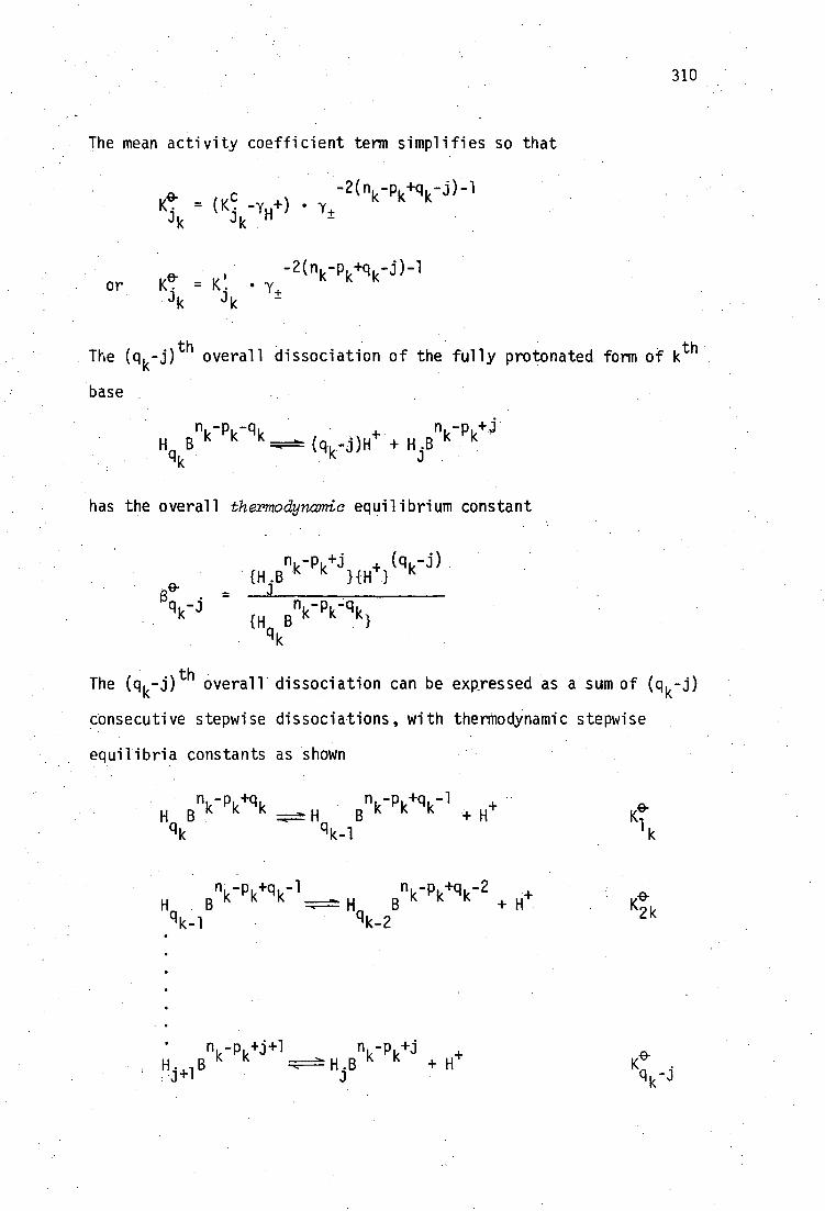
310
The mean activity coefficient term simplifies so that
-2(n6 -pk+q k-j)-1 = -iu+) • 1+ r`
"
-2(n i,-p k+q k-j)-1 or e = K. • y+ "
J k J k
The (q k-j) th overall dissociation of the fully protonated form of k th
base
k- p
k-q
k H B
n -p 6 A-j (q k j)H + H.B N
qk
has the overall thermodynamic equilibrium constant
(q u -j)
_ {H
jB k k N
{H B T}
qk
The (q k-j)th overall dissociation can be expressed as a sum of (q -j)
consecutive stepwise dissociations, with thermodynamic stepwise
equilibria constants as shown
n u -p u +q k H B '•'" H qk qk-1
n k-p k+q,-1 H
e- K
1
q k-1 •
k -POlk -1 nk-p
k+
+ H+ e-
K2k k -2
• nk-p
k+j+1 nk-p k+j
+

311
Hence B, e-
= K, K 9 ...K, with analogous relationships holding 4 k-° 'k k
•for mixed constants V : q k
(3' . = K' . clk -3
1 k 2 k clk-J
(q,-j) [H.B ] -(e} K EH B qk
so that the concentration of the general 3 .th
-protonated form of the k th
base can be written in terms of the fully (qth
k ) protonated form:
... (K' l K; lc, 4 ) 1 1( `k. nu-pl,+qk
[H.Bnk-pk+j
] = .1. DI 1 k-
• [H B " " 1 qk
J
J
We can now consider the N mass-balance equations for the N bases in the
mixture. Consider that Nk mmoles of k
th base was added initially to
the Vo mL titration solution, i.e. the k
th analytical concentration is
N k/Vo .lesftertheadditionof v i .hfl strong monofunctional titrant
(completely dissociated) of normality F, we have N equations of the form:
qk = (Vo + v i ) [H. B
nk-p
k
i=0 Jk k=1 ..,N
n 6 - = (V
o + v
i [Hqk
B "
q (K' K' ..K' .) +q k . 1k 2k ' qk - J ] I
j=0 {H + 1
Similarly, we have the electroneutrality expression at the ith
titration
point:

312
N q k [OW] 1 + (U+-1.1- ) + 1 1 (
k=1 j=o = 0
n-p 6+j -p +j)[H .B "
c "
P= lk j=o
q k K6kKi
k
vi
k
where the terms U+
and U represent concentration sums of all singly
charged countercations and counteranions, other than H i. and OH - ,
respectively. These compounds come from titrant K(OH) or H+
(NO -
) 3
and initial forms of base M Bn+
). -"k Pk
If the titrant is strong base, U 4"-U - -
If the titrant is strong acid, U -1.-U -
v.F- 1 n N k=1 k k
V +v. o
-v 4 F- n i,N u ' k=1 " "
V +v. 0 1
By analogy with Schwartz and Gelb, we write
and
q k (n k -pk+j)K;) Ki _ 4 = k k 4k
j=o {e}cik-j
It should be noted in passing that a minor error seems to have appeared
in the paper by these authors7 °6 On p.1574 of their paper, they write
(in the nomenclature used here)
1 = 1 +
G 1 {H -1- }
9- 4a. K1 K2 G 1 G21H
+1
2
• e- 2K.Tkei K 1 and P 2 - ,
{H} +
G 1 G2{e} 9 where G,
I = Y 'FY 'H .HR- wi th YH 2 R=1
G . 1
and G2 = YH+YR2-/7HR-
The term yH + should not appear in the expressions for G 1 and G 2 .
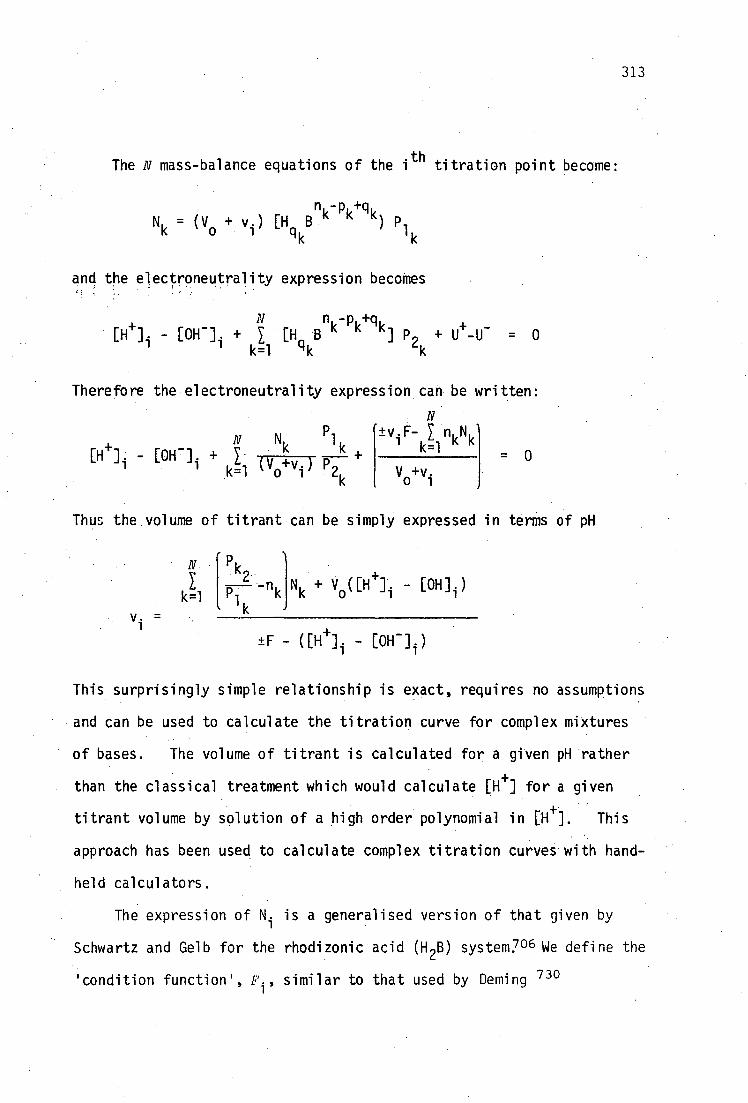
313
The N mass-balance equations of the i th titration point become:
n u -p,+q u Nk = (V
o + v.) [H B " P
k +k) P
lk o
and the electroneutrality expression becomes
n -p +q
[e]i - [OH - ] i + 1 B k k k i p u+
k=1 qk J 2
k -U- 0
Therefore the electroneutrality expression can be written:
1(1 1 Voili ) P i2k
Vo+v
i
l k 1 k=1 - 0
±v.F- N
- [OH - N Nk
Thus the volume of titrant can be simply expressed in terms of pH
kIl P I nk k Vo (
- [OH]1)
±F - ([1-1 -F ] i - [OW])
This surprisingly simple relationship is exact, requires no assumptions
and can be used to calculate the titration curve for complex mixtures
• of bases. The volume of titrant is calculated for a given pH rather
than the classical treatment which would calculate [e] for a given
titrant volume by solution of a high order polynomial in [e]. This
approach has been used to calculate complex titration curves with hand-
held calculators.
The expression of N i is a generalised version of that given by
Schwartz and Gelb for the rhodizonic acid (H 2B) system7 06 We define the
'condition function', F i , similar to that used by Deming 730

'132k
F. = "k
k=1 r k2
314
v i
±F - ([H] 1 -[OW])
For rigorous least-squares curve-fitting, we are required to minimise
the weighted sum-of-squares, S, over all N titration points:
= i1 [Wx. (PH calc - y
PH i W . (vcalc-v
i)2]
=i
where the weighting factors W , W are inversely proportion to a
• •
x Y i
priori variance estimates of the pH i and v i measurements.
SinceF is a non-linear function of most of the titration parameters i
K. , F, Nk etc., the calculation procedure requires an iterative approach.
We have used a Gauss-Newton procedure, which requires values of the
partial derivatives of F. with respect to each parameter, and those with
respect to pH i and v i . Although the algebra for derivation of these
derivatives is tedious, the use of analytical expressions, rather than
numerical estimates of the partial derivates, results in much faster
program operation (a comparison between TITRAT and TITRA showed speed
improvements of an order of magnitude in some systems).
Expressions for derivatives used in TITRAT are given below for
completion:
DF k=1
2k
Plk
N k + Vo ([11 /I i -E0H - ] i )
[±F - ([e] i 4OH - ] i )]
Dv. _ 1
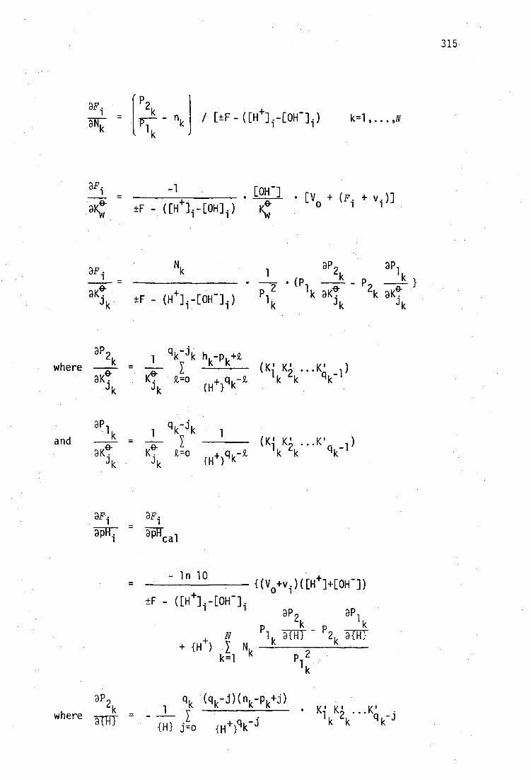
315
3 E P F i 2k
3Nk = p---- nk / [±F-([11 +] ( [0H - ] i )
1 k k= 1 ,.. .,N
-1 _ • [OH - ] . _ + - ([H+],-[OH] 1 )
[ V0 + (F i + 1 )]
N k DP a W. 1 k 1 _ Pi
2k _ f } P 1 e- P 2 e aK. k aK. k aK. ik ±F - (II +) -[OH - ] i ) • • 11
i k J k J k
where ..K' ) qk-1
and DPi
k _ 1 qk-jk e- (K' K' .K' )
Jk K.
k=o qc-32, 1 k 2 ' {H} K
K. qk -1
aF. ap.
TIF31,7 lTca i
- in 10
{(V0+v i )( [ 1-1 1 ]+[0H -]) ±F•- UH /1 1 40H -
DP 2 a P1 k -5TRT P .5TITT 2
+ {1-1 4 } k=1 "
p 2 1
3P2k q k ( qk-j)(nk-P k+j)
1 k
q k-3 where
{H} j=o fey •
1 k--
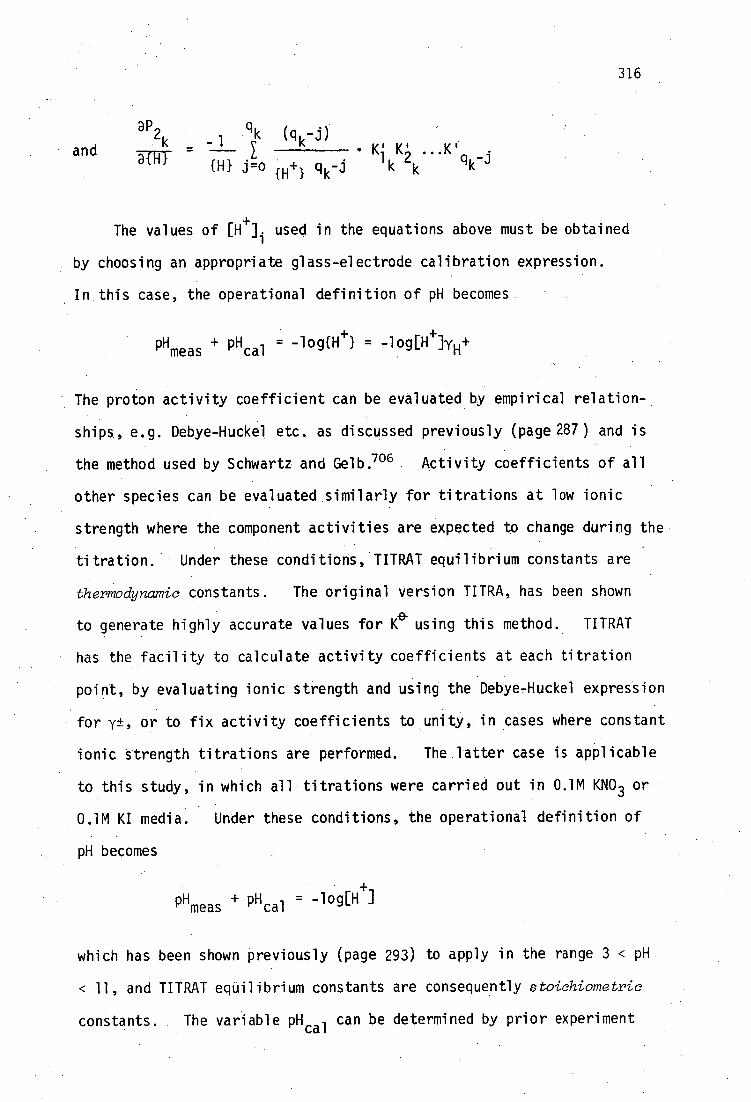
316
DP qk (q -j)
a 2 k _ -1 k
• K' K' athl 1, 2 and q {H} j=0 H+ 1 q k-i K k
The values of [H+I. used in the equations above must be obtained
by choosing an appropriate glass-electrode calibration expression.
In this case, the operational definition of pH becomes
-log{e} -log[e]Ye PHmeas PHcal
The proton activity coefficient can be evaluated by empirical relation-
ships, e.g. Debye-Huckel etc. as discussed previously (page 287) and is
the method used by Schwartz and Gelb. 706 Activity coefficients of all
other species can be evaluated similarly for titrations at low ionic
•strength where the component activities are expected to change during the
titration. Under these conditions, TITRAT equilibrium constants are
thermodynamic constants. The original version TITRA, has been shown
to generate highly accurate values for e using this method. TITRAT
• has the facility to calculate activity coefficients at each titration
point, by evaluating ionic strength and using the Debye-Huckel expression
for y±, or to fix activity coefficients to unity, in cases where constant
ionic strength titrations are performed. The latter case is applicable
to this study, in which all titrations were carried out in 0.1M KNO 3 or
0.1M KI media. Under these conditions, the operational definition of
pH becomes
= -log[H+] PHmeas
pH
which has been shown previously (page 293) to apply in the range 3 < pH
< 11, and TITRAT equilibrium constants are consequently stoichiometric
constants. The variable pH cal can be determined by prior experiment

317
(page 296) or can be treated as a parameter by TITRAT. The latter
alternative is used in this work, since small daily variations in
electrode calibration are conveniently taken into account. Values of
PHcal in 0.1M KI or KNO 3 at 25° using the Philips GA110 glass-electrode/
R44 (sat. KC1,1M KNO 3) double junction reference electrode, invariably
lie in the range -0.04 to -0.08 units when refined by TITRAT from data
below pH 4, in good agreement with directly determined values.
To evaluate [OH - ], the expression K felf0H - 1 = [e][0H- ]yH+y H-
is used. For fixed activity coefficients, this simplifies to K =
[e][0H- ]. Values for Kvci can be determined by prior experiment or
treated as a titration parameter at high pH (page 294). Since the
operational definition of pH involves no non-linearity terms, and since
many titrations in this study are performed up to pH 11, where non-
linearity becomes measurable (page 293), the value of 'Ktc41 ' is always
treated as a parameter in these titrations. This has been shown to
be equivalent to 'calibrating' the electrode at high pH, by evaluation
of a non-linearity term such as j oH KIC4/[e] 'Kvc,/ , /[e] (page 292).
It should be noted that, in principle, any glass-electrode calibra-
tion expression e.g. for using electrode emf rather than pH, could be
incorporated into TITRAT, and the appropriate calibration 'constants'
treated as parameters. Calibration expressions of the form E = E° +
+ c + log[H ].-j
OHKw/[H ] have been treated by other multiparametric curve-fitting
programs (non-rigorous) in this way, and reliable values of the parameters
obtained. 704-5
6.3 Evaluation of formation constants of stable complexes
6.3.1 H+- competition method for obtaining aqueous stability
constants
In this section potentiometric methods for evaluation of equilibrium
constants in a system containing hydrolysable methylmercuric ion and
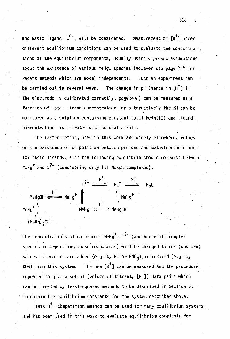
318
and basic ligand, L n- , will be considered. Measurement of [H+] under
different equilibrium 'conditions can be used to evaluate the concentra-
tions of the equilibrium components, usually using a priori assumptions
about the existence of various MeHgL species (however see page 31 9 for
recent methods which are model independent). Such an experiment can
be carried out in several ways. The change in pH (hence in [H f] if
the electrode is calibrated correctly, page 295) can be measured as a
function of total ligand concentration, or alternatively the pH can be
monitored as a solution containing constant total MeHg(II) and ligand
concentrations is titrated with acid of alkali.
The latter method, used in this work and widely elsewhere, relies
on the existence of competition between protons and methylmercuric ions
for basic ligands, e.g. the following equilibria should co-exist betWeen
MeHg+ and L 2- (considering only 1:1 MeHgL complexes).
H+ H+
HL- H2L
MeHg0H
(MeHg) 2
tleHg+ 11 MeHg+
MeHgL--,---- MeHgLH
The concentrations of components MeHg+, L
2- (and hence all complex
species incorporating these components) will be changed to new (unknown)
values if protons are added (e.g. by HL or HNO 3 ) or removed (e.g. by
KOH) from this system. The new [e] can be measured and the procedure
repeated to give a set of (volume of titrant, [e]) data pairs which
can be treated by least-squares methods to be described in Section 6.
to obtain the equilibrium constants for the system described above.
This H - competition method can be used for many equilibrium systems,
and has been used in this work to evaluate equilibrium constants for
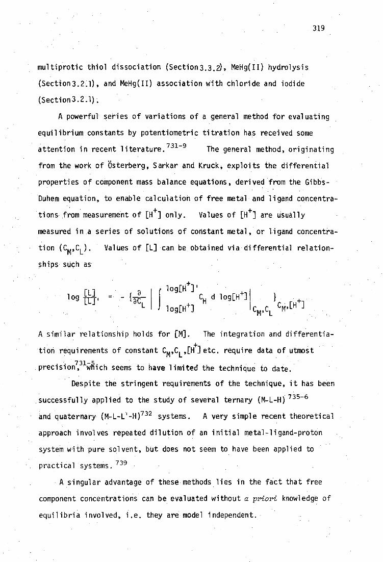
319
multiprotic thiol dissociation (Section3.3.2), MeHg(II) hydrolysis
(Section3.2.1), and MeHg(II) association with chloride and iodide
(Section3.2.1).
A powerful series of variations of a general method for evaluating
equilibrium constants by potentiometric titration has received some
attention in recent literature.731-9
The general method, originating
from the work of Osterberg, Sarkar and Kruck, exploits the differential
properties of component mass balance equations, derived from the Gibbs-
Duhem equation, to enable calculation of free metal and ligand concentra-
tions from measurement of [H+] only. Values of [H
+] are usually
measured in a series of solutions of constant metal, or ligand concentra-
tion (Cm ,CL ). Values of [L] can be obtained via differential relation-
ships such as
lo [1-1 4-] j g C
H dlog[H - ]1 }
C ,[H log[H1
' L
[1..] log =
A - similar relationship holds for [M]. The integration and differentia-
tion requirements of constant Cm ,C L ,[H+
etc. require data of utmost
precision,
731-5 wnich seems to have limited the technique to date.
Despite the stringent requirements of the technique, it has been
successfully applied to the study of several ternary (M-L-H) 735-6
and quaternary (M-L-C-H) 732 systems. A very simple recent theoretical
•approach involves repeated dilution of an initial metal-ligand-proton
system with pure solvent, but does not seem to have been applied to
practical systems. 739
A singular advantage of these methods lies in the fact that free
component concentrations can be evaluated without a priori knowledge of
equilibria involved, i.e. they are model independent.
320
For routine application of this general method, automated
gravimetric titration techniques seem to be necessary to obtain
data of adequate precision. For this reason the method was not used
in this study, although it would seem to be applicable to the study
of MeHg-thiolate-iodide-proton equilibria (Section 3.3.1).
An inherent limitation for evaluation of metal-ligand equilibrium
constants by the H i-- competition method lies in the assumption that
changing [e] can significantly alter the position of metal-ligand
equilibria. The method cannot be used to evaluate systems in which
neither metal or ligand is hydrolysable or, more important in this
study, in which metal-ligand complexation is so strong that the
changing [e] does not affect complex dissociation. The latter
situation occurs for methylmercury(II)-thiolate complexation. Proton
nmr studies have shown that MeHg(II)-monothiolate complexes do not
detectably dissociate in the range 1 < pH < 13, i.e. protons (even at
[e] = 0.1M) cannot compete with thiolate for MeHg + , and hydroxide
(even at [0H - ] = 0.1M) cannot compete with thiolate for MeHg + . When
such stable systems are titrated with acid or base, the pH only
reflects changes in protonation of ligand sites not involved in MeHg-
binding. An example of this effect is shown in Figure 6.2 for the
methylmercury(II)-mercaptoacetate system. The solid curves have been
calculated using the assumptions that only the species shown in Table
6.4 exist in solution (see also page 103for discussion of this system).
Values for stability constants of hydrolysed MeHg(II) and protonated
ligand species were determined independently. The titration
data shown in Figure 6.2 can be treated as a simple monoprotic acid HL,
due to the carboxylate group of the complex, with acid dissociation
constant pl(ca = 3.757(6), reflecting the lack of MeHg - thiolate dissociation.
The data are seen to be very insensitive to changes in the stability

321
1.000 vol. KOH/mL
13H
10
Figure 6.2: Calculateda and experimental titration curves for the
methylmercury(II)-mercaptoacetate system in the absence
of iodide. a Using COMIXH with species formation constants shown in Table 6.4.

322
constant for Melg-thiolate interaction.
Species log (formation constants) used for
calculated curves
A B C
MeHg0Ha -4.607 -4.607 -4.607
(MeHg) 200a -2.234 -2.234 -2.234
HSCH 2CO2-b
10.157 10.157 10.157
HSCH 2CO2Hb 13.710 13.710 13.710
MeHgSCH 2CO2- 10.000 14.000 16.000
MeHgSCH 2CO2Hc 13.757 17.757 19.757
Table 6.4:
Equilibrium data used by COMIXH to calculate solid curves
in Figure 6.2.
alog 1310-1 = -4.607 and log B .20-1 = -2.234 are not true
formation constants (page 64).
bTable 3.6 (page 90).
athis complex has pK c = 3.757 for carboxylate deprotonation
a (Table 3.8, page 98).
Attempts to extract log 13 110 (for formation of MeHgSCH 2CO21 from the
titration data would be fruitless, since any value between 14 and 16
will fit the experimental data satisfactorily. Unfortunately, the
indiscrete use of sophisticated least-squares programs such as those
described in Section 6.3.3 will generate values of log f3. 110 etc. for
high stability constants to which no useful meaning can be ascribed.
Although it should be possible to calculate model-independent values of
'secondary' variables such as the average number of ligands L 2 bound
to MeHg(II), 7, and thereby use only data for which 7 < 1 to obtain
f3110
for example, this approach does not seem to have been widely used for
ternary [MLH] systems. For the situation of inadequate ecompetition,
323
values of !would not deviate significantly from 1 (i.e. complex
MeHgL is undissociated) within the accessible range 2 < pH < 12.
6.3.2 Evaluation of high stability constants for MeHg-thiolate
complexes
In the situations where 14 4-- competition is ineffective, stability
constants for high stability complexes can be obtained indirectly.
By analogy to H - competition, addition of a second metal ion which
competes with MeHg(II) for ligand could be used. This approach has
been used to investigate transition-metal complexation with EDTA,
using Ng2+
as competing meta1. 740 An alternative approach, used here,
is to use a second ligand, which competes with thiolate for protons
and MeHg(II). Chloride, for example, has been used to obtain log
a4 = 63 for Pd(CN) 42 formation, by competition with cyanide 741 in the
equilibria:
Pd(CN) 4 + nC1 -----m-Pd(CN) 4_ nCl n2- + nCN-
Since the formation constants of Pd(C1)n(n-2)-
complexes can-be .
determined. using H+- competition, these can be used, , together with
values of the competition equilibrium constants, to evaluate a 4 - for the
formation of Pd(CN)42-
.
Schwarzenbach and Schellenberg 305 have used iodide competition
to obtain values of formation constants for MeHg(II) with glutathione
and mercaptoethanol. This work has extended their method to other
monothiols by evaluating the equilibrium constants for the competition:
MeHgSR + I -=#===MeNgI + SR-
• The effectiveness of this approach can perhaps best be seen by re-
appraisal of the MeHg(II)-mercaptoacetate system (Figure 6.3) in the
presence of 0.1M KI. The calculated titration curves assume the same

'pH
10
2
324
Figure 6.3: Calculated' and experimental titration curves for the
methylmercury(II)-mercaptoacetate system in the presence of 0.1M KI.
aUsin-g COMIXH with species formation constants shown in Table 6.4 and
log 13(MeHgI) = 8.500. The complex MeHgSCH 2CO2H has pKca = 3.757 for
carboxylate deprotonation (Table 3.8, page 90 ).
325
species as those in Figure 6.2, but in addition, include log 8(MeHgI) =
8.500. It can be seen that the titration data are best fitted using
log= 16.932 and are much more sensitive to values of log .110 .110
than the titration performed in absence of iodine. Methylmercury(II)-
monothiolate stability constants obtained by this method, are discussed
in Section 3.3.
It has been found in this work that the complexes formed between
MeHg(II) and the vicinal dithiols BALH 2 and Unithiol are too stable
even for iodide competition to be used effectively to determine the
formation constants for 1:1 interaction (page138 ). Treatment of
these systems, using mercaptoethanol as a competing ligand, has been
discussed previously (page 139).

326
6.3.3. Computer programs for the evaluation of metal-ligand stability
constants
Except for the simplest mononuclear systems of metal-ligand
complexes, evaluation of species formation constants is almost exclu-
sively performed nowadays by computer programs using multiparametric
curve fitting techniques. Several such programs have been described
to date, and their advantages and idiosyncracies reviewed. Four of
these have achieved dominant use, due to their wide areas of applicability
and ready availability. Sillen's LETAGRO[742was the original in the
field and is still the most comprehensive (versions are available for
treatment of heterogeneous systems, with spectrophotometric, potentio-
metric, coulometric data), but is not widely used outside Scandinavian
743 circles. The program SCOGS by Perrin and Sayce is still widely used,
and has been used to refine the formation constants of some MeHg(II)-
thiol-iodide systems in this study. The main competitor with SCOGS
is Vacca and Gans' MINIQUAD.—"—Teveral versions of this program have
evolved since its inception (1974) and it is now widely used. It has
been found elsewhere, and in this work, that MINIQUAD is much faster
than SCOGS in refinement of many equilibriumsystems and has in
consequence been used nearly exclusively in this study. The most
recent program of which this author is aware is DALSFEK, which is
unique in being able to simultaneously treat spectrophotometric and
potentiometric data7 46 Copies of DALSFEK were only recently available, 690
and so this program has not been used in this study.
All current programs refine initial estimates of complex species
formation constants, by iteratively solving the appropriate system of
component mass-balance equations described below. It must be
emphasised that these procedures require an a priori description of
the equilibrium species which are expected to be sufficient to describe
tThe version used here was kindly provided by Dr. R.N. Sylva (Australian Atomic Energy Commission).747

327
the relevant equilibrium system under study. The programs, as yet,
are not able to determine the nature of the species present, or even
their number. The decision remains with the chemist, who must
subjectively consider the relative chemical and statistical virtues
of the computer generated fits to his experimental data, calculated
with different proposed equilibrium models.
We write the overall formation equilibrium of the complex species
MmL h from its components M,L,H as follows:
MM I- 11_ -I- hi-l:,--=---%.• MmLiFlh
with overall stoichiometric formation constant
[MmL LH]
mih CM]m[L] l [H]h
The three mass balance equations for conservation of M,L,H can be
written in terms of all N supposed species
m. 1 4 h.
mlh MT [M] + m. [M] 1 [L] I [H] 1
i=1
LT = [L] + Z. 8mlh L
rm, 1[L] 4 .[H]
4
i
M 1 114
[H] f h i 8m1 [M] 14 [1.] I
4 [H]
i=1
These equations can of course be generalised to any number of components,
e.g. in this study it has been necessary to consider four components
simultaneously: MeHg + , I - , thiolate and e; or MeHg + , dithiolate,
thiolate and H+
.
It may be informative to mention that it has been shown that it

328
is not compulsory to consider the 'free' or uncomplexed species M,L,H
as components7 48 All equilibria could equally well be written in terms
of formation from the complex species MLH for example, in which case
MLH may be considered as a component and Ws changed accordingly.
This technique has been used in this study to evaluate equilibrium
constants for the addition of a second MeHg(II) group to the 1:1 MeHg(II)
complex of L-cysteine (pagelll ). In this case, the complex
MeHgSCH 2CH(NH 2)CO 2- was considered as a component or 'ligand' which
could be further protonated or coordinated to MeHg + .
A significant weakness of MINIQUAD, and of all similar currently
available programs of which this author is aware, becomes evident with
the relatively simple MeHg(II)-thiolate systems studied here. Such
programs refine values of overall formation constants, e.g. for the
simple MeHg(II)-mercaptoacetate system described previously (page 320),
these are 8110
and a - 111'
MeHg+ + -SCH CO2 - + MeHgSCH 2CO2H 13111
[MeHg41[-SCH2CO2-][e]
The first constant indicates the affinity of MeHg + for the deprotonated
sulfhydryl group of the ligand. The second reflects not only protonation
of the carboxylate group of the MeHg(II)-thiolate complex, but also the
affinity of MeHg+
for the deprotonated sulfhydryl group. Thus the two
constants are highly correlated and simultaneous refinement of them by
SCOGS or by several versions of MINIQUAD, has been found here to be slow.
In several of the other MeHg(II)-thiolate systems, described in Chapter
3, which have two or three acidic groups involved in proton association
in addition to the sulfhydryl group involved in MeHg(II) complexation,
[MeHgSCH 2CO 2 - ] MeHg+ + - SCH 2CO2- -7,==r1MeHgSCH2CO2
110 + - [MeHg ][ SCH 2CO 2 ]
[MeHgSCH 2CO2H]

329
correlation between overall formation constants has been found to be
extreme. Simultaneous refinement of these constants, using data over
a wide pH-range, often fails (except with very good initial estimates)
or at best is very slow.
In a chemical sense, a set of overall constants, e.g. 110 and 3111
above, contains redundant information, i.e. fi ll excludes a component due
to MeHg(II)-thiolate association, namely
log 13111 = lo
g no + pq(CO 2H)
The proton dissociation constant, pq, for the carboxylate group of the
MeHg(II)-thiolate complex can be obtained from highly correlated fi lo and
(Table 3.8) (3111
thus: pq(CO2H) = 20.689(11)- 16.932(12) = 3•757(6) t
An identical value 3,78(2) has been found by titrating the solid proton-
ated complex in the absence of iodide and treating the complex as a
simple monoprotic acid (page 98 ).
The problem of correlation would be alleviated if refinement of
stepwise stability constants, e.g. fi lo and pre could be achieved.
Surprisingly however, general programs do not seem to be available for
this purpose. The major programming problem would seem to be the
difficulty of defining a general system of equilibrium equations such as
those described on page 327 in terms of stepwise constants. Although the
tAs 8.110
and 8.111
are correlated, the standard deviations, a, (in paren-
theses) are correlated. The standard deviations of pK must be determined a
using the general relationship for correlated variables a,b: 749
a 2 = 4. G 2 2 , a ,
a±b - a b 'ab
The correlation coefficient, dab , is obtained from the non-linear least-
squares procedure.

330
term 'stepwise' has been used here to describe successive protonation
equilibria, it could equally well apply to successive metal or ligand
association, requiring a different set of equilibrium equations. Thus
it would appear that programs would need to be specific to the equilibrium
system at hand, and as such, would have limited general appeal.
Specific programs have been written elsewhere when high correlations 1 1 ... 'between overall constants is a problem 750 and the mathematical treatment
of highly correlated multiparametric functions has been elucidated by
Sillen. 751
6.4 Display of titration data and species distributions
As described in the previous section, the somponent mass-balance
equations in complex equilibirum systems can be used to evaluate
equilibrium constants by treating them as unknowns in a multiparametric
least-squares refinement. In this situation, it is necessary to
measure at least one of the component concentrations in order to solve
the set of non-linear equations. (Concentrations of components, other
than those measured, are also treated as unknowns and iteratively refined.)
In order to.efficiently display the contents of such an equilibrium
system it is usual to draw the concentrations of the participating species
as a function of an experimental variable, e.g. volume of base added or pH
etc. so long as the equilibrium constants have been established. To this
author's knowledge there are two generally published computer programs for
this purpose: COMIC (Concentrations in Mixtures of Metal Ions and Complexing
S pecies) by Perrin and Sayce752
and HALTAFALL by Sillen's group.753
The latter program is very comprehensive and can. account for heterogeneous:
as well as homogeneous systems in the gas, liquid and solid phases. Its
main drawback is due to its generality inasmuch as data input is extremely
tedious and the program is large and is therefore necessarily implemented
331
on large computers. COMICS on the other hand is much smaller (and thus
less general) and can only handle homogeneous equilibria, but was
admirably suited to implementation on the departmental Digital PDP8/e
minicomputer.
Experience with the original COMICS indicated that it was abysmally
slow to solve the non-linear mass-balance equations (for a given pH
value) for many of the more complex MeHg(II)-thiolate equilibria
described in Chapter 3. Since the production of a species distribution
diagram may require the solution of these equations at 50-200 pH values
in succession, a more efficient algorithm was devised and incorporated
into the new program COMIX.
6.4.1 COMIX
The major problem with COMICS seems to be due to the use of
numerical differention in the Gauss-Newton iterative procedure. The
mass-balance equation can, however, be analytically differentiated with
respect to the component concentrations. It has been found here that
the use of analytical expressions for these derivatives, increased the
efficiency of COMICS by an order of magnitude, at least.
The advantages of analytical derivatives in equilibrium calculations
was noted independently elsewhere754
and the program MINIQUAD (and
earlier versions) uses analytical derivatives to refine overall formation
constants.
A further difficulty encountered with COMICS was that the iteratively
calculated values of some component concentrations occasionally converged
to negative values. To prevent against this occurrence, component
concentrations were restrained to be positive by rewriting the mass-
balance equations in terms of ln[M] etc., rather than [M] as shown
below for the simple system containing metal, ligand and protons:
Mtot - [M] - mi i=1
m 1. •h . [M] 1 EL] 1 [H] 1
332
To solve the system of two non-linear equations (one in [M] above,
and the analogous one in [L]) by the Gauss-Newton iterative method, it
is necessary to write the left-hand side of this equation as the
function F, and obtain the derivatives 3F/[M] and aF/a[L].
If the mass-balance equation in [M] is logarithmically transformed,
it becomes
=Mtot -expOnDID-277/.(3.exp(m.ln[M]+I.1n[L] + h. ln[H]) i=1
The transformed ligand mass-balance equation is analogous and both
equations are readily differentiated with respect to 1n[M] and ln[L].
The program COMIX using this new logarithm (analytical derivatives and
logarithmic variables) has been used to efficiently calculate all the
species distributions shown in this thesis, and draw them on a Hewlett-
Packard HP7221A plotter.
6.4.2 COMIXH
A closely similar procedure can be applied to the calculation of a
hypothetical 'titration' curve, given the total concentrations of all
components and all equilibrium constants which define the system. 748
However, this situation differs from that described above for the
calculation of species distributions, in that [e] is now no longer
known, but needs to be treated as an unknown in the mass-balance
equations (now three: in ln[M], m[L] and ln[H]).
A computer program, COMIXH, has been written as a modified version
of COMIX, in order to calculate and draw the titration curves shown in
Chapter 3. A listing of COMIXH, which contains all the essential

333
features of the COMIX algorithm, has been included as Appendix 2.
6.5 Appendix. A simple device for monitoring attainment of pH-
equilibrium
Some modern pH-meters incorporate a facility to indicate when a
pH (or mV) reading is 'stable' according to the manufacturer's criteria.
The Model 701A Orion pH-meter used in this study does not have this
facility, and the use of a chart-recorder connected to the 10 mV analog
output of the pH-meter, is one means of monitoring attainment of
equilibrium.
Figure 6.4 describes a simple electronic device which can be
attached to the 31/2 digit BCD output of the Model 701A meter, and which
generates a visible or audible alarm when the digital display remains
unchanged for a predetermined length of time. The device was designed
to detect stability of the least significant digit (lsd) for periods of
30s to 5 minutes, by monitoring the two least significant bits (lsb's)
of the lsd. The particular time period can be varied by the operator
to suit titration conditions. The device ignores one digit oscillations
of the lsd (digital 'noise') by logically combining the lsb of the
second least significant digit with the lsd input.
In principle, most devices with continuous BCD output could be
monitored for stability with this circuit. The audible/visible alarms
could easily be replaced by triggers for automatic DH-orintout or
activation of an automatic burette etc.. allowina inclusion of the
device in a fully automated ootentiometric data acquisition system.
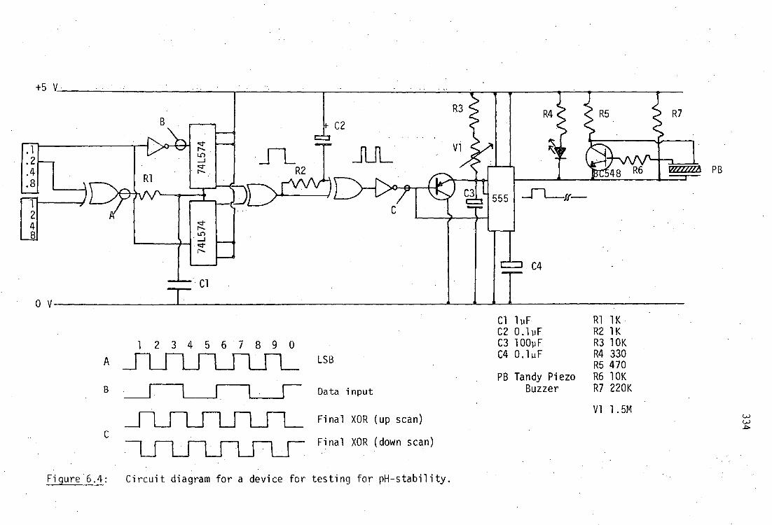
.2
.4
.8
1 2 4 8
R4 R7 R3
V1
+5V
*C2 cz=
R2
C3 555
C4
WYZOA
0 V
1 2 3 4 5 6 7 8 9 0
Cl lpF R1 1K C2 0.1pF R2 1K C3 100pF R3 10K C4 0.11.iF R4 330
R5 470 PB Tandy Piezo R6 10K
Buzzer R7 220K
A
LSB
Data input
Final XOR (up scan)
V1 1.5M
Final XOR (down scan)
Figure 6.4: Circuit diagram for a device for testing for pH-stability.

335
CHAPTER SEVEN
EXPERIMENTAL
Microanalyses were carried out by the Australian Microanalytical
Service, Melbourne, on samples dried over P 205 at room temperature
unless otherwise stated. Melting points were determined with a
Reichart Thermo apparatus and stereomicroscope, and are uncorrected.
The 100 MHz proton magnetic resonance (1H nmr) spectra were
recorded with a JEOL-JNM-4H-100 spectrometer by Mr. R.R. Thomas in
this Department. Tetramethylsilane (TMS) was used as an internal
standard. Chemical shifts (0 are given in parts per million relative
to TMS, coupling constants were measured directly from spectra recorded
with a sweep width of 900 Hz and are reliable to ±1 Hz unless otherwise
stated. Resonances are described as singlets(s), doublets(d),
triplets(t), quartets(q), or as multiplets(m). Spectra were obtained
at a temperature of 15°
Infrared spectra (4000-400 cm-1
) were recorded in Nujol or hexa-
chlorobutadiene mulls (unless otherwise stated) between KBr plates with
a Perkin-Elmer 577 spectrometer. Far infrared spectra were recorded
-1 with a Perkin-Elmer 577 spectrometer (600-200 cm ) or Perkin-Elmer 180
-1 spectrometer (500-100 cm , Monash University, Melbourne) as Nujol mulls
between polyethylene plates. Unless otherwise stated, spectra were
obtained at room temperature.
• Raman spectra were obtained from samples in sealed capillaries with
a Cary 82 Laser Raman spectrometer using Ar + 514.5 nm radiation (60-100
mW at sample) unless otherwise stated. For some samples, particularly
those containing selenium, it was necessary to use a Coherent Radiation
Model 190 dye laser (Rhodamine 6G) as the exciting source to prevent

336
sample decomposition or minimise sample fluorescence. For some samples,
it was further necessary to defocus the incident laser beam by sample
spinning, and for Hg(SeCH 2CO2H) 2 it was necessary to cool the sample to
10 K with a Cryodyne Model 21 cryostat.
-1 Maximum errors are considered to be ±4 am for both i.r. and Raman
spectra. The ultraviolet absorption spectra were obtained on a Cary 17
spectrophotometer, using 1 cm quartz cells.
Mass spectra were obtained with a Vacuum General Micromass 7070F
spectrometer using the direct insertion technique, with electron beam
energy 70 ev. Peaks of more than 10% base-peak intensity are reported
in descending m/e order. High resolution accurate mass measurements
were obtained on this instrument using electron impact ionisation unless
otherwise stated.
Debye-Scherrer X-ray powder diffraction patterns were recorded
photographically from finely ground samples contained in sealed Lindemann 0
capillaries using Ni-filtered CuKa (X = 1.5418 A) or Mn-filtered FeKa 0
(X = 1.9373 A) radiation. Automatically recorded powder diffraction
patterns were obtained from some samples using a Philips diffractometer
and ratemeter with Ni-filtered CuKa
radiation. These patterns were
calibrated with the 2e = 56.12° line of silicon.
7.1 General reagents
Ethanol (95%) was used with no further purification. Benzene,
chloroform and acetone (AR grade) were used as received.
Pyridine and 4-methylpyridine were refluxed and distilled from
potassium hydroxide before Use.
Methylmercuric nitrate for synthetic studies was prepared by
metathesis of silver'nitrate and a slight excess of methylmercuric iodide
(Alfa-Ventron) in water. The filtrate was evaporated to give a

337
colorless crystalline product which was stored over P 205 .
Solutions of methylmercuric nitrate, free of mercuric ion, were
prepared for potentiometric studies. Preparation and analysis of
these solutions for MeHg(II) and H 4 content are recorded on page Z.
Mercuric salts HgC1 2 (B.D.H. Ltd.), Hg(0 2CMe) 2 (Ajax Chemicals
Ltd.), and Hg(CN) 2 (May and Baker Ltd.) were used as received.
Crystalline thiols were used as received and stored under nitrogen
over P205 : meso-2,3-dimercaptosuccinic acid and 2-mercaptosuccinic acid
(Aldrich Chemical Co., Inc.), 2,3-dimercapto-l-propanesulfonate, sodium
salt (Heyl and Co. Chem.-Pharm. Fabrik.,), reduced glutathione (Sigma
Chemical Co.), L-cysteine, DL-homocysteine, N-Acetyl-DL-penicillamine
(Koch-Light Laboratories Ltd.) and DL-penicillamine (Ega-Chemie).
Thiocholine perchlorate was a gift from P. Guerney, University of New
South Wales.
Liquid thiols: 2-mercaptoethanol (Koch-Light), 1,3-dimercapto-2-
propanol, 2,3-dimercapto-l-propanol (Aldrich) were fractionally
distilled under reduced nitrogen pressure and stored under nitrogen at
-20 0 . Mercaptoacetic acid (Aldrich) was treated similarly after
azeotropic distillation from benzene.
The purity of the thiols used in potentiometric titration studies
was determined alkalimetrically prior to each titration, and are
recorded in Chapter 3. Melting and boiling points are recorded in
that chapter together with proton dissociation constants of the thiols.
7.2 General procedures used in organoselenium preparations
The offensive odours of many organoselenium reagents are legend,
and all selenium chemistry was performed in an efficient fumehood.
Much of the problem is due to relatively small amounts of volatile
338
impurities which may be formed during the preparative reactions,
especially hydrogen selenide. All reactions were performed under a
slow stream of nitrogen after rigorous nitrogen purging of apparatus
to remove oxygen. The effluent nitrogen was passed through at least
one trap containing 50% aqueous sodium hydroxide (to form NaSeH) and
then into a lead acetate solution. All glassware, used gloves, etc.
were decontaminated by soaking in 10% aqueous sodium hypochlorite
solution which converts offensive selenols etc. into odourless, non-
volatile, oxidised products.
Many of the organoselenium preparations make use of an ethanolic
solution of sodium hydrogenselenide, NaSeH. This reagent is very
conveniently produced using the method of Klayman and Griffin 487 by
reacting finely ground grey selenium, which had been stored over P 205 ,
with sodium borohydride in 95% ethanol until a nearly colourless solution
is obtained. These authors describe the addition of ethanol to the
mixture of solid selenium and NaBH 4* This reaction was found to be
extremely vigorous on a moderate scale with the rapid evolution of
considerable quantities of hydrogen, and required careful cooling in
ice to moderate the reaction. Some H 2 Se is produced, especially in the
initial stages of ethanol addition. It was subsequently found that
addition of an ethanolic sodium borohydride solution to a vigorously
stirred slurry of grey selenium in ethanol, contained in a 3-necked flask
equipped with nitrogen inlet, double surface reflux condenser and pressure-
equalised dropping funnel, was a much more controllable and cleaner, if
slightly longer, procedure. The purity of sodium borohydride used in
this work was found to be quite variable, despite storage in tightly
closed containers. This seemed to be due to some decomposition to sodium
carbonate, which was difficult to dissolve in ethanol. As a consequence,
it was invariably necessary to add small quantities of solid NaBH4.to
339
the final NaSeH solution to achieve total reduction of red diselenide,
Se22-
' to colorless HSe - .
The use of 95% ethanol in this procedure produces some H 2Se due to
the reaction of selenium and sodium borohydride with water impurity,
however this was a minor inconvenience since efficient traps were used
and substitution with absolute ethanol was not considered warranted.
After reaction (e.g. alkylation) with the ethanolic NaSeH solution
the reactant mixture was often acidified with glacial acetic acid to
pH 4 (tested with indicating paper) in order to ensure that any derived
selenol product was fully protonated, and to facilitate subsequent ether
extraction. This procedure also protonates any anionic selenide impurity
and excess NaSeH to H 2Se, and decomposes excess sodium borohydride to
hydrogen. Dissolved H 2Se was removed before further workup by vigorously
bubbling nitrogen through the reactant mixture. Simultaneously reducing
the pressure inside the apparatus by slight water-pump aspiration aided
H 2Se removal. Absence of hydrogen selenide was confirmed by testing the
effluent nitrogen with lead-acetate paper until no darkening (PbSe)
occurred. The resultant mixture, often containing small amounts of
undissolved grey selenium, was most conveniently filtered in a static
nitrogen atmosphere through a pressure-equalised sinter containing a bed
of acid-washed sand.
Subsequent reduction of solvent volume was performed on a BUchi
evaporator under water-pump vacuum, with nitrogen purging to restore
atmospheric pressure. Solvent extraction was performed using peroxide-
free ether in nitrogen purged separating funnels to avoid spurious
_oxidation of products.
These experimental details are not repeated in specific preparations,
except where significant deviations were required.

7,3 Preparation of complexes [M0Hg(II) and Hg2+]of dithiols and
model thiols
(meso-2,3-dimercaptobutanedioato)bis[methylmercury(II)], (MeHg) 2DMSH 2
A filtered solution of MeHgNO3 (0.445 g, 1-60 mmol) in 15 mL Et0H
was rinsed into 20 mL ethanolic solution of meso-2,3-dimercaptosuccinic
acid, with 5 ml Et0H. Evaporation of the clear solution deposits
(MeHg) 2DMSH 2 as very small needles which were collected by filtration
at low volume and dried in VaCud. The analysis of this product is
shown in Table 2.2, page 38.
Infrared absorption: 3070w,sh, 2980m,br, 1677vs, 1422s, 1311s and 1290w,sh,
1219vw, 1188 and 1176s, 950m, 785m, 686m, 632m, 530w,sh, 537m, 433m, 352s,
279m, 226w.
Raman bands: (below 1200 cm -1 , sample decomposes): 1195m, 1075vw, 974vw,
822s, 782vw, 708vw,sh and 675s, 550s and 535vw,sh, 391w, 358m, 320vw,sh and
309m.
'H nmr (D 20, pH = 7-8) &(CH3Hg) -3.00 (dioxane = 10.0), 1 2J( I H- 199Hg)1 =
172.0 Hz.
po1y-imeso-2,3-dimercaptobutanedioato)mercury(II) dihydrate, Hg(DMSH 2 )- H 20
An ethanolic (20 mL) solution of meso-2,3-dimercaptosuccinic acid
(0.092 g, 0.50 mmol) was added with stirring to mercuric chloride (o.272 g,
1.0 mmol) in 10 ml Et0H. The slightly cloudy solution was filtered and
evaporated to give a white powder at low volume. The product was
collected, washed with acetone and dried in vacuo. Yield 0.13 g (68%).
The analysis shown in Table 2.4, page 49 is consistent with a
dihydrate.
Infrared absorption: 3200-3600m,vbr, 2900w, 1710s,br, 1565w, 1360m,br,
1290w,br, 1220w,br, 1170vw,sh, 800vw, 389m, 306vw, 216w, 180vw.
This sample gives an unusable Raman spectrum.
340
341
(2,3-dimercapto-l-propanesu1fonato)bis[methy1mercury(II)], sodium salt,
MeHgSCH 2CH(SHgMe)CH 2S03Na, NaL(MeHg) 2U11
A solution of MeHgNO3 (1.36 g, 4.91 mmol) in methanol (10 mL) was
filtered into a stirred methanolic solution of Unithiol (0.545 g, 2.46 mmol)
containing 2.5 mL 2M NaOH. When approximately one-third of the MeHgNO 3
was added, cloudiness persisted. The slightly cloudy solution was
filtered and the filtrate evaporated to low volume. Fine microcrystals
were collected by filtration and dried over P 205 (the product seems to be
hygroscopic).
The analysis of anhydrous Na[(MeHg) 2UT] is shown in Table 2.2, page 38.
Infrared absorption: 3200-3650w,br, 1632w, 1187m,br, 1054vs, 967w, 892w,
854vw, 838m, 783s.br . 724m, 660w, 586m, 526m, 4552, 412vw, 333w,br.
Raman bands (below 1200 cm 1184m, 1057w, 780w, 713w, 655vw,
585vw, 531s, 329m,br.
H nmr (D20, pH = 6.2) 6(CH3Hg) -2.80 (dioxane = 10.0), I 2 J(
1 H-
199 Hg)I
171.0 Hz.
po1y-(2,3-dimercapto-l-propaneSU1fOnatO)mercurate(1-), sodium salt,
— Na[Hg(UT)]
Unithiol (1.30 g, 5,68 mmol) was dissolved in water (10 mL) and
partially neutralised with NaOH (5.16 mL 2M, 110:3 mmol). To this solution
was added 10 ml aqueous solution of mercuric acetate (1.65A, 5.16 mmol)
in portions with stirring, waiting for the white precipitate to dissolve
before the next addition. The slightly brown solution was filtered and
evaporated to low volume but no crystallisation occurred. A very small
amount of residue was removed by filtration, ad ethanol added to
precipitate a white powder which was filtered with difficulty. Drying
in vacuo over P20 5 yielded a slightly sticky product which appeared

342
somewhat heterogeneous and consequently was not analysed. Theproduct
is probably hygroscopic like the bis(methylmercury) complex and the
free ligand.
Infrared absorption: 3100-3600m,vbr, 1675m, 1283vw.sh , 1265vw,sh,
1200s,vbr and 1130w,sh, 1050s and 1028w,sh, 970w, 892vw, 853vw, 785vw,
580w and 540vw, 360w,sh.
Raman bands (below 1200 cm poor quality spectrum): 1063w, 790w,
700vw,br, 632vw, 578vw, 510vw,br, 316s,br.
2-mercaptobutanedioato(methyl)mercury(II), MeHg[SCH(CO 2H)CH 2 (gO2H)]
A solution of MeHgNO 3 (0.91 g, 3.26 mmol) in 5 mL acetone was
filtered into an acetone (5 mL) solution of 2-mercaptosuccinic acid
(0.488 g, 3.25 mmol) and 1 mL water. The clear solution was evaporated
under nitrogen and colorless needles were collected by filtration at low
volume, washed with acetone and dried in vacuo. The filtrate deposited
more crystals on further evaporation. Total yield 0.97 g (82%).
The analysis of this product is shown in Table 2.2, page 38.
Infrared absorption: 1705w,sh and 1680vs, 1410w, 1310m,brand 1260vw,sh,
1218vw, 1192m, 1178m and 1160vw,sh, 1054vw, 962m and 945w,sh, 780m, 673m,
617m, 539m and 524vw,sh, 448m,br, 377vw,sh and 361m, 296s,br.
Raman bands: 2985w,br, 2915s, 1635w, 14302, 1265vw, 1193s, 955w, 782w,
710vw, 677w, 620w, 550vs and 526 vw,sh, 420vw, 362m, 298s, 215w, 147m,sh
and 147m,
(D20, pH =5.2) 2988m, 2927s, 2874m, 1415w, 1318vw, 1191s, 1025vw, 950 vw,br,
547vs, 365m.
1H nmr (D20, pH = 5.2) 6(CH3Hg) -2.99 (dioxane = 10.0),
2 -
199
H)I =
173.4 Hz.

343
Attempted preparation of bis(2-mercaptobutanedioato)mercury(II)
Addition of mercuric acetate (3.28 g, 10.3 mmol) in 15 mL water to a
solution of 2-mercaptosuccinic acid (3.09 g, 20.6 mmol) gave a clear
solution which deposited white microcrystals at very low volume but
which rapidly turned black and obviously decomposed.
(1-methyl-3,4-dimercapto)bis[methylmercury(II)]
An aqueous (5 mL) solution of MeHgNO 3 (1_30 g, 4.70 mmol) was added
with stirring to toluene-3,4-dithiol (0.442 g, 2.83 mmol) in 5 ml warm
water containing NaOH (3.266 g, 6.6 mmol). (The dithiol melts at room
temperature, making weighing difficult). The resultant oily mixture
was stirred with ethanol (5 mL) and separated into a gum and white
precipitate which was collected by filtration after 2 hr. The crude
product was dissolved in 30 mL hot toluene and the solution filtered
to remove some yellow residue. The filtrate deposited a white powder
overnight which was collected and dried in air.
The analysis of this product is shown in Table 2.2, page 38.
Infrared absorption: 1594w, 1308vw, 1256w, 1210w, 1175vw,br, 1105w,
1029m, 871 and 858m, 808s, 770m, 685w, 549w, 528w, 354w, 335w, 273m,br.
Raman bands: 3042w, 2913m, 1608m, 1575w, 1472w, 1397w, 1290 and 1275w,
1229m, 1200m, 1126s, 1050m, 887w, 689w, 650vw,br, 526vs, 470w, 447w,
415w, 370 and 357m, 337w, 300vw, 263m.
[N,N,NtrimethylaminoethylMerCaptO(Methyl)Mercury(II)] perchlorate
.Methylmercuric nitrate (0.66 . g, 2.38 mmol) in 1 ml water was added
to thiocholine perchlorate dissolved in hot water (5 mL). Within two
minutes, on cooling, the solution deposits colorless crystals which
were collected by careful Buchner filtration on a small paper and dried
344
over P 205 . (CAUTION - perchlorate salts of sulfur-containing
compounds are potentially explosive. The crystals were collected
using a polyethylene 'spatula' to prevent scratching.)
Yield 0.73 g (71%), mp. 163-5°.
The analysis of this product is shown in Table 2.2, page 38.
Infrared absorption: 3040w, 2000w, 1492w and 1475m, 1418m, 1343w,
1186m, 1085vs,br, 1008m, 966m, 942w, 892m 774 and 764m, 727w, 621vs,
542m, 454w, 381vw, 359m, 329s, 287w.
Raman bands: 3040m, 2985m and 2940w, 2909m, 1452w, 1405vw, 1181s, 1090w,br,
970w, 943vw,sh and 931vs, 908w, 894w, 772m, 725w, 621w, 542vs, 500vw,
478w, 331s.
H nmr (P20): d(CH3Hg) -2.97 Cdioxane = 10.0 , 2J
199Hg)I = 169.6 Hz.
1
• 4,4'-dimercapto-N-methy1piperidine hydrate
This intermediate was prepare according to the method of Barrera and
Lyle406 for subsequent reduction to 4-mercapto-N-methylpiperidine.
N-methyl-4-piperidone (14.2 g, 125 mmol, bp. 56-8°/10 mm) was
dissolved in 100 mL toproponol. Hydrogen sulfide t (CAUTION) was
passed from a Kipp's generator at 2 bubbles/sec through the Pri OH
solution cooled in ice which deposited white crystals of the product
after 31/2 hrs. The crystalline product was collected after 8 hours by
filtration and washed with cold Pr i OH (3 x 5 mL). On standing at -20°
for 48 hrs, the filtrate deposited a second crop of product which was
combined with the first. Total yield 20.32 g (90%), mp. fu54° (dec.)
m it. 406 48-501.
tEffluent H2S was trapped in 10% CuSO4 in 0.3N HC1. The H2S has a distinct tendency to suc back into the reaction vessel.
345
4-mercapto-N-methylp1peridine
4,4'-Dimercapto-N-methylpiperidine hydrate (10.7 g, 59 mmol) was
suspended in 27 mL isopropanol and added in small portions to a stirred
suspension of sodium borohydride (3.55 g 94 mmol) in 27 mL Pri OH
under nitrogen. The mixture was cooled in ice to moderate the exothermic
reaction (evolution of H 2St). After 30 min additional stirring, more
Pri OH was added (20 mL) and the stirred pale yellow mixture heated on a
water bath at 58° for 2 hrs. The solution was then acidified (Congo
Red) by dropwise addition of ethanolic HC1 (12.7M, CAUTION H 2 evolution)
to decompose excess borohydride, and diluted with water (20 mL) to dissolve
inorganic salts. After the addition of solid KOH to pH 12, the aqueous
solution was extracted with ether (4 x 35 mL).
The combined ether extracts were dried over magnesium sulfate then
evaporated by flash rotary distillation under nitrogen to yield a
slightly cloudy colorless oil which was fractionally distilled, bp.
31.5-2°/1 mm [lit. 406 62°/0.8 mm]. The oil freezes to a colorless
crystalline solid, mp.7-9°.
• Mass spectrum: M 130.0684; calc. for C6H 13NS = 130.0690.
The acid dissociation constants of the N-protonated form of this
thiol are recorded in Table 3.6, page 91.
[4-mercapto-N-methylpiperidinium(Methyl)mercury(II)] nitrate
4-Mercapto-N-methylpiperidine (0.207 g, 1.59 mmol) was added by
syringe to a stirred aqueous (5 mL),solution of MeHgNO 3 (0.445 g, 1.60
mmol). The clear, colorless solution was evaporated to low volume
and the resulting colorless crystals collected on a sinter, washed with
tEffluent H2S was trapped in 10% CuSO4 in 0.3N HC1. The H25 has a distinct tendency to suc'back into the reaction vessel.

'zH 9•69l
= 1(6H66CH )PZI = auexolp) LCZ- = ( 6HC-140)9 (0O) awu H L
s561. 4JC9E''wELE WSZt
'mA6st 'mat '5A6E9 ' ,4811 's0SL ' 14£08 'm88 PUP sAEWL 'AWOL
' 1119Lit 'w8121. 'man 'm0£171. matt :( 1(1. 1.40 L'_WO 009t molaq) spueq umeq!
'MBE 'ME
'MK 'mAUt 'm89t 'mt8 .17 puu ys'mAOS '/4611 'AM 'all'w09L
'M808 'sZE8 '4s'w896 Pue •6L6 ' ,4 1201. 'WOW!. g mAL901. ' 111980L 'MOM
Wald 'w0Sil 'mOLLL 419121. 's00£1. Puv Aes98CI. c m0Sti
'acrmSiLl 'acrmOOLZ 'm006 'acrmSt6Z 41SZ0E :uopdaosye pauva4uI
'8E afied 'rz appi Lit umoys s!. 4onpoad lo spXteue aqi
08 - 9L1 '(%ls) 6 tWO Plal4 Le4o4 'a4ea4L14 3114 woal paaanooaa
sem sjeq.sRuo 4o douo puooas V ' 90Zd Jam) Onova 141 PaPP Pue 1oue44a-
9tE
347
7.4 Preparation of selenium cOmpounds and complexes (and S-analogs)
Diselenides R2 Se (R=Me,Et) —2
The diselenides R2Se2 (R=Me,Et) were prepared by the alkylation of
Na2Se2 in liquid ammonia by the alkyl iodides. 755-6 Preparation of
dimethyl diselenide will be described in detail.
Thinly sliced metallic sodium (3.26g, 140 mmol) was dissolved in
approx. 150 mL liquid ammonia which had been previously dried over
sodium and distilled into the 500 mL 3-necked reaction vessel via an
acetone-dry ice condenser. Addition of small quantities of grey
selenium (total 11.5g, 140 mmol) produced a color change from deep blue,
though red brown to dark green. Methyl iodide (25g, 180 mmol) was
added dropwise at a rate sufficient to dissolve the yellow Me3SeI which
formed. Water (50 ml) was added after evaporation of the ammonia at
room temperature, and the deep crimson oil extracted with ether (4 x
50 m1). The combined yellow extracts were dried (CaCl 2 ), evaporated
4nd fractionally distilled in vacuo to give Me 2Se2 as a putrid orange
oil, bp. 42-44°/8-9 mm. [lit. 757 43-4915mm].
Diethyl diselenide was prepared similarly (using ethyl iodide) as a
putrid orange oil, bp. 68-70°/9-10 mm [lit.757
74-5°/14 mm].
catena-Bis(p-methaneselenolato)mercury(II), Hg(SeMe) 2
Excess metallic mercury was stirred with Me2Se 2 (2.81g, 15 mmol) in
pyridine (50 mL) for 2 days. The resulting black suspension was
extracted with hot pyridine until the extract was colorless (200 mL).
The black mercuric selenide and finely divided metallic mercury were
removed by filtration through cellulose powder and a fine sinter under
slight positive pressure (CAUTION).
348
Yellow leaflets of Hg(SeMe) 2 formed on cooling (5.28g, 90%), mp.
119°(dec.). Infrared absorption: 2925vw, 2915vw, 1414m, 1253s, 900w,sh,
890s, 578 and 570w, 161vs,b, 151vs,b, 134vs,b, 103m.
Raman bands (spinning cup): 2995 and 21988m, 2927m, 2913s, 1415w, 1262m,
898 and 890w, 576w,sh and 570s, 182vw,sh, 158m, 131vs, 112W.
The analysis of Hg(SeMe) 2 is shown in Table 4.1, page 174.
Bis(ethaneselenolato)mercury(II), Hg(SeEt) 2
Excess metallic mercury was stirred with Et 2Se2 mL, unweighed
because of stench) in chloroform (50 mL) for 1 day. The yellow powder
which formed was dissolved by addition of hot chloroform (100 mL), and
metallic mercury removed by decantation and filtration through a fine
sinter under slight positive pressure (CAUTION). Chloroform was
removed under vacuum, and the yellow powder recrystallised from pyridine
to form yellow crystalline or glassy Hg(SeEt) 2 , mp. 72°(dec.).
Infrared absorption: 2945m, 2905m, 2865m, 1441s and 1425sh, 1372s, 1221vs,
1041m, 750m, 570w,sh, 555m, 291w, 198w, 175vs,b, 150vs,vb, 130 vs, 110vs,b.
Raman bands: (red excitation, spinning capillary) 2950 and 2933w, 2913m,
2857w, 1442 and 1424w, 1370vw, 1217s, 1041w, 991w, 963w, 745vw, 550m,
298w, 131vs.
The analysis of Hg(SeEt) 2 is shown in Table 4.1, page 174.
cyclo-Tetrakis4chloro-p-ethaneselenolato-pyridinemercury(II)],
[EtSeHgCl(py)]4
Mercuric chloride (0.59g, 2.18 mmol) in 5 mL pyridine was added to
bis(ethaneselenolato)mercury(II) (0.91g, 2.18 mmol) dissolved in 10 mL
warm pyridine, and the mixture filtered to remove a small amount of
unreacted Hg(SeEt) 2 after 10 minutes.
349
Addition of 15 ml Et0H and evaporation overnight yields small,
colorless prisms (1.18g, 64% as EtSeHgCl.py).
Small colorless prismatic crystals, suitable for crystallographic
investigation (page 202) were deposited after 1 hour which were collected
by filtration. The crystals showed negligible weight loss at room
temperature over 36 hrs.
EtSeHgCl.py (C7H 10C1NSeHg) requires: C 19.9, H 2.38, N3.31, Hg 47.4%;
found C 20.2, H 2.46, N 3.50, Hg 47.4%.
Infrared absorption: 3080vw, 3055w, 3025w, 2990vw, 2950w, 2915w, 2860w,
1592m, 1428m, 1449s and 1449s, 1420w,sh, 1325m, 1232, 1222 w,sh and
1222m, 1207m, 1070w, 1062 and 1059m, 1081m 1004s, 961w, 763s, 710s,
621m, 565vw,sh and 544m, 408m, 212s(br), 179s(br), 156m.
The compound rapidly loses pyridine and decomposes in the Raman
laser-beam.
• tert-Butyl selenol
An ethereal solution of Bu tSeH was prepared by the Grignard route
to Bu tSH. 452
A Grignard solution of tert-butylmagnesium chloride was prepared
by the dropwise addition of tBuCl (8.98g, 97 mmol, distilled, bp.50.5-51°
and stored over K2CO3 at -20°) to activated magnesium t (3.62g, 149 mmol).
The reaction was initiated by adding approx. 3 mL tBuCl to the
magnesium under 15 ml sodium-dried diethyl ether after warming the
magnesium with a single iodine crystal in the oven-dried 250 mL three-
tA large portion of magnesium turnings was rinsed successively with four portions of 0.5% HC1, freshly deionised water (until pH q ,5) ethanol and acetone, then dried over CaC1 9 under nitrogen. The turnings were stored in a tightly stoppered contaiher containing a few crystals of iodine.

350
necked flask. The subsequent rate of addition of tBuCl (dissolved
in 50 ml ether) was sufficient to maintain gentle ether reflux.
Powdered grey selenium (3.83g, 49 mmol) was added slowly to the
grey suspension over 30 min. with vigorous mechanical stirring and after
a further 30 min., ether (50 mL) was added. After standing for 12 hr
under nitrogen, the solution was cooled in ice, and ice-cold water (50
mL) then sulfuric acid (150 mL, 2.5M) were added slowly over a period
of two hours. Only a small amount of unreacted selenium remained
undissolved. The ether layer was separated and combined with an
ether extract (50 mL) of the aqueous layer. The combined colorless
extracts (100 mL) were dried over sodium sulfate and stored at -20 0
until use. This solution remained colorless (free of Bu t2Se2 ) under
these conditions for 30 months.
Bis(tert4outaneselenolatO)MerCury(II), Hg(SeBu t ) 2
A portion of the ether extract from the preparation of Bu tSeH
(80 mL) was distilled into a stirred solution of mercuric cyanide
(6.12g, 24 mmol) in methanol (50 mL). The slightly brown precipitate
was collected •(Whatman No.1), dissolved in boiling chloroform (250 mL),
and on cooling, white, hairlike needles of Hg(SeBu t ) 2 were collected on
a coarse sinter, nip. 145 (dec.).
Infrared absorption: 2965m, 2925w, 2880w, 2815vw,sh, 1458sh and 1450m,
1389w, 1365s, 1148s, 1023m, 514m, 397m, 296w, 270w, 135vs.
Raman bands: (yellow excitation, below 1200 cm 1147m, 1027w,
805w, 519s, 299m, 272w, 139vs,b.
The analysis of Hg(SeBu t ) 2 is shown in Table 4.1, page 174.

351
1,30,3-Di-1j-chloro-2,4-dichloro-1,20,4;2,3;3,4-tetrakis-
p-1,1'-di-methylethaneselenolato-2,4-bis(pyridine)-
quadro-tetramercury(II), [Bu tSeHgCl(py) 0. 04
Mercuric chloride (0.106g, 0.39 mmol) dissolved in 1 ml pyridine was
added to a stirred solution of bis(tert-butylselenolato)mercury(II)
(0.185g, 0.39 mmol) suspended in 14 mL pyridine. Stirring for three
minutes resulted in a clear, colorless solution which deposits a grey
crystalline product at low volume. This product was redissolved in
pyridine (10 mL), 10 ml Et0H added, and the solution filtered to remove
a small amount of grey material. Evaporation deposits smell prismatic
crystals which rapidly darken if kept exposed to the atmosphere. A
single crystal of this product was suitable for crystallographic invest-
igation (jage 195) and was mounted in a Lindemann capillary while still
wet with pyridine.
Insufficient sample was availabe for spectral characterisation.
tert-Butaneselenolato(methyl)mercury(II), MeHgSeBu t
A portion of the ether extract from the preparation of Bu tSeH ( ,20
mL, page 348) was distilled into an ice-cold solution of MeHg0H
[prepared from MeHgNO 3 (o.927g, 3.34 mmol) and NaOH (3mL 2M, 6 mon]
in 40 mL methanol. Water (10 mL) was added, and after standing for
12 hr, the solution was filtered through a sinter containing cellulose
powder. The sinter was rinsed with 20 mL Me0H and the combined aqueous
MOH solutions extracted with hexane (2 x 25m1). Additional NaOH
(3 mL 2M) and water (10 mL) were added to the aqueous layer which was
extracted again with hexane (3 x 25 mL). The hexane extracts were taken
to dryness under low vacuum ('25 mm) at 0°C. The crude white solid was
purified by sublimation (50-60 0/15 mm), mp. 57-8.5° (sealed capillary).
MeHgSeBut (C 5 1-1 12HgSe) requires: C 17.1, H 3.44, Hg 57.0%;
found: C 17.0, H 3.50, Hg 57.0%.

352
Infrared absorption: 2970 and 2950vw, 2930w, 2885w, 2855w, 1465s,sh and -•
1450w, 1361m, 1172w, 1148s, 1020w, 756m, 521m, 290w, 194s.
Raman bands: (below 600 cm 526m, 302m, 200s, 142 and 127m.
'H nmr (CDC1 3 ): 6 0.85 [3H,'CH3Hg(II)], 1.65 [9H, C(C) 3], 1 2J('H-
199Hg)1 146.8 Hz (see Figure 3.34, page 168):
• tert-Butanethiolato(methyl)mercury(II), MeHgSBu t
MeHgSBu t was prepared in a similar manner to the selenium analog
but using Bu tSH, and pentane as the extractant. mp. 40-2° (sealed
capillary, sublimes 40°/25 mm) [lit. 250b 41-2° (sublimes 28940 mm)].
Infrared absorption: 2970 and 2960vw, 2937 and 2920vw, 2890w, 2857w,
1470w and 1454m, 1363s, 1152s, 821vw, 766m,br, 581w, 532m, 428m, 383m,
352m, 305w, 234m, 130m.
Raman bands: 2972m, 2958 and 2950m, 2932w, 2910vs, 2888s, 2795vw, 2769w,
2707w, 1456 and 1438m, 1212m, 1180vs, 1150w, 1028vw, 932vw, 822s, 586s,
536vs, 432m, 390s, 368m, 228s, 147m,br.
'H nmr (CDC1 3 ): 6 0.78 E3H, CH3Hg(II)], 1.51 [9H, C(C) 3], 1 2JOH-
199Hg)1=150.1 Hz, [lit. 250b (CDC1 3) 0.77, 1.48, 150.2].
Bis(phenylmethyl)diselenide,(PhCH 2Se) 2 [Dibenzyldiselenide]
Dibenzyldiselenide was prepared by the selenation of benzaldehyde
in the presence of morpholine hydrochloride t . Morpholine hydrochloride
(6.20 g, 50 mmol) was added in one portion to a nearly colourless ethanolic
solution of NaHSe (prepared from 4.74 g, 60 mmol Se, page 338).
Benzaldehyde (5.00 g, 47 mmol) was added in one portion to the resultant
tMorpholine hydrochloride was obtained by protonation of morpholine (100 g, 1.15 mole) dissolved in 100 mL ethanol, by ethanolic HC1 (12.7M, prepared by the addition of HC1 to conc. H2SO4).7'8

353
white suspension, and the mixture heated to reflux on a water bath for
1 hr then cooled in ice. Red polyselenide impurities were reduced
with small portions of solid NaBH4 to produce a yellow solution.
Addition of ice-cold water (100 mL) precipitated the yellow product
which was collected by filtration and rapidly washed with water to
remove easily oxidised selenol impurities which otherwise rapidly
decompose in air to red Se. The yellow powder NO g crude) was
recrystallised from ethanol.
The product was used as a source of benzylselenolate anion, but
subsequent preparation of benzylselenol was more convenient for this
purpose.
Phervimethaheelehol, PhCH2SeH Denzylselenoll
Benzylselenol was prepared by Painter's Grignard method501
from
benzylmagnesium chloride (prepared from 24.3 g Mg, 1 mole and freshly
distilled benzylchloride, 115 g, 1 mole, bp. 61.0-61.5°/8mm) and selenium
(78.98 g, I mole) by the same method as for tBuSeM9C1 (page 349).
Gaseous HC1 (prepared from 53.5 g NH 4C1 and conc. H 2SO4 759) was
passed over the ether solution with vigorous stirring. An appreciable
quantity of H2Se evolved and trapped in 50% sodium hypochlorite solution.
Water (250 ml) was slowly added (CAUTION - vigorous decomposition)
followed by an additional 80 ml conc. HC1 in 250 ml water. The combined
aqueous/ether layers (1000 mL) were filtered (gravity only) into a 1L
separating funnel and separated. The ether layer was dried over sodium
sulfate, filtered, and evaporated to yield 120 mL yellow oil. Fractional
distillation under nitrogen yielded q.50 mL colorless oil, bp. 67-8°/2 mm
[lit. 501
100-5°/20 mm].
354
Bis(phenylmethylselenolato)merCury(II), Hg(SeCH 2Ph) 2
Benzylselenol (5.96 g, 35 mmol) was added with magnetic stirring to
mercuric cyanide (4.95 g, 19 mmol) in 25 ml methanol, under nitrogen.
The immediately precipitated yellow product was collected by filtration
and washed with Me0H (5 x 5 mL). Portions of the crude product were
recrystallisedfrom benzene or chloroform to yield bright yellow,
crystalline Hg(SeCH 2Ph) 2 , mp. 111°(dec.).
Infrared absorption: 3020vw, 1598vw, 1492m, 1175m, 1062w, 1028w, 907w,
827s, 757s, 694s, 598s, 549m, 449m, 328vw, 236w, 158s,br.
Raman bands: (spinning cup below 1700 am-1 only) 1603m, 1219m 1177m,
1002m, 605m, 232w, 149s.
The analysis of Hg(SeCH 2ph) 2 is shown in Table 4.1, page 174.
Bis(phenylmethylthiolato)mercury(II), Hg(SCH 2Ph) 2
This compound was prepared in quantitiative yield in an identical
manner to Hg(SeCH 2Ph) 2 as a white microcrystalline product, mp. 115-7°.
Infrared absorption: 3050 and 3020vw, 1490m, 1460m, 1442m, 1229m, 1195vw,
1151vw, 1065w, 1028m 910vw, 760w, 690vs, 620w, 565m, 472m, 381s, 352vw,
325vw, 246w.
Raman bands: 3050s, 2936m, 1602s, k230s, 1430vw, 1230s, 1202w, 1160w,
1021w, 1004s, 810w, 767m, 688m, 627w, 566w, 476vw, 385vw,sh, 356s, 327vw,sh,
244m, 107s.
Phenylmethanethiolato(methyl)mercury(II), MeHgSCH 2Ph
Benzylthiol (0.25 g, 2.2 mmol) was added by syringe, under nitrogen,
to MeHgNO 3 (0.60 g, 2.2 mmol) in 5 ml Me0H. After one minute, a
transitory cloudiness disappearedto give two clear phases.
Water (5 mL) was added and the cloudy mixture extracted with
pentane (4 x 10 mL). The pentane extracts were dried by passage through

355
a microcolumn of silica t , then evaporated to dryness under nitrogen to
yield 0.25 g (34%) of colorless oil. The product smells of BzSH
impurity confirmed by the Raman spectrum.
Infrared absorption: 3090vw, 3065w, 3030m, 3000vw,sh, 2915m, 2860vw,
2400vw, 1738vw, 1601w, 1494m, 1453m, 1382s,br, 1233m, 1202w, 1183w,
1157w, 1069m, 1029w, 912w, 828m, 767s, 700s, 680m, 566m, 538m, 476w,
346m.
Raman bands: 2055s, 2925w,sh, 2913s,(2580w) , 1601m, 1233s, 1203m,
1179s, 1030m, 1004vs, 804m, 770m, 702vs,sh and 684m, 621w, 565vw,sh and
540vs, 477w, 365vw,sh and 342s, 277w.
Phenylmethaneselenolato(methyl)mercury(II), MeNgSeCH 2Ph
This compound was prepared in a similar manner to MeHgSCH 2Ph, using
benzylselenol (0.30 g, 1.7 mmol) and extracting with carbon tetrachloride.
The silica microcolumn drying step was omitted as significant losses of
sulfur analog appeared to be due to this procedure.
Infrared absorption: 3085vw, 3065w, 3038m, 3000w,sh, 2940w,sh and 2910m,
2400w, 1600m, 1495s, 1464m, 1385vs, 1174m, 1030w, 913w, 829m, 760s, 699s,
600m, 551w, 528m, 447w.
The complex decomposes in the Raman laser-beam.
The crude product (0.58 g, 88%) was pumped under high vacuum (21 mm)
to remove solvents, excess selenol and water, but still smelled of Bu tSeH,
and was consequently not analysed.
Diselenodiacetic acid, (SeCH 2CO2H) 2
Selenocyanatoacetic acid, NCSeCH 2CO2H, 759 was prepared by displacement
tpreviously dried at 250 0 .
356
of bromide from bromoacetic acid (9.57 g, 69 mmol) dissolved in 50 mL dry
acetone. Potassium selenocyanate (9.95 g, 69 mmol) dissolved in 100 mL
dry acetone was added dropwise with magnetic stirring to this solution
cooled in ice. The precipitated KBr (light purple due to Se impurities)
was removed by filtration under slight positive pressure (CAUTION).
Acetone was removed by rotary evaporation of the nearly colorless
filtrate, to leave a pale yellow oil. Acid hydrolysis of the oil by
refluxing 21/2 hrs with 100 mL 1M HC1 produced a deep yellow-orange
solution which was allowed to stand overnight, exposed to the atmosphere,
but in the dark. This solution was extracted with ether (5 x 50 mL)
then with ethyl acetate (2 x 50 mL). The combined extracts were
evaporated and the yellow residue dissolved in 50 ml ethyl acetate.
Addition of 1100 mi. benzene and cooling produced (SeCH 2CO2H) 2 as light
yellow crystals (7.72 g, 53%), mp. 93-5° [lit. 759 101°].
Bis(selenoacetato)mercury(II), Hg(SeCH 2CO2H12
Diselenodiacetic acid (0.64 g, 2.3 mool) was dissolved in 20 mL
water and the pH lowered to 1.5 with 2.5 M H 2SO4 . Excess metallic
mercury was added and the yellow mixture immediately turned brown,
changing to yellow on stirring overnight. The metallic mercury was
removed by dissolving the yellow precipitate with saturated NaHCO 3
solution followed by rapid filtration into 2.5M H 2SO4 (the dianion is
unstable in alkaline solution). This procedure was repeated until the
filtrate was clear yellow (twice), and the reprecipitated yellow powder
filtered with some difficulty.
Infrared absorption: 2700-3300s,br, 2525w, 2515w, 1692s, 1612w, 1452s
and 1394vw,sh, 1301vw,sh and 1275vs, 1172m, 930m and 911w,sh, 854vw,
713vw, 649m, 521vw, 446m and 433w,sh, 241s and 222m.

357
Raman bands (orange excitation at 10K, below 350 cm -1 only because of
sloping background): 313m, 284s, 224m, 192s, 172m.
Bis(mercaptoacetato)mercury(rr), Hg(SCH 2CO2H)2
Mercuric chloride (0.272 g, 1.00 mmol) in 5 ml Et0H was added with
stirring to mercaptoacetic acid (0.187 g, 2.03 mmol) in 25 mL water
containing 1 mL 5M HNO 3 . Glistening, white plates were precipitated
and were collected by filtration and dried in vacuo. Yield 0.255 g
(67%).
The analysis of Hg(SCH 2CO2H) 2 is shown in Table 2.4, page 49.
Infrared absorption: 2800-3300m,vbr, 2960w, 2915w, 1713s, 1688s, 1418m,
1382w, 1305s, 1238vw, 1202s, 1170w, 898m, 862w,br, 787m, 677m, 552m,
464w,sh, 446s,br, 379s, 351w, 226s, 155m.
Raman bands (below 1700 cm 1650vw, 1395w, 1215w, 920w and
900vw,sh, 795m, 670vw, 435w 370w,sh and 347s, 235m, 177w, 142vw.
[bis(mercaptoacetato)mercurate(2-1)],dipotassium salt, K 2[Hg(SCH 2CO2 ) .1
Bis(mercaptoacetato)mercury(II) (3.65 g, 9.5 mmol) was suspended in
50 mL N2-bubbled water and dissolved by the addition of KOH (1.06 g,
19.0 mmol). The solution was filtered and evaporated in a nitrogen-
filled CaC1 2 dessicator to give a grey powder. The powder was dissolved
in 70 mL hot water containing a small amount of activated charcoal,
filtered and cooled. The resulting white microcrystalline precipitate was
filtered off, washed with ethanol and dried in vacuo over CaC1 2 .
The analysis of this product is shown in Table 2.4 page 49.
Infrared absorption: 2900-3600vs,vbr, 2895vw, 1572s, 1382s, 1228m, 1216m,
1160, 919w, 894m, 769m, 678 and 670w, 550m, 431s, 355s, 321w, 235w.
Raman bands (yellow excitation, below 800 cm 710m, 437w, 417w,

360w, 328vs 241w (in H 20, pH 6.3, below 400 cm 336vs,p.
Acetato(methaneselenolato)mercury(II), MeSeHg0 2CMe
Bis(methaneselenolato)mercury(II) (0.120 g, 0.34 mmol) was added to
•a solution of mercuric acetate (0.107 g, 0.34 mmol) in 5 mL water
• containing 3 mL Et0H. After 15 min the colorless solution was filtered
to remove a small amount of unreacted Hg(SeMe) 2 . The filtrate deposited
colorless crystals on slow evaporation which were collected (0.193 g,
81%) and dried over P 205 .
MeSeHg02CMe (C3HSO2SeHg) requires C 10.2, H 1.71, Hg 56.7%;
found: C 10.2, H 1.75, Hg 56.5%.
Infrared absorption: 2995vw, 2955w,sh, 2925w, 2857w, 1535s, 1410m,sh and
1392s, 1339m, 1282m, 1040w, 1008m, 931 and 923m, 661s, 611m, 474m, 218s,
182m, 134s and 114m,sh.
Raman bands (below 2200 cm 1535vw,br, 1400vw, 1334vw, 1279vw,
923w, 663vw, 613vw, 574m, 480w, 230vw,sh, 197 and 182s, 143s.
Selenoacetato(methyl)mercury(II), MeHgSeCH 2CO2H_
Diselenodiacetic acid (.0.388 g, 1.40 mmol) was dissolved in ammonium
hydroxide solution (10 mL 2M) under nitrogen. Rongalite t (0.237 g, 1.54
mmol) was added in one portion with magnetic stirring, and very small
additional portions added until the initial yellow solution remained
colorless for 5 min.
Methylmercuric nitrate (0.78 g, 2.81 mmol) dissolved in 5 mL
deaerated water was added, followed by immediate acidification (Congo
Red) with 5M HNO 3 . The deposited white shiny leaflets were collected
tRongalite = 'sodium formaldehyde hydrosulfite', HOCH 2S02Na.2H 20, was obtained from Fluka AG, Buchs.
358

359
on a sinter (#4) and briefly washed with dilute HNO 3 , then a few drops
of ethanol and dried in vacuo. Yield 0.65 g (67%). Attempted
recrystallisation from ethanol resulted in deposition of grey Se.
The product is photosensitive and was stored at -20 0 .
MeHgSeCH2CO2H (C3H602SeHg) requtres : C 10.2 H 1.68, Hg 56.7, Se 22.3%;
found: C 9.9, H 1.68, Hg 56.7, Se 22.1%.
Infrared absorption: 2995vw, 2955w,sh, 2925w, 2857vw, 1530s,br, 1410w,sh
and 1395s, 1339m, 1282m, 1040w, 1008m, 931 and 923m, 661s, 611w, 474w,
218s,br, 182m, 134s,br, 114w,sh.
Raman bands (below 1600 only): 1535vw br, 1402vw,br, 1334vw, 1279vw,
1006vw, 923w, 663vw, 613vw, 574m, 480w, 230vw sh, 197s,sh, 182m, 143s.
Mercaptoacetato(methyl)mercury(II), MeHgSCH 2q02H._
Mercaptoacetic acid (0.172 g, 1.86 mmol) was added to methylmercuric
nitrate (0.517 g, 1.86 mmol) in 15 ml water with stirring. An additional
5 ml water was added to clarify the slightly cloudy mixture which was
filtered and allowed to evaporate. Fine colorless needles were collected
after 20 hr. The analysis of this product is given in TAble 2.2, page
38.
Infrared absorption: 2985w, 2940vw, 2910m, 1702s, 1440m, 1412w,sh,
1390 and 1375m, 1282m, 1185 and 1179s, 900 and 870m, 778 and 769 m,
722w,sh, 654 and 627m, 550 and 529w, 477m, 426m, 334 and 318m, 243w.
Raman bands: 2991m, 2932m, 2916vs, 1188m, 910vw, 793w, 676m, 580w,
538vs, 329s, 265vw, 180w, 153m, 138m.
(in D20, pH = 5.6): 2983m, 2921s, 2870m, 1450w, 1392w, 1334w, 1189s,
1017w, 932w, 831m, 779w, 546vs, 500vw, 351m.
1 H nmr (D20, pH = 5.6) s -2.98 relative to dioxane = 10.0, I 2J( 1 199Hg)I
= 170.5 Hz.

360
Bis(2-hydroxyethyl)diselenide, (HOCH 2CH2Se) 2
Rongalite (19.7 g, 128 mmol) and sodium hydroxide (12.6 g, 315 mmol)
were dissolved in 250 mL water under nitrogen. This solution was added
in one portion to grey selenium (25.2 g 319 mmol) in a 1L 3-necked
flask and the mixture stirred and heated on a boiling water bath to
dissolve Se (3 hrs). Freshly distilled 2-chloroethanol, bp. 176-7°
(26.3 g, 327 mmol) was added dropwise with stirring to the deep red
Na2Se2 solution. When N95% of the halide was added, an orange oil
separated which was decanted and dissolved in acetone after cooling to
room temperature. The aqueous phase was extracted with chloroform
until the extracts were light yellow. The combined acetone and chloroform
extracts were evaporated to dryness, then pumped at high vacuum (0.1 mm)
to remove water. Yield of crude oil 26.2 g (83%) which was fractionally
distilled, bp. 152-60°/0.05-0.2 mm [lit. 755 169-79°/1.6-4 mm]. ,
Surprisingly, the diselenide is odorless.
,Attempted isolation of 2-hydroxylethaneselenol, HOCH 2CH 2SeH
Bis(2-hydroxyethyl)diselenide (8.78 g, 35 mmol) was dissolved in
30 mL sodium-dried ether, and cooled to -30° with dry ice/acetone.
Sodium-dried liquid ammonia was condensed into this solution (N130 mL)
under a slight flow of nitrogen. Metallic sodium was added as a solution
in liquid ammonia (using a refluxing condenser-thimble arrangement
described in reference 756) until a blue color persisted for 5 min.
At this stage the mixture turned whte and cloudy. The ammonia
was evaporated under nitrogen and 10 mL deaerated water added dropwise
to the residue followed by conc. HC1 (CAUTION - vigorous) until pH 3-4.
The white oily residue (NH 4CL) was pumped (N4 mm) in warm water (<55°)
to remove water, HC1, etc., then at higher vacuum (0.1-0.2 mm) with

361
warming to 60°. A colorless oil was condensed in a cold trap (-15°)
0 Addition of freshly dried 5A molecular sieves to the oil under nitrogen
resulted in gas evolution and complete decomposition overnight.
(2-phenylmethylseleno)butanedioic acid
Bis(phenylmethyl)diselenide (0.655 g, 1.93 mmol) was dissolved in
20 mL Et0H and just reduced with small additions of solid NaBH4 under
nitrogen. A solution of cis-2-butenedioid acid (maleic acid, 0.456 g,
3.93mmol) in 20 mL Et0H containing NaOH (0.3 g, 7.5 mnol) was added by
syringe to the solution of selenolate. The nearly colorless solution
was stirred for 2 hr at 50° and separated into two phases. The bottom
layer was separated and acidified with conc. HC1 (5 mL). The resultant
white precipitate was collected by filtration and washed with 10% HC1.
Yield 0.54 g (48%), rap. 169-71 0 .
PhCH 2SeCH(CO2H)CH 2CO2H (C il H 1204Se): requires C 46.0, H 4.21, Se 27.5%;
found C 46.0, H 4.39 Se 27.2%.
Infrared absorption: 2600-3300s,br, 1690vs, 1494m, 1415s 1319m, 1235m,
1198m, 1065m, 918m,br, 854w, 811vw, 758s, 699s.
'H nmr (acetone-d6 ; Figure 5.3 page 259)
7.35 (m, 5H, C6H5 ), 4.7 (br, 2H COii), 4.15 (s, 2H, CH2Se), 3.75 (q, 1H,
CHCO2H), 2.85 (m, 2H, CH2CO2H). See Table 5.7, page 261 for coupling
.constants. •
Mass spectrum: M+ = 287.9904; calc. 287.9898
(2-phenylmethylmercapto)butanedioic acid
This compound was prepared by base-catalysed addition of benzylthiol
(2.22 g, 17.9 mmol) to the sodium salt of maleic acid (2.08 g, 17.9 msol)
in an identical manner to the selenium analog nip. 173-5°.

362
Infrared absorption: 2600-3300 m,vbr, 1710 and 1692s, 1495w, 1434m,
1410m, 1352vw, 1321m, 1240s, 1206 and 1198m, 1177w, 1081m, 999w, 921m and
714,sh, 853w, 837vw, 771w, 718 and 710m, 672w.
1 H nmr (acetone-d 6 ; Figure 5.2 page 258):
6 7.39 (m, 5H, C6L15), 4.2 (br, 2H, CO 2H), 4.03 (s, 2H, CLI2Se), 3.17 (q,
1H, CHCO2H), 2.80 (m, 2H, CH2CO2H). See Table 5.7, page 261, for coupling
constants.
Mass spectrum: 11 240.0420; ca;c. 240.0456.
trans-(2-phenylmethylseleno)butenedioic acid
Acetylenedicarboxylic acid t (0.99 g,'8.7 mmol) was dissolved in
15 mL 1M NaOH, the pH of this solution adjusted with HC1 until just
alkaline (pH el,8) and solid sodium carbonate (1.84 g, 17 mmol) added.
This solution was deaerated by nitrogen bubbling for 15 min then
added by syringe to neat benzylselenol (1.49 g, 8.7 mmol) under nitrogen.
After stirring for 40 hr, the mixture was extracted with ether (25 mL)
and the aqueous phase acidified with conc. HC1 (Congo REd). The bright
yellow precipitate was collected by filtrat , and recrystallised from
ethanol as bright yellow needles. Yield 2.09 g (84%), mp. 180-3° (dec.).
PhCH 2SeC(CO 2H):CHCO2H (C 1 11-1 1004Se) requires: C 46.7, H 3.53, Se 27.7%;
found: C 46.6, H 3.59, Se 27.8%.
Infrared absorption: 2600-3400s,vbr, 1695w,sh, 1670s, 1557m, 1493m,
1453w, 1414s, 1312m, 1266s, 1231s, 1173w, 1060w, 1028w, 952w, 916w, 888
and 875m, 861m, 778w, 764m, 743w, 695s, 656w, 617w.
1 H nmr (acetone-d 6 ): 6 7.38 (m, 5H, C61:15 ) 6.45 (s, 1H, CH=C), 5.1 (br,
tAcetylenedicarboxylic acid was prepared form the dipotassium salt by acidification with sulfuric acid and extraction with ether.761

2H, CO 2H), 4.25 (s, 2H, Cy).
Mass spectrum: M+.
= 285.9794; calc. 285.9744.
trans-(2-phenylmethylmercapto)butenedioic acid
This compound was prepared in 78% yield by the base-catalysed
addition of benzylthiolate •to acetylenedicarboxylic acid in an identical
manner to the selenium analog.
The product was recrystallised as pale yellow crystals from Et0H,
mp. 151-2°.
Infrared absorption: 2500-3300m,vbr, 1706 and 1674vs, 1560m, 1497w,
1416m, 1326 m and 1313w, sh, 1270m, 1238 and 1232s, 1070w, 1048m, 1028w,
957w, 920w, 890m,br, 857m, 783m, 752m, 729m, 710w and 699m.
1 H nmr (acetone-d6 ) = cS 7.39 (im, 5H, C 6H5 ), 6.69 (s, 1H, CH=C), 5.3
(br, 2H, CO 2H), 4.21 (s, 2H, C).
Mass spectrum: M 238.0306, calc. 238.0300.
363

364
Attempted preparations of 1,3-diseleno-2-propanol (SeDMPH 2 )
1,3-dibromo-2-propanol was prepared for these reactions by hydro-
bromination of epibromohydrin.762
Hydrobromic acid (38 mL 46-48% w/v,
0.342 mole) was added dropwise over a period of 30 minutes to epibromo-
hydrin (Fluka, 41.55 g, 0.303 mole) in 40 ml ethanol at 70-75°, with
magnetic stirring. After stirring for a further 30 minutes at 60-70°,
the solution was cooled to room temperature and ethanol removed under
reduced pressure. The yellow oily residue was stirred with ether
(300 mL) and washed successively with H 20 (3 x 100 mL), 1% w/v sodium
bicarbonate solution (3 x 80 mL), then H 20 (3 x 100 mL). The dried
(MgSO4 ) ether extract was evaporated and vacuum distilled to give the
product as a colourless oil (51-55%) bp. 77-80°/4mm (lit. 762 82°/6 mm).
The product darkens in amber glass at room temperature and was redistilled
before use.
(i) Selenocyanation of 1,3-dibromo-2-propanol
Potassium selenocyanate (1.715 g, 119 mmole) dissolved in 10 mL dry
methanol was added to 1,3-dibromo-2-propanol (1.270 g, 58.3 mmole) under
nitrogen and the resulting red solution refluxed for 30 minutes.
Silica TLC showed no change in product distribution after this
time. The reaction mixture was diluted with water (20 mL) and extracted
with ether (3 x 30 mL) under nitrogen. The dried (Na2SO4) ether extracts
were evaporated under nitrogen to yield a dark yellow oil (1.14 g) having
-1 intense infrared absorption (2135 cm ) due to selenocyanate. This oil
could be separated by Ggel-Rohr distillation, giving two fractions.
The first, bp. 50-53°/0.01 mm, showed no absorption due to CEN in the
infrared spectrum, and was discarded. The second, bp. 60-75°/0.01 mm,
-1 has v CEN 2135 cm and was identified as 1-bromo-3-selenocyanato-2-
propanol.
365
Infrared absorption:
3390 vs(br,OH), 3010 vw, 2955 w, 2880 w, 2135 s(vCEN), 1694 m(br),
1610 vw, br, 1418 s and 1400 sh, 1300 vw, 1274w, 1232m, 1210m,
1170 w, 1147 w, 1074 m, 1046 m, 1020 s, 966 w, 926 vw, 905 vw, 855 sh
and 836 w, 697 w, sh and 670 m, 582 w, 520 w and 498 w.
H nmr (6CDC1 3 ) - seepage
2.9(br,1H,CO2H); 3.28, 3.38(d, 1H, H a ); 3.35(s, 1H, Hb ); 3.56,
3.62(d, 2H, 2H d); 4.23(m, 1H, H e).
Mass spectrum - see page
M+.
accurate mass 242.8814, C 4H6BrNOSe requires 242.8796.
(ii) Alkylation of NaSeH with 1,3-dibromo-2-propanol
A solution of 1,3-dibromo-2-propanol (38.35 g, 176 mmole) in
ethanol (20 mL) was added dropwise to a light grey ethanolic solution
of NaSeH (300 mL containing 25.3 g, 320 mmole Se). After 1 hour, small
portions of solid sodium borohydride were added to produce an orange
solution (which could be decolourised in subsequent preparations by
addition of further small amounts of solid NaBH 4 ) and a white precipitate
(NaBr, H 3803 ). The solution was cooled in ice, and glacial acetic acid
(26 mL) added with vigorous nitrogen bubbling to expel H 2Se after warming
to room temperature. (A prior attempt to steam-distil the reaction
products at this stage produced large quantities of red selenium and
much H2Se). The mixture was filtered, evaporated to low volume, and
the resultant orange slurry shaken with ether and refiltered. Evaporation
of the ether extract produced an orange oil which could be distilled under
reduced pressure to give an orange oil, bp. 79-85°/3.5 mm (11.49 g),
containing some unreacted 1,3-dibromo-2-propanol (nmr).
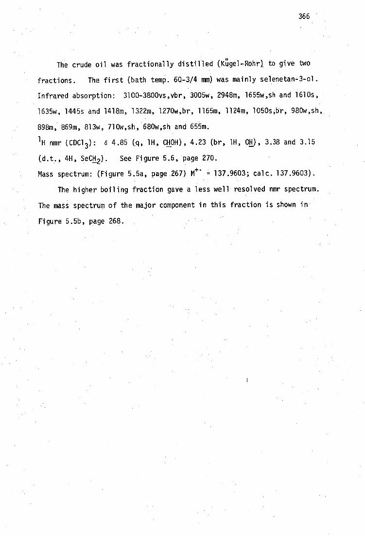
366
The crude oil was fractionally distilled (4gel-Rohr) to give two
fractions. The first (bath temp. 60-3/4 mm) was mainly selenetan-3-ol.
Infrared absorption: 3100-3800vs,vbr, 3005w, 2948m, 1655w,sh and 1610s,
1635w, 1445s and 1418m, 1322m, 1270w,br, 1165m, 1124m, 1050s,br, 980w,sh,
898m, 869m, 813w, 710w,sh, 680w,sh and 655m.
'H nmr (CDC1 3 ): 4.85 (q, 1H, CHOH), 4.23 (br, 1H, OH), 3.38 and 3.15
(d.t. 4H, SeCH2 ). See Figure 5.6, page 270.
Mass spectrum: (Figure 5.5a, page 267) M 4- = 137.9603; calc. 137.9603).
The higher boiling fraction gave a less well resolved nmr spectrum.
The mass spectrum of the major component in this fraction is shown in
Figure 5.5b, page 268.

367
REFERENCES
1. "Minamata Disease: Methylmercury poisoning in Minamata and Niigata, Japan", T. Tsubaki and K. Irukayama (Eds.), Elsevier, Amsterdam (1977).
2. W.E. Smith and A.M. Smith, "Minamata", Holt, Rinehart and Winston, New York. (1975).
3. T.B. Eyl, J. Am. Med. Assoc., 215 287 (1971).
4. a) R.D. Snyder, New Eng. J. Med. 284 1014 (1971); b) Arch. Neurol. 26 379 (1972); c) "Environmental Mercury Contamination", R. Hartung and B.D. Dinman
(Eds.), Ann Arbor Science, Michigan (1972).
5. F. Bakir, S.F. Damluiji, L. Amin-Zaki, M. Murtadha, A. Khalidi, • N.Y. Al-Rani, S. Tikriti , H.I. Dhakir, T.W. Clarkson, • J.C. Smith and R.A. Doherty, Science 181 239 (1973).
K. Al-Takriti and A.W. Al-Mufti, Bull. W.H.O. 53 (Suppl) 15 (1976) and S.B. Skerfving and J.F. Copplestone, W.H.07-54 101 (1976).
7. A.R. Greco, Riv. Neurol. 3 515 (1930) and references therein.
8. J.R. Wood, Environ. Sci. Res. 17 223 (1980).
9. J.M. Wood, F.S. Kennedy and C.G. Rosen Nature (London) 220 173 (1968).
10. L. Landner, Nature (London) 230 452 (1971).
11. T.W. Clarkson, Ann. Rev. Pharmacol. 12 375 (1972).
12. a) "Mercury, Mercurials and Mercaptans", M.W. Miller and T.W. Clarkson (Eds.), C.C. Thomas Publ., Springfield, Illinois (1973);
b) J.T. MacGregor and T.W. Clarkson in Adv. In Expt. Med. Biol. M. Friedman ed. 48 463 (1974). •
13. "Mercury in the Environment: An Epidemiological and Toxicological Appraisal", L. Friberg and J. Vostal (Eds.) C.R.C. Press, Cleveland, Ohio (1972).
14. H.B. Gerstner and J.E. Huff, J. Toxicol. Environ. Health 2 491 (1977).
15. A. Gilman, R.P. Allen, F.S. Philips and E. St. John, J. Clin. Invest. 25 549 (1946).
16. M. Berlin, L.-G. Jerksell and H. Von Ubisch, Arch. Environ. Health 12 33 (1966).
•17. F.A. Cotton and G. Wilkinson, "Advanced Inorganic Chemistry: a Comprehensive Text" (4th ed.) John Wiley and Sons, New York
• (1980).

32. J.H.
J.H. 33. Southard, P. Nitisewojo and D.E. Green
34. R.H.S. Thompson and V.P. Whittaker, Biochem. J. 41 346 (1947).
35. E.S. Barron and G. Kalintsky, Biochem. J. 41 351 (1947).
36. L.R. Roberts, L.K. Steinrauf and R.T. Blickenstaff, J. Pharm. Sci.
S. 37. Nakada, K. Inoue, S. Nojima
R. 38. Magnaval, J. Thiessard, R. Batti and R. Bittel
368
18. J.J. Chisholm, Jnr., Pediatr. Clinics Nth. Am. 17 591 (1970).
19. J.H.R. Kagi and B.L. Vallee, J. Biol. Chem. 236 2435 (1961).
20. S. Jensen and A. Jernel6v, Nature (London) 223 753 (1969).
21. L. Magos and W.H. Butler, Arch. Toxicol. 35 25 (1976).
22. T. Norseth and T.W. Clarkson, Arch. Environ. Health 21 717 (1970).
23. R. Klein et at., Arch. Pathol. 96 83 (1973).
24. J.M. Wood, A. Cheh, L.J. Dizikes, W.P. Ridley, S. Rakow and J.R. Lakowicz, Fed. Proc. Fed. Am. Soc. Exp. Biol. 37 16 (1978).
25. M. Berlin and S. Ullberg, Arch. Environ. Health 6 589 (1963). 0
26. A. Swensson and U. Ulfvarson, Acta Pharmacol. Toxicol. 26 273 (1968) and references therein.
27. P.J. Gallagher and R.L. Lee, Toxicology 15 129 (1980).
28. J.M. Wood, Adv. Environ. Sci. Technol. 2 39 (1971).
29. F.L. Hoch and B.L. Vallee Arch. Biochem. Biophys. 91 1 (1960).
30. T. Norseth and M. Brendeford, Biochem.-Pharmacol. 20 1101 (1971).
31. N. Sone, M.K. Larsstuvold, Y. Kagawa, J. Biochem. 82 859 (1977).
Southard, P. Nitisewojo, Biochem. Biophys. Res. Comm. 52
Fed. Proc. Fed. Am. Soc. Exp. Biol. 33 2147 (1973).
and N. Imura, Chem.-Biol. Interact.
, Rev. Int.
39. Omata, K. Sakimura, T. Ishii and H. Sugano, Biochem. Pharmacol. 27 1700 (1978).
40. K.R. Olson, K.S. Squibb, R.J. Cousins, J. Fish. Res. Board Can. 35 381 (1978).
41. D.L. Rabenstein,Acc. Chem. Res.
Oceanogr. Med. 49 111 (1978) CA89:1226c.
S.
921 (1973).
69 964 (1980).
22 15 (1978).
1 100 (1978).
369
42. M. Ohsawa and L. Magos, Biochem. Pharmacol. 23 1903 (1974).
43. J.W. Daniel, J.C. Gage and P.A. LeFevre, Biochem. J. 121 411 (1971).
44. I.U. Hag, Br. Med. J. 1 1579 (1963).
45. A.J. Canty and R. Kishimoto, Aust. J. Chem. 30 669 (1977).
46. F. Matsamura, Y. Gotch and G.M. Boush, Science 173 49 (1971).
47. K. Tonomura, K. Furukawa and M. Yamada in "Environmental Toxicology of Pesticides", F. Matsamura et al. (Eds.) Academic Press, New York (1972).
48. J.D. Nelson, W. Blair, F.E. Brinckman, R.R. Colwell and W.P. Iverson, Appl. Microbiol. 26 321 (1973). ,
49. M.M. Jones and T.H. Pratt, J. Chem. Ed. 53 342 (1976).
50. P.M. May, D.R. Williams and P.W. Linder in "Metal Ions in Biological Systems", H. Sigel (Ed.) Volume 7, Marcel Dekker, New YOrk (1978).
51. S. Ahrland, J. Chatt and N.R. Davies, Quart. Rev. Chem. Soc. 12 265 (1958).
52. a) R.G. Pearson, J. Am. Chem. Soc. 85 3533(1963); b) R.G. Pearson, "Hard and Soft AcIE and Bases", Dowden, Hutchinson
& Ross, Pennsylvania (1973).
53. L.L. Waters and C. Stock, Science 102 601 (1945).
54. F.W. Oehme, Clin. Toxicol. 5 215 (1972).
55. a) G.W. King, Ariz. Med. 11 335 (1954); b) F.T. Matthes, R. KirscRer, M.D. Vow and J.C. Brennan, Pediatrics
22 675 (1958).
56. W.J. Burke and J.M.Quagltana, J. Occup. Med. 5 157 (1963).
57. H.C. Brugsh, J. Occup. Med. 7 394 (1965).
58. G. Baldi, Med. Lavoro 41 10 (1950).
59. K.R. Adam, Br. J. Pharmacol. 6 483 (1951).
60. R.F. Bell, J.C. Gilliland and W.S. Dunn, Arch. Indust. Health 11 231 (1955).
61. L. Magos, Brit. J. Indust. Med. 24 152 (1968).
62. M. Berlin and T. Lewander, Acta Pharmacol. Toxicol. 22 1 (1965).
63. J.R. Fitzsimons and F.L. Kozelka, J. Pharmacol. Exp. Ther. 98 8 (1950).
64. J.E. Wynn, B. Van't-Reit and J. Borzelleca, Toxicol. Appl. Pharmacol. 16 1807 (1970).
370
65. T. Norseth, Acta Pharmacol. Toxicol. 32 1 (1973).
66. A. Swensson and U. Ulfvarson, Intern. Arch. Gewerbepath. Gewerbehyg. 24 12 (1967).
67. M. Berlin, L.-G. Jerksell and G. Nordberg, Acta Pharmacol. Toxicol. 23 312 (1965).
68. A.K.M. Anisuzzaman and L.N. Owen, J. Chem. Soc. (C) 1021 (1967).
69. W.T. Longcope and A. Luetscher, Ann. Int. Med. 31 545 (1949).
70. L.S. Goodman and A. Gillman, "The Pharmacological Basis of Thera- peutics", 5th ed., McMillan, New York (1975), p.912.
71. P. Zvirbilis and R.I. Ellin, Toxicol. Appl. Pharmacol. 36 297 (1976).
72. H. Eagle, H.J. Magnuson and R. Fleischman, J. Gun. Invest. 25 451 (1946).
73. L. Friedrich and F. Zimmermann, Arzneim:-Forsch. 25 162 (1975).
74. a) H.V. Aposhian and M.M. Aposhian, J. Pharm. Exp. Ther e 131 139 (1959);
b) H.V. Aposhian, Ann. N.Y. Acad. Sci. 179 481 (1971).
75. "The Merck Index", 9th ed. Merck and Co. Inc., Rahway, New Jersey (1976).
76. I.G. Mizyukova and D.S. Lokantsev, Farmakol. i Toxikol. 23 355 (1960); CA 55:12660.
77. V-I. Liang, Ch.-Ch. Chu, Y-L. Tsen and K-Sh. Ting, Acta Physiol. Sinica 21 24 (1957) CA51:14982g
78. E. Friedheim and C. Corvi, J. Pharm. Pharmacol. 27 624 (1975).
79. A.Y. Hadengue, P. Fabiani and A. Queuille, Arch. Mal. Prof. 11 45 (1950).
80. J.M. Walshe, Am. J. Med. 21 487 (1956)
81. a) G. Kazantzis, K.F.R. Schiller, A.W. Asscher and R.G. Drew, Quart. J.
Med. 31 403 (1962);
b) J. Teisinger and J. Srbova, Prac. Lek. 16 433 (1964); c) S.N. Javett and G. Kaplan, Ain-7737. Child. 115 71 (1968).
82. J. Aaseth, Acta Pharmacol. Toxicol. 39 289 (1976).
83. L. Magos, T.W. Clarkson and J. Allen, Biochem. Pharmacol. 27 2203 (1978).
84. K. Hirota, Clin. Neurol. 9 592 (1969).
85. N.S. Ishihara, S. Shiojima and T. Suzuki, Brit. J. Ind. Med. 31 245 (1974).

371
• 86.
J. Aaseth, A. Wannag and T. Norseth, Acta Pharmacol. Toxicol. 39 302 (1976).
• 87.
H. Matsumoto, A. Suzuki, C. Morita, K. Nakamura and S. Saeki, Life Sciences 6 2321 (1967).
88. J. Aaseth, Acta Pharmacol. Toxicol. 36 193 (1975).
89. J. Aaseth, Acta Pharmacol. Toxicol. 39 289 (1976).
90. N.S. Johary and L.N. Owen, J. Chem. Soc. 1307 (1955).
91. V.E. Petrun'kin, Ukrain. Khim. Zh. 22 603 (1956); CA51:5629h, 14537i and CA54:24378f, CA57:16374d.
92. F. Planas-Bohne, B. Gabard and E.H. Schaffer, Arzneim.-Forsch. 30 1291 (1980).
93. B. Gabard, F. Planas-Bohne and G. Regula, Toxicology 12 281 (1979).
94. G.A. Belonozhko, Farmakol. Toxikol. 21 69 (1958) CA52:20677i.
95. E. Ogara, S. Suzuki, N. Tsuzuki, M. Tabe, K. Kobayshi and M. Hojo in "Proc. Int. Symp. on Thiola" Osaka, Japan, p.238 (1970).
96. F. Planas-Bohne, Arzneim.-Forsch. 22 1426 (1972).
97. L.K. Klimova, Farmakol. Toxikol. 21 53 (1958) CA52:20677d.
98. Sh. A. Galoyan and T.M. Turpaev, Dokl. Akad. Nauk. Armyan. S.S. 27 59 (1958); CA53:12465f.
99. B. Gabard, Acta Pharmacol. Toxicol. 39 250 (1976).
100. J. Aaseth and E.A.H. Friedheim, Acta Pharmacol. Toxicol. 42 248 (1978).
101. L.K. Klimova in "Tiolovye soyedineniya v meditsine", N.N. Luganskiy, V.E. Petrunikin, P.V. Rodionov and A.J. Cherkes (Eds.), Gos. Med. Izd. Ukrain S.S.R., Kiev 135 (1959).
102. M.G. Cherian, Nature (London) 287 871 (1980).
103. •J.H. Graziano, D. Cuccia and E. Friedheim, J. Pharm. Exp. Ther. 207 1051 (1978).
•104.
E.A.H. Friedheim, J.R. Da Silva and A.V. Martins, Am. J. Trop. Med. Hyg. 3 714 (1954).
• 105.
E.A-.H. Friedheim, R.T. De Jongh, Trans. Roy. Soc. Trop. Med. Hyg. 53 262 (1959).
106. J.H. Graziano, J.K. Leong and E. Friedheim, J. Pharmacol. Exp. Ther. 206 696 (1978).
107. E. Ogawa, Igaku To Seibutsugaku 94 357 (1977).

372
108. J. Aaseth, E. Jellum and E. Munthe, Scand. J. Rheumatol. 9 157 (1980).
109. I.E. Okoshnikova, Prom. Toksikol. Kim-Prof. Zabolevanii Khim. Etiol. Sbornik. 205 (1962) CA62:12357h
110. L. Magos, Brit. J. Pharmacol. 56 479 (1976).
111. E. Friedheim, C. Corvi and C.H. Wakker, J. Pharm. Pharmacol. 28 711 (1976).
112. E. Ogawa, Igaku To Seibutsugaku 91 295 (1975) CA84:145614x.
113. L. Magos, G.C. Peristianis and R.T. Snowden, Toxicol. Appl. Pharmacol. 45 403 (1978).
114. J.A. Hughes and S.B. Sparber, Res. Comm. Chem. Path. Pharmacol. 22 357 (1978).
115. J. Aaseth and T. Norseth, Acta Pharmacol. Toxicol. 35 23 (1974).
116. T. Norseth, Acta Pharmacol. Toxicol. 34 76 (1974).
117. K. Shirakawa, K. Hirota, T. Katagiri, T. Tsubaki and H. Saito, Niigata Igakkai Zasshi 90 495 (1976) CA89:17979m.
118. M.M. Jones and R.D. Harbison, Res. Comm. Chem. Pathol. Pharmacol. 7 389 (1974).
119. R.T. Blickenstaff, B. Cox, E. Foster, L. Roberts and L.K. Steinrauf, J. Pharm. Sci. 69 556 (1980).
120. V. Ebyl, J. Sykora and F. Mertl, I.R.C.S. Libr..Compend. 2 1169 (1974) CA82:26370p.
121. V. Eybl, J. Sykora, M. Koutenska, J. Koutensky and F. Mertl, Arzneim.-Forsch. 23 867 (1973).
122. T.W. Clarkson, H. Small and T. Norseth, Fed. Proc. 30 543 (1971) and Arch. Environ. Health 26 173 (1973).
123. J. Schubert and S.K. Derr, Nature (London) 275 311 (1978) and 278 581 (1978).
124. J. Schubert and R.A. Bulman, F.E.H. Crawley and D.A. Geden, Nature (London) 281 406 (1979). (correspondence).
125. P.M. May and D.R. Williams, FEBS. Lett. 78 134 (1977).
126. G.E. Jackson, P.M. May and D.R. Williams, FEBS. Lett. 90 173 (1978).
127. P.J. Kostyniak, T.W. Clarkson, R.V. Cestero, R.B. Freeman and A.H. Abbasi, J. Pharmacol. Exp. Ther. 192 260 (1975).
128. P.J. Kostyniak, T.W. Clarkson and A.H. Abbasi, J. Pharmacol. Exp. Ther. 203 253 (1977).
129. a) J.F. Maher and G.E. Schreiner, Gun. Res. 7 298 (1959); b) L. Sanchez-Sicilia, D.S. Seto, S. Nakamoto and W.J. Kolff, Ann.
Intern. Med. 59 692 (1963).

373
130. S. Margel, J. Med. Chem. 24 1263 (1981).
131. M.M. Jones, T.H. Pratt, W.G. Mitchell, R.D. Harbison and J.S. MacDonald, J. Inorg. Nucl. Chem. 38 613 (1976).
132. T. Norseth and T. Clarkson, Arch. Environ. Health 22 568 (1971).
133. H. Takahashi and K. Hirayama, Nature (London) 232 201 (1971).
134. M.J. Benes, J. Stamberg, J. Peska, M. Tichy and M. Cikrt, Angew. Makromol. Chem. 44 67 (1975).
135. R.D. Harbison, M.M. Jones, J.S. MacDonald, T.H. Pratt and R.L. Coates, Toxicol. Appl. Pharmacol. 42 445 (1977).
136. M.M. Jones, H.D. Coble, T.H. Pratt and R.D. Harbison, J. Inorg. Nucl. Chem. 37 2409 (1975).
137. D.E. Trimnell, E.I. Stout, W.M. Doane and C.R. Russell, J. Appl. Polym. Sci. 21 655 (1977).
138. L. Magos and T.W. Clarkson, Chem.-Biol. Interact, 14 325 (1976).
139. K. Schwarz and C.M. Foltz, J. Am. Chem. Soc. 79 3292 (1957).
140. "Selenium", R.A. Zingaro and W.C. Cooper (Eds.), Van Nostrand Rheinhold, New York (1974).
141. "Organic Selenium Compounds: their Chemistry and Biology", D.L. Klayman and W.H.H. Gunther (Eds.), Wiley-Interscience, New York (1973).
142. a) H.E. Ganther, Biochemistry 7 2898 (1968). b) H.E. Ganther and C. Corcoran, Biochemistry 8 2557 (1969).
143. E.P. Painter, Chem. Rev. 28 179 (1941).
144. C.C. Tsen and A.L. Tappel, J. Biol. Chem. 233 1230 (1958).
145. K.J. Jenkins and M. Hidiroglou, Can. J. Physiol. Pharmacol. 50 927 (1972).
146. M. Sandholm and P. Sipponen, Arch. Biochem. Biophys. 155 120 (1975).
147. T.A. Gasiewicz and J.C. Smith, Chem.-Biol. Interact. 23 171 (1978).
148. T.A. Gasiewicz and J.C. Smith, Biochim. Biophys. Acta 428 113 (1976).
149. R.F. Burk, K.A. Foster, P.M. Greenfield and K.W. Kiker, Fed. Proc. 145 782 (1974).
150. M. Sandholm, Acta Pharmacol. Toxicol. 33 1 (1973).
151. a) A.B. Kar and R.P. Das, Acta Biol. Med. Ger. 5 153 (1960). b) A.B. Kar, R.P. Das and B. Mukerji, Proc. Nat. Inst. Sci. India,
Part B (Suppl) 26 40 (1960).

374
152. a) J. Parizek and I. Ostadalova, Experientia 23 142 (1967). b) J. Parizek, I. Ostadalovi, J. Kalouskova, A. Babicky and
J. Benes, Chapter 6 in "Newer Trace Elements in Nutrition", W. Mertz and W.E.•Cornatzer (Eds.), Marcel Dekker, New York (1971).
153. D.V. Frost and P.M. Lish, Ann. Rev. Pharmacol. 259 (1975).
154. J.M. Tobias, C.C. Lushbaugh, H.M. Patt, S. Postel, M.N. Swift and R.W. Gerard, J. Pharmacol. Exp. Ther. Suppl. 87 102 (1946).
155. Z.M. Ho116 and S. Zlatarov, Naturwissenschaften 47 87 (1960).
156. W. Rusiecki and J. Brzezinski, Acta Polon. Pharm. 23 69 (1966).
157. 0.A. Levander and L.C. Argrett, Toxicol. Appl. Pharnacol. 14 308 (1969).
158. D.V. Frost, C.R.C. Grit. Rev. Toxicol. 1 467 (1972).
159. a) J. Parizek, J. Kalouskova, A. Babicky, J. Benes and L. Pavilik in "Trace Element Metabolism in Man and Animals", W.G. Hoekstra, J.W. Suttie, H.E. Ganther and W. Mertz (Eds.), University Park Press, Baltimore (1974).
b) J. Parizek, I. Benes, I.O. Ostadalovi, A. Babick9, J. Benes and J. Pitha, Chapter 8 in "Mineral Metabolism in Pediatrics", D. Barltrop and W.L. Barland (Eds.), Blackwell Scientific Publications, Oxford (1969).
160. L. Magos and M. Webb, C.R.C. Crit. Rev. Toxicol. 8 1 (1980).
161. J. Parizek, J. Kalouskova, J. Benes and L. Pavlik, Ann. N.Y. Acad. Sci. 355 347 (1980).
162. The Task Group on Metal Interaction, Environ. Health Perspect. 25 3 (1978).
163. D.L. Dilworth and R.S. Bandurski, Biochem. J. 163 521 (1977).
164. H.E. Ganther, Biochemistry, 10 4089 (1971).
165. T.A. Gasiewicz and J.C. Smith, Chem. Biol. Interact. 21 299 (1978).
166. H.S. Hsieh and H.E. Ganther, Biochemistry 14 1632 (1975).
167. H.E. Ganther and H.S. Hsieh in ref. 159a.
168. M. Sandholm, Acta Pharmacol. Toxicol. 36 321 (1975).
169. A.T. Diplock, C.R.C. Crit. Rev. Toxicol. 4 271 (1976).
170. K. Nakomuro, Y. Sayato and Y. Ose, Toxicol. App. Pharmacol. 39 521 (1977)
171 I.S. Palmer, R.P. Gunsalus, A.W. Halverson and O.E. Olson, Biochim. Biophys. Acta 208 260 (1970).

375
172. J.L. Byard, Arch. Biochem.. Biophys. 130 556 (1969).
173. a) J.T. Rotruck, W.G. Hoekstra and A.L. Pope, Nature New Biol. 231 223 (1971).
b) J.T. Rotruck, A.L. Pope, H.E. Ganther, A.B. Swanson, D.G. Hafeman and W.G. Hoekstra, Science 179 588 (1973).
174. D.G. Hafeman, R.A. Sunde and W.G. Hoekstra, J. Nutr. 104 580 (1974).
175. S.H. Oh, H.E. Ganther and W.G. Hoekstra, Biochemistry 13 1825 (1974).
176. J.W. Forstrom, J.J. Zakowski and A.L. Tappel, Biochemistry 17 2639 (1978).
177. H.E. Ganther, Chem. Scr. 8A 79 (1975).
178. A. Wendel, W. Pitz, R. Ladenstein, G. Sawatzki and U. Weser, Biochim. Biophys. Acta 377 211 (1975).
179. a) J.E. Cone, R. Martin del Rio and T.C. Stadtman, J. Biol. Chem. 252 5337 (1977).
180. H.G. Enoch and R.L. Lester, J. Biol. Chem. 250 6693 (1975).
181. a) P.D. Whanger, Fed. Proc. 31 691 (1972). b) P.D. Whanger, N.D. Pedersen and P.H. Weswig, p571 in ref.
182. M. Berlin, Environ. Health Perspect. 25 67 (1978).
183. D.A. Levander, Fed. Proc. 36 1683 (1977).
184. S.P. Nygaard and J.C. Hansen, Bull. Environ. Contam. Toxicol. 20 20 (1978).
185. a) S.D. Potter, G. Matrone, Fed. Proc. 32 929 (1973) and b) J. Nutr. 104 638 (1974).
186. D.H. Groth, L. Stettler and G. Machey in "Effects and Dose-Response Relationships of Toxic Metals", G.F. Nordberg (Ed.), Elsevier, Amsterdam (1976), p.527.
187. N.G. Carmichael and B.A. Fowler, J. Environ. Pathol. Toxicol. 3 339 (1980).
188. a) V. Eybl, J. Sykora and F. Mertl, Experientia 25 504 (1969) and b)'Arch. Toxicol. 25 296 (1969).
189. A.E. Moffitt and J.J. Clary, Res. Commun. Chem. Pathol. Pharmacol. 7 593 (1974).
190. B.R. Stillings, H. Lagally, P. Bauersfeld and J. Soares, Toxicol. Appl. Pharmacol. 30 243 (1974).
191. P. Kristensen and J.C. Hansen, Toxicology 16 39 (1980).
192. J. Parizek, I. Ostadalova, I. Bends, A. Babick9 and J. Benes, Cs. Fysiol. 16 41 (1967).
376
193. L. Magos and M. Webb, Arch. Toxicol. 36 63 (1976).
194. a) Y. Yamane, H. Fukino, Y. Aida and M. Imagawa, Chem. Pharm. Bull. 26.703 (1977) and
b) Yakugaku Zasshi 97 667 (1977).
195. a) O. Wada, N. Yamaguchi, T. Ono, M. Nagahashi and T. Morimura, Environ. Res. 12 75 (1976).
b) D.H. Groth et al. in ref.
196. R.W. Chen, V.L. Lacy and P.D. Whanger, Res. Commun. Chem. Pathol. Pharmacol. 12 297 (1975).
197. E. Komsta-Szumska, J. Chmielnicka and J.K. Piotrowski, Biochem. Pharmacol. 25 2539 (1976).
198. J.K. Piotrowski and J.A. Szymanska, Toxicol. Environ. Health 1 991 (1976).
199. a) J.M. Wisniewska, B. Trojanowska, J.K. Piotrowski and M. Jakubowski, Toxicol. Appl. Pharmacol. 16 754 (1970).
b) J.K. Piotrowski, B. TrojanowWa, Wisniewska-Knypl and W. Bolanowska, Toxicol. Appl. Pharmacol. 27 11 (1974).
c) J.K. Piotrowski, B. Trojanowska and A. Sapota, Arch. Toxicol. 351 (1974).
200. E. Komsta-Szumska and J. Chmielnicka, Arch. Toxicol. 38 217 (1977).
201. J. Chmielnicka, E. Komsta-Szumska and B. Sulkowska, Bioinorg. Chem. 8 291 (1978).
202. O.A. Levander, V.C. Morris and D.J. Higgs, Biochemistry 12 4586 (1973).
203. R.W. Chen, P.D. Whanger and S.C. Fang, Pharmacol. Res. Commun. 6 571 (1974).
204. A. Naganuma, S.-K. Pan and N. Imura, Res. Comm. Chem. Pathol. Pharmacol. 20 139 (1978).
205. H. Mengel and O. Karlog, Acta Pharmacol. Toxicol. 46 25 (1980).
206. R.F. Burk, H.E. Jordan Jr., K.W. Kiker, Appl. Pharmacol. 40 71 (1977).
207. J.H. Koeman, W.H.M. Peeters, C.H.M. Koudstaal-Hol, P.S. Tijoe and J.J.M. de Goeij, Nature (London) 245 385 (1973).
208. R. Martoja and D. Viale, Compt. Rend. Acad. Sci. Paris 285D 109 (1977).
209. J.H. Koeman, W.S.M. Van de Ven, J.J.M. De Goeij, P.S. Tjioe, J.L. Van Haaften, Sci. Total Environ. 3 279 (1975).
210. L. Kosta, A.R. Byrne and V. Zelenko,Nature (London) 254 238 (1975).
211. L.C. Rossi, G.F. Clementeand and G. Santaroni Arch. Environ. Health 31 160 (1976).
2

377
I
212. J Parizek, I. Benes, A. Babick9, V. Prochazkova and J. Lener, Physiol. Bohemoslov. 18 105 (1969).
213. A. Hellesberg and N. Gyrd-Hansen, Acta Pharmacol. Toxicol. 38 284 (1976).
214. J. Chmielnicka and E.A. Brzeznicka, Bull. Environ. Contam. Toxicol. 19 183 (1978).
215. S. Skerfving, Environ. Health Perspect. 25 57 (1978).
216. Y. Hirota, S. Yamaguchi, N. Shimojoh and K. Sano, Toxicol. Appl. Pharmacol. 53 174 (1980).
217. a) H.E. Ganther and M.L. Sunde, J. Food Sci. 39 1 (1974). b) H.E. Ganther, C. Goudie, M.L. Sunde, M.J. Kopecky, P. Wagner,
S.-H. Oh, and W.G. Hoekstra, Science 175 1122 (1975). c) H.E. Ganther, Environ. Health Perspect:75- 71 (1978).
218. G.S. Stoewsand, C.A. Bache and D.J. Lisk, Bull. Environ. Contam. Toxicol. 11 152 (1974).
219. S.O. Welsh and J.H. Soares, Nutr. Rep. Int. 13 43 (1976).
220. J.L. Sell and F.G. Horani, Nutr. Rep. Int. 14 439 (1976).
221. a) G. Ohi, S. Nishigaki, H. Seki, Y. Tamura, T. Maki, K. Minowa, Y. Shimamura, I. Mizoguchi, T. Inaba et al. Food Cosmet. Toxicol. 18 139 (1980).
b) G. Ohl, S. Tlishigaki, H. Seki, Y. Tamura, T. Maki, H. Kanno, S. Ochiai, H. Yamada, I. Mizoguchi and H. Yagyu, Environ. Res. 12 49 (1976).
222. H. Iwata, H. Okamoto and Y. Ohsawa, Res. Comm. Chem. Pathol. Pharmacol. 5 673 (1973).
223. L. Magos and M. Webb, Arch. Toxicol. 38 201 (1977).
224. H.E. Ganther, P.A. Wagner, M.L. Sunde and W.G. Hoekstra in "Trace Substances in Environmental Health", D.D. Hemphill (Ed.), Univ. Missouri Press, Columbia (1973).
225. M.M. El-Begearmi, M.L. Sunde and H.E. Ganther, Poultry Sci. 56 313 (1977).
226. J.A. Froseth, R.C. Piper and J.R. Carlson, Fed. Proc. 33 660 (1974).
227. S.L. Johnson and W.G. Pond, Nutr. Rep. Int. 9 135 (1974).
228. S.C. Fang, Res. Comm. Chem. Path. Pharmacol. 9 579 (1974).
229. K. Sumino, R. Yamamoto and S. Kitamura, Nature (London) 268 73 (1977).
230. N.W. Alcock, P.A. Lampe and P. Moore, J. Chem. Soc., Dalton Trans. 1471 (1980).
231. A.J. Canty, R. Kishimoto, G.B. Deacon and G.J. Farquarson, Inorg. Chim. Acta 20 161 (1976).

378
232. a) R.L. Coates and M.M. Jones, J. Inorg. Nucl. Chem. 39 677 (1976). b) J.S. Casas and M.M. Jones, J. Inorg. Nucl. Chem. 42-99 (1980).
233. A. Albert "Selective Toxicity" 6th ed. Chapman & Hall (1979).
234. I.M. Weiner, R.I. Levy and G.H. Mudge, J. Pharmacol. Exp. Ther. 138 96 (1962).
235. B.D. Hilton, M. Man, E. Hsi and R.G. Bryant, J. Inorg. Nucl. Chem. 37 1073 (1975).
236. N.J. Taylor, Y.S. Wong, P.C. Chieh and A.J. Carty, J. Chem. Soc., Dalton Trans. 438 (1975).
237. Y.S. Wong, A.J. Carty and P.C. Chieh, J. Chem. Soc., Dalton Trans. 1801 (1977).
238. A.J. Canty and R. Kishimoto, Inorg. Chim. Acta. 24 109 (1977).
239. A.T. Hutton, H.M.N.H. Irving, L.R. Nassimbeni and G. Gafner, Acta Crystallogr. 36B 2064 (1980).
240. R.D. Bach, A.T. Weibel, W. Schmonsees and M.D. Glick, J. Chem. Soc., Chem. Comm. 961 (1974).
241. L.G. Kuz'mina, N.G. Bokii, Yu. T. Struchkov, D.N. Kravtsov and E.M. Rokhlina, J. Struct. Chem. 15 419 (1974).
242. L. Pauling, "The Nature of the Chemical Bond", 3rd ed., Cornell University Press, Ithaca, New York (1960) p.225.
243. D. Grdenie, Quart. Rev. Chem. Soc. 19 303 (1965).
244. C. Chieh, Can. J. Chem. 56 560 (1978).
245. D.A. Stuart, L.R. Nassimbeni, A.T. Hutton and K.R. Koch, Acta Crystallogr. 36B 2227 (1980).
246. A.J. Carty, Chapter 21 in "Organometals and Organometalloids: Occurrence and Fate in the Environment", F.E. Brinckman & J.M. Bellama (eds), A.C.S. Symposium Series 82 (1978).
247. Y.S. Wong, P.C. Chieh and A.J. Carty, J. Chem. Soc., Chem. Comm. 741 (1973).
248. Y.S. Wong, P.C. Chieh and A.J. Carty, Can. J. Chem. 51 2597 (1973).
249. A.J. Canty, N. Chaichit, B.M. Gatehouse, E.E. George and G. Hayhurst, Inorg. Chem. 20 2414 (1981) and references therein.
250. a) R.D. Bach and A.T. Weibel, J. Am. Chem. Soc. 97 2575 (1975)
b) R.U. Bach and A.T. Weibel, J. Am. Chem. Soc. 98 6241 (1976).
251. R.A. Nyquist and J.R. Mann, Spectrochim. Acta 28A 511 (1972).
252. N. Iwasaki, J. Tomooka and K. Toyoda, Bull. Chem. Soc. Jap. 47 1323 (1974).

379
253. J. Devereux, B.Sc. Hons. thesis, University of Tasmania (1979).
254. Y.S. Wong, A.J. Carty and C. Chieh, J. Chem. Soc., Dalton Trans. 1801 (1977).
255. H.S. Gutowsky, J. Chem. Phys. 17 128 (1949).
256. D. Breitinger, A. Zober and M. Neubauer, J. Organomet. Chem. 30 C49 (1971).
257. J.H.S. Breen, Spectrochim. Acta 24A 863 (1968).
258. Z. Meie and M. Randie, J. Mol. Spectrosc. 39 39 (1971).
259. Z. MeiC and M. RandiC, J. Chem. Soc., Faraday Trans. II 68 444 (1972).
260. Z. MeiC, J. Mol. Struct. 23 131 (1974).
261. P.L. Goggin and L.A. Woodward, Trans. Faraday Soc. 62 1423 (1966).
262. P.L. Goggin, G. Kemeny and J. Mink, J. Chem. Soc., Faraday Trans. II 72 1025 (1976).
263. P.L. Goggin, R.J. Goodfellow and N.W. Hurst, J. Chem. Soc., Dalton Trans. 561 (1978).
264. Z. Mei6 and M. RandiC, Trans. Faraday Soc. 64 1438 (1968).
265. F. Glockling, N.S. Hosmane, V.B. Mahale, J.J. Swindall, L. Magos and T.J. King, J. Chem. Res. (S.) 116 (1977).
266. J.R. Hall and J.C. Mills, J. Organomet. Chem. 6 445 (1966).
267. R.P.J. Cooney and J.R. Hall, Aust. J. Chem. 22 2117 (1969).
268. J. Reif, R.P. Cooney and H.F. Henneike, J. Organomet. Chem. 39 75 (1972).
269. N. Iwasaki, Bull. Chem. Soc. Jap. 49 2735 (1976).
270. A.F. Wells, Z. Kristallogr. 96 435 (1937).
271. a) D.C. Bradley and N.R. Kunchur, J. Chem. Phys. 40 2258 (1964). b) D.C. Bradley and N.R. Kunchur, Chem. Ind. (Lon'En) 1240 (1962).
272. P. Biscarini, L. Fusina and G. Nivellini, J. Chem. Soc., Dalton Trans. 2140 (1974).
273. N.R. Kunchur, Nature (London) 204 468 (1964).
274. D.C. Bradley and N.R. Kunchur, Can. J. Chem. 43 2786 (1965).
275. A.Ozola, J. Ozols andA. Ievins, Latv. PSR. Zinat. Akad. Vestis, Kim. Ser. 361 (1972); CA77: 80694.
276. P. Lavertue, J. HUbert and A.L. Beauchamp, Inorg. Chem. 15 322 (1976).
277. N.J. Taylor and A.J. Carty, J. Am. Chem. Soc. 99 6143 (1977).

380
278. A.J. Carty and N.J. Taylor, J. Chem. Soc., Chem. Comm. 214 (1976).
279. C.N. Myers, J. Lab. Gun. Med. 6 359 (1921); CA15: 2274.
280. A.H. Freidheim, Brit. Patent 716,647 (1954) CA:49 12530d.
281. V.E. Petrun'kin, Ukr. Khim. Zh. 22 787 (1956); CA51: ;4537h.
282. M.B. Mishra and H.L. Nigam, Ind. J. Chem. 9 601 (1971).
283. S.K. Srivastava and H.L. Nigam, Current Science 41 601 (1972).
284. P.R. Patil and V. Krishnan, J. Inorg. Nucl. Chem. 40 1255 (1978).
285. G.E. Coates and D. Ridley, J. Chem. Soc. 166 (1964).
286. G.B. Deacon, J.H.S. Green and W. Kynaston, Aust. J. Chem. 19 603 (1966).
287. M.A. Hooper and D.W. James, Aust. J. Chem. 24 1331 (1971).
288. D.H. Roxenblatt and G.N. Jean, J. Phys. Chem. 59 626 (1955).
289. W.H. Mills and R.E.D. Clark, J. Chem. Soc. 175 (1935).
290. A.J. Downs, E.A.V. Ebsworth andH.J. Emeleus J. Chem. Soc. 3187 (1961).
291. A.J. Canty and R.K. TYson, Inorg. Chim. Acta 29 227 (1978).
292. A.J. Canty and R.K. TYson, Inorg. 'Chim. Acta 24 L77 (1977).
293. A.J. Canty, C.L. Raston and A.H. White, Aust. J. Chem. 31 677 (1978).
294. H. Puff, R. Sievers and G. Elsner, Z. Anorg. Aug. Chem. 413 37 (1975).
295. A.J. Canty, C.L. Raston and A.H. White, Aust. J. Chem. 32 311 (1979).
296. A.J. Canty, C.L. Raston and A.H. White, Aust. J. Chem. 32 1165 (1979).
297. A.J. Canty, Spectrochim. Acta 37A 283 (1981).
298. a) A. Terzis, J.B. Faught and G. Pouskoulelis, Inorg. Chem. 19 1060 (1980).
b) G. Pouskoulelis, P. Kourounakis and T. Theophanides, Inorg. Chim. Acta 24 45 (1977).
299. P. Biscarini, L. Fusina and G. Nivellini, Spectrochim. Acta 36A 593 (1980).
300. a) Y. Mikawa, R.J. Jakobsen and J.W. Brasch, J. Chem. Phys. 45 4528 (1966).
b) H. Poulet and J.P. Mathien, J. Chem. Phys. Phys.-Chim. Biol. 60 442 (1963).
301. A.J. Canty, R. Kishimoto and R.K. TYson, Aust. J. Chem. 31 671 (1978).
302. R.S. Reid and D.L. Rabenstein, Can. J. Chem. 59 1505 (1981).

381
303. R.S. Reid and D.L. Rabenstein (unpublished) ref. [25] in ref. 337.
304. a) A.J. Canty, N. Chaichit, B.M. Gatehouse and E.E. George, Inorm Chem. 20 in press (1981) and references therein.
b) A.J. Canty and C.V. Lee, Inorg. Chim. Acta 54 L205 (1981) and references therein.
305. G. Schwarzenbach and M. Schellenberg, Hely. Chim. Acta 48 28 (1965).
306. D.L. Rabenstein, C.A. Evans, M.C. Tourangeau and M.T. Fairhurst, Anal. Chem. 47 338 (1975).
307. J.H.R. Clarke and L.A. Woodward, Trans. Faraday Soc. 62 3022 (1966).
308. P.L. Goggin and L.A. Woodward, Trans. Faraday Soc. 56 1591 (1960) and 58 1495 (1962).
309. S. Libich and D.L. Rabenstein, Anal. Chem. 45 118 (1973).
310. D. Grdenic, B. Kamenar and S. Pocev, Acta Crystallogr. 34A S127 (1978).
311. M. Bruvo, D. Grdenic, B. Kamenar and S. Pocev, Izc. Jug. Cent. Krist., Ser. A. 11 24 (1976).
312. G. Anderegg, Hely. Chim. Acta 57 1340 (1974).
313. M. Jawaid, F. Ingman and D.H. Liem, Acta Chem. Scand. 32A 333 (1978).
314. P. Zanella, G. Plazzogna and G. Tagliavini, Inorg. Chim. Acta 2 340 (1968).
315. T.D. Waugh, H.F. Watton and J.A. Laswick, J. Phys. Chem. 59 395 (1955).
316. F. Ingman and D.H. Liem, Acta Chem. Scand. 28A 947 (1974).
317. J.L. Sudneier, R.R. BIrge and T.G. Perkins, J. Magn. Reson. 30 491 (1978).
318. C.W. Davies J. Chem. Soc. 2093 (1938).
319. I.W. Erni Ph.D. Dissertation ETH, Zurich (1977).
320. R.P. Bell, "The Proton in Chemistry", Chapman and Hall, London (1973).
321. J. Lorberth and F. Weller, J. Organomet. Chem. 32 145 (1971) see L.A. Fedorov, J. Struct. Chem. 216 (1976).
322. J.V. Hatton, W.G. Schneider and W. Siebrand, J. Chem. Phys. 39 1330 (1963).
323. D.L. Rabenstein, M.C. Tourangeau and C.A. Evans, Can. J. Chem. 54 2517 (1976).
324. R.B. Simpson, J. Am. Chem. Soc. 83 4711 (1961).
325. R. Scheffold, Hely. Chim. Acta 52 56 (1969).
326. R. Scheffold, Hely. Chim. Acta 50 1419 (1967).

382
327. L.F. Sytsma and R.J. Kline, J. Organomet. Chem. 54 15 (1973).
328. R.J. Maguire, S. Anand, H. Chew and W.A. Adams, J. Inorg. Nucl. Chem. 38 1659 (1976).
329. G.C. Hood, O. Redlich, C.A. Reilly, J. Chem. Phys. 23 2229 (1955).
330. P.R. Wells and W. Kitching, Tetrahedron Lett. 1531 (1963).
331. D.L. Rabenstein, R. Ozubko, S. Libich, C.A. Evans, M.T. Fairhurst and C. Suvanprakorn, J. Coord. Chem. 3 263 (1974).
332. A.J. Canty and A. Marker, Inorg. Chem. 15 425 (1976).
333. M. Jawaid, Talanta 25 215 (1978).
334. R.B. SImpson, J. Am. Chem. Soc. 86 2059 (1964).
335. P. Svejda, A.H. Maki and R.R. Anderson, J. Am. Chem. Soc. 100 7138 (1978).
336. P.A. Lampe and P. Moore, Inorg. Chim. Acta 36 27 (1979).
337. M.V. Hershberger and A.H. Maki, J. Inorg. Biochem. 13 273 (1980).
338. M.T. Fairhust and D.L. Rabenstein, Inorg. Chem. 14 1413 (1975).
339. G. Geier, I. Erni and R. Skiner, Hely. Chim. Acta 60 9 (1977).
340. M.D. Rausch and J.R. Van Wazer. Inorg. Chem. 761 (1964).
341. A.J. Brown, 0.W. Howarth and P. Moore, J. Chem. Soc., Dalton Trans. 1589 (1976).
342. M. Jawaid, F. Ingman, D.H. Liem andT. Wallin, Acta Chem. Scand. 32A 7 (1978).
343. Y. Kawasaki, M. Aritomi and J. Iyoda, Bull. Chem. Soc. Jpn. 49 3478 (1976).
344. R.A. Jones and A.R. Katritzky, J. Chem. Soc. 3610 (1958).
345. L. Ciavatta, M. Grimaldi and R. Palombari, J. Inorg. Nucl. Chem. 37 1685 (1975).
346. N.G. Carmichael and B.A. Fowler, J. Environ. Pathol. Toxicol. 399 (1980).
347. V.S. Petroxyan and 0.A. Reutov, J. Organomet. Chem. 76 123 (1974).
348. R.R. Dean and W. McFarlane, Mol. Phys. 13 343 (1967).
349. J.M.T. Raycheba and G. Geier, Inorg. Chem. 18 2486 (1979).
350. M. Eigen, Angew. Chem. Int. Ed. Engl. 3 1 (1964).
351. M. Eigen, G. Ilgenfritz andW. Kruse, Chem. Ber. 98 1623 (1965).

383
352. G. Schwarzenbach and E. Felder, Hely. Chim. Acta 27 1701 (1944).
353. "Stability Constants of Metal-Ion Complexes", L.G. Sillen and A.E. Martell (eds), The Chemical Society (London), Spec. Publ. 17 (1964) and Supplement 1, Spec. Pub. 25 (1971).
354. D.L. Rabenstein, J. Chem. Ed. 55 292 (1978).
355. H.F. Henneike, J. Am. Chem. Soc. 94 5945 (1972).
356. R.B. Simpson, J. Chem. Phys. 46 4775 (1967).
357. L.L. Murrell and T.L. Brown, J. Organomet. Chem. 13 301 (1968).
358. P.R. Wells, W. Kitching and R.F. Henzell, Tetrahedron Lett. 1029 (1964).
359. D.L. Rabenstein and C.A. Evans, Bioinorg. Chem. 8 107 (1978).
360. D.L. Rabenstein and M.T. Fairhurst, J. Am. Chem. Soc. 97 2086 (1975).
361. S. Arakawa, R.D. Bach and T. Kimura, J. Am. Chem. Soc. 102 6847 (1980).
362. G. Schwarzenbach and U. Karlen (unpublished) ref [49j in ref. 361.
363. Y. Hojo, Y. Sugiura and H. Tanaka, J. Inorg. Nucl. Chem. 38 641 (1976).
364. H. Gross (unpublished) ref 1136] in ref. 361.
365. G. Geier and I.W. Erni, Chimia 27 635 (1973).
366. 0 Bud evsky, F. Ingman and D.H. Liem, Acta Chem. Scand. 27 1277 (1973).
367. W.L. Hughes, Ann. N.Y. Acad. Sci. 65 454 (1957).
368. W.L. Hughes, Jr., Cold Spring Harbour Symp. Quant. Biol. 14 79 (1950).
369. D.L. Rabenstein, J. Am. Chem. Soc. 95 2797 (1973).
370. N.C. Li and R.A. Manning, J. Am. Chem. Soc. 77 5225 (1955).
371. D.L. Leussing and L. Newman, J. Am. Chem. Soc. 78 552 (1956).
372. D.L. Leussing and I.M. Kolthoff, J. Am. Chem. Soc. 75 3904 (1953).
373. D.L. Leussing, J. Am. Chem. Soc. 80 4180 (1958).
374. J.L. Kurz and J.C. Harris, J. Org . Chem. 35 3086 (1970).
375. G.R. Lenz and A.E. Martell, Inorg. Chem. 4 378 (1965).
376. G.E. Cheney, Q. Fernando and H. Freiser, J. Phys. Chem. 63 2055 (1959).
377. L.J. Porter and D.D. Perrin, Aust. J. Chem. 22 267 (1969).

384
378. S.J. Backs and D.L. Rabenstein, Inorg. Chem. 20 410 (1981).
379. P.J. Antikainen and K. Tevanen, Suomen Kemi. 35B 224 (1962).
380. R.J. Irving, L. Nelander and I Wadso, Acta Chem. Scand. 18 769 (1964).
381. J.H. Ritsma and F. Jellinek, Red. Tray. Chim. Pays-Bas 91 923 (1972).
382. L. Pillai, R.D. Boss and M.S. Greenberg, J. Solution Chem. 8 635 (1979).
383. G.R. Lenz and A.E. Martell, Biochemistry 3 745 (1969).
384. K. Wallenfels and C. Streffer, Biochem. Z. 346 119 (1966) . .
385. E.J. Kuchinskas and Y. Rosen, Arch. Biochem. Biophys. 97 370 (1962).
386. E.W. Wilson and R.B. Martin, Arch. Biochim. Biophys. 142 445 (1971).
387. N. Kojima, Y. Sugiura and H. Tanaka, Bull. Chem. Soc. Jpn. 49 1294 (1976).
388. D.A. Doornbos, Pharm. Weekblad. 102 269 (1967).
389. R.B. Martin and J.T. Edsall, Bull. Soc. Chim. (Biol). 40 1763 (1958).
390. N.C. Li, O. Gawron and G. Bascuas, J. Am. Chem. Soc. 76 225 (1954).
391. Va. L. Kostyukovskii, Yu. A. Bruk, A.V. Kokushkina, B.S. Mirkin, N.M. Slavachevskaya, L.V. Pavlova and I.A. Belen'kaya, Zh. Obshch. Khim. 42 2104 (1972); CA78: 42615v.
392. L. Larsson and B. Hansen, Svensk, Kem. Tidskr. 68 521 (1956).
393. a) G. Gran, Acta Chem. Scand. 4 559 (1950). b) A. Johansson and G. Gran, Analyst 105 802 (1980). c) G. Gran and A. Johansson, Analyst TM- 231 (1981).
394. a) C. Gran, Analyst 77 661 (1952). b) L. Pehrsson, F. Ingman andA. Johansson, Talanta 23 769 (1976).
395. F.J.C. Rossotti and H. Rossotti, J. Chem. Ed. 42 375 (1965).
396. K. Matsunaga, T. Ishida and T. Oda, Anal. Chem. 48 1421 (1976).
397. F. Ingman, Talanta 18 744 (1971).
398. R.B. Simpson and N.A. Saroff, J. Am. Chem. Soc. 80 2129 (1958).
399. A.I. Vogel, "A Textbook of Quantitative Inorganic Analysis", 3rd ed., Longmans, London (1972), p.349.
400. D.D. Perrin, W.L.F. Armarego and D.R. Perrin, "Purification of Laboratory Chemicals", Pergamon Press, London (1966).
385
401. D.L. Leussing and I.M. Kolthoff, J. Electrochem. Soc. 100 334 (1953).
402. Aldrich Chemical Co. Ltd., Catalog Handbook of Fine Chemicals (1981-2).
403. P.G. Simpson, T.E. Hopkins and R. Hague, J. Phys. Chem. 77 2282 (1973).
404. Y. Sugiura, Y. Tamai and H. Tanaka, Bioinorg. Chem. 9 167 (1978).
405. Y. Sugiura, Y. Hojo, Y. Tamai and H. Tanaka, J. Am. Chem. Soc. 98 2339 (1976).
406. H. Barrera and R.E. Lyle, J. Org . Chem. 27 641 (1962).
407. R.E. Benesch and R. Benesch, J. Am. Chem. Soc. 77 5877 (1955).
408. D.L. Leussing, J. Am. Chem. Soc. 81 4208 (1959).
409. D.L. Leussing and J.P. Mislan, J. Phys. Chem. 64 1908 (1960).
410. I.E. Okonishnikova, L.G. Egorva, V.L. Nironberg and I.Ya. Postovskii, Khim.-Farm. Zh. 4 21 (1970).
411. A. Agren and G. Schwarzenbach, Hely. Chim. Acta 38 1920 (1955).
412. A.T. Pilipenko and 0.P. Ryabushko, Ukrain. Khim. Zh. 32 622 (1966); CA65: 114126.
413. P.J. Antikainen and V.M.K. Rosi, Suomen Kemi. 36B T32 (1963).
414. A.T. Pilipenko and A.P. Kostyshina, Isv. Vysshikh. Uchebn. Zavedanii Khim. i Khim. Tekhnol. 5 502 (1962); CA57:14482c.
415. M.-0. Hedblom, Arkiv Kemi. 31 489 (1969).
416. M. Gerecke, E.A.H. Friedheim andA. Brossi, Hely. Chim. Acta 44 955 (1961).
417. E. Bittar, "Cell pH" Butterworths (Washington D.C.) (1964).
418. see, for example, W. Derbyshire, in "Nuclear Magnetic Resonance", The Chemical Society (LOndon). Specialist Periodical Reports, Vol. 9, Chapter 12.
419. G. Berthon, P.M. May and D.R. Williams, J. Chem. Soc., Dalton Trans. 1433 (1978).
420. O. Siggaard-Andersen, R.A. Durst and A.H.J. Maas, Pure Appl. Chem. 53 1605 (1981).
421. H. Sakurai and S. Takeshima, Inorg. Chim. Acta 56 L29 (1981).
422. J.T. MacGregor and T.W. Clarkson, Adv. Exp. Med. Biol. 48 463 (1974).
423. E.M. Kosower, Chapter 1 in "Glutathione: Metabolism and Function", I.M. Arias and W.B. Jakoby (eds) Raven Press, New York (1976).
424. M.C. Lim and R.B. Martin, J. Inorg. Nucl. Chem. 38 1911 (1976).
386
425. W.H.H. Glinther and H.G. Mautner, 3. Med. Chem. 8 845 (1965)..
426. G. Zdansky, Chapter XIIA in ref. 195.
427. G. Zdansky, ArkiV: Kemi. 17 519 (1961);29 443 (1968), 26 213 (1966).
428, a) C.-G. Spare and A.I. Virtanen; ACta . CheC'SCand: 18 280 (1964). b) S.N. Nigam and W.B. McConnell,'BiOChiC'Bidphyt. TEta 192 185
(1969), and S.N. Nigam, J.I. Tu and W.B. McConnell, Phytochemistry 8 1161 (1969).
429. C. DrAguet and M. Penson;'8U11:*SOC.CheM; Belg: 81 303 (1972).
430. N.N. Yarovenko, M.A. Raksha and V.N. Shemanina, - Zh:'Obshch. KhiM. 30 4069 (1960): CA55:20542f.
431. D. Theodoropolous, I.L. Schwartz and R. waiter, Tetrahedron Lett. 2411 (1967) and. Biochemistry 6 3927.(1907).
432. B. Nygard, Arkiv. Kemi. 27 341 (1967), and Chem. Zvesti 16 320 (1962).; CA58 7609a.
433. S.F. Malone, M.Sc. thesis, University of Waterloo (1979).
434. A.J. Carty, S.F. Malone and N.J. Taylor, J. OrganoMet. Chem. 172 201 (1979).
435. D. Breitinger and W. Morell,'Inorg. NuCl. Chem. Lett. 10 409 (1974).•
436. C. Siemens, Annaleh'det'Chemie uhd Pharmatie 61 360 (1847).
437. E. Wertheim, J. AM. Cherh: ' Soc. -51 3661 (1929).
438. P. Claesson, JustUt LiebigitAhn: Chem. 187 113 (1877).
439. A. Baron -F, Atti. Accach:NaZ: LinCei:Mem., Glasse Sc., Fis., Mat. Nat. 12 234 (1930).
440. A. Fredga, "Studien Ober Selen-di-karbonsatiren under diselen-di-karbonsaOren Uppsala (1935).
441. L. Tschugaeff, Chem. Ber.:42 49 (1909).
442. D.S. Margolis and R.W. Pittman, J,. Chem. Soc. 799 (1957).
443. A. Fredga, SVentk. Kern. Tidtkr:'48 91 (1936).
444. A. Fredga, Atkiv:'KeMi:, Plinetal:'Geol. 11B no.44 (1934) (see also 217).
445. Y. Okamoto and T. Yano,'J: OtganoMet. Chem. 29 99 (1971).
446. D. Spinelli and C. Dell'Erba;Ann: Chim. (Rome) 51 45 (1961).
447. J.W. Dale, H.LL Emeleus and'R.N. Haszeldine, J. Chem. Soc. 2939 (1958).
448. H.J. CLase and E.A.V. Ebsworth; J: Chem.'Soc. 940 (1965).
449. M.L.N. Reddy, M.R. Wiles and A.G. Massey, NatUte (London) 217 740 (1968).
387
449b. H.J. Antweiler, Dissertation, Bonn (1935), see 'Beilsteins Handbuch der Organischen Chemie', 4th ed., Vol.6, Supplement III, p1650.
450. E. Kostlner, M.L.N. Reddy, D.S. Urch and A.G. Massey, J. Organomet. Chem. 15 383 (1968).
451. N.C. Vyazankin, V.T. Bushkov, I.A. Vostokov and 0.V. Pinzina, Zh. Obshch. Khim. 38 663 (1965).
452. H. Rheinboldt, F. Mott and E. Motzkus, . Prakt. Chem. 134 17 (1932).
453. R. Kishimoto, Ph.D. thesis, University of Tasmania (1977).
454. D.T. Cromer and J.B. Mann, Acta Cryst. Allogr. 24A 321 (1968).
455. D.T. Cromer and D. Liberman, J. Chem. Phys. 53 1891 (1970).
456. R.F. Stewart, E.R. Davidson and W.T. Simpson, J. Chem. Phys. 42 3175 (1965).
457. 'The X-RAY system, Version of March 1976', Technical Report TR-446, J.M. Stewart (ed), Computer Science Centre, University of Maryland.
458. L.S. Dent Glasser, L. Ingram, M.G. King and G.P. McQuillan, J. Chem. Soc. (A) 2501 (1969).
459. P. Coppens, Y.W. Yang, R.H. Blessing, W.F. Cooper and F.K. Larsen, J. Am. Chem. Soc. 99 760 (1977).
460. O. Foss and V. Janickis, J. Chem. Soc., Chem. Comm. 834 (1977).
461. P. BOttcher, Z. Anorg. Aus. Chem. 432 167 (1977).
462. A. Cisar and J.D. Corbett, Inorg. Chem. 16 632 (1977).
463. O. Foss, K. Johnsen and T. Reistad, Acta Chem. Scand. 18 2345 (1964).
464. A. Whitaker, Z. Kristallogr., Kristallgeom., Kristallphys., Kristallchem. 148 45 (1979); CA90:144574w.
465. O. Foss and V. Janickis, J. Chem. Soc., Chem. Comm. 833 (1977).
466. C.M. Woodard, D.S. Brown, J.D. Lee and A.G. Massey, J. Organomet. Chem. 121 333 (1976).
467. M. Sacerdoti, G. Gilli and P. Domiano, Acta Crystallogr. 31B 327 (1975).
468. M.R. Spirlet, G. Van den Bossche, O. Dideberg and L. Dupont, Acta Crystallogr. 35B 203 (1979).
469 F.H. Kruse, R.E. Marsh and J.D. McCullough, Acta Crystallogr. 10 201 (1957).
470 A.C. Villa, A.G. Manfredotti, M. Nardelli and M.E. Vidoni Tani, Acta Crystallogr. 28B 356 (1972).

388
471. J.E. Fergusson and K.S. Loh, Aust. J. Chem. 26 2615 (1973).
472. R.J.H. Clark, Spectrochim. Acta 21 955 (1965).
473. R.M. Silverstein and G.C. Bassler, 'Spectrometric identification of Organic Compounds', 2nd ed, Wiley, New York, (1967).
474. J. Liesk and G. Klar, Z. Anorg. AllQ. Chem. 435 103 (1977).
475. N.S. Gill, R.H. Nuttall, D.E. Scaife and D.W.A. Sharp, J. Inorg. Nucl. Chem. 18 79 (1961).
476. S. Aky8z, A.B. Dempster, R.L. Moorehouse and S. Suzuki, J. Mol. Struct. 17 105 (1973).
477. see for example, p.151 of reference 141.
478. M.V. Lakshmikantham, M.P. Cava and A.F. Garito, J. Chem. Soc., Chem. Comm. 383 (1975).
479. A. Baroni, Atti. Accad. Lincei, Classe Sci. Fis., Mat. Nat. 26 460 (1937).
480. W. Levason, C.A. McAuliffe and S.G. Murray, J,. Chem. Soc., Dalton Trans. 269 (1976).
481. A. Davison and E.T. Shawl, J. Chem. Soc., Chem. Comm. 670 (1967) and Inorg. Chem. 9 1820 (1970).
482. B. Nyggrd, Acta Chem. Scand. 15 ;039 (1961).
483. A. Fredga, Ann. N.Y. Acad. Sci. 192 1 (1972).
484. A. Fredga, Arkw. Kemi., Mineral., Geol. 11B (15) (1933); CA27:5079
485. D.L. Klayman, Chapter 4 in reference 141.
486. K.J. Irgolic and M.V. Kudchadker, Chapter 8 in ref. 194.
487. D.L. Klayman and T.S. Griffin, J. A. Chem. Soc. 95 197 (1973).
488. L. Zervas, I. Photaki and N. Ghelis, J. Am. Chem. Soc. 85 1337 (1963).
489. A. Schherl and A. Wagner, Vol. 9, Chapter 1 in ref. 193.
490. L.A. Stocken, J. Chem. Soc. 592 (1947).
491. H.R. Ing, J. Chem. Soc. 1393 (1948).
492. R.M. Evans, J.B. Fraser and L.N. Owen, J. Chem. Soc. 248 (1949).
493. A. Rosenheim and W. Stadler, Chem. Ber. 38 2687 (1905).
494. A.A. Pavlic, W.A. Lazier and F.K. Slgnaigo, J. Org . Chem. 14 59 (1949).
389
495. B.G. Garrilov and V.E. Tischenko, J. Gen. Chem. (USSR) 18 1687 (1948) and 17 967 (1947).
496. L.N. Owen, Chapter 19 in reference 566.
497. E.P. Adams, F.P. Doyle, D.L. Hatt, D.O. Holland, W.H. Hunter, K.R.L. Mansford, J.H.C. Nayler and A. Queen, J. Chem. Soc. 2649 (1960).
498. R.G. Hisckey ; V.R. Rao and . WA. Rhodes, 'Protective groups in Organic Chemistry','J.F.W. McOmie (ed), Plenum Press, London (1973).
499. E. Wunsch, Vol:15(1) in 'Methoden der Organischen Chemie (Houben-Wey1)', Georg Thieme Verlag, Stuttgart.
500. A. Fredga,'SVensk; KeM:sTidtkr. (a) 48 160 (1936) and (b) 49 124 (1937).
501. E.P. Painter; J. AM: Chem: Soc. 69 229 (1947).
502. J. Janicki, J. Skupin and B. Zagalak, RoCz. Chem. 36 353 (1962).
503. W. Frank,'HOppeSeyler'S'L'PhytiOl: Chem.'339 202 (1964).
504. L.R. WIlliams and A. Ravve;',L'AM:'Chem: Soc:'70 1244 (1948).
505. G. Zdansky (a)'ArkiV:'KeMi.'17 . 273 (1961), and (b) 29 443 (1968).
506. S.-H. Chu, W.H:H. Ganther and - H.G. Mautner, Biochem. Prepn. - 10 153 (1963).
507. J. Roy, W. Gordon,'I.L. Schwartz and R. Walter, J. Org . Chem. 35 510 (1970).
508. H.J. Klosterman and'E:P. Painter,.J. AM. Chem: Soc. 69 2009 (1947).
509. E.P. Painter, J. Am: . CheM: Soc. 69 232 (1947).
510. H.-D. Jakubke, J. Fischer, K. Jost and J. Rudinger, Collect. Czech. Chem: COMMun:33 3910 (1968).
511. C.S. Pande, J. Rudick and R. Walter,'J.'Org. Chem. 35 1440 (1970).
• 512. G. Zdandky, ArkiV: Kemi..19 559 (1962).
513. G. Zdansky,'Arkiv. Kemi::21 211 (1963).
514. F. Pan . , Y. Natori and H. Tarver; Biochim. Biophys. Acta 93 521 (1964).
515. F. Pan and H.T. Tarver; ArCh: ' BioChem: Biophys. 119 429 (1967).
516. G. Zdansky, ArkiV:'Kemt.'29 47 (1968) and 21 211 (1963) and 19 559 (1962).
517. G. Zdansky;:ArkiV. Kemi:'27 447 (1967)..

390
518. H. Plieninger, Cheri). Ber. 83 265 (1950).
519. F. Kerdel-Vegas, F. Wagner, P.B. Russel, N.H. Grant, H.E. Album, D.E. Clark and J.A. Miller, Nature (London) 205 1186 (1965).
520. A. Shrift and T.K. Virupaksha, Biochim. Biophy . Acta 100 65 (1965).
521. M.J. Horn and D.B. Jones, J. Biol. Chem. 139 649 (1941).
522. G. Zdansky, Arkiv. Kemi. 26 213 (1966).
523. G. Zdansky, Arkiv. Kemi. 29 449 (1968).
524. W. Frank Hoppe-Seyler's Z. Physiol. Chem. 339 214 (1964).
525. R. Walter and W.Y. Chan, J. A. Chem. Soc. 89 3892 (1967).
526. R. Walter and V. du Yigneaud, J. Am. Chem. Soc. 87 4192 (1965).
527. C.C.'Chiu, R. Walter and rd.. Schwartz, Science 163 925 (1969).
528. R. Walter, in 'Peptides: Chemistry and Biochemistry', B. Weinstein (ed), Marcel Dekker, New York (1970) p467.
•529. W. Frank Hoppe-Seyler's Z. Physiol. Chem. 339 222 (1964).
530. R. Walter and V. du Vigneaud, J. Am. Chem. Soc. 88 1331 (1966).
531. W.H.H. anther and H.G. Mautner J. Am. Chem. Soc. 82 2762 (1960).
• 532. W.H.H. Gunther and H.G. Mautner, J. Am. Chem. Soc. 87 2708 (1965).
533. H. Yajima, N. Fujii, H. Ogawa and H. Kawatani, J. Chem. Soc., Chem. Comm. 107 (1974).
534. L. Chierici and R. Passerini, Ric. Sci. 25 2316 (1955).
535. J. Gosselck, Chem. Ber. 91 2345 (1958).
•536. G. Wagner and G. Lehmann, Pharm. Zentralhalle 100 160 (1961).
537. G. Wagner and P. Nuhn, Arch. Pharm. 296 374 (1973).
538. N. Marziano and R. Passerini, Gass. Chim. Ital. 94 1137 (1964).
539. H. Rheinboldt and E. Giesbrecht, Chem. Ber. 88 666 (1955).
540. B. Kamber, Hely. Chim. Acta 54 398 (1971).
541. I. Photaki, J. Taylor-Papadimitriou, C. Sakarellos, P. Mazarakis and L. Zervas, J. Chem. Soc. (C) 2683 (1970).
542. H. Zahn, W. Danho, H. Klostermeyer, H.G. Gattner and J. Repin, Z. Naturforsch., B24 1127 (1969).
543. J.J. Pastuszak and A. Chimiak J. Org . Chem. 46 1868 (1981).

391
544. S. Yurugi, J. Pharm. Soc. Japan 74 502 (1954).
545. U. Weber and P. Harttner, Z. Physiol. Chem. 351 1384 (1970); CA74:13398p
546. A. Sch8berl, Angew. Chem. 69 478 (1957).
547. P. Hartter and U. Weber, Z. Physiol. Chem. 354 365 (1973); CA79:32281q
548. D.F. Veber, J.D. Milkowski, S.L. Varga, R.G. Denkewalter and R. Hirschmann, J. At. 'Chem. Soc. 94 5456 (1972).
549. L. Schotte, Arkiv. Kee. 9 377 (1956).
550. L.N. Owen and M.U.S. Sultanbawa, J. Chem. Soc. 3109 (1949).
551. B. Sj8berg, Chem. Ber. 75 13 (1942).
552. P. Bladon and L.N. Owen, J. Chem. Soc. 598 (1950).
553. L.W.C. Miles and L.N. Owev, J. Chem. Soc. 817 (1952).
554. P.S. Fitt and L.N. Owen, J. Chem. Soc. 2240 (1957).
555. L.W.C. Miles and L.N. Owev, J. Chem. Soc. 2943 (1950).
556. Q. Mingoia, Gazz. Chin,. Ital. 58 667 (1928) CA23:3439 (1929).
557. M.T. Bogert and Y.G. Chen, J. Am. Chem. Soc. 44 2354 (1922).
558. K.A. Jensen, L. BOje and L. Henriksen, Acta Chem. Scand. 26 1467 (1972).
559. F. Mendlik and JP. Wibaut, Recl. Trav. Chim. Pays-Bas. 50 91 (1931).
560. 'Organic Compounds of Sulphur, Selenium and Tellurium', The Chemical Society(London), Specialist Periodical Reports.
561. E. Fromm and K. Martin, Justus Liebig's Ann. Chem. 401 177 (1913).
562. E.E. Aynsley, N.N. Greenwood and J.B. Leach, Chem. Ind. (London) 379 (1966).
563. A.D. Westland and L. Westland, Can. J. Chem. 43 426 (1965).
564. I.E. Fritzmann, Z. Anotg. Allg. Chem. 73 239 (1912) and 133 119 (1924).
565. A.A. Pavlic, U.S. Pat. 2408094 (1946); CA41:775g.
566. A.A. Oswald and K. Griesbaum, Chapter 9 in 'The Chemistry of Organic Sulfur Compounds' Vol.2, N. Kharasch and C.Y. Meyers (eds) Pergamon Press, Oxford (1966).
567. W.E. Truce, Chapter 12, Vol.1, in reference 566.

392
568. J.G. Hendrickson and L.F. Match J. Org . Chem. 25 1747 (1960).
569. A.L. Guy, L.T. Burka, MM. Jones and M.A. Basinger, J. Inorg. Nucl. Chem. 43 1099 (1981).
570. L.B. Agenas, Arkiv. Kemi. 23 463 (1964).
571. G. Bergson and A. Blends, Arkiv. Kemi. 18 143 (1961).
572. G. Zdansky, Arkiv. Kee. 29 437 (1968).
573. R. Walter and W.'(. Chan, J. Am. Chem. Soc. 89 3892 (1967).
574. E.G. Kataev and F.G. Gabdrakhmanov J. Gen. Chem. USSR 37 725 (1967).
575. L.M. Kataeva, E.G. Kataev, Z.S. Titova and N.A. Aleksandrova, J. Struct. Chem. 7 668 (1966).
576. S.R. Buzilova, I.D. Sadekov, T.V. Lipovich, T.M. Filippova and L.I. Vereschagin, Zh. Obshch. Khim. 47 1999 (1977).
577. L. Chierici and F. Montanart, Bull 78 (1956).
. Sci. Fac. Chim. Ind. Bologna 14
578. I.N. Azerbaev, A.V. Asmanova, L.A. I zv. Akad. Nauk. Kaz. SSR, Ser.
Tsoi and M.K. Dzhamaletdinova,
579. H. Ishihara, S. Sato, and Y. Hirabayashi, Bull. Chem. Soc. Jpn. 50 3007 (1977).
580. E.G. Kataev and V.N. Petrov, J. Gen. Chem. (USSR) 32 3626 (1962).
581. D.H. Wadsworth and M.R. Detty, J. Org . Chem. 45 4611 (1980).
582. J. Gosselck and W. Wolters, Chem. Ber. 95 1237 (1962).
583. J. Gosselck, Angew. Chem. 75 831 (1963).
584. H. Volger and J.F. Arens, Red. Tray. Chim. Pays-Bas 77 170 (1958).
585. D. Liotta, U. Sunay, H. Santiesteban and W. Markiewica, J. Org . Chem. 46 2605 (1981) and references therein.
586. D.S. Tarbell and P.D. Bartlett, J. Am. Chem. Soc. 59 407 (1937).
587. B. Hoomberg, J. Prakt. Chem. 88 553 (1913); (b) Arkiv. Kemi. Mineral., Geol. 6 (1916); a.m. Ber. 60 2194 (1927).
588. O.L. Chapman, P.W. Wojtkowski, W. Adam, O. Rodriquez and R. Rucktaschel, J. Am. Chem. Soc. 94 1365 (1972).
589. R. Wheland and P.D. Bartlett, J. Am. Chem. Soc. 92 6057 (1970).
590. W. Adam, J.-C. Liu and O. Rodriguez, J. Org . Chem: 38 2269 (1973).
591. A. Fredga, Uppsala Untv. Arsskrift 5 (1935); CA29:7281
Khim. 20 75 (1970); CA74"12766v

393
592. N.N. Yarovenko and M.A. Raksha Zh. Obshch. Khim. 30 4064 (1960).
593. W.H.H. Whither, J. Org . Chem. 31 1202 (1966).
594. L.A. Khatzemova and V.M. Al'bitskaya, Zh. Org . Khim. 5 1926 (1969) and 6 935 (1970).
595. D.L. MacPeek and W.H. Rauscher, U.S. Pat. 2,729,676 (1956) CA50 12102 (1956).
596. L.B. Agar& and B. Persson, Acta Chem. Scand. 21 837 (1967).
597. W.H.H. GUnther, J. Org . Chem. 32 3929 (1967).
598. L.B. Agenas, Arkiv. Ktmi. 24 415 (1965) and 573.
599. L.-B. Agenas and B. Persson, Acta Chem. Scand. 21 835 (1967).
600. M. Tazaki and M. Takagi, Chem. Lett. 767 (1979).
601. see for example Beilstein 18:EIV 4424, EII 283a, El 446c, H 318d.
602. see for example 'Rodd's Chemistry of Carbon Compounds' Vol. IE p39.
603. E.G. Kataev, E.M. Faizullin, G.A. Chmutova and V.A. Kuzina, Zh. Org. Chem. 10 722 (1974); CA77:87862n
604. F.P. Doyle and J.H.C. Nayler, Chem. Ind. (London) 714 (1955).
605. N.S. Johary and L.N. Owen, J. Chem. Soc. 1302 (1955).•
606. E. Larsson, Bull. Soc. Chtm. Belg. 84 697 (1975).
607. e.g. L.K. Mustikato and DJ. Sheiko, Zh. Obshch. Khim. 33 157 (1903).
608. L-B. Agenas, Acta Chem. Scand. 17 268 (1963), Arkiv. Kemi. 28 145 (l967);30 417 (1969);3T'159 (1969).
609. J. Gosselck, Chem. Ber. 91 2345 (1958).
610. E. Sekido, Q. Fernando andH. Fretser, Anal. Chem. 36 1768 (1964).
611. a) L.C.F. Blackman, M.J.S. Dewar, J. Chem. Soc. 171 (1957); b) R. Paetzola and D. Lientg, i • Aug. Chem. 335 289 (1965).
612. E. Rebane, Acta Chem. Scand. 21 652 (1967).
613. D.C. Goodall, J. Inorg. Nucl. Chem. 30 1269 (1968).
614. L. Hagelberg, Chem. Ber. 23 1083 (1890).
615. T. Van Es, J. Sth. Afr. Chem. Inst. 21 82 (1968) and Carbohydr. Res. 5 282 (1967).
616. R. Bonnett, R.G. Guy and D. Lantgan, Tetrahedron 32 2439 (1976).

394
617. R.G. Guy and J.J. Thompson, Tetrahedron 34 541 (1978).
618. R.G. Guy, S. Cousins, D.M. Farmer, A.D. Henderson and C.L. Wilson, Tetrahedron 36 1839 (1980).
619. see for example H. Rheinboldt, p.934 in ref. 17b.
620. F. Wille, A. Ascherl, G. Kaupp and L. Capeller, Angew. Chem. 74 753 (1962).
621. C.C.J. Culvenor, W. Davies and K.H. Pausacker, . Chem. Soc. 1050 (1946).
622. S.M. Iqbal and L.N. Oweni J. Chem. Soc. 1030 (1960).
623. T. Taguchi, Y. Kiyoshima, O. Komori and M. Mori, Tetrahedron Lett. 41 3631 (1969).
624. G. Gattow and M. Draeger, Z. Anorg. Allg. Chem. 348 229 (1966).
625. D.J.G. Ives, R.W. Pittman and W. Wardlaw, J. Chem. Soc. 1080 (1947).
626. L. Henriksen, Acta Chem. Scand. 21 1981 (1967).
627. A. Rosenbaum, H. Kirchberg and E. Leibnitz, J. Prakt. Chem. 19 1 (1962).
628. K.A. Jensen and V. Krishnan, Acta Chem. Scand. 21 2904 (1907).
629. a) J.H. Chapman and L.N. Owen, J. Chem. Soc. 579 (1950); b) P. Bladon and L.N. Owen, J. Chem. Soc. 585 (1950); c) N.S. Johary and L.N. Owen, J. Chem. Soc. 1299 (1955).
630. J.M. Lalanecette and M. Laliberte, Tetrahedron Lett. 1401 (1973).
631. J.M. Lalanecette and J.M. Arnac, Can. J. Chem. 47 3695 (1969).
632. T.H. Chan and J.R. Finkenbine, Tetrahedron Lett. 2091 (1974).
633. F.G. Bordwell and H.M. Andersen, J. Am. Chem. Soc. 75 4959 (1953).
634. L.M. Katayeva, E.G. Katayev and D.Ya. Idiyatullina, J. Struct. Chem. 7 361 (1966).
635. H. Hauptmann and W.F. Walter, Chem. Rev. 62 347 (1962).
636. J.W. Lewicki, W.H.H. Ganther and J.Y.C. Chu, J. Org . Chem. 43 2672 (1978).
637. E. Larsson, J. Prakt. Chem. 318 761 (1976).
638. E. Larsson, J. Prakt. Chem. 319 857 (1977).
639. M.G. Voronkov, A.N. Pereferkovich and V.A. Pestinovich, J. Struct. Chem. 9 545 (1968).
640. U. Svanholm, Chapter 15 in ref. 141.
395
641. C. Rodger, N. Sheppard, C. McFarlane and W. McFarlane, Chapter 12 in 'NMR and the Periodic Table', R.K. Harris and B.E. Mann (eds), Academic Press, LOndon (1978).
642. A.T. Nielsen, J. Org .'Chem. 22 1539 (1957).
643. A.F. Scott, Irnterpretations of the Ultraviolet Spectra of Natural Products', Pergamon Press, Oxford (1964) p61.
644. a) A. Smakula, Angew. Chem. 47 657 (1934). b) H. Ley and H. Wingchen, CFEM. Ber. 67 501 (1934).
645. H.G. Mautner and W.D. Kumler, J. Am. Chem. Soc. 78 97 (1956).
646. H.G.•Mautner and E.M. Clayton, J. Am. Chem. Soc, 81 6270 (1959).
647. H.G. Mautner, S-H. Chu and C.M. Lee, J. Org . Chem. 27 3671 (1962).
648. E. Larsson, Kg1. Fytiograph Sallskap. Lund, Forh. 21 17 (1962); CA58:445 (1963).
649. E.W. Garbisch (Jr.), J. Chem. Ed. 45 402 (1968).
650. D.H. Williams and I. Fleming, 'Spectroscopic Methods in Organic Chemistry' 3rd ed., McGraw-Hill, London (1980) p100-104.
651. E.I. Snyder, J. Am. Chem. Soc. 88 1165 (1966).
652. A. Geens, G. Swaelens and M. Anteunis, J. Chem. Soc., Chem. Comm. 439 (1969).
653. W.H. Green and A.B. Harvey, J. Chem. Phys. 49 3586 (1968), and S.G. Frankiss, J. Mol. Struct. 3 89 11961T.
654. K.G. Allum, J.A. Creighton, J.H.S. Green, G.J. Minkoff and L.J.S. Prince, Spectrochim. Acta, Part A24 927 (1968).
655. R. Gaufres, A. Perez, and J-L. Bribes, Bull Soc. Chim. Fr. 2898 (1971).
656. G.T. Morgan and F.H. Burstall, J. Chem. Soc. 1497 (1930).
657. H.J. Backer and H.J. Winter, Recl. Tray. uhim. Pays-Bas. 56 492 (1937).
658. D.A. Ashurov, Zh. Obshch. Khim. 50 953 (1980); CA93:56821f
659. G.T. Morgan and F.H. Burstall, J. Chem. Soc. 1096 (1929).
660. J.D. Mccullough and A. Lefohn, inorg. Chem. 5 160 (1966).
661. A. Fredga, J. Prakt. Chem. 127 103 (1930) and 130 180 (1931).
662. K-Y. Yu, J. Gen. Chem. USSR 16 851 (1946).
663. A. Toshimitsu, S. Uemura and M. Okana, J. Chem. Soc., Perkin Trans. I 1206 (1979).
396
6b4. G.T. Morgan and F.H. Burstall, J. Chem. Soc. 2197 (19z9).
665. W. Haensel and R. Haller, Naturwissenschaften 55 83 (1968).
666. A. Fredga and K. Styrman, Arkiv. Kemi. 14 461 (1959).
667. I. Lalezari, A. Ghanbarpour, F. Ghapgharan, M. Niazi and R. Jafari-Namin J. Heterocycl. Chem. 11 469 (1974).
668. C.H. Chen, G.A. Reynolds, N. Zumbulyadis and J.A. Van Allan, J. Heterocycl. Chem. 15 289 (1978).
669. P. Nanjappan, K. Ramalingam, M.D. Herd, P. Arjunan and K.D. Berlin, J. Org . Chem. 45 4622 (1980).
670. G. Bergson and G. Claeson, Acta Chem. Scand. 11 911 (1957).
671. H.J. Backer and H.J. WInter, Recl. Tray. Chim. Pays-Bas. 56 691 (1937).
672. G. Bergson, Arkiv. Kemi. 19 195 (1962).
673. G. Bergson, Arkiv. Kemi. 13 11 (1958).
674. J.H. Brown, G.P. Gillman and M.H. George, J. Polymer Sci. Part A-1 5 903 (1967).
675. L-B. Agenas in Chapter 15 in ref. 26.
676. H. Remane and R. Herzschuh, Org. Mass. Spectrom. 15 380 (1980).
677. W.B. Moniz, J. Phys. Chem. 73 1124 (1969).
678. B.A. Arbuzov, Yu.Yu. Samitov, A.N. Yereshehagin, 0.N. Nuretdinova and T.A. Kostyleva, Vop. Stereokhim. 1 14 (1971): CA78:3577q
679. W.O. Siegl and C.R. Johnson, Tetrahedron 27 341 (1971).
680. W.D. Keller, T.R. Lusebrink and C.H. Sederholm, J. Chem. Phys. 44 782 (1966).
681. Y. Etienne, R. Soulas and H. Lumbroso, Chapter 5 in Heterocyclic Compounds with Three- and Four-Membered Rings', A. Weisberger (ed), Interscience Publishers, London (1964).
682. E. Lippert and H. Prigge, Ber. Bunsenges. Phys. Chem. 67 554 (1963).
683. D.W. Scott, H.L. Finke, W.N. Hubbard, J.P. McCullough, C, Katz, M.E. Gross, J.F. Messerly, R.E.Pennington and G. Waddinton, J. Am. Chem, Soc. 75 2795 (1953).
684. J.R. Dung, R.C. Lord, J. Chem. Phys. 45 61 (1966).
685. D.O. Harris, H.W. Harrington, A.C. Luntz and W.D. Gwinn, J. Chem. Phys. 44 3467 (1966).
686. A.B. Harvey, J.R. Dung and A.C. Morrissey, J. Chem. Phys. 50 4949 (1969).

397
687. J.A. Moore, D.L. Dalrymple, 'Experimental Methods in Organic Chemistry' 2nd ed., W.B. Saunders, Philadelphia (1976) p45.
688. S. Uemura, A. Toshimitsu, M. Okana and K. Ichikawa, Bull. Chem. Soc. Jpn. 48 1925 (1975).
689. H. Bader, L.C. Cross, I. Heilbron and E.R.H. Jones, J. Chem. Soc. 619 (1949).
690. F.R. Hartley, C. Burgess and R. Alcock, 'Solution Equilibria', Ellis Norwood, Chichester (1980).
691. F.J.C. Rossotti and H.S. Rossotti, 'The Determination of Stability Constants', McGraw-Hill, New York (1961).
692. W.J. Hamer, 'Theoretical Mean-activity Coefficients of Strong Electrolytes in Aqueous Solution from 0 -100°C', U.S. Dept. Commerce, Nat. Bureau Standards NS RDS-NBS.24 (1968).
693. R.G. Bates, 'Determination of pH: Theory and Practice', 2nd ed., Wiley-Interscience, New York (1972).
694. G. Kortilm, W. Vogel and K. Andrusson, 'Dissociation Constants of Organic Acids in Aqueous Solution', Butterworths, London (1961).
695. G.R. Hedwig and H.K.J. Powell, Anal. Chem. 43 1206 (1971).
696. H.S. Rossotti, Talanta 21 809 (1974). 0
697. A. Olin and P. SvanstrOm, Atta Chem. Scand. 32A 283 (1978).
698. F.J.C. Rossotti and H. Rossotti, Acta Chem. Scand. 10 957 (1956).
699. D. Dyrssen, Svensk. Kem. Tidskr. 64 213 (1952).
700. G.L. Cumming, J.S. Rollett, F.J.C. Rossotti and R.J. Whewell, J. Chem. Soc., Dalton Trans. 2652 (1972).
701. F.H. Sweeton, R.E. Mesmer and C.F. Baes, Jr., J. Solution Chem. 3 _ 191 (1974).
702. H.S. Harned and B.B. Owen, 'The Phrical Chemistry of Electrolytic Solutions' A.C.S. Monograph Series, 2nd ed., Rheinhold (1950).
• 703. I. Brown and J.E. Lane, Pure Appl. Chem. 45 1 (1976).
704. G. Arena, E. Rizzarell-, S. Sammartano and C. Rigano, Talanta 26 1(1979).
705. G. Nowogrocki, J. Canonne and M. Woznical., Bull. Soc. Chim. Fr., 1 5 (1976) and 1369 (1976).
706. L.M. Schwartz and R.I. Gelb, Anal. Chem. 50 1571 (1978).
707. W.A.E. McBryde, Analyst (London) 94 337 (1969) and 96 739 (1971).
708. H.M.N.H. Irving, M.G. Miles and L.D. Pettit, Anal. Chim. Acta 38 475 (1967).

398
709. C.W. Childs and D.D. Perrin, J. Chem. Soc. (A) 1039 (1969).
710. L.G. Van Uitert and C.G. Haas, J. Am. Chem. Soc. 75 451 (1953).
711. Y.K. Agrawal, Talanta 20 1354 (1973); 26 599 (1979); and J. Electroanal. Chem. 108 369 (19807
712. D.D. Perrin and I.G. Sayce, Chem. Ind. (London) 661 (1966).
713. J.014. Bockris and R.A. Fredlein, 'A Workbook of Electrochemistry' Plenum Press, New York, (1973) p39.
714. K. Cammann, Workin9 with Ion-selective Electrodes: Chemical Laboratory Practice', translated from German by A.H. Schroeder, Springer-Verlag, Berlin (1979) p41.
715. R.P. Henry, J.E. Prue, F.J.C. Rossotti and R.J. Whewell, J. Chem. Soc., Chem. Comm. 868 (1971).
716. A. Albert and E.P. Serjeant, 'The Determination of Ionisation Constants - A Laboratory Manual', Chapman & Hall, London (1971) p13.
717. C.C. Westcott, pH Measurements', Academic Press, New York (1978) p61.
718. L.C. Gruen and J.P.F. Human, Mikrochim. Acta 2 431 (1975).
719. L. Ciacatta, Kemi. 20 417 (1963).
720. R.A. Durst and J.P. Cali, Pure Appl. Chem. 50 1485 (1978).
721. I.U.P.A.C. 'Manual of Symbols and Terminology for Physicochemical Quantities and Units 2nd revision Pure Appl. Chem. 51 1 (1979).
722. F.J.C. Rossotti, J. Inorg. Nucl. Chem. 33 2051 (1971).
723. P. Gans, Coord. Chem. Rev. 19 99 (1976).
724. F. Gaizer, Coord. Chem. Rev. 27 195 (1979).
725. P. Gans, Adv. Mol. Relax. Inter. Proc. 18 139 (1980).
726. W.E. Wentworth, J. Chem. Ed. 42 96 and 162 (1965).
727. E.R. Still, Talanta 27 573 (1980).
728. K. Tettamanti, R. Stomfai, S. Kemeny and J. Manczinger, Periodia Polytech. 21 333 (1977).
729. G. Nowogrocki, J. Canonne and M Wozniak, Bull. Soc. Chim. Fr., 1 371 (1978).
730. W.E. Deming, 'Statistical Adjustment of Data', Dover Publications, New York (1964).
399
731. A. Avdeef and K.N. Raymond, Inorg. Chem. 18 1605 (1979).
732. R. Guevremont and D.L. Rabenstein, Can. J. Chem. 55 4211 (1977) and 57 466 (1979).
733. T.B. Field and W.A.E. McBryde, Can. J. Chem. 56 1202 (1978).
734. T.P.A. Kruck and B. Sarkar, can. J. Chem. 51 3549 and 3555 (1973).
.735. B. Sarkar and T.P.A. Kruck, Can. J. Chem. 51 3541 (1973).
736. A. Avdeef, Inorg. Chem. 19 3081 (1980).
737. R. Osterberg, Acta Chem. Scand. 14 471 (1960).
738. W.A.E. McBryde, Can. J. Chem. 51 3572 (1973).
739. L.C. Van Poucke, J. Yperman and J.P. Francois, Inorg. Chem. 19 3078 (1980).
740. K.W. Schnid and C.N. Reilly, J. Am. Chem. Soc. 78 5513 (1956).
741. R.D. Hancock and A. Evers, Inorg. Chem. 15 995 (1976).
742. N. Ingri and L.G. Sillen, Acta Chem. Scand. 16 173 (1962) and subsequent papers in this series.
743. I.G. Sayce, Talanta 15 1397 (1968), 18 653 (1971) and 19 831 (1972).
744. A. Sabatini, A. Vacca and P. Gans, Talanta 21 53 (1974).
745. P. Gans, A. Sabatini and A. Vacca, Inorg. Chim. Acta 18 237 (1976).
746. M. Alcock, F.R. Hartley and D.E. Rogers, J. Chem. Soc., Dalton Trans. 115 (1978).
747. R.N. Sylva and M.R. Davidson J. Chem. Soc., Dalton Trans. 465 (1979).
748. I. Nagypgl, Acta Chim. Acad. Sci. Hung. 82 29 (1974).
749. M. Micheloni, A. Sabatini and A. Vacca, Inorg. Chim. Acta 25 41 (1977).
•
750. A. Avdeef, S.R. Sofen, T.L. Bregante and K.N. Raymond, J. Am. Chem. Soc. 100 5362 (1978).
751. L.G. Sillen, Acta Chem. Scand. 18 1085 (1964).
752. D.D. Perrin and I.G. Sayce, Talanta 14 833 (1967).
753. N. Ingri, W. Kakolowicz, L.G. Sillen and B. Warnqvist, Talanta 14 1261 (1967).
754. I. Nagypil, I. Paka and L. Many, Talanta 25 549 (1978).
755. B. Lindgren, Acta Chem. Scand. 31B 1 (1977).
400
756. L. Brandsma and H.E. Wijers, Red. Tray. Chim. Pays-Bas 82 68 (1963).
757. G.M. Bogolybov, Yu.N. Shtyk and N.A. Petrov, Zh. Obshch. Khim. 39 1804 (1969).
758. R. Paetzold, H.-D. Schumann and A. Simon, Z. Anorg. Allg. Chem. 305 96 (1960).
759. O. Behagel and M. Rollmann, Chem. Ber. 62 2696 (1929).
760. B.S. Furniss, A.J. Hannaford, V. Rogers, P.W.G. Smith and A.R. Tatchell, "Vogel 's Textbook of Practical Organic Chemistry including Qualitiative Organic Analysis", 4th ed., Longman, London (1978) p279.
761. T.W. Abbott, R.T. Arnold and R.B. Thompson 'Organic Syntheses' Collective vol.2, p10.
762. J.R. Piper, L.M. ROse and T.P.J. Johnston, J. Med. Chem. 18 803 (1975).

APPENDIX 1
Conference poster-paper PI-1, presented at the 10th annual
meeting of the RACI Division of Coordination and Metal-Organic
Chemistry (COMO-10), Queenstown, New Zealand, May 11-14, 1981.
401

0 C CO- 2 11CCf 2
MeHgS SH
MeHg0H
11
100
0 C CO 2 CCg 2
M' '- eHgS S
HO C CO - 2 ■ 2 HCCH
MeHgS NSH
HO C CO
2C 2
MeHgS ■ SH
Al -1
AQUEOUS SOLUTION EQUILIBRIA FOR MeHgII WITH ANTIDOTES FOR MERCURY POISONING
A.P. Arnold and A.J. Canty, Chemistry Department, University of Tasmania, Hobart, Australia.
The classical antidote for heavy metal poisoning, British Anti-Lewisite (I, 2,3-dimerCapto-l-propanol) is ineffective for MeHgII poisoning in mammals. Administration of T increases the amount of mercury in the brain, contrary to the requirements of an effective Antidote. - In vitroformation of complexes such as (MeHg)2BAL I, which are soluble in pyridine but insoluble in aqueous 'systems, may be relevant to the suggested lipid solubility of Such species in vivo-. A . successful MeHg antidote (animal testing only) is meso-2,1-, dimercaptosuccinic acid (II, DMSA) which is tar less toxic than I and may be. administered orally.
In view of the close structural relationships between land II, we have investigated the aqueous solution equilibria in, systems containing MeHgII with 'these dithiols, as well as with thiol ligands such as 2,1-dimercapto-l-propana. sulfonate-Na+ .H20 (III, Unithiol), 2-mercaptoSUccinic acid (IV) and mercapto7 atetic acid (V). Thiols II-V are expected to form MeHg // complexes which are deprotonated at physiological pH, thus minimising lipid solubility and enhancing
•the possibility of renal . excretion. Solid MeHe / complexes have been obtained with I-V. IR and Raman evidence .
support the expected unifunctional MeHg/I with linear Me-Hg-S coordination, e.g. • (HO2C)(MeHgS)CHCH(SHgMe)(CO2H) for II.
PotentiOmetric titration studies using a modified version of MINIQUAD, 4 show high formation constants, as expected for 'soft' MeHg/I with 'soft' thiol donors. Under condition used in animal studies of II with MeHg I/ poisoning, ([DMSA] >100 [MeHe / ])i'the predominant species at pH ‘,7 is likely to be (-02C)(MeHgS). CHCH(SH)(CO27).as shown in the species distribution diagram-, This is consistent with efficient elimination of MeHg /I as a Water soluble complex via the kidneys • After administration of
pH
1. A.J. Canty and R. Kishimoto, Inorg. Chim: Acta, 24, 109 (1977). 2. M. Berlin, L.-G. Jerksell and G. Nordberg, ACta Pharmacol. Toxicol., 23,
312 (1965). 3., See, for example; J. Aaseth and E.A..H. Friedheim, Acta Pharmacol. Toxicol.,
42, 248 (1978). 4. R.N. Sylva and M.R. Davidson, J.C.S. (Dalton), 465 (1979) and ref. therein. 5. Using model conditions of 10- r mole,MeHei and 5 x 10'6 mole DMSA in 10 ml
• aqueous volume. Species with two MeHgn per dithiol ligand are unimportant • under these conditions (less than 1% total MeHg1/).
APPENDIX 2
Computer program COMIXH.
Written in Algol/8 (a compact implementation of Algol 60 for the
Chemistry Department's PDP8/e computer) and incorporating plotting
routines to suit the Chemistry Department's HP7221A plotter.
The data input formed is described in the initial comments to
the program.
402

'BEGIN 'CUMMENT' PROGRAM COMIXH SIMULATES TITRATION CURVE.GIVEN EXPTL. DATA AND APPROPRIATE FORMATION CONSTANTS. THIS VERSION INCORPORATES A GAUSS-NEWTON PROCEDURE FOR ITERATIVE SOLUTION OF THE NON-LINEAR EQUATIONS FOR FREE-LIGAND.METAL AND PROTON CONCENTRATIONS. FREE COMPONENT CONCENTRATIONS HAVE BEEN CONSTRAINED TO BE POSITIVE BY LOGARITHMIC TRANSFORMATION.
THE PROGRAM WAS WRITTEN IN ALGOL/8 BY ALAN ARNOLD FOR USE ON THE ,CHEMISTRY DEPARTMENT'S PDPEIE COMPUTER.PLOTTING ROUTINES ARE DESIGNED FOR USE WITH THE HP722IA PLOTTER.;
'COMMENT' INPUT DATA IN THE FOLLOWING FORMAT: 1.TITLE OF EXPT.,BETWEEN QUOTATION MARKS(') 2.NUMBER OF COMPONENTS (MAX.4) 3.NUMBER OF COMPLEXES (MAX.20) 4.DATA FOR EACH COMPLEX SPECIES:
-NO.MOLECULES EACH COMPONENT -LOG.OF CUMULATIVE STOICHIOMETRIC ASSOCIATION CONSTANT
5.DATA FOR EACH COMPONENT (SAME ORDER AS 2) -INITIAL MMOLES -CONCENTRATION IN TITRANT(M) -ESTIMATE OF INITIAL CONCENTRATION (M)
6.INITIAL VOLUME (ML.) 7.NUMBER OF TITRATION POINTS(MAX.80) B.PHCAL SUCH THAT -LOGEH+]=PH(EXPTL.)+PHCAL 9.TITRATION DATA IN PAIRS OF VOL(ML.).PH(EXPTL.);
'REAL"ARRAY' VOL,PH[0:80),BETAC1:203. TITCONC.TOT,MMOLES.LNFREE.INITCONCC1: 4 ] , HC 1 : 80,0 : 10] ;
'PEAL" HX.V,VINC,MAXX,MAXY,MINX.MINY,VINIT.VTOT , LrBrTrFOPHCAL; -
'1NTEAEk: NC,NDATA,NrIPT.I.J,KrINDEX,MAXIT,PEN,TTY.COMPl.COMP2; '1, 00LEAN' SINGUL,SUPLAB; 'INTEOER"ARkAY' 011:4.1:20];
'PROCEDURE' SIMUL(N,X0rAPEPS);'VALUE' N.EPS; 'COMMENT' GAUSS-JORDAN SOLUTION OF N EQUATIONS OF AUGMENTED MATPIF.A
USING MAXIMUM-PIVOT STRATEGY. REF..APPLIED NUMERICAL METHODS',CARNAHAN ET.AL .,P289i
'INTEGER' Ni'REAL"ARRAY . XO,A;'PEAL' [PS; 'BEGIN!
'REAL' PIVOTIAIJCK; 'INTEGER' IrJ.KIISCAN,JSCANTIROWK.JCOLKrIPOWIIJCOLI; 'INTEGER"ARRAY' IROW.JCOLEO:NCJ; 'FOR' K:=1 'STEP' 1 'UNTIL' N 'DO' 'COMMENT' BEGIN ELIMINATION; ' 'BEGIN' PIVOT:=0.0;
'FOR' I:=1 'STEP' 1 'UNTIL' N 'DO' 'FOR' J:=1 'STEP' 1 'UNTIL' N 'DO' 'BEGIN' 'COMMENT' SCAN FOR INVALID PIVOT-POW SUBSCRIPTS: 'IF' ti41 'THEN'
'FOR' ISCAN:=1 'STEP' 1 'UNTIL' 5-1 'DO' 'FOR' JSCAN:=1 'STEP' 1 'UNTIL' K-1 'DO' 'IF' I=IROW[ISCAN] 'OR' J=JCOLEJSCAN7 'THEN' 'GOTO' LOOP; 'IF' ABS(AEI,JMABS(PIVOT) 'THEN' 'BEGIN' PIVOT:=AEI,J];
IROWEN]:=I;JCOLCK]:=J; 'END';
LOOP: 'END': 'IF' ABS(PIVOT) EPS 'THEN' 'BEGIN'
SINGUL:='TRUE';IPT:=1; 'GOT' EXIT;
'END': IROWK:=IROWCK);JCOLK:=JCOLEK); 'COMMENT' NORMALISE PIVOT-ROW ELEMENTS; 'FOR' J:=1 'STEP' 1 'UNTIL' N+1 '['O' ACIROWK+j]:=AEIROWK,J1/PIVOT; ACIROWK,JCOLK]:=1.0/PIVOT; 'COMMENT' CARRY OUT ELIMINATION; 'FOR' I:=I 'STEP' 1 'UNTIL' N 'DO' 'BEGIN' AIJCK:=AEIrJCOLK);
'IF' ISIROWK 'THEN' 'BEGIN' ACITJCOLK]:=-AIJCK/PIVOT;
'FOR' J:=1 'STEP' 1 'UNTIL' N+1 'DO' 'IF' J$JCOLK 'THEN' ACI.JJ:=AEI,J]-AIJCK#ACIROWK,JJ;
'END': 'END';
'END': 'COMMENT' ORDER SOLUTION ARRAY; 'FOR' I:=1 'STEP' 1 'UNTIL' N 'DO' 'BEGIN' IROWI:=IROWEIII;
JCOLI:=JCOLCii; XOEJCOLI]:=ACIROWI.N+11;
'END'i 'COMMENT' ARRAY XO CONTAINS THE 'SOLUTION VALUES;
EXIT:
'END' OF SIMUL;
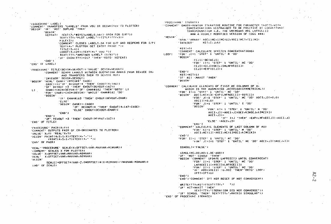
'PROCEDURE' LABEL; 'COMMENT' TRANSFERS 'LABELS FROM VDU OR DECWRITER TO PLOTTER; 'BEGIN"IF"NOT' SUPLAB 'THEN'
'BEGIN' REPEAT: TEXT(7,'*SYS:LABELS.DA/2 OPEN FOR 0/P');
TEXT(TTY,'PLOT LABEL');TITLE(TTY.3); FILE(3);
'COMMENT' CLOSES LABELS.DA FOR 0/P AND REOPENS FOR I/P: TEXT(4,' PLOTTER GET ENTRY PRINT TITLE(3,4): CHOUT(4.129);TEXT(4.' VDU '); TEXT(TTY,'ANOTHER LABEL? (YIN)');
'IF' CHIN(TTY)=217 'THEN"GOTO' REPEAT; 'END':
'END' OF LABEL;
'PROCEDURE' TITLE(DEVIN.DEVOUT)i'VALUE' DEVIN.DEVOUT; 'COMMENT' READS LABELS BETWEEN OUOTAIION MARKS FROM DEVICE IN.
AND TRANSFERS THEM TO DEVICE OUT 'INTEGER' DEVIN.DEVOUT:
'BEGIN' REAL' CHAR:'INTEGER > CASE; CASE:=1;'ff' DEVOUT=4 'THEN' CHOUT(4.146);
'IF' DEVOUT =3 'THEN' CH0UT(DEVOUT,162);
L: CHAR:=CHIN(DEVIN)'IF' CHAR#162 'THEN"GOTO' L; 'FOR' CHAR:=CHIN(DEVIN) 'WHILE' CHAR*162 'DO' 'BEGIN'
'IF' CHAR#163 'THEN' CHOUT(DEVOUT.CHAR) 'ELSE' 'BEGIN' CASE:=—CASE;
'IF' DEVOUT=4 'THEN' CHOUT(4.147—CASE) 'ELSE' CHOUT(DEVOUT.CHAR);
'END'; 'END': 'IF' DEVOUT =3 'THEN' CHOUT(DEVOUT.162);
'END' OF TITLE;
PROCEDURE' - PAIR(X,Y); 'COMMENT' OUTPUTS PAIR OF CO—ORDINATES TO PLOTTER; 'VALUE' X.Y; 'REAL'X,Y; 'DEGIN' PRINT(4.5.1,X);TEXT(4,'.');
PRINT(4,5,11, Y);TEXT(4.' *Pi 'END' OF PAIR;
'PROCEDURE' ITERATE; — COMMENT' GAUSS—NEWTON ITERATIVE ROUTINE FOR PARAMETER SHIFTS.WITH
CONCENTRATIONS RESTRAINED TO BE POSITIVE ,BY LOGARITHMIC' TRANSFORMATION I.E. THE UNKNOWNS ARE LNFRELIII
*** A HIGHLY MODIFIED VERSION OF COGS ##4, ■ 'BEGIN'
'REAL' ARRAY' A011:NC:1:NC+1 .1.X0EI:NCl:CCI:N]: 'INTEGER' NIT.I.J.K;
NIT: =0 'COMMENT' CALCULATE SPECIES CONCENTRATIONS;
LOOP: 'FOR' J:=1 'STEP' 1 'UNTIL' N 'DO' 'BEGIN'
CEJ]:=BETACJ1; 'FOR' I:=1 'STEP' 1 'UNTIL' NC 'DIY CCJ]:=CEMOEI.A*LNFREECII; CIA:=EXP(CCJI);
'END'; NIT:=NIT+1;
'IF' NIT <MAXIT 'THEN' 'BEGIN'
'COMMENT' CALCULATE ELEMENTS OF FIRST NC COLUMNS OF A. WHICH IS THE AUGMENTED JACOBIAN(SYMMETRICAL);
'FOR' I:=1 'STEP' 1 'UNTIL' NC 'DO' 'BEGIN' AOEIPNC4-1]:=EXP(LNFREEEI3)—TOTEI3;
'FOR' J:=1 'STEP' 1 'UNTIL' NC 'DO' AOEI,J]:=0.0; XOEI]:=0.0;
'FOR' J:=1 'STEP' 1 'UNTIL' I 'DO' 'BEGIN'
'FOR: K:= I 'STEP' 1 'UNTIL' N 'DO' AOCI.J]:=A0[I.J]-1411I00*OCJ.K3*CCK]; AOEIFJ]:=
'IF' I=J 'THEN' —EXP(LNFREEEJ])—AOCI,J1 'ELSE' —A0EI,J3;
'END'; • 'COMMENT' CALCULATE ELEMENTS OF LAST COLUMN OF AO; • 'FOR' K: ,=1 'STEP' 1 'UNTIL' N 'DO ;
AOEI,NC+11:=A0[I.NC+1]+OCI.K3*CEK1; 'END': 'FOR' I:=1 'STEP' 1 'UNTIL' NC 'DO'
'FOR' J:=I+1 'STEP' 1 'UNTIL' NC 'DO' AOEI,J]:=AOCJ:IJ;
'REAL' PROCEDURE' SCALE(X.OFFSET.VAR.MAXVAR.MINVAR); SINGUL:='FALSE';
'COMMENT' SCALES X FOR PLOTTER; :
'VALUE' X,OFFSET.VAR,MAXVAR.MINVAR; SIMUL(NC,X0.A0.1.0E-600)1
'REAL' X.OFFSET,VAR,MAXVAR.MINVAR; 'IF' NCIT' SINGUL 'THEN'
'BEGIN' 'BEGIN' COMMENT' UPDATE LNFREECII UNTIL CONVERGENT;
SCALE:=OFFSET+(VAR-2.0*OFFSET)*(X—MINVAR)/(MAXVAR—MINVAR); 'FOR' I:=1 'STEP' 1 'UNTIL' NC 'DO'
END' OF SCALE; LNFREECI]:=X0EIl+LNFREECI]; 'FOR' I:=1 'STEP' I 'UNTIL' NC 'DO' 'IF' ABS(X0EI3) >0.002 'THEN' GOTO' LOOP: IPT:=IPT•1;
'END'; TsO
'END';'COMMENT' IPT NOT RESET IF NOT CONVERGENT; I IND
WRITE(TTY.NIT);TEXT(TTY."); 'IF' NIT=MAXIT 'THEN'
TEXT(TTY.'ITERATION DID NOT CONVERGE*); ''IF' SINGUL 'THEN' TEXT(TTY.'rMATRIX SINGULAR*) . ;
'END' LOF PROCEDURE •ITERATE;

'PKOCEPURE TRINI ■ DEV.N.-M.X); 'VALVE' DEV.K.M.',.; 'INTEGER' DEV,NrMi 'REAL' X:
'COMMENT: vRiNrs <X:,- IN FORMAT (-) NNN.MMM: 'INTEGER . V.E..1,Y; 'REAL' ZT
'RROCEDURI' 5; Y:=ENTIER(Z):
CHOUT(DEVr176+Y): Z:=Z-Yi 'END':
.":=10*ABS(X)+5/10 -Mi V:=0: 'FOR' Z:=0.1* -Z 'WHILE' Z>=1 'DO' V:=V-1-1: 'IF' V>N1-M 'THEN' RWRITE(DEV.X) 'ELSE' 'BEGIN' 'IF' kr,W 'THEN'
'BEGIN' Z:=Z-0.5/10 - (V+M); M:=N+M-Vi N:=V; I:=1; Z:=Z+0.5/10 - (V+11): 'IF' Z>=I 'THEN' 'BEGIN' V:=Vili N:=1,14-1;
M=M-1; Z:=0.1*Zi 'END' FIXING ROUNDING: .
'END' FIXING OVERFLOW; 'FOR' K:=V+1 'STEP' 1 'UNTIL' N 'DO' CHOUT(DEV7160); CHOUT(DEV.'IF' X<0 'THEN' 173 'ELSE' 160); 'FOR' K:=1 'STEP' 1 'UNTIL' V 'DO' Si 'IF' M>0 'OR' I=1 'THEN' CHOUT(DEV,174); 'FOR' K:=1 'STEP' 1 'UNTIL' M 'DO' Si
'END' FORMATTED OUTPUT; 'END' PRINT;
'PROCEDURE' PLOT; 'COMMENT' PLOTS AXES AND EXPERIMENTAL TITRATION POINTS (CIRCLED); -'BEGIN' REAL' X.YrYSTEPFXSTEP; 'INTEGER' 1,PiPEN:
SKIP(TTY): TEXT(TTY.**** LOAD NEW PLOTTER PAPER...*)iSKIP(TTY); TEXT(TTYr*PEN NUMBER FOR AXES= ')iPEN:=READ(TTY)iSKIP(TTY); TEXT(TTY,'PLOTTING SPEED (CM/SEC)= ');P:=READITTY):SKIP(TTY)i TEXT(TTY,'SUPPRESS AXES LABELS (Y/N)'); SUPLAB:='IF' CHIN(TTY)=217. 'THEN"TRUE"ELSE"FALSE'i SKIPUITY)i TEXT(TfY, MAXX:');MAXX:=READ(TTY):SKIP(TTY); TEXT(TTY,* MINX:')iMINXI=READ(TTY);SKIP(TTY): TEXT(TTY,* . MAXY:*);MAXY:=READ(TTY);SKIP(TTY); TEXT(TTY,* MINY:')iMINY:=READ(TTY):SKIP(TTY); TEXT(TTY.*NO.VOLUME STEPS FOR PLOTTER ='): VINC:=READ(TTY):SKIP(TTY)i TEXT(TTYr*MAXIMUM NO.ITERATIONS= *)iMAXITI=READ(TTY); SKIP( TTY); TEXT(TTY.'INPUT GRID LIMITS: LEFT;');L:=READ(TTY);SKIP(TTY); TEXT(TIY,' BOTTOM:");B:READ(TTY);SKIP(TTY); TEXT(TTY.' RIGHT:fliR:=READ(TTY)iSKIP(TTY); TEXT III. TOP:');T:=READ(TTY);SKIP(TTY);
TEXT(4r * PLOTTER INITIALISE DErINL vELOCITY '):WRITE(40 . ): TEXI(4.' DEFINE LIMITS ');PAIR(L , B):PAIK(R.T)i 'COMMENT' SETS UP 0.025MM GRID:- ITX1(4.' DEFINE GRID ')iPAIR(R-L.T-B): 1EXT(4.' LABEL TERMINATOR '):CHOUT01.129):'COMMENT' CNIL/Ai 1EXI- (4.' LABEL LCASE ');CHOUT(4.148)i'COMMENT' 1EXT(4,' LABEL UCASE '):CHOUT(4,146)i'COMMENI . CN1L/P) 1tXT(4,' LABEL SIZE 100.300); TEXT(4,' PEN ');WRITE(4/PEN)i1EXT(4.' MOVE '); PAIR(,500,T-8-500);PAIR(500.500):PAIK(K - L - 500.5 00); PAIR(R-L•500.T-B-500);PAIR(500)T-B -500): XSTEP:=(MAXX-MINX)/10.; 'FOR' X:=MINXTXSTEP 'STEP' XSIEP 'UNTIL' MAXX-XSTET DO' 'BEGIN' TEXT(4,' MOVE ');
PAIR(5CALE(X,00,R-L,MAXX.MINX:.S00); IDRAW 0,'JO IMOVE 0.-250):
'IF' NO1' SUPLAB 'THEN' BEGIN' TEXT(4,' PRINT '): CHOUT(4.136);CHOUT(4,136):CHOUT(4.136); PRINT(4,1,2,X)iCHOUT(4 , 129): 'END';
'END': YSTEP:=(MAXY-MINY)/10.; - 'FOR' Y:=MINYT-YSTEP 'STEP' .YSTEP 'UNTIL' MAXY-YSTEP 'DO' 'BEGIN' TEXT(4,.' MOVE '):
PAIR(500,SCALE(Y.500.1-B.MAXY.MINY)); TEXT(4,' IDRAW 50r0')i TEXT(4,' IMOVE -100,0 ROTATE 90 NOP')) 'IF' NOT' SUPLAB 'THEN' BEGIN' TEXT(A,' PRINT '); CHOUT(4,136)iCHOUT(4,136);CHOUT(4 , 136); PRINT(4,1,2.Y)iCHOUT(4,129); 'END': TEXT(4,' ROTATE*);
TEX1(4,' GET SIZE VDU '); SKIP(TTY)4 TEXT(TTY,'PEN NUMBER FOR DATA =');PEN:=READ(TTY): SKIP(TTY); 1EXT(41' PLOTTER PEN *);WRITE(4,PEN); 'FOR' I:=1 'STEP' 1 'UNTIL' NDATA 'DO' 'BEGIN' TEXT(4,' MOVE*);
PAIR(SCALE((MMOLESECOMP1J+VOLEII*TITCONCECOMP11)/ (MMOLESECOMP2IFVOLEI1*TITCONC(COMV21) ,
500,R-L.MAXX.MINX).
• SCALE(PHLI)7500,T-B.MAXYYMINY)); 'COMMENT' DRAW CIRCLES 0.02 PH UNITS IN RADIUS; TEXT(4,' IMOVE *); PATR(0.02*(T-B)/(MAXY -MINY).0); TEXT(4,' TURN '); RR101- (4.5.1,0.02*(T-B)/(MAXY-MINY)); 'END': TEXT(41' HOP VDU '); LABEL;
'END' OF PLOT; D7=. NJ
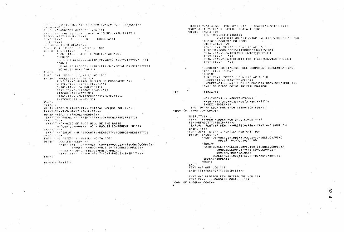
TFX1(11Y.'AIN.N1 PH(EXPI) NII PHICALC)');SKIP(T17); 'FOR' J:=1 . !: .,1-cp' 1 'UN IL' NDA1A - 1 'DO' 'BEGIN' INPLX:=0;
'FOR' V:=VOLCJ11INDEX* tVULTJIll-VOLIJ1)/VINC 'WHILE'• V vOL[J+1) 'DU'
'PEGIN"COMMENT' PH LOOP: VTOT:=YINIT4-V4
'POP' l:=1 'STEP' 1 'UNTIL' NC 'DO' TOTE[1:=IMMOLESC114IITCONCEII*V)/VIOT; 1RIN1(T17,1,3,TOTECOMPU/TOTECUMP21); TEXT (111;* "); PRINICITY,2,3,(PHEJ-M-PHEJ])*INDEX/VINC+PHEA); TEXT( TTY.' •);
'COMMENT' INITIALISE FREE COMPONENT CONCENTRATIONS; 'IF' I11=1 'THEN' 'DES1N' • •
'FOR' i:=1 - 'STEP' 1 'uNTIL' NC-1 '['O' LNFREEEI]:=LN(INITCONCEI1); LNFREE[NC]:=-HX((I'HIJF11-PHLA)*INDEX/vINCEpH(JJ);
'END' OF FIRST POINT INITIALISATION;
L9: ITERATE;
HEJ.INDEX]:=-LNFREEENC7/Hx; PRINTITTY,2,3.HCJ.INDEXDiSKIP(TTY); INDEX:=INDEX+1;
'END' OFF1-1 LOOP FOR EACH TITRATION POINT; 'END' OF TITRATION CURVE;
SKIP(TTY); TEXT(TTy,•PEN NUMBER FOR CALC.CuRVE =*); PEN:=READ(TTY);SKIP(TTY.); TEXT(4,' PLOTTER PEN ');WRITE(4,PEN);TEXT(4,' MOVE •); SKIP(TTY);
'FOR' J:=1 'STEP' 1 'UNTIL' NDATA-1 'DO' 'BEGIN' INDEX:=0;
'FOR' V:=VOLEJ]+INDEX*(WOL[J+1]-VOLEJ))/VINC 'WHILE' V<VOL[J+1] 'DO'
'BEGIN' PAIR(SCALE((MMOLESECOMP1)+V*TITCONCECOMP13) ,
(MMOLESECO7IP2A+V*TITCONCECOMP23), 500,17-LyMAXX,MINX),
SCALE(H[J,INDEX3,500,T-B,MAXY,MINY)); INDEX:=INDEX4-1;
'END'; 'END': - TEXT(4.' NOP VDU *); SKIP(ITY);SKIP(TTY);SKIP(TTY);
TEXT(4,' PLOTTER PEN INITIALISE VDU "); TEXT(TTYr"....PROGRAM ENDS....');
'END' OF PROORAM COMIXH 1
• )• . .t IR( I ) ; TEX N I COMIXH.AL: • );FILE( 1) ; • 1 , );
i • • 1117NOCOPY 0111 pufe (/N)•);
IF" CHIN(1)=:!1 7 ' I u ' 0 'ELSE' 1FSK IP( TTY); ;4 ;• );E:1-.11 - ( II C.: ri .1( ilT.• L 11 IA LOGBETA' );
; (1(IF 1, 1 PU01 ■ 3);N:=RE - Ap();
1 U I EF t 'HNift ' N 'DO' • P1 ■31N' I XIIITY.• •);
'VOR' 1 'UNTIL NC --qua'. 'PEGIN' HFI.J1: -FOADH);WRITE(TTY;OEI,J7);TEXT(TTYr• •);
'END': PEFALJI: RinH(7.);PRINT(TUY;393,BETACJI)1SKIP(TTY); BETALJI: , HX*PLTATJ1;
'END': 'FOR' I:=1 'SiER' 'ONIIL' NC 'DO'
MMOLCSTIA:=RtAD(3); MMOLES OF - COMPONENT ')i
WRITECITY;ti;(11/1(ITY;•='); • PRIN1(TIY.1. -.;MMOLESETI); TEXT(TTX.•,TITRANT CONC.=*); TITCONCLI]:=READ(3)4 PRINT(IIY.1,5rTITCONCEID;SKIPFTTY); INITCONCLIT:=READ(3);
'END'i IPT:=14 ViNIF:=READ(3)4TEXT(TTYy•INITIAL VOLUME (ML.)='); PRINT(TTY,3,3,VINIT)4SKIP(TTY); NDATA:=READ(3);PHCAL:=READ(3); TEX1;TTY.'PHCAL =')4PRINT(TTY.1,3,PHCAL);SKIP(TTY); ',KIP(ITY); irxr(lT(. -X-AXIS OF PLOT WILL BE THE RATIO:
MMOLES COMPONENT (N) / MMOLES COMPONENT (M)'); IP(I1Y)4
IIXF;.11Y.'INPUT N.m:•);cOmPI:=READ(TTy);c0Mp2:=READTTY); il [P ,.TTY);
'FOR' 'STEP' I 'UNTI.L' NOMA 'DO' . 'PFGIN' VOL1JA:=RF6D(3);
PR1N1TIY.1.3.<MMOLFSCCOMP134-VOLCJ3*TITCONCECOMP13)./ (MMOWACUMP214-VOLEJ1*TITCONCECOMP23));
ruuji: ,RKAp((4:1111.11:=PHLJ]fPHCAL: • TEXT(TTY•• •).miNt(TTY.2.3,PHIJ1);SKIP(TTY);
ND';
fi I ;Sk IP( 111>4

403
PUBLICATION
1. Synthesis, Structure and Spectroscopic Studies Of Mercury (II)
Selenolates and MeHgSeBut ,
A.P. Arnold and A.J. Canty, Inorg. Chim Acta, 55 171 (1981)
2. Mercury (II) Selenolates. Crystal structures of Polymeric Hg(SeMe) 2
and the Tetrameric Pyridinates [THgCl(py)(SeEt)1 4 ] and
[{HgC1(py) 0.5 (SeBu t )}4 ],
A.P. Arnold, A.J. Canty B.W. Skelton and A.H. White, J. Chem. Soc.,
Dalton Trans., (1982) in press.
Copyright © 2022 FDOKUMEN