
Electron spectroscopies and inelastic processes in nanoclusters and solids: Theory and experiment

PREPRINT OF: On the Opto-electronic
Properties of Phosphine and Thiolate-protected
Undecagold Nanoclusters
Francesco Muniz-Miranda, Maria Cristina Menziani, and Alfonso Pedone∗
published on Phys. Chem. Chem. Phys., 2014, 16, 18749-18758,
University of Modena and Reggio Emilia, Department of Chemical and Geological Sciences,
Via G. Campi 183, I-41125, Modena, Italy
E-mail: [email protected]
∗To whom correspondence should be addressed
1

Abstract
We present here a detailed time-dependent density-functionl investigation aimed
at systematically dissecting the electronic spectra of two thiolate and phosphine pro-
tected undecagold nanoclusters. Calculations performed on the experimental struc-
tures of Au11(PPh3)7Cl3 and Au11(PPh3)7(SPyr)3 show that ligands give negligible
contributions in the visible region, contrary to what has been previously supposed.
Metal→ligand charge transfer transitions appear at energies well above the visible
threshold, while transitions with some small ligand→metal and ligand→ligand char-
acter occur sporadically at even higher energies. Thus, the conjugation effect between
π-ligand and gold electrons, recently hypothesized to interpret the spectra of phosphine
and thiolate-protected nanoclusters, is not confirmed by our calculations.
Keywords
TD-DFT, Au, Nanoparticle, UV-Vis, Phosphines, Thiolates
Introduction
Gold-based nano-particles find their use in many advanced technological applications,1 in-
cluding optoelectronics,2–6 nano-medicine7,8 and chemical sensors.9,10 Reducing the size of
gold nano-particles and approaching the nm threshold, their behavior changes from preva-
lently metal-like to molecule-like.11–13 At the single-digit nanoscale, the band gap (or, more
correctly, the HOMO-LUMO gap) can widen reaching values exceeding 2 eV.14 Thus, tailor-
ing sizes and shapes of Au-nanoparticles would allow a fine tuning of the electron conduction
properties,15–17 greatly benefiting their adoption in opto-electronics. Due to the inherent ten-
dency of nano-gold to aggregate (so called “aurophilicity”18–21), most gold nano-particles are
protected by an organic coating, usually constituted by phosphines,22 thiols,23 or both.24,25
The organic ligands and their interactions with the inner metal cores play an important role
2

in determining the three-dimensional structure of gold at the nano-scale.
However, it has been very recently observed that also the electronic properties of the
gold cores are affected and modulated by the organic environment that surrounds them,26,27
particularly if the latter is composed by aromatic molecules, like, for example, triphenylphos-
phines and thipyridines. Very recently Wu and Jin26 provided clues of a conjugation between
π-aromatic and gold electrons in the Au11(PPh3)8Br3 nano-cluster (NC), on the basis of pe-
culiar NMR and UV-Vis observed signatures. Wu and Jin26 assigned the Au11(PPh3)8Br3
observed bands to the PPh3 molecule, and explained the red-shift of these bands as due to
the aforementioned conjugation between aromatic π electrons and the metal electrons. The
same authors also investigated the effect played by aromatic ligands on the fluorescence of
metal nanoparticles.? They found that surface ligands bound through sulphur atoms can
influence the fluorescence by (i) establishing charge-transfer transitions from the ligands to
the gold core and (ii) directly donating delocalized electrons of rich atoms or groups of the
ligands to the metal core.
Therefore, understanding the effect of the nature and strength of the interactions arising
between the gold surfaces and the protecting organic molecules on the optical properties
of Au-based nano-particles is becoming fundamental and urgent for designing luminescent
metal NPs for promising optoelectronic and nanomedicine applications.28–31 Steady-state
and time-resolved UV-Vis spectroscopies are valuable tools to investigate structure and dy-
namics of gold nano-particles and their organic-metal inter-phases. However, the subtle
interplay of several different competing effects acting at different length and time scales
makes the interpretation of such spectra quite difficult. Theoretical investigations based
on accurate density functional theory (DFT) and time dependent (TD) DFT calculations32
allow a detailed characterization of ground and excited state properties of different kinds of
medium to large -sized molecules,33–38 metal nano-particles39–43 and hybrid organic-inorganic
structures.13,44–56 Thus, it is possible to dissect all the contributions to the UV-Vis spectra
and investigate in an unbiased way all the effects due to the organic protection onto the
3

optoelectronic features of Au-based NCs containing a few hundreds of atoms.
To the best of our knowledge, no cluster of the type Au11 (PR3)nXm (with n,m integers,
R an aromatic group and X an halogen atom or a thiol group) has both its X-ray resolved
structure and electronic spectrum available in literature, except for Au11(PPh3)7Cl3,57 here-
after referred as “NC1”. We have also investigated another X-ray resolved undecagold58 NC
of stoichiometry Au11(PPh3)7 (SPyr)3 (“NC2”), whose optical spectrum however is not avail-
able. These two NCs have similar metal cores, and differ mainly on the coating. Undecagold
clusters with these stoichiometries were rationalized by Provorse and Aikens42 within the
“superatom” conceptual framework.14,59 Defining n∗, the delocalized electron count for a
closed-shell superatom complex, as
n∗ = (Nν)Au −W − q , (1)
with N and ν the total number and the atomic valence of Au atoms, respectively, W the
total number of monovalent electron-withdrawing group bound to gold atoms (Cl atoms in
the case of NC1, SR groups in the case of NC2), and q the overall charge of the complex
in units of |e| (in these cases, zero), n∗ gets a value of 8 for the NCs investigated here.
A count of 8 delocalized electrons is correlated to a particular stability for approximately
spherical particles, ideally resembling the closed shell of noble gases. The experimental UV-
Vis spectrum57 of Au11(PPh3)7Cl3 is very similar to the one reported by Wu and Jin26 for
Au11(PPh3)8Br3 NC, showing two peaks at ∼ 316 and 406 nm and a (almost unnoticeable)
shoulder between 450 and 550 nm,57 thus being a very reasonable test case to investigate
computationally the extent of the assumed conjugation in phosphine-protected undecagold
NCs.
Unlike a previous work by Provorse and Aikens42 who investigated a undecagold NC
at the LDA level of theory, in this paper we use TD-DFT calculations with GGA, Hybrid
and Long-Range corrected exchange-correlation functionals to the systematic study of the
4
origin of electronic transitions of two experimentally X-ray structurally resolved gold NCs,
including all their organic coating, to understand if and how their electronic properties and
optical spectra are due to the organic ligands, as hypothesized in previous experimental
investigations.26
Computational Methods
All TDDFT calculations have been performed by using the Gaussian09 package.60 Four
exchange-correlation functionals, namely the GGA BPBE functional that combines the
B8861 exchange and the PBE62 correlation functionals, the widely employed global hybrids
B3LYP,63,64 the M06-HF,65 as well as the range-separated hybrid cam-B3LYP66 have been
employed to simulate the absortion spectrum of NC1. Instead, we employed just the cam-
B3LYP functionals for NC2. The adopted pseudo-potential for gold atoms is an improved
version67 of the commonly used small core LanL2DZ (here referred to as “mod-LanL2DZ”)
with added optimized n+1 |p〉 states to the basis set, imported in the calculations through
Basis Set Exchange.68 These computational choices were previously validated by benchmark-
ing more than 20 functionals and 5 pseudo-potentials on three gold NCs (two of them being
NC1 and NC2).51 The solvent in which the optical experimental spectrum of NC1 has been
acquired (dichloromethane) has been simulated with a linear response polarizable continuum
model.69 Dichloromethane is also the solvent used for the cyclic voltammetry measurements
on NC2. Since the number of atoms of the full NC is very large (>250) for excited-states
investigations, test TD-DFT calculations have been performed in order to choose the optimal
basis set for PPh3, as reported in Fig.S2 of the Supporting Information. The 6-31++G basis
set represents the best compromise between accuracy and computational costs, and has been
subsequently adopted in all the calculations reported here on simplified models for C and H
atoms, while for P and Cl atoms two polarization functions have always been added (i.e. for
them 6-31++G(d,p) has been employed). To simulate the full NCs, the smaller 6-31G basis
5

set had to be adopted for C and H atoms to make computations feasible.
Results and Discussions
To understand the components of the electronic spectrum of NC1, five basic models (Fig.1,
left panel) have been set up starting from its experimental three dimensional structure, whose
ground state was previously optimized at the M06-HF level of theory:51
Figure 1: The structural models adopted here to investigate NC1 (left) and NC2 (right). Acronyms areexplained in the text. The picture has been generated adopting the standard CPK color scheme (H is white,C is gray, N is blue, Cl is green, P is orange, S is bright yellow, Au is dark yellow). Positions of the partialcharges for NC1 (model GIC+L+Q, bottom left) are represented by blue dots.
1. isolated PPh3, the organic ligand [L];
2. Au11(PH3)7Cl3, hereafter referred as the “Gold Inner Core” [GIC];
6

3. Au11(PH3)6PPh3Cl3, the Gold Inner Core with one explicit ligand molecule [GIC+L];
4. Au11(PH3)6PPh3Cl3 with Mulliken charges (obtained from full NC calculations with
6-31G basis set adopted for the C and H atoms) for all atoms of the other 6 missing
organic ligands, with exception of the C atoms substituted by H atoms of the PH3
groups, [GIC+L+Q];
5. Au11(PH3)5(PPh3)2Cl3, with two PPh3 adjacent ligands [GIC+2L].
6. full NC
On each of these models, TD-DFT calculations have been performed to obtain the UV-Vis
spectrum of NC1, as reported in Fig.2 (panels A-E); panel F of Fig.2 also shows calculations
performed on the full NC1 with the reduced basis set, and in panel G the experimental
spectra57 are reported as reference.
Three functionals (viz. cam-B3LYP, B3LYP, M06-HF) yield similar spectra for each
model of NC1, differing essentially only for the position of the two peaks on the wavelength
scale, with B3LYP providing the best λmax positions. B-PBE provides a slightly differ-
ent spectrum shape. Anyway, due to the fact that cam-B3LYP usually better reproduces
charge-transfer transitions,70,71 only spectra simulated with this functional (on 200 S0 →Sn
transitions) are reported here for sake of clarity. Fig.S1 of the Supporting Information shows
results obtained with B3LYP, M06-HF, and B-PBE (on 100 transitions) on NC1.
The stick spectra in Fig.2 give a better view of transitions density and intensity, while
their convolutions with Gaussian functions provide results more easily comparable to exper-
imental data (blue lines). The main bands of the isolated PPh3 (panel A of Fig.2) are ∼ 100
nm far from those of the other models (panels B-F), a striking feature of these model spectra.
Moreover, the shape and position of the bands (denoted with blue lines) of the models which
include the gold core (panels B-F) are very similar between them: the higher energy peak
(i.e. the peak found at shorter wavelengths) is somewhat slightly modified passing from the
GIC model (panel B) to the GIC+L model (panel C), albeit the lower energy band seems
7

Figure 2: (A) UV-Visible spectrum calculated on L; (B) calculated on GIC; (C) calculated on GIC+L;(D) calculated on GIC+L+Q; (E) calculated on GIC+2L; (F) calculated on the full NC1 (but with 6-31G basis set for C and H atoms); (G) experimental spectrum of NC1 [Ref. 57] in toluene (magenta) and ofPPh3 [Ref. 26] in CH2Cl2 (green). Blue lines represent the UV-Vis spectra of the different models obtainedfrom the convolution of 200 S0 →Sn transitions (red sticks) with Gaussians of half-width at half-height of0.25 eV. Wavelengths obtained for the calculated spectra (panel A-F) are shifted of +50 nm to super-imposethem with the experimental counterparts (panel G); the actual computed values are reported in Table 2.
8

largely unaffected. On the contrary, transitions at shorter wavelengths are rearranged in
energetics and intensity by adding a ligand to the GIC model (GIC+L model, panel C),
but adding a second ligand to obtain model GIC+2L (panel E) induces only minor changes.
Also including the Mulliken charges of the other ligands (model GIC+L+Q, panel D) seem
to have only minor effects, both on spectra and transitions. While the 6-31++G basis set
has been chosen to better reproduce the PPh3 spectrum, calculations employing smaller 6-
31G basis set for triphenylphosphine ligands (panel F) give rise to a spectrum whose shape
and transitions are very similar to that of the GIC+2L model (panel E) and in excellent
agreement with the experimental one, since it correctly reproduces the two main peaks as
well as the tail at longer wavelengths. The computed spectra of the undecagold-based mod-
els (panels B-F) and of PPh3 (panel A) yield results that can be easily compared to the
experimental ones (panel G, magenta line and green line for NC1 and PPh3, respectively).
It appears that the electronic spectrum of this NC is mainly due to its metal core, at least in
the λ ≥300 nm range, which corresponds to the experimental range.57 The effects induced
by the organic coating seem to be of only secondary relevance.
Calculations on NC2 were limited to its corresponding GIC, GIC+L, and full NC mod-
els. In this case, L still refers to PPh3 (whose effect is the primary interest of this pa-
per), yet the GIC+L model retains a complete SPyr ligand because previous computations
suggest that SPyr can interact with PPh3 through π-stacking interactions.51 Observations
analogous to those made on NC1 can be done on NC2, apart for the comparison with the
experimental spectrum. In fact, even for Au11(PPh3)7 (SPyr)3 the GIC, GIL+L and full
NC2 models provide very similar energetics and band shapes (Fig.3), despite the presence
of SPyr ligands in place of Cl atoms. The spectra of NC2 are red-shifted of about ∼0.4
eV with respect to NC1 (corresponding to ∼60 nm with respect to the lower energy tran-
sition) due to the difference in ligands. In fact, the metal core of the full NC1 is more
positively charged51 than that of the full NC2 of more than +0.8|e| (as computed from Hir-
shfeld charges,72 |e| being the unsigned charge of the electron), resulting in a more stable
9

Figure 3: (A) UV-Visible spectrum calculated on L; (B) calculated on GIC; (C) calculated on GIC+L;(D) calculated on the full NC2 (but with 6-31G basis set for C and H atoms); Blue lines represent the UV-Visspectra of the different models obtained from the convolution of 200 S0 →Sn transitions (red sticks) withGaussians of half-width at half-height of 0.25 eV. Wavelengths obtained for the calculated spectra (panelA-F) are shifted of +50 nm in analogy to Fig. 2; the actual computed values are reported in Table 2.
HOMO state for NC1 (EHOMO(NC1)−EHOMO(NC2)= −0.83 eV at cam-B3LYP level of the-
ory), probably due to the greater electronegativity of Cl atoms with respect to SR groups.
While also the LUMO of NC1 is lower in energy than the LUMO of NC2, this effect is smaller
(ELUMO(NC1)−ELUMO(NC2)= −0.27 eV at cam-B3LYP level of theory), thus resulting in
an estimated net shrinking of the gap of NC2 of ∼ 0.56 eV, in qualitatively agreement with
the previous comment. This shrinking at the cam-B3LYP level of theory is observed only
if model comprising the ligands (fully or in part) are adopted, because for the simplified
GIC models of NC1 and NC2 the computed optical gaps are indeed much closer (difference
between them ≤0.1 eV) and the NC1 metal core is more positively charged72 than the NC2
metal core of less than +0.3|e|, as reported in Table 1.
10
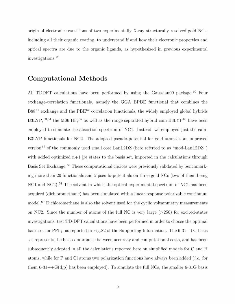
Table 1: Partial average Hirshfeld charges of some selected elements for GIC and full NCmodels of NC1 and NC2 in unsigned electron charge (i.e. |e|). The charge of Au11 is the sumof all metal charges.
GIC model Full NCsNC1 NC2 NC1 NC2
Au11 -1.048 -1.319 -1.276 -2.079P +0.327 +0.302 +0.352 +0.294S — -0.264 — -0.236Cl -0.414 — -0.401 —
Albeit the nature of the ligands (SPyr vs Cl) affect the charge bear by the metal core,
and thus the position of the peaks in the spectra, the similarity between the absorption
spectra of GIC and full NC models suggest that the spectra is dominated by Metal-Metal
transitions.
Therefore, the conjugation between aromatic π-electrons and metal electrons advocated
by Wu and Jin26 to explain the spectra of Au11(PPh3)nX3 clusters in terms of a shifting
of electronic transitions involving ligands does not seem to be observed in the NCs studied
here. However, even if of secondary importance, some small effects due to the coating are
observable in the computed spectra. Thus, we have studied the DFT-computed molecular
orbitals involved into the various optical transitions for the model GIC+L of both clusters in
order to investigate the effect of Ligand-Ligand, Metal-Ligand and Ligand-Metal transitions.
Table 2 lists the features of the 8 more significant S0 →Sn transitions (of the 200 investi-
gated in this work) of the GIC+L model of NC1 (higher panel). The Kohn-Sham molecular
orbitals interacting in these transitions are represented as contour plots in Fig.4. As can be
observed, at low energies (≤4.5 eV, corresponding to the longer wavelength band of Fig.2)
the transitions involve only orbitals localized on the metal core, with some contributions of
P and Cl atoms. No significant contribution due to the aromatic molecules is apparent and
only GIC→GIC transitions are found. Only at energies ≥4.80 eV (corresponding to the
shorter wavelength band of Fig.2) GIC→L charge transfer bands start appearing, as in case
of the transition n=68, which is also one of the most intense in this range of observed en-
ergies. Some small contributions from L→L and L→GIC excitations occur for transitions
11

Table 2: Selected optical S0 →Sn transitions of GIC+L model and their orbital contributionsfor NC1 and NC2. The table lists the transition number (n), the occupied (occ.orb.) andvirtual (virt.orb.) orbitals involved into the transitions, their relative contribution (CI coeff.),oscillator strengths (osc. str.) multiplied by a 104 factor, energies (energy), and correspondingwavelengths (λ). Orbital pairs labeled with ‡ symbols are not the main contribution to thereported transitions (viz. n=144,170), but are reported because of their significance for thediscussion. Calculations are performed at the cam-B3LYP/6-31++G level of theory. Fulldetails for 45 most significant transitions of NC1 can be found in Table S1 of the SupportingInformation.
NC1n 〈occ.orb.| → |virt.orb.〉 CI coeff. osc.str. (·10+4) energy/eV λ/nm
1 HOMO → LUMO +0.65 41 2.91 42613 HOMO-2 → LUMO+3 +0.34 1971 3.48 35714 HOMO-2 → LUMO+4 +0.46 1713 3.57 34715 HOMO → LUMO+4 +0.42 2903 3.58 34615 HOMO-2 → LUMO+3 -0.3940 HOMO-3 → LUMO+3 +0.29 1436 4.46 27868 HOMO → LUMO+10 +0.37 701 4.83 25668 HOMO → LUMO+11 +0.26
144‡ HOMO-25 → LUMO+8 +0.10 158 5.44 228170‡ HOMO-26 → LUMO -0.11 450 5.60 221
NC2n 〈occ.orb.| → |virt.orb.〉 CI coeff. osc.str. (·10+4) energy/eV λ/nm
1 HOMO → LUMO +0.65 42 2.51 49511 HOMO-2 → LUMO +0.54 2418 3.25 38212 HOMO-2 → LUMO+2 +0.41 2202 3.34 37213 HOMO-2 → LUMO+1 +0.35 2596 3.36 36952 HOMO-2 → LUMO+6 +0.18 803 4.27 29065 HOMO-1 → LUMO+13 +0.22 1058 4.42 28067 HOMO-1 → LUMO+12 +0.33 936 4.46 27875 HOMO-12 → LUMO+1 0.26 1684 4.60 27077 HOMO-3 → LUMO+9 0.20 984 4.62 26880 HOMO-9 → LUMO+2 0.29 1546 4.65 267
n=144 (5.44 eV) and n=170 (5.60 eV), respectively. In particular, a small contribution
(CI coefficient =0.10) to the 144th excited state is given by the transition between the or-
bitals HOMO-25 (orbital #228) and LUMO+8 (orbital #262), both localized on the ligand
molecule. This transition is blue-shifted of 8 nm and shows a very small oscillator strength
compared to the first transition of the isolated PPh3 molecule. Moreover, the excited state
n=170 presents a small contribution (CI coefficient=-0.11) given by the transition between
the HOMO (orbital #253) and LUMO+22 (orbital #276, reported in Fig.5) that are lo-
12

Figure 4: Contour plots of the orbitals involved into the transitions of NC1 described in Table 2, computedat the cam-B3LYP/ 6-31++G level of theory. Blue color denotes the negative part of the DFT wavefunction,while red denotes the positive part. The underlying molecular skeleton is fixed and shown from the samepoint of view for better clarity, adopting the standard CPK color scheme. A much more detailed descriptionof the shape of 24 virtual and occupied orbitals can be found in Figures S4 and S5 of the SupportingInformation.
calized on the gold core and on the PPh3 molecule, respectively. However, it has to be
highlighted that these two types of transitions occur only very sporadically in the range of
energy investigated here and that never constitute the main contribution to any absorption
13

peak (red sticks of Fig.2). A much more detailed analysis is reported in Table S1 and Figures
S4 and S5 of the Supporting Information.
Figure 5: Contour plots of the orbitals involved into the transitions of NC2 described in Table 2, computedat the cam-B3LYP/ 6-31++G level of theory. Blue color denotes the negative part of the DFT wavefunction,while red denotes the positive part. The underlying molecular skeleton is fixed and shown from the samepoint of view for better clarity, adopting the standard CPK color scheme.
The interacting orbitals giving rise to the main optical absorption bands of NC2 (reported
in Table 2, lower panel) are reported in Fig.5. Most observations done for NC1 are still valid
for NC2: in particular, most of the spectrum is due to GIC→GIC transitions, with the first
14

GIC→L charge transfer state (n=52) found at 4.27 eV. This latter energy value is about
0.53 eV lower than the corresponding GIC→L absorption peak of NC1, which is similar to
the aforementioned red-shift of ∼0.56 eV observed on the HOMO-LUMO energy difference
of NC2. It should also be pinpointed that some high energy transitions (≥4.6 eV) occur
between states that have a non-negligible electronic density on SPyr toward the PPh3 ligand
(e.g. state n=77).
Concluding Remarks
In conclusion, the main optical features of Au11(PPh3)7Cl3 have been correctly reproduced at
increasing levels of sophistication, both regarding the model (inner core, addition of ligands,
effect of the point charges) and the adopted computational procedures (functionals, basis
sets for the ligands). In this way, each effect has been taken into account in order to provide
a more realistic picture of the interactions occurring in the nano-cluster. The electronic
properties of Au11(PPh3)7SPyr3 have been also investigated finding many similarities with
the previous cluster, apart for a more negatively charged metal core and a red-shift of the
whole optical spectrum, which suggests that ligands induce mainly a sort of small “solvent
effect” on the electronic spectra.
It appears that the optical spectra of undecagold nano-clusters are mainly due to tran-
sitions localized on metal atoms, and the atoms directly bound to them (P, Cl, and S).
These particles are well known examples of “superatom” complexes, which display particu-
lar stability when the number of delocalized electrons matches certain specific numbers (8 in
these cases).14,42,59 The optoelectronic effects due to the triphenilphosphine and thiopyridine
ligands are negligible in first approximation, giving some (minor) contributions only to the
higher energy band. In particular, ligand→ligand transitions are not observed in the Vis re-
gion of frequencies, and only sporadically in UV region, contrary to what has been previously
supposed in literature.26 The conjugation between π-electrons of the aromatic ligands and
15

metal electrons occurs out of the Vis region, and the experimental peak at ∼320 nm cannot
be explained as due to an hypothesized red-shift of triphenilphosphine bands. In fact, the
most relevant features of the spectrum can be reproduced also excluding the aromatic part
of the ligand from the calculations.
The approach adopted here is valuable to dissect optical spectra of hybrid metal-organic
nano-particles, and can be extended to larger nano-systems. We found that the effects of the
ligands on opto-electronic properties appear minor in the energy range of the Vis region, thus
corroborating previous calculations on nano-gold (e.g. Ref. 14,25,39,41,48,51) performed on
simplified models with reduced organic coating.
Acknowledgement
This work was supported by the Italian “Ministero dell’Istruzione, dell’Universita e della
Ricerca” (MIUR) through the “Futuro in Ricerca” (FIRB) grant RBFR1248UI 002 titled
“Novel Multiscale Theorethical/Computational Strategies for the Design of Photo and Thermo
responsive Hybrid Organic-Inorganic Components for Nanoelectronic Circuits” and the “Pro-
gramma di ricerca di rilevante interesse nazionale” (PRIN) grant 2010C4R8M8 titled “Nanoscale
functional Organization of (bio)Molecules and Hybrids for targeted Application in Sensing,
Medicine and Biotechnology” is also acknowledged. Computation time was granted through
the CINECA project AUNANMR-HP10CJ027S.
Supporting Information Available
(a) TD-DFT optical spectra calculated with B3LYP, M06-HF, and B-PBE functionals on
model GIC+L; (b) TD-DFT spectra of PPh3 calculated employing 6-311++G**, 6-311G,
6-31++G, 6-31G, and STO-3G basis sets; (c) Table with 45 optical transitions is provided, as
well as contour plots for 48 virtual and occupied orbitals at the cam-B3LYP/6-31++G level of
theory. This material is available free of charge via the Internet at http://pubs.acs.org/.
16

References
(1) Murray, R. W. Nanoelectrochemistry: Metal Nanoparticles, Nanoelectrodes, and
Nanopores. Chemical Reviews 2008, 108, 2688–2720.
(2) Hu, M.-S.; Chen, H.-L.; Shen, C.-H.; Hong, H. B.-R., Lu-Sheng; Chen, K.-H.; Chen, L.-
C. Photosensitive gold-nanoparticle-embedded dielectric nanowires. Nature Materials
2006, 5, 102–106.
(3) Paltiel, Y.; Aharoni, A.; Banin, U.; Neuman, O.; Naaman, R. Self-assembling of InAs
nanocrystals on GaAs: The effect of electronic coupling and embedded gold nanopar-
ticles on the photoluminescence. Applied Physics Letters 2006, 89, 033108.
(4) Sonnichsen, C.; Alivisatos, A. P. Gold Nanorods as Novel Nonbleaching Plasmon-Based
Orientation Sensors for Polarized Single-Particle Microscopy. Nano Letters 2005, 5,
301–304.
(5) Liu, H. L.; Zhang, X.; Zhai, T. Plasmonic nano-ring arrays through patterning gold
nanoparticles into interferograms. Opt. Express 2013, 21, 15314–15322.
(6) Xu, K.; Li, Y.; Zhang, W.; Zhang, L.; Xie, W. Effect of gold nanoparticles on the
performances of the phosphorescent organic light-emitting devices. Current Applied
Physics 2014, 14, 53 – 56.
(7) Heath, J. R.; Davis, M. E. Nanotechnology and Cancer. Annual Review of Medicine
2008, 59, 251–265.
(8) Homberger, M.; Simon, U. On the application potential of gold nanoparticles in nano-
electronics and biomedicine. Philosophical Transactions of the Royal Society A: Math-
ematical, Physical and Engineering Sciences 2010, 368, 1405–1453.
(9) Liu, X.; Dai, Q.; Austin, L.; Coutts, J.; Knowles, G.; Zou, J.; Chen, H.; Huo, Q.
A One-Step Homogeneous Immunoassay for Cancer Biomarker Detection Using Gold
17

Nanoparticle Probes Coupled with Dynamic Light Scattering. Journal of the American
Chemical Society 2008, 130, 2780–2782.
(10) Guo, L.; Xu, Y.; Ferhan, A. R.; Chen, G.; Kim, D.-H. Oriented Gold Nanoparticle Ag-
gregation for Colorimetric Sensors with Surprisingly High Analytical Figures of Merit.
Journal of the American Chemical Society 2013, 135, 12338–12345.
(11) Chen, S.; Ingram, R. S.; Hostetler, M. J.; Pietron, J. J.; Murray, R. W.; Schaaff, T. G.;
Khoury, J. T.; Alvarez, M. M.; Whetten, R. L. Gold Nanoelectrodes of Varied Size:
Transition to Molecule-Like Charging. Science 1998, 280, 2098–2101.
(12) Yang, Y.; Chen, S. Surface Manipulation of the Electronic Energy of Subnanometer-
Sized Gold Clusters: An Electrochemical and Spectroscopic Investigation. Nano Letters
2003, 3, 759.
(13) Aikens, C. M. Electronic Structure of Ligand-Passivated Gold and Silver Nanoclusters.
The Journal of Physical Chemistry Letters 2011, 2, 99–104.
(14) Walter, M.; Akola, J.; Lopez-Acevedo, O.; Jadzinsky, P. D.; Calero, G.; Ackerson, C. J.;
Whetten, R. L.; Gronbeck, H.; Hakkinen, H. A unified view of ligand-protected gold
clusters as superatom complexes. Proceedings of the National Academy of Sciences
2008, 105, 9157–9162.
(15) Alivisatos, A. P. Perspectives on the Physical Chemistry of Semiconductor Nanocrys-
tals. The Journal of Physical Chemistry 1996, 100, 13226–13239.
(16) Alivisatos, P.; Barbara, P. F.; Castleman, A. W.; Chang, J.; Dixon, D. A.; Klein, M. L.;
McLendon, G. L.; Miller, J. S.; Ratner, M. A.; Rossky, P. J. et al. From Molecules to
Materials: Current Trends and Future Directions. Advanced Materials 1998, 10, 1297–
1336.
18

(17) Schaadt, D. M.; Feng, B.; Yu, E. T. Enhanced semiconductor optical absorption via
surface plasmon excitation in metal nanoparticles. Applied Physics Letters 2005, 86,
063106.
(18) Rawashdeh-Omary, M. A.; Omary, M. A.; Fackler, J. Argento-aurophilic Bonding
in Organosulfur Complexes. The Molecular and Electronic Structures of the Hetero-
bimetallic Complex AgAu(MTP)2. Inorganica Chimica Acta 2002, 334, 376–384.
(19) Schmidbaur, H. Ludwig Mond Lecture. High-carat gold compounds. Chem. Soc. Rev.
1995, 24, 391–400.
(20) Pyykko, P. Theoretical Chemistry of Gold. Angewandte Chemie International Edition
2004, 43, 4412–4456.
(21) Pyykko, P. Theoretical chemistry of gold. III. Chem. Soc. Rev. 2008, 37, 1967–1997.
(22) Maspero, A.; Kani, I.; Mohamed, A. A.; Omary, M. A.; Staples, R. J.; Fackler, J. P.
Syntheses and Structures of Dinuclear Gold(I) Dithiophosphonate Complexes and the
Reaction of the Dithiophosphonate Complexes with Phosphines: Diverse Coordination
Types. Inorganic Chemistry 2003, 42, 5311–5319.
(23) Forward, J. M.; Bohmann, D.; Fackler, J. P.; Staples, R. J. Luminescence Studies of
Gold(I) Thiolate Complexes. Inorganic Chemistry 1995, 34, 6330–6336.
(24) Pettibone, J. M.; Hudgens, J. W. Gold Cluster Formation with Phosphine Ligands:
Etching as a Size-Selective Synthetic Pathway for Small Clusters? ACS Nano 2011, 5,
2989–3002.
(25) Das, A.; Li, T.; Nobusada, K.; Zeng, Q.; Rosi, N. L.; Jin, R. Total Structure and
Optical Properties of a Phosphine/Thiolate-Protected Au24 Nanocluster. Journal of
the American Chemical Society 2012, 134, 20286–20289.
19

(26) Wu, Z.; Jin, R. Exclusive synthesis of Au11(PPh3)8Br3 against the Cl Analogue and the
Electronic Interaction between Cluster Metal Core and Surface Ligands. Chemistry -
A European Journal 2013, 19, 12259–12263.
(27) Wu, Z.; Jin, R. On the Ligand’s Role in the Fluorescence of Gold Nanoclusters. Nano
Letters 2010, 10, 2568–2573.
(28) Diegoli, S.; Manciulea, A. L.; Begum, S.; Jones, I. P.; Lead, J. R.; Preece, J. A. In-
teraction between manufactured gold nanoparticles and naturally occurring organic
macromolecules. Science of The Total Environment 2008, 402, 51–61.
(29) Bain, C. D.; Whitesides, G. M. Modeling Organic Surfaces with Self-Assembled Mono-
layers. Angewandte Chemie International Edition in English 1989, 28, 506–512.
(30) Aradhya, S. V.; Frei, M.; Hybertsen, M. S.; Venkataraman, L. Van der Waals interac-
tions at metal/organic interfaces at the single-molecule level. Nature Materials 2012,
11, 872–876.
(31) Hombergem, M.; Simon, U. On the application potential of gold nanoparticles in na-
noelectronic and nanomedicine. Phil. Trans. R. Soc. A 2010, 368, 1405–1453.
(32) Runge, E.; Gross, E. K. U. Density-Functional Theory for Time-Dependent Systems.
Phys. Rev. Lett. 1984, 52, 997–1000.
(33) Pagliai, M.; Muniz-Miranda, F.; Cardini, G.; Righini, R.; Schettino, V. Spectroscopic
properties with a combined approach of ab initio molecular dynamics and wavelet
analysis. Journal of Molecular Structure 2011, 993, 438–442.
(34) Jacquemin, D.; Preat, J.; Perpete, E. A.; Vercauteren, D. P.; Andre, J.-M.; Ciofini, I.;
Adamo, C. Absorption spectra of azobenzenes simulated with time-dependent density
functional theory. International Journal of Quantum Chemistry 2011, 111, 4224–4240.
20

(35) Muniz-Miranda, F.; Pagliai, M.; Cardini, G.; Righini, R. Hydrogen Bond Effects in
the Vibrational Spectra of 1,3-propanediol in Acetonitrile: Ab initio and Experimental
Study. The Journal of Chemical Physics 2012, 137 .
(36) Biczysko, M.; Bloino, J.; Brancato, G.; Cacelli, I.; Cappelli, C.; Ferretti, A.; Lami, A.;
Monti, S.; Pedone, A.; Prampolini, G. et al. Integrated computational approaches for
spectroscopic studies of molecular systems in the gas phase and in solution: pyrimidine
as a test case. Theoretical Chemistry Accounts 2012, 131, 1–19.
(37) Pedone, A.; Bloino, J.; Barone, V. Role of Host-Guest Interactions in Tuning the Optical
Properties of Coumarin Derivatives Incorporated in MCM-41: A TD-DFT Investiga-
tion. The Journal of Physical Chemistry C 2012, 116, 17807–17818.
(38) Adamo, C.; Jacquemin, D. The calculations of excited-state properties with Time-
Dependent Density Functional Theory. Chem. Soc. Rev. 2013, 42, 845–856.
(39) Aikens, C. M. Origin of Discrete Optical Absorption Spectra of M25(SH)18- Nanoparti-
cles (M = Au, Ag). The Journal of Physical Chemistry C 2008, 112, 19797–19800.
(40) Hakkinen, H. The gold-sulfur interface at the nanoscale. Nature Chemistry 2012, 4,
443–455.
(41) Periyasamy, G.; Remacle, F. Ligand and Solvation Effects on the Electronic Properties
of Au55 Clusters: A Density Functional Theory Study. Nano Letters 2009, 9, 3007–
3011.
(42) Provorse, M. R.; Aikens, C. M. Origin of Intense Chiroptical Effects in Undecagold
Subnanometer Particles. Journal of the American Chemical Society 2010, 132, 1302–
1310.
(43) Aikens, C. M. Modelling small gold and silver nanoparticles with electronic structure
methods. Molecular Simulation 2012, 38, 607–614.
21

(44) Yoon, B.; Koskinen, P.; Huber, B.; Kostko, O.; von Issendorff, B.; Hakkinen, H.;
Moseler, M.; Landman, U. Size-Dependent Structural Evolution and Chemical Re-
activity of Gold Clusters. ChemPhysChem 2007, 8, 157–161.
(45) Zhu, M.; Aikens, C. M.; Hollander, F. J.; Schatz, G. C.; Jin, R. Correlating the Crystal
Structure of A Thiol-Protected Au25 Cluster and Optical Properties. Journal of the
American Chemical Society 2008, 130, 5883–5885.
(46) Lopez-Acevedo, O.; Tsunoyama, H.; Tsukuda, T.; Hakkinen, H.; Aikens, C. M. Chiral-
ity and Electronic Structure of the Thiolate-Protected Au38 Nanocluster. Journal of
the American Chemical Society 2010, 132, 8210–8218.
(47) Hadley, A.; Aikens, C. M. Thiolate Ligand Exchange Mechanisms of Au1 and Sub-
nanometer Gold Particle Au11. The Journal of Physical Chemistry C 2010, 114, 18134–
18138.
(48) Ivanov, S. A.; Arachchige, I.; Aikens, C. M. Density Functional Analysis of Geometries
and Electronic Structures of Gold-Phosphine Clusters. The Case of Au4(PR3)42+ and
Au4(µ2-I)2(PR3)4. The Journal of Physical Chemistry A 2011, 115, 8017–8031.
(49) Muniz-Miranda, M.; Pagliai, M.; Muniz-Miranda, F.; Schettino, V. Raman and com-
putational study of solvation and chemisorption of thiazole in silver hydrosol. Chem.
Commun. 2011, 47, 3138–3140.
(50) Muniz-Miranda, F.; Pagliai, M.; Cardini, G.; Righini, R. Bifurcated Hydrogen Bond in
Lithium Nitrate Trihydrate Probed by ab Initio Molecular Dynamics. The Journal of
Physical Chemistry A 2012, 116, 2147–2153.
(51) Muniz-Miranda, F.; Menziani, M. C.; Pedone, A. Assessment of Exchange-Correlation
Functionals in Reproducing the Structure and Optical Gap of Organic-Protected Gold
Nanoclusters. The Journal of Physical Chemistry C 2014, 118, 7532–7544.
22

(52) Heinecke, C. L.; Ni, T. W.; Malola, S.; Mkinen, V.; Wong, O. A.; Hakkinen, H.; Acker-
son, C. J. Structural and Theoretical Basis for Ligand Exchange on Thiolate Monolayer
Protected Gold Nanoclusters. Journal of the American Chemical Society 2012, 134,
13316–13322.
(53) Pedone, A.; Prampolini, G.; Monti, S.; Barone, V. Realistic Modeling of Fluorescent
Dye-Doped Silica Nanoparticles: A Step Toward the Understanding of their Enhanced
Photophysical Properties. Chemistry of Materials 2011, 23, 5016–5023.
(54) Bae, G.-T.; Aikens, C. M. Time-Dependent Density Functional Theory Studies of Opti-
cal Properties of Ag Nanoparticles: Octahedra, Truncated Octahedra, and Icosahedra.
The Journal of Physical Chemistry C 2012, 116, 10356–10367.
(55) De Angelis, F.; Fantacci, S.; Mosconi, E.; Nazeeruddin, M. K.; Gratzel, M. Absorption
Spectra and Excited State Energy Levels of the N719 Dye on TiO2 in Dye-Sensitized
Solar Cell Models. The Journal of Physical Chemistry C 2011, 115, 8825–8831.
(56) Pedone, A.; Prampolini, G.; Monti, S.; Barone, V. Absorption and emission spectra
of fluorescent silica nanoparticles from TD-DFT/MM/PCM calculations. Phys. Chem.
Chem. Phys. 2011, 13, 16689–16697.
(57) Gutrath, B. S.; Englert, U.; Wang, Y.; Simon, U. A Missing Link in Undecagold Cluster
Chemistry: Single-Crystal X-ray Analysis of [Au11(PPh3)7Cl3]. European Journal of
Inorganic Chemistry 2013, 2013, 2002–2006.
(58) Nunokawa, K.; Onaka, S.; Ito, M.; Horibe, M.; Yonezawa, T.; Nishihara, H.; Ozeki, T.;
Chiba, H.; Watase, S.; Nakamoto, M. Synthesis, single crystal X-ray analysis, and
{TEM} for a single-sized Au11 cluster stabilized by {SR} ligands: The interface be-
tween molecules and particles. Journal of Organometallic Chemistry 2006, 691, 638 –
642.
23

(59) Akola, J.; Walter, M.; Whetten, R. L.; Hakkinen, H.; Gronbeck, H. On the Structure of
Thiolate-Protected Au25. Journal of the American Chemical Society 2008, 130, 3756–
3757.
(60) Frisch, M. J.; Trucks, G. W.; Schlegel, H. B.; Scuseria, G. E.; Robb, M. A.; Cheese-
man, J. R.; Scalmani, G.; Barone, V.; Mennucci, B.; Petersson, G. A. et al. Gaussian
09. Gaussian, Inc., Wallingford CT, 2010.
(61) Becke, A. D. Density-functional exchange-energy approximation with correct asymp-
totic behavior. Phys. Rev. A 1988, 38, 3098–3100.
(62) Perdew, J. P.; Burke, K.; Ernzerhof, M. Generalized Gradient Approximation Made
Simple. Phys. Rev. Lett. 1996, 77, 3865–3868.
(63) Becke, A. D. Density-functional thermochemistry. III. The role of exact exchange. The
Journal of Chemical Physics 1993, 98, 5648–5652.
(64) Lee, C.; Yang, W.; Parr, R. G. Development of the Colle-Salvetti correlation-energy
formula into a functional of the electron density. Phys. Rev. B 1988, 37, 785–789.
(65) Zhao, Y.; Truhlar, D. G. Density Functional for Spectroscopy: No Long-Range Self-
Interaction Error, Good Performance for Rydberg and Charge-Transfer States, and
Better Performance on Average than B3LYP for Ground States. The Journal of Physical
Chemistry A 2006, 110, 13126–13130.
(66) Yanai, T.; Tew, D. P.; Handy, N. C. A new hybrid exchange-correlation functional using
the Coulomb-attenuating method (CAM-B3LYP). Chemical Physics Letters 2004, 393,
51 – 57.
(67) Couty, M.; Hall, M. B. Basis sets for transition metals: Optimized outer p functions.
Journal of Computational Chemistry 1996, 17, 1359–1370.
24

(68) Schuchardt, K.; Didier, B.; Elsethagen, T.; Sun, L.; Gurumoorthi, V.; Chase, J.; Li, J.;
Windus, T. Basis Set Exchange: A Community Database for Computational Sciences.
Journal of Chemical Information and Modeling 2007, 47, 1045–1052.
(69) Scalmani, G.; Frisch, M. J. Continuous surface charge polarizable continuum models of
solvation. I. General formalism. The Journal of Chemical Physics 2010, 132, 114110.
(70) Pedone, A. Role of Solvent on Charge Transfer in 7-Aminocoumarin Dyes: New Hints
from TD-CAM-B3LYP and State Specific PCM Calculations. Journal of Chemical The-
ory and Computation 2013, 9, 4087–4096.
(71) Cai, Z.-L.; Crossley, M. J.; Reimers, J. R.; Kobayashi, R.; Amos, R. D. Density Func-
tional Theory for Charge Transfer: The Nature of the N-Bands of Porphyrins and
Chlorophylls Revealed through CAM-B3LYP, CASPT2, and SAC-CI Calculations. The
Journal of Physical Chemistry B 2006, 110, 15624–15632.
(72) Hirshfeld, F. Bonded-atom Fragments for Describing Molecular Charge Densities. The-
oretica Chimica Acta 1977, 44, 129–138.
25
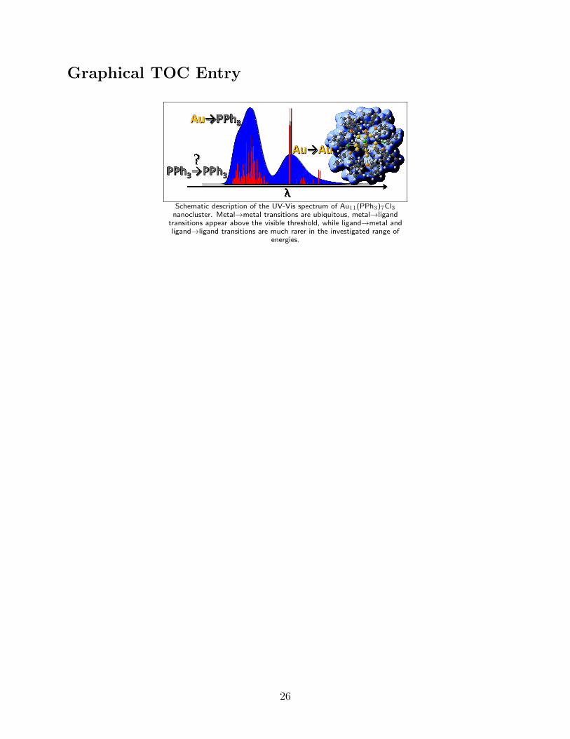
Graphical TOC Entry
Schematic description of the UV-Vis spectrum of Au11(PPh3)7Cl3nanocluster. Metal→metal transitions are ubiquitous, metal→ligand
transitions appear above the visible threshold, while ligand→metal andligand→ligand transitions are much rarer in the investigated range of
energies.
26
Copyright © 2022 FDOKUMEN


 and electrochemical behaviour](https://static.fdokumen.com/doc/165x107/6333101f576b626f850db0e4/ligand-substitution-and-growth-reactions-with-ru-3-se-2-nido-clusters-coordination.jpg)


![“(Acetylacetonato)carbonyl{dicyclohexyl[4-(N,N-dimethylamino)phenyl] phosphine}rhodium(I)](https://static.fdokumen.com/doc/165x107/631b64dc7abff1d7c20ae8e4/acetylacetonatocarbonyldicyclohexyl4-nn-dimethylaminophenyl-phosphinerhodiumi.jpg)
