Red, White, and Blue Eggs as Models of Porphyrin and Heme Metabolism
Upload
independentCategory
view
5download
0
EPR, Resonance Raman, and DFT Calculations on Thiolate- andImidazole-Bound Iron(III) Porphyrin Complexes: Role of the AxialLigand in Tuning the Electronic StructurePradip Kumar Das, Sudipta Chatterjee, Subhra Samanta, and Abhishek Dey*
Department of Inorganic Chemistry, Indian Association for the Cultivation of Science, Kolkata, India 700032
*S Supporting Information
ABSTRACT: Iron(III) porphyrin complexes bearing cova-lently attached imidazole and thiolate axial ligands areinvestigated using resonance Raman, electron paramagneticresonance, and cyclic voltammetry. The thiolate ligandstabilizes a low-spin ground state in solvent-bound six-coordinate species, weakens the Fe−Npyr bonds, and shiftsthe FeIII/II potential more negative by ∼500 mV relative to animidazole-bound species. Density functional theory calcula-tions reproduce the experimental observation and indicate thatthe covalent charge donation from thiolate to iron reduces theZeff on the iron. This increases the Fe3d orbital energies, whichchanges the bonding interaction present in these complexessignificantly. In particular, the increase of the Fe3d energiesactivates an iron-to-porphyrin π*-back-bonding interaction not present in the imidazole-bound complex.
■ INTRODUCTIONThiolate-bound iron porphyrin cofactors are found in severalmetalloenzymes, e.g., cytochrome P450 (cyt P450), NOsynthase, and chloroperoxidase.1−6 Out of these, cyt P450 hasbeen of particular interest to the catalysis community becauseof its capability of oxidizing inert C−H bonds using molecularO2.
2 The remarkable reactivity of this heme enzyme is mostlyattributed to the presence of the proximal thiolate ligand in theactive site, which is proposed to exert a “push effect”.7−13 TheFe center in the active site of cyt P450 in its resting form(Figure 1) is best described as a six-coordinate (6C), low-spin(LS; S = 1/2) FeIII.14,15 Upon substrate binding, a five-coordinate (5C), high-spin (HS; S = 5/2) FeIII site is formedand is reduced to generate a HS (S = 2) FeII active form thatbinds O2.
16−18 The “push effect” of the thiolate ligand has beenproposed to lower the FeIII/II reduction potential, weaken theaxial ligand binding, and affect the heterolytic O−O bondcleavage of a FeIII-OOH species (compound 0)4 withoutrequiring an acidic proton to drive it, resulting in the formationof a highly oxidizing species known as compound I.8,16−25
Several investigations, both theoretical and experimental, havefocused on the nature of the “push effect”, and bothelectrostatic (thiolate is an anionic ligand) and orbital overlap(Fe−S bonds are very covalent) have been invoked to playroles.13,14,17,26−32 However, it is difficult to quantitativelyevaluate these effects in the protein active site because of thepossible involvement of second-sphere interactions, e.g.,hydrogen bonding, steric, etc.33,34 Rather, well-definedsynthetic models offer a simpler platform to investigate theseeffects.
The synthesis of a thiolate-bound iron(III) porphyrincomplex is complicated by the 2FeIII + RS ⇄ 2FeII + RS−SRequilibrium. So far, few synthetic models of an oxidized cytP450 active site are reported that mimic the axial thiolatecoordination to an iron(III) porphyrin.35−41 Some models beararomatic thiolate ligands and are HS in a noncoordinatingsolvent like CH2Cl2.
42−44 On the contrary, the aliphatic/benzylic thiolate-coordinated models offer a 6C LS complex
Received: May 14, 2012Published: September 26, 2012
Figure 1. Active site structure of a cyt P450 enzyme (pdb id: 1AKD).
Article
pubs.acs.org/IC
© 2012 American Chemical Society 10704 dx.doi.org/10.1021/ic3016035 | Inorg. Chem. 2012, 51, 10704−10714

with a solvent molecule bound to the iron as the sixth ligand.45
Some of these models are known to hydroxylate C−H bonds ofintermediate strength using peracids/peroxides.42 Most of thesesynthetic models are extremely unstable under O2 even in theiroxidized ferric state.44,46 Previous studies demonstrated that thenature of the Fe−S bond in the cyt P450 active site and relatedsynthetic model complexes is much more covalent relative tothat of a neutral ligand.43
Thiolate is a weak-field ligand because it can act as σ and πdonors. Imidazole (most abundant heme-coordinating residuein nature), on the other hand, is a strong-field ligand because itmainly acts as a strong σ donor. However, many water (H2O)-bound histidine (imidazole headgroup)-coordinated ferricheme sites are, in fact, HS, whereas the H2O-bound cysteine(thiolate headgroup)-coordinated ferric heme sites are LS,which is counterintuitive.47 The same is true for syntheticmodel complexes; i.e., imidazole-coordinated H2O/solvent-bound complexes are HS, whereas thiolate-coordinated H2O/solvent complexes are LS. This, however, is not the case fornonheme systems.48 The 6C active site of superoxide reductase(SOR), which bears an axial cysteine ligand, is HS in bothresting and several ligand-bound states.49 This implies thepresence of an unusual bonding phenomenon in thiolate-ligatedheme complexes that needs to be understood.In our pursuit of a synthetic analogue of cyt P450, we have
been able to synthesize two thiolate-bound iron(III) porphyrincomplexes in which the porphyrin macrocycle and thiolatefunctional group are held by a flexible linker (PSR or PPSR;Figure 2). One of the 5C molecules (PPSR; Figure 2) has the
thiolate arm sterically protected in a hydrophobic environment,and the other has the thiolate arm exposed (PSR; Figure 2). Tounderstand the effect of thiolate on the properties of the Fecenter, an analogous complex is synthesized in which the samelinker bears an imidazole headgroup (PIM; Figure 2) instead ofa thiolate.50−52 In this study, we use electron paramagneticresonance (EPR), resonance Raman (rR), cyclic voltammetry(CV), and density functional theory (DFT) calculations toinvestigate the electronic structure of these complexes.
■ EXPERIMENTAL DETAILSMaterials. Benzaldehyde (99%), 2-nitrobenzaldehyde (98%),
pyrrole (98%), 5-bromovaleroyl chloride (97%), 5-chlorovaleroylchloride (96%), potassium thioacetate (98%), ferrous bromide
(98%), 2,4,6-collidine (99%), and chloroform-d (99.8%) werepurchased from Aldrich Chemical Company. Tetrahydrofuran(THF), acetonitrile (CH3CN), dimethylformamide (DMF), dichloro-methane (DCM), methanol (MeOH), diethyl ether, acetic acid,stannous chloride (SnCl2·2H2O), and anhydrous Na2SO4 werepurchased from Merck. All solvents were purified by standardprocedures. Pyrrole and 2,4,6-collidine were distilled directly beforeuse. meso-Mono(o-aminophenyl)triphenylporphyrin50 and meso-tetra-(α,α,α,α-o-aminophenyl)porphyrin (TAPP)37 were synthesized usingreported methods. Insertion of the iron into porphyrins followedknown routes.37 Column chromatography was done on 60−120 meshsilica gel, purchased from SRL. Thin-layer chromatography (TLC) wasdone on commercially prepared silica gel or alumina plates purchasedfrom Merck.
Instrumentation. UV−vis absorption data were obtained from anAgilent Technologies model 8453 spectrophotometer fitted with adiode-array detector. All of the 1H NMR spectra were recorded on aBruker DPX-300 or DPX-500 spectrometer at room temperature. TheEPR spectra were recorded on a JEOL instrument. The mass spectrawere recorded by a QTOF Micro YA263 instrument. All electro-chemical experiments were performed using a CH Instruments modelCHI710D electrochemical analyzer. rR data were collected using a413.1 nm excitation wavelength from a Kr+ ion source (Coherent,Sabre Innova SBRC-DBW-K) and a Trivista 555 triple spectropho-tometer (gratings used in the three stages were 900, 900, and 1800grooves/mm) fitted with a Pixis CCD camera (Princeton Instru-ments). The optics (e.g., plano−convex lenses, mirrors, etc.) forcollection of the rR data were purchased from Sigma-koki, Japan.
Electrochemical Measurements. The CV experiments wereperformed on CH Instruments bipotentiostat models 700D and 610D.A glassy carbon electrode was used as the working electrode. Aplatinum wire was used as the counter electrode. The measurementswere made against an Ag/AgCl reference electrode with scan ratesvarying from 50 to 500 mV. The solutions were comprised of 1 mMcomplex and 100 mM tetrabutylammonium perchlorate (TBAP) asthe supporting electrolyte.
Synthesis and Characterization. The synthetic strategy isdescribed in detail in Scheme 1.
1. meso-Mono(o-5-bromovaleramidophenyl)triphenylporphyrin(1). 5-Bromovaleroyl chloride (26 μL, 1.2 equiv) was dissolved in 5mL of dry THF, added dropwise over a period of 30 min to a solutionof meso-mono(o-aminophenyl)triphenylporphyrin50 (100 mg, 0.158mmol) in 20 mL of dry THF, and cooled to 0 °C. When the additionwas complete, the resulting solution turned green. Aqueous ammoniawas added dropwise until the solution became purple. The solutionwas then poured into a separating funnel. A total 50 mL of CH2Cl2was added and washed with 2 × 30 mL of 10% NaHCO3 and 2 × 30mL of H2O. The organic layer was collected and dried over anhydrousNa2SO4. The solvent was then evaporated on a rotary evaporator, andthe residue was purified by chromatography on a column packed with60−120 mesh silica gel in hexane using 80% DCM/hexane mixture asthe eluent. Yield: 119.62 mg (∼95%).
Anal. Calcd for C49H38BrN5O: C, 74.24; H, 4.83; N, 8.83. Found:C, 73.34; H, 4.95, N, 8.72. 1H NMR (CDCl3): δ −2.72 (s, 2H), 1.07(m, 2H), 1.19 (m, 2H), 1.34 (m, 2H), 2.75 (t, 2H, J = 6.6 and 6.3 Hz),6.74 (s, 1H), 7.54 (t, 1H, J = 7.2 and 7.5 Hz), 7.83 (m, 10H), 8.06 (d,1H, J = 7.5 Hz), 8.23 (m, 6H), 8.72 (d, 1H, J = 8.4 Hz), 8.78 (d, 2H, J= 4.8 Hz), 8.89 (s, 6H). ESI-MS (positive-ion mode, MeOH): m/z792.34 (90%; [M]+).
2 . meso-Mono(o -5 - th ioace ta tova le ramidopheny l ) -triphenylporphyrin (2). Potassium thioacetate (17.2 mg, 1.2 equiv)was added to the solution of 1 (100 mg, 0.126 mmol) taken in 25 mLof dry and degassed DMF, and the reaction was refluxed for 8 h in thedark under N2 atmosphere. The formation of the desired product wasmonitored by TLC. After quenching the reaction mixture with H2O,diethyl ether was added to collect the product in the organic layer.Then it was dried over anhydrous Na2SO4. After removal of thesolvent, the product was purified by column chromatography using a1% MeOH/DCM mixture as the eluent. Yield: 92.44 mg (∼93%).
Figure 2. Schematic diagrams of the PSR, PPSR, and PIM complexes.
Inorganic Chemistry Article
dx.doi.org/10.1021/ic3016035 | Inorg. Chem. 2012, 51, 10704−1071410705

Anal. Calcd for C51H41N5O2S: C, 77.74; H, 5.24; N, 8.89. Found: C,76.78; H, 5.62; N, 8.69. 1H NMR (CDCl3): δ −2.72 (s, 2H), 0.86−0.96 (m, 4H), 1.35 (m, 2H), 1.872 (s, 3H), 2.2 (t, 2H, J = 4.2 Hz),6.77 (s, 1H), 7.52 (t, 1H, J = 4.2 and 4.5 Hz), 7.69−7.89 (m, 10H),8.04 (d, 1H, J = 4.2 Hz), 8.18−8.24 (m, 6H), 8.71 (d, 1H, J = 4.8 Hz),8.78 (d, 2H, J = 2.7 Hz), 8.89 (s, 6H). ESI-MS (positive-ion mode,CH3CN): m/z 788.35 (100%; [MH]+).UV−vis (THF): λmax/nm = 418, 515, 550, 595, 650.3 . meso-Mono(o -5 - th ioace ta tova le ramidopheny l ) -
triphenylporphyrinatoiron(III) Bromide (3). Thioacetate-function-alized porphyrin 2 (100 mg, 0.126 mmol) was dissolved in 15 mLof dry and degassed THF. 2,4,6-Collidine (34 μL, 2 equiv) was addedto this solution followed by the addition of FeBr2 (109 mg, 4 equiv).The solution was stirred overnight in a glovebox in the dark. Thereaction mixture was quenched by H2O followed by the addition ofDCM. The organic layer was washed with a brine solution and wascollected and dried over anhydrous Na2SO4. The solvent was removedon a rotary evaporator, and the residue was separated bychromatography on a column packed with 60−120 mesh silica gelin CH2Cl2. The column was eluted with a 4% MeOH/CH2Cl2mixture. Yield: 98 mg (∼92%).Anal. Calcd for C51H39BrFeN5O2S: C, 66.46; H, 4.26; N, 7.60.
Found: C, 66.06; H, 4.52; N, 7.12. ESI-MS (positive-ion mode,CH3CN): m/z 841.24 (100%; [M]+).UV−vis (THF): λmax/nm = 345, 414, 508, 576.4 . m e s o - M o n o ( o - 5 - t h i o l v a l e r a m i d o p h e n y l ) -
triphenylporphyrinatoiron(III) Bromide (4). Thioacetate-function-alized iron porphyrin 3 (100 mg, 0.118 mmol) was taken in 15 mLof a degassed mixture of concentrated HCl and MeOH (1:14). Thesolution was then refluxed under N2 atmosphere in the dark. Thereaction mixture was worked up with H2O and DCM. The organiclayer was washed with a brine solution. It was dried over anhydrous
Na2SO4 and was purified by column chromatography using a 95:5 dryDCM/MeOH mixture in a N2 atmosphere. Yield: 86 mg (∼90%).
Anal. Calcd for C49H37BrFeN5OS: C, 66.90; H, 4.24; N, 7.96.Found: C, 65.84; H, 4.90; N, 7.26. ESI-MS (positive-ion mode,CH3CN): m/z 799.06 (10%; [M]+), 840.98 (25%; [M]+ + CH3CN),683 (100%; [M]+ − tail linker).
UV−vis (THF): λmax/nm = 372, 418, 508, 579.5 . m e s o -Mon o ( o - 5 - t h i o l a t e v a l e r am i d o p h e n y l ) -
triphenylporphyrinatoiron (PSR; 5). Thiol-functionalized porphyrin4 (86 mg, 0.107 mmol) was dissolved in 15 mL of dry and degassedMeOH in the presence of activated K2CO3. The solution was stirredovernight in a glovebox. The reaction mixture was then filtered usingWhatmann 40, and the filtrate was evaporated and dried using avacuum pump inside the glovebox. Yield: 77 mg (∼90%).
UV−vis (THF): λmax/nm = 332, 414, 574.6. meso-Mono[o-5-(N-imidazolyl)valeramidophenyl]-
triphenylporphyrin (6). Collman et al. first reported an imidazoletail porphyrin.50 Here we synthesized PIM by a different approach. 1(100 mg, 0.125 mmol) was dissolved in 10 mL of degassed DMFunder N2 atmosphere. A solution of 18 mg of imidazole in 2 mL ofDMF was added in the presence of K2CO3, and the resulting solutionwas allowed to reflux under a N2 atmosphere in the dark for 8 h. Thesolution was then poured into 100 mL of diethyl ether, washed with100 mL of H2O, and dried over anhydrous Na2SO4. The solvent wasevaporated on a rotary evaporator and the residue separated by flashchromatography (5% MeOH in CH2Cl2). Yield: 90 mg (90%).
Anal. Calcd for C52H41N7O: C, 80.08; H, 5.30; N, 12.57. Found: C,77.94; H, 5.95, N, 10.72. 1H NMR (CDCl3): δ −2.64 (s, 2H), 0.2−3[8H, −(CH2)4−], 7.7−7.79 (m, 15H), 7.8 (5H), 8.83 (3H), 8.79(1H), 8.9 (7H), 8.96 (1H). ESI-MS (positive-ion mode, MeOH): m/z780 (20%; [M]+).
UV−vis (CH2Cl2): λmax/nm = 420, 516, 551, 591, 647.
Scheme 1. General Synthetic Procedure
Inorganic Chemistry Article
dx.doi.org/10.1021/ic3016035 | Inorg. Chem. 2012, 51, 10704−1071410706

7. meso-Mono[o-5-(N-imidazoly l )valeramidophenyl]-triphenylporphyrinatoiron(III) Bromide. This compound was pre-pared as described above for 3. Anal. Calcd for C52H39BrFeN7O: C,68.36; H, 4.30; N, 10.73. Found: C, 67.05; H, 5.10; N, 9.94.UV−vis (CH2Cl2): λmax/nm = 320, 416, 510, 567.1H NMR (CDCl3): δ −5.32 to −0.83 (−CH2− proton), 79.11−
81.12 (β-pyrrole proton).8 . m e s o - M o n o ( o - 5 - c h l o r o v a l e r am i d o p h e n y l ) -
triaminophenylporphyrin (8). To a solution of TAPP37 (100 mg,0.148 mmol) taken in 20 mL of dry THF was added dropwise 5-chlorovaleryl chloride (23 μL, 1.2 equiv), and the reaction mixture wasstirred for 1 h. To this reaction mixture were added dropwise DCMand aqueous ammonia, and after workup, the organic layer wascollected, dried over anhydrous Na2SO4, and purified by columnchromatography using a 90% DCM/hexane mixture as the eluent.Yield: 82 mg (∼70%).Anal. Calcd for C64H65ClN8O4: C, 73.51; H, 6.27; N, 10.72. Found:
C, 72.25; H, 6.75; N, 10.35. ESI-MS (positive-ion mode, CH3CN): m/z 793 (100%; [M]+).9 . m e s o - T r i s ( α , α , α - o - p i v a l am i d o p h e n y l - α - o - 5 -
chlorovaleramidophenyl)porphyrin (9). To the solution of 8 (100mg, 0.125 mmol) in 20 mL of dry THF, (1.2 equiv × 3) of pyridinewas added followed by the dropwise addition of pivolyl chloride (70μL, 4.5 equiv) and the reaction mixture was stirred for 1 h. DCM andaqueous ammonia were added to this reaction mixture and afterworkup, the organic layer was collected, dried over anhydrous Na2SO4,and purified by column chromatography using a 1% MeOH/DCMmixture as the eluent. Yield: 105 mg (∼80%).Anal. Calcd for C64H65ClN8O4: C, 73.51; H, 6.27; N, 10.72. Found:
C, 72.25; H, 6.75; N, 10.35. ESI-MS (positive-ion mode, CH3CN): m/z 1045 (100%; [M]+). 1H NMR (CDCl3): δ −2.62 (s, 2H), 0.06 (s,6H), 0.25 (s, 18H), 0.4 (s, 3H), 1.87 (m, 4H), 2.8 (m, 4H), 7.1 (m,2H), 7.47 (m, 6H), 7.93 (m, 8H), 8.71 (m, 4H), 8.84 (s, 8H).1 0 . me s o - T r i s (α ,α ,α - o - p i v a l am i d oph en y l -α - o - 5 -
thioacetatovaleramidophenyl)porphyrin (10). To the solution of 9(100 mg, 0.095 mmol) taken in 20 mL of dry and degassed acetone,potassium thioacetate (13 mg, 1.2 equiv) was added and the reactionwas stirred for 8 h under N2 atmosphere. After evaporation of thesolvent in a rotary evaporator, the reaction was quenched with H2O,and DCM was added to collect the product in the organic layer. It wasdried over anhydrous Na2SO4 and purified by column chromatographyusing a 5% MeOH/DCM mixture. Yield: 72.67 mg (∼70%).Anal. Calcd for C66H68N8O5S: C, 73.04; H, 6.31; N, 10.32. Found:
C, 72.05; H, 6.80; N, 10.02. ESI-MS (positive-ion mode, MeOH): m/z1085.37 (100%; [M]+).1 1 . me s o - T r i s (α ,α ,α - o - p i v a l am i d oph en y l -α - o - 5 -
thioacetatovaleramidophenyl)porphyrinatoiron(III) Bromide (11).To the solution of thioacetate-functionalized porphyrin 10 (100 mg,0.092 mmol) in 15 mL of dry and degassed THF, 2,4,6-collidine wasadded (25 μL, 2 equiv) followed by the addition of FeBr2 (80 mg, 4equiv) to this solution. The solution was stirred overnight in aglovebox. The reaction mixture was worked up by H2O followed bythe addition of DCM. The organic layer was washed with a brinesolution and collected. It was dried over anhydrous Na2SO4 andpurified by column chromatography using a 93:7 dry DCM/MeOHmixture in a N2 atmosphere. Yield: 89 mg (85%).1 2 . me s o - T r i s (α ,α ,α - o - p i v a l am i d oph en y l -α - o - 5 -
thiolvaleramidophenyl)porphyrinatoiron(III) Bromide (12). Thioa-cetate-functionalized iron porphyrin 11 (100 mg, 0.087 mmol) wasdissolved in 15 mL of a degassed mixture of concentrated HCl andMeOH (1:14) and refuxed overnight under N2 atmosphere in thedark. The reaction mixture was poured into H2O followed by theaddition of DCM. The organic layer was collected and dried overanhydrous Na2SO4. It was purified by column chromatography using a95:5 dry DCM/MeOH mixture in N2 atmosphere. Yield: 77 mg(80%).1 3 . me s o - T r i s (α ,α ,α - o - p i v a l am i d oph en y l -α - o - 5 -
thiolatevaleramidophenyl)porphyrinatoiron(III) (13). To the sol-ution of thiol-functionalized porphyrin 12 (77 mg, 0.069 mmol) in 15mL of dry and degassed MeOH, activated K2CO3 was added. The
solution was stirred overnight in a glovebox. The reaction mixture wasfiltered, and the filtrate was evaporated and dried by a vacuum pumpinside the box. Yield: 70 mg (90%). ESI-MS (positive-ion mode,MeOH): m/z 1093.5742 (100%; [M]+).
Computational Details. All calculations were performed at theIACS computer cluster using Gaussian 03 software.53 Both BP86 andB3LYP functionals were used, and a mixed basis set with 6-311g* onthe Fe, N, O, and S atoms and 6-31g* on the C and H atoms was usedfor optimization.54−57 For the final energy and ground-statecalculations, a 6-311+g* basis set on all atoms was used. Frequencycalculations were performed using the basis set used for optimization,and no negative frequencies were found for the structures reported.The Mulliken populations were analyzed using QMforge software.
■ RESULTS
A. EPR. The X-band EPR data of the PSR, PIM, and PPSRcomplexes in weakly coordinating solvents like THF or DCM
show an axial HS signal at g = 6 (Figure 3), indicative of a S =5/2 ground state (GS). The EPR data of the precursor thiolcomplex (PSHR) also shows a HS signal under the sameconditions (Figure S-17 in the Supporting Information).58
In a coordinating solvent like MeOH, the PIM complexshows an axial EPR signal with rhombic distortion (g = 6.2 and5.4), suggesting the formation of a MeOH-bound complex(PIM/MeOH) but retaining its S = 5/2 GS (Figure 4, green).
Therefore, the PIM complex mimics the coordination and spin-state properties of histidine-coordinated enzyme active sites,e.g., hemoglobin, cytochrome c oxidase, etc.; i.e., the 5C andsolvent-bound 6C states are both HS. On the contrary, boththiolate-bound complexes, PSR and PPSR, show an S = 1/2 GSat 77 K in a coordinating solvent like MeOH (Figure 4, blue
Figure 3. X-band EPR data of the PSR, PIM, and PPSR complexes inTHF at 77 K, 10 mW power, and gain 1 × 10⁴.
Figure 4. X-band EPR data of the PSR, PPSR, and PIM complexes inMeOH at 77 K, 10 mW power, and gain 1 × 10⁴.
Inorganic Chemistry Article
dx.doi.org/10.1021/ic3016035 | Inorg. Chem. 2012, 51, 10704−1071410707

and red). The HS 5C PSR and PPSR complexes are also readilyconverted to their LS analogue upon the addition of 5−10%MeOH (v/v) to their THF solutions. The g values of the LSPSR and PPSR complexes are similar to those reported forseveral cyt P450 models and the oxidized resting state of cytP450 (Table 1).59 The rhombicity of the LS S = 1/2 signal,
calculated using the Taylor analysis,60 is similar to thosecalculated for resting cyt P450 and is consistent with thepresence of a strong π-aniosotropic ligand like thiolate.61 Thus,PSR and PPSR form S = 5/2 species in a weakly coordinatingsolvent and S = 1/2 in a coordinating solvent like MeOH. Thismimics the GS property of cyt P450, where the 5C substrate-bound state is S = 5/2, while the resting 6C H2O-bound state isS = 1/2. Because the linker used to attach the thiolate andimidazole are the same in these complexes, the S = 1/2 GS ofthe 6C MeOH-bound PSR/PPSR complexes relative to the S =5/2 GS of the PIM/MeOH complex must derive from anintrinsic electronic structure difference between these com-plexes (vide infra).B. rR. rR is a powerful tool that has been extensively used to
diagnose the oxidation state, ligation, and spin state of ironporphyrin complexes.62 Exciting the intense Soret transitionobserved in the chromophores yields vibrational information ofthis macrocycle. Further, the direct coordination of a ligand canbe established by observing the corresponding metal−ligandvibration. Only a very few reports of Fe−S vibrations inthiolate-bound iron(III) porphyrin complexes exist in theliterature.63
The low-temperature (77 K) rR data of the PSR complex inTHF show that the oxidation- and spin-state marker ν4 and ν2bands are at 1361 and 1554 cm−1 (Figure 5A, red), respectively.The rR data of the PIM complex in DCM show that the ν4 andν2 bands are at 1360 and 1550 cm−1 (Figure 5A, black),respectively.64 However, the rR spectrum of the PSR complexin MeOH shows that the ν4 and ν2 bands are at 1369 and 1567cm−1, respectively (Figure 5A, blue). These values indicate thatthe Fe center in the PIM and PSR complexes in THF are HSFeIII, while the Fe center in PSR in MeOH is LS FeIII,consistent with the EPR data (Figure 4).62
In the low-frequency region, new vibrations are observed forthe thiolate-bound S = 1/2 PSR/MeOH complex (Figure 5B,blue), at 340, 361, 421, 465, 676, and 770 cm−1, which are notobserved for the MeOH-bound PIM complex. This suggeststhat these vibrations possibly originate because of thiolatecoordination to iron(III) porphyrin. The vibrations in the rangeof 350−470 cm−1 may have contribution from the FeIII−Sstretching mode (Table 2), while those in the range of 650−800 cm−1 may have contribution from the C−S stretchingmode.42,65,63 While confirmed assignment of these vibrationswill require isotopic substitution, DFT calculations supportthese tentative assignments (vide infra). Similarly, the S = 5/2PSR complex in THF shows additional vibrations at 336 and369 cm−1 and at 624, 679, and 765 cm−1. The ν8 vibrations,which represent the Fe−Npyr (Fe−pyrrole nitrogen) symmetricstretch, are observed at 400 and 391 cm−1 for the S = 5/2 PIM
in DCM and the S = 5/2 PSR in THF complexes, respectively.69
The Fe−Npyr vibration reflects the relative donor strengths ofthe axial ligands between complexes having the same spinstates. The data suggest that the S = 5/2 PIM complex, whichhas an axial imidazole ligand, has a stronger Fe−Npyr vibration(400 cm−1) relative to the S = 5/2 PSR (391 cm−1) complex,which has a thiolate axial ligand, indicating that the thiolate is amuch better donor than imidazole. DFT calculations have beenutilized to understand this effect in detail (vide infra).Note that, in general, Fe−S vibrations have only been
observed by using excitation in the UV region (330−360nm).42,65 However, here the rR data suggest that the Fe−Svibrations are possibly observed by exciting the Soret band.This implies mixing of the porphyrin and the Fe−S bondingorbitals in these complexes (vide infra).
C. CV. The PSR complex in MeOH shows two oxidationreduction processes (Figure 6, red). The quasi-reversible wavewith E1/2 of −0.49 V (i.e., −0.3 V vs NHE) represents theFeIII/II couple, consistent with the values reported for theresting cyt P450 (−0.2 V vs NHE) and related LSmodels.15,42,70 There is also a one-electron process at −1.09V, possibly representing the reduction of the porphyrin ring (P)to an anion radical species (P•−).71 In THF solvent, the FeIII/II
E1/2 of the PSR complex is −0.56 V. The P/P•− process isirreversible in this solvent and is observed around −0.9 V. The
Table 1. EPR Parameters for the Heme Thiolate Complexes
spin g1 g2 g3 V/λ
cyt P450 1/2 2.45 2.26 1.91 4.59
PPSR in MeOH 1/2 2.49 2.16 1.90 4.07
PSR in MeOH 1/2 2.50 2.16 1.89 3.94
Figure 5. rR data in the (A) high-energy region (1300−1600 cm−1)and rR data in the (B) low-energy region (300−800 cm−1). Laserexcitation wavelength = 413.1 nm; power = 10 mW.
Table 2. Selected Fe−S Modes of Synthetic Heme ThiolateModels and Native P450 Complexes
complex HS/LS νFe−S (cm−1) ref
PSR in THF HS 369 this workHiguchi’s complex SR HS 366 42cyt P450cam (substrate-bound) HS 350 66, 67cyt P450BM3 HS 356 65, 68PSR in MeOH LS 361, 421, 465 this workHiguchi’s complex 1 LS 394 42
Inorganic Chemistry Article
dx.doi.org/10.1021/ic3016035 | Inorg. Chem. 2012, 51, 10704−1071410708

reason for this phenomenon is unclear at this point. Thedifference between E1/2 observed in THF and MeOH havecontribution from the spin state (PSR is HS in THF and LS inMeOH from the 77 K EPR data) and hydrogen bonding fromthe MeOH solvent to the thiolate ligands, which are known totune reduction potentials up by as much as 300 mV.43,46 Forthe PIM complex, in a noncoordinating THF solvent, theFeIII/II couple is observed at −0.13 V and the one-electron P/P•− redox couple is observed at −1.14 V (Figure 6, blue). Thus,the presence of the axial thiolate ligand in PSR lowers theFeIII/II potential by 0.5 V relative to the neutral imidazole ligandin PIM. Note that the ligand framework, spin state, and solventremain the same between PIM and PSR; this shift in E0 mainlyreflects the effect of substituting a neutral imidazole axial ligandwith an anionic axial thiolate ligand.D. DFT Calculations. I. Geometry. Geometry-optimized
DFT calculations provide insight into the geometric andelectronic structures of these complexes. (Geometry optimiza-tions were performed in Gaussian 03 using both B3LYP andBP86 functionals.54−57,73) The optimized Fe−S bond lengths(Table 3) agree with those reported for HS (2.3−2.4 Å) and LS(2.20−2.25 Å) thiolate-bound heme complexes and active sites,
and these values do not vary significantly between func-tionals.19,42,44,45,74 The optimized bond lengths of PIM agreereasonably well with the values reported for HS imidazole-bound iron porphyrin complexes.61 In general, the Fe−Npyr andFe−L (where L = axial ligand) distances are shorter in the LScomplexes. The Fe−Npyr bonds are longer by 0.03 Å in the HSthiolate-bound PSR complex relative to the HS imidazole-bound PIM complex, indicating a weaker Fe−Npyr bond in theformer.
ii. Vibrations. The calculated vibrational frequencies (usingboth BP86 and B3LYP) are listed in Table 4. The calculationsindicate that the BP86 functional reproduces (Table 4) theexperimentally observed symmetric intraligand modes for thethiolate-bound complexes with good accuracy (within ±10cm−1). The predicted values for the imidazole linker are lowerthan the experimental values by 7−30 cm−1. Note that,although the absolute values of the error are large, the percenterrors are within ±2−3% of the experimental values. Thecalculations reproduce the experimentally observed increase inthe ν4 and ν2 vibrations from 1361 to 1367 cm−1 and from 1550to 1566 cm−1 associated with the change of the spin state fromHS to LS. These also reproduce the experimentally observedrelative magnitudes in the ν8 vibrations (i.e., the Fe−Npyrstretch) of the HS complexes, i.e., PIM > PIM/MeOH >PSR. Thus, the BP86 functional is used for all other calculationspresented in the manuscript.For the 5C HS PSR model, vibrations at 410, 369, and 308
cm−1 have contributions from the Fe−S stretching mode. Thissuggests that the unique vibration observed at 369 cm−1 in therR spectrum of PSR in THF (experimentally 5C HS) mayrepresent a Fe−S vibration. These values are in the range ofFe−S vibrations reported for cyt P450 and related modelcomplexes that vary between 300 and 360 cm−1.42,65,66 The C−S stretching mode is calculated to be at 629 cm−1, and indeedthere are several weak vibrations observed for the PSR (notPIM) complex in this region. The calculations indicate thatvibrations at 446, 414, 375, and 319 cm−1 have components ofthe Fe−S stretching mode in the LS MeOH-bound PSR model.The Fe−S vibrations of LS heme thiolate complexes arereported to be between 360 and 390 cm−1, consistent with thevalues observed here.42 The C−S vibration is calculated to be at637 cm−1. Several vibrations are observed in the rR spectrum ofthe PSR/MeOH complex only in these regions, consistent withthe calculations. Note that while the DFT calculations supportthese tentative assignments, these assignments can only beconfirmed by appropriate isotopic substitution.
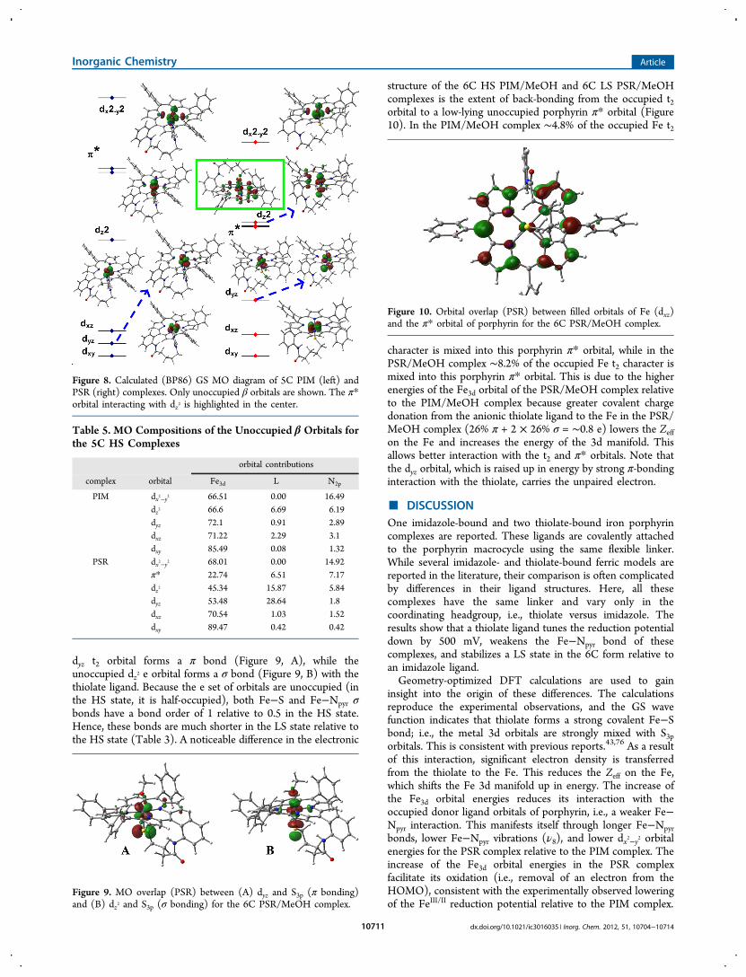
iii. Electronic Structure. A. 5C. The GS wave function of the5C PIM and PSR complexes show a square-pyramidal ligandfield (Figure 8), as reported for similar complexes.19,43,75 Thedx2−y2 orbital is calculated to be highest in energy for both ofthese models because of its strong interaction with theequatorial N atoms. The imidazole ligand forms a σ bondwith the dz2 orbital (Figure 8, left). The dz2 orbital also has itselectron density in the XY plane, which allows overlap of theporphyrin N atoms with it, as reflected by ∼7% porphyrin Ncharacter in the dz2 orbital (Table 5).
43 Note that there is ∼3%mixing between the imidazole N orbitals and the t2 orbitals onthe Fe, i.e., very weak π overlap. Alternatively, the thiolateligand has a π-bonding interaction with the dyz orbital and apseudo-σ-bonding interaction with the dz2 orbital.19,43 As aresult, the d orbitals with antibonding ligand contributions areat higher energy in PSR relative to these orbitals of the PIMcomplex (the energies of these orbitals are normalized with
Figure 6. CV of the PSR (red) and PIM (blue) complexes in a MeOHsolvent having 100 mM TBAP as the supporting electrolyte, glassycarbon as the working electrode, scan rate = 1 V/s, and Ag/AgCl-saturated KCl as the reference electrode. The individual redoxprocesses are described in the text.
Table 3. Optimized Bond Lengths (Å) of the Models andRelevant Mulliken72 Charges
model methodFe−N/S
Fe−Npyr
Fe−Xaxial qFe qN/S qpyr
S =5/2PIM
BP86 2.08 2.05 1.52 −0.58 −0.74
S =5/2PIM/MeOH
BP86 2.17 2.08 2.28 1.61 −0.52 −0.71
S =1/2PIM/MeOH
BP86 1.94 2.00 2.02 1.33 −0.47 −0.66
S =5/2PSR
B3LYP 2.33 2.08 1.53 −0.49 −0.76
BP86 2.31 2.09 1.41 −0.41 −0.71S =1/2PSR/MeOH
B3LYP 2.23 2.00 2.18 1.28 −0.33 −0.69
BP86 2.19 2.00 2.15 1.16 −0.25 −0.64
Inorganic Chemistry Article
dx.doi.org/10.1021/ic3016035 | Inorg. Chem. 2012, 51, 10704−1071410709

respect to the dxy orbital, which is nonbonding in bothcomplexes). The molecular orbital (MO) contributions (Table5) reveal that the dyz orbital in the PSR model has 28% S3pmixed into it, while the dz2 orbital has 16% S3p mixed into it; i.e.,there is significant covalent interaction between Fe and S (bothσ and π). On the contrary, the dz2 orbital of the PIM complexhas only 7% N contribution. Thus, the Fe−S bond is a lot morecovalent than the Fe−imidazole bond. Note that the dx2−y2orbital of the PIM complex is at a higher energy than that ofthe PSR complex. This reflects a weakening of the Fe−Npyrbonds in the PSR complex relative to the PIM complex. Thecalculated Mulliken charges (qFe) suggest a lower Zeff on the Fein the PSR complex due to the covalent donation by thethiolate relative to the Fe in the PIM complex (Table 3; qFe).The higher Zeff on the Fe in the PIM complex will result inlower-energy unoccupied Fe3d orbitals and allows stronger σ-bonding interaction with the occupied anionic porphyrin donorligand orbitals (in-plane pyrrole orbitals). The higher Zeff on theFe will also enhance its electrostatic interaction with thedianionic porphyrin ligand. Overall, these will lead to strongFe−Npyr bonds in the PIM complex relative to the PSRcomplex. This is evident from the shorter Fe−Npyr bondlengths and the stronger, i.e., Fe−Npyr, vibrations.
Furthermore, the strong pseudo-σ-bonding interactionbetween the dz2 orbital and the thiolate 3p orbital transferssignificant charge density from RS− to Fe, lowering its Zeff,which raises its energy and allows it to mix with a low-lying π*orbital of the porphyrin ring (Figure 8, highlighted with a greenbox). Note that this mixing is also enhanced by the fact that theFe is ∼0.45 Å above the porphyrin plane in the optimizedstructure of the PSR complex (Figure 7) relative to 0.3 Å in theoptimized structure of the PIM complex (Figure 7). Thismixing transfers both Fe dz2 and S3p (because it is involved in σbonding with the dz2 orbital) character into the porphyrin π*orbital; i.e., the Fe−S σ* orbital gains some porphyrin π*nature and vice versa. This electronic structure may explain theobservation of Fe−S and C−S vibrations upon excitation of theporphyrin π → π* transitions (Soret band).
B. 6C. The GS wave function of the HS 6C PIM/MeOHcomplex is very similar to that of the PIM complex. It has adistorted octahedral ligand field. Compared to the 5C PIMcomplex, the dz2 orbital in the 6C PIM/MeOH complex has aslightly elevated energy because of interaction with the MeOHligand.The GS wave function of the 6C LS PSR/MeOH complex
shows a normal t25e0 electronic structure. The singly occupied
Table 4. Calculated Vibrational Frequencies (cm−1)
PSR PSR/MeOH PIM PIM/MeOH
mode rR BP86 B3LYP rR BP86 B3LYP rR BP86 rR BP86
ν2 1554 1551 1604 1566 1563 1615 1551 1533 1550 1526ν3 1451 1447 1496 1458 1508 1461 1427 1446 1415ν4 1361 1351 1395 1366 1359 1403 1361 1353 1361 1348ν8 391 378 392 387 395 392 400 383 398 382Fe−S 336 308 305 340 319 319
369 369 319 361 375 359410 369 421 414 377
465 446 456C−S 624 629 640 673 637 653
679
Figure 7. DFT-optimized structures of complexes (A) PIM, (B) PIM/MeOH, (C) PSR, and (D) PSR/MeOH.
Inorganic Chemistry Article
dx.doi.org/10.1021/ic3016035 | Inorg. Chem. 2012, 51, 10704−1071410710
dyz t2 orbital forms a π bond (Figure 9, A), while theunoccupied dz2 e orbital forms a σ bond (Figure 9, B) with thethiolate ligand. Because the e set of orbitals are unoccupied (inthe HS state, it is half-occupied), both Fe−S and Fe−Npyr σbonds have a bond order of 1 relative to 0.5 in the HS state.Hence, these bonds are much shorter in the LS state relative tothe HS state (Table 3). A noticeable difference in the electronic
structure of the 6C HS PIM/MeOH and 6C LS PSR/MeOHcomplexes is the extent of back-bonding from the occupied t2orbital to a low-lying unoccupied porphyrin π* orbital (Figure10). In the PIM/MeOH complex ∼4.8% of the occupied Fe t2
character is mixed into this porphyrin π* orbital, while in thePSR/MeOH complex ∼8.2% of the occupied Fe t2 character ismixed into this porphyrin π* orbital. This is due to the higherenergies of the Fe3d orbital of the PSR/MeOH complex relativeto the PIM/MeOH complex because greater covalent chargedonation from the anionic thiolate ligand to the Fe in the PSR/MeOH complex (26% π + 2 × 26% σ = ∼0.8 e) lowers the Zeffon the Fe and increases the energy of the 3d manifold. Thisallows better interaction with the t2 and π* orbitals. Note thatthe dyz orbital, which is raised up in energy by strong π-bondinginteraction with the thiolate, carries the unpaired electron.
■ DISCUSSIONOne imidazole-bound and two thiolate-bound iron porphyrincomplexes are reported. These ligands are covalently attachedto the porphyrin macrocycle using the same flexible linker.While several imidazole- and thiolate-bound ferric models arereported in the literature, their comparison is often complicatedby differences in their ligand structures. Here, all thesecomplexes have the same linker and vary only in thecoordinating headgroup, i.e., thiolate versus imidazole. Theresults show that a thiolate ligand tunes the reduction potentialdown by 500 mV, weakens the Fe−Npyr bond of thesecomplexes, and stabilizes a LS state in the 6C form relative toan imidazole ligand.Geometry-optimized DFT calculations are used to gain
insight into the origin of these differences. The calculationsreproduce the experimental observations, and the GS wavefunction indicates that thiolate forms a strong covalent Fe−Sbond; i.e., the metal 3d orbitals are strongly mixed with S3porbitals. This is consistent with previous reports.43,76 As a resultof this interaction, significant electron density is transferredfrom the thiolate to the Fe. This reduces the Zeff on the Fe,which shifts the Fe 3d manifold up in energy. The increase ofthe Fe3d orbital energies reduces its interaction with theoccupied donor ligand orbitals of porphyrin, i.e., a weaker Fe−Npyr interaction. This manifests itself through longer Fe−Npyrbonds, lower Fe−Npyr vibrations (ν8), and lower dx2−y2 orbitalenergies for the PSR complex relative to the PIM complex. Theincrease of the Fe3d orbital energies in the PSR complexfacilitate its oxidation (i.e., removal of an electron from theHOMO), consistent with the experimentally observed loweringof the FeIII/II reduction potential relative to the PIM complex.
Figure 8. Calculated (BP86) GS MO diagram of 5C PIM (left) andPSR (right) complexes. Only unoccupied β orbitals are shown. The π*orbital interacting with dz2 is highlighted in the center.
Table 5. MO Compositions of the Unoccupied β Orbitals forthe 5C HS Complexes
orbital contributions
complex orbital Fe3d L N2p
PIM dx2−y2 66.51 0.00 16.49dz2 66.6 6.69 6.19dyz 72.1 0.91 2.89dxz 71.22 2.29 3.1dxy 85.49 0.08 1.32
PSR dx2−y2 68.01 0.00 14.92π* 22.74 6.51 7.17dz2 45.34 15.87 5.84dyz 53.48 28.64 1.8dxz 70.54 1.03 1.52dxy 89.47 0.42 0.42
Figure 9. MO overlap (PSR) between (A) dyz and S3p (π bonding)and (B) dz2 and S3p (σ bonding) for the 6C PSR/MeOH complex.
Figure 10. Orbital overlap (PSR) between filled orbitals of Fe (dxz)and the π* orbital of porphyrin for the 6C PSR/MeOH complex.
Inorganic Chemistry Article
dx.doi.org/10.1021/ic3016035 | Inorg. Chem. 2012, 51, 10704−1071410711

Another interesting consequence of thiolate coordination isstabilization of a LS GS in the PSR/MeOH complex in contrastto a HS GS observed in the imidazole-coordinated PIM/MeOH complex. The same spin states are observed in theheme active sites; histidine-bound 6C sites with a trans H2Oligand are HS, while cysteine-bound 6C sites with a trans H2Oligand are LS. Considering the fact that a thiolate is a weak-fieldligand (i.e., it acts as both σ and π donors), the LS state of a 6Caxial thiolate-bound iron(III) porphyrin is rather unexpected. Infact, the 6C axial thiolate-bound nonheme active site of SORand its synthetic model complexes have a HS GS.48 The Fe−Sbond of this active site is quite covalent, and its covalency iscomparable to the covalency of the Fe−S bond of cyt P450 inits resting HS state.31 The MO diagram of the 6C PSR/MeOHcomplex reveals that charge transfer from thiolate to FeIII raisesthe energy of the Fe3d manifold due to lowering of the Zeff ofthe Fe. This allows back-bonding interaction between theoccupied t2 orbitals on the Fe and the low-lying unoccupiedporphyrin π* orbitals. The extent of back-bonding (measuredby the amount of occupied t2 character mixed into theunoccupied ligand π* orbitals) is calculated to be 1.8 timesmore in a 6C LS PSR/MeOH model relative to a 6C LS PIM/MeOH complex. This back-bonding interaction stabilizes the t2orbitals involved (dxz and dyz) and thus stabilizes the LS GSstate in a thiolate-bound PSR/MeOH complex. The relativeposition of these porphyrin π* orbitals will definitely varydepending on the nature of the substitution on the peripheral Catoms. Thus, the extent of back-bonding, i.e., the extent ofstabilization of the LS GS, may vary depending on the nature of
the porphyrin ligand. Note that the back-bonding is notobserved in the HS complexes because (1) the t2 orbitals arehalf-occupied and (2) the covalent charge transfer is muchmore in the LS state because the Fe−S bond lengths are shorterand the e orbitals are completely unoccupied in the LS stateand not half-occupied as in the HS state.Thus, the thiolate ligand tunes the electronic structure of a
HS iron(III) poprphyrin via direct orbital interactions (Figure11), where it raises the energy of the dz2 and one of the t2orbitals (dyz in the nomenclature used here) because of pseudo-σ- and π-bonding interactions, respectively. The covalentcharge donation from the anionic thiolate ligand also raisesthe energy of the 3d manifold in general (Figure 11, middle)relative to that of a neutral imidazole ligand. This increase ofthe Fe3d manifold affects bonding interactions of the Fe3dorbitals that the thiolate does not have any direct overlapwith it. The increase in energy of the dxy orbital (the redox-active orbital in the HS complexes) lowers E0 of these HScomplexes (as observed experimentally), and the increase inenergy of the dx2−y2 orbital weakens its interaction with theoccupied lower-energy porphyrin pyrrole orbitals weakeningthe Fe−Npyr bond (longer Fe−N bonds in optimized structureand lower ν8 vibration) of the HS complexes. Further, theincrease in energy of the noninteracting t2 orbital along the Fe−S bond (dxz in the nomenclature used here) increases its back-bonding interaction with a low-lying porphyrin π* orbital(Figure 11, right) and lowers its energy. As a result ofdestabilization of the dz
2 orbital due to pseudo-σ-bondinginteraction with thiolate and the trans axial MeOH ligand and
Figure 11. MO interaction between filled orbitals of metal (t2) with empty orbitals of porphyrin (π*) and S3p orbital. The green lines represent thesingly occupied 3d orbitals, the red lines represent the unoccupied 3d and π* orbitals, and the black lines represent the occupied 3d orbitals.
Inorganic Chemistry Article
dx.doi.org/10.1021/ic3016035 | Inorg. Chem. 2012, 51, 10704−1071410712

stabilization of the dxz orbital due to back-bonding, the 6Cthiolate-bound PSR/MeOH complex stabilizes a LS GS; anelectronic structure contribution possibly is present in manythiolate-bound porphyrin active sites and synthetic complexes.
■ ASSOCIATED CONTENT*S Supporting InformationCharacterization of the complexes, EPR data of thiolcomplexes, rR data of thiol complexes, and optimizedgeometries. This material is available free of charge via theInternet at http://pubs.acs.org.
■ AUTHOR INFORMATIONCorresponding Author*E-mail: [email protected] authors declare no competing financial interest.
■ ACKNOWLEDGMENTSThis work is funded by Council of Scientific and IndustrialResearch (CSIR), India [Grant 01(2412)10/EMr-II], and theDepartment of Science and Technology, India (Grant SR/IC-35/2009). P.K.D. and S.C. acknowledge a CSIR JRF fellowship.S.S. acknowledges the IACS integrated Ph.D. program.
■ REFERENCES(1) Green, M. T. Curr. Opin. Chem. Biol. 2009, 13, 84−88.(2) Meunier, B.; de Visser, S. P.; Shaik, S. Chem. Rev. 2004, 104,3947−3980.(3) Averill, B. A. Chem. Rev. 1996, 96, 2951−2964.(4) Green, M. T.; Dawson, J. H.; Gray, H. B. Science 2004, 304,1653−1656.(5) Shaik, S.; Cohen, S.; Wang, Y.; Chen, H.; Kumar, D.; Thiel, W.Chem. Rev. 2012, 110, 949−1017.(6) Riplinger, C.; Neese, F. ChemPhysChem 2011, 12, 3192−3203.(7) Ogliaro, F.; Cohen, S.; Filatov, M.; Harris, N.; Shaik, S. Angew.Chem., Int. Ed. 2000, 39, 3851−3855.(8) Rittle, J.; Green, M. T. Science 2010, 330, 933−937.(9) Cramer, S. P.; Dawson, J. H.; Hodgson, K. O.; Hager, L. P. J. Am.Chem. Soc. 1978, 100, 7282−7290.(10) Dawson, J. H. Science 1988, 240, 433−439.(11) Ogliaro, F.; Cohen, S.; de Visser, S. P.; Shaik, S. J. Am. Chem.Soc. 2000, 122, 12892−12893.(12) Altun, A.; Kumar, D.; Neese, F.; Thiel, W. J. Phys. Chem. A2008, 112, 12904−12910.(13) Green, M. T. J. Am. Chem. Soc. 1998, 120, 10772−10773.(14) Shaik, S.; Kumar, D.; de Visser, S. P. J. Am. Chem. Soc. 2008,130, 10128−10140.(15) Sligar, S. G. Biochemistry 1976, 15, 5399−5406.(16) Newcomb, M.; Zhang, R.; Chandrasena, R. E. P.; Halgrimson, J.A.; Horner, J. H.; Makris, T. M.; Sligar, S. G. J. Am. Chem. Soc. 2006,128, 4580−4581.(17) Ogliaro, F.; de Visser, S. P.; Shaik, S. J. Inorg. Biochem. 2002, 91,554−567.(18) Shaik, S.; Kumar, D.; de Visser, S. P.; Altun, A.; Thiel, W. Chem.Rev. 2005, 105, 2279−2328.(19) Green, M. T. J. Am. Chem. Soc. 1999, 121, 7939−7940.(20) Dawson, J. H.; Sono, M. Chem. Rev. 1987, 87, 1255−1276.(21) Sono, M.; Andersson, L. A.; Dawson, J. H. J. Biol. Chem. 1982,257, 8308−8320.(22) Sligar, S. G. Science 2010, 330, 924−925.(23) Schlichting, I.; Berendzen, J.; Chu, K.; Stock, A. M.; Maves, S.A.; Benson, D. E.; Sweet, R. M.; Ringe, D.; Petsko, G. A.; Sligar, S. G.Science 2000, 287, 1615−1622.(24) Schoeneboom, J. C.; Neese, F.; Thiel, W. J. Am. Chem. Soc.2005, 127, 5840−5853.
(25) Rittle, J.; Younker, J. M.; Green, M. T. Inorg. Chem. 2010, 49,3610−3617.(26) Kumar, D.; Sastry, G. N.; de Visser, S. P. Chem.Eur. J. 2011,17, 6196−6205.(27) de Visser, S. P.; Latifi, R.; Tahsini, L.; Nam, W.-W. Chem.Asian J. 2011, 6, 493−504.(28) Kumar, D.; Karamzadeh, B.; Sastry, G. N.; de Visser, S. P. J. Am.Chem. Soc. 2010, 132, 7656−7667.(29) de Visser, S. P.; Tahsini, L.; Nam, W. Chem.Eur. J. 2009, 15,5577−5587.(30) de Visser, S. P. Biochem. Soc. Trans. 2009, 37, 373−377.(31) Dey, A.; Jiang, Y.; Ortiz de Montellano, P.; Hodgson, K. O.;Hedman, B.; Solomon, E. I. J. Am. Chem. Soc. 2009, 131, 7869−7878.(32) Kumar, D.; Sastry, G. N.; de Visser, S. P. J. Phys. Chem. B 2012,116, 718−730.(33) Deng, T. j.; Macdonald, I. D. G.; Simianu, M. C.; Sykora, M.;Kincaid, J. R.; Sligar, S. G. J. Am. Chem. Soc. 2001, 123, 269−278.(34) Usharani, D.; Zazza, C.; Lai, W.; Chourasia, M.; Waskell, L.;Shaik, S. J. Am. Chem. Soc. 2012, 134, 4053−4056.(35) Chang, C. K.; Dolphin, D. J. Am. Chem. Soc. 1975, 97, 5948−5950.(36) Chang, C. K.; Dolphin, D. J. Am. Chem. Soc. 1976, 98, 1607−1609.(37) Collman, J. P.; Gagne, R. R.; Reed, C.; Halbert, T. R.; Lang, G.;Robinson, W. T. J. Am. Chem. Soc. 1975, 97, 1427−1439.(38) Collman, J. P.; Sorrell, T. N. J. Am. Chem. Soc. 1975, 97, 4133−4134.(39) Traylor, T. G.; Mincey, T. C.; Berzinis, A. P. J. Am. Chem. Soc.1981, 103, 7084−7089.(40) Traylor, T. G.; Nolan, K. B.; Hildreth, R. J. Am. Chem. Soc. 1983,105, 6149−6151.(41) Tani, F.; Matsu-ura, M.; Nakayama, S.; Naruta, Y. Coord. Chem.Rev. 2002, 226, 219−226.(42) Suzuki, N.; Higuchi, T.; Urano, Y.; Kikuchi, K.; Uekusa, H.;Ohashi, Y.; Uchida, T.; Kitagawa, T.; Nagano, T. J. Am. Chem. Soc.1999, 121, 11571−11572.(43) Dey, A.; Okamura, T.; Ueyama, N.; Hedman, B.; Hodgson, K.O.; Solomon, E. I. J. Am. Chem. Soc. 2005, 127, 12046−12053.(44) Ueyama, N.; Nishikawa, N.; Yamada, Y.; Okamura, T.;Nakamura, A. J. Am. Chem. Soc. 1996, 118, 12826−12827.(45) Higuchi, T.; Uzu, S.; Hirobe, M. J. Am. Chem. Soc. 1990, 112,7051−7053.(46) Ueyama, N.; Nishikawa, N.; Yamada, Y.; Okamura, T.-a.; Oka,S.; Sakurai, H.; Nakamura, A. Inorg. Chem. 1998, 37, 2415−2421.(47) Collman, J. P.; Boulatov, R.; Sunderland, C. J.; Fu, L. Chem. Rev.2003, 104, 561−588.(48) Dey, A.; Jenney, F. E.; Adams, M. W. W.; Johnson, M. K.;Hodgson, K. O.; Hedman, B.; Solomon, E. I. J. Am. Chem. Soc. 2007,129, 12418−12431.(49) Clay, M. D.; Jenney, F. E.; Hagedoorn, P. L.; George, G. N.;Adams, M. W. W.; Johnson, M. K. J. Am. Chem. Soc. 2001, 124, 788−805.(50) Collman, J. P.; Brauman, J. I.; Doxsee, K. M.; Halbert, T. R.;Bunnenberg, E.; Linder, R. E.; LaMar, G. N.; Del Gaudio, J.; Lang, G.;Spartalian, K. J. Am. Chem. Soc. 1980, 102, 4182−4192.(51) Mashiko, T.; Reed, C. A.; Haller, K. J.; Kastner, M. E.; Scheidt,W. R. J. Am. Chem. Soc. 1981, 103, 5758−5767.(52) Berto, T. C.; Praneeth, V. K. K.; Goodrich, L. E.; Lehnert, N. J.Am. Chem. Soc. 2009, 131, 17116−17126.(53) Frisch, M. J. T.; et al. Gaussian 03, version C.02; Gaussian, Inc.:Wallingford, CT, 2004.(54) Becke, A. D. Phys. Rev. A 1988, 38, 3098−3100.(55) Becke, A. D. J. Chem. Phys. 1993, 98, 5648−5652.(56) Perdew, J. P.; Burke, K.; Ernzerhof, M. Phys. Rev. Lett. 1996, 77,3865−3868.(57) Perdew, J. P. Phys. Rev. B (Condens. Matter) 1986, 33, 8822−8824.(58) Note that previously a PIM complex with a ClO4
− counterionwas proposed to exist as a mixture of the monomeric complex and a
Inorganic Chemistry Article
dx.doi.org/10.1021/ic3016035 | Inorg. Chem. 2012, 51, 10704−1071410713

bis(imidazole) dimer (where an imidazole arm of one monomer bindsan Fe of another) in equilibrium. This was supported by theobservation of both S = 5/2 and
1/2 signals in the EPR data of the PIMcomplex. Here, when a bromide counterion is present, no S = 1/2signal is observed.(59) Walker, F. A. Coord. Chem. Rev. 1999, 185−186, 471−534.(60) Taylor, C. P. S. Biochim. Biophys. Acta, Protein Struct. 1977, 491,137−148.(61) Ann, W. F. Coord. Chem. Rev. 1999, 185−186, 471−534.(62) Burke, J. M.; Kincaid, J. R.; Peters, S.; Gagne, R. R.; Collman, J.P.; Spiro, T. G. J. Am. Chem. Soc. 1978, 100, 6083−6088.(63) Kitagawa, T.; Dey, A.; Lugo-Mas, P.; Benedict, J. B.; Kaminsky,W.; Solomon, E.; Kovacs, J. A. J. Am. Chem. Soc. 2006, 128, 14448−14449.(64) Note that, as indicated by the EPR data, no LS component isobserved in the rR data of PIM in THF/DCM/MeOH. This furtherdiminishes the possibility of the previously reported equilibrium insolution. This could reflect the role of the bromide counterion that isbound to the PIM complex.(65) Deng, T.; Proniewicz, L. M.; Kincaid, J. R.; Yeom, H.;Macdonald, I. D. G.; Sligar, S. G. Biochemistry 1999, 38, 13699−13706.(66) Champion, P. M.; Stallard, B. R.; Wagner, G. C.; Gunsalus, I. C.J. Am. Chem. Soc. 1982, 104, 5469−5472.(67) Wells, A. V.; Li, P.; Champion, P. M.; Martinis, S. A.; Sligar, S.G. Biochemistry 1992, 31, 4384−4393.(68) Chen, Z.; Ost, T. W. B.; Schelvis, J. P. M. Biochemistry 2004, 43,1798−1808.(69) Paulat, F.; Praneeth, V. K. K.; Nather, C.; Lehnert, N. Inorg.Chem. 2006, 45, 2835−2856.(70) Fisher, M. T.; Sligar, S. G. J. Am. Chem. Soc. 1985, 107, 5018−5019.(71) Lin, C.; Fang, M. Y.; Cheng, S. H. J. Electroanal. Chem. 2002,531, 155−162.(72) Mulliken, R. S. J. Chem. Phys. 1955, 23, 1833−1840.(73) Baerends, E. J.; Ellis, D. E.; Ros, P. Chem. Phys. 1973, 2, 41−51.(74) Denisov, I. G.; Makris, T. M.; Sligar, S. G.; Schlichting, I. Chem.Rev. 2005, 105, 2253−2278.(75) Note that while both occupied α and unoccupied β orbitalscarry the same information; the contours of the unoccupied β orbitalsare cleaner. The occupied α orbitals mix with several other occupiedligand-based orbitals that have similar energies(76) Paulat, F.; Lehnert, N. Inorg. Chem. 2007, 46, 1547−1549.
Inorganic Chemistry Article
dx.doi.org/10.1021/ic3016035 | Inorg. Chem. 2012, 51, 10704−1071410714
Copyright © 2022 FDOKUMEN