Novel metallo-porphyrin based colourimetric amine sensors ...
Upload
independentCategory
view
5download
0
Materials Science and Engineering C 29 (2009) 1010–1017
Contents lists available at ScienceDirect
Materials Science and Engineering C
j ourna l homepage: www.e lsev ie r.com/ locate /msec
Nanostructured morphologies of complexes containing porphyrin bridges betweenPt(II) acetylide tethers
M.E. Amato a,1, A. Licciardello a,1, V. Torrisi a,1, L. Ugo b,2, I. Venditti b,⁎, M.V. Russo b,2
a Department of Chemical Sciences, University of Catania, V.le A. Doria 6, 95125 Catania, Italyb Department of Chemistry, University of Rome “La Sapienza”, P.le A. Moro 5, 00185, Rome, Italy
⁎ Corresponding author. Tel.: +39 0649913347; fax:E-mail address: [email protected] (I. Venditt
1 Tel.: +39 0957385113; fax: +39 095580138.2 Tel.: +39 0649913347; fax: +39 06490324.
0928-4931/$ – see front matter © 2008 Elsevier B.V. Adoi:10.1016/j.msec.2008.08.033
a b s t r a c t
a r t i c l e i n f oArticle history:
A self assembling method Received 9 May 2008Received in revised form 19 August 2008Accepted 25 August 2008Available online 9 September 2008Keywords:PorphyrinNanostructures
based on osmotic procedure allowed fabricating nanostructured aggregates ofdinuclear complexes made of Zn-porphyrin bridges between Pt(II) acetylide tethers. The choice ofexperimental parameters, such as solvent/non solvent couple and temperature allows achievingnanostructures that look like rod-like crystals and spheres, the latter ones showing either solid or concaveinternal structures. The porphyrin bridged di-bis(triphenylphosphine)platinum phenylacetylide, porphyrinbridged di-bis(triphenylphosphine)platinum p-nitrophenylacetylide and porphyrin bridged di-bis(triphe-nylphosphine)platinum p-methoxyphenylacetylide, were synthesized by the coupling of 2,8,12,18-tetraethyl-5,15-diethynyl-3,7,13,17-tetramethylporphyrinatozinc(II) with the corresponding trans-[Pt(PPh3)2(C≡C–R)Cl]complexes (R = C6H5, C6H4NO2, C6H4OCH3). The molecular structures were assessed with NMR and ToF-SIMSstudies and the nanostructure formation was revealed with SEM investigations. The results could be ofinterest for future applications in optoelectronic devices, sensors, biotechnology and light harvesting.
© 2008 Elsevier B.V. All rights reserved.
1. Introduction
Apart from the biomimetic allure of porphyrins which areprepared and studied as models for processes that resemble naturalphenomena, advanced perspectives in the use of porphyrins in energytransfer for photochemical energy conversion and efficient lightharvesting [1–4] optoelectronic devices [5,6], chemical sensors [7] andmedical purposes [8] are envisaged. In these years, porphyrincomplexes have been the subject of intensive study due to the strikingsize-dependence, either molecular or nano-microdimensional, oftheir physical and chemical properties [9–11].
Interesting papers reporting the synthesis and properties of multi-porphyrin arrays (triads, tetrad, pentad) [12] and porphyrin dyads [13],where the porphyrins are linked through phenylethyne linkers withoutbound transition metals, appeared recently. These macromoleculesengender interest for their electron delocalization [14] and chargetransport properties [15], depending on the chemical structure of thealkynyl ligands and, to a minor extent, of the phosphine ligands.
As a further topic, a variety of nanometer or micrometer scalestructures have been generated recently, including three-dimensionalsupramolecular self-organization of porphyrins in hollow hexagonalnanoprisms, nanorods several nanometers wide and composite por-phyrin nanotubes [16–18]. These organizations from molecular self-
+39 06490324.i).
ll rights reserved.
assemblies to controllable architectures and materials with advancedfunctionsmaymeet the requirement ofmany objectives in science andtechnology such as sensor, electronic, and electromechanical devicesbecause of the property of a nanostructured material or self-assemblywill strongly depend on its morphology and dimension [16,19–21]. Forexample, these materials must be processed and in such settingsnanostructured porphyrin complexes are coated on specific substratesfor sensoristic applications [12,22,23]. Further applications can beenvisaged in the field of biotechnology and optoelectronics, thatrepresent the most promising challenges for the DNA binding, andcarbon nanotube/porphyrin hybrids preparation [24,25].
In the framework of the wide research activity on porphyrin self-assembling properties [7], our group has been recently involved in thesynthesis and characterization of ethynyl porphyrins bridging bis-phosphine Pt(II) complexes, which are models for polymericporphyrin-based materials [8].
In this paper, we describe the synthesis and characterization ofcomplexes where the diethynyl–porphyrin bridge joints two platinumcentres bound to external phenylethynyl tethers bearing electrondonor (D=OCH3), electronwithdrawing (A=NO2) and “neutral” (H)substituents in para position; the presence of ligands with A or Dgroups are expected to play a role in tuning the charge transfer reactionwhen these complexes are doped with fullerenes, in analogy withprevious results [26], so that a tuning of the band gap can be achievedas well. In this context, the attainment of assemblies with special andcontrollable structure is an interesting challenge in porphyrin studyand we have exploited the self assembly of these porphyrin systemsinto nanostructures by applying the osmotic method with a new

1011M.E. Amato et al. / Materials Science and Engineering C 29 (2009) 1010–1017
approach. The future perspective deals with the development of novelnanodevices.
2. Experimental details
2.1. Materials
All solvents were reagent grade and used without further pu-rification; argon flux was flown through the reaction solvents fordeoxygenating the reactions that were performed under argonatmosphere using standard procedures. 2,8,12,18-tetraethyl-5,15-diethynyl-3,7,13,17-tetramethylporphyrinatozinc(II) (1) (ZnDEP) wassynthesized according to literature procedures [27] and precursormonochloro acetylyde Pt(II) complexes, i.e. trans-[Pt (PPh3)2(C≡C–C6H5)Cl] (2) [28], trans-[Pt (PPh3)2(C≡C–C6H4NO2)Cl] (3), and trans-[Pt (PPh3)2(C≡C–C6H4OCH3)Cl] (4), were obtained with alreadypublished methods [29].
2.2. Instrumentation
The UV–Vis spectra were recorded using a Perkin Elmer Lambda 16spectrophotometer; weighed amounts of the samples were dissolved inCHCl3 at room temperature before themeasurements. FTIR spectrawererunwith a Perkin Elmer 1700 instrument on nujol mulls of the samples,in the range4000–400 cm−1.NMRspectrawere recordedat300Kwith a500 MHz Varian Unity Inova spectrometer (1H at 499.88 MHz, 31P at202.3 MHz) equipped with pulse field gradient module (Z axis) and atunable 5mm inverse detectionprobe. Theproton chemical shifts (ppm)were referenced to the residual proton resonance in CDCl3 (δ 7.26 ppm)or to TMS (δ 0.0 ppm ) as internal standard. 31P NMR signals wereassigned using H3PO4 (85%) as external reference. gCOSY, NOESY andT-ROESY experiments were recorded using standard sequences. Forthe T-ROESY experiments samples were dissolved in CDCl3 and thesolution was degassed and sealed. The mixing time was set at 200 msand the spin lock field strength was 2 kHz. ToF-SIMS (Time of FlightSecondary Ions Mass Spectrometry) spectra were acquired in a staticmode with a reflector-type spectrometer (ION-TOF ToF-SIMS IV), byusingapulsed69Ga+primary ionbeam(25KeV, ~0.1pA) rasteredover a300×300µm2 area. Primary ionfluencywas kept b3×1011 ions cm−2 inorder to assure static SIMS conditions [30]. Samples for ToF-SIMS wereprepared onto UV–O3 cleaned gold substrates by microsyringe deposi-tion [28] from 10−3 M chloroform solution. Field emission scanningelectron microscopy (instrument SEM LEO1450VP) was used to obtainthe images of metalled nanostructured materials: samples for SEMmeasurements were pre-coated with a thin layer of Au; the samesampleswere analyzed by energy dispersion electronmicroprobe (EDS)for microanalysis (INCA300 system).
2.3. Synthesis
2.3.1. Porphyrin bridged di-bis(triphenylphosphine)platinumphenylacetylide (5)
In a 250 mL three necks flask ZnDEP (1) (53 mg, 0.09 mmol), Ptcomplex (2) (159 mg, 0.18 mmol) and CuI (3 mg) were dissolved in amixture of solvents (NHEt2 40 mL and CH2Cl2 15 mL (previouslydegassed with argon flux for 30 min) and the solution was stirred atT=50 °C for 3 h, maintaining the reaction under argon atmosphere.The reaction course was monitored by UV–Vis spectroscopy until asingle absorption band at 459 nm was detected. The reaction wasinterrupted diving the flask into an ice-salt bath and the mixture wasdried in vacuum to obtain a solid crude product. The crude mixturewas washed thoroughly with water to eliminate the ammonium saltside product and purified by flash chromatography [florisil, toluene].Crystallization of the eluted fraction with THF/ethanol gave the bluecomplex (5) (50 mg, 0.02 mmol: 22.2%). 1H NMR (CDCl3) δ(ppm): 1.70(t,12 H, CH2CH3), 2.96 (s,12 H, CH3), 3.82 (q, 8 H, CH2CH3), 6.24 (m, 4H,
C≡C–C6H5), 6.91 (m, 6H, C≡C–C6H5), 7.06–7.8 (m, 60 H, P–C6H5), 9.66(s, meso 2H). 31P NMR (CDCl3) δ(ppm): 17.33, J(195Pt–31P) 2670 Hz.UV–Vis (CHCl3) (λmax nm) 459, 598, 627. FTIR (nujol) cm−1: 2099(ν C≡C),1597 (νC=C, arom.),1210 (νC–Hpyrrole), 846 (νC–Npyrrole).Anal Calcd (%) for C124H104P4N4Pt2Zn: C, 66.79; H, 4.70; N, 2.51; foundC, 65.80, H, 4.57, N, 2.73. ToF-SIMS: m/q=2226.6 (molecular peak).
2.3.2. Porphyrin bridged di-bis(triphenylphosphine)platinump-nitrophenylacetylide (6)
The reaction betweenZnDEP (1) (52mg, 0.09mmol) andPt complex(3) (165 mg, 0.18 mmol) was carried out for 2 h in the same reactionconditions assessed for the synthesis of porphyrin bridged complex (5)withNHEt2/CH2Cl2 volume ratio 30/20mL, and thepurification of crudecomplex (6) was performed in the same way, by further crystallizationwith THF/methanol. Pure blue complex (6) was obtained (70 mg,0.03mmol: 33.3%).1HNMR(CDCl3) δ(ppm): 1.69 (t,12H, CH2CH3), 2.94(s,12 H, CH3), 3.82 (q, 8 H, CH2CH3), 6.24 (dd, 4 H, C≡C–C6H4NO2), 7.08–7.82 (m, 60H, P–C6H5), 7.79 (dd, 4 H, C≡C–C6H4NO2), 9.68 (s, meso 2H).31P NMR (CDCl3) δ(ppm): 17.24, J(195Pt–31P) 2660 Hz. UV–Vis (CHCl3)(λmax nm) 459, 590, 627. FTIR (nujol) cm−1: 2110 (ν C≡C),1596 (ν C=C,arom.),1508 (νas NO2),1335 (νs NO2) 1250 (ν C–H pyrrole), 856 (ν C–Npyrrole). Anal Calcd (%) for C124H102P4N6O4Pt2Zn: C, 64.21; H, 4.43; N,3.62; found C, 63.19, H, 4.31, N, 3.51. ToF-SIMS:m/q=2316.6 (molecularpeak).
2.3.3. Porphyrin bridged di-bis(triphenylphosphine)platinump-methoxyphenylacetylide (7)
ZnDEP (1) (52 mg, 0.09 mmol) and Pt complex (4) (148 mg,0.18 mmol) were mixed in a reaction vessel using the same reactionconditions assessed for the synthesis of porphyrin bridged complex (5)and (6), except for the NHEt2/CH2Cl2 volume ratio 50/15mL and longerreaction time (4 hours); the purification of crude complex (7) wasperformed in the same way (three flash chromatographies werenecessary in this case), followed by further crystallization with CHCl3/petroleum ether. Pure blue–green complex (7) was obtained (40 mg,0.02 mmol: 22.2%). 1H NMR (CDCl3) δ (ppm): 1.70 (t, 12 H, CH2CH3),2.95 (s,12H, CH3), 3.69 (s, 6H, OCH3), 3.82 (q, 8H, CH2CH3), 6.17 (dd, 4H,C≡C–C6H4OCH3), 6.47 (dd, 4H, C≡C–C6H4OCH3), 7.84–7.08 (m, 60 H,P–C6H5), 9.66 (s,meso2H). 31PNMR(CDCl3) δ(ppm):17.26, J(195Pt–31P)2660 Hz. UV–Vis (CHCl3) (λmax nm) 458, 595, 627. FTIR (nujol) cm−1:2110 (ν C≡C), 1596 (ν C=C, arom.), 1508 (νas NO2),1335 (νs NO2) 1250(ν C–H pyrrole), 856 (ν C–N pyrrole). Anal Calcd (%) for C126H108P4N4-
O2Pt2Zn: C, 66.09; H, 4.75; N, 2.45; Anal Calcd (%) for (C126H108P4N4O2-
Pt2Zn)·CHCl3 (crystallization solvent) C, 61.83, H, 4.46, N, 2.25; found C,62.24, H, 4.52, N, 2.56. ToF-SIMS: m/q=2286.6 (molecular peak).
2.4. Preparation of nanostructures
An osmotic method [31] is used for controlling the morphologyand the dimensions of porphyrinic materials. The used solvents(dimethylformamide, acetone, tetrahydrofuran) are combined withwater and the used membrane is Aldrich D0530-100FT. In a typicalprocedure the suitable concentrations of porphyrin complex (3 mg/ml) was dissolved in appropriate solvent and 10 ml of this solutionwere placed into double membrane; then the double membrane wasdipped in water, using a solvent/water ratio = 1/20 in volume (seeTable 1). The morphologies and the dimensions of nanostructuredmaterials are investigated by SEM and the images of films depositedby casting were elaborate using an image analysis software tool (ScionImage for Windows, Scion Corp, Beta 4.0.2). Moreover EDS measure-ments on several samples were performed by INCA300 system.
3. Results and discussion
The synthetic route to yield the porphyrin bridged Pt complexes(5), (6), (7) is based on the widely applicable dehydrohalogenation
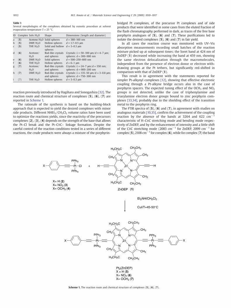
Table 1Several morphologies of the complexes obtained by osmotic procedure at solventevaporation temperature T=25 °C.
ID Complex Solv/H2O Shape Dimensions (length and diameter)
a (5) Acetone/H2O Solid spheres d=380–160 nmb (5) DMF/H2O Hollow spheres d=1.7–0.5 μmc (5) THF/H2O Solid and hollow
spheresd=3–0.3 μm
d (6) Acetone/H2O
Rod-like crystalsand spheres
Crystals: L=50–100 μm/d=4–7 μm;spheres: d=300–900 nm
e (6) DMF/H2O Solid spheres d=500–250–600 nmf (6) THF/H2O Hollow spheres d=6–1 μmg (7) Acetone/
H2ORod-like crystalsand spheres
Crystals: L=24–7 μm/d=350 nm;spheres: d=900–200 nm
h (7) DMF/H2O Rod-like crystalsand spheres
Crystals: L=135–50 μm/d=3–0.8 μm;spheres: d=750–300 nm
i (7) THF/H2O Hollow spheres d=3–0.3 μm
1012 M.E. Amato et al. / Materials Science and Engineering C 29 (2009) 1010–1017
reaction previously introduced by Hagihara and Sonogashira [32]. Thereaction route and chemical structure of complexes (5), (6), (7) arereported in Scheme 1.
The rationale of the synthesis is based on the building-blockapproach that is expected to yield the desired complexes with minorside products. Different NHEt2/CH2Cl2 volume ratios have been usedto optimize the reactions yields, since the reactivity of the precursorscomplexes (2), (3), (4) depends on the strength of the base that allowsthe Pt–Cl break and the Pt–C≡C– linkage formation. Despite thecareful control of the reaction conditions tested in a series of differentreactions, the crude products were always a mixture of the porphyrin-
Scheme 1. The reaction route and chemica
bridged Pt complexes, of the precursor Pt complexes and of sideproducts that were identified in some cases from the eluted fraction ofthe flash chromatography performed in dark, as traces of the free baseporphyrin analogues of (5), (6) and (7). These purifications led toisolate the desired complexes (5), (6) and (7) in fair yield.
In all cases the reaction course was monitored with UV–Visabsorption measurements recording small batches of the reactionmixture picked up at subsequent times; the Soret band at 424 nm ofZnDEP (1) decreased while increasing the band at 459 nm, showingthe same electron delocalization through the macromolecules,independent from the presence of electron donor or electron with-drawing groups at the Pt tethers, but significantly red-shifted incomparison with that of ZnDEP (1).
This result is in agreement with the statements reported forsimpler Pt-alkynyl complexes [12], showing that effective electroniccoupling through a Pt-alkyne bridge occurs also in the case ofporphyrin spacers. The expected tuning effect of the OCH3 and NO2
groups is not detected, unlike the case of triphenylamine andtriarylamine electron donor groups bound to zinc porphyrin com-plexes [33,34], probably due to the shielding effect of the transitionmetal to the porphyrin ring.
The FTIR spectra of (5), (6) and (7), in agreement with studies onanalogous materials [10,35], confirm the achievement of the couplingreaction by the absence of the bands at 3264 and 622 cm−1
characteristic of H–C≡C stretching mode and bending mode respec-tively of ZnDEP, and by the enhancement of intensity and a little shiftof the C≡C stretching mode (2083 cm−1 for ZnDEP, 2099 cm−1 forcomplex (5), 2108cm−1 for complex (6),while for complex (7) theband
l structure of complexes (5), (6), (7).
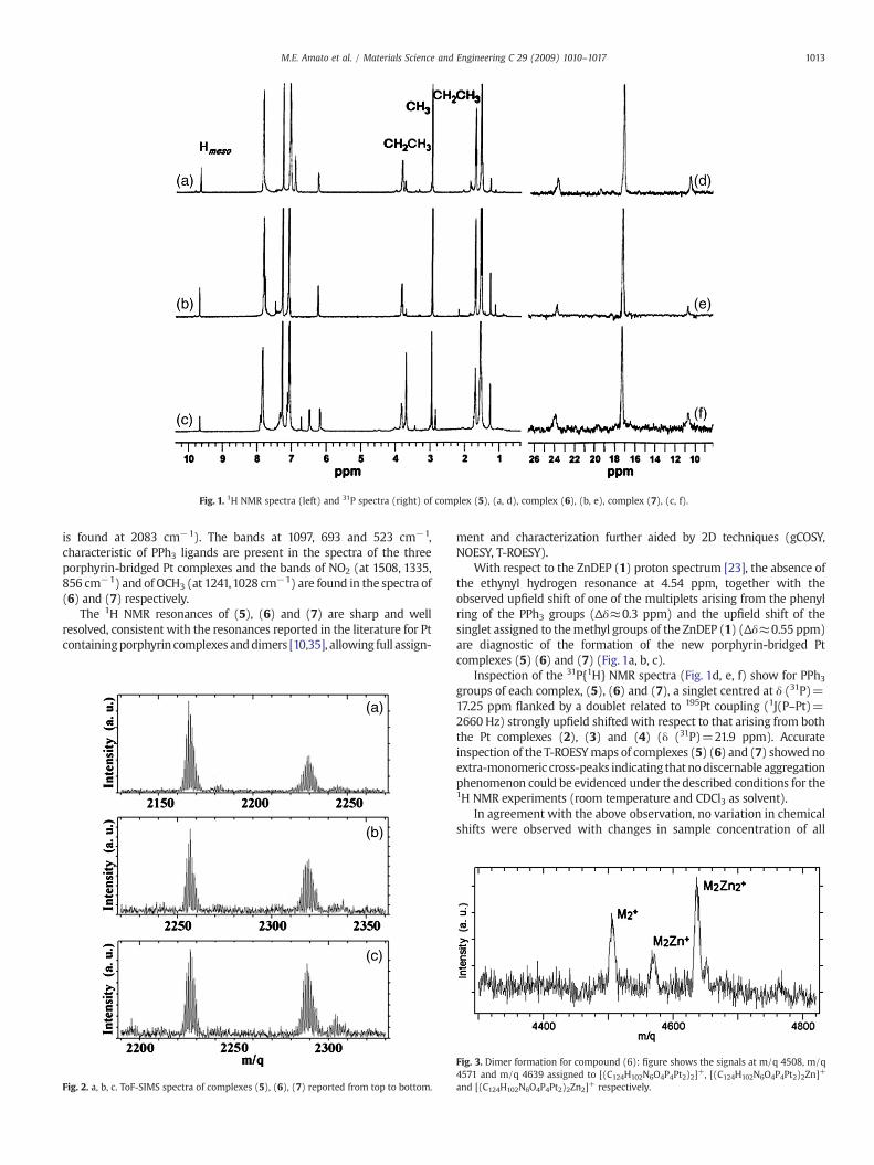
Fig. 1. 1H NMR spectra (left) and 31P spectra (right) of complex (5), (a, d), complex (6), (b, e), complex (7), (c, f).
1013M.E. Amato et al. / Materials Science and Engineering C 29 (2009) 1010–1017
is found at 2083 cm−1). The bands at 1097, 693 and 523 cm−1,characteristic of PPh3 ligands are present in the spectra of the threeporphyrin-bridged Pt complexes and the bands of NO2 (at 1508, 1335,856 cm−1) and of OCH3 (at 1241,1028 cm−1) are found in the spectra of(6) and (7) respectively.
The 1H NMR resonances of (5), (6) and (7) are sharp and wellresolved, consistent with the resonances reported in the literature for Ptcontainingporphyrin complexes anddimers [10,35], allowing full assign-
Fig. 2. a, b, c. ToF-SIMS spectra of complexes (5), (6), (7) reported from top to bottom.
ment and characterization further aided by 2D techniques (gCOSY,NOESY, T-ROESY).
With respect to the ZnDEP (1) proton spectrum [23], the absence ofthe ethynyl hydrogen resonance at 4.54 ppm, together with theobserved upfield shift of one of the multiplets arising from the phenylring of the PPh3 groups (Δδ≈0.3 ppm) and the upfield shift of thesinglet assigned to themethyl groups of the ZnDEP (1) (Δδ≈0.55 ppm)are diagnostic of the formation of the new porphyrin-bridged Ptcomplexes (5) (6) and (7) (Fig. 1a, b, c).
Inspection of the 31P{1H} NMR spectra (Fig. 1d, e, f) show for PPh3groups of each complex, (5), (6) and (7), a singlet centred at δ (31P)=17.25 ppm flanked by a doublet related to 195Pt coupling (1J(P–Pt)=2660 Hz) strongly upfield shifted with respect to that arising from boththe Pt complexes (2), (3) and (4) (δ (31P)=21.9 ppm). Accurateinspection of the T-ROESYmaps of complexes (5) (6) and (7) showed noextra-monomeric cross-peaks indicating that nodiscernable aggregationphenomenon could be evidenced under the described conditions for the1H NMR experiments (room temperature and CDCl3 as solvent).
In agreement with the above observation, no variation in chemicalshifts were observed with changes in sample concentration of all
Fig. 3. Dimer formation for compound (6): figure shows the signals at m/q 4508, m/q4571 and m/q 4639 assigned to [(C124H102N6O4P4Pt2)2]+, [(C124H102N6O4P4Pt2)2Zn]+
and [(C124H102N6O4P4Pt2)2Zn2]+ respectively.

Fig. 4. SEM images of spherical morphologies obtained from each complex by osmoticprocedure: a) complex (5) in THF/H2O: hollow nanospheres (d=0.5–3 μm). b)complex (6) in DMF/H2O: solid nanospheres (d=250–600 nm). c) complex (7) in THF/H2O: hollow nanospheres (d=0.3–3 μm).
1014 M.E. Amato et al. / Materials Science and Engineering C 29 (2009) 1010–1017
porphyrin complexes, which suggested the presence of only mono-meric forms in solution.
The chemical structure of the porphyrin complexes was furthercharacterized by ToF-SIMS investigations.
In Fig. 2a,b,c, we show the spectral region of (quasi) molecularpeaks of compounds (5), (6), (7) respectively.
In Fig. 2a the peaks at m/q 2164,6 and m/q 2226,6 pertaining tocompound (5) are assigned to [C124H106P4N4Pt2]+, and [C124H104P4N4-
Pt2Zn]+ respectively. The Fig. 2b, pertaining to compound (6), showsthe signals at m/q 2254,7 and, m/q 2316,6 assigned to [C124H104N6-
O4P4Pt2]+ and [C124H102N6O4P4Pt2Zn]+. Finally for compound (7) weobserved two peaks at m/q 2224,7 and m/q 2286,6, that are assignedto [C126H110N4O2P4Pt2]+, and [C126H108N4O2P4Pt2Zn]+ (Fig. 2c).
These results show that the core metal Zn is easily removable fromthe tetrapyrrole ring, allowing the replacement with other metals forthe achievement of different ethero-bimetallic complexes.
All the assignments reported have been made on the basis of theexact mass as well as of the isotopic distribution. Although it could notbe evidenced any aggregation phenomenon in solution for theseporphyrin complexes, a trace of dimeric aggregates could be found bycumulative recording of ToF-SIMS (Fig. 3) in the case of complex (6)revealing the presence of three species: dimeric aggregate without
Fig. 5. SEM images of particular nanostructures of complex (7) obtained in THF/H2O:a) rod-like crystals (L=7–24 μm, d=350 nm) and nanospheres (d=200–800 nm);b) detail of nanospheres.
1015M.E. Amato et al. / Materials Science and Engineering C 29 (2009) 1010–1017
core metal Zn ([(C124H102N6O4P4Pt2)2]+, m/q 4508), dimeric aggregatewith one coremetal Zn ([(C124H102N6O4P4Pt2)2Zn]+,m/q4571), dimericaggregate with two Zn atoms ([(C124H102N6O4P4Pt2)2Zn2]+ m/q 4639).
Following the synthesis and characterization our porphyrincomplexes (5), (6), (7), their self-assembly behaviour was investigated.
In particular, we studied the way to easily obtain micro/nano-structured particles from our porphyrin complexes using an osmoticmethod that allows generating different morphologies at mesoscale[29].
Our previous studies showed that the nanostructures can becontrolled, in shape and dimensions, by changing experimentalconditions, such as different membranes, temperature, concentra-tions, solvents mixture and their volume ratio, according to thepublished patent [31]. The advantages of themethod aremanifold: theosmotic procedure is easy, uses bland conditions, gives rise to pureproducts (no need of surfactants) and allows the recovering of thesolvents by distillation. Throughout this osmotic procedure, thermo-dynamic and kinetic features are in competition: when the processbecomes slow, the thermodynamically stable morphology is favoured
Fig. 6. EDS analysis of nanostructured complex (7): (a) hollo
during the precipitation of the material and the increase of theinterface tension pushes the molecules to aggregate in sphericalparticles. On the other hand, when a fast precipitation occurs theprocess is kinetically controlled and non spherical morphologies, suchas sponges, reticule, crystallite, are observed; the modulation of theabovementioned experimental parameters allows controlling the rateof the process, and produces different micro/nano structures [36].
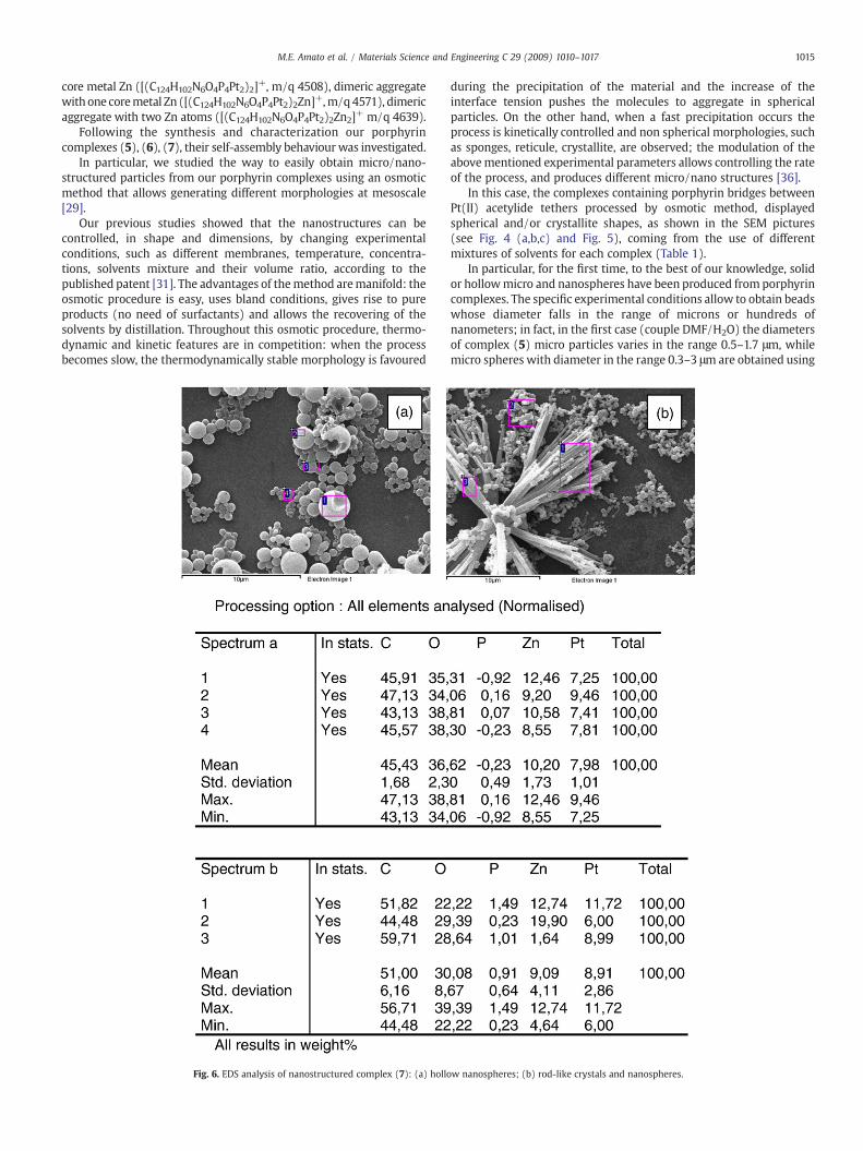
In this case, the complexes containing porphyrin bridges betweenPt(II) acetylide tethers processed by osmotic method, displayedspherical and/or crystallite shapes, as shown in the SEM pictures(see Fig. 4 (a,b,c) and Fig. 5), coming from the use of differentmixtures of solvents for each complex (Table 1).
In particular, for the first time, to the best of our knowledge, solidor hollowmicro and nanospheres have been produced from porphyrincomplexes. The specific experimental conditions allow to obtain beadswhose diameter falls in the range of microns or hundreds ofnanometers; in fact, in the first case (couple DMF/H2O) the diametersof complex (5) micro particles varies in the range 0.5–1.7 μm, whilemicro spheres with diameter in the range 0.3–3 μm are obtained using
w nanospheres; (b) rod-like crystals and nanospheres.

Table 3Number ofmolecules contained inmicro and nanospheres calculated by sphere volume/ellipsoid volume ratio, for the complexes (5), (6) and (7).
Complex Molecularellipsoidvolume (nm3)
Spheresradius (nm)
Spheres volume (nm3)a Number of moleculesin sphere=SpheresVol./Ellipsoids Vol.
(5) 186.73 190–80 28,716,347–2,143,573 153,784–11,479(6) 174.09 300–125 113,040,000–8,177,083 649,327–46,971(7) 18.24 350–150 179,503,333–14,130,000 9,843,864–774,881
a The range of calculated spheres volumes is related with the range of the spheresradius obtained by SEM measurements.
1016 M.E. Amato et al. / Materials Science and Engineering C 29 (2009) 1010–1017
THF/H2O. Complex (6) beads show diameters that increase from 250–600 nm, using DMF/H2O, to 1–6 μm, using THF/H2O. Considering thenanostructures of complex (7), the trend of size dependence on thesolvents couple is not evident and a high polydispersion of micro/nanospheres is found (Fig. 5).
In general, the achievement of the equilibrium morphology isfavoured when the polarity of the material and that of the solventcouple is similar; for complexes whose chemical structure induces alow polarity, such as complexes (5) and (6), spheres were preferablyobtained by the DMF/ H2O mixture, while for complex (7) thespherical morphology was favoured by the THF/H2O mixture. Inparticular, the difference of polarity Δε (DMF/H2O) is lower comparedwith Δε (THF/H2O) (ε=dielectric constant) and then the complexes(5) and (6) are induced to reach their equilibrium morphology.
EDS analysis of nano/micro spheres cast-deposited at room tem-perature confirmed for all the investigated complexes the presence ofZn into the porphyrin core (see Fig. 6(a) for complex (5) reported forexample).
In the case of systems where nanospheres and crystallites are bothpresent, the EDS analysis (see Fig. 6(b) for complex (7)) points out thecoexistence of two different compounds: i) nanospheres in which theZn phorphyrin complex (7) is detected, ii) rod-like crystallites whereZn is absent (see the insert square 3 of Fig. 6(b)); this lattermorphology is related to the free base of (7). The presence or absenceof Zn into the porphyrin core probably induces a difference inmorphology, such as reported in literature [19] for iron oxidenanostructures, that synthesized with addition of a small amount ofCo2+ change their morphology from rods to spheres. This topic needsmore in depth studies for the understanding of the phenomenon.
Further studies were performed on the effect of the temperatureon the morphology and dimensions of these nano/micro structuredmaterials. When the water evaporation of the complexes suspensionwas run at different temperatures, i.e. T=25 °C and T=50 °C, theshape and dimension of several nanostructured particles werechanged, demonstrating the thermosensitivity the materials (seeTable 2). In particular, the morphologies of solid spheres andcrystallites vanished at T=50 °C; the micro-nanostructures becameirregular rough flakes, suggesting that a fusion of the particlesoccurred; in the case of the hollow spheres, for all complexes theshape was preserved and the dimensions decreased.
Further considerations, in view of future applications in bio andoptoelectronic technology, can be made taking into account themolecular dimensions of each complex and exploring the possibility ofa proportional relationship between molecular dimensions andspheres dimensions. In fact, considering the complexes (5), (6), and(7) assimilable to ellipsoids with definite dimensions, we cancalculate the number of molecules of complex that are containedinto a solid nanosphere (see Table 3).
By simple theoretical calculation we can approximatively assessthat smaller molecular dimensions (complex (5), volume186,73 nm3), produce smaller nanospheres (r=190–80 nm) whichcontain fewer molecules (N=153,784–11,479).
Table 2Several morphologies of the complexes (5), (6), (7), obtained by osmotic procedure, atsolvent evaporation temperature T=50 °C.
ID complex Solv/H2O Shape Diameter (μm)
a (5) Acetone/H2O Rough flakesb (5) DMF/H2O Hollow spheres 0.1–1c (5) THF/H2O Hollow spheres 0.1–2d (6) Acetone/H2O Rough flakese (6) DMF/H2O Rough flakesf (6) THF/H2O Hollow spheres 0.6–2.5g (7) Acetone/H2O Rough flakesh (7) DMF/H2O Rough flakesi (7) THF/H2O Hollow spheres 0.1–1.2
Advanced investigations will be devoted to test the best conditionfor obtaining low polydispersion and the self assembling of nano-spheres in well ordered large domains for applications in optoelec-tronics, such as photonic crystals for example. This morphology andthe large surface areas suggest that these nanostructured materialscan be employed in chemical sensors and catalytic systems, introdu-cing an improvement of their performance.
4. Conclusions
Dinuclear porphyrin bridged complexes containing Pt tethers havebeen synthesized. The complexes are obtained in fairly good yieldsand recognized at molecular level by means of NMR and ToF-SIMScharacterizations. We have investigated the way to modulate themorphology of our micro-nanostructured porphyrins: spherical and/or crystallite nanoparticles coming from the use of selected solvent/non solvent pairs. The formations of solid and hollow spheres orcrystallite could be achieved. Under our experimental conditions thecomplexes containing the Zn into the porphyrin core show thetendency to self assembled into solid or hollow spheres, while the freebase derivative of complex (7) leads to the formation of crystallitebunches. An influence of the temperature on the morphology of themicro-nanostructures was also found. At the evaporation temperatureof the water suspensions T=50 °C only the hollow spheres maintaintheir shape. In this work it is demonstrated that controllablemodulations of morphology can provide by a facile approach forpreparing nanoparticles or nanostructures with structural diversity.
Acknowledgement
The authors gratefully acknowledge the financial support of“Sapienza” Progetti di Ateneo60%(2006) (MIUR, Italy).
References
[1] S. Prathapan, T.E. Johnson, J.S. Lindsey, J. Am. Chem. Soc. 115 (1993) 7519.[2] V.S.Y. Lin, S.G. Di Magno, M.J. Therien, Science 264 (1994) 1105.[3] A. Tsuda, A. Osuka, Science 293 (2001) 79.[4] T. Hori, Y. Nakamura, N. Aratani, A. Osuka A, J. Org. Chem. 692 (2007) 148.[5] G. Stegman, P. Likamwa, in: A. Miller, K.R. Welford, B. Daino (Eds.), Nonlinear
Optical materials and Devices for Applications in Information Tecnology, Kluver,The Netherlands, 1995.
[6] V. Parkhutik, V. Chirvony, E. Matveyeva, Biomol. Eng. 24 (2007) 71.[7] R. Rella, A. Serra, P. Siciliano, A. Tepore, L. Valli, A. Zocco, Langmuir 13 (1997) 62.[8] J.M. Pedrosa, C.M. Dooling, T.H. Richardson, R.K. Hyde, C.A. Hunter, M.T. Martin, L.
Camacho, J. Mater. Chem. 12 (2002) 2659.[9] T.S. Balaban, R. Goddard, M. Linke-Schaetzel, J.M. Lehn, J. Am. Chem. Soc. 125
(2003) 4233.[10] A. Ferri, G. Polzonetti, S. Licoccia, R. Paolesse, D. Favretto, P. Traldi, M.V. Russo,
J. Chem. Soc. Dalton Trans. (1998) 4063.[11] H.L. Anderson, S.J.Martin, D.D.C. Bradley, Angew. Chem. Int. Ed. Engl. 33 (1994) 655.[12] K. Tomokazi, A.B. Lysenko, M. Taniguchi, J.S. Lindsey, Tetrahedron 60 (2004) 2011.[13] P. Thamyongkit, J.S. Lindsey, J. Org. Chem. 69 (2004) 5796.[14] S.C. Jones, V. Coropceanu, S. Barlow, T. Kinnibrugh, T. Timofeeva, J.L. Brédas, S.R.
Marder, J. Am. Chem. Soc. 126 (2004) 11782.[15] T.L. Schull, J.G. Kushmerick, C.H. Patterson, C. George, M.H. Moore, S.K. Pollak, R.
Shashidhar, J. Am. Chem. Soc. 125 (2003) 3202.[16] J.-S. Hu, Y.-G. Guo, H.-P. Liang, L.-J. Wan, L. Jiang, J. Am. Chem. Soc. 127 (2005)
17090.

1017M.E. Amato et al. / Materials Science and Engineering C 29 (2009) 1010–1017
[17] T. Ishida, Y. Morisaki, Y. Chujo, Tetrahedron Lett. 47 (2006) 5265.[18] Z. Wang, Z. Li, C.J. Medforth, J.A. Shelnutt, J. Am. Chem. Soc. 129 (2007) 2440.[19] H. Sato, O. Tsutsumi, K. Takeda, H. Takanaka, T. Ogawa, Jpn. J. Appl. Phys. 45 (2006)
2324 3B.[20] Z. Deng, J. Qi, Y. Zhang, Q. Liao, Y. Huang, Nanotechnology 18 (2007) 475603.[21] A. Kumar, A. Singha, Nanotechnology 18 (2007) 475703.[22] D. Beljonne, G.E. O'Keefe, P.J. Hamer, R.H. Friend, H.L. Anderson, J.L. Bredas, J. Chem.
Phys. 106 (1997) 9439.[23] I. Fratoddi, C. Battocchio, R. D'Amato, G. Di Egidio, L. Ugo, G. Polzonetti, M.V. Russo,
Mater. Sci. Eng. C 23 (2003) 867.[24] S. Yellappa, J. Seetharamappa, L.M. Rogers, R. Chitta, R.P. Singhal, F. D'Souza,
Bioconjug. Chem. 17 (2006) 1418.[25] F. Langa, M.J. Gomez-Escalonilla, P. de la Cruz, J. Porphyr. Phthalocyanines 11
(2007) 348.[26] G. Polzonetti, C. Battocchio, A. Goldoni, R. Larciprete, V. Carravetta, R. Paolesse,M.V.
Russo, Chem. Phys. 297 (2004) 307.
[27] H.L. Anderson, Tetrahedron Lett. 33 (1992) 1101.[28] A. Furlani, S. Licoccia, M.V. Russo, A. Chiesi Villa, C. Guastino, J. Chem. Soc. Dalton
Trans. (1984) 2197.[29] R. D'Amato, A. Furlani, M. Colapietro, G. Portatone, M. Casalboni, M. Falconieri, M.V.
Russo, J. Org. Chem. 627 (2001) 13.[30] A. Benninghoven, Angew. Chem. Int. Ed. Engl. 33 (1994) 1023.[31] PCT Int. Appl. 2006 PIXXD2 WO 2006051572.[32] K. Sonogashira, J. Org. Chem. 653 (2002) 46.[33] C.-W. Huang, K.Y. Chiu, H. Cheng, J. Chem. Soc. Dalton Trans. 14 (2005) 2417.[34] J.-C. Chang, C.-J. Ma, G.-H. Lee, S.-M. Peng, C.-Y. Yeh, J. Chem. Soc. Dalton Trans. 8
(2005) 1504.[35] Y. Chen, S. Chen, S. Lo, T. Huang, C. Wu, G. Lee, S. Peng, C. Yeh, Chem. Commun. 9
(2006) 1015.[36] J.M. Stubbs, D.C. Sundberg, J. Appl. Pol. Sci. 91 (2004) 1538.
Copyright © 2022 FDOKUMEN