Mono-and Bisquinoline-Annulated Porphyrins from Porphyrin β,β′‑Dione Oximes
13
Mono- and Bisquinoline-Annulated Porphyrins from Porphyrin β,β′‑Dione Oximes Joshua Akhigbe, † Michael Luciano, † Matthias Zeller, ‡ and Christian Brü ckner* ,† † Department of Chemistry, University of Connecticut, Storrs, Connecticut 06269-3060, United States ‡ Department of Chemistry, Youngstown State University, One University Plaza, Youngstown, Ohio 44555-3663, United States * S Supporting Information ABSTRACT: An acid-induced reaction of meso-tetraphenyl-2-hydroxyimino- 3-oxoporphyrin leads, with concomitant loss of water, to a formal electrophilic aromatic substitution of the ortho-position of the phenyl group adjacent to the oxime, forming a quinoline moiety. Owing in part to the presence of a π-extended chromophore, the resulting meso-triphenylmo- noquinoline-annulated porphyrin (λ max = 750 nm) possesses a much altered optical spectrum from that of the starting oxime (λ max = 667 nm). An oxidative DDQ-induced ring-closure process is also possible, generating the corresponding meso-triphenylmonoquinoline-annulated porphyrin quinoline N-oxide, possessing a slightly shifted and sharpened UV−vis spectrum (λ max = 737 nm). The connectivity of the chromophores was conclusively shown by NMR spectroscopy. Both ketone functionalities in meso-tetraphenyl-2,3- dioxoporphyrin can be converted, via the oxime and using the acid- or oxidant-induced reaction pathways, either in one step or in a stepwise fashion, to bisquinoline-annulated porphyrin (λ max = 775 nm) and its N-oxide (λ max = 779 nm), respectively. This process is complementary to a previously established pathway toward bisquinoline-annulated porphyrins. Their zinc(II), nickel(II), and palladium(II) complexes are also described. Several examples of the quinoline-annulated porphyrins were crystallographically characterized, proving their connectivity and showing their conformations that are extremely distorted from planarity. The work presents a full account on the synthesis, structure, and spectroscopic properties of these classes of NIR-absorbing dyes. ■ INTRODUCTION Even though the high extinction coefficients, fluorescence, and singlet oxygen photosensitization quantum yields of regular porphyrins and chlorins are potentially attractive for their use in biomedical applications, e.g., as photochemotherapeutics, 1 photoantimicrobials, 2 photoantifungals, 3 or fluorescent mar- kers, 1c,4 most regular porphyrins and chlorins do not absorb light within the ∼700−900 nm wavelength regime referred to as the “optical window” of tissue. 5 For instance, the maximum wavelength of absorbance (λ max ) for regular porphyrins, such as meso-tetraphenylporphyrin 1H 2 , rarely exceeds 650 nm. In comparison, the wavelength of maximum penetration of breast tissue is ∼725 nm; whole blood has an absorption minimum at ∼710 nm. 6 Regular porphyrins are thus unsuitable for biomedical in vivo applications. Nonemitting chromophores may also be attractive as photoacoustic imaging agents but, 8 again, only if they also absorb light within the optical window of tissue. 9 Light-harvesting applications also benefit from NIR- absorbing dyes, as a large portion of the irradiance energy of sunlight falls into this range. Panchromatic absorbers are also most desirable for light harvesting, as these chromophores collect the maximum amount of energy. 7 A number of strategies have been employed to achieve this goal. For instance, the expansion of the conjugated π-system by increasing the number of conjugated pyrroles was greatly successful, as the many examples of NIR-absorbing expanded porphyrins demonstrate. 10 One other strategy is the establish- ment of π-systems that are annulated to the porphyrinic chromophore. 7d,11 Among the latter class of porphyrins may be meso-arylporphyrin derivatives that bear a covalent linkage between one or more β-positions and the flanking meso- phenyl/aryl groups. 12 In the absence of the linkage, a H β‑pyrrole − H o‑phenyl steric interaction prevents the meso-aryl groups from adopting low-energy coplanar conformations. The linkage removes this interaction but needs to be short enough to force the phenyl group(s) into (idealized) coplanarity with the porphyrinic chromophore, thereby extending the π-conjugation pathway. This linkage may be a ketone functionality, itself in conjugation with the chromophore (2H 2 ). 13 The resulting bathochromic shift of λ max of 2H 2 compared to 1H 2 is a respectable ∼76 nm. On the other hand, the fusion of one through four anthracenes to a porphyrin, as in 3H 2 , results in an enormous perturbation of their UV−vis spectra, with λ max values red-shifted ∼300 nm upon fusion of a single anthracene; the corresponding nickel(II) complex with four anthracenes shifted the λ max by more than 900 nm, to 1417 nm. 14 Few examples exist in which a chlorin chromophore incorporates an Received: November 2, 2014 Published: December 3, 2014 Article pubs.acs.org/joc © 2014 American Chemical Society 499 dx.doi.org/10.1021/jo502511j | J. Org. Chem. 2015, 80, 499−511
Transcript of Mono-and Bisquinoline-Annulated Porphyrins from Porphyrin β,β′‑Dione Oximes

Mono- and Bisquinoline-Annulated Porphyrins from Porphyrinβ,β′‑Dione OximesJoshua Akhigbe,† Michael Luciano,† Matthias Zeller,‡ and Christian Bruckner*,†
†Department of Chemistry, University of Connecticut, Storrs, Connecticut 06269-3060, United States‡Department of Chemistry, Youngstown State University, One University Plaza, Youngstown, Ohio 44555-3663, United States
*S Supporting Information
ABSTRACT: An acid-induced reaction of meso-tetraphenyl-2-hydroxyimino-3-oxoporphyrin leads, with concomitant loss of water, to a formalelectrophilic aromatic substitution of the ortho-position of the phenylgroup adjacent to the oxime, forming a quinoline moiety. Owing in part tothe presence of a π-extended chromophore, the resulting meso-triphenylmo-noquinoline-annulated porphyrin (λmax = 750 nm) possesses a much alteredoptical spectrum from that of the starting oxime (λmax = 667 nm). Anoxidative DDQ-induced ring-closure process is also possible, generating thecorresponding meso-triphenylmonoquinoline-annulated porphyrin quinolineN-oxide, possessing a slightly shifted and sharpened UV−vis spectrum (λmax =737 nm). The connectivity of the chromophores was conclusively shown byNMR spectroscopy. Both ketone functionalities in meso-tetraphenyl-2,3-dioxoporphyrin can be converted, via the oxime and using the acid- oroxidant-induced reaction pathways, either in one step or in a stepwise fashion,to bisquinoline-annulated porphyrin (λmax = 775 nm) and its N-oxide (λmax = 779 nm), respectively. This process iscomplementary to a previously established pathway toward bisquinoline-annulated porphyrins. Their zinc(II), nickel(II), andpalladium(II) complexes are also described. Several examples of the quinoline-annulated porphyrins were crystallographicallycharacterized, proving their connectivity and showing their conformations that are extremely distorted from planarity. The workpresents a full account on the synthesis, structure, and spectroscopic properties of these classes of NIR-absorbing dyes.
■ INTRODUCTION
Even though the high extinction coefficients, fluorescence, andsinglet oxygen photosensitization quantum yields of regularporphyrins and chlorins are potentially attractive for their use inbiomedical applications, e.g., as photochemotherapeutics,1
photoantimicrobials,2 photoantifungals,3 or fluorescent mar-kers,1c,4 most regular porphyrins and chlorins do not absorblight within the ∼700−900 nm wavelength regime referred toas the “optical window” of tissue.5 For instance, the maximumwavelength of absorbance (λmax) for regular porphyrins, such asmeso-tetraphenylporphyrin 1H2, rarely exceeds 650 nm. Incomparison, the wavelength of maximum penetration of breasttissue is ∼725 nm; whole blood has an absorption minimum at∼710 nm.6 Regular porphyrins are thus unsuitable forbiomedical in vivo applications. Nonemitting chromophoresmay also be attractive as photoacoustic imaging agents but,8
again, only if they also absorb light within the optical window oftissue.9 Light-harvesting applications also benefit from NIR-absorbing dyes, as a large portion of the irradiance energy ofsunlight falls into this range. Panchromatic absorbers are alsomost desirable for light harvesting, as these chromophorescollect the maximum amount of energy.7
A number of strategies have been employed to achieve thisgoal. For instance, the expansion of the conjugated π-system byincreasing the number of conjugated pyrroles was greatly
successful, as the many examples of NIR-absorbing expandedporphyrins demonstrate.10 One other strategy is the establish-ment of π-systems that are annulated to the porphyrinicchromophore.7d,11 Among the latter class of porphyrins may bemeso-arylporphyrin derivatives that bear a covalent linkagebetween one or more β-positions and the flanking meso-phenyl/aryl groups.12 In the absence of the linkage, a Hβ‑pyrrole−Ho‑phenyl steric interaction prevents the meso-aryl groups fromadopting low-energy coplanar conformations. The linkageremoves this interaction but needs to be short enough toforce the phenyl group(s) into (idealized) coplanarity with theporphyrinic chromophore, thereby extending the π-conjugationpathway. This linkage may be a ketone functionality, itself inconjugation with the chromophore (2H2).
13 The resultingbathochromic shift of λmax of 2H2 compared to 1H2 is arespectable ∼76 nm. On the other hand, the fusion of onethrough four anthracenes to a porphyrin, as in 3H2, results inan enormous perturbation of their UV−vis spectra, with λmaxvalues red-shifted ∼300 nm upon fusion of a single anthracene;the corresponding nickel(II) complex with four anthracenesshifted the λmax by more than 900 nm, to 1417 nm.14 Fewexamples exist in which a chlorin chromophore incorporates an
Received: November 2, 2014Published: December 3, 2014
Article
pubs.acs.org/joc
© 2014 American Chemical Society 499 dx.doi.org/10.1021/jo502511j | J. Org. Chem. 2015, 80, 499−511

annulated ring, as in bischromene-annulated chlorin 4H2,leading to a 24 nm-shifted λmax compared to the parentchlorin.15
In 2011, Jeandon and Ruppert, building on the extensivework by Ruppert, Callot, and co-workers in the construction ofperipherally conjugated chelates,16 published the formation ofbisquinoline-annulated porphyrin 7H2 from nickel porphyrin1Ni via a multistep sequence (Scheme 1).17 The key β-to-ortho-linkage formation step involved a Cadogan reaction of
nitroporphyrin 5Ni to form the amine-linked porphyrin 6Ni.Subsequent nitrogen protection, regioselective nitration,thermally induced oxidation and ring-closure, and acid-induceddemetalation produced the bisquinoline-annulated system 7H2in good yield.Concurrently with Jeandon and Ruppert, we independently
described in a preliminary report the conversion of free baseporphyrin 1H2 to free base bisquinoline-annulated porphyrin7H2.
18 The key steps were two consecutive acid-mediatedelectrophilic aromatic substitution reactions of oximes, such as10H2, formed from the corresponding ketone 8H2. Weprepared this well-known dione 8H2 along a dihydroxyla-tion−diol oxidation sequence from porphyrin 1H2,
19 but thisproduct can be made along a number of alternative routes.20
We follow up here on our preliminary report and present thefull account on the formation and reactivity of the mono- anddioximes of 8H2 with respect to their conversion to a numberof free base and metalated mono- and bisquinoline-annulatedporphyrins, and their N-oxide derivatives. These chromophoresare characterized by remarkably bathochromically shiftedoptical spectra when compared to regular porphyrins. We willalso report the X-ray crystal structures of a number ofderivatives, thus highlighting their nonplanar conformationswe interpret as one reason for their red-shifted and broadenedspectra. We recently demonstrated the efficacy of the quinoline-annulated porphyrins to act as photoacoustic imaging contrastagents in tissue phantom studies.9
■ RESULTS AND DISCUSSION
Synthesis of meso-Tetraphenylmonooxime 10H2 and-bisoxime 11H2. Analogous to the previously describedconversion of the nickel(II) and palladium(II) complexes ofporphyrin dione 8Ni and 8Pd,21 free base 2,3-dioxoporphyrin8H2 can also be converted to the corresponding monooxime10H2 by reaction with ∼100-fold stoichiometric excess ofhydroxylamine hydrochloride (NH2OH·HCl) in pyridine over24 h at ambient temperature (Scheme 2). Its analytical andspectroscopic properties were similar to those of its
Scheme 1. Two Independent Paths toward Bisquinoline-Annulated Porphyrin 7H2 Developed by Ruppert (1Ni → 5Ni → 7H2)and Bruckner (1H2 → 10H2 → 7H2)
The Journal of Organic Chemistry Article
dx.doi.org/10.1021/jo502511j | J. Org. Chem. 2015, 80, 499−511500

diamagnetic metal complexes: the 13C NMR of 10H2 indicatedthe presence of one carbonyl (188.2 ppm) and one iminecarbon (151.8 ppm), functionalities that could also beconfirmed by FT-IR spectroscopy (νCO and νCN at 1652.1and 1548.6 cm−1, respectively). A diagnostic 1H NMR spectralfeature for the monooximes is the signal at ∼15.7 ppm(exchangeable with D2O) that is assigned to the oximehydrogen that is H-bonded to the neighboring carbonylgroup (Figure 1). The structural characterization of 10Ni bysingle crystal X-ray diffractometry is described below (Figure4).Next to the major product monooxime 10H2, a minor
amount of the bisoxime 11H2 could also be isolated, possessingall expected spectroscopic properties (see Supporting Informa-tion (SI) for details). The occurrence of the bisoxime was notobserved in the reaction of the dione metallocomplexes 8Niand 8Pd.21
The UV−vis spectra of free base dione 8H2, monooxime10H2, and bisoxime 11H2 are similar to each other in that allpossess, compared to the regular porphyrin spectrum of meso-tetraphenylporphyrin 1H2 (not shown), slightly broadenedSoret bands and very broadened Q-bands (Figure 2). However,with increasing ketone-to-oxime transformations, the Q-bandregion becomes more defined, with increasingly sharper and
distinctive peaks. The strong electronic effects of the β,β′-diketone functionality were discussed previously.19,22 Evidently,this influence extends also to the oximes, albeit to a slightlylesser degree.
Formation of Free Base and Metalated Monoquino-line-Annulated Oxoporphyrins 9. Treatment of the olive-colored free base monooxime 10H2 with a strong organic acid(p-TSA) under forcing conditions (toluene, reflux) generated amajor light brown product, 9H2, with a mass indicating the lossof H2O (HR-MS ESI+, 100% CH3CN, suggests a compositionof C44H28N5O for MH+) (Scheme 2). As the oxime signalvanished from the 1H NMR spectrum of the product, theoxygen lost likely stems from the oxime functionality (Figure1). Further, the 1H NMR spectrum of product 9H2 lacks anyaxial symmetry: For instance, all six β-protons are non-equivalent. A diagnostic set of four peaks assigned to onephenyl group (confirmed by 2D NMRs; see SI) suggestsestablishment of a link between a phenyl group and a flankingβ-position. Its proton signals are much shifted (between 9.0 and7.6 ppm) because of the induced coplanarity of this phenylgroup with the porphyrinic macrocycle. Indaphyrins, forexample, have shown similarly diagnostic patterns for such β-to-o-phenyl linkages. The 13C NMR and IR (neat) spectra of9H2 indicated the preservation of one β-imine and one β-
Scheme 2. Synthetic Pathways toward Monoquinoline-Annulated Porphyrins
The Journal of Organic Chemistry Article
dx.doi.org/10.1021/jo502511j | J. Org. Chem. 2015, 80, 499−511501

ketone carbon (at 151 and 196 ppm, respectively). Thesummation of the spectroscopic data is suggestive of formationof the quinoline-annulated structure shown in Scheme 2. TheUV−vis spectroscopic properties of this compound are,together with the optical spectra of all the other quinoline-annulated porphyrins, described below.This outcome of the reaction highlights an interesting
directing effect of the coordinated metal ion in light of the factthat the metal complexes of monooxime 10Ni and 10Pd, whenreacted under identical acidic conditions, undergo a Beckmannrearrangement to form the corresponding meso-phenylpyrazi-noporphyrin metal complexes 16M.21 This reactivity furtherimplies that metal complexes of the quinoline-annulatedporphyrin 9M needed to be prepared by metal insertion into
free base 9H2. The corresponding metal insertion reactionsusing standard methodologies proceeded smoothly with goodto excellent yields.23 The composition (as determined byDART+ HR-MS) and spectroscopic data of 9Ni and 9Pdconfirmed their expected quinoline-annulated porphyrinstructures.Alongside the major product 9H2, we also isolated a minor
product 12H2 in yields ranging from 6% to 10%. Its mass, asdetermined by HR-MS, indicated the presence of an additionaloxygen atom. Performance of the ring-closing reaction underinert conditions largely suppressed its formation. Consequently,the formation of this novel chromophore was traced to airoxidation. Product 12H2 is also accessible as the major productfrom free base monooxime 10H2 under oxidizing conditions.
Synthesis of Monoquinoline-Annulated Oxoporphyr-in N-Oxides 12. Guided by the discovery of the oxidizedquinoline-annulated product during the acid-catalyzed reaction,we discovered that this product can be prepared as the mainproduct when free base monooxime 10H2 is treated with DDQat ambient temperatures (Scheme 2). Interestingly, the metalcomplexes of the oxime 10M (M = Ni(II), Pd(II)) are alsosusceptible to this conversion, generating the metal analoguesof the product, 12M (M = Ni(II), Pd(II)). A careful NaBH4reduction of N-oxide 12H2 regenerates 9H2, though DDQoxidation of 9H2 does not lead to formation of 12H2. Thissuggests that monoquinoline-annulated chromophore 9H2 isnot an intermediate in the formation of 12H2. The oxidation ofα,β-oximes of unsaturated substrates under a number of
Figure 1. Comparison of the low-field region of the 1H NMR spectra (400 MHz, CDCl3, 25 °C) of the compounds indicated.
Figure 2. UV−vis spectra (CH2Cl2) of the compounds indicated.
The Journal of Organic Chemistry Article
dx.doi.org/10.1021/jo502511j | J. Org. Chem. 2015, 80, 499−511502
conditions frequently results in formation of iminoxy radicalintermediates that undergo ring-cyclization reactions producingisoxazoles,24 including isoxazoles annulated to a chlorinframework,25 though the formation of quinoline N-oxides wasalso observed.26 Presumably the formation of a six-memberedquinoline ring in the cyclization of 10H2 directs this reaction.The presence of the oxygen atom in 12M also has a strong
influence on the UV−vis spectra of these chromophores whencompared to the quinoline-annulated compounds 9M (Figure5; see discussion below). The NMR spectroscopic data for 12Mare significantly different from those of corresponding 9M(Figure 1). Nonetheless, the same number of hydrogens andcarbons are observed and the compound possesses the sameoverall symmetry by 1H NMR spectroscopy (Figure 1). One
hydrogen signal assigned to the CH group adjacent to thequinoline nitrogen is particularly strongly affected by theoxidation, suggestive of the presence of a quinoline N-oxide, asopposed to a porphyrin pyrrole N-oxide. The latter compoundsare known, and their optical spectra are also severely distortedcompared to those of the parent porphyrins.27 A crystalstructure analysis eventually provided unambiguous proof forthe quinoline-annulated porphyrin quinoline N-oxide con-nectivity of product 12M (Figure 4; all crystal structures arediscussed below).
Synthesis of Monoquinoline-Annulated Oxoporphyr-in Oxime 14H2. Treatment of the β-keto functionality of freebase monoquinoline 9H2 with hydroxylamine hydrochlorideunder similar conditions as described above for the formation
Scheme 3. Synthetic Pathways toward Bisquinoline-Annulated Porphyrins
The Journal of Organic Chemistry Article
dx.doi.org/10.1021/jo502511j | J. Org. Chem. 2015, 80, 499−511503
of the oximes 10H2/10M resulted in formation of oxime 14H2(Scheme 3). Similarly, reaction of β-keto N-oxide 12H2 withhydroxylamine converted it to quinoline oxime 14H2. Note thatthe N-oxide moiety is lost in the process; such (reductive)losses are not unusual for heterocycle N-oxides.28 Diagnosticpeaks for the resulting oxime 14H2 in the 1H NMR spectrumare, analogous to oximes 10H2/10M, the signal for the stronglyH-bonded oxime hydrogen (at ∼15.9 ppm) (Figure 1). Allother spectroscopic and analytical data are as expected. The β-ketone-to-oxime conversion (9H2/12H2 → 14H2 conversion)perturbs the UV−visible spectrum of the products much morethan the corresponding dione to oxime conversions (8H2 →10H2/11H2 conversions, Figure 2; see also below).Synthesis of Bisquinoline-Annulated Porphyrins 7H2/
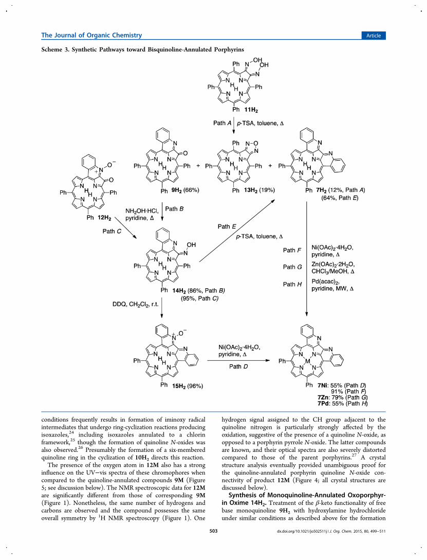
7M and Bisquinoline-Annulated Porphyrin N-Oxide15H2. The oxime functionality shows analogous reactivity inboth 10H2 and the quinoline-annulated porphyrin 14H2(Scheme 3). Reaction of oxime 14H2 with p-TSA under refluxconditions resulted in formation of a product in good yield thatwe were, based on its spectroscopic properties, able to identifyas bisquinoline-annulated porphyrin 7H2; it possessed all thespectroscopic properties as reported by Jeandon and Ruppert,17
including the broadened NMR spectra at rt attributed toextensive stacking and the limited solubility of this compound(Figure 3). Other annulated porphyrins have been observed toform tightly associated stacks in the crystal lattice.14c Thesynthetic routes presented here are thus alternative and fullyindependent routes toward a π-extended porphyrin chromo-phore with much altered optical properties (discussed below).The two pathways toward 7H2 are complementary to eachother. Both possess distinct advantages and disadvantages. TheJeandon and Ruppert synthesis is somewhat shorter (7 steps)versus our pathway (∼9 steps, depending in the chosen path
toward the dione 8H2), and the overall yield of both synthesesare also similar (∼20%). One advantage of our route is itsflexibility with respect to accessing other quinoline-annulatedderivatives (such as the monoquinoline-annulated porphyrinsand N-oxides).The metal ions nickel(II), zinc(II), and palladium(II) could
be inserted into free base 7H2, forming the correspondingmetal complexes in good to satisfying yields. Metal insertion ledto the formation of compounds that possess much sharpened1H NMR spectra (see Figure 3 for the spectrum of 7Ni),presumably because of a lesser degree of stacking.Reaction of oxime 14H2 with DDQ led to a compound of
the molecular mass of bisquinoline-annulated porphyrin 7H2plus an oxygen atom (as per ESI+ HR-MS). Its 1H NMRspectrum also shows the loss of axial symmetry (Figure 3). Thissuggests the formation of bisquinoline N-oxide 15H2, aninterpretation that could also be confirmed by single crystal X-ray diffractometry (discussed below). Nickel(II) insertion intothe N-oxide led to loss of the N-oxide functionality andformation of 7Ni.
A Shorter Route to Bisquinoline-Annulated Porphyrin7H2. The successful stepwise formation of bisquinoline-annulated porphyrin 7H2 via the route dione 8H2 → oxime10H2 → quinoline-annulated oxoporphyrin 9H2 → quinoline-annulated oxoporphyrin oxime 14H2 → 7H2 prompts thequestion whether the direct conversion of bisoxime 11H2 →7H2 can be accomplished (even though the formation ofbisoxime 11H2 is inefficient and could not be optimized in ourhands). The answer is yes but at a loss of overall efficiency.Reaction of 11H2 under the conditions leading to quinolineannulation generated the desired product 7H2, but in only 12%yield. The major product is quinoline-annulated oxoporphyrin9H2. Evidently, hydrolysis of one of the oximes is faster than
Figure 3. Comparison of the low-field region of the 1H NMR spectra (400 MHz, in solvents indicated, 25 °C) of the bisquinoline-annulatedporphyrins shown.
The Journal of Organic Chemistry Article
dx.doi.org/10.1021/jo502511j | J. Org. Chem. 2015, 80, 499−511504

ring-closure. In fact, we observed fast hydrolysis of 11H2 toform 10H2, even under the conditions of silica gelchromatography. The intramolecular condensation of the twooxime moieties with each other is also a more efficient process,generating oxadiazole (furazan)-annulated porphyrin 13H2.This novel product, related to Smith’s pyrroloporphyrins,29
shows very porphyrin-like optical properties (Figure 2) and wasnot further investigated (for details of 13H2, see also SI). Ingeneral, the condensation of α,β-bisoximes resulting in theformation of oxadiazoles is a well-known reaction.30 Thus, eventhough the stepwise route toward product 11H2 is significantlylonger, its overall yield (∼35% from diketone 11H2) is muchbetter than the short route via bisoxime 11H2 (<0.5% fromdiketone 11H2).Reduction of Monoquinoline-Annulated Porphyrin
9H2. Treatment of 9H2 with NaBH4 in CH2Cl2/10% MeOHat ambient temperatures yielded two polar products with acomposition (as per ESI+-MS) corresponding to [M + 5H]+
and another, even more polar product, corresponding to [M +3H]+, suggestive of a reduction of either only the imine doublebond (forming one isomer) or both the imine and the ketone
double bonds (forming two diastereomers), respectively. Thesingle double bond reduction product can also be made as thesole product using NaBH3CN at ice temperatures. Remarkably,all isolated products possessed much more blue-shifted, chlorin-like optical spectra with broadened Soret bands, highlightingthe importance of the presence of the ketone and iminefunctionality for the electronic structure of the chromophore.Much to our disappointment, however, all three productsproved to be intractable, as they all rapidly decomposed uponisolation at preparatively useful scales.
Structural Characterization of the Quinoline-Annu-lated Porphyrins. The considerably ruffled conformation ofthe monooxime 10Ni is comparable to that observed innickel(II) porphyrins and nickel dione 8Ni19 and like in thesecases can be attributed to the small ionic radius of the low-spinnickel(II) (Figure 4).31 The H-bond of the oxime moiety to theadjacent ketone functionality, seen in solution state (cf. toFigure 1), is also expressed in the solid state.The crystal structure of the nickel(II) complex of quinoline-
annulated porphyrin N-oxide 12Ni provided the ultimate proofof the quinoline-annulated porphyrin connectivity, including
Figure 4. Stick representation of the X-ray single crystal structures: (A) 10Ni; (B) 12Ni; (C) 7Pd; (D) 15H2. Top or oblique views (left column),side views (middle column), and deviation of the skeletal heavy atoms from the N4 mean plane defined by the four pyrrolic nitrogens, as seen in thecrystal structures (right column). All disorder and hydrogens bonded to carbon removed for clarity; phenyl groups removed for clarity in A, B, andC. Arrows indicate perspective for side views.
The Journal of Organic Chemistry Article
dx.doi.org/10.1021/jo502511j | J. Org. Chem. 2015, 80, 499−511505

the presence of a quinoline N-oxide, as opposed to a centralporphyrin pyrrole-N-oxide functionality. The ring-closurereaction of the oxime with the neighboring phenyl groupgenerated a pyrrolo[3,2-b]quinolin-4-one moiety that is,presumably because of a steric interaction of this moiety withthe β-hydrogen next to it, slightly bent. The primeconformation of the porphyrinic macrocycle is still ruffled,but it is strongly modulated by the presence of the quinoline-phenyl-to-β-H interaction. The N-oxide and ketone oxygens areeclipsed and form a potential metal binding pocket. Structurallyrelated systems have been used for the formation of externalmetal complexes.16c,d,f,32
The Ni−N bond distances in primarily ruffled porphyrinscan be used to gauge the degree of distortion. In 12Ni, the Ni−N bond distances average 1.910 Å (ranging from 1.901 to 1.925Å) and thus fall into the regime of a moderately nonplanarporphyrin. The Ni−N bond lengths in ruffled porphyrinsshorten with the degree of ruffling. In comparison, the Ni−Nbond lengths in lesser ruffled 10Ni are 1.930 Å, and in onlyslightly ruffled 1Ni 1.931 Å,33 whereas they are 1.892 Å in anextremely ruffled nickel(II) secochlorin bisaldehyde.31c
The porphyrinoid macrocycle in free base bisquinoline-annulated N-oxide 15H2 is particularly nonplanar, while thepentacyclic annulated heterocycle is slightly and asymmetricallybowed. The presence of palladium in the bisquinoline-annulated porphyrin 7Pd planarizes the system somewhatwhen compared to the related free base 15H2, but theporphyrinic chromophore is still considerably ruffled withsignificant saddling and waving modes. Despite thesedeformations, the metal is coordinated in a distorted squareplanar fashion, with regular average N−Pd bond distances of2.019 Å (individual bonds range from 1.983 to 2.041 Å).33
Optical Properties of Quinoline-Annulated Porphyr-ins. Upon cyclization of monooxime 10H2, the spectrum ofquinoline-annulated porphyrin 9H2 is much altered (Figure5A): Most obvious, the Q-band region is much bathochromi-cally shifted (for 10H2, λmax = 661 nm; for 9H2, λmax = 750nm). Key to this is believed to be the extension of the
conjugated π-system and the nonplanar conformation of 9H2(we extend from the conformation of its N-oxide 12H2). Thelatter is also supported by the reduced extinction coefficient forthe broadened Soret band of 10H2.
34 Comparing the spectra ofquinoline-annulated porphyrin 9H2 with that of its N-oxide12H2, the distinct and sharpening effect of the quinoline N-oxidation (λmax = 737 nm) and a red-shift of the Soret regioncan be noticed, highlighting the strong electronic effect thequinoline moiety has on the porphyrinic chromophore.Conversion of the oxo-group in 9H2 to an oxime in 14H2(λmax = 700 nm) similarly has a general sharpening effect on theQ-bands as the conversion of diketone 8H2 to oxime 10H2 (cf.to Figure 2).Cyclization of quinoline-annulated porphyrin monooxime
14H2 leads to the red-shifted spectrum of bisquinolineporphyrin 7H2 described previously (7H2: λSoret = 392, λmax =775 nm) (Figure 5B).17,18 Again, we attribute this shift to theconformation of the chromophore and the presence of thepentacyclic heterocycle that is π-conjugated with the porphyr-inoid ring. As was observed for the oxidation of monoquinoline9H2, N-oxidation of 7H2, forming 15H2, also leads to a red-shift of its Soret band (λSoret = 412, λmax = 779 nm). The closed-shell metal complexes of bisquinoline-annulated porphyrin 7H2,7Zn, 7Ni, and 7Pd show, as expected, all similarly patternedspectra with a reduced number of Q-bands and a split Soretband (Figure 5C).In contrast to the UV−vis spectra of metal complexes of
bisquinoline 7H2, the UV−vis spectra of the nickel andpalladium complexes of monoquinoline porphyrin 9H2, 9Niand 9Pd, are not much different from those of the free basespectrum: they possess a much broadened Q-band region, withno sign of a split Soret band (Figure 5D). In this respect theyare more similar to the metal complexes of dione 8H2.
■ CONCLUSIONS
We demonstrated the annulation of a quinoline moiety onto ameso-tetraphenylporphyrin macrocycle by intramolecular ring
Figure 5. UV−vis spectra of the compounds indicated in CH2Cl2, except compound 7Zn was, for solubility reasons, recorded in DMSO.
The Journal of Organic Chemistry Article
dx.doi.org/10.1021/jo502511j | J. Org. Chem. 2015, 80, 499−511506

closure of the oximes of the well-known porphyrin 2,3-diones.This strategy can be applied twice in a stepwise fashion or, lessefficiently, in a double-ring closure reaction of an α,β-bisoxime.Depending on the reagents used, a condensation reaction toform the quinoline-annulated ring-system takes place, or thiscondensation is also coupled with an oxidation, Thus, mono-and bisquinoline-annulated porphyrins and their quinolone N-oxides became accessible. These classes of porphyrin derivativespossess dramatically bathochromically shifted and broadenedUV−vis spectra compared to the spectrum of the fundamentalstarting material porphyrin 1H2. This is attributed to the directconjugation of the quinoline moiety with the aromaticporphyrinic π-system, the strong influence of β-oxo groups,and the nonplanarity of the chromophore. This synthesis of thebisquinoline-annulated porphyrin is an alternative to thesynthesis presented by Jeandon and Ruppert.17 The solid-state conformations of all quinoline-annulated porphyrinsdisplay pronounced deviations from planarity because ofincreased pyrrole-β-to-o-aryl interactions with the ring-annu-lated systems, while the polycyclic quinoline-annulated part ofthe compounds itself remains idealized planar or slightlycurved. The conformation is modulated by the central metalpresent. The broad and intense absorption of the chromo-phores in the NIR region suggests their use in biomedicalapplications. However, their very low fluorescence and singletoxygen quantum yields, reported earlier,9 prevent their use asPDT agents, but we were able to show that this class ofporphyrinoids is suitable as contrast agents for photoacousticimaging applications.9 We are currently in the process ofpreparing a range of quinoline-annulated porphyrins thatpossess more favorable solubility properties for their use inaqueous biological environments.
■ EXPERIMENTAL SECTIONMaterials and Instruments. Solvents and reagents were used as
received. Aluminum-backed, silica gel 60, 250 μm thickness analyticalplates, 20 × 20 cm, glass-backed, silica gel 60, 500, or 1000 μmthickness preparative TLC plates, and standard grade, 60 Å, 32−63 μmflash column silica gel were used. Diones 8H2, 8Ni, and 8Pd wereprepared as described previously.19,21
X-ray Single Crystal Diffractometry. Single crystals of 10Ni,12Ni, 15H2, and 7Pd were coated in mineral oil, mounted on amicromesh mount, and placed on a goniometer head under a stream ofnitrogen cooled to 100 K. The data were collected on diffractometersusing monochromatic Mo Kα (15H2, 12Ni, and 10Ni) or Cu Kαradiation (7Pd) with the omega scan technique. Data were collected,their unit cells determined, and the data integrated and corrected forabsorption and other systematic errors using the Apex2 suite ofprograms. The frames were integrated with the Bruker SAINTsoftware package using a narrow-frame algorithm. Data were correctedfor absorption effects using the multiscan method (SADABS). Spacegroups were assigned and the structures were solved by direct methodsusing the SHELXTL suite of programs and were refined by full matrixleast-squares against F2 with all reflections using Shelxl-97 orShelxl2013 until the final anisotropic full-matrix, least-squaresrefinement of F2 converged. Details of the data collection andstructural parameters for the structure elucidations of 10Ni, 12Ni,15H2, and 7Pd, including complete cif files, descriptions of disorder,and hydrogen atom treatment, are presented in the SI.meso-Tetraphenyl-2-hydroxyimino-3-oxoporphyrin (10H2)
and meso-Tetraphenyl-2,3-dihydroxyiminoporphyrin (11H2).meso-Tetraphenyl-2,3-dioxochlorin 8H2 (120 mg, 1.85 × 10⁻⁴
mmol) was dissolved in pyridine (25 mL) in a round-bottom flaskequipped with a magnetic stir bar and N2 gas inlet. Hydroxylaminehydrochloride (H2NOH·HCl, 1.2 g, ∼100 equiv) was added, and themixture was stirred for 24 h at rt. When the starting material was
consumed (reaction control by TLC), the reaction mixture wasevaporated to dryness by rotary evaporation, taken up in CH2Cl2, andfiltered through a glass frit (M). The volume of the filtrate was reducedand the mixture separated by column chromatography (silica−CH2Cl2/petroleum ether 3:1), allowing the isolation of 10H2 in91% (111 mg) and 11H2 in 5.0% (6 mg) yields. 10H2: Rf (silica−CH2Cl2) = 0.75; 1H NMR (400 MHz, CDCl3): δ 15.7 (s, 1H,exchangeable with D2O), 8.79 (t, 3J = 4.0 Hz, 2H), 8.67−8.59 (m,4H), 8.17 (d, 3J = 8.0 Hz, 4H), 7.99 (t, 3J = 8.0 Hz, 4H), 7.81−7.66(m, 12H), −2.19 (s, 1H, exchangeable with D2O), −2.33 (s, 1H,exchangeable with D2O) ppm;
13C NMR (100 MHz, CDCl3): δ 188.2,151.8, 145.7, 141.5, 141.4, 141.0, 139.9 138.9, 138.0, 134.6, 134.5,134.0, 133.6, 132.8, 129.0, 128.8, 128.4, 128.3, 128.0, 127.9, 127.6,127.2, 127.1, 124.1, 122.2, 116.5, 114.1 ppm; UV−vis (CH2Cl2) λmax(log ε) 405 (5.37), 460 (4.45), 614 (3.87), 667 (3.75) nm; IR (neat,diamond ATR): see Figure S3, SI; HR-MS (ESI+, cone voltage =30 V,100% CH3CN) m/z calcd for C44H30N5O2 ([M·H]+) 660.2400, found660.2381. 11H2: Rf (silica−CH2Cl2/1% MeOH) = 0.11; 1H NMR(400 MHz, CDCl3): δ 10.8−10.6 (br s, 1H), 8.71 (d, 3J = 4.0 Hz, 1H),8.54 (s, 1H), 8.45 (d, 3J = 4.0 Hz, 1H), 8.14−8.12 (m, 2H), 8.03−7.98(m, 2H), 7.78−7.70 (m, 6H), −2.09 (s, 1H, exchangeable with D2O)ppm; 13C NMR (100 MHz, CDCl3): δ 154.7, 141.6, 141.5, 140.4,137.2, 134.9, 134.8, 134.4, 133.9, 133.5, 128.5, 128.4, 128.2, 128.1,127.6, 127.0, 123.4, 113.2 ppm; UV−vis (CH2Cl2) λmax (log ε) 420(5.11), 530 (4.05), 559 (sh), 610 (3.74), 661 (3.83) nm; IR (neat,diamond ATR): see Figure S12, SI; HR-MS (ESI+, cone voltage = 30V, 100% CH3CN) m/z calcd for C44H31N6O2 ([M·H]+) 675.2508,found 675.2515.
[meso-Tetraphenyl-2-hydroxyimino-3-oxoporphyrinato]-nickel(II) (10Ni). Prepared as nearly black shining crystals in 92%yield (69 mg, 0.96 × 10⁻⁴ mol) from meso-tetraphenyl-2,3-dioxoporphyrinato]nickel(II) (8Ni) (73 mg, 1.04 × 10⁻⁴ mol) inpyridine (25 mL) and H2NOH·HCl (344 mg, ∼50 equiv) as describedfor 10H2: Rf (silica−CH2Cl2) = 0.70; 1H NMR (500 MHz, CDCl3): δ15.3 (s, 1H, exchangeable with D2O), 8.48 (d, 3J = 4.8 Hz, 1H), 8.45,8.43 (two overlapping d, 3J = 4.9 Hz, 2H), 8.36, 8.34 (two overlappingd, 3J = 5.2 Hz, 2H), 8.32 (d, 3J = 4.9 Hz, 1H), 7.88 (d, 3J = 7.2 Hz,4H), 7.65−7.61 (m, 13H), 7.59−7.53 (m, 3H) ppm; 13C NMR (125MHz, CDCl3): δ 184.2, 151.0, 146.0, 143.8, 143.5, 141.7, 140.4 140.0,139.4, 137.7, 134.0, 133.9, 133.0, 132.9, 132.7, 132.3, 131.4, 130.9,130.5, 129.9, 129.3, 128.2, 128.1, 127.7, 127.6, 127.4, 127.2, 124.5,122.5, 116.1, 111.5 ppm; UV−vis (CH2Cl2) λmax (log ε) 410 (5.50),473 (4.73), 505 (sh), ∼560−800 (very broad band) nm; IR (neat,diamond ATR): see Figure S6, SI; MS (DART+, orifice voltage = 20 V,100% CH3CN) m/z calcd for C44H28N5O2Ni ([M·H]+) 716.1596,found 716.1606.
[meso-Tetraphenyl-2-hydroxyimino-3-oxoporphyrinato]-palladium(II) (10Pd). Prepared in 77% yield (3.64 × 10⁻⁴ mol scale)from 8Pd according to procedure described for 10H2: Rf (silica−CH2Cl2) = 0.82; 1H NMR (400 MHz, CDCl3): δ 15.9 (s, 1H,exchangeable with D2O), 8.61 (d, 3J = 5.0 Hz, 1H), 8.57 (d, 3J = 5.0Hz, 1H), 8.55 (d, 3J = 5.2 Hz, 1H), 8.53 (d, 3J = 5.0 Hz, 1H), 8.38 (d,3J = 5.1 Hz, 1H), 8.35 (d, 3J = 5.0 Hz, 1H), 8.04 (d, 3J = 7.9 Hz, 4H),7.85 (d, 3J = 8.1 Hz, 4H), 7.77−7.61 (m, 12H) ppm; 13C NMR (100MHz, CDCl3): δ 185.2, 151.7, 145.6, 143.2, 142.3, 140.9, 140.6 140.2,139.5, 133.8, 133.6, 133.5, 133.4, 132.9, 132.1, 132.0, 131.8, 130.8,130.7, 130.2, 129.9, 129.7, 129.0, 128.6, 128.5, 128.4, 128.24, 128.20127.6, 127.4, 127.3, 127.25, 127.23, 125.1, 119.8, 115.4 ppm; UV−vis(CH2Cl2) λmax (log ε) 400 (5.29), 468 (4.48), 498 (4.23), ∼540−740(br peak) nm; IR (neat, diamond ATR): see Figure S9, SI; MS(DART+, orifice voltage = 20 V, 100% CH3CN) m/z calcd forC44H28N5O2Pd ([M·H]+) 764.1293, found 764.1272.
Quinoline-Annulated Oxoporphyrin 9H2. Free base mono-oxime 10H2 (20.1 mg, 3.05 × 10⁻⁵ mol) was dissolved in toluene(10.0 mL) in a round-bottom flask equipped with a magnetic stir bar.To the stirring solution was added p-TSA (12 mg, 6.31 × 10⁻⁵ mol),and the mixture was heated to reflux for 30 min. When the startingmaterial was consumed (reaction control by UV−vis and TLC), Et3N(3 drops) was added and the mixture was evaporated to dryness byrotary evaporation. The residue was taken up in CH2Cl2 and filtered
The Journal of Organic Chemistry Article
dx.doi.org/10.1021/jo502511j | J. Org. Chem. 2015, 80, 499−511507

through a plug of silica gel. The filtrate was washed with water (2 × 10mL), dried over anhydrous Na2SO4, and evaporated to dryness byrotary evaporation. The residue was subjected to column chromatog-raphy (CH2Cl2−1%MeOH) to yield 9H2 in 76% yield (15 mg): Rf(silica−CH2Cl2) = 0.23; 1H NMR (500 MHz, CD2Cl2): δ 9.04 (d,
3J =4.5 Hz, 1H), 8.96 (d, 3J = 7.5 Hz, 1H), 8.45 (d, 3J = 8.0 Hz, 1H), 8.39(t, 3J = 5.0 Hz, 2H), 8.25 (d, 3J = 4.5 Hz, 1H), 8.18 (t, 3J = 4.0 Hz,2H), 8.08 (d, 3J = 7.0 Hz, 2H), 7.99 (d, 3J = 7.0 Hz, 2H), 7.85−7.69(m, 13H), −0.67 (br s, 2H, exchangeable with D2O) ppm;
13C NMR(100 MHz, CD2Cl2): δ 195.6, 151.4, 145.4, 143.8, 141.3, 141.1, 139.4,137.1, 136.1, 135.0, 134.6, 134.1, 133.3, 133.1, 133.0, 132.9, 130.5,130.4, 130.3, 129.0, 128.7, 128.6, 128.3, 128.2, 128.0, 127.7, 125.6,123.8, 112.9, 109.1 ppm; UV−vis (CH2Cl2) λmax (log ε) 405 (5.17),480 (4.51), 518 (4.39), 674 (4.16), 750 (4.30) nm; IR (neat, diamondATR): see Figure S18, SI; HR-MS (ESI+, cone voltage = 30 V, 100%CH3CN) m/z calcd for C44H28N5O ([M·H]+) 642.2294, found642.2316.[Quinoline-annulated oxoporphyrinato]nickel(II) (9Ni). Free
base 9H2 (14.8 mg, 2.30 × 10⁻⁵ mol) was dissolved in pyridine (5 mL)and added to a hot solution of Ni(CH3CO2)2·4H2O (34.4 mg, 1.38 ×10⁻⁴ mol, 6.0 equiv) in pyridine (10 mL) in a round-bottom flaskequipped with a magnetic stir bar. The mixture was heated to reflux for12 h. When the starting material was consumed (reaction control byUV−vis and TLC), the resulting mixture was allowed to cool, thesolvent was reduced to dryness by rotary evaporation, and the residuewas taken up in minimal CH2Cl2 and subjected to flash columnchromatography (silica−100% CH2Cl2) to yield the main product 9Niin 98% yield (16 mg): Rf (silica−CH2Cl2) = 0.14; 1H NMR (400MHz, CDCl3): δ 8.72 (d, 3J = 4.7 Hz, 1H), 8.41 (t, 3J = 7.5 Hz, 2H),8.30 (d, 3J = 4.7 Hz, 1H), 8.10 (d, 3J = 5.0 Hz, 1H), 7.94 (d, 3J = 4.8Hz, 1H), 7.89 (two overlapping d, 4.7 Hz, 2H), 7.77 (m, 5H), 7.69−7.56 (m, 12H) ppm; 13C NMR (100 MHz, CDCl3): δ 191.3, 150.6,149.7, 145.1, 144.0, 143.3, 142.9, 140.6, 139.0, 138.9, 138.6, 137.0,135.2, 135.1, 134.1, 133.1, 132.8, 132.7, 132.6, 131.6, 131.1, 130.6,130.1, 129.9, 129.7, 129.2, 128.7, 128.6, 128.1, 128.0, 127.9, 127.8,127.6, 127.2, 126.6, 116.1, 108.9 ppm; UV−vis (CH2Cl2) λmax (log ε)407 (4.89), 474 (4.39), 499 (4.31), 713 (4.05), 777 (3.99) nm; IR(neat, diamond ATR): see Figure S21, SI; MS (DART+, orifice voltage= 20 V, 100% CH3CN) m/z calcd for C44H26N5ONi 698.1491 ([M·H]+), found 698.1498.[Quinoline-annulated oxoporphyrinato]palladium(II) (9Pd).
Free base 9H2 (15.2 mg, 2.37 × 10⁻⁵ mol) was dissolved inbenzonitrile (5 mL) and added to a hot solution of benzonitrile (15mL) and PdCl2 (8.5 mg, 4.79 × 10⁻⁵ mol, 2.0 equiv) in a round-bottom flask equipped with a magnetic stir bar and N2 gas inlet, andthe mixture was heated to reflux for 2 h. When the starting materialwas consumed (reaction control by UV−vis and TLC), the resultingmixture was allowed to cool, and the solvent was removed by rotaryevaporation, taken up in the minimal quantity of CH2Cl2, and purifiedby flash column chromatography (silica gel−CH2Cl2/1% MeOH).The main green band was isolated to provide 9Pd in 70% yield (13mg): Rf (silica−CH2Cl2) = 0.25; 1H NMR (400 MHz, CDCl3): δ 9.00(d, 3J = 4.9 Hz, 1H), 8.78 (d, 3J = 8.0 Hz, 1H), 8.49 (two overlappingd, 3J = 7.0 Hz, 4J = 1.1 Hz, 1H), 8.43 (d, 3J = 5.0 Hz, 1H), 8.18 (d, 3J =5.1 Hz, 1H), 8.15 (s, 2H), 7.99−7.97 (m 3H), 7.92−7.90 (twooverlapping d, 3J = 6.3 Hz, 4J = 1.8 Hz, 2H), 7.80−7.63 (m, 13H)ppm; 13C NMR (100 MHz, CDCl3): δ 191.1, 150.4, 148.4, 145.2,142.4, 141.3, 140.3, 140.1, 139.8, 138.2, 134.0, 133.9, 133.5, 133.4,133.3, 133.1, 133.0, 132.2, 131.9, 131.5, 130.0, 129.7, 129.6, 129.4,129.2, 128.7, 128.6, 128.1, 127.9, 127.7, 127.4, 127.2, 126.9, 117.5,111.0 ppm; UV−vis (CH2Cl2) λmax (log ε) 406 (4.79), 471 (4.07), 738(3.11) nm; IR (neat, diamond ATR): see Figure S24, SI; MS (DART+,orifice voltage = 20 V, 100% CH3CN) m/z calcd for C44H26N5OPd746.1193 ([M·H]+), found 746.1204.Monoquinoline-Annulated Porphyrin Quinoline N-Oxide
12H2. Monooxime 10H2 (12 mg, 1.74 × 10⁻⁵ mol) was dissolvedin CH2Cl2 (10 mL) in a round-bottom flask equipped with a magneticstir bar. DDQ (8 mg, 3.5 × 10⁻⁵ mol, 2 equiv) was added, and themixture was stirred for 30 min. When the starting material wasconsumed (reaction control by TLC), the reaction mixture was filtered
through a short plug of silica gel. The filtrate was washed with water (2× 10 mL), dried over anhydrous Na2SO4, and evaporated to drynessby rotary evaporation. The crude product was purified on a preparativeTLC plate (silica−CH2Cl2/2% MeOH) to provide 12H2 in 94% yield(11 mg): Rf (silica−CH2Cl2/5% MeOH) = 0.72; 1H NMR (500 MHz,CDCl3): δ 9.21 (d,
3J = 5.0 Hz, 1H), 9.06 (d, 3J = 8.0 Hz, 1H), 9.00 (d,3J = 7.5 Hz, 1H), 8.58 (d, 3J = 5.0 Hz, 1H), 8.50 (d, 3J = 5.0 Hz, 1H),8.35−8.31 (m, 3H), 8.11 (d, 3J = 8.0 Hz, 2H), 8.04 (d, 3J = 8.0 Hz,2H), 7.96 (t, 3J = 7.5 Hz, 1H), 7.81 (d, 3J = 8.0 Hz, 2H), 7.78−7.70(m, 10H), −0.05 (br s, 2H, exchangeable with D2O) ppm;
13C NMR(125 MHz, CDCl3): δ 184.6, 155.6, 153.7, 146.4, 144.9, 143.7, 141.2141.0, 140.8, 138.7, 138.1, 136.3, 135.7, 134.3, 134.1, 133.7, 133.4,132.4, 132.3, 131.1, 130.8, 129.5, 128.4, 128.2, 127.7, 127.5, 127.3,127.0, 126.9, 125.3, 123.3, 121.9, 114.2, 101.5 ppm; UV−vis (CH2Cl2)λmax (log ε) 416 (5.35), 427 (sh), 496 (4.67), 534 (4.67), 668 (4.21),737 (4.49) nm; IR (neat, diamond ATR): see Figure S29, SI; MS(ESI+, cone voltage = 30 V, 100% CH3CN) m/z calcd for C44H28N5O2([M·H]+), 658.2243 found 658.2213.
[Monoquinoline-annulated porphyrinato quinoline N-oxide]nickel(II) 12Ni. Prepared and purified using a preparativeTLC plate (CH2Cl2/2% MeOH) in 72% yield from 10Ni (2.20 × 10⁻⁵
mol scale) according to the general procedure for 12H2: Rf (silica−CH2Cl2/5% MeOH) = 0.84; 1H NMR (500 MHz, CDCl3): δ 8.89 (d,3J = 7.6 Hz, 3J = 1.1 Hz, 1H), 8.80 (d, 3J = 4.9 Hz, 1H), 8.46 (d, 3J =8.0 Hz, 1H), 8.42 (d, 3J = 5.0 Hz, 1H), 8.19 (d, 3J = 4.9 Hz, 1H), 8.09(d, 3J = 4.9 Hz, 1H), 8.04 (d, 3J = 4.9 Hz, 1H), 8.00 (d, 3J = 4.8 Hz,1H), 7.87 (t, 3J = 7.2 Hz 1H), 7.81−7.78 (m, 4H), 7.70 (t, 3J = 7.0 Hz1H), 7.66−7.56 (m, 11H) ppm; 13C NMR (125 MHz, CDCl3): δ181.5, 148.4, 143.4, 142.9, 142.1, 140.9, 140.3, 138.9, 138.7, 136.9,135.4, 134.1, 133.8, 133.7, 132.7, 132.6, 132.1, 131.3, 131.2, 130.7,130.5, 130.0, 129.3, 128.6, 128.4, 128.3, 127.8, 127.7, 127.5, 127.4,127.3, 127.1, 126.9, 121.7, 116.8, 100.7 ppm; UV−vis (CH2Cl2) λmax(log ε) 420 (5.14), 493 (4.59), ∼580−860 (br peak) nm; MS(DART+, orifice voltage = 20 V, 100% CH3CN) m/z calcd forC44H26N5O2Ni 714.1440 ([M·H]+), found 714.1457.
[Monoquinoline-annulated porphyrinato quinoline N-oxide]palladium(II) 12Pd. Prepared and purified using a preparativeTLC plate (CH2Cl2/2% MeOH) in 84% yield from 10Pd (2.67 ×10⁻⁵ mol scale) according to the general procedure for 12H2: Rf(silica−CH2Cl2/5% MeOH) = 0.70; 1H NMR (400 MHz, CDCl3): δ9.00 (d, 3J = 5.0 Hz, 1H), 8.88 (dd, 3J = 7.4 Hz, 4J = 1.0 Hz, 1H), 8.71(d, 3J = 8.2 Hz 1H), 8.53 (d, 3J = 5.0 Hz, 1H), 8.28 (d, 3J = 4.8 Hz,2H), 8.23 (d, 3J = 4.9 Hz, 1H), 8.10 (d, 3J = 4.9 Hz, 1H), 8.10 (twooverlapping d, 3J = 6.3 Hz, 4J = 1.7 Hz, 2H), 7.95 (two overlapping d,3J = 6.3 Hz, 4J = 1.6 Hz, 2H), 7.81 (t, 3J = 7.0 Hz, 1H), 7.75−7.63 (m,12H) ppm; UV−vis (CH2Cl2) λmax (log ε) 416 (5.12), 489 (4.44), 664(sh), 718 (4.19) nm; IR (neat, diamond ATR): see Figure S33, SI; MS(DART+, orifice voltage = 20 V, 100% CH3CN) m/z calcd forC44H26N5O2Pd 762.1121 ([M·H]+), found 762.1137.
Monoquinoline-Annulated Oxoporphyrin Oxime 14. Eithermonoquinoline-annulated oxoporphyrin 9H2 (51.8 mg, 8.1 × 10⁻⁵
mmol) (path B, Scheme 3) or the corresponding N-oxide 12H2 (60mg, 9.1 × 10⁻⁵ mmol) (path C, Scheme 3) was dissolved in pyridine(20 mL) in a round-bottom flask equipped with a magnetic stir barand N2 gas inlet. Solid H2NOH·HCl (80 mg) was added, and themixture was heated to reflux for 2 h. When the starting material wasconsumed (reaction control by TLC), the reaction mixture wasallowed to cool, the solvent was removed by rotary evaporation anddissolved in some CH2Cl2 (∼10 mL), and the mixture was filteredthrough a glass frit (M). The volume of the filtrate was reduced andloaded onto a flash chromatography column (silica−CH2Cl2/petroleum ether 3:1). Product 14H2 was isolated in 86% (46 mg,path B) or 90% (54 mg, path C) yield. Alternatively, the reaction canbe performed at ambient temperature for 36 h with comparable yieldsRf (silica−CH2Cl2) = 0.35; 1H NMR (400 MHz, CDCl3): δ 15.9 (s,1H, exchangeable with D2O), 9.42 (d, 3J = 8.0 Hz, 1H), 9.27 (d, 3J =4.0 Hz, 1H), 8.50−8.46 (m, 3H), 8.38 (d, 3J = 4.0 Hz, 1H), 8.32 (d, 3J= 4.0 Hz, 1H), 8.26, (d, 3J = 4.0 Hz, 1H), 8.14−8.12 (m, 2H), 8.05−8.02 (m, 2H), 7.97 (t, 3J = 8.0 Hz, 1H), 7.94−7.91 (m, 2H), 7.86 (t, 3J= 8.0 Hz, 1H) 7.77−7.67 (m, 10H), 0.23 (s, 1H, exchangeable with
The Journal of Organic Chemistry Article
dx.doi.org/10.1021/jo502511j | J. Org. Chem. 2015, 80, 499−511508
D2O), 0.18 (s, 1H, exchangeable with D2O) ppm; the limited solubilityof this compound prevented the recording of high-quality 13C NMRspectra; UV−vis (CH2Cl2) λmax (log ε) 406 (sh), 417 (5.19), 461 (sh),489 (sh), 574 (sh), 635 (sh), 659 (4.37), 700 (4.43) nm; IR (neat,diamond ATR): see Figure S39, SI; MS (ESI+, cone voltage = 30 V,100% CH3CN) m/z calcd for C44H29N6O ([M·H]+) 657.2403, found657.2414.meso-Tetraphenyloxadiazoleporphyrin 13H2 and Bisquino-
line-Annulated Porphyrin 7H2. Path A (Scheme 3): Bisoxime 11H2(13.3 mg, 1.97 × 10⁻⁵ mol) was dissolved in toluene (10 mL) in around-bottom flask equipped with a magnetic stir bar, p-TSA (7.5 mg,4.0 × 10−5 mol, ∼2.0 equiv) was added, and the mixture was heated toreflux for 30 min. When the starting material was consumed (reactioncontrol by TLC), Et3N (2 drops) was added and the mixture wasevaporated to dryness by rotary evaporation. The residue was taken upin CH2Cl2 and filtered through a plug of silica gel. The filtrate waswashed with water (2 × 10 mL), dried over anhydrous Na2SO4, andevaporated to dryness by rotary evaporation. The residue wasseparated on a preparative TLC plate (silica−CH2Cl2/petroleumether 1:1) to furnish brown 9H2 in 66% (8.3 mg), purple 13H2 in 19%(2.5 mg), and green 7H2 in 12% (1.5 mg) yields. Path E (Scheme 3):Bisquinoline-annulated porphyrin 7H2 was prepared in 64% isolatedyield from monofused oxime 14 using the procedure described in pathA. Only minimal hydrolysis of the oxime and no meso-tetraphenylox-adiazoleporphyrin 13H2 was observed. 13H2: Rf (silica−CH2Cl2) =0.86; 1H NMR (400 MHz, CDCl3): δ 8.95 (d, 3J = 4.0 Hz, 1H), 8.86(d, 3J = 4.0 Hz, 1H), 8.71 (s, 1H), 8.21 (d, 3J = 8.0 Hz, 2H), 8.16 (d, 3J= 8.0 Hz, 2H), 7.89−7.74 (m, 6H), −2.72 (s, 1H, exchangeable withD2O) ppm; 13C NMR (100 MHz, CDCl3): δ 163.8, 155.4, 141.7,140.8, 139.1, 138.0, 137.5, 134.7, 134.6, 133.4, 129.1, 129.0, 128.3,128.0, 127.8, 127.1, 123.2, 117.3 ppm; UV−vis (CH2Cl2) λmax (log ε)418 (5.48), 524 (4.27), 586 (4.13), 588 (4.08), 641 (3.60) nm; IR(neat, diamond ATR): see Figure S37, SI; MS (ESI+, cone voltage =30 V, 100% CH3CN) m/z calcd for C44H29N6O ([M·H]+) 657.2403,found 657.2413. 7H2 (the spectroscopic data corresponds well to thedata reported by Jeandon and Ruppert and is included here forcomparison):17 Rf (silica−CH2Cl2/1% MeOH) 0.30; 1H NMR (400MHz, CD2Cl2, 25 °C): δ 8.89 (br s, 1H), 8.71−8.69 (m, 1H), 8.28 (brs, 1H), 7.90 (d, two overlapping doublets, 3J = 4.0 Hz, 2H), 7.83 (d, 3J= 8.0 Hz, 2H), 7.71−7.67 (m, 4H), 7.40 (d, 3J = 4.0 Hz, 2H), 7.30 (brs, 1H), 1.43 (br s, 1H) ppm; see Jeandon and Ruppert for high-temperature data; limited solubility at rt does not allow the recordingof high-quality 13C NMR spectra; UV−vis (CH2Cl2) λmax (log ε) 392(4.60), 432 (sh), 513 (3.77), 571 (sh), 619 (3.95), 675 (4.03), 775(3.79) nm; IR (neat, diamond ATR): see Figure S41, SI; MS (ESI+,cone voltage = 30 V, 100% CH3CN) m/z calcd for C44H27N6 ([M·H]+) 639.2292, found 639.2294.[Bisquinoline-annulated porphyrinato]nickel(II) (7Ni). Free
base 7H2 (11.5 mg, 1.8 × 10⁻⁵ mol) or free base 15H2 (11.4 mg, 1.7 ×10⁻⁵ mol) (path D, Scheme 3) was dissolved in pyridine (5 mL) andadded to a hot solution of pyridine (10 mL) and Ni(CH3CO2)2·4H2O(27 mg, 1.08 × 10⁻⁴ mol, 6.0 equiv) in a round-bottom flask equippedwith a magnetic stir bar. The mixture was heated to reflux for 30 min.When the starting material was consumed (reaction control by UV−vis and TLC), the resulting mixture was allowed to cool, and thesolvent was removed by rotary evaporation and taken up in minimalCH2Cl2. The crude product was separated on a preparative TLC plate(CH2Cl2/5% MeOH) to furnish green 7Ni in 91% (11.7 mg, path F)or 55% (7.1 mg, path D) yields, respectively. The spectroscopic datacorresponds well to the data reported by Jeandon and Ruppert and isincluded here for comparison:17 Rf (silica−CH2Cl2/2% MeOH) =0.19; 1H NMR (500 MHz, CDCl3): δ 8.65 (d, 7.5 Hz, 1H), 8.62 (d,7.5 Hz, 1H), 8.58 (d, 3J = 4.5 Hz, 1H), 8.03 (d, 3J = 4.5 Hz, 1H),7.82−7.77 (m, 2H), 7.50 (d, 2H), 7.64 (s, 1H) 7.62−7.58 (m, 3H)ppm; 13C NMR (100 MHz, CDCl3): δ 151.1, 147.5, 145.0, 143.9,139.1, 138.8, 135.5, 132.7, 132.6, 131.9, 130.9, 129.9, 129.6, 128.7,127.9, 127.7, 127.6, 126.5, 124.8, 109.6 ppm; UV−vis (CH2Cl2) λmax(log ε) 402 (4.52), 473 (4.42), 691 (3.91), 727 (4.00), 765 (4.05) nm;IR (neat, diamond ATR): see Figure S44, SI; MS (ESI+, cone voltage =
30 V, 100% CH3CN) m/z calcd for C44H25N6Ni ([M·H]+) 695.1489,found 695.1488.
[Bisquinoline-annulated porphyrinato]zinc(II) (7Zn). Bisqui-noline porphyrin 7H2 (11.5 mg, 1.8 × 10⁻⁵ mol) was dissolved inwarm chloroform (10 mL) in a round-bottom flask equipped with amagnetic stir bar. A warm solution of Zn(OAc)2 (3 equiv) in MeOH(1 mL) was added and the mixture heated to reflux for 1 h. When thestarting material was consumed (reaction control by UV−vis andTLC), the mixture was diluted with MeOH (15 mL) and allowed tocool to ambient temperature, and the precipitated product was filteredoff and dried to yield 7Zn in 79% (10 mg) yield: Rf (silica−CH2Cl2/2% MeOH) = 0.54; 1H NMR (400 MHz, DMSO-d6): δ 9.15 (d, 3J =7.6 Hz, 1H), 8.74 (m, 3J = 11.0 Hz, 2H). 8.01−7.89 (m, 5H), 7.73 (s,3H), 7.54 (s, 1H) ppm; high quality 13C NMR spectra could not beobtained because of the limited solubility of this complex; UV−vis(DMSO) λmax (log ε) 329 (4.21), 409 (4.40), 473 (4.21), 687 (3.80),756 (4.14) nm; MS (ESI+, 100% CH3CN, TOF) m/z cald forC44H25N6Zn ([M·H+]) 701.1432, found 701.1448.
[Bisquinoline-annulated porphyrinato]palladium(II) (7Pd).Free base bisquinoline porphyrin 7H2 (11.8 mg, 1.8 × 10⁻⁵ mol)was dissolved in pyridine (2.7 mL) in a thick-walled 2 cm OD glasstube equipped with a magnetic stir bar. Pd(acac)2 (16.4 mg, 5.4 × 10⁻⁵
mol, 3 equiv) was added, and the vessel was sealed and placed in amicrowave cavity. Using an initial microwave power of 300 W, thecontents of the reaction vessel were heated to 180 °C where it washeld for 15 min. Upon completion, the vessel was cooled to ambienttemperature, the solvent was evaporated to dryness using rotaryevaporation, and the residue was separated on a preparative TLC plate(CH2Cl2/2% MeOH) to provide 7Pd in 55% yield (7.5 mg): Rf(silica−CH2Cl2/2% MeOH) = 0.38; 1H NMR (400 MHz, CD2Cl2): δ8.28 (d, 3J = 7.7 Hz, 1H), 8.19 (d, 3J = 7.6, 4J = 1.1 Hz, 1H), 8.09 (d, 3J= 4.8 Hz, 1H), 7.73−7.58 (m, 9H) ppm; 13C NMR (100 MHz,CD2Cl2): δ 149.9, 144.7, 143.8, 140.7, 139.5, 137.0, 133.6, 133.0,131.9, 131.4, 129.8, 129.7, 128.3, 128.2, 127.0, 126.9, 126.0, 125.5,124.5, 109.2 ppm; UV−vis (CH2Cl2) λmax (log ε) 323 (4.43), 401(4.64), 472 (4.48), 668 (4.00), 710 (4.22) nm; IR (neat, diamondATR): see Figure S48, SI; MS (ESI+, 100% CH3CN, TOF) m/z caldfor C44H25N6Pd ([M·H]+), 743.1191 found 743.1184.
Bisquinoline-Annulated Porphyrin Quinoline N-Oxide 15H2.Prepared in 96% isolated yields (20.8 mg) from monofused oxime14H2 (22.3 mg, 3.31 × 10⁻⁴ mmol) dissolved in CH2Cl2 (10 mL) andDDQ (16.9 mg, 7.44 × 10⁻⁵ mol, ∼2.3 equiv) as described for N-oxide12: Rf (silica−CH2Cl2/10% MeOH) = 0.78; 1H NMR (400 MHz,CDCl3): δ 9.20 (d,
3J = 8.0 Hz, 1H), 8.98−8.93 (m, 2H), 8.89 (d, 3J =8.0 Hz, 1H), 8.77 (br s, 2H), 8.15 (br s, 1H), 8.10 (d, 3J = 4.0 Hz, 1H),7.95−7.82 (m, 10H), 7.75−7.65 (m, 6H), 1.79 (br s, 2H, exchangeablewith D2O) ppm;
13C NMR (100 MHz, CDCl3): δ 145.1, 140.3, 139.6,133.6, 133.5, 132.2, 131.2, 130.7, 129.6, 128.8, 128.7, 128.5, 127.9,127.5, 127.4, 126.9, 126.5, 126.0, 125.5, 125.2, 121.2, 102.6 ppm; UV−vis (CH2Cl2) λmax (log ε) 412 (4.80), 439 (sh), 485 (sh), 521 (4.12),568 (3.66), 638 (3.77), 703 (4.14), 779 (4.44) nm; IR (neat, diamondATR): see Figure S51, SI; MS (ESI+, cone voltage = 30 V, 100%CH3CN) m/z calcd for C44H27N6O ([M·H]+) 655.2241, found655.2221.
■ ASSOCIATED CONTENT
*S Supporting Information1H and 13C NMR and IR spectra of all compounds obtainedand experimental details for the crystal structure determinationsof 10Ni, 12Ni, 7Pd, and 15H2, including X-ray data (CIF).This material is available free of charge via the Internet athttp://pubs.acs.org.
■ AUTHOR INFORMATION
Corresponding Author*Tel: (+1) 860 486-2743. Fax: (+1) 860 486-2981. E-mail: [email protected].
The Journal of Organic Chemistry Article
dx.doi.org/10.1021/jo502511j | J. Org. Chem. 2015, 80, 499−511509

NotesThe authors declare no competing financial interest.
■ ACKNOWLEDGMENTSThis work was supported by the US National ScienceFoundation (CHE-0517782 and CHE-1058846). The ApexIIX-ray diffractometer was funded by NSF Grant 0087210, OhioBoard of Regents Grant CAP-491, and Youngstown StateUniversity. The Prospector X-ray diffractometer was funded byNSF Grant 1337296. We thank Mathias Senge, Trinity CollegeDublin, for technical advice on the skeletal deviation plots.
■ REFERENCES(1) (a) Bonnett, R. Chemical Aspects of Photodynamic Therapy;Gordon & Breach: Langhorne, PA, 2000. (b) Sternberg, E. D.;Dolphin, D.; Bruckner, C. Tetrahedron 1998, 54, 4151−4202.(c) Ethirajan, M.; Chen, Y.; Joshi, P.; Pandey, R. K. Chem. Soc. Rev.2011, 40, 340−362.(2) Wainwright, M. Photodiagn. Photodyn. Ther. 2009, 6, 167−169.(3) Donnelly, R. F.; McCarron, P. A.; Tunney, M. M. Microbiol. Res.2008, 163, 1−12.(4) Weissleder, R.; Pittet, M. J. Nature 2008, 452, 580−589.(5) Wilson, P. C. Photosensitizing Compouds: Their Chemistry, Biologyand Clinical Use; Wiley Interscience: Chichester, 1989.(6) Cerussi, A. E.; Berger, A. J.; Bevilacqua, F.; Shah, N.; Jakubowski,D.; Butler, J.; Holcombe, R. F.; Tromberg, B. J. Acad. Radiol. 2001, 8,211−218.(7) (a) Li, L.-L.; Diau, E. W.-G. Chem. Soc. Rev. 2013, 42, 291−304.(b) Gust, D.; Moore, T. A.; Moore, A. L. Pure Appl. Chem. 1998, 70,2189−2200. (c) Ball, P. New Sci. 1999, 161, 38−41. (d) Banala, S.;Ruhl, T.; Sintic, P.; Wurst, K.; Krautler, B. Angew. Chem., Int. Ed. 2009,48, 599−603. (e) Alexy, E. J.; Yuen, J. M.; Chandrashaker, V.; Diers, J.R.; Kirmaier, C.; Bocian, D. F.; Holten, D.; Lindsey, J. S. Chem.Commun. 2014, 50, 14512−14515.(8) (a) Kim, C.; Favazza, C.; Wang, L. V. Chem. Rev. 2010, 110,2756−2782. (b) Wang, L. V. Nat. Photonics 2009, 3, 503−509.(9) Abuteen, A.; Zanganeh, S.; Akhigbe, J.; Samankumara, L. P.;Aguirre, A.; Biswal, N.; Braune, M.; Vollertsen, A.; Roder, B.;Bruckner, C.; Zhu, Q. Phys. Chem. Chem. Phys. 2013, 15, 18502−18509.(10) (a) Sessler, J. L.; Gebauer, A.; Weghorn, S. J. In The PorphyrinHandbook; Kadish, K. M., Smith, K. M., Guilard, R., Eds.; AcademicPress: San Diego, 2000; Vol. 2, pp 55−124. (b) Sessler, J. L.; Davis, J.M. Acc. Chem. Res. 2001, 34, 989−997. (c) Pareek, Y.; Ravikanth, M.;Chandrashekar, T. K. Acc. Chem. Res. 2012, 45, 1801−1816.(11) (a) Crossley, M. J.; King, L. G.; Newsom, I. A.; Sheehan, C. S. J.Chem. Soc., Perkin Trans. 1 1996, 2675−2684. (b) Kozyrev, A. N.;Suresh, V.; Das, S.; Senge, M. O.; Shibata, M.; Dougherty, T. J.;Pandey, R. K. Tetrahedron 2000, 56, 3353−3364. (c) Lash, T. D.;Werner, T. M.; Thompson, M. L.; Manley, J. M. J. Org. Chem. 2001,66, 3152−3159. (d) Gill, H. S.; Harmjanz, M.; SantamarIa, J.; Finger,I.; Scott, M. J. Angew. Chem., Int. Ed. 2004, 485−490. (e) McCarthy, J.R.; Hyland, M. A.; Bruckner, C. Org. Biomol. Chem. 2004, 2, 1484−1491. (f) Nakamura, Y.; Aratani, N.; Shinokubo, H.; Takagi, A.; Kawai,T.; Matsumoto, T.; Yoon, Z. S.; Kim, D. Y.; Ahn, T. K.; Kim, D.;Muranaka, A.; Kobayashi, N.; Osuka, A. J. Am. Chem. Soc. 2006, 128,4119−4127. (g) Diev, V. V.; Hanson, K.; Zimmerman, J. D.; Forrest,S. R.; Thompson, M. E. Angew. Chem., Int. Ed. 2010, 49, 5523−5526.(h) Jiao, C.; Huang, K.-W.; Guan, Z.; Xu, Q.-H.; Wu, J. Org. Lett.2010, 12, 4046−4049. (i) Dudkin, S. V.; Makarova, E. A.; Fukuda, T.;Kobayashi, N.; Lukyanets, E. A. Tetrahedron Lett. 2011, 52, 2994−2996. (j) Krayer, M.; Yang, E.-K.; Diers, J. R.; Bocian, D. F.; Holten,D.; Lindsey, J. S. New J. Chem. 2011, 35, 587−601. (k) Bruckner, C.;Gotz, D. C. G.; Fox, S. P.; Ryppa, C.; McCarthy, J. R.; Bruhn, T.;Akhigbe, J.; Banerjee, S.; Daddario, P.; Daniell, H. W.; Zeller, M.;Boyle, R. W.; Bringmann, G. J. Am. Chem. Soc. 2011, 133, 8740−8752.(l) Samankumara, L. P.; Wells, S.; Zeller, M.; Acuna, A. M.; Roder, B.;Bruckner, C. Angew. Chem., Int. Ed. 2012, 51, 5757−5760.
(m) Ishizuka, T.; Saegusa, Y.; Shiota, Y.; Ohtake, K.; Yoshizawa, K.;Kojima, T. Chem. Commun. 2013, 49, 5939−5941.(12) Fox, S.; Boyle, R. W. Tetrahedron 2006, 62, 10039−10054.(13) (a) Barloy, L.; Dolphin, D.; Dupre, D.; Wijesekera, T. P. J. Org.Chem. 1994, 59, 7976−7985. (b) Callot, H. J.; Schaeffer, E.; Cromer,R.; Metz, F. Tetrahedron 1990, 46, 5253−5262. (c) Richeter, S.;Jeandon, C.; Ruppert, R.; Callot, H. J. Tetrahedron Lett. 2001, 42,2103−2106.(14) (a) Davis, N. K. S.; Pawlicki, M.; Anderson, H. L. Org. Lett.2008, 10, 3945−3947. (b) Davis, N. K. S.; Thompson, A. L.;Anderson, H. L. Org. Lett. 2010, 12, 2124−2127. (c) Davis, N. K. S.;Thompson, A. L.; Anderson, H. L. J. Am. Chem. Soc. 2011, 133, 30−31.(15) (a) Ishkov, Y. V.; Zhilina, Z. I. Zh. Org. Khim. 1995, 31, 136−139. (b) Fouchet, J.; Jeandon, C.; Ruppert, R.; Callot, H. J. Org. Lett.2005, 7, 5257−5260. (c) Hyland, M. A.; Morton, M. D.; Bruckner, C.J. Org. Chem. 2012, 77, 3038−3048.(16) (a) Callot, H. J.; Ruppert, R.; Jeandon, C.; Richeter, S. J.Porphyrins Phthalocyanines 2004, 8, 111−119. (b) Jimenez, A. J.;Jeandon, C.; Gisselbrecht, J.-P.; Ruppert, R. Eur. J. Org. Chem. 2009,5725−5730. (c) Richeter, S.; Jeandon, C.; Gisselbrecht, J.-P.; Graff, R.;Ruppert, R.; Callot, H. J. Inorg. Chem. 2004, 43, 251−263. (d) Richeter,S.; Jeandon, C.; Gisselbrecht, J.-P.; Ruppert, R.; Callot, H. J. Inorg.Chem. 2007, 46, 10241−10251. (e) Richeter, S.; Jeandon, C.;Kyritsakas, N.; Ruppert, R.; Callot, H. J. J. Org. Chem. 2003, 68,9200−9208. (f) Richeter, S.; Christophe, J.; Jean-Paul, G.; Romain, R.In Handbook of Porphyrin Science; World Scientific PublishingCompany: Hackensack, NJ, 2010; Vol. Vol. 3, pp 429−483.(17) Jeandon, C.; Ruppert, R. Eur. J. Org. Chem. 2011, 4098−4102.(18) Akhigbe, J.; Zeller, M.; Bruckner, C. Org. Lett. 2011, 13, 1322−1325.(19) Daniell, H. W.; Williams, S. C.; Jenkins, H. A.; Bruckner, C.Tetrahedron Lett. 2003, 44, 4045−4049.(20) (a) Crossley, M. J.; King, L. G. J. Chem. Soc., Chem. Commun.1984, 920−922. (b) Crossley, M. J.; Burn, P. L.; Langford, S. J.; Pyke,S. M.; Stark, A. G. J. Chem. Soc., Chem. Commun. 1991, 1567−1568.(c) Starnes, S. D.; Arungundram, S.; Saunders, C. H. Tetrahedron Lett.2002, 43, 7785−7788. (d) Starnes, S. D.; Rudkevich, D. M.; Rebek, J.,Jr. Org. Lett. 2000, 2, 1995−1998.(21) Akhigbe, J.; Bruckner, C. Eur. J. Org. Chem. 2013, 3876−3884.(22) Bruckner, C.; McCarthy, J. R.; Daniell, H. W.; Pendon, Z. D.;Ilagan, R. P.; Francis, T. M.; Ren, L.; Birge, R. R.; Frank, H. A. Chem.Phys. 2003, 294, 285−303.(23) (a) Buchler, J. W. In Porphyrins; Dolphin, D., Ed.; AcademicPress: New York, 1978; Vol. 1, pp 389−483. (b) Dean, M. L.;Schmink, J. R.; Leadbeater, N. E.; Bruckner, C. Dalton Trans. 2008,1341−1345.(24) (a) Xiao, H.-L.; Zeng, C.-C.; Tian, H.-Y.; Hu, L.-M.; Little, R. D.J. Electroanal. Chem. 2014, 727, 120−124. (b) Li, W.; Jia, P.; Han, B.;Li, D.; Yu, W. Tetrahedron 2013, 69, 3274−3280. (c) Dolka, C.; VanHecke, K.; Van Meervelt, L.; Tsoungas, P. G.; Van der Eycken, E. V.;Varvounis, G. Org. Lett. 2009, 11, 2964−2967. (d) Desai, V. G.; Naik,S. R.; Dhumaskar, K. L. Synth. Commun. 2014, 44, 1453−1460.(25) Yashunsky, D. V.; Morozova, Y. V.; Ponomarev, G. V. Chem.Heterocycl. Compd. 2001, 37, 380−381.(26) Wang, H.; Zhu, M.; Ye, S.; Wu, J. RSC Adv. 2013, 3, 13626−13629.(27) (a) Andrews, L. E.; Bonnett, R.; Ridge, R. J.; Appelman, E. H. J.Chem. Soc., Perkin Trans. 1 1983, 103−107. (b) Groves, J. T.;Watanabe, Y. J. Am. Chem. Soc. 1986, 108, 7836−7837. (c) Banerjee,S.; Zeller, M.; Bruckner, C. J. Org. Chem. 2009, 74, 4283−4288.(28) Pietra, A. A. S. Heterocyclic N-Oxides; CRC Press: Boca Raton,1991.(29) (a) Jaquinod, L.; Gros, C.; Olmstead, M. M.; Antolovich, M.;Smith, K. M. Chem. Commun. 1996, 1475−1476. (b) Gros, C. P.;Jaquinod, L.; Khoury, R. G.; Olmstead, M. M.; Smith, K. M. J.Porphyrins Phthalocyanines 1997, 1, 201−212.(30) (a) Olofson, R. A.; Michelman, J. S. J. Org. Chem. 1965, 30,1854−1859. (b) Behr, L. C.; Brent, J. T. Org. Synth. 1954, 34, 40−41.
The Journal of Organic Chemistry Article
dx.doi.org/10.1021/jo502511j | J. Org. Chem. 2015, 80, 499−511510

(31) (a) Barkigia, K. M.; Renner, M. W.; Furenlid, L. R.; Medforth,C. J.; Smith, K. M.; Fajer, J. J. Am. Chem. Soc. 1993, 115, 3627−3635.(b) Sparks, L. D.; Medforth, C. J.; Park, M. S.; Chamberlain, J. R.;Ondrias, M. R.; Senge, M. O.; Smith, K. M.; Shelnutt, J. A. J. Am.Chem. Soc. 1993, 115, 581−592. (c) Bruckner, C.; Hyland, M. A.;Sternberg, E. D.; MacAlpine, J.; Rettig, S. J.; Patrick, B. O.; Dolphin, D.Inorg. Chim. Acta 2005, 358, 2943−2953.(32) Richeter, S.; Jeandon, C.; Ruppert, R.; Callot, H. J. Chem.Commun. 2001, 91−92.(33) Senge, M. O. In Porphyrin Handbook; Kadish, K. M., Smith, K.M., Guilard, R., Eds.; Academic Press: San Diego, 2000; Vol. 10, pp 1−218.(34) (a) Ryeng, H.; Ghosh, A. J. Am. Chem. Soc. 2002, 124, 8099−8103. (b) Lebedev, A. Y.; Filatov, M. A.; Cheprakov, A. V.;Vinogradov, S. A. J. Phys. Chem. A 2008, 112, 7723−7733.(c) Roder, B.; Buchner, M.; Ruckmann, I.; Senge, M. O. Photochem.Photobiol. Sci. 2010, 9, 1152−1158.
The Journal of Organic Chemistry Article
dx.doi.org/10.1021/jo502511j | J. Org. Chem. 2015, 80, 499−511511