Nonlinear photonics properties of porphyrins nanocomposites and self-assembled porphyrins
11
Journal of Porphyrins and Phthalocyanines J. Porphyrins Phthalocyanines 2012; 16: 985–995 DOI: 10.1142/S1088424612501076 Published at http://www.worldscinet.com/jpp/ Copyright © 2012 World Scientific Publishing Company INTRODUCTION Metallo-porphyrins are a specific class of naturally occurring large molecules with a central metallic element [1, 2]. They are involved in a variety of biological processes ranging from oxygen transport (Heme/Fe), photosynthesis (Chlorophyll/Mg) and electron transfer (Vitamin B12/Zn, Cu, Ni, Co) among others. More accurately, they exhibit a characteristic heterocyclic macrocycle made from four pyrrole subunits linked on opposite sides via four methine bridges (=CH-) (Fig. 1). The macrocycle is an aromatic system with highly conjugated 22-p electrons. This high quasi- planar electronic conjugation is responsible for their typical strong optical absorption with typical optical absorption bands: —B-band ≈ 400 nm (Soret band) are electronic transition from S 0 to S 2 , Q-bands are electronic transition from S 0 to S 1 over 450 nm, and J-aggregate band are bands arising from aggregation, refer to Fig. 1. The exact value of these characteristic absorptions depends very much on the nature of the metal, its axially bound ligands, and on the substitution pattern of the porphyrin. The number of Q-band peaks and their relative intensities depend on the same features as for the Nonlinear photonics properties of porphyrins nanocomposites and self-assembled porphyrins Malik Maaza* a , Nametso Mongwaketsi a-c , Mola Genene a,d , Girma Hailu a,e , Gyozo Garab a,f , Bouchta Sahraoui g and Dalila Hamidi a a Nanosciences African Network, iThemba LABS, Materials Research Department, National Research Foundation, PO Box 722, Somerset West, South Africa b Chemistry and Polymer Science Department, Stellenbosch University, Matieland, Stellenbosch, 7602, South Africa c Synthetic Biology-Biosciences, CSIR, Meiring Naude, Pretoria 0001, South Africa d Dept. of Physics, University of Kwazulu-Natal, Private Bag X01, Scottsville 3209, Scottsville, South Africa e Angstrom laboratory, Uppsala University, Uppsala, Sweden f Institute of Plant Biology, Biological Research Centre, PO Box 521, Szeged H-6701, Hungary g University of Angers, UFR Sciences, Institute of Sciences and Molecular Technologies of Angers, Moltech Anjou - UMR CNRS 6200, Molecular Interaction Nonlinear optics and Structuring MINOS, 2 Bd Lavois, 49000 Angers, France Dedicated to Professor Tebello Nyokong on the occasion of her 60 th birthday Received 26 April 2012 Accepted 19 May 2012 ABSTRACT: Two major reasons limit porphyrins photonic applications: (i) the difficulty of handling them in liquid solutions and (ii) their degradation with long exposure to light. This necessitates the use of appropriate solid matrices to host the porphyrin compounds such as Nafion (117), a stable and inert ion exchange polymer. The first part of this publication confirms such a possibility. In addition to their effective NLO properties, an enhancement of the Soret and Q-bands’ absorbance width have been observed by blending three different porphyrin molecules in the Nafion column matrix membrane. This is an important development towards achieving efficient photon-harvesting medium for possible application in photonic devices. The second part of this contribution reports on the self-assembly/ molecular recognition of a specific class of porphyrins giving rise to tubular nano-systems with potential THG nonlinear properties. KEYWORDS: porphyrins, Nafion, self assembly, NLO, SHG, THG, Z-scan. *Correspondence to: Malik Maaza, email: [email protected] J. Porphyrins Phthalocyanines 2012.16:985-995. Downloaded from www.worldscientific.com by TSHWANE UNIVERSITY OF TECHNOLOGY LIBRARY & INFORMATION SERVICES on 11/04/12. For personal use only.
-
Upload
independent -
Category
Documents
-
view
0 -
download
0
Transcript of Nonlinear photonics properties of porphyrins nanocomposites and self-assembled porphyrins

1st Reading
Journal of Porphyrins and PhthalocyaninesJ. Porphyrins Phthalocyanines 2012; 16: 985–995
DOI: 10.1142/S1088424612501076
Published at http://www.worldscinet.com/jpp/
Copyright © 2012 World Scientific Publishing Company
INTRODUCTION
Metallo-porphyrins are a specific class of naturally occurring large molecules with a central metallic element [1, 2]. They are involved in a variety of biological processes ranging from oxygen transport (Heme/Fe), photosynthesis (Chlorophyll/Mg) and electron transfer (Vitamin B12/Zn, Cu, Ni, Co) among others. More accurately, they exhibit a characteristic heterocyclic macrocycle made from four pyrrole subunits linked on opposite sides via four methine bridges (=CH-)
(Fig. 1). The macrocycle is an aromatic system with highly conjugated 22-p electrons. This high quasi-planar electronic conjugation is responsible for their typical strong optical absorption with typical optical absorption bands: —B-band ≈ 400 nm (Soret band) are electronic transition from S0 to S2, Q-bands are electronic transition from S0 to S1 over 450 nm, and J-aggregate band are bands arising from aggregation, refer to Fig. 1. The exact value of these characteristic absorptions depends very much on the nature of the metal, its axially bound ligands, and on the substitution pattern of the porphyrin. The number of Q-band peaks and their relative intensities depend on the same features as for the
Nonlinear photonics properties of porphyrins nanocomposites and self-assembled porphyrins
Malik Maaza*a, Nametso Mongwaketsia-c, Mola Genenea,d, Girma Hailua,e, Gyozo Garaba,f, Bouchta Sahraouig and Dalila Hamidia
a Nanosciences African Network, iThemba LABS, Materials Research Department, National Research Foundation, PO Box 722, Somerset West, South Africa b Chemistry and Polymer Science Department, Stellenbosch University, Matieland, Stellenbosch, 7602, South Africa c Synthetic Biology-Biosciences, CSIR, Meiring Naude, Pretoria 0001, South Africa d Dept. of Physics, University of Kwazulu-Natal, Private Bag X01, Scottsville 3209, Scottsville, South Africa e Angstrom laboratory, Uppsala University, Uppsala, Sweden f Institute of Plant Biology, Biological Research Centre, PO Box 521, Szeged H-6701, Hungary g University of Angers, UFR Sciences, Institute of Sciences and Molecular Technologies of Angers, Moltech Anjou - UMR CNRS 6200, Molecular Interaction Nonlinear optics and Structuring MINOS, 2 Bd Lavois, 49000 Angers, France
Dedicated to Professor Tebello Nyokong on the occasion of her 60th birthday
Received 26 April 2012Accepted 19 May 2012
ABSTRACT: Two major reasons limit porphyrins photonic applications: (i) the difficulty of handling them in liquid solutions and (ii) their degradation with long exposure to light. This necessitates the use of appropriate solid matrices to host the porphyrin compounds such as Nafion (117), a stable and inert ion exchange polymer. The first part of this publication confirms such a possibility. In addition to their effective NLO properties, an enhancement of the Soret and Q-bands’ absorbance width have been observed by blending three different porphyrin molecules in the Nafion column matrix membrane. This is an important development towards achieving efficient photon-harvesting medium for possible application in photonic devices. The second part of this contribution reports on the self-assembly/molecular recognition of a specific class of porphyrins giving rise to tubular nano-systems with potential THG nonlinear properties.
KEYWORDS: porphyrins, Nafion, self assembly, NLO, SHG, THG, Z-scan.
*Correspondence to: Malik Maaza, email: [email protected]
1250107.indd 985 7/14/2012 5:34:05 PM
J. P
orph
yrin
s Ph
thal
ocya
nine
s 20
12.1
6:98
5-99
5. D
ownl
oade
d fr
om w
ww
.wor
ldsc
ient
ific
.com
by T
SHW
AN
E U
NIV
ER
SIT
Y O
F T
EC
HN
OL
OG
Y L
IBR
AR
Y &
IN
FOR
MA
TIO
N S
ER
VIC
ES
on 1
1/04
/12.
For
per
sona
l use
onl
y.

1st Reading
Copyright © 2012 World Scientific Publishing Company J. Porphyrins Phthalocyanines 2012; 16: 986–995
986 M. MAAZA ET AL.
B-band. Handbook tables with data are numerous [3], and there is no point within this contribution in going into details of the aspects influencing the overall spectrum of a given porphyrin. However, generally a bathochromic shift is induced when an axial ligand is bound, which is sometimes accompanied by a hypochromicity of the absorbance. This feature allows for both the detection of ligand coordination and for the measurement of association constants through titration experiments using UV-vis spectroscopy. The very high molar absorption coefficient e of about 105 allows the detection of interactions in the concentration range of 10-4 to 10-6 M as typically described in Fig. 1. The fluorescence, if it is not quenched by internal relaxation processes as in iron or nickel based porphyrins, has a maximum which can be found in the region of 500 to 700 nm. The emission intensities are very sensitive to the nature of the axially bound ligand(s), thus fluorescence spectroscopy can be applied to detect the metal coordination, usually at very low concentrations too (10-7 to 10-10 M). The increment in electron conjugation results in the change in the electronic structure: there is a linear red shift of both Soret and Q-bands upon increasing p-electrons in conjugation, resulting in the reduction of the energy band gap. Moreover, metallo-porphyrins have large polarizable p-conjugated systems that could induce 2-D/3-D-dimensional frameworks for electronic transport by self-complementary coordination known also as self-assembly. Self-assembly taking place in such systems, is a powerful tool for supramolecular organization of various novel porphyrins based nanostructures for photonics applications [2]. The electrostatic forces between the porphyrins’ tectons, in addition to the van der Waals, H-bonding, axial coordination, and other weak intermolecular interactions, enhance their structural
stability. In the field of nano-photonics, porphyrins and self-assembled based porphyrins have recently attracted the attention of the photonics community owing to their interesting nonlinear optical (NLO) properties caused by their conjugated p-electrons. Their tunable optical properties can be utilized among others in the area of optical communications and optical power limiting. This contribution reports on linear and nonlinear optical properties of selected porphyrin based nano-composites and self-assembled nano-structured porphyrins. Likewise the self assembly of a class of porphyrins giving birth to tubular nano-structures will be presented.
THEORETICAL BACKGROUND
Because of the presence of large polarizable conjugated p-electrons in general, and thus large Soret type absorption cross sections, which are directly related to the imaginary part of 2nd hyperpolarizabilty, metallo-porphyrins are, indeed, excellent candidates for NLO photonic applications such as c3 and optical limiting in two-photon absorption regime. In general, the mechanisms that lead to the NLO property generally involve sequential and simultaneous multiphoton absorptions [5–11]. The attenuation of intensity of the electromagnetic radiation that propagates in the porphyrin medium can often be described by Beer’s law which is the solution of a differential equation of the form:
I
- Iz
∂= a(w)
∂ (1)
where I(z) is the intensity of the incident radiation at the propagation depth z inside the porphyrin rich medium. For low incident intensity, the porphyrin rich medium’s linear absorption behavior can be described by an exponential function as:
I = I0 exp-(a(w)z) (2)
where I0 and I (z) are the intensities of the non attenuated incident light and at depth z inside the medium, respectively. The linear absorption coefficient is denoted by a(w). For large incident intensity, the porphyrin based system would exhibit a nonlinear optical property then the solution of Eq. (1) must be modified in order to include the effect of multi-photon absorptions. Thus, the Beer’s law would be extended to incorporate higher order intensity terms [11], that is,
2 3I- I - I - I
z
∂= a(w) b(w) γ(w)
∂ (3)
where b(w) and g(w) are two-photon and three-photon absorption coefficients, respectively. The materials that exhibit negligible linear absorption coefficient might be involved in the process of two or multi-photon absorptions. However, the two-photon absorption is often regarded as the dominant process compared with higher order terms
Fig. 1. Typical UV-vis-NIR absorption spectrum of metalloporphyrin and its characteristic molecular configuration
1250107.indd 986 7/14/2012 5:34:05 PM
J. P
orph
yrin
s Ph
thal
ocya
nine
s 20
12.1
6:98
5-99
5. D
ownl
oade
d fr
om w
ww
.wor
ldsc
ient
ific
.com
by T
SHW
AN
E U
NIV
ER
SIT
Y O
F T
EC
HN
OL
OG
Y L
IBR
AR
Y &
IN
FOR
MA
TIO
N S
ER
VIC
ES
on 1
1/04
/12.
For
per
sona
l use
onl
y.

1st Reading
Copyright © 2012 World Scientific Publishing Company J. Porphyrins Phthalocyanines 2012; 16: 987–995
NONLINEAR PHOTONICS PROPERTIES OF PORPHYRINS NANOCOMPOSITES AND SELF-ASSEMBLED PORPHYRINS 987
in nonlinear absorptions. Third order nonlinear optical properties of several tetraphenyl-porphyrin compounds were first reported by Blau et al. [12]. Since then there have been a number of reports on the third-order nonlinear optical properties of porphyrins and their derivatives [13–18]. However, most of these reports involve the degenerate four-wave mixing (DFWM) technique on the solutions of porphyrins which are photo-unstable. The Z-scan technique developed by Sheik-Bahae et al. [18] can be used to determine the nonlinear optical refractive index (n2) and the nonlinear absorption coefficients (b) of the porphyrins based materials. The advantage of this technique, which is used in this contribution, over DFWM is that one can readily determine the real and imaginary components of the cubic hyper-polarizabilities. Furthermore, the theory of Sheik-Bahae offers a suitable transmittance function T(z) that can be used to fit the open aperture Z-scan data. It allows one deriving important optical parameters based on conditions imposed on q0(z,t) = bI0(t)Leff/(1 + z2/z0
2) [14]. By arranging the irradiance for which |q0| < 1 is satisfied, the normalized transmittance T (z) can be approximated by:
[ ] 20 eff
3/2 20
I (t)L zT(z) 1- 1
2 z
b = +
(4)
where the transmittance T(z) is taken at position z away from axis of irradiance, while I0 stands for the intensity of the incident beam. The diffraction length z0 is related with the beam waist radius at the focus (w0) by the relation z0 = kw0
2/2 with k = 2p/l, where k and l are wave number and wavelength of the laser in free space. The parameter Leff, which contains information about the linear absorption coefficients, is given by:
( )a
=a
- L
eff
1- eL (5)
where L and a are sample length and the linear absorption coefficient, respectively.
EXPERIMENTAL
Synthesis of porphyrin-Nafion based nano-composites
As mentioned previously, the attractive optical application potentials of porphyrins mainly lie in the extended p-conjugated macro cyclic ring that induces various nonlinear optical phenomena due to the large optical absorbance in the optical spectral range. By changing the metal center, the axial ligands and the nature of the substituent at the peripheral sites of the macrocycle, the optical properties of the molecule can be tuned to respond selectively to specific photonics applications such as the targeted optical limiting [19, 20]. With the development of more powerful and compact
laser sources, there is a need for solid optical power limiters that provide protection for human eye as well as optical detection systems. Optical limiting is a process in which the transmittance through NLO materials such as porphyrin based systems, decreases under high intensity illumination. As mentioned previously, the high absorption coefficient of the porphyrins in the VIS and NIR ranges renders these compounds suitable for NLO applications in general and optical limiting specifically [6–13]. Optical switching is another area of application for porphyrins based materials as they provide very fast response time compared with those conventional mechanical and electrical switches. Two major reasons limit porphyrins photonic applications: (i) difficulty of handling them in liquid solutions and (ii) degradation with long exposure to light. This necessitates the use of appropriate solid matrices to host the porphyrin compounds such as stable and inert polymer host matrices. In this section, the optical absorption properties of five different types of porphyrin compounds in both liquid and solid forms. More precisely, column matrix patterned Nafion (117) polymer membrane is demonstrated in these experiments for the purpose of protecting the photoactive porphyrins from possible environmental degradation. The thickness of the membrane was 0.15 mm. Nafion is a stable and inert ion exchange polymer often used for fuel cell applications. Recently, it has been successfully used as substrate matrix for nonlinear optical applications [21, 22], especially for optical limiting devices. The porphyrin molecules were confined in the membrane matrix by rinsing the membrane in porphyrin solution. Five different types of artificial porphyrins were studied in the present investigation. These are zinc meso-tetraphenyl-porphine (ZnTPP); 5,10,15,20-tetraphenyl 21H, 23H-porphine copper(II) (CuTPP); vanadium(IV) oxide meso-tetraphenyl-porphine (VTPP); palladium(II) 2,4-pentanedionate (PdPD) and manganese(III) acetate meso-tetraphenyl-porphine (MnTPP). They were commercially obtained from Sigma-Aldrich and Alfa-Aesar. The samples were prepared from toluene based porphyrin solutions at concentrations ranging from 10−6 M up to 4 × 10⁻³ M. The Nafion membranes were first cut to size of 8 mm × 8 mm and then rinsed with warm concentrated nitric acid for 30 min to remove inorganic impurities. They were cleaned with deionized water and warm toluene. The pure membranes were immersed into the porphyrin solutions and kept for an average 16 h in an oven at 50 °C. Finally, the membranes were dried at room temperature and later sandwiched between two thin transparent float-glass plates for further investigation.
Synthesis of self-assembled porphyrin nano-structures
The self assembled 1-D porphyrin nanotubes/nanorods were obtained using Shelnut’s group method [30–33]. In this typical case, a mixture of tetrakis(4-sulfonatophenyl)porphyrin and Sn(IV) tetrakis(4-pyridyl)porphyrin was
1250107.indd 987 7/14/2012 5:34:06 PM
J. P
orph
yrin
s Ph
thal
ocya
nine
s 20
12.1
6:98
5-99
5. D
ownl
oade
d fr
om w
ww
.wor
ldsc
ient
ific
.com
by T
SHW
AN
E U
NIV
ER
SIT
Y O
F T
EC
HN
OL
OG
Y L
IBR
AR
Y &
IN
FOR
MA
TIO
N S
ER
VIC
ES
on 1
1/04
/12.
For
per
sona
l use
onl
y.
1st Reading
Copyright © 2012 World Scientific Publishing Company J. Porphyrins Phthalocyanines 2012; 16: 988–995
988 M. MAAZA ET AL.
used to form such a mixture of nanotubes and nanorods. More accurately, the porphyrin nanorods/nanotubes were synthesized via ionic self-assembly technique by mixing aqueous solutions of two porphyrins similar to the previous procedures [33]. Meso-tetrakis(4-phenylsulfonicacid)porphyrin (H2TPPS4) dihydrochloride and Sn(IV) tetrakis(4-pyridyl)porphyrin (SnTPyP) dichloride were purchased from Frontier scientific and used without further purification. Hydrochloric acid (98%) was purchased from Sigma-Aldrich and prepared with deionized water. To obtain porphyrins nanorods, equal volumes of an acidified [H4TPPS4]
2- solution (10.5 µM) and a Sn(IV)-tetrakis(4-pyridyl)porphyrin [Sn(IV)TPyP]2+ dichloride solution (3.5 µM) were mixed and left undisturbed in the dark for ~72 h at 25 °C. One should point out that we have also used various acids as the pH seems to play an important role.
RESULTS AND DISCUSSION
NLO properties of porphyrin-Nafion based nano-composites
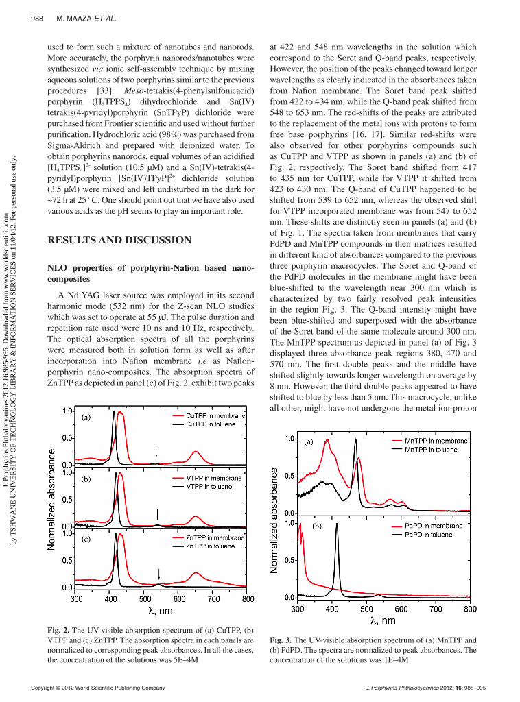
A Nd:YAG laser source was employed in its second harmonic mode (532 nm) for the Z-scan NLO studies which was set to operate at 55 µJ. The pulse duration and repetition rate used were 10 ns and 10 Hz, respectively. The optical absorption spectra of all the porphyrins were measured both in solution form as well as after incorporation into Nafion membrane i.e as Nafion-porphyrin nano-composites. The absorption spectra of ZnTPP as depicted in panel (c) of Fig. 2, exhibit two peaks
at 422 and 548 nm wavelengths in the solution which correspond to the Soret and Q-band peaks, respectively. However, the position of the peaks changed toward longer wavelengths as clearly indicated in the absorbances taken from Nafion membrane. The Soret band peak shifted from 422 to 434 nm, while the Q-band peak shifted from 548 to 653 nm. The red-shifts of the peaks are attributed to the replacement of the metal ions with protons to form free base porphyrins [16, 17]. Similar red-shifts were also observed for other porphyrins compounds such as CuTPP and VTPP as shown in panels (a) and (b) of Fig. 2, respectively. The Soret band shifted from 417 to 435 nm for CuTPP, while for VTPP it shifted from 423 to 430 nm. The Q-band of CuTPP happened to be shifted from 539 to 652 nm, whereas the observed shift for VTPP incorporated membrane was from 547 to 652 nm. These shifts are distinctly seen in panels (a) and (b) of Fig. 1. The spectra taken from membranes that carry PdPD and MnTPP compounds in their matrices resulted in different kind of absorbances compared to the previous three porphyrin macrocycles. The Soret and Q-band of the PdPD molecules in the membrane might have been blue-shifted to the wavelength near 300 nm which is characterized by two fairly resolved peak intensities in the region Fig. 3. The Q-band intensity might have been blue-shifted and superposed with the absorbance of the Soret band of the same molecule around 300 nm. The MnTPP spectrum as depicted in panel (a) of Fig. 3 displayed three absorbance peak regions 380, 470 and 570 nm. The first double peaks and the middle have shifted slightly towards longer wavelength on average by 8 nm. However, the third double peaks appeared to have shifted to blue by less than 5 nm. This macrocycle, unlike all other, might have not undergone the metal ion-proton
Fig. 2. The UV-visible absorption spectrum of (a) CuTPP, (b) VTPP and (c) ZnTPP. The absorption spectra in each panels are normalized to corresponding peak absorbances. In all the cases, the concentration of the solutions was 5E–4M
Fig. 3. The UV-visible absorption spectrum of (a) MnTPP and (b) PdPD. The spectra are normalized to peak absorbances. The concentration of the solutions was 1E–4M
1250107.indd 988 7/14/2012 5:34:07 PM
J. P
orph
yrin
s Ph
thal
ocya
nine
s 20
12.1
6:98
5-99
5. D
ownl
oade
d fr
om w
ww
.wor
ldsc
ient
ific
.com
by T
SHW
AN
E U
NIV
ER
SIT
Y O
F T
EC
HN
OL
OG
Y L
IBR
AR
Y &
IN
FOR
MA
TIO
N S
ER
VIC
ES
on 1
1/04
/12.
For
per
sona
l use
onl
y.

1st Reading
Copyright © 2012 World Scientific Publishing Company J. Porphyrins Phthalocyanines 2012; 16: 989–995
NONLINEAR PHOTONICS PROPERTIES OF PORPHYRINS NANOCOMPOSITES AND SELF-ASSEMBLED PORPHYRINS 989
exchange reaction as expected because the spectrum by large remains unchanged except slight changes in position of the absorbance peaks. The drastic change in the position of the Soret and Q-band in PdPD towards blue indicated a significant structural change that might have taken place in the compound due to palladium replacement by hydrogen. The disappearance of the energy gap between the Soret and Q-band would even further suggest the alteration of the energy band structure of the macrocycle. In fact, it could be also associated with new other possible reaction channels that involve further protonation to give [H4TPP]2+ in the Nafion polymer host matrix. It is to be noted here that each panel of Fig. 2 and Fig. 3 carries the absorption spectra of the porphyrins both in toluene solution and in Nafion membranes. The absorption spectrum profiles of the porphyrins in Nafion host membrane are generally characterized by enhanced absorbance and broadening of the Soret and Q-bands compared with their corresponding results in solution, which may be due to higher concentration of the molecules in the membrane matrix which was dried from toluene solvent at 50 °C. Furthermore, the environmental stability of porphyrins in Nafion membrane was investigated too. This experiment was carried out because some porphyrins in solution are known to undergo degradation when exposed to an external environment [22–24]. The environmental stability tests were carried out by comparing the absorption spectra of the fresh samples with that exposed to external environments for a period of 30 days. The absorption spectra taken from both groups of samples turned out to be quasi-similar, confirming the stability of porphyrin-Nafion nano-composites including those newly studied in this work.
The nonlinear optical properties of the porphyrin molecules in Nafion membrane were investigated using the Z-scan technique. The method is very sensitive to small variation of intensity of the incident laser beam away from the focus area as described in Eq. (4). In order to verify the optical property of the host matrix first, a pure Nafion membrane was scanned firstly using an open aperture Z-scan setup. The data taken from the pure Nafion membrane showed no sign of any nonlinear optical behavior of its own. Consequently, measurements were also carried out using same setup for the various porphyrin-Nafion nano-composites. All nano-composites exhibit nonlinear optical characteristics that can be seen from the valley around the focus of the lens (z = 0) as depicted in Fig. 4. Thus, the creation of such a valley around the focus, where the intensity of the laser beam is very high, describes non- linear intensity dependent absorptions, which is attributed to the presence of porph-yrins in the Nafion host membrane. The experimental investigations were further carried out even at various porphyrin concentrations and the effect turned out to be the same. In all the cases, the spectra showed similar pattern around z = 0, suggesting the existence of a strong NLO property. The normalized open aperture Z-scan data
were then fitted according to the transmittance Eq. (4) with the intent to derive the nonlinear optical parameters of each nano-composite. However, the linear coefficients (a) are the required parameters in order to determine b. Therefore, the values of the linear coefficients can be estimated from the same Z-scan data by careful inspection of the optical characteristics of the spectra. The nonlinear absorption phenomenon occurred at high intensity of the laser beam which starts around the focus of the lens (z = 0). Around this point, Beer’s law that describes the linear absorption phenomenon cannot be applied to determine the linear absorption coefficients (a’s) of the material. However, Beer’s law can be applied to the points where the intensity of the laser source is relatively low. These points are found at the flanks of the Z-scan data (around z = ±12 mm). Hence, by measuring the intensities of the transmitted laser light (I) through the sample and that of non-attenuated intensities (I0) at z = ±12 mm, the linear absorption coefficients of the different porphyrins were determined according to Eq. (2). Using these values of a, we were able to determine b according to Eq. (4). The open aperture Z-scan traces for ZnTPP, as displayed in panel (c) of Fig. 4, showed a very interesting nonlinear optical phenomenon. As it can be observed from the depth of the spectra in Fig. 4, ZnTPP exhibits the strongest nonlinear absorption around z = 0 compared to CuTPP and VTPP. It has effectively blocked the transmission of the high intense laser beam at the concentration 10-4 M. The nonlinear absorption coefficient of ZnTPP is, in fact, greater than the two other porphyrins at 10-4 M (Table 1). The maximum value of b = 0.257 cm/MW is derived for the ZnTPP compound at the same concentration. The fit
Fig. 4. Open aperture Z-scan traces of (a) CuTPP (b) VTPP and (c) ZnTPP along with the fitted function (solid line). The concentration of the solutions was 5E–4M
1250107.indd 989 7/14/2012 5:34:07 PM
J. P
orph
yrin
s Ph
thal
ocya
nine
s 20
12.1
6:98
5-99
5. D
ownl
oade
d fr
om w
ww
.wor
ldsc
ient
ific
.com
by T
SHW
AN
E U
NIV
ER
SIT
Y O
F T
EC
HN
OL
OG
Y L
IBR
AR
Y &
IN
FOR
MA
TIO
N S
ER
VIC
ES
on 1
1/04
/12.
For
per
sona
l use
onl
y.

1st Reading
Copyright © 2012 World Scientific Publishing Company J. Porphyrins Phthalocyanines 2012; 16: 990–995
990 M. MAAZA ET AL.
results in Table 1 showed that the nonlinear absorption coefficients depend naturally upon the concentration of ZnTPP in the solution where the membranes were rinsed for several hours. The values of the absorption coefficients get higher with the concentrations of ZnTPP provided that all other conditions are controlled. Using the same technique employed above, the linear and nonlinear absorption coefficients for the rest of porphyrin compounds are calculated and summarized in Table 1. It is to be noted here that the table contains the absorption coefficients determined at various concentrations of the porphyrin compounds in toluene solvent where the Nafion membranes were kept for 16 h. In all the cases, the values of the nonlinear as well as linear absorption coefficients have increased with increasing concentration of the porphyrins. This is in agreement with the results reported by Couris et al. [24] who have employed a simple two-photon absorption of the on-step type to determine the coefficients. For instance, the nonlinear two-photon absorption coefficient of an inorganic ZnSe compound is reported to be b = 0.0058 cm/MW [13], which is about 1/25 of the value obtained for ZnTPP in this work. The significant difference between the values of the coefficients could be due to the nature of the material and the absorption processes involved in the determination of b. In the current measurements, the depth of the Z-scan valley is very large compared to earlier reports, and hence it cannot be explained by only two-photon absorption process which is often regarded as weak. Therefore, the nonlinear optical properties observed in the investigated porphyrins-nafion nanocomposites could generally result from two-photon and multi-photon absorption processes. As a consequence, the derived nonlinear absorption coefficients are effective nonlinear absorptions instead of two-photon nonlinear absorption.
NLO properties of self-assembly based porphyrin nano-structures
By their chemical nature, the molecular recognition between complementary arrangements of opposite charges and H bond donors and acceptors on the porphyrin tectons contribute significantly to self-assembly phenomena. The self-assembly induced longitudinal geometry in several nanostructured porphyrins where the porphyrin ligands serving as a platform [1–3], could erect novel desirable molecular and nano-materials with large dipole moments and polarizabilities (Fig. 5a) Supramolecular self-assembly is, in general, emerging as a new strategy in chemical synthesis, with the potential of generating non-biological structures having dimensions
Table 1. Summary of the absorption coefficients of the porphyrins
Porphyrin concentration
Linear absorption coefficient (a(mm-1)) and effective nonlinear absorption coefficients (b(cm/MW))
ZnTPPb/a
CuTPPb/a
VTPPb/a
PdPDb/a
MnTPPb/a
5E–4M 0.257/4.480 0.211/3.220 0.198/2.962 0.063/3.370
1E–4M 0.154/3.439 0.178/2.559 0.176/2.521 0.049/2.859
5E–5M 0.148/2.876 0.150/2.323 0.163/2.189 0.034/2.724 0.283/9.940
2.5E–5M 0.254/7.512
Fig. 5. (a) Porphyrins precursors used in nanorods synthesis. The Sn-(IV) center is comprised of (X, X′ ) Cl-, OH-, H2O) bound above and below the porphyrin plane. At pH = 2, X = OH¯ and X′ = H2O (net charge +5) or X = X′ = OH¯ (net charge +4). (b) Schematical view of aspatially oriented self-assembled tubular porphyrin based nano-structures
1250107.indd 990 7/14/2012 5:34:09 PM
J. P
orph
yrin
s Ph
thal
ocya
nine
s 20
12.1
6:98
5-99
5. D
ownl
oade
d fr
om w
ww
.wor
ldsc
ient
ific
.com
by T
SHW
AN
E U
NIV
ER
SIT
Y O
F T
EC
HN
OL
OG
Y L
IBR
AR
Y &
IN
FOR
MA
TIO
N S
ER
VIC
ES
on 1
1/04
/12.
For
per
sona
l use
onl
y.

1st Reading
Copyright © 2012 World Scientific Publishing Company J. Porphyrins Phthalocyanines 2012; 16: 991–995
NONLINEAR PHOTONICS PROPERTIES OF PORPHYRINS NANOCOMPOSITES AND SELF-ASSEMBLED PORPHYRINS 991
of 1–100 nanometers with molecular weights in the range of 104–1010 Daltons. Structures in the upper part of this range of sizes are presently inaccessible through chemical synthesis, and the ability to prepare them would open a route to structures comparable in size to those that can be prepared by microlithography and other techniques of micro-fabrication. As depicted in Fig. 5b in the case of spatially oriented porphyrin nanorods, combining for example the 1-D geometry with the intrinsic strong Soret optical absorption, one could expect high polarization dependence of photonic response of this novel porphyrin based nanostructures. Generally, self-assembly in porphyrins consists of the spontaneous generation of ordered supramolecular architectures from a given set of components under thermodynamic equilibration [1]. In general, the self-assembly process involves various types of bonds and interactions among them one could mention the following [26–30]: (i) covalent bonds that can be formed and broken reversibly (disulfides, vanadate and borate esters), (ii) inorganic metal-ligand bonds (metal salts, organometallic complexes, zinc fingers), (iii) hydrogen bonds (crystalline urea, melamine cyanurate, nucleotide base pairs, h-bonds in proteins), electrostatic interactions involving charges (salt bridges in proteins, cadmium arachidate bilayers), (iv) electrostatic interactions involving dipoles (crystalline IC6H4CN), and (v) hydrophobic interactions (Micelles, Langmuir–Blodgett monolayers on H2O, lipid bilayers, inclusion complexes with cyclodextrins), (vi) van der Waals interactions (n-alkane crystals, urea inclusion complexes) and (vii) aromatic p-stacking and charge transfer (nucleic acids, J-aggregates) [26]. The process, driven by non-covalent interactions, plays a paramount role in determining the remarkable behavior observed in various porphyrins based biological systems, where the final functionality depends ultimately on the shape, size, and binding properties of a limited number of building blocks: the porphyrins and phthalocyanines form a part of such a singular class. Not surprisingly, the self-assembly of porphyrin derivatives so called self-complementary conjugation as well, has attracted considerable attention as an effective path to the synthesis of nanometer scale photonic devices bio-mimicking photosynthetic functions such as light harvesting and charge separation [1–3]. Engineered porphyrin architectures have been formed mainly by hydrogen bonding and metal-ligand coordination; the exploitation of the defined directionality of these bonds has allowed the formation of several topologically complex structures which have been characterized in solution and/or in the solid state through molecular self-assembly.
Molecules equipped with a set of complementary coordination (i.e. ligands and metalloporphyrins) spontaneously self-organize into their self-complementary structures, if they are appropriately designed. Due to this self-complementarity, the stability constants of these substances are far greater than those
of the same ligand-metalloporphyrin combinations without complementarity. The structures must be rigid by the complementarity requirement. Therefore, this is the process of choice for designing specific arrangements of molecular systems with large stability constants. The smallest product is the coordination dimer, where the porphyrin p planes must be slip-stacked by the requirement of mutual self-coordination. In other words, the dimer is essentially a J-type aggregate. This results in some of the key advantages of this coordination organization, since the fluorescence of the substance is not quenched and the absorption band is red-shifted. When the coordination angle is adjusted, larger rings or box-type organizations become possible. The employment of hexa-coordinating metal ions in the porphyrin center is a way to extend the supramolecular structure from the dimer to various macrocycles. In such cases, however, photonic application is limited due to the quenching of singlet excited states by these hexa-coordinating metal ions (mostly transition metal ions). An alternative way to obtain higher-level organized structures is through the connection of self-complementary sets. Various connection methods are associated with new organized structures. Direct connection of two zinc porphyrins at the meso–meso position gives rise to the formation of linear arrays, along which excitons, electrons, or holes can be transferred. Connection via acetylenic bonds allows the highly conjugated zinc porphyrin arrays to develop improved electronic conduction levels. The attachment of energy (or electron) donor or acceptor substituents provides materials for nonlinear optics by increasing the polarization levels of the material. Meta-phenylene linkage of two porphyrins provides a method for obtaining artificial light-harvesting complexes. Not only two but also three or more porphyrins can be connected to yield other interesting organized structures.
Figure 6 depicts a typical self-assembled porphyrins obtained using Shelnut’s group method as it was mentioned previously [31–33]. The typical TEM image of Fig. 6 shows solid cylindrical nanorods and nanotubes with length in micrometres and diameter size ranging between 20 nm and 60 nm which is similar as in our previous study [34]. The difference is attributed to the light sensitivity of these structures, and the ease of the nanostructures’ transition from nanotubes to nanorods when exposed to light [33–34]. One should point out that the nanorods formation is very sensitive to solution conditions, especially pH, because it alters the charge balance and hence the synthesis was conducted under acidic conditions of pH ~ 2. Figure 7 illustrates a typical S, Q and J absorption bands of the porphyrin precursors as well as the nanorods/nanotubes solutions [35]. Exciton theory states that when molecules lie in a head-to-tail arrangement (J-aggregation), the allowed state is lower in energy and hence producing a red-shift to the monomer [35], and since the S band of [H4TPPS4]
2- is red-shifted upon aggregation (J-aggregate). This implies
1250107.indd 991 7/14/2012 5:34:09 PM
J. P
orph
yrin
s Ph
thal
ocya
nine
s 20
12.1
6:98
5-99
5. D
ownl
oade
d fr
om w
ww
.wor
ldsc
ient
ific
.com
by T
SHW
AN
E U
NIV
ER
SIT
Y O
F T
EC
HN
OL
OG
Y L
IBR
AR
Y &
IN
FOR
MA
TIO
N S
ER
VIC
ES
on 1
1/04
/12.
For
per
sona
l use
onl
y.

1st Reading
Copyright © 2012 World Scientific Publishing Company J. Porphyrins Phthalocyanines 2012; 16: 992–995
992 M. MAAZA ET AL.
that the planar porphyrin molecules are stacked in an arrangement where individual porphyrin units are not positioned directly one on top of the other [36–37]. The nanorods composed of [H4TPPS4]
2- J-aggregates are photoconductive and hence the higher absorbance peak of [H4TPPS4]
2- in near UV region (400–460 nm). The decrease in the main absorbance peaks of the precursors and the several peaks identified in the spectrum for the nanorods — 417, 434.5, 495.5, 711 and 974 nm — are similar to what has been reported before [38].
The nonlinear optical properties of the nanotubes/nanorods solutions as well as in form of dried films by
self-assembly are studied in comparison with those of the parent porphyrins. The third order NLO properties of the samples were measured by the Z-scan technique, which is, as mentioned before, the transmittance of the sample through the aperture monitored in the far field as a function of the position of the nonlinear sample in the vicinity of the linear optics focal position. A diode pumped passively mode-locked Nd:YVO4 laser with 30 ps pulse and a repetition rate of 10 Hz was used. In order to determine the nonlinear absorption of these molecules, “open aperture” Z-scan measurements have been carried out. The measurements were performed by using a 1 and 2 mm cell placed to move along the direction of the incident light with various laser beam intensities for each sample and experimental curves were obtained. In the sequel NLO properties of nanorods thin films were investigated by SHG and THG Maker fringes experimental techniques in air. Maker’s fringes characterize the change in the length of the interaction length when this is subjected to a rotational movement. The Gaussian laser beam was focused onto the sample with lens of 250 mm focal length. A motorized rotation stage with the mounted sample allowed the variation of the incident angle with a resolution of 0.50. After the sample, an interferential filter was placed to select the harmonic wave (at 532 nm for SHG or 355 nm for THG). Finally, the SHG and THG signals were registered by rotating the sample. All the experiments were performed at room temperature.
All the samples used in this study showed linear absorption features typical of the free base porphyrins and metallo-porphyrins namely, the high energy B- (Soret) and the low energy Q-bands. The detail view of results of the porphyrin nanorods nonlinear absorption results in comparison with a reference material -C60 are presented in Fig. 8. The obtained curves show that the porphyrin nanorods do not exhibit any nonlinear absorption properties. Unlike the porphyrins, the porphyrins nanostructures exhibited rather low optical nonlinearity
Fig. 6. Transmission electron microscopy micrograph of a typical porphyrin tubular nanostructure obtained via ionic self-assembly of SnTPyP2+ & H4TPPS4
2- based used precursors
Fig. 7. UV-vis spectra of the precursors and porphyrin nanorods formed after mixing of the precursors. Peak labels with the letter ‘s’ are for the SnTPyP2+ monomer, the values with an ‘h’ are for the H4TPPS4
2- monomer, and the remaining values are for the porphyrin nanorods formed
Fig. 8. Normalized Z-scan transmittance curves measured with 2 mm cell
1250107.indd 992 7/14/2012 5:34:11 PM
J. P
orph
yrin
s Ph
thal
ocya
nine
s 20
12.1
6:98
5-99
5. D
ownl
oade
d fr
om w
ww
.wor
ldsc
ient
ific
.com
by T
SHW
AN
E U
NIV
ER
SIT
Y O
F T
EC
HN
OL
OG
Y L
IBR
AR
Y &
IN
FOR
MA
TIO
N S
ER
VIC
ES
on 1
1/04
/12.
For
per
sona
l use
onl
y.

1st Reading
Copyright © 2012 World Scientific Publishing Company J. Porphyrins Phthalocyanines 2012; 16: 993–995
NONLINEAR PHOTONICS PROPERTIES OF PORPHYRINS NANOCOMPOSITES AND SELF-ASSEMBLED PORPHYRINS 993
which is purely refractive and not absorptive in Z-scan measurements. Unfortunately, a trend in the behavior of the cubic nonlinear susceptibility depends of the type of incorporated metal ion. To clearly understand this feature, one must bear in mind that one of the important characteristics of a molecule which determines the optical linearity is the molecule’s dipole moment, which in turn is determined by the space symmetry of the molecule. The free base form of the studied porphyrins was completely symmetric in the plane of the p-conjugated ring and cannot produce a dipole moment in this place as that the central nitrogen atoms of porphyrin in a free base form can be protonated depending on the porphyrin structure. In both protonated or non-protonated state, porphyrin structure of the free base is symmetric with respect to the ring plane and as a result they have no dipole moment perpendicular to the ring plane. This may explain why the spectra only show negligible nonlinear absorption properties for the porphyrin as compared to the reference, C60 [39]. The second and third order nonlinear optical properties of nanorods thin films were investigated by Second and Third harmonic generation using maker fringes experimental techniques. Porphyrins are a class of macrocyclic compounds with a tetrapyrrolic rings system. The symmetry groups to which tetrapyrrole molecules belong contain the inversion operator i. For such systems, the tensor of nonlinear susceptibility c(2) = 0. However, the molecular symmetry could be reduced in several instances so that quadratic nonlinear effects become allowed. The SHG efficiency of the investigated nanorods was found to be very low. The results are highlighted in Fig. 9. Nonlinear optical properties due to a second-order nonlinear susceptibility c(2) does not appear for porphyrins nanorods because of their molecular symmetry. On the other hand the THG measurements showed more promising results. Figure 10 displays an example of characteristic maker fringes patterns corresponding to the nanorods samples. All THG measurements performed in s and p polarizations. A negligible difference was
found between the experimental data obtained for s and p polarizations. For the THG measurements in picoseconds regime, just the electronic contribution was estimated (c3elec). For data analysis, the model of Kubodera and Koayashi was used [40].
( )
( )
3 3C,S
33SS
2L I.
1 I
w
w
c= p c
(6)
( )3
Lc6 n - n
w
w w
l= (7)
Here Lc,S – coherence length, l is thickness of sample (~500 nm), IS
w and I3w are THG intensities of the silica and the studied material, respectively (I3w = I3w[sample+substrate] - I3w[substrate]), n3w = 1.476, nw = 1.449. This model compares directly the maximal amplitudes of third harmonic light intensity for studied material to those of a 1 mm silica plate used as reference. c(3)
silica = 2.0 × 10⁻²² m2/V2 [15] at lw = 1064 nm. The obtained results are summarized in Table 2. In the table, from one side it seems that the solvents affect the effective third order nonlinear susceptibility (c(3)
elec) but also the differences between the calculated values are very small and the differences are within the experimental error. Moreover, the samples are not ideally
Fig. 10. An example of THG Maker fringes signal as function of the angle of incidence in self-assembled nanorods sample
Table 2. The values of the third harmonic generation measu rements
Sample X3, (10-21 m2/V2)
Fused silica 0.2
H2SO4 1.37 ± 0.13
H2C2O4 1.87 ± 0.18
H3PO4 1.12 ± 0.10
HNO3 1.94 ± 0.10
HCL 2.01 ± 0.27Fig. 9. SHG Maker fringes signal as function of the angle of incidence of self-assembled nanorods
1250107.indd 993 7/14/2012 5:34:13 PM
J. P
orph
yrin
s Ph
thal
ocya
nine
s 20
12.1
6:98
5-99
5. D
ownl
oade
d fr
om w
ww
.wor
ldsc
ient
ific
.com
by T
SHW
AN
E U
NIV
ER
SIT
Y O
F T
EC
HN
OL
OG
Y L
IBR
AR
Y &
IN
FOR
MA
TIO
N S
ER
VIC
ES
on 1
1/04
/12.
For
per
sona
l use
onl
y.

1st Reading
Copyright © 2012 World Scientific Publishing Company J. Porphyrins Phthalocyanines 2012; 16: 994–995
994 M. MAAZA ET AL.
homogenous, and thicknesses which are important for calculation of c(3)
elec are different for each sample. This also affects the final result. Nevertheless, the third order nonlinear susceptibility was observed to be by one order larger than for fused silica, and the value of the cubic nonlinear susceptibility c(3)
elec depends substantially on the type of chelated metal ion. The enhanced third-order nonlinearities are mainly attributed to strong interfacial interaction in and between nanorods and local field enhancements.
CONCLUSION
The nonlinear optical absorption of two different major porphyrins based system were presented: (i) porphyrins-Nafion nano-composites and (ii) self-assembled porphyrins based nanorods/nanotubes. In the first part, it was demonstrated that the porphyrins in Nafion matrix exhibited strong nonlinear optical absorption properties at high intense laser irradiation of wavelength 532 nm. The ZnTPP-nafion nano-composite compound exhibited the strongest NLO property at high concentration while that of the MnTPP was higher at low concentration. The values of the nonlinear absorption coefficients generally depend on the amount of porphyrins embedded in Nafion host membranes. Furthermore, this experiments confirmed that all porphyrins, once protected by Nafion host matrix, are found to be well-protected from environmental degradation after 30 days of exposure to external environment. The second part dealt with artificial porphyrin nanorods, synthesized by self-assembly and molecular recognition method. Several aspects of formation of nanotubes and other rodlike structures based on porphyrins are yet to be understood properly. Unlike the prophyrin precursors, the self-assembled porphyrins nanostructures exhibited rather low optical nonlinearity which was purely refractive and not absorptive in the Z-scan measurements. While their SHG efficiency was found to be very low, the THG investigations have yielded very interesting results. It was observed one order larger the third order nonlinear susceptibility than for fused silica. Thus, the porphyrin nanorods and nanotubes may have many potential uses in photonics applications if spatially oriented.
Acknowledgements
The authors are grateful to the Nanosciences African Network, the Abdus Salam ICTP, Trieste-Italy, the Science & Technology Directorate of the French Embassy in South Africa, the Centre National pour la Recherche Scientifique, Paris-France, the National Research Foundation of South Africa as well as to iThemba LABS.
REFERENCES
1. Kadish KM, Smith KM and Guilard R. xxxxxxxxxxxxxxxxxx, Vol. 6, Academic Press: New York, 2000.
2. Kobuke Y. Struct. Bond. 2006; 121: 49–104. 3. Gunter MJ. Struct. Bond. 2006; 121: 265–295. 4. Senge MO, Fazekas M, Notaras EGA, Blau WJ,
Zawadzka M, Locos OB and Ni Mhuircheartaigh EM. Adv. Mater. 2007; 19: 2737.
5. Fu ST, Zhu XJ, Zhou GJ, Wong WY, Ye C, Wong K and Li ZY. Euro. J. Inorg. Chem. 2007; 14: xxx-xxx.
6. Gu B, Guo B, Wang XJ, Chen J, Ding J and Wang HT. Phys. Rev. A 2006; 73: 065603.
7. Barbosa Neto NM, Oliveria SL, Guedes I, Dinelli LR, Mis- oguti L, Mendonca CR, Batista AA, Zilio SC and Braz J. J. Chem. Soc. 2006; 17: 1377.
8. Suslick KS, Rakow NA, Kosal ME and Chou JH. J. Porphyrins Phthalocyanines 2000; 4: 407.
9. Miles P. Appl. Opt. 1999; 38: 566. 10. Ajgaonkar MU. New Jersey Institute of Technology
2001; Nonlinear Optical Properties of Nanostruc-tures, PhD dissertation.
11. Blau WJ, Byrne H, Dennis WM and Kelly JM. Opt. Comms. 1985; 56: 25.
12. Martin RB, Li HP, Gu L, Kumar S, Sanders SM and Sun YP. Opt. Mater. 2005; 27: 1340.
13. Meloney C, Byrne H, Dennis WH, Blau W and Kelly JM. J. Chem. Phys. 1988; 21: 121.
14. Rao VDGLN, Aranda FJ, Roach JF and Remy DE. Appl. Phys. Lett. 1991; 58: 1241.
15. Wen TC, Hwang LC, Lin WY, Chen CH and Wu CH. Chem. Phys. 2003; 45: 286.
16. Humphrey J and Kuciauskas D. J. Phys. Chem. 2004; 108: 12016.
17. Sheik-Bahae M, Said AA, Wei TH, Hagan DJ and Van Stryland EW. J. Quantum Electron. 1990; 26: 760.
18. Hailu G, Tessema G. Diop Ngom B and Maaza M. Appl. Phys. A 2009; 96: 685–689.
19. Tessema G, Hailu G and Maaza M. JNOPM. 2011; 20: 175–182.
20. Nandakumar P, Vijayan C and Murti VYGS. Opt. Commun. 2000; 185: 457.
21. Nandakumar P, Vijayan C and Murti YVGS. J. Appl. Phys. 2002; 91: 1509.
22. Sendhil K, Vijayan Cand Kothiyal MP. Opt. Mater. 2005; 27: 1606.
23. Zhao F, Zhang J and Kaneko M. J. Photochem. Pho-tobiol., A 1998; 53: 119.
24. Courise S. J. Phys. B: At. Mol. Opt. Phys. 1995; 28: 4537.
25. Sate CT and Whitesides GM. J. Am. Chem. Soc. 1991; 113: 712.
26. Rebek J. Angew. Chem., Int. Ed. EngI. 1990; 29: 245. 27. Hunter CA and Sanders JKM. J. Am. Chem. Soc.
1990; 112: 5525. 28. Pavletlch NP and Pabo CO. Science 1991; 252: 809. 29. Breslow R. Science 1982; 218: 532. 30. Shen YR. Nature 1989; 337: 519–525. 31. Rodenberger DC, Heflin JR and Garito AF. Phys.
Rev. B 1995; 51: 3234.
1250107.indd 994 7/14/2012 5:34:13 PM
J. P
orph
yrin
s Ph
thal
ocya
nine
s 20
12.1
6:98
5-99
5. D
ownl
oade
d fr
om w
ww
.wor
ldsc
ient
ific
.com
by T
SHW
AN
E U
NIV
ER
SIT
Y O
F T
EC
HN
OL
OG
Y L
IBR
AR
Y &
IN
FOR
MA
TIO
N S
ER
VIC
ES
on 1
1/04
/12.
For
per
sona
l use
onl
y.

1st Reading
Copyright © 2012 World Scientific Publishing Company J. Porphyrins Phthalocyanines 2012; 16: 995–995
NONLINEAR PHOTONICS PROPERTIES OF PORPHYRINS NANOCOMPOSITES AND SELF-ASSEMBLED PORPHYRINS 995
32. Wang Z, Medforth CJ and Shelnutt JA. J. Am. Chem. Soc. 2004; 126: 15954–15955.
33. Mongwaketsi N, Ndungu P, Nechaev A, Maaza M and Sparrow R. J. Porphyrins Phthalocyanines 2010; 14: 446–451.
34. Egharevba GO, George RC and Maaza M. Synth. React. Inorg. Met.-Org. Chem. 2008; 38: 681–687.
35. Schwab AD, Smith DE, Bond-Watts B, Johnston DE, Hone J, Johnson AT, de Paula JC and Smith WF. Nano Letters 2004; 4: 1261–1265.
36. Ruthard C, Schmidt M and Grohn Macromol F. Rapid Commun. 2011; 32: 706–711.
37. Medforth CJ, Wang J, Martin KE, Song Y, Jacob-sen L and Shelnutt JA. Chem. Commun. 2009; 47: 7261–7277.
38. Bezerra Jr AG, Borissevitch IE, De Araujo ASL and De Araujo CB. Chem. Phys. Lett. 2000; 318: 511–516.
39. Kubodera K and Kobayashi H. Mol. Cryst. Liq. Cryst. 1990; 182: 103–113.
1250107.indd 995 7/14/2012 5:34:13 PM
J. P
orph
yrin
s Ph
thal
ocya
nine
s 20
12.1
6:98
5-99
5. D
ownl
oade
d fr
om w
ww
.wor
ldsc
ient
ific
.com
by T
SHW
AN
E U
NIV
ER
SIT
Y O
F T
EC
HN
OL
OG
Y L
IBR
AR
Y &
IN
FOR
MA
TIO
N S
ER
VIC
ES
on 1
1/04
/12.
For
per
sona
l use
onl
y.