HLA-A2 Subtypes Are Functionally Distinct in Peptide Binding and Presentation
http://www.elsevier.com/locate/bba
Biochimica et Biophysica Act
Review
A hierarchy of functionally important relaxations within myoglobin based
on solvent effects, mutations and kinetic model
David Dantskera, Uri Samunia, Joel M. Friedmana,*, Noam Agmonb,*
aDepartment of Physiology and Biophysics, Albert Einstein College of Medicine, Bronx, New York 10461, USAbDepartment of Physical Chemistry and the Fritz Haber Research Center, The Hebrew University, Jerusalem 91904, Israel
Received 20 February 2005; received in revised form 30 March 2005; accepted 6 April 2005
Available online 25 April 2005
Abstract
Geminate CO rebinding in myoglobin is studied for two viscous solvents, trehalose and sol–gel (bathed in 100% glycerol) at several
temperatures. Mutations in key distal hemepocket residues are used to eliminate or enhance specific relaxation modes. The time-resolved data
are analyzed with a modified Agmon–Hopfield model which is capable of providing excellent fits in cases where a single relaxation mode is
dominant. Using this approach, we determine the relaxation rate constants of specific functionally important modes, obtaining also their
Arrhenius activation energies. We find a hierarchy of distal pocket modes controlling the rebinding kinetics. The ‘‘heme access mode’’
(HAM) is responsible for the major slow-down in rebinding. It is a solvent-coupled cooperative mode which restricts ligand return from the
xenon cavities. Bulky side-chains, like those His64 and Trp29 (in the L29W mutant), operate like overdamped pendulums which move over
and block the binding site. They may be either unslaved (His64) or moderately slaved (Trp29) to the solvent. Small side-chain relaxations,
most notably of leucines, are revealed in some mutants (V68L, V68A). They are conjectured to facilitate inter-cavity ligand motion. When all
relaxations are arrested (H64L in trehalose), we observe pure inhomogeneous kinetics with no temperature dependence, suggesting that
proximal relaxation is not a factor on the investigated timescale.
D 2005 Elsevier B.V. All rights reserved.
Keywords: Conformational dynamics; Diffusion; Geminate recombination; Myoglobin; Sol–gel; Trehalose; Relaxation
1. Introduction
It has become apparent that proteins are inherently
complex materials. Even the simplest protein reactions
exhibit layers of complexity that were not anticipated a few
decades ago [1,2]. A case in point is the heavily studied
process by which carbon monoxide binds to the heme in
myoglobin (Mb) [3]. What is truly remarkable is that Mb, on
an almost continual basis, persists in yielding new insights
into protein properties and behavior. Most notably is the
exposure of the functional role of protein–solvent inter-
actions, conformational disorder and conformational
dynamics.
1570-9639/$ - see front matter D 2005 Elsevier B.V. All rights reserved.
doi:10.1016/j.bbapap.2005.04.002
* Corresponding authors. Joel M. Friedman is to be contacted at fax: +1
718 4308819. Noam Agmon, fax: +972 2 6513742.
E-mail addresses: [email protected] (J.M. Friedman),
[email protected] (N. Agmon).
Much of the work onMb has been directed at exposing the
biophysical principles responsible for the observed kinetic
patterns associated with ligand binding or rebinding. Early
solution phase work on Mb supported the view that ligand
binding and release were straight forward processes that could
be understood in terms of a simple ligand entry into and
escape from the protein and bond formation and dissociation
between the heme-iron and the ligand. The pioneering work
of Frauenfelder and coworkers [4] exposed the inherent
complexity of this seemingly simple biophysical process. At
cryogenic temperatures, multiple kinetic phases were
observed instead of the single bimolecular phase observed
in solution. With increasing temperature or decreasing
viscosity, new phases appeared, ultimately yielding the simple
pattern observed in solution. A comparison of the kinetics at
the extremes in temperature revealed an inverse temperature
effect where the average barrier controlling the ligand binding
process exhibits a substantial increase with temperature.
a 1749 (2005) 234 – 251
D. Dantsker et al. / Biochimica et Biophysica Acta 1749 (2005) 234–251 235
Further studies conducted at cryogenic temperatures or
high viscosities [5–20] have exposed multiple CO recombi-
nation phases for Mb. In the early cryogenic studies [4], the
fastest phase, attributed to rebinding from within the distal
hemepocket (DHP), was referred to as Process I. Over a range
of very low temperatures (10–120 K in a 3:1 glycerol–water
glass), this single phase exhibits an Arrhenius temperature
dependence and clear indications that it is derived from a
frozen distribution of conformations with vastly different
kinetic barriers. With increasing temperature or decreasing
viscosity, another phase emerges that is significantly slower
and kinetically more homogeneous than the distributed
kinetics of Process I. This slower phase was referred to as
the matrix process based on the assumption that the rebinding
for this phase originated from the ligands that diffused from
the DHP and were now localized somewhere in the protein
matrix. At still higher temperatures, the emergent slowest
phase, termed the solvent phase, was attributed to rebinding
involving ligands that had escaped into the solvent. In this
description, the inverse temperature effect arises from the
temperature dependent shift from the initially dominant
Process I to the slower matrix process.
Alternative explanations for the origin of the emergent
phases and the inverse temperature effect were based on
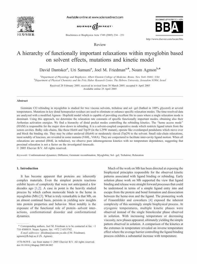
relaxation effects [21,22]. The idea is demonstrated with the
aid of Fig. 1. The initial Mb+CO distribution prior to
relaxation arises from the MbCO structure and has a lower
distribution of kinetic barriers for recombination. Upon
photodissociation, the initial distribution starts to relax so
that the recombination reaction slows down. At cryogenic
temperatures or high viscosity, relaxation is slowed and the
recombination from within the protein (geminate recombi-
nation) is governed by a more favorable distribution of
Fig. 1. The relaxation model using the relations of Section 2.4 with
parameters from Table 2 and x0=5 d.u. The initial distribution, p(x,0) was
the equilibrium distribution on the MbCO potential, VA(x). On the potential
V(x) of the photodissocited protein, it can undergo either recombination
(with the x-dependent rate function k(x)), or relaxation to regions of smaller
k(x).
barriers than for the relaxed population. The temperature/
viscosity dependence of the observed kinetics is then
explained primarily as an effect on the relaxation rate.
The relaxation scenario was corroborated by several
authors [6,23–28], but has not become universally accepted
[29]. One reason for this is that explanations for the origin
of the different kinetic phases based on ligand diffusion
within the protein gained significant momentum based on
the identification of discrete non-heme docking sites for the
ligand within the globin. It was directly demonstrated that
dissociated ligands can access internal cavities that arise
from packing defects referred to as Xe cavities [30–33]. A
new set of labels for the different phases emerged when
cryogenic and time-resolved X-ray crystallography led to
the identification of well-defined ligand localization sites
within the protein matrix [33–42]. A, B, C, D and S were
used to specify the state in which the ligand is bound to the
heme, dissociated but localized in the DHP, in the Xe4
cavity, in the Xe1 cavity and in solution respectively. The
Xe4 cavity is adjacent to the DHP, while Xe1 is well
removed from the DHP being located on the proximal side
of the heme plane. Temperature derivative FTIR measure-
ments at cryogenic temperatures [33,43–45] showed that
with decreasing viscosity, increasing temperature or increas-
ing intervals of optical pumping, the dissociated ligand
could be found localized in sites that are progressively more
distant from the binding site.
While ligand rebinding from the different sites contribute
to the origin of the multiple phases seen at high viscosity,
this does not preclude a possibly significant role for
relaxation phenomena. Several studies point to a role for
relaxation in modulating the kinetics of ligand rebinding. To
account for the different sites, the relaxation model [21,22]
was extended to include relaxation in two different tiers
(say, the B and Xe1 states) and this yielded good fits to the
rebinding data over the whole temporal and temperature
ranges [46]. Sol–gel encapsulation techniques show that
populations derived from different encapsulation protocols
exhibit different CO rebinding kinetics [19]. Encapsulation
protocols designed to trap Mb populations having deoxy
conformations yield samples that show slower CO kinetics
[18] and higher dioxygen affinity [47] than for samples
initially encapsulated as the fully liganded derivatives.
These results are consistent with an increasing kinetic
barrier associated with relaxation from the CO conforma-
tional population to the deoxy conformational population.
Finally, temperature derivative spectroscopy also indicates
that protein conformational dynamics and ligand migration
can both play an important role in ligand binding [43].
The question, therefore, is not whether protein relaxa-
tion is functionally important, but rather how to separate
out the plethora of different ‘‘functionally important
motions’’. There could be proximal vs. distal control
[48,49], slow helix motions vs. faster side-chain relaxa-
tions, iron out-of-plane motion vs. heme plane distortions.
Which one contributes to the observed kinetics under what
D. Dantsker et al. / Biochimica et Biophysica Acta 1749 (2005) 234–251236
conditions and during which time window remains largely
an enigma.
The present work seeks to illustrate and highlight the
solution-sensitive factors that contribute to the kinetic
phases of Mb. The study builds experimentally on the
earlier studies that used trehalose and sol–gel encapsulation
to modulate dynamics and kinetics [18,19]. In these
environments, large amplitude solvent-coupled protein
fluctuations can be arrested, leaving side-chain and other
local relaxation modes as the main affectors. In addition,
point mutations are employed either to eliminate (H64L) or
enhance (L29W) the relaxation of a specific side-chain. The
double mutants H64L/V68X are also analyzed. The time-
resolved kinetics are compared quantitatively with a
modification of the bounded-diffusion model [21,22], in
which the sink term was modified from exponential to a
steeper, Gaussian dependence on the conformational coor-
dinate. This allows for a more accurate description of the
steep power-law dependence seen in these mutants. The
analysis provides an estimate for the activation energies of
the different relaxation modes as a function of solvent. The
emerging picture is that all of the barriers for ligand
migration in Mb reside in various protein coordinates,
which either retard or promote the transitions between the
ligand residence sites.
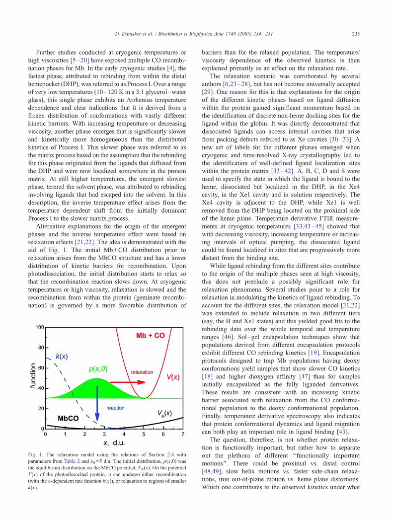
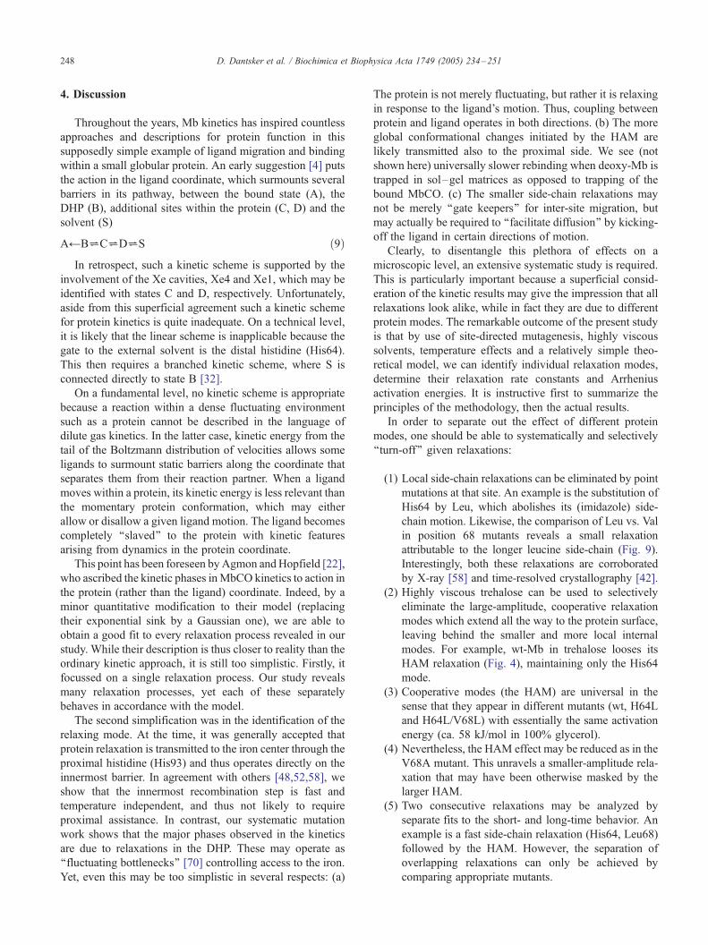
Fig. 2. The hemepocket of SW-Mb for (A) wt (under 7 atm of Xe) and its
(B) H64L and (C) L29W mutants. Mutated residues are yellow; Xe
cavities in Panel (A) are dotted white and the bound ligand is rendered
red: (A) water, (B,C) CO. Atomic coordinates taken from the following
PDB files (resolution): (A) 1J52 (1.9 A) [79]; (B) 2MGC (1.9 A) [50];
(C) 1DO1 (1.5 A) [33].
2. Methodology
Our methodology involves a judicious combination of
Mb mutants, viscous solvents and a theoretical model for
relaxation-dominated kinetics, which are used for disentan-
gling some of the protein modes involved in MbCO
kinetics.
2.1. Mb mutants
In the present study, the focus is on wild-type (wt) Mb
and a few of its mutants. The wt-Mb is either horse-heart
myoglobin (HMb) or sperm whale myoglobin (SWMb). The
structure of the DHP for wt Mb is depicted in Fig. 2(A).
This structure was measured under 7 atm of Xe, so that three
(out of the four) xenon atoms (residing in the corresponding
Xe cavities) are depicted in the figure (dotted spheres). It is
seen that protein fluctuations can open a migration pathway
for the ligand (between residues V68 and L29), leading
from the DHP to the Xe4 cavity. From there, the ligand
could migrate behind Ile107 to Xe2, which connects to the
Xe1 cavity on the back-side of the heme plane. This is
where the trajectory of the photodissociated CO often
appears to end [33,41] (particularly under conditions
disfavoring its exit via the His64 gate).
A set of mutant SW-Mbs was chosen to help expose the
role of DHP architecture, side-chain relaxation (of DHP
residues) and water in the partitioning of CO recombination
into kinetic phases originating from CO within either the
DHP or the Xe cavities. Fig. 2(B,C) shows the DHP for two
of the mutants used in the present studies. The first mutation
(H64L) eliminates one relaxation mode, whereas the second
(L29W) enhances another mode.
In the H64L mutant [50], the ‘‘distal histidine’’ H64 (or
E7 in the helix-oriented notation) is replaced by a leucine
(Fig. 2(B)). This replacement introduces a side-chain that
does not shift position upon CO binding to the heme. As a
result, the Leu64 side-chain, in contrast to the His64
imidazole, is not anticipated to be a major contributing
factor with respect to the relaxation related phenomena [50].
This conclusion is corroborated by studies on the Soret band
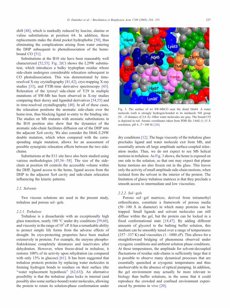
Fig. 3. The surface of wt SW-MbCO near the distal His64. A water
molecule (red) is strongly hydrogen-bonded to its imidazole NH group
(N. . .O distance of 2.6 A). Other water molecules are gray. The bound CO
is depicted in red. Atomic coordinates taken from PDB file 1A6G (1.15 A
resolution, pH 6, T=100 K) [36].
D. Dantsker et al. / Biochimica et Biophysica Acta 1749 (2005) 234–251 237
shift [48], which is markedly reduced by leucine, alanine or
valine substitutions at position 64. In addition, these
replacements make the distal pocket hydrophobic [50], thus
eliminating the complications arising from water entering
the DHP subsequent to photodissociation of the heme-
bound CO [51].
Substitutions at the B10 site have been reasonably well
characterized [52,53]. Fig. 2(C) shows the L29W substitu-
tion, which introduces a bulky tryptophan residue whose
side-chain undergoes considerable relaxation subsequent to
CO photodissociation. This was demonstrated by time-
resolved X-ray crystallography [41,42], cryo-trapping X-ray
studies [33], and FTIR-time derivative spectroscopy [43].
Relaxation of the tyrosyl side-chain of Y29 in multiple
mutations of SW-Mb has been observed in X-ray studies
comparing their deoxy and liganded derivatives [54,55] and
in time-resolved crystallography [40]. In all of these cases,
the relaxation positions the aromatic side-chain over the
heme-iron, thus blocking ligand re-entry to the binding site.
The studies on Mb mutants with aromatic substitutions in
the B10 position also show that the dynamics of the
aromatic side-chain facilitates diffusion out of the DHP into
the adjacent Xe4 cavity. We also consider the H64L/L29W
double mutation, which when compared with the corre-
sponding single mutation, allows for an assessment of
possible synergistic relaxation effects between the two side-
chains.
Substitutions at the E11 site have also been studied using
various methodologies [45,56–58]. The size of the side-
chain at position 68 controls the accessible volume within
the DHP, ligand access to the heme, ligand access from the
DHP to the adjacent Xe4 cavity and side-chain relaxation
influencing the kinetic patterns.
2.2. Solvents
Two viscous solutions are used in the present study,
trehalose and porous sol–gels.
2.2.1. Trehalose
Trehalose is a dissacharide with an exceptionally high
glass transition, nearly 100 -C under dry conditions [59,60],
and viscosity in the range of 105 cP. It has a remarkable ability
to protect simple life forms from the adverse effects of
drought. Its cryo-protecting properties have been studied
extensively in proteins. For example, the enzyme phospho-
fruktokinase completely denatures and inactivates after
dehydration. However, when freeze-dried in trehalose, it
restores 100% of its activity upon rehydration (as compared
with only 13% in glucose) [61]. It has been suggested that
trehalose protects proteins by replacing water molecules in
forming hydrogen bonds to residues on their surface (the
‘‘water replacement hypothesis’’ [62,63]). An alternative
possibility is that the trehalose glass locks in internal (and
possibly also some surface-bound) water molecules, allowing
the protein to retain its solution-phase conformation under
dry conditions [12]. The huge viscosity of the trehalose glass
precludes ligand and water molecule exit from Mb, and
essentially arrests all large amplitude surface-coupled relax-
ation modes. Thus, we do not expect to see Mb helical
motions in trehalose. As Fig. 3 shows, the heme is exposed on
one side to the solution, so that one may expect that planar
heme motions are also frozen out in the glass. This leaves
only the activity of small amplitude side-chain motions, when
isolated from the solvent in the interior of the protein. The
limitation of glassy trehalose matrices is that they preclude a
smooth access to intermediate and low viscosities.
2.2.2. Sol–gels
Porous sol–gel matrices, derived from tetramethyl
orthosilicates, constitute a framework of porous media
(50–100 A in diameter) in which many proteins can be
trapped. Small ligands and solvent molecules can still
diffuse within the gel, but the protein can be locked in a
fixed conformational state [18,47]. By adding different
amounts of glycerol to the bathing buffer solution, this
medium can be smoothly tuned over a range of temperatures
(257–337 K) and viscosities (1–1000 cP). This allows for a
straightforward bridging of phenomena observed under
cryogenic conditions and ambient solution phase conditions.
At these temperatures, the amplitude for solvent-decoupled
fluctuations of residue side-chains is sufficiently large that it
is possible to observe many dynamical processes that are
essentially quenched at cryogenic temperatures and thus
unobservable in the absence of optical pumping. In addition,
the gel environment may actually be more relevant to
biology than buffer solutions, in the sense that it could
reproduce the crowded and confined environment experi-
enced by proteins in vivo [20].
D. Dantsker et al. / Biochimica et Biophysica Acta 1749 (2005) 234–251238
2.3. Experimental procedures for determining rebinding
kinetics
The experimental data reported herein is based on the
classical procedure of laser-photolysis of bound MbCO [4],
which monitors CO recombination by time-dependent
absorption measurements in the Soret region of the heme.
The normalized absorbance is proportional to the survival
probability of the unbound heme, S(t), by time t after
photolysis. The kinetic traces are displayed on a log–log
plot of S(t) versus time. This helps expose multiple phases
occurring over a very wide time range. In this plot, a straight
line represents power-law (e.g., inhomogeneous) kinetics,
whereas exponential rebinding shows up as a nearly flat
plateau which turns rapidly into a vertical line, intersecting
the time axis at a point that is roughly the inverse of the
exponential rate constant.
Horse heart Mb was commercially obtained from Sigma
(Saint Louis) and used without further purification other
than centrifugation to eliminate particulates. Samples of
SW-Mb, both the wild type and mutants, were generously
provided by John S. Olson of Rice University. Sol–gel
encapsulated samples were prepared as a thin layer lining
the bottom portion of either 5 or 10 mm diameter NMR
tubes as previously described [18]. After sample preparation
and a period of aging, the bathing buffer in which the sol–
gel samples were immersed was replaced by an excess of
pure glycerol.
Glassy samples derived from trehalose were prepared as
previously described [19]. Briefly, COMb was dissolved in
a CO purged pH7.0 solution of trehalose (100 mg/ml) to
make a 0.5–1.0 mM stock solution of Mb. A drop of stock
solution was placed on a glass plate and allowed to dry in a
dessicator under a CO atmosphere. After several hours, the
resulting highly viscous sample was heated at 65 -C for 30
to 40 min. The resulting glassy sample was then stored in a
dessicator in order to maintain its very low level of
hydration. For the kinetic measurements, the sample was
transferred to a temperature controlled chamber that is
purged with dry nitrogen.
Kinetic measurements were typically carried out on
samples contained in standard 10 mm or 1 mm stoppered
cuvettes placed in a custom-built dry N2 purged variable
temperature cuvette holder (�15 to 65 -C). Time-resolved
kinetic measurements were carried out using 8 ns 532 nm
pulses at 1 Hz from a Nd:YAG laser (Minilite, Continuum,
Santa Clara, CA) as a photodissociation source and a greatly
attenuated continuous wave 442 nm probe beam from a
He:Cd laser to monitor time-dependent changes in absorp-
tion. Details of the apparatus, data collection and data
display can be found in previous publications [18,19,64,65].
We have checked the CO concentration dependence for
the 100% glycerol case at 3.5 -C. The kinetic trace for wt-
MbCO was insensitive to the replacement of the CO purged
glycerol with N2 purged glycerol (after waiting several days
for equilibration). The trehalose-trapped MbCO was also
insensitive to external [CO]. Thus, these high-viscosity
samples appear to be maintaining a ‘‘closed’’ system where
the internal CO recycles over and over again with minimal
connection with the solvent population.
2.4. Theoretical model
The bounded diffusion model [21,22] is a simple kinetic
model which couples ligand migration with protein relax-
ation, allowing for a smooth transition from inhomogeneous
to relaxation dominated kinetics. Inhomogeneous (distrib-
uted) kinetics is observed at short times, high viscosities or
low temperatures. It switches over to relaxation at longer
times. At very long times, a steady-state distribution is
established, and only then the ligand migration step can be
described by a single rate constant, as assumed when
writing down chemical kinetic schemes.
Using a relaxation model, it has been possible to fit
Mb–CO kinetics up to intermediate times [12,66]. This is
because the kinetics are complex, involving at least two
relaxation phases. Recently, a two-tier extension of this
model was shown to describe HMb–CO kinetics over the
whole time and temperature range [46]. A similar approach
was also used to depict single-enzyme action [67].
Here, we focus on conditions where a single relaxation
mode is dominant. Hence, we recourse to the simpler
original model, but with a modified ‘‘sink term’’ which
better describes the fast inhomogeneous phase observed for
these mutants [18,19]. The simple model (Fig. 1) recognizes
two states for the ligand. State A represents a bound ligand,
as usual. The second state is best described as separated
from A by a single ‘‘relaxation mode’’. Depending on the
sample, solvent, etc., the ligand in this state may be in the
pocket (B) or in one of the Xe cavities, and the mode may
be, for example, a side-chain rotation which controls a
‘‘gate’’ providing access to the heme iron.
The gate conformation is described by a continuous
variable x. We denote by x =0 the extreme situation where
the gate moves completely out of the way, allowing the ligand
free access to the binding site. As x increases, the gate
progressively blocks access to the heme iron, so that the
binding rate coefficient, k(x), decreases monotonically. In the
applications of the model, thus far, k(x) was an exponential
[12,21,22,46,66]. This dependence is not sufficiently strong
to describe the data. A stronger x-dependence is depicted by a
Gaussian sink adopted in our work
k xð Þ ¼ A exp � bx2� �
; ð1Þ
which depends on the two parameters, A and b. The parabolic
activation energy in k(x) could be explained by a simple
model in which both transition state and reactants are
harmonic in x, but the force constant in the transition state
is much larger than that of the reactants.
The remaining aspects of the model are as previously des-
cribed by Agmon and Hopfield [21,22]. In state A, the gating
D. Dantsker et al. / Biochimica et Biophysica Acta 1749 (2005) 234–251 239
mode is subject to effective harmonic potentials, e.g., VA(x)=
aA(x�xA)2 (the potentials are given here in units of the therQ
mal energy, kBT). Thus, prior to photodissociation, the bound
protein had the (normalized) conformational distribution
pA xð Þ ¼ exp � VA xð Þ½ �=Z V
0
exp � VA xð Þ½ �dx: ð2Þ
Following the dissociation process, the gate experiences
a modified field of force, depicted by the potential
V xð Þ ¼ a x � x0ð Þ2; ð3Þ
where x >0 and x0>xA. Under these new conditions, the
distribution p(x,t) of unbound Mb can either rebind with the
rate function k(x) or relax toward the deoxy conformation
(i.e., from xA to x0). The relaxation is described as a
diffusion process, giving rise to the corresponding Smolu-
chowski equation
flp x;tð Þflt
¼ Dfl
flxe�V xð Þ fl
flxeV xð Þp x;tð Þ � k xð Þ p x;tð Þ: ð4Þ
This equation is subject to the initial condition that
p(x ,0) =pA(x), the equilibrium distribution prior to
photolysis.
A few limiting cases can be solved analytically [21].
When k(x)=0, Eq. (4) reduces to the so-called Ornstein–
Uhlenbeck problem. In particular, the average value of the
coordinate relaxes exponentially, <x>=exp(�krelt), with the
(uni-molecular) relaxation rate coefficient
krel ¼ 2aD: ð5Þ
A major goal of the present work is to extract krel from
the observed kinetics and study its temperature dependence.
When k(x)m0, Eq. (4) can be solved only in special cases
e.g., when k(x) is a delta function [68] or a parabola [69].
The first problem may represent relaxation in an excited-
state undergoing conical intersection, the second, passage
through a fluctuating bottleneck [70]. For a general form of
k(x), one may obtain only asymptotic solutions for protein
relaxation which is infinitely slow or infinitely fast (the
inhomogeneous and homogeneous limits, respectively)
p x;tð Þ¨pA xð Þexp � k xð Þt½ �; DY0;
p x;tð Þ¨peq xð Þexp � keqt� �
; DYV; ð6Þ
where peq(x ) = exp[�V(x )] / Xexp[�V (x )]dx and keq =
Xk(x)peq(x)dx is the average reaction rate constant under
equilibrium conditions. More detailed expansions in D and
1/D can be found elsewhere [71,72]. Hence, this model
interpolates naturally between the inhomogeneous and
homogeneous (or chemical kinetic) limits.
When D is finite, p(x ,t) tends to a steady-state
distribution at asymptotically long-times, and the kinetics
are again controlled by a single rate constant obtained by
averaging k(x) over this distribution. It is close in magnitude
to keq and depicts the ultimate return process from the cavity
to the iron. Thus, for times which are long as compared to
the relaxation time, exponential kinetics are established, in
agreement with conventional single-step chemical kinetics.
However, at intermediate times, the model shows much
richer behavior due to relaxational effects.
For arbitrary D and k(x), Eq. (4) can be solved for p(x,t)
only numerically. The survival probability of the unbound
Mb is then calculated from the integral
S tð Þ ¼Z V
0
p x;tð Þdx: ð7Þ
The numerical solution of the partial differential Eq. (4) is
repeated many times until appropriate parameters are found
for fitting experimental data. This task is accomplished by the
user-friendly Windows application for solving Spherically-
Symmetric Diffusion Problem (SSDP, ver. 2.65) [73].
In this work, we focus on fitting S(t) for a given system
(mutant/solvent) at several temperatures (T). In such a
temperature series, we typically vary at most two parame-
ters. One is always the diffusion constant, D. The second is
one of the two parameters defining the potential in Eq. (3):
either x0 or a. From these, we calculate the relaxation rate
constant, krel, via Eq. (5). Its temperature dependence can
differ from that of D when also a varies with T. We
invariably find that krel obeys an Arrhenius equation
krel ¼ Arel exp � ED=kBTð Þ; ð8Þ
where Arel is a pre-exponential, and kB is Boltzmann’s
constant. The activation energy is denoted by ED, to indicate
that it represents activation of a diffusive motion. This may
arise from diffusion on an underlying rough potential [74].
ED plays an important role in the identification of the
relaxation mode because, we conjecture, a localized side-
chain motion will have a small ED, whereas a more extended
cooperative mode (which involves many amino-acid resi-
dues) should exhibit a larger ED value. The larger ‘‘rough-
ness’’ in the latter case arises from the more extensive
coupling to the hierarchy of protein conformational substates.
3. Results
Our main goal is to utilize the approach discussed above
to expose the role of the various residues aligning the DHP
on the observed rebinding kinetics. For this purpose, we
focus on extracting the relaxation rate coeffcients krel from
the intermediate-time kinetics, discussing the short- and
long-time behavior on a more qualitative level.
3.1. The innermost barrier
It may be thought that the innermost barrier at the iron is
the major determinant of the CO rebinding rate, and
therefore the modulation of the iron out-of-plane distance
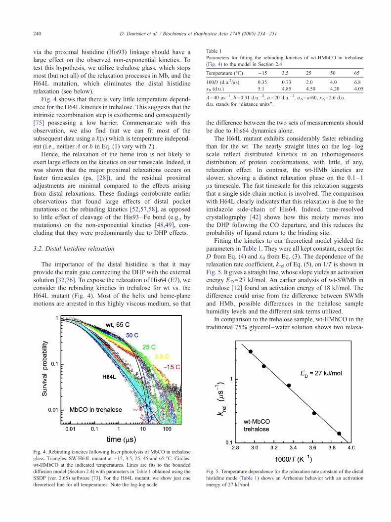
Table 1
Parameters for fitting the rebinding kinetics of wt-HMbCO in trehalose
(Fig. 4) to the model in Section 2.4
Temperature (-C) �15 3.5 25 50 65
100D (d.u.2/As) 0.35 0.73 2.0 4.0 6.8
x0 (d.u.) 5.1 4.85 4.50 4.20 4.05
A=40 As�1, b =0.31 d.u.�2, a =20 d.u.�2, aA=a/60, xA=2.6 d.u.
d.u. stands for ‘‘distance units’’.
D. Dantsker et al. / Biochimica et Biophysica Acta 1749 (2005) 234–251240
via the proximal histidine (His93) linkage should have a
large effect on the observed non-exponential kinetics. To
test this hypothesis, we utilize trehalose glass, which stops
most (but not all) of the relaxation processes in Mb, and the
H64L mutation, which eliminates the distal histidine
relaxation (see below).
Fig. 4 shows that there is very little temperature depend-
ence for the H64L kinetics in trehalose. This suggests that the
intrinsic recombination step is exothermic and consequently
[75] possessing a low barrier. Commensurate with this
observation, we also find that we can fit most of the
subsequent data using a k(x) which is temperature independ-
ent (i.e., neither A or b in Eq. (1) vary with T).
Hence, the relaxation of the heme iron is not likely to
exert large effects on the kinetics on our timescale. Indeed, it
was shown that the major proximal relaxations occurs on
faster timescales (ps, [28]), and the residual proximal
adjustments are minimal compared to the effects arising
from distal relaxations. These findings corroborate earlier
observations that found large effects of distal pocket
mutations on the rebinding kinetics [52,57,58], as opposed
to little effect of cleavage of the His93–Fe bond (e.g., by
mutations) on the non-exponential kinetics [48,49], con-
cluding that they were predominantly due to DHP effects.
3.2. Distal histidine relaxation
The importance of the distal histidine is that it may
provide the main gate connecting the DHP with the external
solution [32,76]. To expose the relaxation of His64 (E7), we
consider the rebinding kinetics in trehalose for wt vs. the
H64L mutant (Fig. 4). Most of the helix and heme-plane
motions are arrested in this highly viscous medium, so that
Fig. 4. Rebinding kinetics following laser photolysis of MbCO in trehalose
glass. Triangles: SW-H64L mutant at �15, 3.5, 25, 45 and 65 -C. Circles:
wt-HMbCO at the indicated temperatures. Lines are fits to the bounded
diffusion model (Section 2.4) with parameters in Table 1 obtained using the
SSDP (ver. 2.65) software [73]. For the H64L mutant, we show just one
theoretical line for all temperatures. Note the log-log scale.
the difference between the two sets of measurements should
be due to His64 dynamics alone.
The H64L mutant exhibits considerably faster rebinding
than for the wt. The nearly straight lines on the log–log
scale reflect distributed kinetics in an inhomogeneous
distribution of protein conformations, with little, if any,
relaxation effect. In contrast, the wt-HMb kinetics are
slower, showing a distinct relaxation phase on the 0.1–1
As timescale. The fast timescale for this relaxation suggests
that a single side-chain motion is involved. The comparison
with H64L clearly indicates that this relaxation is due to the
imidazole side-chain of His64. Indeed, time-resolved
crystallography [42] shows how this moiety moves into
the DHP following the CO departure, and this reduces the
probability of ligand return to the binding site.
Fitting the kinetics to our theoretical model yielded the
parameters in Table 1. They were all kept constant, except for
D from Eq. (4) and x0 from Eq. (3). The dependence of the
relaxation rate coefficient, krel of Eq. (5), on 1/T is shown in
Fig. 5. It gives a straight line, whose slope yields an activation
energy ED=27 kJ/mol. An earlier analysis of wt-SWMb in
trehalose [12] found an activation energy of 18 kJ/mol. The
difference could arise from the difference between SWMb
and HMb, possible differences in the trehalose sample
humidity levels and the different sink terms utilized.
In comparison to the trehalose sample, wt-HMbCO in the
traditional 75% glycerol–water solution shows two relaxa-
Fig. 5. Temperature dependence for the relaxation rate constant of the distal
histidine mode (Table 1) shows an Arrhenius behavior with an activation
energy of 27 kJ/mol.
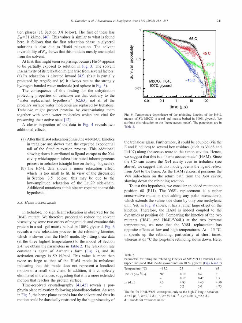
Table 2
Parameters for fitting the rebinding kinetics of SW-MbCO mutants H64L
(upper lines) and H64L/V68L (lower lines) in 100% glycerol (Figs. 6 and 9)
Temperature (-C) �15.2 25 45 65
100 D (d.u.2/As) ‘‘0’’ 0.12 0.6 2
– 0.12 0.42 1.5
x0 (d.u.) 5.5 4.83 4.65 4.50
– 5.2 5.0 4.75
The fits for H64L/V68L correspond only to the high-T long-t behavior.
A=60 As�1, b =0.37 d.u.�2, a =35 d.u.�2, aA=a/60, xA=2.6 d.u.
d.u. stands for ‘‘distance units’’.
Fig. 6. Temperature dependence of the rebinding kinetics of the H64L
mutant of SW-MbCO in a sol–gel matrix bathed in 100% glycerol. We
attribute this relaxation to the ‘‘heme access mode’’. The parameters are in
Table 2.
D. Dantsker et al. / Biochimica et Biophysica Acta 1749 (2005) 234–251 241
tion phases (cf. Section 3.8 below). The first of these has
ED=31 kJ/mol [46]. This values is similar to what is found
here. It follows that the first relaxation phase in glycerol
solutions is also due to His64 relaxation. The solvent
invariability of ED shows that this mode is mostly uncoupled
from the solvent.
At first, this might seem surprising, because His64 appears
to be partially exposed to solution in Fig. 3. The solvent
insensitivity of its relaxation might arise from several factors:
(a) Its relaxation is directed inward [42]; (b) it is partially
protected by Arg45; and (c) it always retains the strongly
hydrogen-bonded water molecule (red sphere in Fig. 3).
The consequence of this finding for the dehydration
protecting properties of trehalose are that contrary to the
‘‘water replacement hypothesis’’ [62,63], not all of the
protein’s surface water molecules are replaced by trehalose.
Trehalose might protect proteins by encapsulating them
together with some water molecules which are vital for
preserving their active state [12].
A closer inspection of the data in Fig. 4 reveals two
additional effects:
(a) After theHis64 relaxation phase, thewt-MbCOkinetics
in trehalose are slower than the expected exponential
tail of the fitted relaxation process. This additional
slowing down is attributed to ligand escape to the Xe4
cavity,whichappearstobeadistributed,inhomogeneous
process in trehalose (straight line on the log–log scale).
(b) The H64L data shows a minor relaxation effect,
which is too small to fit. In view of the discussion
in Section 3.5 below, this may be due to the
low-amplitude relaxation of the Leu29 side-chain.
Additional mutations at this site are required to test this
hypothesis.
3.3. Heme access mode
In trehalose, no significant relaxation is observed for the
H64L mutant. We therefore proceed to reduce the solvent
viscosity by some two orders of magnitude and examine this
protein in a sol–gel matrix bathed in 100% glycerol. Fig. 6
reveals a new relaxation process in the rebinding kinetics,
which is slower than the His64 mode. By fitting these data
(at the three highest temperatures) to the model of Section
2.4, we obtain the parameters in Table 2. The relaxation rate
constant is again of Arrhenius form (Fig. 7), and its
activation energy is 59 kJ/mol. This value is more than
twice as large as that of the His64 mode in trehalose,
indicating that this mode does not represent a localized
motion of a small side-chain. In addition, it is completely
eliminated in trehalose, suggesting that it is a more extended
motion that reaches the protein surface.
Time-resolved crystallography [41,42] reveals a por-
phyrin plane relaxation following photodissociation. As seen
in Fig. 3, the heme plane extends into the solvent and thus its
motion could be drastically restricted by the huge viscosity of
the trehalose glass. Furthermore, it could be coupled (via the
E and F helices) to several key residues (such as Val68 and
Ile107) along the access route to the xenon cavities. Hence,
we suggest that this is a ‘‘heme access mode’’ (HAM). Since
the CO can access the Xe4 cavity even in trehalose (see
above), we suggest that this mode governs the ligand return
from Xe4 to the heme. As the HAM relaxes, it positions the
V68 side-chain on the return path from the Xe4 cavity,
slowing down the rebinding reaction.
To test this hypothesis, we consider an added mutation at
position 68 (E11). The V68L replacement is a rather
conservative mutation (not adding any polar interactions),
which extends the valine side-chain by only one methylenic
unit. Yet, as Fig. 8 shows, it has a rather large effect on the
kinetics. Therefore, the HAM is indeed coupled to the
dynamics at position 68. Comparing the kinetics of the two
mutants (H64L and H64L/V64L) at the two extreme
temperatures, we note that the V68L replacement has
opposite effects at low and high temperatures. At �15 -C,it speeds up the rebinding, particularly at short times,
whereas at 65 -C the long-time rebinding slows down. Here,

Fig. 7. Temperature dependence for the relaxation rate coefficients for the
H64L mutant in 100% glycerol (full circles; Table 2) and for the slow
relaxation phase of H64L/V68L (empty squares; Table 2) and wt-SWMb
(empty circles; lower lines in Table 9). The linear fit is to the H64L data.
D. Dantsker et al. / Biochimica et Biophysica Acta 1749 (2005) 234–251242
we focus on the high temperature behavior and leave the
low T behavior to the next subsection.
First, we suggest that the slower long-time kinetics at 65
-C for the V68L replacement indicates that the longer Leu68
side-chain is more efficient in blocking ligand return from the
cavities, making the HAM extend over a longer period of
time. Incidentally, this can be used to argue that the observed
kinetics are due to CO migration to the xenon cavities, and
not to the solvent process (CO escape and bimolecular re-
entry from solution). To corroborate this conclusion, we
compare our results with the bimolecular oxygen entry
kinetics of Scott et al. [32]. By fitting their bi-exponential
decays to simple kinetic schemes, they obtain entry, binding
and escape rate constants. Comparing these data for wt and
the V68L mutant shows a 25% increase in the binding rate
constant for the mutant, which agrees with its enhanced
Fig. 8. Comparison of the rebinding kinetics of the H64L and H64L/V68L
mutants at two temperatures. Solvent: sol–gel bathed in 100% glycerol.
See text for discussion.
rebinding as observed at�15 -C in Fig. 8. However, the entry
and escape rate constants increase for V68L even more [32].
Hence, if the long-time exponential decay observed at high
temperatures in our figure were due to the solvent process, it
would be expected to speed-up for the H64L/V64L mutant
(as compared with H64L). The fact that we find just the
opposite indicates that this phase is not due to the solvent
process, but rather to migration between the DHP and the
internal xenon cavities, as suggested above.
Fig. 9 shows the complete temperature series for the
H64L/V68L double mutant. To check that the HAM is also
operative here, we first fit its long-time kinetics at the three
highest temperatures, ignoring the short-time behavior
(which will be considered below). We use the same
parameters as for the H64L mutant above (Table 2), except
that we increase x0 to account for the longer temporal extent
of the relaxation process, and modify D accordingly. These
fits are depicted in Fig. 9 by the dashed lines. The
temperature dependence of krel shown in Fig. 7 is similar
to that of H64L, giving ED=53 kJ/mol and ED=57 kJ/mol
when the three or two highest temperatures are used,
respectively. To within the experimental and fitting errors,
this is identical with ED for the HAM in the H64L mutant.
The HAM time-constants are thus independent of the X68
side-chain (X =V or L), commensurate with its origin as a
many-residue cooperative mode.
3.4. Leu68 side-chain relaxation
As Fig. 8 shows, at low temperatures, CO rebinding
within the H64L/V68L mutant is observably faster than for
the H64L mutant. This also holds at short times for higher T.
The longer Leu68 side-chain may enhance reactivity in
several ways: (a) obstruction of the escape route into Xe4;
(b) reduction in the DHP volume; and (c) interference with
Fig. 9. The rebinding kinetics for the H64L/V68L mutant of SW-MbCO (in
a sol–gel matrix bathed in 100% glycerol) reveals two different relaxation
modes. The theoretical model was fitted separately to each mode: the HAM
at the 3 highest temperatures (dashed lines; parameters in Table 2) and the
Leu68 side-chain at the 3 lowest temperatures (lines; parameters in Table 3).

Table 3
Parameters for fitting the low-temperature rebinding kinetics of SW-
Mb(H64L/V68L)CO in 100% glycerol (Fig. 9, full lines)
Temperature (-C) �15 3.5 25
100D (d.u.2/As) 1.55 2.5 4.5
A=65 As�1, b =0.16 d.u.�2, a =35 d.u.�2, aA=a/60, xA=2.6 d.u.,
x0=6.55 d.u.
d.u. stands for ‘‘distance units’’.
D. Dantsker et al. / Biochimica et Biophysica Acta 1749 (2005) 234–251 243
the DHP docking site for CO [77], observed in the X-ray
structure [78] of Fig. 10. Valine in position 68 may be the
most favorable residue for stabilizing the CO in this docking
site. As seen in the figure, the shortest distances between the
docked-CO carbon and the three nearest residues are all
around 3.3 A. This must be a ‘‘comfortable’’ distance for
CO docking. The lengthening of residue 68 side-chain by
one methylenic unit could shorten this distance sufficiently
to push the CO to a less favorable docking location, from
which rebinding is faster.
In addition to faster reactivity, a closer inspection of Fig.
9 shows that the H64L/V64L double-mutant exhibits a small
amplitude relaxation already at �15 -C, which is slightly
faster than 1 As. We fit the fast relaxation phase to our model
at the three lowest temperatures (full lines, parameters
collected in Table 3). At 25 -C, the HAM relaxation already
sets in, and it overshadows the smaller side-chain relaxation
which is superimposed on it. Fig. 11 shows that krel is rather
large and its activation energy is small, ED=17 kJ/mol
(considerably smaller than the value of ca. 58 kJ/mol found
for the HAM). This is reasonable for a small side-chain
moving within a cavity. Thus, we attribute it to the Leu68
side-chain relaxation within the DHP.
Indeed, an X-ray study of position 68 mutants [58] found
large differences in side-chain conformation only when
leucine or isoleucine is substituted for Val68. Fig. 12 shows
a top view of the DHP of the V68L mutant in the bound
(carbonmonoxy) and unbound (deoxy) states. The two side-
chains that undergo large changes are those of His64 and
Leu68, and both relaxations were accordingly identified in
our analysis. The leucine rotates around its Ch–Cg angle,
executing a ‘‘flip-flop’’ like motion. This motion may be of
functional significance because, as the side-chain flips, it
can give a small ‘‘kick’’ to the CO, propelling it from one
cavity to another.
Fig. 10. The docked CO (red) in the hemepocket of photolyzed SW-MbCO
at 40 K. Some key distances from the carbon (white lines) or oxygen (cyan
lines) are given (in A). Atomic coordinates from the PDB file 1AJH
representing X-ray measurements at pH 6 and 1.7 A resolution [78].
3.5. Leu29 side-chain relaxation
We have considered above two amino-acids at position 68
possessing side-chains of intermediate size. It is instructive
to consider the two limiting cases, of very bulky vs. very
small side-chains. These are represented by phenylalanine
and alanine, respectively. It has been shown [58] that in the
first case the Xe4 cavity is nearly blocked, resulting in very
fast geminate recombination, whereas in the second case the
DHP and Xe4 merge into a single large cavity, in which the
ligand moves quite freely. With this in mind, we have studied
the two SW-Mb double mutants, H64L/V68F and H64L/
V68A, in 100% glycerol sol–gel matrices.
Fig. 13 shows the CO rebinding kinetics of H64L/V68A
(Panel A) and H64L/V68F (Panel B) as a function of
temperature. The fitting parameters are collected in Tables 4
and 5, respectively, while their relaxation rate coeffcients
(krel) are summarized in Fig. 11. Commensurate with the
larger size of Phe, its short-time/low-temperature inhomo-
geneous rebinding kinetics are faster and krel is somewhat
smaller than for Ala in position 68. The remarkable
observation, however, is that for these two extreme cases,
the activation energy (ED) is, within the error limits,
identical. It is also considerably lower than that of the
HAM.
Thus, this relaxation mode cannot be due to the side-
chain motion of either mutant nor to the HAM. Since its
Fig. 11. Temperature dependence for the relaxation rate coefficients of the
H64L/V68X mutants of Mb in glycerol, where X =L (Table 3), A (Table 4)
and F (Table 5).
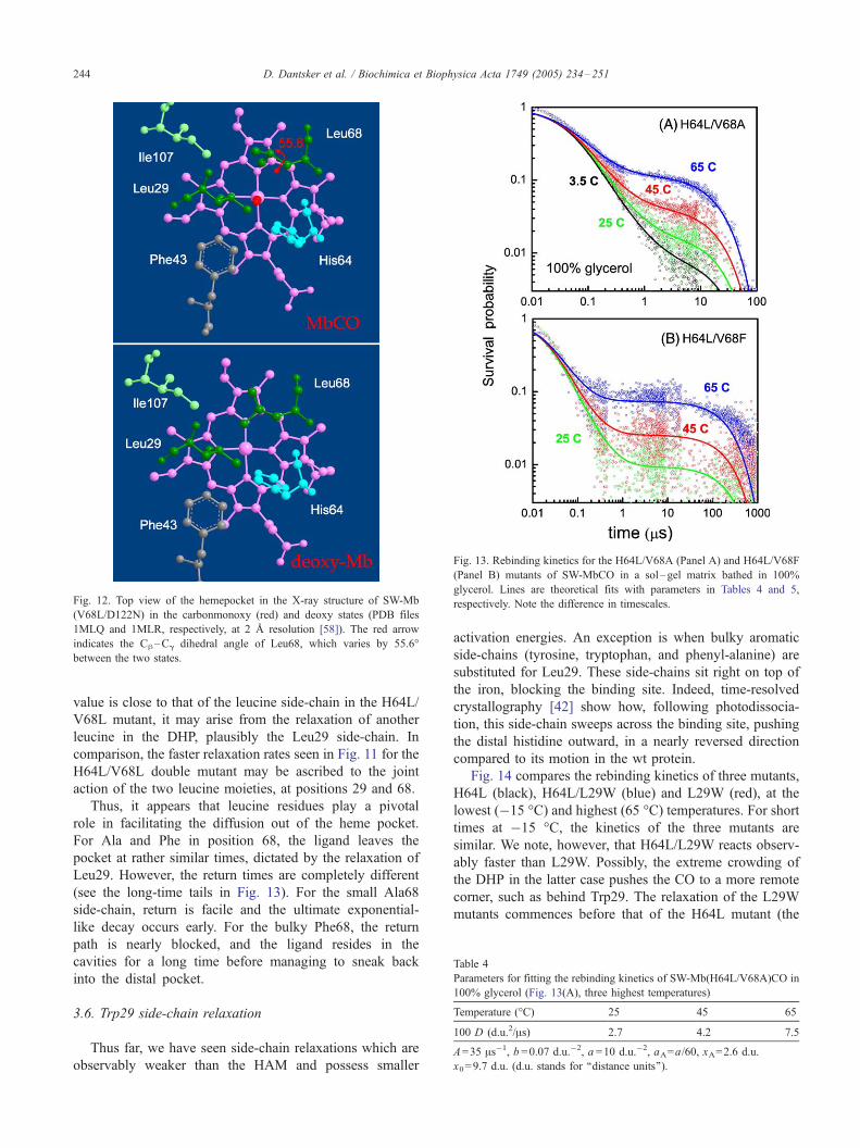
Fig. 12. Top view of the hemepocket in the X-ray structure of SW-Mb
(V68L/D122N) in the carbonmonoxy (red) and deoxy states (PDB files
1MLQ and 1MLR, respectively, at 2 A resolution [58]). The red arrow
indicates the Ch–Cg dihedral angle of Leu68, which varies by 55.6-
between the two states.
Fig. 13. Rebinding kinetics for the H64L/V68A (Panel A) and H64L/V68F
(Panel B) mutants of SW-MbCO in a sol–gel matrix bathed in 100%
glycerol. Lines are theoretical fits with parameters in Tables 4 and 5,
respectively. Note the difference in timescales.
Table 4
Parameters for fitting the rebinding kinetics of SW-Mb(H64L/V68A)CO in
100% glycerol (Fig. 13(A), three highest temperatures)
Temperature (-C) 25 45 65
100 D (d.u.2/As) 2.7 4.2 7.5
A=35 As�1, b =0.07 d.u.�2, a =10 d.u.�2, aA=a/60, xA=2.6 d.u.
x0=9.7 d.u. (d.u. stands for ‘‘distance units’’).
D. Dantsker et al. / Biochimica et Biophysica Acta 1749 (2005) 234–251244
value is close to that of the leucine side-chain in the H64L/
V68L mutant, it may arise from the relaxation of another
leucine in the DHP, plausibly the Leu29 side-chain. In
comparison, the faster relaxation rates seen in Fig. 11 for the
H64L/V68L double mutant may be ascribed to the joint
action of the two leucine moieties, at positions 29 and 68.
Thus, it appears that leucine residues play a pivotal
role in facilitating the diffusion out of the heme pocket.
For Ala and Phe in position 68, the ligand leaves the
pocket at rather similar times, dictated by the relaxation of
Leu29. However, the return times are completely different
(see the long-time tails in Fig. 13). For the small Ala68
side-chain, return is facile and the ultimate exponential-
like decay occurs early. For the bulky Phe68, the return
path is nearly blocked, and the ligand resides in the
cavities for a long time before managing to sneak back
into the distal pocket.
3.6. Trp29 side-chain relaxation
Thus far, we have seen side-chain relaxations which are
observably weaker than the HAM and possess smaller
activation energies. An exception is when bulky aromatic
side-chains (tyrosine, tryptophan, and phenyl-alanine) are
substituted for Leu29. These side-chains sit right on top of
the iron, blocking the binding site. Indeed, time-resolved
crystallography [42] show how, following photodissocia-
tion, this side-chain sweeps across the binding site, pushing
the distal histidine outward, in a nearly reversed direction
compared to its motion in the wt protein.
Fig. 14 compares the rebinding kinetics of three mutants,
H64L (black), H64L/L29W (blue) and L29W (red), at the
lowest (�15 -C) and highest (65 -C) temperatures. For short
times at �15 -C, the kinetics of the three mutants are
similar. We note, however, that H64L/L29W reacts observ-
ably faster than L29W. Possibly, the extreme crowding of
the DHP in the latter case pushes the CO to a more remote
corner, such as behind Trp29. The relaxation of the L29W
mutants commences before that of the H64L mutant (the
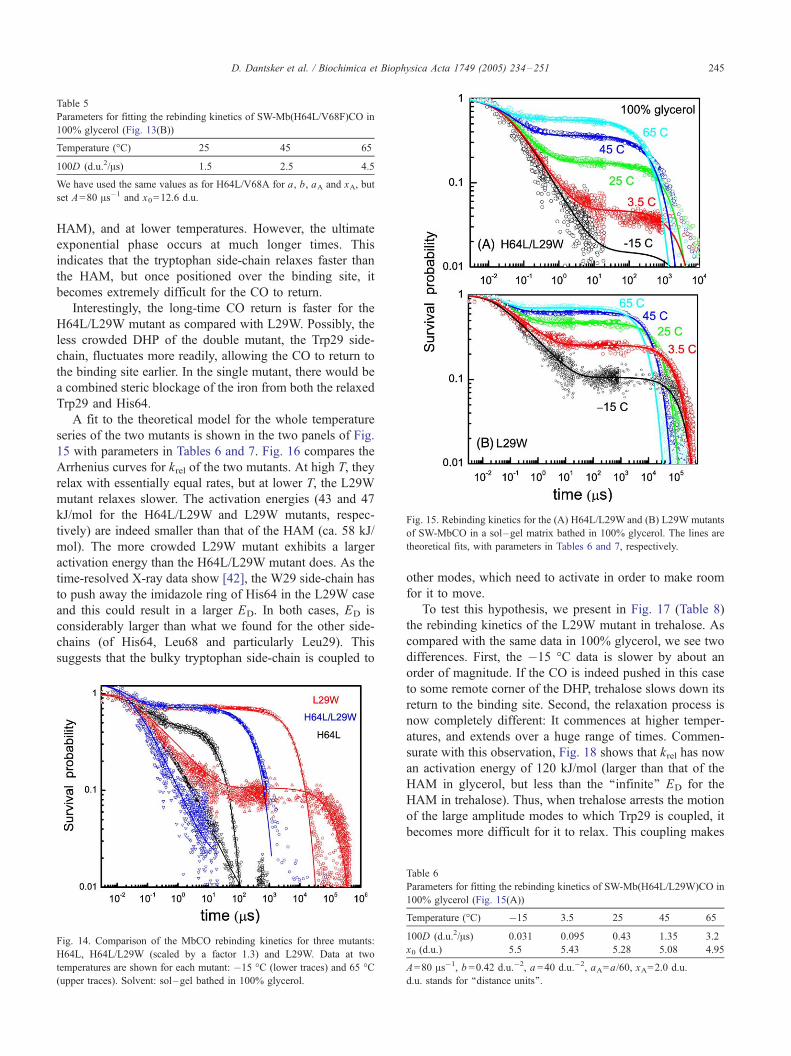
Table 5
Parameters for fitting the rebinding kinetics of SW-Mb(H64L/V68F)CO in
100% glycerol (Fig. 13(B))
Temperature (-C) 25 45 65
100D (d.u.2/As) 1.5 2.5 4.5
We have used the same values as for H64L/V68A for a, b, aA and xA, but
set A=80 As�1 and x0=12.6 d.u.
Fig. 15. Rebinding kinetics for the (A) H64L/L29Wand (B) L29W mutants
of SW-MbCO in a sol–gel matrix bathed in 100% glycerol. The lines are
theoretical fits, with parameters in Tables 6 and 7, respectively.
D. Dantsker et al. / Biochimica et Biophysica Acta 1749 (2005) 234–251 245
HAM), and at lower temperatures. However, the ultimate
exponential phase occurs at much longer times. This
indicates that the tryptophan side-chain relaxes faster than
the HAM, but once positioned over the binding site, it
becomes extremely difficult for the CO to return.
Interestingly, the long-time CO return is faster for the
H64L/L29W mutant as compared with L29W. Possibly, the
less crowded DHP of the double mutant, the Trp29 side-
chain, fluctuates more readily, allowing the CO to return to
the binding site earlier. In the single mutant, there would be
a combined steric blockage of the iron from both the relaxed
Trp29 and His64.
A fit to the theoretical model for the whole temperature
series of the two mutants is shown in the two panels of Fig.
15 with parameters in Tables 6 and 7. Fig. 16 compares the
Arrhenius curves for krel of the two mutants. At high T, they
relax with essentially equal rates, but at lower T, the L29W
mutant relaxes slower. The activation energies (43 and 47
kJ/mol for the H64L/L29W and L29W mutants, respec-
tively) are indeed smaller than that of the HAM (ca. 58 kJ/
mol). The more crowded L29W mutant exhibits a larger
activation energy than the H64L/L29W mutant does. As the
time-resolved X-ray data show [42], the W29 side-chain has
to push away the imidazole ring of His64 in the L29W case
and this could result in a larger ED. In both cases, ED is
considerably larger than what we found for the other side-
chains (of His64, Leu68 and particularly Leu29). This
suggests that the bulky tryptophan side-chain is coupled to
Fig. 14. Comparison of the MbCO rebinding kinetics for three mutants:
H64L, H64L/L29W (scaled by a factor 1.3) and L29W. Data at two
temperatures are shown for each mutant: �15 -C (lower traces) and 65 -C
(upper traces). Solvent: sol–gel bathed in 100% glycerol.
other modes, which need to activate in order to make room
for it to move.
To test this hypothesis, we present in Fig. 17 (Table 8)
the rebinding kinetics of the L29W mutant in trehalose. As
compared with the same data in 100% glycerol, we see two
differences. First, the �15 -C data is slower by about an
order of magnitude. If the CO is indeed pushed in this case
to some remote corner of the DHP, trehalose slows down its
return to the binding site. Second, the relaxation process is
now completely different: It commences at higher temper-
atures, and extends over a huge range of times. Commen-
surate with this observation, Fig. 18 shows that krel has now
an activation energy of 120 kJ/mol (larger than that of the
HAM in glycerol, but less than the ‘‘infinite’’ ED for the
HAM in trehalose). Thus, when trehalose arrests the motion
of the large amplitude modes to which Trp29 is coupled, it
becomes more difficult for it to relax. This coupling makes
Table 6
Parameters for fitting the rebinding kinetics of SW-Mb(H64L/L29W)CO in
100% glycerol (Fig. 15(A))
Temperature (-C) �15 3.5 25 45 65
100D (d.u.2/As) 0.031 0.095 0.43 1.35 3.2
x0 (d.u.) 5.5 5.43 5.28 5.08 4.95
A=80 As�1, b =0.42 d.u.�2, a =40 d.u.�2, aA=a/60, xA=2.0 d.u.
d.u. stands for ‘‘distance units’’.

Table 7
Parameters for fitting the rebinding kinetics of SW-Mb(L29W)CO in 100%
glycerol (Fig. 15(B))
Temperature (-C) �15 3.5 25 45 65
100D (d.u.2/As) 0.03 0.13 0.8 2.9 5.0
x0 (d.u.) 6.8 6.15 5.8 5.65 5.5
A=30 As�1 (except at the 2 lowest temperatures, which originate from a
different experimental series, where A=47 As�1), b =0.42 d.u.�2 (except for
�15 -C, where b =0.35 d.u.�2), a =14 d.u.�2, aA=a/60, xA=2.6 d.u.
Fig. 17. Rebinding kinetics for the L29W mutant of SW-MbCO in
trehalose. Lines are theoretical fits, with parameters in Table 8.
D. Dantsker et al. / Biochimica et Biophysica Acta 1749 (2005) 234–251246
the Trp29 mode ‘‘slaved’’ to the solvent to a larger extent
than the other side-chain modes.
3.7. Promoting and retarding modes
The modes we identified in the various Mb mutants can
be divided into ‘‘promoting’’ and ‘‘retarding’’ modes.
Relaxation in either mode slows down CO rebinding
following its photolysis, but they do so in different ways.
Promoting modes (the Leu side-chains) enhance the
transitions between sites, so they speed up migration from
the DHP to the Xe cavities (a phenomenon sometimes
termed ‘‘facilitated diffusion’’). In contrast, retarding modes
(His64, Leu29, HAM) block the access to the iron.
The difference between the two types is visually observ-
able at long times, as the ligand returns to the binding site
from the cavities. Promoting modes present no barrier for the
return process, so that the final exponential is nearly
temperature independent. For example, the long-time tails
in Fig. 13 are superimposable by proper scaling of the curves.
The retarding modes introduce a measurable obstacle for the
return process, so that the rebinding rate is enhanced with
increasing T. Therefore, we note, e.g., in Figs. 4, 6 and 15,
that the kinetic traces intersect at long times.
This difference between the two types of modes is evident
also in the fitting parameters. For the promoting modes, we
need vary only D with temperature (see Tables 3, 4 and 5).
The long-time steady-state binding rate-constant is then
Fig. 16. Temperature dependence for the relaxation rate coefficients of the
two W29 mutants (Tables 6 and 7).
close to keq of Eq. (6), which is independent of T because
k(x) is so. For the retarding modes, we need to vary also one
of the parameters of the potential, e.g., reducing x0 with
increasing T. This has the effect of increasing keq with
increasing T, mimicking the enhanced rate of the return
process.
3.8. Wild-type Mb
Having considered the various relaxation modes in Mb
mutants, it is appropriate to return to the wt case and identify
the most conspicuous modes operating here. Fig. 19 (Table
9) shows that the rebinding kinetics appear to exhibit at least
two relaxation phases. We fit separately the short- and long-
time relaxations in the upper and lower panels.
The fast relaxation rate constants are depicted in Fig. 20.
They are about twice as large as those for the distal histidine
mode in trehalose, but the activation energies are remark-
ably similar (25 vs. 27 kJ/mol there). The slow relaxation
rates are nearly superimposable on those of the HAM (see
Fig. 7). It follows that in wt-Mb, one has fast distal histidine
relaxation followed by a slower and more extensive HAM
relaxation. This conclusion is in accord with time-resolved
crystallography [42].
Yet, while the HAM relaxation times are almost identical
with those of the H64L mutant (Fig. 6), the ultimate
exponential rebinding is clearly slower (by over a factor of
10). In the mutant [50], the hydrophopic leucine in position
64 (E7) is thought to eliminate water entry into the DHP
[51]. In wt-Mb, the ligand returning to the DHP from the Xe
Table 8
Parameters for fitting the rebinding kinetics of SW-Mb(L29W)CO in
trehalose (Fig. 17)
Temperature (-C) �13.5 25 45 65
100D (d.u.2/As) 5 � 10�5 2.5 � 10�4 6.5 � 10�3 0.22
a (d.u.�2) 19 14 11 5.5
A=80 As�1, b =0.55 d.u.�2, aA=a/60, xA=2.6 d.u., x0=5.6 d.u.
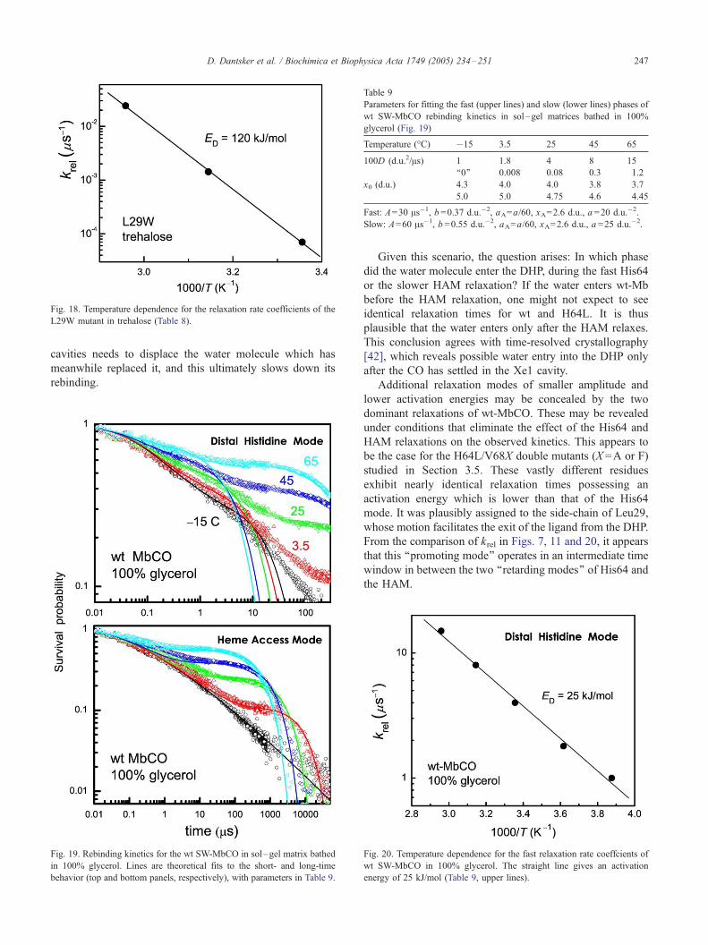
Fig. 18. Temperature dependence for the relaxation rate coefficients of the
L29W mutant in trehalose (Table 8).
Table 9
Parameters for fitting the fast (upper lines) and slow (lower lines) phases of
wt SW-MbCO rebinding kinetics in sol–gel matrices bathed in 100%
glycerol (Fig. 19)
Temperature (-C) �15 3.5 25 45 65
100D (d.u.2/As) 1 1.8 4 8 15
‘‘0’’ 0.008 0.08 0.3 1.2
x0 (d.u.) 4.3 4.0 4.0 3.8 3.7
5.0 5.0 4.75 4.6 4.45
Fast: A=30 As�1, b =0.37 d.u.�2, aA=a/60, xA=2.6 d.u., a =20 d.u.�2.
Slow: A=60 As�1, b =0.55 d.u.�2, aA=a/60, xA=2.6 d.u., a =25 d.u.�2.
D. Dantsker et al. / Biochimica et Biophysica Acta 1749 (2005) 234–251 247
cavities needs to displace the water molecule which has
meanwhile replaced it, and this ultimately slows down its
rebinding.
Fig. 19. Rebinding kinetics for the wt SW-MbCO in sol–gel matrix bathed
in 100% glycerol. Lines are theoretical fits to the short- and long-time
behavior (top and bottom panels, respectively), with parameters in Table 9.
Given this scenario, the question arises: In which phase
did the water molecule enter the DHP, during the fast His64
or the slower HAM relaxation? If the water enters wt-Mb
before the HAM relaxation, one might not expect to see
identical relaxation times for wt and H64L. It is thus
plausible that the water enters only after the HAM relaxes.
This conclusion agrees with time-resolved crystallography
[42], which reveals possible water entry into the DHP only
after the CO has settled in the Xe1 cavity.
Additional relaxation modes of smaller amplitude and
lower activation energies may be concealed by the two
dominant relaxations of wt-MbCO. These may be revealed
under conditions that eliminate the effect of the His64 and
HAM relaxations on the observed kinetics. This appears to
be the case for the H64L/V68X double mutants (X =A or F)
studied in Section 3.5. These vastly different residues
exhibit nearly identical relaxation times possessing an
activation energy which is lower than that of the His64
mode. It was plausibly assigned to the side-chain of Leu29,
whose motion facilitates the exit of the ligand from the DHP.
From the comparison of krel in Figs. 7, 11 and 20, it appears
that this ‘‘promoting mode’’ operates in an intermediate time
window in between the two ‘‘retarding modes’’ of His64 and
the HAM.
Fig. 20. Temperature dependence for the fast relaxation rate coeffcients of
wt SW-MbCO in 100% glycerol. The straight line gives an activation
energy of 25 kJ/mol (Table 9, upper lines).
D. Dantsker et al. / Biochimica et Biophysica Acta 1749 (2005) 234–251248
4. Discussion
Throughout the years, Mb kinetics has inspired countless
approaches and descriptions for protein function in this
supposedly simple example of ligand migration and binding
within a small globular protein. An early suggestion [4] puts
the action in the ligand coordinate, which surmounts several
barriers in its pathway, between the bound state (A), the
DHP (B), additional sites within the protein (C, D) and the
solvent (S)
A@B†C†D†S ð9Þ
In retrospect, such a kinetic scheme is supported by the
involvement of the Xe cavities, Xe4 and Xe1, which may be
identified with states C and D, respectively. Unfortunately,
aside from this superficial agreement such a kinetic scheme
for protein kinetics is quite inadequate. On a technical level,
it is likely that the linear scheme is inapplicable because the
gate to the external solvent is the distal histidine (His64).
This then requires a branched kinetic scheme, where S is
connected directly to state B [32].
On a fundamental level, no kinetic scheme is appropriate
because a reaction within a dense fluctuating environment
such as a protein cannot be described in the language of
dilute gas kinetics. In the latter case, kinetic energy from the
tail of the Boltzmann distribution of velocities allows some
ligands to surmount static barriers along the coordinate that
separates them from their reaction partner. When a ligand
moves within a protein, its kinetic energy is less relevant than
the momentary protein conformation, which may either
allow or disallow a given ligand motion. The ligand becomes
completely ‘‘slaved’’ to the protein with kinetic features
arising from dynamics in the protein coordinate.
This point has been foreseen byAgmon andHopfield [22],
who ascribed the kinetic phases inMbCO kinetics to action in
the protein (rather than the ligand) coordinate. Indeed, by a
minor quantitative modification to their model (replacing
their exponential sink by a Gaussian one), we are able to
obtain a good fit to every relaxation process revealed in our
study. While their description is thus closer to reality than the
ordinary kinetic approach, it is still too simplistic. Firstly, it
focussed on a single relaxation process. Our study reveals
many relaxation processes, yet each of these separately
behaves in accordance with the model.
The second simplification was in the identification of the
relaxing mode. At the time, it was generally accepted that
protein relaxation is transmitted to the iron center through the
proximal histidine (His93) and thus operates directly on the
innermost barrier. In agreement with others [48,52,58], we
show that the innermost recombination step is fast and
temperature independent, and thus not likely to require
proximal assistance. In contrast, our systematic mutation
work shows that the major phases observed in the kinetics
are due to relaxations in the DHP. These may operate as
‘‘fluctuating bottlenecks’’ [70] controlling access to the iron.
Yet, even this may be too simplistic in several respects: (a)
The protein is not merely fluctuating, but rather it is relaxing
in response to the ligand’s motion. Thus, coupling between
protein and ligand operates in both directions. (b) The more
global conformational changes initiated by the HAM are
likely transmitted also to the proximal side. We see (not
shown here) universally slower rebinding when deoxy-Mb is
trapped in sol–gel matrices as opposed to trapping of the
bound MbCO. (c) The smaller side-chain relaxations may
not be merely ‘‘gate keepers’’ for inter-site migration, but
may actually be required to ‘‘facilitate diffusion’’ by kicking-
off the ligand in certain directions of motion.
Clearly, to disentangle this plethora of effects on a
microscopic level, an extensive systematic study is required.
This is particularly important because a superficial consid-
eration of the kinetic results may give the impression that all
relaxations look alike, while in fact they are due to different
protein modes. The remarkable outcome of the present study
is that by use of site-directed mutagenesis, highly viscous
solvents, temperature effects and a relatively simple theo-
retical model, we can identify individual relaxation modes,
determine their relaxation rate constants and Arrhenius
activation energies. It is instructive first to summarize the
principles of the methodology, then the actual results.
In order to separate out the effect of different protein
modes, one should be able to systematically and selectively
‘‘turn-off’’ given relaxations:
(1) Local side-chain relaxations can be eliminated by point
mutations at that site. An example is the substitution of
His64 by Leu, which abolishes its (imidazole) side-
chain motion. Likewise, the comparison of Leu vs. Val
in position 68 mutants reveals a small relaxation
attributable to the longer leucine side-chain (Fig. 9).
Interestingly, both these relaxations are corroborated
by X-ray [58] and time-resolved crystallography [42].
(2) Highly viscous trehalose can be used to selectively
eliminate the large-amplitude, cooperative relaxation
modes which extend all the way to the protein surface,
leaving behind the smaller and more local internal
modes. For example, wt-Mb in trehalose looses its
HAM relaxation (Fig. 4), maintaining only the His64
mode.
(3) Cooperative modes (the HAM) are universal in the
sense that they appear in different mutants (wt, H64L
and H64L/V68L) with essentially the same activation
energy (ca. 58 kJ/mol in 100% glycerol).
(4) Nevertheless, the HAM effect may be reduced as in the
V68A mutant. This unravels a smaller-amplitude rela-
xation that may have been otherwise masked by the
larger HAM.
(5) Two consecutive relaxations may be analyzed by
separate fits to the short- and long-time behavior. An
example is a fast side-chain relaxation (His64, Leu68)
followed by the HAM. However, the separation of
overlapping relaxations can only be achieved by
comparing appropriate mutants.
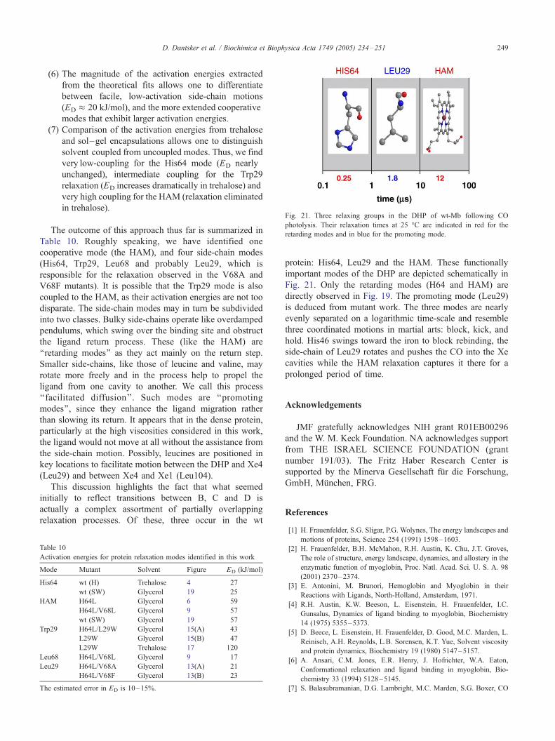
Fig. 21. Three relaxing groups in the DHP of wt-Mb following CO
photolysis. Their relaxation times at 25 -C are indicated in red for the
retarding modes and in blue for the promoting mode.
D. Dantsker et al. / Biochimica et Biophysica Acta 1749 (2005) 234–251 249
(6) The magnitude of the activation energies extracted
from the theoretical fits allows one to differentiate
between facile, low-activation side-chain motions
(ED � 20 kJ/mol), and the more extended cooperative
modes that exhibit larger activation energies.
(7) Comparison of the activation energies from trehalose
and sol–gel encapsulations allows one to distinguish
solvent coupled from uncoupled modes. Thus, we find
very low-coupling for the His64 mode (ED nearly
unchanged), intermediate coupling for the Trp29
relaxation (ED increases dramatically in trehalose) and
very high coupling for the HAM (relaxation eliminated
in trehalose).
The outcome of this approach thus far is summarized in
Table 10. Roughly speaking, we have identified one
cooperative mode (the HAM), and four side-chain modes
(His64, Trp29, Leu68 and probably Leu29, which is
responsible for the relaxation observed in the V68A and
V68F mutants). It is possible that the Trp29 mode is also
coupled to the HAM, as their activation energies are not too
disparate. The side-chain modes may in turn be subdivided
into two classes. Bulky side-chains operate like overdamped
pendulums, which swing over the binding site and obstruct
the ligand return process. These (like the HAM) are
‘‘retarding modes’’ as they act mainly on the return step.
Smaller side-chains, like those of leucine and valine, may
rotate more freely and in the process help to propel the
ligand from one cavity to another. We call this process
‘‘facilitated diffusion’’. Such modes are ‘‘promoting
modes’’, since they enhance the ligand migration rather
than slowing its return. It appears that in the dense protein,
particularly at the high viscosities considered in this work,
the ligand would not move at all without the assistance from
the side-chain motion. Possibly, leucines are positioned in
key locations to facilitate motion between the DHP and Xe4
(Leu29) and between Xe4 and Xe1 (Leu104).
This discussion highlights the fact that what seemed
initially to reflect transitions between B, C and D is
actually a complex assortment of partially overlapping
relaxation processes. Of these, three occur in the wt
Table 10
Activation energies for protein relaxation modes identified in this work
Mode Mutant Solvent Figure ED (kJ/mol)
His64 wt (H) Trehalose 4 27
wt (SW) Glycerol 19 25
HAM H64L Glycerol 6 59
H64L/V68L Glycerol 9 57
wt (SW) Glycerol 19 57
Trp29 H64L/L29W Glycerol 15(A) 43
L29W Glycerol 15(B) 47
L29W Trehalose 17 120
Leu68 H64L/V68L Glycerol 9 17
Leu29 H64L/V68A Glycerol 13(A) 21
H64L/V68F Glycerol 13(B) 23
The estimated error in ED is 10–15%.
protein: His64, Leu29 and the HAM. These functionally
important modes of the DHP are depicted schematically in
Fig. 21. Only the retarding modes (H64 and HAM) are
directly observed in Fig. 19. The promoting mode (Leu29)
is deduced from mutant work. The three modes are nearly
evenly separated on a logarithmic time-scale and resemble
three coordinated motions in martial arts: block, kick, and
hold. His46 swings toward the iron to block rebinding, the
side-chain of Leu29 rotates and pushes the CO into the Xe
cavities while the HAM relaxation captures it there for a
prolonged period of time.
Acknowledgements
JMF gratefully acknowledges NIH grant R01EB00296
and the W. M. Keck Foundation. NA acknowledges support
from THE ISRAEL SCIENCE FOUNDATION (grant
number 191/03). The Fritz Haber Research Center is
supported by the Minerva Gesellschaft fur die Forschung,
GmbH, Munchen, FRG.
References
[1] H. Frauenfelder, S.G. Sligar, P.G. Wolynes, The energy landscapes and
motions of proteins, Science 254 (1991) 1598–1603.
[2] H. Frauenfelder, B.H. McMahon, R.H. Austin, K. Chu, J.T. Groves,
The role of structure, energy landscape, dynamics, and allostery in the
enzymatic function of myoglobin, Proc. Natl. Acad. Sci. U. S. A. 98
(2001) 2370–2374.
[3] E. Antonini, M. Brunori, Hemoglobin and Myoglobin in their
Reactions with Ligands, North-Holland, Amsterdam, 1971.
[4] R.H. Austin, K.W. Beeson, L. Eisenstein, H. Frauenfelder, I.C.
Gunsalus, Dynamics of ligand binding to myoglobin, Biochemistry
14 (1975) 5355–5373.
[5] D. Beece, L. Eisenstein, H. Frauenfelder, D. Good, M.C. Marden, L.
Reinisch, A.H. Reynolds, L.B. Sorensen, K.T. Yue, Solvent viscosity
and protein dynamics, Biochemistry 19 (1980) 5147–5157.
[6] A. Ansari, C.M. Jones, E.R. Henry, J. Hofrichter, W.A. Eaton,
Conformational relaxation and ligand binding in myoglobin, Bio-
chemistry 33 (1994) 5128–5145.
[7] S. Balasubramanian, D.G. Lambright, M.C. Marden, S.G. Boxer, CO
D. Dantsker et al. / Biochimica et Biophysica Acta 1749 (2005) 234–251250
recombination to human myoglobin mutants in glycerol–water
solutions, Biochemistry 32 (1993) 2202–2212.
[8] W. Doster, T. Kleinert, F. Post, M. Settles, Effect of solvent on protein
internal dynamics: the kinetics of ligand binding to myoglobin, in:
R.B. Gregory (Ed.), Protein–Solvent Interactions, Marcel Dekker,
1993, p. 375.
[9] N. Agmon, W. Doster, F. Post, The transition from inhomogeneous to
homogeneous kinetics in CO binding to myoglobin, Biophys. J. 66
(1994) 1612–1622.
[10] Y. Abadan, E.Y.T. Chien, K. Chu, C.D. Eng, G.U. Nienhaus, S.G.
Sligar, Ligand binding to heme proteins: V. Light-induced relaxation
in proximal mutants L89I and H97F of carbonmonoxymyoglobin,
Biophys. J. 68 (1995) 2497–2504.
[11] S.J. Hagen, J. Hofrichter, W.A. Eaton, Geminate rebinding and
conformational dynamics of myoglobin embedded in a glass at room
temperature, J. Phys. Chem. 100 (1996) 12008–12021.
[12] G.M. Sastry, N. Agmon, Trehalose prevents protein collapse
and preserves its internal mobility, Biochemistry 36 (1997)
7097–7108.
[13] T. Kleinert, W. Doster, H. Leyser, W. Petry, V. Schwarz, M. Settles,
Solvent composition and viscosity effects on the kinetics of CO
binding to horse myoglobin, Biochemistry 37 (1998) 717–733.
[14] Y. Kholodenko, E.A. Gooding, Y. Dou, M. Ikeda-Saito, R.M.
Hochstrasser, Heme protein dynamics revealed by geminate nitric
oxide recombination in mutants of iron and cobalt myoglobin,
Biochemistry 38 (1999) 5918–5924.
[15] S. Abbruzzetti, C. Viappiani, S. Bruno, A. Mozzarelli, Enhanced
geminate ligand rebinding upon photo-dissociation of silica gel-
embedded myoglobin-CO, Chem. Phys. Lett. 346 (2001) 430–436.
[16] S. Abbruzzetti, C. Viappiani, S. Bruno, S. Bettati, M. Bonaccio, A.
Mozzarelli, Functional characterization of heme proteins encapsu-
lated in wet nanoporous silica gels, J. Nanosci. Nanotechnol. 1
(2001) 407–415.
[17] F. Librizzi, C. Viappiani, S. Abbruzzetti, L. Cordone, Residual water
modulates the dynamics of the protein and of the external matrix in
trehalose coated MbCO: an infrared and flash-photolysis study, J.
Chem. Phys. 116 (2002) 1193–1200.
[18] U. Samuni, D. Dantsker, I. Khan, A.J. Friedman, E. Peterson,
J.M. Friedman, Spectroscopically and kinetically distinct conforma-
tional populations of sol–gel-encapsulated carbonmonoxy myoglo-
bin. A comparison with hemoglobin, J. Biol. Chem. 277 (2002)
25783–25790.
[19] D. Dantsker, U. Samuni, A.J. Friedman, M. Yang, A. Ray, J.M.
Friedman, Geminate rebinding in trehalose-glass embedded myo-
globins reveals residue-specific control of intramolecular trajecto-
ries, J. Mol. Biol. 315 (2002) 239–251.
[20] S. Sottini, C. Viappiani, L. Ronda, S. Bettati, A. Mozzarelli, CO
rebinding kinetics to myoglobin- and R-state-hemoglobin-doped
silica gels in the presence of glycerol, J. Phys. Chem. B 108
(2004) 8475–8484.
[21] N. Agmon, J.J. Hopfield, Transient kinetics of chemical reactions with
bounded diffusion perpendicular to the reaction coordinate: intra-
molecular processes with slow conformational changes, J. Chem.
Phys. 78 (1983) 6947–6959 (erratum, ibid. 80 (1984) 592).
[22] N. Agmon, J.J. Hopfield, CO binding to heme proteins: a model for
barrier height distributions and slow conformational changes, J. Chem.
Phys. 79 (1983) 2042–2053.
[23] P.J. Steinbach, A. Ansari, J. Berendzen, D. Braunstein, K. Chu, B.R.
Cowen, D. Ehrenstein, H. Frauenfelder, J.B. Johnson, D.C. Lamb, S.
Luck, J.R. Mourant, G.U. Nienhaus, P. Ormos, R. Philipp, A. Xie,
R.D. Young, Ligand binding to heme proteins: connection between
dynamics and function, Biochemistry 30 (1991) 3988–4001.
[24] J.W. Petrich, J.-C. Lambry, K. Kuczera, M. Karplus, C. Poyart, J.-L.
Martin, Ligand binding and protein relaxation in heme proteins: a
room temperature analysis of NO geminate recombination, Biochem-
istry 30 (1991) 3975–3987.
[25] M. Meuwly, O.M. Becker, R. Stote, M. Karplus, NO rebinding to
myoglobin: a reactive molecular dynamics study, Biophys. Chem. 98
(2002) 183–207.
[26] W.D. Tian, J.T. Sage, V. Srajer, P.M. Champion, Relaxation dynamics
of myoglobin in solution, Phys. Rev. Lett. 68 (1992) 408–411.
[27] A. Ansari, C.M. Jones, E.R. Henry, J. Hofrichter, W.A. Eaton, The
role of solvent viscosity in the dynamics of protein conformational
change, Science 256 (1992) 1796–1798.
[28] M. Lim, T.A. Jackson, P.A. Anfinrud, Nonexponential protein
relaxation: dynamics of conformational change in myoglobin, Proc.
Natl. Acad. Sci. U. S. A. 90 (1993) 5801–5804.
[29] H. Frauenfelder, P.W. Fenimore, B.H. McMahon, Hydration, slaving
and protein function, Biophys. Chem. 98 (2002) 35–48.
[30] R.F. Tilton Jr., I.D. Kuntz Jr., G.A. Petsko, Cavities in proteins:
structure of a metmyoglobin–xenon complex solved to 1.9 A
resolution, Biochemistry 23 (1984) 2849–2857.
[31] E.E. Scott, Q.H. Gibson, Ligand migration in sperm whale myoglobin,
Biochemistry 36 (1997) 11909–11917.
[32] E.E. Scott, Q.H. Gibson, J.S. Olson, Mapping the pathways for O2
entry into and exit from myoglobin, J. Biol. Chem. 276 (2001)
5177–5188.
[33] A. Ostermann, R. Waschipky, F.G. Parak, G.U. Nienhaus, Ligand
binding and conformational motions in myoglobin, Nature 404 (2000)
205–208.
[34] I. Schlichting, J. Berendzen, G.N. Phillips Jr., R.M. Sweet, Crystal
structure of photolysed carbonmonoxy–myoglobin, Nature 371
(1994) 808–812.
[35] V. Srajer, T.-Y. Teng, T. Ursby, C. Pradervand, Z. Ren, S.-I. Adachi,
W. Schildkamp, D. Bourgeois, M. Wulff, K. Moffat, Photolysis of the
carbon monoxide complex of myoglobin: nanosecond time-resolved
crystallography, Science 274 (1996) 1726–1729.
[36] J. Vojtechovsky, K. Chu, J. Berendzen, R.M. Sweet, I. Schlichting,
Crystal structures of myoglobin– ligand complexes at near-atomic
resolution, Biophys. J. 77 (1999) 2153–2174.
[37] I. Schlichting, K. Chu, Trapping intermediates in the crystal:
ligand binding to myoglobin, Curr. Opin. Struct. Biol. 10 (2000)
744–752.
[38] K. Chu, J. Vojtchovsky, B.H. McMahon, R.M. Sweet, J. Berendzen, I.
Schlichting, Structure of a ligand-binding intermediate in wild-type
carbonmonoxy myoglobin, Nature 403 (2000) 921–923.
[39] V. Srajer, Z. Ren, T.-Y. Teng, M. Schmidt, T. Ursby, D. Bourgeois, C.
Pradervand, W. Schildkamp, M. Wulff, K. Moffat, Protein conforma-
tional relaxation and ligand migration in myoglobin: a nanosecond to
millisecond molecular movie from time-resolved Laue X-ray diffrac-
tion, Biochemistry 40 (2001) 13802–13815.
[40] D. Bourgeois, B. Vallone, F. Schotte, A. Arcovito, A.E. Miele, G.
Sciara, M. Wulff, P. Anfinrud, M. Brunori, Complex landscape of
protein structural dynamics unveiled by nanosecond Laue crystallog-
raphy, Proc. Natl. Acad. Sci. U. S. A. 100 (2003) 8704–8709.
[41] F. Schotte, M. Lim, T.A. Jackson, A.V. Smirnov, J. Soman, J.S. Olson,
G.N. Phillips Jr., M. Wulff, P.A. Anfinrud, Watching a protein as it
functions with 150-ps time-resolved X-ray crystallography, Science
300 (2003) 1944–1947.
[42] F. Schotte, J. Soman, J.S. Olson, M. Wulff, P.A. Anfinrud, Picosecond
time-resolved X-ray crystallography: probing protein function in real
time, J. Struct. Biol. 147 (2004) 235–246.
[43] K. Nienhaus, P. Deng, J.M. Kriegl, G.U. Nienhaus, Structural dynamics
of myoglobin: spectroscopic and structural characterization of ligand
docking sites in myoglobin mutant L29W, Biochemistry 42 (2003)
9633–9646.
[44] K. Nienhaus, P. Deng, J.M. Kriegl, G.U. Nienhaus, Structural
dynamics of myoglobin: effect of internal cavities on ligand migration
and binding, Biochemistry 42 (2003) 9647–9658.
[45] K. Nienhaus, P. Deng, J.S. Olson, J.J. Warren, G.U. Nienhaus,
Structural dynamics of myoglobin. Ligand migration and binding in
valine 68 mutants, J. Biol. Chem. 278 (2003) 42532–42544.
[46] N. Agmon, Coupling of protein relaxation to ligand binding and
migration in myoglobin, Biophys. J. 87 (2004) 1537–1543.
D. Dantsker et al. / Biochimica et Biophysica Acta 1749 (2005) 234–251 251
[47] N. Shibayama, S. Saigo, Oxygen equilibrium properties of myoglobin
locked in the liganded and unliganded conformations, J. Am. Chem.
Soc. 125 (2003) 3780–3783.
[48] D.G. Lambright, S. Balasubramanian, S.G. Boxer, Dynamics of protein
relaxation in site-specific mutants of human myoglobin, Biochemistry
32 (1993) 10116–10124.
[49] S. Franzen, S.G. Boxer, On the origin of heme absorption band shifts
and associated protein structural relaxation in myoglobin following
flash photolysis, J. Biol. Chem. 272 (1997) 9655–9660.
[50] M.L. Quillin, R.M. Arduini, J.S. Olson, G.N. Phillips Jr., High-
resolution crystal structures of distal histidine mutants of sperm whale
myoglobin, J. Mol. Biol. 234 (1993) 140–155.
[51] W. Cao, J.F. Christian, P.M. Champion, F. Rosca, J.T. Sage, Water
penetration and binding to ferric myoglobin, Biochemistry 40 (2001)
5728–5737.
[52] Q.H. Gibson, R. Regan, R. Elber, J.S. Olson, T.E. Carver, Distal
pocket residues affect picosecond ligand recombination in myoglobin:
an experimental and molecular dynamics study of position 29 mutants,
J. Biol. Chem. 267 (1992) 22022–22034.
[53] T. Uchida, K. Ishimori, I. Morishima, The effects of heme pocket
hydrophobicity on the ligand binding dynamics in myoglobin as
studied with leucine 29 mutants, J. Biol. Chem. 272 (1997)
30108–30114.
[54] M. Brunori, B. Vallone, F. Cutruzzola, C. Travaglini-Allocatelli, J.
Berendzen, K. Chu, R.M. Sweet, I. Schlichting, The role of cavities in
protein dynamics: crystal structure of a photolytic intermediate of a
mutantmyoglobin,Proc.Natl.Acad.Sci.U.S.A.97 (2000)2058–2063.
[55] F. Draghi, A.E. Miele, C. Travaglini-Allocatelli, B. Vallone, M.
Brunori, Q.H. Gibson, J.S. Olson, Controlling ligand binding in
myoglobin by mutagenesis, J. Biol. Chem. 277 (2002) 7509–7519.
[56] K.D. Egeberg, B.A. Springer, S.G. Sligar, T.E. Carver, R.J. Rohlfs,
J.S. Olson, The role of Val68 (E11) in ligand binding to sperm whale
myoglobin, J. Biol. Chem. 265 (1990) 11788–11795.
[57] M. Ikeda-Saito, Y. Dou, T. Yonetani, J.S. Olson, T. Li, R. Regan, Q.H.
Gibson, Ligand diffusion in the distal heme pocket of myoglobin. A
primary determinant of geminate rebinding, J. Biol. Chem. 268 (1993)
6855–6857.
[58] M.L. Quillin, T. Li, J.S. Olson, G.N. Phillips Jr., Y. Dou, M. Ikeda-
Saito, R. Regan, M. Carlson, Q.H. Gibson, H. Li, R. Elber, Structural
and functional effects of apolar mutations of the distal valine in
myoglobin, J. Mol. Biol. 245 (1995) 416–436.
[59] J.L. Green, C.A. Angell, Phase relations and vitrification in saccha-
ride–water solutions and the trehalose anomaly, J. Phys. Chem. 93
(1989) 2880–2882.
[60] L.M. Crowe, D.S. Reid, J.H. Crowe, Is trehalose special for preserving
dry biomaterials? Biophys. J. 71 (1996) 2087–2093.
[61] J.F. Carpenter, L.M. Crowe, J.H. Crowe, Stabilization of phospho-
fructokinase with sugars during freeze-drying: characterization of
enhanced protection in the presence of divalent cations, Biochim.
Biophys. Acta 923 (1987) 109–115.
[62] S.J. Prestrelski, N. Tedeschi, T. Arakawa, J.F. Carpenter, Dehydration
induced conformational transitions in proteins and their inhibition by
stabilizers, Biophys. J. 65 (1993) 661–671.
[63] J.F. Carpenter, J.H. Crowe, An infrared spectroscopic study of the
interactions of carbohydrates with dried proteins, Biochemistry 28
(1989) 3916–3922.
[64] U. Samuni, D. Dantsker, A. Ray, J.B. Wittenberg, B.A. Wittenberg,
S. Dewilde, L. Moens, Y. Ouellet, M. Guertin, J.M. Friedman,
Kinetic modulation in carbonmonoxy derivatives of truncated
hemoglobins. The role of distal heme pocket residues and extended
apolar tunnel, J. Biol. Chem. 278 (2003) 27241–27250.
[65] D. Dantsker, U. Samuni, Y. Ouellet, B.A. Wittenberg, J.B. Wittenberg,
M. Milani, M. Bolognesi, M. Guertin, J.M. Friedman, Viscosity-
dependent relaxation significantly modulates the kinetics of CO
recombination in the truncated hemoglobin TrHbN from Mycobacte-
rium tuberculosis, J. Biol. Chem. 279 (2004) 38844–38853.
[66] N. Agmon, G.M. Sastry, A temperature-dependent effective potential
explains CO binding to myoglobin, Chem. Phys. 212 (1996) 207–219.
[67] N. Agmon, Conformational cycle of a single working enzyme, J. Phys.
Chem. B 104 (2000) 7830–7834.
[68] B. Bagchi, G.R. Fleming, D.W. Oxtoby, Theory of electronic
relaxation in solution in the absence of an activation barrier, J. Chem.
Phys. 78 (1983) 7375–7385.
[69] G.H. Weiss, A perturbation analysis of the Wilemski-Fixman
approximation for diffusion controlled reactions, J. Chem. Phys. 80
(1984) 2880–2887.
[70] R. Zwanzig, Dynamical disorder: passage through a fluctuating
bottleneck, J. Chem. Phys. 97 (1992) 3587–3589.
[71] N. Agmon, Viscosity expansions in reactive diffusion processes,
J. Chem. Phys. 90 (1989) 3765–3775.
[72] S. Rabinovich, N. Agmon, The slow diffusion limit for the survival
probability in reactive diffusion equations, Chem. Phys. 148 (1990)
11–19.
[73] E.B. Krissinel’, N. Agmon, Spherical symmetric diffusion problem,
J. Comput. Chem. 17 (1996) 1085–1098.
[74] R. Zwanzig, Diffusion in a rough potential, Proc. Natl. Acad. Sci.
U. S. A. 85 (1988) 2029–2030.
[75] N. Agmon, From energy profiles to structure– reactivity correlations,
Int. J. Chem. Kinet. 13 (1981) 333–365.
[76] M.F. Perutz, F.S. Matthews, An X-ray study of azide methaemoglobin,
J. Mol. Biol. 21 (1966) 199–202.
[77] M. Lim, T.A. Jackson, P.A. Anfinrud, Ultrafast rotation and trapping
of carbon monoxide dissociated from myoglobin, Nat. Struct. Biol. 3
(1997) 209–214.
[78] T.-Y. Teng, V. Srajer, K. Moffat, Initial trajectory of carbon monoxide
after photodissociation from myoglobin at cryogenic temperatures,
Biochemistry 36 (1997) 12087–12100.
[79] W. Radding, G.N. Phillips Jr., Kinetic proofreading by the cavity
system of myoglobin: protection from poisoning, BioEssays 26 (2004)
422–433.
Copyright © 2022 FDOKUMEN