Biosynthesis of coumarins in plants: a major pathway still to be unravelled for cytochrome P450...
16
Biosynthesis of coumarins in plants: a major pathway still to be unravelled for cytochrome P450 enzymes F. Bourgaud A. Hehn R. Larbat S. Doerper E. Gontier S. Kellner U. Matern Received: 12 February 2006 / Accepted: 11 October 2006 / Published online: 15 November 2006 Ó Springer Science+Business Media B.V. 2006 Abstract Coumarins (1,2-benzopyrones) are ubiquitously found in higher plants where they originate from the phenylpropanoid pathway. They contribute essentially to the persistence of plants being involved in processes such as defense against phytopathogens, response to abiotic stres- ses, regulation of oxidative stress, and probably hormonal regulation. Despite their importance, major details of their biosynthesis are still largely unknown and many P450-dependent enzymatic steps have remained unresolved. Ortho-hydrox- ylation of hydroxycinnamic acids is a pivotal step that has received insufficient attention in the literature. This hypothetical P450 reaction is critical for the course for the biosynthesis of simple coumarin, umbelliferone and other hydroxylated coumarins in plants. Multiple P450 enzymes are also involved in furanocoumarin synthesis, a major class of phytoalexins derived from umbelliferone. Several of them have been characterized at the biochemical level but no monooxygenase gene of the furanocoumarin pathway has been identified yet. This review highlights the major steps of the coumarin path- way with emphasis on the cytochrome P450 enzymes involved. Recent progress and the out- comes of novel strategies developed to uncover coumarin-committed CYPs are discussed. Keywords Cytochrome P450 Á Monooxygenase Á Ortho-hydroxylase Á Coumarin Á Hydroxycinnamic acid Á Furanocoumarin Á Biosynthesis Introduction Coumarins are derived from 1,2-benzopyrones. These molecules are found in higher plants where they originate from the general phenylpropanoid pathway (Harborne 1999) and are subject to numerous modifications. Coumarins continue to receive attention for their diverse bioactivities. Some natural coumarins have been used as human therapeutics, while 4-hydroxycoumarins are prominent examples of microbial modification which gave rise to the first generation molecules developed along with aspirin and heparin as anti- coagulants (Mueller 2004). Other applications appear possible in the course of new develop- ments in various therapeutic fields, like F. Bourgaud (&) Á A. Hehn Á R. Larbat Á S. Doerper Á E. Gontier UMR INPL(ENSAIA)-INRA Agronomie et Environnement Nancy-Colmar, ENSAIA, 2 Avenue Fore ˆ t de Haye, 54500 Vandoeuvre, France e-mail: [email protected] S. Kellner Á U. Matern Institute of Pharmaceutical Biology, Deutschhausstrasse 17 A, D-35037 Marburg, Germany 123 Phytochem Rev (2006) 5:293–308 DOI 10.1007/s11101-006-9040-2
-
Upload
independent -
Category
Documents
-
view
5 -
download
0
Transcript of Biosynthesis of coumarins in plants: a major pathway still to be unravelled for cytochrome P450...

Biosynthesis of coumarins in plants: a major pathway still tobe unravelled for cytochrome P450 enzymes
F. Bourgaud Æ A. Hehn Æ R. Larbat Æ S. Doerper ÆE. Gontier Æ S. Kellner Æ U. Matern
Received: 12 February 2006 / Accepted: 11 October 2006 / Published online: 15 November 2006� Springer Science+Business Media B.V. 2006
Abstract Coumarins (1,2-benzopyrones) are
ubiquitously found in higher plants where they
originate from the phenylpropanoid pathway.
They contribute essentially to the persistence of
plants being involved in processes such as defense
against phytopathogens, response to abiotic stres-
ses, regulation of oxidative stress, and probably
hormonal regulation. Despite their importance,
major details of their biosynthesis are still largely
unknown and many P450-dependent enzymatic
steps have remained unresolved. Ortho-hydrox-
ylation of hydroxycinnamic acids is a pivotal step
that has received insufficient attention in the
literature. This hypothetical P450 reaction is
critical for the course for the biosynthesis of
simple coumarin, umbelliferone and other
hydroxylated coumarins in plants. Multiple P450
enzymes are also involved in furanocoumarin
synthesis, a major class of phytoalexins derived
from umbelliferone. Several of them have been
characterized at the biochemical level but no
monooxygenase gene of the furanocoumarin
pathway has been identified yet. This review
highlights the major steps of the coumarin path-
way with emphasis on the cytochrome P450
enzymes involved. Recent progress and the out-
comes of novel strategies developed to uncover
coumarin-committed CYPs are discussed.
Keywords Cytochrome P450 �Monooxygenase �Ortho-hydroxylase � Coumarin �Hydroxycinnamic acid � Furanocoumarin �Biosynthesis
Introduction
Coumarins are derived from 1,2-benzopyrones.
These molecules are found in higher plants where
they originate from the general phenylpropanoid
pathway (Harborne 1999) and are subject to
numerous modifications. Coumarins continue to
receive attention for their diverse bioactivities.
Some natural coumarins have been used as
human therapeutics, while 4-hydroxycoumarins
are prominent examples of microbial modification
which gave rise to the first generation molecules
developed along with aspirin and heparin as anti-
coagulants (Mueller 2004). Other applications
appear possible in the course of new develop-
ments in various therapeutic fields, like
F. Bourgaud (&) � A. Hehn � R. Larbat �S. Doerper � E. GontierUMR INPL(ENSAIA)-INRA Agronomie etEnvironnement Nancy-Colmar, ENSAIA, 2 AvenueForet de Haye, 54500 Vandoeuvre, Francee-mail: [email protected]
S. Kellner � U. MaternInstitute of Pharmaceutical Biology,Deutschhausstrasse 17 A, D-35037 Marburg,Germany
123
Phytochem Rev (2006) 5:293–308
DOI 10.1007/s11101-006-9040-2
symptomatic treatment of multiple sclerosis
(Wulff et al. 1998), photochemotherapy of T cell
lymphoma (Plumas et al. 2003), chemotherapy of
multidrug resistant tumors (Kawase et al. 2005),
organ transplants (Damjanovich et al. 2004), or
treatment of smokers for nicotine addiction
(Malaiyandi et al. 2005).
Despite the importance of coumarins for plant
life and human uses, major details of their
biosynthesis have remained unresolved. This
review will give an update of coumarin biogenesis
in plants with emphasis on the cytochrome P450
enzymes involved.
Occurrence and functions of coumarins in plants
Coumarins may be subclassified as simple cou-
marins (benzo-a-pyrones syn 1,2-benzopyrone),
7-oxygenated coumarins (furanocoumarins syn.
furobenzo-a-pyrones or furocoumarins), pyra-
nocoumarins (benzodipyran-2-ones), and phen-
ylcoumarins (benzo-benzopyrones) (Estevez-
Braun and Gonzalez 1997; Murray 1991; Murray
et al. 1982). Simple coumarins, furanocoumarins
and pyranocoumarins derive from the same
pathway, whereas the most common phen-
ylcoumarins (i.e., coumestans) originate from
isoflavone metabolism and will not be considered
in this review.
Simple coumarins
These compounds are widespread in plants and
more than 700 structures have already been
described (Harborne 1999).
Coumarin
Coumarin (Fig. 1) is a natural product well-
known for its pleasant vanilla-like odor. It was
reported from many plants of a variety of fam-
ilies, including Fabaceae i.e., Tonka bean (Cou-
marouna odorata) (Ehlers et al. 1995) or
sweetclover (Melilotus alba) (Akeson et al.
1963), Lamiaceae i.e., lavender (Lavandula offi-
cinalis) (Brown 1962), and Lauraceae i.e., cinna-
mon (Cinnamonum verum) (Miller et al. 1996).
More recent studies have revealed the presence
of o-coumaric acid in Arabidopsis thaliana root
exudates (Walker et al. 2003). As cis-o-coumaric
acid is unstable under acidic or neutral conditions
and lactonizes spontaneously to coumarin it is
conceivable that coumarin is formed in Arabid-
opsis thaliana.
There have been many reports on the effect of
coumarin in plants, at the organ, tissue and
cellular levels (Brown 1981). These observations
tend to demonstrate that coumarin acts as a plant
hormone. However, until now neither solid evi-
dence for a physiological function nor the molec-
ular mode of action of coumarin has been
provided in plant tissues.
Hydroxylated and methoxylated coumarins
Prevalent hydroxylated coumarins are umbellif-
erone, herniarin and scoparone (2 methoxylated
derivatives of umbelliferone), esculetin, fraxetin,
isofraxidin, isoscopoletin, daphnetin and their
corresponding glucosides (Fig. 1). As for scopo-
letin, these molecules are involved in plant
responses to stressors like salicylic acid (Pastirova
et al. 2004; Repcak et al. 2001). Herniarin was
demonstrated to be demethylated to umbellifer-
one by C4H from Helianthus tuberosus
(CYP73A1) heterologously expressed in yeast
(Pierrel et al. 1994); However, the Km was so
high compared to cinnamate substrate that the
implication of C4H for herniarin demethylation
remains questionable.
Scopoletin and scopolin (7-b-D-glucoside of
scopoletin, Fig.1) were reported from many
plants, e.g., rubber tree (Giesemann et al. 1986;
Silva et al. 2002) and cassava (Gomez-Vasquez
et al. 2004) or carrot (Coxon et al. 1973) and
cotton (Zeringue 1984), but have been mainly
studied in tobacco (Fraissinet-Tachet et al. 1998;
Maier et al. 2000) and sunflower (Cabello-Hurta-
do et al. 1998; Gutierrez et al. 1995). Scopoletin is
a typical phytoalexin (Kuc 1982). Its synthesis is
post-infectionally activated in plants (Sharan
et al. 1998), but can also be triggered by various
abiotic stresses (Gutierrez et al. 1995). Scopoletin
also displays radical scavenging properties toward
reactive oxygen species and may be involved in
294 Phytochem Rev (2006) 5:293–308
123

the reduction of oxidative stress in plant cells
.(Chong et al. 2002). Until recently, there was no
report of hydroxylated coumarins in Arabidopsis,
however, recent metabolic studies have revealed
that this plant can accumulate scopolin in stems
(Rohde et al. 2004) and roots (Bednarek et al.
2005). These findings demonstrate that stress-
induced hydroxylated coumarins are more com-
mon in higher plant species than previously
assumed. As frequently described for other sec-
ondary metabolites (Harborne 1999), scopoletin
is glucosylated to scopolin (Fig. 1) in the cytosol
and then transferred to the vacuole (Taguchi
et al. 2000).
Derivatives of daphnetin have attracted most
attention recently. Cold acclimated rye expresses
an O-methyltransferase with attenuated specific-
ity for position 8. The product, 7-hydroxy-8-
methoxycoumarin (hydrangetin) (Fig. 1), had
been reported as a protein kinase inhibitor (Yang
et al. 1999), and the modulating effect on protein
kinases was proposed to function during exposure
of rye to high photosystem II excitation pressure
and cold acclimation (Ndong et al. 2003). This
might be the first example of a coumarin involved
in hormone-like signaling.
Polyhydroxylated coumarins, like 6,7,8-trihydr-
oxycoumarin, have been described from Pelargo-
nium sinoides (Kayser and Kolodziej 1995; Latte
et al. 2000), which demonstrates that plants are
capable of multiple-step hydroxylations leading to
more complex coumarin patterns.
Minor coumarins
There have been reports on many other minor
coumarins in the phytochemical literature, which
are beyond the scope of this review. Amongst this
vast chemical diversity, methylenedioxy-substi-
tuted coumarins, i.e., ayapin (Fig. 1), and preny-
lated coumarins, like osthole and puberulin
(Fig. 1), deserve mentioning. Ayapin has been
described from Asteraceae only (Cabello-Hurta-
do et al. 1998; Scio et al. 2003) and was charac-
terized as a phytoalexin (Gutierrez et al. 1995).
Methylenedioxy bridge-formation commonly
occurs through cyclization of an ortho-methoxy-
phenol and is catalyzed by cytochrome P450-
dependent activities (Clemens and Barz 1996;
Ikezawa et al. 2003). Such compounds are diffi-
cult to detoxify by phytopathogenic fungi
(George and VanEtten 2001), and it is notewor-
thy that the methylenedioxy moiety is known as a
potent P450 inhibitor group requiring bioactiva-
tion (Murray and Redy 1990). Osthole and
puberulin have been frequently reported from
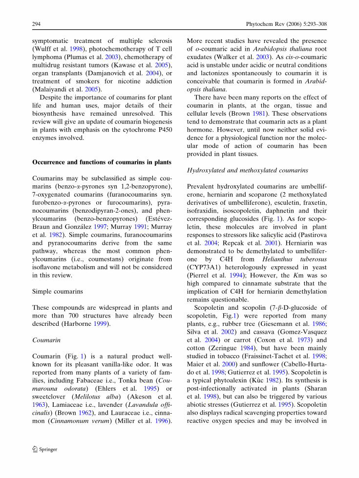
H :8R ,7R ,6R ,5R :niramuoC,HO :7R ,eMO :6R :nitelopocS
lG-O :7R ,eMO :6R :nilopocSHO :7R :enorefillebmU
eMO :7R :nirainreHeMO :7R :6R :enorapocS
HO :7R ,6R :nitelucsEHO :8R ,7R ,eMO :6R :nitexarF
HO :7R ,eMO :8R ,6R :nidixarfosIeMO :7R ,HO :6R :nitelopocsosI
HO :8R ,7R :nitenhpaD eMO :8R ,HO :7RnitegnardyH
HC :8R ,eMO :7R :elohtsO 2 eMC=HC- 2
HC-O :7R ,HO :6R :nilurebuP 2 eMC=HC- 2 eMO :8R , :rehtelynetubenorefillebmU
HC ,eM(C-HC=HC-O :7R 2 ro )HC ,eM(HC-HC=HC-O :7R 2 )HO
nipayA
H :8R ,5R :nelarosPeMO :5R :netpagreB
eMO :8R :nixotohtnaXeMO :8R ,5R :nillenipmiposI
H :6R ,5R :nicilegnAeMO :6R :nidnohpS
eMO :6R ,5R :nillenipmiP
sniramuoC
sniramuoCdetalynerP
sniramuoc-yxoidenelyhteM
sniramuoconarufraeniL
sniramuoconarufralugnAsniramuoconaryP
nitelyhtnaX
OO
6R
5R
O
O
5R
O
6R
OO
5R
O
8R
6R
7R
O O
O
O O OO
Fig. 1 Types of coumarins found in higher plants
Phytochem Rev (2006) 5:293–308 295
123
Rutaceae (Brophy et al. 2002; Brown et al. 1984)
and Apiaceae (Tosun et al. 2005). O-Prenylated
coumarins may be desaturated further to the
corresponding butenylethers (Fig. 1) as shown in
Ammi majus (Hamerski et al. 1990a), and these
reactions are likely also catalyzed through P450
enzymes. The butenylethers are labile and release
a potentially toxic aldehyde moiety, which con-
tributes to their role as phytoalexins. Thus, the
aliphatic substitution of umbelliferone may pro-
vide new substrates for further cytochrome P450
modifications, but neither of these enzymes has so
far been identified. As in case of ayapin in
Asteraceae, the P450 monooxygenases must be
considered as essential ecological factors (see
below).
Furanocoumarins
Furanocoumarins can be grouped into the linear
type, where the (dihydro)furan ring is attached at
C(6) and C(7), and the angular type, carrying the
substitution at C(7) and C(8). Linear furocouma-
rins (syn. psoralens) are principally distributed in
four angiosperm families: Apiaceae, Moraceae,
Rutaceae and Leguminosae (restricted to Psora-
lea and Coronilla generae). The angular (dihy-
dro)furanocoumarins are less widely distributed
and primarily confined to the Apiaceae and
Leguminosae (Bourgaud et al. 1989). The most
abundant linear furanocoumarins are psoralen,
xanthotoxin, bergapten and isopimpinellin,
whereas the angular type is mostly represented
by angelicin, sphondin, and pimpinellin (Fig 1).
As was mentioned for the simple coumarins,
numerous minor furocoumarins have been de-
scribed in the literature, like bergamottin (5-
geranoxy-psoralen) (Stanley and Vannier 1967)
which has received attention recently as a major
grapefruit component interfering with drug
metabolism by intestinal CYP3A4 (Paine et al.
2005; Wen et al. 2002).
Furanocoumarins are recognized as potent
phytoalexins (Beier and Oertli 1983) and allelo-
chemical compounds (Baskin et al. 1967; Beier
1990). An outstanding feature of linear fur-
anocoumarins is their ability to intercalate into
dsDNA and create covalent cross-links primarily
with thymidine residues (Dall’Acqua et al. 1978).
Crosslinking proceeds readily under photoactiva-
tion and potentially blocks DNA replication and
transcription. Accordingly, psoralens exhibit
strong genotoxicity toward all living organisms,
whereas the angular furanocoumarins are just
capable of forming mono-adducts with DNA
creating much less damage (Wamer et al. 1995).
Another remarkable property of furanocouma-
rins is their reactivity to inactivate P450 enzymes
as mentioned above for bergamottin. This kind of
enzyme inhibition has been demonstrated for
P450s from vertebrate (Koenigs and Trager
1998), insect (Zumwalt and Neal 1993) and plant
sources (Gravot et al. 2004). Psoralens inactivate
by a mechanism-based inhibition (also referred as
suicide inhibition) which requires their conver-
sion to reactive intermediates by the enzyme
itself. These intermediates form covalent links to
the apoprotein and permanently inactivate the
enzyme (Fouin-Fortunet et al. 1986; Mays et al.
1989).
The reactivity of furanocoumarins bears con-
siderable ecotoxicological consequences, i.e.,
attributing these compounds an important role
as allelochemicals during plant-insect interactions
(Schuler and Berenbaum 2003). Only herbivores
able to tolerate furanocoumarins can feed on
psoralen-rich plants, and xanthotoxin-insensitive
P450 forms have been described from Papilio
polyxenes, a papilionid butterfly adapted to fur-
anocoumarin-accumulating host plants. This
insensitivity was supposed to be the result of
coevolution of insect detoxifying enzymes and the
particular phytochemical defense since Papilio
glaucus—whose host-plants do not contain fur-
anocoumarins—exhibits sensitive P450s (Zumw-
alt and Neal 1993). The race of coevolution of
butterflies on Apiaceae host plants has been
studied in detail. The capacity of Papilio polyx-
enes to detoxify furanocoumarins through
CYP6B1 follows the order xanthotoxin > psor-
alen > angelicin (Wen et al. 2003), but a syner-
gistic effect has been described between angular
furanocoumarins and psoralen or xanthotoxin in
response to insect attack (Berenbaum and Zan-
gerl 1993). Considering the minor direct toxicity
of angular furanocoumarins, the synergism is
conceivably based on the inhibition of psoralen-
detoxifying CYP by angelicin. Furthermore, the
296 Phytochem Rev (2006) 5:293–308
123

accumulation of angular furanocoumarins is con-
fined to a few taxons only. It was hypothesized,
therefore, that the capacity for angular furano-
coumarin biosynthesis has evolved later and
presumably as a consequence to compensate for
the success of herbivores in the detoxification of
psoralens. (Berenbaum and Zangerl 1998). It
remains to be established, whether the enzymes
for angular furanocoumarin biosynthesis have
evolved from the biosynthesis of linear
furanocoumarins.
Most plants accumulating furanocoumarins
possess a highly inducible biosynthetic pathway.
which can be triggered by various biotic
(Hagemeier et al. 1999; Hamerski and Matern
1988b) and abiotic stresses (Eckey-Kaltenbach
et al. 1994; Katz et al. 1998). Ruta graveolens, and
possibly other Rutaceae, are exceptional because
they do not respond to stressors and synthesize
constitutively furanocoumarins in all tissues (Eil-
ert 1989). However, the elicitation is still possible
in Ruta graveolens dedifferentiated cells (Bohl-
mann et al. 1995).
The tissue-specific distribution of fur-
anocoumarins has been studied in Apiaceae
(Nitao and Zangerl 1987) and Rutaceae (Milesi
et al. 2001). Obviously, these compounds accu-
mulate in cells as well on the surface of plants.
The pronounced accumulation on seeds and
reproductive organs matches the optimal defense
theory which predicts that defense compounds
are principally allocated to the organs that play a
key-role in plant fitness (McKey 1979). The sub-
cellular localization of furanocoumarins is still
unknown, but glucosylated forms have been
frequently reported (Nguyen et al. 1997; Zobel
and Brown 1988), suggesting a probable vacuolar
compartmentation.
Pyranocoumarins
Pyranocoumarins, like xanthyletin (Fig. 1), have
been mainly described from Rutaceae (Anaya
et al. 2005; Sarker et al. 2002) and Apiaceae
(Zgorka et al. 1998). As for furanocoumarins,
linear and angular forms can be distinguished. To
our knowledge, there is no proposal on their
functions in plants, however, due to the structural
relationship with furanocoumarins, a role as phy-
toalexins may be assumed. The biosynthesis of
pyranocoumarins has not yet been investigated.
Biosynthesis of coumarins in plants
Main enzymes and genes implicated in coumarins
biosynthesis and that have been sufficiently doc-
umented are presented in Table 1.
Cinnamic acid to coumarin
The pathway of coumarin biosynthesis has been
largely outlined during the ‘60s and ‘70s, with the
help of tracer feeding experiments (Brown 1981).
Radiolabeled cinnamic acid was incorporated
into coumarin and 7-hydroxycoumarins (Brown
et al. 1960). Other tracer experiments conducted
with Lavandula officinalis, a plant that produces
coumarin as well as 7-hydroxylated coumarins,
revealed that in the latter instance para-hydrox-
ylation preceded the ortho-hydroxylation re-
quired for lactonization (Brown 1962). This
indicated that umbelliferone (Fig. 1) is derived
from cis-p-coumaric acid, whereas coumarin
originates from cis-cinnamic acid (Fig. 2), and
may imply different enzymes for the ortho-
hydroxylation/lactonization of coumarin versus
umbelliferone.
The ortho-hydroxylation is a key step of
coumarin biosynthesis, that has received insuffi-
cient attention. In initial experiments, double-
labeled (ortho-3H, ring-1-14C) cinnamic acid was
fed to Melilotus alba shoots or Gaultheria proc-
umbens leaves, and the retention of label was
monitored upon conversion to o-coumaric acid
(Ellis and Amrhein 1971). An NIH shift was
proposed because of insignificant decrease of the3H:14C ratio, which is an indication of a cyto-
chrome P450 monooxygenase reaction mecha-
nism. A following report addressed the formation
of coumarin with extracts from Melilotus alba, a
plant that produces high levels of coumarin. This
study allocated the ortho-hydroxylation of cin-
namic acid to the chloroplast and again suggested
a P450-dependent hydroxylation mechanism
(Gestetner and Conn 1974). Unfortunately, the
in vitro results could not be reproduced, and the
class of the enzyme involved as well as its
Phytochem Rev (2006) 5:293–308 297
123

Ta
ble
1E
nzy
me
so
fco
um
ari
na
nd
fura
no
cou
ma
rin
bio
syn
the
sis
cha
ract
eri
zed
inv
itro
or
by
pre
curs
or
fee
din
gst
ud
ies.
Cin
na
ma
te4
-hy
dro
xy
lase
,n
ot
me
nti
on
ed
he
re,
isd
esc
rib
ed
by
Eh
ltin
ge
ta
l.in
this
issu
e.
N.D
.m
ea
ns
no
td
ete
rmin
ed
,P
mg
:P
hy
top
hth
ora
meg
asp
erm
a
Act
ivit
yS
ub
stra
teK
ine
tic
con
sta
nts
Pla
nt
Co
mm
en
tsA
uth
ors
Be
nzo
ica
cid
2—
hy
dro
xy
lase
Be
nzo
ica
cid
N.D
.N
ico
tia
na
tab
acu
mP
45
0,
So
lub
lee
nzy
me
Le
on
et
al.
19
95
Co
um
ara
te/C
inn
am
ate
/F
eru
late
2-
hy
dro
xy
lase
p-C
ou
ma
ric
aci
d;
Fe
ruli
ca
cid
;C
inn
am
ica
cid
N.D
.H
yd
ran
gea
ma
cro
ph
yll
aC
hlo
rop
last
ica
ctiv
ity
Kin
dl
19
71
Esc
ule
tin
syn
tha
seU
mb
ell
ife
ron
eN
.D.
Cic
ho
riu
min
tyb
us
Pre
curs
or
fee
din
gst
ud
ies
on
lyB
row
n1
98
5
Da
ph
ne
tin
syn
tha
seU
mb
ell
ife
ron
eN
.D.
Da
ph
ne
mez
ereu
mP
recu
rso
rfe
ed
ing
stu
die
so
nly
Bro
wn
19
86
Um
be
llif
ero
ne
7-O
-p
ren
ylt
ran
sfe
rase
Um
be
llif
ero
ne
Km
Um
be
llif
ero
ne
=6
.5lM
Km
DM
AP
P=
30l
M
Am
mi
ma
jus
Pm
ge
lici
ted
cell
cult
ure
sH
am
ers
ki
et
al.
19
90
Um
be
llif
ero
ne
6-
pre
ny
ltra
nsf
era
se
Um
be
llif
ero
ne
N.D
.A
mm
im
aju
sP
mg
eli
cite
dce
llcu
ltu
res
Ha
me
rsk
ie
ta
l.1
99
0
Um
be
llif
ero
ne
6-
pre
ny
ltra
nsf
era
se
Um
be
llif
ero
ne
N.D
.R
uta
gra
veo
len
sP
last
idic
en
zym
eD
hil
lon
an
dB
row
n1
97
6
Ma
rme
sin
syn
tha
seD
em
eth
yls
ub
ero
sin
Km
DM
S=
10
.3l
MK
mN
AD
PH
=1
9.6
lMA
mm
im
aju
sP
45
0,
Pm
ge
lici
ted
cell
cult
ure
so
pti
ma
lp
H=
7.5
/op
tim
al
tem
p.
=2
0�C
Ha
me
rsk
ia
nd
Ma
tern
19
88
Ma
rme
sin
5-h
yd
rox
yla
se
(+)-
Ma
rme
sin
N.D
.R
uta
gra
veo
len
sF
icu
sca
rica
Pre
curs
or
fee
din
gst
ud
ies
on
lyC
ap
ora
lee
ta
l.1
98
1
Pso
rale
nsy
nth
ase
(+)-
Ma
rme
sin
Km
NA
DP
H=
52lM
Pet
rose
lin
um
cris
pu
mP
45
0,
op
tim
al
pH
=7
–7
.25
We
nd
orf
an
dM
ate
rn1
98
6
An
ge
lici
nsy
nth
ase
(+)-
Co
lum
bia
ne
tin
N.D
.H
era
cleu
mm
an
teg
az
zia
nu
mP
recu
rso
rfe
ed
ing
stu
die
so
nly
Sta
nje
ke
ta
l.1
99
8
Pso
rale
n5
-m
on
oo
xy
ge
na
se
Pso
rale
nK
mP
sora
len
=1
2lM
Km
NA
DP
H=
20
0lM
Am
mi
ma
jus
P4
50
,P
mg
eli
cite
dce
llcu
ltu
res
Ha
me
rsk
ia
nd
Ma
tern
19
88
Be
rga
pto
lO
-me
thy
ltra
nsf
era
se
Be
rga
pto
lK
mB
erg
ap
tol
=2
.8lM
Km
SA
M=
6.5
lMA
mm
im
aju
sG
en
ba
nk
acc
ess
ion
AY
44
30
06
,P
mg
eli
cite
dce
llcu
ltu
res,
op
tim
al
pH
=8
/te
mp
=4
2�C
He
hm
an
ne
ta
l.2
00
4
Be
rga
pto
lO
-me
thy
ltra
nsf
era
se
Be
rga
pto
l/5
-H
yd
rox
yx
an
tho
tox
inK
mB
erg
ap
tol
=4lM
Vm
ax
=9
80lk
at/
kg
Km
SA
M=
3.1
lMV
max
=1
16
0lk
at/
kg
Km
5-H
-Xa
nth
oto
xin
=1l
MV
max=
12
50lk
at/
kg
Pet
rose
lin
um
cris
pu
mP
mg
eli
cite
dce
llcu
ltu
res,
pu
rifi
ed
en
zym
eo
pti
ma
lp
H=
8–
8.5
Ha
uff
ee
ta
l.1
98
6
298 Phytochem Rev (2006) 5:293–308
123

subcellular site remain to be established. As
revealed later, the early experiments may have
suffered from fundamental analytical problems,
since the chromatography and recrystallization
techniques employed were likely insufficient to
separate the various cinnamic acids. Nevertheless,
the proposed conversion of cinnamic to o-coum-
aric acid received some support by precursor
feeding studies done with Petunia chloroplasts,
which ascribed cinnamate 2-hydroxylase, includ-
ing the formation of coumarin, and lack of
cinnamate 4-hydroxylase to these organelles
(Conn 1984; Ranjeva et al. 1977). In light of the
studies done since with Ammi majus microsomes
(Hamerski and Matern 1988a, b; Stanjek et al.
1999a) on the biosynthesis of furanocoumarins it
appears possible that the ‘ortho-hydroxylase’ is an
exceptionally labile CYP enzyme, in contrast to
the CYPs hydroxylating cinnamic acids in para-
or meta-position (Fig. 2). Overall, the ortho-
hydroxylation of cinnamic (or 4-coumaric) acid,
being of pivotal importance for all coumarins,
remains a missing link in the network of phenyl-
propanoid biosynthesis.
Cinnamic acid to umbelliferone and other
hydroxylated coumarins
The formation of umbelliferone proceeds from
4-coumaric acid or its ester derivatives (Fig. 2).
The conversion of cinnamic acid to 4-coumaric
acid is catalyzed by cinnamate 4-hydroxylase, a
cytochrome P450 monooxygenase from the
CYP73A family (Teutsch et al. 1993). This
enzyme constitutes the P450 enzyme most studied
to date and sets the stage for several branch
pathways, such as the lignification (Anterola and
Lewis 2002) or flavonoid biosynthesis (Harborne
1999) (see Ehlting et al. 2006 in this issue for a
review).
Following the pertaining literature, 4-coumaric
acid is ortho-hydroxylated to 2,4-dihydroxycin-
namic acid. The respective enzyme activity was
reported exclusively from Hydrangea macrophy-
lla and assigned to the chloroplasts (Kindl 1971).
This enzyme fraction was demonstrated to slowly
convert cinnamic acid to o-coumaric acid but was
more active to transform p-coumaric acid and
ferulic acid respectively to umbelliferone andTa
ble
1co
nti
nu
ed
Act
ivit
yS
ub
stra
teK
ine
tic
con
sta
nts
Pla
nt
Co
mm
en
tsA
uth
ors
Be
rga
pto
lO
-me
thy
ltra
nsf
era
seB
erg
ap
tol/
5-
Hy
dro
xy
xa
nth
oto
xin
N.D
.R
uta
gra
veo
len
sS
ep
ara
ted
fro
mx
an
tho
tox
ol
OM
Tp
uri
fie
de
nzy
me
Sh
arm
ae
ta
l.1
97
9
Xa
nth
oto
xo
lO
-m
eth
ylt
ran
sfe
rase
Xa
nto
tho
xo
lK
mX
an
tho
tox
ol
=9
.8l
MV
max
=5
75
0lk
at/
kg
Km
SA
M=
4.4
lMV
max
=7
48
0lk
at/
kg
Pet
rose
lin
um
cris
pu
mP
mg
Eli
cite
dce
llcu
ltu
res,
pu
rifi
ed
en
zym
eH
au
ffe
et
al.
19
86
Xa
nth
oto
xo
lO
-m
eth
ylt
ran
sfe
rase
Xa
nth
oto
xo
l,8
-H
yd
rox
yb
erg
ap
ten
N.D
.R
uta
gra
veo
len
sS
ep
ara
ted
fro
mb
erg
ap
tol
OM
Tp
uri
fie
de
nzy
me
Sh
arm
ae
ta
l.1
97
9
Iso
pim
pin
ell
insy
nth
ase
Xa
nth
oto
xin
,B
erg
ap
ten
N.D
.H
era
cleu
mla
na
tum
Pre
curs
or
fee
din
gst
ud
ies
on
lyB
row
na
nd
Sa
mp
ath
ku
ma
r1
97
7
Iso
pim
pin
ell
insy
nth
ase
5,8
-Hy
dro
xy
pso
rale
nN
.D.
Ru
tag
rav
eole
ns
Pre
curs
or
fee
din
gst
ud
ies
on
lyIn
no
cen
tie
ta
l.1
98
3
Phytochem Rev (2006) 5:293–308 299
123
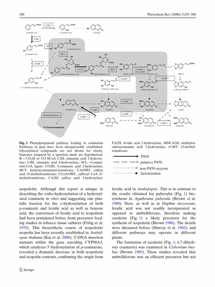
scopoletin. Although this report is unique in
describing the ortho-hydroxylation of a hydroxyl-
ated coumarin in vitro and suggesting one plas-
tidic fraction for the o-hydroxylation of both
p-coumaric and ferulic acid as well as benzoic
acid, the conversion of ferulic acid to scopoletin
had been postulated before from precursor feed-
ing studies in tobacco tissue cultures (Fritig et al.
1970). This biosynthetic course of scopoletin/
scopolin has been recently established in Arabid-
opsis thaliana (Kai et al. 2006). T-DNA insertion
mutants within the gene encoding CYP98A3,
which catalyzes 3’-hydroxylation of p-coumarate,
revealed a dramatic decrease in both scopoletin
and scopolin contents, confirming the origin from
ferulic acid in Arabidopsis. This is in contrast to
the results obtained for puberulin (Fig. 1) bio-
synthesis in Agathosma puberula (Brown et al.
1988). Here, as well as in Daphne mezereum,
ferulic acid was not readily incorporated as
opposed to umbelliferone, therefore making
esculetin (Fig. 1) a likely precursor for the
synthesis of scopoletin (Brown 1986). The details
were discussed before (Murray et al. 1982), and
different pathways may operate in different
plants.
The formation of esculetin (Fig. 1; 6,7-dihydr-
oxy coumarin) was examined in Cichorium inty-
bus (Brown 1985). These studies revealed that
umbelliferone was an efficient precursor but not
dicacimanniC
p dicaciramuoC-
-’4,’2 dica cimannicyxordyhiD
enorefillebmU
niramuoCo dicaciramuoC-
H4C
? H2OC
? H2C
TCH
esarefsnartlynerP
p dicacimikihslyoramuoC-
H’3C
dicacimikihslyoeffaC
nitelucsE
TCH
dicacieffaC
AC O ro TMAoCC O TM
nitelopocS
dicacilureF
? H2AF? H2AC
dica cilurefyxordyH-’2
sniramuoconaruF oT
ACDM
nipayA
nirainreH nitelopocsosI
O ? TM-
enorapocS
O ? TM-
O ? TM-
LC4p AoC-lyoramuoC-
O ? TM-
dicacieffac yxordyH-’2 ACDM-yxordyH-’2
OC 2H
OC 2H
OH
OH
AoCS
O
HO
OC 2H
O O
HO
R
OH
OH
O
O
OH
HO
OC 2H
OH
O
O
OH
HO
OC 2H
HO
R
OH
OH R
OH
OeM O
O
HOOC
R
HOOH
OHR
HOOH
OeM R
HO
O
O
O
O O OO O
OeM
OHO O
OH
OHO OOH
O OOeM O OOeM
OH
O OOeM
OeM
H4C
Fig. 2 Phenylpropanoid pathway leading to coumarinsPathways in gray have been unequivocally established.Glycosylated compounds are not shown for clarity.Enzymes assigned by a question mark are hypothetical.R = CO2H or CO-SCoA C2H, cinnamic acid 2-hydroxy-lase; C4H, cinnamic acid 4-hydroxylase; 4CL, 4-couma-rate:CoA ligase; CO2H, 4-coumaric acid 2-hydroxylase;HCT, hydroxycinnamoyl-transferase; CAOMT, caffeicacid O-methyltransferase; CCoAOMT, caffeoyl CoA O-methyltransferase, CA2H, caffeic acid 2-hydroxylase;
FA2H, ferulic acid 2-hydroxylase; MDCA2H, methylen-edioxycinnamic acid 2-hydroxylase; O-MT, O-methyl-transferase
lactonization
P450
putative P450
non-P450 enzyme
300 Phytochem Rev (2006) 5:293–308
123

caffeic acid, suggesting 6-hydroxylation of umbel-
liferone, probably by the action of a P450 mono-
oxygenase. This deserves mentioning, because the
conversion of caffeic acid to esculetin is readily
accomplished in vitro with various plant extracts
containing phenoloxidase activity (Kneusel 1987;
Sato 1967), but has not been confirmed in planta.
Similar to esculetin, daphnetin (Fig. 1, 7,8-dihydr-
oxycoumarin) in Daphne mezereum, was shown
to be derived from umbelliferone rather than
caffeic acid.
The ortho-hydroxylation: a common route
with salicylic acid
Analogous to C2H, another major ortho-hydrox-
ylation step in phenolic metabolism is still con-
troversial. Salicylic acid is a pivotal signal
molecule in plant defense mechanisms (Shah
2003) but the biosynthesis pathway is still matter
of debate. Two routes have been proposed. A
pathway already shown to occur in bacteria has
been proposed in tobacco through chorismate and
isochorismate, via the general shikimic acid
metabolism (Wildermuth et al. 2001). Another
route has been documented in tobacco (Coquoz
et al. 1998; Yalpani et al. 1993) and rice (Silver-
man et al. 1995), via decarboxylation of trans-
cinnamic acid to benzoic acid and subsequent
2-hydroxylation. This benzoic acid 2-hydroxylase
was characterized as a P450 enzyme but impor-
tant biochemical characteristics are atypical for
an eucaryotic P450 as it appears to be soluble and
it exhibits an unusually high molecular weight
(Leon et al. 1995). The corresponding P450 gene
has not been reported so far. This benzoic acid
2-hydroxylase is unable to transform cinnamic
acid into o-coumaric acid (Yalpani et al. 1993)
and consequently is unlikely to interfere with the
coumarin pathway.
Biosynthesis of furanocoumarins in plants
While coumarin biosynthesis remains a black box,
several enzymes of the furanocoumarin pathway
have been isolated and characterized (Fig. 3).
Entry of umbelliferone into the
furanocoumarin pathway
Umbelliferone rather than coumarin is the parent
compound of furanocoumarins, as was reported a
long time ago (Floss and Mothes 1964). It is first
prenylated in 6- (for linear furanocoumarins) or
8-position (for angular furanocoumarins) to yield
demethylsuberosin and osthenol, respectively
(Fig. 3). Dimethylallyl diphosphate required for
the 6-prenylation at least is provided in celery
(Apium graveolens) by the deoxy-D-xylulose
pathway and not through the mevalonate-depen-
dent pathway (Stanjek et al. 1999b). This is
conceivably also the case in other plants, because
the prenyltransferase has been identified in Ruta
graveolens as a plastidic enzyme (Dhillon and
Brown 1976; Ellis and Brown 1974), and the
activity was also documented in Ammi majus
(Hamerski et al. 1990b) The homologous enzyme
for the angular furanocoumarins has not been
isolated so far.
Linear furanocoumarins
Demethylsuberosin is transformed to marmesin
and further to psoralen by two separate cyto-
chrome P450 enzymes (Hamerski and Matern
1988b; Wendorff and Matern 1986). The enzymes
were biochemically characterized, and evidence
for their P450 nature was obtained from charac-
teristic blue-light-reversible inhibition of the
activities by carbon monoxide, and the use of
specific inhibitors. The two enzymes formally
catalyze very different reactions, the first forming
the dihydrofuran-ring from the ortho-prenylated
phenol (marmesin synthase) and the second
catalyzing the oxidative carbon-carbon chain
cleavage reaction (psoralen synthase). The mech-
anism of marmesin synthase has not been solved
yet, but it might be speculated that some analogy
exists to menthofuran synthase from Mentha
piperita which belongs to the CYP71 family
(Bertea et al. 2001; Croteau et al. 2005). Psoralen
synthase was found to operates by syn-elimina-
tion of acetone and one hydrogen from position 3’
(Fig. 3) (Stanjek et al. 1999a). This release of
acetone is unique in plants. Psoralen synthase is
Phytochem Rev (2006) 5:293–308 301
123
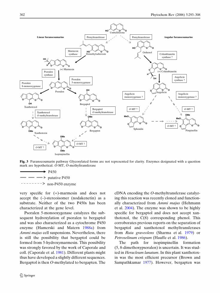
very specific for (+)-marmesin and does not
accept the (–)-stereoisomer (nodakenetin) as a
substrate. Neither of the two P450s has been
characterized at the gene level.
Psoralen 5-monooxygenase catalyzes the sub-
sequent hydroxylation of psoralen to bergaptol
and was also characterized as a cytochrome P450
enzyme (Hamerski and Matern 1988a) from
Ammi majus cell suspensions. Nevertheless, there
is still the possibility that bergaptol could be
formed from 5-hydroxymarmesin. This possibility
was strongly favored by the work of Caporale and
coll. (Caporale et al. 1981). Different plants might
thus have developed a slightly different sequences.
Bergaptol is then O-methylated to bergapten. The
cDNA encoding the O-methyltransferase catalyz-
ing this reaction was recently cloned and function-
ally characterized from Ammi majus (Hehmann
et al. 2004). The enzyme was shown to be highly
specific for bergaptol and does not accept xan-
thotoxol, the C(8) corresponding phenol. This
corroborates previous reports on the separation of
bergaptol and xanthotoxol methyltransferases
from Ruta graveolens (Sharma et al. 1979) or
Petroselinum crispum (Hauffe et al. 1986).
The path for isopimpinellin formation
(5, 8-dimethoxypsoralen) is uncertain. It was stud-
ied in Heracleum lanatum. In this plant xanthotox-
in was the most efficient precursor (Brown and
Sampathkumar 1977). However, bergapten was
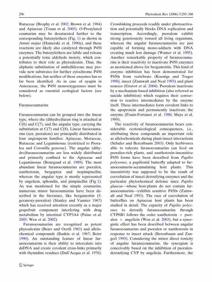
enorefillebmU
nisorebuslyhtemeD
nisemraM )+(
Marmesinesahtnys
nitenaibmuloC )+(
nitenaibmuloC? esahtnys
esarefsnartlynerP
nelarosPesahtnys
nelarosPesanegyxoonom-5
nelarosP
lotpagreB
nicilegnA
lotpagreBO esarefsnartlyhtem-
netpagreB
sniramuoconarufralugnAesarefsnartlynerP
lonehtsO
loxotohtnaX
nixotohtnaX
nelarosPesanegyxoonom-8
loxotohtnaXO esarefsnartlyhtem-
nillenipmiposI
O ? TM-
O ? TM-
sniramuoconarufraeniL
nidnohpSnillenipmiP
nicilegnA? esanegyxoonom
O ? TM-
nicilegnA? esanegyxoonom
O ? TM-
? TM-O
nicilegnA? esahtnys
O OOH
O OOH
O OO
O OO
HO
O OO
HO
O OO
eMO
O OO
eMO
O OO
eMO
eMO
O OOH
O OO
O OO
OeM
O OO
OeM
eMO
H
O OO
HO
O OO
OH
H
Fig. 3 Furanocoumarin pathway Glycosylated forms are not represented for clarity. Enzymes designated with a questionmark are hypothetical. O-MT, O-methyltransferase
P450
putative P450
non-P450 enzyme
302 Phytochem Rev (2006) 5:293–308
123
found to be converted into isopimpinellin,
although at a lower rate. Both 5- and 8-hydroxyl-
ation pathways can thus lead to final product, but
5,8-dihydroxypsoralen was also demonstrated to
be a possible precursor in Ruta graveolens (Inno-
centi et al. 1983). Enzymatic turnover of the
pathways could simply explain the prevalence of
one of the three routes in a given plant.
Angular furanocoumarins
The transformation of columbianetin to angelicin
is very similar from a mechanistic and stereo-
chemical point of view to the conversion of
marmesin to psoralen (Stanjek and Boland
1998). As demonstrated by feeding studies using
fluor- or deuterium-labeled columbianetin with
plants or leaf tissues. It is, thus, conceivable that
the enzymes for angular furanocoumarin biosyn-
thesis may have emerged by evolutionary adap-
tation from the linear pathway. This would be
consistent with the fact that angular fur-
anocoumarins are less abundant in plants than
the linear type and that angular furanocoumarins
are always found concomitantly with linear fur-
anocoumarins. This hypothesis will be investi-
gated once the genes for marmesin synthase and
psoralen synthase, as well as those for umbellif-
erone 6- and 8-prenyltransferases, will be identi-
fied (Fig. 3). Unfortunately, no information is
available yet at the genetic level.
Implication of P450s in furanocoumarin
synthesis
Cytochrome P450 enzymes are pivotal enzymes
of furanocoumarin biosynthesis, i.e., the forma-
tion of xanthotoxin relies, at least, on four
sequential P450 reactions catalyzed by C4H,
marmesin synthase, psoralen synthase and psor-
alen 8-monooxygenase. This was at a first glance
puzzling because of the intrinsic capacity of
furanocoumarins to inhibit very different cyto-
chrome P450 enzymes, irrespective of the species,
through a mechanism-based inactivation process
(Fouin-Fortunet et al. 1986).
To understand how plants cope with this
problem Gravot and co-workers compared
inactivation by furanocoumarins (Gravot et al.
2004) of three different C4H: one from a plant that
does not contain furanocoumarins (Helianthus
tuberosus, CYP73 A1) and two from plants that
synthesize furanocoumarins (Ruta graveolens,
CYP73A32; Petroselinum crispum CYP73A10).
They showed that Kinact and 1/Ki in presence of
psoralen were lower for CYP73A32 and
CYP73A10 compared to CYP73A1, and, accord-
ingly, the cinnamate hydroxylation activity of
CYP73A32 and CYP73A10 appears more resis-
tant to mechanism-based inactivation than that of
CYP73A1 (respectively kinact/Ki 7, 4.4, and
45 min–1mM–1). This would suggest that plants
producing furanocoumarins have adapted their
P450 enzyme repertoire to the need for reduced
inactivation while retaining the high catalytic
efficiency. It is reasonable to expect a similar
adaptation of all the P450 enzymes in the same
pathway.
The evolution toward furanocoumarin accu-
mulation must have occurred under strong selec-
tion pressure, since the biocidal and enzyme
inactivation properties of furanocoumarins ap-
pear to be lethal to plants unless quick adaptation
can be accomplished. This pressure might have
built up by the exposure to herbivores and the
need for efficient antifeedant metabolites. This
would be fully compatible with the scheme of
furanocoumarins as allelochemicals in the warfare
with insects only adapted to hatch on furano-
coumarin producing plants (Schuler and Beren-
baum 2003). It will be interesting to compare the
cytochrome P450 families recruited for the syn-
thesis of furanocoumarins in the plant and their
detoxification in insects.
Perspectives
Although no monooxygenase of the furano-
coumarin pathway stricto sensu has been
characterized at the gene level, techniques such
as differential display and RT-PCR strategies
have been developed for P450s (Schoendorf
et al. 2001; Schopfer and Ebel 1998) which
should be readily applicable to furanocoumarin
pathway. Such techniques already led to the
characterization of the C4H and C3’H in the
relevant plants. Inducible systems are needed to
Phytochem Rev (2006) 5:293–308 303
123

differentiate and correlate the individual tran-
script abundances with product accumulation
Elicitor-treated Ammi majus cultures appear to
qualify for this purpose.
Numerous recent studies focused on the role of
furanocoumarins as key allelochemicals, but the
physiological relevance of coumarins reaches far
beyond in the producing plants. This includes the
potential role of simple coumarins as hormones
and signaling molecules, which were shown in the
past decade to be much more widespread in plant
kingdom than previously assumed. More func-
tional insight should be obtained once the mech-
anism, regulation of their biosynthesis and their
subcellular localization will be known. Biosyn-
thesis of L-phenylalanine proceeds in plastids
while phenylalanine ammonia-lyase and C4H
activities reside in the cytosol and endoplasmic
reticulum. Subcellular localization of the pivotal
ortho-hydroxylation of cinnamic or 4-coumaric
acid, so far, remains unresolved. Investigation in
Ruta graveolens assigned the subsequent 6-preny-
lation of umbelliferone to plastidial membranes
(Dhillon and Brown 1976). Clarification of the
localization of the 2-hydroxylation will be the
further step to understand the physiological role
of coumarins.
It is probable that different routes to couma-
rins will be discovered to operate in plants, some
of them might be confined to a taxonomic group.
The formation of scopoletin is an example and
derives either from esculetin or ferulic acid
according to the plant species considered. It is
currently unknown, whether the P450s involved
in the furocoumarin pathway belong to a single
family, as is the case with CYP71s in benzoxazine
synthesis, or to multiple P450 families as shown
for biosynthesis of cyanogenic glucosides
(CYP71E1 and CYP79A1). In either case, the
discovery of genes involved in coumarin synthesis
will add another stage of complexity to the
phenylpropanoid pathway.
The recent detection of coumarin and hydrox-
ylated coumarins in Arabidopsis thaliana have
opened the way for new approaches. Metabolo-
mics in conjunction with screening of mutant
libraries is likely to reveal new players in the
coumarin pathway.
References
Akeson WR, Gorz HJ, Haskins FA (1963) Effect ofgenotype and growth stage on distribution of mellil-otic acid, O-coumaric acid, and coumarinic acid inMelilotus alba Desr. Crop Sci 3:167–171
Anaya AL, Macıas-Rubalcava M, Cruz-Ortega R, Garcıa-Santana C, Sanchez-Monterrubio PN, Hernandez-Bautista BE, Mata R (2005) Allelochemicals fromStauranthus perforatus, a Rutaceous tree of the Yuca-tan Peninsula, Mexico. Phytochemistry 66:487–494
Anterola AM, Lewis NG (2002) Trends in lignin modifi-cation: a comprehensive analysis of the effects ofgenetic manipulations/mutations on lignification andvascular integrity. Phytochemistry 61:221–294
Baskin JM, Ludlow CJ, Harris TM, Wolf FT (1967)Psoralen, an inhibitor in the seeds of Psoraleasubacaulis (Leguminosae). Phytochemistry 6:1209–1213
Bednarek P, Schneider B, Svatos A, Oldham N, HahlbrockK (2005) Structural complexity, differential responseto infection, and tissue specificity of indolic andphenylpropanoid secondary metabolism in Arabidop-sis roots. Plant Physiol 138:1058–1070
Beier RC (1990) Natural pesticides and bioactive compo-nents in foods. Rev Environ Contam Toxicol 113:47–137
Beier RC, Oertli EH (1983) Psoralen and other linearfurocoumarins as phytoalexins in celery. Phytochem-istry 22:2595–2597
Berenbaum MR, Zangerl AR (1993) Furanocoumarinmetabolism in Papilio polyxenes—Biochemistry, ge-netic variability, and ecological significance. Oecolo-gia 95:370–375
Berenbaum MR, Zangerl AR (1998) Chemical phenotypematching between a plant and its insect herbivore.Proc Natl Acad Sci 95:13743–13748
Bertea CM, Schalk M, Karp F, Maffei M, Croteau R (2001)Demonstration that menthofuran synthase of mint(Mentha) is a cytochrome P450 monooxygenase:cloning, functional expression, and characterizationof the responsible gene. Arch Biochem Biophys390:279–286
Bohlmann J, Gibraltarskaya E, Eilert U (1995) Elicitorinduction of furanocoumarin biosynthetic pathway incell cultures of Ruta graveolens. Plant Cell TissueOrgan Cult 43:155–161
Bourgaud F, Allard N, Guckert A, Forlot P (1989) Naturalsources for furocoumarins. In: Fitzpatrick T, Forlot P,Pathak MA, Urbach F (eds) Psoralens, past, presentand future of Photochemoprotection and other bio-logical activities. J. Libbey Eurotext, Paris, pp 301–306
Brophy JJ, Goldsack RJ, Fookes CJR, Forster PI (2002)The essential oils of Pentaceras australe (Rutaceae). JEssent Oil Res 14:348–350
Brown SA (1962) Biosynthesis of coumarin and herniarinin lavender. Science 137:977–978
Brown SA (1981) Coumarins. In: Stumpf PK, Conn EE(eds) The biochemistry of plants—A comprehensivetreatise, vol 7. Academic Press, New York, pp 269–300
304 Phytochem Rev (2006) 5:293–308
123

Brown SA (1985) Biosynthesis of 6,7-dihydroxycoumarinin Cichorium intybus. Can J Biochem Cell Biol63:292–295
Brown SA (1986) Biosynthesis of Daphnetin in Daphnemezereum L. Z Naturforsch 41c:247–252
Brown SA, March RE, Rivett DEA, Thompson HJ (1988)Intermediates in the formation of puberulin byAgathosma puberula. Phytochemistry 27:391–395
Brown SA, Rivett DEA, Thompson HJ (1984) Elabora-tion of the 6,7,8-hydroxylation pattern in simplecoumarins: biosynthesis of puberulin. Zeitschrift FurNaturforschung 39:31–37
Brown SA, Sampathkumar S (1977) The biosynthesis ofisopimpinellin. Can J Biochem Cell Biol 55:686–692
Brown SA, Towers GH, Wright D (1960) Biosynthesis ofthe coumarins. Tracer studies on coumarin formationin Hierochloe odorata and Melilotus officinalis. Can JBiochem Physiol 38:143–156
Cabello-Hurtado F, Durst F, Jorrin JV, Werck-ReichhartD (1998) Coumarins in helianthus tuberosus: charac-terization, induced accumulation and biosynthesis.Phytochemistry 49:1029–1036
Caporale G, Innocenti G, Guiotto A, Rodighiero P,Dall’Acqua F (1981) Biogenesis of linear O-al-kylfuranocoumarins: a new pathway involving 5-hydroxymarmesin. Phytochemistry 20:1283–1287
Chong J, Baltz R, Schmitt C, Belffa R, Fritig B, Saindre-nan P (2002) Downregulation of a pathogen-respon-sive tobacco UDP-Glc: phenylpropanoidglucosyltransferase reduces scopoletin glucoside accu-mulation, enhances oxidative stress, weakens virusresistance. Plant Cell 14:1093–1107
Clemens S, Barz W (1996) P450-dependent methylenedi-oxy bridge formation in Cicer arietinum. Phytochem-istry 41:457–460
Conn EE (1984) Compartmentation of secondary com-pounds. In Boudet AM, Alibert G, Lea PJ (eds)Membranes and compartmention in the regulaton ofplant function. Oxford University Press, Oxford, pp1–28
Coquoz JL, Buchala A, Metraux JP (1998) The biosyn-thesis of salicylic acid in potato plants. Plant Physiol117:1095–1101
Coxon DT, Curtis RF, Price KR, Levett G (1973)Abnormal metabolites produced by Daucus carotaroots stored under conditions of stress. Phytochemis-try 12:1881–1885
Croteau RB, Davis EM, Ringer KL, Wildung MR (2005)(–)-Menthol biosynthesis and molecular genetics.Naturwissenschaften 92:562–577
Dall’Acqua F, Vedaldi D, Recher M (1978) The photore-action between furocoumarins and various DNA withdifferent base compositions. Photochem Photobiol27:33–36
Damjanovich S, Gaspar R, Panyi G (2004) An alternativeto conventional immunosuppression: Small-moleculeinhibitors of K(v)1.3 channels. Mol Interv 4:250–+
Dhillon DS, Brown SA (1976) Localization, purificationand characterization of a dimethylallylpyrophosphate:umbelliferone dimethylallyltransferase from Rutagraveolens. Arch Biochem Biophys 177:74–83
Eckey-Kaltenbach H, Ernst D, Heller W, Sandermann HJr (1994) Biochemical Plant Responses to Ozone (IV.Cross-Induction of Defensive Pathways in Parsley(Petroselinum crispum L.) Plants). Plant Physiol104:67–74
Ehlers D, Pfister M, Bork WR, Toffelnadolny P (1995)HPLC Analysis of Tonka Bean Extracts. ZeitschriftFur Lebensmittel-Untersuchung Und-Forschung201:278–282
Ehlting J, Hamberger B, Million-Rousseau R, Werck-Reichhart D (2006) Cytochromes P450 in the phenolicmetabolism. Phytochem Rev DOI 10.1007/s11101-006-9025-1 (this issue)
Eilert U (1989) Elicitor induction of secondary metabolismin dedifferenciated in vitro system of Ruta graveolens.In: Kurz WGW (ed) Primary and secondary metab-olism of plant cell cultures. Springer, Berlin, pp 219–228
Ellis BE, Amrhein N (1971) The ‘NIH-shift’ duringaromatic ortho-hydroxylation in higher plants. Phyto-chemistry 10:3069–3072
Ellis BE, Brown SA (1974) Isolation of dimethylallylpy-rophosphate: umbelliferone dimethylallyltransferasefrom Ruta graveolens. Can J Biochem 52:734–738
Estevez-Braun A, Gonzalez AG (1997) Coumarins. In:Natural Product Reports. pp 465–475
Floss HG, Mothes U (1964) On the biochemistry offurocoumarin in Pimpinella magna. Z Naturforsch B19:770–771
Fouin-Fortunet H, Tinel M, Descatoire V, Letteron P,Larrey D, Geneve J, Pessayre D (1986) Inactivationof cytochrome P-450 by the drug methoxsalen. JPharmacol Exp Ther 236:237–247
Fraissinet-Tachet L, Baltz R, Chong J, Kauffmann S, FritigB, Saindrenan P (1998) Two tobacco genes induced byinfection, elicitor and salicylic acid encode glucos-yltransferases acting on phenylpropanoids and ben-zoic acid derivatives, including salicylic acid. FEBSLett 437:319–323
Fritig B, Hirth L, Ourisson G (1970) Biosynthesis of thecoumarins: scopoletin formation in tobacco tissuecultures. Phytochemistry 9:1963–1975
George HL, VanEtten HD (2001) Characterization ofpisatin-inducible cytochrome P450s in fungal patho-gens of pea that detoxify the pea phytoalexin pisatin.Fungal Genet Biol 33:37–48
Gestetner B, Conn EE (1974) 2-hydroxylation of trans-cinnamic acid by chloroplasts from Melilotus alba.Arch Biochem Biophys 163:617–624
Giesemann A, Biehl B, Lieberei R (1986) Identification ofscopoletin as a phytoalexin of the rubber tree Heveabrasiliensis. J Phytopathol 117:373–376
Gomez-Vasquez R, Day R, Buschmann H, Randles S,Beeching JR, Cooper RM (2004) Phenylpropanoids,phenylalanine ammonia lyase and Peroxidases inelicitor-challenged cassava (Manihot esculenta) sus-pension cells and leaves. Ann Bot 94:87–97
Gravot A, Larbat R, Hehn A, Lievre K, Gontier E,Goergen J, Bourgaud F (2004) Cinnamic acid4-hydroxylase mechanism-based inactivation bypsoralen derivatives : cloning and characterization
Phytochem Rev (2006) 5:293–308 305
123

of a C4H from a psoralen producing plant - Rutagraveolens-exhibiting a low sensitivity to psoraleninactivation. Arch Biochem Biophys 422:71–80
Gutierrez M-C, Parry A, Tena M, Jorrin J, Edwards R(1995) Abiotic elicitation of coumarin phytoalexins insunflower. Phytochemistry 38:1185–1191
Hagemeier J, Batz O, Schmidt J, Wray V, Hahlbrock K,Strack D (1999) Accumulation of phthalides inelicitor-treated cell suspension cultures of Petroseli-num crispum. Phytochemistry 51:629–635
Hamerski D, Beier RC, Kneusel RE, Matern U, Him-melspach K (1990a) Accumulation of coumarins inelicitor-treated cell suspension cultures of Ammimajus. Phytochemistry 29:1137–1142
Hamerski D, Matern U (1988a) Biosynthesis of psoralens.Psoralen 5-monooxygenase activity from elicitor-treated Ammi majus cells. FEBS Lett 239:263–265
Hamerski D, Matern U (1988b) Elicitor-induced biosyn-thesis of psoralens in Ammi majus L. suspensioncultures. Microsomal conversion of demethylsubero-sin into (+)marmesin and psoralen. Eur J Biochem171:369–375
Hamerski D, Schmitt D, Matern U (1990b) Induction oftwo prenyltransferases for the accumulation of cou-marin phytoalexins in elicitor-treated Ammi majuscell suspension cultures. Phytochemistry 29:1131–1135
Harborne JB (1999) Classes and functions of secondaryproducts from plants. In Walton NJ, Brown DE (eds)Chemicals from plants. Imperial College Press,London, pp 1–25
Hauffe KD, Hahlbrock K, Scheel D (1986) Elicitor-stimulated furanocoumarin biosynthesis in culturedparsley cells: S-adenosyl-L-methionine: bergaptol andS-adenosyl-L-methionine: xanthotoxol O-meth-yltransferases. Z Naturforsch 41:228–239
Hehmann M, Lukacin R, Ekiert H, Matern U (2004)Furanocoumarin biosynthesis in Ammi majus L.Cloning of bergaptol O-methyltransferase. Eur JBiochem 271:932–940
Ikezawa N, Tanaka M, Nagayoshi M, Shinkyo R, Sakaki T,Inouye K, Sato F (2003) Molecular cloning andcharacterization of CYP719, a methylenedioxybridge-forming enzyme that belongs to a novel P450family, from cultured Coptis japonica cells. J BiolChem 278:38557–38565
Innocenti G, Dall’Acqua F, Caporale G (1983) The role of5,8-dihydroxypsoralen in the biosynthesis of isopim-pinellin. Phytochemistry 22:2207–2209
Kai K, Shimizu B-i, Mizutani M, Watanabe K, Sakata K(2006) Accumulation of coumarins in Arabidopsisthaliana. Phytochemistry 67:379–386
Katz VA, Thulke OU, Conrath U (1998) A benzothi-adiazole primes parsley cells for augmented elicitationof defense responses. Plant Physiol 117:1333–1339
Kawase M, Sakagami H, Motohashi N, Hauer H, Chat-terjee SS, Spengler G, Vigyikanne AV, Molnar A,Molnar J (2005) Coumarin derivatives with tumor-specific cytotoxicity and multidrug resistance reversalactivity. In Vivo 19:705–711
Kayser O, Kolodziej H (1995) Highly oxygenated couma-rins from Pelargonium sidoides. Phytochemistry39:1181–1185
Kindl H (1971) Ortho-hydroxylation of aromatic carbox-ylic acids in higher plants. Hoppe Seylers Z PhysiolChem 352:78–84
Kneusel RE (1987) Phenolische Verbindungen in derpflanzlichen Abwehr. Eine 4-Cumaroyl-CoA 3-Hydroxylase und eine S-Adenosyl-L-methionin:Kaf-feoyl-CoA 3-O-Methyltransferase in Zellsuspen-sionskulturen von Petersilie (Petroselinum crispum)Diploma Thesis, Universitat Freiburg
Koenigs LL, Trager WF (1998) Mechanism-based inacti-vation of cytochrome P450 2B1 by 8-methoxypsoralenand several other furocoumarins. Biochemistry-US37:13184–13193
Kuc J (1982) Phytoalexins from the Solanaceae. In: BaileyJA, Mansfield JW (eds) Phytoalexins. Blackie & Sons,Glasgow, pp 81–105
Latte KP, Kayser O, Tan N, Kaloga M, Kolodziej H (2000)Unusual coumarin patterns of Pelargonium speciesforming the origin of the traditional herbal medicineumckaloabo. Zeitschrift Fur Naturforschung C-a JBiosci 55:528–533
Leon J, Shulaev V, Yalpani N, Lawton MA, Raskin I(1995) Benzoic acid 2-hydroxylase, a soluble oxygen-ase from tobacco catalyses salicylic acid biosynthesis.Proc Natl Acad Sci USA 92:10413–10417
Maier W, Schmidt J, Nimtz M, Wray V, Strack D (2000)Secondary products in mycorrhizal roots of tobaccoand tomato. Phytochemistry 54:473–479
Malaiyandi V, Sellers EM, Tyndale RF (2005) Implica-tions of CYP2A6 genetic variation for smokingbehaviors and nicotine dependence. Clin PharmacolTher 77:145–158
Mays DC, Hilliard JB, Wong DD, Gerber N (1989)Activation of 8-methoxypsoralen by cytochrome P-450 Enzyme kinetics of covalent binding and influenceof inhibitors and inducers of drug metabolism. Bio-chem Pharmacol 15:1647–1655
McKey D (1979) The distribution of secondary com-pounds within plants. In: Rosenthal GA, Janzn DH(eds) Herbivores: their interactions with secondaryplant metabolites. Academic Press, New York, pp56–133
Milesi S, Massot B, Gontier E, Bourgaud F, Guckert A(2001) Ruta graveolens L.: a promising species for theproduction of furocoumarins. Plant Sci 161:189–199
Miller KG, Poole CF, Pawlowski TMP (1996) Classifica-tion of the botanical origin of cinnamon by solid-phase microextraction and gas chromatography.Chromatographia 42:639–646
Mueller RL (2004) First-generation agents: aspirin, hepa-rin and coumarins. Best Pract Res Clin Haematol17:23–53
Murray M, Redy GF (1990) Selectivity in the inhibition ofmammalian cytochromes P-450 by chemical agents.Pharmacol Rev 42:85–101
Murray RD (1991) Progress in the chemistry of organicnatural products. In: Naturally occurring plant cou-marins, vol 58. pp 83–316
306 Phytochem Rev (2006) 5:293–308
123
Murray RD, Mendez J, Brown SA (1982) The NaturalCoumarins. Occurrence Chemistry and Biochemistry.Wiley, New York
Ndong C, Anzellotti D, Ibrahim RK, Huner NPA, SarhanF (2003) Daphnetin methylation by a novel O-methyltransferase is associated with cold acclimationand photosystem II excitation pressure in rye. Journalof Biological Chemistry 278:6854–6861
Nguyen C, Bouque V, Bourgaud F, Guckert A (1997)Quantification of daidzein and furanocoumarin con-jugates of Psoralea cinerea L (Leguminosae). Phyto-chem anal 8:27–31
Nitao JK, Zangerl AR (1987) Floral development andchemical defense allocation in wild parsnip (Pastinacasativa). Ecology 68:521–529
Paine MF, Criss AB, Watkins PB (2005) Two majorgrapefruit juice components differ in time to onset ofintestinal CYP3A4 inhibition. J Pharmacol Exp Ther312:1151–1160
Pastirova A, Repcak M, Eliasova A (2004) Salicylic acidinduces changes of coumarin metabolites in Matricar-ia chamomilla L. Plant Sci 167:819–824
Pierrel MA, Batard Y, Kazmaier M, Mignotte-Vieux C,Durst F, Werck-Reichhart D (1994) Catalytic prop-erties of the plant cytochrome P450 CYP73 expressedin yeast Substrate specificity of a cinnamate hydrox-ylase. Eur J Biochem 224:835–844
Plumas JL, Drillat P, Jacob MC, Richard MJ, Favrot MC(2003) Extracorporeal photochemotherapy for treat-ment of clonal T cell proliferations. Bull Cancer90:763–770
Ranjeva R, Alibert G, Boudet AM (1977) Metabolismedes composes phenoliques chez le petunia V Utilisa-tion de la phenylalanine par des chloroplastes isoles.Plant Sci Lett 10:225–234
Repcak M, Imrich J, Franekova M (2001) Umbelliferone,a stress metabolite of Chamomilla recutita (L)Rauschert. J Plant Physiol 158:1085–1087
Rohde A, Morreel K, Ralph J, Goeminne G, Hostyn V,De Rycke R, Kushnir S, Van Doorsselaere J, JoseleauJ-P, Vuylsteke M, Van Driessche G, Van Beeumen J,Messens E, Boerjan W (2004) Molecular phenotypingof the pal1 and pal2 mutants of Arabidopsis thalianareveals far-reaching consequences on phenylpropa-noid, amino acid, and carbohydrate metabolism. PlantCell 16:2749–2771
Sarker SD, Armstrong JA, Gray AI, Waterman PG (2002)Pyranocoumarins from Eriostemon apiculatus. Phy-tochemistry 22:641–644
Sato M (1967) Metabolism of phenolic substances by thechloroplasts–III : Phenolase as an enzyme concerningthe formation of esculetin. Phytochemistry 6:1363–1373
Schoendorf A, Rithner CD, Williams RM, Croteau RB(2001) Molecular cloning of a cytochrome P450taxane 10 beta-hydroxylase cDNA from Taxus andfunctional expression in yeast. Proc Natl Acad SciUSA 98:1501–1506
Schopfer CR, Ebel J (1998) Identification of elicitor-induced cytochrome P450s of soybean (Glycine max
L) using differential display of mRNA. Mol GenGenet 258:315–322
Schuler MA, Berenbaum MR (2003) Diversification offuranocoumarin-metabolizing cytochrome P450monooxygenases in two papilionids: Specificity andsubstrate encounter rate. Proc Natl Acad Sci USA100:14593–14598
Scio E, Ribeiro A, Alves TMA, Romanha A, De SousaFilho JD, Cordell G, Zani CL (2003) Diterpenes fromAlomia myriadenia (Asteraceae) with cytotoxic andtrypanocidal activity. Phytochemistry 64:1125–1131
Shah J (2003) The salicylic acid loop in plant defense. CurrOpin Plant Biol 6:365–371
Sharan M, Taguchi G, Gonda K, Jouke T, Shimosaka M,Hayashida N, Okazaki M (1998) Effects of methyljasmonate and elicitor on the activation of phenylal-anine ammonia-lyase and the accumulation of scopo-letin and scopolin in tobacco cell cultures. Plant Sci132:13–19
Sharma SK, Garrett JM, Brown SA (1979) Separation ofthe S-adenosylmethionine:5- and 8-hydroxyfuano-coumarin O-methyltransferases of Ruta graveolensL. by general ligand affinity chromatography.Z Naturforsch 34:387–391
Silva WPK, Deraniyagala SA, Wijesundera RLC, Karun-anayake EH, Priyanka UMS (2002) Isolation ofscopoletin from leaves of Hevea brasiliensis and theeffect of scopoletin on pathogens of H brasiliensis.Mycopathologia 153:199–202
Silverman P, Seskar M, Kanter D, Schweizer P, MetrauxJP, Raskin I (1995) Salicylic-Acid in Rice—Biosyn-thesis, Conjugation, and Possible Role. Plant Physiol108:633–639
Stanjek V, Boland W (1998) Biosynthesis of angularfuranocoumarins: mechanism and stereochemistry ofthe oxydative dealkylation of columbianetine toangelicin in Heracleum mantegazzianum (Apiaceae).Helvetica Chimica Acta 81:1596–1607
Stanjek V, Miksch M, Lueer P, Matern U, Boland W (1999a)Biosynthesis of psoralen: Mechanism of a cytochromep450 catalyzed oxidative bond cleavage. AngewandteChemie-International Edition 38:400–402
Stanjek V, Piel J, Boland W (1999b) Synthesis offuranocoumarins: mevalonate-independent prenyla-tion of umbelliferone in Apium graveolens (Apia-ceae). Phytochemistry 50:1141–1145
Stanley WL, Vannier SH (1967) Psoralens and substitutedcoumarins from expressed oil of lime. Phytochemistry6:585–596
Taguchi G, Fujikawa S, Yazawa T, Kodaira R, HayashidaN, Shimosaka M, Okazaki M (2000) Scopoletinuptake from culture medium and accumulation inthe vacuoles after conversion to scopolin in 2,4-D-treated tobacco cells. Plant Sci 151:153–161
Teutsch HG, Hasenfratz MP, Lesot A, Stoltz C, GarnierJM, Jeltsch JM, Durst F, Werckreichhart D (1993)Isolation and Sequence of a Cdna-Encoding theJerusalem-Artichoke Cinnamate 4-Hydroxylase, aMajor Plant Cytochrome-P450 Involved in the Gen-eral Phenylpropanoid Pathway. Proc Natl Acad SciUSA 90:4102–4106
Phytochem Rev (2006) 5:293–308 307
123
Tosun A, Ozkal N, Baba M, Okuyama T (2005)Pyranocoumarins from Seseli gummiferum subspcorymbosum growing in Turkey. Turk J Chem29:327–334
Walker TS, Bais HP, Halligan KM, Stermitz FR, VivancoJM (2003) Metabolic profiling of root exudates ofArabidopsis thaliana. J Agric Food Chem 51:2548–2554
Wamer WG, Timmer WC, Wei RR, Miller SA, Kornha-user A (1995) Furocoumarin-Photosensitized Hydrox-ylation of Guanosine in Rna and DNA. PhotochemPhotobiol 61:336–340
Wen YH, Sahi J, Urda E, Kulkarni S, Rose K, Zheng XX,Sinclair JF, Cai HB, Strom SC, Kostrubsky VE (2002)Effects of bergamottin on human and monkey drug-metabolizing enzymes in primary cultured hepato-cytes. Drug Metab Dispos 30:977–984
Wen Z, Berenbaum MR, Schuler MA (2003) Metabolismof linear and angular furanocoumarins by Papiliopolyxenes CYP6B1 co-expressed with NADPH cyto-chrome P450 reductase. Insect Biochem Mol Biol33:937–947
Wendorff H, Matern U (1986) Differential response ofcultured parsley cells to elicitors from two non-pathogenic strains of fungi Microsomal conversionof (+)marmesin into psoralen. Eur J Biochem161:391–398
Wildermuth MC, Dewdney J, Wu G, Ausubel FM (2001)Isochorismate synthase is required to synthesizesalicylic acid for plant defence. Nature 414:562–565
Wulff H, Rauer H, During T, Hanselmann C, Ruff K,Wrisch A, Grissmer S, Hansel W (1998) Alkoxyps-oralens, novel nonpeptide blockers of shaker-type K+channels: Synthesis and photoreactivity. J Med Chem41:4542–4549
Yalpani N, Leon J, Lawton MA, Raskin I (1993) Pathwayof Salicylic-Acid Biosynthesis in Healthy and Virus-Inoculated Tobacco. Plant Physiol 103:315–321
Yang EB, Zhao YN, Zhang K, Mack P (1999) Daphnetin,one of coumarin derivatives, is a protein kinaseinhibitor. Biochem Biophys Res Commun 260:682–685
Zeringue HJ Jr (1984) The accumulation of five fluores-cent compounds in the cotton leaf induced by cell-freeextracts of Aspergillus flavus. Phytochemistry23:2501–2504
Zgorka G, Dragan T, Glowniak K, Basiura E (1998)Determination of furanochromones and pyra-nocoumarins in drugs and Ammi visnaga fruits bycombined solid-phase extraction—high-performanceliquid chromatography and thin-layer chromatogra-phy—high-performance liquid chromatography. JChromatogr A 797:305–309
Zobel AM, Brown SA (1988) Determination of fur-anocoumarins on the leaf surface of Ruta graveolensL. with an improved extraction technique. J Nat Prod51:941–946
Zumwalt JG, Neal JJ (1993) Cytochromes P450 fromPapilio polyxenes: adaptations to host plant allelo-chemicals. Comp Biochem Physiol 106C:111–118
308 Phytochem Rev (2006) 5:293–308
123