Structural basis for carbon dioxide binding by 2-ketopropyl coenzyme M oxidoreductase/carboxylase
Upload
univ-batnaCategory
view
3download
0
Evaluation of Antioxidant and Anti-Xanthine Oxidoreductase Activities ofNigella sativa Linn seeds’ extracts
Kaouthar BOUDIAF1 Zahira HOUCHER1 Widad SOBHI2 Mustapha BENBOUBETRA1
1Laboratory of Applied Biochemistry, Faculty of Sciences, University of Setif, ALGERIA2Department of Physical and Chemical Biology, Faculty of Life and Nature Sciences, University of Bejaia; ALGERIA
*Corresponding Author Received : January 12, 2010e-mail: [email protected] Accepted : February 17, 2010
AbstractNigella sativa , one of the most studied medicinal plants over the world, possesses a number of well established pharmacological properties. In
this study, we have prepared different extracts from Nigella sativa’s seeds. The results show that hexane (HxE), chloroform (ChE) and ethyl acetate (AcE) extracts present an apparent inhibitory effect on both xanthine oxidase (XO) and xanthine dehydrogenase (XDH) activities of the enzyme xanthine oxidoreductase (XOR), with a markedly better efficiency for XDH activity.
Scavenging power on radical anion superoxide (O2•¯) by different N. sativa extracts has been evaluated using chemiluminescence assay where
two extracts, HxE and ChE, exhibited good scavenger activity, with IC50 of 0.18 µg/ml and 0.3 µg/ml, respectively. Reducing power (RP) of the different obtained extracts has been assessed by the FRAP assay. ChE and AcE showed the highest RP, which is proportional to the polyphenol content. The obtained data revealed that the antioxidant activity of Nigella sativa’s seeds resides in the oil fraction of HxE (inhibition of XO and XDH, and scavenging O2
•¯) and in ChE and AcE fractions expressing the most important reducing power.Key Words: Antioxidants; Reactive oxygen species (ROS); Nigella sativa; Reducing power (RP); Xanthine oxidoreductase (XOR).
Research funded by : The Algerian Ministry of Higher Education (MESR) (Grant number F01220060052 to MB) and the Algerian Agency for the Development of Research in Health (ANDRS) (Grant number 03/04/03/04/48 to MB)
INTRODUCTION
It is generally accepted that free radicals and reactive oxygen species (ROS) play an important role in the development of tissue damage and pathological changes. At moderate concentrations, ROS play an important role in physiological control of cell function. At high concentrations however, they may cause major damage of cellular constituents of living organisms [1]. Free radicals and their derivatives (also called pro-oxidants) exist in living tissues at low concentrations that are determined by the balance between their production and clearance, by antioxidants, rates. An antioxidant is therefore defined as a compound capable of preventing the pro-oxidation process, or biological oxidative damage [2]. Halliwell and Gutteridge [3] took a much wider view and defined an antioxidant as “any substance that, when present at low concentration, compared to that of an oxidizable substrate, significantly delays, or inhibits, oxidation of that substrate” [4]. The organism must confront and control the presence of both pro-oxidants and antioxidants continuously. Changing the balance toward an increase in the pro-oxidants over the capacity of antioxidants is defined as oxidative stress [5]. Antioxidants can influence the oxidation process by different means. In this regard, antioxidants can be
categorized into two major types capable of preventing oxidative damage by direct and indirect interaction with ROS [5]. The indirect mechanism includes suppression of ROS formation by inhibition of enzymes or chelating transition metals that prevents them for participating in the metal-mediated Haber-Weiss reaction [6]. The direct acting molecules share a similar chemical trial that allows them to donate electrons to the oxygen radical so that they can scavenge the radical and prevent it from attacking the biological target [5].
There is increasing evidence that oxidative stress leads to many biochemical changes and is an important contributing factor in several human chronic diseases, such as atherosclerosis and cardiovascular diseases, cancer, several neurodegenerative disorders, diabetes and likely the aging process [1]. Oxidative stress can result either from the weakening of cellular antioxidant defense and also excess of endogenous ROS production. Xanthine oxidoreductase (XOR) is one of the important biological sources of ROS and is therefore incriminated in several pathological processes [7].
The enzyme XOR catalyses the oxidation of hypoxanthine and xanthine to uric acid, which plays a crucial role in gout [8]. XOR occurs as a homodimer of approximately 300 kDa; each subunit contains four redox centers; a molybdenum cofactor (Mo-co), one FAD
Journal of Applied Biological Sciences 4 (1): 7-16, 2010ISSN: 1307-1130, E- ISSN: 2146-0108, www.nobel.gen.tr

8 K. Boudiaf et al / JABS, 4 (1): 7-16, 2010
and two Fe2S2 sites [9]. Mammalian XOR exists under two interconvertible forms; xanthine dehydrogenase (XDH; EC 1.1.1.204), which predominates in vivo, and xanthine oxidase (XO;1.1.3.22). These forms can be interconverted reversibly by sulphide reagents or irreversibly (XDH to XO) by proteolysis [10]. XDH preferentially reduces NAD+, whereas XO cannot reduce NAD+, preferring molecular oxygen. Reduction of molecular oxygen by either forms of the enzyme yields superoxide and hydrogen peroxide and it is the capacity of XOR to generate such ROS that is of major interest in clinically related studies [7]. To date, the only commercially available XOR inhibitor is allopurinol, a purine analogue in clinical use for treatment of gout [11]. Despite generally acceptable efficacy and safety profiles, very rare but serious adverse reactions of allopurinol administration can occur [8]. Therefore, the research of new natural molecules is a priority, finding compounds with such pharmacological properties is of great medical importance.
Medicinal plants constitute an inexhaustible source of interesting bioactive compounds. Nigella sativa (N. sativa), an herbaceous annual plant (family Ranunculaceae), is one of the important medicinal plants which has long been used as a natural remedy for a number of human illnesses and disorders. Nigella sativa L. seeds have many acclaimed medicinal properties such as bronchodilatory, hypotensive, antibacterial, antifungal, analgesic, anti-inflammatory and immunopotentiating and are universally accepted as a panacea [12]. Therefore, this interesting plant has been widely studied with regard to its composition and its biological activities. N. sativa seeds have been reported to possess anti-inflammatory [13], anti-diabetic [14-16], immuno-potentiating [17,18], anti-tumoral [19,20], antihepatotoxic [21] and antinephrotoxic [22] properties. N. sativa has also been reported to have antioxidant activities in vitro [23, 24] and in vivo [25,26], principally related to lipidic compounds of the seeds. Besides, Atta and Imaizumi [26] demonstrated that ethanolic and aqueous extracts of delipidated N. sativa seeds show important antioxidant activity which was comparable to the activity of TBHQ. The present study aims to investigate, in vitro, antioxidant activities of different extracts from Nigella sativa’s seeds, as well as their inhibitory effects on both XO and XDH activities of XOR.
EXPERIMENTAL
ChemicalsXanthine, NAD+, allopurinol, SOD (superoxide
dismutase), heparin-agarose gel TPTZ (2,4,6-tripyridyl-s-triazine), FeCl3, luminol, salts and standard polyphenols (gallic acid, quercetin, rutin) were purchased from Sigma-Aldrich. All other reagents and solvents were of analytical grade and came from Panreac, Cheminova, Prolabo, Organics et Janssen Chemica.
Preparation of Nigella sativa crud extract and its fractions
Nigella sativa’ s seeds were purchased from a local market (Batna city, Algeria) and their identification was confirmed in the laboratory. The seeds (100g) were first grounded to a fine powder, soaked in 1L of a hydroalcoholic mixture (methanol/water 7:3 v/v) and left overnight under slight agitation. The filtrate was evaporated at 50°C to eliminate methanol; an aqueous extract was then obtained and considered as N. sativa crude extract or CrE. Fractionation of CrE was carried out by using solvents with increasing polarities as described by Gilani et al (2001) with modifications. Solvents used were hexane, chloroform and ethyl acetate. This series of extractions gave rise to four fractions; hexane fraction (HxE), chloroform fraction (ChE), ethyl acetate fraction (AcE) and residual aqueous fraction (AqE). CrE and AqE were lyophilized and conserved with the other extracts at -20°C until use.
Total polyphenols and flavonoid contentTotal phenolic content of prepared N. sativa extracts
was determined according to the Prussian blue method [27] modified by Graham [28]. The reaction mixture consisted of 0,1ml of extract (dissolved in methanol), 3ml of distilled water, 1ml of K3Fe (CN6) (0.016 M) and 1ml of FeCl3 (0.02 M). The flasks were mixed and allowed to stand for 15min. 5ml of stabilizing solution (30ml of gum Arabic 1% v/v + 30ml phosphoric acid + 90ml of distilled water) were then added and absorbance at 700nm was measured. The total phenolic content was determined as µg of gallic acid equivalents per milligram of extract (µg GAE/mg). Flavonoid content of N. sativa extracts was determined by aluminum chloride method [29]. One ml of each sample, with appropriate dilution, was added to 1ml of AlCl3 (2% in methanol). After 10min, absorbencies were measured at 430nm. Flavonoid concentration was expressed in microgram of quercetin equivalents per milligram of extract (µg QE/mg).
Inhibition of XO and XDH activities of the enzyme XOR
XOR was purified from fresh bovine milk according to the procedure described by Sanders et al [30]. Purified XOR is 100% under XO form in the absence of reducing agents. XDH form was obtained by incubating the enzymatic solution with 10mM DTT for 1h at 37°C in sodium phosphate buffer (50mM, pH 7.4) containing 0,1mM EDTA [31,32]. DTT was eliminated by gel filtration on G-25 sephadex column. The rate of prepared XDH form must be higher than 50%. All activity and inhibition tests of XOR were carried out in ambient temperature in sodium phosphate buffer (50mM, pH 7.4) containing 0.1mM EDTA.
XO activity was determined spectrophotometrically following production of uric acid at 295nm (εuric acid = 9600 M-1cm-1) in presence of 100μM of xanthine in

9K. Boudiaf et al / JABS, 4 (1): 7-16, 2010
air-saturated phosphate buffer [32]. XDH activity was measured spectrophotometrically following increasing absorbance at 340nm corresponding to the reduction of NAD+ to NADH (εNADH = 6220 M-1cm-1) in presence of 100μM of xanthine and 500μM of NAD+. Enzymatic reactions were initiated by adding 0.1 to 0.2 UI/ml (final concentration) of XOR to the reaction mixture [31].
The percentage of inhibition of XO and XDH activities of XOR by prepared N. sativa extracts was calculated as follows: [% inhibition = (A-B/A) × 100] (A and B correspond respectively to the enzymatic activity in the absence and presence of the inhibitor). IC50 of the different extracts were then determined by ‘Logit-Log probit’ method. Solutions of different extracts were prepared in dimethyl sulphoxide (DMSO), which never exceeded 0.2% (v/v) in the final reaction volume.
Superoxide anion scavenger activitySuperoxide scavenging activity of N. sativa extracts
was determined by chemiluminescence method. The method is based on the inhibition of light generated by a radical intermediate of the oxidation of luminol. The xanthine-XO system produces superoxide subsequently detected by luminol-dependent chemiluminescence. The reaction was carried out at 37°C in PBS buffer (50 mM, pH 7.4 + 0.1 mM EDTA) in presence of xanthine (160μM) and luminol (100μM) [33]. Superoxide generation was induced by addition of XO solution (0.1UI/ml final concentration) and the reaction was then monitored for 90min in a Microlumat LB 96P Luminometer (EG & G Berthold, Wildbad, Germany). The specificity of the reaction was controlled by SOD (100 UI/ml) as a specific inhibitor of superoxide-induced luminescence.
Scavenging ability of the extracts was defined as the percentage of luminescence inhibition with reference to positive control. IC50 of tested extracts were determined by the “Logit-Log probit” method with different concentration ranges of 0-50µg/ml, 0-1µg/ml, 0-1µg/ml, 0-5µg/ml, and 0-200µg/ml for CrE, HxE, ChE, AcE and AqE, respectively.
Ferric Reducing/Antioxidant PowerFRAP assay (Ferric Reducing/Antioxidant Power)
was performed according to the method initially described by Benzie and Strain [34] and modified by Pulido et al [35]. In the FRAP assay, reductants (“antioxidants”) in the sample reduce Fe3+/tripyridyltriazine complex, present in stoichiometric excess, to the blue ferrous form (Fe2+-TPTZ), with an increase in absorbance at 593 nm. ΔA is proportional to the combined (total) ferric reducing/antioxidant power (FRAP value) of the antioxidants in the sample. Fresh solution of FRAP reagent was prepared by mixing 2.5ml of TPTZ solution (10mM in 40mM HCl) with 2.5ml FeCl3.6H2O (20mM) and 25ml of acetate buffer (300 mM sodium acetate, pH driven to 3.6 par acetic acid). 900 μL of FRAP reagent, warmed at 37 °C, was mixed with 90 μL of bidistilled water and 30 μL of test sample (with appropriate dilutions). The increasing of absorbance in reaction mixtures was followed at 593nm for 30min at 37°C. Lectures were performed against bidistilled water. Absorbencies of reagent blanks (in which sample volume was replaced by either bidistilled water or methanol) and sample blanks (in which the FRAP reagent is absent) were subtracted from the final absorbance value.
A calibration curve of reducing standard (FeSO4.7H2O) was achieved with a concentration range of 0-2000 μmole/L. Reducing power was then expressed in micromoles of FeSO4.7H2O equivalents per mg of sample (µmole Eq FeSO4.7H2O /mg). The parameter Equivalent Concentration 1 or EC1 was also determined; it has been defined by Pulido et al [36] as the concentration of antioxidant having a ferric-TPTZ reducing ability equivalent to that of 1mmol/L FeSO4.7H2O.
Statistical Analysis The data were expressed as mean ± standard deviation
(SD). Student test was used for simple comparisons, one way ANOVA followed by Dunnett test were performed for multiple comparisons (significance level p≤ 0.05).
RESULTS AND DISCUSSION
Total phenolic and flavonoid contentResults show that N.sativa crude extract (CrE)
contains considerable amounts of polyphenols (24.36 ± 2.89 µgGAE/mg) and flavonoids (3.28 ± 0.16 µgQE/
Extract Total polyphenol content (a) Flavonoid content (b)
CrE 24.36 ± 2.89 3.28 ± 0.16
HxE 11.95 ± 1.36 0.49 ± 0.07
ChE 191.06 ± 23.34 6.93 ± 0.28
AcE 79.48 ± 8.8 5.96 ± 0.25
AqE 23.81 ± 2.67 3.17 ± 0.16
Table 1. Total polyphenol and flavonoid content of N.sativa extracts
(a) µg of gallic acid equivalents per milligram of extract(b) µg of quercetin equivalents per milligram of extract
Values are means of 3 to 4 measures ± SD.

10 K. Boudiaf et al / JABS, 4 (1): 7-16, 2010
mg). After fractionation of CrE by different solvents, these compounds were principally concentrated in ChE and AcE fractions. ChE contains the highest amounts of both polyphenols and flavonoids (Table 1), the lowest amounts were found in HxE. According to the data, and comparing with other studies in which similar extraction and analysis procedures were used [36], N.sativa crude extract is relatively poor in polyphenols and flavonoids, however, ChE content in polyphenols is considerably important.
Inhibition of XO and XDH activities of XOR/ Superoxide anion scavenger activity
N.sativa extracts exert relatively important inhibition on XO and XDH activities of XOR (Table 2) in a dose-dependent manner (p<0.05). HxE activity is remarkably important especially on XDH form (IC50=42.02 µg/ml). The weakest inhibitions were observed with CrE and AqE. At 500µg/ml, CrE can only reduce XO activity by 55%. At the same dose, Jiwajinda et al [37] considered methanolic extract of N.sativa seeds as inactive toward XO activity (inhibition < 30%). Inhibitions exerted by HxE, ChE and AcE on XO were approximately similar (Table 2), on XDH however, the inhibitions were distinctly remarkable with the following order of efficiency: HxE>ChE>AcE.
Inhibition of XOR by N. sativa extracts is related to the presence of compounds acting independently or in synergy. With the exception of AqE, all extracts inhibit XDH form more efficiently than XO (Table 2). This could be explained by the presence, in the extracts, of compounds exerting inhibition on both Mo and FAD active sites of the enzyme. Those extracts might contain inhibitors of XO which react at the Mo site; other compounds could react at the FAD site, more accessible in XDH form of the enzyme. In fact, XDH to XO conversion results in conformational changes affecting mostly FAD site [9]. Combined action of compounds
acting at Mo and at FAD sites contributes to the increase of inhibition in the case of XDH.
Inhibition of XDH presents a great interest not only because it is the most predominant form in vivo [7], but also because, as well as XO, XDH can produce ROS by its NADH oxidase activity [38].With NADH as reducing substrate, XDH is more active than XO, and amounts of ROS produced are more important [30,31]. Numerous studies aimed to evaluate inhibitory effects of different plant extracts on XO activity. The inhibitory activities were attributed to polyphenols [39], flavonoids [40] and anthocyanosids [41]. In the present study, inhibitory activity can be partially attributed to flavonoids. With the exception of HxE, we observed a satisfactory correlation between flavonoid content and XO inhibition by Nigella sativa extracts (r=0.645, p<0.01); the correlation was more pronounced with XDH form (r=0,833, p<0.01). Flavonoids are well known as potent XO inhibitors [42,11], their activity is related to their chemical structure [43,44], mostly the similarity between purinic cycle of XO substrates (xanthine and hypoxanthine) and A cycle of flavonoids [42].
Scavenging effect on superoxide anion produced by XO
Superoxide scavenging activity of N. sativa extracts was determined by chemiluminescence in a xanthine/XO/luminol system. The extracts inhibit light emission in a dose-dependent manner (p<0.05). They showed impressive scavenging activities, with IC50 ranging from 0.179 to 12.23μg/ml (Table 2). The most important activities were obtained with HxE, ChE and AcE. In this method, inhibition of superoxide generation by XO can be caused by either direct removal (scavenging effect) or inhibition of XO by the inhibitors present in the extracts. To distinguish between XO inhibition and superoxide scavenging activities of the extracts we used allopurinol as reference.
IC50 (µg/ml)
XO inhibition XDH inhibitionInhibition of superoxide
generation by XO
Allopurinol 6.66 ± 0.41 6.16 ± 0.82 ns 0.0365 ± 0.0007 **
EBr 432.99 ± 11.3 382.874 ± 22.97 * 6.86 ± 0.59 **
EHx 295.7 ± 12.7 42.02 ± 2.68 ** 0.179 ± 0.023 **
ECh 355.87 ± 6.71 76.16 ± 9.43 ** 0.2 ± 0.05 **
EAc 313.29 ± 53.43 157.98 ± 12.85 ** 1.41 ± 0.35 **
EAq 620.11 ± 41.82 698.73 ± 34.18 ns 12.23 ± 0.34 **
Table 2. Inhibitory effect of N.sativa extracts on XO and XDH activities, and on superoxide generation by XO.
Values are means of 3 to 4 essays ± SD. Comparisons were set in regard to XO inhibition (* p<0.05, ** p<0.01, ns: no significant).

11K. Boudiaf et al / JABS, 4 (1): 7-16, 2010
Allopurinol is a strict inhibitor of XO and has no scavenging effect on superoxide produced by this enzyme [8]. IC50 obtained for allopurinol in chemiluminescence test reflects its inhibitory effect on XO; it is 182 times smaller than that obtained in XO inhibition using spectrophotometric method. Table 3 shows ratios between IC50 for all extracts obtained in spectrophotometric and in chemiluminescence methods. The extracts that inhibit XO without any scavenging properties should have a Spect/Lum ratio close to that of allopurinol, as in the case of AcE which may have slight scavenging activity. However, if this ratio is higher, this means that, beside inhibition of XO, the extract exerts scavenging effect on superoxide produced by this enzyme (cases of HxE and ChE).
HxE scavenging activity is probably due to essential oils, which are considered to be good antioxidants in vivo [15, 25] and in vitro [23]. It has been demonstrated that thymoquinone, a major compound of N. sativa essential oil, inhibits superoxide generation by XO with no effect on enzymatic activity [23]. Oil fraction of N. sativa contains also tocopherols, phenols and lipids having antiradical properties [15]. Those compounds could contribute to the high scavenging activity observed with HxE. In the case of ChE and AcE, scavenging activity of superoxide is mostly related to their high content of flavonoids. In fact, we observed a linear and highly significant correlation (r=0.995, p<0.01) between flavonoid content and superoxide scavenging activity for all extracts studied. Moreover, Flavonoids are known for their superoxide radical scavenging properties [43]; their minor redox potential makes them thermodynamiquly able to reduce oxidative free radicals such as superoxide [44].
Ferric Reducing/Antioxidant PowerReducing power of N. sativa extracts as well as
known antioxidants (gallic acid, quercetin, rutin and ascorbic acid) was determined by FRAP assay. In the original method of Benzie and Strain [45], measurements were monitored for 4min since the absorbance of the reduced ferrous-TPTZ complex was stable at this time. In their study, Pulido and coworkers observed that the reaction with certain antioxidants had not finished after 4 min and reduction of the ferric-TPTZ complex continued [35]. They prolonged the reaction for 30 min because, at this interval, the order of antioxidant efficiency of the studied samples was maintained. Lectures at 4 min were yet kept for comparison with the original method.
Figure 1 shows the kinetics of TPTZ-Fe3+ to TPTZ-Fe2+ reduction by standard antioxidants and N. sativa extracts. Some antioxidants continue to react after 4 initial min; their corresponding absorbance even doubled after 30min (case of quercetin). For others, 4min were
sufficient to get stabilized lectures (case of ascorbic acid). The behavior of tested extracts was not different, the reaction with some of them (HxE and ChE) was fast in initial times and stabilized right after. For the others, absorbance continued to increase with time.
EC1 and RP values for chosen antioxidants and N. sativa extracts were calculated (Table 4). Results show that gallic acid is the strongest among the four standard antioxidants used. Their order of antioxidant efficiency was as follows: gallic acid>quercetin>ascorbic acid>rutin (EC1 expressed in μg/ml). When EC1 is expressed in μmole/L, the order of antioxidant efficiency changed to Quercetin>gallic acid>rutin>ascorbic acid. The same order was obtained by Pulido et al (2000). Among N. sativa extracts, ChE and AcE expressed the most important reducing power. Their RP values were quite similar in the initial times of the reaction (4 min). After 30min, AcE showed significantly higher RP (p<0.05).
Reducing power of HxE estimated with FRAP assay was very weak. This extract which showed impressive superoxide scavenging capacity didn’t react in this system. In fact, antioxidant activity is not necessarily equal to reducing capacity. For example, glutathione has a very weak reducing power in FRAP assay [45] because its redox potential is lower than Fe3+/Fe2+ couple [48].
Comparisons between RP obtained with ChE and AcE and those of standard antioxidants (Figure 2) show that the two extracts are 20-28 times less efficient than gallic acid, 12-23 times than quercetin, 8-13 times than rutin and only 4 to 6 times weaker than ascorbic acid. These extracts are good source of antioxidants as, in spite of their complexity, they exhibited reducing capacity not much different from ascorbic acid which is considered as one of the most powerful antioxidants [5].
High RP of ChE and AcE could be related to their high flavonoid and polyphenolic content; a linear and highly significant correlation was observed between reducing power and flavonoid content in the four extracts CrE, ChE, AcE and AqE (r= 0.976, p<0.001). Phenolic compounds, particularly flavonoids, are strong antioxidants. In FRAP assay, they express the most important activities compared to other antioxidants [35]. FRAP assay was used in many studies to evaluate total antioxidant capacity of dietary plants [47-50]. It has been found that reducing power depends mostly on fruit’s content in polyphenols, anthocyanins and ascorbic acid [50].
It is worth mentioning that various methods are used in different laboratories for the evaluation of total antioxidant capacity. The results of various assays of the same material can differ significantly [51], mainly because of the differences in the reactivity of various antioxidants with respective indicators.

12 K. Boudiaf et al / JABS, 4 (1): 7-16, 2010
Figure 1. TPTZ-Fe3+ to TPTZ-Fe2+ Reduction kinetics by (a) standard antioxydants (100 µg/ml), and (b) Nigella sativa extracts (1 mg/ml) (n=4).

13K. Boudiaf et al / JABS, 4 (1): 7-16, 2010
Figure 2. Reducing power of ChE and AcE, compared to standard antioxidants.Comparisons set between PR at 4 and at 30 minutes. ** p≤0.01; ns: no significant. (n=8).
IC50 Spect # IC50 Lum # Spect/Lum ratio
Allopurinol 6.66 0.0365 182.45
CrE 432.99 6.86 63.12
HxE 295.7 0.179 1651.2
ChE 355.87 0.2 1779.35
AcE 313.29 1.41 222.19
AqE 620.11 12.23 50.7
Table 3. Ratios between IC50 obtained in XO inhibition test (Spectrophotometry) and those obtained in chimioluminescence test (Lum).
# IC50 in µg/ml
14 K. Boudiaf et al / JABS, 4 (1): 7-16, 2010
CONCLUSION
In the present study, we evaluated the in vitro antioxidant capacity of N. sativa extracts by different methods. In all tests used, we observed a significant correlation between flavonoid content and antioxidant efficiency, suggesting that the observed antioxidant activity could be due, at least partly, to flavonoids; although, the contribution of other compounds such as the oil fraction cannot be ruled out. Further studies would be necessary to identify the molecules responsible for antioxidant activity especially in HxE, ChE and AcE fractions.
REFERENCES
[1] W. Dröge, Free radicals in the physiological control of cell function, Physiol. Rev. 82, 47 (2002).
[2] R.L. Prior, G. Cao, In vivo total antioxidant capacity: Comparison of different analytical methods, Free Radic. Biol. Med. 27, 1173 (1999).
[3] B. Halliwell, J.M.C. Gutteridge, Free Radicals in Biology and Medicine, 3rd ed. (Oxford University Press, London, 1998).
[4] J.M.C. Gutteridge, B. Halliwell, Antioxidant Protection and Oxygen Radical Signaling, in: D.L. Gilbert, C.A. Colton (Eds), Reactive Oxygen Species in Biological Systems: An Interdisciplinary Approach. (Kluwer Academic Publishers, New York, 2002).
[5] R. Kohen, A. Nyska, Oxidation of biological systems: Oxidative stress phenomena, antioxidants, redox reactions and methods for their quantification,
Toxicolo. Pathol. 30, 620 (2002).[6] P.G. Pietta, Flavonoids as antioxidants, J. Nat. Prod.
63, 1035 (2000). [7] R. Harrison, Structure and function of xanthine
oxidoreductase: Where are we now?, Free Radic. Biol. Med. 33, 774 (2002).
[8] M.R. Tarkka, S. Kaukinen, P. Holm, U. Kaukinen, T. Sisto, J. Kataja, W.Q. Huang, Allopurinol does not increase free radical scavenging capacity during reperfusion in coronary artery bypass graft patients, Scand. Cardiovasc. 34, 409 (2000).
[9] C. Enroth, B.T. Eger, K. Okamoto, T. Nishino, T. Nishino, E.P. Pai, Crystal structures of bovine milk xanthine dehydrogenase and xanthine oxidase: Structure-based mechanism of conversion, Proc. Natl. Acad. Sci. USA, 97, 10723 (2000).
[10] E. Della Corte, F. Stirpe, The regulation of rat liver xanthine oxidase. Involvement of thiol groups in the conversion of the enzyme activity from dehydrogenase (type D) into oxidase (type O) and purification of the enzyme, Biochem. J. 126, 739 (1972).
[11] F. Borges, E. Fernandes, F. Rolivera, Progress towards the discovery of xanthine oxidase inhibitors, Curr. Med. Chem. 9, 195 (2002).
[12] B.H. Ali, G. Blunden, Pharmacological and toxicological properties of Nigella sativa, Phytother. Res. 17, 299 (2003).
[13] M. El-Dakhakhny, N.J. Madi, N. Lembert, H.P.T. Ammon,. Nigella sativa oil, nigellone and derived thymoquinone inhibit synthesis of 5-lipoxygenase products in polymorphonuclear leucocytes from
EC1(a) PR(c)
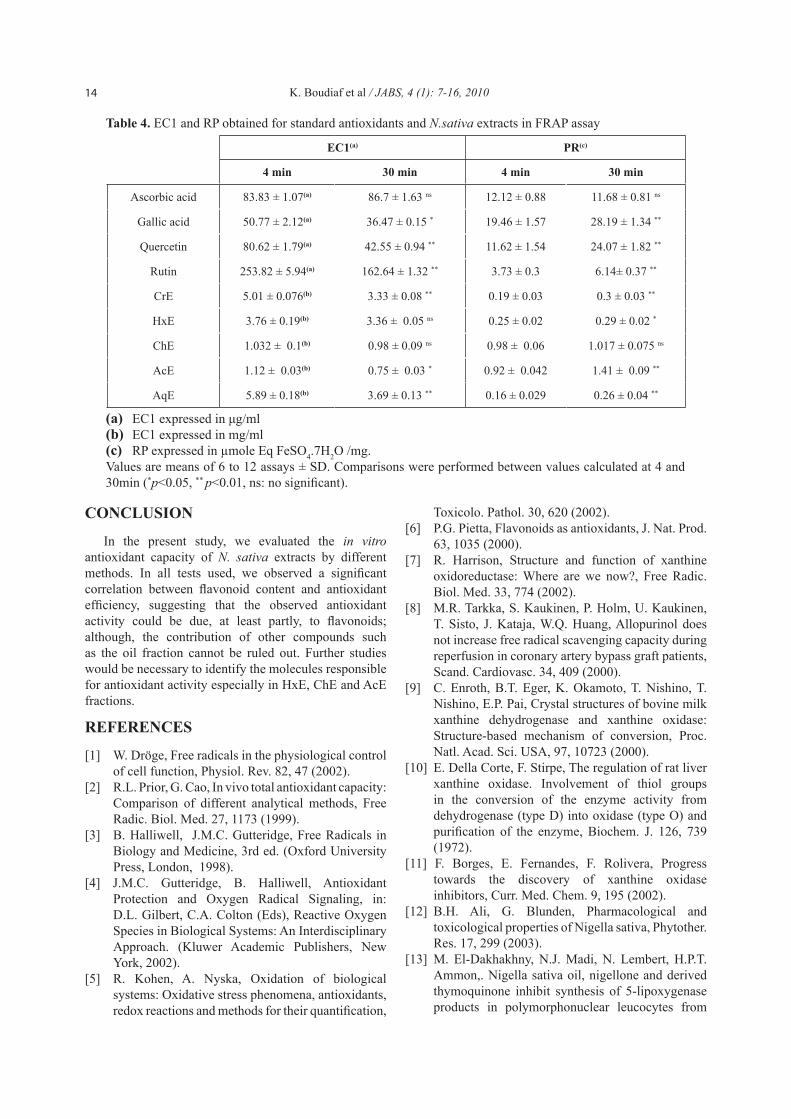
4 min 30 min 4 min 30 min
Ascorbic acid 83.83 ± 1.07(a) 86.7 ± 1.63 ns 12.12 ± 0.88 11.68 ± 0.81 ns
Gallic acid 50.77 ± 2.12(a) 36.47 ± 0.15 * 19.46 ± 1.57 28.19 ± 1.34 **
Quercetin 80.62 ± 1.79(a) 42.55 ± 0.94 ** 11.62 ± 1.54 24.07 ± 1.82 **
Rutin 253.82 ± 5.94(a) 162.64 ± 1.32 ** 3.73 ± 0.3 6.14± 0.37 **
CrE 5.01 ± 0.076(b) 3.33 ± 0.08 ** 0.19 ± 0.03 0.3 ± 0.03 **
HxE 3.76 ± 0.19(b) 3.36 ± 0.05 ns 0.25 ± 0.02 0.29 ± 0.02 *
ChE 1.032 ± 0.1(b) 0.98 ± 0.09 ns 0.98 ± 0.06 1.017 ± 0.075 ns
AcE 1.12 ± 0.03(b) 0.75 ± 0.03 * 0.92 ± 0.042 1.41 ± 0.09 **
AqE 5.89 ± 0.18(b) 3.69 ± 0.13 ** 0.16 ± 0.029 0.26 ± 0.04 **
Table 4. EC1 and RP obtained for standard antioxidants and N.sativa extracts in FRAP assay
(a) EC1 expressed in μg/ml (b) EC1 expressed in mg/ml (c) RP expressed in µmole Eq FeSO4.7H2O /mg.Values are means of 6 to 12 assays ± SD. Comparisons were performed between values calculated at 4 and 30min (*p<0.05, ** p<0.01, ns: no significant).

15K. Boudiaf et al / JABS, 4 (1): 7-16, 2010
rats, J. Ethnopharmacol. 81, 161 (2002).[14] K.M. Fararh, Y. Atoji, Y. Shimuzu, T. Takewaki,
Insulinotropic properties of Nigella sativa oil in streptozotocin plus nicotinamide diabetic hamster, Res. Vet. Sci. 73, 279 (2002).
[15] M. Kanter, I. Meral, Z. Yener, H. Ozbek, H. Demir, Partial regeneration / proliferation of the β-cells in the islets of Langerhans by Nigella sativa L. in streptozotocin induced diabetic rats, Tohoku J. Exp. Med. 201, 213 (2003).
[16] Z. Houcher, K. Boudiaf, M. Benboubetra, B. Houcher, Effects of methanolic extract and commercial oil of Nigella sativa L. on blood glucose and antioxidant capacity in alloxan-induced diabetic rats, Pteridins 18, 8 (2007).
[17] A. Haq, P.I. Lobo, M. Al-Tufail, N. Rama, S.T. Al-Sedairy, Immunomodulatory effect of Nigella sativa proteins fractionated by ion exchange chromatography, Int. J. Immunopharmaol. 21, 283 (1999).
[18] K.M. Fararh, Y. Atoji, Y. Shimuzu, T. Shiina, H. Nikami, T. Takewaki, Mechanisms of hypoglycaemic and immunopotentiating effects of Nigella sativa L. oil in streptozotocin-induced diabetic hamsters, Res. Vet. Sci. 77, 123 (2004).
[19] H. Gali-Muhtasib, M. Diab-Assaf, C. Boltz, J. Al-Hmaira, R. Hartig, A. Roessner, R. Schneider-Stock, Thymoquinone extracted from black seed triggers apoptotic cell death in human colorectal cancer cell via p53-dependant mechanism, Int. J. Oncology, 25, 857 (2004).
[20] E.M. Awad, In vitro decrease of the fibrinolytic potential of cultured human fibrosarcoma cell line, HT1080, by Nigella sativa oil, Phytomedicine. 12, 100 (2005).
[21] M.S. Al-Ghamdi, Protective effect of Nigella sativa seeds against carbon tetrachloride-induced liver damage, Am. J. Chin. Med. 31, 721 (2003).
[22] B.H. Ali, The effect of Nigella sativa oil on gentamicin nephrotoxicity in rats, Am. J. Chin. Med. 32, 49 (2004).
[23] O.A. Badary, R.A. Taha, A.M. Gamal El-Din, M.H. Abdel-Wahab, Thymoquinone is a potent superoxide anion scavenger, Drug Chem. Toxicol. 26, 87 (2003).
[24] M.F. Ramadan, L.W. Kroh, J-T Mörsel, Radical scavenging activity of black cumin (Nigella sativa L.), coriander (Coriandrum sativum L.), and niger (Guizotia abyssinica Cass.) crude seeds oils and oil fractions, J. Agric. Food Chem. 51, 6961 (2003).
[25] S.C. El-Saleh, O.A. Al-Sagair, M.I. Al-Khalaf, Thymoquinone and Nigella sativa oil protection against methionine-induced hyperhomocysteinemia in rats, Int. J. Cardiol. 93, 19 (2004).
[26] M.B. Atta, K. Imaizumi, Antioxidant activity of nigella (Nigella sative L.) seeds extracts, J. Jpn. Oil
Chem. Soc. 47, 49 (1998).[27] M.L. Price, L.G. Butler, Rapid visual estimation
and spectrophotometric determination of the tannin content of sorghum grain, J. Agric. Food Chem. 25, 1269 (1977).
[28] H.D. Graham, Modified Prussian blue assay for total polyphenols, J. Agric. Food Chem. 40, 801 (1992).
[29] T. Bahorun, B. Gressier, F. Trotin, C. Brunet, T. Dine, M.Luckv, J. Vasseur, M. Cazin, J.C. Cazin, M. Pinkas, Oxygen species scavenging activity of phenolic extract from hawthorn fresh plant organs and pharmaceutical preparations, Arzneimittelforschung 46, 1086 (1996).
[30] S.A. Sanders, R. Eisenthal, R. Harrison, Oxidation of NADH by human xanthine oxidoreductase: generation of superoxide anion, Eur. J. Biochem. 245, 541 (1997).
[31] A. Baghiani, R. Harrison, M. Benboubetra, Purification and partial characterisation of camel milk xanthine oxidoreductase, Arch. Physiol. Biochem. 111, 407 (2003).
[32] D. Atmani, A. Baghiani, M. Benboubetra, R. Harrison, NADH oxidation and superoxide production of goat milk xanthine oxidoreductase. Int. Dairy J. 15, 1113 (2005).
[33] H. Bouriche, Modulation of inflammation by Cleome arabica leaf extract, rutin and quercetin, studied in vitro and in vivo. Ph. D. Thesis, University of Setif, Algeria, 2005.
[34] I.F. Benzie, J.J. Strain, The ferric reducing ability of plasma (FRAP) as a measure of “antioxidant power”: The FRAP assay, Anal. Biochem. 239, 70 (1996).
[35] R. Pulido, L. Bravo, F. Saura-Calixto, Antioxidant activity of dietary polyphenols as determined by a modified ferric reducing/antioxidant power, J. Agric. Food Chem. 48, 3369 (2000).
[36] I. Parejeo, F. Viladomat, J. Bastida, A. Rosas-Romero, G. Saavedra, M.A. Murcia, C. Codina, Investigation of Bolivian plant extracts foe their radical scavenging activity and antioxidant activity, Life Sci. 73, 1667 (2003).
[37] S. Jiwajinda, V. Santisopasri, A. Murakami, O.K. Kim, H.W. Kim, H. Ohigashi,. Suppressive Effects of Edible Thai Plants on Superoxide and Nitric Oxide Generation, Asian Pac. J. Cancer Prev. 3, 251 (2002).
[38] Z. Zhang, D.R. Blake, C.R. Stevens, J.M. Kanczler, P.G. Winyard, M.C.R. Symons, M. Benboubetra, R. Harrison, A reappraisal of xanthine dehydrogenase and oxidase in hypoxic reperfusion injury: The role of NADH as an electron acceptor, Free Rad. Res.. 28, 151 (1998).
[39] L. Costantino, A. Albasini, G. Rastelli, S. Benvenuti, Activity of polyphenolic crude extracts

16 K. Boudiaf et al / JABS, 4 (1): 7-16, 2010
as scavengers of superoxide radicals and inhibitors of xanthine oxidase, Planta Med. 58, 342 (1992).
[40] R.J. Safitri, P. Tarigan, H.J. Freisleben, R.J. Rumampuk, A. Murakami, Antioxidant activity in vitro of two aromatic compounds from Caesalpinia sappan L, Bio. Factors, 19, 71 (2003).
[41] S. Martín-Aragón, B. Basabe, J.M. Benedí, A.M. Villar, In vitro and in vivo antioxidant properties of Vaccinum myrtillus, Pharm. Biol. 37, 109 (1999).
[42] N. Cotelle, Role of flavonoids in oxidative stress, Curr. Top. Med. Chem. 1 (2001) 569-590.
[43] P. Cos, L. Ying, M. Calomme, J.P. Hu, K. Cimanga, B. Van Poel, L. Pieters, A.V. Vlientinck, D.K. Berghe, Structure-activity relationship and classification of flavonoids as inhibitors of xanthine oxidase and superoxide scavengers, J. Nat. Prod. 61, 71 (1998).
[44] D.E.C. Van Hoorn, R.J. Nijveldt, P.A.M. Van Leeuwen, Z. Hofman, L. M’Rabet, D.B.A. De Bont, K. Van Norren, K. Accurate prediction of xanthine oxidase inhibition based on the structure of flavonoids, Eur. J. Pharmacol. 451, 111 (2002).
[45] S.V. Javanovic, S. Steenken, M. Tosic, B. Marjanovic, M.J. Simic, Flavonoids as antioxidants, J. Am. Chem. Soc. 116, 4846 (1994).
[46] G. Cao, R.K. Prior, Comparison of different analytical methods for assessing total antioxidant
capacity of human serum, Clin. Chem. 44, 1309 (1998).
[47] B. Ou, D. Huang, M. Hampsch-Woodill, J.A. Flanagan, E.K. Deemer, Analysis of antioxidant activities of common vegetables employing oxygen radical absorbance capacity (ORAC) and ferric reducing antioxidant power (FRAP) assays: a comparative study, J. Agric. Food Chem. 50, 3122 (2002).
[48] B.L. Halvorsen, K. Holte, M.C.W. Myhrstad, I. Barikmo, E. Hvattum, S.F. Rernberg, A.B. Wold, K. Haffner, H. Baugerød, L.F. Andersen, J. Ø. Moskaug, D.R. Jacobs, R. Blomhoff, A systematic screening of total antioxidants in dietary plants, J. Nutr. 132, 461 (2002).
[49] N. Pellegrini, M. Serafini, B. Colombi, D. Del Rio, S. Salvatore, M. Bianchi, F. Brighenti, Total antioxidant capacity of plant foods, brverages, and oils consumed in Italy assessed by three different in vitro methods, J. Nutr. 133, 2812 (2003).
[50] I. Tosun, S. Ustun, S. An investigation about antioxidant capacity of fruit nectars, Pak. J. Nutr. 2, 167 (2003).
[51] A. Janaszewska, G. Bartosz, Assay of total antioxidant capacity: comparison of four methods as applied to human blood plasma, Scand. J. Clin. Lab. Invest. 62, 231 (2002).
Copyright © 2022 FDOKUMEN


![Xanthine oxidase-derived reactive oxygen metabolites contribute to liver necrosis: protection by 4-hydroxypyrazolo [3, 4-d] pyrimidine](https://static.fdokumen.com/doc/165x107/6326b1a48212ebdc9e0a50e2/xanthine-oxidase-derived-reactive-oxygen-metabolites-contribute-to-liver-necrosis.jpg)