Immobilization of Saccharomyces cerevisiae on Apple Pieces ...
Upload
independentCategory
view
4download
0
Copyright �9 1997 by Humana Press Inc. All rights of any nature whatsoever reserved. 0273-2289/97/620203---0317511.00
Immobilization of Xanthine Oxidase and Its Use in the Quantitation
of Hypoxanthine in Fish Muscle Tissue Extracts Using a Flow Injection Method
DERRICK A. BALLADIN, *'1 DYER NARINESINGH, 2 VALERIE A. STOUTE, 3 AND T. T. NGO 4
1Chemistry Department, University of the West Indies, Cave Hill Campus, Barbados, WI; 2Chemistry Department,
University of the West Indies, St. Augustine Campus, Trinidad, WI; and 3V.A.S. Consulting, New York, NY, 4AMDL, Tustin, CA
Received December 5, 1995; Accepted January 4, 1996
ABSTRACT
Fish muscle extracts (Scomberomorus-brasiliensis--carite) were ana- lyzed for their hypoxanthine content using a flow injection system incor- porating an immobilized xanthine oxidase bioreactor. The xanthine oxidase was immobilized under mild conditions to a 2-fluoro-1- methylpyridinium Fractogel support. The uric acid produced from the oxidation of hypoxanthine by the immobilized xanthine oxidase at pH 7.0 and 35~ was monitored at 290 nm. Hypoxanthine concentrations as low as 4.4 ~tmol/L can be detected. Up to 30 samples per hour can be ana- lyzed at a flow rate of 1 mL/min, using 150 gL sample volumes and a bioreactor dimension of 1.0 cm x 2.0 mm id. Recovery yields were between 92 and 99%. Both within day and between day precisions gave CVs < 5.00% (n = 30). Good correlation (r -- 0.998) is obtained when 78 fish samples were analyzed for their hypoxanthine content both by this FI method and a reference HPLC method.
Index Entries: Immobilized xanthine oxidase; flow injection; hypox- anthine; fish muscle tissue.
*Author to whom all correspondence and reprint requests should be addressed.
Applied Biochemistry and Biotechnology 317 Voi. 62, 1997

31 8 Balladin et al.
INTRODUCTION
As the popularity and demand for fish grow, so will the need for some functional indicator of this food's freshness since fish is an extremely perishable food source. As soon as a fish dies or is caught spoilage begins to occur. In the high ambient temperatures of the trop- ics, fish will spoil within 12-20 h depending on the species and method of capture. In fish muscle nucleotide, degradation commences at death and continues throughout storage. The major stages (breakdown prod- ucts) in this autolytic process involve the conversion of ATP to inosine monophosphate, inosine, hypoxanthine, xanthine, and eventually to uric acid.
Inosine monophosphate is one of the major contributing factors to the "pleasant" flavor of fresh fish whereas the accumulation of hypoxan- thine/xanthine during storage contributes to the "off taste." Hence the concentrations of inosine and/or hypoxanthine can both be used as fish freshness indicators (1-7).
Several methods have been reported for the quantitation of hypoxan- thine in biological materials such as fish muscle tissue. These include pre- cipitation (1), paper chromatography (2), ion exchange chromatography (3) and HPLC (4,5). Hypoxanthine has also been quantified enzymatically (3,6,7) using xanthine oxidase (Eqs. 1 and 2).
hypoxanthine + 02 xanthine oxidase
xanthine + H202 (1)
xanthine Xanthine + 02 oxidase uric acid + H202 (2)
Either the uric acid (290 nm) or the H202 can be monitored.
MATERIALS AND METHODS
Materials
Xanthine oxidase (EC 1.1.3.22), Grade II, from buttermilk, 2.1 U/mg protein), hypoxanthine and uric acid were obtained from Sigma Chemical Company (St. Louis, MO). The support material, a 2-fluoro-l-methylpyri- dinium salt activated Fractogel (FMP-Fractogel) was purchased from BioProbe International Inc. (Tustin, CA). FMP-Fractogel is now available from UniSyn Technologies (Hopkinton, MA). Fish samples (Scomberomorus brasiliensis-carite) were purchased from various supermarkets and high- way fishmongers in Trinidad, West Indies. All other materials were of the analytical reagent grade (BDH, Poole, UK) and were used without further purification.
Applied Biochemistry and Biotechnology Vol. 62, 1997

Immobilization of Xanthine Oxidase 319
Methods
Immobilization of Xanthine Oxidase on FMP-activated Fractogel
The FMP-Fractogel (2 mL) was washed first with cold, deionized dis- tilled water (5 x 5 mL), followed by aqueous NaC1 (0.10M, 5 x 5 mL), and hydrochloric acid (0.10 mol/L, 5 x 5 mL) to ensure removal of any traces of free, unreacted FMP that may be present in the commercial product. The gel was further washed with aqueous NaHCO3 coupling buffer (0.05M, pH 8.0, 5 x 5 mL) and then immediately added to 10 mL of xanthine oxidase previously dialyzed in the coupling buffer. This suspension was tumbled end-over-end at 15~ for 24 h following which the gel was washed with 30 mL each of the coupling buffer and the working aqueous NaRHPO4 buffer (0.05M pH 8.0). When not in use, the gel was stored at 4~ in the working phosphate buffer, containing sodium azide (0.01% w/v).
The amount of protein bound to the gel was determined (9) from the differences in protein concentrations (at 280 nm) between the enzyme solu- tions offered for coupling and the supernatant and wash enzyme solutions after coupling. Bioreactors were prepared by packing small lengths of Tygon tubing with the immobilized xanthine oxidase. Porex filters were used to plug both ends of the bioreactor to prevent gel loss.
The Loading Capacity of Immobilized Xanthine Oxidase
In these studies varying concentrations of xanthine oxidase were coupled to a fixed volume (1 mL) of FMP-Fractogel under identical con- ditions to that outlined in above. In each case, both the amount of protein bound under the coupling conditions as well as the activity of the immo- bilized enzyme (35~ pH 8.0) were determined. The activity was deter- mined by allowing a fixed volume of the immobilized enzyme (0.1 mL) to react for 3 min with a known volume (1.0 mL) of a hypoxanthine solu- tion (110 gmol/L) made up in the working buffer (pH 8.0). The reaction mixture was then centrifuged (2976g) for exactly 1 min and the absorbance of the uric acid in the supernatant measured at 290 nm. The activity of the free enzyme was determined under similar conditions using the free enzyme solution (0.1 mL, 1 mg/mL protein), instead of the immobilized enzyme.
pH Profile of the Immobilized Xanthine Oxidase
To obtain the pH profile, a hypoxanthine solution (55.1 gM), buffered at each of seven different pHs (Na2HPO4, 0.05M, pH 6-9) was pumped at a fixed flow rate (0.7 mL/min) and temperature (35~ through an immobilized xanthine oxidase--Fractogel bioreactor. The absorbance of the uric acid liberated at each of these pHs was monitored after steady state conditions were obtained.
Applied Biochemistry and Biotechnology Vol. 62, 1997

320 Balladin et al.
SAMPLE INJECTION
U -
VALVE
I BLANK COLUMN I
I
2-WAY
O~GIDASE
FRACTOCEL I
L _ _ ~_W_ _1~_ 9.t..O_X . . . . J I'H]mMO S T A I I ~
w x 1 " ~ B a r n ( 3 s ' C )
STRIP CHART I~CORi)ER
FLOW THROUCH DETECTOR
WASTE
Fig. 1. Flow injection manifold for the quantitation of hypoxanthine.
Temperature Profile The temperature profile was obtained by pumping a hypoxanthine
solution (55.10 ~tM, pH 8.0) at each of nine fixed temperatures (in the range 10-50~ through the bioreactor at a constant flow rate of 0.7 mL/min . The quantity of uric acid produced was determined as outlined previously.
Storage Stabili~, Studies In this study, a bioreactor was stored at 4~ in working buffer over a
period of 9 mo. The activity of this bioreactor was periodically determined by pumping a buffered hypoxanthine solution (pH 8.0, 55.10 W~I) at a fixed flow rate (0.7 mL/min) and temperature (35~ through the system and monitoring the absorbance of uric acid at 290 nm.
The Flow Injection Manifold The basic configuration of the FI manifold incorporating an immobi-
lized xanthine oxidase-Fractogel bioreactor as well as a blank column with only the support material is shown in Fig. 1. Samples of hypoxanthine were injected into the thermostatted buffer stream via a pneumatically activated port injection valve and allowed to pass alternately over the ana- lyte and blank columns. Solutions were pumped through the manifold using a four-channel peristaltic pump (Gilson Minipuls II). The uric acid produced as a result of hypoxanthine oxidation by the immobilized xan- thine oxidase-Fractogel bioreactor was monitored at 290 nm on a UV/VIS spect rophotometer (PU 8625, Philips Scientific). The difference in absorbances between the blank and analyte columns is related to the quan-
Applied Biochemistry and Biotechnology Vol. 62, 1997

Immobilization of Xanthine Oxidase 3 21
tity of hypoxanthine present in the sample. To determine the optimum FI operating conditions, experiments were carried out utilizing varying loop sizes (90-500 ~tL), flow rates (0.5-1.9 mL/min), and bioreactor lengths (0.5-2.0 cm).
Preparation of Fish Extracts Samples (10 g) of the fish muscle tissue (fillet) were removed and
homogenized at high speed (2000 rpm, MSE Homogenizer, equipped with stainless steel blades) in perchloric acid (0.6M, 50 mL) for 1 min. The homogenate was suction filtered through Whatman No. I filter paper. The filtrate (25 mL) was diluted with distilled water (10 mL) and a solution (15 mL) consisting of KOH (3.0M, 6.3 mL) and KH2PO 4 (0.4M, 8.7 mL) added. This diluted solution was then adjusted to pH 8.0 using HC1 as required. It was then allowed to stand for 30 min at -15~ to facilitate the crystalliza- tion of KC104, which was removed by suction filtration. This filtrate was further filtered through Millipore filters (0.22 ~tm) just prior to analysis by FI or HPLC.
Analysis of Fish Extracts by Flow Injection Fish extracts were analyzed for their hypoxanthine content by the
standard addition technique. Aliquots (5 mL) of the extract, prepared as outlined in the above section, were spiked with varying known concentra- tions of hypoxanthine solution (11-55 ~tM) and the volumes adjusted to 10 mL with phosphate buffer (0.05 mM, pH 8.0). Aliquots (150 ~tL) of these spiked samples were then injected into the carrier stream of the FI mani- fold (Fig. 1). By means of a three-way valve, samples were channeled alter- nately over the analyte and blank columns. The blank column consisted of only the support material and as such would compensate for substances (such as free uric acid and or other metabolites present in the fish matrix) that may absorb at 290 nm.
The difference in the absorbances from the two columns was taken as that as a result of the uric acid produced by the oxidation of hypoxanthine by the immobilized xanthine oxidase-Fractogel bioreactor. The hypoxan- thine content in the fish sample was obtained from standard addition plots by extrapolation to the x-axis.
Determination of Hypoxanthine in Fish Muscle Tissue Extracts by High Pressure Liquid Chromatography (HPLC) The same fish extracts, spiked with hypoxanthine and used in the FI
analyses, were also used for the quantitation of hypoxanthine by the HPLC method (4,5). The analyses were done using a Laboratory Data Control Liquid Chromatograph (Milton Roy Company), fitted with a RP- C18-reverse phase analytical column (18 cm x 4.6 mm id 10 ~tm particle size, Brownlee Labs. Inc.), coupled to a guard column (RP-C18, 3 cm x 4.6 mm id, 10 ~tm particle size), a loop injector (50 ~tL loop, Model 7125,
Applied Biochemistry and Biotechnology Vol. 62, 1997
322 Balladin et al.
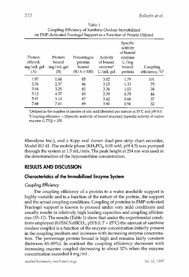
Table 1 Coupling Efficiency of Xanthine Oxidase Immobilized
on FMP-Activated Fractogel Support as a Function of Protein Offered
Specific activity
of bound Protein Protein Percentages Activity enzyme offered, bound protein of bound U/rag
mg/mL gel mg/mL gel bound enzyme a bound Coupling (A) (B) (B/A x 100) U/mL gel protein efficiency; %b
1.97 1.68 85 3.02 1.79 101 2.76 2.37 86 3.15 1.33 75 3.94 3.25 82 3.36 1.03 58 5.12 4.37 85 3.39 0.78 44 5.91 5.14 87 3.42 0.66 37 7.88 7.01 89 3.90 0.56 32
~Defined as the number of gmoles of uric acid liberated per minute at 35~ and p H 8.0. bCoupling efficiency = ([Specific acativity of bound enzyme]/[specific activity of native enzyme {1.77}]) x 100.
Rheodyne Inc.), and a Kipp and Zonen dual pen strip chart recorder, Model BD 41. The mobile phase (KHaPO4, 0.01 mM, pH 4.5) was pumped through the system at 1.5 mL/min. The peak height at 254 nm was used in the determination of the hypoxanthine concentration.
RESULTS AND DISCUSSION
Characteristics of the Immobilized Enzyme System
Coupling Efficiency The coupling efficiency of a protein to a water insoluble support is
highly variable and is a function of the nature of the protein, the support and the actual coupling conditions. Coupling of proteins to FMP-activated Fractogel support is known to proceed under very mild conditions and usually results in relatively high loading capacities and coupling efficien- cies (10-12). The results (Table 1) show that under the experimental condi- tions employed (0.05M NaHCO3, pH 8.0; T = 15~ the amount of xanthine oxidase coupled is a function of the enzyme concentration initially present in the coupling medium and increases with increasing enzyme concentra- tion. The percentage protein bound is high and remains fairly constant (between 85-89%). In contrast the coupling efficiency decreases with increasing enzyme coupled decreasing to about 32% when the enzyme concentration exceeded 8 mg/mL.
Applied Biochemistry and Biotechnology Vol. 62, 1997

Immobilization of Xanthine Oxidase 3 2 3
pH and Temperature~Activity Profiles The bound enzyme shows a relatively broad pH optimum in the
range 7.5-8.5. This is similar to the value reported for the native enzyme (13) and for xanthine oxidase immobilized on gelatin (14) and on cellulose triacetate (15). The optimal temperature for the free enzyme is reported to be in the range 47-57~ (13). In contrast the immobilized enzyme shows a temperature optimum between 35-40~ Such lowering of the tempera- ture optimum on immobilization of xanthine oxidase has also been reported by other workers. Luong et al. (16) reported an optimum between 27-37~ when bound to preactivated nylon membrane whereas Karube et al. (15) reported an optimum between 30-34~ when attached to a poly(vinylbutyral) membrane. The lower temperature optimum for immobilized xanthine oxidase systems may be because of a diffusional limitation in the rate of substrate supply to the enzymes immobilized in the inner sphere of the support that becomes more pronounced as the reaction temperature increases.
Stability Xanthine oxidase immobilized on to Fractogel is found to be very
stable when stored at low temperatures (4~ in phosphate buffer (0.05M, pH 8.0). Under such conditions, it retains up to 60% of its initial activity after 9 mo.
The immobilized enzyme also shows excellent operational stabilities at temperatures at or just below its optimum. For example, a bioreactor maintained continuously at 35~ showed an activity loss of only 4% after 84 h. In contrast at 50~ the immobilized enzyme lost 48% of its activity after only 30 min.
Optimization of the Flow Injection Manifold The flow injection manifold (Fig. 1) was optimized with respect to
sample loop size, flow rate, and bioreactor length at pH 8.0 and 35~ (optimum pH and temperature of the immobilized enzyme) in an attempt to obtain a compromise set of operating conditions that would provide maximum conversion of reactant to product with minimal dispersion lead- ing towards high sensitivity, low detection limits and at the same time a reasonable sample throughput rate.
Under a fixed set of operating conditions (pH 8.0; T = 35~ the sample loop sizes (90-500 pL), bioreactor lengths (0.5-2.0 cm x 2 mm id), and flow rates (0.5-1.9 mL/min) were varied. Under each set of condi- tions, hypoxanthine standards were injected into the FI manifold (Fig. 1) and the amount of uric acid produced monitored. Also the degree of dis- persion was determined in each case. With the 500 pL sample volumes, the peaks were broad with relatively rapid onset (1 min) but long decay times (4 mins). In contrast, the 90-150 pL loop sizes yielded peaks with
Applied Biochemistry and Biotechnology Vol. 62, 1997

324 Balladin et al.
Table 2 Effect of Bioreactor Length, Sample Injection Volume, and
Flow Rate on the Dispersion Coefficient for the Oxidation of Hypoxanthine by Xanthine Oxidase Immobilized on an Activated Fractogel Support
Flow rate (mL min -1)
Dispersion coefficient at various sample injection volumes
Bioreactor length (cm) 90 gL 150 gL 500 gL
0.5 4.38 3.70 2.95 0.5 1.0 6.85 4.91 3.65
2.0 8.21 7.91 6.70 0.5 4.08 2.21 2.72
1.0 1.0 6.43 3.25 2.31 2.0 8.18 7.27 5.58 0.5 3.75 2.08 2.00
1.9 1.0 6.09 3.02 2.15 2.0 8.07 7.11 5.21
both rapid onset and decay times (1 min) resulting in very little carry- over between repeated sample injections. The reduced sample detection time (approx 2 min) led to the compromise choice of 150 gL loop sizes for analyses.
The results in Table 2 show that under the operating conditions selected, the degree of dispersion decreases with both increasing flow rate (only marginally) and increasing sample volumes, but increases with increasing bioreactor lengths. From the results in Table 3, most of the bioreactors of length 0.5 cm can be eliminated on the basis of poor sensi- tivity (although there was low dispersion and high sample throughput). Also with the 2 cm bioreactors there was relatively high dispersion and low sample throughput rates. Bioreactors of 1.0 cm length, a flow rate of 1.0 mL/min and a loop size of 150 gL were eventually chosen as the best set of optimized conditions for analyzing the fish extracts. Standard curves obtained under these compromise optimized conditions were lin- ear (r = 0.999) up to 110 gmol/L. The following least squares regression equation (Equation 3) was found to hold over this concentration range
Absorbance = 0.003 (hypoxanthine, gmol/L) + 0.0004 (3)
Under these sets of conditions, the analytical system operates under con- ditions of low dispersion (3.25), has a good manual throughput rate of 30 samples per hour, and gives a detection limit (the y intercept + 3 times the value of the statistical standard deviation) of 4.4 gmol/L. This is within the hypoxanthine concentration ranges usually found in the carite sam- ples (6-100 gmol/L). This detection limit compares favorably with that of
Applied Biochemistry and Biotechnology Vol. 62, 1997

Immobilization of Xanthine Oxidase 325
~J
�9 ~ o ~ o o ~ o ~"~ . . . . . ~ o ~ ~ o . . . . . . . . . . . . . . . . .
o "..U .,,,~ , .~
0 L ~
b~ ".U
o o~ -,~,q o o o o
~ ~,~o ~176176 oo~ ~0~ ~ o,~ o ~0oo ~- 0~ ~- ~ o ~0o~
O~
h ~ ooo ooo ooo ooo ooo ooo ooo ooo ooo
00 U0
Ln~m ooo ooo 0~r-~ " m ' '
~0 ~ ~ ~o ~H ooo I ooo ooo ~o ~ ..... o ...... o.~, ~ ~ ~ ~ o ~ o o ~ ~ o o o o ~ - " , - ~ o ~ ~0.~ . , ~ .
m ,,~ I--. r l r ~ ' ~ d o , ~ , . , ~ , - ,~ ,~ 0 ; ~ . . . . . , - . ~ . ~
0"3 e .~ ~ o~0 ~.,,o . . . . . . ~ o . . . . . . . . . . . . . . .
~ o_,.._,~ d,-: o . . . . . . j , . :o . . . . . . ~ , , . . ~ o o . . . . . . o o o
21 oo oo ~ r~o ~ ~ o o o ~ ~ g~-~ o~o ooo ooo <~oo <~oo~176176 ooo ~176 ~ oo o o o oo o o oOO o
�9 . o o . . .
"-U
L~
t U
,-C L J
~oo ooo ooo .oo ooo ooo oo i oo o ooo , , o
o o o o o o o o o ~ o o o o o o o o ~ o o o ~ o o i i i o
. . . . . . ~ . . . . . . . o o
~d O ~
0 0 U1 0 0 ~ 0 0
0 ,-4 ~ 0 ~ ~ 0 ~
0 O~
0 ~
Applied Biochemistry and Biotechnology Vot 62, 1997

326 Balladin et al.
Table 4 Percentage Recoveries of Hypoxanthine Added
to a Fish Matrix (Scomberomorus brasiliensis)
[Hypoxanthine] added (gmol L -1)
[Hypoxanthine] recovered (gmol L -1) Percentage recovery
-- 4.76 -- 11.02 15.05 95.4 22.04 25.80 96.3 33.06 36.98 97.8 55.10 59.16 98.8
110.21 105.42 91.7
3.6 gmol/L reported by Luong and coworkers (16) for a hypoxanthine biosensor using xanthine oxidase immobilized on nylon.
Recovery Yields Recovery studies were carried out under FI conditions (Fig. 1) by ana-
lyzing a fish muscle tissue matrix that was spiked with hypoxanthine stan- dards in the range 5-114 gmol/L (Table 4). These recovery yields (92-99%) compare quite favorably with those reported by Ke and Burns (5) in their liquid chromatographic (93-98%) and xanthine oxidase methods (83-86%) for the hypoxanthine content in the Atlantic cod (Gadus morhera).
Analysis of Fish Samples Samples of carite (Scomberomorus brasiliensis) used for the quantita-
tion of hypoxanthine were purchased from various supermarkets (chain operators and single operators), highway fishmongers (purchased during morning and evening periods), and directly from fishing boats. Some of these fish samples were analyzed immediately upon purchase whereas others were stored at various fixed temperatures (-15 ~ 4~ and 29~ [R.T.]) in order to mimic typical conditions under which fish may be kept in our country. Each sample was analyzed by both the FI and reference HPLC methods for their hypoxanthine content. Hypoxanthine values for 78 samples (Table 5) obtained from analyses by these two methods show a linear relationship (r = 0.998) and has the regression equation (Eq. 4).
[HX, ~tmo1/g]F I = 0.94 [HX, ~tmo1/g]HPL c --0.07 (4)
A two-way ANOVA design was used to simultaneously test both the location/storage effect and the HPLC/FI method on the hypoxanthine concentration of the various samples analyzed. This interaction effect in the two-way design yields information about whether the relationship between the values measured for hypoxanthine concentration in fish from the various locations is the same whether HPLC or FI is the analytical
Applied Biochemistry and Biotechnology VoL 62, 1997

Immobilization of Xanthine Oxidase 327
~ , 0
0
2 ~
~ m
(13
r ~
o ~ r , l i
o
~ ~ ~ ~ o ~ ~ ~ ~ ~ ~ o ~ ~ ~ ~ ~ ~ ~
h . . . . . . . . . . . . i
A p p l i e d Biochemist ry a n d B io techno logy Vol. 62, 1 9 9 7

3 2 8 Balladin et al.
method of choice. The analysis shows there is no significant difference between the HPLC and FIA values. Where a fish is purchased or how it is stored however does affect the hypoxanthine concentration.
CONCLUSIONS
The proposed FI system utilizing xanthine oxidase immobilized on FMP-activated synthetic gel (Fractogel) for the quantitation of hypoxan- thine in fish muscle tissue appears to offer considerable advantages over existing methods. With the HPLC method, for example, although it pro- vides good precision and recovery yields, its high cost is normally beyond the budget of most small laboratories especially those in develop- ing countries. The high storage and operational stabilities of xanthine oxi- dase immobilized on to Fractogel, coupled with low reagent consumption (<2 mL), relatively high sample throughput rate (up to 30 samples/h can be analyzed manually), low detection limits (4.4 ~tmol/L), good precision (RSD < 5%) and robustness of the system make this method of analysis economically very attractive and simple to operate.
REFERENCES
1. Burt, J., Stroud, G. D., and Jones, N. R. (1969), in Freezing and Irradiation ofFish, Kreuzer, R., ed., Fishing News Books Ltd., London, p. 367.
2. Jones, N. R., Murray, J., Ligingston, E. 1., and Murray, C. K. (1964), J. Sci. Food Agric. 15, 763.
3. Watanabe, E., Okuma, H., Takahashi, H., Yazawa, S., and Sekimukai, S. (1992), Anal. Chim. Acta 260, 93.
4. Warthensen, J. J., Waletzko, P. T., and Bunsta, F. F. (1980), J. Agric. Food Chem. 28, 1308. 5. Ke, P., and Bums, G. B. J. Assoc. Off. Anal. Chem. 66, 444. 6. Jones, N. R., Murray, J., and Burt, J. R. (1965), J. Food Sci. 30, 971. 7. Jahns, F. D., Howe, J. L., Coduri, R. J., and Rand, A. G. (1976), Food Technol. 33, 27. 8. Beuchant, L. R. (1973), J. Agric. Food Chem. 21, 453. 9. Lowry, O. H., Rosebrough, N. J., Farr, A. L., and Randall, R. J. Biol. Chem. 193, 265.
10. Narinesingh, D., Jaipersad, D., and Chang-Yen, I. (1988), Anal. Biochem. 172, 89. 11. Narinesingh, D. and Ngo, T. T. (1987), BiotechnoL Applied Biochem. 9, 450. 12. Narinesingh, D., Stoute, V. A., Shaama, F., and Ngo, T. T. (1989), J. Mol. Catalysis 54,147. 13. De Renzo, E. C. (1956), in Advances in EnzymoIogy, vol. 17, Nord, F. F., ed., Interscience,
NY, pp. 293-328. 14. Kenndey, J. F. and Cabral, J. M. S. (1983), in Solid Phase Biochemistry--Analytical and
Synthetic Aspects, vol. 66, Scouten, W.H., ed., John Wiley and Sons, NY pp. 253-390. 15. Karube, I., Matsuoka, H., Suzuki, S., Watanabe, E., and Toyama, K. (1984), J. Agric. Food
Chem. 32, 314. 16. Luong, J. H. T., Mulchandani, A., and Male, K. B. (1989), Anal. Chim. Acta 221, 215.
Applied Biochemistry and Biotechnology Vol. 62, 1997
Copyright © 2022 FDOKUMEN