Xanthine oxidase-activated prodrugs of thymidine phosphorylase inhibitors
13
Original article Xanthine oxidase-activated prodrugs of thymidine phosphorylase inhibitors Philip Reigan, Abdul Gbaj, Ian J. Stratford, Richard A. Bryce, Sally Freeman * School of Pharmacy and Pharmaceutical Sciences, The University of Manchester, Oxford Road, Manchester M13 9PL, UK Received 26 March 2007; accepted 13 July 2007 Available online 6 August 2007 Abstract Thymidine phosphorylase (TP) is over-expressed in various tumour types and plays an important role in tumour angiogenesis, growth, in- vasion and metastasis. The enzymatic activity of TP is required for the angiogenic effect of TP, therefore, inhibitors of TP are of significant interest in cancer chemotherapy. A series of xanthine oxidase (XO) activated prodrugs of known inhibitors of TP have been designed and syn- thesized with the ultimate intent of improving tumour selectivity and pharmacokinetic characteristics. These prodrugs were not inhibitors of TP, but were selectively oxidized by XO at C-2 and/or C-4 of the uracil ring moiety to generate the desired TP inhibitor. Molecular modelling of both the TP inhibitors and XO-activated prodrugs rationalized their binding in the active site of the human TP crystal structure. Ó 2007 Elsevier Masson SAS. All rights reserved. Keywords: Thymidine phosphorylase; Xanthine oxidase; Enzyme inhibition; Prodrug activation 1. Introduction Thymidine phosphorylase (TP, EC 2.4.2.4) is a highly ex- pressed protein in many solid human tumours, and the level of expression is associated with tumour neovascularization, in- vasiveness and metastasis [1]. TP is predominantly expressed in hypoxic regions of solid tumours [2], promoting tumour growth by angiogenesis, metastasis and suppressing apoptosis [3,4]. TP is also known as platelet-derived endothelial cell growth factor (PD-ECGF), a novel angiogenic protein, distinct from other angiogenic growth factors, since it exerts its actions through its enzymatic activity, as both site-directed mutagene- sis of active-site residues and inhibitors prevent the angiogenic activity of TP [3,4]. TP catalyzes the reversible phosphorolysis of thymidine (1) to thymine (2) and a-D-2-deoxyribose-1- phosphate (3)(Scheme 1). The dephosphorylated product, D- 2-deoxyribose (4) has been shown to have chemotactic activity in vitro and angiogenic activity in vivo [3,4] and is considered to play a key role in the invasiveness and metastasis of TP- expressing solid tumours [1]. Since TP is over-expressed in tumours, the protein is an at- tractive cancer chemotherapy target for selective inhibition of TP-dependent angiogenesis and subsequent inhibition of tu- mour growth. In addition, specific inhibitors of human TP could also enhance the efficacy of thymidine analogues such as 5-fluoro-2 0 -deoxyuridine and 5-iodo-2 0 -deoxyuridine, which would no longer be metabolized and inactivated by TP [5]. The classical TP inhibitors include 6-amino-5-bromouracil (6A5BU, 5) [6e8] and 7-deazaxanthine (7-DX, 6)(Fig. 1), which was the first purine derivative to display inhibition of both E. coli TP and angiogenesis in a chorioallantoic mem- brane assay [9]. More potent nanomolar inhibitors of human TP are 5-chloro-6-[(2-iminopyrrolidin-1-yl)methyl]uracil hy- drochloride (TPI, 7) (IC 50 35 nM, K i 20 nM) [10,11] and 5- bromo-6-[(2-aminoimidazol-1-yl)methyl]uracil hydrochloride (8) (IC 50 20 nM) (Fig. 1) [12], these cationic tight-binding Abbreviations: TP, thymidine phosphorylase; XOR, xanthine oxidoreduc- tase; XO, xanthine oxidase; XDH, xanthine dehydrogenase; PD-ECGF, plate- let-derived endothelial cell growth factor; 6A5BU, 6-amino-5-bromouracil; 7-DX, 7-deazaxanthine; TPI, 5-chloro-6-[(2-iminopyrrolidin-1-yl)methyl]uracil hydrochloride; AO, aldehyde oxidase. * Corresponding author. Tel.: þ44 (0) 161 275 2366; fax: þ44 (0) 161 275 2396. E-mail address: [email protected] (S. Freeman). 0223-5234/$ - see front matter Ó 2007 Elsevier Masson SAS. All rights reserved. doi:10.1016/j.ejmech.2007.07.015 Available online at www.sciencedirect.com European Journal of Medicinal Chemistry 43 (2008) 1248e1260 http://www.elsevier.com/locate/ejmech
-
Upload
independent -
Category
Documents
-
view
0 -
download
0
Transcript of Xanthine oxidase-activated prodrugs of thymidine phosphorylase inhibitors

Available online at www.sciencedirect.com
European Journal of Medicinal Chemistry 43 (2008) 1248e1260http://www.elsevier.com/locate/ejmech
Original article
Xanthine oxidase-activated prodrugs of thymidinephosphorylase inhibitors
Philip Reigan, Abdul Gbaj, Ian J. Stratford, Richard A. Bryce, Sally Freeman*
School of Pharmacy and Pharmaceutical Sciences, The University of Manchester, Oxford Road, Manchester M13 9PL, UK
Received 26 March 2007; accepted 13 July 2007
Available online 6 August 2007
Abstract
Thymidine phosphorylase (TP) is over-expressed in various tumour types and plays an important role in tumour angiogenesis, growth, in-vasion and metastasis. The enzymatic activity of TP is required for the angiogenic effect of TP, therefore, inhibitors of TP are of significantinterest in cancer chemotherapy. A series of xanthine oxidase (XO) activated prodrugs of known inhibitors of TP have been designed and syn-thesized with the ultimate intent of improving tumour selectivity and pharmacokinetic characteristics. These prodrugs were not inhibitors of TP,but were selectively oxidized by XO at C-2 and/or C-4 of the uracil ring moiety to generate the desired TP inhibitor. Molecular modelling of boththe TP inhibitors and XO-activated prodrugs rationalized their binding in the active site of the human TP crystal structure.� 2007 Elsevier Masson SAS. All rights reserved.
Keywords: Thymidine phosphorylase; Xanthine oxidase; Enzyme inhibition; Prodrug activation
1. Introduction
Thymidine phosphorylase (TP, EC 2.4.2.4) is a highly ex-pressed protein in many solid human tumours, and the levelof expression is associated with tumour neovascularization, in-vasiveness and metastasis [1]. TP is predominantly expressedin hypoxic regions of solid tumours [2], promoting tumourgrowth by angiogenesis, metastasis and suppressing apoptosis[3,4]. TP is also known as platelet-derived endothelial cellgrowth factor (PD-ECGF), a novel angiogenic protein, distinctfrom other angiogenic growth factors, since it exerts its actionsthrough its enzymatic activity, as both site-directed mutagene-sis of active-site residues and inhibitors prevent the angiogenicactivity of TP [3,4]. TP catalyzes the reversible phosphorolysis
Abbreviations: TP, thymidine phosphorylase; XOR, xanthine oxidoreduc-
tase; XO, xanthine oxidase; XDH, xanthine dehydrogenase; PD-ECGF, plate-
let-derived endothelial cell growth factor; 6A5BU, 6-amino-5-bromouracil;
7-DX, 7-deazaxanthine; TPI, 5-chloro-6-[(2-iminopyrrolidin-1-yl)methyl]uracil
hydrochloride; AO, aldehyde oxidase.
* Corresponding author. Tel.:þ44 (0) 161 275 2366; fax:þ44 (0) 161 275 2396.
E-mail address: [email protected] (S. Freeman).
0223-5234/$ - see front matter � 2007 Elsevier Masson SAS. All rights reserved.
doi:10.1016/j.ejmech.2007.07.015
of thymidine (1) to thymine (2) and a-D-2-deoxyribose-1-phosphate (3) (Scheme 1). The dephosphorylated product, D-2-deoxyribose (4) has been shown to have chemotactic activityin vitro and angiogenic activity in vivo [3,4] and is consideredto play a key role in the invasiveness and metastasis of TP-expressing solid tumours [1].
Since TP is over-expressed in tumours, the protein is an at-tractive cancer chemotherapy target for selective inhibition ofTP-dependent angiogenesis and subsequent inhibition of tu-mour growth. In addition, specific inhibitors of human TPcould also enhance the efficacy of thymidine analogues suchas 5-fluoro-20-deoxyuridine and 5-iodo-20-deoxyuridine, whichwould no longer be metabolized and inactivated by TP [5].The classical TP inhibitors include 6-amino-5-bromouracil(6A5BU, 5) [6e8] and 7-deazaxanthine (7-DX, 6) (Fig. 1),which was the first purine derivative to display inhibition ofboth E. coli TP and angiogenesis in a chorioallantoic mem-brane assay [9]. More potent nanomolar inhibitors of humanTP are 5-chloro-6-[(2-iminopyrrolidin-1-yl)methyl]uracil hy-drochloride (TPI, 7) (IC50 35 nM, Ki 20 nM) [10,11] and 5-bromo-6-[(2-aminoimidazol-1-yl)methyl]uracil hydrochloride(8) (IC50 20 nM) (Fig. 1) [12], these cationic tight-binding

Scheme 1. The catalytic action of TP.
1249P. Reigan et al. / European Journal of Medicinal Chemistry 43 (2008) 1248e1260
inhibitors have been proposed to mimic the oxocarbenium ion-like transition state for thymidine phosphorolysis (Scheme 1)[13]. However, despite the nanomolar potency of these nucle-obase-derived inhibitors, their ionic nature leads to poor phar-macokinetic properties [12].
TP inhibitors are reportedly selective to cells expressing TPand this expression correlates with hypoxia in tumours [2].However, further selectivity and improved pharmacokineticcharacteristics could be achieved using the xanthine oxidase(XO, EC 1.1.3.22) prodrug strategy, since XO activity and ex-pression is increased under hypoxic conditions [14e16]. Fur-thermore, human tissues have increased XO activities incolorectal and prostate tumours as compared to their corre-sponding normal tissues [17,18]. In addition, XO levels in hu-man brain tumour tissues have been found to be elevatedcompared to that of normal brain tissue, in meningioma andastrocytoma oncotypes [19]. The selective inhibition of TPin tumours via an enzymatically-activated prodrug approachwould be advantageous, since TP is also expressed at highlevels in human blood platelets and normal tissues, such asthe salivary glands, ovaries and brain, and in addition TP playsan important role in nucleotide homeostasis [5].
XO catalyzes the oxidation of hypoxanthine (9) and xanthine(10) to uric acid (11) (Scheme 2). In addition, XO bioreductivelymetabolizes a number of quinone-based anticancer drugs [20,21]and a range of nitroimidazoles [12,22]. 2/4-Nitroimidazole pro-drugs of the 6-[(2/4-aminoimidazol-1-yl)methyl]uracils, includ-ing 8, which exploit the reductive half-reaction at the FAD-siteof XO have been reported previously [12]. The oxidation reaction
Fig. 1. Uracil-based TP inhibitors.
catalyzed at the molybdenumepterin site of XO has been previ-ously utilized to increase the bioavailability, solubility and selec-tivity of 20-F-ara-ddI (20-F-ara-ddP), an anti-HIV nucleoside [23]and acyclovir (6-deoxyacyclovir), an anti-herpetic agent [24],which like TPI are administered orally. The TP inhibitors, beinguracil-based heterocycles, are attractive targets for activation bythe oxidative reaction catalyzed by XO.
Here, we report the synthesis and evaluation of a series ofXO-activated prodrugs of the TP inhibitors 6A5BU (5), 7-DX(6) and TPI (7). The XO prodrugs of the known TP inhibitorswere designed to lack carbonyl substitution at C-2 and/or C-4of the uracil ring system (Scheme 3). The rationale is thatthese prodrugs would be converted by selective oxidation, atthe molybdenumepterin site of XO, to the active TP inhibitor.The absence of the carbonyl substituent at the C-2 and/or C-4positions of the prodrugs may increase lipophilicity and bio-availability and therefore improve the pharmacokinetics ofthe TP inhibitors. We have previously communicated the ini-tial findings of this research [25], here extended to detail thechemical synthesis and enzymology, and include molecularmodelling studies using the human TP crystal structure.
2. Results and discussion
2.1. Chemistry
The proposed XO-activated prodrugs of 6A5BU (Scheme3A): 6-amino-5-bromo-3H-pyrimidin-4-one (12), 6-amino-5-bromo-1H-pyrimidin-2-one (13) and 6-amino-5-bromopyrimidine(14), were synthesized by electrophilic substitution of the appro-priate 6-aminopyrimidine with molecular bromine (Scheme 4).The conditions required optimization to prevent di- or even tri-bromination of the 6-amino-pyrimidines. The bromination of6-amino-3H-pyrimidin-4-one (21) and 6-amino-1H-pyrimi-din-2-one (22) was possible at room temperature [26], whereasthe bromination of 6-amino-pyrimidine (23) required heating at55e60 �C, to activate the aminopyrimidine ring [27].
The proposed series of XO-activated prodrugs of 7-DX(Scheme 3B): 7H-pyrrolo[2,3-d]pyrimidin-4(3H )-one (15), 7H-pyrrolo[2,3-d]pyrimidin-2(1H )-one (16) and 7H-pyrrolo[2,3-d]pyrimidine (17), were synthesized via cyclo-condensationreactions. The synthesis of 16 was initially attempted by thesame method used to obtain 7-DX [28], using 22 and chloro-acetaldehyde (Scheme 5A), however, this reaction gave

Scheme 2. The catalytic action of XO.
1250 P. Reigan et al. / European Journal of Medicinal Chemistry 43 (2008) 1248e1260
6H-imidazo[1,2-c]pyrimidin-5-one (24) as themajorproduct [29].Prodrug 15, was synthesized by cyanoacetic ester synthesis by re-action of bromoacetaldehyde diethyl acetal (25) and excess ethylcyanoacetate (26) to yield ethyl 2-cyano-4,4-diethoxybutanoate(27). The condensation of 27 with thiourea, gave the desired diac-etal protected uracil, 6-amino-5-(2,2-diethoxyethyl)-2-thiouracil(28). Acid hydrolysis of 28 gave 2-mercapto-7H-pyrrolo[2,3-d]-pyrimidin-4(3H )-one (29) which was then desulphurized byhydrogenolysis with Raney nickel to give 15 (Scheme 5B) [30].The chlorination of 15 at C-4 with POCl3, followed by palladiumcatalyzed hydrogenation afforded prodrug 17 (Scheme 5B) [31].The palladium catalyzed Stille coupling [27] of 13 with Z-1-eth-oxy-2-(tributylstannyl)ethene (31) followed by deprotection ofthe resulting enol ether, (Z )-6-amino-5-(2-ethoxyethenyl)-1H-pyrimidin-2-one (32), with concomitant cyclization, gave thenovel XO prodrug substrate 16 (Scheme 5C).
The novel 5-chloro-6-[(2-iminopyrrolidin-1-yl)methyl]-3H-pyrimidin-4-one hydrochloride (18) was the only XO-activated prodrug of TPI to be synthesized (Scheme 6). Thedesulphurization of 6-methyl-2-thiouracil (33) by hydrogenol-ysis with an alkaline solution of Raney nickel gave 6-methyl-3H-pyrimidin-4-one (34) [32]. The chlorination of 34 at C-5was achieved in high yield, by nucleophilic substitution withNCS in AcOH [33]. The resulting 5-chloro-6-methyl-3H-pyr-imidin-4-one (35) was used to synthesize 5-chloro-6-(chloro-methyl)-3H-pyrimidin-4-one (36) by radical halogenationusing benzoyl peroxide and NCS. Subsequent coupling of 36with 2-iminopyrrolidine hydrochloride (37) gave the TPI pro-drug 18 (Scheme 6). The synthesis of the other proposed TPI
Scheme 3. Proposed oxidative prodrugs of TP in
prodrugs, 19 and 20 (see Scheme 3 for structures), by variousmethods were unsuccessful.
2.2. TP and XO enzymology
The enzymological evaluation of the TP inhibitors and XO-activated prodrugs with E. coli and human recombinant TPwas determined by a continuous spectrophotometric assay[34,35]. The enzymological evaluation of substrate and pro-drug activation by bovine XO, to determine the kineticconstants (Km and Vmax), were also determined using a con-tinuous spectrophotometric assay [36] measuring the rate ofproduct formed. The initial assays were carried out with E.coli TP and the potent inhibitors were further evaluated usingthe recombinant human enzyme.
The IC50 values of the competitive inhibitor 6A5BU withE. coli TP and human TP were 1.6 and 6.8 mM, respectively(Table 1). The literature IC50 values for 6A5BU were 30 mMwith horse liver TP [8] and 17 mM with recombinant humanTP [37]. The Ki value of 2.0 mM was determined, which com-pared well with the previously reported value of 0.80 mM [38].The XO prodrug substrates, the 6-amino-5-bromopyrimidines12e14, displayed no inhibition of E. coli TP at high concen-tration (Table 1). Previous studies have shown that the iminoand carbonyl moieties of the uracil ring are important for TPinhibition [13,39]. The lack of carbonyl substitution at C-2and/or C-4 would therefore affect the acidity of N1eH andN3eH, in addition to preventing hydrogen-bonding interac-tions with the putative active site residues of TP. The IC50
hibitors for XO-mediated biotransformation.

Scheme 4. The synthesis of 6-amino-5-bromopyrimidines (A) 12, (B) 13 and
(C) 14.
1251P. Reigan et al. / European Journal of Medicinal Chemistry 43 (2008) 1248e1260
data confirm that the 6-amino-5-bromopyrimidines are idealcandidates to evaluate the XO prodrug strategy. A comparativestudy of the specificities of XO and aldehyde oxidase (AO, EC1.2.3.1) with a number of heterocyclic-based XO substrates[40] showed that the pyrimidine-based compounds, notably4-aminopyrimidine and 4-hydroxy-6-aminopyrimidine, weregood substrates for XO.
The XO-catalyzed rate of 6A5BU formation was evaluatedfor each 6-amino-5-bromopyrimidine over a concentrationrange (50e150 mM). The 6-amino-5-bromopyrimidines 12e14were shown to be good substrates for XO (Table 1) and theformation of 6A5BU was observed in each case. The 6-amino-5-bromopyrimidine prodrug 12 displayed a Km valuewith XO comparable to that of the natural substrate xanthine.
Scheme 5. The synthesis of (A) 6H-imidazo[1,2-c]pyrimidin-5-one (2
From this data, the oxidation at the C-2 position of 12 is mar-ginally favoured over that of the C-4 position of the 6-amino-5-bromopyrimidine 13. The unsubstituted 6-amino-5-bromo-pyrimidine 14 had a lower affinity (higher Km) for the enzymeand was oxidized at a slower rate by XO than the other 6-amino-5-bromopyrimidines, since 14 had to undergo two oxi-dations at C-2 and C-4 to form 6A5BU.
In the spectrophotometric assay, 7-DX (6) displayed modestinhibition of E. coli TP and human TP, with IC50 values of 6.5and 45.0 mM, respectively, (Table 2). The 7H-pyrrolo[2,3-d]-pyrimidine XO prodrugs 15e17, displayed no inhibition ofTP at high concentration (Table 2). These 7H-pyrrolo[2,3-d]-pyrimidines further demonstrate the importance of the car-bonyl substitution at the C-2 and C-4 positions in the uracilring with regard to TP inhibition. The mercapto-substituted7H-pyrrolo[2,3-d]pyrimidine compound 29, exhibited 25%inhibition at 45 mM (Table 2), which may be a result ofpoor hydrogen-bonding interactions with the thione and theactive site of TP, due to steric and electronic factors. Theimidazo compound 24, showed no TP inhibition at high con-centration (Table 2), which again demonstrates the impor-tance of the C-2 carbonyl and N1eH imino groups in theuracil ring system on TP inhibition. The natural substrateof XO, xanthine (10), was also evaluated producing 13% in-hibition of TP at 158 mM (Table 2). The poor inhibition of 10compared to 7-DX (6) was attributed to the poor binding ofthe hydrophilic fused-imidazole ring of 10, in the hydropho-bic region of the TP active site.
A comparative study [40] showed that 17 was a good sub-strate for XO. Another study that probed the active site of XOwith a range of 7H-pyrrolo[2,3-d]pyrimidines [36] found that
4) and the 7H-pyrrolo[2,3-d]pyrimidines (B) 15, 17 and (C) 16.

Scheme 6. The synthesis of 5-chloro-6-[(2-iminopyrrolidin-1-yl)methyl]-3H-
pyrimidin-4-one hydrochloride (18).
Table 2
The TP inhibition data for 7-DX (6) and the 7H-pyrrolo[2,3-d]pyrimidines
15e17 and 29, xanthine (10), and 6H-imidazo[1,2-c]pyrimidin-5-one (24)
and the XO kinetic data for xanthine (10) and the 7H-pyrrolo[2,3-d]pyrimi-
dines 15e17
Compound E. coli TP IC50 (mM) Human
TP IC50
(mM)
Bovine
XO Km
(mM)
Bovine
XO Vmax
(mM/min)
6 6.5� 0.92 45.0� 7.0 e e
15 No inhibition at 103.6e207.2 e 45.1 0.507
16 No inhibition at 74.0e148.0 e 29.8 0.484
17 No inhibition at 81.4e162.8 e 67.2 0.242
10 13% inhibition at 158.0 e 21.4 2.768
24 No inhibition at 163.0 e e e
29 25% inhibition at 45.0 e e e
1252 P. Reigan et al. / European Journal of Medicinal Chemistry 43 (2008) 1248e1260
15, the 7-deaza analogue of hypoxanthine, was oxidized exclu-sively at C-2 by XO, yielding 7-DX (6). In the aforementionedstudy, 7-DX displayed some inhibition of XO with hypoxan-thine (Ki 25 mM) and xanthine (Ki 70 mM) as substrate [36],which supports the proposal that 15 acts as a prodrug for 7-DX activated by XO, and demonstrates that N-7 of the purinering system is essential for enzymatic activation at C-8. In thisstudy, the XO-catalyzed rate of 7-DX formation was evaluatedfor each 7H-pyrrolo[2,3-d]pyrimidine prodrug substrate overa concentration range (50e150 mM). The data confirmed thatthe 7H-pyrrolo[2,3-d]pyrimidines 15e17 act as prodrugs forXO generating 7-DX (Table 2). Prodrug 15 displayed kineticconstants of a similar magnitude to those reported [36]. Thenovel 7H-pyrrolo[2,3-d]pyrimidine 16 appeared to havea greater affinity for the XO active site than 15, indicatingthat oxidation at the C-4 position is favoured over that ofthe C-2 position, in contrast to that found for the 6-amino-5-bromopyrimidines. However, 7H-pyrrolo[2,3-d]pyrimidine 17had a lower affinity and was oxidized at a slower rate byXO than the other 7H-pyrrolo[2,3-d]pyrimidines, since oxida-tion at both C-2 and C-4 was required to form 7-DX. The re-placement of the imidazole ring by a pyrrole moiety preventsoxidation at C-8 and no further oxidation of 7-DX was ob-served. The kinetic data of the 7H-pyrrolo[2,3-d]pyrimidinessupports the finding that 7-DX is a competitive inhibitor of
Table 1
The TP inhibition data for 6A5BU (5) and the 6-amino-5-bromopyrimidines
12e14 and the XO kinetic data for the 6-amino-5-bromopyrimidines 12e14
Compound E. coli
TP IC50
(mM)
Human
TP IC50
(mM)
Bovine
XO Km
(mM)
Bovine
XO Vmax
(mM/min)
5 1.6� 0.17 6.8� 0.7 e e
12 73.7e147.7 e 23.8 1.253
No inhibition
13 68.4e136.9 e 32.5 1.198
No inhibition
14 106.3e212.6 e 44.4 0.982
No inhibition
XO. 7-DX resembles the natural substrate xanthine in struc-ture, binds to the active site of XO and competes with the ap-propriate 7H-pyrrolo[2,3-d]pyrimidine prodrug substrate. Thecompetitive inhibition of 7-DX is supported by the significantlow rate and increase in the Km of the 7H-pyrrolo[2,3-d]pyrim-idines in comparison with xanthine.
The TP inhibitor, TPI (7) was evaluated by the spectropho-tometric method with E. coli and human TP to give IC50
values of 20 and 23 nM, respectively. A previous study re-ported an IC50 value of 35 nM for TPI with human TP usinga [3H]thymidine assay and a Ki of 20 nM [11]. The halogensubstitution at the C-5 position of the uracil ring confers sig-nificant TP inhibition. The increased acidity of the uracilN1eH and N3eH groups within these compounds promotesthe oxyanion character of the carbonyl substituents at C-2and C-4, through the electron-withdrawing halogen group, re-sulting in strong hydrogen-bonding interactions of the uracilanion with residues in the active site of TP. The resemblanceof TPI to the zwitterionic transition state proposed for thymi-dine phosphorolysis (Scheme 1) explains the high potency ofTPI, which has been shown to inhibit TP in a tight-bindingstoichiometric manner [34].
The TPI prodrug 18 displayed no inhibition of E. coli or hu-man TP at high concentration (Table 3). This lack of inhibitionfurther highlights the importance of the C-2 carbonyl group inthe uracil ring system. Therefore compound 18 would be anideal potential TPI prodrug substrate for XO, with a largeTP inhibition selectivity ratio. The selective oxidation at C-2in the uracil ring system of 18 by XO results in the formationof TPI. In addition, the synthetic intermediate 5-chloro-6-methyl-3H-pyrimidin-4-one (35) displayed no inhibition ofE. coli TP at 66.0 mM, again demonstrating the importanceof the C-2 carbonyl group in the uracil ring system with re-spect to TP inhibition.
The XO-catalyzed rate of TPI formation was evaluated forthe prodrug substrate 18 over a concentration range (50e150 mM). In comparison to xanthine and the 6-amino-5-bromo-pyrimidines, the prodrug substrate 18 displayed a high Km valueof 64.9 mM, indicative of a low affinity for the XO active site.This was reflected in a low turnover rate, which was surprisingfor a pyrimidin-2-one ring based compound with one site avail-able for oxidation, especially at the C-2 position, which appears
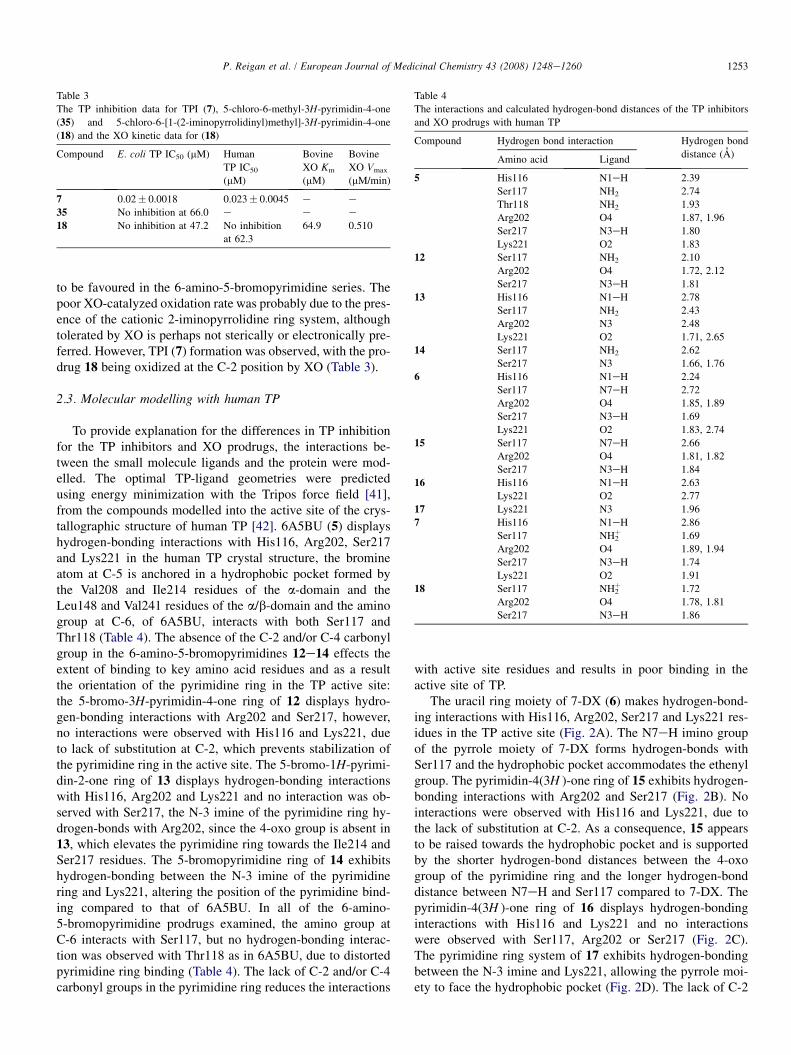
Table 3
The TP inhibition data for TPI (7), 5-chloro-6-methyl-3H-pyrimidin-4-one
(35) and 5-chloro-6-[1-(2-iminopyrrolidinyl)methyl]-3H-pyrimidin-4-one
(18) and the XO kinetic data for (18)
Compound E. coli TP IC50 (mM) Human
TP IC50
(mM)
Bovine
XO Km
(mM)
Bovine
XO Vmax
(mM/min)
7 0.02� 0.0018 0.023� 0.0045 e e
35 No inhibition at 66.0 e e e
18 No inhibition at 47.2 No inhibition
at 62.3
64.9 0.510
Table 4
The interactions and calculated hydrogen-bond distances of the TP inhibitors
and XO prodrugs with human TP
Compound Hydrogen bond interaction Hydrogen bond
distance (A)Amino acid Ligand
5 His116 N1eH 2.39
Ser117 NH2 2.74
Thr118 NH2 1.93
Arg202 O4 1.87, 1.96
Ser217 N3eH 1.80
Lys221 O2 1.83
12 Ser117 NH2 2.10
Arg202 O4 1.72, 2.12
Ser217 N3eH 1.81
13 His116 N1eH 2.78
Ser117 NH2 2.43
Arg202 N3 2.48
Lys221 O2 1.71, 2.65
14 Ser117 NH2 2.62
Ser217 N3 1.66, 1.76
1253P. Reigan et al. / European Journal of Medicinal Chemistry 43 (2008) 1248e1260
to be favoured in the 6-amino-5-bromopyrimidine series. Thepoor XO-catalyzed oxidation rate was probably due to the pres-ence of the cationic 2-iminopyrrolidine ring system, althoughtolerated by XO is perhaps not sterically or electronically pre-ferred. However, TPI (7) formation was observed, with the pro-drug 18 being oxidized at the C-2 position by XO (Table 3).
6 His116 N1eH 2.24
Ser117 N7eH 2.72
Arg202 O4 1.85, 1.89
2.3. Molecular modelling with human TPSer217 N3eH 1.69
Lys221 O2 1.83, 2.74
15 Ser117 N7eH 2.66
Arg202 O4 1.81, 1.82
Ser217 N3eH 1.84
16 His116 N1eH 2.63
Lys221 O2 2.77
17 Lys221 N3 1.96
7 His116 N1eH 2.86
Ser117 NH2þ 1.69
Arg202 O4 1.89, 1.94
Ser217 N3eH 1.74
Lys221 O2 1.91
18 Ser117 NH2þ 1.72
Arg202 O4 1.78, 1.81
Ser217 N3eH 1.86
To provide explanation for the differences in TP inhibitionfor the TP inhibitors and XO prodrugs, the interactions be-tween the small molecule ligands and the protein were mod-elled. The optimal TP-ligand geometries were predictedusing energy minimization with the Tripos force field [41],from the compounds modelled into the active site of the crys-tallographic structure of human TP [42]. 6A5BU (5) displayshydrogen-bonding interactions with His116, Arg202, Ser217and Lys221 in the human TP crystal structure, the bromineatom at C-5 is anchored in a hydrophobic pocket formed bythe Val208 and Ile214 residues of the a-domain and theLeu148 and Val241 residues of the a/b-domain and the aminogroup at C-6, of 6A5BU, interacts with both Ser117 andThr118 (Table 4). The absence of the C-2 and/or C-4 carbonylgroup in the 6-amino-5-bromopyrimidines 12e14 effects theextent of binding to key amino acid residues and as a resultthe orientation of the pyrimidine ring in the TP active site:the 5-bromo-3H-pyrimidin-4-one ring of 12 displays hydro-gen-bonding interactions with Arg202 and Ser217, however,no interactions were observed with His116 and Lys221, dueto lack of substitution at C-2, which prevents stabilization ofthe pyrimidine ring in the active site. The 5-bromo-1H-pyrimi-din-2-one ring of 13 displays hydrogen-bonding interactionswith His116, Arg202 and Lys221 and no interaction was ob-served with Ser217, the N-3 imine of the pyrimidine ring hy-drogen-bonds with Arg202, since the 4-oxo group is absent in13, which elevates the pyrimidine ring towards the Ile214 andSer217 residues. The 5-bromopyrimidine ring of 14 exhibitshydrogen-bonding between the N-3 imine of the pyrimidinering and Lys221, altering the position of the pyrimidine bind-ing compared to that of 6A5BU. In all of the 6-amino-5-bromopyrimidine prodrugs examined, the amino group atC-6 interacts with Ser117, but no hydrogen-bonding interac-tion was observed with Thr118 as in 6A5BU, due to distortedpyrimidine ring binding (Table 4). The lack of C-2 and/or C-4carbonyl groups in the pyrimidine ring reduces the interactions
with active site residues and results in poor binding in theactive site of TP.
The uracil ring moiety of 7-DX (6) makes hydrogen-bond-ing interactions with His116, Arg202, Ser217 and Lys221 res-idues in the TP active site (Fig. 2A). The N7eH imino groupof the pyrrole moiety of 7-DX forms hydrogen-bonds withSer117 and the hydrophobic pocket accommodates the ethenylgroup. The pyrimidin-4(3H )-one ring of 15 exhibits hydrogen-bonding interactions with Arg202 and Ser217 (Fig. 2B). Nointeractions were observed with His116 and Lys221, due tothe lack of substitution at C-2. As a consequence, 15 appearsto be raised towards the hydrophobic pocket and is supportedby the shorter hydrogen-bond distances between the 4-oxogroup of the pyrimidine ring and the longer hydrogen-bonddistance between N7eH and Ser117 compared to 7-DX. Thepyrimidin-4(3H )-one ring of 16 displays hydrogen-bondinginteractions with His116 and Lys221 and no interactionswere observed with Ser117, Arg202 or Ser217 (Fig. 2C).The pyrimidine ring system of 17 exhibits hydrogen-bondingbetween the N-3 imine and Lys221, allowing the pyrrole moi-ety to face the hydrophobic pocket (Fig. 2D). The lack of C-2

Fig. 2. Representations of the key interactions of (A) 7-DX (6) and the 7H-pyrrolo[2,3-d]pyrimidines (B) 15, (C) 16 and (D) 17 with the active site of human TP.
Figures display hydrogen-bond contacts (blue dashed lines) between the ligand and amino acid residues (coloured by atom type, except ligand carbons atoms
which are coloured purple). The figures were constructed using Discovery Studio Viewer Professional Software (Accelrys, Inc., San Diego, CA). (For interpretation
of the references to color in this figure legend, the reader is referred to the web version of this article.)
1254 P. Reigan et al. / European Journal of Medicinal Chemistry 43 (2008) 1248e1260
and/or C-4 substitution in the 7H-pyrrolo[2,3-d]pyrimidines15e17, results in reduced hydrogen-bonding interactionswith active site residues (Table 4) and affords explanationfor the absence of TP inhibition at high concentration.
The uracil ring system of TPI (7) makes a number of hydro-gen-bonding interactions with His116, Arg202, Ser217 andLys221. The C-5 chlorine atom occupies the hydrophobicpocket formed by Val208, Ile214, Leu148 and Val241 andthe 2-iminopyrrolidinyl ring moiety lies perpendicular to theplane of the uracil ring and allows the amino group to interactwith Ser117 (Fig. 3A). The absence of C-2 carbonyl in the TPIprodrug, 18, affects the binding interaction and orientation ofthe pyrimidin-4-one ring in the active site of TP. The 5-chloro-3H-pyrimidin-4-one ring of 18 displays hydrogen-bondinginteractions with Arg202 and Ser217, however, no hydrogen-bonding interactions with His116 or Lys221 were observed,due to lack of substitution at C-2 (Fig. 3B). The 5-chloro-3H-pyrimidin-4-one moiety of 18 appears to be raised towards
the hydrophobic pocket and this observation is supported bythe shorter hydrogen-bond distances between the 4-oxo groupof the 5-chloro-3H-pyrimidin-4-one moiety and the Arg202residue, and the longer hydrogen-bond distance between theamino group of the 2-iminopyrrolidinyl and the Ser117 residue,compared to the equivalent interactions with TPI (Table 4).The conformation of the 2-iminopyrrolidinyl ring appearsunchanged in TPI and 18. The lack of C-2 substitution inthe pyrimidin-4-one ring of 18 reduced the interaction withactive site residues and as a result no inhibition of TP wasachieved with 18 at high concentration.
3. Conclusion
In this article, we have described the XO-mediated oxida-tion of a series of prodrug compounds generating the appropri-ate TP inhibitor. The 6-amino-5-bromopyrimidines 12e14,prodrugs of the TP inhibitor 6A5BU (5), were synthesized

Fig. 3. Representations of the key interactions of (A) TPI (7) and (B) 5-chloro-6-[1-(2-iminopyrrolidinyl)methyl]-3H-pyrimidin-4-one (18) with the active site of
human TP. Figures display hydrogen-bond contacts (blue dashed lines) between the ligand and amino acid residues (coloured by atom type, except ligand carbons
atoms which are coloured purple). The figures were constructed using Discovery Studio Viewer Professional Software (Accelrys, Inc., San Diego, CA). (For in-
terpretation of the references to color in this figure legend, the reader is referred to the web version of this article.)
1255P. Reigan et al. / European Journal of Medicinal Chemistry 43 (2008) 1248e1260
and displayed no inhibition of E. coli TP at high concentra-tions. Molecular modelling confirmed that the C-2 and C-4carbonyl groups of the uracil ring are essential for TP inhibi-tion. The 6-amino-5-bromopyrimidine prodrugs were all oxi-dized at an appreciable rate by XO, forming 6A5BU. The7H-pyrrolo[2,3-d]pyrimidines 15e17, prodrugs of 7-DX (6),were synthesized and displayed no inhibition of E. coli TPat high concentration, further demonstrating the importanceof the C-2 and C-4 carbonyl groups of the uracil moiety onTP inhibition. This was supported by molecular modellingstudies, where fewer hydrogen-bonding interactions were ob-served between the 7H-pyrrolo[2,3-d]pyrimidines and the TPactive site. The 7H-pyrrolo[2,3-d]pyrimidine prodrugs wereall oxidized by XO, albeit at a slow rate, forming 7-DX with-out further oxidation. 5-Chloro-6-[1-(2-iminopyrrolidinyl)me-thyl]pyrimidine (18), a prodrug of TPI (7), lacking the C-2carbonyl, displayed no inhibition of E. coli or human TP.This was supported by molecular modelling studies, where nohydrogen-bonding was observed with Lys221 or His116, whichsuggests that the 1-amido group of the uracil ring is essential forTP inhibition. The prodrug substrate 18 was selectively oxi-dized at C-2, at a slow rate by XO, yielding TPI. The prodrugsmay be useful in assisting the oral delivery of TP inhibitors byimproving solubility, bioavailability and tumour selectivity.
4. Experimental
4.1. Materials and instrumentation
Chemicals and reagents were obtained from the Sigma-AldrichChemical Co. Dorset, UK and Lancaster Synthesis Ltd. Lanca-shire, UK. Silica gel 33e70 mm, 200e400 mesh, pH 6.5e7.5,for column chromatography were obtained from BDH, VWR In-ternational, Leicestershire, UK. Deuterated solvents (DMSO-d6,D2O and CDCl3) and tetramethylsilane (TMS) were supplied by
Cambridge Isotope Laboratories Inc., Andover, USA. Xanthine ox-idase (X4376, 0.4 unit/mg protein, one unit will convert 1.0 mmolof xanthine to uric acid per minute at pH 7.5 at 25 �C) from butter-milk, recombinant E. coli thymidine phosphorylase (T2807,1060 units/ml, one unit will convert 1.0 mmol each of thymidineand phosphate to thymine and 2-deoxyribose-1-phosphate per min-ute at pH 7.4 at 25 �C) and recombinant human thymidine phos-phorylase (T9319, 9e10 unit/mg protein, one unit will form1 nmol thymine per minute at pH 7.0 at 37 �C), expressed inV79 Chinese hamster cells, were obtained from the Sigma-AldrichChemical Co. Dorset, UK.
Analytical thin-layer chromatography (TLC) was per-formed on Merck pre-coated silica gel 60 F254 plates and visu-alized by ultra-violet illumination (254 nm) using a UV GL-58mineral-light lamp. Melting points (m.pt.) were determined inopen glass capillary tubes on Gallenkamp M.P.D.350.BM2.5micromelting point apparatus and are uncorrected. Nuclearmagnetic resonance (NMR) spectra were recorded ona 300 MHz Bruker Avance 300 spectrometer. 1H NMR spectrawere recorded in dH parts per million (ppm) in DMSO-d6 orCDCl3 with TMS as the reference. The coupling constants(J ) are expressed in Hertz (Hz). 13C NMR spectra were re-corded in dC parts per million (ppm) in DMSO-d6 or CDCl3with TMS and the solvent peak as internal standards, 13CDEPT-135 NMR spectra confirmed assignment. Infra-red(IR) spectra were recorded on a Satellite FTIR Mattson spec-trometer, with Mattson WinFIRST software. The sampleswere prepared on a Specac golden gate single reflection ATIsystem. Ultra-violet (UV) spectra were recorded on a UnicamUV 300 spectrophotometer, with Thermospectronic Vision 32software, using Hellma UV quartz cuvettes, for compoundanalysis and for XO enzyme kinetics. A Molecular DevicesSpectraMax M2 UVevisible spectrophotometer, with Soft-Max Pro software, using a 96-well microplate was used forTP enzyme kinetics. Mass spectra and elemental analysis

1256 P. Reigan et al. / European Journal of Medicinal Chemistry 43 (2008) 1248e1260
were determined at the Department of Chemistry, Universityof Manchester. Electron ionization and positive and negativechemical ionization mass spectra were recorded on an auto-mated Fisons TRIO 2000 quadrapole spectrometer. Bothpositive and negative electrospray (ES) ionization and atmo-spheric pressure chemical ionization (APCI) mass spectrawere recorded using a Micromass Platform II atmosphericpressure ionization spectrometer coupled to an HPLC system.
4.2. Chemistry
6-Amino-5-bromouracil (6A5BU, 5) was prepared from 6-aminouracil and NBS [43]. 7-Deazaxanthine (7-DX, 6) wasprepared from 6-amino-2,4-hydroxypyrimidine and chloroace-taldehyde [28]. Ethyl 2-cyano-4,4-diethoxybutanoate (27) wasprepared from bromoacetaldehyde diethyl acetal (25) andethyl cyanoacetate (26) in the presence of K2CO3 and sodiumiodide [31]. 5-Chloro-6-[1-(2-iminopyrrolidinyl)methyl]uracilhydrochloride (TPI, 7) [10] was prepared by the reaction of5-chloro-6-(chloromethyl)uracil [44] and 2-iminopyrrolidinehydrochloride [45] in the presence of sodium ethoxide. (Z )-1-Ethoxy-2-(tributylstannyl)ethene (31) was prepared fromtributyltin hydride, ethoxyacetylene and PdCl2(PPh3)2 [46].6-Amino-5-(2,2-diethoxyethyl)-2-thiouracil (28) was preparedby the reaction of thiourea with ethyl 2-cyano-4,4-diethoxybuta-noate [30]. 2-Mercapto-7H-pyrrolo[2,3-d]pyrimidin-4(3H )-one(29) was prepared by the reaction of 6-amino-5-(2,2-diethox-yethyl)-2-thiouracil with HCl [30]. 6-Methyl-3H-pyrimidin-4-one (34) was prepared by the reaction of 6-methyl-2-thiouracil(10.0 g, 70.3 mmol, 1 eq) with Raney nickel [32].
4.2.1. 6-Amino-5-bromo-3H-pyrimidin-4-one (12)Bromine (0.87 ml, 16.9 mmol, 1.25 eq) was added drop-
wise to a stirred suspension of 6-amino-4-hydroxypyrimidine(1.50 g, 13.5 mmol, 1 eq) in water (20 ml). The reaction mix-ture was stirred for 1 h at room temperature. The solution wasthen made alkaline with 20% aqueous K2CO3 (pH w 9) anda white precipitate was formed. The reaction mixture was fil-tered giving a colourless solid, which was recrystallized fromwater and vacuum-dried. The title compound 12 was isolatedas a white powder (1.21 g, 47.3%). TLC Rf 0.57 (1:1, MeOH/DCM); m.pt. 266.5e267.5 �C, m.pt. [26] 268.0 �C; 1H NMR(DMSO-d6) d: 6.75 (2H, br s, NH2), 7.82 (1H, s, C2eH),10.5 (1H, br s, N3eH); 13C NMR (DMSO-d6) d: 82.7 (C-5),147.7 (C-2), 157.7 (C-6), 160.4 (C-4); MS (ESþ) 190 (79BrMþH, 95%), 192 (81Br MþH, 93%). Elemental analysis:C4H4BrN3O, calculated (%): C 25.29, H 2.12, N 22.12, Br42.06. Found (%): C 25.21, H 2.08, N 22.04, Br 41.97.
4.2.2. 6-Amino-5-bromo-1H-pyrimidin-2-one (13)6-Amino-5-bromo-1H-pyrimidin-2-one was prepared as de-
scribed for 12 using 6-amino-2-hydroxypyrimidine (4.0 g,36.0 mmol, 1 eq) as starting material to give the product 13as a colourless powder (3.83 g, 56.0%). TLC Rf 0.58 (1:1,MeOH/DCM); m.pt. 241.5e242.5 �C, m.pt. [26] 240e242 �C; 1H NMR (DMSO-d6) d: 6.85 (2H, br s, NH2), 7.76(1H, s, C4eH), 10.5 (1H, br s, N1eH); 13C NMR (DMSO-
d6) d: 85.1 (C-5), 144.1 (C-4), 155.2 (C-6), 162.5 (C-2); MS(ESþ) 190 (79Br MþH, 100%), 192 (81Br MþH, 94%). El-emental analysis: C4H4BrN3O, calculated (%): C 25.29, H2.12, N 22.12, Br 42.06. Found (%): C 24.97, H 1.98, N21.33, Br 42.15.
4.2.3. 6-Amino-5-bromopyrimidine (14)Bromine (1.58 ml, 30.8 mmol, 1.95 eq) was added drop-
wise to a mixture of 6-amino-pyrimidine (1.50 g, 15.8 mmol,1 eq), CaCO3 (0.39 g, 3.9 mmol, 0.25 eq) and water (20 ml)and stirred for 30 min at 55e60 �C. The solution was made al-kaline with 20% aqueous K2CO3 (pH w 9) and extracted withEtOAc (2� 100 ml). The EtOAc layer was dried over K2CO3,filtered and then reduced to dryness in vacuo, giving a yellowcoloured residue, which was purified by Al2O3 column chro-matography using EtOAc as eluent. The required fractionswere collected, pooled and reduced to dryness in vacuo, theresidue was recrystallized from methanol and vacuum-driedto give the title compound 14 as a colourless powder(1.46 g, 53.1%). TLC Rf 0.60 (1:1, MeOH/DCM); m.pt.208.5e209.5 �C, m.pt. [27] 208e210 �C; 1H NMR (DMSO-d6)d: 8.32 (2H, s, C2eH and C4eH), 7.25 (2H, br s, NH2); 1HNMR (CDCl3) d: 7.05 (2H, br s, NH2), 8.40 (1H, s, C4eH),8.47 (1H, s, C2eH); 13C NMR (DMSO-d6) d: 102.4 (C-5),156.0 (C-2), 156.7 (C-4), 160.0 (C-6); MS (APCIþ) 174(79Br MþH, 100%), 176 (81Br MþH, 97%). Elemental anal-ysis: C4H4BrN3, calculated (%): C 27.61, H 2.32, N 24.15, Br45.92. Found (%): C 27.43, H 2.42, N 23.98, Br 45.99.
4.2.4. 7H-Pyrrolo[2,3-d]pyrimidin-4(3H )-one (15)7H-Pyrrolo[2,3-d]pyrimidin-4(3H )-one was prepared by an
adaptation of a known procedure [30]. Concentrated aqueousammonia (40 ml) was added to a suspension of 2-mercapto-7H-pyrrolo[2,3-d]pyrimidin-4(3H )-one (15.0 g, 89.7 mmol,1 eq) in water (100 ml). The solution was stirred at 50 �Cand Raney nickel (75 ml, slurry in water) was added por-tion-wise. The reaction mixture was then heated at reflux for3 h then the catalyst was removed by filtration. The filtratewas evaporated to dryness in vacuo and the resulting precipi-tate was collected by filtration, washed serially with water,methanol and ether, and vacuum-dried to provide the titlecompound 15 as a colourless powder (9.82 g, 81.0%). TLCRf 0.55 (4:1, EtOAc/MeOH); m.pt.> 320 �C, m.pt. [30]340e345 �C; 1H NMR (DMSO-d6) d: 6.45 (1H, d, 3JHH¼3.3 Hz, C5eH), 7.04 (1H, d, 3JHH¼ 3.2 Hz, C6eH), 7.84(1H, s, C2eH), 11.78 (1H, s, N7eH), 11.86 (1H, s, N3eH);13C NMR (DMSO-d6) d: 101.9 (C-5), 107.6 (C-9), 120.3(C-6), 143.1 (C-2), 148.0, (C-8), 158.4 (C-4); IR: 1710 cm�1
(C]O), 3470 cm�1 (NeH); MS: (APCIþ) 136 (MþH,100%), (APCI�) 134 (M�H, 100%). Elemental analysis:C6H5N3O, calculated (%): C 53.33, H 3.73, N 31.10. Found(%): C 53.31, H 3.62, N 30.50.
4.2.5. 4-Chloro-7H-pyrrolo[2,3-d]pyrimidine (30)4-Chloro-7H-pyrrolo[2,3-d]pyrimidine was prepared by an
adaptation of a known procedure [31]. A mixture of 7H-pyr-rolo[2,3-d]pyrimidin-4(3H )-one (1.35 g, 10.0 mmol, 1 eq)

1257P. Reigan et al. / European Journal of Medicinal Chemistry 43 (2008) 1248e1260
and POCl3 (15 ml, 160.9 mmol, 16 eq) was stirred for 1 h withheating at reflux. Excess POCl3 was removed under vacuumthen dichloromethane (200 ml) and iced water (100 ml) wereadded and the mixture stirred until the black solid dissolved.The organic layer was separated, washed with bicarbonate so-lution, dried over MgSO4 and then evaporated to dryness invacuo. The yellow residue was recrystallized (1:1, v/v, tolu-ene/hexane) to give pure 30 as fine colourless needles(1.02 g, 65.6%). TLC Rf 0.68 (4:1, EtOAc/hexane); m.pt.186e188 �C, m.pt. [31] 188e190 �C; 1H NMR (DMSO-d6)d: 6.62 (1H, d, 3JHH¼ 3.2 Hz, C5eH), 7.71 (1H, d,3JHH¼ 3.2 Hz, C6eH), 8.60 (1H, s, C2eH), 12.58 (1H, s,N7eH); 13C NMR (DMSO-d6) d: 98.7 (C-5), 116.5 (C-9),128.3 (C-6), 150.2 (C-8), 150.4, (C-2), 151.7 (C-4); MS:(APCIþ) 154 (35Cl Mþ, 20%), (APCI�) 152 (35Cl M�,100%). Elemental analysis: C6H4ClN3, calculated (%): C46.93, H 2.63, N 27.36, Cl 23.09. Found (%): C 47.39, H2.32, N 26.41, Cl 23.54.
4.2.6. 7H-Pyrrolo[2,3-d]pyrimidine (17)7H-Pyrrolo[2,3-d]pyrimidine was prepared by an adapta-
tion of a known procedure [31]. A solution of 4-chloro-7H-pyrrolo[2,3-d]pyrimidine (0.12 g, 0.8 mmol, 1 eq) in ethanol(20 ml) and concentrated aqueous NH3 (0.5 ml, 6.6 mmol,8.25 eq) and 10% Pd/C (0.04 g) was stirred in a H2 atmospherefor 6 h at room temperature. The catalyst was removed by fil-tration and the solvent was evaporated. The residue waswashed with H2O, filtered and dried under high vacuum givingthe title compound 17 as a fine colourless powder (0.08 g,83.7%). TLC Rf 0.62 (4:1, EtOAc/MeOH); m.pt. 129e130 �C, m.pt. [31] 131e133 �C; 1H NMR (DMSO-d6) d:6.61 (1H, d, 3JHH¼ 3.5 Hz, C5eH), 7.59 (1H, d,3JHH¼ 3.5 Hz, C6eH), 8.77 (1H, s, C4eH), 9.02 (1H, s,C2eH), 12.26 (1H, s, N7eH); 13C NMR (DMSO-d6) d: 99.3(C-5), 118.1 (C-9), 127.2 (C-6), 148.7 (C-8), 150.7, (C-2),151.0 (C-4); IR: 3450 cm�1 (NeH); MS: (APCIþ) 120(MþH, 100%). Elemental analysis: C6H5N3, calculated(%): C 60.50, H 4.23, N 35.27. Found (%): C 59.26, H 5.29,N 34.16.
4.2.7. (Z )-6-Amino-5-(2-ethoxyethenyl)-1H-pyrimidin-2-one (32)
A mixture of 6-amino-5-bromo-1H-pyrimidin-2-one(0.95 g, 5.0 mmol, 1 eq), (Z )-1-ethoxy-2-(tributylstannyl)e-thene (2.17 g, 6.0 mmol, 1.2 eq), tetraethylammonium chlo-ride (0.92 g, 5.0 mmol, 1 eq) and PdCl2(PPh3)2 (140 mg,0.2 mmol, 0.04 eq) in DMF (20 ml) was heated at reflux for5 h. The reaction mixture was reduced to dryness in vacuo,giving a brown coloured residue and purified by fractionationthrough a short silica gel column (4:1, v/v, EtOAc/hexane), therequired fractions were pooled and evaporated, recrystalliza-tion from ethanol gave compound 32 as a beige powder(0.58 g, 64.0%). TLC Rf 0.49 (4:1, EtOAc/MeOH); m.pt.89.5e90 �C; 1H NMR (DMSO-d6) d: 1.27 (3H, t,J¼ 7.2 Hz, eCH2eCH3), 4.07 (2H, q, J¼ 7.2 Hz, CH2eCH3), 5.28 (1H, d, 3JHH¼ 7.0 Hz, C10eH), 6.42 (1H, d,3JHH¼ 7.0 Hz, C20eH), 7.50 (1H, s, C4eH), 6.48 (2H, s,
NH2), 10.42 (1H, s, N1eH); 13C NMR (DMSO-d6) d: 14.8(CH3), 63.1 (CH2), 85.1 (C20eH), 99.8 (C-5), 145.2 (C10eH), 145.4 (C-6) 160.7 (C-2), 163.7 (C-4); MS: (APCIþ) 182(MþH, 100%), (APCI�) 180 (M�H, 100%).
4.2.8. 7H-Pyrrolo[2,3-d]pyrimidin-2(1H )-one (16)A solution of (Z )-6-amino-5-(2-ethoxyethenyl)-1H-pyrimi-
din-2-one (0.50 g, 2.76 mmol, 1 eq) and concentrated HCl(3.0 ml) in water (20 ml) was heated at reflux for 30 min.The solution was evaporated and the residue was made alka-line (pH w 9) with aqueous 20% K2CO3 and extracted withEtOAc (2� 75 ml). The solvent was removed by distillationunder reduced pressure and the resulting precipitate was col-lected by filtration, washed serially with water and ethanol,and vacuum-dried to provide the title compound 16 as a beigepowder (0.37 g, 85.0%). TLC Rf 0.54 (4:1, EtOAc/MeOH);m.pt. 159e160 �C; 1H NMR (DMSO-d6) d: 6.46 (1H, d,3JHH¼ 3.3 Hz, C5eH), 7.04 (1H, d, 3JHH¼ 3.3 Hz, C6eH),8.47 (1H, s, C4eH), 11.80 (1H, s, N1eH), 11.85 (1H, s,N7eH); 13C NMR (DMSO-d6) d: 101.7 (C-5), 107.6 (C-9),120.3 (C-6), 143.1 (C-8), 148.0 (C-2), 158.4 (C-4); IR:1710 cm�1 (C]O), 3450 cm�1 (NeH); MS: (APCIþ) 136(MþH, 100%), (APCI�) 134 (M�H, 100%). Elementalanalysis: C6H5N3O, calculated (%): C 53.33, H 3.73, N31.10. Found (%): C 53.08, H 3.56, N 30.51.
4.2.9. 6H-Imidazo[1,2-c]pyrimidin-5-one (24)Chloroacetaldehyde (50%, v/v, solution in water, 1.4 ml,
10.8 mmol, 1.2 eq) was added dropwise to a stirred mixtureof 6-amino-1H-pyrimidin-2-one (1.0 g, 9.0 mmol, 1 eq) inDMF (6.0 ml) at 50 �C. A colour change of white to pinkwas observed after the addition of chloroacetaldehyde. The re-action mixture was heated at 50 �C for 12 h. The solvent wasremoved by distillation under reduced pressure and the result-ing precipitate was collected by filtration, washed serially withethanol and ether, and dried to provide the title compound 24as a beige powder (0.91 g, 74.7%). TLC Rf 0.45 (4:1, EtOAc/MeOH); m.pt. 278.0e278.5 �C, m.pt. [29] 272e274 �C; 1HNMR (DMSO-d6) d: 6.87 (1H, d, 3JHH¼ 7.4 Hz, C8eH),7.79 (1H, d, 3JHH¼ 7.4 Hz, C7eH), 7.92 (1H, d,3JHH¼ 2.2 Hz, C3eH), 8.10 (1H, d, 3JHH¼ 2.2 Hz, C2eH),12.72 (1H, br s, N6eH); 13C NMR (DMSO-d6) d: 93.7 (C-3),115.0 (C-7), 123.8 (C-2), 138.8 (C-8), 145.6 (C-9), 145.9(C-5); IR: 1720 cm�1 (C]O), 3340 cm�1 (NeH); MS:(ESþ) 136 (MþH, 60%), (ES�) 134 (M�H, 100%). Ele-mental analysis: C6H5N3O, calculated (%): C 53.33, H 3.73,N 31.10. Found (%): C 53.15, H 3.67, N 30.96.
4.2.10. 5-Chloro-6-methyl-3H-pyrimidin-4-one (35)A mixture of 6-methyl-3H-pyrimidin-4-one (5.0 g,
45.4 mmol, 1 eq) in glacial AcOH (100 ml) was heated to80 �C until the solid dissolved. The mixture was cooled to60 �C and NCS (6.67 g, 49.9 mmol, 1.1 eq) was added. The re-action mixture was heated at reflux for 5 h. The reaction mix-ture was evaporated to dryness in vacuo, the residue wastreated with a minimum amount of water and placed in the re-frigerator at 5 �C overnight. A colourless precipitate formed

1258 P. Reigan et al. / European Journal of Medicinal Chemistry 43 (2008) 1248e1260
was collected by filtration. The solid was vacuum-dried giving35 as a colourless powder (4.83 g, 73.7%). TLC Rf 0.62 (4:1,EtOAc/MeOH); m.pt. 210.0e210.5 �C, m.pt. [33] 209e211 �C; 1H NMR (DMSO-d6) d: 2.34 (3H, s, CH3), 8.11(1H, s, C2eH), 12.98 (1H, s, N3eH); 13C NMR (DMSO-d6)d: 21.7 (CH3), 119.8 (C-5), 146.7 (C-2), 157.1 (C-6), 160.3(C-4); MS: (ESþ) 145 (35Cl MþH, 45%), 147 (37ClMþH, 12%), (ES�) 143 (35Cl MþH, 50%). Elemental anal-ysis: C5H5ClN2O, calculated (%): C 41.54, H 3.49, N 19.38,Cl 24.52. Found (%): C 41.24, H 3.20, N 19.17, Cl 24.22.
4.2.11. 5-Chloro-6-(chloromethyl)-3H-pyrimidin-4-one (36)A mixture of 5-chloro-6-methyl-3H-pyrimidin-4-one
(4.00 g, 27.7 mmol, 1 eq), NCS (3.70 g, 27.7 mmol, 1 eq) andbenzoyl peroxide (0.67 g, 2.77 mmol, 0.1 eq) in CCl4 (200 ml)was heated at 80 �C for 96 h. The mixture was cooled to roomtemperature, the insoluble material was removed by filtrationand the solvent was distilled off under reduced pressure. Theresidue was purified by column chromatography (DCM). Therequired fractions were pooled and evaporated giving the titlecompound 36 as a cream powder (0.51 g, 10.2%). TLC Rf 0.44(4:1, EtOAc/MeOH); m.pt. 267e269.5 �C; 1H NMR (DMSO-d6) d: 4.15 (2H, s, eCH2e), 8.05 (1H, s, C2eH), 12.80 (1H, s,N3eH); 13C NMR (DMSO-d6) d: 43.3 (CH2), 120.3 (C-5),147.3 (C-2), 156.3 (C-6), 160.5 (C-4).
4.2.12. 5-Chloro-6-[1-(2-iminopyrrolidinyl)methyl]-3H-pyrimidin-4-one hydrochloride (18)
A mixture of 5-chloro-6-(chloromethyl)-3H-pyrimidin-4-one (0.40 g, 2.24 mmol, 1 eq), 2-iminopyrrolidine hydrochlo-ride (37) [45] (0.81 g, 6.70 mmol, 3 eq) and sodium ethoxide(0.46 g, 6.70 mmol, 3 eq) was stirred in anhydrous DMF(5.0 ml) under a N2 atmosphere for 48 h at room temperature.The reaction mixture was filtered, the solid was added to water(3.5 ml) and neutralized with AcOH. The insoluble materialwas filtered, dissolved in 0.1 M HCl (15 ml), decolourizedwith activated charcoal, filtered through Celite and the filtratewas evaporated to dryness in vacuo. The solid was washedwith a minimum volume of ethanol, filtered and dried togive the title compound 18 as a colourless powder (0.22 g,38.1%). TLC Rf 0.18 (4:1, EtOAc/MeOH); m.pt. 286.5e287 �C; 1H NMR (DMSO-d6) d: 2.04 (2H, quint, 3JHH¼7.5 Hz, eCH2eCH2eCH2e), 2.87 (2H, t, 3JHH¼ 7.9 Hz,eCH2eC]NH2
þ), 3.59 (2H, t, 3JHH¼ 7.3 Hz, eCH2eNHe),4.22 (2H, s, eCH2e), 7.90 (1H, s, C2eH), 9.80 (1H, s,NþHeH), 9.88 (1H, s, NþHeH ), 11.77 (1H, s, N3eH); 13CNMR (DMSO-d6) d: 18.6 (C-40), 31.5 (C-30), 44.6 (C-50),52.3 (eCH2e), 106.3 (C-5), 144.8 (C-6), 150.2 (C-2), 159.8(C-4), 170.3 (C-20); MS: (ESþ) 228 (35Cl Mþ, 50%), 230(37Cl Mþ, 16%). Elemental analysis: C9H12Cl2N4O, calculated(%): C 41.08, H 4.60, N 21.29. Found (%): C 40.81, H 4.31, N21.02.
4.3. TP enzymology
The inhibition of recombinant E. coli TP and recombinanthuman TP, to determine the IC50 values of the compounds
synthesized, was determined using a continuous spectrophoto-metric assay, adapted for a Molecular Devices SpectramaxUVevisible spectrophotometer with a 96-well microplatereader, at a kinetic wavelength of 355 nm, at 25 �C. TP can con-vert 5-nitro-20-deoxyuridine to 5-nitrouracil, this conversionwas accompanied by a considerable change of absorption, at355 nm, which was continuously monitored as a function oftime [35]. The synthetic substrate 5-nitro-20-deoxyuridine dis-plays a higher extinction coefficient (3max 13,300 M�1 cm�1
at 322 nm) at a longer wavelength than thymidine (3max
9300 M�1 cm�1 at 266 nm), which provides additional sensi-tivity for assays. The nitro group decreases the value of kcat of5-nitro-20-deoxyuridine (kcat 20 s�1; Km 0.23 mM) by 88-foldcompared to the methyl group of thymidine (kcat 1770 s�1;Km 0.24 mM), but has little effect on the Km value, with re-combinant E. coli TP. Therefore the compromise in acceptingthe spectral convenience of the 5-nitro-20-deoxyuridine sub-strate is decreased sensitivity [34].
The reaction mixtures (0.2 ml) contained 0.13 mM of 5-nitro-20-deoxyuridine as the substrate and 0.1 M potassiumphosphate buffer (pH 7.4). The reaction was initiated by addi-tion of TP (0.00883 U/0.2 ml). The initial inhibitor stockswere prepared as 2.0e8.0 mM solutions and 50 ml of the stockwas used in the assay, giving a final assay volume of 200 ml. If50% inhibition was exceeded with 50 ml of the inhibitor stock,at 0.5e2.0 mM final cuvette concentration, the initial stockwas serially diluted in 10-fold increments until a suitable levelof TP inhibition was achieved. If no significant inhibition wasobserved at 1.0 mM the compound was not considered an in-hibitor of TP. In the calculation of the IC50 value at least fiveinhibitor concentrations were used spanning the experimentalvalue. The initial velocity was plotted against inhibitor con-centration and the IC50 value was determined as the concentra-tion causing a 50% decrease in the initial velocity relative tothe mean of at least two controls with no inhibitor present.The human TP assay was conducted similarly except that0.33 mM of 5-nitro-20-deoxyuridine (Km 0.16 mM) was usedas substrate [34].
4.4. XO enzymology
The evaluation of the kinetic constants (Km and Vmax) forthe natural substrate and prodrug activation by XO, from but-termilk, was determined using a continuous spectrophotomet-ric assay, using a Unicam UV 300 Spectrometer with 3.5 mlHelma quartz cuvettes (1 cm light path) at 25 �C [36]. Thereaction mixtures (3.0 ml) contained 50 mM potassiumphosphate buffer (pH 7.5), the substrates in five different con-centrations (25e150 mM). The reaction was initiated by inj-ection of XO (0.9 U/ml). In the XO assay the enzymeconcentration remained fixed and limiting so that the observedactivity was proportional to the amount of substrate present.The change in absorbance was continually monitored overa time period at a specific predetermined wavelength, corre-sponding to the formation of the corresponding TP inhibitor.The absorption maxima was measured at a selected wave-length for each compound, substrate and product, in 50 mM

1259P. Reigan et al. / European Journal of Medicinal Chemistry 43 (2008) 1248e1260
potassium phosphate buffer (pH 7.5), and plotted against con-centration, the slope giving the molar absorption coefficient ofthe given compound. The initial reaction rates were recalcu-lated using the molar absorption coefficients at the relevantwavelength for the product formed.
In the case of xanthine, the reaction mixtures (3.0 ml) con-tained 50 mM potassium phosphate buffer (pH 7.5) and0.15 mM xanthine solution (pH 7.5). The reaction was initiatedby injection of XO (0.9 U/ml). The formation of uric acid wasmonitored at 292 nm over a period of time [47]. The change ofabsorbance was taken as a measure of the reaction and the en-zyme velocity was defined in terms of the change of absorbanceper unit time [48]. The linear part of the curve is proportional tothe initial velocity, the enzyme concentration remained fixedand limiting, therefore the absorbance is directly proportionalto the concentration of product formed or substrate consumed.A series of reactions were performed at varying xanthine con-centrations (50e150 mM). The initial rate generally followssaturation kinetics with respect to the concentration of the sub-strate. The determination of the values Km and Vmax were madefrom rearrangements of the MichaeliseMenten equation [49].The velocity was recalculated from change in absorbance perminute using the molar absorption coefficient to determinethe velocity in terms of mM/min (calculated from the mean ofthree independent measurements). The plot of absorbance at292 nm versus concentration of uric acid gave a molar extinc-tion coefficient of 12,500 M�1 cm�1, which was in agreementwith the literature [36]. The absorption maxima of 6A5BUwas measured over a concentration range at a wavelength of270 nm giving a molar extinction coefficient of13,600 M�1 cm�1. The absorption maxima of 7-DX was mea-sured over a concentration range at 278 nm giving a molar extinc-tion coefficient of 14,700 M�1 cm�1. The absorption maxima ofTPI was measured over a concentration range at 302 nm givinga molar extinction coefficient of 11,200 M�1 cm�1.
4.5. Molecular modelling with human TP
Computational modelling was performed using the model-ling package SYBYL 6.8 (Tripos Ltd.). The crystallographiccoordinates for the 2.1 A structure of human TP bound to thepotent inhibitor TPI (PDB code: 1UOU) were obtained fromAstraZeneca [42]. The ligands (both TP inhibitors and XO pro-drugs) were constructed using SYBYL 6.8, and the atomsassigned partial atomic charges according to the GasteigereMarsili scheme. Once constructed, the inhibitors were modelledinto the active site of the human TP crystal structure, using theexperimental bound geometry of the inhibitor TPI as a guide(in a particular orientation of the nucleobase moiety). A 6 A ra-dius subset encompassing the TP active siteeligand complexwas then minimized to convergence of 0.001 kcal/mol usingthe Tripos force field [41].
Acknowledgements
We thank Professor K.T. Douglas and Dr. E. Chinje forhelpful discussions. This work was funded in part by the
MRC and The Great Socialist People’s Libyan Arab Jamahir-iya (The Secretariat of Higher Education, Professor AkeelHussain Akeel).
References
[1] S. Takao, S.I. Akiyama, A. Nakajo, H. Yoh, M. Kitazono, S. Natsugoe,
K. Miyadera, M. Fukushima, Y. Yamada, T. Aikou, Cancer Res. 60
(2000) 5345e5348.
[2] M. Kitazono, Y. Takebayashi, K. Ishitsuka, S. Takao, A. Tani,
T. Furukawa, K. Miyadera, Y. Yamada, T. Aikou, S. Akiyama, Biochem.
Biophys. Res. Commun. 253 (1998) 797e803.
[3] K. Miyadera, T. Sumizawa, M. Haraguchi, H. Yoshida, W. Konstanty,
Y. Yamada, S. Akiyama, Cancer Res. 55 (1995) 1687e1690.
[4] M. Haraguchi, K. Miyadera, K. Uemura, T. Sumizawa, T. Furukawa,
K. Yamada, S.I. Akiyama, Nature 368 (1994) 198.
[5] M. Fukushima, N. Suzuki, T. Emura, S. Yano, H. Kazuno, Y. Tada,
Y. Yamada, T. Asao, Biochem. Pharmacol. 59 (2000) 1227e1236.
[6] B.C. Pan, Z.H. Chen, M.Y. Wang Chu, S.H. Chu, Nucleosides Nucleotides
17 (1998) 2367e2382.
[7] C. Desgranges, G. Razaka, M. Rabaud, P. Picard, F. Dupuch, H. Bricaud,
Biochem. Pharmacol. 31 (1982) 2755e2759.
[8] P. Langen, G. Etzold, D. Barwolff, B. Preussel, Biochem. Pharmacol. 16
(1967) 1833e1837.
[9] J. Balzarini, A.E. Gamboa, R. Esnouf, S. Liekens, J. Neyts, E. De
Clercq, M.J. Camarasa, M.J. Perez-Perez, FEBS Lett. 438 (1998)
91e95.
[10] S. Yano, Y. Tada, H. Kazuno, T. Sato, J. Yamashita, N. Suzuki, T. Emura,
M. Fukushima, T. Asao, PCT Int. Appl. (1996) 108.
[11] N. Suzuki, M. Fukushima, T. Asao, Y. Yamada, S. Akiyama, Proc. Am.
Assoc. Cancer Res. 38 (1997) 101.
[12] P. Reigan, P.N. Edwards, A. Gbaj, C. Cole, S.T. Barry, K.M. Page,
S.E. Ashton, R.W.A. Luke, K.T. Douglas, I.J. Stratford, M. Jaffar,
R.A. Bryce, S. Freeman, J. Med. Chem. 48 (2005) 392e402.
[13] C. Cole, D.S. Marks, M. Jaffar, I.J. Stratford, K.T. Douglas, S. Freeman,
Anticancer Drug Des. 14 (1999) 411e420.
[14] L.S. Terada, D. Piermattei, G.N. Shibao, J.L. McManaman, R.M. Wright,
Arch. Biochem. Biophys. 348 (1997) 163e168.
[15] W.B. Poss, T.P. Huecksteadt, P.C. Panus, B.A. Freeman, J.R. Hoidal, Am.
J. Physiol. 270 (1996) L941eL946.
[16] N.M. Hasan, R.B. Cundall, G.E. Adams, Free Radic. Biol. Med. 11
(1991) 179e185.
[17] H. Biri, H.S. Ozturk, M. Kacmaz, K. Karaca, H. Tokucoglu, I. Durak,
Cancer Invest. 17 (1999) 314e319.
[18] H.S. Ozturk, M. Karaayvaz, M. Kacmaz, M. Kavutcu, H. Akgul,
I. Durak, Cancer Biochem. Biophys. 16 (1998) 157e168.
[19] E. Kokoglu, A. Belce, E. Ozyurt, Z. Tepeler, Cancer Lett. 50 (1990)
179e181.
[20] M. Maliepaard, A. Wolfs, S.E. Groot, N.J. de Mol, L.H. Janssen, Br. J.
Cancer 71 (1995) 836e839.
[21] C.A. Pritsos, D.L. Gustafson, Oncol. Res. 6 (1994) 477e481.
[22] E.D. Clarke, K.H. Goulding, P. Wardman, Biochem. Pharmacol. 31
(1982) 3237e3242.
[23] K. Shanmuganathan, T. Koudriakova, S. Nampalli, J. Du, J.M. Gallo,
R.F. Schinazi, C.K. Chu, J. Med. Chem. 37 (1994) 821e827.
[24] T.A. Krenitsky, W.W. Hall, P. de Miranda, L.M. Beauchamp,
H.J. Schaeffer, P.D. Whiteman, Proc. Natl. Acad. Sci. U.S.A. 81
(1984) 3209e3213.
[25] P. Reigan, A. Gbaj, E. Chinje, I.J. Stratford, K.T. Douglas, S. Freeman,
Bioorg. Med. Chem. Lett. 14 (2004) 5247e5250.
[26] D.J. Brown, J.S. Harper, J. Chem. Soc. 1 (1961) 1298e1303.
[27] T. Sakamoto, C. Satoh, K. Yoshinori, H. Yamanaka, Chem. Pharm. Bull.
41 (1993) 81e86.
[28] D.E. Ayer, G.L. Bundy, E.J. Jacobsen, PCT Int. Appl. (1993) 120.
[29] D.G. Bartholomew, P. Dea, R.K. Robins, G.R. Revankar, J. Org. Chem.
40 (1975) 3708e3713.

1260 P. Reigan et al. / European Journal of Medicinal Chemistry 43 (2008) 1248e1260
[30] F. Seela, R. Richter, Chem. Ber. 111 (1978) 2925e2930.
[31] J. Davoll, J. Chem. Soc. (1960) 131e138.
[32] H.R. Snyder, H.M. Foster, G.A. Nussberger, J. Am. Chem. Soc. 76
(1954) 2441e2443.
[33] T. Nishiwaki, Tetrahedron 22 (1966) 2401e2408.
[34] A. Gbaj, P. Reigan, P.N. Edwards, S. Freeman, M. Jaffar, K.T. Douglas,
J. Enzyme Inhib. Med. Chem. 21 (2006) 69e73.
[35] Y. Wataya, D.W. Santi, Anal. Biochem. 112 (1981) 96e98.
[36] H. Rosemeyer, F. Seela, Eur. J. Biochem. 134 (1983) 513e515.
[37] R.S. Klein, M. Lenzi, T.H. Lim, K.A. Hotchkiss, P. Wilson,
E.L. Schwartz, Biochem. Pharmacol. 62 (2001) 1257e1263.
[38] J. Balzarini, B. Degreve, A. Esteban-Gamboa, R. Esnouf, E. De Clercq,
Y. Engelborghs, M.J. Camarasa, M.J. Perez-Perez, FEBS Lett. 483
(2000) 181e185.
[39] P.E. Murray, V.A. McNally, S.D. Lockyer, K.J. Williams, I.J. Stratford,
M. Jaffar, S. Freeman, Bioorg. Med. Chem. 10 (2002) 525e530.
[40] T.A. Krenitsky, S.M. Neil, G.B. Elion, G.H. Hitchings, Arch. Biochem.
Biophys. 150 (1972) 585e599.
[41] M. Clark, R.D. Cramer, N. van Opdenbosch, J. Comput. Chem. 10 (1989)
982e1012.
[42] R.A. Norman, S.T. Barry, M. Bate, J. Breed, J.G. Colls, R.J. Ernill, R.W. Luke,
C.A. Minshul, M.S. McAlister, E.J. McCall, H.H. McMiken, D.S.
Paterson, D. Timms, J.A. Tucker, R.A. Pauptit, Structure 12 (2004) 75e84.
[43] E.F. Schroeder, US Patent, Searle 038 US 2731465. Chem. Abstr. 51
(1957) 1257.
[44] R.A. West, H.W. Barrett, J. Am. Chem. Soc. 76 (1953) 3146e3148.
[45] E.J. Moriconi, A.A. Cevasco, J. Org. Chem. 33 (1968) 2109e2111.
[46] R.H. Wollenberg, K.F. Albizati, R. Peries, J. Am. Chem. Soc. 99 (1977)
7365e7367.
[47] H. Kalckar, J. Biol. Chem. 167 (1947) 429e443.
[48] M. Dixon, E.C. Webb (Eds.), Enzymes, Longman, London, 1979.
[49] H. Lineweaver, D. Burk, J. Am. Chem. Soc. 56 (1934) 658e666.


![Synthesis and in Vitro Evaluation of 5-[ 18 F]Fluoroalkyl Pyrimidine Nucleosides for Molecular Imaging of Herpes Simplex Virus Type 1 Thymidine Kinase Reporter Gene Expression](https://static.fdokumen.com/doc/165x107/6313a9af3ed465f0570ac71d/synthesis-and-in-vitro-evaluation-of-5-18-ffluoroalkyl-pyrimidine-nucleosides.jpg)