Stabilization of the Nitric Oxide (NO) Prodrugs and Anticancer Leads, PABA/NO and Double JS-K,...
22
Stabilization of the Nitric Oxide (NO) Prodrugs and Anti-Cancer Leads, PABA/NO and Double JS-K through Incorporation into PEG-Protected Nanoparticles Varun Kumar † , Sam Y. Hong § , Anna E. Maciag ‡ , Joseph E. Saavedra ‡ , Douglas H. Adamson ¶ , Robert K. Prud'homme †,* , Larry K. Keefer § , and Harinath Chakrapani T,* † Department of Chemical Engineering, Princeton University, Princeton, New Jersey 08544 § Chemistry Section, Laboratory of Comparative Carcinogenesis, National Cancer Institute at Frederick, Frederick, Maryland 21702 ‡ Basic Science Program, SAIC-Frederick, National Cancer Institute at Frederick, Frederick, Maryland 21702 ¶ Department of Chemistry and Institute for Material Science, University of Connecticut, Storrs, Connecticut 06269 T Department of Chemistry, Indian Institute of Science Education and Research, Pune 411008, India Abstract Here we report the stabilization of the nitric oxide (NO) prodrugs and anti-cancer lead compounds, PABA/NO (O 2 -{2,4-dinitro-5-[4-(N-methylamino)benzoyloxy]phenyl} 1-(N,N-dimethylamino) diazen-1-ium-1,2-diolate) and “Double JS-K” (1,5-bis{[1-[(4-ethoxycarbonyl)piperazin-1-yl] diazen-1-ium-1,2-diol-2-ato]-2,4-dinitrobenzene), through their incorporation into polymer- protected nanoparticles. The prodrugs were formulated in block copolymer-stabilized nanoparticles with sizes from 220 to 450 nm by a novel rapid precipitation process. The block copolymers, with polyethylene glycol (PEG) soluble blocks, provide a steric barrier against NO prodrug activation by glutathione. Too rapid activation and NO release has been a major barrier to effective administration of this class of compounds. The nanoparticle stabilized PABA/NO from attack by glutathione as evidenced by a significant increase in time taken for 50% decomposition from 15 min (unformulated) to 5 h (formulated); in the case of Double JS-K, the 50% decomposition time was extended from 4.5 min (unformulated) to 40 min (formulated). The more hydrophobic PABA/NO produced more stable nanoparticles and correspondingly more extended release times in comparison with Double JS-K. The hydrophobic blocks of the polymer were either polystyrene or polylactide. Both blocks produced nanoparticles of approximately the same size and release kinetics. This combination of PEG- [email protected]. [email protected]. NIH Public Access Author Manuscript Mol Pharm. Author manuscript; available in PMC 2011 February 1. Published in final edited form as: Mol Pharm. 2010 February 1; 7(1): 291. doi:10.1021/mp900245h. NIH-PA Author Manuscript NIH-PA Author Manuscript NIH-PA Author Manuscript
-
Upload
independent -
Category
Documents
-
view
2 -
download
0
Transcript of Stabilization of the Nitric Oxide (NO) Prodrugs and Anticancer Leads, PABA/NO and Double JS-K,...

Stabilization of the Nitric Oxide (NO) Prodrugs and Anti-CancerLeads, PABA/NO and Double JS-K through Incorporation intoPEG-Protected Nanoparticles
Varun Kumar†, Sam Y. Hong§, Anna E. Maciag‡, Joseph E. Saavedra‡, Douglas H.Adamson¶, Robert K. Prud'homme†,*, Larry K. Keefer§, and Harinath ChakrapaniT,*†Department of Chemical Engineering, Princeton University, Princeton, New Jersey 08544§Chemistry Section, Laboratory of Comparative Carcinogenesis, National Cancer Institute atFrederick, Frederick, Maryland 21702‡Basic Science Program, SAIC-Frederick, National Cancer Institute at Frederick, Frederick,Maryland 21702¶Department of Chemistry and Institute for Material Science, University of Connecticut, Storrs,Connecticut 06269TDepartment of Chemistry, Indian Institute of Science Education and Research, Pune 411008, India
Abstract
Here we report the stabilization of the nitric oxide (NO) prodrugs and anti-cancer lead compounds,PABA/NO (O2-{2,4-dinitro-5-[4-(N-methylamino)benzoyloxy]phenyl} 1-(N,N-dimethylamino)diazen-1-ium-1,2-diolate) and “Double JS-K” (1,5-bis{[1-[(4-ethoxycarbonyl)piperazin-1-yl]diazen-1-ium-1,2-diol-2-ato]-2,4-dinitrobenzene), through their incorporation into polymer-protected nanoparticles. The prodrugs were formulated in block copolymer-stabilized nanoparticleswith sizes from 220 to 450 nm by a novel rapid precipitation process. The block copolymers, withpolyethylene glycol (PEG) soluble blocks, provide a steric barrier against NO prodrug activation byglutathione. Too rapid activation and NO release has been a major barrier to effective administrationof this class of compounds. The nanoparticle stabilized PABA/NO from attack by glutathione asevidenced by a significant increase in time taken for 50% decomposition from 15 min (unformulated)to 5 h (formulated); in the case of Double JS-K, the 50% decomposition time was extended from 4.5min (unformulated) to 40 min (formulated). The more hydrophobic PABA/NO produced more stablenanoparticles and correspondingly more extended release times in comparison with Double JS-K.The hydrophobic blocks of the polymer were either polystyrene or polylactide. Both blocks producednanoparticles of approximately the same size and release kinetics. This combination of PEG-
[email protected]. [email protected].
NIH Public AccessAuthor ManuscriptMol Pharm. Author manuscript; available in PMC 2011 February 1.
Published in final edited form as:Mol Pharm. 2010 February 1; 7(1): 291. doi:10.1021/mp900245h.
NIH
-PA Author Manuscript
NIH
-PA Author Manuscript
NIH
-PA Author Manuscript

protected nanoparticles with sizes appropriate for cancer targeting by enhanced permeation andretention (EPR) and delayed release of NO may afford enhanced therapeutic benefit.
KeywordsNitric oxide; PABA/NO; glutathione; glutathione S-transferase; nanoparticles; formulation
INTRODUCTIONNitric oxide (NO) is a mediator of diverse physiological processes and has been shown to haveanti-proliferative activity.1–3 Due to the poor bioavailability of NO, typically prodrugs of nitricoxide such as diazeniumdiolates are used as surrogates of NO.4 O2-(2,4-Dinitrophenyl)diazeniumdiolates are important members of the diazeniumdiolate class of nitric oxideprodrugs that have shown promising anti-cancer activity.5 Some important examples are JS-K (O2-(2,4-dinitrophenyl) 1-[(4-ethoxycarbonyl)piperazin-1-yl]diazen-1-ium-1,2-diolate)6–15 and PABA/NO,16–22 which have both shown potent tumoristatic effects in animal models(Figure 1). For example, JS-K was found to inhibit the in vivo growth of xenografts of cellsderived from human leukemia, multiple myeloma, and prostate carcinoma. PABA/NO showedtumoristatic activity against A2780 human ovarian cancer xenografts in female SCID micewith a potency comparable to that of cisplatin.
The general mechanism of activation of such prodrugs involves a nucleophilic aromaticsubstitution by glutathione (GSH) followed by spontaneous decomposition of the ensuingdiazeniumdiolate anion to generate nitric oxide under physiological conditions (Scheme 1).5The aforementioned reaction was found to be catalyzed by glutathione S-transferase (GST), aclass of detoxification enzymes that is frequently over-expressed in cancers.14, 21
Double JS-K12 (Figure 1) is a second-generation JS-K analogue that was designed to generateup to 4 moles of NO per mole of compound and has shown potent in vitro anti-proliferativeeffects against human leukemia cells.17
As JS-K and PABA/NO react with glutathione even in the absence of GST, our goal duringpreclinical development of such prodrugs was to minimize reactivity of the prodrug withglutathione in order to increase circulation time and ultimately its efficacy. Our approachtowards stabilizing nitric oxide prodrugs was to encapsulate these prodrugs in the form ofsuitable nanoparticles.
Nanoparticles (NP) have previously been reported as potential delivery vehicles for varioustherapeutic agents.23–25 Gref et al.25 reported the advantages of PEGylated polymericnanoparticles in terms of the circulation time and release of the encapsulated drug. Polyethyleneglycol-b-poly(lactic-co-glycolic acid) (PEG-b-PLGA)-based nanospheres showed longercirculation time and reduced liver uptake compared to PLGA nanospheres with no PEGcoating.25 The concept of a protected prodrug has been widely used to sustain release ofcytotoxic agents. Schoenmakers et al. have tailored the cleavage kinetics of ester bonds tocontrol paclitaxel release.26 The research groups of Baker27 and Kannan28, 29 have preparedprodrugs based on dendrimer scaffolds. Sengupta et al.30 conjugated doxorubicin with PLGAand nucleated the prodrug inside a nanoscale PEGylated phospholipid envelope. Theconjugation led to a shift in concentration (~1.8 times) corresponding to half-maximalresponse. Ansell et al.31conjugated paclitaxel with various lipophilic alcohols to tune thehydrophobicity of the drug and hence its partitioning rate, which directly correlated to efficacy.
Kumar et al. Page 2
Mol Pharm. Author manuscript; available in PMC 2011 February 1.
NIH
-PA Author Manuscript
NIH
-PA Author Manuscript
NIH
-PA Author Manuscript

In this study, nitric oxide prodrug molecules have been encapsulated into the nanoparticle formand the impact of the polymer protecting layer has been analyzed. The formation and processingof the nanoparticles were observed not to affect the molecular activity of the drug compounds.The formulated nanoparticles of the drug compounds showed significant improvement instability against glutathione, and a similar anti-proliferative activity as the naked drugmolecules.
EXPERIMENTAL SECTIONMaterials
The nitric oxide prodrugs JS-K, PABA/NO, and Double JS-K were prepared using previouslyreported methods.12,19,32 Poloxamer® 188 was obtained from BASF Corporation, Parsippany,NJ. Trehalose (D-trehalose dihydrate) was purchased from Sigma-Aldrich Inc., Milwaukee,WI . The stabilizer, poly-L-lactide-b-poly (ethylene glycol) (PLA-b-PEG, 4.2k-b-5k) waspurchased from Polymer Source, Inc., Richmond, IN. Polystyrene-b-poly (ethylene glycol)( PS-b-PEG, 1.5k-b-3k) was synthesized by high vacuum anionic polymerization methods32
using hydroxide capped PS initiated by potassium naphthalenide followed by addition ofethylene oxide.
Nanoparticle formationAqueous solution—Poloxamer® 188 and trehalose were dissolved in milli-Q water at theconcentration of 9.5 and 12 mg/mL respectively. The aqueous solution was filtered through0.2-μm syringe filter (Whatman Inc., Florham park, NJ).
Organic solution—Block copolymer (PS-b-PEG or PLA-b-PEG) was dissolved in 3.5 mLof tetrahydrofuran, (THF), at the concentration of 8.6 mg/mL. The mixture was sonicated toget a clear solution. NO prodrug (~10.5 mg) was then added to the clear solution. The mixedsolution was sonicated and filtered through a 0.2 μm syringe filter.
Particle Formation—The nanoparticles were produced by a rapid micromixing, rapidprecipitation, and block copolymer-directed encapsulation process called FlashNanoPrecipitation (FNP), which is shown schematically in Figure 2. The design and operationof the multi-inlet vortex mixer (MIVM) used to formulate the nanoparticles have beendescribed previously.34–38 The organic stream (12 mL/min) was mixed against the aqueousstreams (3 streams, each at 40 mL/min) in the MIVM to precipitate the drug followed bystabilization with the block copolymers. The nanoparticle suspension was collected in a 50 mLsterile tube (Becton Dickinson & Co., Franklin lakes, NJ).
Dynamic laser light scattering was used to determine the particle size (expressed as the intensityweighted diameter) with a Zeta sizer (Malvern Instruments, Inc., Westborough, MA). Allmeasurements were made at 532 nm wavelength at the scattering angle of 173° using normalresolution mode as the analysis model.
Processing of nanoparticle dispersionThe nanoparticle dispersion was processed using two different routes: dialysis andlyophilization, to remove the residual organic solvent.
Dialysis—The nanoparticle dispersion was dialyzed against pure water using a Spectra/Pordialysis membrane (Spectrum laboratories, Inc., CA) with a Molecular Weight Cut-Off(MWCO) of 6k-8k Daltons.
Kumar et al. Page 3
Mol Pharm. Author manuscript; available in PMC 2011 February 1.
NIH
-PA Author Manuscript
NIH
-PA Author Manuscript
NIH
-PA Author Manuscript

Lyophilization—The undialyzed nanoparticle suspension was immediately transferred intoa series of 2 mL sterile cryogenic vials (Corning Inc., Corning, NY) dipped into a dry ice/acetone bath to rapidly freeze the sample. The vials were left in the bath for 30–40 min. Thevials were lyophilized at −76 °C and 70 mTorr for 3 days. The lyophilized powder wasreconstituted in PBS (pH = 7.4) to the desired concentration. 2 mL of PBS was added to eachcryogenic vial containing the lyophilized powder and the solution was sonicated with a probetip sonication for ~3 min at 2–3 watts (Fisher scientific model 100 sonic dismembrator, witha 3.2 mm tip) with the vial dipped in an ice bath to avoid heating during sonication.
Stability StudiesA glutathione stock solution (40 mM) was prepared in 0.1 M phosphate buffer (pH = 7.4). ADMSO stock solution of the prodrug (1 mM) was prepared. To 800 μL of 0.1 M phosphatebuffer (pH = 7.4), 100 μL of glutathione (40 mM) and 100 μL of prodrug (1 mM) were addedand the disappearance of the prodrug was monitored using an Agilent 1100 series HPLC fittedwith a C-18 reverse phase column (Phenomenex Luna 250 × 4.60 mm) operating at 300 nmand run isocratically with acetonitrile:water (75:25). The studies with the nanoparticles wereconducted as described above except that the reconstituted nanoparticles were used instead ofthe DMSO stock solution of the prodrug.
Cell Culture and Cytotoxicity AssaysHuman leukemia U937 and human lung cancer H1703 cell lines were obtained from AmericanType Culture Collection (ATCC, Manassas, VA). Cells were maintained in RPMI 1640medium (Gibco, Invitrogen, Carlsbad, CA) supplemented with 10% fetal calf serum (GeminiBio-Products, Sacramento, CA), 100 U/mL penicillin and 2 mM glutamine, at 37 °C and 5%CO2. The CellTiter 96 non-radioactive cell proliferation assay (MTT assay, Promega, Madison,WI), performed according to the manufacturer's protocol, was used to measure cell growth.Cells were seeded in 96-well plates at the density of 104 per well and allowed to grow for 24h before addition of the drugs. Diazeniumdiolate prodrugs were prepared as 10 mM stocksolution in DMSO (Sigma, St. Louis, MO). Increasing drug concentrations in 10 μL of PBSwere added to 100 μL of the culture medium for 72 h. Each compound concentration wasrepresented in six repeats, and the screening was performed as at least two independentexperiments. IC50 values were determined using curve fitting algorithm by SigmaPlot (Systat,San Jose, CA).
RESULTSPABA/NO nanoparticles
Nanoparticles of 240 and 225 nm diameter were obtained for PS-b-PEG- and PLA-b-PEG-stabilized particles, respectively (Figure 3). A slow increase in size was observed for theundialyzed dispersion during its storage at 4 °C (Figure 3) so that the average particle size grewto 440 nm over 20 h. The process of diffusion-induced growth of particle size is termed Ostwaldripening and has been modeled quantitatively for these polymer-protected nanoparticles.39 Theslow growth resulted in macroscopic precipitation after two to three days. PS-b-PEG stabilizedPABA/NO nanoparticles were formed with 1×PBS (pH = 7.3) as the aqueous stream. Thestability results were qualitatively similar; as 195 nm sized particles at t = 0 h grew to 250 nmover 2.5 h and precipitated over 24 h. The particles in DI water grew in size by 65% over thesame period but were more stable after 24 h. The process of particle growth by Ostwald ripeningis relatively insensitive to ionic strength.
Dialysis of the nanoparticle dispersion resulted in precipitation in the dialysis membrane withina few hours during dialysis. However, freeze-drying of the sample immediately after formation
Kumar et al. Page 4
Mol Pharm. Author manuscript; available in PMC 2011 February 1.
NIH
-PA Author Manuscript
NIH
-PA Author Manuscript
NIH
-PA Author Manuscript

led to a stable powder form that could be reconstituted to the original (initial) particle size atthe time of administration. Freeze drying also removes the THF co-solvent used in processing.
Double JS-K nanoparticlesStable nanoparticles of the JS-K drug could not be formed. The product stream from the MIVMimmediately precipitated as a macroscopic solid. This lack of protection by the blockcopolymer during precipitation arises from two effects. First, JS-K was relatively soluble andso the Ostwald ripening kinetics40 was rapid and the high surface energy nanoparticles re-formas larger, lower surface area, precipitates. Also, the hydrophobic block of the polymer has aless favorable interaction energy with the more hydrophilic JS-K particle surface. This lack ofstrong interaction leads to poor coverage and protection by the block copolymer. We havestudied this phenomenon in the context of hydrophobic polypeptide nanoparticle formation.41 In order to increase the hydrophobicity, Double JS-K was synthesized (Figure 1).12
Nanoparticles formed of Double JS-K with sizes 200 and 195 nm were obtained using PS-b-PEG and PLA-b-PEG as stabilizers, respectively (Figure 4).
Both dialyzed and undialyzed dispersions gave a macroscopic precipitation within a few hours,displaying a higher instability compared to PABA/NO nanoparticles. Figure 4 shows the initialsizes of the Double JS-K nanoparticles and then the particle size for the PS-b-PEG-stabilizednanoparticles where the particle size has grown to 290 nm after 1 h. This observation isconsistent with the somewhat lower calculated solid phase partition coefficients (i.e. logP) ofDouble JS-K, logP = 2.12 and PABA/NO, logP = 3.30.42 Immediate freeze-drying of thesample was carried out to prepare stable Double JS-K nanoparticle powders.
Since the relevant assays and applications involve time scales of hours, and the release of drug,which is the driver of Ostwald ripening, is the desired outcome of drug delivery, long-termchanges in particle size in vitro are not a significant problem. The dispersion from the mixer,with the shown particle distributions, was immediately frozen and lyophilized. Redispersionat the time of use resulted in particle sizes identical to the initial distribution. The molecularactivity/effective concentration of the formulated PABA/NO in lyophilized nanoparticle formwas compared to the unformulated PABA/NO using UV-absorbance via a UV-visspectrophotometer (Evolution 300, Thermo Electron Corporation, England). The PABA/NOdisplays a characteristic absorbance peak at 321 nm. The lyophilized powder was dissolved inTHF to yield ~9.4×10⁻³ mg/mL PABA/NO and the UV absorbance from the solution wascompared with the naked PABA/NO drug solution in THF at ~8.56×10⁻³ mg/mL. Absorbancepeaks at ~321 nm of around the same intensity were observed from both the samples (Figure5).
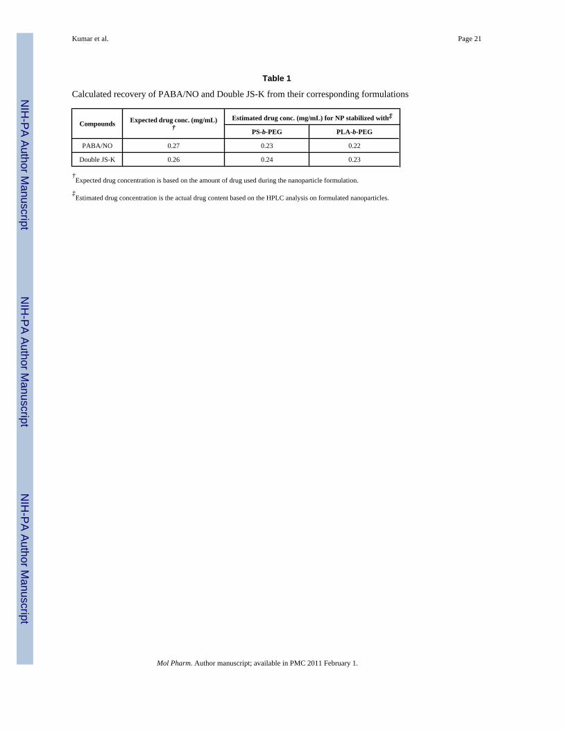
Next, these nanoparticles were reconstituted in phosphate buffered saline (PBS, pH 7.4). Basedon a calibration curve generated by HPLC analysis of PABA/NO standards, the amount ofPABA/NO in the formulation was calculated (Table 1). A similar procedure was carried outfor Double JS-K nanoparticles and the recovery of Double JS-K was found to be comparableto that of PABA/NO.
Reaction of Formulated and Unformulated Prodrug with GlutathioneThe reaction of PABA/NO with 4 mM glutathione was monitored via HPLC analysis. Underpseudo first-order conditions, a time-dependent disappearance of PABA/NO was observed anda rate constant of 4.6 × 10⁻² s−1 was obtained; the calculated half-life was 15 min (Figure 6).
Under identical reaction conditions, Double JS-K was found to disappear with a half-life of4.5 min and a pseudo first order rate constant of 0.154 s−1 (Figure 7).
Kumar et al. Page 5
Mol Pharm. Author manuscript; available in PMC 2011 February 1.
NIH
-PA Author Manuscript
NIH
-PA Author Manuscript
NIH
-PA Author Manuscript
Next, the nanoparticle formulations of PABA/NO were tested. In the case of the PS-b-PEG-stabilized PABA/NO formulation, roughly 50% of the PABA/NO remained after ~5 h whilefor PLA-b-PEG-stabilized PABA/NO formulation, roughly 50% remained after ~3 h (Figure8).
Similarly, the formulation was found to protect Double JS-K. The PS-b-PEG and PLA-b-PEGformulations were similar in behavior: roughly 50% Double JS-K disappeared in 40 min(Figure 9).
Anti-proliferative activity of PABA/NOThe anti-proliferative effects of PABA/NO were determined in two cell lines, human leukemiaU937 and human non-small cell lung cancer H1703 cells. Nearly identical IC50 values forformulated and unformulated PABA/NO were obtained (Table 2). A comparison of inhibitoryactivity of PABA/NO against U937 cells can be found in Figure 10.
DISCUSSIONThe encapsulation and NO release of JS-K, Double JS-K, and PABA/NO were consistent withtheir respective calculated hydrophobicities. The calculated solid phase partition coefficients(i.e. logP) of the compounds are: JS-K, logP = 1.99; Double JS-K, logP = 2.12; and PABA/NO, logP = 3.30.42 Attempts at formulating JS-K into nanoparticle form were unsuccessfuldue to its relatively higher solubility in aqueous solution. However, the precipitation intonanoparticle form of the more hydrophobic PABA/NO and Double JS-K were successful withboth PS-b-PEG and PLA-b-PEG block copolymers. There is little difference in sizes of thenanoparticles produced by either block copolymer, and only a small difference in the releaserates of drug from either polymer. The dependence of drug release on the interaction betweendrug and polymer has been previously studied.43 A slightly better protection of PABA/NO byPS block (solubility parameter, δPS = 18.6 MPa1/2 )40 as compared to PLA block (solubilityparameter, δPLA = 23.3 MPa1/2)42 could be due to the stronger interaction between the morehydrophobic PS block and PABA/NO as compared to less hydrophobic PLA. However it mightalso be related to the higher glass transition temperature of PS (Tg = 95 °C)40 providing higherrigidity on the nanoparticle surface as compared to PLA (Tg = 60–65 °C).44 But thebiodegradability of the PLA block relative to the PS block is probably the most importantcriterion for selection of the polymer for eventual therapeutic applications. The stability of theDouble JS-K nanoparticles was significantly better than that of JS-K because the Ostwaldripening rate depends both on the solubility of the compound40 (Double JSK being morehydrophobic) and also upon the diffusion coefficient.39 Double JS-K, being a larger moleculeis expected to have a diffusion coefficient approximately 40% smaller than JS-K. Therefore,Double JS-K nanoparticles were sufficiently stable that they could be freeze-dried andadministered. The increased rate of glutathione-decomposition of Double JS-K relative toPABA/NO is related to the higher solubility of the Double JS-K which favors dissolution.
CONCLUSIONSNitric oxide prodrugs with anti-proliferative activity can be stabilized as nanoparticles withsignificant protective effects from activation by glutathione. The protection for times of 5–6hours is believed to be optimal for delivery to solid cancer tumors by the mechanism ofenhanced permeation and retention (EPR).45–47 By this mechanism particles in the size rangeof 100–300 nm can pass through the defects in the rapidly growing vasculature in tumors anddeposit in the tissue. This requires effective passivation of the nanoparticle to prevent prematureremoval by the reticuloendothelial system (RES). We have demonstrated that PEG-protectedparticles produced by Flash NanoPrecipitation using the block copolymers in this study havecirculation times in mice of over 24 hours.31 The ability to co-precipitate drugs in the same
Kumar et al. Page 6
Mol Pharm. Author manuscript; available in PMC 2011 February 1.
NIH
-PA Author Manuscript
NIH
-PA Author Manuscript
NIH
-PA Author Manuscript

nanoparticle makes it possible to formulate nanoparticle “drug cocktails” where multiple drugscan be released from the same nanoparticle in a dose-controlled and time-controlled manner.Our demonstration of tuned release of paclitaxel from these nanoparticles31 makes thecombination of it with these NO-releasing compounds an ideal candidate for a dual drugdelivery study. The paclitaxel release in our previous study could be tuned to release drug overtimes from 20 min to 24 h depending on the method of conjugation of the drug. This time scalecould be matched to the 5–6 h release of PABA/NO.
AcknowledgmentsThis research was supported in part by the Intramural Research Program of the NIH, National Cancer Institute, Centerfor Cancer Research, as well as by National Cancer Institute contract HHSN261200800001E and the National ScienceFoundation under the NIRT grant for Nanoparticle Formation Technologies.
References1. Furchgott RF. Endothelium-Derived Relaxing Factor: Discovery, Early Studies, and Identifcation as
Nitric Oxide (Nobel Lecture). Angew. Chem. Int. Ed 1999;38:1870–1880.2. Ignarro LJ. Nitric Oxide: A Unique Endogenous Signaling Molecule in Vascular Biology (Nobel
Lecture). Angew. Chem. Int. Ed 1999;38:1882–1892.3. Murad F. Discovery of Some of the Biological Effects of Nitric Oxide and Its Role in Cell Signaling
(Nobel Lecture). Angew. Chem. Int. Ed 1999;38:1856–1868.4. Keefer LK. Nitric Oxide (NO)- and Nitroxyl (HNO)-generating Diazeniumdiolates (NONOates):
Emerging Commercial Opportunities. Curr. Top Med. Chem 2005;5:625–636. [PubMed: 16101424]5. Maciag A, Saavedra J, Chakrapani H. The Nitric Oxide Prodrug JS-K and Its Structural Analogues as
Cancer Therapeutic Agents. Anticancer Agents Med. Chem 2009;9:798–803. [PubMed: 19538173]6. Kitagaki J, Yang Y, Saavedra JE, Colburn N, Keefer LK, Perantoni AO. Nitric Oxide Prodrug JS-K
Inhibits Ubiquitin E1 and Kills Tumor Cells Retaining Wild-type p53. Oncogene 2009;28:619–624.[PubMed: 18978812]
7. Simeone A-M, McMurtry V, Nieves-Alicea R, Saavedra J, Keefer L, Johnson M, Tari A. TIMP-2Mediates the Anti-invasive Effects of the Nitric Oxide-releasing Prodrug JS-K in Breast Cancer Cells.Breast Cancer Res 2008;10:R44. [PubMed: 18474097]
8. Chakrapani H, Kalathur RC, Maciag AE, Citro ML, Ji X, Keefer LK, Saavedra JE. Synthesis,Mechanistic Studies, and Anti-proliferative Activity of Glutathione/glutathione S-transferase-activated Nitric Oxide Prodrugs. Bioorg. Med. Chem 2008;16:9764–9771. [PubMed: 18930407]
9. Chakrapani H, Goodblatt MM, Udupi V, Malaviya S, Shami PJ, Keefer LK, Saavedra JE. Synthesisand in vitro Anti-leukemic Activity of Structural Analogues of JS-K, an Anti-cancer Lead Compound.Bioorg. Med. Chem. Lett 2008;18:950–953. [PubMed: 18178089]
10. Kiziltepe T, Hideshima T, Ishitsuka K, Ocio EM, Raje N, Catley L, Li C-Q, Trudel LJ, Yasui H,Vallet S, Kutok JL, Chauhan D, Mitsiades CS, Saavedra JE, Wogan GN, Keefer LK, Shami PJ,Anderson KC. JS-K, a GST-activated Nitric Oxide Generator, Induces DNA Double-strand Breaks,Activates DNA Damage Response Pathways, and Induces Apoptosis in vitro and in vivo in HumanMultiple Myeloma Cells. Blood 2007;110:709–718. [PubMed: 17384201]
11. Udupi V, Yu M, Malaviya S, Saavedra JE, Shami PJ. JS-K, A Nitric Oxide Prodrug, InducesCytochrome c Release and Caspase Activation in HL-60 Myeloid Leukemia Cells. Leuk. Res2006;30:1279–1283. [PubMed: 16439016]
12. Shami PJ, Saavedra JE, Bonifant CL, Chu J, Udupi V, Malaviya S, Carr BI, Kar S, Wang M, Jia L,Ji X, Keefer LK. Antitumor Activity of JS-K [O2-(2,4-Dinitrophenyl) 1-[(4-Ethoxycarbonyl)piperazin-1-yl]diazen-1-ium-1,2-diolate] and Related O2-Aryl Diazeniumdiolates in Vitro and inVivo. J. Med. Chem 2006;49:4356–4366. [PubMed: 16821795]
13. Liu J, Li C, Qu W, Leslie E, Bonifant CL, Buzard GS, Saavedra JE, Keefer LK, Waalkes MP. NitricOxide Prodrugs and Metallochemotherapeutics: JS-K and CB-3-100 Enhance Arsenic and CisplatinCytolethality by Increasing Cellular Accumulation. Mol. Cancer Ther 2004;3:709–714. [PubMed:15210857]
Kumar et al. Page 7
Mol Pharm. Author manuscript; available in PMC 2011 February 1.
NIH
-PA Author Manuscript
NIH
-PA Author Manuscript
NIH
-PA Author Manuscript
14. Shami PJ, Saavedra JE, Wang LY, Bonifant CL, Diwan BA, Singh SV, Gu Y, Fox SD, Buzard GS,Citro ML, Waterhouse DJ, Davies KM, Ji X, Keefer LK. JS-K, a Glutathione/Glutathione S-Transferase-activated Nitric Oxide Donor of the Diazeniumdiolate Class with Potent AntineoplasticActivity1. Mol. Cancer Ther 2003;2:409–417. [PubMed: 12700285]
15. Ren Z, Kar S, Wang Z, Wang M, Saavedra JE, Carr BI. JS-K, A Novel Non-ionic DiazeniumdiolateDerivative, Inhibits Hep 3B Hepatoma Cell Growth and Induces c-Jun Phosphorylation via MultipleMAP Kinase Pathways. J. Cell. Physiol 2003;197:426–434. [PubMed: 14566972]
16. Townsend DM, Manevich Y, He L, Hutchens S, Pazoles CJ, Tew KD. Novel Role for GlutathioneS-Transferase {pi}: Regulator of Protein S-Glutathionylation Following Oxidative And NitrosativeStress. J. Biol. Chem 2009;284:436–445. [PubMed: 18990698]
17. Andrei D, Maciag A, Chakrapani H, Citro ML, Keefer LK, Saavedra JE. Aryl Bis(diazeniumdiolates):Potent Inducers of S-Glutathionylation of Cellular Proteins and Their in Vitro AntiproliferativeActivities. J. Med. Chem 2008;51:7944–7952. [PubMed: 19053760]
18. Townsend DM, Findlay VJ, Fazilev F, Ogle M, Fraser J, Saavedra JE, Ji X, Keefer LK, Tew KD. AGlutathione S-Transferase {pi}-Activated Prodrug Causes Kinase Activation Concurrent with S-Glutathionylation of Proteins. Mol. Pharmacol 2006;69:501–508. [PubMed: 16288082]
19. Saavedra JE, Srinivasan A, Buzard GS, Davies KM, Waterhouse DJ, Inami K, Wilde TC, Citro ML,Cuellar M, Deschamps JR, Parrish D, Shami PJ, Findlay VJ, Townsend DM, Tew KD, Singh S, JiaL, Ji X, Keefer LK. PABA/NO as an Anticancer Lead: Analogue Synthesis, Structure Revision,Solution Chemistry, Reactivity toward Glutathione, and in Vitro Activity. J. Med. Chem2006;49:1157–1164. [PubMed: 16451080]
20. Findlay VJ, Townsend DM, Morris TE, Fraser JP, He L, Tew KD. A Novel Role for HumanSulfiredoxin in the Reversal of Glutathionylation. Cancer Res 2006;66:6800–6806. [PubMed:16818657]
21. Findlay VJ, Townsend DM, Saavedra JE, Buzard GS, Citro ML, Keefer LK, Ji X, Tew KD. TumorCell Responses to a Novel Glutathione S-Transferase-Activated Nitric Oxide-Releasing Prodrug.Mol. Pharmacol 2004;65:1070–1079. [PubMed: 15102935]
22. Chakrapani H, Wilde TC, Citro ML, Goodblatt MM, Keefer LK, Saavedra JE. Synthesis, Nitric OxideRelease, and Anti-leukemic Activity of Glutathione-activated Nitric Oxide Prodrugs: StructuralAnalogues of PABA/NO, an Anti-cancer Lead Compound. Bioorg. Med. Chem 2008;16:2657–2664.[PubMed: 18060792]
23. Allen C, Maysinger D, Eisenberg A. Nano-engineering Block Copolymer Aggregates for DrugDelivery. Colloids Surf., B 1999;16:3–27.
24. Kumar V, Prud'homme RK. Thermodynamic Limits on Drug Loading in Nanoparticle Cores. J.Pharm. Sci 2008;97:4904–4914. [PubMed: 18300278]
25. Gref R, Minamitake Y, Peracchia M, Trubetskoy V, Torchilin V, Langer R. Biodegradable Long-circulating Polymeric Nanospheres. Science 1994;263:1600–1603. [PubMed: 8128245]
26. Schoenmakers RG, Wetering P. v. d. Elbert DL, Hubbell JA. The Effect of the Linker on theHydrolysis Rate of Drug-linked Ester Bonds. J. Controlled Release 2004;95:291–300.
27. Patri AK, Kukowska-Latallo JF, Baker JR. Targeted Drug Delivery with Dendrimers: Comparisonof the Release Kinetics of Covalently Conjugated Drug and Non-covalent Drug Inclusion Complex.Adv. Drug Deliv. Rev 2005;57:2203–2214. [PubMed: 16290254]
28. Kurtoglu YE, Navath RS, Wang B, Kannan S, Romero R, Kannan RM. Poly(amidoamine) Dendrimer-drug Conjugates with Disulfide Linkages for Intracellular Drug Delivery. Biomaterials2009;30:2112–2121. [PubMed: 19171376]
29. Perumal O, Khandare J, Kolhe P, Kannan S, Lieh-Lai M, Kannan RM. Effects of BranchingArchitecture and Linker on the Activity of Hyperbranched Polymer-Drug Conjugates. BioconjugateChem 2009;20:842–846.
30. Sengupta S, Eavarone D, Capila I, Zhao G, Watson N, Kiziltepe T, Sasisekharan R. TemporalTargeting of Tumour Cells and Neovasculature with a Nanoscale Delivery System. Nature2005;436:568–572. [PubMed: 16049491]
31. Ansell SM, Johnstone SA, Tardi PG, Lo L, Xie S, Shu Y, Harasym TO, Harasym NL, Williams L,Bermudes D, Liboiron BD, Saad W, Prud'homme RK, Mayer LD. Modulating the Therapeutic
Kumar et al. Page 8
Mol Pharm. Author manuscript; available in PMC 2011 February 1.
NIH
-PA Author Manuscript
NIH
-PA Author Manuscript
NIH
-PA Author Manuscript
Activity of Nanoparticle Delivered Paclitaxel by Manipulating the Hydrophobicity of ProdrugConjugates. J. Med. Chem 2008;51:3288–3296. [PubMed: 18465845]
32. Saavedra JE, Srinivasan A, Bonifant CL, Chu J, Shanklin AP, Flippen-Anderson JL, Rice WG, TurpinJA, Davies KM, Keefer LK. The secondary amine/nitric oxide complex ion R2N[N(O)NO]− asnucleophile and leaving group in SNAr reactions. J. Org. Chem 2001;66:3090–3098. [PubMed:11325274]
33. Hillmyer MA, Bates FS. Synthesis and Characterization of Model Polyalkane-Poly(ethylene oxide)Block Copolymers. Macromolecules 1996;29:6994–7002.
34. Gindy ME, Ji S, Hoye TR, Panagiotopoulos AZ, Prud'homme RK. Preparation of Poly(ethyleneglycol) Protected Nanoparticles with Variable Bioconjugate Ligand Density. Biomacromolecules2008;9:2705–2711. [PubMed: 18759476]
35. Gindy ME, Panagiotopoulos AZ, Prud'homme RK. Composite Block Copolymer StabilizedNanoparticles: Simultaneous Encapsulation of Organic Actives and Inorganic Nanostructures.Langmuir 2008;24:83–90. [PubMed: 18044945]
36. Johnson BK, Prud'homme RK. Chemical Processing and Micromixing in Confined Impinging Jets.AIChE J 2003;49:2264–2282.
37. Liu Y, Cheng C, Liu Y, Prud'homme RK, Fox RO. Mixing in a Multi-inlet Vortex Mixer (MIVM)for Flash Nano-precipitation. Chem. Eng. Sci 2008;63:2829–2842.
38. Akbulut M, Ginart P, Gindy ME, Theriault C, Chin KH, Soboyejo W, Prud'homme RK. GenericMethod of Preparing Multifunctional Fluorescent Nanoparticles Using Flash NanoPrecipitation.Adv. Funct. Mater 2009;19:718–725.
39. Liu Y, Kathan K, Saad W, Prud'homme RK. Ostwald Ripening of Beta-Carotene Nanoparticles. Phys.Rev. Lett 2007;98:036102. [PubMed: 17358697]
40. Kumar V, Wang L, Riebe M, Tung H, Prud'homme RK. Formulation and Stability of Itraconazoleand Odanacatib Nanoparticles: Governing Physical Parameters. Mol. Pharmaceutics 2009;6:1118–1124.
41. Chen T, D'Addio SM, Kennedy MT, Swietlow A, Kevrekidis IG, Panagiotopoulos AZ, Prud'hommeRK. Protected Peptide Nanoparticles: Experiments and Brownian Dynamics Simulations of theEnergetics of Assembly. Nano Lett 2009;9:2218–2222. [PubMed: 19413305]
42. Molinspiration Cheminformatics. Bratislava, Slovak Republic:http://www.molinspiration.com/services/properties.html. Accessed on June 20, 2009
43. Liu J, Xiao Y, Allen C. Polymer-drug Compatibility: A Guide to the Development of DeliverySystems for the Anticancer Agent, Ellipticine. J. Pharm. Sci 2004;93:132–143. [PubMed: 14648643]
44. Daniels AU, Chang MKO, Andriano KP, Heller J. Mechanical Properties of Biodegradable Polymersand Composites Proposed for Internal Fixation of Bone. J. Appl. Biomater 1990;1:57–78. [PubMed:10148987]
45. Matsumura Y, Maeda H. A New Concept for Macromolecular Therapeutics in Cancer Chemotherapy:Mechanism of Tumoritropic Accumulation of Proteins and the Antitumor Agent Smancs. CancerRes 1986;46:6387–6392. [PubMed: 2946403]
46. Maeda H. SMANCS and polymer-conjugated macromolecular drugs: advantages in cancerchemotherapy. Adv. Drug Deliv. Rev 1991;6:181–202.
47. Maeda H, Sawa T, Konno T. Mechanism of tumor-targeted delivery of macromolecular drugs,including the EPR effect in solid tumor and clinical overview of the prototype polymeric drugSMANCS. J. Controlled Release 2001;74:47–61.
Kumar et al. Page 9
Mol Pharm. Author manuscript; available in PMC 2011 February 1.
NIH
-PA Author Manuscript
NIH
-PA Author Manuscript
NIH
-PA Author Manuscript

Figure 1.Chemical structures of the diazeniumdiolate nitric oxide prodrugs used in this study.
Kumar et al. Page 10
Mol Pharm. Author manuscript; available in PMC 2011 February 1.
NIH
-PA Author Manuscript
NIH
-PA Author Manuscript
NIH
-PA Author Manuscript

Scheme 1.Glutathione-activated nitric oxide release from NO prodrug
Kumar et al. Page 11
Mol Pharm. Author manuscript; available in PMC 2011 February 1.
NIH
-PA Author Manuscript
NIH
-PA Author Manuscript
NIH
-PA Author Manuscript
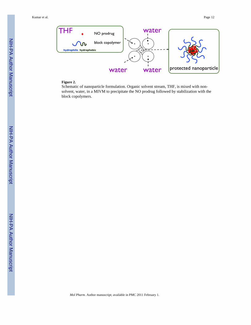
Figure 2.Schematic of nanoparticle formulation. Organic solvent stream, THF, is mixed with non-solvent, water, in a MIVM to precipitate the NO prodrug followed by stabilization with theblock copolymers.
Kumar et al. Page 12
Mol Pharm. Author manuscript; available in PMC 2011 February 1.
NIH
-PA Author Manuscript
NIH
-PA Author Manuscript
NIH
-PA Author Manuscript

Figure 3.Particle size distributions of undialyzed PABA/NO nanoparticles stabilized by PS-b-PEG (240nm) (▼) and PLA-b-PEG (225 nm) (■). The slow ripening of undialyzed nanoparticlesstabilized by PS-b-PEG shows growth to 440 nm in 20 h (▽).
Kumar et al. Page 13
Mol Pharm. Author manuscript; available in PMC 2011 February 1.
NIH
-PA Author Manuscript
NIH
-PA Author Manuscript
NIH
-PA Author Manuscript

Figure 4.Particle size distributions of undialyzed Double JS-K nanoparticles stabilized by PS-b-PEG(200 nm) (▼) and PLA-b-PEG (195 nm) (■).The growth of the PS-b-PEG stabilized particles(to 290 nm) over 1 h is shown (▽).
Kumar et al. Page 14
Mol Pharm. Author manuscript; available in PMC 2011 February 1.
NIH
-PA Author Manuscript
NIH
-PA Author Manuscript
NIH
-PA Author Manuscript
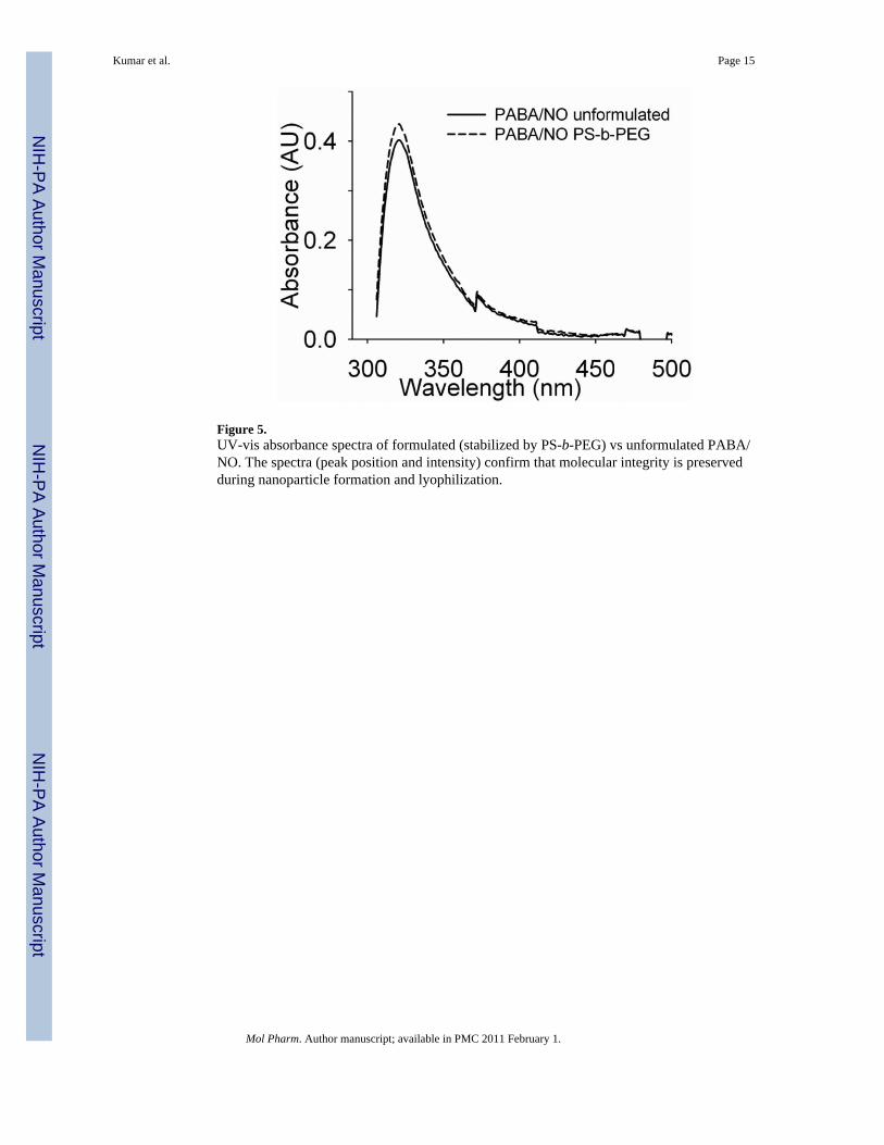
Figure 5.UV-vis absorbance spectra of formulated (stabilized by PS-b-PEG) vs unformulated PABA/NO. The spectra (peak position and intensity) confirm that molecular integrity is preservedduring nanoparticle formation and lyophilization.
Kumar et al. Page 15
Mol Pharm. Author manuscript; available in PMC 2011 February 1.
NIH
-PA Author Manuscript
NIH
-PA Author Manuscript
NIH
-PA Author Manuscript

Figure 6.Decomposition of PABA/NO in 4 mM glutathione solution in pH 7.4 phosphate buffer at 25°C.
Kumar et al. Page 16
Mol Pharm. Author manuscript; available in PMC 2011 February 1.
NIH
-PA Author Manuscript
NIH
-PA Author Manuscript
NIH
-PA Author Manuscript

Figure 7.Decomposition of Double JS-K in 4 mM glutathione solution in pH 7.4 phosphate buffer at 25°C.
Kumar et al. Page 17
Mol Pharm. Author manuscript; available in PMC 2011 February 1.
NIH
-PA Author Manuscript
NIH
-PA Author Manuscript
NIH
-PA Author Manuscript

Figure 8.Decomposition of formulated PABA/NO in 4 mM glutathione solution in pH 7.4 phosphatebuffer at 25 °C.
Kumar et al. Page 18
Mol Pharm. Author manuscript; available in PMC 2011 February 1.
NIH
-PA Author Manuscript
NIH
-PA Author Manuscript
NIH
-PA Author Manuscript
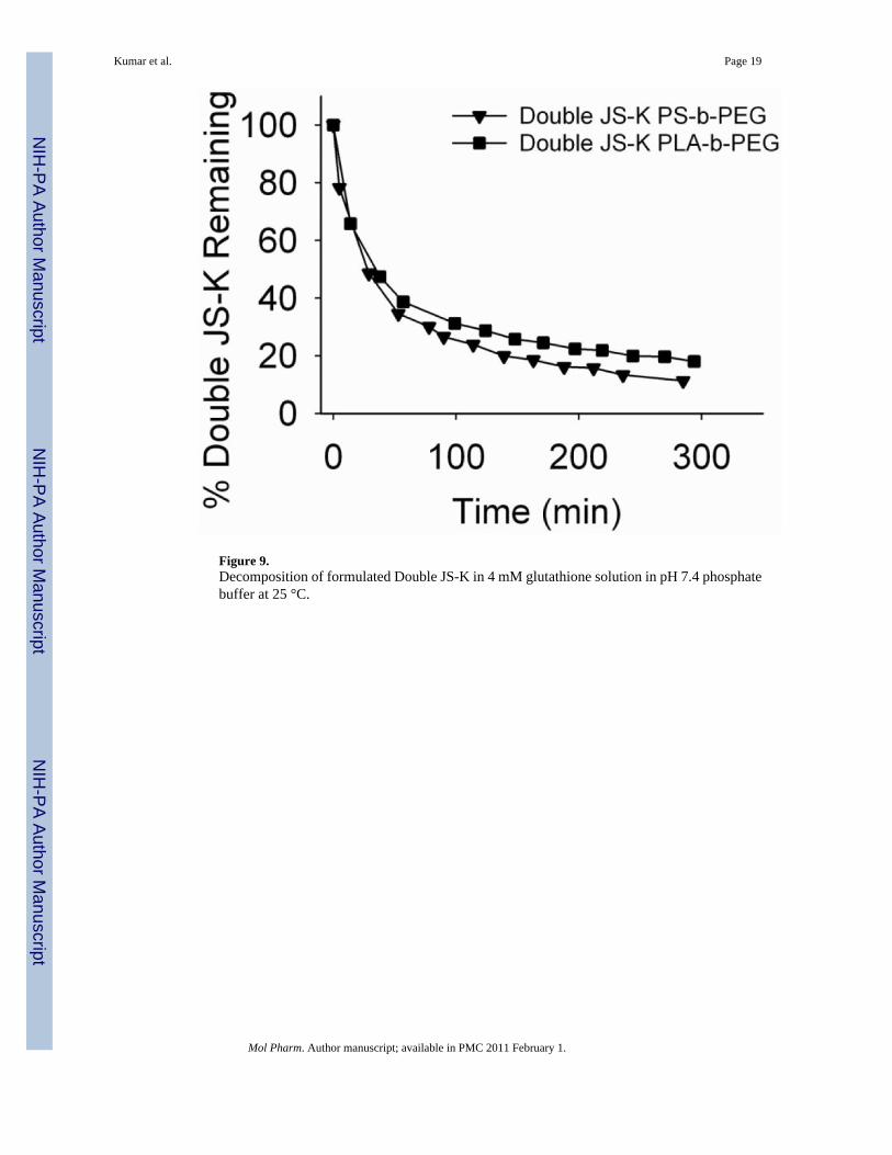
Figure 9.Decomposition of formulated Double JS-K in 4 mM glutathione solution in pH 7.4 phosphatebuffer at 25 °C.
Kumar et al. Page 19
Mol Pharm. Author manuscript; available in PMC 2011 February 1.
NIH
-PA Author Manuscript
NIH
-PA Author Manuscript
NIH
-PA Author Manuscript

Figure 10.Anti-proliferative activity of PABA/NO, PS-b-PEG-stabilized PABA/NO and PLA-b-PEG-stabilized PABA/NO.
Kumar et al. Page 20
Mol Pharm. Author manuscript; available in PMC 2011 February 1.
NIH
-PA Author Manuscript
NIH
-PA Author Manuscript
NIH
-PA Author Manuscript
NIH
-PA Author Manuscript
NIH
-PA Author Manuscript
NIH
-PA Author Manuscript
Kumar et al. Page 21
Table 1
Calculated recovery of PABA/NO and Double JS-K from their corresponding formulations
Compounds Expected drug conc. (mg/mL)†
Estimated drug conc. (mg/mL) for NP stabilized with‡
PS-b-PEG PLA-b-PEG
PABA/NO 0.27 0.23 0.22
Double JS-K 0.26 0.24 0.23
†Expected drug concentration is based on the amount of drug used during the nanoparticle formulation.
‡Estimated drug concentration is the actual drug content based on the HPLC analysis on formulated nanoparticles.
Mol Pharm. Author manuscript; available in PMC 2011 February 1.

NIH
-PA Author Manuscript
NIH
-PA Author Manuscript
NIH
-PA Author Manuscript
Kumar et al. Page 22
Table 2
Anti-proliferative activities (IC50) of PABA/NO, PS-b-PEG stabilized PABA/NO & PLA-b-PEG stabilizedPABA/NO
Cell Lines PABA/NO (μM) PS-b-PEG-stabilized PABA/NO (μM) PLA-b-PEG-stabilized PABA/NO (μM)
U937 6.5 5.0 4.4
H1703 8.7 6.3 5.3
Mol Pharm. Author manuscript; available in PMC 2011 February 1.