Function of the coxsackie and adenovirus receptor (CAR) in ...

ANNALS OF SURGERYVol. 222, No. 1, 78-86© 1995 Lippincott-Raven Publishers
Treatment of Experimental HumanMesothelioma Using AdenovirusTransfer of the Herpes SimplexThymidine Kinase GeneW. Roy Smythe, M.D.,* Harry C. Hwang, B.S.,t Ashraf A. Elshami, M.D.,t Kunjlata M. Amin,Ph.D.,* Stephen L. Eck, M.D., Ph.D.,t II Beverly L. Davidson, Ph.D.,¶ James M. Wilson, M.D.,Ph.D.,§II1 Larry R. Kaiser, M.D.,* II and Steven M. Albelda, M.D.t II
From the Thoracic Surgery Section, Department of Surgery,* Pulmonary/Critical Care Section,Department of Medicine,t University of Pennsylvania Thoracic Oncology Research Laboratory,Hematology-Oncology Section, the Department of Medicine, t the Department of Molecular andCellular Engineering,§ and the Institute for Human Gene Therapy, 11 University of PennsylvaniaMedical Center, Philadelphia, Pennsylvania; and the Rheumatology Section, Department ofMedicine,¶ University of Michigan Medical Center, Ann Arbor, Michigan
ObjectiveThe authors demonstrate the ability of an adenovirus vector expressing the herpes simplexthymidine kinase (HSVtk) gene to treat human malignant mesothelioma growing within theperitoneal cavity of severe combined immunodeficient (SCID) mice.
Background DataIntroduction of the HSVtk gene into tumor cells renders them sensitive to the antiviral drugganciclovir (GCV). This approach has been used previously to treat experimental brain tumors.Although malignant mesothelioma is refractory to current therapies, its localized nature and theaccessibility of the pleural space make it a potential target for a similar type of in vivo gene therapyusing adenovirus.
MethodsAn adenovirus containing the HSVtk gene (Ad.RSVtk) was used to transduce mesothelioma cellsin vitro. These cells were then injected into the flanks of SCID mice. Ad.RSVtk was also injecteddirectly into the peritoneal cavity of SCID mice with established human mesothelioma tumors.Mice were subsequently treated for 7 days with GCV at a dose of 5 mg/kg.
ResultsMesothelioma cells transduced in vitro with Ad.RSVtk formed nodules when injected in thesubcutaneous tissue. These tumors could be eliminated by the administration of GCV, even whenas few as 10% of cells were transduced to express HSVtk (bystander effect). Administration ofAd.RSVtk into the peritoneal space of animals with established multifocal human mesotheliomafollowed by GCV therapy resulted in the eradication of macroscopic tumor in 90% of animals andmicroscopic tumor in 80% of animals when evaluated after 30 days. The median survival ofanimals treated with Ad.RSVtk/GCV was significantly longer than that of control animals treatedwith similar protocols.
78

Gene Therapy for Mesothelioma 79
ConclusionThese results indicate that an adenoviral vector containing the HSVtk gene is effective in treatingestablished malignant mesothelioma in an in vivo setting and raise the possibility of usingadenovirus transfer of HSVtk for clinical trials in mesothelioma and other localized tumors.
Malignant mesothelioma is a neoplasm of the meso-thelial lining of the pleural or peritoneal cavity linkedto prior exposure to asbestos. Although relatively rare,mesothelioma accounts for approximately 4000 deathsper year in the United States. Morbidity and mortality inmesothelioma primarily are the result of aggressive localspread rather than from distant metastases, as is charac-teristic of most malignancies. Unfortunately, mesotheli-oma is extremely resistant to aggressive multimodalitytherapies, with reported median survivals of only 18 to24 months after diagnosis." 2 Although unresponsive toconventional treatments, this combination of extremelypoor prognosis, lack of a clinically important metastaticcomponent, and the fact that the tumor grows in the ac-cessible potential space of the thoracic cavity make me-sothelioma an attractive candidate for local somatic genetherapy.
Culver et al.3 have reported success in treating local-ized brain tumors by injection of fibroblasts producingretroviruses engineered to contain the herpes simplexthymidine kinase (HSVtk)' gene. These studies and oth-ers have demonstrated that introduction of the HSVtkgene into mammalian cells renders them highly sensitiveto subsequent treatment with the relatively nontoxic nu-cleoside analog ganciclovir (GCV).4-9 One key feature ofthis system is a "bystander effect," in which tumorgrowth may be slowed, or tumor tissue eliminated, afterGCV administration, even when only 10% to 15% ofcells are transfected with HSVtk. This effect is thought tobe caused by intercellular transfer of toxic GCV metab-olites.3~To date, most of the reported studies using transfer of
HSVtk have used recombinant retrovirus.3'6-9 Althoughpotentially effective, certain features of retrovirus-medi-ated gene transfer are not optimal for in vivo gene ther-apy. These include difficulties in producing and purify-ing high-titer viral stock, the fact that only actively divid-ing cells allow retroviral integration and transgeneexpression, and the possibility of insertion mutagene-
Partially supported by a development grant from the University ofPennsylvania Cancer Center.
Mr. Hwang received a grant from the American Heart Association.Dr. Smythe is the recipient of an American Cancer Society Clinical
Oncology Fellowship Award.Address reprint requests to Steven Albelda, M.D., The Thoracic On-
cology Research Laboratory, Hospital of the University of Penn-sylvania, 809 Maloney Building, 3600 Spruce Street, Philadelphia,PA 19104.
Accepted for publication December 8, 1994.
sis.'° In addition, because recombinant retroviral super-natant may not reliably infect tissues, in vivo, "packagingcells" are required,9 confounding interpretation of sub-sequent effects and raising potential safety issues.For these reasons, we have explored the use of recom-
binant adenovirus to deliver the HSVtk gene to treat ma-lignant mesothelioma. "12 In contrast to retroviruses, ad-enoviruses are very efficient in directly transducing tis-sues in vivo, can easily be produced in high titer, do notrequire cell division for transduction of cells, and havean established record of safety as vaccination material.'3Although the duration of gene expression is relativelyshort with regard to treatment ofgenetic disorders,'4 thislimitation may be less important when the goal of ther-apy is rapid eradication oftumor cells.We have recently completed a series of studies that
demonstrate that the delivery of HSVtk via adenovirusfollowed by the administration of GCV may be aneffective therapeutic approach. These studies haveshown that 1) a replication-deficient recombinant ade-novirus carrying the Escherichia coli lacZ marker genedriven by a heterologous promoter effectively infectedmesothelioma cells in vitro"; 2) introduction of recom-binant adenovirus directly into the peritoneal cavity ofsevere combined immunodeficiency (SCID) mice withestablished human mesothelioma tumor nodules re-sulted in extensive gene transfer at the tumor surfacesand deeper within the tumor nodules' '-these findingsare similar to those recently reported by Brody et al.'5; 3)recombinant adenovirus Ad.RSVtk successfully trans-ferred the HSVtk gene into human mesothelioma cellsin vitro and resulted in expression ofenzyme as assessedby immunohistochemical staining'2 4); infection withAd.RSVtk rendered human mesothelioma cells sensitiveto doses of GCV that were 2 to 4 log doses lower thanthat required to kill cells infected with control virus invitro 2; and 5) infection of mesothelioma cells in vitrowith Ad.RSVtk was accompanied by a strong "bystandereffect" with no diminution in the efficacy ofGCV treat-ment until the ratio of infected:uninfected cells was lessthan 1:10.12
Based on these preliminary studies, experiments wereconducted to determine if a recombinant adenoviruscarrying the HSVtk gene could be used to treat tumorsin animal models of human mesothelioma. The resultsdemonstrate that an adenoviral vector containing theHSVtk gene is effective in treating an established, local-ized, malignancy in an in vivo setting and raise the possi-bility ofusing adenovirus transfer ofHSVtk for the treat-
Vol. 222 - No. 1
80 Smythe and Others
ment of human tumors, such as malignant mesotheli-oma, that exert morbidity by growing within localizedbody cavities.
METHODS
Adenoviral Vectors
The production of recombinant replication-deficientadenoviral vectors has been described in detail else-where.'2",6"7 Briefly, the vectors were constructed froman adenovirus type 5 (Ad5) mutant that lacks most ofviral sequence regions E la and E lb and a portion of E3.By homologous recombination techniques, either the E.coli lacZ marker gene or the herpes simplex type I thy-midine kinase gene driven by the Rous sarcoma virus 3'promoter region were inserted into the viral genome.The resultant vectors were termed Ad.RSVlacZ andAd.RSVtk., respectively. Viral stocks were propagated in293 cells, and titers were quantified by 293 plaque assayor spectrophotometric density.
Cell Lines and Animal Models
Two cell lines derived from human mesothelioma, 1-45 and REN were used in this study. The characteriza-tion ofthese cells is described elsewhere.12Two animal models of mesothelioma were developed
in SCID mice, as described previously.'2 All protocolswere approved by the animal use committees of theWistar Institute and the University of Pennsylvania incompliance with the Guidefor the Care and Use ofLab-oratory Animals (National Institutes of Health No. 85-23, revised 1985).
Briefly, to study the growth ofsubcutaneous tumors, 2X 10⁷ cells from the 1-45 human malignant mesotheli-oma cell line were injected subcutaneously into theflanks ofSCID mice. Tumor length and width were mea-sured using calipers, and tumor volumes were calculatedusing the formula: tumor volume = L X W2/2.To study a more clinically relevant model of mesothe-
lioma, 4 X 10⁷ cells from the REN cell line were sus-pended in 1 mL of RPMI medium and injected with a23-gauge needle into the peritoneal cavities of5-week oldSCID mice. A peritoneal model was chosen rather thana pleural model because it is difficult to introduce cellsinto the pleural cavity ofmice without causing death dueto iatrogenic pneumothorax. Within 5 days after cell in-stillation, 1- to 2-mm macroscopic tumor nodules wereobvious on peritoneal surfaces. By 30 days, large tumornodules were present on the lesser omentum, adherentto the pancreas, in the small bowel mesentery, in the areaof the porta hepatis, and in smaller scattered deposits onthe diaphragm and lateral abdominal wall. Animals usu-ally succumbed to bowel or portal obstruction within 5
to 6 weeks (median survival 35 days; Table 1). In contrastto other mesothelioma cell lines,5",8 the cells used inthese studies produced minimal amounts of ascites.
Experimental Protocols
Subcutaneous Nodule Experiments
To determine if the adenovirus HSVtk system wouldbe effective in animal models after systemicGCV admin-istration, confluent flasks of 1-45 human mesotheliomacells were treated with either Ad.RSVtk or Ad.RSVlacZat a ratio of 100 particles per cell (2 plaque-forming unitsper cell) for 6 hours. At this ratio, virtually all cells weretransduced.'2"'3 The cells were washed and allowed to in-cubate overnight. After 24 hours, the cells were removedwith trypsin/EDTA and injected into the flanks ofSCIDmice. For each animal, one side was injected withAd.RSVtk-infected cells, and the contralateral flank wasinjected with either uninfected cells or cells infected withAd.RSVlacZ. To study the bystander effect, some ani-mals were injected with Ad.RSVtk-infected cells mixedwith varying numbers of uninfected cells to achieve ra-tios of infected:uninfected cells of 1:1, 1:5, and 1:10. Sixdays after injection, when all tumors measured 4 to 6mm in diameter, the animals were treated with intraperi-toneal GCV (250 mg/kg/day) or saline daily for 7 days.Tumor growth was measured for the next 6 weeks.
Intraperitoneal Experiments
Four X 107 REN cells were injected into the peritonealcavities of 33 SCID mice. Five days later, when macro-scopic tumor nodules of 1 to 2 mm were present, 3 X10"l particles (6 X 10⁹ plaque forming units) of virus,diluted in 0.5 mL of sterile saline, were injected into theperitoneal cavity using a 23-gauge needle. Experimentalgroups were injected with a control vector (Ad.RSVlacZ;6 mice), the therapeutic gene construct (Ad.RSVtk; 19mice), and diluent (sham-infected; 6 mice). Selectedgroups of animals were treated 2 days later with intra-peritoneal GCV at a dose of 5 mg/day (250 mg/kg) orsaline for 7 days. One month after initial tumor inocula-tion, animals underwent necropsies, and the extent oftumor was quantified.To evaluate the effects of treatment on survival using
the same protocol, 4 x 10⁷ REN cells were injected againinto the peritoneal cavities of 33 SCID mice. Fivedays later, animals received either control vector(Ad.RSVlacZ; 7 mice), the therapeutic gene construct(Ad.RSVtk; 14 mice), diluent (sham-infected; 7 mice), orno treatment (5 mice). Selected groups of animals weretreated 2 days later with intraperitoneal GCV at a doseof 5 mg/day (250 mg/kg) or saline for 7 days. The ani-mals were carefully observed and underwent necropsy assoon as possible after death.
Ann. Surg. *July 1995

Gene Therapy for Mesothelioma 81
deposits of human tumor, other portions of tumor andall intra-abdominal organs were removed and snap-fro-zen in OCT-embedding media (Miles Inc. Elkhart, IN)at -70 C. Frozen sections were cut and stained with anantibody (W632-PelFreeze Biologicals, Rogers, AR)against the human lymphocyte antigen (HLA)- 1 shareddeterminant using the Vectastain Elite kit (Vector, Burl-ingame, CA).13To detect areas of lacZ expression, frozen tumor sec-
tions were cut, placed on glass microscope slides, andstained using the X-gal protocol described previously.'2Before mounting, the sections were counterstained withhematoxylin for 30 seconds. Sections then were evalu-ated for lacZ gene expression by light microscopy.
36
Days Post Tumor Cell Injection Statistics
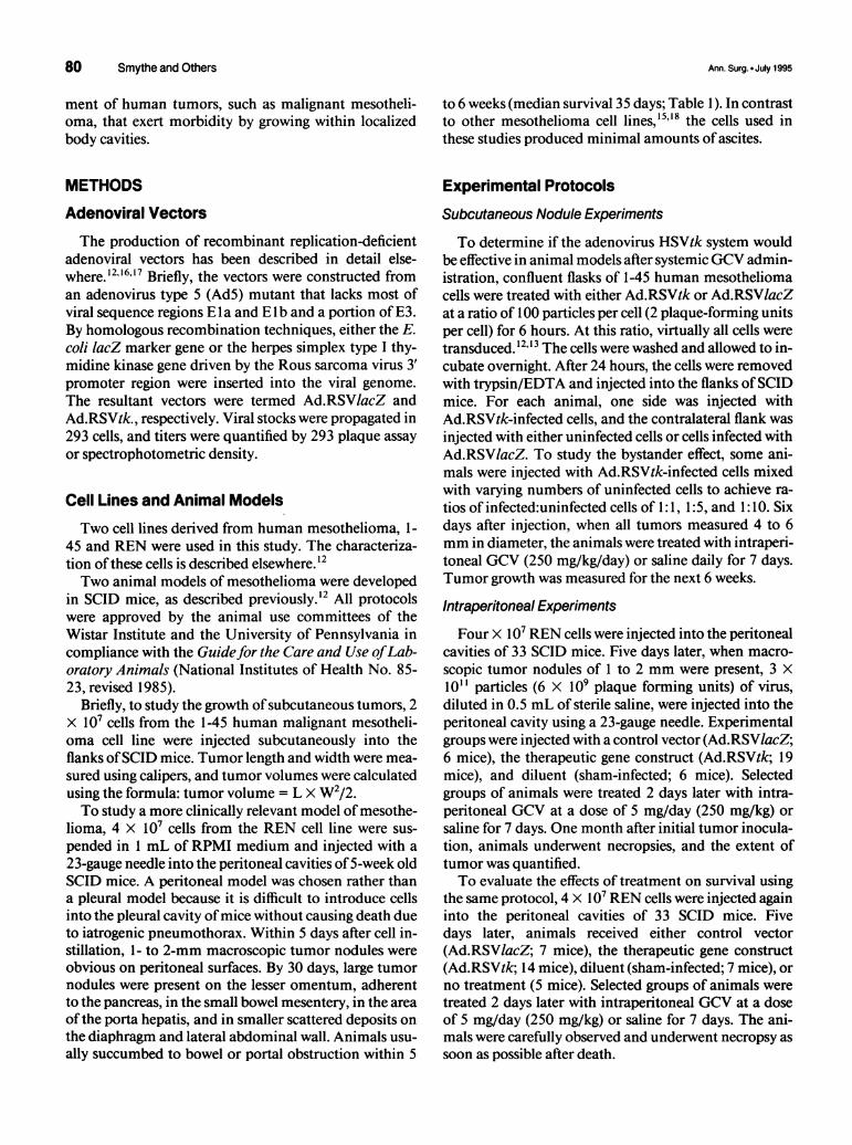
Tumor 450.
Volume 450
(mm3)300/
150
00 1 18 24 30 36
GCVB Days Post Tumor Cell Injection
Figure 1. Ganciclovir-induced regression of subcutaneous human ma-lignant mesothelioma tumors developing from cells transduced in vitrowith Ad.RSVtk. (A) Animals received cells infected with Ad.RSVtk in oneflank and Ad.RSV/acZ in the other. One group then received intraperito-neal GCV with the other receiving saline. Tumors arising from Ad.RSVtk-infected cells were eliminated in animals receiving GCV, whereas tumorsarising from Ad.RSV/acZ-infected cells continued to grow (Error bars =
standard error of the mean [SEM]). (B) Cells infected with Ad.RSVtk were
mixed with uninfected cells at ratios of 1:1, 1:5, and 1:10 and injectedsubcutaneously in one flank. All animals received uninfected cells in thecontralateral flank as a control. After GCV treatment, all tumors developingfrom mixtures of infected and uninfected cells exhibited complete regres-sion. Tumors arising from the uninfected cells continued to grow (Errorbars = SEM).
Histochemistry and Evaluation of LacZGene Expression
To detect microscopic tumor growth, portions of tu-mor and intra-abdominal organs were fixed in 10%buffered formalin, embedded in paraffin, and stainedwith hematoxylin and eosin. As a more sensitive test for
Survival data among all groups were compared usinganalysis of variance. Comparison of treatment survivaldata to the combined control group was performed byuse ofa log-rank test.
RESULTS
Systemic Administration of GanciclovirInduces Tumor Regression ofMesothelioma Cells Transduced withAd.RSVtk In Vitro
To test if the adenovirus HSVtk system would beeffective in animal models after systemic GCV admin-istration, we infected 1-45 human mesothelioma cells invitro with Ad.RSVtk or Ad.RSV/acZ and injected thesecells subcutaneously into the flanks of SCID mice. Sixdays later, when all tumors measured 4 to 6 mm in di-ameter, the animals were treated with intraperitonealGCV or saline.As shown in Figure 1A, a 7-day course ofGCV rapidly
eliminated all Ad.RSVtk-infected tumors, whereas tu-mors composed of cells.treated with Ad.RSVlacZ con-tinued to grow to large size. No inhibition ofgrowth wasseen when animals containing tumors composed of cellstransduced with Ad.RSVtk were treated with saline.There was no regrowth ofthe Ad.RSVtk-infected tumorsafter more than 6 weeks of observation. In addition,there was no evidence of injury to normal subcutaneoustissues. Microscopic examination ofthe tumor site in theAd.RSVtk/GCV animals after 6 weeks showed no evi-dence of microscopic disease.A similar degree oftumor regression was seen in these
models at a lower dose ofGCV (10 mg/kg/day), a dosethat approximates that used clinically to treat viral infec-tion'9 (data not shown).
TumorVolume(mm3)
A
Vol. 222 * No. 1

82 Smythe and Others
A Strong Bystander Effect Exists In Vivo
To determine if a bystander effect occurred in tumorsgrowing in vivo, cells were infected with Ad.RSVtk in vi-tro and mixed with various concentrations of nontrans-duced cells 24 hours after infection (under these condi-tions, no contamination of cells via residual adenoviruscould occur). The cell mixtures were injected into theflanks ofSCID mice and allowed to grow to measurablesize. The animals were then treated with intraperitonealGCV for 7 days. As shown in Figure 1 B, all tumors con-taining Ad.RSVtk-transduced cells demonstrated equiv-alent regression, even those derived from mixtures of in-fected:uninfected cells at ratios 1:1, 1:5, and 1:10. Theseexperiments show that a strong bystander effect is opera-tive in adenovirus-transduced solid tumors in vivo.
Ad.RSVtk/GCV Treatment InducesRegression of Established IntraperitonealMesothelioma Tumor at 30 Days
Based on these results, we designed experiments toevaluate in vivo transduction of pre-existing mesotheli-oma in a model that approximated human disease. Fourgroups of SCID mice were injected intraperitoneallywith 4 X 10⁷ REN tumor cells. Five days later, two ani-mals were killed to confirm the presence of tumor.Multiple small macroscopic (1-2 mm) tumor noduleswere present throughout the peritoneum and were espe-cially prominent in the mesentery (Fig. 2A). At this timepoint, animals were injected intraperitoneally with 3 X10" particles of Ad.RSVlacZ virus, Ad.RSVtk virus, orsaline (sham infection control). Two days later, groupsof animals were treated intraperitoneally with either sa-line or with GCV (Table 1).
Efficient gene transfer was confirmed by necropsy ofone of the animals injected with Ad.RSVlacZ 2 days af-ter viral injection. As previously observed with an ade-novirus vector containing the lacZ gene driven by a cy-tomegalovirus promoter,12 the Ad.RSVlacZ vector alsoreadily infected tumors and showed ,-galactosidase ac-tivity on the surface of tumor nodules, as well as muchdeeper within the tumor mass (Fig. 2B).Two weeks after the intraperitoneal GCV or saline
treatments were completed (i.e., 28 days after the initialtumor cell inoculations), the animals were killed andcarefully examined for the presence of gross and micro-scopic disease (Table 1). There was no grossly detectabledisease in nine of the ten mice in the Ad.RSVtk/GCVgroup (Figs. 3B, 3D, and 3F) The one exception was ananimal with a solitary 3-mm macroscopic tumor nodule.In contrast, all but one of the 20 animals in the othergroups had an extensive burden ofintraperitoneal tumor(Table 1 and Figs. 1 and 3). Scattered tumor nodules ofup to 1.5 cm in diameter were noted in these animals,
many of which were partially obstructing the bowel orbiliary tree (Figs. 3A, 3C, and 3E). In addition to macro-scopic disease (found primarily in the upper abdomen,pancreatic mesentery, region of the porta hepatis, anddiaphragm), all control animals exhibited microscopictumor uniformly throughout the upper abdomen (Figs.2D-2F).The intra-abdominal organs and tissues removed from
animals in the HSVtk/GCV group were examined fortumor microscopically by conventional H&E staining ofparaffin sections and by a more sensitive staining tech-nique using an antibody against the human HLA-class1 shared determinant. Examination of stained sections(Table 1 and Figs. 2D-2F) showed evidence oftumor inonly two of the ten mice. In one animal (which also hadthe macroscopic nodule), the disease was detected in onesmall area in the porta hepatis. In the other mouse, therewas one small nest of tumor cells (smaller than thoseshown in Fig. 2F) adherent to the surface of the smallintestine, detectable only by antihuman HLA immuno-staining.
Animals with Established IntraperitonealMesothelioma Exhibit Significant SurvivalBenefit When Treated with Ad.RSVtk/GCVTo evaluate longer-term benefit from HSVtk/GCV
treatment ofSCID mice with established intraperitonealhuman mesothelioma, and to assess tumor regrowth, asurvival experiment was performed. Five groups werestudied which included animals receiving no treatment,as well as those that received no virus (sham)/GCV,Ad.RSVtk/GCV, Ad.RSVtk/no GCV, and Ad.RSVlacZ/GCV. After treatment, survival of each group of micewas observed closely. Animals underwent careful nec-ropsy as soon as possible after death.The mean and median survival times for all animals
are reported in Table 2. Median survival of control ani-mals ranged from 35 to 51 days; however, Ad.RSVtk/GCV-treated animals exhibited a median survival of76.5 days. Because the survival data did not significantlydiffer among control groups, the data from all controlswere combined for further analysis. When the survivaldata of treatment animals were compared with controlanimals, a significant survival advantage was noted (Fig.4, p < 0.005, log rank). Necropsy ofanimals in all controlgroups revealed large intraperitoneal tumor burdensconsisting of 1) large pancreatic mesentery tumors, 2)multiple small bowel mesenteric nodules, and 3) exten-sive portal tumor. Death of these animals appears tohave been caused by small bowel or portal tumor massobstruction. Additionally, several of the control animalsexhibited diaphragmatic and retroperitoneal tumorgrowth. On autospy, no treatment animal appeared tohave died a direct tumor-related death. Necropsy of
Ann. Surg. *-July 1995

Gene Therapy for Mesothelioma 83
Figure 2. Microscopic evaluation of human mesotheli- Aoma tumor growing in SCID mice. (A) Appearance (H&Estaining) of tumornodules (TN) growing within the mes- -entery adjacent to the small intestine (SI) and pancreas 4ftN'(P) within the peritoneum 5 days after tumor cell injection T.~7'(200x). (B) Frozen sections of tumor in mesentery (7 days 4after tumor cell injection) stained with X-gal to identify the .-transduced p-galactosidase gene. Blue areas mark ex- SIpression of lacZ in tumor cells at the surface of the tumor 4Slt,i'(large arrows) and deep within the tumor (small arrows;1OOX). (C) Histologic appearance of normal mesothelium(NM) and mesenteric adipose (MA) tissue 30 days after / V *; *.mesothelioma cell injection and after treatment with U-Ad.RSVtk/GCV, demonstrating no microscopic tumor ormesothelial damage (200X). (D) Corresponding region ofmesentery of animal treated with Ad.RSV/acZ shows MAclear tumor involvement (200X). (E) Histologic appear-ance of pancreas in an animal 30 days after mesotheli- F - -oma cell injection after treatment with Ad.RSV./acZEshows extensive tumor infiltration (arrow). Corresponding ' - -sections from animals treated with Ad.RSVtk showed no _ i_evidence of tumor. (F) Appearance of a pancreatic me-
sothelioma micrometastasis (arrows) detected by immu- I,, .Inohistochemical staining of frozen sections (hematoxylincounterstain) with an antibody against the human HLA-1 _shared determinant (200X).
Figure 3. Effect of Ad.RSVtk/ganciclovir treatment onintraperitoneal growth of human mesothelioma in SCIDmice. (A) Photomicrograph of upper abdominal organs ofan animal treated with Ad.RSVIacZ/GCV. Tumor nodules(arrows) are noted in the upper abdomen near the stom-ach (S), pancreatic mesentery (P), porta hepatis (whitearrow), and in the mesentery (M) of the small bowel. (B)An animal treated with Ad.RSVtk/GCV shows completeabsence of tumor in these same regions. (C) Higherpower view of the hepatic portal region in an animal a'treated with Ad.RSV/acZ/GCV shows tumor (arrows) inthe porta hepatis (black arrow) with common bile ductobstruction (straight white arrow) and enlargement of thegallbladder (curved white arrow). (D) Similar view of an ;Aanimal treated with Ad.RSVtk/GCV illustrates lack of tu-mor. (E) Higher power view of the jejunal mesentery (MJ) Min an animal treated with Ad.RSV/acZ/GCV shows numer-ous tumor nodules (arrows). (F) A comparable view in ananimal treated with Ad.RSVtk/GCV with no visible tumor.
-bs .MJ i
Ad.RSVtk/GCV-treated animals was most remarkable the large or small intestine with these adhesions, but nofor extensive benign-appearing intra-abdominal adhe- obstructing tumor nodules were noted. Of the animalssions. All animals necropsied had obstruction of either that were necropsied to date, two had no evidence of
Vol. 222 . No. 1

84 Smythe and Others
Table 1. EFFECT OF GENE THERAPY ONTHE GROWTH OF ESTABLISHED HUMANMESOTHELIOMA IN THE PERITONEAL
CAVITY OF SCID MICE
No. of Animals No. of AnimalsTreatment with Gross with MicroscopicGroup Tumor Tumor
Ad.RSVtk/GCVAd.RSVtk/salineAd.RSV/acZIGCVSham/GCV
1/109/95/55/6
2/109/95/55/6
Ad.RSVtk = adenovirus containing the LacZ gene; GCV = ganciclovir; Sham = micetreated intraperitoneally with saline.
gross tumor, two each had only one 2 X 2-mm nodule inthe peripancreatic region, and two animals did not un-
dergo necropsies because of extensive tissue autolysis.Microscopic evaluation of all animals was not possiblebecause of tissue autolysis after death and preceding au-
topsy; however, microscopic examination of extra-ab-dominal organs (lung, heart, brain) ofone ofthe animalswithout gross disease showed neither evidence of treat-ment regimen toxicity nor metastatic disease. No adhe-sions were seen in animals treated with GCV alone.
DISCUSSIONCurrent approaches to gene therapy of malignancy in-
clude transfer of chemotherapy resistance genes to bonemarrow stem cells, targeting of dominant oncogenes or
tumor suppressor genes, transfer ofgenes coding for im-mune-enhancing substances, and the delivery oftoxic or
drug susceptibility genes into tumor cells.2023 With theexception of immunotherapy, one major obstacle to allthese approaches will be an inability to reliably introducegenetic material into most, or all, ofthe tumor cells grow-ing in vivo. For this reason, successful approaches to thetreatment of solid tumors will require the careful selec-tion oftumor type, vector delivery system, and therapeu-tic gene paradigm.We have shown that established tumor nodules ofma-
lignant mesothelioma growing within the peritoneal cav-
ity ofSCID mice can be largely eradicated by instillationof the Ad.RSVtk vector followed by systemic GCV ad-ministration. These results indicate that the adenovirusis capable ofeffective HSVtk gene delivery within a phys-iologically relevant milieu and are able to penetratedeeply within solid tumor after topical administration.Brody et al.'5 also have shown efficient delivery oftransgene via adenovirus into mesothelioma tumorgrowing within the peritoneal cavity ofimmunodeficientmice. We theorize that the adenoviral particles may have
gained access to deeper tumor areas via morphologicallyimperfect tumor capillaries. Newly formed microvesselsin tumor tissue often lack a complete pericyte invest-ment, have poorly-formed or absent basement mem-branes, and have larger-than-normal endothelial fenes-trations.24 Adenovirus could have entered these mi-crovessels at the tumor surface, then exited the vessels indeeper regions to infect tumor cells.
It is pertinent to address the issue ofpotential viral dis-semination in this experimental model. In previous stud-ies using transfer of the lacZ gene in this intraperitonealmesothelioma model, we did not find evidence of dis-seminated gene transfer because the normal mesothe-lium appeared to form a barrier." The only exception tothis observation was infrequent f3-galactosidase positive-staining hepatic cells. In this study, careful review of he-patic histology in HSVtk/GCV-treated animals at 30days did not reveal evidence of hepatocyte damage.These data are consistent with those of Setoguchi et al.,who, after intraperitoneal administration of recombi-nant adenovirus carrying the a I-antitrypsin gene, werenot able to detect transgene in other organs, even by sen-sitive reverse transcription-polymerase chain reactionmethods.25 Our preliminary studies using reverse tran-scription-polymerase chain reaction to detect the HSVtkgene in other organs have confirmed this observation.Although significant benign intraperitoneal adhesionsformed in the survival experiment, the normal mesothe-lium appeared intact at 30 days (Fig. 2C), suggesting thatthis tissue is not irreversibly damaged. The developmentofadhesions should not pose a significant problem in thetreatment ofintrathoracic malignancy, such as mesothe-lioma, but may impact on the potential use ofthis systemto treat intraperitoneal malignancy such as ovarian car-cinoma.
This study also demonstrates that not all tumor cellsneed to be infected with the Ad.RSVtk vector to effecttumor eradication, thus illustrating the power of the by-stander effect. Explanations for the bystander effect haveincluded continued viral infection in situ,3 transfer ofanintegrated retroviral HSVtk gene during mitosis, transferof the toxic purine analog GCV-triphosphate via gapjunctions or apoptotic vesicles,6,9 or primarily immune-mediated effects.8 Although the mechanisms of the by-stander effect were not specifically examined, our resultsclearly argue against retroviral-specific or T cell-medi-ated immune mechanisms. Of course, an immune com-ponent to the bystander effect via natural killer cells,macrophages, eosinophils, and neutrophils still is possi-ble because SCID mice are not deficient in these celltypes. Regardless of mechanism, however, the presenceof a strong bystander effect using adenovirus in vivo isparticularly important in the treatment of solid tumorsbecause the infection rate of a mass oftumor cells is notlikely to be high.
Ann. Surg. -July 1995

Gene Therapy for Mesothelioma 85
Table 2. SURVIVAL AFTER TREATMENTFOR ESTABLISHED HUMAN
MESOTHELIOMA
Treatment Mean Survival Median SurvivalGroup (Days ± S.E.M.) (Days)
No treatment (n = 5)Ad.RSVtk/saline (n = 7)Ad.RSV/acZ/GCV (n = 7)Sham/GCV (n = 7)Ad.RSVtk/GCV (n = 7)
35.8 ± 1.539.9 ± 2.748.6 ± 4.550.7 ± 14.390.8 ± 15.8
3537513976.5
Ad.RSVtk = adenovirus containing the HSVtk gene; Ad.RSV/acZ = adenovirus con-
taining the LacZ gene.
An interesting issue that will likely have an impact onthe clinical utility ofthe Ad.RSVtk/GCV approach is theimmune response generated against recombinant adeno-virus in immunocompetent organisms. Transduction ofcells with recombinant adenovirus results in the produc-tion ofboth neutralizing antibodies and cytotoxic T lym-phocytes14; however, it is difficult to predict how theseresponses will affect therapeutic efficacy. It is possiblethat the generation of cytotoxic T lymphocytes againstadenovirally transduced tumor cells actually may en-
hance tumor destruction.26 On the other hand, too rapida destruction oftransduced tumor cells will limit the ex-
pression of the HSVtk gene and diminish the resultantbystander effect. Comparison of Ad.RSVtk therapy inanimals in which the same rat mesothelioma cell line canbe grown in syngeneic immunocompetent Fischer ratsand in immunodeficient nu/nu rats currently are un-
derway and will help answer this question. The genera-tion of neutralizing antibodies against adenovirus alsomay provide a potential problem and limit the possibilityofrepeated administration ofvector. Clearly, a better un-derstanding ofthe immune response to recombinant ad-enovirus will be helpful in designing useful clinical ap-proaches (i.e., immunosuppresive therapy), and guidingdevelopment of new vector technology (i.e., less immu-nogenic adenoviruses, adenoviruses of different sero-
types, or alternative vectors such as adeno-associated vi-rus).
In addition to issues of immune response, there are
a number of additional questions raised by this study.Selective destruction of tumor versus normal tissues isone such issue. Because adenoviruses have the ability toinfect nondividing cells, they are not dependent on thetumor cells being in active cell cycle during the period ofinfection. This is important because only a minority ofcells within a tumor mass may be dividing at any one
point in time. Selective destruction oftumor cells appar-ently is achieved by the fact that GCV preferentially de-stroys cells undergoing division.27 The recent report of
Chen et al., showing that direct intratumoral injection ofan adenovirus vector carrying an HSVtk gene in gliomascaused tumor regression, supports the potential use ofthis system in the treatment of cancer.28 Another poten-tial limitation of this study is that the size of the intra-peritoneal tumors was relatively small, although clearlymacroscopically visible. Although this animal modeldoes not simulate the massive tumor burden ofa patientwith advanced mesothelioma, it suggests the possibilityofusing gene therapy as adjuvant to a surgical debulkingprocedure. We currently are evaluating the possibility ofsingle and multiple administrations of Ad.RSVtk/GCVfor the treatment ofmuch bulkier disease. Currently, theoptimal dose of virus for use in in vivo delivery also isunclear. For reasons not yet well understood, adenoviraltransduction of cells in vitro usually is much more effi-cient than when the same virus is administered in vivo.29In this report, we used a dose of virus in the mouse thatwould be the equivalent of2 X 1012 plaque-forming unitsin a 70 kg human subject. Administration ofthis dose ofvirus is feasible in a clinical setting, but would be largerthan those used to date in trials for the treatment ofcysticfibrosis. Preliminary dose response experiments in theSCID mouse model have shown significant antitumoreffects at doses ofvirus 100-fold lower than those used inthese studies. Finally, although we saw no gross evidenceof local or systemic damage from this treatment, morecareful analysis of the toxicity of this therapy is war-ranted.
This study demonstrates that recombinant adenovirustransfer of HSVtk, followed by systemic administrationofGCV, can effectively treat established intracavitary tu-mor in vivo. In conjunction with the only other pub-lished report describing recombinant adenovirus-medi-ated in vivo tumor treatment,28 these results raise the pos-sibility of using this system for the treatment of human
100- Ad.RSVtkl GCV0Combined Controls
80-
Animals 60-
Surviving(Percent) 40-
20
0.0 20 40 60 80 100 120 140 160 180
Time (Days)
Figure 4. Survival benefit after treatment of intraperitoneal mesotheliomawith Ad.RSVtk/GCV. Survival curves comparing SCID mice treated withAd.RSVtk/GCV (heavy line) to all control groups combined (thin line). Asignificant treatment advantage was noted in Ad.RSVtk/GCV-treated ani-mals (p < 0.005, log rank).
Vol. 222 - No. 1

86 Smythe and Others
neoplasms that exert morbidity by growing within de-fined anatomic spaces. In addition to malignant meso-thelioma, other examples include carcinoma of theovary and bladder. Palliative treatment ofmetastatic dis-ease, such as leptomeningeal carcinomatosis, malignantascites, and pleural effusion arising from a variety ofma-lignancies, are also potential therapeutic goals.
Author's NoteTwo other studies recently have been published.30'3'
Acknowledgments
The authors thank Mildred Daise and June Chilkotowsky for theirtechnical assistance.
References
1. Rusch VW, Piantadosi S, Holmes EC. The role of extrapleuralpneumonectomy in malignant mesothelioma. J Thorac Cardio-vasc Surg 1991; 102:1-9.
2. Sugarbaker DJ, Heher EC, Lee TH, et al. Extrapleuralpneumonec-tomy, chemotherapy and radiotherapy in the treatment of diffusemalignant mesothelioma. J Thorac Cardiovasc Surg 1991; 102: 10-15.
3. Culver KW, Ram Z, Walbridge S, et al. In vivo gene transfer withretroviral vector-producer cells for treatment ofexperimental braintumors. Science 1992; 256:1550-1552.
4. Moolten FL, Wells JM. Curability of tumors bearing herpes thy-midine kinase genes transferred by retroviral vectors. J Natl CancerInst 1990; 82:297-300.
5. Moolten FL, Wells JM, Mroz PJ. Multiple transduction as a meansof preserving ganciclovir chemosensitivity in sarcoma cells carry-ing retrovirally transduced herpes thymidine kinase genes. CancerLett 1992; 64:257-263.
6. Freeman SM, Abboud CN, Whartenby KA, et al. The "bystandereffect": tumor regression when a fraction of the tumor mass is ge-netically modified. Cancer Research 1993; 53:5274-5283.
7. Hasegawa Y, Emi N, Shimokata K, et al. Gene transfer of herpessimplex virus type I thymidine kinase gene as a drug sensitivitygene into human lung cancer lines using retroviral vectors. Am JResp Cell Mol Bio 1993; 8:655-661.
8. Caruso M, Panis Y, Gagandeep S, et al. Regression of establishedmacroscopic liver metastases after in situ transduction of a suicidegene. Proc Natl Acad Sci U S A 1993; 90:7024-7028.
9. Ram Z, Culver KW, Walbridge S, et al. In situ retroviral-mediatedgene transfer for the treatment of brain tumors in rats. Cancer Res1993; 53:83-88.
10. Mulligan RC. The basic science ofgene therapy. Science 1993; 26:926-932.
11. Smythe WR, Kaiser LR, Amin KM, et al. Successful adenovirus-mediated gene transfer in an in vivo model of human malignantmesothelioma. Ann Thor Surg 1994; 57:1395-1401.
12. Smythe WR, Hwang HC, Amin KM, et al. Recombinant adenovi-rus transfer of the HSV-thymidine kinase gene to thoracic neo-
Ann. Surg. * July 1995
plasms: an effective in vitro drug sensitization system. Cancer Res1994; 54:2055-2059.
13. Kozarsky KF, Wilson JM. Gene therapy: adenovirus vectors. CurrOpin Genet Dev 1993; 3:499-503.
14. Yang Y, Nunes F, Berenci K, et al. Cellular immunity to viral an-tigens limits El adenovirus for gene therapy. Proc Natl Acad Sci US A 1994; 91:4407-4411.
15. Brody SL, Jaffe HA, Han SK, et al. Direct in vivo gene transfer andexpression in malignant cells using adenovirus vectors. Hum GeneTher 1994; 5:437-447.
16. Berkner KL. Development of adenovirus vectors for the expres-sion ofheterologous genes. Biotechniques 1998; 6:616-629.
17. Englehardt JF, Yang Y, Stratford-Perricaudet LD, et al. Directgene transfer of human CFTR into human bronchial epithelia ofxenografts with El-deleted adenoviruses. Nature Genet 1993; 4:27-34.
18. Reale FR, Griffin TW, Compton JM, et al. Characterization of ahuman malignant mesothelioma cell line (H-MESO- 1): a biphasicsolid and ascitic tumor model. Cancer Res 1987; 47:3199-3205.
19. Paul S, Dummer S. Topics in clinical pharmacology: ganciclovir.Am J Med Sci 1992; 304:272-277.
20. Golumbek PT, Lazenby AJ, Levitsky HI, et al. Treatment ofestab-lished renal cancer by tumor cells engineered to secrete interleukin-4. Science 1991; 254:713-716.
21. Mullen CA, Kilstrup M, Blaese RM. Transfer ofthe bacterial genefor cytosine deaminase to mammalian cells confers lethat suscep-tibility to 5-fluorocytosine: a negative selection system. Proc NatlAcad Sci U S A 1992; 89:33-37.
22. Georges RN, Mukhopadhyay T, Zhang Y, et al. Prevention of or-thotopic human lung cancer growth by intratracheal instillation ofa retroviral antisense K-ras construct. Cancer Res 1993; 53:1743-1746.
23. Sorrentino BP, Brandt SJ, Bodine D, et al. Selection of drug-resis-tant bone marrow cells in vivo after retroviral transfer of humanMDR 1. Science 1992; 257:99- 101.
24. Weiss L, Orr WF, Honn KV. Interactions of cancer cells with themicrovasculature during metastasis. FASEB J 1988; 2:12-21.
25. Setoguchi Y, Jaffe HA, Chu C-S, et al. Intraperitoneal in vivo genetherapy to deliver a I -antitrypson to the systemic circulation. Am JRespir Cell Mol Bio 1994; 10:369-377.
26. Barba D, Hardin J, Sadelain M, Gage F. Development of anti-tu-mor immunity following thymidine kinase-mediated killing of ex-perimental tumors. Proc Natl Acad Sci U S A 1994; 91:4348-4352.
27. St. Clair MH, Lambe CU, Furman PA. Inhibition by ganciclovirof cell growth and DNA synthesis of cells biochemicallytransformed with herpesvirus genetic information. AntimicrobAgents Chemother 1987; 31:844-849.
28. Chen S-H, Shine HD, Goodman JC, et al. Gene therapy for braintumors: regression of experimental gliomas by adenovirus-medi-ated gene transfer in vivo. Proc NatI Acad Sci U S A 1994; 91:3054-3057.
29. Pilewski JM, Engelhardt JF, Bavaria JE, et al. Distribution andefficiency of adenovirus-mediated gene transfer to human bron-chus using xenografts. Am J Physiol (268): Lung Cell Mol Physiol1995; 12:L657-L665.
30. O'Malley BW, Chen S, Schwartz M, Woo SLC. Adenovirus-medi-ated gene therapy for human head and neck squamous cell cancerin a nude mouse model. Ca Res 1995; 55:1080-1085.
31. Bonnekoh B, Greenhalgh DA, Bundman DS, et al. Inhibition ofmelanoma growth by adenoviral-mediated HSV thymidine kinasegene transfer in vivo. J Invest Dermatol 1995; 104:313-317.
Copyright © 2022 FDOKUMEN