Epithelioid Pleural Mesothelioma Is Characterized by Tertiary ...
Upload
independentCategory
view
0download
0
2005;11:2026-2037. Published online March 8, 2005.Clin Cancer Res Alfredo Cesario, Alessia Catassi, Luigi Festi, et al.
StudyMesothelioma: A First-Step Comparative Translational Farnesyltransferase Inhibitors and Human Malignant Pleural
Updated Version 10.1158/1078-0432.CCR-04-1450doi:
Access the most recent version of this article at:
E-mail alerts related to this article or journal.Sign up to receive free email-alerts
SubscriptionsReprints and
[email protected] Department atTo order reprints of this article or to subscribe to the journal, contact the AACR
To request permission to re-use all or part of this article, contact the AACR Publications
American Association for Cancer Research Copyright © 2005 on November 19, 2012clincancerres.aacrjournals.orgDownloaded from
DOI:10.1158/1078-0432.CCR-04-1450
Farnesyltransferase Inhibitors and Human Malignant Pleural
Mesothelioma: A First-Step Comparative Translational Study
Alfredo Cesario,1,5 Alessia Catassi,2 Luigi Festi,4
Andrea Imperatori,4 Andrea Pericelli,4
Domenico Galetta,1 Stefano Margaritora,1
Venanzio Porziella,1 Vittorio Cardaci,5
Pierluigi Granone,1 Lorenzo Dominioni,4 and
Patrizia Russo3
1Department of Surgical Science, Division of General Thoracic Surgery,Catholic University, Rome, Italy; 2Department of Biology, University ofGenoa; 3Laboratory of Translational Research B (Lung Cancer),Department of Integrated Medical Oncology, National Cancer Institute,Genoa, Italy; 4Center of Thoracic Surgery, University of Insubria, Varese,Italy; and 5Pulmonary Rehabilitation, IRCCS San Raffaele, Rome, Italy
ABSTRACT
It is known that the potential clinical use of farnesyl-
transferase inhibitors (FTI) could be expanded to include
cancers harboring activated receptor tyrosine kinases.
Approximately 70% of malignant pleural mesotheliomas
(MPM) overexpress epidermal growth factor receptors
(EGFR) and a subset express both EGFR and transforming
growth factor A (TGF-A), suggesting an autocrine role for
EGFR in MPM. We checked on MPM cells (10 human cell
lines, 11 primary cultures obtained by human biopsies, and
7 short-term normal mesothelial cell cultures) concerning the
following: (a) the relative overexpression of EGFR (Western
blotting, flow cytometry, immunohistochemistry), (b) the
relative expression of EGFR ligands (EGF, amphiregulin,
TGF-A, ELISA), (c) the relative increase of the activated
form of Ras (Ras-bound GTP) after EGF stimulation (Ras
activation assay), (d) the efficacy of five different FTIs (HDJ2
prenylation, cell cytotoxicity, and apoptosis using ApopTag
and gel ladder). EGFR was overexpressed in MPM cells
compared with normal pleural mesothelial cells in equiva-
lent levels as in non––small cell lung cancer cells A549.
MPM cells constitutively expressed EGFR ligands; however,
Ras activation was attenuated at high EGF concentrations
(100 ng/mL). Growth of MPM cells was substantially not
affected by treatment with different FTIs (SCH66336, BMS-
214662, R115777, RPR-115135, and Manumycin). Among
these, BMS-214662 was the only one moderately active.
BMS-214662 triggered apoptosis in a small fraction of cells
(not higher than 30%) that was paralleled by a slight
decrease in the levels of TGF-A secreted by treated MPM
cells. Our data highlighted the concept that the same
signaling pathway can be regulated in different ways and
these regulations can differ between different cells of
different origin.
INTRODUCTION
The incidence of malignant pleural mesothelioma (MPM), a
primary tumor of the pleural, peritoneal, and pericardial cavities,
with an estimated incidence of 2,000 to 3,000 cases annually in
the United States, is relatively high in Italy: about 1,000 patients
are observed every year. The total number of male deaths from
pleural mesothelioma in France, Germany, and Italy has
continued to increase during the last decade of the 20th century,
growing from f7,550 in 1985-1989 to 8,750 in 1990-1994, and
9,550 in 1995-1999 (1). However, for the latest period, all the
observed values were lower than predicted (2). This, together
with the stabilization of age-standardized mortality rates and the
consistent decrease in mortality at younger ages, supports the
hypothesis that the rising trend in deaths from mesothelioma in
men could level off in the near future (3).
A unique feature of MPM is its strong relationship with
asbestos fiber exposure, which has recently led to great public
concern in view of the ubiquitous presence of that mineral.
Exposure can be occupational, environmental, or even familial
by household contamination. The median interval between onset
of symptoms and death is usually 6 to 8 months (4) and long-
term survivors are rare. MPM usually progresses locally, causing
death by cardiac/respiratory failure in most cases.
To date there is no standard effective treatment for MPM
(5, 6). Complex therapeutic protocols, including the combination
of surgery, chemotherapy, and radiation therapy, as pioneered by
Sugarbaker et al. (7) in 1991 in highly selected groups of
patients represent the best-documented potentially curative
approach, with 5-year survival not exceeding 15%. Such dismal
prognosis status despite the aggressive therapeutic effort
represents the rationale to foster investigation (6).
Very recently, some interesting positive results have been
obtained by introducing new chemotherapeutic agents such as
pemetrexed + cisplatin supplemented with folate and B12 (8),
but the profound sense of nihilism surrounding treatment of this
disease remains and the state of the art still seems unsatisfactory.
Moreover, knowledge of the biology of this cancer is still
somewhat confused. Further efforts in the evaluation of the
basics and of the response of this tumor to the newly proposed
therapies are needed.
Specialized information is necessary for the planning of
studies with a translational attitude, based both on clinical failure
experiences and on advances in basic molecular oncology.
To date, we know that f70% of MPMs have high levels of
expression of the epidermal growth factor receptor (EGFR), and
Received 7/22/04; revised 11/30/04; accepted 12/16/04.The costs of publication of this article were defrayed in part by thepayment of page charges. This article must therefore be hereby markedadvertisement in accordance with 18 U.S.C. Section 1734 solely toindicate this fact.Note: D. Galetta is currently in the Division of Thoracic Surgery,European Institute of Oncology, Via Ripamonti, 43520141 Milan, Italy.Requests for reprints: Patrizia Russo, Laboratory of TranslationalResearch B (Lung Cancer), Department of Integrated MedicalOncology, National Cancer Institute, Largo Rosanna Benzi 10,I-16132 Genoa, Italy. Phone: 39-0105600212; Fax: 39-0105600217;E-mail: [email protected].
D2005 American Association for Cancer Research.
Vol. 11, 2026–2037, March 1, 2005 Clinical Cancer Research2026
American Association for Cancer Research Copyright © 2005 on November 19, 2012clincancerres.aacrjournals.orgDownloaded from
DOI:10.1158/1078-0432.CCR-04-1450
a subset of cell lines derived from MPM patients express both
EGFR and transforming growth factor a (TGF-a) suggesting an
autocrine role for EGFR in MPM (9–11).
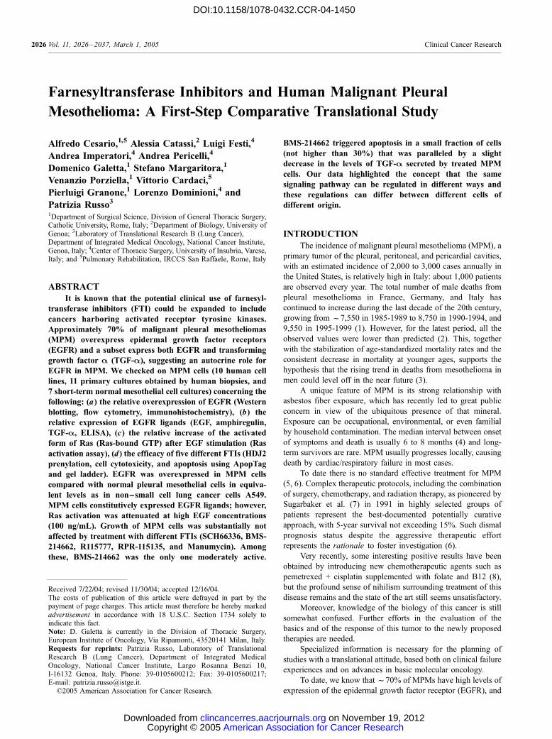
Ras-mitogen-activated protein kinase pathway is an impor-
tant signaling pathway activated by EGF as shown in Fig. 1 (12–
17). Studies have shown that farnesylation of Ras is the obligatory
first step in a series of posttranslational modifications leading to
membrane association which, in turn, determines the switch from
an inactive to an active Ras-GTP bound form (18–22). Based on
the theoretical assumption that preventing Ras farnesylationmight
result in the inhibition of Ras functions, a range of farnesyl-
transferase inhibitors (FTI) have been synthesized (21, 22).
Farnesylated proteins such as Ras, RhoB, the centromere-
binding proteins CENP-E and CENP-F, the phosphatases PRL1,
2 and 3, the chaperone protein DnaJ, and Rheb are involved in
cell growth crucial pathways and may represent an ideal target for
novel molecular targeted therapies with a wide anticancer
spectrum (21, 22). Preclinical experiments have confirmed that
FTIs are able to effectively inhibit farnesylation of several targets,
leading to powerful anticancer effect in a variety of cell lines and
xenograft animal models (21, 22). Moreover, it is well known that
FTIs block the growth of a variety of tumor cells without Ras
activation (21, 22). It is possible that this occurs because activated
receptor tyrosine kinases constitutively activate Ras that is then
inhibited by FTIs; alternatively, FTIs may inhibit TGF-a and
amphiregulin expression, as shown by Sizemore et al. (23).
In support of this hypothesis, Noorgaard et al. (24, 25) reported
that the FTI L744,382 decreases levels of TGF-a in mammary
cystic fluid and inhibits the growth of mammary tumors in
MMTV-TGF-a transgenic mice. Furthermore, we have shown
that the FTI RPR-115135 inhibits the growth of MCF10A cells
transfected with erb-B2 gene (26).
In a translational attitude, Trombino et al. (27) and others
(28–30) have focused attention on MPM. We studied 10 human
MPM cell lines, 11 MPM cell lines in primary cultures obtained
by human biopsies, and 7 short-term normal mesothelial cell
cultures to investigate the following:
1. The relative overexpression of EGFR using different
methodologies: Western blotting, flow cytometry, and
immunohistochemistry;
2. The relative expression of EGFR ligands (TGF-a and
amphiregulin) using ELISA;
3. The relative increase of the activated form of Ras (Ras-bound
GTP) after EGF stimulation (Ras activation assay);
4. The relative extracellular signal-regulated kinase (ERK)
activation after EGF stimulation (Western blotting);
5. The efficacy of five different FTIs using cell cytotoxicity
assays, HDJ2 mobility shift, and apoptosis detection
(ApopTag, gel ladder). The efficacy of other potential Ras-
targeting agents such as methotrexate, which interferes with
carboxymethyl transferase [3-(4,5-dimethylthiazol-2-yl)-5-(3-
carboxymethoxyphenyl)-2- (4-sulfophenyl)-2H- tetrazolium,
inner salt (MTS) assay and cloning in agar].
The results of this analysis are described herein.
MATERIALS AND METHODS
Cell Lines and Primary Cell Cultures. Human meso-
thelioma cancer cell line MSTO-211H, non–small cell lung
cancer (NSCLC) A549 cell line, and human colon cancer
HCT116 cell line were obtained from American Type Culture
Collection (Rockville, MD). Human mesothelioma cancer cell
lines H28, H513, H2052, and H290 were courtesy of Dr J.D.
Minna (Hamon Center for Therapeutic Oncology Research,
University of Texas Southwestern Medical Center, Dallas, Texas).
Human mesothelioma cancer cell lines IST-MES-1, MPP-89,
ZL55, and ZL34 were courtesy of Dr. S. Ferrini (National Institute
for Research on Cancer, Genoa, Italy). They were grown in RPMI
1640 (Life Technologies, Inc., Grand Island, NY) supplemented
with 10% non–heat inactivated fetal bovine serum (FBS, Life
Technologies). Cell counts were determined using a Coulter
Counter with Channelyzer attachment to monitor cell size
(Coulter Electronics, Hialeah, FL). Cell membrane integrity was
determined by trypan blue dye exclusion assay.
Fig. 1 EGFR and Ras cascade. The adaptor protein growth factorreceptor binding protein 2 (Grb2) plays a critical role in coupling signalfrom EGFR kinase with Ras. Grb2 contains a single SH2 domain flankedby two Src homology 3 (SH3) domains. The SH2 domain of Grb2recognizes and binds to specific phosphotyrosine-containing motifs onEGFR, whereas the SH3 domains bind to the guanine nucleotide-releasingfactor son of sevenless (SOS), which catalyzes the exchange of GDP toGTP on Ras, resulting in Ras activation (12–16). Interestingly, it has beenreported that Grb2 directly binds to the cytoplasmic domain of EGFR atphosphorylated residues Tyr1068 and Tyr1086 to initiate the activation of theRas-mitogen-activated protein kinase pathway. Ras, in turn, activates thedownstream kinases sequentially, which eventually leads to the activationof mitogen-activated protein kinases/ERKs. ERKs phosphorylate tran-scription factors that regulate gene transcription (12–16).
Clinical Cancer Research 2027
American Association for Cancer Research Copyright © 2005 on November 19, 2012clincancerres.aacrjournals.orgDownloaded from
DOI:10.1158/1078-0432.CCR-04-1450

Tumor tissue samples from mesothelioma patients were
taken from the operating room at room temperature immediately
after resection. Primary cells were obtained as described
previously (27). Briefly, the specimens were dissected with
scalpels into <5-mm cubes. The pieces of tumor were placed in
triple enzyme medium (1� collagenase, 1� hyaluronidase, and
1� DNase; Sigma, St. Louis, MO) in HBSS (Life Technologies)
with a magnetic bar and were then spun on a stir plate at room
temperature for 2 to 3 hours until most of the solid tumor was
dissociated. The cells were filtered through a 70-Am nylon cell
strainer (Becton Dickinson, Lincoln Park, NJ) and suspended in
RPMI 1640 with 10% FBS (Life Technologies).
Normal pleural tissues samples were obtained from resected
specimens of patients who underwent surgery for nonneoplastic
reasons. Short-term mesothelial cell cultures were established as
described previously (27) after triple enzyme medium (Sigma)
disaggregation. Mesothelial cells were grown as primary cultures
on fibronectin-coated culture flasks in RPMI 1640 supplemented
with 20% heat-inactivated FBS (Life Technologies), epidermal
growth factor (20 ng/mL), hydrocortisone (1 Amol/L) and insulin
(10 Ag/mL), transferrin (5 Ag/mL), and gentamicin (50 Ag/mL,
Life Technologies) at 37jC in a humidified 5% CO2 atmosphere.
Fresh complete medium was replaced every 2 to 3 days until
cells were confluent. Upon confluence the cells were lifted by
1� trypsin-EDTA (Life Technologies) and subcultured at 1:2
dilution. The cells were identified as mesothelial cells by
immunocytochemical staining with antikeratin antibodies (Dako,
Glostrup, Denmark). Third- to fourth-passage confluent cultures
were used for binding assay and cell growth.
EGFR determination was evaluated by immunostaining,
Western blot, and flow cytometry. Immunostaining against
EGFR was done as follows: fixed cells were rinsed in cold
PBS containing 0.5 mol/L glycine, blocked with PBS containing
1% bovine serum albumin and 5% FBS. The incubation was
carried out overnight at 4jC in the primary antibody EGFR
(SC-03; Santa Cruz Biotechnology, Santa Cruz, CA) diluted
1:200 in PBS-0.1% bovine serum albumin-1% FBS. After
incubation, samples were rinsed with PBS and stained with the
appropriate secondary anti-IgG, diluted 1:100 (Santa Cruz
Biotechnology) in PBS containing 0.1% bovine serum albumin
and 1% FBS. Specificity controls were done by use of normal
serum as primary antibody or by omitting the incubation in
the primary antibody. Peroxidase controls were done by
preincubating cells in PBS containing 0.1% hydrogen peroxide
before the incubation in the secondary antibody.
For Western blot analysis, equal amounts of protein were
subjected to 12.5% SDS-PAGE and then transferred electropho-
retically to a nitrocellulose membrane. Nonspecific binding sites
were blocked with blocking buffer containing TBS and 0.1%
Tween 20 with 5% nonfat milk powder for 2 hours at room
temperature, and the blot was incubated with specific antibody in
blocking buffer. The EGFR (SC-03) or the h-actin antibody
(AC-15 (Sigma) was incubated at 4jC overnight. After washing,
the blot was incubated with an appropriate secondary antibody
(Santa Cruz Biotechnology) for 1 hour at room temperature. After
extensive washing, detection was done using the enhanced
chemiluminescence system with exposure to Hyperfilm MP.
For flow cytometry, cells were incubated in 1% bovine
serum albumin in PBS for 30 minutes and then stained with the
FITC-conjugated anti-EGFR monoclonal antibody (PharMingen,
San Diego, CA; 1:50) for 1 hour on ice. As a negative control, an
aliquot of the cells was stained with the FITC-conjugated
monoclonal antibody of the same phenotype (PharMingen;
1:50). Flow cytometry was done on a FACScan analyzer (Becton
Dickinson, Mountain View, CA), and median EGFR-positive
values in cells were calculated by using the CellQuest program
(Becton Dickinson).
Analysis of Growth Factor Release. MPM cell lines and
primary MPM cells were seeded into 24-well culture trays,
whereas primary pleural mesothelial cells were seeded into
24-well fibronectin-coated culture trays at 0.75 � 105 cells per
well in complete medium, as described above, and cultured for 96
hours. After 24 hours, plating cells were treated for an additional
48 hours with FTI. The conditioned medium was then removed
from the cells, clarified by centrifugation, and stored frozen until
assay. ELISA kits for TGF-a (CN Biosciences, Nottingham,
United Kingdom.), amphiregulin (R&D Systems, Abingdon,
United Kingdom), and EGF were used according to the
manufacturer’s instructions; the minimum detectable amount of
each factor in these assays was 10, 15, and 30 pg/mL, respectively.
Ras Activation Assay. A Ras activation assay kit was
obtained from Upstate Biotechnology Inc. (Waltham, MA), and
the experiment was conducted according to the manufacturer’s
protocol. Briefly, cells were serum starved overnight followed by
growth factor stimulation (EGF for 10 minutes). Cells were lysed
followed by addition of Raf-1 Ras-binding domain agarose to cell
lysates followed by incubation for 30 minutes at 4jC. Beads werewashed twice and the bound Ras was detected by incubation in
SDS sample buffer and boiling followed by electrophoresis and
Western blotting with an anti-Ras antibody (clone RAS 10).
Cell Proliferation Assay. All of the experiments for each
drug were done at least twice with a minimum of six replicates per
data point per experiment. Mesothelioma cell lines were plated
with an eight-channel pipette at 250 cells per well in 96-well
plates, whereas human normal mesothelial cells at 500 cells per
well on fibronectin-coated 96-well plates. Drugs were added
immediately after cell plating. The final medium volume of each
well was 200 AL. At 10 days of incubation, an MTS-based assay,
as described previously (27) was used to measure cell growth.
Twenty microliters of MTS reagent (cell Titer 96 Aqueous;
Promega Corporation, Madison, WI) were added per well, and
absorbance at 490 nm was recorded 2 hours later.
Cell Proliferation in Soft Agar Assay. Primary human
mesothelioma cells (106) were cultured in 60-mm dishes in 0.5%
low-gelling agarose (Sea Plaque) on a base layer of 1% noble
agar (Difco-BD, Franklin Lakes, NJ) in the presence of indicated
amounts of FTI (added on day 1) or vehicle control in complete
medium (according to American Type Culture Collection
recommendations) and colonies were scored after 10 days.
During the experiment, 0.5 mL of fresh complete medium (with
or without drug) was added every 5 days. The cell clonogenic
fraction was calculated using the following equation:
Clonogenic fraction
¼ ðColoniescounted=Numberof cells seededÞ � 100
Detection of Apoptosis. Apoptosis was detected with
two different methods: ApopTag and internucleosomal DNA
fragmentation.
FTIs in Mesothelioma2028
American Association for Cancer Research Copyright © 2005 on November 19, 2012clincancerres.aacrjournals.orgDownloaded from
DOI:10.1158/1078-0432.CCR-04-1450
ApopTag. After cytospinning the cells to slides, the
ApopTag Peroxidase in situ apoptosis assay was done as
described by the manufacturer (Intergen, Purchase, NY) and by
ourselves previously (31). Briefly, 1.0 � 104 cells were
cytospinned to the poly-L-lysine–precoated slides at 750 � g
for 5 minutes. The cells were fixed in 4% paraformaldehyde for
30 minutes. Cells were incubated in a humidified chamber with
terminal deoxynucleotidyltransferase enzyme for 1 hour at 37jC(for the negative control, water was used instead of the enzyme),
then soaked in a stop-wash buffer (Intergen) for 30 minutes,
rinsed thrice in PBS, and finally incubated with antidigoxigenin-
peroxidase (Intergen) at room temperature in a humidified
chamber for 30 minutes. The brown color development was
achieved by incubating for 6 minutes at room temperature with a
substrate solution containing 0.008% 3,30-diaminobenzidine-
tetrahydrochloride and 0.02% hydrogen peroxide. The slides
were counterstained in a methyl green solution for 10 minutes and
visualized and scored under a light microscope. One thousand
cells were randomly scored per slide to evaluate dark brown color
in the nuclei. Pictures were taken at 400� magnification.
Internucleosomal DNA Fragmentation. Internucleosomal
DNA fragmentation was shown by the harvesting of total
cellular DNA, as previously described (27). Briefly, adherent and
detached cells were harvested separately, washed, and lysed with
50 mmol/L Tris (pH 7.5) 10 mmol/L EDTA, 0.5% Triton X-100,
and 0.5 mg/mL proteinase K for 2 hours at 50jC. Samples were
then extracted twice with phenol/chloroform/isoamyl alcohol
and precipitated with ethanol. The pellet was resuspended in
Tris-EDTA and 10 Ag/mL Rnase A, and the DNAwas separated
on a 2% agarose gel.
Analysis of Protein Prenylation. Cells were treated with
inhibitors for 24 hours and then lysed in radioimmunoprecipi-
tation assay buffer [40 mmol/L Tris-HCl (pH 7.5), 150 mmol/L
NaCl, 0.1% SDS, 0.5% sodium deoxycholate, 1% Triton X-100,
and 0.1 mmol/L EDTA] and protease inhibitors. Lysates were
subjected to SDS-PAGE using 8% precast Novex Tris-glycine
gels (Invitrogen, Carlsbad, CA) and then immunoblotted using
antibodies specific for HDJ2 (Lab Vision Inc., Fremont, CA).
Blots were developed using alkaline phosphatase–conjugated
anti-IgG (Cappel Laboratories, West Chester, PA) followed by
detection with fluorescent alkaline phosphatase substrate (ECF,
Amersham Pharmacia Biotech, Piscataway, NJ). Unfarnesylated
and farnesylated proteins were distinguished by virtue of their
different electrophoretic mobilities; thus the unfarnesylated
forms of these proteins display reduced mobility in SDS-PAGE
relative to their farnesylated versions.
p-ERK Detection. Standard protocols were used for
Western blotting, as described above. A detergent-compatible
protein assay kit (Bio-Rad, Hercules, CA) was used for
quantitation of proteins. Western blots were developed with
enhanced chemiluminescence reagents (Amersham). Anti p-ERK
and anti total ERK antibodies were used (Santa Cruz
Biotechnology).
Statistical Analysis. Student’s t test (P > 0.05, not
significant) was used.
RESULTS
EGFR Expression. The relative expression of EGFR in
MPM cells (cell lines and primary cultures) as well as in primary
short-term normal pleural mesothelial cells was determined using
different methodologies: Western blotting, flow cytometry, and
immunohistochemistry (Fig. 2).
A quantitative analysis of the intensity of the bands
obtained by Western blotting (Fig. 2) revealed that the apparent
EGFR/actin ratio was substantially near to 1.0 in human normal
pleural mesothelial cells. In MPM cells, on the contrary, it was
much higher than 1.0, such as in the NSCLC cell line A459
(positive control). These data suggest that in MPM EGFR is
overexpressed in comparison with normal mesothelial cells at a
level comparable with that of NSCLC cells, in agreement with
data previously reported by Janne et al. (11).
Flow cytometry and immunohistochemistry confirmed
Western blotting results (Fig. 2).
TGF-A, Amphiregulin, and EGF Release from Primary
Pleural Mesothelial Cells, MPM Cell Lines, and Primary
Cultures. Overexpression of released EGF or EGFR ligands
(TGF-a and amphiregulin) was observed in MPM cells. In
normal pleural mesothelial cells, their levels were at the limit of
sensitivity of the assay (Table 1).
FTIs Sensitivity. Different FTIs [i.e., SCH66336
(Sarasar), BMS-214662, and R115777 (Zarnestra), which have
reached clinical testing (21, 22), RPR-115135, a new non-
peptidomimetic compound (26, 32, 33), and Manumycin
(a natural compound)] were studied in human MPM cell lines,
in short-term normal mesothelial cells, and in MPM cells in
primary culture. In MTS assay, MPM cell lines were very
resistant to the effect of all FTIs (Table 2). Only BMS-214662
was moderately active; hence, it was less active than in the
human colon cancer cell line HCT116 or in the NSCLC cell line
A549 (from 20- to 60-fold).
In short-term human normal mesothelial cells the IC25
was not reached after administration of each drug under test
(Table 3).
Effects on human MPM cells in primary culture were
evaluated on soft agar. Cells obtained from human samples were
more resistant than cell lines. Although BMS-214662 was the
only active drug among the five tested, its activity was
drastically reduced (from 195- to 480-fold in comparison with
HCT116 cells; Table 4).
Methotrexate, one of the oldest chemotherapeutic drugs,
may work, in part, by inhibiting carboxyl methylation of Ras
(34, 35). Methotrexate was studied in human MPM cell lines
and in MPM cells in primary culture. Both MPM cell lines
(Table 2, MTS assay) and MPM cells in primary culture
(Table 4, soft agar assay) were very sensitive to the effect of
this drug.
To gain insight into the mechanisms of cell inhibition
induced by BMS-214662, its possible effect on apoptosis
induction was studied. MPM cells were treated with the
respective IC50 for 10 days. The ApopTag peroxidase in situ
apoptosis assay revealed that BMS-214662 triggers moderate
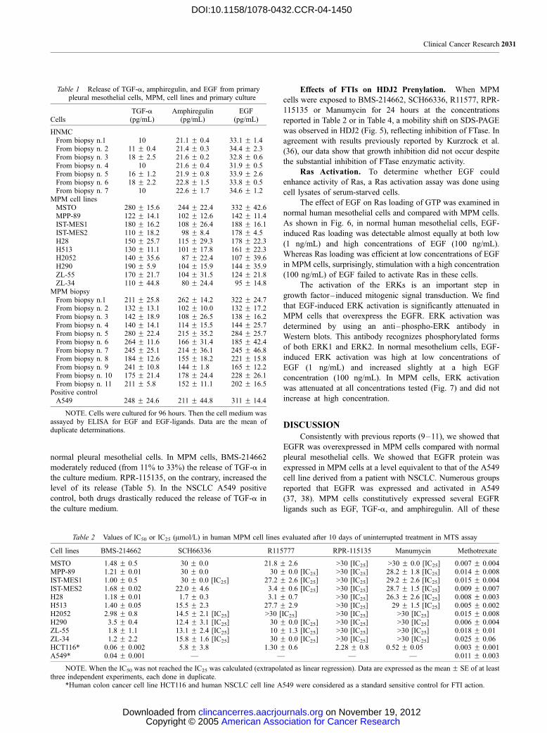
apoptosis in MPM cells (Fig. 3); only a fraction corresponding to
30% of all MPM cells presented the typical features of apoptosis.
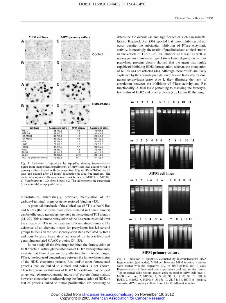
Gel ladder confirmed these data (Fig. 4).
Effects of FTIs on EGFR Ligand Release. The release
of TGF-a was measured after treatment with two different FTIs,
namely, BMS-214662 (moderately active) and RPR-115135
(nonactive). Both drugs did not affect the release of TGF-a in
Clinical Cancer Research 2029
American Association for Cancer Research Copyright © 2005 on November 19, 2012clincancerres.aacrjournals.orgDownloaded from
DOI:10.1158/1078-0432.CCR-04-1450
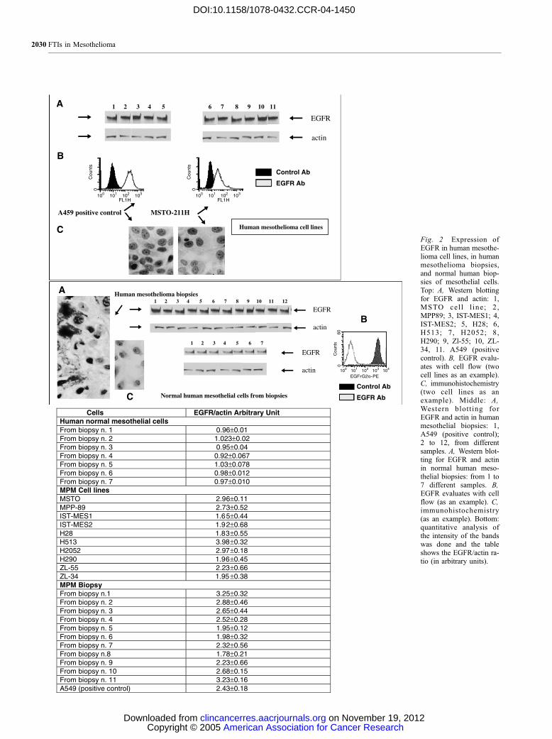
Fig. 2 Expression ofEGFR in human mesothe-lioma cell lines, in humanmesothelioma biopsies,and normal human biop-sies of mesothelial cells.Top: A, Western blottingfor EGFR and actin: 1,MSTO cel l l ine ; 2 ,MPP89; 3, IST-MES1; 4,IST-MES2; 5, H28; 6,H513; 7, H2052; 8,H290; 9, Zl-55; 10, ZL-34, 11. A549 (positivecontrol). B, EGFR evalu-ates with cell flow (twocell lines as an example).C, immunohistochemistry(two cell lines as anexample). Middle: A,Western blotting forEGFR and actin in humanmesothelial biopsies: 1,A549 (positive control);2 to 12, from differentsamples. A, Western blot-ting for EGFR and actinin normal human meso-thelial biopsies: from 1 to7 different samples. B,EGFR evaluates with cellflow (as an example). C,immunohistochemistry(as an example). Bottom:quantitative analysis ofthe intensity of the bandswas done and the tableshows the EGFR/actin ra-tio (in arbitrary units).
FTIs in Mesothelioma2030
American Association for Cancer Research Copyright © 2005 on November 19, 2012clincancerres.aacrjournals.orgDownloaded from
DOI:10.1158/1078-0432.CCR-04-1450
normal pleural mesothelial cells. In MPM cells, BMS-214662
moderately reduced (from 11% to 33%) the release of TGF-a in
the culture medium. RPR-115135, on the contrary, increased the
level of its release (Table 5). In the NSCLC A549 positive
control, both drugs drastically reduced the release of TGF-a in
the culture medium.
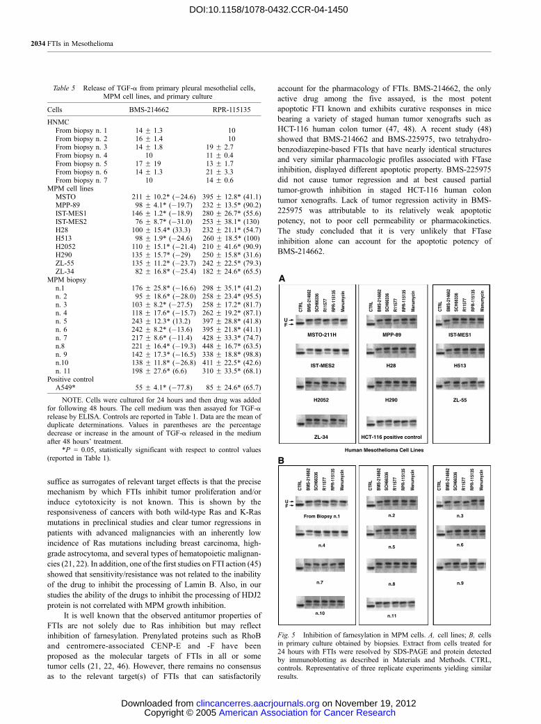
Effects of FTIs on HDJ2 Prenylation. When MPM
cells were exposed to BMS-214662, SCH66336, R11577, RPR-
115135 or Manumycin for 24 hours at the concentrations
reported in Table 2 or in Table 4, a mobility shift on SDS-PAGE
was observed in HDJ2 (Fig. 5), reflecting inhibition of FTase. In
agreement with results previously reported by Kurzrock et al.
(36), our data show that growth inhibition did not occur despite
the substantial inhibition of FTase enzymatic activity.
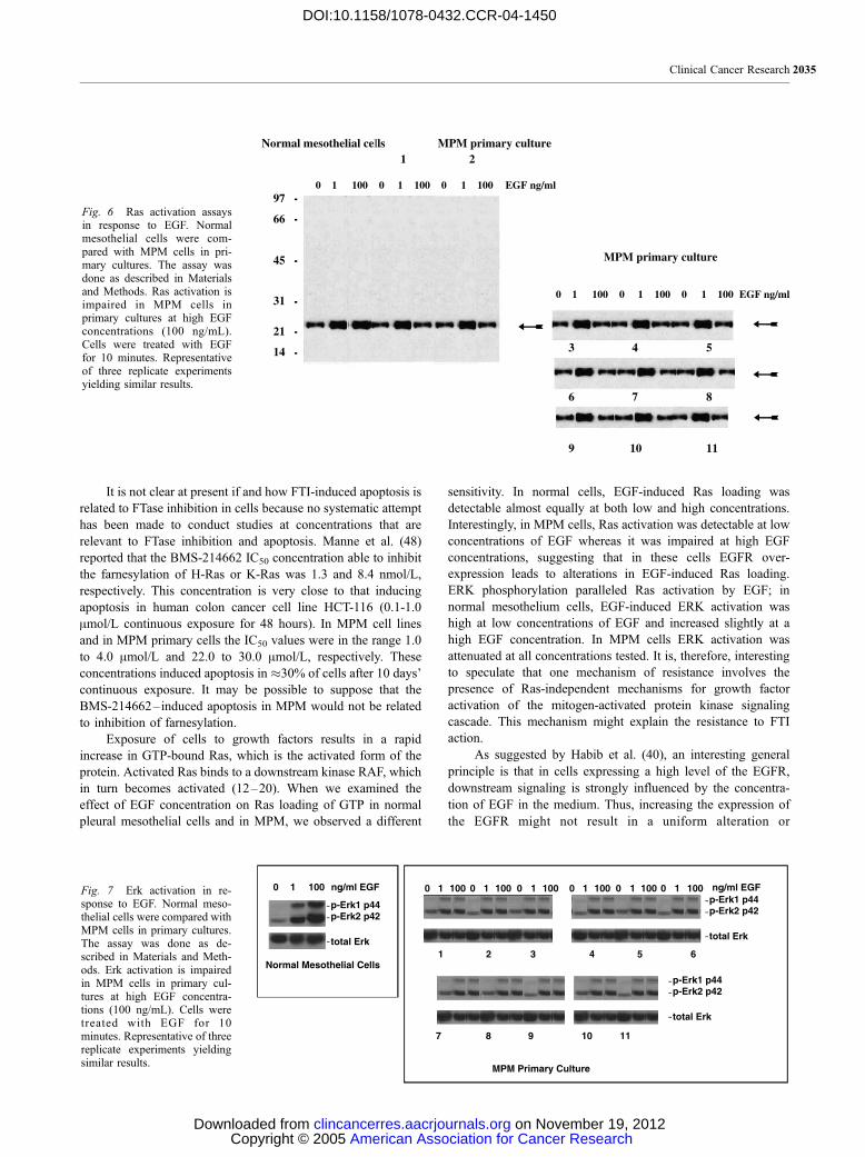
Ras Activation. To determine whether EGF could
enhance activity of Ras, a Ras activation assay was done using
cell lysates of serum-starved cells.
The effect of EGF on Ras loading of GTP was examined in
normal human mesothelial cells and compared with MPM cells.
As shown in Fig. 6, in normal human mesothelial cells, EGF-
induced Ras loading was detectable almost equally at both low
(1 ng/mL) and high concentrations of EGF (100 ng/mL).
Whereas Ras loading was efficient at low concentrations of EGF
in MPM cells, surprisingly, stimulation with a high concentration
(100 ng/mL) of EGF failed to activate Ras in these cells.
The activation of the ERKs is an important step in
growth factor– induced mitogenic signal transduction. We find
that EGF-induced ERK activation is significantly attenuated in
MPM cells that overexpress the EGFR. ERK activation was
determined by using an anti–phospho-ERK antibody in
Western blots. This antibody recognizes phosphorylated forms
of both ERK1 and ERK2. In normal mesothelium cells, EGF-
induced ERK activation was high at low concentrations of
EGF (1 ng/mL) and increased slightly at a high EGF
concentration (100 ng/mL). In MPM cells, ERK activation
was attenuated at all concentrations tested (Fig. 7) and did not
increase at high concentration.
DISCUSSION
Consistently with previous reports (9–11), we showed that
EGFR was overexpressed in MPM cells compared with normal
pleural mesothelial cells. We showed that EGFR protein was
expressed in MPM cells at a level equivalent to that of the A549
cell line derived from a patient with NSCLC. Numerous groups
reported that EGFR was expressed and activated in A549
(37, 38). MPM cells constitutively expressed several EGFR
ligands such as EGF, TGF-a, and amphiregulin. All of these
Table 2 Values of IC50 or IC25 (Amol/L) in human MPM cell lines evaluated after 10 days of uninterrupted treatment in MTS assay
Cell lines BMS-214662 SCH66336 R115777 RPR-115135 Manumycin Methotrexate
MSTO 1.48 F 0.5 30 F 0.0 21.8 F 2.6 >30 [IC25] >30 F 0.0 [IC25] 0.007 F 0.004MPP-89 1.21 F 0.01 30 F 0.0 30 F 0.0 [IC25] >30 [IC25] 28.2 F 1.8 [IC25] 0.014 F 0.008IST-MES1 1.00 F 0.5 30 F 0.0 [IC25] 27.2 F 2.6 [IC25] >30 [IC25] 29.2 F 2.6 [IC25] 0.015 F 0.004IST-MES2 1.68 F 0.02 22.0 F 4.6 3.4 F 0.6 [IC25] >30 [IC25] 28.7 F 1.5 [IC25] 0.009 F 0.007H28 1.18 F 0.01 1.7 F 0.3 3.1 F 0.7 >30 [IC25] 26.3 F 2.6 [IC25] 0.008 F 0.003H513 1.40 F 0.05 15.5 F 2.3 27.7 F 2.9 >30 [IC25] 29 F 1.5 [IC25] 0.005 F 0.002H2052 2.98 F 0.8 14.5 F 2.1 [IC25] >30 [IC25] >30 [IC25] >30 [IC25] 0.015 F 0.008H290 3.5 F 0.4 12.4 F 3.1 [IC25] 30 F 0.0 [IC25] >30 [IC25] >30 [IC25] 0.006 F 0.004ZL-55 1.8 F 1.1 13.1 F 2.4 [IC25] 10 F 1.3 [IC25] >30 [IC25] >30 [IC25] 0.018 F 0.01ZL-34 1.2 F 2.2 15.8 F 1.6 [IC25] 30 F 0.0 [IC25] >30 [IC25] >30 [IC25] 0.025 F 0.06HCT116* 0.06 F 0.002 5.8 F 3.8 1.30 F 0.6 2.28 F 0.8 0.52 F 0.05 0.003 F 0.001A549* 0.04 F 0.001 — — — — 0.011 F 0.003
NOTE. When the IC50 was not reached the IC25 was calculated (extrapolated as linear regression). Data are expressed as the mean F SE of at leastthree independent experiments, each done in duplicate.
*Human colon cancer cell line HCT116 and human NSCLC cell line A549 were considered as a standard sensitive control for FTI action.
Table 1 Release of TGF-a, amphiregulin, and EGF from primarypleural mesothelial cells, MPM, cell lines and primary culture
CellsTGF-a(pg/mL)
Amphiregulin(pg/mL)
EGF(pg/mL)
HNMCFrom biopsy n.1 10 21.1 F 0.4 33.1 F 1.4From biopsy n. 2 11 F 0.4 21.4 F 0.3 34.4 F 2.3From biopsy n. 3 18 F 2.5 21.6 F 0.2 32.8 F 0.6From biopsy n. 4 10 21.6 F 0.4 31.9 F 0.5From biopsy n. 5 16 F 1.2 21.9 F 0.8 33.9 F 2.6From biopsy n. 6 18 F 2.2 22.8 F 1.5 33.8 F 0.5From biopsy n. 7 10 22.6 F 1.7 34.6 F 1.2
MPM cell linesMSTO 280 F 15.6 244 F 22.4 332 F 42.6MPP-89 122 F 14.1 102 F 12.6 142 F 11.4IST-MES1 180 F 16.2 108 F 26.4 188 F 16.1IST-MES2 110 F 18.2 98 F 8.4 178 F 4.5H28 150 F 25.7 115 F 29.3 178 F 22.3H513 130 F 11.1 101 F 17.8 161 F 22.3H2052 140 F 35.6 87 F 22.4 107 F 39.6H290 190 F 5.9 104 F 15.9 144 F 35.9ZL-55 170 F 21.7 104 F 31.5 124 F 21.8ZL-34 110 F 44.8 80 F 24.4 95 F 14.8
MPM biopsyFrom biopsy n.1 211 F 25.8 262 F 14.2 322 F 24.7From biopsy n. 2 132 F 13.1 102 F 10.0 132 F 17.2From biopsy n. 3 142 F 18.9 108 F 26.5 138 F 16.2From biopsy n. 4 140 F 14.1 114 F 15.5 144 F 25.7From biopsy n. 5 280 F 22.4 215 F 35.2 284 F 25.7From biopsy n. 6 264 F 11.6 166 F 31.4 185 F 42.4From biopsy n. 7 245 F 25.1 214 F 36.1 245 F 46.8From biopsy n. 8 184 F 12.6 155 F 18.2 221 F 15.8From biopsy n. 9 241 F 10.8 144 F 1.8 165 F 12.2From biopsy n. 10 175 F 21.4 178 F 24.4 228 F 26.1From biopsy n. 11 211 F 5.8 152 F 11.1 202 F 16.5
Positive controlA549 248 F 24.6 211 F 44.8 311 F 14.4
NOTE. Cells were cultured for 96 hours. Then the cell medium wasassayed by ELISA for EGF and EGF-ligands. Data are the mean ofduplicate determinations.
Clinical Cancer Research 2031
American Association for Cancer Research Copyright © 2005 on November 19, 2012clincancerres.aacrjournals.orgDownloaded from
DOI:10.1158/1078-0432.CCR-04-1450

ligands are made as membrane-spanning prohormones that are
processed and released through regulated proteolysis. The
current paradigm is that membrane-anchored growth factors act
as juxtacrine regulators of cell-cell signaling (39, 40). The
observations that MPM cells overexpress amphiregulin, EGFR,
EGF, and TGF-a suggest that excessive EGFR activation can
contribute to mesothelioma aggressiveness.
Nørgaard et al. (24, 25) have shown data suggesting that the
potential clinical use of FTIs could be expanded to include
cancers harboring activated receptor tyrosine kinases as well as
those containing activated Ras. Thus, working with MMTV-
TGF-a mice, they showed that L-744,832 induced reversible
regression of mammary tumors that was paralleled by a decrease
in serum levels of TGF-a secreted by the tumor cells.
We tested the activity of five different FTIs [i.e., SCH66336
(Sarasar), BMS-214662, R115777 (Zarnestra), which have
reached clinical testing (21, 22), RPR-115135, a new non-
peptidomimetic compound (26, 32, 33), and Manumycin (a
natural compound)] on MPM cells. Growth of MPM cells was
substantially not affected by treatment with these FTIs. Among
these five, BMS-214662 was the only one moderately active.
BMS-214662 triggered apoptosis in a small fraction of cells (not
higher than 30%) that was paralleled by a slight decrease in the
levels of TGF-a secreted by treated MPM cells.
Interestingly, MPM cells are sensitive to methotrexate. The
antifolate methotrexate is one of the most successful drugs in
cancer chemotherapy. Although its efficacy is widely attributed
to a decrease in nucleotide biosynthesis (41), methotrexate is
known to increase homocysteine. A potential mechanism for the
detrimental effects of homocysteine is cellular hypomethylation
from an increase in S-adenosylhomocysteine, an inhibitor of
methyltransferases including isoprenylcysteine carboxyl meth-
yltransferase (Icmt). Among the substrates of Icmt is Ras, and
carboxyl methylation of Ras is important for proper plasma
membrane localization and function (42, 43). Recent results (34)
showed that after methotrexate treatment, Ras is mislocalized to
the cytosol, and its signaling functions are impaired, suggesting
that methotrexate has an additional mechanism of action that
leads to an inhibition in Ras signaling. Proteins that terminate
with a carboxyl-terminal CAAX motif, such as the Ras and
RhoB proteins, undergo three sequential posttranslational
processing events:
1. The cysteine (i.e., the C of the CAAX sequence) is
isoprenylated by FTase or geranylgeranyltransferase type I.
2. The last three amino acids of the protein (i.e., the -AAX) are
cleaved off by Rce1, an integral membrane protein of the
endoplasmic reticulum.
3. The newly exposed isoprenylcysteine is methylated by an
endoplasmic reticulum membrane–bound methyltransferase,
Icmt.
These modifications render the C terminus of CAAX
proteins more hydrophobic, facilitating binding to membranes
(21, 22, 42, 43). A search for factors involved in the
intracellular trafficking of Ras has identified a specific and
prenylation-dependent interaction between tubulin/microtubules
and K-Ras. It was found that the polylysine region of K-Ras
located immediately upstream of the prenylation site is required
for binding of K-Ras to microtubules. Removal of the three
carboxyl-terminal amino acids of farnesylated K-Ras with
the specific endoprotease Rce1 abolished its binding to
Table 4 Values of IC50 or IC25 (Amol/L) in human mesothelioma cells in primary culture obtained by human biopsies evaluated after 10 days ofuninterrupted treatment in soft agar
MPM biopsy BMS-214662 SCH66336 R115777 RPR-115135 Manumycin Methotrexate
n. 1 28.8 F 1.4 4.5 F 0.9 [IC25] 12.5 F 2.6 [IC25] >30 25.2 F 1.1 [IC25] 0.017 F 0.02n. 2 27.0 F 1.3 12.1 F 2.4 [IC25] 11 F 3.1 [IC25] >30 27.2 F 0.3 [IC25] 0.019 F 0.07n. 3 21.7 F 0.4 5.0 F 0.8 [IC25] >30 [IC25] >30 29 F 1.3 [IC25] 0.025 F 0.04n. 4 26.5 F 1.1 4.0 F 0.7 [IC25] 4.2 F 0.3 [IC25] >30 28 F 1.2 [IC25] 0.009 F 0.002n. 5 28.7 F 0.7 30 F 0.0 [IC25] 30 F 0.0 [IC25] >30 22.2 F 0.5 [IC25] 0.018 F 0.03n. 6 22.1 F 4.2 30 F 0.0 [IC25] 30 F 0.0 [IC25] >30 26.0 F 0.7 [IC25] 0.015 F 0.03n. 7 24.5 F 1.6 14.0 F 1.5 [IC25] 14.6 F 1.3 [IC25] >30 28 F 1.2 [IC25] 0.025 F 0.07n. 8 29.5 F 1.4 22.2 F 1.5 [IC25] 25.3 F 0.8 [IC25] >30 >30.0 [IC25] 0.009 F 0.002n. 9 27.5 F 0.4 30 F 0.0 [IC25] 30 F 0.0 [IC25] >30 >30.0 [IC25] 0.028 F 0.01n. 10 26.5 F 1.8 300 F 0 [IC25] 30 F 0.0 [IC25] >30 >30.0 [IC25] 0.032 F 0.08n. 11 30 F 0.0 30 F 0.0 [IC25] 30 F 0.0 [IC25] >30 >30.0 [IC25] 0.017 F 0.02
NOTE. When the IC50 was not reached the IC25 was calculated (extrapolated as linear regression). Data are expressed as the mean F SE of at leastthree independent experiments, each done in duplicate.
Table 3 Values of IC25 (Amol/L) in human short-term culture of normal epithelium cells evaluated after 10 days of uninterrupted treatmentin MTS assay
HNMC BMS-214662 SCH66336 R115777 RPR-115135 Manumycin
From biopsy n. 1 >30 >30 >30 >30 >30From biopsy n. 2 >30 >30 >30 >30 >30From biopsy n. 3 >30 >30 >30 >30 >30From biopsy n. 4 >30 >30 >30 >30 >30From biopsy n. 5 >30 >30 >30 >30 >30From biopsy n. 6 >30 >30 >30 >30 >30From biopsy n. 7 >30 >30 >30 >30 >30
NOTE. Data are expressed as the mean F SE of at least three independent experiments, each done in duplicate.
FTIs in Mesothelioma2032
American Association for Cancer Research Copyright © 2005 on November 19, 2012clincancerres.aacrjournals.orgDownloaded from
DOI:10.1158/1078-0432.CCR-04-1450
microtubules. Interestingly, however, methylation of the
carboxyl-terminal prenylcysteine restored binding (43).
A potential drawback of the clinical use of FTIs is that K-Ras
and N-Ras (the isoforms most often mutated in human tumors)
can be efficiently geranylgeranylated in the setting of FTI therapy
(21, 22). This alternate prenylation of the Ras proteins could limit
the efficacy of FTIs in the treatment of Ras-induced tumors. The
existence of an alternate means for prenylation has led several
groups to focus on the postisoprenylation steps mediated by Rce1
and Icmt because those steps are shared by farnesylated and
geranylgeranylated CAAX proteins (34, 35).
In our study all the five drugs inhibited the farnesylation of
HDJ2 protein. Although the inhibition of HDJ2 farnesylation may
indicate that these drugs are truly affecting their intended target,
FTase, the degree of concordance between the farnesylation status
of the HDJ2 chaperone protein, Ras, and/or other farnesylated
proteins that are linked to clinical end points is not known.
Therefore, serial evaluations of HDJ2 farnesylation may be used
as general pharmacodynamic indices of protein farnesylation;
however, concordant studies relating the farnesylation of HDJ2 to
that of proteins linked to tumor proliferation are necessary to
determine the overall use and significance of such assessments.
Indeed, Kurzrock et al. (36) reported that tumor inhibition did not
occur despite the substantial inhibition of FTase enzymatic
activity. Interestingly, the results of preclinical and clinical studies
of the effects of L-778,123, an inhibitor of FTase, as well as
geranylgeranyltransferase type I (to a lesser degree) on various
prenylated proteins clearly showed that the agent was highly
capable of inhibiting HDJ2 farnesylation, whereas the prenylation
of K-Ras was not affected (44). Although these results are likely
explained by the alternate prenylation of N- and K-Ras by residual
geranylgeranyltransferase type I, they illustrate the lack of
correlation between the inhibition of FTase activity and Ras
functionality. A final issue pertaining to assessing the farnesyla-
tion status of HDJ2 and other proteins (i.e., Lamin B) that might
Fig. 3 Detection of apoptosis by ApopTag staining (representativefigure from independent experiments) of MPM cell lines and of MPM inprimary culture treated with the respective IC50 of BMS-214662 for 10days and stained after 24 hours’ incubation in drug-free medium. Thenuclei of apoptotic cells were stained dark brown. A, MSTO; B, MPP89;C, from biopsy n. 1; D, from biopsy n.2. The table reports the percentage(over controls) of apoptotic cells.
Fig. 4 Induction of apoptosis evaluated by internucleosomal DNAfragmentation (gel ladder). MPM cell lines and MPM in primary culturewere treated with the respective IC50 of BMS-214662 for 10 days.Representative of three replicate experiments yielding similar results.Top, untreated cells; bottom, treated cells. m, marker. MPM cell lines: 1,MSTO cell line; 2, MPP89; 3, IST-MES1; 4, IST-MES2; 5, H28; 6,H513; 7, H2052; 8, H290; 9, Zl-55; 10, ZL-34; 11, HCT116 (positivecontrol). MPM primary culture from 1 to 11 different samples.
Clinical Cancer Research 2033
American Association for Cancer Research Copyright © 2005 on November 19, 2012clincancerres.aacrjournals.orgDownloaded from
DOI:10.1158/1078-0432.CCR-04-1450
suffice as surrogates of relevant target effects is that the precise
mechanism by which FTIs inhibit tumor proliferation and/or
induce cytotoxicity is not known. This is shown by the
responsiveness of cancers with both wild-type Ras and K-Ras
mutations in preclinical studies and clear tumor regressions in
patients with advanced malignancies with an inherently low
incidence of Ras mutations including breast carcinoma, high-
grade astrocytoma, and several types of hematopoietic malignan-
cies (21, 22). In addition, one of the first studies on FTI action (45)
showed that sensitivity/resistance was not related to the inability
of the drug to inhibit the processing of Lamin B. Also, in our
studies the ability of the drugs to inhibit the processing of HDJ2
protein is not correlated with MPM growth inhibition.
It is well known that the observed antitumor properties of
FTIs are not solely due to Ras inhibition but may reflect
inhibition of farnesylation. Prenylated proteins such as RhoB
and centromere-associated CENP-E and -F have been
proposed as the molecular targets of FTIs in all or some
tumor cells (21, 22, 46). However, there remains no consensus
as to the relevant target(s) of FTIs that can satisfactorily
account for the pharmacology of FTIs. BMS-214662, the only
active drug among the five assayed, is the most potent
apoptotic FTI known and exhibits curative responses in mice
bearing a variety of staged human tumor xenografts such as
HCT-116 human colon tumor (47, 48). A recent study (48)
showed that BMS-214662 and BMS-225975, two tetrahydro-
benzodiazepine-based FTIs that have nearly identical structures
and very similar pharmacologic profiles associated with FTase
inhibition, displayed different apoptotic property. BMS-225975
did not cause tumor regression and at best caused partial
tumor-growth inhibition in staged HCT-116 human colon
tumor xenografts. Lack of tumor regression activity in BMS-
225975 was attributable to its relatively weak apoptotic
potency, not to poor cell permeability or pharmacokinetics.
The study concluded that it is very unlikely that FTase
inhibition alone can account for the apoptotic potency of
BMS-214662.
Table 5 Release of TGF-a from primary pleural mesothelial cells,MPM cell lines, and primary culture
Cells BMS-214662 RPR-115135
HNMCFrom biopsy n. 1 14 F 1.3 10From biopsy n. 2 16 F 1.4 10From biopsy n. 3 14 F 1.8 19 F 2.7From biopsy n. 4 10 11 F 0.4From biopsy n. 5 17 F 19 13 F 1.7From biopsy n. 6 14 F 1.3 21 F 3.3From biopsy n. 7 10 14 F 0.6
MPM cell linesMSTO 211 F 10.2* (�24.6) 395 F 12.8* (41.1)MPP-89 98 F 4.1* (�19.7) 232 F 13.5* (90.2)IST-MES1 146 F 1.2* (�18.9) 280 F 26.7* (55.6)IST-MES2 76 F 8.7* (�31.0) 253 F 38.1* (130)H28 100 F 15.4* (33.3) 232 F 21.1* (54.7)H513 98 F 1.9* (�24.6) 260 F 18.5* (100)H2052 110 F 15.1* (�21.4) 210 F 41.6* (90.9)H290 135 F 15.7* (�29) 250 F 15.8* (31.6)ZL-55 135 F 11.2* (�23.7) 242 F 22.5* (79.3)ZL-34 82 F 16.8* (�25.4) 182 F 24.6* (65.5)
MPM biopsyn.1 176 F 25.8* (�16.6) 298 F 35.1* (41.2)n. 2 95 F 18.6* (�28.0) 258 F 23.4* (95.5)n. 3 103 F 8.2* (�27.5) 258 F 17.2* (81.7)n. 4 118 F 17.6* (�15.7) 262 F 19.2* (87.1)n. 5 243 F 12.3* (13.2) 397 F 28.8* (41.8)n. 6 242 F 8.2* (�13.6) 395 F 21.8* (41.1)n. 7 217 F 8.6* (�11.4) 428 F 33.3* (74.7)n.8 221 F 16.4* (�19.3) 448 F 16.7* (63.5)n. 9 142 F 17.3* (�16.5) 338 F 18.8* (98.8)n.10 138 F 11.8* (�26.8) 411 F 22.5* (42.6)n. 11 198 F 27.6* (6.6) 310 F 33.5* (68.1)
Positive controlA549* 55 F 4.1* (�77.8) 85 F 24.6* (65.7)
NOTE. Cells were cultured for 24 hours and then drug was addedfor following 48 hours. The cell medium was then assayed for TGF-arelease by ELISA. Controls are reported in Table 1. Data are the mean ofduplicate determinations. Values in parentheses are the percentagedecrease or increase in the amount of TGF-a released in the mediumafter 48 hours’ treatment.
*P = 0.05, statistically significant with respect to control values(reported in Table 1).
Fig. 5 Inhibition of farnesylation in MPM cells. A, cell lines; B, cellsin primary culture obtained by biopsies. Extract from cells treated for24 hours with FTIs were resolved by SDS-PAGE and protein detectedby immunoblotting as described in Materials and Methods. CTRL,controls. Representative of three replicate experiments yielding similarresults.
FTIs in Mesothelioma2034
American Association for Cancer Research Copyright © 2005 on November 19, 2012clincancerres.aacrjournals.orgDownloaded from
DOI:10.1158/1078-0432.CCR-04-1450
It is not clear at present if and how FTI-induced apoptosis is
related to FTase inhibition in cells because no systematic attempt
has been made to conduct studies at concentrations that are
relevant to FTase inhibition and apoptosis. Manne et al. (48)
reported that the BMS-214662 IC50 concentration able to inhibit
the farnesylation of H-Ras or K-Ras was 1.3 and 8.4 nmol/L,
respectively. This concentration is very close to that inducing
apoptosis in human colon cancer cell line HCT-116 (0.1-1.0
Amol/L continuous exposure for 48 hours). In MPM cell lines
and in MPM primary cells the IC50 values were in the range 1.0
to 4.0 Amol/L and 22.0 to 30.0 Amol/L, respectively. These
concentrations induced apoptosis in �30% of cells after 10 days’
continuous exposure. It may be possible to suppose that the
BMS-214662–induced apoptosis in MPM would not be related
to inhibition of farnesylation.
Exposure of cells to growth factors results in a rapid
increase in GTP-bound Ras, which is the activated form of the
protein. Activated Ras binds to a downstream kinase RAF, which
in turn becomes activated (12–20). When we examined the
effect of EGF concentration on Ras loading of GTP in normal
pleural mesothelial cells and in MPM, we observed a different
sensitivity. In normal cells, EGF-induced Ras loading was
detectable almost equally at both low and high concentrations.
Interestingly, in MPM cells, Ras activation was detectable at low
concentrations of EGF whereas it was impaired at high EGF
concentrations, suggesting that in these cells EGFR over-
expression leads to alterations in EGF-induced Ras loading.
ERK phosphorylation paralleled Ras activation by EGF; in
normal mesothelium cells, EGF-induced ERK activation was
high at low concentrations of EGF and increased slightly at a
high EGF concentration. In MPM cells ERK activation was
attenuated at all concentrations tested. It is, therefore, interesting
to speculate that one mechanism of resistance involves the
presence of Ras-independent mechanisms for growth factor
activation of the mitogen-activated protein kinase signaling
cascade. This mechanism might explain the resistance to FTI
action.
As suggested by Habib et al. (40), an interesting general
principle is that in cells expressing a high level of the EGFR,
downstream signaling is strongly influenced by the concentra-
tion of EGF in the medium. Thus, increasing the expression of
the EGFR might not result in a uniform alteration or
Fig. 6 Ras activation assaysin response to EGF. Normalmesothelial cells were com-pared with MPM cells in pri-mary cultures. The assay wasdone as described in Materialsand Methods. Ras activation isimpaired in MPM cells inprimary cultures at high EGFconcentrations (100 ng/mL).Cells were treated with EGFfor 10 minutes. Representativeof three replicate experimentsyielding similar results.
Fig. 7 Erk activation in re-sponse to EGF. Normal meso-thelial cells were compared withMPM cells in primary cultures.The assay was done as de-scribed in Materials and Meth-ods. Erk activation is impairedin MPM cells in primary cul-tures at high EGF concentra-tions (100 ng/mL). Cells weretreated with EGF for 10minutes. Representative of threereplicate experiments yieldingsimilar results.
Clinical Cancer Research 2035
American Association for Cancer Research Copyright © 2005 on November 19, 2012clincancerres.aacrjournals.orgDownloaded from
DOI:10.1158/1078-0432.CCR-04-1450

amplification of downstream signals. For example, even among
signals that are attenuated, differences can be detected. In MPM
cells, Ras activation is attenuated at high EGF concentrations.
Our data highlighted the concept that the same signaling
pathwaycanberegulatedindifferentwaysandtheseregulationscan
differ between different cells of different origin. Furthermore,
distinct signaling pathways cross talk with each other. Therefore, a
compensatory pathway can emerge or selectively strengthen after
inhibitorsblockonespecificpathway.Differentsignalingpathways
can control or affect the same cellular function, and one single
signaling pathway can regulate different cellular functions. This
apparently redundant cell signaling network actually may reflect a
minor change mechanism for cells to respond and adjust to
combined effects of simultaneous or sequential stimulation by
many extracellular or internal signals and to control duration and
intensity of each signal.
The results reported and commented in this article confirm the
peculiar nature of MPM and add new information on its typical
resistance to virtually any adopted chemotherapeutic strategy. The
message we would like to draw from our experience is that by
realizing a translational study,wehave gathered froma single set of
experiments a double set of information: an explanation for the
clinical failure of drugs, together with a deeper basic knowledge on
the disease.
Ideally, translational research is defined as the process of
translating findings derived in basic science to the development
of new understanding of disease mechanisms, diagnosis, and
therapeutics. It is a functional bidirectional bridge among basic
and clinical researchers realized through ad hoc collaborative
efforts (48–53).
The data derived from our experience could help in
planning future translational studies in MPM.
REFERENCES
1. Pelucchi C, Malvezzi M, La Vecchia C, Levi F, Decarli A, Negri E.The mesothelioma epidemic in Western Europe: an update. Br J Cancer2004;90:1022–4.
2. Hemminki K, Li X. Mesothelioma incidence seems to have leveledoff in Sweden. Int J Cancer 2003;103:145–6.
3. Peto J, Decarli A, La Vecchia C, Levi F, Negri E. The Europeanmesothelioma epidemic. Br J Cancer 1999;79:666–72.
4. Cugell DW, Kamp DW. Asbestos and the pleura: a review. Chest2004;125:1103–17.
5. van Ruth S, Baas P, Zoetmulder FA. Surgical treatment of malignantpleural mesothelioma: a review. Chest 2003;123:551–61.
6. Treasure T, Waller D, Swift S, Peto J. Radical surgery formesothelioma. Br Med J 2004;328:237–8.
7. Sugarbaker DJ, Heher EC, Lee TH, et al. Extrapleural pneumonec-tomy, chemotherapy, and radiotherapy in the treatment of diffusemalignant pleural mesothelioma. J Thorac Cardiovasc Surg 1991;102:10–4; discussion 14–5.
8. Vogelzang NJ, Rusthoven JJ, Symanowski J, et al. Phase III study ofpemetrexed in combination with cisplatin versus cisplatin alone inpatients with malignant pleural mesothelioma. J Clin Oncol 2003;21:2636–44.
9. Trupiano JK, Geisinger KR, Willingham MC, et al. Diffuse malignantmesothelioma of the peritoneum and pleura, analysis of markers. ModPathol 2004;17:476–81.
10. Cai YC, Roggli V, Mark E, Cagle PT, Fraire AE. Transforminggrowth factor a and epidermal growth factor receptor in reactive and
malignant mesothelial proliferations. Arch Pathol Lab Med 2004;128:68–70.
11. Janne PA, Taffaro ML, Salgia R, Johnson BE. Inhibition ofepidermal growth factor receptor signaling in malignant pleuralmesothelioma. Cancer Res 2002;62:5242–7.
12. Yamada S, Taketomi T, Yoshimura A. Model analysis of differencebetween EGF pathway and FGF pathway. Biochem Biophys ResCommun 2004;314:1113–20.
13. Zhang T, Ma J, Cao X. Grb2 regulates Stat3 activation negatively inepidermal growth factor signalling. Biochem J 2003;376:457–64.
14. Cai T, Nishida K, Hirano T, Khavari PA. Gab1 and SHP-2 promoteRas/MAPK regulation of epidermal growth and differentiation. J CellBiol 2002;159:103–12.
15. Saito T, Okada S, Ohshima K, et al. Differential activation ofepidermal growth factor receptor (ErbB1/EGFR) downstream signal-ing pathways by betacellulin and EGF. Endocrinology 2004;149:4232-43.
16. Holgado-Madruga M, Wong AJ. Role of the Grb2-associated binder1/SHP-2 interaction in cell growth and transformation. Cancer Res2004;64:2007–15.
17. Johnson GL, Lapadat R. Mitogen-activated protein kinase pathwaysmediated by ERK, JNK, and p38 protein kinases. Science 2002;298:1911–2.
18. Cox AD, Der CJ. The dark side of Ras: regulation of apoptosis.Oncogene 2003;22:8999–9006.
19. Kinbara K, Goldfinger LE, Hansen M, Chou FL, Ginsberg MH. RasGTPases: integrins’ friends or foes? Nat RevMol Cell Biol 2003;4:767–76.
20. Herrmann C. Ras-effector interactions: after one decade. Curr OpinStruct Biol 2003;13:122–9.
21. Sebti SM, Adjei AA. Farnesyltransferase inhibitors. Semin Oncol2004;31:28–39.
22. Russo P, Loprevite M, Cesario A, Ardizzoni A. Farnesylatedproteins as anticancer drug targets: from laboratory to the clinic. CurrMed Chem Anti-Canc Agents 2004;4:123–38.
23. Sizemore N, Cox AD, Barnard JA, et al. Pharmacological inhibitionof Ras-transformed epithelial cell growth is linked to down-regulation ofepidermal growth factor-related peptides. Gastroenterology 1999;117:567–76.
24. Norgaard P, Law BK, Plovisson HS, Moses HL. Farnesyltransfer-ase inhibitor-induced regression of mammary tumors in TGF a andTGF a/neu transgenic mice correlates with inhibition of map kinaseand p70s6 kinase phosphorylation. Ann N Y Acad Sci 1999;886:265–8.
25. Norgaard P, Law B, Joseph H, et al. Treatment with farnesyl-proteintransferase inhibitor induces regression of mammary tumors in trans-forming growth factor (TGF) a and TGF a/neu transgenic mice byinhibition of mitogenic activity and induction of apoptosis. Clin CancerRes 1999;5:35–42.
26. Ottoboni C, Crippa A, Falugi C, Russo P. Induction of micronucleiby a new non-peptidic mimetic farnesyltransferase inhibitor RPR-115135: role of gene mutations. Mutagenesis 2001;16:423–30.
27. Trombino S, Cesario A, Margaritora S, et al. a7-Nicotinicacetylcholine receptors affect growth regulation of human mesotheliomacells: role of mitogen-activated protein kinase pathway. Cancer Res2004;64:135–45. Erratum in Cancer Res 2004;64:1559.
28. Pass HI, Liu Z, Wali A, et al. Gene expression profiles predictsurvival and progression of pleural mesothelioma. Clin Cancer Res 2004;10:849–59.
29. Gordon GJ, Jensen RV, Hsiao LL, et al. Using gene expression ratiosto predict outcome among patients with mesothelioma. J Natl Cancer Inst2003;95:598–605.
30. Gordon GJ, Jensen RV, Hsiao LL, et al. Translation of microarraydata into clinically relevant cancer diagnostic tests using gene expressionratios in lung cancer and mesothelioma. Cancer Res 2002;62:4963–7.
31. Valente P, Arzani D, Cesario A, Margaritora S, Carbone E, Russo P.TNF increases camptothecin-induced apoptosis by inhibition of NF-nB.Eur J Cancer 2003;39:1468–77.
FTIs in Mesothelioma2036
American Association for Cancer Research Copyright © 2005 on November 19, 2012clincancerres.aacrjournals.orgDownloaded from
DOI:10.1158/1078-0432.CCR-04-1450

32. Russo P, Arzani D, Trombino S, Falugi C. c-myc down-regulationinduces apoptosis in human cancer cell lines exposed to RPR-115135(C31H29NO4), a non-peptidomimetic farnesyltransferase inhibitor.J Pharmacol Exp Ther 2003;304:37–47.
33. Russo P, Malacarne D, Falugi C, Trombino S, O’Connor PM. RPR-115135, a farnesyltransferase inhibitor, increases 5-FU-cytotoxicity inten human colon cancer cell lines: role of p53. Int J Cancer 2002;100:266–75.
34. Winter-Vann AM, Kamen BA, Bergo MO, et al. Targeting Rassignaling through inhibition of carboxyl methylation: an unexpectedproperty of methotrexate. Proc Natl Acad Sci U S A 2003;100:6529–34.
35. Philips MR. Methotrexate and Ras methylation: a new trick for anold drug? Sci STKE 2004:225:pe13.
36. Kurzrock R, Kantarjian HM, Cortes JE, et al. Farnesyltransferaseinhibitor R115777 in myelodysplastic syndrome: clinical and biologicactivities in the phase 1 setting. Blood 2003;102:4527–34.
37. Ono M, Hirata A, Kometani T, et al. Sensitivity to gefitinib (Iressa,ZD1839) in non-small cell lung cancer cell lines correlates withdependence on the epidermal growth factor (EGF) receptor/extracellularsignal-regulated kinase 1/2 and EGF receptor/Akt pathway forproliferation. Mol Cancer Ther 2004;3:465–72.
38. Janmaat ML, Kruyt FA, Rodriguez JA, Giaccone G. Response toepidermal growth factor receptor inhibitors in non-small cell lung cancercells: limited antiproliferative effects and absence of apoptosis associatedwith persistent activity of extracellular signal-regulated kinase or Aktkinase pathways. Clin Cancer Res 2003;9:2316–26.
39. Burke P, Schooler K, Wiley HS. Regulation of EGF receptorsignaling by endocytosis and intracellular trafficking. Mol Biol Cell2001;12:1897–910.
40. Habib AA, Chun SJ, Neel BG, Vartanian T. Increased expression ofepidermal growth factor receptor induces sequestration of extracellularsignal-related kinases and selective attenuation of specific epidermalgrowth factor-mediated signal transduction pathways. Mol Cancer Res2003;1:219–33.
41. Zhao R, Goldman ID. Resistance to antifolates. Oncogene 2003;22:7431–57.
42. Young SG, Ambroziak P, Kim E, Clarke S. Postisoprenylation proteinprocessing: CV (CaaX) endoproteases and isoprenylcysteine carboxylmethyltransferase. In: Tamanoi F, Sigman DS, editors. The enzymes. Vol.21. San Diego (CA): Academic Press; 2000. p. 155–213.
43. Chen Z, Otto JC, Bergo MO, Young SG, Casey PJ. TheC-terminal polylysine region and methylation of K-Ras are critical forthe interaction between K-Ras and microtubules. J Biol Chem 2000;275:41251–7.
44. Lobell RB, Liu D, Buser CA, et al. Preclinical and clinicalpharmacodynamic assessment of L-778, 123, a dual inhibitor of farnesyl:protein transferase and geranylgeranyl:protein transferase type-1. MolCancer Ther 2002;1:747–58.
45. Sepp-Lorenzino L, Ma Z, Rands E, et al. A peptidomimeticinhibitor of farnesyl:protein transferase blocks the anchorage-dependentand -independent growth of human tumor cell lines. Cancer Res 1995;55:5302–9.
46. Falugi C, Trombino S, Granone P, Margaritora S, Russo P.Increasing complexity of farnesyltransferase inhibitors activity: role inchromosome instability. Curr Cancer Drug Targets 2003;3:109–18.
47. Rose WC, Lee FY, Fairchild CR, et al. Preclinical antitumor activityof BMS-214662, a highly apoptotic and novel farnesyltransferaseinhibitor. Cancer Res 2001;61:7507–17.
48. Manne V, Lee FY, Bol DK, et al. Apoptotic and cytostaticfarnesyltransferase inhibitors have distinct pharmacology and efficacyprofiles in tumor models. Cancer Res 2004;64:3974–80.
49. Cesario A, Galetta D, Russo P, Margaritora S, Granone P. The role ofthe surgeon in translational research. Lancet 2003;362:1082.
50. Yeatman TJ. The future of cancer management: translating thegenome, transcriptome, and proteome. Ann Surg Oncol 2003;10:7–14.
51. Kreeger K. From bench to bedside. Nature 2003;424:1090–1.
52. Apolone G. Clinical and outcome research in oncology. The need forintegration. Health Qual Life Outcomes 2003;1:3.
53. Wong S, Witte ON. The BCR-ABL story: bench to bedside andback. Annu Rev Immunol 2004;22:247–306.
Clinical Cancer Research 2037
American Association for Cancer Research Copyright © 2005 on November 19, 2012clincancerres.aacrjournals.orgDownloaded from
DOI:10.1158/1078-0432.CCR-04-1450
Copyright © 2022 FDOKUMEN