
Up-Regulation of EphB4 in Mesothelioma and Its Biological Significance
11
Up-Regulation of EphB4 in Mesothelioma and Its Biological Significance Guangbin Xia, 1 S. Ram Kumar, 2,3 Rizwan Masood, 2 Michael Koss, 2 ClaireTempleman, 4 David Quinn, 1 Sutao Zhu, 1 Ramachandra Reddy, 5 Valery Krasnoperov, 5 and Parkash S. Gill 1,2 Abstract Purpose: Mesothelioma is a rare malignancy that is incurable and carries a short survival despite surgery, radiation, or chemotherapy. This study was designed to identify novel targets for diagnos- tic, prognostic, and therapeutic approaches. Experimental Design: The expression and functional significance of the receptor tyrosine kinase EphB4 was studied in vitro and in a murine model of mesothelioma. Results: EphB4 was highly expressed in mesothelioma cell lines and primary tumor tissues but not in normal mesothelium. Knockdown of EphB4 using small interfering RNA and antisense oligodeoxynucleotide showed reduction in cell survival, migration, and invasion. EphB4 knock- down initiated a caspase-8-mediated apoptosis and down-regulation of the antiapoptotic protein bcl-xl. EphB4 knockdown also resulted in reduced phosphorylation of Akt and down-regulation of matrix metalloproteinase-2 transcription. In addition, murine tumor xenograft studies using EphB4 oligodeoxynucleotides showed a marked reduction in tumor growth accompanied by a specific decline in EphB4 protein levels, reduced cell division, apoptosis in tumor tissue, and decreased microvascular density. Conclusions: EphB4 is expressed in mesothelioma, provides a survival advantage to tumor cells, and is therefore a potential novel therapeutic target. Mesothelioma is a rare neoplasm that most often arises from the serous surface of the pleural and peritoneal cavity (1). There are three main histologic types of malignant mesothelioma (i.e., epitheloid, mixed or biphasic, and sarcomatoid), with survival being higher with the epitheloid and shorter with the sarcomatoid type (2). There is a strong association with asbestos exposure with f80% of malignant mesothelioma occurring in individuals who have ingested or inhaled asbestos. The disease has an incidence of 3,000 cases per year in United States with 2,500 deaths ascribed to it (3). Most patients present with locally advanced tumor that is incurable with surgery or radiation therapy (4 – 6). Response to immune modulatory therapy, such as interleukin-2 or IFN, has also been limited (7). Hence, most patients with advanced disease are treated with systemic chemotherapy. Agents with activity in mesothelioma include anthracyclines, cisplatinum, Gemsar, and, more recently, pemetrexed (8). A large, randomized trial combining pemetrexed and cisplatinum compared favorably with a response in 41.3% versus 16.7% response following cisplatinum treatment alone, associated with median duration to progression of 5.7 versus 3.9 months. Although there was an increase in survival to a median of 12 months compared with 9 months for cisplatinum alone, long-term survival remained dismal (9). Thus, new treatment approaches are needed with an emphasis on targeted therapeutics. Progress has been made in identifying alterations in gene expression in mesothelioma to better understand tumor cell biology and identify novel therapeutic targets. For example, we and others have shown that vascular endothelial growth factor (VEGF) and VEGF-C function as autocrine growth factors for mesothelioma (10, 11). VEGF inhibitors, including neutraliz- ing antibodies and antisense molecules, are currently in clinical investigations (12). Epidermal growth factor receptor (EGFR) and platelet-derived growth factor (PDGF) are also expressed in mesothelioma and could provide growth advantage to tumor cells (13–16). The potential benefit of EGFR and PDGF inhibitors is also the focus of clinical trials (17, 18). To discover additional molecular therapeutic targets that might be incorporated into a multimodality regimen for the treatment of mesothelioma, we studied the expression and function of the receptor tyrosine kinase (RTK) EphB4 and its cognate ligand, EphrinB2. EphB4 is a member of the largest family of RTKs and plays an important role in a variety of processes during embryonic development, including pattern formation, cell aggregation and migration, segmentation, neural development, angiogenesis, and vascular hierarchical remodeling (19 – 22). EphrinB2, the transmembrane ligand of EphB4, also participates in vascular remodeling via reverse Human Cancer Biology Authors’ Affiliations: Departments of 1 Medicine, 2 Pathology, 3 Surgery, and 4 Obstetrics and Gynecology, Keck School of Medicine, University of Southern California and 5 VasGene Therapeutics, Inc., Los Angeles, California Received 10/15/04; revised 2/26/05; accepted 3/17/05. Grant support: NIH grant RO1CA79218 (P.S. Gill) and Mesothelioma Research Foundation of America. The costs of publication of this article were defrayed in part by the payment of page charges. This article must therefore be hereby marked advertisement in accordance with 18 U.S.C. Section 1734 solely to indicate this fact. Requests for reprints: Parkash S. Gill, University of Southern California/Norris Comprehensive Cancer Center, 1441 Eastlake Avenue, NOR 6332, Los Angeles, CA 90033-9172. Phone: 323-865-3909; Fax: 323-865-0092; E-mail: parkashg@usc.edu. F 2005 American Association for Cancer Research. www.aacrjournals.org Clin Cancer Res 2005;11(12) June 15, 2005 4305
-
Upload
independent -
Category
Documents
-
view
1 -
download
0
Transcript of Up-Regulation of EphB4 in Mesothelioma and Its Biological Significance

Up-Regulation of EphB4 inMesothelioma and ItsBiological SignificanceGuangbin Xia,1 S. Ram Kumar,2,3 RizwanMasood,2 Michael Koss,2 ClaireTempleman,4 David Quinn,1
Sutao Zhu,1 Ramachandra Reddy,5 Valery Krasnoperov,5 and Parkash S. Gill1,2
Abstract Purpose:Mesothelioma is a rare malignancy that is incurable and carries a short survival despitesurgery, radiation, or chemotherapy.This studywas designed to identify novel targets for diagnos-tic, prognostic, and therapeutic approaches.Experimental Design: The expression and functional significance of the receptor tyrosinekinase EphB4was studied in vitro and in amurine model of mesothelioma.Results: EphB4 was highly expressed in mesothelioma cell lines and primary tumor tissues butnot in normal mesothelium. Knockdown of EphB4 using small interfering RNA and antisenseoligodeoxynucleotide showed reduction in cell survival, migration, and invasion. EphB4 knock-down initiated a caspase-8-mediated apoptosis anddown-regulationof the antiapoptoticproteinbcl-xl. EphB4 knockdown also resulted in reduced phosphorylation of Akt and down-regulationof matrix metalloproteinase-2 transcription. In addition, murine tumor xenograft studies usingEphB4 oligodeoxynucleotides showed a marked reduction in tumor growth accompanied by aspecific decline in EphB4 protein levels, reduced cell division, apoptosis in tumor tissue, anddecreasedmicrovascular density.Conclusions: EphB4 is expressed inmesothelioma, provides a survival advantage to tumor cells,and is therefore a potential novel therapeutic target.
Mesothelioma is a rare neoplasm that most often arises fromthe serous surface of the pleural and peritoneal cavity (1). Thereare three main histologic types of malignant mesothelioma(i.e., epitheloid, mixed or biphasic, and sarcomatoid), withsurvival being higher with the epitheloid and shorter with thesarcomatoid type (2). There is a strong association withasbestos exposure with f80% of malignant mesotheliomaoccurring in individuals who have ingested or inhaled asbestos.The disease has an incidence of 3,000 cases per year in UnitedStates with 2,500 deaths ascribed to it (3). Most patientspresent with locally advanced tumor that is incurable withsurgery or radiation therapy (4–6). Response to immunemodulatory therapy, such as interleukin-2 or IFN, has also beenlimited (7). Hence, most patients with advanced disease aretreated with systemic chemotherapy. Agents with activity inmesothelioma include anthracyclines, cisplatinum, Gemsar,and, more recently, pemetrexed (8). A large, randomized trial
combining pemetrexed and cisplatinum compared favorablywith a response in 41.3% versus 16.7% response followingcisplatinum treatment alone, associated with median durationto progression of 5.7 versus 3.9 months. Although there was anincrease in survival to a median of 12 months compared with9 months for cisplatinum alone, long-term survival remaineddismal (9). Thus, new treatment approaches are needed with anemphasis on targeted therapeutics.
Progress has been made in identifying alterations in geneexpression in mesothelioma to better understand tumor cellbiology and identify novel therapeutic targets. For example, weand others have shown that vascular endothelial growth factor(VEGF) and VEGF-C function as autocrine growth factors formesothelioma (10, 11). VEGF inhibitors, including neutraliz-ing antibodies and antisense molecules, are currently in clinicalinvestigations (12). Epidermal growth factor receptor (EGFR)and platelet-derived growth factor (PDGF) are also expressed inmesothelioma and could provide growth advantage to tumorcells (13–16). The potential benefit of EGFR and PDGFinhibitors is also the focus of clinical trials (17, 18).
To discover additional molecular therapeutic targets thatmight be incorporated into a multimodality regimen for thetreatment of mesothelioma, we studied the expression andfunction of the receptor tyrosine kinase (RTK) EphB4 and itscognate ligand, EphrinB2. EphB4 is a member of the largestfamily of RTKs and plays an important role in a variety ofprocesses during embryonic development, including patternformation, cell aggregation and migration, segmentation,neural development, angiogenesis, and vascular hierarchicalremodeling (19–22). EphrinB2, the transmembrane ligand ofEphB4, also participates in vascular remodeling via reverse
Human Cancer Biology
Authors’ Affiliations: Departments of 1Medicine, 2Pathology, 3Surgery, and4Obstetrics and Gynecology, Keck School of Medicine, University of SouthernCalifornia and 5VasGene Therapeutics, Inc., Los Angeles, CaliforniaReceived10/15/04; revised 2/26/05; accepted 3/17/05.Grant support: NIH grant RO1CA79218 (P.S. Gill) and Mesothelioma ResearchFoundation of America.The costs of publication of this article were defrayed in part by the payment of pagecharges.This article must therefore be hereby marked advertisement in accordancewith18 U.S.C. Section1734 solely to indicate this fact.Requests for reprints: Parkash S. Gill, University of Southern California/NorrisComprehensive Cancer Center, 1441Eastlake Avenue, NOR 6332, Los Angeles,CA 90033-9172. Phone: 323-865-3909; Fax : 323-865-0092; E-mail:[email protected].
F2005 American Association for Cancer Research.
www.aacrjournals.org Clin Cancer Res 2005;11(12) June15, 20054305

signaling. Targeted knockout of either EphB4 or EphrinB2 inmice shows similar phenotypes with disrupted vessel matura-tion and early embryonic lethality (20). This receptor-ligandpair is not only important in vasculogenesis in the embryo butalso essential for defining the boundary between arterial andvenous domains (23), which persists in the adult life (24).Venous endothelial cells express EphB4, whereas arterialendothelial cells express EphrinB2 (20, 23). In addition totheir role in embryonic development, recent studies haveshown that they are implicated in tumor development (25).Aberrant expression of EphB4 in cancer has been suggestedin colon cancer, breast cancer, and endometrial carcinoma(26–30).
In the current study, we first show the expression of variousreceptors of the EphB family and their ligands in mesothe-lioma. We then show that EphB4 plays an important role in cellsurvival, migration, and invasion. EphB4 knockdown leads tocaspase-8-mediated (extrinsic pathway) apoptosis and a reduc-tion in the antiapoptotic protein bcl-xl. Targeting EphB4 in vivoled to a specific reduction in EphB4 protein levels that resultedin a significant inhibition in tumor growth combined withreduced mitotic index, tumor cell apoptosis, and reducedmicrovascular density. Together, these results provide evidencethat EphB4 is a novel therapeutic target for the treatment ofmesothelioma.
Materials andMethods
Cell lines and reagents. NCI H28, NCI H2052, NCI H2373, andMSTO 211H mesothelioma cell lines, MCF-7 breast cancer cell line,5637 bladder cancer cell line, and 293T human embryonic kidney cellline were obtained from the American Type Culture Collection(Manassas, VA). Cells were maintained in RPMI 1640 supplementedwith 10% heat-inactivated fetal bovine serum (FBS; Life Technologies,Gaithersburg,MD)and penicillin/streptomycin (Invitrogen,Carlsbad,CA).211H cells were cultured on plates coated with fibronectin (50 Ag/mL;Sigma, St. Louis, MO). Primary mesothelioma cells were obtained fromtumor samples. Tumor biopsy specimens fixed in formalin were alsoobtained from 16 patients with mesothelioma. Normal pleura andperitoneum were obtained from patients undergoing surgery forunrelated reasons. EphB4-specific small interfering RNAs (siRNA;Table 1) and phosphorothioate-modified antisense oligodeoxynucleo-tides (Table 2) were synthesized at University of Southern California/Norris Cancer Center Microchemical Core Laboratory. Effective andspecific antisense oligodeoxynucleotide (AS-10) and siRNA (siRNA 472)that knockdown EphB4 expression in 293T cells transiently transfectedwith full-length EphB4 were selected for subsequent study (data notshown). Polyclonal EphrinB2 (P20), EphB1 (Q20), EphB2 (C20), EphB3(L19), EphB4 (C-16), and EphB6 (N19) antibodies were obtained fromSanta Cruz Biotechnology (Santa Cruz, CA). Monoclonal EphB4antibodies (M91 and VK138, Vasgene, Inc., Los Angeles) were used inWestern blotting and immunoprecipitation. Monoclonal antibodyagainst Ki-67 was obtained from DAKO (Carpinteria, CA), h-actin fromSigma, bcl-2 and bcl-xl from Calbiochem (San Diego, CA), and Mcl-1
from BD PharMingen (San Diego, CA). FITC-labeled lycopersicon lectinwas obtained from Sigma.
Reverse transcription-PCR. Total RNA was extracted using RNASTAT-60 (Tel-Test, Inc., Friendswood, TX). First-strand cDNA wassynthesized from 5 Ag total RNA using SuperScript III (Invitrogen).Primer sequences are as follows: EphB4 forward primer 5V-TGA-GGTCACTGCATTGAACGGG-3V and reverse primer 5V-AACTCGCT-CTCATCCAGTT-3V (product size, 205 bp); EphrinB2 forwardprimer 5V-AGACAAGAGCCATGAAGATC-3V and reverse primer 5V-ATGATAATGTCACTGGGCTCTGA-3V (product size, 412 bp); andglyceraldehyde-3-phosphate dehydrogenase forward primer 5V-TGAAGGTCGGAGTCAACGGATTTGGT-3V and reverse primer 5V-CATGTGGGCCATGAGGTCCACCAC-3V (product size, 983 bp). Theprimers for EphB1, EphB2, EphB3, EphB6, EphrinB1, and EphrinB3(31) and matrix metalloproteinase (MMP)-2 and MMP-9 (32) were asdescribed previously. PCR was done using AB PCR System 2700(Applied Biosystems, Foster City, CA). The PCR conditions were 95jCfor 5 minutes followed by 35 cycles of denaturation at 95jC for30 seconds, annealing at 60jC for 30 seconds, and extension at 72jCfor 1 minute. Quantitative reverse transcription-PCR (RT-PCR) wasdone on the Stratagene MX3000P system (La Jolla, CA) using SYBRGreen I Brilliant Mastermix according to the manufacturer’s instruc-tions.
Preparation of digoxigenin-labeled RNA probes. EphrinB2 andEphB4 PCR products were cloned using the pGEM-T Easy System(Promega, Madison, WI) according to the manufacturer’s description.Primer sequences were 5V-TCCGTGTGGAAGTACTGCTG-3V (forward)and 5V-TCTGGTTTGGCACAGTTGAG-3V (reverse) for EphrinB2 thatyielded a 296-bp product and 5V-CTTTGGAAGAGACCCTGCTG-3V(forward) and 5V-AGACGGTGAAGGTCTCCTTG-3V (reverse) for EphB4that yielded a 297-bp product. The authenticity and insert orientationwere confirmed by DNA sequencing. The pGEM-T Easy plasmidscontaining the PCR product of the human EphrinB2 or EphB4 genewere linearized with SpeI or NcoI. Antisense or sense digoxigenin-labeled RNA probes were transcribed from T7 or SP6 promoters byrunoff transcription using a digoxigenin RNA labeling kit (Roche,Indianapolis, IN). RNA probes were quantitated by spot assay asdescribed in the digoxigenin RNA labeling kit instructions.
In situ hybridization. In situ hybridization was reformed asdescribed previously (33). In brief, cells were cultured in Labtech IIfour-well chamber slides (Nalge Nunc International, Naperville, IL).Cells were washed in PBS at 37jC and fixed for 30 minutes at 25jC ina solution of 4% (w/v) formaldehyde, 5% (v/v) acetic acid, and 0.9%(w/v) NaCl. Slides were rinsed with PBS and stored in 70% ethanol at4jC until use. Before in situ hybridization, cells were dehydrated,washed in 100% xylene to remove residual lipid, and then rehydrated.Cells were permeabilized by incubating at 37jC with 0.1% (w/v)pepsin in 0.1 N HCl for 20 minutes and postfixed in 1%formaldehyde for 10 minutes. Prehybridization was done for30 minutes at 37jC in a solution of 4� SSC containing 50% (v/v)deionized formamide. Slides were hybridized overnight at 42jC with25 ng antisense or sense RNA probes in 40% deionized formamide,10% dextran sulfate, 1� Denhardt’s solution, 4� SSC, 10 mmol/LDTT, 1 mg/mL yeast tRNA, and 1 mg/mL denatured and sheared
Table1. Oligodeoxynucleotides
Name Sequence
Scrambledoligodeoxynucleotide
5V-TACCTGAAGGTCAGGCGCAC-3V
EphB4AS-10 5V-ATGGAGGCCTCGCTCAGAAA-3V
Table 2. siRNAs
Name siRNA sequence
GFP 5V-CGCUGACCCUGAAGUUCATUU-3V3V-UUGCGACUGGGACUUCAAGUA-5V
EphrinB2 5V-GCAGACAGAUGCACUAUUAUU-3V3V-UUCGUCUGUCUACGUGAUAAU-5V
EphB4 472 5V-GGUGAAUGUCAAGACGCUGUU-3V3V-UUCCACUUACAGUUCUGCGAC-5V
Human Cancer Biology
www.aacrjournals.orgClin Cancer Res 2005;11(12) June15, 2005 4306

salmon sperm DNA in a total volume of 40 AL. Slides were thenwashed at 37jC as follows: twice each for 15 minutes with 2� SSC,for 15 minutes with 1� SSC, for 15 minutes with 0.5� SSC, and for30 minutes with 0.2� SSC. Hybridization signal was detected usingalkaline phosphatase–conjugated anti-digoxigenin antibodies (Roche,Indianapolis, IN) according to the manufacturer’s instructions. Colordevelopment was stopped by two washes in 0.1 mol/L Tris-HCl,1 mmol/L EDTA (pH 8.0) for 10 minutes. Cells were visualized bycounterstaining of nucleic acids with Nuclear Fast Red (VectorLaboratories, Burlingame, CA). Slides were mounted with Immu-Mount(Shandon, Astmoor, United Kingdom).
Western blot. Crude cell lysates were prepared by incubation in celllysis buffer [10 mmol/L Tris (pH 7.5), 1 mmol/L EDTA, 150 mmol/LNaCl, 1% Triton X-100, 1 mmol/L DTT, 10% glycerol]. Lysates werecleared by centrifugation at 10,000 � g for 10 minutes. Total proteinwas determined by Bradford assay (Bio-Rad, Hercules, CA). Samples(20 Ag protein) were fractionated on a 4% to 20% Tris-glycinepolyacrylamide gel and transferred to polyvinylidene difluoridemembrane (Bio-Rad) by electroblotting. Membranes were blocked with5% nonfat milk before incubation with primary antibody at 4jCovernight. Secondary antibody conjugated with horseradish peroxidasewas applied for 1 hour at room temperature. The membranes weredeveloped using the SuperSignal West Femto Maximum sensitivitychemiluminescent substrate (Pierce, Rockford, IL). To ensure equalloading and transfer of protein, membranes were stripped with RestoreWestern Blot stripping buffer (Pierce) and reprobed with h-actin.
Immunofluorescence studies. Immunofluorescence was done asdescribed previously (33). Cells were cultured on Labtech II four-wellchamber slides and fixed in 4% paraformaldehyde in PBS (pH 7.4) for30 minutes. The slides were rinsed twice in PBS and preincubated withblocking buffer (0.2% Triton X-100, 1% bovine serum albumin in PBS)for 20 minutes. The slides were then incubated with antibodies toEphB4 or EphrinB2 (1:100) in blocking buffer at 4jC overnight. Afterwashing thrice, the slides were incubated with the fluorescein-conjugated secondary antibodies (Sigma-Aldrich, St. Louis, MO).Nuclei were counterstained with 4V,6-diamidino-2-phenylindole(DAPI), washed extensively with PBS, and mounted with Vecta-shield antifade mounting solution (Vector Laboratories). Imageswere obtained using an Olympus AX70 fluorescence microscope(Melville, NY) and Spot version 2.2.2 (Diagnostic Instruments, Inc.,Sterling Heights, MI) digital imaging system.
Immunohistochemistry. Sections (5 Am) were deparaffinized inxylene and rehydrated in graded ethanol. Antigen retrieval was donein 10 mmol/L sodium citrate (pH 6.0) at 95jC for 10 minutes.Endogenous peroxide activity was blocked with 3% hydrogenperoxide. The sections were then incubated with the specific primaryantibodies (anti-EphB4, 1:50; anti-EphrinB2, 1:100; Ki-67, 1:100) at4jC overnight. For negative controls, the primary antibodies werereplaced with isotype control IgG. Immunostaining was carried outusing the Vector Laboratories ABC kit and color was developed with3,3V-diaminobenzidine (Vector Laboratories). Sections were counter-stained with hematoxylin.
Fig. 1. EphB and EphrinBmRNA expression inmesothelioma. A, reverse transcription was done on 5 Agtotal RNA extracted from cultured cells. RT-PCR shows thatmRNA for EphB and EphrinB in all four mesothelioma celllines, whereas control samples (no reverse transcriptase)show no signal. B, in situ hybridization for EphB4 andEphriB2. H28mesothelioma cells cultured in chamber slideswere hybridized with digoxigenin-labeled probes. Positivesignal is seenwith antisense probes against EphB4 andEphrinB2, whereas sense probes show no signal.
Expression of EphB4 inMesothelioma
www.aacrjournals.org Clin Cancer Res 2005;11(12) June15, 20054307

Cell viability assay. Cells were seeded on 48-well plates at a densityof f1 � 104 cells per well in a total volume of 200 AL. Follo-wing attachment, cells were treated with various concentrations(0-10 Amol/L) of EphB4 antisense oligodeoxynucleotide (AS-10) orscrambled oligodeoxynucleotide as control. After 3 days, medium waschanged and fresh oligodeoxynucleotides were added. Following afurther 48-hour incubation, cell viability was assessed using a 3-(4,5-dimethylthiazol-2-yl)-2,5-diphenyltetrazolium bromide (MTT) assay asdescribed previously (10). EphB4 siRNA or EphrinB2 siRNA (0-100nmol/L) were introduced into 1 � 104 cells per well in 48-well platesusing LipofectAMINE 2000 according to the manufacturer’s instruc-tions. Cell viability was assayed by MTT 48 hours followingtransfection.
Apoptosis ELISA. Apoptosis was studied in vitro using the CellDeath Detection ELISAplus kit (Roche, Piscataway, NJ) according to themanufacturer’s instructions. Briefly, cells were cultured in 24-wellplates to 80% confluence and treated appropriately. After 48 hours,1 � 104 cells were incubated in 200 AL lysis buffer. Nuclei werepelleted by centrifugation and the supernatant (20 AL) containing themononucleosomes or oligonucleosomes was incubated with anti-histone-biotin and anti-DNA-POD in streptavidin-coated 96-well platefor 2 hours at room temperature. Color was developed with ABST andabsorbance at 405 nm was read in a microplate reader (MolecularDevices, Sunnyvale, CA). Apoptosis was detected in deparaffinizedsections of animal tumors by terminal deoxynucleotidyl transferase–mediated dUTP nick end labeling (TUNEL) assay using the In situ Cell
Fig. 2. EphB4 and EphrinB2 proteinexpression inmesothelioma. A, total celllysate (20 Ag) from each of themesothelioma cell lines was probedsequentially for EphB1, EphB2, EphB3,EphB4, EphB6, and EphrinB2 expression.293Tcells transiently transfected withempty vector or full-length EphB4wereused as negative and positive controls forEphB4 expression, respectively (right).B, signals for EphB4 and EphrinB2 weredetected by immunofluorescence inmesothelioma cell lines and primary cellisolate frompleural effusion of a patient withmalignant mesothelioma. Specificity ofimmunofluorescence staining is shownby lack of signal with no primary antibody.Original magnification,�200.C, immunohistochemical staining of EphB4and EphrinB2 onmesothelioma biopsyspecimen. H&E staining shows the tumorarchitecture of mesothelioma. Originalmagnification,�200.D, protein (20 Ag)extracted from fresh normal peritoneum andmesothelioma tissue was used in aWesternblot to detect expression of EphB4.
Human Cancer Biology
www.aacrjournals.orgClin Cancer Res 2005;11(12) June15, 2005 4308

Death Detection kit (Roche, Piscataway, NJ) according to manufac-turer’s instructions.
Caspase-8 and caspase-9 activity analysis. Caspase activity wasmeasured by colorimetric assay (R&D Systems, Inc., Minneapolis,MN) according to manufacturer’s instructions. Briefly, cells weretransfected with EphB4 siRNA or green fluorescent protein (GFP)siRNA, and after 18 hours, cell lysates were incubated with reactionbuffer and the appropriate colorimetric substrate at 37jC for 2 hours.Color development was quantified by measuring absorbance at405 nm. Routine controls included deletion of cell lysate orsubstrate.
Wound healing migration assay. Cells were seeded onto six-wellplates and cultured until confluent. AS-10 or scrambled oligodeox-ynucleotide were added to the medium 24 hours before woundingthe monolayer by scraping with a sterile pipette tip. To test theeffect of siRNA, confluent cultures were transfected with 50 nmol/LEphB4 siRNA or GFP siRNA 12 hours prior wounding. Therepopulation of cell-free zone was examined dynamically andrecorded with a Nikon Coolpix 5000 digital camera (Melville, NY)with microscope adapter.
Invasion assay. Cells were transfected with EphB4 siRNA or GFPsiRNA using LipofectAMINE 2000, and 6 hours later, 0.5 � 105 cellswere transferred into 8 Am Matrigel-precoated inserts (BD Bioscience,Palo Alto, CA). The inserts were placed in companion wellscontaining RPMI 1640 supplemented with 5% FBS and 5 Ag/mLfibronectin as a chemoattractant. Following 24-hour incubation, theinserts were removed and the noninvading cells on the upper surfacewere removed with a cotton swab. The cells on the lower surface ofthe membrane were fixed in 100% methanol for 15 minutes, airdried, and stained with Giemsa stain for 2 minutes. The cells werecounted in five individual high-power fields for each membrane
under a light microscope. Assays were done in triplicate for eachtreatment group.
Phosphorylation analysis. Cells grown in six-well plates were serumstarved (0.1% FBS supplemented RPMI 1640 for 24 hours) and treatedwith 3 Ag/mL EphrinB2/Fc (R&D Systems) or Fc fragment alone(The Jackson Laboratory, Bar Harbor, ME) clustered with anti-humanFc (The Jackson Laboratory) for 60 minutes. Cleared cell lysates wereincubated with 2 Ag/mL EphB4 monoclonal antibody (VK138)overnight at 4jC. Antigen-antibody complexes were immunoprecipi-tated by the addition of 20 AL protein G-Sepharose (Santa CruzBiotechnology) in PBS for 1 hour at room temperature. Immunopre-cipitates were analyzed by Western blot with phosphotyrosine–specificantibody (clone 4G10, Upstate, Lake Placid, NY) at 1:1,000 dilution.To monitor immunoprecipitation efficiency, a duplicate membranewas probed with EphB4-specific monoclonal antibody (M91).
In vivo tumor growth studies. Male BALB/c nu/nu mice (9 weeksold) were implanted with 5 � 106 cells in the flank. After confirmingequal tumor sizes, mice bearing the xenografts were randomly dividedinto three groups (12 mice per group) and treatment started 5 daysafter tumor implantation. EphB4 AS-10 or scrambled oligodeoxynu-cleotide dissolved in sterile physiologic saline (0.9% NaCl) was giveni.p. at a dose of 20 mg/kg daily for 3 weeks. The mice were monitoredclinically and body weight was measured. Tumor volume wascalculated as 0.52 � length � width2, where length and width arethe largest and smallest extents of the palpable tumor. FITC-labeledlectin (4.5 mg/kg) was infused into animals 30 minutes beforesacrifice. Frozen tumor sections were counterstained with DAPI andmicrovascular density was evaluated using a fluorescent microscope.
Statistical analysis. Mean tumor volumes and number of cellsstaining positive on immunohistochemistry were compared usingStudent’s t test, with P < 0.05 considered significant.
Fig. 3. EphB4 regulates cell viability. A, H28 and 211H cells were seeded in equal density on 48-well plates and transfected with different concentrations of EphrinB2 orEphB4 siRNAwith LipofectAMINE 2000. Cell viability was evaluated withMTT 48 hours later and expressed as percentage of LipofectAMINE 2000^ only treated cells.GFP siRNAwas used as control. SLK (Kaposi sarcoma) cells that lack EphB4 expressionwere used to study the specificity of effect of EphB4 knockdown. B, effect ofEphB4-specific antisense oligodeoxynucleotide (AS-10) and control scrambled oligodeoxynucleotide on the viability of H28, 211H, and SLK cells was studied following5 days of treatment.
Expression of EphB4 inMesothelioma
www.aacrjournals.org Clin Cancer Res 2005;11(12) June15, 20054309
Results
EphB4 and EphrinB2 are expressed in mesothelioma cell lines.The expression of different EphB receptors and Ephrin ligandsin malignant mesothelioma cell lines was determined at theRNA and protein levels. RT-PCR showed that all of thefour mesothelioma cell lines express EphB1, EphB2, EphB3,EphB4, EphB6, EphrinB1, EphrinB2, and EphrinB3 (Fig. 1A).We confirmed the expression of EphB4 and EphrinB2 by in situhybridization. Representative data are shown for H28 cell line.Specific signal for EphB4 and EphrinB2 transcripts was detectedusing antisense probes. Sense probes for both served as negativecontrols (Fig. 1B).
Protein expression was determined by Western blot in allfour cell lines. Varying levels of EphB receptors were expressedin the cells (Fig. 2A, left). We further characterized theexpression of EphB4 and EphrinB2. A specific band for EphB4was seen at 120 kDa and EphrinB2 at 37 kDa on Western blot(Fig. 2A, right) in all four mesothelioma cell lines but not inthe negative control 293T human embryonic kidney cell line.
Expression of EphB4 and EphrinB2 was confirmed in all thecell lines cultured on chamber slides by immunofluorescence(Fig. 2B).
Evidence of expression of EphB4 and EphrinB2 in clinicalsamples. Tumor cells isolated from the pleural effusion of apatient with pleural malignant mesothelioma were spun ontoslides and stained for EphB4 and EphrinB2. Both EphB4 andEphrinB2 were observed in freshly isolated cells (Fig. 2B,rightmost lane). To determine whether these results were a truereflection of expression in the diseased state, tumor biopsysamples were subjected to immunohistochemical staining forEphB4 and EphrinB2. Twelve of the 16 cases showed positivestaining confined to tumor cells (Fig. 2C). We also analyzedprotein expression by Western blot analysis on freshlyharvested mesothelioma samples. Whereas tumor specimensshowed high levels of EphB4 expression, normal peritonealtissue showed no expression (Fig. 2D).
EphB4 is involved in the cell growth, apoptosis, migration, andinvasion of mesothelioma. To define the significance ofexpression of EphB4 and EphrinB2 on the biological behavior
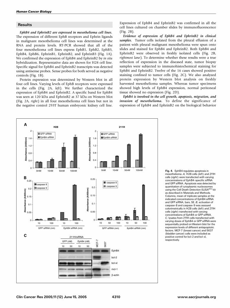
Fig. 4. EphB4 regulates apoptosis inmesothelioma. A, H28 cells (left) and 211Hcells (right) were transfected with varyingconcentrations of EphB4-specific siRNAandGFP siRNA. Apoptosis was detected byquantitation of cytoplasmic nucleosomesusing the Cell Death Detection ELISAplus kitas described in Materials andMethods.Columns, mean of triplicate samples at theindicated concentrations of EphB4 siRNAand GFP siRNA; bars, SE. B, activation ofcaspase-8 and caspase-9 was assayedcolorimetrically in H28 cells (left) and 211Hcells (right) transfected with varyingconcentrations of EphB4 or GFP siRNA.C, lysates from 211H cells transfected withvarying doses of EphB4 or GFP siRNAweresequentially probed onWestern blot for theexpression levels of different antiapoptoticfactors. MCF-7 (breast cancer) and 5637(bladder cancer) cells were included aspositive control for bcl-2 and bcl-xl,respectively.
Human Cancer Biology
www.aacrjournals.orgClin Cancer Res 2005;11(12) June15, 2005 4310

of mesothelioma, we first examined their effect on cell growthby knocking down the expression with siRNA. siRNA specific toEphB4 and EphrinB2 were screened in 293T cell line lackingendogenous expression but transiently transfected with expres-sion vectors for EphB4 and EphrinB2. siRNAs that knockdownexpression >80% after 48-hour treatment were selected forfuture use (data not shown). Activity of these active EphB4 andEphrinB2 siRNA was then confirmed in mesothelioma cell lines(data not shown). Transfection of H28 cell line with EphB4siRNA 472 produced a dose-dependent inhibition of cellviability. Transfection of EphrinB2 siRNA alone did not havethe inhibitory effect. Combination of EphB4 and EphrinB2siRNA did not augment growth inhibition over EphB4 siRNAalone (Fig. 3A, left). The EphB4 siRNA was active in a similar
dose-dependent manner in the 211H cell line as well (Fig. 3A,middle) but had no effect on the EphB4-negative Kaposisarcoma cell line, SLK (Fig. 3A, right).
The involvement of EphB4 in cell survival was furtherconfirmed with EphB4 antisense oligodeoxynucleotides. Incu-bation of cultured H28 or 211H cells with EphB4 antisense(AS-10) generated the same dose-dependent inhibition of cellgrowth (Fig. 3B, left and middle , respectively). AS-10 had noeffect on survival of SLK cells (Fig. 3B, right). These results indi-cate that EphB4 plays a critical role in mesothelioma cell survival.
Apoptosis as a result of EphB4 knockdown was tested bymeasuring levels of cytoplasmic nucleosomes. A dose-dependentincrease in apoptosis was observed when H28 cells (Fig. 4A, left)or 211H cells (Fig. 4A, right) were transfected with EphB4 siRNA
Fig. 5. EphB4 regulates cell migration andinvasion. A, H28 cells (left) and 211H cells(right) were seeded on six-well plates andcultureduntil 90% confluent. EphB4 siRNAor GFP siRNA (50 nmol/L each) wastransfected into cells with LipofectAMINE2000.The cell layer was scraped andphotographed immediately and after 24hours. Photomicrographs of representative�20 fields.B, a similar assay was done onH28 cells (left) and 211H cells (right) treatedwith AS-10 or sense oligodeoxynucleotide(10 Amol/L each). Representativephotomicrographs were taken immediatelyand 24 hours after wounding. C, Matrigelinvasion assay. InvasionofH28 cells (top) or211H cells (bottom) into Matrigel wasstudied in a modified Boyden chamberassay. Cells were transfected with EphB4siRNA or GFP siRNA (50 nmol/L each).Cells migrating to the underside of theMatrigel-coated insert in response to5 Ag/mL fibronectin in the lower chamberwere fixed and stained with Giemsa.Representative photomicrographs. Cellnumbers were counted in five individualhigh-powered fields. Columns, mean;bars, SE.
Expression of EphB4 inMesothelioma
www.aacrjournals.org Clin Cancer Res 2005;11(12) June15, 20054311

472, whereas GFP siRNA had no effect. DNA fragmentation inEphB4 siRNA 472–transfected H28 and 211H cells increasednearly 5-fold at a dose of 100 nmol/L when compared with GFPsiRNA–treated cells. Induction of apoptosis corresponded to anincrease in caspase-8 activity, whereas little change was observedin caspase-9 activity in both H28 cells (Fig. 4B, left) and 211Hcells (Fig. 4B, right). 211H cell lysates were also evaluated forlevels of antiapoptotic proteins. Knockdown of EphB4 resultedin a significant (60%) decline in bcl-xl levels, whereas bcl-2and mcl-1 remained unchanged (Fig. 4C). Similar results wereobtained with H28 cells (data not shown).
Mesothelioma is a locally advancing disease with frequentextension and growth into adjacent vital structures, such as thechest wall, heart, and esophagus. In an effort to study thisprocess in vitro , we did cell migration study with wound healingassay and invasion study with a modified Boyden chamberassay. When a wound was introduced into subconfluentmesothelioma cell culture over the course of 24 hours, cellsprogressively migrate and repopulate the area of the wound.However, when cells were transfected with EphB4 siRNA(Fig. 5A) or pretreated with AS-10 for 24 hours (Fig. 5B), amarked reduction in cell migration was observed comparedwith controls. In addition, knockdown of EphB4 expression inH28 or 211H cells with EphB4 siRNA 472 significantly reducedthe ability of these cells to invade Matrigel compared with GFP
siRNA treatment (Fig. 5C). Similar results were obtained withAS-10 treatment (data not shown).
EphB4 is biologically active in mesothelioma cell lines. Wenext examined whether EphB4 is phosphorylated in response tostimulation by ligand (EphrinB2). H28 cells were treated with3 Ag/mL clustered EphrinB2/Fc for 15 minutes. A low-levelbasal signal was detected in cells grown under serum-freeconditions, and this signal markedly increased followingexposure to external clustered ligand (Fig. 6A). Similar resultswere obtained with 211H cells (data not shown).
To ascertain the downstream effects of EphB4 stimulation,we studied the effect of EphB4 knockdown on the levels ofphosphorylated Akt. Treatment with EphB4 siRNA 472 led toan 85% reduction in phosphorylated Akt levels, whereas GFPsiRNA treatment had no effect (Fig. 6B). We also evaluatedthe role of EphB4 on MMP expression by quantitative RT-PCR. EphB4 knockdown by siRNA 472 led to a dose-dependent reduction in mRNA levels of MMP-2 (Fig. 6C) butnot MMP-9 (data not shown), whereas GFP siRNA had noeffect.
In vivo activity of EphB4 antisense oligodeoxynucleotide in axenograft model of malignant mesothelioma. H28 and 211Hcells were implanted s.c. in the flank of male athymic mice andanimals were treated with EphB4 AS-10, scrambled oligodeox-ynucleotide, or the diluent (PBS). Tumor volumes weresignificantly smaller in AS-10 oligodeoxynucleotide-treatedgroup compared with both control groups (Fig. 7A and B;P < 0.01). Western blot of extracts of 211H tumor tissueconfirmed that the expression of EphB4 in AS-10-treated groupwas markedly reduced compared with levels in the othertreatment groups (Fig. 7B, inset). Furthermore, AS-10 treatmentreduced cell proliferative index as measured by Ki-67 stainingby 50%, increased apoptosis 7-fold, and reduced microvasculardensity in the tumor (Fig. 6C). Similar effects were seen in H28tumors (data not shown).
Discussion
The successful treatment of mesothelioma to improvesurvival may depend on the identification of novel targets. Inthe present study, we found that EphB4 is expressed in all threesubtypes of mesothelioma (epitheloid, H28 and H2052;sarcomatoid, H2373; and biphasic, 211H). Each cell lineshowed a different level of EphB4 expression, with the highestlevel observed in epitheloid type cell line H28. The expressionof EphB4 in mesothelioma was confirmed in clinical samples.Due to the small number of cases, we are unable to analyze therelationship between the expression of EphB4 and tumorsubtype, grade, and stage. However, studies have shown thatoverexpression of EphB4 is inversely related to prognosis inendometrial carcinoma, squamous cell carcinoma of the headand neck, and breast cancer (29, 34–36). Further investigationin a large prospective study is required to determine theimportance of EphB4 as a marker of progression and metastasisfor mesothelioma.
We have shown that EphB4 expressed in mesothelioma cellscan be activated by stimulation with EphrinB2, and in tumors,EphB4 stimulation may be similarly achieved by thecoexpression of its ligand, EphrinB2. Similar coexpression ofthis receptor-ligand pair has been shown in other tumor types,including endometrial, lung, and colon carcinoma (30, 31, 37).
Fig. 6. EphB4 regulates Akt andMMP-2. A, H28 cells were serum starvedovernight and stimulatedwith 3 Ag/mLEphrinB2/Fc or Fc alone for15 minutes. Celllysates (100 Ag protein) were immunoprecipitated with EphB4 monoclonalantibody and duplicate blots were probedwith phosphotyrosine (p-Tyr) and EphB4antibody. B, H28 cells were transfected with 50 nmol/L EphB4 or GFP siRNA andcell lysates were probed for phosphorylated and total Akt levels.C, H28 cells weretransfected with varying doses of EphB4 or GFP siRNA and cellular mRNAwasextracted. Level of expression of MMP-2 mRNAwas quantitated by real-timeRT-PCR.
Human Cancer Biology
www.aacrjournals.orgClin Cancer Res 2005;11(12) June15, 2005 4312

On the other hand, a ligand-independent mechanism is alsoconceivable, relying on activation as a consequence ofoverexpression. Overexpression leading to constitutive activa-tion has been observed with EphB2 (38). To determine thepossible biological role of EphB4 in mesothelioma, weexamined changes in cell viability by targeted disruption ofEphB4 expression using antisense and siRNA in two differentmesothelioma cells (211H and H28). Down-regulation ofEphB4 led to a dose-dependent inhibition in cell viability. Thearrest in cell growth was mediated by an increase in apoptosis.The up-regulation of caspase-8 activity and relatively un-
changed caspase-9 activity indicate that apoptosis occurredthrough activation of the extrinsic pathway. We also foundthat bcl-xl levels fell with EphB4 knockdown, whereas noeffect was observed in the levels of bcl-2 and Mcl-1. Similar toour results, it has been reported previously that bcl-xl isexpressed at high levels in mesothelioma, whereas theexpression of bcl-2 and Mcl-1 is very low (39). Reduction inbcl-xl as observed in our studies may thus have significanteffect in mesothelioma cell viability. Further work is needed todefine the role of bcl-xl, if any, in EphB4-mediated cellsurvival signal in mesothelioma.
Fig. 7. EphB4 inhibits tumor growth inmurine mesotheliomaxenografts. A, 5� 106 H28 cells were implanted s.c. in theflank of male athymic mice.Treatment was started onday 5 after implant. Scrambled (S) orAS-10 (AS)oligodeoxynucleotide (20 mg/kg) or diluent (C) was injectedi.p. daily.Tumor volumewasmeasured thriceweekly as length�width2� 0.52 (mm3). B, a similar experiment was donewith 211H cells. At the completion of the study, tumors wereharvested and EphB4 levels were studied byWestern blottingonwhole cell lysates (inset). C, harvested 211H tumors weresectioned and histologic evaluationwas done by H&Estaining. Cell proliferative index (Ki-67) was evaluated byimmunohistochemistry and apoptosis was detected byTUNEL.The median number of positive cells in five randomhigh-power fields was quantitated by a blinded observer(shown below each picture) and difference between controland AS-10 treated tumors was compared by Student’s t test.Premortem infusion of FITC-labeled lectin was used toevaluate microvascular density. C, control (diluent);S, scrambled oligodeoxynucleotide; AS, EphB4-specificAS-10 oligodeoxynucleotide.
Expression of EphB4 inMesothelioma
www.aacrjournals.org Clin Cancer Res 2005;11(12) June15, 20054313

EphB4 activation allows interaction with a variety ofcytoplasmic signaling proteins. Many of these proteins havebeen implicated in regulating cell morphology, cytoskeletalorganization, attachment, and motility (40–42). Activation ofEphB4 leads to downstream signaling via PI3K; in agreementwith which, we show a decline in the activation status of thedownstream molecule Akt following EphB4 knockdown. To ourknowledge, this is the first report that shows a role for EphB4 inregulation of Akt with a potential role in tumor cell survival. Ithas been shown that EphB4 receptor signaling mediatesendothelial cell migration (40). EphB4/NeuT double transgenicanimals develop metastatic tumors in contrast to NeuTtransgenic animals, which form tumors only in the mammarygland without metastases (43). We found that the inhibition ofEphB4 significantly inhibits migration and invasion. MMPs playa significant role in tumor invasion and angiogenesis. MMP-2 isoverexpressed in mesothelioma (44). In a recent study, it hasbeen shown that MMP-2 and MMP-9 are activated and functionin endothelial migration on activation of EphB4 (40). Ourfindings that EphB4 regulates MMP-2 but not MMP-9 inmesothelioma may suggest a potential role for EphB4 inregulation of tumor cell migration and invasion. The inhibition
of EphB4, apart from directly inhibiting tumor survival, maythus prevent tumor progression as well as metastasis.
To further elucidate the possible therapeutic role oftargeting EphB4 in the treatment of mesothelioma, weconducted in vivo efficacy studies. EphB4-specific oligodeox-ynucleotide significantly inhibited tumor growth, reducedtumor cell proliferation, and induced apoptosis in comparisonwith control or scrambled oligodeoxynucleotide treatments.Tumor growth inhibition can result from a direct effect ontumor cells as observed in vitro. In addition, EphB4 on tumorcells bind EphrinB2 on tumor vessels and thereby induce anangiogenic response (45). Down-regulation of EphB4 maythus have an additional and independent effect on tumorvasculature as seen in our study. Hence, EphB4 knockdownhas a dual inhibitory effect on tumor growth by directlyinhibiting tumor cell survival as well as inhibiting tumor-associated angiogenesis.
In conclusion, we show the expression of the RTK EphB4 inmalignant mesothelioma. Receptor overexpression may favoruncontrolled cell growth, migration, and tumor progression.EphB4 targeting could serve as novel therapy for patients withmesothelioma.
References1. Carbone M, Kratzke RA,Testa JR. The pathogenesisof mesothelioma. Semin Oncol 2002;29:2^17.2. Johansson L, Linden CJ. Aspects of histopathologicsubtype as a prognostic factor in 85 pleural mesothe-liomas. Chest1996;109:109^14.3. Carbone M, Kratzke RA,Testa JR. The pathogenesisof mesothelioma. Semin Oncol 2002;29:2^17.4. BrennerJ, Sordillo PP, Magill GB, Golbey RB. Malig-nant mesothelioma of the pleura: review of 123 pa-tients. Cancer1982;49:2431^5.5. Rusch VW, Piantadosi S, Holmes EC. The role ofextrapleural pneumonectomy in malignant pleural me-sothelioma. ALungCancer StudyGroup trial. JThoracCardiovasc Surg1991;102:1^9.6. Pass HI, Kranda K,Temeck BK, Feuerstein I, SteinbergSM. Surgically debulked malignant pleural mesotheli-oma: results and prognostic factors. Ann Surg Oncol1997;4:215^22.7. Bard M, Ruffie P. Malignant mesothelioma. Medicaloncology: standards, new trends, trialsJthe Frenchexperience. Lung Cancer 2004;45:S129^31.8. Steele JP, Klabatsa A. Chemotherapy options andnew advances in malignant pleural mesothelioma.Ann Oncol 2005;16:345^51.9.Vogelzang NJ, Rusthoven JJ, Symanowski J, et al.Phase III study of pemetrexed in combination withcisplatin versus cisplatin alone in patients with malig-nant pleural mesothelioma. J Clin Oncol 2003;21:2636^44.10.Masood R, Kundra A, Zhu S, et al. Malignant me-sothelioma growth inhibition by agents that targetthe VEGF and VEGF-C autocrine loops. Int J Cancer2003;104:603^10.11. Strizzi L, CatalanoA,Vianale G, et al.Vascular endo-thelial growth factor is an autocrine growth factor inhuman malignant mesothelioma. J Pathol 2001;193:468^75.12. Kindler HL,Vogelzang NJ, Chien K, et al. SU5416 inmalignant mesothelioma: a University of ChicagoPhase II Consortium Study. Proc Am Soc Clin Oncol2001;20:341.13. Janne PA,Taffaro ML, Salgia R, Johnson BE. Inhibi-tion of epidermal growth factor receptor signaling inmalignant pleural mesothelioma. Cancer Res 2002;62:5242^7.14. LangerakAW,DeLaat PA,VanDerCA, et al.Expres-
sion of platelet-derived growth factor (PDGF) andPDGF receptors in human malignant mesotheliomain vitro and in vivo. JPathol1996;178:151^60.15.GerwinBI, LechnerJF, Reddel RR, et al. Comparisonof production of transforming growth factor-h andplatelet derived growth factor by normalhumanmeso-thelial cells and mesothelioma cell lines. Cancer Res1987;476:180^4.16.Versnel MA, Claesson-Welsh L, Hammacher A,et al. Human malignant mesothelioma cell lines ex-press PDGF h-receptors whereas cultured normalmesothelial cells express predominantly PDGFareceptors. Oncogene 1991;6:2005^11.17. Ciardiello R, Caputo R, BiancoV, et al. Antitumoreffect and potentiation of cytotoxic drug activity inhuman cells by ZD1839 (Iressa), an epidermal growthfactor selective tyrosine kinase inhibitor. Clin CancerRes 2000;6:2053^63.18. Dorai T, Kobayashi H, HollandJF, OhnumaT.Modu-lation of platelet-derived growth factor-h mRNA ex-pression and cell growth in a human mesotheliomacell line by a hammerhead ribozyme. Mol Pharmacol1994;46:437^44.19. Pasquale EB.The Eph family of receptors. Curr OpinCell Biol1997;9:608^19.20. Gerety SS, Wang HU, Chen ZF, Anderson DJ.Symmetrical mutant phenotypes of the receptorEphB4 and its specific transmembrane ligand Ephrin-B2 in cardiovascular development. Mol Cell 1999;4:403^13.21. Tickle C, Altabef M. Epithelial cell movements andinteractions in limb, neural crest and vasculature. CurrOpin Genet Dev1999;9:455^60.22. Holder N, Klein R. Eph receptors and ephrins: effec-tors of morphogenesis. Development 1999;126:2033^44.23.WangHU,ChenZF,AndersonDJ.Molecular distinc-tion and angiogenic interaction between embryonicarteries and veins revealed by ephrin-B2 and its recep-tor Eph-B4. Cell1998;93:741^53.24. Shin D, Garcia-Cardena G, Hayashi S, et al. Expres-sion of ephrinB2 identifies a stable genetic differencebetween arterial and venous vascular smooth muscleas well as endothelial cells, and marks subsets ofmicrovessels at sites of adult neovascularization. DevBiol 2001;230:139^50.
25. DodeletVC, Pasquale EB. Eph receptors and ephrinligands: embryogenesis to tumorigenesis. Oncogene2000;19:5614^9.26. Stephenson SA, Slomka S, Douglas EL, Hewett PJ,Hardingham JE. Receptor protein tyrosine kinaseEphB4 is up-regulated in colon cancer. BMCMol Biol2001;2:15.27. Berclaz G, Andres AC, Albrecht D, et al. Expressionof the receptor protein tyrosine kinase myk-1/htk innormal and malignant mammary epithelium. BiochemBiophys Res Commun1996;226:869^75.28. Andres AC, Zuercher G, Djonov V, Flueck M,Ziemiecki A. Protein tyrosine kinase expressionduring the estrous cycle and carcinogenesis of themammary gland. Int J Cancer 1995;63:288^96.29. Berclaz G, Karamitopoulou E, Mazzucchelli L, et al.Activation of the receptor protein tyrosine kinaseEphB4 in endometrial hyperplasia and endometrialcarcinoma. Ann Oncol 2003;14:220^6.30.Takai N, Miyazaki T, Fujisawa K, Nasu K, MiyakawaI. Expression of receptor tyrosine kinase EphB4 andits ligand ephrin-B2 is associated with malignant po-tential in endometrial cancer. Oncol Rep 2001;8:567^73.31. Tang XX, Brodeur GM, Campling BG, Ikegaki N.Coexpression of transcripts encoding EPHB receptorprotein tyrosine kinases and their ephrin-B ligands inhuman small cell lung carcinoma. Clin Cancer Res1999;5:455^60.32. Munaut C, Noel A, Hougrand O, Foidart JM,Boniver J, Deprez M. Vascular endothelial growthfactor expression correlates with matrix metallo-proteinases MT1-MMP, MMP-2 and MMP-9 inhuman glioblastomas. Int J Cancer 2003;106:848^55.33.MasoodR, XiaG, SmithDL, et al. Ephrin B2expres-sion in Kaposi sarcoma is inducedby humanherpesvi-rus type 8: phenotype switch from venous to arterialendothelium. Blood 2005;105:1310^8.34. Sinha UK, Kundra A, Scalia P, et al. Expression ofEphB4 in head and neck squamous cell carcinoma.Ear NoseThroatJ 2003;82:866, 869^70, 887.35. CromerA, Carles A, Millon R, et al. Identification ofgenes associated with tumorigenesis and metastaticpotential of hypopharyngeal cancer by microarrayanalysis. Oncogene 2004;23:2484^98.
Human Cancer Biology
www.aacrjournals.orgClin Cancer Res 2005;11(12) June15, 2005 4314

36.Wu Q, Suo Z, Risberg B, Karlsson MG, VillmanK, Nesland JM. Expression of Ephb2 and Ephb4in breast carcinoma. Pathol Oncol Res 2004;10:26^33.37. Liu W, Ahmad SA, Jung YD, et al. Coexpressionof ephrin-Bs and their receptors in colon carcinoma.Cancer 2002;94:934^9.38. Zisch AH, StallcupWB, Chong LD, et al. Tyrosinephosphorylation of L1 family adhesion molecules:implication of the Eph kinase Cek5. J NeurosciRes 1997;47:655^65.39. Smythe WR, Mohuiddin I, Ozveran M, Cao XX.Antisense therapy for malignant mesotheliomawith oligonucleotides targeting the bcl-xl gene
product. J Thorac Cardiovasc Surg 2002;123:1191^8.40. Steinle JJ, Meininger CJ, Forough R, Wu G,Wu MH, Granger HJ. Eph B4 receptor signalingmediates endothelial cell migration and proliferationvia the phosphatidylinositol 3-kinase pathway. J BiolChem 2002;277:43830^5.41. Oates AC, Lackmann M, Power MA, et al. Anearly developmental role for eph-ephrin interactionduring vertebrate gastrulation. Mech Dev 1999;83:77^94.42. O’Leary DD,Wilkinson DG. Eph receptors and eph-rins in neural development. Curr Opin Neurobiol 1999;9:65^73.
43. Munarini N, Jager R, Abderhalden S, et al. Alteredmammary epithelial development, pattern formationand involution in transgenic mice expressing theEphB4 receptor tyrosine kinase. J Cell Sci 2002;115:25^37.44. Edwards JG, McLaren J, Jones JL, Waller DA,O’Byrne KJ. Matrix metalloproteinases 2 and 9(gelatinases A and B) expression in malignant me-sothelioma and benign pleura. Br J Cancer 2003;88:1553^9.45. Noren NK, Lu M, Freeman AL, Koolpe M, PasqualeEB. Interplay between EphB4 on tumor cells and vas-cular ephrin-B2 regulates tumor growth. Proc NatlAcad Sci US A 2004;101:5583^8.
Expression of EphB4 inMesothelioma
www.aacrjournals.org Clin Cancer Res 2005;11(12) June15, 20054315