
Rapid development of vaccine protection in macaques by live-attenuated simian immunodeficiency virus
Upload
independentCategory
view
1download
0
JOURNAL OF VIROLOGY, Oct. 2010, p. 10087–10101 Vol. 84, No. 190022-538X/10/$12.00 doi:10.1128/JVI.02425-09Copyright © 2010, American Society for Microbiology. All Rights Reserved.
Development of a Targeted Gene Vector Platform Basedon Simian Adenovirus Serotype 24�
Natalya Belousova,1§ Galina Mikheeva,1§ Chiyi Xiong,1 Suren Soghomonian,1 Daniel Young,1
Lucia Le Roux,1 Katherine Naff,2 Luc Bidaut,3 Wei Wei,4 Chun Li,1Juri Gelovani,1 and Victor Krasnykh1*
Departments of Experimental Diagnostic Imaging,1 Veterinary Medicine and Surgery,2 Imaging Physics,3 and Biostatistics,4
the University of Texas M. D. Anderson Cancer Center, Houston, Texas 77030
Received 17 November 2009/Accepted 22 June 2010
Efforts to develop adenovirus vectors suitable for genetic interventions in humans have identified threemajor limitations of the most frequently used vector prototype, human adenovirus serotype 5 (Ad5). Theselimitations—widespread preexisting anti-Ad5 immunity in humans, the high rate of transduction ofnormal nontarget tissues, and the lack of target-specific gene delivery—justify the exploration of other Adserotypes as vector prototypes. In this paper, we describe the development of an alternative vectorplatform using simian Ad serotype 24 (sAd24). We found that sAd24 virions formed unstable complexeswith blood coagulation factor X and, because of that, transduced the liver and other organs at low levelswhen administered intravenously. The overall pattern of biodistribution of sAd24 particles was similar,however, to that of Ad5, and the intravenously injected sAd24 was cleared by Kupffer cells, leading to theirdepletion. We modified the virus’s fiber protein to design a Her2-specific derivative of sAd24 capable ofinfecting target human tumor cells in vitro. In the presence of neutralizing anti-Ad5 antibodies, Her2-mediated infection with targeted sAd24 compared favorably to that with the Ad5-derived vector. Whenused to target Her2-expressing tumors in animals, this fiber-modified vector achieved a higher level ofgene transfer to metastasis-containing murine lungs than to tumor-free lungs. In aggregate, these studiesprovide important insights into sAd24 biology, identify its advantages and limitations as a vector proto-type, and are thus essential for further development of an sAd24-based gene delivery platform.
Ads are a highly diverse family of nonenveloped, DNA-containing viruses isolated from humans, various mammals,and other vertebrates. Despite this diversity, the structure of atypical Ad particle is highly conserved among various Ad se-rotypes. The double-stranded DNA genome of Ad associatedwith the core proteins is encapsidated in an icosahedral proteinshell. The most abundant structural component of an Ad par-ticle is the hexon protein that forms the facets of the capsid.The other two major capsid proteins, the penton base and thefiber, associate at a 5-to-3 ratio to form the penton capsomerslocated at each of the apexes of the virion.
Efforts to develop efficient Ad vectors for gene delivery inhumans have centered primarily on human Ad serotype 5,owing to its well-elucidated biology. However, testing of Ad5vectors in preclinical studies and gene therapy clinical trialsidentified several important drawbacks: widespread preexistinganti-Ad5 immunity in humans (12, 55), an unacceptably largeamount of damage to normal nontarget tissues, which causessevere toxicity (28, 37), and low levels of target-specific genedelivery (15, 18, 19). Collectively, these findings warranted thesearch for Ad vector prototypes better suited for gene transferin humans.
Recent improvements in vector technology have exploitedthe unique biological properties of Ad serotypes other thanAd5. For instance, the low seroprevalence of certain Ads inhumans has justified the use of these viruses as alternativevector prototypes to overcome preexisting anti-Ad5 immunity.Indeed, it has been shown that the efficacy of in vivo genedelivery by vectors derived from simian Ads is minimally af-fected by Ad5-neutralizing antibodies (16, 45, 55), makingthese viruses promising vector prototypes.
The use of vectors based on Ads other than Ad5 may alsooffer the solution to the problem of the undesired in vivotransduction of nontarget tissues. Recent studies showedthat the high-affinity association of FX with the hexon ofsystemically injected Ad5 directs the virus to FX receptorsabundantly expressed in the liver, causing massive hepatictransduction (22, 56). Importantly, some Ad serotypes, suchas Ad26 and Ad48, do not bind FX and cause no detectabletransduction of the liver on vascular delivery (56). Thesefindings provided the rationale for the recently reportedimprovements of Ad5 vectors through genetic engraftinginto their hexons of either the hypervariable regions or theindividual amino acids from the hexons of these FX-non-binding serotypes (2, 42). They also provided an additionaljustification for developing such Ad serotypes as alternativegene vector platforms.
While the use of alternative Ad serotypes promises to solvetwo of the main drawbacks of Ad5 vectors, it does not addressthe problem of inefficient gene delivery to target tissues. Thus,development of an Ad vector targeting strategy applicable to
* Corresponding author. Mailing address: Department of Experi-mental Diagnostic Imaging, The University of Texas M. D. AndersonCancer Center, 1515 Holcombe Boulevard, Unit 59, Houston, TX77030. Phone: (713) 563-4873. Fax: (713) 563-4894. E-mail: [email protected].
§ These authors contributed equally to this work.� Published ahead of print on 14 July 2010.
10087

these promising vector prototypes would constitute an impor-tant step toward the use of Ads in humans.
Most attempts to design gene delivery vectors with selectiv-ity for specific markers on target cells have involved geneticalteration of the Ad5 fiber protein. The fiber protein is themediator of the initial high-affinity interaction in vitro betweenthe Ad virion and the primary receptor (14, 41) that is followedby secondary contact between the Ad penton base protein andcellular integrins (58, 59), which triggers internalization of Adby the cell. The primary receptor binding is facilitated by theglobular, carboxy-terminal knob domain of the fiber (20, 29).The knob also initiates the fiber trimerization (36) that isessential for its encapsidation (38) through anchoring of theamino-terminal tail of the protein within the penton base pen-tamer. The knob is extended away from the capsid surface bythe fiber’s central shaft domain, which is structured as a �-spi-ral (54). The primary Ad receptors identified to date, such asCAR (8, 51), CD46 (17), sialic acid (4), and CD80 and CD86(50), are suboptimal targets for gene therapeutics becausetheir expression is not correlated with disease.
The approach to Ad targeting initially developed for theAd5 fiber protein includes identification of the receptor-bind-ing site within the knob (9, 23, 43), its inactivation by mutagen-esis (24, 43), and introduction of a target-specific ligand withinthe structure of the modified knob (6, 15, 31, 33, 60, 61). Theunique structures of Ad knobs and their receptor-binding sites,together with the limited tolerance of the knob structure forligand incorporation, made it difficult to apply this strategy tounderexplored fibers, including those of the Ads that are beingdeveloped as alternative vector prototypes.
To overcome these challenges, alternative strategies basedon fiber knob replacement rather than modification have beenproposed. In these approaches, the knob of the Ad5 fiber isdeleted and trimerization of the knobless protein restored byfusing it with a trimerization moiety, such as the coiled-coildomain of the retrovirus envelope glycoprotein (53), the neckregion peptide of human lung surfactant D (32, 48), or reovirussigma-1 protein (35, 47, 52).
We previously reported successful targeting of the Ad5 fiberthrough replacement of its knob with trimeric fragments of thefibritin protein of phage T4. Fusing the peptide and polypep-tide ligands to the fiber-fibritin chimeras allowed successfultargeting of Ad5 to a designed artificial receptor (26) and tonatural receptors such as CD40 (5) and Her2 (7).
Given the promise of non-Ad5 serotypes as gene vectorprototypes, we sought to test whether the fiber of one of theseAds could be targeted by knob replacement. We applied thisstrategy to the fiber of sAd24 (also known as Pan7), which isbeing developed as a vector for genetic immunization owing toits low seroprevalence in humans (45, 64) and its antigenicdistinction from Ad5 (44–46, 64). The sAd24 fiber is an un-characterized protein, and neither its native receptor nor itsreceptor-binding site have been identified. Nevertheless, as wereport here, it has been possible to target this protein entirelyon the basis of its predicted domain structure. The designedsAd24 fiber chimera efficiently incorporated in sAd24 particlesand made viral infection dependent on the presence of theintended target receptor, Her2, which was chosen on the basisof the established correlation of its expression by human tu-mors, the aggressiveness of those tumors, and the poor prog-
nosis for patients. We also demonstrated in this study the poorstability of FX-sAd24 complexes, which resulted in greatlydiminished liver transduction by the sAd24-derived vectorcompared to that of the Ad5-derived vectors. In addition, westudied aspects of sAd24 biology directly relevant to the in vivouse of sAd24-derived vectors and tested an Her2-targeted ver-sion of sAd24 in two animal models.
Collectively, these studies provide important insights intosAd24 biology and identify its advantages and limitations as avector prototype and are therefore essential for further devel-opment of an sAd24-based gene delivery platform.
MATERIALS AND METHODS
Abbreviations. aa, amino acid; Ab, antibody; Ad, adenovirus; Ad5, adenovirusserotype 5; ANOVA, analysis of variance; CAR, coxsackievirus and Ad receptor;CT, computed tomography; EGFP, enhanced green fluorescent protein; FEAU,fluoro-5-ethyl-1-�-D-arabinofuranosyluracil; Fluc, firefly luciferase; FX, bloodcoagulation factor X; Her2, human epidermal growth factor receptor type 2;hRluc, codon-optimized Renilla luciferase; HVR, hypervariable region; IL, in-terleukin; IFN, interferon; kd, dissociation rate constant; KD, equilibrium disso-ciation constant; KCs, Kupffer cells; MAb, monoclonal antibody; MCP, mono-cyte chemotactic protein; MOI, multiplicity of infection; ORF, open readingframe; PBS, phosphate-buffered saline solution; PET, positron emission tomog-raphy; qPCR, quantitative PCR; Rluc, Renilla luciferase; ROI, region of interest;sAd24, simian adenovirus serotype 24; SDS-PAGE, sodium dodecyl sulfate poly-acrylamide gel electrophoresis; TNF, tumor necrosis factor; TL, genetic fusion ofHSV tk and Fluc; HSV tk, herpes simplex virus thymidine kinase; VP, viralparticles; wt, wild type.
Cells and antibodies. Cell lines 293, 293T, HCC1954, SKOV3, BT474, MDA-MB-361, and SK-BR-3 (all from American Type Culture Collection, Manassas,VA), human breast carcinoma cell line MDA-MB-231 (provided by Janet Price,the University of Texas M. D. Anderson Cancer Center), 293/Her2 cells (7), and293/F28 cells (5) were maintained as described previously.
Cell line MDA-MB-231/hRluc-EGFP was made to express a hRluc-EGFPfusion protein by transduction of MDA-MB-231 cells with a retroviral vectorencoding this protein. To generate this cell line, MDA-MB-231 cells were incu-bated overnight with medium containing the hRluc/EGFP-expressing retroviralvector LhRluc/EGFP (described below) and polybrene at a final concentration of8 �g/ml. The transduced cells were selected by adding G418 at a final concen-tration of 1 mg/ml, and the GFP-positive MDA-MB-231/hRluc-EGFP cells wereselected from the G418-resistant pool by four sequential rounds of cell sorting.
Cell line MDA-MB-231/Her2, a Her2-expressing derivative of MDA-MB-231/hRluc-EGFP cells, was made by transduction of MDA-MB-231/hRluc-EGFPcells with a Her2-expressing retrovirus. To generate this cell line, MDA-MB-231/hRluc-EGFP cells were incubated overnight with medium containing theHer2-expressing retroviral vector QCXIP.Her2 and polybrene. The transducedcells were selected in medium containing puromycin at a final concentration of0.7 �g/ml. Her2 expression in individual puromycin-resistant clones was con-firmed by fluorescence-activated cell sorting.
293/FsAd24 cells constitutively expressing wt sAd24 fiber were derived from 293cells through transfection with the pFXsAd24 plasmid (described below) con-taining the wt sAd24 fiber ORF. Hybridoma line 20C1, expressing anti-sAd24fiber MAb, was generated using recombinant N100sAd24 protein (describedbelow) in a standard protocol established at the Monoclonal Antibody Facility ofthe M. D. Anderson Cancer Center.
Anti-fiber tail mouse MAb 4D2 (21) was a kind gift from Jeff Engler (Uni-versity of Alabama, Birmingham). Anti-Her2 mouse MAb 3B5 was purchasedfrom Merck KGaA (Darmstadt, Germany). Anti-fibritin mouse MAb 5E1 wasdescribed in our previous report (7).
Genetic engineering. (i) Plasmid for N100sAd24 protein expression. To ex-press the amino-terminal segment of the sAd24 fiber protein for the subsequentgeneration of MAbs, a fragment of this fiber ORF containing codons 2 through102 was PCR amplified and cloned in pET20b (EMD Biosciences, Gibbstown,NJ). In the resultant plasmid, pET20b.6H.N100sAd24, this ORF was fused at its5� end with the six-histidine-tag-encoding sequence.
(ii) Plasmids for expression of wt and modified sAd24 fiber proteins. Theseplasmids were designed for the subsequent generation of cell line 293/FsAd24 andfor transient expression of the fiber chimera. To this end, the ORF of the wt Ad5fiber was deleted from the previously described pVSII (25) by using inverse PCRand was replaced with either the ORF of the wt sAd24 or the ORF of the sAd24
10088 BELOUSOVA ET AL. J. VIROL.

fiber chimera, FsAd2411FHer2:7, yielding pFXsAd24 and pFusion2724, respec-tively. FsAd2411FHer2:7 was designed to contain the tail and the shaft domains ofthe sAd24 fiber (aa 1 to 264 [GenBank accession number AY530878]) followedby the carboxy-terminal fragment of the phage T4 fibritin protein comprising thelast two a-helical repeats of the stalk and the foldon domain (aa 265 to 359 of thechimera, corresponding to aa 393 to 487 of the fibritin [GenBank accessionnumber L43611]), the (Gly4Ser)3 linker (aa 360 to 374), and the Her2-specificaffibody Zher2:7 (aa 375 to 432 [62]).
(iii) sAd24 shuttle plasmids. The shuttle plasmid for replacement of the E1region, pE1sAd24002, is a derivative of pZeRo2 (Invitrogen, Carlsbad, CA),which contains the previously described TL-expressing gene cassette (7) drivenby the immediate-early cytomegalovirus promoter, which is flanked by the seg-ments of the wt sAd24 genome that are immediately adjacent to the E1 region.The left and right flanks correspond to positions 1 to 474 and 3420 to 5893 in thewt sAd24 genome, respectively. The fiber gene shuttle plasmid, pFs24027, also aderivative of pZeRo2, contains the FsAd2411FHer2:7-encoding sequence flankedby the sequences of the wt sAd24 genome that surround the wt fiber gene(positions 29999 to 32111 and 33423 to 34306 in the wt sAd24 genome, respec-tively).
(iv) sAd24 rescue plasmids. The master rescue plasmid containing the unmod-ified sAd24 genome was designed by cloning full-sized sAd24 genomic DNA inplace of the Ad5 genome in the previously made pVK50 (27), yielding pRs24000.To design pRs24002, the TL-expressing cassette was transferred into the sAd24genome by recombination between pRs24000 linearized with SnaBI and thepE1sAd24002 shuttle. Similarly, the wt fiber gene in pRs24002 was replaced withthe FsAd2411FHer2:7 gene, using the pFs24027 shuttle and SwaI-cut pRs24002,thus yielding pRs24004. Ad genomes in all rescue vectors are flanked with PacIsites that are used to release these genomes for subsequent virus rescue bytransfection.
(v) Retrovirus plasmid vectors. The genome of retroviral vector LhRluc/EGFP, which expresses the fusion of hRluc and EGFP, was obtained by trans-ferring the hRluc coding sequence from phRL null (Promega, Madison, WI) intothe pLEGFP-N1 plasmid (BD Biosciences, San Jose, CA). The genome ofretroviral vector QCXIP.Her2, which expresses Her2, was obtained by transfer-ring the ORF of Her2 from the pIRES.neo3-c-erbB2 plasmid vector (7) intopQCXIP (Clontech, Mountain View, CA).
Additional experimental details, maps, and sequences of the designed DNAmolecules are available upon request.
Viruses. Stock of sAd24 was obtained from the American Type Culture Col-lection (VR-593) and expanded in 293 cells. The genomes of recombinant sAd24vectors were assembled in plasmid vectors using homologous recombination inEscherichia coli strain BJ5183 between the corresponding shuttle and rescueplasmids as previously described (11). The genes of the E1 region (positions 475to 3419 in the sAd24 genome) within these genomes were replaced with acytomegalovirus promoter-driven expression cassette containing the TL trans-gene. The viruses were rescued by transfecting either 293 or 293/FsAd24 cells withthe Ad genomes released from the corresponding rescue plasmids by restrictionendonuclease digestion. All Ads were purified by double banding in CsCl gra-dients, subjected to dialysis against a buffer containing 10 mM Tris (pH 8.0), 50mM NaCl, 2 mM MgCl, and 10% glycerol, and stored at �80°C. The titers of theviral preparations were determined by using the total protein concentration aspreviously described (7).
hRluc/EGFP- and Her2-expressing retroviruses were rescued by transfectingthe packaging 293GPG cells (40) with pLhRluc/EGFP and pQCXIP.Her2 ret-roviral plasmids, respectively, using Lipofectamine reagent (Invitrogen). Themedia containing retroviruses was collected at 72 to 96 h after transfection,filtered through a 0.45-�m-pore-size filter, and stored at �80°C.
Surface plasmon resonance experiments. Biosensor studies were done essen-tially as described by Kalyuzhniy et al. (22). In brief, purified Ad virions wereimmobilized on a CM5 sensor chip (GE Healthcare, Piscataway, NJ) by using anamine-coupling reaction as described by the manufacturer. Affinity measure-ments were done using a Biacore 3000 instrument (GE Healthcare). The chipwas probed with murine factor X (Haematologic Technologies, Essex Junction,VT) in an HBSP running buffer (0.01 M HEPES [pH 7.4], 0.15 M NaCl, and0.005% [vol/vol] Surfactant P20) supplemented with 1 mM CaCl2, 0.5 mMMgCl2, and 0.1 mg/ml of bovine serum albumin. The 5-min injection of FX wasfollowed by 40 min of dissociation. The chips were regenerated after eachbinding cycle, using 30-s pulses of buffer containing 3 mM EDTA. The data wereevaluated with BIAevaluation software version 3.1 (GE Healthcare) with theapplication of a simple 1:1 binding mass transfer model. The obtained sensor-grams were fitted globally over the whole range of injected concentrations andsimultaneously over the association and dissociation phases. Equilibrium disso-ciation constants were calculated from the measured rate constants.
Expression and purification of recombinant proteins. The N100sAd24 proteinwas expressed in E. coli Rosetta2(DE3)pLysS cells (EMD Biosciences) trans-formed with pET20b.6H.N100sAd24. Protein expression was induced by growingthe culture in Overnight Express medium (EMD Biosciences) per the vendor’srecommendations. The bacteria were collected by centrifugation and subjectedto lysis by sonication, and the six-histamine-tagged N100sAd24 was purified fromthe lysate by immobilized metal ion affinity chromatography on a HisTrap col-umn (GE Healthcare).
Methods used for bacterial expression and purification of Her2-specific affi-bodies were described elsewhere (7).
In vitro gene transfer. Transduction of cells was done as previously described(7). Briefly, cells grown in 24-well plates were rinsed with fresh medium andincubated for 10 min with either medium alone or medium containing theHer2-binding Zher2:4 affibody. Next, the cells were infected for 30 min with oneof the Ad vectors. The medium was replaced with fresh medium, and theincubation was continued for 24 h, after which the cells were subjected to lysis,using reporter lysis buffer (Promega). The Fluc activity in the lysate was mea-sured in a tube luminometer (Sirius; Berthold, Pforzheim, Germany) using aluciferase assay system (Promega).
In the virus neutralization assay, the viruses were mixed with the dilutedanti-Ad5 sera prior to being added to cells.
Flow cytometry. Cell attachment by the targeted fiber was detected accordingto a previously described protocol (7). In brief, the cells, suspended in PBS with0.1% bovine serum albumin and 0.01% NaN3 were incubated with aliquots ofcleared lysates of 293T cells either mock transfected or transfected with thechimera-expressing pFXsAd24 plasmid. The cell-bound chimera was detected byanti-fibritin stalk MAb 5E1 (7) and secondary goat anti-mouse IgG Ab labeledwith Alexa Fluor 488 (Invitrogen).
Western blotting. Fibers, either transiently expressed in 293T cells or con-tained in purified viruses, were detected by Western blotting essentially as pre-viously reported (7). However, because the anti-sAd24 fiber MAb 20C1 recog-nizes its epitope only in a fully denatured protein, detection of the bandscorresponding to nondenatured wt sAd24 fiber and the FsAd2411FHer2:7 fiberchimera required a modified, two-step procedure. First, these proteins wereseparated by electrophoresis without prior denaturation; second, after elec-trotransfer of the proteins on a membrane, they were denatured by boilingthe membrane in the transfer buffer for 10 min and were then probed withantibodies.
Studies with mice and experiments using murine tissues. All experimentsinvolving animals were done according to protocols approved by the InstitutionalAnimal Care and Use Committee of the M. D. Anderson Cancer Center. Onlyreplication-deficient Ad vectors with E1 deleted were used in these studies.
In vivo gene expression. To study the in vivo patterns of transgene expressionby Ad vectors, 6- to 8-week-old female NCr nu/nu mice (Taconic, Hudson, NY)were injected intravenously (via the tail vein) with either PBS or the tested Advector diluted in 100 �l of PBS. Forty-eight hours later, the mice were anesthe-tized by isoflurane inhalation and injected in the tail vein with a solution of theFluc substrate D-luciferin (Caliper, Hopkinton, MA) in PBS (30 mg/ml, 100 �lper injection). The animals were transferred into the chamber of an IVIS 200imaging system (Caliper) and imaged while under continuous anesthesia. Theregion of interest was drawn over the body area corresponding to the liver. Thebioluminescence signals measured within the region of interest were then ana-lyzed using Living Image software version 3.0 (Caliper). Twenty-four hours later,the mice were euthanized by CO2 inhalation, and individual organs were col-lected.
Pieces of the tissue samples were weighed and homogenized in PBS, usingTissueLyser (Qiagen, Valencia, CA) at 30 Hz for 1 min. Aliquots of homoge-nized tissues were subjected to lysis in reporter lysis buffer (Promega); thesamples were cleared of debris by centrifugation, and the Fluc activity in thesupernatants was measured as already described under “In vitro gene transfer.”
The Ad-treated mice were also subjected to PET scans 48 h after beinginjected with the virus. The animals were anesthetized and injected via the tailvein with 18F-labeled FEAU (100 �Ci in 100 �l of PBS) prepared as previouslydescribed (1). Two hours later, anesthetized mice were secured on customholders to minimize motion across modalities and underwent a 10-min session ofstatic PET imaging done in two-dimensional mode using an R4 microPETscanner (Concorde Microsystems, Knoxville, TN). The images were recon-structed using an ordered subset expectation maximization algorithm. Immedi-ately after the PET scan, each animal underwent CT scanning in an RS-9Micro-CT instrument (General Electric, London, Ontario) using the followingparameters: 80 kVp, 450 A, 100 ms per frame, and calibration images (bright anddark fields). Next, PET data were registered to the reference anatomical CT datathrough a combination of interactive translations and rotations to match major
VOL. 84, 2010 SIMIAN ADENOVIRUS VECTOR TARGETED TO Her2 10089

structures such as body outlines that are visible on both modalities. This initialregistration was then further refined through the multiresolution iterative max-imization of a normalized mutual-information cost function (30). Once regis-tered, the PET and CT data sets were fused together, processed, and visualizedas described elsewhere (10) to localize tracer signals in relation to the anatomy.
Ad biodistribution experiments. To establish patterns of biodistribution of Adparticles, the mice were injected intravenously (tail vein) with 1011 VP of one ofthe tested Ads and were killed at either 1 h or 24 h after injection. The organswere collected, weighed, and homogenized in PBS as described above. Protein-ase K, SDS, and EDTA were added to aliquots of these homogenates to finalconcentrations of 200 �g/ml, 5%, and 100 mM, respectively, and the sampleswere incubated overnight at 56°C. Proteinase K was inactivated by incubating thelysates at 95°C for 10 min; 100-fold dilutions of these lysates in water were usedas templates for qPCR (3 �l per 25 �l of reaction mixture). Ad DNA wasdetected using primers and probes complementary to the E4 regions of therespective genomes. Ad5 genomes were detected with primers Ad5.F (TGG CTTCGG GTT CTA TGT AAA CTC) and Ad5.R (TTC TGC GGT GGT GGATGT TA) and probe Ad5.Pr (TTC ATG CGC CGC TGC CCT G), which waslabeled with 6-carboxyfluorescein (6FAM); sAd24 genomes were detected withprimers sAd24.F (TGG CCG GCG TGA ATG) and sAd24.R (TCG CGG ACGCCA ATG) and a 6FAM-labeled probe, sAd24.Pr (TTC GAC AAG ATG AATACA CCC CCG GA). Reactions were run in an ABI 7500 instrument, and thedata were processed using ABI 7500 software. Statistical comparison of Ad DNAcopy numbers per organ for the different vectors was performed separately foreach organ at each time point. No multiple-comparison adjustment was imple-mented for these analyses. DNA copy numbers were analyzed on the logarithmicscale by two-sample t tests. The tests were two sided, and P values of 0.05 or lesswere considered statistically significant. Statistical analysis was carried out usingSAS version 9 software (SAS Institute, Cary, NC).
Sequestration of Ad by KCs. For immunofluorescent detection of Ad virionsand KCs, livers were collected from mice injected with 4 � 1010 VP of Ads (3mice per group) at either 10 min or 16 h after intravenous administration of thevectors. The livers were fixed and embedded in paraffin. The 5-�m sections wereprepared using a rotary microtome Leica RM2255 instrument (Leica Microsys-tems, Bannockburn, IL). To detect KCs, the sections were incubated with ratanti-mouse IgG Ab (BD Biosciences) to F4/80, a recognized biomarker of murinemacrophages, followed by the secondary goat anti-rat IgG Ab labeled with AlexaFluor 488 (Invitrogen). The cells containing Ad virions of both serotypes weredetected with rabbit polyclonal anti-Ad5 Ab (Abcam, Cambridge, MA) andstained with Alexa Fluor 546-labeled goat anti-rabbit IgG Ab (Invitrogen). Nu-clei were stained with Hoechst 33342. Images of liver sections were acquiredusing an Olympus BX51 microscope (Olympus America, Center Valley, PA) andwere processed with Olympus Micro software. To quantify KCs, they werecounted by two readers in five randomly selected field images (per animal)acquired at �40. A linear mixed model was used to estimate the number of KCsfor each vector and to compare between vectors. The linear mixed model tookinto account that KCs in each field were measured twice. A Tukey-Krameradjustment was used for all pairwise comparisons between vectors to control theoverall type I error rate at 5%. The tests were two sided, and P values of 0.05 orless were considered statistically significant. Statistical analysis was carried outusing SAS version 9 software.
Cytokine release study. For the cytokine response study, C57BL/6 mice wereinjected intravenously with Ads (4 � 1010 VP per injection), and blood sampleswere collected in heparinized tubes (Multivette 600; Sarstedt, Newton, NC).Blood cells were pelleted by centrifugation (2,000 � g; 10 min), and plasmasamples were frozen. Plasma samples diluted 1:4 in assay diluent were analyzedin a FACSCalibur flow cytometer using a mouse inflammation cytometric beadarray kit (all from BD Biosciences) according to the protocol provided with thekit. Concentrations of cytokines and chemokines in samples were determined byprocessing the raw data with FCAP Array software. The duplicate concentrationmeasurements obtained from the same mouse were averaged to obtain a singlemeasurement for each animal. A two-sample t test with unequal variance wasused to compare the average final concentrations of Ad5 and sAd24 by cytokineand time point. All tests were two sided, and P values of 0.05 or less wereconsidered statistically significant.
Generation of neutralizing anti-Ad5 Ab. To raise anti-Ad5 antibodies, a groupof six 6-week-old C57BL/6 mice were injected in the tail veins with 1010 VP ofAd5TL (7). On postinjection day 26, blood samples were collected by cardiacpuncture and pooled. The sera obtained from these pooled samples were used asa source of polyclonal anti-Ad5 antibodies.
In vivo transduction studies. To target tumor cells in circulation, nude micewere injected intravenously with MDA-MB-231/Her2 cells (106 cells/injection)and, 5 min later, with either nontargeted sAd24 vector, sAd24TL, or its Her2-
targeted counterpart, sAd24TL.11FHer2:7, (4 � 1010 VP/injection). The patternsof the Rluc reporter activities in these animals were established by using whole-body bioluminescence imaging as described above. Mice were killed and theirorgans were collected at 26 h after the administration of the virus.
To establish the target metastases in the lungs, the animals were injected withMDA-MB-231/Her2 cells as outlined in the previous paragraph. The develop-ment of metastases was monitored by the whole-body imaging of Rluc activityand was also corroborated by visual examination of lungs isolated from randomlyselected mice showing bioluminescence in the thoracic area. Viral injections intumor-positive mice started on day 66 after administration of the cells, at whichpoint each animal was given 1011 VP intravenously. A second dosing of 1011 VPwas given 24 h later. The same two-injection regimen was used in control,tumor-free mice. Animals in both groups were imaged and killed 48 h after thesecond administration of the virus. Homogenates of isolated lungs were pre-pared, and measurements of Fluc activity were done as described under “In vitrogene transfer.”
For statistical analysis, the measurements of Fluc activity were transformed tothe logarithmic scale and a one-way ANOVA was carried out to compare mea-surements between treatment groups. All tests were two sided, and P values of0.05 or less were considered statistically significant. Pairwise comparisons wereperformed only if the overall likelihood ratio test for groups was significant.Statistical analysis was carried out using SAS software.
RESULTS
Complexes formed through association of FX with virions ofsAd24 are unstable. Two recent studies have demonstrated adirect correlation between the efficacy of binding of bloodcoagulation factor FX to particles of a particular Ad serotypeand that serotype’s ability to transduce the liver (22, 56). Giventhese findings, we wished to assess the stability of FX in acomplex with the virions of sAd24. We conducted a surfaceplasmon resonance study on immobilized sAd24 particles usingpurified FX as an analyte. Similarly prepared, biosensor-boundvirions of Ad5 were used as controls.
This experiment showed that compared to the expected sta-ble complexes of FX with Ad5 particles, the FX-sAd24 asso-ciation is significantly weak (as evidenced by rapid dissociationof the FX-sAd24 complex [Fig. 1A]). In particular, while theequilibrium dissociation constants, KDs, measured in this ex-periment were not dramatically different (6.95 � 10�10 M forFX-Ad5 and 3.08 � 10�9 M for FX-sAd24), the dissociationrate constant, kd, whose value inversely correlates with thestability of the complex, was much higher for the FX-sAd24complex (1.17 � 10�3 s�1) than for the FX-Ad5 complex(7.16 � 10�5 s�1).
The relative instability of the FX-sAd24 complex suggestedthat systemically delivered sAd24 may transduce the liver to alesser extent than does Ad5.
Systemically administered sAd24-based gene vectors greatlyreduced transduction of liver. Liver transduction by Ad5 andsAd24 was compared in vivo by monitoring reporter trans-gene expression in mice that were intravenously injectedwith either the Ad5-derived vector (Ad5TL) or the sAd24-derived vector (sAd24TL). These vectors, designed to ex-press Fluc, were administered to nude mice through tail veininjection, and the pattern of in vivo reporter expression wasestablished 48 h later by noninvasive bioluminescence im-aging. Comparison of the reporter expression patterns forAd5TL and sAd24TL revealed a dramatic serotype-relateddifference in the strength of bioluminescence signals in thelivers of the vector-injected mice. On average, those signalswere 1.9 � 103-fold weaker in mice that were given 2 � 1010
VP of sAd24TL (7.7 � 106 photon/s/ROI) than in animals
10090 BELOUSOVA ET AL. J. VIROL.

injected with the same dose of Ad5TL (1.4 � 1010 photon/s/ROI) (Fig. 1B, upper row of images). When the viral dosewas elevated to 1011 VP, the average liver-localized signalsincreased to 2.5 � 107 photon/s/ROI and 6.5 � 1011 photon/s/ROI in sAd24TL- and Ad5TL-injected mice, respectively(Fig. 1B, lower row of images). These bioluminescence datawere further corroborated by [18F]-FEAU-enabled PET im-
aging, which was facilitated by the HSV tk activity of thevector-encoded TL reporter (Fig. 1C).
The results of these imaging studies were then confirmed bymeasurements of luciferase activity in homogenates of organsisolated from the vector-treated animals after image acquisi-tion. These measurements demonstrated that the levels of bio-luminescence were significantly higher in all tested organs
FIG. 1. Instability of FX-sAd24 complexes results in reduced liver transduction by sAd24-derived vector. (A) The interactions of Ad5 andsAd24 virions with FX were analyzed using surface plasmon resonance. Viral particles bound to the biosensor chip (Ad5 at a coating level of 1,387resonance units [ru] and sAd24 at 1,699 ru) were probed with soluble FX. Colored lines correspond to the signals obtained in duplicate runs ateach tested concentration of FX (blue, 12.5 nM; red, 25 nM; brown, 50 nM; green, 100 nM; and black, 200 nM). As evidenced by the dissociationpart of the sensorgrams, the FX-sAd24 complexes were much less stable than the FX-Ad5 complexes. (B) Whole-body images of female nude micethat were injected intravenously (tail vein) with an Ad5- or an sAd24-derived vector, each expressing a dual-modality TL reporter (HSV tk fusedwith Fluc). Mice injected with 2 � 1010 VP (upper panels) and 1011 VP (lower panels) of either Ad5TL (left panels) or sAd24TL (right panels)are shown. The intensities of the light signals in the selected regions of interest (red squares) are shown as pseudocolor overlays. Here and in Fig.8 and 9, the pseudocolor scale shows the signal intensity range. Notably, the relative infectivities of Ad5TL and sAd24TL, which had beendetermined by the spot assay in 293/Her2 cells in vitro, were 10 VP/infectious units and 53 VP/infectious units, respectively. p/s/cm2/sr, photons/s/cm2/steradian. (C) On the left on each panel, PET images (coronal slices) of Ad-injected animals enabled by 18F-labeled FEAU are shown. Onthe right, these PET images are superimposed on the CT images that provide anatomic landmarks. The white-dotted contours show the traceruptake by the livers (L) and the spleens (S) of the animals. The signals in the bladders (B) are due to physiologic accumulation of thesmall-molecule tracer in the urine rather than vector-expressed HSV tk activity. The pseudocolor scale between the panels shows the PET signalintensity range, from 0 (black) to 6 times the mean muscle uptake value (white). (D) Activities of Fluc reporter in homogenates of tissue samplescollected from mice injected with 2 � 1010 VP or 1011 VP of either Ad5TL (black bars) or sAd24TL (white bars) is shown. Each bar representsan average signal intensity (shown in relative light units [rlu] per entire organ) measured in organ samples obtained from five animals. Error barsindicate the standard deviations calculated for duplicate data points.
VOL. 84, 2010 SIMIAN ADENOVIRUS VECTOR TARGETED TO Her2 10091
from the Ad5TL-injected mice than in organs from sAd24TL-injected animals (Fig. 1D and Table 1).
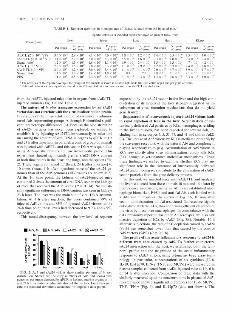
The pattern of in vivo transgene expression by an sAd24vector does not correlate with the virus biodistribution profile.Prior study of the in vivo distribution of systemically adminis-tered Ads representing groups A through F identified signifi-cant interserotype differences (3). Because the biodistributionof sAd24 particles has never been explored, we wished toestablish it by injecting sAd24TL intravenously in mice andmeasuring the amount of viral DNA in organs by qPCR at 1 hand 24 h after injection. In parallel, a control group of animalswas injected with Ad5TL, and this vector DNA was quantifiedusing Ad5-specific primers and an Ad5-specific probe. Thisexperiment showed significantly greater sAd24 DNA contentat both time points in the heart, the lungs, and the spleen (Fig.2). These organs contained 1.7 (heart, 24 h after injection) to10 times (heart, 1 h after injection) more of the sAd24 ge-nomes than of the Ad5 genomes (all P values are below 0.02).At the 1-h time point, the kidneys of sAd24-injected micecontained 2 times the amount of viral DNA seen in the kidneysof mice that received the Ad5 vector (P � 0.014). No statisti-cally significant difference in DNA content was seen in kidneys23 h later. The liver was the main site of viral DNA accumu-lation. At 1 h after injection, the livers contained 79% ofinjected Ad5 virions and 85% of injected sAd24 virions; at the24-h time point, these levels had decreased to 9.9% and 4.5%,respectively.
This noted discrepancy between the low level of reporter
expression by the sAd24 vector in the liver and the high con-centration of its virions in the liver strongly suggested an in-volvement of virus retention mechanisms that do not yieldgene transfer.
Sequestration of intravenously injected sAd24 virions leadsto rapid depletion of KCs in the liver. Sequestration of sys-temically delivered Ad particles by KCs, macrophages residingin the liver sinusoids, has been reported for several Ads, in-cluding human serotypes 3, 5, 31, 37, and 41 and simian Ad23(3). The uptake of Ad5 virions by KCs is mediated primarily bythe scavenger receptors, with the natural Abs and complementplaying secondary roles (63). Accumulation of Ad5 virions inKCs very shortly after virus administration rapidly kills KCs(34) through as-yet-unknown molecular mechanisms. Giventhese findings, we wished to examine whether KCs play anysignificant role in the clearance of intravenously deliveredsAd24 and, in doing so, contribute to the elimination of sAd24vector particles from the gene delivery process.
To this end, we injected mice with sAd24TL and analyzedthe livers collected from these animals 10 min and 16 h later byfluorescence microscopy, using an Ab to an established mac-rophage biomarker, F4/80, and anti-Ad Ab, each labeled witha different fluorophore. As shown in Fig. 3A, 10 min aftervector administration all Ad-associated fluorescence signalscolocalized with the KCs, thus confirming efficient clearance ofthe virus by these liver macrophages. In concordance with thedata previously reported for other Ad serotypes, we also sawmassive depletion of KCs by sAd24 (Fig. 3B). Notably, 16 hafter virus injections, the rate of KC depletion caused by sAd24(89%) was somewhat lower than that caused by the controlAd5 vectors (94%) (P � 0.018).
The profile of the acute inflammatory response to sAd24 isdifferent from that caused by Ad5. To further characterizesAd24 interaction with the host, we established both the tem-poral profile and the magnitude of the acute inflammatoryresponse to sAd24 virions, using cytometric bead array tech-nology. In particular, concentrations of six cytokines (IL-6,IL-10, IL-12p70, IFN-�, TNF, and MCP-1) were measured inplasma samples collected from sAd24-injected mice at 1 h, 6 h,or 24 h after injection. Comparison of these data with thesimilarly measured cytokine concentrations in plasma of Ad5-injected mice showed significant differences for IL-6, MCP-1,TNF, IFN-� (Fig. 4), and IL-12p70 (data not shown). The
TABLE 1. Reporter activities in homogenates of tissues isolated from Ad-injected micea
Vector (dose)
Reporter activities in indicated organs per organ or gram of tissue (rlu)a
Liver Spleen Lung Heart Kidney
Per organ Per gramof tissue Per organ Per gram
of tissue Per organ Per gramof tissue Per organ Per gram
of tissue Per organ Per gramof tissue
Ad5TL (2 � 1010 VP) 3.6 � 1012 2.8 � 1012 8.1 � 109 6.0 � 1010 3.8 � 108 1.2 � 109 3.4 � 108 2.5 � 109 3.5 � 108 1.0 � 109
sAd24TL (2 � 1010 VP) 3.1 � 108 2.2 � 108 5.0 � 106 5.3 � 107 5.5 � 106 1.6 � 107 2.3 � 106 1.8 � 107 7.4 � 106 2.4 � 107
Signal ratiob 1.2 � 104 1.3 � 104 1.6 � 103 1.1 � 103 6.9 � 10 7.4 � 10 1.5 � 102 1.3 � 102 4.7 � 10 4.2 � 10Ad5TL (1011 VP) 2.8 � 1013 1.6 � 1013 7.0 � 109 6.3 � 1010 1.1 � 109 2.9 � 109 6.2 � 108 3.3 � 109 2.4 � 109 6.8 � 109
sAd24TL (1011 VP) 2.0 � 109 1.2 � 109 3.0 � 107 3.4 � 108 1.1 � 108 4.1 � 108 7.0 � 106 4.7 � 107 5.9 � 107 1.7 � 108
Signal ratiob 1.4 � 104 1.3 � 104 2.4 � 102 1.8 � 102 9.9 7.0 8.8 � 10 7.1 � 10 4.1 � 10 3.1 � 10PBS 1.1 � 107 8.5 � 106 7.3 � 105 8.4 � 106 3.1 � 106 9.3 � 106 1.4 � 106 9.6 � 106 3.3 � 106 1.0 � 107
a The activities of the reporter averaged for groups of five animals is shown in relative light units (rlu) per entire organ and per gram of tissue.b Ratios of bioluminescence signals measured in Ad5TL-injected mice to those measured in sAd24TL-injected mice.
FIG. 2. Ad5 and sAd24 virions show similar patterns of in vivodistribution. Shown are the copy numbers of Ad5 and sAd24 viralgenomes per organ detected by qPCR in isolated murine organs at 1 hand 24 h after systemic administration of the vectors. Error bars indi-cate the standard deviations calculated for duplicate data points.
10092 BELOUSOVA ET AL. J. VIROL.

differences in concentrations of IL-6, MCP-1, and TNF in-duced by sAd24 and Ad5 vectors were most significant at the6-h time point and, to a lesser extent, at the 24-h time point;IFN-� and IL-12p70 concentrations were different only at the24-h time point (P values of 0.02 and 0.047, respectively).Importantly, cytokine release by the sAd24-injected mice wasinvariably more robust than that by the animals that receivedAd5. Compared to those for mock-injected animals, no statis-tically significant changes in the concentrations of IL-10 were
seen in mice injected with either serotype of Ad (data notshown).
The modification of sAd24 fiber by knob replacement strat-egy yields protein chimera suitable for Ad vector targeting. Wepreviously succeeded in altering the receptor specificity of Ad5through genetic replacement of the CAR-binding knob do-main of its fiber with a trimerization domain and a targetingligand (5, 7, 26). We then reasoned that because of evolution-ary conservation of the overall Ad fiber structure, which is very
FIG. 3. Uptake of sAd24 virions by KCs in the liver leads to KC depletion. (A) Immunofluorescence staining of the liver collected from a mouseinjected with sAd24 vector 10 min after injection. The upper panels show the liver section stained with either anti-F4/80 (left, green fluorescence)or anti-Ad Ab (right, red fluorescence). The lower panels show the overlay of the upper images, either alone (left) or merged with the stainingof nuclei (right, blue staining). KCs containing sAd24 particles are orange. (B) Immunofluorescence staining of the livers collected from miceinjected with PBS (left), Ad5 (center), or sAd24 (right) 16 h after injection. All images show Hoechst-stained nuclei (blue).
VOL. 84, 2010 SIMIAN ADENOVIRUS VECTOR TARGETED TO Her2 10093

similar in most known fiber proteins, the same approachshould also be applicable to the sAd24 fiber. Importantly,application of this strategy to sAd24 did not require identifi-cation of its natural fiber receptor, which is not yet known, and,if successful, would further support the applicability of thisstrategy to fibers of other Ads.
To test this assumption, we designed a recombinant geneencoding the tail and shaft domains of the sAd24 fiber proteinfused to the 12th �-helical coiled-coil of the phage T4 fibritinprotein, a peptide linker, and the Her2-specific affibody Zher2:7
and transiently expressed it in 293T cells. The product of thisexpression (designated FsAd2411FHer2:7) was analyzed by West-ern blotting to test whether the designed chimera assumes thetrimeric configuration that is both characteristic of Ad fibersand essential for their encapsidation.
The results of this analysis, however, were unconvincing: incontrast to the well-defined bands corresponding to the tri-meric wt Ad5 fiber (Fig. 5A, lane 2), the sAd24 fiber chimerafailed to produce such a pattern. Instead, the major band seenin the nondenatured sample of FsAd2411FHer2:7 migrated con-siderably faster than had been expected (Fig. 5A, lane 4) andcorresponded to a protein with an apparent molecular mass ofapproximately 105 kDa, not the 136 kDa predicted for thetrimeric FsAd2411FHer2:7. The fact that the same sample, whenfully denatured, clearly showed the monomeric chimera of theexpected size (Fig. 5A, lane 3) ruled out the possibility that thesmaller-than-expected size of the nondenatured protein wasdue to degradation or truncation of its subunits. Also, com-parison of the electrophoretic mobilities of the FsAd2411FHer2:7
chimera and the wt sAd24 fiber showed that the nonboiledsamples of both proteins produced very similar patterns of gelmigration (Fig. 5A, lanes 4 and 6). On the basis of this com-parison, we concluded that the trimer of FsAd2411FHer2:7 wassufficiently stable to be incorporated into sAd24 virions.
The FsAd2411FHer2:7 protein was designed to mediate effi-
cient attachment of sAd24 viral particles to Her2 and, as aprerequisite to this, was expected to bind the cell-associatedHer2. To test this function of FsAd2411FHer2:7, the transientlyexpressed protein was used in a flow cytometry experiment toprobe the surfaces of cells either expressing or not expressingHer2. As shown in Fig. 5F, the strength of the fluorescencesignal measured in Her2-positive cell targets was significantlyhigher than that measured in cells lacking the target receptor.
Collectively, our data confirm both the expected trimericconfiguration of the FsAd2411FHer2:7 protein and the ability ofthis molecule to recognize its intended target receptor on thecell surface, thereby providing a rationale for development ofan sAd24 vector containing this chimera.
sAd24 virions incorporating designed fiber chimeras infectcells in a Her2-dependent manner. The final proof of theFsAd2411FHer2:7 chimera’s functionality was obtained by usingthis protein to replace the wt fiber in sAd24 virions. To thisend, we constructed a replication-deficient genome of sAd24with the E1 region deleted and containing the ORF ofFsAd2411FHer2:7 in place of the wt fiber-coding sequence; thevirus was rescued and propagated using the two-step approachdescribed in Materials and Methods. The efficacy of incorpo-ration of FsAd2411FHer2:7 into sAd24 virions was gauged andcompared with that of the wt sAd24 fiber by using Westernblotting of purified viral particles. As Fig. 6A illustrates, theoutcome of the trimerization assays of the transiently ex-pressed FsAd2411FHer2:7 correctly predicted efficient encapsi-dation of the designed protein. This efficient encapsidation wasin agreement with the high yield of the modified sAd24 vector(5,500 VP/infected cell), which was comparable to the yield forsAd24 containing the wt fibers (5,000 VP/infected cell). Com-parison of the protein compositions of the virions of both typesshowed no differences other than the presence of the expectedfiber proteins, the wild type or the fiber chimera, in the respec-
FIG. 4. Comparison of cytokine release in response to intravenousinjections of Ad5 and sAd24. Concentrations of IL-6, MCP-1, TNF,and IFN-� in plasma samples collected from mice at 1 h, 6 h, or 24 hafter injection with either Ad5- or sAd24-derived vectors are shown inpg/ml. Three mice were injected with a given Ad to generate data foreach time point shown. Error bars indicate the standard deviationcalculated for duplicate data points corresponding to each animal inthe group.
FIG. 5. sAd24 fiber-derived targeting chimera forms stable trimersand binds to cell-associated Her2. (A) Western blotting of lysates of293T cells transiently expressing the fiber constructs. Lanes 1 and 2, wtAd5 fiber; lanes 3 and 4, FsAd2411FHer2:7, lanes 5 and 6, wt sAd24 fiber;lane S, protein standards with the molecular masses shown in kDa(here and in Fig. 6). Samples in lanes 1, 3, and 5 were boiled prior tobeing loaded onto the gel and thus show proteins in their fast-migrat-ing monomeric form; samples in lanes 2, 4, and 6 were not boiled.Fibers were detected with either the anti-Ad5 fiber tail MAb 4D2(wt Ad5 fiber), the anti-fibritin MAb 5E1 (FsAd2411FHer2:7), or theanti-sAd24 fiber MAb 20C1 (wt sAd24 fiber) followed by a fluores-cence-labeled secondary antibody. Predicted molecular masses for tri-meric wt Ad5, FsAd2411FHer2:7, and wt sAd24 fiber are 185 kDa, 136kDa, and 142 kDa, respectively. (B) Flow cytometry detected bindingof the transiently expressed FsAd2411FHer2:7 chimera (shown by solidblack line) to Her2-expressing 293/Her2 cells but no binding above thebackground to Her2-negative 293 cells. The background signals gen-erated in both cell lines by the lysate of mock-transfected 293T cellsare shown by the dotted lines.
10094 BELOUSOVA ET AL. J. VIROL.

tive vector particles (Fig. 6B), thus confirming the proper as-sembly and maturation of the modified sAd24 particles.
Further, in good agreement with the cell-binding data shownin Fig. 5B, our subsequent gene transfer experiments demon-strated that the FsAd2411FHer2:7 chimera contained insAd24TL.11FHer2:7 virions enables Her2-dependent trans-duction of target cells (Fig. 7A). The Her2 specificity ofsAd24TL.11FHer2:7 infection was confirmed by transducing twoisogenic cell lines, MDA-MB-231 and MDA-MB-231/Her2,that differ from each other by their Her2 phenotypes. In addi-tion, augmentation of gene transfer to the target receptor-expressing cells by the tropism-modified sAd24 was demon-strated through transduction of a panel of Her2-positive
human tumor cell lines. In this experiment, gene delivery bythe targeted sAd24 vector was 3.7 to 14 times more efficientthan delivery by the vector bearing unmodified wt fibers.Importantly, by using the free Her2-binding affibody com-petitor we showed selective inhibition of transduction bysAd24TL.11FHer2:7 but not by the nontargeted control Ad(Fig. 7A).
The efficacy of this gene transfer was then compared withthat by the previously designed Her2-targeted derivative ofAd5 (7) (Fig. 7B). In this experiment, two pairs of cell lines inwhich the parental line was Her2 negative (293 and MDA-MB-231) and the derivative line was Her2 expressing (293/Her2and MDA-MB-231/Her2) were transduced with eitherAd5TL.11FHer2:7 or sAd24TL.11FHer2:7 at various MOIs. Withthe exception of the transduction of MDA-MB-231/Her2 at anMOI of 10, at which both Ads were equally efficient, the Ad5-derived vector was more efficient in transducing both Her2-expressing cell lines. The differences between the levels of geneexpression directed by the vectors depended on the vectors’replication. Specifically, in 293/Her2 cells, which complementdeletion of the E1 genes in both the Ad5- and the sAd24-derived vectors, reporter expression by Ad5TL.11FHer2:7 was 3to 6 times more efficient. In comparison, in MDA-MB-231/Her2 cells, which do not complement deletion of the E1 genes,reporter expression by Ad5TL.11FHer2:7 was only 1.3 to 1.6times that by sAd24TL.11FHer2:7. Notably, the relative infec-tivities of the tested vectors, which had been determined bythe spot assay in 293/Her2 cells, were 51 VP/infectiousunit for sAd24TL.11FHer2:7 and 33 VP/infectious unit forAd5TL.11FHer2:7.
In light of the potential use of sAd24TL.11FHer2:7-derivedvectors for gene delivery in humans with preexisting anti-Ad5antibodies, moreover, we used the established virus neutraliza-
FIG. 6. Efficient encapsidation of FsAd2411FHer2:7 protein yieldsfully matured sAd24 virions. (A) Western blot of sAd24TL (1010 VP,lane 1) and sAd24TL.11FHer2:7 (1010 VP, lane 2) virions. The fullydenatured fibers were detected with the anti-sAd24 fiber tail MAb20C1. (B) SDS-PAGE-resolved proteins of sAd24TL (2 � 1010 VP,lane 1) and sAd24TL.11FHer2:7 (2 � 1010 VP, lane 2) virions stainedwith silver using a PageSilver kit (Fermentas, Glen Burnie, MD). Indi-cated are the migration positions of the following sAd24 proteins: hexon(II), penton base (III), peripentonal protein (IIIa), FsAd2411FHer2:7 fiberchimera (FC), major core protein (V), fiber (F), hexon-associated protein(VI), and minor core protein (VII).
FIG. 7. Gene transfer by the targeted sAd24TL.11FHer2:7 vector is Her2 dependent. (A) In vitro transduction of human tumor cell lines bysAd24TL and sAd24TL.11FHer2:7 vectors. Viral infections were done in either standard medium (white and black bars) or in medium containingfree affibody Zher2:4 used at a concentration of 100 �g/ml (gray and striped bars). Shown are the activities of Fluc in lysates of infected cells. Insertsbelow the graph show the signals detected in each of the tested cell lines (7 � 103 cells/lane) by Western blotting with anti-Her2 antibody.(B) Comparison of the efficacies of gene transfer by Her2-targeted vectors derived from Ad5 (Ad5TL.11FHer2:7, white bars) and sAd24(sAd24TL.11FHer2:7, black bars). Levels of transgene expression in the lysates of cells infected with Ads at the MOIs indicated below the graph(in VP/cell) are shown. Error bars indicate the standard deviations calculated for triplicate data points.
VOL. 84, 2010 SIMIAN ADENOVIRUS VECTOR TARGETED TO Her2 10095
tion assay to test whether gene delivery by this vector wouldbe affected by such antibodies. This test showed thatsAd24TL.11FHer2:7 largely evaded inactivation by Ad5-specificantibodies and retained most of its infectivity in their presence(data not shown).
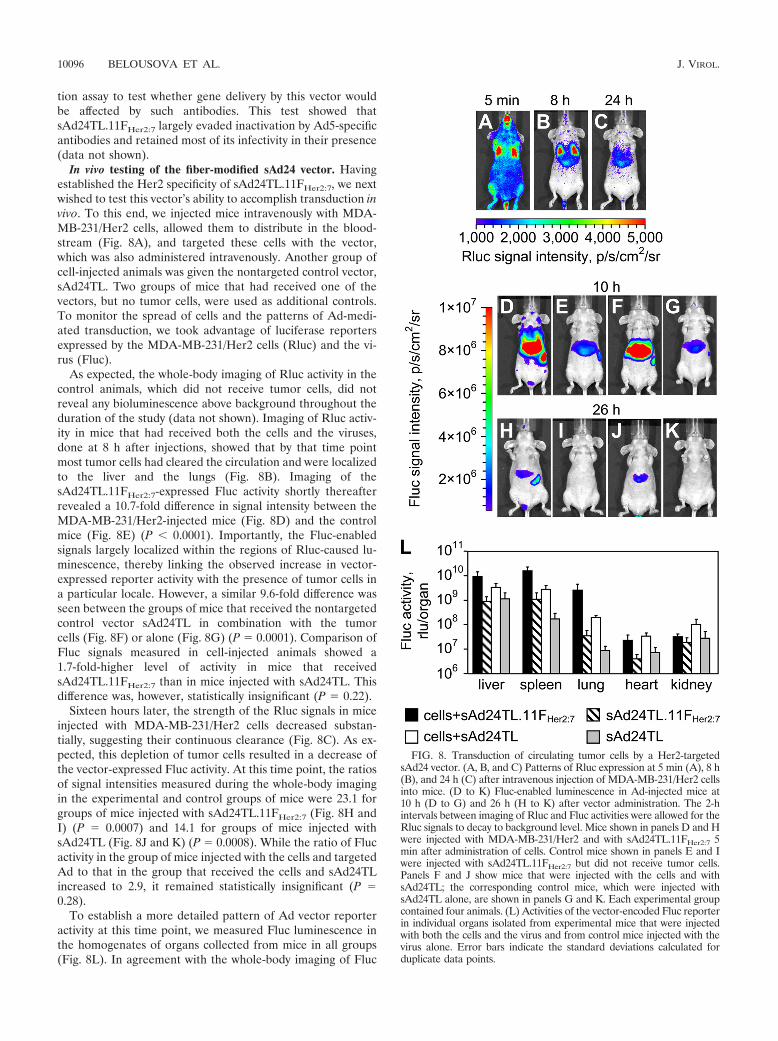
In vivo testing of the fiber-modified sAd24 vector. Havingestablished the Her2 specificity of sAd24TL.11FHer2:7, we nextwished to test this vector’s ability to accomplish transduction invivo. To this end, we injected mice intravenously with MDA-MB-231/Her2 cells, allowed them to distribute in the blood-stream (Fig. 8A), and targeted these cells with the vector,which was also administered intravenously. Another group ofcell-injected animals was given the nontargeted control vector,sAd24TL. Two groups of mice that had received one of thevectors, but no tumor cells, were used as additional controls.To monitor the spread of cells and the patterns of Ad-medi-ated transduction, we took advantage of luciferase reportersexpressed by the MDA-MB-231/Her2 cells (Rluc) and the vi-rus (Fluc).
As expected, the whole-body imaging of Rluc activity in thecontrol animals, which did not receive tumor cells, did notreveal any bioluminescence above background throughout theduration of the study (data not shown). Imaging of Rluc activ-ity in mice that had received both the cells and the viruses,done at 8 h after injections, showed that by that time pointmost tumor cells had cleared the circulation and were localizedto the liver and the lungs (Fig. 8B). Imaging of thesAd24TL.11FHer2:7-expressed Fluc activity shortly thereafterrevealed a 10.7-fold difference in signal intensity between theMDA-MB-231/Her2-injected mice (Fig. 8D) and the controlmice (Fig. 8E) (P 0.0001). Importantly, the Fluc-enabledsignals largely localized within the regions of Rluc-caused lu-minescence, thereby linking the observed increase in vector-expressed reporter activity with the presence of tumor cells ina particular locale. However, a similar 9.6-fold difference wasseen between the groups of mice that received the nontargetedcontrol vector sAd24TL in combination with the tumorcells (Fig. 8F) or alone (Fig. 8G) (P � 0.0001). Comparison ofFluc signals measured in cell-injected animals showed a1.7-fold-higher level of activity in mice that receivedsAd24TL.11FHer2:7 than in mice injected with sAd24TL. Thisdifference was, however, statistically insignificant (P � 0.22).
Sixteen hours later, the strength of the Rluc signals in miceinjected with MDA-MB-231/Her2 cells decreased substan-tially, suggesting their continuous clearance (Fig. 8C). As ex-pected, this depletion of tumor cells resulted in a decrease ofthe vector-expressed Fluc activity. At this time point, the ratiosof signal intensities measured during the whole-body imagingin the experimental and control groups of mice were 23.1 forgroups of mice injected with sAd24TL.11FHer2:7 (Fig. 8H andI) (P � 0.0007) and 14.1 for groups of mice injected withsAd24TL (Fig. 8J and K) (P � 0.0008). While the ratio of Flucactivity in the group of mice injected with the cells and targetedAd to that in the group that received the cells and sAd24TLincreased to 2.9, it remained statistically insignificant (P �0.28).
To establish a more detailed pattern of Ad vector reporteractivity at this time point, we measured Fluc luminescence inthe homogenates of organs collected from mice in all groups(Fig. 8L). In agreement with the whole-body imaging of Fluc
FIG. 8. Transduction of circulating tumor cells by a Her2-targetedsAd24 vector. (A, B, and C) Patterns of Rluc expression at 5 min (A), 8 h(B), and 24 h (C) after intravenous injection of MDA-MB-231/Her2 cellsinto mice. (D to K) Fluc-enabled luminescence in Ad-injected mice at10 h (D to G) and 26 h (H to K) after vector administration. The 2-hintervals between imaging of Rluc and Fluc activities were allowed for theRluc signals to decay to background level. Mice shown in panels D and Hwere injected with MDA-MB-231/Her2 and with sAd24TL.11FHer2:7 5min after administration of cells. Control mice shown in panels E and Iwere injected with sAd24TL.11FHer2:7 but did not receive tumor cells.Panels F and J show mice that were injected with the cells and withsAd24TL; the corresponding control mice, which were injected withsAd24TL alone, are shown in panels G and K. Each experimental groupcontained four animals. (L) Activities of the vector-encoded Fluc reporterin individual organs isolated from experimental mice that were injectedwith both the cells and the virus and from control mice injected with thevirus alone. Error bars indicate the standard deviations calculated forduplicate data points.
10096 BELOUSOVA ET AL. J. VIROL.

activity, these measurements confirmed invariably higher levelsof transduction in animals injected with the cells and the vi-ruses than in animals treated with the Ads only. They alsoshowed that the livers, spleens, and lungs were the primarysites of viral transduction. In these organs, sAd24TL.11FHer2:7-caused transduction was 2.8-fold (livers; P � 0.07), 5.7-fold(spleens; P � 0.003), and 13.5-fold (lungs; P � 0.003) higherthan transduction caused by sAd24TL. This difference in theliver was, however, statistically insignificant, as was the differ-ence in reporter expression in the hearts (P � 0.25). Thekidneys of cell-injected animals that received sAd24TL con-tained a 3.3-fold-higher level of Fluc activity than kidneys inmice injected with the cells and sAd24TL.11FHer2:7 (P � 0.04).
We also tested the performance of sAd24TL.11FHer2:7 in anin vivo model of metastatic Her2-expressing cancer. For thisstudy, we established target metastases in the lungs of mice bysystemically injecting the animals with MDA-MB-231/Her2cells (Fig. 9A). Next, the tumor-bearing animals and the tu-mor-free control animals were injected intravenously withsAd24TL.11FHer2:7 at a dose of 1011 VP/injection, using thetwo-injection regimen described in Materials and Methods.Bioluminescence imaging of Fluc activity done 48 h after thesecond vector administration detected weak signals in the tho-racic areas of mice in both groups (Fig. 9B and C). Measure-ments in homogenates of the lungs isolated from animals inboth groups showed that, on average, Fluc activity was 2.5times higher in mice with metastases than in tumor-free mice(Fig. 9D). This difference was statistically significant (P �0.005).
DISCUSSION
Prior work on modifying the tropism of Ad vectors wasfocused mostly on Ad5-derived vectors and was based primar-ily on modification of the fiber knob domain with ligands. Thenatural diversity of Ad serotypes and the advantages offered bysome of those Ads as targeted vector prototypes remain un-derexplored. In this study, we sought to test whether the Adtropism alteration approach based on fiber knob domain re-placement, which we had previously developed for Ad5, couldbe applied to another Ad serotype. The rationale for this workwas that its success would support the feasibility of developinga versatile targeting approach that would be applicable tomany Ads, thus making it possible to fully exploit the potentialbenefits offered by underexplored Ad serotypes.
Toward these goals, we sought to make a tropism-modified
vector based on sAd24, which was chosen for this study for thefollowing reasons. First, owing to the widespread preexistingimmunity to Ad5 in humans (12, 55), the serological distinctionof sAd24 from Ad5 makes this virus a preferred alternative toAd5 as a vector platform. Second, the fiber of sAd24 is anunexplored molecule and is thus a reasonable starting point totest an approach that is expected to be applicable to manyfibers, including the uncharacterized ones. This protein hasnever been studied in detail; at the level of the primary struc-ture it shares little homology with the Ad5 fiber and otherwell-characterized Ad fibers. Neither its primary receptor northe receptor-binding site within its knob has been identified.Against this background, successful targeting of the sAd24fiber through the knob replacement strategy would yield avector prototype of potential clinical utility and would alsosuggest the feasibility of targeting of other Ads.
sAd24 would be an even stronger candidate as a vector forgene delivery in humans should it avoid liver tissue on systemicdelivery. The massive transduction of the liver by the Ad5vectors causes severe systemic toxicity and raises serious safetyconcerns. Recent studies have identified the high-affinity bind-ing of FX to the Ad5 hexon protein as the molecular basis forhepatic transduction (22, 56). These studies also showed thatthe particles of several Ad serotypes bind FX poorly or do notbind it at all and that the stability of the resultant complexescorrelates with the degree of liver transduction by the Ads.Given these findings, we studied the association of FX with thevirions of sAd24 and showed that FX-sAd24 complexes arerather unstable. In particular, while the equilibrium dissocia-tion constant, KD, for the FX-sAd24 complexes was onlyslightly higher than the KD for the control FX-Ad5 complex,the FX-sAd24 dissociation rate constant, kd, was 16 times theFX-Ad5 kd. To put these findings in perspective, recent suc-cesses in drug development have more often correlated withimprovements of the kd values of the most efficacious drugsrather than with improvements of their KD values. In particu-lar, the kd values correlated much better with the agent-targetcomplex residence time, its dissociative half-life, and targetselectivity (13). On this basis, the dissociation rate constant isnow seen as a more meaningful parameter in assessing impor-tant biological interactions.
The observed instability of the FX-sAd24 complexes couldbe explained by variations in the primary sequences of theHVRs of the Ad5 and sAd24 hexons. Notably, recent studieshave collectively suggested that FX binding to the Ad5 hexon
FIG. 9. Gene delivery by sAd24TL.11FHer2:7 in mice with metastatic tumors. (A) Whole-body images of mice bearing Her2- and Rluc-expressing breast cancer metastases in the lungs. (B and C) Fluc bioluminescence in the mice shown in panel A (B) and in control tumor-free mice(C) 48 h after animals in both groups were injected intravenously with sAd24TL.11FHer2:7. (D) Activity of Fluc reporter in the lungs isolated fromAd-injected mice after image acquisition. The black and white bars show the averaged values of signals in the lungs of tumor-bearing and controlmice, respectively. Each experimental group contained six animals. Error bars indicate the standard deviations calculated for duplicate data points.
VOL. 84, 2010 SIMIAN ADENOVIRUS VECTOR TARGETED TO Her2 10097

involves HVRs 3, 5, and 7 (2, 22). At the level of the primaryamino acid sequence, these HVRs within the sAd24 hexonshare little homology with their Ad5 counterparts (the identitylevels are 0%, 29%, and 33% for HVRs 3, 5, and 7, respec-tively). In addition, of the five amino acid residues within theAd5 hexon HVR7 that have been confirmed to contribute toFX binding (22, 56), only two, T404 and E431, are conservedwithin the sAd24 hexon structure. It is thus reasonable tospeculate that these differences in the structures of the Ad5and sAd24 HVRs contribute to the much weaker association ofFX with the sAd24 hexon.
The instability of FX-sAd24 complexes results in much lowerhepatic transduction by sAd24 than by Ad5 upon systemicdelivery to mice. Imaging of the vector-injected mice detectedreporter expression in the livers of the sAd24-injected animalsonly at very low levels, while injections with the Ad5 vectoryielded massive liver transduction. Notably, these findings cor-relate well with the earlier report by Roy et al., who usedhistochemical staining of murine livers to show reduced trans-gene expression by an intravenously administered sAd24 vec-tor (45).
Our optical imaging data were corroborated by PET-CTresults and by measurements of luciferase activity in homoge-nates of tissues isolated from vector-treated mice. With itsgreater sensitivity, the latter assay has confirmed very signifi-cant differences in reporter expression by the tested Ads inthose tissues, where signals were undetectable by whole-bodyimaging. Depending on the particular tissue assayed, the re-porter activities expressed by the sAd24 vector (per organ)were 10 to 14,000 times less than those induced by the Ad5vector.
Measurements of viral DNA content in organs isolated fromAd-injected mice showed that, like Ad5 virions, sAd24 parti-cles accumulated primarily in the liver shortly after vectoradministration. This accumulation of sAd24 did not yield anysignificant reporter expression, suggesting that most of theinjected viral dose was sequestered by the sinusoidal liver mac-rophages, KCs, which had been shown to clear other serotypeAds after systemic delivery (3). Examination of the livers ofsAd24-injected mice by fluorescence microscopy confirmed therapid uptake of the vector by KCs and the nearly total deple-tion of these cells 24 h after vector administration.
Next, by measuring the levels of six inflammatory cytokinesin the blood of animals treated with Ads, we have shown thatexpression of five of these cytokines was elevated in sAd24-injected animals to significantly higher levels than in mice thathad received the Ad5 vector. These data are in line with thepreviously reported differences in inflammatory responses toAds, which were found to be Ad serotype related (3). More-detailed studies of immune responses to sAd24 are now war-ranted to identify the challenges related to the use of thisserotype for gene therapy and to develop strategies to over-come those challenges.
Together the greatly reduced transduction of nontarget tis-sues by sAd24 revealed by our studies and the low seropreva-lence of this Ad in humans reported by others (45, 64) pro-vided a rationale for developing a tropism-modified derivativeof sAd24 as a prototype of targeted gene vectors for clinicaluse. This was accomplished by applying the fiber knob replace-ment strategy that we had previously developed for the Ad5
fiber to the sAd24 fiber protein. We reasoned that together theevolutionary conservation of the domain-based structure of theAd fiber proteins and the fact that the same functions arecarried out by the same domains in various fibers suggest thepromise of this approach.
Despite the fact that the sAd24 fiber protein has never beenstudied before and its structure has not been elucidated, theestablished general principles of the Ad fiber structure couldbe applied to this protein’s primary structure to map its do-mains. Thus, the amino-terminal aa 1 to 44 and the carboxy-terminal aa 265 to 443 could be predicted to correspond to theprotein’s tail and knob, respectively. On the basis of the estab-lished folding pattern for an Ad fiber shaft (54), the central220-aa-long shaft of the sAd24 fiber could be modeled toconsist of 13 pseudorepeats. The sAd24 fiber shaft appears tocontain an extended third repeat, the feature known to makethe fiber flexible, and in this respect is similar to the Ad5 fiber.Furthermore, both fibers share a substantial similarity neartheir carboxy termini: not only do they both contain theKLGxGLxFD/N consensus motif seen in the last full repeat ofmost Ad fibers, but this homology region also covers most ofthe downstream shaft-to-knob transition region, with 62%of the amino acid residues here being identical in both Ads.These structural similarities were supportive of the rationalefor modifying the sAd24 fiber by the knob replacement thathad been successfully applied before to the Ad5 fiber. How-ever, a potential confounding factor was the difference be-tween the two fibers’ shaft lengths. According to the modelproposed by van Raaij et al., nine intersubunit hydrogen bondsare formed within each repeat of the Ad fiber shaft, and there-fore each repeat contributes to the overall stability of thetrimeric fiber (54). With the sAd24 shaft being much shorterthan the Ad5 shaft (354 aa, 22 repeats), the former could thusform less-stable trimers. Whether fusion of this relatively shortshaft with the fibritin would yield a trimer stable enough forencapsidation was unclear. In spite of this concern, our testingof the designed FsAd2411FHer2:7 chimera confirmed that itstrimer was stable and thus predicted its successful encapsida-tion.
In our flow cytometry experiments, this protein showedbinding to Her2-expressing cells but not to Her2-negative cells.The magnitude of this binding was lower, however, than thatwe saw previously for the similarly designed Ad5 fiber-derivedchimera (7). Additional experiments showed that this differ-ence was due not to the weaker binding of the FsAd2411FHer2:7
to Her2 but likely to suboptimal detection of cell-bound fiberswith the anti-fibritin MAb 5E1, which was the only availableMAb suitable for this comparative-binding assay. When usedin parallel with anti-Ad5 fiber tail MAb to detect the cell-attached Ad5 fiber-derived chimera, the 5E1 MAb produced amuch weaker signal (unpublished data), perhaps because ofsteric hindrance caused by the proximity of its epitope to theaffibody-Her2 interface.
Successful rescue and propagation of the sAd24 vector inwhich its wt fiber gene was replaced with the gene for thetargeting chimera and the efficient encapsidation of the chi-mera have further confirmed the structural equivalency of thedesigned protein to the wt fiber. The functional adequacy ofthis protein was then demonstrated through successful Her2-dependent gene delivery by this vector to Her2-expressing tu-
10098 BELOUSOVA ET AL. J. VIROL.
mor cells in vitro. The efficacy of this gene delivery was invari-ably and substantially greater than that of the sAd24 vectorcarrying unmodified fibers and was comparable to that of thepreviously designed Ad5-based vector targeted to Her2. Com-pared with unmodified Ad5 vector in the presence of neutral-izing anti-Ad5 antibodies, moreover, the Her2-targeted sAd24revealed a much lower degree of inactivation, thus suggestingthat a tropism-modified sAd24 would be better suited for geneinterventions in patients with Her2-expressing tumors than arethe Ad5-based vectors.
Testing of the fiber-modified sAd24 in vivo by intravenousinjection into immunocompromised mice suggested that thevirus could transduce Her2-expressing human tumor cellspresent in murine vasculature. In particular, the whole-bodyimaging of the reporter activities expressed by the target cellsand the vector showed colocalization of these signals. Thisimaging also showed a significant increase in Ad-producedluminescence in animals injected with both the cells and thevector compared to that in mice treated with the vector alone.However, despite our encouraging data on the Her2 depen-dence of sAd24TL.11FHer2:7 gene transfer in vitro, the in vivocomparison of this vector with the control nontargeted sAd24showed a marginal increase in transduction that was not sta-tistically significant. By measuring reporter activity in organsisolated from the experimental animals, we have been able toshow statistically significant enhancement of gene transfer bysAd24TL.11FHer2:7 to two of the three organs containing thehighest levels of Ad-expressed reporter, the lungs (13.5-fold)and the spleens (5.7-fold), while the 2.8-fold enhancement inthe livers was not statistically significant.
The same approach was also used to assess the levels of genetransfer by the tropism-modified sAd24 vector to lung-local-ized Her2-expressing tumor metastases. In this scenario, wesaw low levels of lung transduction in both the tumor-free andthe tumor-bearing animals. Notably, however, the statisticalsignificance of the higher level of reporter activity measured inthe metastasized lungs was established.
Taken together, the results of these in vivo experimentssuggest that the selectivity of the fiber-modified sAd24 vectorfor Her2, although clearly shown in vitro, was insufficient tosupport robust targeted transduction of tumor cells under thechallenging conditions of systemic administration. While thedemonstrated substantial and statistically significant enhance-ment of transduction in the lungs and spleens of animals sys-temically injected with the tumor cells and sAd24TL.11FHer2:7
is encouraging, future improvements of the vector are neededto achieve the desired levels of target-specific transduction onvascular delivery.
Given the diversity of soluble molecules encountered by theblood-borne Ad vectors, some level of nonspecific transductionresulting from interactions between the virus and these mole-cules, both known (22, 49) and yet unknown, appears practi-cally unavoidable. The goal of vector targeting is, therefore, toachieve a level of target-specific gene transfer that is substan-tially higher than that of off-target transduction. In a recentstudy, the efficacy of on-target gene transfer by a systemicallyinjected Ad vector was significantly improved by increasing theaffinity of the virus’s fiber protein for its receptor (57). Thissuccess suggests that the in vivo target selectivity of an sAd24-derived vector can also be augmented by using ligands with
higher affinity for the target receptor. To this end, the nextversion of the Her2-targeted sAd24 can be designed to containa fiber-fibritin chimera fused with a higher-affinity affibody,which is now available. While the Zher2:7 affibody ligand used inour study has an affinity for Her2 in the midnanomolar range(KD � 1.4 � 10�7 M) (62), Her2-specific affibodies with muchhigher affinities (up to 2.2 � 10�11 M) have recently beendeveloped (39).
It could be speculated that once the desired level of targetspecificity is achieved for sAd24, similarly designed targetedvectors could be derived from other Ad serotypes that havebeen shown to evade neutralization by anti-Ad5 antibodies.Availability of multiserotype panels of vectors targeted to thesame disease biomarker would provide a means of overcomingvector-induced immunity in patients and would thus makerepeated gene delivery in humans more efficient. However,additional studies are needed to see whether the knob replace-ment approach would produce functional derivatives of thoseserotypes’ fibers.
In conclusion, as the first developmental step toward ansAd24 vector suitable for targeted gene delivery in humans, wedemonstrated that the systemically administered virions ofsAd24 transduce normal tissues at decreased levels comparedwith those of Ad5 particles; studied the in vivo biodistributionof intravenously injected sAd24 virions and showed active se-questration of the virus by Kupffer cells; compared the inflam-matory responses to sAd24 and Ad5 vectors; designed a proof-of-concept, fiber-modified sAd24 vector and confirmed itsaltered tropism in vitro; studied in vivo transduction by thesystemically administered, modified sAd24; and identified thelimitations of this novel vector.
The results of this work add significantly to the rather lim-ited knowledge of sAd24 biology and also suggest a potentialuse of sAd24-derived vectors beyond the previously proposedgenetic vaccination.
ACKNOWLEDGMENTS
We thank the personnel of the Monoclonal Antibody Facility, theDNA Analysis Facility, the Vector Core Laboratory, the Radiochem-istry Section of the Department of Experimental Diagnostic Imaging,and the Small Animal Imaging Facility of the M. D. Anderson CancerCenter for their help with producing hybridoma cell lines and antibod-ies to the sAd24 fiber, with DNA sequencing, with production andpurification of wt sAd24, with production of 18F-labeled tracer, andwith animal imaging, respectively. We are grateful to Andrey Volginand Amer Najjar (both with the M. D. Anderson Cancer Center) andthe personnel of the Flow Cytometry and Cellular Imaging Core Fa-cility for their help with the cell-sorting experiments and to OleksandrKalyuzhniy (University of Washington, Seattle) for his advice on thesetup of surface plasmon resonance studies. Michael Hulsey (BD Bio-sciences) is thanked for processing of the flow cytometry data. KathrynHale and Sunita Patterson are thanked for editing the manuscript.
This work was supported by Public Health Service grants R01CA119387 (to C.L.) and R01 CA116621 and R01 CA128807 (to V.K.).Additional support was provided by the University Cancer Foundationof the University of Texas M. D. Anderson Cancer Center (to V.K.),Cancer Center Support Grant P30CA16672, and Experimental CancerImaging Research Program CA126577.
REFERENCES
1. Alauddin, M. M., P. S. Conti, and J. D. Fissekis. 2003. A general synthesisof 2�-deoxy-2�-[18F]fluoro-5-methyl-1-�-D-arabinofuranosyluracil and its5-substituted nucleosides. J. Label. Comp. Radiopharm. 46:285–289.
2. Alba, R., A. C. Bradshaw, A. L. Parker, D. Bhella, S. N. Waddington, S. A.Nicklin, N. van Rooijen, J. Custers, J. Goudsmit, D. H. Barouch, J. H.
VOL. 84, 2010 SIMIAN ADENOVIRUS VECTOR TARGETED TO Her2 10099
McVey, and A. H. Baker. 2009. Identification of coagulation factor (F)Xbinding sites on the adenovirus serotype 5 hexon: effect of mutagenesis onFX interactions and gene transfer. Blood 114:965–971.
3. Appledorn, D. M., A. Kiang, A. McBride, H. Jiang, S. Seregin, J. M. Scott,R. Stringer, Y. Kousa, M. Hoban, M. M. Frank, and A. Amalfitano. 2008.Wild-type adenoviruses from groups A-F evoke unique innate immune re-sponses, of which HAd3 and SAd23 are partially complement dependent.Gene Ther. 15:885–901.
4. Arnberg, N., K. Edlund, A. H. Kidd, and G. Wadell. 2000. Adenovirus type37 uses sialic acid as a cellular receptor. J. Virol. 74:42–48.
5. Belousova, N., N. Korokhov, V. Krendelshchikova, V. Simonenko, G. Mikhe-eva, P. L. Triozzi, W. A. Aldrich, P. T. Banerjee, S. D. Gillies, D. T. Curiel,and V. Krasnykh. 2003. Genetically targeted adenovirus vector directed toCD40-expressing cells. J. Virol. 77:11367–11377.
6. Belousova, N., V. Krendelchtchikova, D. T. Curiel, and V. Krasnykh. 2002.Modulation of adenovirus vector tropism via incorporation of polypeptideligands into the fiber protein. J. Virol. 76:8621–8631.
7. Belousova, N., G. Mikheeva, J. Gelovani, and V. Krasnykh. 2008. Modifica-tion of adenovirus capsid with a designed protein ligand yields a gene vectortargeted to a major molecular marker of cancer. J. Virol. 82:630–637.
8. Bergelson, J. M., J. A. Cunningham, G. Droguett, E. A. Kurt-Jones, A.Krithivas, J. S. Hong, M. S. Horwitz, R. L. Crowell, and R. W. Finberg. 1997.Isolation of a common receptor for coxsackie B viruses and adenoviruses 2and 5. Science 275:1320–1323.
9. Bewley, M. C., K. Springer, Y. B. Zhang, P. Freimuth, and J. M. Flanagan.1999. Structural analysis of the mechanism of adenovirus binding to itshuman cellular receptor, CAR. Science 286:1579–1583.
10. Bidaut, L. M., R. Pascual-Marqui, J. Delavelle, A. Naimi, M. Seeck, C.Michel, D. Slosman, O. Ratib, D. Ruefenacht, T. Landis, N. de Tribolet, J. R.Scherrer, and F. Terrier. 1996. Three- to five-dimensional biomedical mul-tisensor imaging for the assessment of neurological (dys) function. J. Digit.Imaging 9:185–198.
11. Chartier, C., E. Degryse, M. Gantzer, A. Dieterle, A. Pavirani, and M.Mehtali. 1996. Efficient generation of recombinant adenovirus vectors byhomologous recombination in Escherichia coli. J. Virol. 70:4805–4810.
12. Chirmule, N., K. Propert, S. Magosin, Y. Qian, R. Qian, and J. Wilson. 1999.Immune responses to adenovirus and adeno-associated virus in humans.Gene Ther. 6:1574–1583.
13. Copeland, R. A., D. L. Pompliano, and T. D. Meek. 2006. Drug-targetresidence time and its implications for lead optimization. Nat. Rev. DrugDiscov. 5:730–739.
14. Defer, C., M. T. Belin, M. L. Caillet-Boudin, and P. Boulanger. 1990. Humanadenovirus-host cell interactions: comparative study with members of sub-groups B and C. J. Virol. 64:3661–3673.
15. Dmitriev, I., V. Krasnykh, C. R. Miller, M. Wang, E. Kashentseva, G.Mikheeva, N. Belousova, and D. T. Curiel. 1998. An adenovirus vector withgenetically modified fibers demonstrates expanded tropism via utilization ofa coxsackievirus and adenovirus receptor-independent cell entry mechanism.J. Virol. 72:9706–9713.
16. Farina, S. F., G. P. Gao, Z. Q. Xiang, J. J. Rux, R. M. Burnett, M. R. Alvira,J. Marsh, H. C. Ertl, and J. M. Wilson. 2001. Replication-defective vectorbased on a chimpanzee adenovirus. J. Virol. 75:11603–11613.
17. Gaggar, A., D. M. Shayakhmetov, and A. Lieber. 2003. CD46 is a cellularreceptor for group B adenoviruses. Nat. Med. 9:1408–1412.
18. Hasenburg, A., D. C. Fischer, X. W. Tong, A. Rojas-Martinez, R. H. Kauf-man, I. Ramzy, P. Kohlberger, M. Orlowska-Volk, E. Aguilar-Cordova, andD. G. Kieback. 2002. Adenovirus-mediated thymidine kinase gene therapyfor recurrent ovarian cancer: expression of coxsackie-adenovirus receptorand integrins alphavbeta3 and alphavbeta5. J. Soc. Gynecol. Invest. 9:174–180.
19. Hemminki, A., N. Belousova, K. R. Zinn, B. Liu, M. Wang, T. R. Chaudhuri,B. E. Rogers, D. J. Buchsbaum, G. P. Siegal, M. N. Barnes, J. Gomez-Navarro, D. T. Curiel, and R. D. Alvarez. 2001. An adenovirus with enhancedinfectivity mediates molecular chemotherapy of ovarian cancer cells andallows imaging of gene expression. Mol. Ther. 4:223–231.
20. Henry, L. J., D. Xia, M. E. Wilke, J. Deisenhofer, and R. D. Gerard. 1994.Characterization of the knob domain of the adenovirus type 5 fiber proteinexpressed in Escherichia coli. J. Virol. 68:5239–5246.
21. Hong, J. S., and J. A. Engler. 1996. Domains required for assembly ofadenovirus type 2 fiber trimers. J. Virol. 70:7071–7078.
22. Kalyuzhniy, O., N. C. Di Paolo, M. Silvestry, S. E. Hofherr, M. A. Barry,P. L. Stewart, and D. M. Shayakhmetov. 2008. Adenovirus serotype 5 hexonis critical for virus infection of hepatocytes in vivo. Proc. Natl. Acad. Sci.U. S. A. 105:5483–5488.
23. Kirby, I., E. Davison, A. J. Beavil, C. P. Soh, T. J. Wickham, P. W. Roelvink,I. Kovesdi, B. J. Sutton, and G. Santis. 2000. Identification of contactresidues and definition of the CAR-binding site of adenovirus type 5 fiberprotein. J. Virol. 74:2804–2813.
24. Kirby, I., E. Davison, A. J. Beavil, C. P. Soh, T. J. Wickham, P. W. Roelvink,I. Kovesdi, B. J. Sutton, and G. Santis. 1999. Mutations in the DG loop ofadenovirus type 5 fiber knob protein abolish high-affinity binding to itscellular receptor CAR. J. Virol. 73:9508–9514.
25. Korokhov, N., G. Mikheeva, A. Krendelshchikov, N. Belousova, V. Simo-nenko, V. Krendelshchikova, A. Pereboev, A. Kotov, O. Kotova, P. L. Triozzi,W. A. Aldrich, J. T. Douglas, K. M. Lo, P. T. Banerjee, S. D. Gillies, D. T.Curiel, and V. Krasnykh. 2003. Targeting of adenovirus via genetic modifi-cation of the viral capsid combined with a protein bridge. J. Virol. 77:12931–12940.
26. Krasnykh, V., N. Belousova, N. Korokhov, G. Mikheeva, and D. T. Curiel.2001. Genetic targeting of an adenovirus vector via replacement of the fiberprotein with the phage T4 fibritin. J. Virol. 75:4176–4183.
27. Krasnykh, V., I. Dmitriev, G. Mikheeva, C. R. Miller, N. Belousova, andD. T. Curiel. 1998. Characterization of an adenovirus vector containing aheterologous peptide epitope in the HI loop of the fiber knob. J. Virol.72:1844–1852.
28. Lieber, A., C. Y. He, L. Meuse, D. Schowalter, I. Kirillova, B. Winther, andM. A. Kay. 1997. The role of Kupffer cell activation and viral gene expressionin early liver toxicity after infusion of recombinant adenovirus vectors. J. Vi-rol. 71:8798–8807.
29. Louis, N., P. Fender, A. Barge, P. Kitts, and J. Chroboczek. 1994. Cell-binding domain of adenovirus serotype 2 fiber. J. Virol. 68:4104–4106.
30. Maes, F., A. Collignon, D. Vandermeulen, G. Marchal, and P. Suetens. 1997.Multimodality image registration by maximization of mutual information.IEEE Trans. Med. Imaging 16:187–198.
31. Magnusson, M. K., P. Henning, S. Myhre, M. Wikman, T. G. Uil, M.Friedman, K. M. Andersson, S. S. Hong, R. C. Hoeben, N. A. Habib, S. Stahl,P. Boulanger, and L. Lindholm. 2007. Adenovirus 5 vector genetically re-targeted by an Affibody molecule with specificity for tumor antigen HER2/neu. Cancer Gene Ther. 14:468–479.
32. Magnusson, M. K., S. S. Hong, P. Boulanger, and L. Lindholm. 2001.Genetic retargeting of adenovirus: novel strategy employing “deknobbing”of the fiber. J. Virol. 75:7280–7289.
33. Magnusson, M. K., S. See Hong, P. Henning, P. Boulanger, and L. Lind-holm. 2002. Genetic retargeting of adenovirus vectors: functionality of tar-geting ligands and their influence on virus viability. J. Gene Med. 4:356–370.
34. Manickan, E., J. S. Smith, J. Tian, T. L. Eggerman, J. N. Lozier, J. Muller,and A. P. Byrnes. 2006. Rapid Kupffer cell death after intravenous injectionof adenovirus vectors. Mol. Ther. 13:108–117.
35. Mercier, G. T., J. A. Campbell, J. D. Chappell, T. Stehle, T. S. Dermody, andM. A. Barry. 2004. A chimeric adenovirus vector encoding reovirus attach-ment protein sigma1 targets cells expressing junctional adhesion molecule 1.Proc. Natl. Acad. Sci. U. S. A. 101:6188–6193.
36. Mitraki, A., A. Barge, J. Chroboczek, J. P. Andrieu, J. Gagnon, and R. W.Ruigrok. 1999. Unfolding studies of human adenovirus type 2 fibre trimers.Evidence for a stable domain. Eur. J. Biochem. 264:599–606.
37. Muruve, D. A., M. J. Barnes, I. E. Stillman, and T. A. Libermann. 1999.Adenoviral gene therapy leads to rapid induction of multiple chemokinesand acute neutrophil-dependent hepatic injury in vivo. Hum. Gene Ther.10:965–976.
38. Novelli, A., and P. A. Boulanger. 1991. Assembly of adenovirus type 2 fibersynthesized in cell-free translation system. J. Biol. Chem. 266:9299–9303.
39. Orlova, A., M. Magnusson, T. L. Eriksson, M. Nilsson, B. Larsson, I.Hoiden-Guthenberg, C. Widstrom, J. Carlsson, V. Tolmachev, S. Stahl, andF. Y. Nilsson. 2006. Tumor imaging using a picomolar affinity HER2 bindingaffibody molecule. Cancer Res. 66:4339–4348.
40. Ory, D. S., B. A. Neugeboren, and R. C. Mulligan. 1996. A stable human-derived packaging cell line for production of high titer retrovirus/vesicularstomatitis virus G pseudotypes. Proc. Natl. Acad. Sci. U. S. A. 93:11400–11406.
41. Philipson, L., K. Lonberg-Holm, and U. Pettersson. 1968. Virus-receptorinteraction in an adenovirus system. J. Virol. 2:1064–1075.
42. Roberts, D. M., A. Nanda, M. J. Havenga, P. Abbink, D. M. Lynch, B. A.Ewald, J. Liu, A. R. Thorner, P. E. Swanson, D. A. Gorgone, M. A. Lifton,A. A. Lemckert, L. Holterman, B. Chen, A. Dilraj, A. Carville, K. G. Mans-field, J. Goudsmit, and D. H. Barouch. 2006. Hexon-chimaeric adenovirusserotype 5 vectors circumvent pre-existing anti-vector immunity. Nature 441:239–243.
43. Roelvink, P. W., G. Mi Lee, D. A. Einfeld, I. Kovesdi, and T. J. Wickham.1999. Identification of a conserved receptor-binding site on the fiber proteinsof CAR-recognizing Adenoviridae. Science 286:1568–1571.
44. Roy, S., G. Gao, D. S. Clawson, L. H. Vandenberghe, S. F. Farina, and J. M.Wilson. 2004. Complete nucleotide sequences and genome organization offour chimpanzee adenoviruses. Virology 324:361–372.
45. Roy, S., G. Gao, Y. Lu, X. Zhou, M. Lock, R. Calcedo, and J. M. Wilson.2004. Characterization of a family of chimpanzee adenoviruses and devel-opment of molecular clones for gene transfer vectors. Hum. Gene Ther.15:519–530.
46. Roy, S., G. P. Kobinger, J. Lin, J. Figueredo, R. Calcedo, D. Kobasa, andJ. M. Wilson. 2007. Partial protection against H5N1 influenza in mice with asingle dose of a chimpanzee adenovirus vector expressing nucleoprotein.Vaccine 25:6845–6851.
47. Schagen, F. H., F. M. Wensveen, J. E. Carette, T. S. Dermody, W. R.Gerritsen, and V. W. van Beusechem. 2006. Genetic targeting of adenovirus
10100 BELOUSOVA ET AL. J. VIROL.

vectors using a reovirus sigma1-based attachment protein. Mol. Ther. 13:997–1005.
48. Sebestyen, Z., J. de Vrij, M. Magnusson, R. Debets, and R. Willemsen. 2007.An oncolytic adenovirus redirected with a tumor-specific T-cell receptor.Cancer Res. 67:11309–11316.
49. Shayakhmetov, D. M., A. Gaggar, S. Ni, Z. Y. Li, and A. Lieber. 2005.Adenovirus binding to blood factors results in liver cell infection and hepa-totoxicity. J. Virol. 79:7478–7491.
50. Short, J. J., A. V. Pereboev, Y. Kawakami, C. Vasu, M. J. Holterman, andD. T. Curiel. 2004. Adenovirus serotype 3 utilizes CD80 (B7.1) and CD86(B7.2) as cellular attachment receptors. Virology 322:349–359.
51. Tomko, R. P., R. Xu, and L. Philipson. 1997. HCAR and MCAR: the humanand mouse cellular receptors for subgroup C adenoviruses and group Bcoxsackieviruses. Proc. Natl. Acad. Sci. U. S. A. 94:3352–3356.
52. Tsuruta, Y., L. Pereboeva, J. N. Glasgow, C. L. Luongo, S. Komarova, Y.Kawakami, and D. T. Curiel. 2005. Reovirus sigma1 fiber incorporated intoadenovirus serotype 5 enhances infectivity via a CAR-independent pathway.Biochem. Biophys. Res. Commun. 335:205–214.
53. van Beusechem, V. W., A. L. van Rijswijk, H. H. van Es, H. J. Haisma, H. M.Pinedo, and W. R. Gerritsen. 2000. Recombinant adenovirus vectors withknobless fibers for targeted gene transfer. Gene Ther. 7:1940–1946.
54. van Raaij, M. J., A. Mitraki, G. Lavigne, and S. Cusack. 1999. A triplebeta-spiral in the adenovirus fibre shaft reveals a new structural motif for afibrous protein. Nature 401:935–938.
55. Vogels, R., D. Zuijdgeest, R. van Rijnsoever, E. Hartkoorn, I. Damen, M. P.de Bethune, S. Kostense, G. Penders, N. Helmus, W. Koudstaal, M. Cec-chini, A. Wetterwald, M. Sprangers, A. Lemckert, O. Ophorst, B. Koel, M.van Meerendonk, P. Quax, L. Panitti, J. Grimbergen, A. Bout, J. Goudsmit,and M. Havenga. 2003. Replication-deficient human adenovirus type 35vectors for gene transfer and vaccination: efficient human cell infection andbypass of preexisting adenovirus immunity. J. Virol. 77:8263–8271.
56. Waddington, S. N., J. H. McVey, D. Bhella, A. L. Parker, K. Barker, H.
Atoda, R. Pink, S. M. Buckley, J. A. Greig, L. Denby, J. Custers, T. Morita,I. M. Francischetti, R. Q. Monteiro, D. H. Barouch, N. van Rooijen, C.Napoli, M. J. Havenga, S. A. Nicklin, and A. H. Baker. 2008. Adenovirusserotype 5 hexon mediates liver gene transfer. Cell 132:397–409.
57. Wang, H., Y. Liu, Z. Li, S. Tuve, D. Stone, O. Kalyushniy, D. Shayakhmetov,C. L. Verlinde, T. Stehle, J. McVey, A. Baker, K. W. Peng, S. Roffler, and A.Lieber. 2008. In vitro and in vivo properties of adenovirus vectors withincreased affinity to CD46. J. Virol. 82:10567–10579.
58. Wickham, T. J., E. J. Filardo, D. A. Cheresh, and G. R. Nemerow. 1994.Integrin �v�5 selectively promotes adenovirus mediated cell membrane per-meabilization. J. Cell Biol. 127:257–264.
59. Wickham, T. J., P. Mathias, D. A. Cheresh, and G. R. Nemerow. 1993.Integrins �v�3 and �v�5 promote adenovirus internalization but not virusattachment. Cell 73:309–319.
60. Wickham, T. J., P. W. Roelvink, D. E. Brough, and I. Kovesdi. 1996. Ade-novirus targeted to heparan-containing receptors increases its gene deliveryefficiency to multiple cell types. Nat. Biotechnol. 14:1570–1573.
61. Wickham, T. J., E. Tzeng, L. L. Shears II, P. W. Roelvink, Y. Li, G. M. Lee,D. E. Brough, A. Lizonova, and I. Kovesdi. 1997. Increased in vitro and invivo gene transfer by adenovirus vectors containing chimeric fiber proteins.J. Virol. 71:8221–8229.
62. Wikman, M., A. C. Steffen, E. Gunneriusson, V. Tolmachev, G. P. Adams,J. Carlsson, and S. Stahl. 2004. Selection and characterization of HER2/neu-binding affibody ligands. Protein Eng. Des Sel. 17:455–462.
63. Xu, Z., J. Tian, J. S. Smith, and A. P. Byrnes. 2008. Clearance of adenovirusby Kupffer cells is mediated by scavenger receptors, natural antibodies, andcomplement. J. Virol. 82:11705–11713.
64. Zhi, Y., J. Figueredo, G. P. Kobinger, H. Hagan, R. Calcedo, J. R. Miller,G. Gao, and J. M. Wilson. 2006. Efficacy of severe acute respiratorysyndrome vaccine based on a nonhuman primate adenovirus in the pres-ence of immunity against human adenovirus. Hum. Gene Ther. 17:500–506.
VOL. 84, 2010 SIMIAN ADENOVIRUS VECTOR TARGETED TO Her2 10101
Copyright © 2022 FDOKUMEN