Consistent foraging routes and benthic foraging behaviour in yellow-eyed penguins
Viruses 2014, 6, 2052-2061; doi:10.3390/v6052052
virusesISSN 1999-4915
www.mdpi.com/journal/viruses
Short Communication
A Novel Adenovirus in Chinstrap Penguins
(Pygoscelis antarctica) in Antarctica
Sook-Young Lee 1, Jeong-Hoon Kim
2, Yon Mi Park
1, Ok Sarah Shin
3, Hankyeom Kim
4,
Han-Gu Choi 2 and Jin-Won Song
1,*
1 Department of Microbiology, College of Medicine, Institute for Viral Diseases, Korea University,
Seoul 136-705, Korea; E-Mails: [email protected] (S.-Y.L.);
[email protected] (Y.M.P.) 2
Division of Life Sciences, Korea Polar Research Institute, Incheon 406-840, Korea;
E-Mails: [email protected] (J.-H.K.); [email protected] (H.-G.C.) 3
Department of Biomedical Science, College of Medicine, Korea University, Seoul 136-705, Korea;
E-Mail: [email protected] 4
Department of Pathology, College of Medicine, Korea University, Guro Hospital, Seoul 152-703,
Korea; E-Mail: [email protected]
* Author to whom correspondence should be addressed; E-Mail: [email protected];
Tel.: +82-2-2286-1011; Fax: +82-2-927-1036.
Received: 19 March 2014; in revised form: 26 April 2014 / Accepted: 28 April 2014 /
Published: 7 May 2014
Abstract: Adenoviruses (family Adenoviridae) infect various organ systems and cause
diseases in a wide range of host species. In this study, we examined multiple tissues from
Chinstrap penguins (Pygoscelis antarctica), collected in Antarctica during 2009 and 2010,
for the presence of novel adenoviruses by PCR. Analysis of a 855-bp region of the hexon
gene of a newly identified adenovirus, designated Chinstrap penguin adenovirus 1
(CSPAdV-1), showed nucleotide (amino acid) sequence identity of 71.8% (65.5%) with
South Polar skua 1 (SPSAdV-1), 71% (70%) with raptor adenovirus 1 (RAdV-1), 71.4%
(67.6%) with turkey adenovirus 3 (TAdV-3) and 61% (61.6%) with frog adenovirus 1
(FrAdV-1). Based on the genetic and phylogenetic analyses, CSPAdV-1 was classified as a
member of the genus, Siadenovirus. Virus isolation attempts from kidney homogenates in
the MDTC-RP19 (ATCC® CRL-8135™) cell line were unsuccessful. In conclusion, this
study provides the first evidence of new adenovirus species in Antarctic penguins.
OPEN ACCESS

Viruses 2014, 6
2053
Keywords: adenovirus; Siadenovirus; Chinstrap penguin; Antarctica
1. Introduction
Adenoviruses are linear, double-stranded DNA viruses, with a genome ranging from 26 to 45-kbp
and an icosahedral capsid [1]. Adenoviruses, which can infect the respiratory and gastrointestinal
tracts, eyes and other organs, cause gastroenteritis and respiratory disease in many species [2].
The family, Adenoviridae, comprises five genera: Mastadenovirus, Aviadenovirus, Atadenovirus,
Siadenovirus and Ichtadenovirus [3]. Mastadenoviruses infect a wide range of mammalian species,
including man, monkey, dog, cattle, swine, mouse and bat [4–10]. Aviadenovirus have been identified
in birds [11,12], and Atadenovirus have been isolated from reptiles, birds and mammals [13–15].
Siadenovirus has been detected in birds, frog and a tortoise [16–19], and Ichtadenovirus has been
detected in fish [20].
Previously, adenoviruses had been isolated from various vertebrate species on all continents, except
Antarctica. Recently, the South Polar skua adenovirus 1 (SPSAdV-1), the single known member of the
species, Skua siadenovirus A, was discovered in dead South Polar skua (Catharacta maccormicki)
collected near the King Sejong Station in Antarctica [21]. However, there are limited surveillance
studies of adenovirus infection in Antarctic wild birds. The South Polar skua can be observed in
Antarctica only during the breeding season and feed on the chicks of penguins. Moreover, the South
Polar skua shares breeding grounds with the Chinstrap penguin (Pygoscelis antarctica), an endemic
species [22]. To ascertain if Chinstrap penguins in Antarctica are also infected with adenoviruses, we
conducted an exploratory study on samples collected during 2009 and 2010.
2. Results and Discussion
Two of the 10 Chinstrap penguin carcasses (designated CSP09-1 and CSP09-2) were collected in
the summer of 2009 and eight (CSP10-1 to CSP10-8) in early 2010. Fifty-six tissues, comprising lung,
liver, kidney, heart, intestine, brain, colon, lymph node, spleen, trachea and wounded-bill, were tested.
An approximately 1240-bp genomic fragment, including parts of two adjacent genes, that of pVI and
hexon, was amplified from 28 tissues (lung, liver, kidney, heart, intestine and/or trachea) from eight
Chinstrap penguins (CSP09-1, CSP10-1, CSP10-2, CSP10-3, CSP10-5, CSP10-6, CSP10-7 and
CSP10-8), by nested PCR (Table 1). The Chinstrap penguin adenoviruses (CSPAdV-1) from eight
Chinstrap penguins (from CSP09-1 to CSP10-8) were designated as CSPAdVno1 to CSPAdVno8,
respectively. Adenovirus gene sequences, detected in PCR-positive tissues of individual Chinstrap
penguins, were almost identical, suggesting widespread systemic infection.
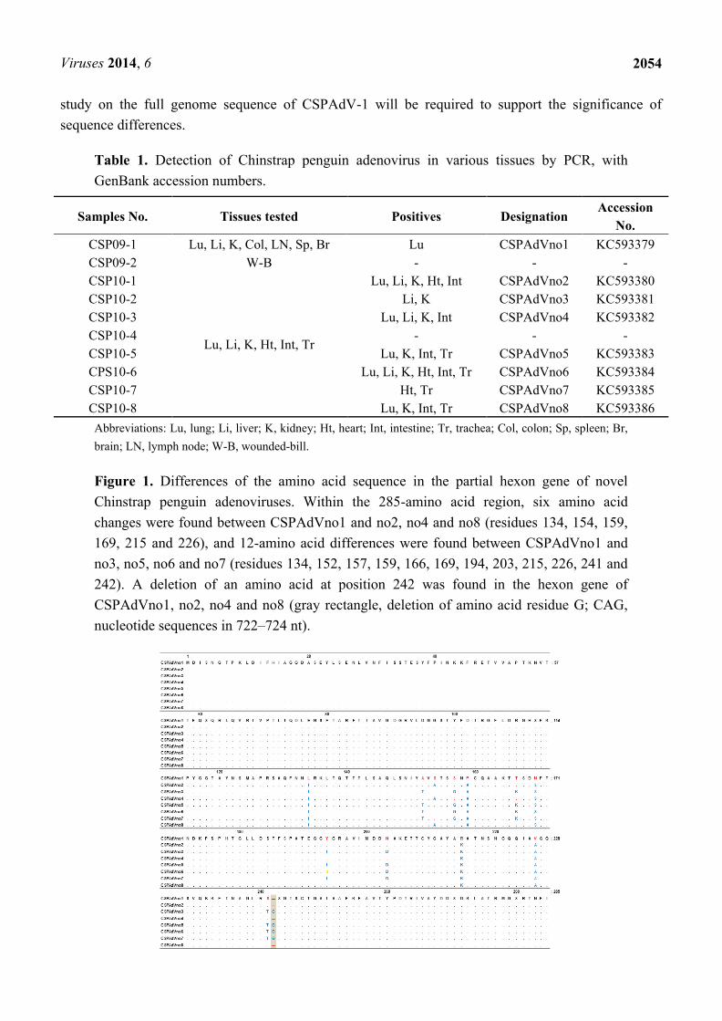
Pair-wise alignment by ClustalW showed a six-amino acid difference between CSPAdVno1
originating from a Chinstrap penguin collected in 2009 and CSPAdVno2, no4 and no8. Twelve amino
acid differences were found between CSPAdVno1 and CSPAdVno3, no5, no6 and no7 (Figure 1).
Particularly, CSPAdVno1, no2, no4 and CSPAdVno8 showed a deletion at position 242. As a result of
sequence comparison with other types within the genus, Siadenovirus, the insertion of amino acid
residue S at position 243 was found in the amino acid sequences of some CSPAdV-1 variants. Further
Viruses 2014, 6
2054
study on the full genome sequence of CSPAdV-1 will be required to support the significance of
sequence differences.
Table 1. Detection of Chinstrap penguin adenovirus in various tissues by PCR, with
GenBank accession numbers.
Samples No. Tissues tested Positives Designation Accession
No.
CSP09-1 Lu, Li, K, Col, LN, Sp, Br Lu CSPAdVno1 KC593379
CSP09-2 W-B - - -
CSP10-1
Lu, Li, K, Ht, Int, Tr
Lu, Li, K, Ht, Int CSPAdVno2 KC593380
CSP10-2 Li, K CSPAdVno3 KC593381
CSP10-3 Lu, Li, K, Int CSPAdVno4 KC593382
CSP10-4 - - -
CSP10-5 Lu, K, Int, Tr CSPAdVno5 KC593383
CPS10-6 Lu, Li, K, Ht, Int, Tr CSPAdVno6 KC593384
CSP10-7 Ht, Tr CSPAdVno7 KC593385
CSP10-8 Lu, K, Int, Tr CSPAdVno8 KC593386
Abbreviations: Lu, lung; Li, liver; K, kidney; Ht, heart; Int, intestine; Tr, trachea; Col, colon; Sp, spleen; Br,
brain; LN, lymph node; W-B, wounded-bill.
Figure 1. Differences of the amino acid sequence in the partial hexon gene of novel
Chinstrap penguin adenoviruses. Within the 285-amino acid region, six amino acid
changes were found between CSPAdVno1 and no2, no4 and no8 (residues 134, 154, 159,
169, 215 and 226), and 12-amino acid differences were found between CSPAdVno1 and
no3, no5, no6 and no7 (residues 134, 152, 157, 159, 166, 169, 194, 203, 215, 226, 241 and
242). A deletion of an amino acid at position 242 was found in the hexon gene of
CSPAdVno1, no2, no4 and no8 (gray rectangle, deletion of amino acid residue G; CAG,
nucleotide sequences in 722–724 nt).

Viruses 2014, 6
2055
Within the genus, Siadenovirus, CSPAdVno1 was shown to have a nucleotide (amino acid) identity
of 71.8% (65.5%) with SPSAdV-1, 71% (70%) with raptor adenovirus 1 (RAdV-1), 71.4% (67.6%)
with turkey adenovirus 3 (TAdV-3), 69.4% (66.5%) with great tit adenovirus 1 (GTAdV-1) and 61%
(61.6%) with frog adenovirus 1 (FrAdV-1). The nucleotide and amino acid sequence identity of each
strain of CSPAdV-1 was more than 97% and 96%, respectively (Table 2). However, the nucleotide and
amino acid sequences of CSPAdVno3, no5 and no7 and CSPAdVno2, no4 and no8 were identical
(Figures 1 and 2). The nucleotide sequence identity of CSPAdV-1 was less (<33%) with other
adenovirus genera, such as Atadenovirus, Aviadenovirus and Mastadenovirus. The G+C content of
CSPAdVno1 and CSPAdVno2, no4 and no8 was found to be 37.44% and 37.2%, respectively. The
partial hexon of CSPAdVno3, no5 and no7 had a G+C content of 37.42%.
Table 2. Nucleotide and amino acid sequence identity (%) between CSPAdVno1 and other
CSPAdV-1 strains and representative Siadenoviruses.
Virus strain Sequence identity (%)
nucleotide amino acid
CSPAdVno2 98.7 97.9
CSPAdVno3 97.5 96.1
CSPAdVno4 98.7 97.9
CSPAdVno5 97.5 96.1
CSPAdVno6 97.8 96.1
CSPAdVno7 97.5 96.1
CSPAdVno8 98.7 97.9
SPSAdV-1 71.8 65.5
RAdV-1 71.0 70.0
TAdV-3 71.4 67.6
GTAdV-1 69.4 66.5
FrAdV-1 61.0 61.6
GenBank accession No.: CSPAdVno1 to CSPAdVno8 (KC593379 to KC593386), SPSAdV-1 (HM585353),
RAdV-1 (EU715130), TAdV-3 (AC000016), GTAdV-1 (FJ849795), FrAdV-1 (AF224336).
For phylogenetic analysis, approximately 855-bp of the hexon gene, which contains structural loop
regions that encode serotype-specific epitopes, were selected [23]. CSPAdV-1 showed the highest
similarity with SPSAdV-1 and RAdV-1 and was classified into the genus, Siadenovirus, based on
phylogenetic trees generated by the maximum-parsimony and neighbor-joining methods, implemented
in MEGA5.1 (Molecular Evolutionary Genetics Analysis, 5.1) (Figure 2) and PAUP version 4.0b
(Phylogenetic Analysis Using Parsimony, 4.0b) [24,25]. A novel adenovirus species is usually defined
as one detected in a new host species and having more than a 5%–15% sequence difference at the
nucleotide and amino acid levels compared with previously characterized adenovirus species [3].
Based on these criteria, we conclude that CSPAdV-1 (Penguin siadenovirus A) seems to merit the
establishment of a new species for it.

Viruses 2014, 6
2056
Figure 2. Phylogenetic relationship between Chinstrap penguin adenovirus (CSPAdV-1)
and other adenoviruses. The phylogenetic tree, based on nucleotide (a) and amino acid
sequences (b) of the hexon protein contained loop regions, was generated by the
neighbor-joining method. CSPAdVno1 to no8 (KC593379 to KC593386) were compared
with adenoviruses of five genera: bat adenovirus 3 (BtAdV-3, GU226970), canine
adenovirus 2 (CAdV-2, U77082), bat adenovirus 2 (BtAdV-2. NC015932), porcine
adenovirus 5 (PAdV-5, AF289262), porcine adenovirus 3 (PAdV-3, AB126117), bovine
adenovirus 10 (BAdV-10, AAF82136), murine adenovirus 1 (MAdV-1, NC000942),
human adenovirus 11 (HAdV-11, AY163756)), simian adenovirus 25 (SAdV-25,
AF394196), human adenovirus 2 (HAdV-2, J01917), human adenovirus 12 (HAdV-12,
X73487), human adenovirus 40 (HAdV-40, L19443), bovine adenovirus 3 (BAdV-3,
AF030154), tree shrew adenovirus 1 (TSAdV-1, AF258784), duck adenovirus 1 (DAdV-1,
Y09598), ovine adenovirus 7 (OAdV-7, U40839), fowl adenovirus 9 (FAdV-9,
AF083975), fowl adenovirus 1 (FAdV-1, U46933), fowl adenovirus 4 (FAdV-4,
NC015323), frog adenovirus 1 (FrAdV-1, AF224336), great tit adenovirus 1 (GTAdV-1,
FJ849795), turkey adenovirus 3 (TAdV-3, AC000016), raptor adenovirus 1 (RAdV-1,
EU715130) and South Polar skua adenovirus 1 (SPSAdV-1, HM585353). Scale bars
indicate the number of nucleotide and amino acid substitutions per site. Bootstrap values
are given at the respective nodes, as determined for 1000 iterations using the MEGA5.1
software [26].
Isolation of CSPAdV-1 was attempted by inoculating the MDTC-RP19 (ATCC® CRL-8135™)
cell line [27] with kidney homogenates, but all such attempts were unsuccessful. Failure of the

Viruses 2014, 6
2057
isolation of RAdV-1 on the chicken embryonic liver cell or MDTC-RP19 cell line has been reported
previously [28,29].
3. Experimental Section
3.1. Sample Collection
Chinstrap penguin carcasses were gathered at Narębski Point, located on the southeast
coast of Barton Peninsula, King George Island, Antarctica (6213'40''S–6214'23''S and
5845'25''W–5847'00''W) during 2009 and early 2010 (Figure 3). All carcasses were identified,
weighed and measured. Internal organs (lung, liver, kidney, heart, intestine, trachea, brain, lymph node
and spleen) were dissected using sterile instruments and stored at −70 °C until use for
adenovirus identification.
Figure 3. Collection site of Chinstrap penguin carcasses. Dead Chinstrap penguins were
collected at Narębski Point (Antarctic Specially Protected Area; ASPA No. 171, red circle)
on King George Island, Antarctica.
3.2. DNA Extraction and PCR
Total genomic DNA was extracted from tissue samples, using the High Pure PCR Template
preparation kit (Roche, Indianapolis, IN), according to the manufacturer’s instructions. For screening
of adenovirus infection, polymerase chain reaction (PCR) was used. PCR assay targeting capsid
protein precursor pVI and the capsid protein hexon gene was performed using oligonucleotide primer
pairs (outer: 5'-ACC (C/T)GG ATT AGC TGG TGA T-3', 5'-TAA TTT CTG TAT TCC TGT CCT-3';
inner: 5'-CCT GC(A/T) GAT CAA CTG GCT-3', 5'-GGA TCC CTA ACC ATT ATC GTA ATA-3').
The sequences of PCR primers were designated from the conserved region by the alignment of

Viruses 2014, 6
2058
adenovirus sequences within the genus, Siadenovirus. PCR conditions were performed as follows:
1 cycle of 95 °C for 5 min followed by 14 cycles of denaturation at 95 °C for 40 s, one degree
step-down each of 1 cycle annealing from 50 °C to 37 °C for 40 s, extension at 72 °C for 1 min,
then 25 cycles of denaturation at 95 °C for 40 s, annealing at 42 °C for 40 s, extension at 72 °C
for 1 min and, finally, at 72 °C for 5 min in a Mastercycler (Eppendorf, Germany).
3.3. Sequencing and Sequence Analysis
All PCR products were sequenced with the Big Dye terminator v3.1 cycle sequencing kit (ABI) and
ABI3730 Automated DNA Sequencer (ABI). Nucleotide sequences were analyzed by ClustalW in
MegAlign of DNAstar programs. Phylogenetic trees were generated by maximum-parsimony and
neighbor-joining methods, implemented in MEGA5.1 and PAUP version 4.0b [24,25]. Topologies
were evaluated by a bootstrap analysis of 1000 iterations by using MEGA5.1 software [26].
3.4. Isolation Attempts
For isolation attempts of CSPAdV-1, 5% (w/v) kidney homogenates of virus-infected
penguins (CSP10-2 and CSP10-3) were inoculated onto MDTC-RP19 (ATCC® CRL-8135™), a
lymphoblastoid cell line of turkey origin (Meleagris gallopavo) with high susceptibility to TAdV-3, a
member of the genus, Siadenovirus [27]. Cultures were observed daily for the cytopathic effect, and
the cells and supernatant were screened for the adenoviral hexon gene by PCR at each passage.
4. Conclusions
Previous studies on sub-Antarctic and Antarctic penguins have shown that avian species in this
region may become infected with various viruses, including paramyxoviruses, Newcastle disease virus,
infectious bursal disease virus and influenza viruses [30–38]. The identification of a novel adenovirus
species in Chinstrap penguins suggests the possibility of other viruses, including additional previously
unrecognized adenoviruses in Antarctic birds.
There is an increasing risk of infectious diseases being introduced into the Antarctic fauna, because
of the increased numbers of people travelling to and within Antarctica [38,39]. Consequently, further
studies in Antarctic birds may provide new insights into the emergence and dissemination of viral
infectious diseases from Antarctica.
Acknowledgments
We thank Sung-Ho Kang, Min-Goo Lee, Kyeong Hoon Cho and Yeong Woong Kim for assistance
in the sample collection, Seo Tae-Kun for help with phylogenetic analysis and Richard Yanagihara for
editorial assistance. This work was supported by grants PD12010 (Polar Academic Program) and
PE13030/PE14020 provided by the Korea Polar Research Institute, Korea.
Author Contributions
S.Y.L. and Y.M.P. performed primer design, DNA extraction, PCR and DNA sequencing reactions.
S.Y.L. and O.S.S. performed phylogenetic analysis and S.Y.L. attempt to isolate virus. H.K.K.

Viruses 2014, 6
2059
performed histopathological study and ecological analysis. J.H.K. and H.G.C. provided penguin
samples. J.W.S. conceived the project, and provided overall scientific oversight. All authors
contributed to the preparation of the final manuscript.
Conflicts of Interest
The authors declare no conflict of interest.
References and Notes
1. Davison, A.J.; Benkö, M.; Harrach, B. Genetic content and evolution of adenoviruses. J. Gen.
Virol. 2003, 84, 2895–2908.
2. David, M.K.; Peter, M.H.; Diane, E.G.; Robert, A.L.; Malcolm, A.M.; Bernard, R.; Stephen, E.S.
Adenoviruses. In Fields Virology, 4th ed.; David, M.K., Peter, M.H., Eds.; Lippincott Williams &
Wilkins: Philadelphia, PA, USA, 2001; Volume 2, pp. 2404–2408.
3. Harrach, B.; Benkö, M.; Both, G.W.; Brown, M.; Davison, A.J.; Echavarría, M.; Hess, M.;
Jones, M.S.; Kajon, A.; Lehmkuhl, H.D.; et al. Family adenoviridae. In Virus Taxonomy: IXth
Report of the International Committee on Taxonomy of Viruses.; King, A.M.Q., Lefkowitz, E.,
Adams, M.J., Carstens, E.B., Eds.; Elsevier: New York, NY, USA, 2011; Volume 9, pp. 125–141.
4. Kovács, G.M.; Davison, A.J.; Zakhartchouk, A.N.; Harrach, B. Analysis of the first complete
genome sequence of an Old World monkey adenovirus reveals a lineage distinct from the six
human adenovirus species. J. Gen. Virol. 2004, 85, 2799–2807.
5. Morrison, M.D.; Onions, D.E.; Nicolson, L. Complete DNA sequence of canine adenovirus type
1. J. Gen. Virol. 1997, 78, 873–878.
6. Rusvai, M.; Harrach, B.; Bánrévi, A.; Evans, P.S.; Benkö, M. Identification and sequence analysis
of the core protein genes of bovine adenovirus 2. Virus Res. 2000, 70, 25–30.
7. Aggarwal, N.; Mittal, S.K. Sequence analysis of procine adenovirus type 3 E1 region, pIX and
pIVa2 genes, and two novel open reading frames. Intervirology 2000, 43, 6–12.
8. Klempa, B.; Krüger, D.H.; Auste, B.; Stanko, M.; Krawczyk, A.; Nickel, K.F.; Uberta, K.; Stang,
A. A novel cardiotropic murine adenovirus representing a distinct species of mastadenoviruses.
J. Virol. 2009, 83, 5749–5759.
9. Maeda, K.; Hondo, E.; Terakawa, J.; Kiso, Y.; Nakaichi, N.; Endoh, D.; Sakai, K.; Morikawa, S.;
Mizutani, T. Isolation of novel adenovirus from fruit bat (Pteropus dasymallus yayeyamae).
Emerg. Infect. Dis. 2008, 14, 347–249.
10. Sonntag, M.; Mühldorfer, K.; Speck, S.; Wibbelt, G.; Kurth, A. New adenovirus in bats,
Germany. Emerg. Infect. Dis. 2009, 15, 2052–2055.
11. Schrenzel, M.; Oaks, J.L.; Rotstein, D.; Maalouf, G.; Snook, E.; Sandfort, C.; Rideout, B.
Characterization of a new species of adenovirus in falcons. J. Clin. Microbiol. 2005, 43,
3402–3412.
12. Chiocca, S.; Kurzbauer, R.; Schaffner, G.; Baker, A.; Mautner, V.; Cotton, M. The complete
DNA sequence and genomic organization of the avian adenovirus CELO. J. Virol. 1996, 70,
2939–2949.

Viruses 2014, 6
2060
13. Dán, A.; Ruzsics, Z.; Russell, W.C.; Benkö, M.; Harrach, B. Analysis of the hexon gene sequence
of bovine adenovirus type 4 provides further support for a new adenovirus genus (Atadenovirus).
J. Gen. Virol. 1998, 79, 1453–1460.
14. Farkas, S.L.; Harrach, B.; Benkö, M. Completion of the genome analysis of snake adenovirus type
1, a representative of the reptilian lineage within the novel genus Atadenovirus. Virus Res. 2008,
132, 132–139.
15. Hess, M.; Blöcker, H.; Brandt, P. The complete nucleotide sequence of the egg drop syndrome
virus: An intermediate between mastadenoviruses and aviadenoviruses. Virology 1997, 238,
145–156.
16. Davison, A.J.; Wright, K.M.; Harrach, B. DNA sequence of frog adenovirus. J. Gen. Virol. 2000,
81, 2431–2439.
17. Pitcovski, J.; Mualem, M.; Rei-Koren, Z.; Krispel, S.; Shmueli, E.; Peretz, Y.; Gutter, B.;
Gallili, G.E.; Michael, A.; Goldberg, D. The complete DNA sequence and genome organization of
the avian adenovirus, hemorrhagic enteritis virus. Virology 1998, 249, 307–315.
18. Kovács, E.R.; Benkö, M. Complete sequence of raptor adenovirus 1 confirms the characteristic
genome organization of siadenoviruses. Infect. Genet. Evol. 2011, 5, 1058–1065.
19. Kovács, E.R.; Jánoska, M.; Harrach, B.; Benkö, M. Recognition and partial genome
characterization by non-specific DNA amplification and PCR of a new siadenovirus species in a
sample originating from Parus major, a great tit. J. Virol. Methods 2010, 163, 262–268.
20. Kovács, G.M.; LaPatra, S.E.; D’Halluin, J.C.; Benkö, M. Phylogenetic analysis of the hexon and
protease genes of a fish adenovirus isolated from white sturgeon (Acipenser transmontanus)
supports the proposal for a new adenovirus genus. Virus Res. 2003, 98, 27–34.
21. Park, Y.M.; Kim, J.H.; Gu, S.H.; Lee, S.Y.; Lee, M.G.; Kang, Y.K.; Kang, S.H.; Kim, H.J.;
Song, J.W. Full genome analysis of a novel adenovirus from the South Polar skua (Catharacta
maccormicki) in Antarctica. Virology 2012, 422, 144–150.
22. Yogui, G.T.; Sericano, J.L. Levels and pattern of polybrominated diphenyl ethers in eggs of
Antarctic seabirds: Endemic versus migratory species. Environ. Pollut. 2009, 157, 975–980.
23. Raue, R.; Gerlach, H.; Müller, H. Phylogenetic analysis of the hexon loop 1 region of an
adenovirus from psittacine birds supports the existence of a new psittacine adenovirus (PsAdV).
Arch. Virol. 2005, 150, 1933–1943.
24. Tamura, K.; Peterson, D.; Peterson, N.; Stecher, G.; Nei, M.; Kumar, S. MEGA5: Molecular
Evolutionary Genetics Analysis using Maximum Likelihood, Evolutionaly Distance, and
Maximum Parsimony methods. Mol. Biol. Evol. 2011, 28, 2731–2739.
25. PAUP*4.0: Phylogenetic Analysis Using Parsimony, version 4.0; Sinauer Associates, Inc:
Sunderland, MA, USA, 2002.
26. MEGA: Molucular Evolutionary Genetics Analysis. Available online: http://www.megasoftware.net/
(accessed on 8 March 2013).
27. Nazerian, K.; Fadly, A.M. Propagation of virulent and avirulent turkey hemorrhagic enteritis virus
in cell culture. Avian. Dis. 1982, 26, 816–827.
Viruses 2014, 6
2061
28. Zsivanovits, P.; Monks, D.J.; Forbes, N.A.; Ursu, K.; Raue, R.; Benkö, M. Presumptive
identification of a novel adenovirus in a Harris hawk (Parabuteo unicinctus), a Bengal eagle owl
(Bubo bengalensis), and a Verreaux’s eagle owl (Bubo lacteus). J. Avian Med. Sug. 2006, 20,
105–112.
29. Kovács, E.R.; Benkö, M. Confirmation of a novel siadenovirus species detected in raptors: partial
sequence and phylogenetic analysis. Virus Res. 2009, 140, 64–70.
30. Barbosa, A.; Palacios, M.J. Health of Antarctic birds: A review of their parasites, pathogens and
diseases. Polar Biol. 2009, 32, 1095–1115.
31. Pearce, D.A.; Wilson, W.H. Viruses in Antarctic ecosystems. Antarctic Sci. 2003, 15, 319–331.
32. Alexander, D.J.; Manvell, R.J.; Collins, M.S.; Brockman, S.J.; Westbury, H.A.; Morgan, I.;
Austin, F.J. Characterization of paramyxoviruses isolated from penguins in Antarctica and
sub-Antarctica during 1976–1979. Arch. Virol. 1989, 109, 135–143.
33. Austin, F.J.; Webster, R.G. Evidence of ortho and paramyxoviruses in fauna from Antarctica.
J. Wild. Dis. 1993, 29, 568–571.
34. Thomazelli, L.M.; Araujo, J.; Oliveira, D.B.; Sanfilippo, L.; Ferreira, C.S.; Brentano, L.; Pelizari,
V.H.; Nakayama, C.; Duarte, R.; Hurtado, R.; et al. Newcastle disease virus in penguins from
King George Island on the Antarctic region. Vet. Microbiol. 2010, 146, 155–160.
35. Wallensten, A.; Munster, V.J.; Osterhaus, A.D.M.E.; Waldenström, J.; Bonnedahl, J.; Broman, T.;
Fouchier, R.A.M.; Olson, B. Mounting evidence for the presence of influenza A virus in the
avifauna of the Antarctic region. Antarctic Sci. 2006, 18, 353–356.
36. Chang, C.M.; Lebarbenchon, C.; Gauthier-Clerc, M.; Bohec, C.L.; Beaune, D.; Maho, Y.L.; Werf,
S. Molecular surveillance for avian influenza A virus in king penguins (Aptenodytes patagonicus).
Polar Biol. 2009, 32, 663–665.
37. Abad, F.X.; Busquets, N.; Sanchez, A.; Ryan, P.G.; Majö, N.; Gonalez-Solís, J. Serological and
virological surveys of the influenza A viruses in Antarctic and sub-Antarctic penguins. Antarctic
Sci. 2013, 25, 339–344.
38. Kerry, K.; Riddle, M.; Clarke, J. Diseases of Antarctic wildlife; 18; Australian Antartic Division:
Channel Highway, Kingston, Australia, August 1998; pp. 89–91.
39. Chown, S.L.; Lee, J.E.; Hughes, K.A.; Barrett, P.J.; Bergstrom, D.M.; Convey, P.; Cowan, D.A.;
Crosbie, K.; Dyer, G.; Frenot, Y.; et al. Challenges to the future conservation of the Antarctica.
Science 2012, 337, 158–159.
© 2014 by the authors; licensee MDPI, Basel, Switzerland. This article is an open access article
distributed under the terms and conditions of the Creative Commons Attribution license
(http://creativecommons.org/licenses/by/3.0/).
Copyright © 2022 FDOKUMEN