Evidence for a role of the Simian Virus 40 in human breast carcinomas
Upload
independentCategory
view
2download
0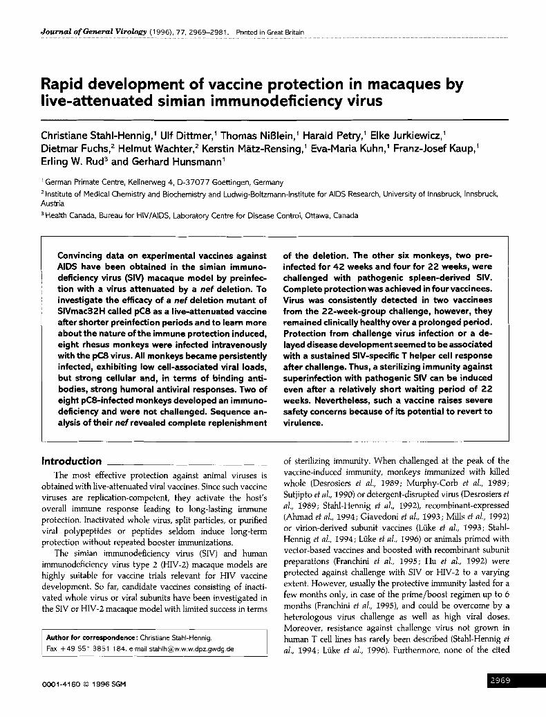
Downloaded from www.microbiologyresearch.org by
IP: 54.224.121.223
On: Tue, 03 May 2016 21:17:03
J o u r n a l o f General Virology (1996), 77, 2969-2981. Printed in Great Britain
Rapid development of vaccine protection in macaques by live-attenuated simian immunodeficiency virus
Christiane Stah I -Henn ig , 1 Ulf D i t tmer , 1 T h o m a s NiBle in , ~ Hara ld Petry, 1 Elke Jurk iewicz , ~
D i e t m a r Fuchs, 2 H e l m u t Wachte r , 2 Kers t in M~ tz -Rens ing , ~ Eva -Mar ia Kuhn, ~ Franz-Jose f Kaup, 1
Er l ing W. Rud 3 and Gerha rd H u n s m a n n 1
i German Primate Centre, Kellnerweg 4, D-37077 Goettingen, Germany 2 Institute of Medical Chemistry and Biochemistry and Ludwig-Boltzmann-lnstitute for AIDS Research, University of Innsbruck, Innsbruck, Austria 3 Health Canada, Bureau for HIVIAIDS, Laboratory Centre for Disease Control, Ottawa, Canada
Convincing data on experimental vaccines against AIDS have been obtained in the simian immuno- deficiency virus (SIV) macaque model by preinfec- tion with a virus attenuated by a nef deletion. To investigate the efficacy of a nef deletion mutant of SIVmac32H called pC8 as a live-attenuated vaccine after shorter preinfection periods and to learn more about the nature of the immune protection induced, eight rhesus monkeys were infected intravenously with the pC8 virus. All monkeys became persistently infected, exhibiting low cell-associated viral loads, but strong cellular and, in terms of binding anti- bodies, strong humoral antiviral responses. Two of eight pC8-infected monkeys developed an immuno- deficiency and were not challenged. Sequence an- alysis of their nef revealed complete replenishment
of the deletion. The other six monkeys, two pre- infected for 42 weeks and four for 22 weeks, were challenged with pathogenic spleen-derived SIV. Complete protection was achieved in four vaccinees. Virus was consistently detected in two vaccinees from the 22-week-group challenge, however, they remained clinically healthy over a prolonged period. Protection from challenge virus infection or a de- layed disease development seemed to be associated with a sustained SIV-specific T helper cell response after challenge. Thus, a sterilizing immunity against superinfection with pathogenic SIV can be induced even after a relatively short waiting period of 22 weeks. Nevertheless, such a vaccine raises severe safety concerns because of its potential to revert to virulence.
In t roduct ion
The most effective protection against animal viruses is obtained with live-attenuated viral vaccines. Since such vaccine viruses are replication-competent, they activate the host's overall immune response leading to long-lasting immune protection. Inactivated whole virus, split particles, or purified viral polypeptides or peptides seldom induce long-term protection without repeated booster immunizations.
The simian immunodeficiency virus (SIV) and human immunodeficiency virus type 2 (HIV-2) macaque models are highly suitable for vaccine trials relevant for HIV vaccine development. So far, candidate vaccines consisting of inacti- vated whole virus or viral subunits have been investigated in the SIV or HIV-2 macaque model with limited success in terms
Author for correspondence: Christiane StahI-Hennig. Fax +49 551 3851 184. e-mail [email protected],de
of sterilizing immunity. When challenged at the peak of the vaccine-induced immunity, monkeys immunized with killed whole (Desrosiers et al., 1989; Murphy-Corb et al., 1989; Sutjipto et al., 1990) or detergent-disrupted virus (Desrosiers et al., 1989; Stahl-Hennig et al., 1992), recombinant-expressed (Ahmad et al., 1994; Giavedoni et al., 1993; Mills et al., 1992) or virion-derived subunit vaccines (LGke et al., 1993; Stahl- Hennig et al., 1994; LUke et al., 1996) or animals primed with vector-based vaccines and boosted with recombinant subunit preparations (Franchini et al., 1995; Hu et al., 1992) were protected against challenge with SIV or HIV-2 to a varying extent. However, usually the protective immunity lasted for a few months only, in case of the prime/boost regimen up to 6 months (Franchini et al., 1995), and could be overcome by a heterologous virus challenge as well as high viral doses. Moreover, resistance against challenge virus not grown in human T cell lines has rarely been described (Stahl-Hennig et al., 1994; L/ike et al., 1996). Furthermore, none of the cited
!96! 0001-4160 © 1996 SGM

Downloaded from www.microbiologyresearch.org by
IP: 54.224.121.223
On: Tue, 03 May 2016 21:17:03
studies convincingly demonstrated clear-cut immune correlates of protection, but in one report protection seemed to depend on the presence of neutralizing antibodies (L/ike et al., 1996).
Deletions in some auxiliary genes have been shown to weaken (Lang et al., 1993; Hoch et al., 1995) or abrogate SIV pathogenicity (Kestler el al., 1991; Rud et al., 1994 a). The most convincing results with experimental AIDS vaccines have so far been produced by preinfecting macaques with live- attenuated SIV containing a net gene deletion. In a first experiment with a virus lacking 182 bp in net (Kestler et al., 1991) protection was achieved against a high dose of challenge virus after a 2"25 year waiting period (Daniel et al., 1992). Another attenuated StV, a molecular done of SIVmac32H called pCS, contains an in-flame deletion of four amino acids in net (Rud et al., I992) and obviously induced protection against cell-flee and cell-associated virus challenge after much shorter preinfection periods (Almond et al., 1995; Whatmore ef al., 1995; Norley et al., 1996). So far almost nothing is known about whether an immunological mechanism mediates the observed protection. Recent results on vaccine protection with a triple-deleted SIV (SIVd3) suggest a correlation with the level of specific neutralizing and binding antibodies (Wyand et aI., 1996). On the other hand, data on the pathogenicity of this SIVd3 in neonatal macaques (Baba et al., 1995) and on the reversion to virulence of an SIV mutant attenuated by a smaller net deletion (Whatmore et al., 1995) question the potential use of similar live-attenuated HIV in man. Nevertheless, studying both the host's cellular and humoral immune response to such attenuated experimental vaccines should provide important clues to their mechanism of immune protection.
As part of a concerted European AIDS vaccine experiment using attenuated SIV and involving a total of 49 macaques for immunization, we have infected eight rhesus macaques (Macaca muhtta) with the above-mentioned naturally oc- curring net-deletion mutant, pC8 (Rudet al., 1992). At the time of inoculation the vaccine virus was shown to have an attenuated phenotype in both Chinese rhesus monkeys (Rud el al., 1992) and cynomolgus macaques (Macaca fascicularis) (Rud et al., 1992, 1994 a). Some time later we learned that the vaccine virus may revert to virulence in rhesus monkeys of Indian origin (Whatmore ef al., 1995). Six preinfected monkeys were exposed to challenge with a highly pathogenic spleen-derived SIVmac virus swarm 22 or 42 weeks after pC8 infection. Primary infection led to complete protection in four out of six animals. The remaining two monkeys became infected with the challenge virus, but disease development seemed to be delayed. However, in two of the pCS-infected monkeys a reversion to virulence by a repair of the net deletion was also observed.
M e t h o d s • Animals, clinical observation and necropsy. Twelve purpose- bred rhesus monkeys of Indian origin between 2 and 3 years old with a body weight of 2"5-3"5 kg were obtained either from the breeding
colony of the German Primate Centre or from the Cayo Santiago Island, Puerto Rico, USA. All monkeys tested negative for antibodies against STLV-1, simian type-D retrovims and SIV. Physical examinations as well as bleedings including the determination of white blood cell (WBC) counts were performed on all macaques at regular intervals following first and second virus inoculation. Necropsy was conducted on monkeys developing severe AIDS-like symptoms and included routine gross pathology and histological examinations. One inguinal or axillary lymph node from each vaccinee was removed surgically under ketamine xylacin anaesthesia at 47 weeks post-challenge and subjected to virological, immunological, histological and molecular biological investigations.
• Immunization and challenge. Four rhesus monkeys were inoculated intravenously with I04.7 TCIDs0 in I in[ RPMI culture medium supplemented with 1% human AB-serum of a ceil-free preparation of the molecular clone pC8 of SIVmac32H. This virus represents a naturally occurring SIV deletion mutant with a 12 bp deletion in the net gene and three further nucleotide substitutions in the 3' LTR overlapping net encoding two conservative amino acid changes and one silent change (Rudet al., 1992). Twenty weeks later another set of four monkeys was inoculated with the pC8 virus as described above. The inoculum was grown in the human T cell line C81--66. Its attenuated phenotype for rhesus and cynomolgus macaques has been reported recently (Rud et al., 1994a). After another 22 weeks six of the eight preinfected monkeys, all four from the second group (22-week-group) and two from the group infected first (42-week-group) together with four naive control monkeys were challenged intravenously with 50 median monkey infectious doses (MIDs0) of SIVmac32H/spl. This challenge virus was prepared from the spleen of a vaccinated but unprotected rhesus monkey after challenge with SIVmac32H (Cranage et al., 1990) and was never kept in vitro. Details of this virus stock are described elsewhere (L6ke et al., 1996). The remaining two monkeys from the 42- week-group were not challenged because they had developed signs of immunodeficiency (Dittmer et al., 1995 b). • Determination of viral load. For virus recovery from citrated blood, monkey peripheral blood mononuclear cells (MPBMC) were separated by centrifugation on Ficoll density gradients (Histopaque; Sigma-Aldrich) at 1000 g for 15 min in Leucoprep separation tubes (Greiner, T/~bingen, Germany) without predilution of the blood. To determine the cell-associated viral load, cocultures were initiated by mixing 5 x I05, I x 105 and 3 x 10⁶ MPBMC with 3 x 10⁶ C81--66 cells each in 50 ml tissue culture flasks. Twenty-four-well tissue culture plates were used when 2"5 x 10⁶ to 122 MPBMC were cocultured with 3 x 10⁵ C81--66 cells in 2 ml medium per well. Cells were diluted every 3 to 4 days (cultures split in flasks in a ratio of 1:3, the well cultures 1:2) and assessed for virus-induced cytopathic effect (CPE). Viral replication obvious by CPE was confirmed by detection of viral antigen in culture supematants with an HIV-1 antigen-capture assay (Innogenetics). Cultures were regarded as virus-negative if after 4 weeks of cultivation neither CPE nor supernatant antigen could be detected. The lowest MPBMC dilution leading to virus replication was the endpoint, expressed as the number of infectious cells per I x 106 MPBMC. Low viral load was defined as 0'33-2 infectious cells per 108 MPBMC whereas 4--64 and 128-4096 per 106 MPBMC indicated moderate or high viral load.
Mononuclear cells from lymph nodes were obtained by passing the organs through 200 ,m stainless steel mesh. One part of the recovered lymphocytes was subjected to the virus isolation protocol described above; another part was stained for flow cytometry analysis and a third part was analysed by PCR.
Antigenaemia was determined by a commercial SIV p27-capture assay (Coulter). • Serological testing. Antibodies binding to whole lysed SIV
-~97(
Downloaded from www.microbiologyresearch.org by
IP: 54.224.121.223
On: Tue, 03 May 2016 21:17:03
iiiiiii i iii ii i@ii!iiiiigijiiiiiiiiiiiiii!!ili!ii virions were measured by an ELISA using disrupted SIVmac32H in optimal concentrations for antigen coating (Stahl-Hennig et al., 1992).
Antibodies against structural viral proteins of SIVmac were evaluated by Westem blotting (WB) as described previously (Stahl-Hennig et al., 1992) applying the antigen used for ELISA.
Nef antibodies were detected by WB performed with lysates of CV- 1 cells infected at an m.o.i, of I p.f.u, per cell of a recombinant vaccinia virus (VV) encoding the nef gene of the full-length molecular clone pJ5 of SIVmac32H (kindly provided by M. Mackett and B. E. Clarke, Department of Molecular Sciences, Wellcome Research Laboratories, Langley Court, Beckenham, UK). Infected cells were harvested after 24 h, resuspended in PBS, washed and lysed by three freeze-thaw cycles. Lysates were separated by SDS-PAGE and processed as described previously (Stahl-Hennig et al., I992). Monkey sera were diluted 1:10 and incubated with Nef-containing nitrocellulose strips. As one of the positive controls a mouse monoclonal antibody (MAb) reacting with pJ5 Nef (KK75; kindly provided by Karen Kent, NIBSC, UK) was included.
A sensitive MT4 cell-killing assay was employed to detect SIV- neutralizing serum antibodies (Harada et al., 1986; Jurkiewicz et al., 1992).
• Cell-mediated immunity, T cell subsets and urinary neop- terin concentrations. During the investigation period both the T helper (TH) cell and cytotoxic T lymphocyte (CTL) response were investigated regularly (Dittmer et al., 1994, 1995 b). For proliferation assays MPBMC were stimulated with purified heat-inactivated whole SIVmac32H. The CTL response of the pCS-infected monkeys was measured in a commercial CytoTox98 non-radioactive assay (Promega). Autologous B-lymphoblastoid cell lines infected either with recombinant VV expressing GagPol, Env or Nef proteins of SIVmac32H(pJ5) (Rud et al., 1994a) or with wild-type VV served as target cells.
Lymphocyte subsets were determined by labelling Ficoll-separated MPBMC with fluorescein- or phycoerythrin-conjugated mouse MAbs specific for CD4 (OKT4; Ortho Diagnostic Systems) or CD8 (Leu 2a; Becton Dickinson). Such labelled cells were analysed with an EPICS Profile 1 flow cytometer (Coulter).
Urinary neopterin is a marker of immune activation (Fuchs et al., 1988) and has been shown to peak between 10 and 15 days after SIV infection (Stahl-Hennig et al., 1992; Dittmer et al., 1995 a; Hoch et al., 1995). It rises steeply in most SIV-infected monkeys shortly before AIDS-related death (Dittmer et al., 1995a; Hoch et al., 1995; C. Stahl-Hennig, unpublished data). Neopterin was determined in early morning urine (Fendrich et ,I., 1989) during the first 30 days after pC8 infection or challenge at 3-day and later at weekly intervals. Differences in the specific weight of the urine were corrected for by means of creatinine concentrations. Because of varying baseline levels of urinary neopterin in each individual monkey, neopterin concentrations measured after first or second inoculation were related to concentrations measured before infection and expressed as n- fold of the individual preinfection values.
• Detection of proviral nefsequences and sequence analysis. To amplify SIV-specific sequences from the ne f reg ion of SIVmac, a nested PCR procedure was employed. For each reaction I o,g DNA equivalent to 8000-10000 cells was added to the PCR mixture according to standard techniques. The outer primer pair were SE9044N (5' GAC CTA CCT ACA ATA TGG G 3') and SN9866C (5' TCA GCG AGT TTC CTT CTT GT 3'). For the second-round PCR, 2 ~tl of the first PCR was transferred as template to a flesh PCR procedure applying the inner
primer pair SN9272N (5" GAA TAC TCC ATG GAG AAA CC 3') and SN9763C (5' GGG TAT CTA ACA TAT GCC TC 3'). The numbering of primers is based on the Los Alamos Database entry SIVMM32H, accession no. D01065. Amplification products were separated by agarose gel electrophoresis and analysed by direct sequencing as described
previously (Bachmann et al., 1992) using the primer pair SN9272N and SN9763C as sequencing primers.
Results The course of infection with SlVmac32H(pC8) reflected the attenuated phenotype in all monkeys within the first 20 weeks
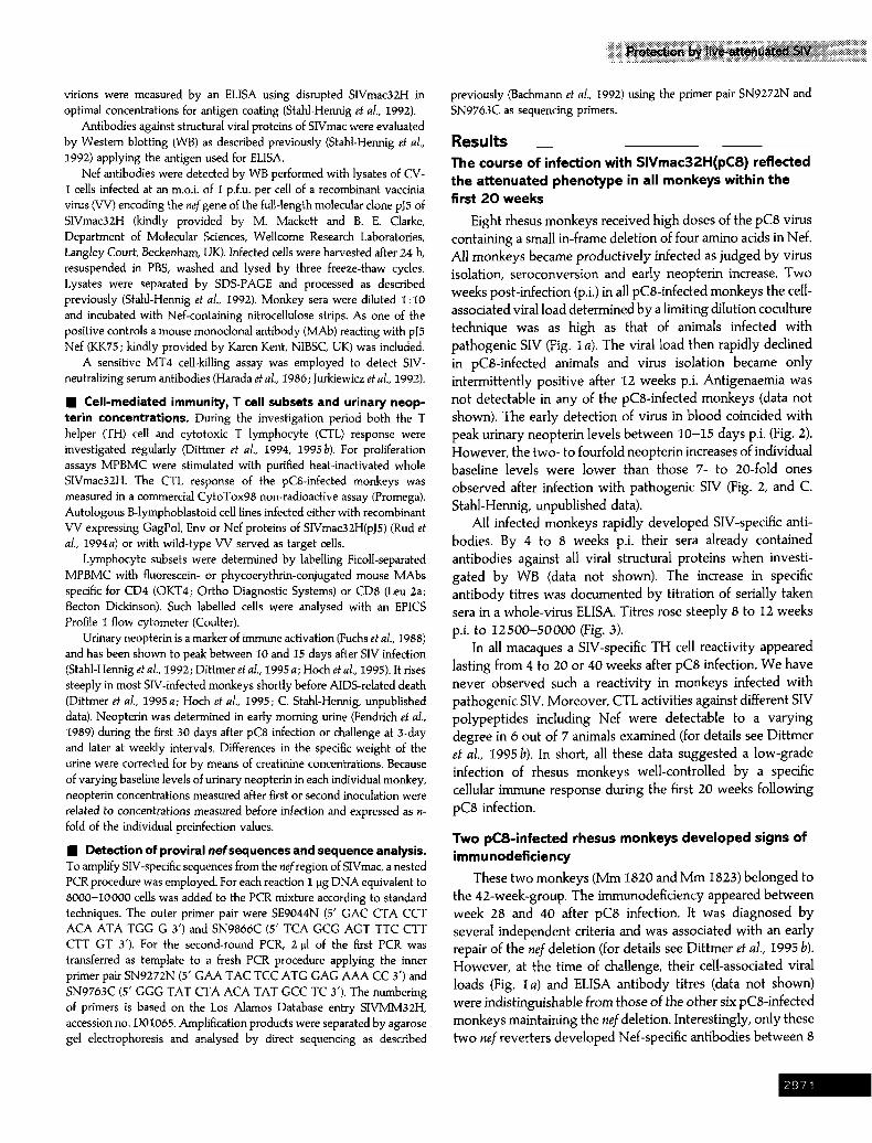
Eight rhesus monkeys received high doses of the pC8 virus containing a small in-frame deletion of four amino acids in Nef. All monkeys became productively infected as judged by virus isolation, seroconversion and early neopterin increase. Two weeks post-infection (p.i.) in all pCS-infected monkeys the cell- associated viral load determined by a limiting dilution coculture technique was as high as that of animals infected with pathogenic SIV (Fig. I a). The viral load then rapidly declined in pCS-infected animals and virus isolation became only intermittently positive after 12 weeks p.i. Antigenaemia was not detectable in any of the pCS-infected monkeys (data not shown). The early detection of virus in blood coincided with peak urinary neopterin levels between 10-15 days p.i. (Fig. 2). However, the two- to fourfold neopterin increases of individual baseline levels were lower than those 7- to 20-fold ones observed after infection with pathogenic SIV (Fig. 2, and C. Stahl-Hennig, unpublished data).
All infected monkeys rapidly developed SIV-specific anti- bodies. By 4 to 8 weeks p.i. their sera already contained antibodies against all viral structural proteins when investi- gated by WB (data not shown). The increase in specific antibody titres was documented by titration of serially taken sera in a whole-virus ELISA. Titres rose steeply 8 to 12 weeks p.i. to 1 2 5 0 0 - 5 0 0 0 0 (Fig. 3).
In all macaques a SIV-specific TH cell reac t iv i ty appeared lasting f rom 4 to 20 or 40 weeks after pC8 infection. W e have never obse rved such a react iv i ty in monkeys infected with pa thogen ic SIV. Moreover , CTL activit ies against different SIV po lypep t ides including Nef were detectable to a va ry ing degree in 6 out of 7 animals examined (for details see Di t tmer et al., 1995 b), In short, all these data sugges ted a low-grade infection of rhesus monkeys wel l -contro l led b y a specific cellular immune response dur ing the first 20 weeks fo l lowing
pC8 infection.
Two pC8-infected rhesus monkeys developed signs of immunodeficiency
These two monkeys (Mm 1820 and Mm 1823) belonged to the 42-week-group. The immunodeficiency appeared between week 28 and 40 after pC8 infection. It was diagnosed by several independent criteria and was associated with an early repair of the nef deletion (for details see Dittrner et al., 1995 b). However, at the time of challenge, their cell-associated viral loads (Fig. I a) and ELISA antibody titres (data not shown) were indistinguishable from those of the other six pCS-infected monkeys maintaining the nefdeletion. Interestingly, only these two nef reverters developed Nef-specific antibodies between 8
!97

Downloaded from www.microbiologyresearch.org by
IP: 54.224.121.223
On: Tue, 03 May 2016 21:17:03
r,J
Lo
...j
Po
u~
6 Z
(a)
~o,oo
o ~
42-w
eek-
grou
p, i
mm
uno
defic
ient
1,
000
..
..
..
,oo~
1 I ~
182o
/I
lO 1
o.1
2 4
8 12
16
20
24
28
32
36
40
46
50
54
58
62
66
70
74
86
10
,000
1,00
0
100 10 1
0.1
10,0
00
1,00
0
100 10
1
0.1
(b) 0
2 4
8 12
16
20
d d
I Co
ntro
ls
| |
[] 7
070
~7
07
1
•70
73
[]
183
9
i i
i |
24
28
32
36
40
44
48
52
2 4
8 12
16
20
24
28
32
36
40
42
22-w
eek-
grou
p
42-w
eek-
grou
p
t 1
0 2
4 8
12
16
20
24
28
32
36
40
44
48
52
22-w
eek-
grou
p
i []
179
8 ~-
I il~
I
......
...
.... I
2
4 8
12
16
20
22
Tim
e af
ter
pC8
infe
ctio
n (w
eeks
)
D179
8"
~ ~
N .
• 18
25
R
[]
1828
_
__
_
J
_
""~"
~'"
I~"
g'i~
" ~'
~"
~'~"
~'"
~"=
[]
0 2
4 8
12
16
20
24
28
32
36
40
44
48
52
Tim
e a~
er c
hall
enge
(wee
ks)
Fig.
1.
Cel
l-ass
ocia
ted
vira
l lo
ad in
rhe
sus
mac
aque
s fo
llow
ing
(a)
infe
ctio
n w
ith t
he p
C8
viru
s an
d (b
) af
ter
chal
leng
e w
ith t
he p
atho
geni
c sp
leen
-der
ived
SIV
. Viru
s w
as
isol
ated
fro
m M
PB
MC
by
a lim
iting
dilu
tion
cocu
lture
tec
hniq
ue a
nd t
he e
ndpo
int
was
det
erm
ined
. V
iral
load
s ar
e ex
pres
sed
as th
e nu
mbe
r of
inf
ectio
us c
ells
per
106
M
PBM
C.
The
resp
ectiv
e an
imal
gro
ups
as w
ell
as e
ach
indi
vidu
al a
nim
al n
umbe
r an
d de
ath
(d)
of t
he a
nim
al a
re i
ndic
ated
, The
dot
ted
line
mar
ks t
he c
ut-o
ff va
lue
of t
he
assa
y, C
ultu
res
belo
w th
e cu
t-of
f ar
e co
nsid
ered
neg
ativ
e. I
n an
imal
s m
arke
d w
ith a
n as
teris
k ch
alle
nge
viru
s w
as d
etec
tabl
e. A
t on
e tim
e po
int
not
all a
nim
als
wer
e in
vest
igat
ed (
rid,
not
done
).
~iiiiiiiii
iiiiiiiiiiii
ili~iiiiiii
iiiili~ilil

Downloaded from www.microbiologyresearch.org by
IP: 54.224.121.223
On: Tue, 03 May 2016 21:17:03
J7,7 iii6 i ili iiiiiiiiii',iii',!!ii!i!i! 16
Controls Challenge d i
, i /,o 12 , 7070
7071
6 -t- i
4 ~ { I l l ~ ,i / ,,
0 , . , . . . . . . . . . ~ ~ d ,
0 10 20 30 60 120 180 300
16
J 42-week-group i Challenge
12 o
, ~ o
1821 i o) ~ .~ 8 i 1822 . .~ --e- =
a
a U o
4 .
~ o 0 10 20 30 60 120 180 240300•0 10 20 30 60 120 180 300
16 i : Challenge 22-week-group 0 i I
12 l
1799*
8 i e
1828 . - 0 - -
4 I I
0 ' I ' I 1 I 1 I ' I ' I ' I ' ' I ' I ' I ' I ' I ' I ' I '
0 10 20 30 60 120 163/0 10 20 30 60 120 180 300
Time after pC8 infection/challenge (days) Fig. 2. Urinary neopterin levels during the preinfection period and after challenge in preinfected and non-infected rhesus monkeys. Neopterin values obtained after first infection or challenge were related to preinfection data and expressed as the n- fold of the mean of six to ten neopterin concentrations determined independently before infection. The diagram shows the neopterin concentrations of the non-infected monkeys (Controls), of those preinfected for 42 weeks (42-week-group), and those preinfected for 22 weeks (22-week-group). Individual animal numbers are indicated. For the first 30 days mean values for 5 day intervals are shown; for days 31-40 mean values of a I 0 day interval, for days 41-60 of a 20 day interval, for days 61- I 80 of a 30 day interval and then for 60 day intervals are shown. The dotted line indicates the time of challenge and 'd ' the death of the animal. In animals marked with an asterisk challenge virus was detectable.
and 24 weeks after pC8 infection. These antibodies persisted for the remaining observation period until 98 weeks p.i. (Table 1). Neopterin concentrations were elevated only slightly
(twofold in Mm 1820) or moderately (four- to fivefold over 360 days in Mm 1823). The latter animal also exhibited clinical alterations, e.g. progressing peripheral lymphadenopathy
~_97~

Downloaded from www.microbiologyresearch.org by
IP: 54.224.121.223
On: Tue, 03 May 2016 21:17:03
Table 1. Nef-speciflc antibody response in pC8-infected rhesus macaques before and after challenge
The antibody responses were scored as follows: + +, strong positive; +, positive; --, negative by immunoblot.
Reaction with Nef protein Time (weeks) 1820" 1823" 1821 1822 1825 1828 1798-~ 1799$
Post-infection 0 - - - - - -
4 - - - - - -
8 -- -4- -- 16 -- + + - 22/24 + q- + - 32 ND ND ND 42 ND hiD --
Post-challenge 4 N D N D - -
8 ND ND --
12 ND ND --
l 0 N D N D N D
24 ND N D --
3 6 N D N D - -
48 + +% + +% -
N D N D - - - -
N D N D - - - -
N D N D - - - -
N A N A N A N A
N A N A N A N A
_ _ _ + + - -
q- -- -- ND ND
ND ND ND n u -~- -~-
++ - - _ ++ ++
-{- -{- -- -- ND ND
~- -~- -- -- ND ND
* Macaques exhibiting signs of immunodeficiency and exempted from challenge. -I Macques in which challenge virus was detectable. % Sera obtained 90 weeks after infection with pCg. ND, Not done; NA, data not available because of the shorter preinfection period.
beginning at 42 weeks pi. and a splenomegaly observed by 62 weeks p.i. Both monkeys remained persistently virus positive over the whole observation period, but at invariably low levels (0"33-2 infectious cells/1 x 10 ~ PBMC). Since the alterations described indicated the development of immunodeficiency these two monkeys were not challenged.
The immune status of the non-reverters was homogenous at the time of challenge
None of the virological or immunological parameters investigated in the six remaining pCg-infected monkeys discriminated them from each other at the time of challenge. They all had maintained the nef deletion as shown by sequencing of respective PCR products (data not presented). Four of the six monkeys were virus isolation positive but had low viral loads (Fig. lb). Their antibody titres against whole SIV had reached 50000 to 400000, but neither neutralizing (data not shown) nor Nef antibodies (Table 1) were detectable. All monkeys were healthy and maintained normal CD4/CD8 ratios (above 1; data not shown). Their neopterin levels remained close to the baseline (Fig. 2) and all monkeys exhibited a considerable SIV-specific TH cell reactivity with stimulation indices ranging between 4 and 6 (Fig. 4). Unfortu- nately, CTL data were not available at time of challenge.
However, 4 weeks before challenge two of five monkeys examined showed a clear CTL activity against the structural SIV proteins, in one case also against Nef (for details see Dittmer et al., 1995 b).
Challenged animals displayed three distinct patterns of reactivity
The six pCg-infected monkeys without signs of immuno- deficiency were challenged along with four naive controls with 50 MIDs0 of the spleen-derived pathogenic SIVmac, four animals at 22 weeks p.i. and two animals at 42 weeks p.i.
The four control monkeys became productively infected thereby presenting one pattern of reactivity. Two weeks post- challenge proviral DNA was recovered from their PBMCs containing high viral loads (Fig. I b) and viral plasma antigen was detected (data not shown). All controls maintained the high cell-associated viral load until their death between 17 and 35 weeks post-challenge (Fig. l b). Three of them were euthanized in advanced AIDS stages. Pathological findings were giant cell disease, encephalopathy and opportunistic infections with Pneumocystis carinii and Cryptosporidium. The remaining animal (Mm 7073) had to be killed due to an irreparable rectal prolapse. However, at necropsy this monkey also showed AIDS-related alterations, e.g. a severe generalized
~_97 L

Downloaded from www.microbiologyresearch.org by
IP: 54.224.121.223
On: Tue, 03 May 2016 21:17:03
Controls
100,000
10.000
1,000
100
7 0 7 0 ~.4~-O d . ~ _ _ 1 . . . . . . . . . . . . . . . . . . . . . . . . . . . . . . . . . . . . . . . . . . . . . . . . . . . . . . . . . . . . . . . . . . . . . . . . . . . . . . . . . . . . . . . . . . . . . . . / . . . . . . . . . . . . . . . . . . . . . . . . . . . . . . . . . . . . . . . . . . . . . . . . . . . . . . . . . . . . . . . . . . . . . . . . . . . . . . . . . . . . . . . . . .
7 0 7 3 ! .~ J _ ...............................................
1 8 3 9 ! ~ .
~ e d 1 0 , I . I . I , I , I . I , I . I . I , I I . i l , . I , . d i ~ l , . d ~ l m d k . l , I . f . I , I * I * I . I . t .
O
"O
300,000
100,000
30,000
10,000
3.000
1,000
42-week-group
IZ;I;;;II;;I ii IIIIIIZIIII;II ;;;;IZZZ ii.;; i
! 300 .......................................................................................... -,- .............................................................................................................................. e
100 I l l l ' l ' l ' l ' l ' l l l ' l ' l ; l ' t ' l ' l l l ' l ' l ' I I t ' l ' l ' l ' l l
22-week-group 300,000 ~ ............................................................. ; . . . . . . . . . . . ; . . . . . . . . . . . . . . . . . . . . ~ - . , - . . . . - - .
30,000iq .................. i . . . . . . . . . . . . . . . . . . . . . . . r . ..............................................................................
I0,000 .................
3,000 .~28 1 ~ ................. ~ -.
1,000 .............................................................. : i ......................................................................................................................
300 . . . . . . . . . . . . . . . . . . . . . . . . . . . . . . . . . . . . . . . . . . . . . . . . . . . . . . . . . -~ . . . . . . . . . . . . . . . . . . . . . . . . . . . . . . . . . . . . . . . . . . . . . . . . . . . . . . . . . . . . . . . . . . . . . . . . . . . . . . . . . . . . . . . . . . . . . . . . . . . . . . . . . . . . . . . . . . . . . . . . . . . . . . . . . . . . . . . . . . . . . . . . . . . . . . . . . . . . . . . . . . . .
e I
cha/k)t~) g 100
-42 -34 -26 -18 -10 -2 2 10 18 26 34 42 50
Time after challenge (weeks)
Fig. 3. SIV-specific antibody titres of sera obtained during the preinfection period and after challenge from preinfected and non- infected rhesus macaques. Antibodies were determined by ELISA using whole lysed SIV as antigen. The diagram shows the titres of the non-infected monkeys (Controls), of those monkeys preinfected for 42 weeks (42-week-group), and of those preinfected for 22 weeks (22-week-group), Times of preinfection and challenge (dotted line), death (d) of the animal as well as symbols for individual animal numbers are indicated.
lymphadenopathy confirmed histologically by a severe hy- perplasia of the germinal centres as well as opportunistic infections in the intestine. Furthermore, three of the controls
seroconverted against SIV antigen. However, one monkey from this group (Mm 707I) remaining ELISA (Fig. 3) and WB (data not shown) antibody-negative was severely ill by I7
/

Downloaded from www.microbiologyresearch.org by
IP: 54.224.121.223
On: Tue, 03 May 2016 21:17:03
)',)))',)i)
I0 Controls
N 7 0 7 0 1~t7071 m 7 o 7 3 1~1839
o e o o o . o o J o o o o o ~ 6 e = o o . . o o e o e e o = = o o ~ o o = 6 o o o e o o o e = ~ o o o d d
0 $ $ I ! ! ! $ $ I $ I
10
8
X o~
.~ 6
4 Lr~
2
0
42-week-group
I
I~ 1821 • 1822 I
lO68 22-week-group~= , 11.7 - [] 1798- i I
- [ ] 1799" / I
I m182~ II l - [ ] 1828
2 .,_== . . . . . . . . . . . .
0 - 0 2 4 8 12 16 20 24 28 36 44 52
Time after challenge (weeks) Fi 9. 4. For legend see facing page.
:97~

Downloaded from www.microbiologyresearch.org by
IP: 54.224.121.223
On: Tue, 03 May 2016 21:17:03
ii!.i.i.i.i.lii.ii. i iii!Ji!ii!i!iiii.lil.ii.ii.iii.!!!i weeks p.i. and had to be euthanized. In addition, in this group a typical initial neopterin increase was recorded which was 9- to 12-fold higher than baseline levels (Fig. 2).
By contrast, only two of the six vaccinees (Mm 1798 and Mm 1799) were evidently superinfected, thus forming a second pattern of reactivity. They had been challenged 22 weeks p.i. The earliest indicator of replicating challenge virus was the complete replenishment of the nef deletion by 2 weeks p.i. shown by direct sequencing of nef PCR products. The respective nudeotide sequences were identical to those of the control monkeys and the original pJ5 sequence (Rudet al., 1994a) (data not shown). Despite the presence of full-length nef in these two monkeys, no other early signs of infection with the spleen-derived challenge virus were observed. The animals had low cell-associated viral loads (Fig. I b) and did not show a neopterin increase (Fig. 2), antigenaemia (data not shown) or an anamnestic antibody response when compared with the controls. The slightly rising antibody titres in these two monkeys of 1-2 log~ steps after challenge were attributable to still rising antibody titres after the primary infection. This became evident when the titres were compared with the antibody kinetics of the vaccinees from the 42-week-group. Only 4 to 8 weeks after challenge, cell-associated viral loads rose to moderate levels in the two vaccinees containing virus with intact nef (Fig. I b). One monkey (Mm 1798) maintained this level until the end of the observation period, the other (Mm 1799) reached high levels by 36 weeks p.i. However, these elevated levels were still approximately eightfold lower than the mean viral load of the controls (1850 infectious cells per I x 108 PBMC) determined over 36 weeks (Fig. I b). Like the two pCS-infected but immunodeficient monkeys these two vaccinees replicating SIV with full-length nef also mounted anti-Nef antibodies by 8 to 16 weeks p.i. (Table 1). Ad- ditionally, in one of these two monkeys (Mm 1798) the CD4/CD8 ratio dropped slightly by 40 weeks p.i, (data not shown). In spite of these changes both vaccinees remained clinically healthy.
In the other four preinfected monkeys superinfection could not be detected, thus representing the third reactivity pattern. All of them were still replicating the nef deletion mutant by 47 weeks p.i. (data not shown). Their cell-associated viral load (Fig. I b) and urinary neopterin concentrations (Fig. 2) remained invariably low. They showed no secondary antibody response (Fig. 3) and their clinical condition was normal. Unexpectedly, one monkey (Mm 1822) from this group in which only nef- deleted virus was detectable, generated persisting antibodies against Nef by 12 weeks post-challenge (Table 1) although all other parameters remained unchanged.
Monkeys sustaining a proliferative TH cell response after challenge were protected to various extents
To examine a possible influence of the TH cell immune response on the post-challenge period we continued measuring SIV-specific TH cell proliferation after challenge. All vaccinees maintained their virus-specific TH cell proliferative activity over 28 to 44 weeks post-challenge (Fig. 4). However, by 52 weeks post-challenge this TH cell function was no longer detectable. Unexpectedly, the two obviously superinfected vaccinees (Mm 1798 and Mm 1799) also showed a long- lasting proliferative response (Fig. 4), but each to a different extent. In one superinfected vaccinee (Mm 1799) even a sort of booster effect was observed. Its stimulation indices rose as high as 12 between 20 and 24 weeks post-challenge. Both monkeys showed no obvious clinical alterations except a mild lymphadenopathy and palpable splenomegaly in one monkey (Mm 1799) by 40 weeks post-challenge. The four completely protected animals kept their TH cell responsiveness on a low to moderate level up to 44 weeks post-challenge indicating an intact function of this cellular immune compartment.
Results from physical and blood examination reflected lymph node histology and viral status
A superficial lymph node from each vaccinee was surgically removed at 47 weeks post-challenge to test for viral load, genotypic characteristics of retained virus and for morpho- logical alterations in the lymphatic periphery. Results from investigations with lymph node mononuclear cells (LNMC) were compared with those from simultaneously prepared MPBMC. The cell-associated viral load was invariably low ( < 0-3--0"5 infectious cells per 106 LNMC) in the lymph nodes of the four vaccinees obviously protected from challenge in- fection (Mm 1821, Mm 1822, Mm 1825 and Mm 1828). Generally, the viral load of these animals was similar in blood and lymph node. One animal (Mm 1828) was even virus negative in both PBMC and LNMC. nef sequence analysis of proviral DNA from LNMC only demonstrated the pC8 genotype, but no evidence for superinfecting virus. The lymph nodes had an intact histological architecture with a mild to moderate activation similar to healthy animals. In contrast, in the two vaccinees replicating challenge virus with full-length nef, cell-associated viral loads of LNMC were as high as or even ten times higher (Mm 1798) than those of the PBMC (256 and 128 infectious cells per 106 LNMC versus 256 and 16 infectious cells per 106 PBMC). Sequence analysis of proviral DNA from LNMC confirmed the presence of wild-type SIV. Clinically Mm 1798 developed a mild splenomegaly and
Fig. 4. SlY-specific TH cell proliferation of rhesus macaques after challenge. PBMC from non-vaccinated controls (Controls), vaccinees preinfected for 42 weeks (42-week-group), and vaccinees preinfected for 22 weeks (22-week-group) were restimulated with 0.25 I~g purified heat-inactivated whole SIV antigen. Stimulation indices are shown at the time of challenge and thereafter. The cut-oft value is marked by a dotted line. At some time points not all monkeys were investigated (nd, not done). The last sample obtained before the death of the animal is indicated (d, death) as well as individual animal numbers. The monkeys marked by an asterisk replicated challenge virus.
:97:

Downloaded from www.microbiologyresearch.org by
IP: 54.224.121.223
On: Tue, 03 May 2016 21:17:03
lymphadenopathy which was consistent with a histologically detectable moderate follicular hyperplasia of the germinal centres found in one lymph node of this animal. Apart from these mild alterations both monkeys were clinically stable until 56 weeks post-challenge (time point of sacrifice).
Discussion Two different attenuated SIV nef deletion mutants and a
triple-deleted SIV conferred protection against challenge after varying waiting periods of 8 weeks to 2"25 years (Daniel et al., 1992; Almond et al., 1995 ; Whatmore et al., 1995; Norley et aL, 1996; Wyand et al., 1996). The mechanism responsible for protection still remains obscure. However, in none of these experiments had the cellular immune response been analysed. The present study using the attenuated SIVmac32H(pCS) (Rud et al., 1994a) for preinfection had a double purpose: firstly, to investigate whether shorter intervals after the initial infection would still be sufficient to allow development of protection against a pathogenic virus swarm; and secondly to elucidate the mechanisms mediating the protective immunity.
The initial high viral load appears to be immunologically contained in pC8-infected macaques
We inoculated eight rhesus monkeys with the pC8 virus. The deletion in Nef of four amino acids (Rud et al., 1992) is much smaller than that of the mutated SIV described earlier (Kestler et aL, 1991) but still rendered the virus apathogenic (Rud et aL, I992). All monkeys became readily infected by the pC8 virus as assessed by several independent criteria. An unexpected finding was that despite attenuation of the virus by the nef deletion the initial cell-associated viral loads were as high as those measured in monkeys infected with pathogenic SIV. In an earlier report (Daniel et al., 1992) viral load had only been examined 14 weeks p.i. when those monkeys had obviously already cleared the virus from the periphery. The viral loads of our monkeys also dropped and virus isolation became only intermittently positive 12 weeks p.i. but three monkeys maintained a persistent low-level viraemia. Fur- thermore, we were able to measure a long-lasting cellular immune response in the pCS-infected macaques. Six out of seven monkeys examined had developed an antiviral CTL activity (Dittmer et al., 1995 b) indicating virus replication (Stahl-Hennig et al., 1992). Most interestingly, all infected macaques generated an SIV-specific TH cell response lasting over the respective waiting periods in six of the eight monkeys. Therefore our finding fits the observation of early over- expression of T cell cytokines in pCS-infected monkeys compared to no or rather a down-regulation of expression in monkeys infected with pathogenic SIV (Benveniste et al., 1996). We had earlier observed a similar proliferative TH cell reactivity in animals infected with apathogenic HIV-2 (Dittmer et al., 1994) or SIVAvpr (Hoch et al., 1995) but never after
infection with highly pathogenic SIV strains (Dittmer et at., 1994). This underlines the attenuation of the pC8 virus.
Two monkeys with reverting pC8 virus are slow progressors
Unexpectedly, two pCS-infected monkeys developed signs of immunodeficiency (Dittmer et aI., 1995 b) and therefore were exempted from challenge. However, in these two animals only mild clinical symptoms developing later than in the controls were observed during 2 years of follow-up. Sequential analysis revealed a partially incorrect but functionally complete re- version to full-length nef by I2 weeks p.i. (Dittmer et al., 1995b). In one monkey we were able to demonstrate a duplication of the regions flanking the deletion at 10 weeks p.i. (data not shown). Such reversion to virulence by molecular evolution has been demonstrated recently for rhesus monkeys of Indian origin (Whatmore et al., 1995) but not for Chinese rhesus monkeys (Rud et al., 1992, 1994a) or for cynomolgus macaques (Rud et al., 1994a). Thus, it seems that in Indian rhesus monkeys, which are known to control SIV replication less effectively than Chinese rhesus monkeys (Joag et al., 1994 and C. Stahl-Hennig, unpublished data), reversion of pC8 to virulence is a more likely event.
A longer waiting period warrants better protection By contrast, the other six pCS-infected monkeys remained
normal by all laboratory and clinical criteria examined and were challenged with the pathogenic ex vivo derived SIV (Stahl-Hennig et aL, 1994; L/Jke et al., 1996) at 22 or 42 weeks p.i.
All vaccinees were primarily protected against the patho- genic consequences of the challenge virus while the four control animals rapidly progressed to immunodeficiency. Three of them had to be euthanized in a moribund condition within six months after challenge. By 16 weeks post-challenge two vaccinees from the 22-week-group exhibited median levels of cell-bound viral load lasting throughout the observation period. In these two monkeys virus with full-length nef was already detected by 2 weeks post-challenge. Since the nef sequences were identical to that of the challenge virus and the nef repair takes several weeks (Whatmore et al., 1995) we conclude that the challenge virus rather than a late revertant replicated in these two vaccinees. However, in these two animals disease development was apparently delayed since only a few very mild pathogenic changes were observed in the later phase of their superinfection. Although such a conclusion is difficult to draw due to the small number of animals involved, a mortality rate of 60% in 20 historical control animals within the first year after infection with our spleen- derived SIVmac makes this assumption more likely.
We have observed one of the shortest waiting periods so far required for the induction of complete protective immunity after experimental infection with an attenuated SIV. Sterile
97~

Downloaded from www.microbiologyresearch.org by
IP: 54.224.121.223
On: Tue, 03 May 2016 21:17:03
immunity was already present at 22 weeks p.i. whereby earlier results were confirmed and extended (Daniel el al., 1992; Almond el al., 1995 ; Whatmore et al., 1995; Norley et al., 1996; Wyand et al., 1996).
The protection seems to be mediated by cellular immune responses and mostly correlates with lack of Nef antibodies
Several functional and non-functional immune parameters were examined before and after challenge in order to find immunological correlates of protection. In the present trial, neutralizing antibodies did not seem to be important for protection although our failure to detect neutralizing anti- bodies might be due to the method applied. However, the role of vaccine virus-induced neutralizing antibodies is contra- dictory (Daniel el al., 1992; Almond et aI., 1995 ; Norley et aI., 1996; Wyand et al., 1996). In contrast, we were able to repeatedly measure cytotoxic T cells against various SIV proteins in the pCS-infected animals before challenge (Dittmer et al., 1995 b). These effector cells might contribute to the protection observed. Remarkably, the vaccine virus infection induced a long-lasting virus-specific TH cell response which, however, did not discriminate between protected and un- protected vaccinees. We presume that this immune function plays a crucial role in the protection either against primary challenge virus replication (Stahl-Hennig el al., 1992) or at least against the fatal consequences following infection with pathogenic challenge virus (Petry et al., 1995). Such a TH cell reactivity may also have suppressed disease development in those monkeys in which primary challenge virus replication was not completely prevented by vaccination (Shafferman el aL, 1991; Israel et al., 1994) but was perhaps suppressed by neutralizing antibodies. Surprisingly, chronic infection of macaques with the pathogenic pJ5 virus also appeared to confer protection against superinfection with the attenuated pC8 virus. Unfortunately, the identity of virus reisolated after this challenge was unknown and no data on cellular immunity was presented (Rudet al., 1994 b). However, the pJ5 virus has been reported to be only moderately pathogenic with a mortality rate of less than 15 % within I year (Rudet al., 1994a). Therefore, even in such infected macaques a certain TH cell reactivity cannot be completely ruled out which might have contributed to the protection observed in the above- mentioned challenge experiment.
Our hypothesis is further supported by recent findings from retroviral infections of mice. Immunization of mice with VV recombinants containing TH and B cell epitopes from the envelope gtycoprotein of Friend murine leukaemia virus conferred protection in mice, Additional inclusion of an immunodominant CTL epitope in the vaccine did not abrogate the fatal effects of systemic CD4 + cell depletion, thus underlining the critical role of TH priming for retrovirus immunity (Hasenkrug et al., 1996). In another experiment it
was shown that a peptide from the gp70 C-terminal TH epitope alone was sufficient to protect mice against subsequent Friend virus infection (Miyazawa et al., 1995).
The pC8 virus only carries a small net deletion but it was not clear whether Net is expressed in infected monkeys. Sera serially taken from all eight pCS-infected monkeys after first or second inoculation were examined for Net-specific antibodies. Formation of anti-Net antibodies seemed to depend on the expression of full-length Net in four monkeys, i.e. in the two monkeys repairing net early and the two vaccine failures. Thus, for antibody production Net must be either presented in its native tertiary structure which perhaps is altered in the deleted form. Alternatively, the immunodominant epitope was located in the deleted region. Surprisingly, one completely protected monkey also generated Net-specific antibodies although neither in PBMC nor in the lymph node was there any evidence for detectable full-length net. In this case, amplification of net-specific proviral sequences always generated slightly smaller fragments compared to PCR products from animals infected with the challenge virus (data not shown) thus hinting at the presence of net in its deleted form. However, one could argue that direct sequencing of PCR products might have failed to detect a minor virus subpopulation representing either late reverted pC8 virus or the challenge virus in this animal but which was nevertheless sufficient for Net antibody induction. Such an assumed subpopulation could be contained by Net- specific CTL which have previously been reported to influence viral replication (Gallimore et al., 1995). Another explanation of these unexpected anti-Net antibodies could be that the induction of such an antibody response needs a persistent viral replication on a certain as yet unidentified level. However, if this proves to be so, such antibodies should have been detected in this obviously protected animal before and not only after challenge.
Net-deleted viruses as candidate vaccines have been met with justified safety concems since even attenuated SIV with a triple deletion appears to be pathogenic for neonatal macaques (Baba et al., I995). Moreover, we and Whatmore et al. (1995) have observed that viruses with small deletions can more easily revert to virulence. Thus, a safe and effective AIDS vaccine should induce the protective immunity of live- attenuated virus including a long-lasting cellular immune response but should not contain replication-competent retro- viruses.
We are grateful to Dr M.P. Cranage, Centre for Applied Micro- biology and Research, Porton Down, UK for providing the pC8 virus. Dr M. Mackett and Dr B.E. Clarke, Department of Molecular Sciences, WeUcorne Research Laboratories, Langley Court, Beckenham, UK and the EU-Programme EVA are acknowledged for providing the vaccinia virus recombinants expressing Net (VV9011), Env (VV9002) or Gag/Pol (VVg019) of SIVmac32H(pJS). We thank K. Borchardt, K. Heinrichs, K. W/ise and U. Zedler for excellent technical assistance. This work was funded by the European Community, Concerted Action on Monkey Models for AIDS Research (grant No. MR4*-042-D).
/

Downloaded from www.microbiologyresearch.org by
IP: 54.224.121.223
On: Tue, 03 May 2016 21:17:03
References Ahmad, S., Lohmann, B., Harthas, H., Giavedoni, L., EI-Ahmad, Z., Haigwood, N., Scandella, C., Gardner, M., Luciw, P. & Yilma, T. (1994). Reduced virus load in rhesus macaques immunised with recombinant gp160 and challenged with simian immunodeficiency virus. AIDS Research and Human Retroviruses 10, 195-204.
Almond, N., Kent, K., Cranage, M., Rud, E., Clarke, B. & Stott, E. I. (1995). Protection by attenuated simian immunodeficiency virus in macaques against challenge with virus-infected cells. Lancet 345, 1342-1344.
Baba, T. W., Jeong, Y. S., Penninck, D., Bronson, R., Greene, M. F. & Ruprecht, R.M. (1995)0 Pathogenicity of live, attenuated SIV after mucosa[ infection of neonatal macaques. Science 267, 1820-1825.
Bachmann, B., Liike, W. & Hunsmann, G. (1992). Improvement of PCR amplified DNA sequencing with the aid of detergents. Nucleic Acids Research 18, 1309.
Benveniste, 0., Vaslin, B., Le Grand, R., Cheret, A., Matheux, F., Theodoro, F., Cranage, M. P. & Dormont, D. (1996). Comparative interleukin (IL)-2/interferon (IFN)-~ and IL-4/IL-10 responses during acute infection of macaques inoculated with attenuated her-truncated or pathogenic SIVmac251 virus. Proceedings of the National Academy of Sciences, USA 93, 3658-3663.
Cranage, M. P., Cook, N., Johnstone, P., Greenaway, P. l., Kitchin, P. A., Stott, E. J., Almond, N. & Baskerville, A. (1990). SIV infection of rhesus macaques: in vitro titration of infectivity and development of an experimental vaccine. In Animal Models in AIDS, pp. 103-114. Edited by H. Schellekens & M.C. Horzinek. Amsterdam, Holland: Elsevier, Biomedical Press.
Daniel, M. D., Kirchoff, F., Czajak, S. C., Sehgal, P. K. & Desrosiers, R. C. (1992). Protective effects of a live attenuated SIV vaccine with a deletion in the nef gene. Science 258, 1938-1941.
Desrosiers, R. C., Wyand, M. S., Kodama, T., Ringler, D. J., Arthur, L. 0., Segal, P. K., Letvin, N. L., King, N.W. & Daniel, M. D. (1989). Vaccine protection against simian immunodeficiency virus infection. Proceedings of the National Academy of Sciences, USA 86, 6353--6357.
Dittmer, U., Liike, W., StahI-Hennig, C., Coulibaly, C., Petry, H., Bodemer, H., Hunsmann, G. & Voss, G. (1994). Early helper T-cell dysfunction in simian immunodeficiency virus but not in human immunodeficiency virus type-2-infected macaques. Journal of Medical Primatology 23, 298-303.
Dittmer, U., StahI-Hennig, C., Coulibaly, C., Nisslein, T., Liike, W., Fuchs, D., Bodemer, W., Petry, H. & Hunsmann, G (1995 ¢0. Repeated exposure of rhesus macaques to low doses of simian immunodeficiency virus (SIV) did not protect them against the consequences of a high-dose SIV challenge. Journal of General Virology 76, 1307-1315.
Dittmer, U., Nissiein, T., Bodemer, W., Perry, H., Sauermann, U., Stahl- Hennig, C. & Hunsmann, G. (1995b). Cellular immune response of rhesus monkeys infected with a partially attenuated nef mutant of the simian immunodeficiency virus. Virology 212, 392-397.
Fendrich, C., Liike, W., StahI-Hennig, C., Herchenr6der, 0., Fuchs, D., Wachter, H. & Hunsmann, G. (1989). Urinary neopterin concentrations in rhesus monkeys after infection with simian immunodeficiency virus strain 251. AIDS 3, 305-307.
Franchini, G., Robert-Guroff, M., Tartaglia, J., Aggarwal~ A., Abimiku, A., Benson, J., Markham, P., Limbach, K., Hurteau, G., Fullen, J., Aldrich, K., Miller, N., Sadoff, J., Paoletti, E. & Gallo, R. C. (1995). Highly attenuated HIV type 2 recombinant poxviruses, but not HIV-2 recombinant Salmonella vaccines, induce long-lasting protection in rhesus macaques. AIDS Research and Human Retroviruses 11, 909-920.
Fuchs, D., Hausen, R., Reibnegger, G., Werner, E. R., Dierich, M. P. & Wachter, H. (1988). Neopterin as a marker for activated cell-mediated immunity: application in HIV infection. Immunology Today 9, 150-155.
Gallimore, A., Cranage, M., Cook, N., Almond, N., Bootman, J., Rud, E., Silvera, P., Dennis, H., Corcoran, T., Stott~ J., McMichael, A. & Gotch, F. (1995). Early suppression of SIV replication by CD8 + nef-specific cytotoxic T cells in vaccinated macaques. Nature Medicine 1, 1167-1173.
Giavedoni, L., Planelles, V., Haighwood, N., Ahmad, S., Klug, J., Marthas, M., Gardner, M., Luciw, P. & Yilma, T. (1993). Immune response of rhesus macaques to recombinant SIV gpI30 does not protect from challenge infection. Journal of Virology 67, 577-583.
Harada, S., Purtilo, D., Koyanagi, Y., Sonnabend, J. & Yamamoto, N. (1986). Sensitive assay for neutralising antibodies against AIDS-related viruses (HTLV-III/LAV). Journal of Immunological Methods 92, 177-181.
Hasenkrug, K.J., Brooks, D. M., Nishio, J. & Chesebro, B. (1996). Differing T-cell requirements for recombinant retrovirus vaccines. Journal of Virology 70, 386--372.
Hoch, J., Lang, S. M., Weeger, H., StahI-Hennig, C., Coulibaly, C., Dittmer, U., Hunsmann, G., Fuchs, D., Miiller, J., Sopper, S., Fleckenstein, B. & Oberla, K. (1995). vpr deletion mutant of simian immunodeficiency virus induces AIDS in rhesus monkeys. Journal of Virology 69, 4807-4813.
Hu, S.-L., Abrams, K., Barber, G. N., Moran, P., Zarling, I. M., Langlois, A. J., Kuller, L., Mortan, W. R. & Benveniste, R. E. (1992). Protection of macaques against SIV infection by subunit vaccines of SIV envelope glycoprotein gp160. Science 255, 456-459.
Israel, Z. R., Edmonson, P. F., Haul, D. H., O'Neil, S. P., Hossman, S. P., Thiriart, C., Farby, L., Van Opstal, 0., Bruck, C., Bex, F., Burny, A., Fultz, P. N., Mullins, J. I. & Hoover, E. A. (1994). Incomplete protection, but suppression of virus burden, elicited by subunit simian immuno- defciency virus vaccines. Journal of Virology 68, 1843-1853.
Joag, S. V., Stephens, E. B., Adams, R. J., Foresman, L. & Narayan, O. (1994). Pathogenesis of SIVmac infection in Chinese and Indian rhesus macaques: effects of splenectomy on virus burden. Virology 200, 436-466.
Jurkiewicz, E., Jansen, R., Kunze, B., Trowitzsch-Kienast, W., Forche, E., Reichenbach, H., H6fle, G. & Hunsmann, G. (1992). Three new potent HIV-1 inhibitors from myxobacteria. Antiviral Chemistry and Chemotherapy 3, 189--193.
Kestler, H.W., Ringler, D.J., Mori, K., Panicali, D.L., Sehgal, P., Daniel, M. D. & Desrosiers, R. C. (1991). Importance of the nefgene for maintenance of high virus loads and for development of AIDS. Cell 65, 651-662.
Lang, S. M., Weeger, M., StahI-Hennig, C., Coulibaly, C., Hunsmann, G., Hiiller, J., Miiller-Hermelink, H., Fuchs, D., Wachter, H., Daniel, M. D., Desrosiers, R. C. & Fleckenstein, B. (1993). Importance of vpr for infection of rhesus monkeys with simian immunodeficiency virus. Journal of Virology 67, 902-912.
Liike, W., Voss, G., StahI-Hennig, C., Coulibaly, C., Petry, H. & Hunsmann, G. (1993). Protection of cynomolgus macaques against infection by the human immunodeficiency virus type 2 strain ben (HIV- 2ben) through immunization with the virion-derived envelope gly- coprotein gp130. AIDS Research and Human Retroviruses 9, 387-394.
Liike, W., Coulibaly, C., Dittmer, U., Voss, G., Oesterle, R., Makoschey, B., Sauermann, U., Jurkiewicz, E., StahI-Hennig, C., Petty, H. & Hunsmann, G. (1996). Simian immunodeficiency virus (SIV) gpI30 oligomers protect rhesus macaques (Macaca mulatta) against infection with SIVmac32H grown on T-cells or derived ex vivo. Virology 216, 444-450.
~-98(

Downloaded from www.microbiologyresearch.org by
IP: 54.224.121.223
On: Tue, 03 May 2016 21:17:03
iiiiiiiiii ',iii i' iiii iiiN iiiiiiiiiiiiiiiiiiiiiiiiii
Hills, K., Page, H., Chan, L., Kitchin, P., Stott, J., Taffs, F., Jones, W., Rose, J., Ling, C., Silvera, P., Corcoran, T., Flanagan, B., Burny, A., Bex, F., Delchambre, H., Vanopstal, 0., Fabry, L., Thirart, C., Delers, A., Dewilde, M. & Bruck, C. (1992). Protection against SIV infection in macaques by immunization with inactivated virus from the BK28 molecular done, but not with BK28-derived recombinant env and gag proteins. Journal of Medical Primatology 21, 50-57.
Hiyazawa, H., Fujisawa, R., Ishihara, C., Takei, Y.A., Shimizu, T., Uenishi, H., Yamagishi, H. & Kuribayashi, K. (1995). Immunization with a single T helper cell epitope abrogates Friend virus-induced early erythroid proliferation and prevents late leukemia development. Journal of Immunology 155, 748--758.
Hurphy-Corb, H., Hartin, L. N., Davison-Fairburn, B., Hontelaro, R., Hiller, H., West, H., Ohkawa, S., Baskin, G. B., Zhang, J. Y., Putney, S., Allison, A. S. & Eppstein, D. (1989). A formalin-inactivated whole SIV vaccine confers protection in macaques. Science 246, 1293-1297.
Norley, S., Beer, B., Binninger-Schinzel, D., Cosma, C. & Kurth, R. (1996). Protection from pathogenic SIVmac challenge following short- term infection with a Nef-deficient attenuated virus. Virology 219, 195-205.
Petry, H., Dittmer, U., StahI-Hennig, C., Coulibaly, C., Hakoschey, B., Fuchs, D., Wachter, H., Tolle, T., Morys-Wortman, C., Kaup, F.-J., Jurkiewicz, E., Liike, W. & Hunsmann, G. (1995). Reactivation of human immunodeficiency virus type 2 in macaques after simian immuno- deficiency virus SIVmac superinfection. Journal of Virology 69, 5117- 5123.
Rud, E. W., Jon, I. R., Larder, B. A., Clarke, B. E., Cook, N. & Cranage, M. P. (1992). Infectious molecular clone of SIVmac32H: nef deletion controls ability to reisolate virus from rhesus macaques. In Vaccines 92, pp. 229-235. Edited by F. Brown, R. M. Chanock, H. S. Ginsberg & R. A. Lemer. New York: Cold Spring Harbor Laboratory Press.
Rud, E.W., Cranage, H., Yon, J., Quirk, J., Ogilvie, L., Cook, N., Webster, S., Dennis, M. & Clarke, B.E. (1994o). Molecular and biological characterization of simian immunodeficiency virus macaque strain 32H proviral clones containing size variants. Journal of General Virology 75, 529-543.
Rud, E. W., Ogilvie, L., Clarke, B. E., Almond, N., Kent, K., Chan, L., Page, H., Kitchin, P., Stott, J., Cook, N., Sharpe, S., Ashworth, T., Dennis, H., Hall, G. & Cranage, H. P. (1994/)). A naturally attenuated SIVmac32H vaccine or viral interference. In Vaccines 94, pp. 217-223. Edited by E. Norrby, F. Brown, R. M. Chanock & H. S. Ginsberg. New York: Cold Spring Harbor Laboratory Press.
Shafferman, A., Jahrling, P. B., Benveniste, R. E., Lewis, H. G., Phipps, T.J., Eden-HcCutchan, F., Sadoff, J., Eddy, G.A. & Burke, D.S. (1991 ). Protection of macaques with a simian immunodeficiency virus envelope peptide vaccine based on conserved human immunodeficiency virus type I sequences. Proceedings of the National Academy of Sciences, USA 88, 7126--7130.
StahI-Hennig, C., Voss, G., Nick, S., Petty, H., Fuchs, D., Wachter, H., Coulibaly, C., LiJke, W. & Hunsmann, G. (1992). Immunization with Tween-ether-treated S[V adsorbed onto aluminium hydroxide protects monkeys against experimental SIV infection. Virology I86, 588-596.
Stahl-Henni9, C., Coulibaly, C., Petry, H., Voss, G., Dittmer, U., Bodemer, W., Hakoschey, B., Jurkiewicz, E., L/Jke, W. & Hunsmann, G. (1994). Immunization with virion-derived glycoprotein 130 from HIV- 2 or SIV protects macaques against challenge virus grown in human or simian cells or prepared ex vivo. AIDS Research and Human Retroviruses 10, $27-$32.
Sutjipto, S., Pedersen, N. C., Hiller, C. J., Gardner, M. B., Hanson, C. V., Gettie, A., Jennings, M., Higgins, J. & Marx, P. A. (1990). Inactivated simian immunodeficiency virus vaccine failed to protect rhesus macaques from intravenous or genital mucosal infection but delayed disease in intravenously exposed animals. Journal of Virology 64, 2290-2297.
Whatmore, A. H., Cook, N., Hall, G.H., Sharpe, S., Rud, E.W. & Cranage, H. P. (1995). Repair and evolution of nef in vivo modulates simian immunodeficiency virus virulence. Journal of Virology 69, 5117- 5123.
Wyand, H.S., Hanson, K.H., Garcia-Holl, H., Hontefiori, D. & Desrosiecs, R. C. (1996). Vaccine protection by a triple deletion mutant of simian immunodeficiency virus. Journal of Virology 70, 3724-3733.
Received 29 Hay 1996; Accepted 7 August 1996
:9,'
Copyright © 2022 FDOKUMEN