External brain morphology in rhesus macaques (Macaca mulatta)
Upload
independentCategory
view
4download
0
RESEARCH ARTICLE
Prevalence of Enteric Parasites in Pet Macaques inSulawesi, Indonesia
LISA JONES-ENGEL1,2n, GREGORY A. ENGEL2,3, MICHAEL A. SCHILLACI4, KELLYKYES2, JEFFERY FROEHLICH5, UMAR PAPUTUNGAN6, and RANDALL C. KYES1,2
1Department of Psychology, University of Washington, Seattle, Washington2Washington National Primate Research Center, University of Washington, Seattle,Washington3Swedish/Providence Family Medicine, Seattle, Washington4Department of Social Sciences, University of Toronto at Scarborough, Scarborough,Canada5Department of Anthropology, University of New Mexico, Albuquerque, New Mexico6Department of Animal Production, Sam Ratulangi University, Manado, Sulawesi,Indonesia
On the Indonesian island of Sulawesi, nonhuman primate pets come intofrequent contact with humans, presenting the possibility of zoonotic andanthropozoonotic disease transmission. We collected fecal samples from88 pet macaques representing six of the seven macaque species currentlyrecognized as endemic to Sulawesi (Macaca nigra, M. nigrescens,M. hecki, M. tonkeana, M. maura, and M. ochreata) as well as two non-endemic species (M. fascicularis and M. nemestrina) in order todetermine the prevalence of intestinal parasitic infection in thispopulation. Seven taxa of intestinal protozoa (Blastocystis hominis,Iodamoeba butschlii, Entamoeba coli, Entamoeba hartmanni, Chilomas-trix mesnili, Endolimax nana, and Retortamonas intestinalis) and threetaxa of nematodes (hookworm, Trichuris spp., and Ascaris spp.) weredetected. The overall parasitization rate was 59.1%. Commensal organ-isms predominated in this population. Parasitization was not statisticallycorrelated with macaque age group, sex, species, or location, or withthe owner’s level of education. These findings are discussed in the contextof primate pet ownership practices in Sulawesi. Am. J. Primatol.62:71–82, 2004. r 2004 Wiley-Liss, Inc.
Key words: Sulawesi; pet primates; Macaca; intestinal parasites; pro-tozoa; nematodes; zoonoses
Contract grant sponsor: Sigma Xi; Contract grant sponsor: University of New Mexico; Contractgrant sponsor: American Society of Primatologists; Contract grant sponsor: Chicago ZoologicalSociety; Contract grant sponsor: Sandia National Laboratories; Contract grant sponsor:Unconventional Concepts, Inc.
nCorrespondence to: Lisa Jones-Engel, Washington National Primate Research Center, Box 357330,University of Washington, Seattle, WA 98195. E-mail: [email protected]
Received 6 July 2003; revision accepted 26 November 2003
DOI 10.1002/ajp.20008Published online in Wiley InterScience (www.interscience.wiley.com).
r 2004 Wiley-Liss, Inc.
American Journal of Primatology 62:71–82 (2004)
INTRODUCTION
The morphological, physiological, behavioral, and genetic similarities betweenhumans and nonhuman primates contribute to their susceptibility to a similarspectrum of pathogens [Brack, 1987]. This shared susceptibility enables zoonotic(nonhuman primate to human) and anthropozoonotic (human to nonhumanprimate) transmission of infectious agents to occur in situations where humanscome into contact with nonhuman primates. The cross-species transmission ofinfectious agents poses a potential threat to both primates and humans.
Ownership of pet primates is a common practice in source countries thatcontain wild primate populations [Jones-Engel et al., 2002; McCann & Taylor,2002; Fitch-Snyder et al., 2002; Abernathy et al., 2002]. In countries such asIndonesia, the destruction of primate habitats, bushmeat-hunting, and trappingas a means of eliminating crop-raiding lead to the acquisition of nonhumanprimates (most often infants and juveniles) that become pets. Although nocomplete census data on pet primates in Sulawesi are available, the conditionsand practices that can result in the acquisition of pets are on the rise [Jones-Engelet al., in press]. Since pet primates live in constant and close proximity tohumans, there is the possibility of cross-species transmission of infectious agentsin this context; however, it is yet to be well studied [Jones-Engel et al., 2001].
Prior research on infectious diseases in pet primates has included work onleprosy, [Hagstad, 1983], SIV [Georges-Courbot et al., 1998], and enteric bacteria[Fox, 1975; Tresierra-Ayala & Fernandez, 1997]. Zoonotic transmission of entericparasites from domestic animals, such as cats, dogs, and cattle, is a well-describedphenomenon [Stehr-Green et al., 1987; Harvey et al., 1991; Olson et al., 1997; Foket al., 2001; Mazyad & El-Nemr, 2002]. To date, however, enteric parasitizationamong pet primates has not been well studied. The present cross-sectional studytakes a first step toward learning about transmission of enteric parasites betweenhumans and pet primates on Sulawesi. Our aim was to characterize the preva-lence of intestinal fauna in pet primates in order to determine whether theparasitic fauna of pet primates pose an infectious threat to human populations.
MATERIALS AND METHODS
Pet Macaques on Sulawesi
Sulawesi is home to at least seven morphologically distinct species ofmacaque [Fooden, 1969]. Each species occupies a distinct range, with hybridpopulations occurring naturally in areas of sympatry [Bynum et al., 1999; Evanset al., 2001; Froehlich & Supriatna, 1996; Groves, 1980; Watanabe et al., 1991].Previous research suggests that on Sulawesi, primate pets are acquired throughtrapping and as a by-product of hunting and crop protection (Jones-Engel,unpublished data). Macaque species not native to Sulawesi also make up part ofthe pet population. Most of these non-native primates are acquired on otherIndonesian islands and accompany their owners when the owners move.Generally, infants and juveniles are considered most desirable as pets becausemacaques often become aggressive as they reach sexual maturity. Primate-petowners frequently sell or give away their pets as the animals mature.
Data Collection and Analysis
Fecal samples from 88 pet macaques were collected during two field seasons(June–August 2000, and May 2001) on the northern, east-central, and southern
72 / Jones-Engel et al.
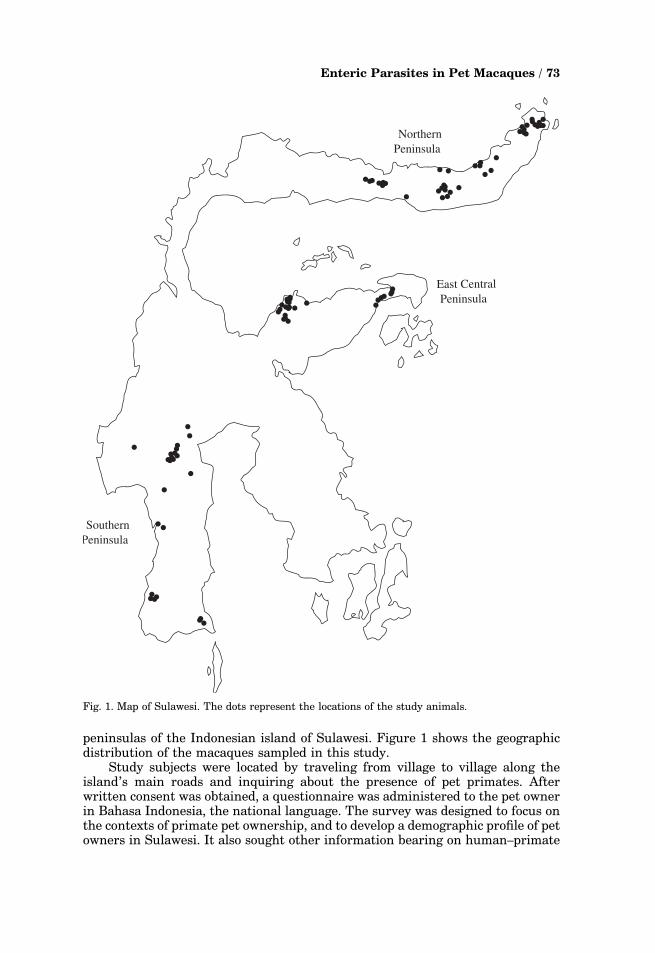
peninsulas of the Indonesian island of Sulawesi. Figure 1 shows the geographicdistribution of the macaques sampled in this study.
Study subjects were located by traveling from village to village along theisland’s main roads and inquiring about the presence of pet primates. Afterwritten consent was obtained, a questionnaire was administered to the pet ownerin Bahasa Indonesia, the national language. The survey was designed to focus onthe contexts of primate pet ownership, and to develop a demographic profile of petowners in Sulawesi. It also sought other information bearing on human–primate
... .
.. ..
....
...
.
... .... . ..... ....
.
.
.
.
.. ... .. ... ....... .
.
..
.
..
.
..
.
...
..
..
.
..
.
...
...
..
.
.
Northern Peninsula
Southern Peninsula
East Central Peninsula
Fig. 1. Map of Sulawesi. The dots represent the locations of the study animals.
Enteric Parasites in Pet Macaques / 73
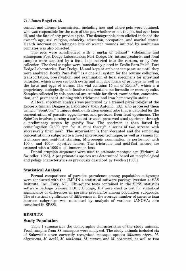
contact and disease transmission, including how and where pets were obtained,who was responsible for the care of the pet, whether or not the pet had ever beenill, and the fate of any previous pets. The demographic data elicited included theowner’s age, sex, religion, ethnicity, education, occupation, and marital status.Health information relating to bite or scratch wounds inflicted by nonhumanprimates was also collected.
The pets were anesthetized with 3 mg/kg of Telazols (tiletamine andzolazepam; Fort Dodge Laboratories; Fort Dodge, IA) intramuscularly, and fecalsamples were acquired by a fecal loop inserted into the rectum, or by free-collection. The fecal samples were immediately placed in Ecofix Para-Paks; FortDodge Laboratories; Fort Dodge, IA and kept at ambient temperature until theywere analyzed. Ecofix Para-Paks is a one-vial system for the routine collection,transportation, preservation, and examination of fecal specimens for intestinalparasites, which preserves both cystic and amoebic forms of protozoa as well asthe larva and eggs of worms. The vial contains 15 ml of Ecofixs, which is aproprietary, ecologically safe fixative that contains no formalin or mercury salts.Samples collected by this protocol are suitable for direct examination, concentra-tion, and permanent staining with trichrome and iron hematoxylin stains.
All fecal specimen analysis was performed by a trained parasitologist at theEsoterix Simian Diagnostic Laboratory (San Antonio, TX), who processed themusing a ‘‘SpinCon,’’ a unique double-filtration conical tube that is patented for theconcentration of parasite eggs, larvae, and protozoa from fecal specimens. TheSpinCon involves passing a surfactant-treated, preserved stool specimen througha preliminary screen by gravity flow. The specimen is then forced bycentrifugation (2,500 rpm for 10 min) through a series of two screens withsuccessively finer mesh. The supernatant is then decanted and the remainingconcentration is subjected to a direct microscopic technique, as well as a smear fortrichrome and acid-fast staining. Microscopic examination is performed with100� and 400� objective lenses. The trichrome and acid-fast smears arescanned with a 1000� oil immersion lens.
Dental eruption sequences were used to estimate macaque age [Sirianni &Swindler, 1985]. A pet primate’s species was determined based on morphologicaland pelage characteristics as previously described by Fooden [1969].
Statistical Analysis
Formal comparisons of parasite prevalence among population subgroupswere conducted with the JMP-IN 4 statistical software package (version 4; SASInstitute, Inc., Cary, NC). Chi-square tests contained in the SPSS statisticssoftware package (release 11.0.1; Chicago, IL) were used to test for statisticalsignificance of differences in parasite prevalence among population subgroups.The statistical significance of differences in the average number of parasite taxabetween subgroups was calculated by analysis of variance (ANOVA; alsocontained in SPSS).
RESULTS
Study Population
Table I summarizes the demographic characteristics of the study animals.Fecal samples from 88 macaques were analyzed. The study animals included sixof Sulawesi’s seven currently recognized macaque species (Macaca nigra, M.nigrescens, M. hecki, M. tonkeana, M. maura, and M. ochreata), as well as two
74 / Jones-Engel et al.

TA
BL
EI.
Dem
og
rap
hic
Pro
file
of
the
Stu
dy
Sa
mp
le
Ag
e-s
ex
cla
ssMacaca
nigra
Macaca
nigrescen
sMacaca
hecki
Macaca
tonkeana
Macaca
maura
Macaca
och
reata
Macaca
fascicularis
Macaca
nem
estrina
Macaca
hybrid
To
tal
AM
a3
00
22
01
00
8S
AM
b2
10
41
00
02
10
JM
c4
54
92
12
02
29
Su
bto
tal
ma
les
96
41
55
13
04
47
AF
d3
10
30
01
03
11
SA
Fe
10
02
00
11
27
JF
f5
10
73
00
07
23
Su
bto
tal
fem
ale
s9
20
12
30
21
12
41
To
talg
18
84
27
81
51
16
88
Geo
loca
tio
nN
Pen
h1
88
40
00
20
84
0E
-CP
eni
00
01
80
00
06
24
SP
enj
00
09
81
31
22
4
aA
du
ltm
ale
s.bS
ub
ad
ult
male
s.cJu
ven
ile
male
s.dA
du
ltfe
male
s.eS
ub
ad
ult
fem
ale
s.f J
uven
ile
fem
ale
s.gS
um
of
all
male
san
dfe
male
s.hN
ort
her
np
enin
sula
.i E
ast
-cen
tral
pen
insu
la.
j Sou
ther
np
enin
sula
.
Enteric Parasites in Pet Macaques / 75

non-native species (M. fascicularis and M. nemestrina) that were brought toSulawesi from other Indonesian islands. Sixteen of the animals studied werehybrids of Sulawesi species.
Juvenile macaques (defined as individuals between 1 and 3 years of age)predominated in the study population (n¼ 52; 59%). The absence of infants inthis sample may be explained by anecdotal reports of high mortality among veryyoung pet macaques in Sulawesi; however, the mortality rates of infant pets havenot been measured. Subadults (n¼ 17; 19%) and adults (n¼ 19; 22%) may berelatively underrepresented (compared to wild populations) among pets inSulawesi because owners frequently give them away or sell them at these ages.
Prevalence of Enteric Parasites
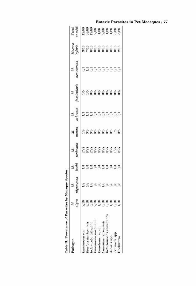
Ten taxa (seven protozoan taxa and three nematode taxa) of enteric parasiteswere encountered in this study sample. The most frequently encounteredprotozoa were Blastocystis hominis (present in 43% of macaques), Iodamoebabutschlii (present in 22%), and Entamoeba coli (present in 14%). The nematodeshookworm, Trichuris spp., and Ascaris were found in fecal samples from 6%, 3%,and 1%, respectively, of the pet macaques in this study. Table II presentsprevalence data for the parasites found in each species of macaque.
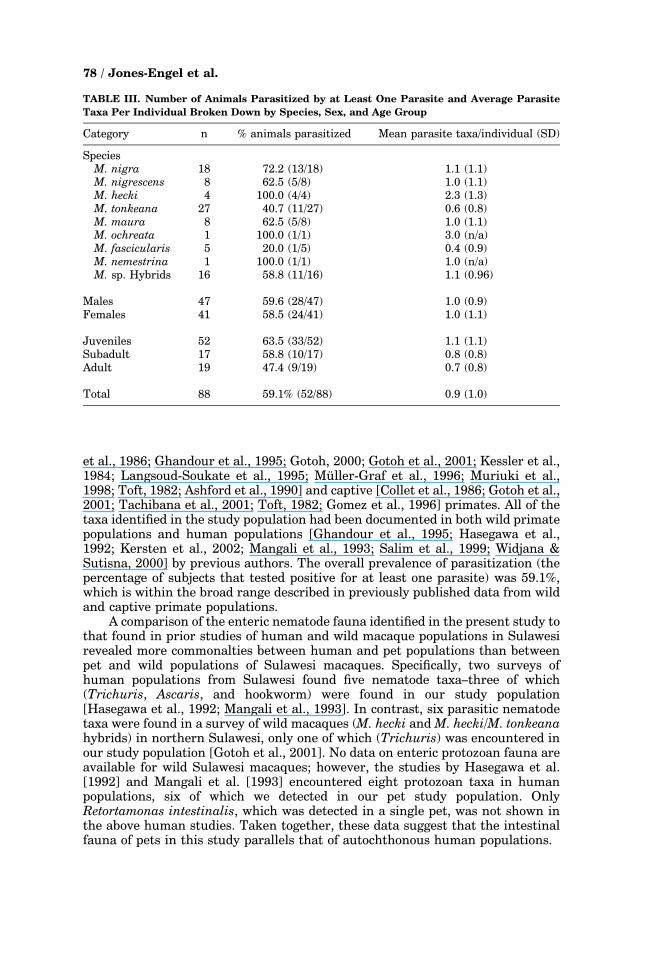
Table III presents data on the prevalence of parasitization (which is definedas the percentage of animals infected with at least one parasite) and the averagenumber of parasite taxa per individual. Prevalence is tabulated by species, sex,and age group.
Parasite prevalence was higher among the northern-peninsula macaques(73%) than among macaques from the east-central (54%) and southern (42%)peninsulas. A comparison of mean prevalence in the three geographic areas byone-way ANOVA showed a significant effect (F (2,87)¼ 3.23, P¼ 0.04). A post-hocTukey test revealed that the difference in parasite prevalence between thenorthern and southern peninsulas was the source of the effect (P¼ .04). Nogeographic differences in prevalence were observed for the individual parasites.No statistical differences in parasitization were detected for sex, age, species,owner education, or pet location. Similarly, a comparison of the prevalence ofindividual parasite taxa among sample subgroups revealed no statisticallysignificant correlations.
Pets without parasites in their feces accounted for 40.9% of the studypopulation, those with one parasite taxon represented 34.1% of the studypopulation, and those with two parasite taxa accounted for 15.9% of the total.Three or four parasite taxa were detected in the feces of less than 10% of the pets.There were no statistically significant correlations between the number ofparasite taxa detected per individual and sex, age class, species, or geolocation.Tests for correlation between individual parasites were run for those pets thattested positive for two or more parasites. The results showed no significantrelationships, with one exception: the presence of B. hominis was negativelycorrelated with that of hookworm (r(22)¼�.50, P¼ .02).
DISCUSSION
Taxa of Enteric Parasites Identified
Microscopic analysis detected 10 distinct taxa of enteric parasites, a faunaldiversity consistent with previously published data from populations of wild[Abbot & Majeed, 1984; Appleton & Boinski, 1991; Appleton et al., 1994; Collet
76 / Jones-Engel et al.
Ta
ble
II.
Pre
va
len
ce
of
Pa
rasi
tes
by
Ma
ca
qu
eS
pecie
s
Path
ogen
M.
nigra
M.
nigrescen
sM.
hecki
M.
tonkeana
M.
maura
M.
och
reata
Mfascicularis
M.
nem
estrina
Macaca
hyb
rid
Tota
l(n
=8
8)
Entamoebacoli
2/1
81
/81
/42
/27
1/8
1/1
1/5
0/1
3/1
61
2/8
8Blastocystis
hominis
8/1
85
/84
/48
/27
3/8
1/1
1/5
1/1
7/1
63
8/8
8Iodamoebabutsch
lii
5/1
81
/81
/42
/27
3/8
1/1
0/5
0/1
6/1
61
9/8
8Entamoebahartmanni
2/1
80
/80
/40
/27
0/8
0/1
0/5
0/1
0/1
62
/88
Endolimaxnana
1/1
80
/80
/40
/27
0/8
0/1
0/5
0/1
0/1
61
/88
Chilomastrixmesnili
0/1
81
/81
/40
/27
0/8
0/1
0/5
0/1
0/1
62
/88
Retortamonasintestinalis
0/1
80
/81
/40
/27
0/8
0/1
0/5
0/1
0/1
61
/88
Ascaris
spp
.1
/18
0/8
0/4
0/2
70
/80
/10
/50
/10
/16
1/8
8Trich
uris
spp
.0
/18
0/8
1/4
1/2
71
/80
/10
/50
/10
/16
3/8
8H
ook
worm
1/1
80
/80
/42
/27
0/8
0/1
0/5
0/1
2/1
65
/88
Enteric Parasites in Pet Macaques / 77
et al., 1986; Ghandour et al., 1995; Gotoh, 2000; Gotoh et al., 2001; Kessler et al.,1984; Langsoud-Soukate et al., 1995; Muller-Graf et al., 1996; Muriuki et al.,1998; Toft, 1982; Ashford et al., 1990] and captive [Collet et al., 1986; Gotoh et al.,2001; Tachibana et al., 2001; Toft, 1982; Gomez et al., 1996] primates. All of thetaxa identified in the study population had been documented in both wild primatepopulations and human populations [Ghandour et al., 1995; Hasegawa et al.,1992; Kersten et al., 2002; Mangali et al., 1993; Salim et al., 1999; Widjana &Sutisna, 2000] by previous authors. The overall prevalence of parasitization (thepercentage of subjects that tested positive for at least one parasite) was 59.1%,which is within the broad range described in previously published data from wildand captive primate populations.
A comparison of the enteric nematode fauna identified in the present study tothat found in prior studies of human and wild macaque populations in Sulawesirevealed more commonalties between human and pet populations than betweenpet and wild populations of Sulawesi macaques. Specifically, two surveys ofhuman populations from Sulawesi found five nematode taxa–three of which(Trichuris, Ascaris, and hookworm) were found in our study population[Hasegawa et al., 1992; Mangali et al., 1993]. In contrast, six parasitic nematodetaxa were found in a survey of wild macaques (M. hecki and M. hecki/M. tonkeanahybrids) in northern Sulawesi, only one of which (Trichuris) was encountered inour study population [Gotoh et al., 2001]. No data on enteric protozoan fauna areavailable for wild Sulawesi macaques; however, the studies by Hasegawa et al.[1992] and Mangali et al. [1993] encountered eight protozoan taxa in humanpopulations, six of which we detected in our pet study population. OnlyRetortamonas intestinalis, which was detected in a single pet, was not shown inthe above human studies. Taken together, these data suggest that the intestinalfauna of pets in this study parallels that of autochthonous human populations.
TABLE III. Number of Animals Parasitized by at Least One Parasite and Average Parasite
Taxa Per Individual Broken Down by Species, Sex, and Age Group
Category n % animals parasitized Mean parasite taxa/individual (SD)
SpeciesM. nigra 18 72.2 (13/18) 1.1 (1.1)M. nigrescens 8 62.5 (5/8) 1.0 (1.1)M. hecki 4 100.0 (4/4) 2.3 (1.3)M. tonkeana 27 40.7 (11/27) 0.6 (0.8)M. maura 8 62.5 (5/8) 1.0 (1.1)M. ochreata 1 100.0 (1/1) 3.0 (n/a)M. fascicularis 5 20.0 (1/5) 0.4 (0.9)M. nemestrina 1 100.0 (1/1) 1.0 (n/a)M. sp. Hybrids 16 58.8 (11/16) 1.1 (0.96)
Males 47 59.6 (28/47) 1.0 (0.9)Females 41 58.5 (24/41) 1.0 (1.1)
Juveniles 52 63.5 (33/52) 1.1 (1.1)Subadult 17 58.8 (10/17) 0.8 (0.8)Adult 19 47.4 (9/19) 0.7 (0.8)
Total 88 59.1% (52/88) 0.9 (1.0)
78 / Jones-Engel et al.

Pet PrimatesFA Unique Population
These findings should be regarded in the context of what we know about petprimate ownership in Sulawesi. Our data indicate that most pet macaques inSulawesi are acquired as young animals (usually juveniles or infants) [Jones-Engel et al., in press]. As a result, these animals may be removed from theirnatural groups before they are exposed to most of the parasites enzootic in thosegroups. When they subsequently become pets, their exposure to parasites islargely, if not wholly, determined by their human-shaped environment (i.e.,where they are kept, their water and food supply, and contact with otherdomesticated animals). Hence, their intestinal fauna might be expected toresemble that of the surrounding human populations.
Pet primates thus constitute a unique population of primates with uniqueepizootiological considerations. Unlike wild primates, they live in constant, closeproximity to humans. Unlike most zoo primates, they have little if any havecontact with other primates (although there are accounts of pets being courted bywild primates). Unlike the situation with laboratory primates, contact withhumans is unregulated, and transmission of infection is undeterred by gloves,masks, or other protective measures designed to protect both humans andprimates from pathogen transmission. Furthermore, and also in contrast tolaboratory primates, the health of pets in habitat countries is generally notmonitored and maintained by veterinary medical professionals who are capable ofdiagnosing and treating intestinal infections.
Paucity of Pathogenic Parasites
It is interesting to note that in this primate sample the three most commonlyencountered parasite taxa (Blastocystis hominis, Iodamoeba butschlii, andEntamoeba coli, occurring in 43%, 21%, and 14% of macaques, respectively) aregenerally regarded as minimally pathogenic or nonpathogenic in both human andnonhuman primates. Of the parasitic taxa encountered in this study, only thenematode taxa hookworm, Trichuris, and Ascaris (found in 6%, 3%, and 1% of thesubjects, respectively) are considered significant pathogens. This paucity ofpathogenic fauna contrasts with data from wild primate populations [Abbot &Majeed, 1984; Appleton & Boinski, 1991; Appleton et al., 1994; Collet et al., 1986;Ghandour et al., 1995; Gotoh, 2000; Gotoh et al., 2001; Kessler et al., 1984;Langsoud-Soukate et al., 1995; Muller-Graf et al., 1996; Muriuki et al., 1998; Toft,1982; Ashford et al., 1990], which show higher infection rates with pathogens,including Balantidium coli, Entamoeba histolytica, Giardia lamblia, hookworm,Trichuris, and Strongyloides. One possible explanation for this observation is thatmobility may play an important role in the acquisition of enteric parasites. Byvirtue of their ability to travel, humans and wild macaques may have morefrequent contact with intestinal parasites and hence a higher incidence ofinfection than pets, which are usually tethered in a fixed location. Also, sinceantiparasitic drugs are readily available in Sulawesi, and people infected withpathogenic parasites would be expected to use antiparasitic medications morefrequently than those infected only with commensals, pets may be exposed topathogenic parasites less frequently than commensals. An alternate explanationfor the lack of pathogenic fauna in these pets is that infection causes significantmortality in pets.
Enteric Parasites in Pet Macaques / 79

Future Work
The present study provides a preliminary glimpse of enteric parasiticinfections among pet primates in a source country, and points the way to furtherresearch. Data on the prevalence of pet ownership are needed to better define thescope of the phenomenon. Research comparing parasite loads among pets andowners would help to clarify the epidemiology and epizootiology of zoonotic andanthropozoonotic transmission in this context. Ideally, such data would becollected and correlated with more complete data on the human environment-Fespecially as regards the water supply, sanitation, and the presence of otherdomesticated animals.
CONCLUSIONS
Humans and nonhuman primates share a susceptibility to a wide range offecal parasites. In Sulawesi, primate-pet ownership provides a context for thecross-species transmission of pathogens. Our data suggest that infection withenteric parasites is common in pet macaques on Sulawesi, but that relatively fewof these infections are caused by pathogenic organisms. The intestinal faunadetected in this population parallels that detected in previously publishedanalyses of human populations in Sulawesi. These observations may be explainedby the unique circumstances relating to pet macaques and their environment inSulawesi.
ACKNOWLEDGMENTS
The Institutional Animal Care and Use Committee of the University of NewMexico approved the research protocol for this study. The authors thank Drs. D.Cohn, J. Heidrich, A.L. Sobel, M. Stuart, and R. Heberling, as well as A. Cookeand J. Garcia for their guidance and assistance. Travel support for this researchwas provided by Singapore Airlines. We also thank M. Novak and two anonymousreviewers for their helpful comments.
REFERENCES
Abbot DP, Majeed SK. 1984. A survey ofparasitic lesions in wild-caught, laboratory-maintained primates: (rhesus, cynamol-gous, and baboon). Vet Pathol 21:198–207.
Abernathy K, Peignot P, McCann C, Teffler P.2002. Solving the problem of illegal captiveprimates in Gabon. In: Abstracts of theXIXth Congress of the International Prima-tological Society. p 166–167.
Appleton CC, Boinski S. 1991. A preliminaryparasitological analysis of fecal samplesfrom a wild population of Costa Ricansquirrel monkeys (Saimiri oerstedi). J MedPrimatol 20:402–403.
Appleton CC, Krecek RC, Verster A, BrourtonMR, Lawes MJ. 1994. Gastro-intestinalparasites of the Samango monkey, Cerco-pithecus mitis, in Natal, South Africa. J MedPrimatol 23:52–55.
Ashford RW, Reid GDF, Butynski TM. 1990.The intestinal faunas of man and mountain
gorillas in a shared habitat. Ann Trop MedParasitol 84:337–340.
Brack M. 1987. Agents transmissable fromsimians to man. Berlin: Springer-Verlag.
Bynum EL, Kohlhaas AK, Pramono AH. 1999.Conservation status of Sulawesi macaques.Trop Biodivers 6:123–144.
Collet JY, Galdikas BM, Sugarjito J, Jojosud-harmo S. 1986. A coprological study ofparasitism in orangutans (Pongo pygmaeus)in Indonesia. J Med Primatol 15:121–129.
Evans BJ, Supriatna J, Melnick DJ. 2001.Hybridization and population genetics oftwo macaque species in Sulawesi, Indonesia.Evolution 55:86–1602.
Fitch-Snyder H, Thanh VN, Dang NX, SchulzH, Bottcher-Law L. 2002. Conservation andcaptive management of Vietnamese pri-mates. In: Abstracts in the XIXth Congressof the International Primatological Society.p 163–164.
80 / Jones-Engel et al.

Fok E, Szatmari V, Busak K, Rozgonyi F.2001. Prevalence of intestinal parasites indogs in some urban and rural areas ofHungary. Vet Q 23:96–98.
Fooden J. 1969. Taxonomy and evolution ofthe monkeys of Celebes (Primates: Cerco-pithecidae). Bibliotheca primatologica. Vol.X. Basel: S. Karger.
Fox JG. 1975. Transmissible drug resistancein shigella and salmonella isolated from petmonkeys and their owners. J Med Primatol4:165–171.
Froehlich JW, Supriatna J. 1996. Secondaryintergradation between Macaca maurusand M. tonkeana in south Sulawesi, andthe species status of M. togeanus. Evolutionand ecology of macaque societies. Fa JE,Lindburg DG, editors. New York: Cam-bridge University Press. p 43–70.
Georges-Courbot MC, Lu CY, Mauwa M,Telfer P, Onanga R, Dubreuil B, Chen Z,Smith SM, Gao F, Hahn BH, Marx PA.1998. Natural infection of a household petred-capped mangabey (Cercocebus torquatustorquatus) with a new simian immunodefi-ciency virus. J Virol 72:600–608.
Ghandour AM, Zahid NZ, Banaja AA, KamalKB, Bouq AI. 1995. Zoonotic intestinalparasites of hamadryas baboons Papio ha-madryas in the western and northernregions of Saudi Arabia. J Trop MedHygiene 98:431–439.
Gomez MS, Gracenea M, Montoliu I, Feliu C,Monleon A, Fernandez J, Ensenat C. 1996.Intestinal parasitismFprotozoa and hel-minthsFin primates at the Barcelona Zoo.J Med Primatol 25:419–423.
Gotoh S. 2000. Regional differences in theinfection of wild Japanese macaques bygastrointestinal helminth parasites. Pri-mates 41:291–298.
Gotoh S, Takenada O, Watanabe K, HamadaY, Kawamoto Y. 2001. Hematological valuesand parasite fauna in free-ranging Macacahecki and M. hecki/ M. tonkeana hybridgroup of Sulawesi Island, Indonesia. Pri-mates 42:27–34.
Groves C. 1980. Speciation in Macaca: theview from Sulawesi. In: Lindberg DG, VanNostrand R, editors. The macaques: studiesin ecology, behavior and evolution. NewYork: Reinhold.
Hagstad HV. 1983. Leprosy in sub-humanprimates: potential risk for transfer ofM. leprae to humans. Int J Zoonoses 10:127–131.
Harvey JB, Roberts JM, Schantz PM. 1991.Survey of veterinarians’ recommendationsfor treatment and control of intestinalparasites in dogs: public health implica-tions. J Am Vet Med Assoc 199:702–707.
Hasegawa H, Miyagi I, Toma T, Kamimura K,Nainggolan IJJ, Tumewu-Wagei M, Manda-
gi-Waworuntu HG, Kapojos FX, RuntuweneJ, Paath-Runtupalit C, Syafruddin. 1992.Intestinal parasitic infections in Likupang,North Sulawesi, Indonesia. Southeast AsianJ Trop Med Public Health 23:219–227.
Jones-Engel L, Engel GA, Schillaci MA, BaboR, Froehlich JW. 2001. Detection of anti-bodies to selected human pathogens amongwild and pet macaques (Macaca tonkeana)in Sulawesi, Indonesia. Am J Primatol54:171–178.
Jones-Engel L, Engel GA, Schillaci MA,Paputungan U, Froehlich J, Kyes R. 2002.A pet’s life: characterizing primate petownership in Sulawesi. In: Abstracts of theXIXth Congress of the International Prima-tological Society. p 163.
Jones-Engel L, Schillaci MA, Engel GA,Paputungun U, Froehlich JW. Characteriz-ing primate pet ownership in Sulawesi:implications for disease transmission. In:Patterson J, editor. Primate commensalism:the primate–human interface (in press).
Kersten S, Haas W, Haberl B, Kallert D,Stiegeler P, Syafruddin. 2002. Host-findingof the human pathogenic hookworms Ancy-lostoma duodenale and Necator americanus.In: Abstracts of the Joint Annual MeetingParasitology Lubeck Travemunde.www.dgp-nvp-2002.de/abstract/c07.htm
Kessler MJ, Yarbrough B, Rawlins, Beard J.1984. Intestinal parasites of the free-ran-ging Cayo Santiago rhesus monkeys (Maca-ca mulatta). J Med Primatol 13:57–66.
Langsoud-Soukate J, Tutin CEG, FernandezM. 1995. Intestinal parasites of sympatricgorillas and chimpanzees in the LopeReserve, Gabon. Ann Trop Med Parasitol89:1:73–79.
Mangali A, Sasbone P, Syafruddin, HasegawaH, Toma T, Kamimura K, Miyagi I. 1993.Intestinal parasitic infections in Campala-gian district, South Sulawesi, Indonesia.Southeast Asian J Trop Med Public Health24:313–320.
Mazyad SA, El-Nemr HI. 2002. The endopar-asites of sheep and goats, and a shepherd inNorth Sinai Governorate, Egypt. Egypt SocParasitol 32:119–126.
McCann C, Taylor S. 2002. Primates in peril:captive primates in situ. In: Abstracts of theXIXth Congress of the International Prima-tological Society. p 162.
Muller-Graf CDM, Collins DA, WoolhouseMEJ. 1996. Intestinal parasite burdenin five troops of olive baboons (Papiocynocephalus anubis) in Gombe StreamNational Park, Tanzania. Parasitology112:487–497.
Muriuki SMK, Murugu RK, Munene E, Kar-ere GM, Chai DC. 1998. Some gastro-intestinal parasites of zoonotic (publichealth) importance commonly observed in
Enteric Parasites in Pet Macaques / 81
Old World non-human primates in Kenya.Acta Tropica 71:73–82.
Olson ME, Thorlakson CL, Deselliers L,Morck DW, McAllister TA. 1997. Giardiaand cryptosporidium in Canadian farmanimals. Vet Parasitol 68:375–381.
Salim HR, Kumar GS, Vellayan S, Mak JW,Anuar AK, Init I, Vennila GD, SaminathanR, Ramakrishnan K. 1999. Blastocystisin animal handlers. Parasitol Res 85:1032–1033.
Sirianni JE, Swindler DR. 1985. Growth anddevelopment of the pigtailed macaque. BocaRaton: CRC Press.
Stehr-Green JK, Murray G, Schantz PM,Wahlquist SP. 1987. Intestinal parasites inpet store puppies in Atlanta. Am J PublicHealth 77:345–346.
Tachibana H, Cheng X, Kobayashi S, Matsu-bayashi N, Gotoh S, Matsubayashi K. 2001.High prevalence of infection with Entamoe-
ba dispar, but not E. histolytica, in captivemacaques. Parasitol Res 87:14–17.
Toft JD. 1982. Pathoparasitology of the ali-mentary tract and pancreas of nonhumanprimates: a review. Vet Pathol Suppl 7:44–92.
Tresierra-Ayala A, Fernandez H. 1997. Occur-rence of thermotolerant Campylobacter spe-cies in domestic and wild monkeys fromPeru. Zentralbl Vet 44:61–64.
Watanabe K, Lapasere H, Tantu R. 1991.External characteristics and associated de-velopmental changes in two species ofSulawesi macaques, Macaca tonkeana andMacaca hecki, with special reference tohybrids and the borderland between spe-cies. Primates 32:61–76.
Widjana DP, Sutisna P. 2000. Prevalence ofsoil-transmitted helminth infections in therural population of Bali, Indonesia. South-east Asian J Trop Med Public Health31:454–459.
82 / Jones-Engel et al.
Copyright © 2022 FDOKUMEN