Craniofacial variability and modularity in macaques and mice
JOURNAL OF VIROLOGY, Apr. 2004, p. 3455–3461 Vol. 78, No. 70022-538X/04/$08.00�0 DOI: 10.1128/JVI.78.7.3455–3461.2004Copyright © 2004, American Society for Microbiology. All Rights Reserved.
Molecular Evidence for Rhesus Lymphocryptovirus Infection ofEpithelial Cells in Immunosuppressed Rhesus Macaques
Jeffery L. Kutok,1 Sherry Klumpp,2 Meredith Simon,2† John J. MacKey,2 Vuong Nguyen,1Jaap M. Middeldorp,3 Jon C. Aster,1 and Fred Wang4*
Departments of Pathology1 and Medicine,4 Brigham and Women’s Hospital, Harvard Medical School,Boston, Massachusetts 02115; Department of Pathology, New England Primate Research Center,
Harvard Medical School, Southborough, Massachusetts 017722; and Department ofPathology, Academic Hospital Vrije Universiteit, Amsterdam, The Netherlands3
Received 10 October 2003/Accepted 2 December 2003
Epstein-Barr virus (EBV) is a human oncogenic herpesvirus associated with epithelial cell and B-cellmalignancies. EBV infection of B lymphocytes is essential for acute and persistent EBV infection in humans;however, the role of epithelial cell infection in the normal EBV life cycle remains controversial. The rhesuslymphocryptovirus (LCV) is an EBV-related herpesvirus that naturally infects rhesus macaques and can beused experimentally to model persistent B-cell infection and B-cell lymphomagenesis. We now show that therhesus LCV can infect epithelial cells in immunosuppressed rhesus macaques and can induce epithelial celllesions resembling oral hairy leukoplakia in AIDS patients. Electron microscopy, immunohistochemistry, andDNA-RNA in situ hybridization were used to identify the presence of a lytic rhesus LCV infection in theseproliferative, hyperkeratotic, or parakeratotic epithelial cell lesions. These studies demonstrate that the rhesusLCV has tropism for epithelial cells, in addition to B cells, and is a relevant animal model system for studyingthe role of epithelial cell infection in EBV pathogenesis.
Epstein-Barr virus (EBV) can infect both lymphocytes andepithelial cells. EBV infection of B lymphocytes is a key step inthe pathogenesis of acute EBV infection, persistent EBV in-fection, and EBV-associated B-cell malignancies (4, 7). Therole of epithelial cell infection in EBV biology is more contro-versial. The association of EBV infection with nasopharyngealand gastric carcinomas clearly demonstrates that EBV caninfect epithelial cells, and latent EBV infection is believed tocontribute to the malignant transformation of epithelial cells inthese instances (14, 21, 23, 25). EBV infection and lytic repli-cation in lingual epithelial cells can also cause oral hairy leu-koplakia in immunosuppressed AIDS patients (8). However, itis less certain whether EBV infection of epithelial cells is aroutine, or required, component of the virus life cycle in allindividuals or whether EBV infection of epithelial cells is anatypical event associated with immunosuppression or an in-creased risk of oral malignancies.
The traditional paradigm for primary EBV infection placesvirus infection and replication in oral epithelial cells as a crit-ical step between oral inoculation of virus and invasion of theperipheral blood B-cell compartment (22). However, thismodel has been challenged by the inability to detect EBVinfection in the tonsillar epithelium of infectious mononucle-osis patients (10, 13). This has led some to speculate that EBVinfection of epithelial cells is not an important step for primaryEBV infection and that orally inoculated virus may bypassepithelial cells and directly infect B cells. The apparent lack ofpersistent EBV infection and virus in oral secretions of pa-tients with X-linked agammaglobulinemia who lack B cells is
also consistent with the possibility that epithelial cell infectionis not important and that the principal source of infectiousEBV in oral secretions is from EBV-infected B cells in tonsillartissue rather than infected epithelial cells (4).
Other explanations may contribute to the apparent inabilityto detect EBV infection in epithelial cells of immunocompe-tent hosts. The tonsil may not be the optimal site for EBVinfection of oral epithelium, and patients presenting with clin-ically advanced infectious mononucleosis requiring tonsillec-tomy may be beyond the optimal time points for peak epithe-lial cell infection. Persistent EBV infection in epithelial cellsmay be cryptic in immunocompetent hosts, with only occa-sional latently infected cells dispersed in various anatomicallocations. Patterns of viral gene expression may be different inepithelial cells than in B cells. For example, in situ hybridiza-tion (ISH) for the abundant small EBV-encoded RNAs(EBERs) is frequently used as a sensitive assay for EBV in-fection in B cells, but EBERs are not commonly expressed inthe EBV-infected epithelial cells of oral hairy leukoplakia (6).Reactivation of lytic EBV infection in epithelial cells shouldresult in easily detectable viral gene expression, but reactiva-tion may be occurring in sites that are difficult to access andbiopsy in otherwise healthy individuals. Thus, it remains un-certain whether epithelial cell infection with EBV is a normalcomponent of the acute and persistent EBV life cycle.
Studies of nonhuman primate herpesviruses closely relatedto and in the same lymphocryptovirus (LCV) genus as EBVcan provide an animal model system to study EBV pathogen-esis (12). The rhesus LCV genome has been completely se-quenced, and the viral gene repertoire is identical to that ofEBV (18). Like EBV, the rhesus LCV efficiently immortalizesB cells in tissue culture (15). As is the case for humans, nearlyall rhesus monkeys raised in conventional domestic colonies
* Corresponding author. Mailing address: Channing Laboratory,181 Longwood Ave., Boston, MA 02115. Phone: (617) 525-4258. Fax:(617) 525-4257. E-mail: [email protected].
† Present address: Charles River Laboratories, Wilmington, Mass.
3455
on Decem
ber 13, 2014 by guesthttp://jvi.asm
.org/D
ownloaded from
have a persistent, asymptomatic rhesus LCV infection that canbe detected in the peripheral blood lymphocytes of healthy,seropositive animals (16). Rhesus LCV is also associated withthe development of B-cell lymphomas in immunosuppressedmonkeys (9, 15). Thus, there is considerable genetic and bio-logic evidence that rhesus LCV infection of B cells is verysimilar to EBV infection. Viral DNA can be detected in theoral secretions of persistently infected monkeys, and naiverhesus macaques can be successfully infected by experimentaloral virus inoculation, suggesting that the mode of transmissionis also similar to that of EBV infection in humans (12). How-ever, to date there has been no definitive evidence that therhesus LCV can infect epithelial cells. In the present study,we describe naturally occurring rhesus LCV-infected epithelialcell lesions in immunosuppressed macaques that resemble oralhairy leukoplakia. These studies demonstrate that like EBV,rhesus LCV has tropism for both B cells and epithelial cells invivo and that this animal model system should be useful forexploring the role of epithelial cells in EBV infection.
MATERIALS AND METHODS
Animals. Histopathologic reports for 585 simian immunodeficiency virus(SIV)-infected rhesus macaques necropsied between 1997 and 2002 at the NewEngland Primate Research Center were reviewed, and eight cases with balloon-ing degeneration of mucosal or epidermal epithelial cells containing intranuclearinclusion bodies suggestive of herpesvirus infection were identified. All ma-caques were assigned to studies funded by the National Institutes of Healththat were conducted in accordance with federal and institutional guidelinesfor animal care. The monkeys in this report were experimentally inoculatedwith SIVmac251, SIVmac239, SIVM5, or SIVmac239/YE, which have beenpreviously described (3, 11, 17). Briefly, SIVmac251 is an uncloned pathogenicvirus. SIVmac239 is a molecularly cloned pathogenic virus, SIVM5 lacks fiveN-glycan attachment sites from SIVmac239 Env, and SIVmac239/YE has twoamino acid changes in the Nef region of SIVmac239. All animals in this studyacquired LCV infection naturally. The macaques were euthanized either at thestudy endpoint or on the basis of established criteria for SIV-infected macaqueswith end-stage AIDS. Thorough necropsies were performed, and tissue speci-mens of all organs were collected for fixation in 10% buffered formalin.
Electron microscopy. A 5.0-�m section of formalin-fixed esophagus (A02-303)immunostained with anti-BZLF1 was processed for transmission electron mi-croscopy as previously described (2). In brief, toluene was used to remove thecoverslip and in the preparation of the fixative and resin. The fixed tissue waspostfixed in 1% osmium tetroxide (Stevens Metallurgical Corp., New York,N.Y.), dehydrated, and embedded in eponate 12 resin (Ted Pella, Redding,Calif.). Ultrathin sections were cut on a Leica Ultracut R ultramicrotome andstained with uranyl acetate and Sato’s lead stain (19). The sections were exam-ined on a JEOL 1010 transmission electron microscope.
Rhesus LCV serology. Serum antibodies to the rhesus LCV small viral capsidantigen (sVCA) were detected by using an sVCA peptide enzyme immunoassayas previously described (16).
EBER ISH. RNA ISH for EBER expression was performed as previouslydescribed (20).
CISH. Chromogenic ISH (CISH) for the presence of LCV DNA was per-formed on formalin-fixed, paraffin-embedded tissue after pretreatment withmethanol–0.5% peroxide for 20 min followed by proteinase K (1:5 dilution;Dako, Carpinteria, Calif.) at room temperature for 10 min. Rhesus LCV cosmidsCC1, QA15, and LV28 (18) were mixed in equal portions, and the rhesus LCVDNA was labeled with digoxigenin (Roche). Hybridization with the digoxigenin-labeled rhesus LCV DNA probe was carried out for 10 min at 75°C followed by4 h at 37°C. Slides were washed three times and incubated with 10% goat serumin 50 mM Tris-Cl (pH 7.4) for 20 min to block nonspecific binding sites. Next,rabbit antidigoxigenin (1:150 dilution; Dako) was applied in 50 mM Tris-HCl(pH 7.4) with 3% goat serum at room temperature for 1 h. Slides were washedin 50 mM Tris-HCl (pH 7.4), and goat anti-rabbit horseradish peroxidase-con-jugated antibody (Envision� detection kit; Dako) was applied for 30 min. Afterfurther washing, immunoperoxidase staining was developed by using a diamino-benzidine (DAB) chromogen kit (Dako) according to the instructions of themanufacturer and counterstained with methyl green counterstain.
Anti-BZLF1 and anti-EBNA-2 immunohistochemistry. Immunohistochemis-try for LCV immediate-early viral lytic protein (BZLF1, clone BZ.1; Dako) andEBV-encoded nuclear antigen 2 (EBNA-2, clone PE2; Dako) was performed on5.0-�m sections from formalin-fixed, paraffin-embedded tissues. Both antibodiesare known to cross-react with the respective rhesus LCV homologues based onthe detection of an appropriately sized protein by Western blot analysis of cellstransfected with recombinant expression plasmids and expression of the respec-tive recombinant rhesus LCV genes. The sections were deparaffinized and rehy-drated. Endogenous peroxidase activity was quenched with a hydrogen peroxideblock for 5 min. Antigen retrieval was achieved by microwaving for 20 min. Thesections were treated with Dako Protein Block for 10 min and incubated at 4°Covernight with the primary antibody (BZLF1 diluted 1:80 or EBNA-2 diluted1:6400). The secondary antibody, biotinylated horse anti-mouse immunoglobulinG (Vector Laboratories, Burlingame, Calif.) diluted 1:200, was applied for 30min, followed by a 30-min incubation with Vectastain ABC Elite. The reactionwas visualized by using DAB chromogen (Dako), and the slides were counter-stained with Mayer’s hematoxylin.
Anti-sVCA immunohistochemistry. Immunohistochemistry for LCV sVCA(anti-sVCA-p18; BFRF3) was performed with 5.0-�m-thick formalin-fixed, par-affin-embedded tissue sections. Briefly, slides were deparaffinized and pretreatedwith 10 mM sodium citrate buffer (pH 6.0) (Zymed, South San Francisco, Calif.)in a steam pressure cooker (Decloaking Chamber; BioCare Medical, WalnutCreek, Calif.) according to the manufacturer’s instructions, followed by washingin distilled water. All further steps were performed at room temperature in ahydrated chamber. Slides were pretreated with Peroxidase Block (Dako) for5 min to quench endogenous peroxidase activity followed by a 1:5 dilution of goatserum in 50 mM Tris-HCl (pH 7.4) for 20 min to block nonspecific binding sites.Primary rat anti-sVCA antibody (OT15E, anti-sVCA-p18) (BFRF3; Cyto-BarrBV, Bergen, The Netherlands) was applied at a 1:250 dilution in 50-mM Tris-HCl (pH 7.4) with 3% goat serum for 1 h. After washing in 50 mM Tris-HCl (pH7.4), secondary rabbit anti-rat antibody (Dako) was applied at a 1:7,500 dilutionin 50 mM Tris-HCl (pH 7.4) with 3% goat serum for 30 min. Slides were washedagain in 50 mM Tris-HCl (pH 7.4), and goat anti-rabbit horseradish peroxidase-conjugated antibody (Envision� detection kit; DAKO) was applied for 30 min.After further washing, immunoperoxidase staining was developed by using aDAB chromogen kit (Dako) according to the instructions of the manufacturerand counterstained with Harris hematoxylin.
RESULTS
Epithelial abnormalities suggestive of viral infection werenoted by light microscopic examination of hematoxylin- andeosin-stained sections from routine necropsies of SIV-infectedrhesus macaques at the New England Primate Research Cen-ter from 1997 to 2002. These lesions were found most fre-quently within the mucosal tissues of the upper digestive tract(tongue in four cases and esophagus in five cases); however, inone case (A98-563), a hyperkeratotic lesion of the skin of thepenis was grossly apparent (Table 1). Similar lesions have notbeen observed in routine necropsies of immunocompetent an-imals. The histologic findings were similar to those seen in oralhairy leukoplakia, a proliferative epithelial cell lesion mostfrequently seen on the tongue and associated with EBV infec-tion in AIDS patients (8). The macaque epithelial cell lesionswere characterized by irregularities or corrugations in a hyper-plastic epithelium with mounds of parakeratotic cells at thetips of prominent papillae (Fig. 1A). Within the prickle andfunctional cell layers of the esophagus or stratum spinosum ofthe epidermis, a patchy band of lightly stained “balloon cells”or koilocytes was present (Fig. 1A and B). Nuclei within thesekoilocytic cells demonstrated markedly condensed chromatinwith vacuolization of the surrounding nucleoplasm. No obviousabnormality in the basal epithelial cell layer was identified, andaccompanying inflammation was not present.
Sections from A02-303 were examined by electron micros-copy to identify potential virus particles. High magnification of
3456 KUTOK ET AL. J. VIROL.
on Decem
ber 13, 2014 by guesthttp://jvi.asm
.org/D
ownloaded from
a cell nucleus from the prickle cell layer (Fig. 1C) shows mar-ginated chromatin in a nucleus filled with nucleocapsids com-patible in size (approximately 100 nm) with a herpesvirus.Structures resembling enveloped virions with a size of approx-imately 160 nm were visible in regions consistent with theinterdigitating cytoplasmic processes of adjacent epithelialcells (Fig. 1D). Thus, electron microscopy confirmed the pres-ence of marginated chromatin, nucleocapsids, and envelopedviruses consistent with an active herpesvirus infection.
Since the hyperplastic mucosal epithelium and evidence foractive herpesvirus infection were reminiscent of oral hairy leu-koplakia, additional studies were focused on definitively iden-tifying whether infection with an EBV-related herpesvirus wasassociated with the macaque epithelial lesions. Testing of avail-able serum samples showed that all animals had serologicevidence of infection with the EBV-related LCV that naturallyinfects rhesus macaques (Table 1). Histologic sections werestained with an EBV BZLF1 monoclonal antibody that wasknown to cross-react with the rhesus LCV BZLF1 homologue.All lesions were positive for the immediate-early viral lyticprotein, BZLF1 (Fig. 1E and Table 1). To provide confirma-tion and further characterization of lytic infection in theselesions, a second monoclonal antibody, OT15E, which is spe-cific for the EBV sVCA, was identified based on its ability tocross-react with an 18-kDa protein in rhesus LCV-infectedcells induced for lytic viral replication (data not shown). Im-munohistochemistry with the OT15E antibody showed strongexpression of sVCA in all lesions examined, demonstratingactive lytic infection throughout the histologically abnormalareas of all lesions (Fig. 1F and 2B, D, F, and H; Table 1). BothBZLF1 and sVCA antibodies detected viral gene expression invirtually all cells with intranuclear inclusions and were re-stricted to cells in the upper stratified epithelium. There was asharp demarcation with no staining of adjacent normal epithe-lium, and control sections from animals without epithelial celllesions did not stain with the BZLF1 and sVCA antibodies,demonstrating the specificity of the immunohistochemicalstaining (Fig. 2J).
Infection with rhesus LCV was also confirmed by DNA ISHwith cloned rhesus LCV DNA probes. Cells with intranuclearinclusions were strongly positive by DNA ISH, consistent withviral DNA replication (Fig. 1G and H and 2A, C, E, and G).Histologically normal epithelium was negative (Fig. 2I). DNAISH with the rhesus LCV DNA probes is specific for rhesusLCV infection, cross-reacting only weakly with EBV-infectedcells and not at all with cells infected with the closely relatedgamma-2 herpesvirus rhesus rhadinovirus (data not shown).DNA CISH and immunohistochemistry (BZLF1 and sVCA)generally stained identical areas of abnormal epithelial cellhistology; i.e., most cells were positive for LCV DNA, BZLF1,and sVCA expression (Fig. 2). DNA CISH and sVCA immu-nohistochemistry showed a gradient of increasing intensity inthe upper levels of the stratified epithelium, consistent withprogressive lytic infection (Fig. 1F and G and 2), and BZLF1expression was generally more uniform throughout the epithe-lial cell layers (Fig. 1E and data not shown). Thus, in five cases,there is strong expression of an immediate-early lytic viralprotein, a structural viral capsid protein, and abundant viralDNA, which definitively identify an active rhesus LCV infec-tion in these epithelial cell lesions. There was no convincing
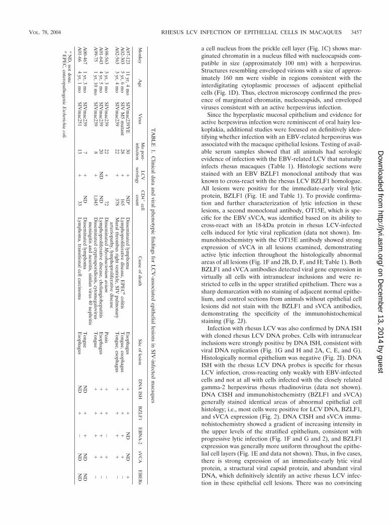
TA
BL
E1.
Clinicaldata
andviralphenotypic
findingsfor
LC
V-associated
epitheliallesionsin
SIV-infected
macaques
Monkey
Age
Virus
Mo
post-infection
LC
Vserology
CD
4�
cellcount
Cause
ofdeath
Siteof
lesionD
NA
ISHB
ZL
F1
EB
NA
-2sV
CA
EB
ER
s
A97-123
11yr,4
mo
SIVm
ac239YE
30�
ND
aD
isseminated
lymphom
aE
sophagus�
�N
DN
D�
A02-303
5yr,6
mo
SIVM
5m
utant28
�165
Lym
phoproliferativedisease,E
PEC
bcolitis
Tongue,esophagus
��
��
�A
02-5633
yr,4m
oSIV
mac239
22�
378M
uralthrombus
rightventricle,SIV
pulmonary
arteriopathy,lymphoproliferative
diseaseT
ongue,esophagus�
��
��
A98-563
3yr,3
mo
SIVm
ac23922
�72
Dissem
inatedM
ycobacteriumavium
Penis�
��
��
A01-643
4yr,5
mo
SIVm
ac23920
ND
ND
Lym
phoproliferativedisease,cholangiohepatitis
Esophagus
��
��
�A
99-751
yr,10m
oSIV
mac239
8�
1,045D
isseminated
cryptosporidiosis,cytomegalovirus
meningitis
andneuritis,sim
lanvirus
40nephritis
Tongue
��
��
�
A00-467
1yr,3
mo
SIVm
ac2397
�N
DD
isseminated
lymphom
aT
ongueN
D�
�N
DN
DA
01-664
yr,1m
oSIV
mac251
13�
33L
ymphom
a,transitionalcellcarcinoma
Esophagus
ND
��
ND
ND
aN
D,not
done.b
EPE
C,enteropathogenic
Escherichia
coli.
VOL. 78, 2004 RHESUS LCV INFECTION OF EPITHELIAL CELLS IN MACAQUES 3457
on Decem
ber 13, 2014 by guesthttp://jvi.asm
.org/D
ownloaded from
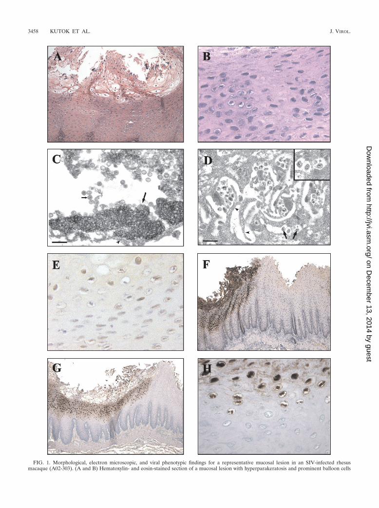
FIG. 1. Morphological, electron microscopic, and viral phenotypic findings for a representative mucosal lesion in an SIV-infected rhesusmacaque (A02-303). (A and B) Hematoxylin- and eosin-stained section of a mucosal lesion with hyperparakeratosis and prominent balloon cells
3458 KUTOK ET AL. J. VIROL.
on Decem
ber 13, 2014 by guesthttp://jvi.asm
.org/D
ownloaded from
evidence for latent LCV infection in the more undifferentiatedcells of the basal layer, i.e., cells that were positive for an epi-somal pattern of viral DNA and negative for BZLF1 expres-sion; however, at this time, technical issues limit the ability toperform DNA CISH and BZLF1 immunohistochemistry onthe same tissue sections, and rare cells of this type may bemissed by studies of serial tissue sections.
Five of seven lesions were also positive for EBNA-2 expres-sion by using a monoclonal antibody that is known to detect aconserved epitope in the carboxy-terminal acidic transactivat-ing domain of the EBV, baboon, and rhesus LCV EBNA-2homologues (Table 1). EBNA-2 is an EBV latent infectionprotein and is not typically associated with lytic replication.However, aberrant EBNA-2 expression has also been de-scribed in association with active viral replication in oral hairyleukoplakia (24). ISH for the rhesus LCV EBER homologueswas negative in five of six cases, and in one case epithelial cellswere positive for EBER expression (Table 1). These small,nonpolyadenylated RNAs are typically expressed at very highcopy numbers in latently infected cells, and EBER ISH iscommonly used to detect EBV infection in tissue sections.However, the frequent lack of EBER expression in the rhesusepithelial cell lesions is similar to the lack of EBER expressionreported in EBV-associated oral hairy leukoplakia from AIDSpatients (6). In addition, EBER ISH, EBNA-2 immunohisto-chemistry, and DNA CISH failed to detect LCV-positive lym-phocytes infiltrating the subepithelial cell layers associatedwith these lesions.
DISCUSSION
These are the first studies to unequivocally identify thatrhesus LCV can infect and is associated with disease in epi-thelial cells. These studies show that the rhesus LCV is similarto EBV, with cell tropism and a disease spectrum that includesepithelial cells, in addition to B lymphocytes. The role of epi-thelial cell infection in the normal life cycle of EBV infectionis controversial. EBV infection can clearly spread to and causedisease in epithelial cells, but whether EBV routinely infectsand persists in epithelial cells in healthy, immunocompetenthosts is unclear. The presence of rhesus LCV infection inepithelial cells demonstrates that the epithelial cell tropism isnot peculiar to EBV infection of humans but is likely shared byother EBV-related herpesviruses in the LCV genus.
The histologic appearance of these proliferative, hyperker-atotic, and parakeratotic epithelial cell lesions is very similar tothat of oral hairy leukoplakia in AIDS patients. The electronmicroscopic appearance, immunohistochemical staining for animmediate-early protein and a viral capsid protein, and strongDNA ISH clearly demonstrate the association of these epithe-lial cell lesions with rhesus LCV infection in multiple cases.
Furthermore, they characterize the rhesus LCV infection as anactive lytic process in the lesions, similar to that of EBV in-fection in oral hairy leukoplakia. Baskin et al. had previouslyobserved squamous epithelial lesions with intranuclear inclu-sions in eight SIV-infected macaques (1). Hyperkeratosis andacanthosis were present in lesions on external epithelial cellsurfaces (e.g., penis, chest skin, and hand), and lesions oninternal epithelial surfaces (e.g., tongue and esophagus) werecharacterized by intranuclear inclusions and ballooning degen-eration but without hyperkeratosis. Bacterial and fungal infec-tions with inflammation were also frequently found associatedwith these lesions. The data implicating infection with an EBV-related herpesvirus were limited to one of four tissue samplesthat stained with a monoclonal antibody against the EBVLMP1 protein and positive DNA ISH with a 3.1-kb EBV DNAprobe in one of three cases. In our hands, the EBV LMP1monoclonal antibodies do not cross-react with the rhesus LCVLMP1 homologue. In addition, the rhesus LCV genome se-quence reveals that the LMP1 gene is one of the least well-conserved genes between EBV and rhesus LCV, with no sig-nificant amino acid homology in the carboxy terminus targetedby various EBV LMP1 monoclonal antibodies (5). Overall, thevirologic data linking an EBV-related herpesvirus to theselesions were limited. We did not find any characteristic epithe-lial cell lesions that failed to test positive for rhesus LCV DNA,but we cannot rule out the possibility that other viruses orpathogens may be associated with similar lesions in immuno-suppressed hosts.
The presence of lesions on external epithelial cell surfacessuggests that infection may have occurred as a result of bitingand secondary infection from oral secretions containing virus.In six of seven of our cases, the rhesus LCV-positive lesionsoccurred on internal sites, i.e., in the esophagus and/or tongue,eliminating self-induced trauma as a potential portal of infec-tion. The frequent involvement of the esophagus in SIV-in-fected macaques raises the question of whether this might alsobe a common site for EBV-induced epithelial cell abnormali-ties in AIDS patients. At these internal sites, epithelial cellinfection most likely originated from viremia and infectionthrough the basal epithelial cell surfaces, but one cannot ruleout the possibility of infection from virus in oral secretionsbathing and infecting the apical epithelial cell surface. TheDNA ISH technique used here has the sensitivity to detectsingle-copy episomes in paraffin-embedded tissue (J. L. Kutoket al., unpublished data). Thus, it is interesting that no low-copy viral infection was noted in the basal epithelial cell layers,suggesting that these lesions are not associated with persistent,latent rhesus LCV infection in the basal epithelium.
Multiple questions regarding EBV infection of epithelialcells in immunocompetent, healthy hosts remain unresolved.Are oral epithelial cells the first cells infected with incoming
within the upper epithelial cell layers. Magnifications, �100 (A) and �400 (B). (C) Transmission electron micrograph showing the nucleus of adegenerative mucosal epithelial cell undergoing karyolysis with marginated chromatin (long arrow) and numerous intranuclear herpesvirusnucleocapsids (short arrow). The arrowhead indicates the nuclear membrane. Bar, 200 nm. (D) Transmission electron micrograph showing an areaof mucosa revealing herpesvirus virions (arrows) within the intercellular spaces between interdigitating cytoplasmic projections (arrowheads) ofadjacent epithelial cells. Bar, 500 nm. The inset shows a higher magnification of virions which measure 160 nm in diameter. (E and F)Immunohistochemical staining showing expression of BZLF1 (magnification, �400) (E) and sVCA (magnification, �50) (F). (G and H) CISHdemonstrating strong nuclear staining with a probe specific for LCV DNA. Magnifications, �50 (G) and �400 (H).
VOL. 78, 2004 RHESUS LCV INFECTION OF EPITHELIAL CELLS IN MACAQUES 3459
on Decem
ber 13, 2014 by guesthttp://jvi.asm
.org/D
ownloaded from
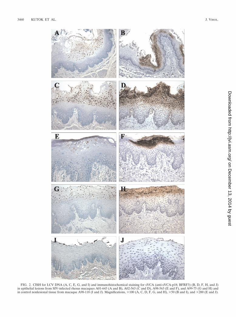
FIG. 2. CISH for LCV DNA (A, C, E, G, and I) and immunohistochemical staining for sVCA (anti-sVCA-p18; BFRF3) (B, D, F, H, and J)in epithelial lesions from SIV-infected rhesus macaques A01-643 (A and B), A02-563 (C and D), A98-563 (E and F), and A99-75 (G and H) andin control nonlesional tissue from macaque A98-118 (I and J). Magnifications, �100 (A, C, D, F, G, and H), �50 (B and I), and �200 (E and J).
3460 KUTOK ET AL. J. VIROL.
on Decem
ber 13, 2014 by guesthttp://jvi.asm
.org/D
ownloaded from
virus in primary EBV infection? Is viral replication in oralepithelial cells required for entry into the peripheral bloodcompartment during primary EBV infection? Does EBV per-sist and latently infect oral epithelial cells in convalescent,seropositive hosts? Is viral replication in oral epithelial cellsthe source of virus shed in the oral secretions of persistentlyinfected hosts? The rhesus LCV model can be helpful forinvestigating these questions by allowing for more thoroughdissection of the oral and upper digestive tracts of naturallyand experimentally infected animals to search for reservoirs ofvirus infection. In addition, the experimental rhesus LCVmodel is conducive for kinetic studies immediately after viralinfection, whereas studies with humans are limited to clinicalpresentation with infectious mononucleosis as the earliest timepoint for potential study. The development of genetic systemsfor rhesus LCV will provide an experimental system for study-ing alterations in viral genes that may affect epithelial cellinfection or for using markers to more easily identify isolatedfoci of viral infection. The development of reagents and probesto detect rhesus LCV infection will be particularly importantfor detecting reservoirs of LCV infection in the oral cavity, asthese sites are likely to be subtle in immunocompetent hosts.The complete rhesus LCV genome sequence is useful foridentifying well-conserved targets where existing EBV-specificmonoclonal antibodies, e.g., the EBV BZLF1 and sVCA mono-clonal antibodies used in the present studies, might cross-react.In addition, the development of a sensitive rhesus LCV DNAISH assay for paraffin-embedded tissue provides an importantassay for detection of low-copy rhesus LCV infection indepen-dent of viral gene expression. These are important new toolsfor demonstrating epithelial cell tropism in rhesus LCV infec-tion and will aid in the use of this model system to investigatethe role of epithelial cell infection in EBV pathogenesis.
ACKNOWLEDGMENTS
This work was supported by Public Health Service grants DE14388and CA68051. Services from the New England Primate Research Cen-ter were supported by a base grant to the institution (Public HealthService grant P51RR00168).
We thank Kristen Toohey for photographic assistance and JaniceWilliams for histologic work.
REFERENCES
1. Baskin, G. B., E. D. Roberts, D. Kuebler, L. N. Martin, B. Blauw, J. Heeney,and C. Zurcher. 1995. Squamous epithelial proliferative lesions associatedwith rhesus Epstein-Barr virus in simian immunodeficiency virus-infectedrhesus monkeys. J. Infect. Dis. 172:535–539.
2. Chien, K., R. Van de Velde, and R. Heusser. 1982. A one-step method forre-embedding paraffin embedded specimens for electron microscopy, p. 356–357. In G. W. Bailey (ed.), Fortieth Annual Proceedings of the EMSAMeeting. Claitor’s Publishing Co., Baton Rouge, La.
3. Du, Z., S. M. Lang, V. G. Sasseville, A. A. Lackner, P. O. Ilyinskii, M. D.Daniel, J. U. Jung, and R. C. Desrosiers. 1995. Identification of a nef allelethat causes lymphocyte activation and acute disease in macaque monkeys.Cell 82:665–674.
4. Faulkner, G. C., S. R. Burrows, R. Khanna, D. J. Moss, A. G. Bird, D. H.Crawford, J. W. Gratama, M. A. Oosterveer, F. E. Zwaan, J. Lepoutre, G.Klein, and I. Ernberg. 1999. X-linked agammaglobulinemia patients are notinfected with Epstein-Barr virus: implications for the biology of the virus.J. Virol. 73:1555–1564.
5. Franken, M., O. Devergne, M. Rosenzweig, B. Annis, E. Kieff, and F. Wang.1996. Comparative analysis identifies conserved tumor necrosis factor recep-tor-associated factor 3 binding sites in the human and simian Epstein-Barrvirus oncogene LMP1. J. Virol. 70:7819–7826.
6. Gilligan, K., P. Rajadurai, L. Resnick, and N. Raab-Traub. 1990. Epstein-Barr virus small nuclear RNAs are not expressed in permissively infectedcells in AIDS-associated leukoplakia. Proc. Natl. Acad. Sci. USA 87:8790–8794.
7. Gratama, J. W., M. A. Oosterveer, F. E. Zwaan, J. Lepoutre, G. Klein, andI. Ernberg. 1988. Eradication of Epstein-Barr virus by allogeneic bone mar-row transplantation: implications for sites of viral latency. Proc. Natl. Acad.Sci. USA 85:8693–8696.
8. Greenspan, J. S., D. Greenspan, E. T. Lennette, D. I. Abrams, M. A. Conant,V. Petersen, and U. K. Freese. 1985. Replication of Epstein-Barr virus withinthe epithelial cells of oral “hairy” leukoplakia, an AIDS-associated lesion.N. Engl. J. Med. 313:1564–1571.
9. Habis, A., G. B. Baskin, M. Murphey-Corb, and L. S. Levy. 1999. SimianAIDS-associated lymphoma in rhesus and cynomolgus monkeys recapitu-lates the primary pathobiological features of AIDS-associated non-Hodgkin’s lymphoma. AIDS Res. Hum. Retrovir. 15:1389–1398.
10. Karajannis, M. A., M. Hummel, I. Anagnostopoulos, and H. Stein. 1997.Strict lymphotropism of Epstein-Barr virus during acute infectious mononu-cleosis in nonimmunocompromised individuals. Blood 89:2856–2862.
11. Kestler, H., T. Kodama, D. Ringler, M. Marthas, N. Pedersen, A. Lackner,D. Regier, P. Sehgal, M. Daniel, N. King, et al. 1990. Induction of AIDS inrhesus monkeys by molecularly cloned simian immunodeficiency virus. Sci-ence 248:1109–1112.
12. Moghaddam, A., M. Rosenzweig, D. Lee-Parritz, B. Annis, R. P. Johnson,and F. Wang. 1997. An animal model for acute and persistent Epstein-Barrvirus infection. Science 276:2030–2033.
13. Niedobitek, G., A. Agathanggelou, H. Herbst, L. Whitehead, D. H. Wright,and L. S. Young. 1997. Epstein-Barr virus (EBV) infection in infectiousmononucleosis: virus latency, replication and phenotype of EBV-infectedcells. J. Pathol. 182:151–159.
14. Pathmanathan, R., U. Prasad, R. Sadler, K. Flynn, and N. Raab-Traub.1995. Clonal proliferations of cells infected with Epstein-Barr virus in pre-invasive lesions related to nasopharyngeal carcinoma. N. Engl. J. Med. 333:693–698.
15. Rangan, S. R., L. N. Martin, B. E. Bozelka, N. Wang, and B. J. Gormus.1986. Epstein-Barr virus-related herpesvirus from a rhesus monkey (Macacamulatta) with malignant lymphoma. Int. J. Cancer 38:425–432.
16. Rao, P., H. Jiang, and F. Wang. 2000. Cloning of the rhesus lymphocrypto-virus viral capsid antigen and Epstein-Barr virus-encoded small RNA homo-logues and use in diagnosis of acute and persistent infections. J. Clin. Mi-crobiol. 38:3219–3225.
17. Reitter, J. N., and R. C. Desrosiers. 1998. Identification of replication-competent strains of simian immunodeficiency virus lacking multiple attach-ment sites for N-linked carbohydrates in variable regions 1 and 2 of thesurface envelope protein. J. Virol. 72:5399–5407.
18. Rivailler, P., H. Jiang, Y. G. Cho, C. Quink, and F. Wang. 2002. Completenucleotide sequence of the rhesus lymphocryptovirus: genetic validation foran Epstein-Barr virus animal model. J. Virol. 76:421–426.
19. Sato, T. 1968. A modified method for lead staining of thin sections. J. Elec-tron Microsc. (Tokyo) 17:158–159.
20. Schmidtko, J., R. Wang, C. L. Wu, S. Mauiyyedi, N. L. Harris, P. Della Pelle,N. Brousaides, L. Zagachin, J. A. Ferry, F. Wang, T. Kawai, D. H. Sachs,B. A. Cosimi, and R. B. Colvin. 2002. Posttransplant lymphoproliferativedisorder associated with an Epstein-Barr-related virus in cynomolgus mon-keys. Transplantation 73:1431–1439.
21. Shibata, D., L. M. Weiss, K. Takada, H. zur Hausen, H. Schulte-Holthausen,G. Klein, W. Henle, G. Henle, P. Clifford, and L. Santesson. 1992. Epstein-Barr virus-associated gastric adenocarcinoma. Am. J. Pathol. 140:769–774.
22. Sixbey, J. W., J. G. Nedrud, N. Raab-Traub, R. A. Hanes, and J. S. Pagano.1984. Epstein-Barr virus replication in oropharyngeal epithelial cells. N. Engl.J. Med. 310:1225–1230.
23. Takada, K. 2000. Epstein-Barr virus and gastric carcinoma. Mol. Pathol.53:255–261.
24. Walling, D. M., A. G. Perkins, J. Webster-Cyriaque, L. Resnick, and N.Raab-Traub. 1994. The Epstein-Barr virus EBNA-2 gene in oral hairy leu-koplakia: strain variation, genetic recombination, and transcriptional expres-sion. J. Virol. 68:7918–7926.
25. zur Hausen, H., H. Schulte-Holthausen, G. Klein, W. Henle, G. Henle, P.Clifford, and L. Santesson. 1970. EBV DNA in biopsies of Burkitt tumoursand anaplastic carcinomas of the nasopharynx. Nature 228:1056–1058.
VOL. 78, 2004 RHESUS LCV INFECTION OF EPITHELIAL CELLS IN MACAQUES 3461
on Decem
ber 13, 2014 by guesthttp://jvi.asm
.org/D
ownloaded from
Copyright © 2022 FDOKUMEN