Evaluation of BBG2Na in infant macaques: specific immune responses after vaccination and RSV...
18
Chapter 4.1 BBG2Na vaccination in macaques 67 Chapter 4.1 Evaluation of BBG2Na in infant macaques: specific immune responses after vaccination and RSV challenge. L. de Waal, U.F. Power, S. Yüksel, G. van Amerongen, T.N. Nguyen, H.G.M. Niesters, R.L. de Swart, A.D.M.E. Osterhaus Vaccine (in press)
-
Upload
independent -
Category
Documents
-
view
0 -
download
0
Transcript of Evaluation of BBG2Na in infant macaques: specific immune responses after vaccination and RSV...
Chapter 4.1 BBG2Na vaccination in macaques
67
Chapter 4.1
Evaluation of BBG2Na in infant macaques: specific immune responses after vaccination and RSV challenge.
L. de Waal, U.F. Power, S. Yüksel, G. van Amerongen, T.N. Nguyen,
H.G.M. Niesters, R.L. de Swart, A.D.M.E. Osterhaus
Vaccine (in press)

Chapter 4.1 BBG2Na vaccination in macaques
68
Abstract
We have addressed the safety of alum-adsorbed BBG2Na, a recombinant hRSV subunit vaccine, in infant macaques. Animals received two vaccinations, and were challenged four months later with hRSV. In two of four BBG2Na-vaccinated animals, specific IL-13 producing T cells were detected. Upon challenge low level pulmonary eosinophilia was observed in the same two animals. Although the levels of these responses were substantially lower than those observed in the FI-hRSV controls, these data suggest that more extensive studies focusing on immunopathological safety of alum-adsorbed BBG2Na in non-human primates would be required before proceeding to clinical trials in seronegative infants.
Introduction
Human respiratory syncytial virus (hRSV) is a major cause of severe respiratory tract disease in infants and the elderly [17]. Despite its clinical relevance, no licensed hRSV vaccine is available. Several different vaccine approaches have been explored in pre-clinical studies, and a number of candidate vaccines are presently being evaluated in human clinical trials [118]. However, the potential use of non-replicating hRSV vaccines in seronegative infants is still being held back by observations in the 1960s, when experimental FI-hRSV preparations formulated in alum were found to predispose for severe disease upon subsequent natural hRSV infection [85]. During the same period, vaccine-mediated enhanced disease was also observed after vaccination with a similar formalin-inactivated whole virus preparation of another member of the family Paramyxoviridae, namely measles virus [238]. Since the mechanisms of these apparently immunopathological phenomena are still debated [64,86,128,129,172], it remains unclear which specific information would be required for a new generation non-replicating hRSV vaccine to proceed to clinical trials in seronegative infants.
A large number of hRSV vaccination and challenge studies have been carried out in mice and cotton rats. Since correlates of FI-hRSV-mediated immunopathology have been or are being established in these species, they allow comparison of the immunopathological safety of candidate new vaccines with FI-hRSV. However, it remains difficult to extrapolate results obtained in SPF inbred rodents to humans. As a final step in the pre-clinical evaluation of promising hRSV vaccine candidates before proceeding to clinical trials in seronegative infants, hRSV models in non-human primates should be considered for testing the immunopathological safety. We have previously developed a vaccination and challenge model in infant cynomolgus monkeys, in which we reproduced components of the FI-hRSV-mediated immunopathology [128].

Chapter 4.1 BBG2Na vaccination in macaques
69
BBG2Na is an hRSV subunit vaccine based on a recombinant prokaryote-expressed protein [143]. It consists of the central conserved domain of the hRSV attachment G protein (G2Na, amino acids 130-230) fused to an albumin-binding region (BB) of the streptococcal protein G, and is formulated in alum. BBG2Na was shown to induce protective immune responses against hRSV subgroups A and B in different animal models, without evidence of FI-hRSV-like enhanced immunopathology [143,157-160]. Furthermore, BBG2Na induced protective immune responses in 1-week-old mice, even in the presence of high levels of hRSV-A-specific maternal antibodies [161]. Evaluation of BBG2Na in a phase I/II study in healthy young adults showed that it was safe, well tolerated and highly immunogenic [137].
In the present study, we have evaluated the safety and efficacy of BBG2Na in hRSV seronegative infant macaques, in comparison with animals vaccinated with FI-hRSV also formulated in alum. As non-hRSV vaccinated controls we used animals vaccinated with BB or a wild-type modified vaccinia virus Ankara (MVA-wt), which was included as part of a parallel study on the efficacy and safety of recombinant MVA expressing the hRSV F and G genes. All animals were vaccinated twice, and were challenged four months later intra-tracheally with a macaque-passaged hRSV strain. Emphasis of the study was on the characterization of specific humoral and cellular immune responses after vaccination and challenge and monitoring of previously identified correlates of FI-hRSV-mediated immunopathology.
Materials & Methods Study design
Ten infant cynomolgus monkeys (Macaca fascicularis, age 31 ± 18 weeks) were vaccinated with BBG2Na (n=4), BB (n=2), MVA-wt (n=2) or FI-hRSV (n=2). All animals received two vaccinations at a four-week interval and EDTA blood samples were collected at 0, 2, 4, 6, 8, 12 and 22 weeks after the first vaccination. Heat-inactivated (HI; 30 min, 56°C) plasma samples were stored at -20°C. Peripheral blood mononuclear cells (PBMC) were isolated by density gradient centrifugation and stored at -135°C. Animals 1-4 were vaccinated intra-muscularly (i.m.) with BBG2Na (100µg dose) formulated in Adju-phos (as used by CIPF in clinical trials). Animals 5-6 were vaccinated i.m. with BB (66µg dose) formulated in Alhydrogel (kindly provided by Superfos Biosector, Frederikssund, Denmark). The different adjuvantia were chosen on basis of the differences in isoelectric points of the two proteins, see Dagouassat et al. [239]. Animals 7-8 were vaccinated with MVA-wt (provided by B. Moss, NIH, Bethesda, USA). Each animal received a dose of 2 x 10⁸ plaque forming units, which was divided over two anatomical sites: half the dose was administered i.m., the other half intra-nasally (analogous to Stittelaar et al. [229]).

Chapter 4.1 BBG2Na vaccination in macaques
70
Animals 9-10 were vaccinated i.m. with FI-hRSV (10 µg dose) formulated in Adju-phos (Superfos Biosector), prepared as described before [128]. Antibody responses
BBG2Na-, BB-, G2Na-, G172-187-, G144-159-, G164-176- and G190-204-specific indirect IgG ELISA’s were performed as described previously [137]. hRSV-specific IgG responses were measured in an indirect ELISA (Genzyme Virotech GmbH, Rüsselsheim, Germany), according to the manufacturer’s protocol.
Virus neutralizing (VN) antibody responses were measured as previously described [137]. Briefly, serial two-fold dilutions (2-3 to 2-14) of HI plasma samples were incubated with approximately 100 TCID50 of hRSV (Long strain) for one hr at 37°C, HEp-2 cells were added and cytopathic effect was monitored during the subsequent seven days. 50% VN-titers were calculated from triplicate cultures using the Reed and Muench method [240]. Cellular immune responses
PBMC were thawed, washed and counted, and cultured in RPMI1640 (Biowhittaker, Verviers, Belgium) supplemented with penicillin (100 U/ml; BioWhittaker), streptomycin (100 µg/ml; BioWhittaker), L-glutamin (2mM; BioWhittaker), 2-mercapto-ethanol (10-5M; Merck KGaA, Darmstadt, Germany), 10% HI foetal bovine serum (FBS; Greiner, Frickenhausen, Germany) and 1% HI pooled monkey serum, in 96-well round-bottom plates (Greiner) at a concentration of 1.5 x 10⁵ cells per well. Triplicate wells were cultured in the presence of medium alone, BB (10µg/ml), G2Na (5µg/ml) or BBG2Na (10µg/ml). At day 4 supernatants were harvested and cytokine responses (IL-2, IFN-γ, IL-5 and IL-13) were measured using macaque-specific ELISA systems (U-Cytech, Utrecht, The Netherlands) according to the manufacturer’s protocol. The detection limits of these assays were 122, 40, 46 and 12 pg/ml, respectively. Subsequently, medium containing 3H-thymidine (0.5µCi per well) was added. Cultures were harvested the next day and cell-associated radioactivity was measured. hRSV challenge
Eighteen weeks after the second vaccination all animals were challenged intra-tracheally with a macaque-passaged wild-type hRSV A strain (106 TCID50 [128]). EDTA-blood samples were collected at days –6, 0, 3, 6, 9, 13, 17, 23, 37, 52 and 72 after challenge. Broncho-alveolar lavage (BAL)-samples and throat swabs were collected six days before the challenge, and at days 3, 6, 9, 13 and 72 after challenge. Throat swabs were stored at -70°C. BAL samples were processed for cytospins and RT-PCR. Cytospin slides were later stained (May-Grünwald-Giemsa) and differential cell counts were obtained by light microscopy with 1,000 cells per slide counted.

Chapter 4.1 BBG2Na vaccination in macaques
71
0 2 4 6 8 10 12 14 16 18 20 22
BBG2Na-vaccinated monkeys
BB
G2N
a-sp
ecifi
c Ig
G(O
D45
0)
0.0
0.4
0.8
1.2
0 2 4 6 8 10 12 14 16 18 20 22
control monkeys
0.0
0.4
0.8
1.2
0 2 4 6 8 10 12 14 16 18 20 22
BB
-spe
cific
IgG
(OD
450)
0.0
0.4
0.8
1.2
0 2 4 6 8 10 12 14 16 18 20 220.0
0.4
0.8
1.2
0 2 4 6 8 10 12 14 16 18 20 22
G2N
a-sp
ecifi
c Ig
G(O
D45
0)
0.0
0.4
0.8
1.2
1.6
0 2 4 6 8 10 12 14 16 18 20 220.0
0.4
0.8
1.2
1.6
0 2 4 6 8 10 12 14 16 18 20 22
G17
2-18
7-sp
ecifi
c Ig
G(O
D45
0)
0.0
1.0
2.0
3.0
0 2 4 6 8 10 12 14 16 18 20 220.0
1.0
2.0
3.0
weeks after first vaccination
0 2 4 6 8 10 12 14 16 18 20 22
RS
V-s
peci
fic Ig
G(O
D45
0)
0.0
0.4
0.8
1.2
1.6
2.0
weeks after first vaccination
0 2 4 6 8 10 12 14 16 18 20 220.0
0.4
0.8
1.2
1.6
2.0
A B
DC
E F
G H
I J
Figure 1: BBG2Na- (A and B), BB- (C and D), G2Na- (E and F), G172-187- (G and H) and hRSV-specific (I and J) IgG responses in BBG2Na-vaccinated (A, C, E, G and I) or control (B, D, F, H and J) animals. Symbols representing the BBG2Na-vaccinated animals are circle, square, triangle up and triangle down for animals #1, #2, #3 and #4, respectively. For the control animals, white symbols indicate BB-vaccinated animals, grey symbols MVA-wt-vaccinated animals and black symbols FI-RSV-vaccinated animals. Symbols representing the control animals are circle, square, triangle up, triangle down, diamond and hexagon for animals #5, #6, #7, #8, #9 and #10, respectively.
Chapter 4.1 BBG2Na vaccination in macaques
72
Real-time hRSV-specific RT-PCR For RT-PCR, 5 x 10⁵ BAL cells were frozen as a dry pellet at -70°C.
RNA was isolated using the High Pure viral RNA kit (Roche Diagnostics, Almere, The Netherlands). hRSV A was specifically detected and semi-quantified using a real-time detection assay. RNA was amplified using an EZ RT-PCR amplification kit (Applied Biosystems, Nieuwerkerk a/d IJssel, The Netherlands) on an ABI7000 sequence detection system. Briefly, 10µl of RNA was amplified in a total volume of 50µl using 45 pmol of each of the primers hRSV-A forward 5'-AGATCAACTTCTGTCATCCAGCAA-3' and hRSV-A reverse 5'-TTCTGCACATCATAATTAGGAGTATCAAT-3', and 5 pmol of a FAM labelled probe 5'-CACCATCCAACGGAGCACAGGAGAT-3'. After initial reverse transcription (2 min at 50°C, 30 min at 60°C) followed by an UNG inactivation step of 5 min at 95°C, the cDNA was amplified for 40 cycles of 20 s at 94°C and 1 min at 62°C. The cycle threshold (Ct) value was calculated automatically when the FAM-specific hRSV-A signal was above the background and used to get a semi-quantitative impression of the hRSV viral load. All reactions contained both negative as well as a low positive controls.
medium BB G2Na BBG2Na
BBG2Na-vaccinated monkeys
lym
phop
rolif
erat
ion
befo
re v
acci
natio
n (c
pm)
0
2500
5000
7500
10000
12500
>15000
medium BB G2Na BBG2Na
negative control monkeys
medium BB G2Na BBG2Na
FI-RSV control monkeys
medium BB G2Na BBG2Na
lym
phop
rolif
erat
ion
four
wee
ks
afte
r firs
t vac
cina
tion
(cpm
)
0
2500
5000
7500
10000
12500
>15000
medium BB G2Na BBG2Na medium BB G2Na BBG2Na
A
FED
CB
medium BB G2Na BBG2Na
lym
phop
rolif
erat
ion
four
wee
ks
afte
r firs
t vac
cina
tion
(cpm
)
0
2500
5000
7500
10000
12500
>15000
medium BB G2Na BBG2Na medium BB G2Na BBG2Na
IHG
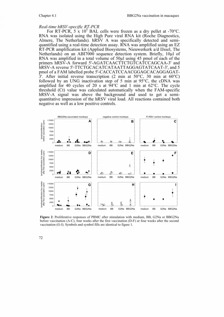
Figure 2: Proliferative responses of PBMC after stimulation with medium, BB, G2Na or BBG2Na before vaccination (A-C), four weeks after the first vaccination (D-F) or four weeks after the second vaccination (G-I). Symbols and symbol fills are identical to figure 1.

Chapter 4.1 BBG2Na vaccination in macaques
73
Results Humoral immune responses after vaccination
Induction of BBG2Na-specific IgG responses was detected in all animals vaccinated with BBG2Na, BB or FI-hRSV, but not in animals vaccinated with MVA-wt (figure 1A-B). Induction of BB-specific IgG responses was detected in all animals vaccinated with BBG2Na or BB, but not in animals vaccinated with MVA-wt or FI-hRSV (figure 1C-D). Induction of IgG responses to the hRSV-specific component of the BBG2Na vaccine, G2Na, was detected in all animals vaccinated with BBG2Na or FI-hRSV, but not in animals vaccinated with BB or MVA-wt (figure 1E-F). Of the four G2Na-peptides tested, induction of peptide G172-187-specific IgG responses was detected in all animals vaccinated with BBG2Na or FI-hRSV, but not in animals vaccinated with BB or MVA-wt (figure 1G-H). In one of the animals (animal #2) vaccinated with BBG2Na, and in both FI-hRSV-vaccinated animals, IgG responses to peptide G144-159 were also detected (results not shown). Peptide G164-176- or G190-204-specific IgG responses could not be
medium BB G2Na BBG2Na
BBG2Na-vaccinated monkeys
IL-1
3 pr
oduc
tion
befo
re
vacc
inat
ion
(pg/
ml)
det.lim.
50
100
150
>200
medium BB G2Na BBG2Na
IL-1
3 pr
oduc
tion
four
wee
ksaf
ter f
irst v
acci
natio
n(pg
/ml)
det.lim.
50
100
150
>200
medium BB G2Na BBG2Na
negative control monkeys
medium BB G2Na BBG2Na
medium BB G2Na BBG2Na
FI-RSV control monkeys
medium BB G2Na BBG2Na
A
FE
CB
D
medium BB G2Na BBG2Na
IL-1
3 pr
oduc
tion
four
wee
ks
afte
r sec
ond
vacc
inat
ion
(pg/
ml)
det.lim.
50
100
150
>200
medium BB G2Na BBG2Na medium BB G2Na BBG2Na
G H I
Figure 3: IL-13 levels in supernatants of PBMC cultures shown in figure 2. PBMC collected before vaccination (A-C), four weeks after the first vaccination (D-F) or four weeks after the second vaccination (G-I) were stimulated with medium, BB, G2Na or BBG2Na. Symbols and symbol fills are identical to the previous figures.
Chapter 4.1 BBG2Na vaccination in macaques
74
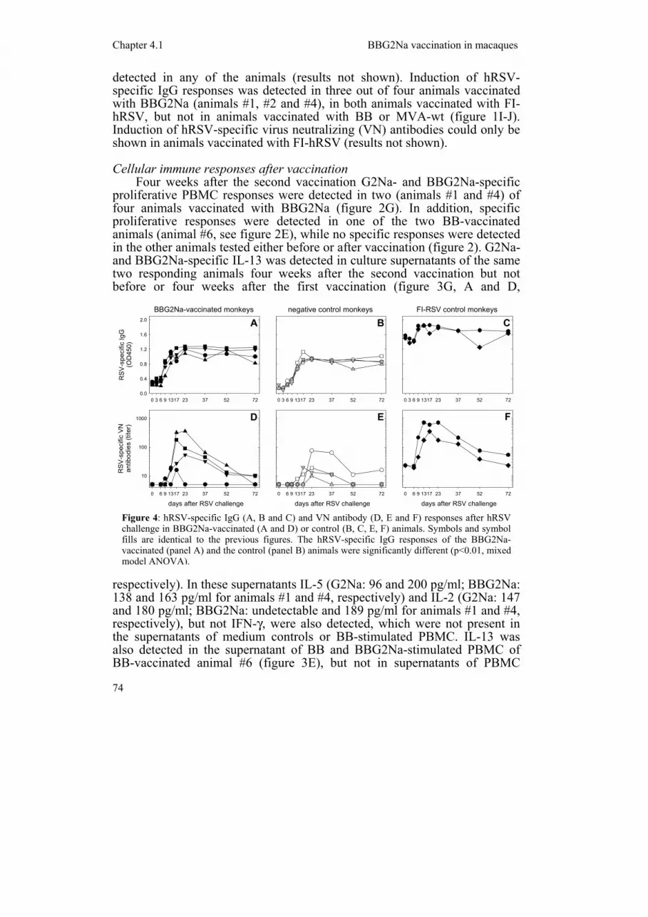
detected in any of the animals (results not shown). Induction of hRSV-specific IgG responses was detected in three out of four animals vaccinated with BBG2Na (animals #1, #2 and #4), in both animals vaccinated with FI-hRSV, but not in animals vaccinated with BB or MVA-wt (figure 1I-J). Induction of hRSV-specific virus neutralizing (VN) antibodies could only be shown in animals vaccinated with FI-hRSV (results not shown). Cellular immune responses after vaccination
Four weeks after the second vaccination G2Na- and BBG2Na-specific proliferative PBMC responses were detected in two (animals #1 and #4) of four animals vaccinated with BBG2Na (figure 2G). In addition, specific proliferative responses were detected in one of the two BB-vaccinated animals (animal #6, see figure 2E), while no specific responses were detected in the other animals tested either before or after vaccination (figure 2). G2Na- and BBG2Na-specific IL-13 was detected in culture supernatants of the same two responding animals four weeks after the second vaccination but not before or four weeks after the first vaccination (figure 3G, A and D,
respectively). In these supernatants IL-5 (G2Na: 96 and 200 pg/ml; BBG2Na: 138 and 163 pg/ml for animals #1 and #4, respectively) and IL-2 (G2Na: 147 and 180 pg/ml; BBG2Na: undetectable and 189 pg/ml for animals #1 and #4, respectively), but not IFN-γ, were also detected, which were not present in the supernatants of medium controls or BB-stimulated PBMC. IL-13 was also detected in the supernatant of BB and BBG2Na-stimulated PBMC of BB-vaccinated animal #6 (figure 3E), but not in supernatants of PBMC
0 3 6 9 1317 23 37 52 72
BBG2Na-vaccinated monkeys
RS
V-s
peci
fic Ig
G(O
D45
0)
0.0
0.4
0.8
1.2
1.6
2.0
days after RSV challenge0 6 9 1317 23 37 52 72
RS
V-s
peci
fic V
Nan
tibod
ies
(tite
r)
10
100
1000
0 3 6 9 1317 23 37 52 72
negative control monkeys
0 3 6 9 1317 23 37 52 72
FI-RSV control monkeys
days after RSV challenge0 6 9 1317 23 37 52 72
days after RSV challenge0 6 9 1317 23 37 52 72
A
D E F
CB
Figure 4: hRSV-specific IgG (A, B and C) and VN antibody (D, E and F) responses after hRSV challenge in BBG2Na-vaccinated (A and D) or control (B, C, E, F) animals. Symbols and symbol fills are identical to the previous figures. The hRSV-specific IgG responses of the BBG2Na-vaccinated (panel A) and the control (panel B) animals were significantly different (p<0.01, mixed model ANOVA).

Chapter 4.1 BBG2Na vaccination in macaques
75
collected from the other animals vaccinated with BB or MVA-wt (figure 3 and results not shown).
In culture supernatants of the FI-hRSV-vaccinated animals collected four weeks after the first vaccination IL-13 was detected in supernatant of PBMC collected from animal #9, but not animal #10. Four weeks after the second vaccination we were only able to test supernatant from animal #9, which resulted in a similar picture as on the previous sampling point. In all cultures from FI-hRSV-vaccinated animals production of IL-13 was largely irrespective of the in vitro stimulation (figure 3F and I and results not shown). However, in both FI-hRSV-vaccinated animals higher IL-13 levels could be detected in the supernatants of PBMC stimulated in vitro with beta propiolactone (BPL)-inactivated hRSV than in those stimulated with a BPL-inactivated Vero cell control antigen (486 pg/ml and 111 pg/ml versus 104 pg/ml and less than 12 pg/ml for animals #9 and #10, respectively). Upon stimulation of PBMC obtained from BBG2Na-vaccinated animals with the same antigens, IL-13 was again only detectable in the supernatants of animals #1 (30 pg/ml) and #4 (59 pg/ml). In the FI-hRSV-vaccinated animals hRSV-specific IL-5 (n=2) and/or IL-2 (n=1: animal #9), but not IFN-γ, were detected. These cytokines were undetectable in all BBG2Na-, BB- or MVA-wt-vaccinated animals. hRSV challenge
Eighteen weeks after the second vaccination all animals were challenged intra-tracheally with hRSV. At this moment only the FI-hRSV-vaccinated animals still had detectable hRSV-specific serum antibodies (figure 4A-C). In the other animals hRSV-specific IgG responses were rapidly induced, with the BBG2Na-vaccinated animals responding earlier and reaching slightly higher values than the BB- or MVA-wt-vaccinated control animals (p<0.01, mixed model ANOVA, figure 4A-B). hRSV-specific IgG responses were also boosted in the FI-hRSV-vaccinated animals, although this effect could not optimally be shown at the 1:100 plasma dilution used according to the ELISA manufacturer's instructions (figure 4C). The ELISA was later repeated using a 1:1000 plasma dilution, resulting in a curve similar to the virus neutralization response shown in figure 4F (not shown).
All animals also showed a transient VN antibody response upon challenge, again with a tendency of slightly earlier and higher responses in the BBG2Na-vaccinated animals (no significant difference, mixed model ANOVA, figure 4D-E). Interestingly, the animals with detectable vaccine-induced cellular immune responses (#1 and #4) had lower VN antibody responses than the other two BBG2Na-vaccinated animals. The two FI-hRSV-vaccinated animals also showed a rapid but transient VN antibody booster response after challenge (figure 4F).
The experimental design was not optimized to enable hRSV re-isolation from BAL cells. As an alternative approach to monitor pulmonary virus loads, an hRSV-specific real-time RT-PCR was performed on BAL cells collected before and after challenge. As shown in figure 5A-C, no significant

Chapter 4.1 BBG2Na vaccination in macaques
76
differences in pulmonary virus loads could be demonstrated. These results were confirmed in another semi-quantitative RT-PCR performed as described previously ([128], results not shown). hRSV isolations in HEp-2 cells and hRSV-specific RT-PCR on frozen throat swab samples were all negative (results not shown). Virus isolations were not performed on BAL cells due to limitations in sample quantity.
Cytospins were prepared of BAL cells collected before and after challenge, and eosinophil percentages were determined by differential counting. In accordance with our previous results, high eosinophil percentages were detected in both FI-hRSV vaccinated animals (figure 5F), with a peak at 6 days after challenge. In contrast, eosinophils could hardly be detected in the BB- or MVA-wt-vaccinated control animals (figure 5E). In two of four BBG2Na-vaccinated animals (#1 and #4), but not in the other two animals of this group, low percentages of eosinophils were detected after hRSV challenge (figure 5D). Interestingly, these were the same two animals that also showed in vitro BBG2Na- and G2Na-induced IL-13, IL-5 and IL-2 responses after vaccination.
Discussion
We have evaluated the immunopathological safety of the hRSV subunit vaccine BBG2Na formulated in alum in seronegative infant macaques, in comparison with FI-hRSV. BBG2Na did not predispose monkeys for
-6 3 6 9 13 72
FI-RSV control monkeys
-6 3 6 9 13 72
negative control monkeys
days after RSV challenge-6 3 6 9 13 72
% e
osin
ophi
ls in
BA
L
0
20
40
60
-6 3 6 9 13 72
BBG2Na-vaccinated monkeys
RS
V-s
peci
fic R
T-P
CR
(Ct v
alue
s)
20
25
30
35
40
A
D
days after RSV challenge-6 3 6 9 13 72
days after RSV challenge-6 3 6 9 13 72
B C
E F
Figure 5: hRSV-specific real-time RT-PCR signals (A-C) and percentages of eosinophils (D-F) in BAL samples collected at different time points after hRSV challenge. Symbols and symbol fills are identical to the previous figures. In panels D and E animals #1 to #4 and #5 to #8 are represented by bars with black, white, grey or hatched fills, respectively. In panel F animals #9 and #10 are represented by bars with black and white fills, respectively.

Chapter 4.1 BBG2Na vaccination in macaques
77
immunopathology similar to FI-hRSV. However, in two of four BBG2Na-vaccinated animals IL-13 producing specific T cells were detected upon vaccination, and in the same two animals a low-level eosinophilia was detected in BAL cells upon hRSV challenge.
Vaccination of infant macaques with BBG2Na induced good G2Na-specific IgG antibody responses in three out of four animals. However, in a commercial hRSV ELISA specific IgG responses were considered in the gray zone according to the kit's cut-off serum, and no hRSV-specific VN antibodies could be detected. This contrasts with data in mice and cotton rats, in which strong hRSV-specific IgG and low to moderate VN antibody responses were observed [143]. Furthermore, the data also differ from the strong although non-neutralizing hRSV-specific IgG responses detected in BBG2Na-vaccinated African green monkeys [241](U.F. Power, personal observations). In a clinical trial in healthy adults we had previously detected VN antibody responses after vaccination with BBG2Na [137], but these boosters of pre-existing immunity are difficult to compare with the primary immune responses described in the present study.
In two of four animals, vaccination with BBG2Na induced vaccine-specific proliferative T cell responses, which proved to be associated with the production of IL-13. This cytokine was previously identified as a correlate of FI-hRSV-mediated immunopathology in this model [128]. This is in accordance with results in BALB/c mice, where vaccination with BBG2Na formulated in alum was found to induce type 2 immune responses [157]. However, in BALB/c mice it was found that these primed responses were not recalled upon hRSV challenge [157,158]. In the present study, we detected eosinophils in the BAL cells of the two BBG2Na-vaccinated macaques that had developed IL-13 producing T cells, although the levels were substantially lower than those found in the two FI-hRSV controls. However, these results could indicate that in these two animals the primed type 2 responses were recalled by the challenge, although it remains questionable if this would have clinical relevance in humans.
Using semi-quantitative RT-PCR analysis, we could not detect significant differences in pulmonary viral loads between BBG2Na-vaccinated and control animals. However, it is debatable if this allows conclusions about the protective capacity of BBG2Na. Macaques are less susceptible to hRSV than humans, which forced us to choose a relatively high challenge dose [128]. In addition, the challenge was given intra-tracheally to ensure delivery of the virus into the lungs, which makes the model artificial when compared to the natural route of hRSV infection. In our previous study, we could detect a partial protection mediated by FI-hRSV immunization by using hRSV-specific semi-quantitative RT-PCR on BAL-cells, but these animals had also developed VN antibody titers upon vaccination [128]. In the present study a similar partial protection was detected in one of two FI-hRSV vaccinated animals, but in none of the BBG2Na-vaccinated animals. To optimize the model for detection of protection, larger numbers of BBG2Na-vaccinated and control animals should be challenged with different doses of hRSV given by

Chapter 4.1 BBG2Na vaccination in macaques
78
different routes of infection. In addition, where we have largely focused on characterization of specific immune responses, higher priority should be given to re-isolation of the virus from different organs. Studies in non-human primates are usually limited in numbers of animals, due to ethical, practical and financial constraints. Large-scale studies would be justified, however, with a vaccine formulation that is being considered for administration to seronegative infants, and should be combined with monitoring of immunopathology.
In conclusion, BBG2Na formulated in alum induced specific IL-13 responses following vaccination and predisposed for a low level eosinophilia upon hRSV challenge in two out of four animals. Although these data were obtained in a relatively limited number of animals and are in contrast with previous observations in rodents and African green monkeys [143,157-160,241], they indicate that more extensive studies in non-human primates focusing on immunopathological safety would be required before proceeding to clinical trials with BBG2Na in seronegative infants.
Acknowledgements
This work was supported by EC 5th FWP grant no. QLK2-1999-01044. We thank Arno Andeweg, Judith Guldemeester, Bernard Moss, Paul Mulder, Martin Schutten, Helma Vos and Linda Wyatt for their contributions to these studies.
Chapter 4.2 MVA-based hRSV vaccine in infant macaques
79
Chapter 4.2
Vaccination of infant macaques with a recombinant modified vaccinia virus Ankara expressing the respiratory syncytial
virus F and G genes does not predispose for immunopathology.
L. de Waal, L.S. Wyatt, S. Yüksel, G. van Amerongen, B. Moss, H.G.M. Niesters, A.D.M.E. Osterhaus, R.L. de Swart
Vaccine (in press)
Chapter 4.2 MVA-based hRSV vaccine in infant macaques
80
Abstract
We have evaluated the safety and immunogenicity of a recombinant modified vaccinia virus Ankara (MVA) vector expressing the human respiratory syncytial virus (hRSV) fusion (F) and attachment (G) proteins in infant macaques. Animals were vaccinated twice and four months later challenged with hRSV. Although vaccination did not predispose for immunopathology upon challenge, we were also unable to demonstrate protection. Since vaccination had resulted in priming for secondary immune responses upon challenge, we suggest that vaccination efficacy will have to be improved by using MVA in a prime-boost strategy.
Introduction
We have evaluated the safety and efficacy of two candidate human respiratory syncytial virus (hRSV) vaccines in infant macaques, using a common group of control animals. While the results obtained with BBG2Na were described before [242], we report here the data obtained with a recombinant modified vaccinia virus Ankara vector (rMVA) expressing the hRSV fusion (F) and attachment (G) proteins [156].
Since inactivated hRSV vaccines have been associated with predisposition for enhanced disease in hRSV seronegative individuals, live attenuated vaccines or recombinant viral vectors may represent a safer approach when looking for new candidate hRSV vaccines in infants. Several studies have focused on the development of a live attenuated hRSV vaccine, although it has been difficult to find a proper balance between attenuation and immunogenicity [130]. As an alternative, live vectors mediating the expression of hRSV genes have been considered [131-134,136]. MVA is a replication-deficient poxvirus, which was used in the late stage of the smallpox eradication campaign. When used as a vector, it induced similar expression levels of the recombinant genes as compared to the fully competent strains of vaccinia virus (VV) [162,163], and induced equal or better humoral and cellular immune responses in animals [164-166]. The fact that MVA is replication-deficient in mammalian cells ensures safe usage of this vector, as has been described in a safety study in immunocompromised macaques [167]. In addition, the vaccination dose can be relatively high, which would enable breaking the maternal antibody barrier in young infants.
Vaccination of mice with recombinant MVA expressing the hRSV transmembrane glycoproteins F, G or both F and G resulted in strong specific antibody responses and protection from challenge [156]. In the present study, we have vaccinated infant macaques twice with a combination of the two single recombinants (rMVA-F and rMVA-G), using animals vaccinated with wild-type MVA (MVA-wt), BB (the albumin-binding domain of streptococcal protein G) or FI-hRSV as controls as described earlier [242]. All animals were challenged intra-tracheally with a macaque-passaged hRSV
Chapter 4.2 MVA-based hRSV vaccine in infant macaques
81
strain four months after the second vaccination. The induction of humoral and cellular immune responses by vaccination and challenge was monitored, with special emphasis on the previously described ex vivo and in vitro correlates of FI-hRSV-mediated immunopathology.
Materials & Methods
The study design of this experiment has been described previously [242]. In addition to the ten animals described, four infant cynomolgus monkeys (Macaca fascicularis) were vaccinated with a combination of rMVA-F and rMVA-G (rMVA-F/G). These animals, which are referred to as animals M1 to M4, were vaccinated with a dose of 2 x 10⁸ plaque forming units, of which half was administered intra-muscularly and half intra-nasally. The vaccinations were given twice with an interval of four weeks.
hRSV-specific IgG responses were measured in an indirect ELISA (Genzyme Virotech GmbH, Rüsselsheim, Germany), according to the manufacturer’s protocol. Virus neutralizing (VN) antibody responses were measured as previously described [137]. Briefly, serial two-fold dilutions (2-3 to 2-14) of HI plasma samples were incubated with approximately 100 TCID50 of hRSV (Long Strain) for one hour at 37°C, HEp-2 cells were added and cytopathic effect was monitored during the subsequent seven days. 50% VN-
0 6 9 13 17 23 370 2 4 6 8 12 22
RS
V-s
peci
fic Ig
G(O
D45
0)
0.0
0.5
1.0
1.5
0 3 6 9 13 17 23 37
days after RSV challenge
0 6 9 13 17 23 370 2 4 6 8 12 22
RS
V-s
peci
fic Ig
G(O
D45
0)
0.0
0.5
1.0
1.5
weeks after 1st vaccination
0 2 4 6 8 12 22
RSV
-spe
cific
VN
antib
odie
s (ti
ter)
10
100
1000
weeks after 1st vaccination
0 2 4 6 8 12 22
RS
V-s
peci
fic V
Nan
tibod
ies
(tite
r)10
100
1000
A
C
days after RSV challenge
0 6 9 13 17 23 37
D
BrMVA-F/G vaccination rMVA-F/G vaccination
control vaccination control vaccination
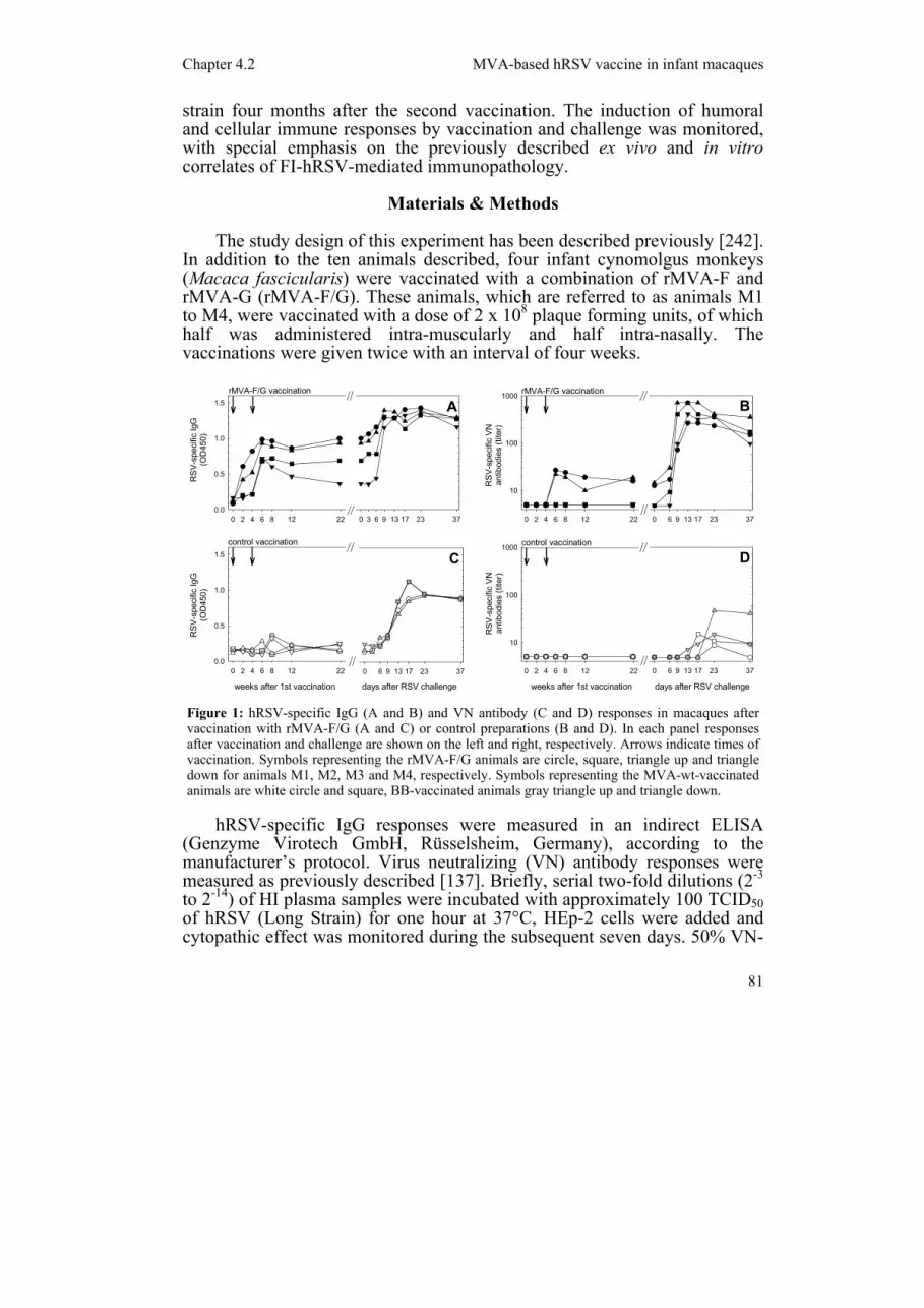
Figure 1: hRSV-specific IgG (A and B) and VN antibody (C and D) responses in macaques after vaccination with rMVA-F/G (A and C) or control preparations (B and D). In each panel responses after vaccination and challenge are shown on the left and right, respectively. Arrows indicate times of vaccination. Symbols representing the rMVA-F/G animals are circle, square, triangle up and triangle down for animals M1, M2, M3 and M4, respectively. Symbols representing the MVA-wt-vaccinated animals are white circle and square, BB-vaccinated animals gray triangle up and triangle down.
Chapter 4.2 MVA-based hRSV vaccine in infant macaques
82
titers were calculated from triplicate cultures using the Reed and Muench method [240].
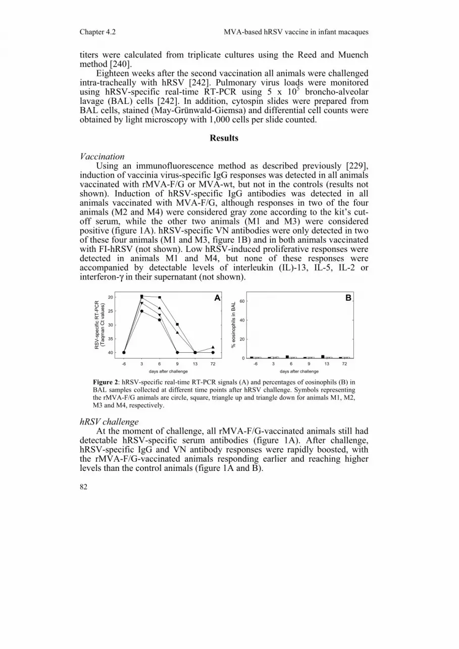
Eighteen weeks after the second vaccination all animals were challenged intra-tracheally with hRSV [242]. Pulmonary virus loads were monitored using hRSV-specific real-time RT-PCR using 5 x 10⁵ broncho-alveolar lavage (BAL) cells [242]. In addition, cytospin slides were prepared from BAL cells, stained (May-Grünwald-Giemsa) and differential cell counts were obtained by light microscopy with 1,000 cells per slide counted.
Results Vaccination
Using an immunofluorescence method as described previously [229], induction of vaccinia virus-specific IgG responses was detected in all animals vaccinated with rMVA-F/G or MVA-wt, but not in the controls (results not shown). Induction of hRSV-specific IgG antibodies was detected in all animals vaccinated with MVA-F/G, although responses in two of the four animals (M2 and M4) were considered gray zone according to the kit’s cut-off serum, while the other two animals (M1 and M3) were considered positive (figure 1A). hRSV-specific VN antibodies were only detected in two of these four animals (M1 and M3, figure 1B) and in both animals vaccinated with FI-hRSV (not shown). Low hRSV-induced proliferative responses were detected in animals M1 and M4, but none of these responses were accompanied by detectable levels of interleukin (IL)-13, IL-5, IL-2 or interferon-γ in their supernatant (not shown).
hRSV challenge At the moment of challenge, all rMVA-F/G-vaccinated animals still had
detectable hRSV-specific serum antibodies (figure 1A). After challenge, hRSV-specific IgG and VN antibody responses were rapidly boosted, with the rMVA-F/G-vaccinated animals responding earlier and reaching higher levels than the control animals (figure 1A and B).
days after challenge
-6 3 6 9 13 72
% e
osin
ophi
ls in
BA
L
0
20
40
60
days after challenge
-6 3 6 9 13 72
RS
V-s
peci
fic R
T-P
CR
(Taq
man
Ct v
alue
s)
20
25
30
35
40
A B
Figure 2: hRSV-specific real-time RT-PCR signals (A) and percentages of eosinophils (B) in BAL samples collected at different time points after hRSV challenge. Symbols representing the rMVA-F/G animals are circle, square, triangle up and triangle down for animals M1, M2, M3 and M4, respectively.

Chapter 4.2 MVA-based hRSV vaccine in infant macaques
83
As shown in figure 2A, pulmonary virus loads were not lower than those measured in the control or BBG2Na-vaccinated animals [242]. Similar to the control animals, low numbers of eosinophils were detected in BAL samples (figure 2B), while high numbers had been detected in both FI-hRSV-vaccinated animals [242].
Discussion
In the present study we have addressed the immunological safety and efficacy of recombinant MVA encoding the F- and G-genes of hRSV in seronegative infant macaques, in comparison with FI-hRSV. Vaccination with rMVA-F/G did not induce the previously described correlates of FI-hRSV-mediated immunopathology, but we were also unable to demonstrate vaccination-mediated protection.
Vaccination of macaques with rMVA resulted in comparable levels of vaccinia virus-specific antibodies as observed in a previous study in our lab, in which juvenile macaques were vaccinated with rMVA expressing the measles virus F and H genes [229]. However, whereas in the measles study high measles virus-specific VN antibody titers were induced, vaccination with rMVA-F/G induced low or undetectable hRSV-specific VN antibody levels. In contrast, Wyatt et al. [156] had shown that vaccination of mice with this construct resulted in high levels of hRSV-specific antibodies.
In addition to the absence of detectable proliferative responses or type 2 cytokine production by PBMC, priming with rMVA-F/G also did not result in pulmonary eosinophilia or neutrophilia (not shown) upon hRSV challenge. This suggests that the hRSV-specific immune responses induced by vaccination did not predispose the animals for immunopathology similar to that induced by FI-hRSV.
Despite two consecutive vaccinations with rMVA-F/G we were unable to demonstrate protection from challenge with a macaque-passaged hRSV strain. This might be related to the use of a relatively high challenge dose (106 TCID50). The absence of rMVA-F/G-mediated protection could also be related to the young age of the animals used. Whereas Stittelaar et al. [229] were able to show protection from measles challenge after vaccination with MVA-FH in juvenile monkeys (3-4 years old), Zhu et al. [243] were unable to show protection after vaccination with the same construct in infant monkeys (1-2 weeks old). All rMVA-F/G-vaccinated animals showed strong secondary antibody responses when compared to the controls, demonstrating that vaccination had successfully primed the immune system. However, the hRSV-specific VN antibody response appeared between days 6 and 9 after challenge, which is just after the peak of the hRSV replication. Therefore, although the vaccine was unable to prevent hRSV infection, it would probably prevent or limit the extent of severe hRSV bronchiolitis when applied in infants.
In future experiments, the possibility to improve the efficacy of rMVA-F/G vaccination could be evaluated by the application of different
Chapter 4.2 MVA-based hRSV vaccine in infant macaques
84
vaccination schemes, or combination with other vaccine candidates in prime-boost regimens [244,245].
Acknowledgements
This work was supported by EC 5th FWP grant no. QLK2-1999-01044. We thank Arno Andeweg, Judith Guldemeester, Ultan Power, Martin Schutten and Helma Vos for their contribution to these studies.