Cloning of the koi herpesvirus (KHV) gene encoding thymidine kinase and its use for a highly...
9
BioMed Central Page 1 of 9 (page number not for citation purposes) BMC Microbiology Open Access Methodology article Cloning of the koi herpesvirus (KHV) gene encoding thymidine kinase and its use for a highly sensitive PCR based diagnosis Herve Bercovier* 1 , Yolanta Fishman 1 , Ronen Nahary 1 , Sharon Sinai 1 , Amir Zlotkin 4 , Marina Eyngor 2 , Oren Gilad 3 , Avi Eldar 2 and Ronald P Hedrick 3 Address: 1 Institute of Microbiology, Department of Clinical Microbiology, The Hebrew University-Hadassah Medical School, Ein Karen, Jerusalem, Israel, 2 Department of Poultry and Fish Diseases, The Kimron Veterinary Institute, Beit Dagan, Israel, 3 Department of Medicine and Epidemiology, School of Veterinary Medicine, University of California, Davis, California 95616, USA and 4 Department of Infectious diseases, Sheba medical center, Tel Hashomer, 52621, Israel Email: Herve Bercovier* - [email protected]; Yolanta Fishman - [email protected]; Ronen Nahary - [email protected]; Sharon Sinai - [email protected]; Amir Zlotkin - [email protected]; Marina Eyngor - [email protected]; Oren Gilad - [email protected]; Avi Eldar - [email protected]; Ronald P Hedrick - [email protected] * Corresponding author Abstract Background: Outbreaks with mass mortality among common carp Cyprinus carpio carpio and koi Cyprinus carpio koi have occurred worldwide since 1998. The herpes-like virus isolated from diseased fish is different from Herpesvirus cyprini and channel catfish virus and was accordingly designated koi herpesvirus (KHV). Diagnosis of KHV infection based on viral isolation and current PCR assays has a limited sensitivity and therefore new tools for the diagnosis of KHV infections are necessary. Results: A robust and sensitive PCR assay based on a defined gene sequence of KHV was developed to improve the diagnosis of KHV infection. From a KHV genomic library, a hypothetical thymidine kinase gene (TK) was identified, subcloned and expressed as a recombinant protein. Preliminary characterization of the recombinant TK showed that it has a kinase activity using dTTP but not dCTP as a substrate. A PCR assay based on primers selected from the defined DNA sequence of the TK gene was developed and resulted in a 409 bp amplified fragment. The TK based PCR assay did not amplify the DNAs of other fish herpesviruses such as Herpesvirus cyprini (CHV) and the channel catfish virus (CCV). The TK based PCR assay was specific for the detection of KHV and was able to detect as little as 10 fentograms of KHV DNA corresponding to 30 virions. The TK based PCR was compared to previously described PCR assays and to viral culture in diseased fish and was shown to be the most sensitive method of diagnosis of KHV infection. Conclusion: The TK based PCR assay developed in this work was shown to be specific for the detection of KHV. The TK based PCR assay was more sensitive for the detection of KHV than previously described PCR assays; it was as sensitive as virus isolation which is the golden standard method for KHV diagnosis and was able to detect as little as 10 fentograms of KHV DNA corresponding to 30 virions. Published: 17 March 2005 BMC Microbiology 2005, 5:13 doi:10.1186/1471-2180-5-13 Received: 07 November 2004 Accepted: 17 March 2005 This article is available from: http://www.biomedcentral.com/1471-2180/5/13 © 2005 Bercovier et al; licensee BioMed Central Ltd. This is an Open Access article distributed under the terms of the Creative Commons Attribution License (http://creativecommons.org/licenses/by/2.0 ), which permits unrestricted use, distribution, and reproduction in any medium, provided the original work is properly cited.
Transcript of Cloning of the koi herpesvirus (KHV) gene encoding thymidine kinase and its use for a highly...

BioMed CentralBMC Microbiology
ss
Open AcceMethodology articleCloning of the koi herpesvirus (KHV) gene encoding thymidine kinase and its use for a highly sensitive PCR based diagnosisHerve Bercovier*1, Yolanta Fishman1, Ronen Nahary1, Sharon Sinai1, Amir Zlotkin4, Marina Eyngor2, Oren Gilad3, Avi Eldar2 and Ronald P Hedrick3Address: 1Institute of Microbiology, Department of Clinical Microbiology, The Hebrew University-Hadassah Medical School, Ein Karen, Jerusalem, Israel, 2Department of Poultry and Fish Diseases, The Kimron Veterinary Institute, Beit Dagan, Israel, 3Department of Medicine and Epidemiology, School of Veterinary Medicine, University of California, Davis, California 95616, USA and 4Department of Infectious diseases, Sheba medical center, Tel Hashomer, 52621, Israel
Email: Herve Bercovier* - [email protected]; Yolanta Fishman - [email protected]; Ronen Nahary - [email protected]; Sharon Sinai - [email protected]; Amir Zlotkin - [email protected]; Marina Eyngor - [email protected]; Oren Gilad - [email protected]; Avi Eldar - [email protected]; Ronald P Hedrick - [email protected]
* Corresponding author
AbstractBackground: Outbreaks with mass mortality among common carp Cyprinus carpio carpio and koiCyprinus carpio koi have occurred worldwide since 1998. The herpes-like virus isolated fromdiseased fish is different from Herpesvirus cyprini and channel catfish virus and was accordinglydesignated koi herpesvirus (KHV). Diagnosis of KHV infection based on viral isolation and currentPCR assays has a limited sensitivity and therefore new tools for the diagnosis of KHV infections arenecessary.
Results: A robust and sensitive PCR assay based on a defined gene sequence of KHV wasdeveloped to improve the diagnosis of KHV infection. From a KHV genomic library, a hypotheticalthymidine kinase gene (TK) was identified, subcloned and expressed as a recombinant protein.Preliminary characterization of the recombinant TK showed that it has a kinase activity using dTTPbut not dCTP as a substrate. A PCR assay based on primers selected from the defined DNAsequence of the TK gene was developed and resulted in a 409 bp amplified fragment. The TK basedPCR assay did not amplify the DNAs of other fish herpesviruses such as Herpesvirus cyprini (CHV)and the channel catfish virus (CCV). The TK based PCR assay was specific for the detection of KHVand was able to detect as little as 10 fentograms of KHV DNA corresponding to 30 virions. TheTK based PCR was compared to previously described PCR assays and to viral culture in diseasedfish and was shown to be the most sensitive method of diagnosis of KHV infection.
Conclusion: The TK based PCR assay developed in this work was shown to be specific for thedetection of KHV. The TK based PCR assay was more sensitive for the detection of KHV thanpreviously described PCR assays; it was as sensitive as virus isolation which is the golden standardmethod for KHV diagnosis and was able to detect as little as 10 fentograms of KHV DNAcorresponding to 30 virions.
Published: 17 March 2005
BMC Microbiology 2005, 5:13 doi:10.1186/1471-2180-5-13
Received: 07 November 2004Accepted: 17 March 2005
This article is available from: http://www.biomedcentral.com/1471-2180/5/13
© 2005 Bercovier et al; licensee BioMed Central Ltd. This is an Open Access article distributed under the terms of the Creative Commons Attribution License (http://creativecommons.org/licenses/by/2.0), which permits unrestricted use, distribution, and reproduction in any medium, provided the original work is properly cited.
Page 1 of 9(page number not for citation purposes)
BMC Microbiology 2005, 5:13 http://www.biomedcentral.com/1471-2180/5/13
BackgroundCommon carp Cyprinus carpio carpio is the most widelycultivated fish for human consumption mainly in Asianand in European countries including Israel [1]. The sub-species Cyprinus carpio koi or koi is cultivated as an expen-sive pet fish especially in Japan but also worldwide [2].Since 1998, mass mortality among common carp and koihas occurred in the U.S.A, Israel, Germany, England, Italy,Netherlands, and more recently in Indonesia and Japan[3-8]. These outbreaks were due to a deadly infection witha newly recognized virus, the koi herpesvirus (KHV) [5].Intensive fish culture, koi shows, domestic and interna-tional trading in the absence of health certifications orinspections have contributed to the rapid global spread ofKHV [9]. Initial characterization of the virus showed thatKHV resembles members of the family Herpesviridaealthough the virus was clearly different from two previ-ously described herpes-like viruses from fish, Herpesviruscyprini (CHV) and the channel catfish virus (CCV) [5].Further characterization, including taxonomic relation-ships of KHV to other viruses will be dependent on morecomplete genome analyses. The definitive diagnosis ofKHV is based on the isolation of the virus from infectedfish and is time consuming. Therefore a polymerase chainreaction (PCR) assays to specifically detect the virus in fishtissues was developed by Gilad et al. and Gray et al.[10,11] and currently these tests are used to complementor even replace virus isolation as a means to detect KHVinfections in koi and common carp. The PCR assays basedon KHV DNA sequences fortuitously chosen and with nodefined function [10,11] are more prone to lose theirdiagnostic value due to random mutations or deletionsthan a PCR assay based on sequences of essential viru-lence or structural genes. We identified from a genomicKHV DNA library an open reading frame (ORF) thatencodes a protein with significant homology to thymi-dine kinase (TK) and developed a new PCR assay based onthese sequences that is shown here to be significantlymore sensitive than the previously described PCR assays.
Results and discussionCharacterization of the KHV TKWhen analyzing the sequence of the clones of thegenomic KHV library, we identified a hypothetical thymi-dine kinase gene (AJ535112). At the DNA level, a lowhomology (Expect = 6e-05) was found only with the TK ofLeishmania major Friedlin (chromosome 21 cosmidL6294) (AL354533). However, when translating the DNAsequence into deduced amino acid sequences, similaritieswere found between the hypothetical KHV TK and theTrypanosoma brucei thymidine kinase (AF395663), the TKof Leishmania major (AL354533), and the TKs of Poxviridae(AF163844 and U94848) (Figure 1). We could not findany significant similarity between KHV TK and the equiv-alent genes in any described fish viruses. Southern blot
hybridization with the labelled TK gene showed defini-tively that the KHV TK gene hybridized only with the DNAof KHV and not with carp DNA (Figure 2).
The KHV TK gene had an open reading frame of 216amino acids encoding a polypeptide with a putativemolecular mass of 23.7 kDa. The similarity to eukaryoticTK may reflect a gene acquired from the host [12,13]. Theproper nomenclature of the KHV will be solved by a com-plete genome analysis and not by the analysis of a singlegene like in this study or by the histopathology performedon diseased fish [6].
The KHV TK gene was expressed in vivo in experimentallyinfected KF-1 cells as shown by RT-PCR analysis. ThemRNA of the TK gene was not detected during the first 7hours of infection (data not shown) but was detected forat least 7 days after infection (Figure 3A). By Southernhybridization, we proved that the fragment amplified byRT-PCR was indeed the TK KHV gene (Figure 3B).
The KHV TK gene cloned into plasmid pYB1 was overexpressed in E. coli ER2566. SDS-PAGE analysis of therecombinant TK showed a unique band of about 24 kDa,in agreement with the size predicted from the DNAsequence (23.7 kDa) (data not shown). Enzyme activitywas determined using tritium labeled deoxy thymidineand deoxy cytidine as substrates. [14]. Preliminary analy-ses show that the enzyme is specifically active usingmethyl-3H Thymidine (dThd) but had no activity ondeoxy (5-3H)cytidine (dCyd) similar to other herpesvirusTK. Acyclovir (Samchully Pharmaceuticals South Korea)and Gancyclovir (Hoffmann La Roche Basel Switzerland)in up to 100 molar excess over dThd, did not compete atall with the substrate.
TK based PCR assayTK has been shown to be essential for virulence in herpesviruses [15] and therefore is a good candidate for thedevelopment of a PCR assay. The KHV infection is anemerging viral disease for common carp and koi. Thehighly contagious nature of this disease will unfortunatelycontinue to negatively impact the aquaculture industry byincreasing the mortality and restrictions on productmovements. Rapid diagnosis of the virus is thereforeessential for the control of KHV outbreaks. Currently,KHV outbreaks are assessed by multifactorial analysescomprising the post mortem observation of gross pathol-ogy lesions, the dynamics of mortality in all sizes of fish,the water temperature and the continuing mortalitydespite chemotherapy. The gold standard method for thediagnosis of KHV infection is based on virus isolationusing KF-1 cells followed by PCR testing of the virus iso-late [10]. This protocol is time consuming and therefore
Page 2 of 9(page number not for citation purposes)

BMC Microbiology 2005, 5:13 http://www.biomedcentral.com/1471-2180/5/13
PCR assays alone have been suggested as possible substi-tutes for diagnosis of KHV disease [10,11,16,17].
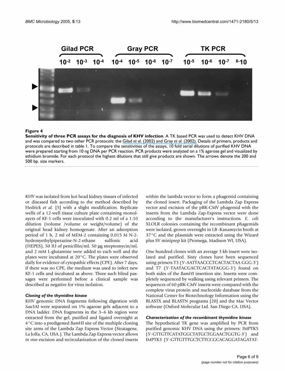
Based on the TK sequence a newly developed PCR assayusing the primers, KHV-TKf (5'-GGGTTACCTGTAC GAG-3') and KHV-TKr (5'-CACCCAGTAGATTA TGC-3') hasbeen developed. Under optimal conditions (see Methods)this PCR assay amplified a 409 bp fragment when KHVDNA was used as template (Figure 4). This assay was spe-cific for KHV and did not amplify any fragment whenusing CCV, CHV or KF-1 cell line DNAs as DNA templates(data not shown). A comparison of the sensitivity of two
published PCR assays to the TK PCR assay (Table 1) usingfor each assay the conditions described by the authorsGilad et al. [10] and Gray et al. [11] are shown in Figure 4and Table 2. Using purified KHV DNA as template, the TKPCR assay, whose product was visualized by agarose gelelectrophoresis, ethidium bromide staining and UV illu-mination, was able to detect as little as 10 fg of KHV DNA(Figure 4). Preliminary unpublished data showing thatthe genome size of KHV is around 290 kb (R. Nahary andH. Bercovier, unpublished data) is in accordance with thatestimated by Ronen et al. [18] indicate that the 10 fgdetected by the TK PCR assay correspond to
Amino acids sequence alignment of the KHV TK geneFigure 1Amino acids sequence alignment of the KHV TK gene. Multiple sequence alignment of KHV TK with representative TK sequences from Poxviridae, Herpesviridae and Trypanosomatidae families, performed by the Clastall W algorithm.
KHV MAMLELV IGPMFAGKS~ ~~~~~~TESC RRLERLSYSG RRCI~AVKHA
Trypanosoma brucei NGAHGRIELI IGPMFAGKT~ ~~~~~~TELM RRVQRHKHAQ RSCY~IIKYA 250
Cowpox NGGH~~IQLI IGPMFSGKS~ ~~~~~~TELI RRVRRYQIAQ YKCV~TIKYS
HSV-1 GMPYAVTDAV LAPHIGGEAG SSHAPPPALT LIFDRHPIAA LLCYPAARYL
KHV IDQRYTEESK VAMHSGATYP AISAGYLYE~ VMQRLEEYDA VAVDE~~GQF
Trypanosoma brucei GDTRYSEGAI TSHDQRALTA NVSVSNLHD~ VGDEWRKYDV IAVDE~~GQF 300
Cowpox NDNRYGTGLW THDKN~~NFA ALEATKLYD~ VLESITDFSV VGIDE~~GQF
HSV-1 MGSMTPQAVL AFVAL~~IPP TLPGTNIVLG ALPEDRHIDR LAKRQRPGER
KHV FPDLYEGVVQ LLTAGKYVIV AALD~~GDFM QQPFKQVT~A LVPMADKLDK
Trypanosoma brucei FPGVAAFCSK AADSGKVVIV SALD~~ADYL QEPFEEIC~L LVSRADSVVK 350
Cowpox FPDIVEFCER MANEGKIVIV AALD~~GTFQ RKPFNNIL~N LIPLSEMVVK
HSV-1 L~DLAMLAAI RRVYGLLANT VRYLQGGGSW REDWGQLSGA AVP~PQGAEP
KHV LTAVCMKCKM RDAPFTVRIS QGTDLVQVGG AESYQAVCRP CLTGFR~MAQ
Trypanosoma brucei LSAVCMECHN RKASFTYRTV KSDERKLVGG SDMYMSVCRS CYETKRNMVQ 400
Cowpox LTAVCMKCF~ KEASFSKRLG EETEIEIIGG NDMYQSVCRK CYIDS
HSV-1 QSNAGPRPHI GDTLFTLFRA P~~ELLAPNG ~DLYNVFAWA LDVLAKRLRP
KHV YELYGP~~~~ ~~~~~~~~~P PPPPAHNLLG APVVSAAP~~ ~PRSCNISIL
Trypanosoma brucei TEKYIYSCVG INEGSYSECS PGPSERSSAG TSGVQTSVKV DEQNCTEPNT 450
Cowpox
HSV-1 MHVFILDYDQ ~~~~SPAGCR DALLQLTSGM VQTHVTTPGS IPTICDLART
KHV N
Trypanosoma brucei EAKKMPLKRK RNQMAVDTT
Cowpox
HSV- FAREMG~~~E AN
Page 3 of 9(page number not for citation purposes)
BMC Microbiology 2005, 5:13 http://www.biomedcentral.com/1471-2180/5/13
approximately 30 virions. The two previously describedPCR assays were consistently 10 to 1000 times less sensi-tive (Figure 4) than the new TK PCR. Similar results wereobtained when DNA extracted from kidneys of diseasedfish were tested by the three PCR assays (data not shown).Recently, real time PCR assay [16] and LAMP assay [17]have been developed for the detection of KHV DNA. Thesensitivity of these two methods was not shown to bemuch higher than that obtained by the TK based directPCR assay developed in this work.
In addition to assessments of the specificity and sensitivityof the PCR assays, the new assay was compared to virusisolation from koi with different histories in relation toKHV infection. Koi were examined from ponds with activedisease, or that had been voluntarily exposed to a KHVchallenge and that were with or without disease signs atdifferent times after the experimental exposure. Lastly,populations of koi surviving KHV in Israel were alsoexamined. The survivor groups were the result of a proce-dure of exposing fish to experimental KHV infection atdifferent seasons of the year and different water tempera-tures [18] with the claim that they developed resistance toKHV challenge and were free of virus (Table 2). The pres-ence of KHV virus or KHV DNA in the kidney tissues offreshly collected koi was determined with the three PCR
assays and the virus isolation method. The results showedthat only the TK based PCR assay paralleled the virus iso-lation method (7 samples positive with both techniquesand 6 samples negative with both) proving its sensitivityand its usefulness (Table 2). The two other PCR assayswere similar to each other in their performances and didnot produce any false positive results but resulted in falsenegative results (5 false negative) when compared to TKbased PCR assay or virus culture. We were able to detectKHV and its DNA in fish that were intentionally infectedeven two months after the experimental infection (Table2). These data match with the results obtained by Gilad etal. [16] using a Taqman PCR assay to detect the presenceof KHV DNA in experimentally infected koi and raise thequestion of potential carriers in fish exposed to KHV inorder to obtain immune survivors.
The kidney was selected as a representative organ for virusand DNA detection because it was shown previously thatKHV is found in this organ [10,11,16]. Aliquots of threekidney samples that were positive with the three PCRassays were frozen and were shown also to be positiveafter 3 months storage at -70°C using the three PCRassays. However the TK based PCR assay was always ableto detect viral DNA in the positive samples at a 10 to 100higher dilution than the two other PCR assays (data notshown) confirming by this its sensitivity and robustness.
These first field evaluations show the advantage of the TKbased PCR for detection of KHV DNA as compared to twopreviously described PCR assays. The TK PCR assay isbased on an essential virulence gene that has little chanceto be deleted and is therefore potentially more stable/robust than the published PCR assays based on KHVsequences of no defined function [10,11]. These initial tri-als show that the three PCR assays do indeed give differentsensitivities with TK-PCR clearly performing the best. Itwas more sensitive, and unlike the other assays was ableto detect viral DNA in fish samples even two months afterexperimental exposure to KHV. KHV DNA was notdetected among koi two years post experimental exposureif survivors were not held in an infected environment. Thecarrier status of the survivor population remains unclear.Negative results by PCR may or may not indicate freedomof KHV infection or a viral load lower than the detectionthreshold. In conclusion, in all cases in our study, the TKPCR was the only direct PCR assay that always paralleledthe gold standard viral isolation method. As the PCR assayis easier and less time consuming, this newly developedTK PCR assay is an excellent candidate for a simple andrapid diagnosis of KHV infection.
ConclusionThe TK based PCR assay developed in this work wasshown to be specific for the detection of KHV. The TK
Southern blot hybridization of Carp and KHV DNA with the TK geneFigure 2Southern blot hybridization of Carp and KHV DNA with the TK gene. KHV (A) and Carp (B) DNA were digested with BsaAI (I) or PvuII (II) and hybridized with a labeled TK probe. BCG (Bacillus Calmette Guerin) DNA (C) was used as a negative control.
A B C A B C
1.0 kb
1.5 kb
3.0 kb
0.6 kb
0.4 kb
I II
Page 4 of 9(page number not for citation purposes)

BMC Microbiology 2005, 5:13 http://www.biomedcentral.com/1471-2180/5/13
based PCR assay was as sensitive as virus isolation whichis the golden standard method for KHV diagnosis, andwas able to detect as little as 10 fentograms of KHV DNAcorresponding to 30 virion particles. It was found to be 10to 1000 times more sensitive for the detection of KHVthan previously described PCR assays. Although RealTime PCR and LAMP technologies may be as sensitive ifnot more, than the direct PCR assay developed in thisstudy, they have not been yet tested in field conditionsand they require, at least for the real time PCR method,more sophisticated equipment and more expensive rea-gents than the direct PCR.
MethodsViral strains, virus isolation and DNA extractionKHV-I originating from koi in Israel [10] was propagatedon the koi fin cell line (KF-1) which supports the growth
of KHV as described by Hedrick et al. [5]. Cells wereincubated at 20°C. The channel catfish herpesvirus (CCV)strain CA80-5 served as the unrelated herpes-like viruscontrol and was propagated on channel catfish ovary(CCO) cell line. The CCO cell line was grown in mini-mum essential media (MEM) supplemented with 7.5%fetal bovine serum (FBS), 50 IU penicillin/ml, 50 µg strep-tomycin/ml, and 2 mM L-glutamine (MEM-7.5). The FBSconcentrations of the growth medium were reduced to 2% (MEM-2) when CCO cells were infected with CCV.Cells were incubated at 25°C following inoculation withCCV.
The CHV DNA was a gift from Dr. T. Sano and Dr. H.Fukuda, Tokyo University of Fisheries [19]. Viral and cellDNAs were extracted according to Gilad et al. [10].
Analysis of KHV TK mRNA in KF-1 cell line infected with KHVFigure 3Analysis of KHV TK mRNA in KF-1 cell line infected with KHV. KF-1 cells were infected with KHV. At 0 to 7 days post infection total mRNA was prepared from the infected cells and was used in an RT-PCR reaction using primers TK-Rtfor-ward- TK and RTr long (A). 0 time control mRNA was prepared immediately after infection. lanes: M- size markers, 1–8- mRNA of days 0–7 post infection, 9- direct PCR of KHV DNA, 10- uninfected KF-1 cells 11- RNase treated mRNA of day 5 post infection. To confirm the identity of the RT-PCR products, Southern blot hybridization (B) was performed using the cloned TK gene as probe and mRNA of days 0–7 lanes 1–8 consecutively, control KHV – lane 9, and uninfected KF-1 cells as negative control – lane10. The arrow marks the position of the 621 bp product.
M 0 1 2 3 4 5 6 7 8 9 10 11 M 0 1 2 3 4 5 6 7 8 9
A B
Page 5 of 9(page number not for citation purposes)
BMC Microbiology 2005, 5:13 http://www.biomedcentral.com/1471-2180/5/13
KHV was isolated from koi head kidney tissues of infectedor diseased fish according to the method described byHedrick et al. [5] with a slight modification. Replicatewells of a 12-well tissue culture plate containing monol-ayers of KF-1 cells were inoculated with 0.2 ml of a 1:10dilution (volume /volume or weight/volume) of theoriginal head kidney homogenate. After an adsorptionperiod of 1 h, 2 ml of MEM-2 containing 0.015 M N-2-hydroxyethylpiperazine-N-2-ethane sulfonic acid(HEPES), 50 IU of penicillin/ml, 50 µg streptomycin/ml,and 2 mM L-glutamine were added to each well and theplates were incubated at 20°C. The plates were observeddaily for evidence of cytopathic effects (CPE). After 7 days,if there was no CPE, the medium was used to infect newKF-1 cells and incubated as above. Three such blind pas-sages were performed before a clinical sample wasdescribed as negative for virus isolation.
Cloning of the thymidine kinaseKHV genomic DNA fragments following digestion withSau3AI were separated on 1% agarose gels adjacent to aDNA ladder. DNA fragments in the 3–6 kb region wereextracted from the gel, purified and ligated overnight at4°C into a predigested BamHI site of the multiple cloningsite arms of the Lambda Zap Express Vector (Stratagene,La Jolla, CA, USA.). The Lambda Zap Express vector allowsin vivo excision and recircularization of the cloned inserts
within the lambda vector to form a phagemid containingthe cloned insert. Packaging of the Lambda Zap Expressvector and excision of the pBK-CMV phagemid with theinserts from the Lambda Zap-Express vector were doneaccording to the manufacturer's instructions. E. coliXLOLR colonies containing the recombinant phagemidswere isolated, grown overnight in LB -Kanamycin broth at37°C and the plasmids were extracted using the Wizardplus SV miniprep kit (Promega, Madison WI, USA).
One hundred clones with an average 3 kb insert were iso-lated and purified. Sixty clones have been sequencedusing primers T3 (5'-AATTAACCCTCACTACTAA GGG-3')and T7 (5'-TAATACGACTCACTATAGGG-3') found onboth sides of the BamHI insertion site. Inserts were com-pletely sequenced by walking using relevant primers. Thesequences of 60 pBK-CMV inserts were compared with thecomplete virus protein and nucleotide database from theNational Center for Biotechnology Information using theBLASTX and BLASTN programs [20] and the Mac Vectorsoftware (Oxford Molecular Ltd. San Diego CA. USA).
Characterization of the recombinant thymidine kinaseThe hypothetical TK gene was amplified by PCR frompurified genomic KHV DNA using the primers: IMPTK5(5'-GTTGTTCATATGGCTATGCTGGAACTGGTG-3') andIMPTK3 (5'-GTTGTTTGCTCTTCCGCACAGGATAGATAT-
Sensitivity of three PCR assays for the diagnosis of KHV infectionFigure 4Sensitivity of three PCR assays for the diagnosis of KHV infection. A TK based PCR was used to detect KHV DNA and was compared to two other PCR protocols: the Gilad et al. (2002) and Gray et al. (2002). Details of primers, products and protocols are described in table 1. To compare the sensitivities of the assays, 10 fold serial dilutions of purified KHV DNA were prepared starting from 10 ng DNA per PCR reaction. PCR products were analyzed on a 1% agarose gel and visualized by ethidium bromide. For each protocol the highest dilutions that still give products are shown. The arrows denote the 200 and 500 bp. size markers.
10-2 10-3 10-4 10-4 10-5 10-6 10-7 10-5 10-6 10-7 10-8
Gilad PCR Gray PCR TK PCR
Page 6 of 9(page number not for citation purposes)

BMC Microbiology 2005, 5:13 http://www.biomedcentral.com/1471-2180/5/13
GTTACAAGAA CG-3'). To prove that the sequences of theamplified TK originated from KHV DNA and not from acontaminating agent or fish cellular sequences, Southernblot analyses were performed. The TK gene was labeledwith digoxigenin (DIG) (Roche Applied Science, Man-nheim Germany) according to the manufacturerinstructions. Purified TK DNA and Carp DNA (1–2 µg)
were incubated with 10 U of BsaA1 or with PvuII that cutonly once within the TK sequence for 4 h at 37°C. DNAfragments were separated by electrophoresis on 1.0% aga-rose gels and observed after staining with 1% ethidiumbromide. DNA was transferred to positively charged nylonmembranes (Roche Applied Science, Mannheim Ger-many) and treated as recommended by the manufacturer
Table 1: PCR coanditions and primers for the detection of KHV
Gilad Gray TK
Primer sequences 5'-GACGACGCCGGA GACCTTGTG-3'5'-CACAAGTTCAGTCT GTTCCTCAAC-3'
5'-GACACCACATCTGCAA GGAG-3'5'-GACACATGTTACAAT GGTGGC-3'
5'-GGGTTACCTGTACGAG-3'5'-CACCCAGTAGAT TATGC-3'
Fragment size 484 bp 290 bp 409 bpEnzyme 1 unit Taq DNA polymerase platinum
(Invitrogen)1 unit Taq DNA polymerase (Promega)
1 unit Taq DNA polymerase platinum (Invitrogen)
Reaction buffer *Tris-HCl 20 nM pH = 8.4KCl 50 mMMgCl2 2 mM(NH4)2SO4 15 mM*Taq platinum buffer
Tris-HCl 60 nM pH = 8.3MgCl2 2 mM(NH4)2SO415 mM
*Tris-HCl 20 nM pH = 8.4KCl 50 mMMgCl2 1.5 mM*Taq platinum buffer
Primers / dNTP's 30 pmole /reaction 400 µM 0.25 µg/reaction 10 µM 0.4 µg/reaction 200 µMDNA concentration 157 ng/reaction) 157 ng/reaction 157 ng/reaction
PCR reaction volume 50 µl 50 µl 50 µlProgram 95°c for 5 min
94°c for 1 min68°c for 1 min72°c for 30 sec39 cycles72 C for 7 min
94°c for 1 min55°c for 2 min72°c for 3 min30 cycles72°c for 7 min
95°c 5 min95°c 30 sec52°c 30 sec72°c 1 min35 cycles72°c for 10 min
PCR apparatus Biometra -T Gradient Biometra -T Gradient Biometra -T Gradient
Table 2: Comparison of three PCR assays and virus isolation from koi carp exposed to KHV
Sample TK PCR Gilad PCR Gray PCR Virus Isolation History of the koi carp
1 + - - + sick fish with clinical symptoms2 - - - - survivor (unknown time lap after exposure).3 - - - - survivor (unknown time lap after exposure) from a tank with mortality.4 + - - + survivor (unknown time lap after exposure) from a tank with mortality.5 - - - - survivor moved to 30°C, then to 24°C. no mortality in the tank.6 + - - + survivor moved to 30°C, then moved to 24°C. 25% mortality in the tank.7 - - - - naïve fish that lived at 16°C. No mortality in the tank.8 + + + + fish from tank with mortality.9 + - - + fish from tank with mortality.10 - - - Pooled survivor, 2 years after experimental infection11 - - - - survivor, 2 years after experimental infection12 - - - Pooled survivor, 1 year after experimental infection13 - - - - survivor, 1 year after experimental infection14 - - - Pooled survivor, 2 months after experimental infection15 - - - survivor, 2 months after experimental infection16 + - - + survivor, 2 months after experimental infection17 + - - survivor, 2 months after experimental infection18 + - - + survivor from tank with disease- two weeks post infection.KF+ + + + + KF-1 cell line infected with KHV- Positive control.KF- - - - - KF-1 cell line- Negative control.
Page 7 of 9(page number not for citation purposes)

BMC Microbiology 2005, 5:13 http://www.biomedcentral.com/1471-2180/5/13
(Roche Applied Science, Mannheim Germany). Prehy-bridization was performed in 10 ml DIG easy hyb solu-tion (Roche Applied Science, Mannheim Germany)within a roller bottle at 65°C for 30 min. Hybridizationwas carried out in 6 ml of DIG easy hyb containing 250 ngof DIG labeled probe at 65°C overnight. Membranes werewashed twice with low stringency wash buffer (2 ×SSC+0.1% SDS) at room temperature followed by twowashes with high stringency wash buffer (0.1 × SSC +0.1%SDS) at 60°C. The membrane was blocked for 30 minuteswith 40 ml blocking solution and then incubated with a1:10000 dilution of anti Dig AP conjugated antibody(Roche Applied Science, Mannheim Germany). Afterwashing, the membrane was placed on a plastic film,evenly covered with 500 µl of ready-to-use chemilumines-cent alkaline phosphatase substrate (CSPD) (RocheApplied Science, Mannheim Germany), and incubated for5 min. at room temperature. The membranes were thenexposed to super RX X-ray film (Fuji photo film, Tokyo,Japan).
The amplified TK gene was cloned into vector pYB1(Impact system, New England Biolabs, Beverly MA. USA)with an intein tag that allows the purification of therecombinant protein in a non-denaturated form withoutany extra residues. The TK protein was expressed in E. coliER2566 and purified according to the manufacturer'sinstructions (New England Biolabs). After affinity purifi-cation on a chitin column, SDS-PAGE analysis was per-formed to characterize the recombinant TK and theenzymatic activity of the recombinant TK was tested.
Thymidine kinase activity was determined by the DE-81filter paper assay using tritium labeled deoxy thymidineand deoxy cytidine as substrates. [14]. A typical reactionmixture contained in 100 µl of Tris-Cl (140.0 mM) MgCl2(2.0 mM), DTT (1.7 mM), NaF (8.0 mM), PMSF (5.0mM), ATP (10.0 mM), KHV TK enzyme, and substratesmethyl-3H thymidine (dThd) (25 ci/mM) or deoxy(5-3H)cytidine (dCyd) (22 ci/mM) (Amersham Biosciences,UK). Substrates were used at concentrations between 5–30 mM. Reactions were carried out at 37°C. At 15 secondsintervals, 20 µl samples were removed, and spotted ontothe filters. The filters were washed 4 times in 5.0 mMammonium formate followed by a 15 min wash in Etha-nol 95 %. Amount of radioactivity was determined in ascintillation counter (LS 6000TA, Beckman InstrumentsFullerton CA. USA).
To demonstrate that the KHV TK is expressed in an exper-imentally infected cell line, total RNA was extracted usingthe SV total RNA isolation system (Promega, Madison WI.USA) from KF-1 cells infected with KHV. Uninfected KF-1cells served as a control. RT-PCR (Access RT-PCR system,Promega, Madison WI. USA) was performed using inter-
nal primers of the TK gene (Upstream primer TK-RTf: (5'-ATGGCTATGCTGGAAC-3'); downstream primer TK-RTrl:(5'-TGGAGCGGCT GACACG-3') on experimentallyinfected KF-1 cells at different time points post infection.PCR products were analyzed on 1% agarose gel. The iden-tity of the amplified products was confirmed by Southernblot hybridization with the labeled KHV TK gene asdescribed above.
PCR assaysHead kidneys were removed from koi carp infected natu-rally or intentionally by KHV in order to obtain survivorsin Israeli farms [18]. Template DNA from head kidney ofkoi carp was prepared using High Pure PCR TemplatePreparation Kit (Roche Diagnostics, Mannheim Ger-many) according to the manufacturer's recommenda-tions. A TK based PCR assay to detect KHV DNA wasdeveloped using specific primers, KHV-TKf (5'-GGGT-TACCTGTACGAG-3') and KHV-TKr (5'-CACCCAGTAGATTATGC-3'), derived from for the thymidine kinase gene(AJ535112) that produced a 409 bp amplicon. The TKbased PCR was done using the above primers for 35 cyclesof amplification under standard conditions (95°C 5 min,then 35 cycles: 95°C 30 sec, 52°C 30 sec, 72°C 1 min andfinally 72°C 10 min).
Two other PCR assays were performed in parallel accord-ing to their original description and are denominatedGilad PCR (400 bp product obtained with Primers (5'-GACGACGCCGGAGACCTTGTG-3') and (5'-CACAAGT-TCAGTCTGTTC CTCAAC-3') [10] and Gray PCR (290 bpproduct obtained with primers (5'-GACACCACATCT-CAAGGG-3') and (5'-GAC ACATGTTACAATGGTGGC-3')[11] (Table 1).
The specificity of the PCR assays (discrimination of KHVfrom other viruses) was assessed as described by Gilad etal. [10]. A total of 50 ng of DNA obtained from purifiedKHV-I, CHV, CCV, or uninfected KF-1 cells were used astemplates for the PCR assays. The sensitivity of the PCRassays was determined using 10 fold serial dilutions from10 ng to 0.001 pg of KHV-I DNA and 10 fold serial dilu-tions of head kidneys (boiled in distilled water for 5 min)from diseased koi carp with proven virus isolation or theextracted DNA. Whole tissue or DNA extracted from neg-ative head kidney samples from uninfected koi carp wererun as controls. The resulting amplified products wereanalyzed by electrophoresis on 1% agarose gels after stain-ing with ethidium bromide.
Authors' contributionsHB, AE and RPH conceived the study, and participated inits design and coordination and drafted the manuscript.RN, OG, SS and AZ carried out the molecular studies(DNA preparation, PCR development and calibration)
Page 8 of 9(page number not for citation purposes)
BMC Microbiology 2005, 5:13 http://www.biomedcentral.com/1471-2180/5/13
Publish with BioMed Central and every scientist can read your work free of charge
"BioMed Central will be the most significant development for disseminating the results of biomedical research in our lifetime."
Sir Paul Nurse, Cancer Research UK
Your research papers will be:
available free of charge to the entire biomedical community
peer reviewed and published immediately upon acceptance
cited in PubMed and archived on PubMed Central
yours — you keep the copyright
Submit your manuscript here:http://www.biomedcentral.com/info/publishing_adv.asp
BioMedcentral
and participated in the sequence alignment. YF clonedand expressed the recombinant TK and carried out theenzymatic assays. ME prepared virus cultures and per-formed PCR assays in diseased fish. All authors read andapproved the final manuscript.
AcknowledgementsThe study was supported by the US-Israel BARD contract No US-3539-04C.
References1. FAO: [http://www.fao.org/fi/statist/summtab/default.asp].2. Balon EK: Origin and domestication of the wild carp, Cyprinus
carpio : from Roman gourmets to the swimming flowers.Aquaculture 1995, 129:3-48.
3. Body A, Lieffrig F, Charlier C, Collard A: Isolation of virus-like par-ticles from koi (Cyprinus carpio) suffering gill necrosis. Bull EurAssoc Fish Pathol 2000, 20:87-88.
4. Bretzinger A, Fischer-Scherl T, Oumouna M, Hoffman , Truyen U:Mass mortalities in koi, Cyprinus carpio, associated with gilland skin disease. Bul Eur Assoc Fish Pathol 1999, 19:182-185.
5. Hedrick RP, Gilad O, Yun S, Spangenberg JV, Marty GD, NordhausenRW, Kebus MJ, Bercovier H, Eldar A: A Herpesvirus associatewith mass mortality of juvenile and adult koi, a strain of acommon carp. J Aquat An Health 2000, 12:44-57.
6. Pikarsky E, Ronen A, Abramowitz J, Levavi-Sivan B, Hutoran M, Sha-pira Y: Pathogenesis of acute viral disease induced in fish bycarp interstitial nephritis and gill necrosis virus. J Virol 2004,78:9544-9551.
7. Neukirch M, Bottcher K, Bunnarjirakul S: Isolation of a virus fromkoi with altered gills. Bull Eur Assoc Fish Pathol 1999, 19:221-224.
8. Koi Herpesvirus infection in Indonesia: Suspicion [http://www.promedmail.org]. archive number 20020630.4639 PublishedDate 9 Nov 2004.
9. Gilad O, Yun S, Andree KB, Adkison MA, Way K, Willits NH, Bercov-ier H, Hedrick RP: Molecular comparison of isolates of anemerging fish pathogen, the koi herpesvirus, and the effectof water temperature on mortality of experimentallyinfected koi. J Gen Virol 2003, 84:1-8.
10. Gilad O, Yun S, Andree KB, Adkison MA, Zlotkin A, Bercovier H,Eldar A, Hedrick RP: Initial characteristics of koi herpesvirusand development of a polymerase chain reaction assay todetect the virus in koi, Cyprinus carpio koi. Dis Aquat Org 2002,48:101-108.
11. Gray WL, Mullis L, LaPatra SE, Groff JM, Goodwin A: Detection ofkoi herpesvirus DNA in tissues of infected fish. J Fish Dis 2002,25:171-178.
12. Blasco R, Lopez-Otin C, Munoz M, Bockam EO, Simon-Mateo C, Vin-uela E: Sequence and evolutionary relationships of Africanswine fever virus thymidine kinase. Virology 1990, 178:301-304.
13. Harrison PT, Thompson R, Davidson A: Evolution of herpes virusthymidine kinase from cellular deoxycytidine kinase. J GenVirol 1991, 72:2583-2586.
14. Gustafson EA, Schinazi RF, Fingeroth JD: Human Herpes virus 8open reading frame 21 is a thymidine kinase and thymidylatekinase of narrow substrate specificity that efficiently phos-phorylates Zivudine but not Ganciclovir. J Virol 2000, 74:84-692.
15. Boivin G, Coulombe Z, Rivest S: Intranasal herpes simplex virustype 2 inoculation causes a profound thymidine kinasedependent cerebral inflammatory response in the mousehindbrain. Eur J Neurosci 2002, 16:29-43.
16. Gilad O, Yun S, Zagmut F, Leutenegger CM, Bercovier H, Hedrick RP:Concentrations of a herpes-like virus (KHV) in tissues ofexperimentally-infected Cyprinus carpio koi as assessed byreal-time TaqMan PCR. Dis Aquat Org 2004, 60:179-187.
17. Gunimaladevi I, Kono T, Venugopal MN, Sakai M: Detection of koiherpesvirus in common carp, Cyprinus carpio L., by loop-mediated isothermal amplification. J Fish Dis 2004, 27:583-589.
18. Ronen A, Perelberg A, Abramowitz J, Hutoran M, Tinman S, BejeranoI, Steinitz M, Kotler M: Efficient vaccine against the virus caus-ing a lethal disease in cultured Cyprinus carpio. Vaccine 2003,21:4677-4684.
19. Sano T, Fukuda H, Furukawa M, Hosoya H, Moriya Y: A herpesvirusisolated from carp papilloma in Japan. Fish Shell Pathol 1985,32:307-311.
20. Altschul SF, Madden TL, Schaffer AA, Zhang J, Zhang Z, Miller W, Lip-man DJ: Gapped BLAST and PSI-BLAST: a new generation ofprotein database search programs. Nucleic Acids Res 1997,25:3389-3402.
Page 9 of 9(page number not for citation purposes)
http://www.ncbi.nlm.nih.gov/entrez/query.fcgi?cmd=Retrieve&db=PubMed&dopt=Abstract&list_uids=2389555
http://www.ncbi.nlm.nih.gov/entrez/query.fcgi?cmd=Retrieve&db=PubMed&dopt=Abstract&list_uids=2389555
http://www.ncbi.nlm.nih.gov/entrez/query.fcgi?cmd=Retrieve&db=PubMed&dopt=Abstract&list_uids=1919533
http://www.ncbi.nlm.nih.gov/entrez/query.fcgi?cmd=Retrieve&db=PubMed&dopt=Abstract&list_uids=1919533
http://www.ncbi.nlm.nih.gov/entrez/query.fcgi?cmd=Retrieve&db=PubMed&dopt=Abstract&list_uids=9254694