Prodrugs Design Based on Inter- and Intramolecular Processes
76
In: Prodrugs Design – A New Era ISBN: 978-1-63117-701-9 Editor: Rafik Karaman © 2014 Nova Science Publishers, Inc. Chapter I Prodrugs Design Based on Inter- and Intramolecular Processes Rafik Karaman 1,2 * 1 Pharmaceutical Sciences Department, Faculty of Pharmacy Al-Quds University, Jerusalem, Palestine 2 Department of Science, University of Basilicata, Potenza, Italy Abstract In this chapter we have presented the past and current status of the prodrug approach and its applications and highlighted its many successes in solving problems associated with drug delivery. The two main prodrugs approaches that are presented in this chapter are the traditional approach by which a prodrug interconversion occurs via enzyme catalysis and the second approach is based on enzyme models that have been advocated to understand enzyme catalysis. In the latter approach, a design of prodrugs is accomplished using computational calculations based on molecular orbital and molecular mechanics methods. Correlations between experimental and calculated rate values for some intramolecular processes opened the door widely to predict thermodynamic and kinetic parameters for other processes that can be utilized as prodrugs linkers. This approach does not require any enzyme to catalyze the prodrug interconversion. The interconversion rate is solely dependent on the factors govern the limiting step of the intramolecular process. It is believed that the use of this approach might eliminate many disadvantages related to prodrug interconversion by the metabolic approach. For example, the activity of many prodrug activating enzymes may be varied due to genetic polymorphisms, age-related physiological changes, or drug interactions, leading to undesired pharmacokinetic, pharmacodynamics, and clinical effects. Furthermore, there are wide interspecies variations in both the expression and function of the major enzymes activating prodrugs, and these can pose some obstacles in the preclinical optimization phase. * Corresponding author: Rafik Karaman, e-mail: [email protected]; Tel and Fax +972-2-2790413. No part of this digital document may be reproduced, stored in a retrieval system or transmitted commercially in any form or by any means. The publisher has taken reasonable care in the preparation of this digital document, but makes no expressed or implied warranty of any kind and assumes no responsibility for any errors or omissions. No liability is assumed for incidental or consequential damages in connection with or arising out of information contained herein. This digital document is sold with the clear understanding that the publisher is not engaged in rendering legal, medical or any other professional services.
-
Upload
khangminh22 -
Category
Documents
-
view
0 -
download
0
Transcript of Prodrugs Design Based on Inter- and Intramolecular Processes
In: Prodrugs Design – A New Era ISBN: 978-1-63117-701-9
Editor: Rafik Karaman © 2014 Nova Science Publishers, Inc.
Chapter I
Prodrugs Design Based on Inter-
and Intramolecular Processes
Rafik Karaman1,2*
1Pharmaceutical Sciences Department, Faculty of Pharmacy
Al-Quds University, Jerusalem, Palestine 2Department of Science, University of Basilicata, Potenza, Italy
Abstract
In this chapter we have presented the past and current status of the prodrug approach
and its applications and highlighted its many successes in solving problems associated
with drug delivery. The two main prodrugs approaches that are presented in this chapter
are the traditional approach by which a prodrug interconversion occurs via enzyme
catalysis and the second approach is based on enzyme models that have been advocated
to understand enzyme catalysis. In the latter approach, a design of prodrugs is
accomplished using computational calculations based on molecular orbital and molecular
mechanics methods. Correlations between experimental and calculated rate values for
some intramolecular processes opened the door widely to predict thermodynamic and
kinetic parameters for other processes that can be utilized as prodrugs linkers. This
approach does not require any enzyme to catalyze the prodrug interconversion. The
interconversion rate is solely dependent on the factors govern the limiting step of the
intramolecular process. It is believed that the use of this approach might eliminate many
disadvantages related to prodrug interconversion by the metabolic approach. For
example, the activity of many prodrug activating enzymes may be varied due to genetic
polymorphisms, age-related physiological changes, or drug interactions, leading to
undesired pharmacokinetic, pharmacodynamics, and clinical effects. Furthermore, there
are wide interspecies variations in both the expression and function of the major enzymes
activating prodrugs, and these can pose some obstacles in the preclinical optimization
phase.
* Corresponding author: Rafik Karaman, e-mail: [email protected]; Tel and Fax +972-2-2790413.
No part of this digital document may be reproduced, stored in a retrieval system or transmitted commercially in any form or by any means. The publisher has taken reasonable care in the preparation of this digital document, but makes no expressed or implied warranty of any kind and assumes no responsibility for any errors or omissions. No liability is assumed for incidental or consequential damages in connection with or arising out of information contained herein. This digital document is sold with the clear understanding that the publisher is not engaged in rendering legal, medical or any other professional services.

Rafik Karaman 2
Keywords: Predrugs, prodrugs, molecular orbital calculations, molecular mechanics
calculations, intramolecular reactions, prodrugs design, enzyme models, aza-nucleosides,
Kirby‘s enzyme model, statins, Bruice‘s enzyme model, Menger‘s enzyme model,
paracetamol, dopamine, tranexamic acid, DFT calculations
Abbreviations
ADME Absorption, Distribution, Metabolism and Excretion.
QSAR Quantitative Structure Activity Relationship
HLB Hydrophilic Lipophilic Balance
ADEPT Antibody-directed enzyme prodrug therapy
VDEPT Virus-direct enzyme prodrug therapy
GDEPT Gene-directed enzyme prodrug therapy
CYP Cytochrome P450
GI Gastro Intestinal.
NSAIDs Non-steroidal Anti-inflammatory Drugs
PEG Polyethylene glycol
QM Quantum Mechanics
MM Molecular Mechanics.
DFT Diffused Functional Theory .
HF Hartree-Fock
IGAC Intramolecular General Acid Catalysis .
GAC General Acid Catalysis
EM Effective Molarity
Introduction
A drug is a chemical entity which is used in the diagnosis, cure, relief, treatment or
prevention of disease, or intended to affect the structure or function of the body. Evidence of
the use of medicines and drugs can be found three thousand years back. For long time, drug
discovery has been a trial-and-error process. The conventional drug development has relied
on blind screening approach, which was very time-consuming and labor costly. The
disadvantages of conventional drug development and discovery as well as the desire for
finding a more deterministic approach to combat disease have led to the concept of "Rational
drug design" starting in the sixties of the past century. The process of drug discovery is very
complex and requires an interdisciplinary effort to design effective and commercially
effective drugs. The objective of drug design is to find a chemical entity that can fit to an
active site of a receptor or enzyme. After passing the in vivo animal tests and human clinical
trials, this entity becomes a drug available in the drug market. The conventional drug design
methods were based on random screening of natural compounds or synthesized natural
analogs in the laboratory. The main disadvantages of this method are time consuming cycle
and high cost. Modern approach utilizing structure-based drug design with the aid of
informatics technologies and a variety of computational methods has accelerated the drug
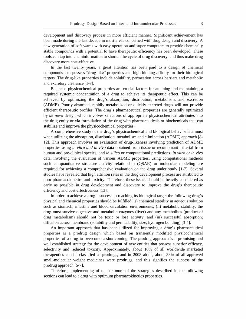
Prodrugs Design Based on Inter- and Intramolecular Processes 3
development and discovery process in more efficient manner. Significant achievement has
been made during the last decade in most areas concerned with drug design and discovery. A
new generation of soft-wares with easy operation and super computers to provide chemically
stable compounds with a potential to have therapeutic efficiency has been developed. These
tools can tap into cheminformation to shorten the cycle of drug discovery, and thus make drug
discovery more cost-effective.
In the last twenty years, a great attention has been paid to a design of chemical
compounds that possess ―drug-like‖ properties and high binding affinity for their biological
targets. The drug-like properties include solubility, permeation across barriers and metabolic
and excretory clearance [1-7].
Balanced physicochemical properties are crucial factors for attaining and maintaining a
required systemic concentration of a drug to achieve its therapeutic effect. This can be
achieved by optimizing the drug‘s absorption, distribution, metabolism, and excretion
(ADME). Poorly absorbed, rapidly metabolized or quickly excreted drugs will not provide
efficient therapeutic profiles. The drug‘s pharmaceutical properties are generally optimized
by de novo design which involves selections of appropriate physicochemical attributes into
the drug entity or via formulation of the drug with pharmaceuticals or biochemicals that can
stabilize and improve the physicochemical properties.
A comprehensive study of the drug‘s physicochemical and biological behavior is a must
when utilizing the absorption, distribution, metabolism and elimination (ADME) approach [8-
12]. This approach involves an evaluation of drug-likeness involving prediction of ADME
properties using in vitro and in vivo data obtained from tissue or recombinant material from
human and pre-clinical species, and in silico or computational predictions. In vitro or in vivo
data, involving the evaluation of various ADME properties, using computational methods
such as quantitative structure activity relationship (QSAR) or molecular modeling are
required for achieving a comprehensive evaluation on the drug under study [1-7]. Several
studies have revealed that high attrition rates in the drug development process are attributed to
poor pharmacokinetics and toxicity. Therefore, these issues should be heavily considered as
early as possible in drug development and discovery to improve the drug‘s therapeutic
efficiency and cost effectiveness [13].
In order to achieve a drug‘s success in reaching its biological target the following drug‘s
physical and chemical properties should be fulfilled: (i) chemical stability in aqueous solution
such as stomach, intestine and blood circulation environments, (ii) metabolic stability; the
drug must survive digestive and metabolic enzymes (liver) and any metabolites (product of
drug metabolism) should not be toxic or lose activity, and (iii) successful absorption;
diffusion across membrane (solubility and permeability; size, hydrogen bonding) [3-4].
An important approach that has been utilized for improving a drug‘s pharmaceutical
properties is a prodrug design which based on transiently modified physicochemical
properties of a drug to overcome a shortcoming. The prodrug approach is a promising and
well established strategy for the development of new entities that possess superior efficacy,
selectivity and reduced toxicity. Approximately, about 10% of all worldwide marketed
therapeutics can be classified as prodrugs, and in 2008 alone, about 33% of all approved
small-molecular weight medicines were prodrugs, and this signifies the success of the
prodrug approach [5-7].
Therefore, implementing of one or more of the strategies described in the following
sections can lead to a drug with optimum pharmacokinetics properties.

Rafik Karaman 4
Improving Hydrophilic/Lipophilic Balance
(Absorption)
Among the important factors that determine the drug‘s absorption is the drug‘s
hydrophilic hydrophobic balance (HLB) value, a measure which depends on polarity and
ionization. Too polar or strongly ionized drugs, having high HLB values, cannot efficiently
cross the cell membranes of the gastrointestinal (GI) barrier. Therefore, they are administered
by the I.V. route, however, being rapidly eliminated is considered to be disadvantage.
Lipophilic (non-polar) drugs, on the other hand, have low HLB values and poor aqueous
solubility, therefore, their absorption into membranes is limited. If to be given by injection,
they will be retained in fat tissues [14-22].
The drug‘s polarity and/or ionization can be modified by altering one or more of its
functional groups. Examples for such alteration: (1) variation of alkyl or acyl substituents and
polar functional groups to vary polarity, (2) variation of N-alkyl substituents to vary pKa. Use
of amines with pKa = 6-9. If the pKa is out of range, changing the structure of the amine will
provide change in the pKa. Drugs with a pKa outside the range 6-9 tend to be ionized and are
poorly absorbed through membrane tissues, (3) variation of aromatic substituents to vary pKa;
the pKa of carboxylic acid can be varied by adding electron donating or electron withdrawing
groups to the ring. The position of the substituent, ortho, meta or para, is important too if the
substituent interacts with the ring through resonance, (4) bioisosteres are substituents with
similar physical or chemical properties which produce relatively similar biological properties
to a drug moiety. The purpose of exchanging one bioisostere for another is to improve the
desired biological or physical properties of a given biological moiety without making
significant changes in its chemical structure. Bioisostere is used to reduce toxicity or improve
the activity of the lead compound, and may alter the metabolism of the lead compound.
Among examples of bioisosteres: (i) the replacement of a hydrogen atom with a fluorine atom
at a site of metabolic oxidation (such as cytochromes) in a drug to inhibit or slow such
metabolism from taking place, thus the drug candidate may have a longer half-life. This
approach is generally successful because the fluorine atom is similar in size to the hydrogen
atom and thus the overall topology and size of the molecule is not significantly affected,
leaving the desired biological activity almost untouched, (ii) the replacement of oxygen atom
with a nitrogen atom; a successful example for such replacement is procainamide, an amide,
has a longer duration of action than procaine, an ester, (iii) changing the position of a double
bond such as in the case of alloxanthine which is an inhibitor of xanthine oxidase and an
isostere of xanthine, the normal substrate for the enzyme, (iv) another example is aromatic
rings, a phenyl ring can often be replaced by a different aromatic ring such as thiophene or
naphthalene which may either improve efficacy or change specificity of binding and (v)
carboxylic acid is a highly polar group which can be ionized and hence decreases the
absorption of any drug containing it. To overcome this problem blocking the free carboxyl
group by making the corresponding ester prodrug or replacing it with a bioisostere group,
which has similar physiochemical properties and has advantage over carboxylic acid in
regards to its pKa, such as 5-substituted tetrazoles, is essential; 5-substituted tetrazole ring
contains acidic proton like carboxylic acid and is ionized at pH 7.4. On the other hand, most
of the alkyl and aryl carboxylic group have a pKa in the range of 2-5. Other examples of
bioisosteres, which may be equivalent in some cases but not in others depending on what

Prodrugs Design Based on Inter- and Intramolecular Processes 5
factors are important in binding (electronegativity, size, polarity etc.). For example, a chlorine
group may often be replaced by a trifluoromethyl group, or by a cyano group, but depending
on the particular biological moiety used the substitution may result in little change in activity,
or either increases or decreases affinity or efficacy depending on what factors are important
for target binding, (5) use of carrier proteins to deliver drugs; this approach utilizes the
advantage of carrier proteins in cell membranes that transport sugars, amino acids,
neurotransmitters, and metal ions. If a drug resembles the above mentioned biological
substances, it might be the drug that can be transported across membranes. Examples of use
of carrier proteins to deliver drugs: levodopa is transported by phenylalanine transporter
(Figure 1), fluorouracil is transported by thymine and uracil transporters and lisinopril
(antihypertensive) is transported by dipeptide transporters (Figure 2), and (6) use of medicinal
chemistry to improve hydrophobic hydrophilic balance; change functional groups: alcohol
(ROH) versus ether (ROR‘) or ester (RO2R‘), change the number or size of alkyl groups.
Change of rings; an example for ring change is tioconazole, a non-polar antifungal agent,
which is used in topical treatment and fluconazole which is more polar compound due to the
presence of more polar groups in its moiety and it is used as antifungal for systemic use
(Figure 3) [14-30].
Figure 1. Chemical structures of levodopa and phenylalanine.
Figure 2. Chemical structure of lisinopril.
HO
HO
COOH
NH2
H
Levodopa
COOH
NH2
H
Phenylalanine
NH
NH2
O
N
COOH
O
HO
Lisinopril

Rafik Karaman 6
Figure 3. Chemical structures of the antifungal agents, tioconazole and fluconazole.
Improving Metabolism
Metabolism of drugs occurs in liver, kidneys, intestine, lungs, blood and skin. It is mostly
catalyzed by enzymes. Generally, metabolic products (metabolites) are more water soluble
than their corresponding parent drugs, so they may be readily excreted. Drug metabolism can
occur by two phases: phase I by which metabolic reactions include oxidations (cytochrome
P450 enzymes, flavinmonooxygenase and others), reductions, and hydrolyses, and phase II
which involves metabolic reactions as a result of the conjugation of metabolic products or
parent drugs to other small molecules via carboxyl, hydroxyl, thiol and amino groups.
Conjugated products are even more water soluble than the drug metabolites and have no
toxicity or pharmacological activity.
Strategies to Make Drugs More Resistant to Hydrolysis and Metabolism,
Prolonging Reactivity
The strategies that are taken to make drugs more resistant to hydrolysis and metabolism
are:
(1) Steric shields; some functional groups are more susceptible to chemical and
enzymatic degradation than others. For example, esters and amides are much more affordable
to hydrolysis than other organic compounds such as carbamates and oximes. Placing steric
shields to these drugs increases their stability. Steric shields, designed to hinder the approach
of a nucleophile or an enzyme having nucleophilic moiety to the susceptible group. These
usually involve the addition of a bulky alkyl group like t-butyl in close proximity to the
functional group: one of such examples is shown in Figure 4.
Cl
Cl
O
H
SCl
N
N
F
F
O
OH
NN
N
N
N N
Ticonazole (topical antifungal) Non-polar
Fluconazole (systemic antifungal) Polar

Prodrugs Design Based on Inter- and Intramolecular Processes 7
Figure 4. An illustration of how steric shields block a peptide hydrolysis.
(2) Isosteric/bioisostere replacement: changing a more reactive ester to a less reactive
amide. Such an example is acetylcholine and carbachol shown in Figure 5.
Figure 5. Chemical structures of acetylcholine and carbachol.
In this example the methyl groups on the phenyl ring impose steric shielding, while
replacing the ester (procaine) to a less reactive amide (lidocaine) slows the enzymatic
hydrolysis reaction (Figure 6).
Figure 6. An illustration of a combination of both effects; steric shields and isosteric effect; procaine is
a short-lasting anesthetic because of ester hydrolysis.
N OO
NH
O
HSHN
CH3H3C
OCH3
CH3
H3C
O
NH
CH3
Steric sheilds block hydrolysis of peptide link
H3C O
N
O
H2N O
N
O
Acetylcholine (neurotransmitter) Carbachole (cholinergic agonist)more resistant to hydrolysis
H2N
O
O
N
NH
N
O
Procaine Lidocaine

Rafik Karaman 8
(3) Removal of functional group that is susceptible to metabolic enzymes. Aryl methyl
groups such as in tolbutamide are oxidized to carboxylic acids and eliminated from the body.
Replacing the toluene methyl group in tolbutamide with a chloro group makes the drug
(chlorpropamide) more resistant to metabolism and as a result a long duration of the drug‘s
action (Figure 7). Other common metabolic reactions include aliphatic and aromatic
C-hydroxylation, O and S-dealkylations, N- and S-oxidations and deamination.
Figure 7. Chemical structures of tolbutamide and chlorpropamide.
(4) Electronic effects of bioisosteres. This approach is used to protect a labile functional
group by electronical stabilization. For example: replacing the methyl group of an ethanolate
ester with an amine group gives a urethane functional group which is more stable than its
corresponding ester. The amine group has the same size and valence as the methyl group,
however, it has no steric effect, but it has totally different electronic properties, since it can
donate electrons into the carbonyl group resulting in reducing the electophilicity of the
carbonyl carbon and hence hydrolysis stabilization.
Carbachol, a cholinergic agonist, and cefoxitin, an antibacterial cephalosporin, are
stabilized by this way.
(5) Metabolic blockers; some drugs are easily metabolized since they have certain polar
functional groups at particular positions in their skeleton. For instance, megestrol acetate, a
cortisone derivative used as an oral contraceptive, is readily oxidized at position 6 to yield a
hydroxyl group, when replacing the hydrogen at position 6 with methyl group its metabolism
is blocked and consequently it increases its duration of action (Figure 8).
Figure 8. Chemical structure of megestrol acetate.
H3C S
O
O
NH
NH
O
(CH2)3CH3
Cl S
O
O
NH
NH
O
(CH2)3CH3
Tolbutamide (antidiabetic)ChlorpropoamideLonger-lasting
O
H
O
O
O
HH
Megestrol acetate

Prodrugs Design Based on Inter- and Intramolecular Processes 9
(6) Group shifts; removing or replacing a metabolically vulnerable group is feasible when
the group concerned is not engaged in the binding interactions with the active site of the
receptor or enzyme. If the group is important, then different strategy should be considered,
either by masking the vulnerable group by making a prodrug or placing the vulnerable group
within the molecule skeleton. An example for such approach is salbutamol (Figure 9) which
was developed from its analogue neurotransmitter, noradrenaline (Figure 9). The
Noradrenaline metabolism is via methylation of one of its phenolic groups by catechol O-
methyl transferase. The other phenolic group is crucial for the neurotransmitter binding
interaction with the receptor. Replacing the hydroxyl group with a methyl group or removing
it prevents metabolism but also prevent hydrogen bonding interaction with the receptor
binding site. On the other hand, placing the vulnerable hydroxyl group out from the ring by
one carbon unit as in salbutamol makes this compound unrecognizable by the enzyme
involved in the metabolism, but not to the receptor binding site (prolonged action) and
Figure 9. Chemical structures of salbutamol and noradrenaline.
(7) ring variation; a number of biological systems some having cyclic rings are often
found to be susceptible to enzymatic metabolism, hence, replacing those rings with more
stable ones can often improve the drug metabolic stability. For example, replacement the
imidazole ring which is susceptible to metabolism in tioconazole with 1, 2, 4-triazole ring
gives fluconazole which is relatively much resistant to enzymatic metabolism (Figure 3).
Strategies to Make Drugs Less Resistant to Metabolic Enzymes
If a drug is too resistant to metabolism, it can pose problems as well (toxicity, long-
lasting side effects). Therefore, designing drugs with decreased chemical and metabolic
stability can sometimes be beneficial. Methods for applying such strategy are: (a) introducing
groups that are susceptible to metabolism is a good way of shorting the lifetime of a drug. For
example, addition of functional groups such as a methyl on aromatic ring provides a drug
with adequate duration of action (Figure 10).
HO
HO
OH
HN
OH
HO
NH2
HO
Salbutamol Noradrenaline

Rafik Karaman 10
Figure 10. Chemical structures of anti-asthmatic drugs.
(b) A self-destruct drug is one which is chemically stable under one set of conditions but
becomes unstable and spontaneously cleaved under another set of conditions. The advantage
of a self-destruct drug is that inactivation does not depend on the activity of metabolic
enzymes, which could vary from patient to patient. For example, atracurium, a neuromuscular
blocking agent, is stable at acidic pH but self-destruct when it is exposed to the slightly
alkaline conditions of the blood (pH 7.4). Thus, the drug has a short duration of action,
allowing anesthetists to control its blood concentration levels during surgery by providing it
as a continuous intravenous drip [23-30].
Reducing Toxicity
One way to measure the dangers of various drugs is to examine how toxic the drug is at
various levels. The most toxic recreational drugs, such as gamma-hydroxybutyrate and
heroin, have a lethal dose less than 10 times their typical effective dose. The largest cluster of
substances has a lethal dose that is 10 to 20 times the effective dose; these include cocaine,
methylenedioxymethamphetamine, often called "ecstasy" and alcohol. A less toxic group of
substances, requiring 20 to 80 times the effective dose to cause death, include flunitrazepam
and mescaline. The least physiologically toxic substances, those requiring 100 to 1,000 times
the effective dose to cause death, include psilocybin mushrooms and marijuana, when
ingested. The above mentioned drugs have found their way in the drug market however there
are many that did not succeed to enter the market because they failed the clinical trials stage
due to their toxic adverse effects.
In most cases the toxic side effects of those drugs might be attributed to their toxic
metabolites, in those cases the drug should be made more resistant to metabolism. It is well
known that functional groups such as aromatic nitro groups, aromatic amines, bromoarenes,
hydrazines, hydroxylamines, or polyhalogenated groups are generally metabolized to toxic
metabolites. Replacing such groups with harmless substituents or shifting them from the drug
metabolism center might reduce or eliminate their side effects.
For example, addition of a fluorine atom to UK 47265, antifungal agent, gives the less
toxic antifungal, fluconazole (Figure 3) [30-35].
N
N
Cl
SO2CH3
N
N
Cl
SO2CH3
CH3
Anti-asthmatic drugShorter lifetime
Metabolically suseptibleConverted to COOH or CH2OH

Prodrugs Design Based on Inter- and Intramolecular Processes 11
Targeting Drugs
The principle of targeting drugs was advocated by Paul Ehrlich who developed
antimicrobial drugs that were selectively toxic for microbial cells over human cells. Today,
targeting tumor cells is considered one of the most important issues that under concern among
the health community. The main goal in cancer chemotherapy is to target drugs efficiently
against tumor cells rather than normal cells. Cancer chemotherapeutics are toxic and
nonselective which limit its use for cancer. Their selectivity depends on that rapidly dividing
cells are more prone to the toxic effect, so they are toxic for rapidly proliferating normal
tissue such as hair follicles, gut epithelia, bone marrow, and red blood cells. Therefore, to
improve toxicity and efficacy chemotherapy prodrugs were designed to target tumor cells.
This targeting is achieved by binding drugs to a ligand that has high affinity to specific
antigens, receptors, or transporters that are over expressed in tumor cells. One important
method for targeting cancer cells without affecting normal cells is to design drugs which
make use of specific molecular transport systems. The idea is to link the anti-cancer active
drug to an important building block molecule that is needed in large amounts by the rapidly
divided tumor cells. The block molecule could be an amino acid or a nucleic acid base such as
uracil mustard. In the cases where the drug is intended to target against gastrointestinal tract
infections it must be prevented from being absorbed into the blood circulation system. This
can be accomplished by using a fully ionized drug which is incapable of crossing cell
membrane barriers. For example, highly ionized sulfonamides are used against
gastrointestinal tract infections because they cannot cross the gut wall. It is often possible to
target drugs such that they act peripherally and not in the central nervous system (CNS). By
increasing the polarity of drugs, they are less likely to cross the blood-brain barrier and thus
they are less likely to give CNS adverse effects [36-38].
Prodrugs
The term "prodrug" or ―prodrug‖ was first introduced by Albert to signify
pharmacologically inactive chemical moiety that can be used to temporarily alter the
physicochemical properties of a drug in order to increase its usefulness and decrease its
associated toxicity. The use of the term usually implies a covalent link between a drug and a
chemical entity. Generally, prodrugs can be enzymatically or chemically degraded in vivo to
furnish the parent active drug which exerts a therapeutic effect. Ideally, the prodrug should be
converted to the parent drug and non-toxic moiety as soon as its goal is achieved, followed by
the subsequent rapid elimination of the released linker group [30-42].
Targeted drugs are drugs or prodrugs that exert their biological action only in specific
cells or organs such as in the cases of omeprazole and acyclovir. The active metabolite term
refers to the degradation of the drug by the body into a modified form that has a biological
effect. Usually these effects are similar to those of the parent drug but are weaker yet still
significant. Examples of such metabolites are 11-hydroxy-THC and morphine-6-glucuronide.
In certain drugs, such as codeine and tramadol, the corresponding metabolites (morphine and
O-desmethyltramadol, respectively) are more potent than the parent drug [43-45].

Rafik Karaman 12
The rationale behind the use of prodrugs is to optimize the absorption, distribution,
metabolism, and excretion properties (ADME). In addition, the prodrug strategy has been
used to increase the selectivity of drugs for their intended target. Development of a prodrug
with improved properties may also represent a life-cycle management opportunity.
The prodrug approach is a very versatile strategy to increase the utility of biologically
active compounds, because one can optimize any of the ADME properties of potential drug
candidates. In most cases, prodrugs contain a promoiety (linker) that is removed by an
enzymatic or chemical reaction, while other prodrugs release their active drugs after
molecular modification such as an oxidation or reduction reaction. The prodrug candidate can
also be prepared as a double prodrug, where the second linker is attached to the first
promoiety linked to the parent drug molecule. These linkers are usually different each from
other and are cleaved by different mechanisms. In some cases, two biologically active drugs
can be linked together in a single molecule called a codrug. In a codrug, each drug acts as a
promoiety for the other [46-47].
The prodrug approach has been used to overcome various undesirable drug properties and
to optimize clinical drug application. Recent advances in molecular biology provide direct
availability of enzymes and carrier proteins, including their molecular and functional
characteristics. Prodrug design is becoming more elaborate in the development of efficient
and selective drug delivery systems. The targeted prodrug approach, in combination with
gene delivery and controlled expression of enzymes and carrier proteins, is a promising
strategy for precise and efficient drug delivery and enhancement of the therapeutic effect.
The prodrug design can be utilized in the following:
(i) Improving active drug solubility and consequently bioavailability; dissolution of the
drug molecule from the dosage form may be the rate-limiting step to absorption [47]. It has
been reported that more than 30% of drug discovery compounds have poor aqueous solubility
[48]. Prodrugs are an alternative way to increase the aqueous solubility of the parent drug
molecules by improving dissolution rate via attached ionizable or polar neutral functions,
such as phosphates, amino acids, or sugar moieties [15, 40, 47, 49]. These prodrugs can be
used not only to enhance oral bioavailability but also to prepare parenteral or injectable drug
delivery.
(ii) Increasing permeability and absorption; membrane permeability has a significant
effect on drug efficacy [41]. In oral drug delivery, the most common absorption routes are
unfacilitated and largely nonspecific passive transport mechanisms. The lipophilicity of
poorly permeable drugs can be enhanced by hydrocarbon moiety modification. In such cases,
the prodrug strategy can be an extremely valuable option. Improvement of lipophilicity has
been the most widely investigated and successful field of prodrug research. It has been
achieved by masking polar ionized or nonionized functional groups to enhance either oral or
topical absorption [50].
An example of such approach is esterification of enalprilate (polar and not permeable) to
the less polar and permeable antihypertensive enalapril (Figure 11). Another example of
increasing lipophilicity via a prodrug approach is a barbitone prodrug, hexabarbitone: since
N-demethylation is a common liver metabolic reaction, amines may be methylated to increase
hydrophobicity. These N-methyl groups will be removed in the liver (Figure 12).

Prodrugs Design Based on Inter- and Intramolecular Processes 13
Figure 11. Chemical structures of the prodrug enalapril and its parent drug enalprilate.
Figure 12. Chemical structures of barbitone and its prodrug hexabarbitone.
(iii) Modifying the distribution profile; before the drug reaches its physiological target
and exerts the desired effect, it has to bypass several pharmaceutical and pharmacokinetic
barriers.
Today, one of the most promising site-selective drug delivery strategies is the prodrug
approach that utilizes target cell- or tissue-specific endogenous enzymes and transporters.
One of the few examples that were designed to increase the efficiency of a drug by
accumulation into a specific tissue or organ is the antiParkinson agent L-DOPA. Because of
its hydrophilic nature, the neurotransmitter dopamine is not able to cross the blood-brain
barrier and distribute into brain tissue. However, the prodrug of dopamine, L-DOPA, enables
the uptake and accumulation of dopamine into the brain via the L-type amino acid transporter
1 [40, 51]. After L-type amino acid transporter 1-mediated uptake, L-DOPA is bio activated
by aromatic L-amino acid decarboxylase to hydrophilic dopamine, which is concentrated in
dopaminergic nerves (Figure 13). Because L-DOPA is extensively metabolized in the
NH
CH3
O
N
COOH
O
HO
Enalaprilate (anti-hypertensive agent)
NH
CH3
O
N
COOH
O
EtO
Enalapril (prodrug)can cross membrane
N NH
O
O O
H3C
HN NH
O
O O
H3C
Hexobarbital (prodrug)

Rafik Karaman 14
peripheral circulation, DOPA decarboxylase inhibitors (carbidopa, benserazide and
methyldopa) and/or catechol-O-methyltransferase inhibitors (entacapone, tolcapone and
nitecapone) are co administered with levodopa to prevent the unwanted metabolism [52-53].
Figure 13. Chemical structures of the neurotransmitter dopamine and levodopa.
(iv) Prevention fast metabolism and excretion; the first-pass effect in the gastrointestinal
tract and liver may greatly reduce the total amount of active drug reaches the systemic
circulation and consequently its target. This problem has been overcome by sublingual or
buccal administration or by controlled release formulations. Fast metabolic drug degradation
can also be prevented by a prodrug strategy. This is usually done by masking the
metabolically labile but pharmacologically essential functional group(s) of the drug. In the
case of the bronchodilator and β2-agonist terbutaline, sustained drug action has been achieved
by converting its phenolic groups, which are susceptible to fast and extensive first pass
metabolism, into bis-dimethylcarbamate. The prodrug bambuterol is slowly bio activated to
terbutaline predominantly by nonspecific butyrylcholinesterases outside the lungs [54-56]. As
a result of the slower release and prolonged action, once-daily administration of bambuterol
provides relief of asthma with a lower incidence of adverse effects than terbutaline (Figure
14) [57].
Figure 14. Chemical structures of bambuterol and terbutaline.
HO
HO
COOH
NH2
H
Levodopa
HO
HO
NH2
Dopamine
Too PolarCannot cross blood brain barrier
Non-polarCan cross blood brain barrier
OH
HOHN
OH
O
OHN
OH
O
N
ON
Bambuterol Terbutaline

Prodrugs Design Based on Inter- and Intramolecular Processes 15
(v) Reducing toxicity; adverse drug reactions can change the structure or function of
cells, tissues, and organs and can be detrimental to the organism. Reduced toxicity can
sometimes be accomplished by altering one or more of the ADME barriers but more often is
achieved by targeting drugs to desired cells and tissues via site-selective drug delivery. A
successful site-selective prodrug must be precisely transported to the site of action, where it
should be selectively and quantitatively transformed into the active drug, which is retained in
the target tissue to produce its therapeutic effect [40, 58]. The ubiquitous distribution of most
of the endogenous enzymes that are responsible for bioactivating prodrugs diminishes the
opportunities for selective drug delivery and targeting. Therefore, exogenous enzymes are
selectively delivered via antibody-directed enzyme prodrug therapy or as genes that encode
prodrug activating enzymes. This approach is particularly used with highly toxic compounds
such as anticancer drugs to reduce the toxicity of the drugs at other sites in the body [59-60].
Another type of prodrugs to mask toxicity and/or side effects is aspirin: salicylic acid is a
pain killer, but phenolic -OH causes gastric bleeding. Aspirin has an ester to mask this toxic
group until it is hydrolyzed (Figure 15).
Figure 15. Chemical structures of aspirin and salicylic acid.
Similarly, Antiviral drugs such as AZT and acyclovir are nontoxic until they are
converted to toxic triphosphates by viral enzymes in infected cells. These phosphorylated
compounds are both competitive inhibitors and chain terminators (Figure 16).
Figure 16. Chemical structures of the anti-viral drug AZT and its phosphorylated derivative.
O
COOH
O
CH3
OH
COOH
Aspirin (prodrug) Salicylic acid
HN
N
O
O
CH3
O
N3
HO
HN
N
O
O
CH3
O
N3
OP
O
O
OP
O
O
OP
O
O
HO
AZT
Enzyme Inhibitor
Chain Terminating Group

Rafik Karaman 16
(vi) Prolong drug activity; 6-mercaptopurine is used to suppress the immune system
(organ transplants), but is eliminated from the body quickly. A prodrug that slowly is
converted to the drug allows a sustained activity (Figure 17).
Figure 17. Chemical structures of the prodrug azathioprine and its active moiety 6-mercaptopurine.
There are two major challenges facing the prodrug approach strategy: (1) hydrolysis of
prodrugs by esterases; the most common approaches for prodrug design are aimed at prodrugs
undergoing in vivo cleavage to the active drug by catalysis of hydrolases such as peptidases,
phosphatases, and carboxylesterases [50]. The less than complete absorption observed with
several hydrolase-activated prodrugs of penicillins, cephalosporins, and angiotensin-
converting enzyme inhibitors highlights yet another challenge with prodrugs susceptible to
esterase hydrolysis. These prodrugs typically have bioavailabilities around 50% because of
their premature hydrolysis during the absorption process in the enterocytes of the
gastrointestinal tract [50]. Hydrolysis inside the enterocytes releases the active drug, which in
most cases is more polar and less permeable than the prodrug and is more likely to be effluxes
by passive and carrier-mediated processes back into the lumen than to proceed into blood,
therefore limiting oral bioavailability.
(2) Bioactivation of the prodrug by cytochrome P450 enzymes. The P450 enzymes are
superfamily enzymes that account for up to 75% of enzymatic metabolism of drugs, including
several prodrugs. There is accumulating evidence that genetic polymorphisms of prodrug-
activating P450s contribute substantially to the variability in prodrug activation and thus to
the efficacy and safety of drugs using this bioactivation pathway [61-62].
Bioconversion of prodrugs is perhaps the most vulnerable link in the chain, because there
are many intrinsic and extrinsic factors that can influence the process. For example, the
activity of many prodrug activating enzymes may be decreased or increased due to genetic
polymorphisms, age-related physiological changes, or drug interactions, leading to adverse
pharmacokinetic, pharmacodynamics, and clinical effects. In addition, there are wide
interspecies variations in both the expression and function of the major enzyme systems
activating prodrugs, and these can pose challenges in the preclinical optimization phase.
N
N
N
NH
SN
N
CH3
O2N
N
N
N
NH
SH
Azathioprine (prodrug) 6-Mercaptopurine
Prodrugs Design Based on Inter- and Intramolecular Processes 17
Nonetheless, developing a prodrug can still be a more feasible and faster strategy than
searching for an entirely new therapeutically active agent with suitable ADMET properties.
An ideal drug candidate needs to have specific properties, including chemical and
enzymatic stability, solubility, and low clearance by the liver or kidney, permeation across
biological membranes, potency, and safety.
The conversion of a prodrug to the parent drug at the target site is crucial for the prodrug
approach to be successful. Generally, activation involves metabolism by enzymes that are
distributed throughout the body [10-11, 50]. The major problem with these prodrugs is the
difficulty in predicting their bioconversion rates, and thus their pharmacological or
toxicological effects. Moreover, the rate of hydrolysis is not always predictable, and
bioconversion can be affected by various factors such as age, health conditions and gender
[63-65].
There are two major prodrug design approaches that are considered as widely used
among all other approaches to minimize or eliminate the undesirable drug physicochemical
properties while maintaining the desirable pharmacological activity. The first approach is the
targeted drug design approach by which prodrugs can be designed to target specific enzymes
or carriers by considering enzyme-substrate specificity or carrier-substrate specificity in order
to overcome various undesirable drug properties. This type of "targeted-prodrug" design
requires considerable knowledge of particular enzymes or carriers, including their molecular
and functional characteristics [66-77]. An example for such approach is the antibody-directed
enzyme prodrug therapy (ADEPT) or antibody-directed catalysis, antigens expressed on
tumors cells are utilized to target enzymes to the tumor site. In this approach, at the
beginning, an enzyme-antibody conjugate is administered and given sufficient time to interact
with tumor cells and to be eliminated from the circulation. Subsequently, a prodrug is given
and selectively activated extracellular at the tumor site.
Alternative approaches designed to overcome the limitations of ADEPT are gene-directed
enzyme prodrug therapy (GDEPT) and virus-directed enzyme prodrug therapy (VDEPT). In
these approaches, genes encoding prodrug-activating enzymes are targeted to tumor cells
followed by prodrug administration. In GDEPT, nonviral vectors that contain gene-delivery
agents, such as peptides, cationic lipids or naked DNA, are used for gene targeting. In
VDEPT, gene targeting is achieved using viral vectors, with retroviruses and adenoviruses
being the most commonly used viruses. For both GDEPT and VDEPT, the vector has to be
taken up by the target cells, and the enzyme must be stably expressed in tumor cells. This
process is called transduction [66-77].
GDEPT and VDEPT effectiveness has been limited to date by insufficient transduction of
tumor cells in vivo.
The second approach is the chemical design approach by which the drug is linked to
inactive organic moiety which upon exposure to physiological environment releases the
parent drug and a non-toxic linker which should be eliminated without affecting the clinical
profile.
The prodrug chemical approach can be classified into two sub-classes (1) carrier-linked
prodrugs; contains a group that can be easily removed enzymatically, such as an ester or
labile amide, to provide the parent drug. Ideally, the group removed is pharmacologically
inactive and nontoxic, while the linkage between the drug and promoiety must be labile for in
vivo efficient activation. Carrier-linked prodrugs can be further subdivided into (a) bipartite
which is composed of one carrier group attached to the drug, (b) tripartite which is a carrier

Rafik Karaman 18
group that is attached via linker to drug, and (c) mutual prodrugs consisting of two drugs
linked together and (2) bioprecursors; chemical entities that are metabolized into new
compounds that may be active or further are metabolized to active metabolites, such as amine
to aldehyde to carboxylic acid [15, 31, 41-42, 78].
The suitability of a number of functional groups like carboxyl, hydroxyl, amine,
phosphate, phosphonate and carbonyl groups for undergoing different chemical
modifications, facilitate their utilization in prodrug design [40, 78] In the past few decades a
variety of prodrugs based on the chemical approach have been designed, synthesized and
tested. Among those are:
Ester Prodrugs
The main trend in prodrug research is toward developing ester prodrugs. This is owing to
their acceptable in vitro chemical stability, and so reducing formulation problems, along with
their susceptibility to the action of esterases, which allows subsequent release of the active
drug once it enters the body. Carboxylic acid (-COOH), hydroxyl (-OH), phosphate (-PO4)
and thiol groups (-SH) can easily undergo esterification. Ester prodrugs undergo a rapid
conversion into the parent drug via the action of esterases that present everywhere in the body
including liver, blood, and other tissues, or via oxidative cleavage catalyzed by cytochrome
P450 (CYP) [79-81].
Carboxyl esterases, acetylcholinesterases, butyrylcholinesterases, paraoxonases,
arylesterases and biphenyl hydrolase-like protein (BPHL) are some examples of enzymes that
are responsible for the hydrolysis catalysis of ester prodrugs [81]. For example, biphenyl
hydrolase-like protein (BPHL) is known to catalyze the hydrolysis of prodrugs like
valacyclovir and ganciclovir (Figure 18) [81], as well as a number of other amino acid esters
of nucleoside analogues including valyl-AZT, prodrugs of floxuridine (5-fluoro-20-
deoxyuridine or FUdR) and gemcitabine [82-86].
Ester prodrugs are commonly used to enhance lipophilicity, thus increasing membrane
permeation through masking the charge of polar functional groups [79]. For example,
acyclovir aliphatic ester prodrugs were prepared by esterification of the hydroxyl group with
lipophilic acid anhydride or acyl chloride, thus an enhanced lipophilicity can be achieved.
Utilizing lipophilic ester acyclovir prodrugs showed an enhanced nasal and skin absorption
[79, 82-86]. It has been revealed that an increase in the length of the alkyl chain can result in
an easy cleavage of the ester bond. Therefore, it can be concluded that improved binding to
the hydrophobic pocket of carboxylesterase can be accomplished by increasing the length of
the alkyl chain, while branching the alkyl chain can result in reduced hydrolysis due to steric
hindrance [79].
Other examples of ester prodrugs that were investigated and synthesized for different
purposes are thioester of erythromycin, palmitate ester of clindamycin [83], a number of
angiotensin converting enzyme inhibitors that are presently marketed as ester prodrugs such
as enalapril, ramipril , benazepril and fosinopril for the treatment of hypertension [80, 83] and
ibuprofen guiacol ester which was reported to have fewer GI side effects with similar anti-
inflammatory/antipyretic action to the parent drug when given in equimolar doses [87].

Prodrugs Design Based on Inter- and Intramolecular Processes 19
Figure 18. Chemical structures of acyclovir, valacyclovir, gancyclovir.
Benorylate is a mutual prodrug of aspirin and paracetamol (Figure 19), coupled through
an ester linkage, which is postulated to have reduced gastric irritancy with synergistic
analgesic effect [88, 89]. Besides, Mutual prodrugs of ibuprofen with paracetamol and
salicylamide have been reported with better lipophilicity and diminished gastric toxicity than
their parent drugs. Naproxen-propyphenazone mutual prodrugs were synthesized to prevent
GI irritation and bleeding. Esterification of naproxen with different alkyl esters and thioesters
led to prodrugs with retained anti-inflammatory activity and exhibited greatly reduced GI
erosive properties and analgesic potency, but esterification with ethyl piperazine showed that
analgesic activity was conserved, whereas anti-inflammatory activity was generally reduced
[88].
Figure 19. Chemical structure of benorylate.
HN
N N
N
O
H2N
O
OH
HN
N N
N
O
H2N
O
O
O
NH2
HN
N N
N
O
H2N
O
O
O
NH2
CH2OH
Aciclovir
ValacyclovirGancyclovir
O
O
NH
O
H3C
O
O
CH3
Benorylate

Rafik Karaman 20
Another strategy in mutual prodrugs is linking NSAIDs with histamine H2 antagonist in
order to reduce gastric damage like flurbiprofen, histamine H2 antagonist conjugate, that have
been reported [90].
In addition to reduced GI toxicity achieved using NSAIDs mutual prodrugs, an
antiarthritic activity and enhanced analgesic/anti-inflammatory activity can be accomplished
using the same approach. For example, mutual prodrugs of ketoprofen, ibuprofen, diclofenac
and flurbiprofen with an antiarthritic nutraceutical D-glucosamine [90-92].
Mutual prodrugs approach can also be applied to other therapeutic groups. For instance,
sultamicillin in which the irreversible β-lactamase inhibitor sulbactam has been joined
chemically via ester linkage with ampicillin (Figure 20); this mutual prodrug possesses a
synergistic effect and upon oral administration, sultamicillin is completely hydrolyzed to
equimolar proportions of sulbactam and ampicillin, thereby acting as an efficient mutual
prodrug [93].
Figure 20. Chemical structure of sultamicillin.
Amide Prodrugs
This approach can be exploited to enhance the stability of drugs, to provide targeted drug
delivery and to change lipophilicity of drugs like acids and acid chlorides [94]. Drugs that
have carboxylic acid or amine group can be converted into amide prodrugs. Generally they
are used to a limited extent due to high in vivo stability. However, prodrugs using facile
intramolecular cyclization reactions have been exploited to overcome this obstacle [95].
In similar to mutual ester prodrugs, mutual amide prodrugs, where the two active drugs
are linked together by an amide linkage, such as, atorvastatin-amlodipine which has been
synthesized and has shown in vivo fast amide hydrolysis to provide the active parent drugs.
Amide prodrugs can be converted back to the parent drugs either by nonspecific amidases or
specific enzymatic activation such as renal γ-glutamyl transpeptidase. Dopamine double
prodrug; γ-glutamyl-L-dopa (gludopa) undergoes specific activation by renal γ-glutamyl
transpeptidase where it achieves relatively 5-fold increase in dopamine level compared to L-
dopa prodrug. However, since gludopa has low oral bioavailability, docarpamine [N-(N-
acetyl-L-methionyl)-O,O-bis(ethoxycarbonyl)dopamine), a pseudopeptide prodrug of
S
NO
NH
O
NH2
O OO
O S
N
O
OO
H
H
Sultamicillin

Prodrugs Design Based on Inter- and Intramolecular Processes 21
dopamine, was developed and has shown improved oral absorption , hence, it is given orally
and is used in the treatment of renal and cardiovascular diseases (Figure 21). Basically,
dopamine prodrugs are developed due to dopamine inactivation by COMT and MAO when
administered by the oral route [96-97].
Figure 21. Chemical structures of docarpamine [N-(N-acetyl-L-methionyl)-O, O-bis (ethoxycarbonyl)
dopamine), a pseudopeptide prodrug of dopamine and dopamine.
A respected number of amine conjugates with amino acids through amide linkage have
been considered for providing active drugs with remarkable enhancement in solubility such as
dapsone [98].
Other examples of amide based prodrugs are allopurinol N-acyl derivatives which were
found to be more lipophilic than allpurinol itself [99].
Carbonates and Carbamates Prodrugs
Generally, carbonates and carbamates are more stable than their corresponding esters but
less stable than amides [100]. Carbamates and carbonates have no specific enzymes for their
hydrolysis reactions; however, they are degraded by esterases to give the corresponding
active parent drugs [100-101]. Co-carboxymethylphenyl ester of amphetamine is an example
of carbamate prodrug that can be hydrolyzed by esterase to yield amphetamine [101]. Another
example of carbamate prodrug is the one obtained by linking phosphorylated steroid, an
estradiol, to normustard, an alkylating agent, through a carbamate linkage which yields
estramustine prodrug. The latter is used in the treatment of prostate cancer. The steroid
moiety has an anti-androgenic action and acts to concentrate the prodrug in the prostate gland
where prodrug hydrolysis takes place and normusard action can then be exerted [89].
Carbamate prodrugs can also be used to increase the solubility of active drugs like
cephalosporins [102]. In addition, carbamate prodrugs have been exploited in targeted therapy
such as ADEPT. In this case, the carbamate group is susceptible to the action of tyrosinase
enzyme present in melanomas. This approach is usually utilized in cancer targeted therapy
[94].The list of carbamate prodrugs is long, among other examples is the nonsedating
antihistamine loratadine, an ethylcarbamate, that undergoes in vivo interconversion to its
active form, desloratadine, through the action of CYP450 enzymes (Figure 22) [95], and
capecitabine, an anticancer agent, that undergoes a multistep activation, to finally yield 5-
flurouracil in the liver [96-98].
HO
HO
Dopamine
NH2
O
ONH
OEtO
OEtO
O
HN
OCH3
S

Rafik Karaman 22
Figure 22. Chemical structures of loratadine and its active form desloratidine.
Oxime Prodrugs
These prodrugs serve to increase the permeability of the corresponding active drugs and
they are converted back to their parent drugs by microsomal cytochrome P450 enzymes
(CYP450) [79].
Dopaminergic prodrug 6-(N,N-Di-n-propylamino)-3,4,5,6,7,8-hexahydro-2H-naphthalen-
1-one is a representative example for such approach [103].
N- Mannich Bases, Enaminones and Schiff -Bases (Imines)
N- Mannich base formation is another approach which can be utilized to enhance drug‘s
solubility. Rolitetracycline is the Mannich derivative of tetracycline, and it is the only one
available for intravenous administration (Figure 23) [104]. N-Mannich bases of dipyrone
(metamizole), the methane sulfonic acid of the analgesic 4-(methylamino) antipyrine, is water
soluble and suitable for parenteral route and when given orally it is hydrolyzed in the stomach
to give the parent active drug [105].
Despite the success of N-Mannich base prodrugs to improve bioavailability of active
drugs there still some stability/formulation problems arise from poor in vitro stability of some
of the prodrugs [104]. In addition, the in vivo formation of formaldehyde upon enzymatic
breakdown [106-109] of these prodrugs is considered a limitation of these prodrug approach.
Figure 23. Chemical structures of tetracycline and Rolitetracycline.
N
Cl
N
O O
Loratadine
N
Cl
NH
Desloratadine
O O OH OHOH
H OHN
O
OH
NH
H
N
OH OH O OH
N
O
H2N
HO
Tetracycline Rolitetracycline

Prodrugs Design Based on Inter- and Intramolecular Processes 23
Phosphate and Phosphonite Prodrugs
Phosphorylation offers increased aqueous solubility to the parent drugs. A traditional
example of phosphate prodrugs is prednisolone sodium phosphate, a water soluble prodrug of
prednisolone, its water solubility exceeds that of its active form, prednisolone, by 30 times
[3], it is often used as an immunosuppressant and it is formulated as a liquid dosage form [3].
Another common phosphate prodrug is fosamprenavir. In similar to prednisolone; the
phosphate promoiety in fosamprenavir is linked to a free hydroxyl group and the prodrug is
10-fold more water soluble than amprinavir. An enhanced patient compliance is achieved
when using this antiviral prodrug; instead of administering the drug 8 times daily, dosage
regimen is reduced into 2 times per day [110]. In the gut and via the action of alkaline
phosphatases, phosphate prodrugs are cleaved back to their corresponding active drugs and
then absorbed into the systemic circulation [3]. Another application of this approach is
fosphenytoin a prodrug of the anticonvulsant agent pheytoin. Fospheytoin has an enhanced
solubility over its parent drug [111].
Azo Compounds
Colonic bacteria can be exploited in prodrug approach as an activator for prodrugs
through the action of azo reductases; this approach is applied specially in targeted drug
release [104]. Sulfasalazine (Figure 24) a prodrug of 5-aminosalycilic acid and sulfapyridine
is used in the treatment of ulcerative colitis [112]. Upon reaching the colon, sulfasalazine
undergoes a cleavage at the azo bond which results in a release of the active moieties [89].
Osalazine, a dimer of 5-aminosalycilic acid, balsalazide and ipsalazide in which 5-
aminosalicylic acid moiety is conjugated to 4-aminobenzoyl-β-alanine and 4-
aminobenzoylglycine, respectively [113], are other examples of prodrugs that are activated by
azo reductases. A prodrug by which 5-aminosalicylic acid is linked to L-aspartic acid is
another example for such class that has shown a desirable colon specific delivery and a 50%
release of 5-aminosalicylic acid from the administered dose [114]. Usually this approach is
limited to aromatic amines, since azo compounds of aliphatic amines exhibits significant
instability [115].
Figure 24. Chemical structure of sulfasalazine.
N
NH
S
O
O
N N
OOH
OH
Sulfasalazine

Rafik Karaman 24
Poly Ethylene Glycol (PEG) Conjugates
PEG can be linked to drugs either to increase drug solubility or to prolong drug plasma
half-life [115]. An ester, carbamate, carbonate or amide spacer can be used to link the drug to
PEG. Upon enzymatic breakdown of the spacer the resultant ester or carbamate drug can be
liberated by 1,4- or 1,6-benzyl elimination [116].
Dounorobicine conjugated to PEG is an example of this kind of prodrugs. In this prodrug
system PEG is conjugated to the phenol group of the open lactone via a spacer. Controlling
the active drug‘s release can be accomplished by manipulation of the substituents on the
aromatic ring [117].
Intramolecular Processes Used for the Design
of Potential Prodrugs
The striking efficiency of enzyme catalysis has inspired many organic chemists and
biochemists to explore enzyme mechanisms by investigating particular intramolecular
processes such as enzyme models which proceed faster than their intermolecular counterparts.
This research brings about the important question of whether enzyme models will replace
natural enzymes in the conversion of prodrugs to their parent drugs.
Enzymes are mandatory for the interconversion of many prodrugs to their active parent
drugs. Among the most important enzymes in the bioconversion of prodrugs are those for
amides, such as, trypsin, chymotrypsin, elastase, carboxypeptidase, and aminopeptidase, and
for esters, such as paraoxonase, carboxylesterase, cetylcholinesterase and cholinesterase.
Most of these enzymes are hydrolytic enzymes, however, non-hydrolytic ones, including all
cytochrome P450 enzymes, are also capable of catalyzing the bioconversion of ester and
amide-based prodrugs.
In this chapter, the novel prodrug approach discusses the design, synthesis and in vitro
kinetics of prodrugs based on intramolecular processes (enzyme models), that were advocated
to assign the factors playing dominant role in enzyme catalysis. The design of the studied
prodrugs is based on computational calculations using different molecular orbital and
molecular mechanics methods, and correlations between experimental and calculated rate
values for some intramolecular processes.
This approach does not require enzyme catalysis of the intraconversion of a prodrug to its
active parent drug. The release rate of the active drug is determined only by the factors
playing dominant role in the rate limiting step of the intraconversion process. Knowledge
gained from the mechanisms of the previously studied enzyme models was used in the design.
Using this approach might have a potential to eliminate all disadvantages associated with
prodrug interconversion by enzymes. As discussed before, prodrug bioconversion is perhaps
the most vulnerable link in the chain, since many intrinsic and extrinsic factors can affect the
interconversion process
Prodrugs Design Based on Inter- and Intramolecular Processes 25
Enzyme Models Used in the Prodrug Design
Several organic chemists and biochemists, such as Bender, Jencks, Bruice, Menger,
Kirby and Walesh have extensively studied a variety of intramolecular systems (enzyme
models) for understanding how enzymes catalyze biochemical reactions [118-121].
Today, the consensus is that the catalytic activity of enzymes is based on the combined
effects of catalysis by functional groups and the ability to reroute intermolecular reactions
through alternative pathways by which substrates bind to preorganized active sites. Rate
acceleration by enzymes can be due to (1) covalently enforced proximity, as in chymotrypsin,
(2) non-covalently enforced proximity, as in the catalytic activity of metallo-enzymes, (3)
covalently enforced strain, and (4) non-covalently enforced strain, which has been heavily
studied in models that mimic the enzyme lysozyme.
The rate constants for a large majority of enzymatic reactions exceed 1010
to 1018
-fold the
non-enzymatic bimolecular counterparts. For example, reactions catalyzed by cyclophilin are
enhanced by 105 and those by orotidine monophosphate decarboxylase are enhanced by 10
17.
The significant rate of acceleration achieved by enzymes is brought about by the binding of
the substrate within the confines of the enzyme pocket called the active site. The binding
energy of the resulting enzyme-substrate complex is the dominant driving force and the major
contributor to catalysis. It is believed that in all enzymatic reactions, binding energy is used to
overcome prominent physical and thermodynamic factors that create barriers for the reaction
(ΔG) [118-122].
The similarity between intramolecularity and enzymes has promoted a design of enzyme
models based on intramolecular processes by which two reactive centers interaction might
reveal to the mode and mechanism of enzymes catalysis. In the past five decades proposals
have been made from attempts to interpret changes in reactivity versus structural variations in
intramolecular systems. Among these proposals: (i) Koshland ‗‗orbital steering‘‘ which
suggests a rapid intramolecularity arises from a severe angular dependence of organic
reactions, such as in the lactonization of rigid hydroxy acids [123]; (ii) ‗‗proximity‘‘ in
intramolecular processes (near attack conformation) model as proposed by Bruice and
demonstrated in the lactonization of di-carboxylic acids semi-esters[124-126]; (iii)
‗‗stereopopulation control‘‘ based on the concept of freezing a molecule into a productive
rotamer as advocated by Cohen [127-129], (iv) Menger‘s ‗‗spatiotemporal hypothesis‘‘ which
postulates that the rate of reaction between two reactive centers is proportional to the time
that the two centers reside within a critical distance [130-134] and (v) Kirby‘s proton transfer
models on the acid-catalyzed hydrolysis of acetals and N-alkylmaleamic acids which
demonstrated the importance of hydrogen bonding formation in the products and transition
states leading to them [135-143].
Investigation on intramolecularity have played a fundamental role in elucidating the
chemistry of functional groups involved in enzyme catalysis as well as in unraveling the
mechanisms proposed for particular processes. Thus, it is highly believed that these
investigations have the potential to provide an adequate understanding of how efficiency
depends on structure in intramolecular catalysis which in turns could shed light on related
problems in enzyme catalysis. In addition, understanding these intramolecular processes can
provide a basis for a design of prodrugs that are able to release their active parent drugs in
predicted rates.

Rafik Karaman 26
Computational Methods Background
In the past sixty years, the use of computational chemistry for calculating molecular
properties of ground and transition states has been a progressive task of organic, bioorganic
and medicinal chemists alike. Computational chemistry uses principles of computer science to
assist in solving chemical problems. It uses the theoretical chemistry results, incorporated into
efficient computer programs, to calculate the structures and physical and chemical properties
of molecules.
Reaction rates and equilibriums energy-based calculations for biological systems that
have pharmaceutical and bio medicinal interests are a very important challenge to the health
community. Nowadays, quantum mechanics (QM) such as ab initio, semi-empirical and
density functional theory (DFT), and molecular mechanics (MM) are increasingly being used
and broadly accepted as reliable tools for providing structure-energy calculations for an
accurate prediction of potential drugs and prodrugs alike [144].
The above mentioned computational methods can handle both static and dynamic
situations. In all cases the computer time, memory and disk space increase drastically with the
studied system‘s size. Ab initio methods generally are useful only for small systems. They are
based entirely on theory from first principles. The ab initio molecular orbital methods (QM)
such as HF, G1, G2, G2MP2, MP2, MP3 and MP4 are based on rigorous use of the
Schrodinger equation with a number of approximations. Ab initio electronic structure methods
have the advantage that they can be made to converge to the exact solution, when all
approximations are sufficiently small in magnitude and when the finite set of basis functions
tends toward the limit of a complete set. The disadvantage of ab initio methods is their time-
consuming cost [145-146].
Other less accurate methods are the semi-empirical because they make many
approximations and obtain some parameters from empirical data. The semi-empirical
quantum chemistry methods are based on the Hartree–Fock formalism and they are very
important in computational chemistry for treating large molecules where the full Hartree–
Fock method without the approximations is too expensive. Semi-empirical calculations are
much faster than their ab initio counterparts. Their results, however, can be very wrong if the
molecule being computed is not close enough to the molecules in the data base used to
parameterize the method. The most used semiempirical methods are MINDO, MNDO,
MINDO/3, AM1, PM3 and SAM1 [147-150].
Another quantum mechanical method that is commonly utilized in chemistry and physics
to calculate the electronic structure, especially the ground state of variety of systems, in
particular atoms, molecules, and the condensed phases is the density functional theory (DFT).
With this theory, the properties of many systems can be predicted by using functionals, i.e.,
functions of another function, which in this case is the spatially dependent electron density.
Therefore, the name density functional theory comes from the use of functionals of the
electron density. The DFT method is used to calculate geometries and energies for medium-
sized systems (up to 60 atoms depending on the basis set used) of biological and
pharmaceutical interest and is not restricted to the second row of the periodic table [151-153].
On the other hand, molecular mechanics is a mathematical approach used for the
computation of structures, energy, dipole moment, and other physical properties. It is widely
used in calculating many diverse biological and chemical systems such as proteins, large

Prodrugs Design Based on Inter- and Intramolecular Processes 27
crystal structures, and relatively large solvated systems. However, this method is limited by
the determination of parameters such as the large number of unique torsion angles present in
structurally diverse molecules [154].
Ab initio is an important tool to investigate functional mechanisms of biological
macromolecules based on their 3D and electronic structures. The system size which ab initio
calculations can handle is relatively small despite the large sizes of biomacromolecules
surrounding solvent water molecules. Accordingly, isolated models of areas of proteins such
as active sites have been studied in ab initio calculations. However, the disregarded proteins
and solvent surrounding the catalytic centers have also been shown to contribute to the
regulation of electronic structures and geometries of the regions of interest.
To overcome these discrepancies, quantum mechanics/molecular mechanics (QM/MM)
calculations are utilized, in which the system is divided into QM and MM regions where QM
regions correspond to active sites to be investigated and are described quantum mechanically.
MM regions correspond to the remainder of the system and are described molecular
mechanically. The pioneer work of the QM/MM method was accomplished by Warshel and
Levitt, and since then, there has been much progress on the development of a QM/MM
algorithm and applications to biological systems [155-157].
In similar to that utilized for drug discovery, modern computational methods such as
those based on QM and MM methods could be exploited for the design of innovative
prodrugs for common used drugs having functional groups, such as hydroxyl, phenol, or
amine. For example, mechanisms of intramolecular processes for a number of enzyme models
that have been previously studied by others to understand enzyme catalysis have been
recently computed by us and were used for a design of some novel prodrug linkers [158-176].
Using DFT, molecular mechanics and ab initio methods, several enzyme models were
explored for assigning the factors govern the intramolecular reaction rate in such models.
Among the enzyme models that have been investigated: (i) proton transfer between two
oxygens and proton transfer between nitrogen and oxygen in Kirby‘s acetals [135-143]; (ii)
intramolecular acid-catalyzed hydrolysis in maleamic acid amide derivatives [135-143]; (iii)
proton transfer between two oxygens in rigid systems as investigated by Menger [130-134];
(iv) acid-catalyzed lactonization of hydroxy-acids as researched by Cohen [127-129] and
Menger [130-134]; and (v) SN2-based ring-closing as studied by Bruice [124-126].
In the past seven years a respected number of studies by our group on the above
mentioned enzyme models (intramolecular processes) revealed the necessity to further
explore the intramolecular processes mechanisms the following: (1) The driving force for
enhancements in rate for intramolecular processes are both entropy and enthalpy effects. In
the cases by which enthalpic effects were predominant such as ring-cyclization and proton
transfer reactions proximity or/and steric effects were the driving force for rate accelerations.
(2) The nature of the reaction being intermolecular or intramolecular is determined on the
distance between the two reactive centers. (3) In SN2-based ring-closing reactions leading to
three-, four- and five-membered rings the gem-dialkyl effect is more dominant in processes
involving the formation of an unstrained five-membered ring, and the need for directional
flexibility decreases as the size of the ring being formed increases. (4) Accelerations in the
rate for intramolecular reactions are a result of both entropy and enthalpy effects, and (5) an
efficient proton transfer between two oxygens and between nitrogen and oxygen in Kirby‘s
acetal systems were affordable when strong hydrogen bonds are developed in the products
and the corresponding transition states leading to them [158-176].
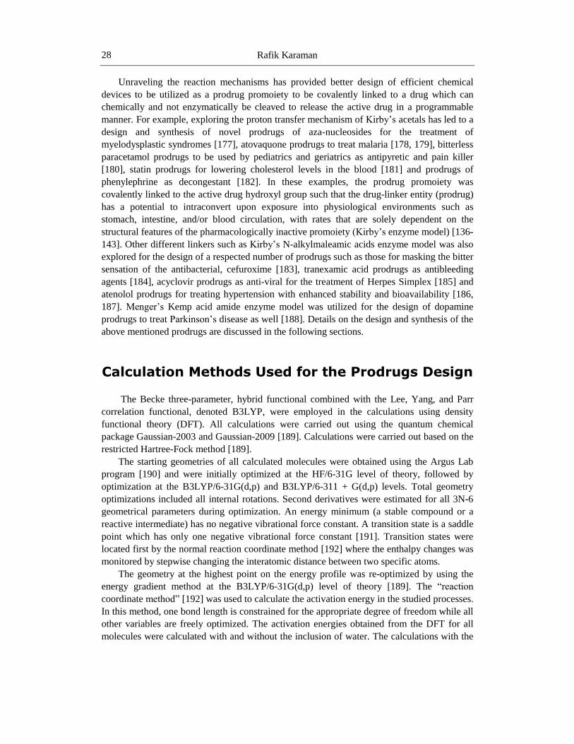
Rafik Karaman 28
Unraveling the reaction mechanisms has provided better design of efficient chemical
devices to be utilized as a prodrug promoiety to be covalently linked to a drug which can
chemically and not enzymatically be cleaved to release the active drug in a programmable
manner. For example, exploring the proton transfer mechanism of Kirby‘s acetals has led to a
design and synthesis of novel prodrugs of aza-nucleosides for the treatment of
myelodysplastic syndromes [177], atovaquone prodrugs to treat malaria [178, 179], bitterless
paracetamol prodrugs to be used by pediatrics and geriatrics as antipyretic and pain killer
[180], statin prodrugs for lowering cholesterol levels in the blood [181] and prodrugs of
phenylephrine as decongestant [182]. In these examples, the prodrug promoiety was
covalently linked to the active drug hydroxyl group such that the drug-linker entity (prodrug)
has a potential to intraconvert upon exposure into physiological environments such as
stomach, intestine, and/or blood circulation, with rates that are solely dependent on the
structural features of the pharmacologically inactive promoiety (Kirby‘s enzyme model) [136-
143]. Other different linkers such as Kirby‘s N-alkylmaleamic acids enzyme model was also
explored for the design of a respected number of prodrugs such as those for masking the bitter
sensation of the antibacterial, cefuroxime [183], tranexamic acid prodrugs as antibleeding
agents [184], acyclovir prodrugs as anti-viral for the treatment of Herpes Simplex [185] and
atenolol prodrugs for treating hypertension with enhanced stability and bioavailability [186,
187]. Menger‘s Kemp acid amide enzyme model was utilized for the design of dopamine
prodrugs to treat Parkinson‘s disease as well [188]. Details on the design and synthesis of the
above mentioned prodrugs are discussed in the following sections.
Calculation Methods Used for the Prodrugs Design
The Becke three-parameter, hybrid functional combined with the Lee, Yang, and Parr
correlation functional, denoted B3LYP, were employed in the calculations using density
functional theory (DFT). All calculations were carried out using the quantum chemical
package Gaussian-2003 and Gaussian-2009 [189]. Calculations were carried out based on the
restricted Hartree-Fock method [189].
The starting geometries of all calculated molecules were obtained using the Argus Lab
program [190] and were initially optimized at the HF/6-31G level of theory, followed by
optimization at the B3LYP/6-31G(d,p) and B3LYP/6-311 + G(d,p) levels. Total geometry
optimizations included all internal rotations. Second derivatives were estimated for all 3N-6
geometrical parameters during optimization. An energy minimum (a stable compound or a
reactive intermediate) has no negative vibrational force constant. A transition state is a saddle
point which has only one negative vibrational force constant [191]. Transition states were
located first by the normal reaction coordinate method [192] where the enthalpy changes was
monitored by stepwise changing the interatomic distance between two specific atoms.
The geometry at the highest point on the energy profile was re-optimized by using the
energy gradient method at the B3LYP/6-31G(d,p) level of theory [189]. The ―reaction
coordinate method‖ [192] was used to calculate the activation energy in the studied processes.
In this method, one bond length is constrained for the appropriate degree of freedom while all
other variables are freely optimized. The activation energies obtained from the DFT for all
molecules were calculated with and without the inclusion of water. The calculations with the

Prodrugs Design Based on Inter- and Intramolecular Processes 29
incorporation of a water molecule were performed using the integral equation formalism
model of the Polarizable Continuum Model (PCM) [193-196]. In this model the cavity is
created via a series of overlapping spheres. The radii type employed was the United Atom
Topological Model on radii optimized for the PBE0/6-31G (d) level of theory. The MM2
molecular mechanics strain energy calculations were done using Allinger‘s MM2 program
[154].
In this chapter, the mechanisms of some enzyme models that have been advocated to
understand how enzymes work were computationally explored. The tool used in the study is
computational approach consisting of calculations using a variety of different molecular
orbital and molecular mechanics methods and correlations between experimental and
calculated reactions rates [166-184].
Kirby’s Enzyme Model Based on Intramolecular
Proton Transfer in acetals [135, 136, 138-143]
Intramolecular processes are faster and more efficient than their intermolecular
counterparts because of the two reacting centers proximity orientation which mimics that of
functional groups when brought together in the enzyme active site [118-143].
For assessing the factors determining the rate-limiting step in Kirby‘s enzyme, using
molecular orbital methods, we have investigated the mode and scope of the proton transfers
efficiency in Kirby‘s enzyme models 1-18 (Figures 25- 27) [135, 136, 138-143].
The aim of this study was to: (i) assign the driving force(s) for the high extreme
efficiency of the intramolecular general acid catalysis (IGAC) in 1-15 (Figures 25 and 26),
and (ii) to locate possible intramolecular hydrogen bonding along the reaction pathway (in
reactants, transition state and products) and to evaluate their role in the intramolecular process
efficiency.
Using ab initio at HF/6-31G (d,p) and DFT at B3LYP/6-31G (d,p) basis sets the kinetic
and thermodynamic parameters for the IGAC in processes 1-15 (Figures 25 and 26) were
calculated.
The intermolecular processes 16 and 17 (Figure 27) were selected as intermolecular
proton transfer processes for calculating the effective molarity (EM) values of the
corresponding intramolecular processes 1-8 and 9-15, respectively. Process 18 (Figure 27)
was calculated to represent a proton transfer process driven by ―classical‖ general catalysis
(GAC) for comparisons with IGAC.
The calculation results demonstrated that the structural features required for a system to
achieve an efficient intramolecular proton transfer are: (1) the distance between the two
reactive centers (rGM, see Figure 28) in the ground state (GM) should be short which
subsequently results in strong intramolecular hydrogen bonding, and (2) the attack angle α
(Figure 28) in the ground state should be about 180° for maximizing the orbital overlap of the
two reactive centers upon their engagement along the reaction pathway. Among the first set
of systems, 1-8, that were calculated, systems 4 and 8 were the most reactive ones because
they both fulfill, to a high extent, the two requirements (α= 170° and rGM = 1.7 Å ). On the
other hand, system 6 was found to be with the lowest rate as a result of having an attack angle

Rafik Karaman 30
and distance between the two reactive centers far away from the optimal values (α=48° and
rGM = 3.7 Å, see Table 1).
In order to examine whether the reaction mechanism for systems such as 4 occurs via an
efficient intramolecular general acid catalysis (IGAC) or via ―classical‖ general acid catalysis
(GAC), calculations for processes 16 and 18 were conducted; where process 16 involves
intermolecular proton transfer from acetic acid to the acetal, and process 18 is similar to that
of 4 except that an acetic acid molecule is replaced with a water molecule as a proton donor to
the acetal (Figure 27).
Comparison of the calculated DFT activation energy values for processes 16 and 18 with
that of 4 indicates that IGAC for 4 is much more efficient than that of 16 and 18 (∆Gffi value
for 4 is 24.15 kcal/mol, and for 16 and 18 are 38.55 kcal/mol and 53.14 kcal/mol,
respectively).
This result confirms that catalysis of the acetal cleavage in 4 must be supplied by the
proton of the carboxyl group. Thus the mechanism in systems such as 4 is via IGAC and not
via GAC. The EM values depicted in Table 1 show that 4 and 8 are the most efficient
processes among 1-15 (log EM 10-13) and among the least efficient processes are 2, 11 and
13-15 with log EM <1. In addition, the calculations revealed that the proton transfer rate in
systems 1–15 is quite responsive to geometric disposition, especially in relation to the
distance between the two reactive centers, rGM, and the attack angle, α. Requirements for a
system to achieve a high intramolecular proton transfer rate are: (a) a short distance between
the two reactive centers (rGM) in the ground state which subsequently results in strong
intramolecular hydrogen bonding, and (b) a hydrogen bonding angle (αGM) close to linearity
in order to maximize orbitals overlapping.
Table 1. DFT (B3LYP/6-31G (d,p)) calculated kinetic and thermodynamic properties
for the proton transfer in 1-15
System
H2-O3
rGM (Å) α ∆Hffi
TΔSffi
∆Gffi
log EM (calc.) log EM [135, 143]
(exp.)
1 1.69 143 33.09 -1.25 34.34 7.40 -----
2 3.60 48 48.29 -2.23 46.52 -1.52 -----
3 1.74 147 34.32 -3.42 37.74 5.17 4.00
4 1.70 170 21.47 -2.68 24.15 10.58 10.00
5 1.69 149 26.64 -3.71 30.35 6.04 3.45
6 3.66 48 40.15 1.04 39.11 -0.41 ------
7 1.74 147 26.22 -5.12 31.34 5.30 3.98
8 1.72 171 21.22 0.03 21.19 12.72 12.60
9 1.72 146 23.62 0.07 23.55 1.67 -----
10 2.04 125 24.73 -3.34 28.07 -1.65 -----
11 3.33 123 25.41 -4.15 29.56 -2.75 -----
12 1.73 144 22.54 0.90 21.64 3.07 -----
13 3.33 124 23.59 -4.15 27.74 -1.41 -----
14 1.92 122 29.33 0.61 28.72 -2.13 -----
15 2.51 107 34.71 -2.00 36.71 -8.01 -----
∆Hffi
is the activation enthalpic energy (kcal/mol). TΔSffi is the activation entropic energy in kcal/mol.
∆Gffi
is the activation free energy (kcal/mol). α is the hydrogen bonding angle in the ground state
structure ( For H2O3 (rGM ), see Figure 28). ln EM =-(∆Gffi
intra- ∆Gffi
inter)/RT.
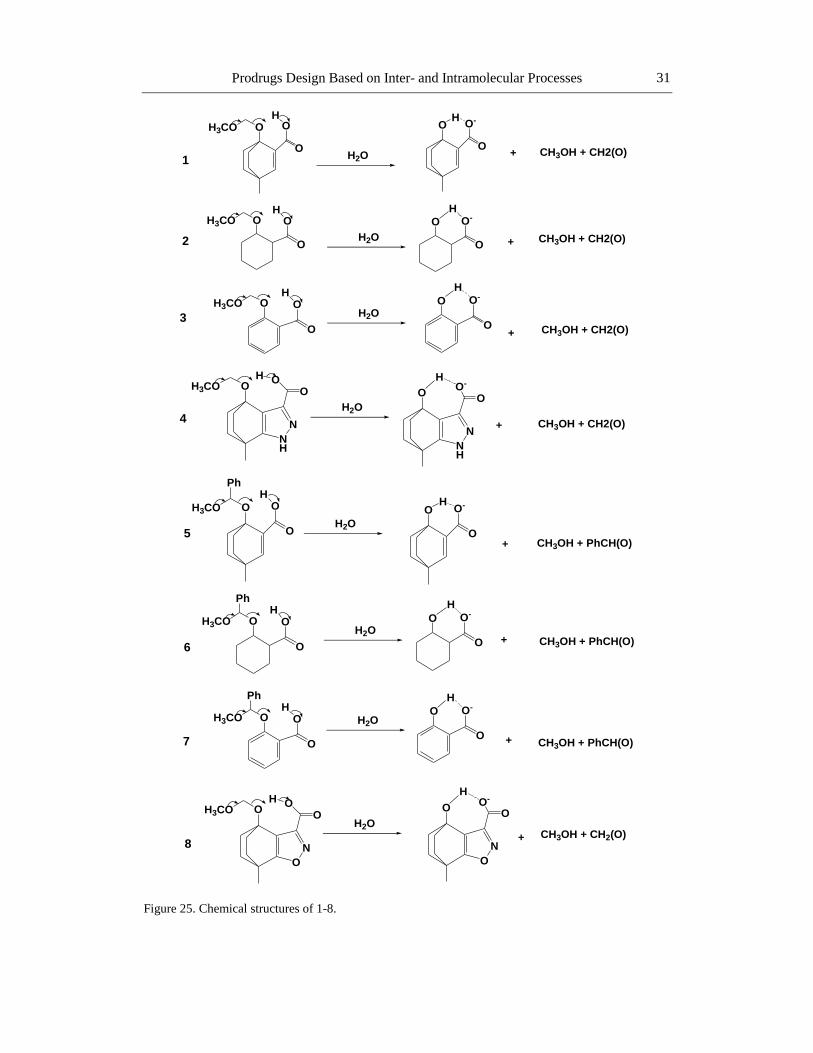
Prodrugs Design Based on Inter- and Intramolecular Processes 31
Figure 25. Chemical structures of 1-8.
NH
N
OO
OH3COH
NH
N
O-
OO
H
CH3OH + CH2(O)+
O
N
OO
OH3COH
O
N
O-
OO
H
OH3CO
+
O
O
PhH
O O-
O
H
CH3OH + PhCH(O)
OH3CO
+
O
O
PhH O O-
O
H
CH3OH + PhCH(O)
CH3OH + CH2(O)+
OH3CO
H2O +
O
O
HO O-
O
H
CH3OH + CH2(O)
OH3CO O
O
HO O-
O
H
+ CH3OH + CH2(O)
OH3CO
+
O
O
HO O-
O
H
CH3OH + CH2(O)
OH3CO O
O
PhH
O O-
O
H
+ CH3OH + PhCH(O)
1
H2O
H2O
H2O
H2O
H2O
H2O
H2O
2
3
4
5
6
7
8

Rafik Karaman 32
Figure 26. Chemical structures of 9-15.
OH3CO
Ph
NMe2
H
NMe2
HOH3CO
NMe2
HOH3CO
Ph
OO
MeO
O
Me2N O OCH3H
OH3CO NMe2
H
O NMe2
H
CH3OH + PhCH(O)H2O
NMe2
H
O
CH3OH + CH2(CO)
NMe2OH
OO
MeO
O
CH3OH + PhCH(O)
O NMe2
H
CH3OH + CH2(CO)
NMe2 OH
CH3OH + CH2(CO)
Me2N
O
O CH3H
NMe2
OH
CH3OH + CH2(CO)
NMe2
HOH3CO
OO
MeO
O
NMe2OH
OO
MeO
OCH3OH + CH2(CO)
H2O
H2O
H2O
H2O
H2O
H2O
9
10
11
12
13
14
15

Prodrugs Design Based on Inter- and Intramolecular Processes 33
Figure 27. Chemical structures of 16-18.
Figure 28. Schematic representation for the cleavage in 1-15 showing the distance between the reacting
centers (rGM) and the angle of attack (α).
The strong correlation found between the calculated ΔGffi
values, which reflect the
experimental reaction rate, and geometrical parameters such as r2GM and α provides a
mathematical tool (ΔGffi
vs. r2
GM x (1 + sin (180 - α)) which can predict reaction rate based on
a calculated GM for a certain system. The equation slope can be used as an indicator of the
mode by which the two reactive centers are orchestrated in an intramolecular process.
O
OH
CH3
NH
N
COOHOH3COCH3OH + CH2(O)
NH
N
COOHOH
CH3COOHH2O
Me2N
OCH3
O
CH3H NMe2
CH3OH + CH2(CO)H2O
16
17
NH
N
COOHOH3CO CH3OH + CH2(O)
NH
N
COOHOH
H
O
H
H2O18
C O
O
OO
H3C
H
3
2 1
N
rGM CH3
H
OO
1
2
3CH3
R
rGM
H3C
R
37-44 45-51R = H or phenyl
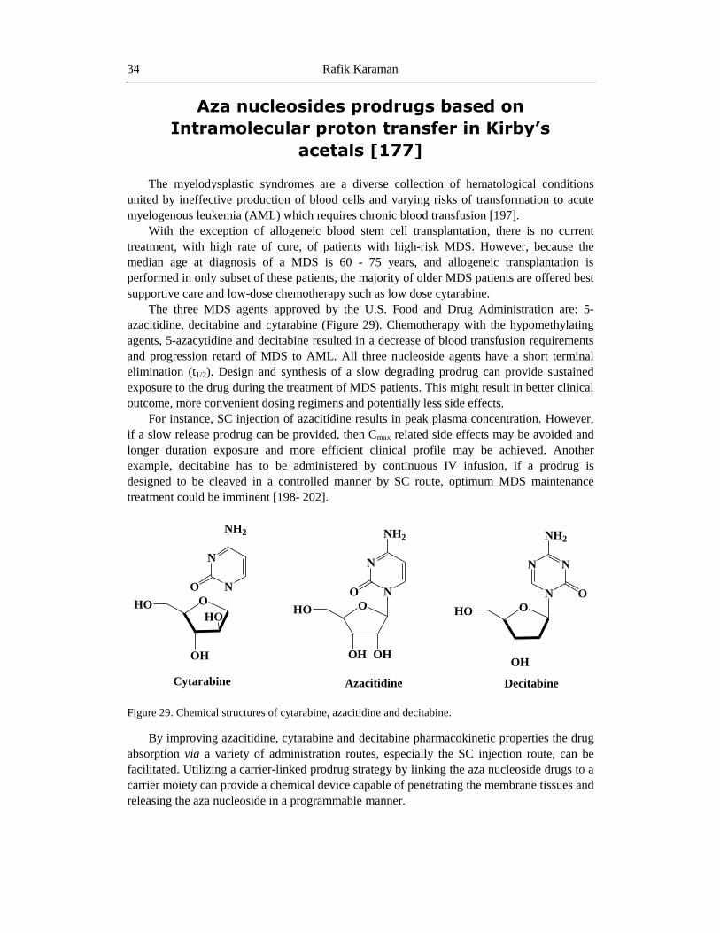
Rafik Karaman 34
Aza nucleosides prodrugs based on
Intramolecular proton transfer in Kirby’s
acetals [177]
The myelodysplastic syndromes are a diverse collection of hematological conditions
united by ineffective production of blood cells and varying risks of transformation to acute
myelogenous leukemia (AML) which requires chronic blood transfusion [197].
With the exception of allogeneic blood stem cell transplantation, there is no current
treatment, with high rate of cure, of patients with high-risk MDS. However, because the
median age at diagnosis of a MDS is 60 - 75 years, and allogeneic transplantation is
performed in only subset of these patients, the majority of older MDS patients are offered best
supportive care and low-dose chemotherapy such as low dose cytarabine.
The three MDS agents approved by the U.S. Food and Drug Administration are: 5-
azacitidine, decitabine and cytarabine (Figure 29). Chemotherapy with the hypomethylating
agents, 5-azacytidine and decitabine resulted in a decrease of blood transfusion requirements
and progression retard of MDS to AML. All three nucleoside agents have a short terminal
elimination (t1/2). Design and synthesis of a slow degrading prodrug can provide sustained
exposure to the drug during the treatment of MDS patients. This might result in better clinical
outcome, more convenient dosing regimens and potentially less side effects.
For instance, SC injection of azacitidine results in peak plasma concentration. However,
if a slow release prodrug can be provided, then Cmax related side effects may be avoided and
longer duration exposure and more efficient clinical profile may be achieved. Another
example, decitabine has to be administered by continuous IV infusion, if a prodrug is
designed to be cleaved in a controlled manner by SC route, optimum MDS maintenance
treatment could be imminent [198- 202].
Figure 29. Chemical structures of cytarabine, azacitidine and decitabine.
By improving azacitidine, cytarabine and decitabine pharmacokinetic properties the drug
absorption via a variety of administration routes, especially the SC injection route, can be
facilitated. Utilizing a carrier-linked prodrug strategy by linking the aza nucleoside drugs to a
carrier moiety can provide a chemical device capable of penetrating the membrane tissues and
releasing the aza nucleoside in a programmable manner.
N
NO
NH2
OHO
OH
HO
N
N
N
NH2
OHO
OH
O
N
NO
NH2
OHO
OH OH
Cytarabine Azacitidine Decitabine

Prodrugs Design Based on Inter- and Intramolecular Processes 35
Based on our reported DFT calculations on a proton transfer reaction in some of Kirby‘s
enzyme models (Figures 26), three prodrugs of aza nucleoside were proposed. As shown in
Figure 30, the aza nucleoside prodrugs ProD 1- ProD 3 have N, N-dimethylanilinium group
(hydrophilic moiety) and a lipophilic moiety (the rest of the prodrug), where the combination
of both moieties secures a moderate HLB. Furthermore, in a physiologic environment of pH
5.5, SC, aza nucleoside prodrugs ProD 1- ProD 3 may have a better bioavailability than their
parent drug due to improved absorption. In addition, those prodrugs may be used in different
dosage forms (i.e., enteric coated tablets) because of their potential solubility in organic and
aqueous media due to the ability of the anilinium group to be converted to the corresponding
aniline group in a physiological pH of 6.5.
It should be emphasized that at pH 5.5-6.5 (SC and intestine physiologic environments)
the anilinium form of the three prodrugs (ProD 1- ProD 3) will equilibrate with the free
aniline form. Subsequently, the former will undergo proton transfer reaction (rate limiting
step) to yield the anti-preleukemia aza nucleoside drug and the inactive carrier as by-product.
It should be emphasized, that our proposal is to exploit prodrugs ProD 1 – ProD 3 for
S.C. use. The physiological environment of the skin has a pH of 5.5. At pH 5.5, prodrugs
ProD 1 – ProD 3 are predicted to exist in the unionized (free base) and ionized (ammonium)
forms where the equilibrium constant for the exchange between both forms is dependent on
the pKa of the given prodrug. The experimental determined pKa for N, N-dimethylanilin is
5.15 [203] hence it is expected that the pKa values for prodrugs ProD 1 – ProD 3 will be in
the same range. Since the pH for the skin lies in the range around 5.5, the calculated ionized
(ammonium) /unionized (free base) ratio will be 69/31.
It is quite safe to assume that the unionized forms for prodrugs ProD 1 – ProD 3 will be
more lipophilic than their parent drug due the presence of a relatively bulky group (Kirby‘s
enzyme model linker). Therefore, their absorption through the skin membranes and the
efficiency for delivering their parent drug are expected to be increased. If the prodrugs, ProD
1 – ProD 3, are intended to be administered by per os dosage form, for example as an enteric
coated tablets, the absorption and consequently the efficiency of those prodrugs will be
relatively reduced compared to the SC route.
This is because the calculated percentage of the unionized form, in the case where the
absorption is taking place in the small intestine (pH = 6.5), is predicted in the range of 5%-
31%.
The selection of Kirby‘s enzyme model to be utilized as carriers to aza nucleosides is
based on the fact that those carriers undergo proton transfer reaction to yield an aldehyde, an
alcohol and a hydroxy amine (Figures 26 and 30). The rate-limiting step in these processes (9-
15 in Figure 26) is a proton transfer from the anilinium group into the neighboring ether
oxygen. Furthermore, the proton transfer rate is strongly dependent on the strength of the
hydrogen bonding in the products and in the transition states leading to them. Therefore, it
will be safe to assume that the reaction rate will be greatly affected by the structural features
of Kirby‘s enzyme model system as evident from the different experimental rate values
determined for the different processes (Table 1) [135-143].
Replacing the methoxy group in 9-15 (Figure 26) with aza nucleoside drug, as shown for
ProD 1- ProD 3 in Figure 30, is not expected to have any significant effect on the relative
rates of these processes. Therefore, computational calculations of the kinetic and
thermodynamic properties for these models will shed light on the rates for the intraconversion
of prodrugs ProD 1 – ProD 3 to their active aza nucleoside drug.

Rafik Karaman 36
Figure 30. Intraconversion of aza-nucleoside prodrugs ProD 1-ProD 3 to their active drug.
DFT calculation results for intramolecular proton transfer reactions in Kirby‘s enzyme
models 9-15 (Figure 26) demonstrate that the reaction rate is quite responsive to geometric
disposition, especially to the distance between the two reactive centers, rGM, and the angle of
attack, α (the hydrogen bonding angle, see Figure 28).
Hence, the study on the systems reported herein could provide a good basis for a design
of aza nucleoside prodrugs that are less hydrophilic than their parent drugs and can be used,
in different dosage forms, to release the parent drug in a controlled manner. For example,
based on the calculated log EM (Table 1), the cleavage process for prodrug ProD 1 is
predicted to be about 1010
times faster than for prodrug ProD 2 and about 104 times faster
than prodrug ProD 3:rateProD1> rateProD3> rate ProD2. Hence, the rate by which the prodrug
releases the aza nucleoside can be determined according to the structural features of the linker
(Kirby‘s enzyme model).
Decitabine
ONMe
OO
MeO
O
N
Decitabine
O
H2O
H2O
H2O
Decitabine
ProD 1
ProD 2
ProD 3
N
N
N
NH2
OO
OH
O
Ph
ONH
N
N
N
NH2
OHO
OH
OPhCH(O)
ONHMe
MeMe
Me
PhCH(O)
N
N
N
NH2
OO
OH
OPh
Me
HN
N
N
NH2
OHO
OH
OON
Me
OO
MeO
O
Me
H
N
N
N
NH2
OHO
OH
O
N
N
N
NH2
OO
OH
OMeMe
H
Ph
CH2(O)
N OMeMe
H

Prodrugs Design Based on Inter- and Intramolecular Processes 37
Statin Prodrugs Based on Intramolecular Proton
Transfer in Kirby’s Acetals [181]
Hyperlipidemia is a common heterogeneous group of disorders, most commonly treated
with statin medications. Statins are selective and competitive inhibitors of HMG-CoA
reductase, rate-limiting enzyme that converts 3-hydroxy-3-methylglutaryl coenzyme A to
mevalonate, a precursor of cholesterol [204]. Statins are hepatoselective and the mechanisms
contributing to this are governed by their solubility profiles. Passive diffusion through
hepatocellular membranes is the key for effective first pass uptake for hydrophobic statins,
which can also be diffused to non-hepatocellular parts, whereas extensive carrier-mediated
uptake is the major uptake route [205]. This can prove that hydrophilic statins acquire greater
hepatoselectivity. All satin are absorbed rapidly after administration, reaching peak plasma
concentration within 4 hours [205]. Statins in general have a low bioavailability mostly due to
the extensive first pass effect and/or bad solubility.
Simvastatin is a hydrophobic fungal derivative compound [204]; it is administered as a
lactone prodrug, simvastatin A (Figure 31) that after oral ingestion undergoes a hydroxylation
to yield its corresponding (β)-hydroxy acid form, simvastatin B (Figure 31), an inhibitor of
HMG-CoA reductase [206]. The percentage of simvastatin dose retained by the liver is >80%
[204].
The solubility of simvastatin is the limiting factor for its bioavailability [207].
Simvastatin undergoes an extensive first pass effect and has a bioavailability of only 5%; it
has a poor absorption rate from the gastro-intestinal tract (GIT). Hence, improving its
solubility and its dissolution rate is a crucial for enhancing its bioavailability [205, 208].
Many attempts have been made in order to improve simvastatin bioavailability. One
strategy was by co-solvent evaporation method, where a hydrophilic, low viscosity grade
polymer hydroxypropylmethylcellulose (HPMC) was used to enhance simvastatin solubility
and dissolution rate.
The study results revealed a significant enhancement in its solubility by converting
simvastatin particles from regular to amorphous form via reducing the particle size and
increasing wettability [208].
In another study glycerylmonooleate (GMO)/poloxamer 407 cubic nanoparticles was
investigated as potential oral drug delivery system to enhance simvastatin bioavailability. The
study showed that oral bioavailability of simvastatin cubic nanoparticles was enhanced
significantly at 2.41-fold compared to that in micronized crystal powder. In addition, it
demonstrated a sustained plasma simvastatin level for over 12 hours [209]. In 2012,
developments of stable pellets-layered simvastatin nano-suspensions were tested for
simvastatin dissolution and bioavailability. The study revealed an improved dissolution and
bioavailability of the drug [206].
Atorvastatin and rosuvastatin (Figure 31) are fully synthetic compounds; atorvastatin is
relatively lipophilic [204], whereas rosuvastatin is more hydrophilic due to its polar hydroxyl
and methane sulphonamide groups [205]. Atorvastatin has a bioavailability of only 14% and
half-life of 14 hours, however, the half-life of its inhibitory activity (HMG-CoA reductase) is
20 - 30 hours due to contribution of its active metabolites [210].

Rafik Karaman 38
Figure 31. Chemical structures of simvastatin A, simvastatin B, atorvastatin and rosuvastatin.
Solid forms of atorvastatin are unstable when exposed to heat, moisture or light since its
hydroxy acid form is converted to the corresponding lactone form. It is worth noting, that
atorvastatin hydroxy acid form is about 15 times more soluble than its lactone [207].In
addition, atorvastatin is vulnerable to destabilization upon contact with other excipients
present in the formulation as they might negatively interact with the drug. Therefore, most of
the commercial atorvastatin medications consist of an alkaline metal salt such as calcium
carbonate [207]. Atorvastatin instability which leads to poor solubility is the main reason for
its low bioavailability [207].One method that was performed in an attempt to improve the oral
O
OHO
H
H
CH3
H
H3C
O
O
H3C
CH3
H3C
N F
O
NH
OH
COOHO
OH
COO
HO
N N
CH3
CH3
N
CH3S
O O
H3C
F
Rosuvastatin
Simvastatin
Atorvastatin
OH
COOH
HO
H
H
CH3
H
H3C
O
O
H3C
CH3
H3C
Simvastatin
A B
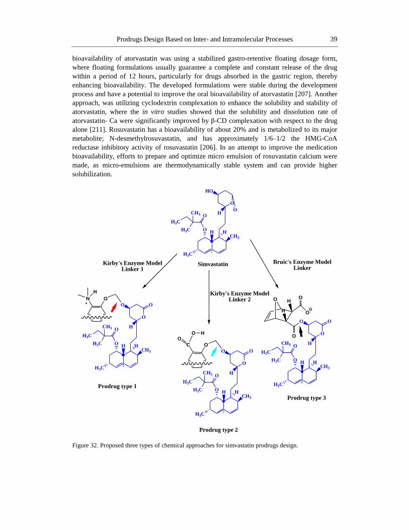
Prodrugs Design Based on Inter- and Intramolecular Processes 39
bioavailability of atorvastatin was using a stabilized gastro-retentive floating dosage form,
where floating formulations usually guarantee a complete and constant release of the drug
within a period of 12 hours, particularly for drugs absorbed in the gastric region, thereby
enhancing bioavailability. The developed formulations were stable during the development
process and have a potential to improve the oral bioavailability of atorvastatin [207]. Another
approach, was utilizing cyclodextrin complexation to enhance the solubility and stability of
atorvastatin, where the in vitro studies showed that the solubility and dissolution rate of
atorvastatin- Ca were significantly improved by β-CD complexation with respect to the drug
alone [211]. Rosuvastatin has a bioavailability of about 20% and is metabolized to its major
metabolite; N-desmethylrosuvastatin, and has approximately 1/6–1/2 the HMG-CoA
reductase inhibitory activity of rosuvastatin [206]. In an attempt to improve the medication
bioavailability, efforts to prepare and optimize micro emulsion of rosuvastatin calcium were
made, as micro-emulsions are thermodynamically stable system and can provide higher
solubilization.
Figure 32. Proposed three types of chemical approaches for simvastatin prodrugs design.
Prodrug type 1
Prodrug type 2
Prodrug type 3
O
O
HO
H
HCH3
H
H3C
O
O
H3C
CH3
H3C
ON
O
OO
H
HCH3
H
H3C
O
O
H3C
CH3
H3C
H
OC
O
OO
H
HCH3
H
H3C
O
O
H3C
CH3
H3C
OO H O
OO
H
HCH3
H
H3C
O
O
H3C
CH3
H3C
O
O
O
H
H
O
SimvastatinKirby's Enzyme ModelLinker 1
Kirby's Enzyme ModelLinker 2
Bruic's Enzyme ModelLinker

Rafik Karaman 40
The study demonstrated a potential use of micro emulsion system to enhance the
solubility and hence the bioavailability for the poorly water soluble drug, rosuvastatin [212].
The solubility of simvastatin is the limiting factor for its bioavailability due to slow tissue
penetration [207], hence, it undergoes an extensive first pass effect; therefore, improving its
solubility and hence its dissolution rate is a must to enhance its bioavailability. Therefore,
development of more hydrophilic prodrugs that have the capability to release the parent drug
in physiological environments such as intestine is a significant challenge.
Figure 33. Intraconversion of simvastatin ProD 1- ProD 3.
In principle, three approaches could be considered to fulfill the requirements mentioned
above: (Figure 32): (1) linking the statin free hydroxyl group to Kirby‘s enzyme model
(ammonium linker), (2) blocking the statin hydroxyl group with Kirby‘s enzyme model
(carboxylic acid linker) and (3) attaching the statin free hydroxyl group with Bruice‘s model
(dicarboxylic semi-ester linker.
O
N O
H2O
H2O
NH
HCH(O)
NHOMe
Me
Me
Me
HCH(O)
MeMe
H
NMe
Me
HO
ON
HMe
Me
HCH(O)
NHOMe
Me
O
OO
H
HCH3
H
H3C
O
O
H3C
CH3
H3C
O
OHO
H
HCH3
H
H3C
O
O
H3C
CH3
H3C
O
OO
H
HCH3
H
H3C
O
O
H3C
CH3
H3C
O
OO
H
HCH3
H
H3C
O
O
H3C
CH3
H3C
O
OHO
H
HCH3
H
H3C
O
O
H3C
CH3
H3C
H2O
O
OHO
H
HCH3
H
H3C
O
O
H3C
CH3
H3C
Simvastatin ProD 1
Simvastatin ProD 2
Simvastatin ProD 3
Linker 1
Linker 2
Linker 3
Simvastatin
Simvastatin
Simvastatin

Prodrugs Design Based on Inter- and Intramolecular Processes 41
Based on our DFT calculations on a proton transfer reaction in some of Kirby‘s enzyme
models (8-15, Figure 26), three prodrugs of simvastatin are proposed. As shown in Figure 33,
the simvastatin prodrugs, ProD 1- ProD 3,have N,N-dimethylanilinium group (hydrophilic
moiety) and a lipophilic moiety (the rest of the prodrug), where the combination of both
moieties secures a moderate hydrophilic lipophilic balance (HLB). Furthermore, in a
physiological environment of pH around 6, intestine, prodrugs ProD 1- ProD 3 may have a
better bioavailability than their parent drug due to improved absorption. In addition, those
prodrugs may be used in different dosage forms (i.e., enteric coated tablets) because of their
potential solubility in organic and aqueous media due to the ability of the anilinium group to
be converted to the corresponding aniline group in a physiological pH of 6.5.
DFT calculations at B3LYP 6-31G (d,p) for intramolecular proton transfer in the three
simvastatin prodrugs revealed that the interconversion of simvastatin prodrug ProD 3 to
simvastatin is predicted to be about 10 times faster than that of either simvastatin ProD 1 or
simvastatin ProD 2. Hence, the rate by which the prodrug releases the statin drug can be
determined according to the structural features of the promoiety (Kirby‘s enzyme model).
Menger’s Enzyme Model Based on Intramolecular
Proton Transfer in Kemp’s Acid Amides [130-134]
Menger‘s singularity model and its partner the spatiotemporal hypothesis predict that an
efficient enzyme model could be achieved using chemical systems that rigidly hold a
nucleophile and an electrophile at a distance at or near the critical distance. One of the most
interesting enzyme models that obeys the requirements imposed for high efficiency is an
intramolecular-catalyzed cleavage of an aliphatic amide (Figure 34) [130-134].Taking into
consideration that an aliphatic amide is very difficult to be hydrolyzed; a 10-hour reflux in
concentrated hydrochloric acid is not unusual. However, when a carboxyl group is held in a
1,3-diaxial orientation to the amide, the amide bond cleavage occurs with a half-life of only 8
minutes at neutral pH and 21.5 ˚C. Although the structure shown in Figure 34 has two
carboxyl groups, only a single carboxyl is actually needed for this enzyme-like rate
acceleration, the other carboxyl being merely a ―spectator‖. It should be emphasized, that the
aliphatic amide reaction occurs via the more stable structure B rather than structure A (Figure
34).
Figure 34. Chemical structures of conformations A and B for Menger‘s reactive amide, where B is
presumably the most reactive conformer.
O
OO
NOO
H
HO
O ONO
O
HH
A B
2.8 Å
3.76 Å
2.76 Å
Rafik Karaman 42
One striking example of enzyme models that were proposed for understanding enzyme
catalysis is the intramolecular cleavage reaction of Kemp‘s di-acid amide 19 that was
exploited by Menger and Ladika to unravel the mechanism of chymotrypsin enzyme
catalyzed biotransformation reactions [130-134]. Chymotrypsin cleaves peptide bonds by
attacking the unreactive carbonyl group of the peptide with its serine 195 residue located in
its active site, which briefly becomes covalently bonded to the substrate, forming an enzyme–
substrate intermediate. It was demonstrated that the biotransformation of chymotrypsin with
its substrate involves two stages, an initial ‗‗burst‘‘ phase at the start of the reaction and a
steady-state phase called ‗‗ping-pong‘‘ stage. Thus the enzyme‘s mode of action is as
hydrolysis takes place in two steps. First acylation of the substrate which involves a proton
transfer to form an acyl-enzyme intermediate and in the second step, deacylation in order to
return the enzyme to its original state [214, 215]. In the laboratory, the hydrolysis reaction
rate of a peptide linkage is too slow; at neutral pH and ambient temperature, the half-life is
about 500 years, unless a strong acid catalyst is added [216, 217], yet in the small intestines,
where the conditions are about neutral rather than acidic, most of the proteins hydrolysis takes
place rather quickly. This is due to the presence of enzymes called peptidases that catalyze the
amide cleavage via a proton transfer reaction [218].
Molecular orbital calculations using ab initio at HF/6-31G(d,p) and DFT at B3LYP/6-
31G (d,p) levels for the amide cleavage reactions of di-carboxylic aliphatic amides 19–29
(Figure 35) were done to unravel the unusual striking rates acceleration brought about when
these amides stand in neutral aqueous solution at ambient temperature. The calculations
aimed to: (1) determine whether amides 19–29 undergo fast cleavage due to a proton transfer
stemming from a proximity orientation of the nucleophile (O6, see Figure 36) to the
electrophile (H5, see Figure 36), as was suggested by Menger [130-134], or due to
pseudoallylic (pseudo-A) strain relief that occurs upon the cleavage of the distorted amide
bond to a less strained tetrahedral intermediate that is formed from the approach of the
carboxylic oxygen (O4, see Figure 36) towards the amidic carbonyl carbon (C7, see Figure
36), as was proposed by Curran and others [219, 220].
The mechanistic calculation results shown in Figure 37 and listed in Table 2 revealed: (i)
The activation energy barriers for the cleavage of amides 19– 29 at a pH range where the
carboxylic group is in its free acid form (not ionized) are much smaller via a proton transfer
(route b) than via an approach of the carboxylic oxygen onto the amide carbonyl carbon
(route a). For example, the gas phase B3LYP/6-31G (d,p) calculated activation energy for the
hydrolysis of amide 19 via a proton transfer (route b) is 45.18 kcal/mol less than that for the
cleavage via route a. (ii) The calculated activation energy values for the cleavage reactions of
amides 19 and 20 are almost the same (~15 kcal when calculated by B3LYP/6-31G (d,p) in
the presence of water as a solvent). This result suggests that the presence of only one
carboxylic group, such as in amide 20, is needed to supply a proton transfer for achieving the
same acceleration as for the reaction of amide 19. The calculation results are in accordance
with the experimental findings by Menger [130-134]. (iii) The calculated activation energy in
the presence of water for the cleavage of amide 21 was found to be ~3 kcal/mol smaller than
that of amide 19. This slight difference in energy might be due to the difference between the
calculated distances of the electrophile (carboxylic proton) and the nucleophile (amide
carboxylic carbon) in the more strained amide 19 and the less strained amide, 21; H5—O6
distance (Figure 36) in 19 is 2.9 Å and in 21 is 3.0 Å. This is in accordance with Menger‘s

Prodrugs Design Based on Inter- and Intramolecular Processes 43
‗‗spatiotemporal hypothesis‖ and our computational studies on other enzymes models [158-
161, 213].
Figure 35. Chemical structures of aliphatic amides 19-29.
Figure 36. Schematic representation for the cleavage of 19-29 showing the distance between the
reacting centers (r) and the angle of attack (α).
H3C
H3C
CH3
OO
O
OH
OH N
H3CCH3
OO OH
N
OO OH
NO
O OH N
OO OH NH2
CH2CH2Ph
H3CCH3
OO OH
NH
H3C
H3C
CH3
OO
O
OH
OH NH
CH2CH2Ph
OO
O
OH
OH N
OO
O
OH
OCH3N
27 28 29
19
24 25
2122
23
H3C
H3C
CH3
OO
O
OH
OH N
20
HOOC
H3C
H3C
CH3
OO
O
OH
OH
26
N
r
1
23
GM TS
4
5
OO
O
N
H6
7
8 OO
O
N
H
O
O
OH
OH
P
H2O
NH

Rafik Karaman 44
Table 2. DFT calculated properties for the proton transfers in 19-25, 26-28 and ProD1-2
System
∆Hffi
(GP)
T∆Sffi
(GP)
∆Gffi
(GP)
∆Hffi
(Water)
∆G ffi
(Water)
rGM
(Å)
α
(degree)
t1/2
(hour)
19 27.61 -1.11 28.72 12.55 13.66 3.00 70.90 0.028
20 28.39 -2.92 31.31 14.64 17.56 3.01 75.92 0.03
21 29.50 0.04 29.46 15.06 15.02 3.10 72.18 ----
22 32.00 -0.92 32.92 15.69 16.61 3.09 71.55 ----
23 29.12 -1.23 30.35 17.69 18.92 3.06 75.23 0.1
24 30.56 -1.28 31.84 28.82 30.10 3.62 56.63 283
25 34.22 -2.73 36.95 27.74 30.47 3.16 75.70 ----
27 29.36 -1.88 31.24 29.81 31.69 3.33 73.96 324
28 30.48 -1.44 31.92 17.85 19.31 3.20 67.60 2.2
29 32.57 -1.68 34.25 21.96 23.64 3.20 68.00 90
ProD 1 31.12 -2.22 33.34 24.85 27.07 3.19 75.40 ----
ProD 2 28.72 -1.59 30.31 23.00 24.59 3.25 74.88 ----
T∆Sffi is the entropic activation energy (kcal/mol), ∆H
ffi is the enthalpic activation energy (kcal/mol), ∆G
ffi
is the free activation energy (kcal/mol), rGM and α are the H6-O5 distance and the angle O5-H6-O7
in the global minimum structures, respectively (Figure 36). GP and Water refer to calculated in the
gas phase and water, respectively. t1/2 is the time needed to cleave 50% of the amide to the
corresponding amine and carboxylic acid.
Figure 37. Proposed mechanism for the hydrolysis of aliphatic amides 19-29.
N
NH
GM
TS1
TS2
TS3
P1
P2
Route b
Route b
Route a
Route a
H
H
H
OO
O
OH
OH
N
H
H
H
OO
O
O
OH
H
N
H
H
H
O
O
O
O
OH
H
INT
N
H
H
H
HO
O
O
O
OH
N
H
H
H
HO
O
O
O
OH
H
H
H
OO
O
O
OH
Prodrugs Design Based on Inter- and Intramolecular Processes 45
Furthermore, the DFT results for the cleavage reactions in Kemp‘s mono- and di-acid
amides 19–29 demonstrate that the rate limiting step in the acylolysis process is a proton
transfer from the carboxyl group onto the amide carbonyl oxygen. It is proposed that
accelerations in rate are mainly due to the distance between the two reactive centers (r) and
the angle of attack (α) (Figure 36). In fact, a linear correlation was found between the
activation energy (∆Gffi) and r
2 x sin (180 - α). On the other hand, contrarily to previous
studies the ground-state pseudoallylic strain effect has a little contribution if any to the
cleavage rates of Kemp‘s triacid tertiary amides reactions. Further, the calculations suggest a
change in the mode and the mechanism of the amide cleavage upon changing the pH of the
reaction medium. Thus, peptidases are extremely reactive around neutral pH while their
activities diminish under basic medium.
Dopamine Prodrugs Based on a Proton Transfer
in Menger’s Aliphatic Amide Enzyme Model [188]
Dopamine is a neurotransmitter that is naturally produced in the body. Dopamine is
produced in several areas of the brain, including the substantial nigra and the ventral
tegmental area and it activates the five types of dopamine receptors D1, D2, D3, D4 and D5.
Dopamine is also a neurohormone released by the hypothalamus and its main function to
inhibit the release of prolactin from the anterior lobe of the pituitary. Dopamine is present in
the regions of the brain that regulate movement, emotion, motivation and the feeling of
pleasure. Shortage of dopamine, particularly the death of dopamine neurons in the
nigrostriatal pathway, causes Parkinson's disease, in which a person loses the ability to
execute smooth, controlled movements. Dopamine can be supplied as a medication that acts
on the sympathetic nervous system, producing effects such as increased heart rate and blood
pressure. However, because dopamine cannot cross the blood-brain barrier, dopamine given
as a drug does not directly affect the central nervous system. To increase the amount of
dopamine in the brains of patients with diseases such as Parkinson's disease and dopa-
responsive dystonia, L-DOPA (levodopa), a precursor of dopamine, is given because it can
cross the blood-brain barrier. Levodopa is used in various forms to treat Parkinson's disease
and dopa-responsive dystonia. It is typically co-administered with an inhibitor of peripheral
decarboxylation (DDC, dopa decarboxylase such as carbidopa or benserazide (Figure 38)
[221, 222]. The main objective of this study was to design and synthesize new prodrugs for
the treatment of Parkinson‘s disease that have the potential to be with a higher bioavailability
than the current medications. For achieving this goal, the dopamine prodrugs
physicochemical properties are: (i) adequate solubility in physiological environment (ii) a
moderate hydrophilic lipophilic balance (HLB) value (iii) providing upon chemical cleavage
the parent drug in a controlled manner, and (iv) providing upon cleavage safe and non-toxic
by-products. By fulfilling these requirements the following objectives may be achieved: (1) a
high absorption and permeability of the prodrug into the body tissues. (2) The possibility to
use the anti-Parkinson‘s drug in different administration routes. (3) A chemically driven
system that able to release dopamine in a controlled manner, and (4) a drug with a high
bioavailability and efficient clinical profile.

Rafik Karaman 46
Figure 38. Enzymatic conversion of L-dopa to dopamine along with the chemical structures of
carbidopa and benserazide.
Continuing our investigations for utilizing enzyme models as potential linkers for weak
amine-drugs, we have researched the driving forces responsible for accelerations in rate of
proton transfer reactions in some of Kemp‘s acid amide derivatives, 19-29. It is expected that
molecules such as 19-29 will have a potential of being appropriate linkers to dopaminergic
agents such as dopamine. The proposed dopamine prodrugs based on proton transfer reaction
are depicted in Figure 39. Based on our reported DFT calculations on proton transfer
reactions in Kemp‘s acid amide derivatives, 19-29, two dopamine prodrugs are proposed. As
shown in Figure 39, ProD 1 and ProD 2 have a carboxylic group as a hydrophilic moiety and
the rest of the prodrug as a lipophilic moiety, where the combination of both moieties secures
a moderate HLB. Furthermore, in a physiologic environment of the blood circulation (pH 7.4)
dopamine is expected to be primary in the ionized forms while its prodrugs ProD1-2 are
expected to equilibrate between the ionic and the free acid forms. Thus, prodrugs ProD 1-2
may have a better bioavailability than the parent drugs due to improved absorption. In
addition, these prodrugs can be used in different dosage forms (i.e., enteric coated tablets)
because of their potential solubility in organic and aqueous media due to the ability of the
carboxylic group to be converted to the corresponding carboxylate anion in physiological
environments of pH 5.0-7.4 (intestine and blood circulation). It is worth noting, that at pH
5.0-7.4 the carboxylic group in prodrugs ProD 1-ProD 2 will equilibrate with the
corresponding carboxylate form. Subsequently, the free acid form will undergo proton
transfer reaction (rate limiting step) after being transferred through the membrane to yield
dopamine and the inactive linker as a byproduct (Figure 39). The proposed prodrugs ProD 1-
ProD 2 will be exploited for per os use in the form of enteric coated tablets. It is well known,
that enteric coated tablets are stable at the high acidic pH found in the stomach, but break
down rapidly at a less acidic pH. For example, the enteric coated tablets will not dissolve in
the acidic juices of the stomach (pH ~3), but they will in the higher pH (above pH 5.5)
present in the small intestine. In the intestine, prodrugs ProD 1- ProD 2 will exist in the
OH
O
OH
HO NH2
NH2
OH
HO
OH
O
OH
HONHNH2
H3CHO
HO
OH
HN
NH
NH2
O
OH
CarbidopaBenserazide
L-Dopa Dopamine
Dopa decarboxylase (DDC)

Prodrugs Design Based on Inter- and Intramolecular Processes 47
acidic and ionic forms where the equilibrium constant for the exchange between both forms is
dependent on the pKa of the given prodrug. The experimental determined pKa‘s for ProD-1-
ProD 2 linkers are in the range of 5.0-6.0. Therefore, it is expected that the pKa‘s of the
corresponding prodrugs will be in the same range. Since the pH for the small intestine lies in
the range of 5.0-7.5, the calculated unionized (acidic) /ionized ratio will be 40-50%. Although
the percentage of the acidic form is not significantly high, we anticipate that prodrugs
undergoing an efficient proton transfer (rate limiting step) to yield dopamine and Kemp‘s
carboxylic acid by-products (Figure 39) will have the potential to be effective prodrugs. In the
blood circulation (pH 7.4), the calculated acidic form for those prodrugs is around 10- 30%
and it is expected that the rate for delivering the parental drug will be reduced.
In summary, we conclude that the driving force for rate accelerations witnessed in the
cleavage reactions of Kemp‘s acid amides 19-29 and ProD 1- ProD 2 under physiological
environment is the proximity of the carboxylic proton and the amide carbonyl oxygen. The
proximity parameters, rGM and α in the ground state are determined by the strain energy of the
reactants (Es). In systems with high strain energies the global minimum structures tend to
reside in a conformation by which the distance between the two reactive centers (distance
between the nucleophile and the electrophile, rGM) is short and the angle of attack α is close to
linearity. Thus the activation energy of the system is minimized resulting in an enhancement
in the reaction rate due to close resemblance of the ground state to the corresponding
transition state (Table 2). Furthermore, the DFT calculation results predict a t1/2 value of the
cleavage reactions of ProD 1- ProD 2 in pH 6 to be between 12 to 20 hours, whereas at pH 7
the value is expected to be higher (Table 2). The strategy to achieve desirable prodrugs for the
treatment of Parkinson‘s disease that possess a high bioavailability values are: (1) synthesis of
the linker moiety and attaching it with the parent drug Menger‘s synthetic method [130]; (2)
kinetic studies (in vitro) of the synthesized prodrugs (ProD 1- ProD 2) in physiological
environment (37 °C, pH = 6.0 in aqueous medium) and (3) for the prodrugs that show
desirable controlled release in the in vitro studies, in vivo pharmacokinetic studies should be
launched in order to determine the bioavailability and the duration of action of the tested
prodrugs.
Figure 39. Intraconversion of dopamine prodrugs ProD 1- ProD 2 to dopamine.
H3CCH3
OO OH
NH
ProD 1
ProD 2
H2O
pH = 5.5-7.5
H2O
pH = 5.5-7.5
OO OH
OH
OH
OH
H3CCH3
OO OH
OH
H2N
OH
OH
OO OH
NH
OH
OH
H2N
OH
OHH3C H3C
CH3 CH3

Rafik Karaman 48
Bruice’s Enzyme Model Based on Cyclization
of Di-Carboxylic Semi-Esters [125, 126]
Bruice and Pandit have studied the cyclization of di-carboxylic semi-esters 30-35
illustrated in Figure 40 and found that the relative rate (krel) for 30>31>32>33>34>35. They
attributed the discrepancy in rates for these di-carboxylic semi-esters to proximity orientation.
Using the observation that alkyl substitution on succinic acid influences rotamer distributions,
the ratio between the reactive gauche and the unreactive anti-conformers, they proposed that
gem-dialkyl substitution increased the probability of the resultant rotamer adopting the more
reactive conformation. Therefore, for efficient ring-closing to occur, the two reactive centers
must be in the gauche conformation. In the unsubstituted reactant, the reactive centers are
almost completely in the anti-conformation in order to minimize steric interactions [124-126].
Menger in his ―spatiotemporal‖ hypothesis developed an equation correlating activation
energy with distance and based on this equation, he concluded that significant rate
accelerations in reactions catalyzed by enzymes are achieved by imposing short distances
between the reacting centers of the substrate and enzyme [130-134]. On the other hand,
Bruice attributed enzyme catalysis to favorable ‗near attack conformations‘. According to
Bruice‘s hypothesis, systems that have a high quota of near attack conformations will have a
higher intramolecular reaction rate and vice versa. Bruice‘s idea invokes a combination of
distance between the two reacting centers and the angle of attack of the nucleophile towards
the electrophile [124-126]. In contrast to the proximity proposal, others suggested that high
rate enhancements in intramolecular processes area result of steric effects (strain energy relief
of the ground state reactant) [223, 224].
Figure 40. Chemical structures for di-carboxylic semi-esters 30-35.
To test whether the discrepancy in the ring-closing rates of di-carboxylic semi-esters 30–
35 is due to proximity orientation (difference in the distance between the nucleophile and the
electrophile) or to strain effects, calculations using ab initio molecular orbital methods at
B3LYP/6-31G (d,p) and HF/6-31G (d,p) levels, of the ground state, intermediate and
transition state structures as well as the activation energy values for the cyclization reactions
of 30–35 were conducted. The calculations revealed that the ring-closing reaction proceeds by
one mechanism, by which the rate-limiting step is the tetrahedral intermediate collapse and
Br
O
O
O
Br
O
O
O
Me
Me
Br
O
O
O
Et
Et
Br
O O O
O
O
O
O
Br
OH
H
O
O
O
O
30 31 32
33 35
Br
O
O
O
O
34

Prodrugs Design Based on Inter- and Intramolecular Processes 49
not its formation (Figure 41), and the acceleration in rate is due to steric effects rather than to
proximity orientation stemming from the ―rotamer effect‖.
Allinger‘s MM2 strain energy calculations were done to examine whether the
discrepancy in the rates of 30–33 and 35 (Figure 40) stems from proximity orientation or due
to steric effects (strain energy) [154]. The calculated MM2 strain energy values for the
reactants and intermediates in systems 30–33 and 35 are listed in Table 3 and they were
correlated with the experimental relative rate (log krel) values [124-126]. The correlation
results revealed strong correlation between the two parameters (equations 1 and 2). Attempts
to correlate the distance between the two reacting centers (rGM) and log krel failed to give any
linearity between the two parameters. This suggests that the driving force for acceleration in
the cyclization process is driven by strain effects. This is in contrast, to that suggested by
Bruice‘s near attack proximity orientation [224-226]. Further support to this conclusion was
obtained by a strong correlation found between the activation energy values (ΔGffiH2Oand
ΔGffi
GP) for 30–33 and 35 with both log krel and the MM2strain energy values, ΔEs (TS - AN)
(equations 3 and 4).
(eq. 1) ΔGffi
H2O B3LYP/6-31G (d,p) = -2.0657logkrel + 17.653 0.95
(eq. 2) ΔGffi
GP B3LYP/6-31G (d,p) = -1.7747logkrel + 14.729 0.90
(eq. 3) ΔGffi
H2O B3LYP/6-31G (d,p) = -1.0467ΔEs (TS - AN) + 3.262 0.99
(eq. 4) ΔGffi
GP B3LYP/6-31G (d,p) = -0.9101ΔEs (TS -AN) + 3.647 0.98
Figure 41. Proposed mechanism for the ring-closing of 30-35.
The followings summarize the study emerging points: (i) the activation energy values for
30-35 are solely dependent on the strain energies of the transition states and the reactants, and
not on the distance between the nucleophile (O1) and the electrophile (C2). (ii) The
observation of opening the cyclic ring during the reaction rate limiting step supports the
notion that the difference in the strain energies of the reactant and the transition states plays a
crucial role in the discrepancy in the rates of cyclization of the di-carboxylic semi-esters 30-
Br
O
O
O
O
Br
O
O
O
O
BrO
O
O
O
BrO
O
O
O
O
O
O
Br O
Products
Formation
Step
Dissociation
Step
Di-carboxylic semiesterTetrahedral intermediate
TSf
TSd

Rafik Karaman 50
35, (iii) strained reactants such as 35 are more reactive than the less strained reactants, and the
reactivity extent is linearly correlated with the strain energy difference between the transition
state and the reactant (ΔEs), and (iv) The energy needed to provide a stable transition state for
a strained system is less than that for the unstrained system, since the conformational change
from the reactant to the transition state in the strained systems is smaller.
Table 3. DFT calculated properties for the cyclization reactions of 30-33 and 35
System
log krel [125,126]
(exp.)
ESINT-GM
(MM2
calc.)
∆Gffi (GP)
B3L
∆Gffi (H2O)
B3L rGM
B3L
∆Gffi (GP)
B3L311
∆Gffi (H2O)
B3L311
30 3.00 8.70 19.09 29.37 4.24 9.26 20.33
31 3.30 9.30 12.22 21.10 4.34 13.13 22.03
32 5.26 8.07 12.83 16.13 4.31 10.27 13.98
33 5.36 4.24 1.43 9.03 4.08 2.76 12.54
35 7.90 2.31 10.48 16.51 2.37 ------ ------
log krel is the experimental relative rate [125,126]. ∆Gffi is the activation free energy (kcal/mol). rGM is
the distance between the nucleophile (O1) and the electrophile (C6) in the reactant. B3L and
B3L311 refer to calculated by B3LYP/6-31 G (d,p) and B3LYP/6-311 + G (d,p), respectively. GP
and H2O refer to calculated in the gas phase and water, respectively.
Paracetamol Prodrugs without Bitter Sensation
Based on Bruice’s Enzyme Model [180]
Taste is an important issue for orally administered drugs; one of the most serious
problems for drug formulation is the undesirable drug‘s taste. Drugs bitterness might reduce
patient compliance thus decreases therapeutic effect especially in children patients. Therefore,
masking the bitter taste of a drug is an important issue in the pharmaceuticals industry [225].
There are five basic tastes; sweet, sour, salt, bitter and umami. When a molecule dissolves in
saliva it binds to a taste receptor that is found in taste buds which are distributed throughout
the tongue. Sweet receptors are located on the tip of the tongue, sour receptors on the bilateral
sides of tongue, salty taste receptors are on the bilateral sides and the tip of the tongue, bitter
taste receptors are on the posterior part of the tongue and umami taste receptors are located all
over the tongue. When a molecule binds to a receptor, a signal will be transmitted to the brain
via facial, glossopharyngeal and vagus cranial nerves.
Chemical molecules dissolve in saliva and bind to taste receptors on the tongue to give a
bitter, sweet, salty, sour, or umami sensation. The sensation is a result of signal transduction
from taste receptors located in areas known as taste buds. The taste buds contain very
sensitive nerve endings, which are responsible for the production and transmission of
electrical impulses via cranial nerves VII, IX, and X to certain areas in the brain that are
devoted to the perception of taste [226]. Molecules with bitter taste [227–231] are very
diverse in their chemical structure and physicochemical properties [232, 233]. In humans,
bitter taste perception is mediated by 25 G-protein coupled receptors of the hTAS2R gene
family. Drugs such as macrolide antibiotics, non-steroidal anti-inflammatory and penicillin
derivatives have a pronounced bitter taste [234]. Masking the taste of water soluble bitter
drugs, especially those given in high doses, is difficult to achieve by using sweeteners alone.

Prodrugs Design Based on Inter- and Intramolecular Processes 51
As a consequence, several approaches were developed for achieving more efficient
techniques for masking the bitter taste of molecules. Techniques available for masking drug‘s
bitterness are: (1) taste masking with flavors, sweeteners, and amino acids [235] ; (2) taste
masking with lipophilic vehicles such as lipids, lecithin, and lecithin- like substances[236];
(3) coating which is classified based on the type of coating material, coating solvent system,
and the number of coating layers [237] ; (4) microencapsulation based on the principle of
solvent extraction or evaporation [238]; (5) sweeteners are generally used in combination
with other taste masking technologies [239]; (6) taste suppressants and potentiators, such as
Linguagen‘s bitter blockers [240]; (7) resins are utilized to mask pharmaceuticals bitterness
by forming insoluble resonates [241, 242]; (8) inclusion complex by which the drug molecule
fits into the cavity of a complexing agent and forms a stable complex that masks the bitter
taste of a drug by decreasing its oral solubility [243]; (9) pH modifiers [244]; (10) and
adsorbates; [245]. All of the developed techniques are based on the physical modification of
the formulation containing the bitter tastant. Although these approaches have helped to
improve the taste of some drugs formulations, the problem of the bitter taste of drugs in
pediatric and geriatric formulations still creates a serious challenge to the health community.
Thus, different strategies should be developed in order to overcome this serious problem.
Altering the ability of the drug to interact with its bitter taste receptors could reduce or
eliminate its bitterness. This could be achieved by an appropriate modification of the structure
and the size of a bitter compound. Bitter molecules bind to the G-protein coupled receptor-
type T2R on the apical membrane of the taste receptor cells located in the taste buds. In
humans, about 25 different T2R‘s are described. Additionally, several alleles are known and
about 1000 different bitter phenotypes exist in human beings [228-234]. Due to the large
variation of structural features of bitter taste molecules, it is difficult to generalize the
molecular requirements for bitterness. Nevertheless, it was reported that a bitter tastant
molecule requires a polar group and a hydrophobic moiety. Structural modifications, such as
an increase in the number of amino groups/residues to more than 3 and a reduction in the
poly-hydroxyl group/ COOH, have been proven to decrease bitterness. Moreover, changing
the configuration of a bitter tastant molecule by making isomer analogues was found to be
important for binding affinity to enhance bitterness agonist activity (e.g., L-tryptophan is
bitter while D-tryptophan is sweet) [246].
Paracetamol, (N-(4-hydroxyphenyl)ethanamide, is an over the counter analgesic and anti-
pyretic drug; it is used as pain killer by decreasing the synthesis of prostaglandin due to
inhibiting cyclooxygenases . Paracetamol is favored over aspirin as pain killer in patients who
have excessive gastric secretion or prolonged bleeding. It was approved to be used as fever
reducer in all ages. Bitter unpleasant taste is one of paracetamol undesired properties.
Paracetamol has a strong bitter taste. The bitter unpleasant taste of a drug might reduce
patient compliance. Paracetamol was found in the urine of patients who had taken phenacetin
and later on, it was demonstrated that paracetamol was a urinary metabolite of acetanilide
(Figure 42). Phenacetin (Figure 42), on the other hand, lacks or has a very slight bitter taste.
Examination of the structures of paracetamol and phenacetin revealed that the only difference
in the structural features is the nature of the group in the para position of the benzene ring.
While in the case of paracetamol the group is hydroxy, in phenacetin it is ethoxy. Acetanilide
has a chemical structure similar to that of paracetamol and phenacetin but lacks the group in
the para position of the benzene ring, making it lack the bitter taste characteristic of
paracetamol. These combined facts suggest that the presence of a hydroxy group on the para
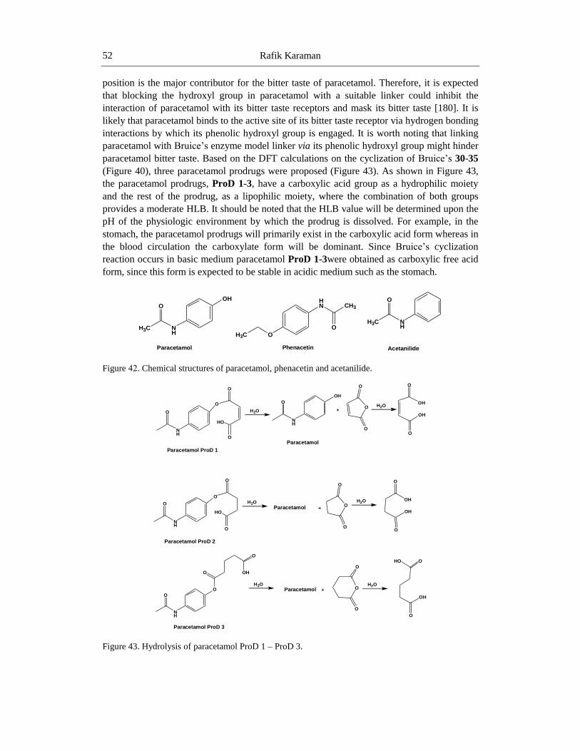
Rafik Karaman 52
position is the major contributor for the bitter taste of paracetamol. Therefore, it is expected
that blocking the hydroxyl group in paracetamol with a suitable linker could inhibit the
interaction of paracetamol with its bitter taste receptors and mask its bitter taste [180]. It is
likely that paracetamol binds to the active site of its bitter taste receptor via hydrogen bonding
interactions by which its phenolic hydroxyl group is engaged. It is worth noting that linking
paracetamol with Bruice‘s enzyme model linker via its phenolic hydroxyl group might hinder
paracetamol bitter taste. Based on the DFT calculations on the cyclization of Bruice‘s 30-35
(Figure 40), three paracetamol prodrugs were proposed (Figure 43). As shown in Figure 43,
the paracetamol prodrugs, ProD 1-3, have a carboxylic acid group as a hydrophilic moiety
and the rest of the prodrug, as a lipophilic moiety, where the combination of both groups
provides a moderate HLB. It should be noted that the HLB value will be determined upon the
pH of the physiologic environment by which the prodrug is dissolved. For example, in the
stomach, the paracetamol prodrugs will primarily exist in the carboxylic acid form whereas in
the blood circulation the carboxylate form will be dominant. Since Bruice‘s cyclization
reaction occurs in basic medium paracetamol ProD 1-3were obtained as carboxylic free acid
form, since this form is expected to be stable in acidic medium such as the stomach.
Figure 24. Chemical structures of paracetamol, phenacetin and acetanilide.
Figure 43. Hydrolysis of paracetamol ProD 1 – ProD 3.
O
NH
OH
H3C
Paracetamol
H3C O
HN CH3
O
Phenacetin
H3C
O
NH
Acetanilide
O
NH
O
O
O
HO
H2O
O
NH
OH
Paracetamol
+
O
O
O H2O
OH
O
O
OH
Paracetamol ProD 1
O
NH
O
O
O
HO
H2O
Paracetamol +
O
O
OH2O
OH
O
O
OH
Paracetamol ProD 2
O
NH
OH2O
Paracetamol +H2O
O
O
OH
Paracetamol ProD 3
O
O
O
O
OHO
OH

Prodrugs Design Based on Inter- and Intramolecular Processes 53
In Vitro Intra-Conversion of Paracetamol
Prodrugs to Their Active Drug, Paracetamol
The hydrolysis of paracetamol ProD 1-ProD 2 was studied in four different media; 1N
HCl, buffer pH 3, and buffer pH 7.4. The prodrug hydrolysis was monitored using HPLC
analysis. At constant pH and temperature the release of paracetamol from its prodrug was
followed and showed a first order kinetics. kobs (h-1
) and t1/2 values for the intraconversion of
paracetamol ProD 1-ProD 2 was calculated from regression equation obtained from plotting
log concentration of residual of the prodrug vs. time. The kinetics results in the different
media are summarized in Table 4 and Figure 44.
Table 4. Observed k and t1/2 values for the intraconversion of paracetamol ProD 1-ProD
2 in 1N HCl and buffers pH 3 and 7.4
Medium
ProD 1
kobs (h-1)
ProD 2
kobs (h-1)
ProD 1
t½ (h)
ProD 2
t½ (h)
1N HCl No reaction No reaction No reaction No reaction
Buffer pH 3 ----------- 6.3 x 10⁻⁵ Very fast 3
Buffer pH 7.4 ------------ 6.1 x 10⁻⁴ Very fast 0.3
As shown in Table 4 the hydrolysis rate of paracetamol ProD 1 at pH 7.4 was the fastest
among all media, followed by pH 3 medium. In 1N HCl no conversion of the prodrug to the
parent dug was observed.
On the other hand, paracetamol ProD 2 underwent fast hydrolysis (within seconds) in pH
7.4 whereas in pH 3.0 and in 1N HCl was entirely stable. The discrepancy in the behavior
between the two prodrugs is due to the fact that the strain energy of maleic anhydride is
higher than that of succinic anhydride. It should be emphasized that the reaction rate in these
processes is determined on the strain energy of the system.
Figure 44. First order hydrolysis plot of paracetamol ProD 1 in (a) buffer pH 3and (b) buffer pH 7.4.

Rafik Karaman 54
Kirby’s Enzyme Model Based on the Acid-
Catalyzed Hydrolysis of N-Alkylmaleamic
Acids [137]
Proton transfer reactions are the most common processes catalyzed by enzymes.
Examples for such catalysis are the proton transfers catalyzed by triose phosphate isomerase
(kcat = 53,000 s-1
) and Δ5-3-ketosteroid isomerase (kcat = 8300 s-1
) which involve weakly
basic and acid groups to achieve such significant rates. Scientists have encouraged exploiting
intramolecularity in modeling enzyme catalysis due to the fact that reactions of an enzyme
active site and substrate are between functional groups held in a close proximity. Both,
enzymes and intramolecularity are similar in that the reacting centers are held together, non-
covalently with the enzymes, and covalently with the intramolecular process. The tremendous
high efficiency of enzymes catalysis depends on a combination of some factors that most of
them have been recognized but none of them was fully understood. Although the devoted
research to the chemistry of enzyme catalysis is growing rapidly a number of several factors
remain to be studied [247-252].
Kirby and coworkers have studied the mechanism of the acid-catalyzed hydrolysis of N-
alkylmaleamic acids 36-42 to maleamic acid derivatives and amines (Figure 45). Their study
revealed that the reaction is remarkably sensitive to the pattern of substitution on the carbon-
carbon double bond and the hydrolysis rates range over more than ten powers of ten, and the
―effective concentration‖ of the carboxyl group of the most reactive amide, dimethyl-N-n-
propylmaleamic acid, is greater than 1010
M. This acid amide was found to be converted into
the more stable dimethylmaleic anhydride with a half-life of less than one second at 39 ˚C
below pH 3 [137]. In addition, the results have showed that the amide bond breakdown is due
to intramolecular nucleophilic catalysis by the adjacent carboxylic acid group. Furthermore,
based on the fact that the tetrahedral intermediate, isomaleimide, was converted quantitatively
into N-methylmaleamic acid (Figure 46), the authors suggested that the rate-limiting step is
the dissociation of the tetrahedral intermediate [137]. Some years later, Kluger and Chin
investigated the intramolecular hydrolysis mechanism of a series of N-alkylmaleamic acids
derived from aliphatic amines having a wide range of basicity [253]. Their study
demonstrated that the identity of the rate-limiting step is a function of both the basicity of the
leaving group and the acidity of the solution. In 1990, based on AM1 semiempirical
calculations, Katagi had concluded that the rate- limiting step is the formation of the
tetrahedral intermediate and not its dissociation [254].
For determining the factors playing dominant role in proton transfer processes we have
computationally studied Kirby‘s intramolecular acid catalyzed hydrolysis of (4-amino- 4-oxo-
2-butenoic) acids (N-alkylmaleamic acids) 36–42.
The goal of our study was to: (a) determine whether the rate-limiting step in 36-42 is the
tetrahedral intermediate formation or collapse, and to assign the driving force(s) responsible
for the extremely high rates for the acid-catalyzed hydrolysis of 37and 40, and (b) determine
the structural features associated with high reactivity in the acid-catalyzed hydrolysis, with
the expectation that similar factors will be operative in enzyme catalysis.

Prodrugs Design Based on Inter- and Intramolecular Processes 55
Figure 45. Chemical structures of N-alkylmalic acids 36-42.
Figure 46. Conversion of isomaleimide to N-methylmaleamic acid.
Using DFT calculation method at B3LYP/6-31G (d,p) level the acid catalyzed hydrolysis
of maleamic (4-amino-4-oxo-2-butenoic) acids (Kirby‘s N-alkylmaleamic acids) 36-42
(Figure 45) were computed. The results confirmed that the reaction proceeds in three steps: (i)
proton transfer from the carboxylic group to the adjacent amide carbonyl carbon followed by,
(ii) nucleophilic attack of the carboxylate anion onto the protonated carbonyl carbon and the
final step of the reaction involves (iii) dissociation of the tetrahedral intermediate to provide
products (Figure 47). In addition, the calculation results demonstrate that the rate-limiting
step is dependent on the reaction medium. In the gas phase the rate-limiting step was the
formation of the tetrahedral intermediate, whereas in the presence of water the dissociation of
the tetrahedral intermediate was the rate-limiting step. Further, when the leaving group
(CH3NH2) in 36-42 was replaced with a group having a low pKa value the rate-limiting step
was the formation of the tetrahedral intermediate, such as in the case where CH3NH2 was
replaced with CF3NH2 (see Figure 47).The results revealed that the efficiency of the
intramolecular acid-catalyzed hydrolysis by the carboxyl group is remarkably sensitive to the
pattern of substitution on the carbon-carbon double bond. The rate of hydrolysis was found to
be linearly correlated with the strain energy of the tetrahedral intermediate or the product
O
NHCH3
O
OH
R1
R2
O
NH2CH3
O
O
R1
R2
pH = 1- 4
36; R1=R2=H37; R1=R2=Me38 ;R1=H; R2=Me39; R1,R2 CycIopent-l-ene-1,2-diyl40; R1, R2 Cyclohex-l-ene-1,2-diyl41; R1=H; R2=Et42; R1=H; R2=n-Propyl
H2O
O
NMe
O
Isomaleimide N-methylmaleamic acid
OH
O
O
NHMe

Rafik Karaman 56
(Table 5). Systems having strained tetrahedral intermediates or products were found to be
with low rates and vice versa [164, 183,185].
Figure 47. Proposed mechanism for the hydrolysis of N-alkylmaleamic acids 30-36.
Table 5. DFT (B3LYP) calculated kinetic and thermodynamic properties for the acid
catalyzed hydrolysis of 36-42
System
∆GbGPffi
(kcal/
mol)
∆GbWffi
(kcal/
mol)
∆GfGPffi
(kcal/
mol)
∆GfWffi
(kcal/
mol) log krel [137]
log EM [137, 255]
(Exp)
log EM
(Calc)
Es (INT2)
(kcal/mol)
Es (GM)
(kcal/mol)
36 28.08 33.06 33.53 26.10 0 7.724 8.52 20.55 10.16
37 16.42 20.05 27.08 17.90 4.371 15.86 18.08 16.16 10.82
38 24.90 28.42 32.57 24.80 1.494 7.742 11.93 17.32 9.40
39 36.77 38.11 45.37 32.16 -4.377 1.255 4.81 27.89 12.30
40 17.41 23.12 26.87 17.89 2.732 15.190 15.82 19.25 9.18
41 23.92 27.28 32.12 23.87 1.516 6.962 12.76 17.59 5.12
42 25.03 27.55 32.3 24.40 1.648 8.568 12.57 18.55 6.20
B3LYP refers to values calculated by B3LYP/6-31G (d, p) method. ∆Gffi
is the calculated
activation free energy (kcal/mol). Es refers to strain energy calculated by Allinger‘s MM2
NHR
O
O
R1
R2
OH
NHR
O
O
R1
R2
OH
NHR
O
O
R1
R2
OH
NHR
O
O
R1
R2
OHRHN
O
O
R1
R2
OH
NHR
O
O
R1
R2
OH
N-Alkylmaleamic acidTS1 INT1
NHR
O
O
R1
R2
OH
TS2 INT2
TS4
H
H
O
O
R1
R2
O
HN
H Me
INT3
Proton Transfer
Intermediate Formation
Intermediate DissociationH
NHR = atenolol, acyclovir, cefuroxime, tranexamic acid or methylR1 and R2; H, methyl or trifluoromethyl
An amine An anhydride
Prodrugs Design Based on Inter- and Intramolecular Processes 57
method. GM and INT2 refer to reactant and intermediate 2, respectively. EM = e -(∆Gffiinter-
∆Gffiintra)/RT. BW and FW refer to tetrahedral intermediate breakdown and tetrahedral
intermediate formation calculated in water, respectively. Exp refers to experimental value.
Calc refers to DFT calculated values.
The linear correlation between the calculated EM values and the experimental EM values
(Table 5) demonstrates the credibility of using DFT methods in predicting energies as well as
rates for reactions of the type described herein [164, 183,185, 186].
Designed Tranexamic Acid Prodrugs Based
On Intramolecular Acid-Catalyzed Hydrolysis
of Kirby’s N-Alkylmaleamic Acids [184]
Tranexamic acid (trans-4 (aminomethyl) cyclohexanecarboxylic acid) is a synthetic
lysine amino acid derivative. It is used to prevent and reduce excessive hemorrhage in
hemophilia patients and reduce the need for replacement therapy during and following tooth
extraction. It is often prescribed for excessive bleeding. The mechanism by which tranexamic
acid exerts its antifibrinolytic activity is by it is competitive inhibition of plasminogen that
prevents the activation of plasminogen to plasmin; plasmin is an enzyme used to degrade
fibrin clot. Tranexamic acid has roughly 8 times the antifibrinolytic activity of an older
analogue, ε-aminocaproic acid. Over the past few years, the use of tranexamic acid has been
expanding beyond the small number of hemophilia patients; it is an important agent in
decreasing mortality rate due to bleeding in trauma patients. It can be used safely in women
whom undergo lower segment cesarean section, in this operation it was found that tranexamic
acid reduces the blood loss during and after surgery, and it is pharmacologically active in
reducing intra-operative using of blood heart surgery, hip and knee replacement surgery and
liver transplant surgery.
Recently, a new oral formulation of tranexamic acid was shown to be safe and effective
for treatment of heavy menstrual bleeding. Oral administration of tranexamic acid results in a
45% oral bioavailability. The total oral dose recommended in women with heavy menstrual
bleeding was two 650 mg tablets three times daily for 5 days. Accumulation following
multiple dosing was reported to be minimal. Post-partum hemorrhage is a leading cause of
maternal mortality, accounting for about 100000 maternal deaths every year. In third world
countries, availability of blood and fluid replacement may be an issue. One approach to
decrease the risk of maternal hemorrhage may be to improve the availability of blood and
fluid replacement. An alternative approach is to decrease the likelihood of maternal
hemorrhage. Furthermore, all the treatment options mentioned above are intended for
intravenous administration; this may not be a viable option in under-developed countries.
Therefore, a cheaper oral alternative may be better suited for such circumstances.
Oral tranexamic acid dosage form was found to be effective and safe in treating malesma,
a hypermelanosis disease that occurs in Asian women. Since tranexamic acid is an amino acid
derivative and undergoes ionization in physiologic environments its oral bioavailability is
expected to be low due to inefficient absorption through membranes. Note the log P (partition
coefficient) for tranexamic acid is -1.6. Hence, there is a necessity to design and synthesis

Rafik Karaman 58
relatively more lipophilic tranexamic acid prodrugs that can provide the parent drug in a
sustained release manner which might result in better clinical outcome, more convenient
dosing regimens and potentially fewer side effects than the original medication. For example,
tranexamic acid is given by continuous IV infusion resulting in peak plasma concentration
following administration. If a slow release prodrug can be prepared, then Cmax related side
effects may be avoided and longer duration exposure may be achieved resulting in potentially
better maintenance paradigm. Improvement of tranexamic acid pharmacokinetic properties
and hence its effectiveness may increase the absorption of the drug via a variety of
administration routes, especially the oral and SC injection routes [256- 261].
Based on DFT calculations for the acid-catalyzed hydrolysis of several N-alkylmaleamic
acid derivatives (Figure 45) four tranexamic acid prodrugs were designed (Figure 48). The
DFT results on the acid-catalyzed hydrolysis demonstrated that the reaction rate-limiting step
is determined on the nature of the amine leaving group. When the amine leaving group was a
primary amine or tranexamic acid moiety, the tetrahedral intermediate collapse was the rate-
limiting step, whereas in the cases where the amine leaving group was aciclovir or
cefuroxime the rate-limiting step was the tetrahedral intermediate formation. Based on the
DFT calculated rates the predicted t1/2 (a time needed for 50% of the prodrug to be converted
into drug) values for tranexamic acid prodrugs ProD 1- ProD 4 (Figure 48)at pH 2 were 556
hours, 253 hours, 70 seconds and 1.7 hours, respectively.
The kinetic study for the acid-catalyzed hydrolysis of tranexamic acid ProD 1was carried
out in aqueous buffer in the same manner as that done by Kirby on Kirby‘s enzyme model 36-
42. This is in order to explore whether the prodrug hydrolyzes in aqueous medium and to
what extent or not, suggesting the fate of the prodrug in the system. Acid-catalyzed hydrolysis
kinetics of the synthesized tranexamic acid ProD 1 was studied in four different aqueous
media: 1 N HCl, buffer pH 2, buffer pH 5 and buffer pH 7.4. Under the experimental
conditions the target compounds hydrolyzed to release the parent drug (Figure 49) as evident
by HPLC analysis. At constant pH and temperature the reaction displayed strict first order
kinetics as the kobs was fairly constant and a straight plot was obtained on plotting log
concentration of residual prodrug verves time. The rate constant (kobs) and the corresponding
half-lives (t1/2) for tranexamic acid prodrug ProD 1 in the different media were calculated
from the linear regression equation correlating the log concentration of the residual prodrug
verses time. The kinetic data are listed in Table 6. The 1N HCl, pH 2 and pH 5 were selected
to examine the interconversion of the tranexamic acid prodrug in pH as of stomach, because
the mean fasting stomach pH of adult is approximately 1-2 and increases up to 5 following
ingestion of food. In addition, buffer pH 5 mimics the beginning small intestine pathway.
Finally, pH 7.4 was selected to examine the interconversion of the tested prodrug in blood
circulation system. Acid-catalyzed hydrolysis of the tranexamic acid ProD 1 was found to be
higher in 1N HCl than at pH 2 and 5 (Figure 49). At 1N HCl the prodrug was hydrolyzed to
release the parent drug in less than one hour. On the other hand, at pH 7.4, the prodrug was
entirely stable and no release of the parent drug was observed. Since the pKa of tranexamic
acid ProD1 is in the range of 3-4, it is expected at pH 5 the anionic form of the prodrug will
be dominant and the percentage of the free acidic form that undergoes the acid-catalyzed
hydrolysis will be relatively low. At 1N HCl and pH 2 most of the prodrug will exist as the
free acid form and at pH 7.4 most of the prodrug will be in the anionic form. Thus, the
difference in rates at the different pH buffers.

Prodrugs Design Based on Inter- and Intramolecular Processes 59
Figure 48. Acid-catalyzed hydrolysis of tranexamic acid prodrugs ProD 1 –ProD 4.
Comparison between the calculated t1/2value (556 h) for tranexamic acid ProD 1 to the
experimental value (23.9 h) indicates that the calculated value is about 23 times larger than
the experimental. This discrepancy between the calculated and the experimental values might
be attributed to the fact that the PCM model (calculations in presence of solvent) is not
capable for handling calculations in acidic aqueous solvent (medium) since the dielectric
constant for pH 2 aqueous solutions is not known. The t1/2 experimental value at pH 5 was
270 hours and at pH 7.4 no interconversion was observed. The lack of the reaction at the
latter pH might be due to the fact that at this pH tranexamic acid ProD 1 exists solely in the
ionized form (pKa about 4). As mentioned before the free acid form is a mandatory
requirement for the reaction to proceed. On the other hand, tranexamic acid ProD 4 has a
higher pKa than tranexamic acid ProD 1 (about 6 vs. 4). Therefore, it is expected that the
interconversion rate of tranexamic acid ProD 4 to its parent drug, tranexamic acid, at all pHs
studied will be higher (log EM for ProD 4 is 14.33 vs. 9.53 for ProD 1). Future study to
achieve desirable tranexamic acid prodrugs capable of releasing tranexamic acid in a
controlled manner and enhancing the parent drug bioavailability is: (i) synthesis of
tranexamic acid ProD 4; (ii) kinetic studies (in vitro) on ProD 4 at pH 6.5 (intestine) and pH
O
O
NH
OH
H
H
COOHO
O
O
H
H
O
O
OH
H
H
OHH2O H2O
ProD 1Tranexamic Acid
O
O
NH
OH
Me
Me
COOHO
O
O
Me
Me
O
O
OH
Me
Me
OHH2O
ProD 2
Tranexamic Acid
O
O
NH
OH
H
Me
COOHO
O
O
H
Me
O
O
OH
H
Me
OHH2O
ProD 3
Tranexamic Acid
O
O
NH
OH
Me
Me
COOHO
O
O
Me
Me
O
O
OH
Me
Me
OHH2O
ProD 4
Tranexamic Acid
Me
Me
Me
Me
Me
Me
COOH
H2N
H2O
H2O
H2O

Rafik Karaman 60
7.4 (blood circulation system) (iii) in vivo pharmacokinetic studies to determine the
bioavailability and the duration of action of the tested prodrug. Furthermore, based on the in
vivo pharmacokinetics characteristics of tranexamic acid ProD 4 new prodrugs may be
designed and synthesized.
Figure 49. First order hydrolysis plots of tranexamic acid ProD1in (a) 1N HCl, (b) buffer of pH 2and
(c) buffer pH 5.
Table 6. The observed k value and t1/2 of tranexamic acid prodrug (ProD 1) in 1N HCl
and at pH 2, 5 and 7.4
Medium kobs (h-1) t½ (h)
1N HCl 5.13 x 10⁻³ 0.9
Buffer pH 2 3.92 x 10⁻⁵ 23.9
Buffer pH 5 3.92 x 10⁻⁶ 270
Buffer pH 7.4 No reaction No reaction

Prodrugs Design Based on Inter- and Intramolecular Processes 61
Conclusion
Unraveling the mechanisms of a number of enzyme models has allowed for the design of
efficient chemical devices having the potential to be utilized as prodrug linkers that can be
covalently attached to commonly used drugs which can chemically, and not enzymatically, be
converted to release the active drugs in a programmable manner. For instance, exploring the
mechanism for a proton transfer in Kirby‘s N-alkylmaleamic acids (enzyme model) has led to
the design of a number of prodrugs such as tranexamic acid for bleeding conditions, acyclovir
as antiviral drug for the treatment for herpes simplex [185], atenolol for treating hypertension
with enhanced stability and bioavailability, and lacks a bitter sensation [186, 187]. In
addition, prodrugs for masking the bitter sensation of paracetamol and some antibacterial
drugs, such as cefuroxime, amoxicillin and cephalexin were also designed and synthesized
[183]. The role of the linkers in atenolol, paracetamol and the antibacterial prodrugs is to
block the free hydroxyl or amine, which is responsible for the drug bitterness, and to enable
the release of the drug in a controlled manner. Menger‘s Kemp acid enzyme model was
utilized for the design of dopamine prodrugs for the treatment for Parkinson‘s disease [188].
Prodrugs of dimethyl fumarate for the treatment psoriasis was also designed, synthesized and
studied [262]. Furthermore, unraveling the mechanism of Kirby‘s acetals has led to the design
and synthesis of novel prodrugs of aza-nucleosides for the treatment for myelodysplastic
syndromes [177], atovaquone prodrugs for the treatment for malaria [178, 179], less bitter
paracetamol prodrugs to be administered to children and elderly as antipyretic and pain killer
[180], statin prodrugs [181] and prodrugs of phenylephrine as decongestant [182]. In these
examples, the prodrug moiety was linked to the hydroxyl group of the active drug such that
the drug-linker moiety (prodrug) has the potential to interconvert when exposed into
physiological environments such as stomach, intestine, and/or blood circulation, with rates
that are solely dependent on the structural features of the pharmacologically inactive
promoiety (Kirby‘s enzyme model). Further details on this approach could be found in
references [263-269].
Acknowledgments
The author would like to acknowledge funding by the German Research Foundation
(DFG, ME 1024/8-1).
References
[1] DiMasi, J.A.; Hansen, R.W. & Grabowski, H.G. (2003) The price of innovation: new
estimates of drug development costs. J. Health Econ. 22, 151–185.
[2] DiMasi, J.A. (2002) The value of improving the productivity of the drug development
process: faster times and better decisions. Pharmacoeconomics20, 1–10.
[3] Huttunen, K.M.; Raunio, H. & Rautio, J. (2011) Prodrugs from serendipity to rational
design. Pharmacol. Rev. 63, 750–771.
Rafik Karaman 62
[4] Stella, V.J. & Nti-Addae, K.W. (2007) Prodrug strategies to overcome poor water
solubility. Adv. Drug Deliv. Rev. 59, 677–694.
[5] Dahan, A.; Khamis, M.; Agbaria, R. & Karaman, R. (2012) Targeted prodrugs in oral
delivery: the modern molecular biopharmaceutical approach. Expert Opinion on Drug
Delivery9, 1001–1013.
[6] Karaman, R.; Fattash B. & Qtait A. (2013) The future of prodrugs – design by quantum
mechanics methods. Expert Opinion on Drug Delivery10, 713–729.
[7] Karaman, R. (2013) Prodrugs design based on inter- and intramolecular processes.
Chem. Biol. Drug. Des. 82, 643–668.
[8] Gonzalez, F.J. & Tukey, R.H. (2006) Drug metabolism. In: Brunton, L.L., Lazo, J.S. &
Parker, K.L., editors. Goodman & Gilman‘s The Pharmacological Basis of
Therapeutics. New York: The McGraw-Hill Companies, Inc.; p. 71–91.
[9] Testa, B. & Kramer, S.D. (2007) The biochemistry of drug metabolism–an
introduction: part 2. Redox reactions and their enzymes. Chem. Biodivers. 4, 257–405.
[10] Gangwar, S.; Pauletti, G.M.; Wang, B.; Siahaan, T.J.; Stella, V.J. & Borchardt, R.T.
(1997) Prodrug strategies to enhance the intestinal absorption of peptides. DDT2, 148–
155.
[11] Wang W.; Jiang J.; Ballard C.E. & Wang B. (1999) Prodrug approaches to the
improved delivery of peptide drugs. Curr. Pharm. Des. 5, 265–287.
[12] Chan, O.H. & Stewart, B.H. (1996) Physicochemical and drug-delivery considerations
for oral drug bioavailability. Drug. Discov. Today1, 461–473.
[13] Ohlstein, E. H.; Ruffolo, R. R. Jr. & Elliott, J. D. (2000) Drug discovery in the next
millennium. Annu. Rev. Pharmacol.Toxicol.40, 177-91.
[14] Stella, V.J. (2010) Prodrugs: some thoughts and current issues. J. Pharm. Sci. 99(12),
4755–4765.
[15] Muller, C.E. (2009) Prodrug approaches for enhancing the bioavailability of drugs with
low solubility. Chem. Biodivers. 6, 2071–2083.
[16] Tunek, A.; Levin, E. & Svensson, L.A. (1988) Hydrolysis of 3H-bambuterol, a
carbamate prodrug of terbutaline, in blood from humans and laboratory animals in
vitro. Biochem. Pharmacol. 37, 3867–3876.
[17] Browne, T.R.; Kugler, A.R. & Eldon, M.A. (1996) Pharmacology and
pharmacokinetics of fosphenytoin. Neurology 46, S3–S7.
[18] Wolff, M.E. (editor) (1995) Medicinal Chemistry and Drug Discovery, 5th edn. New
York: John Wiley & Sons.
[19] Williams, D.A., Foye, W.O. & Lemke, T.L. editors. (2002) Foye‘s Principles of
Medicinal Chemistry. Baltimore, MD: Wolters Kluwer Health; p. 26–53.
[20] Kenny, B.A.; Bushfield M.; Parry-Smith D.J.; Fogarty S. & Trehene M. (1998) The
application of high throughput screening to novel lead discovery, Prog. Drug Res 41,
246–269.
[21] Blundell, T. (1996) Structure-based drug design. Nature384, 23–26.
[22] Williams, M. (1993) Strategies for drug discovery. NIDA Res.Monogr.132, 1–22.
[23] Beaumont, K.; Webster, R.; Gardner, I. & Dack, K. (2003) Design of ester prodrugs to
enhance oral absorption of poorly permeable compounds: challenges to the discovery
scientist. Curr. Drug Metab.4, 461–485.

Prodrugs Design Based on Inter- and Intramolecular Processes 63
[24] Bellott, R.; Le Morvan, V.; Charasson, V.; Laurand, A.; Colotte, M.; Zanger, U.M. &
Robert, J. (2008) Functional study of the 830C >G polymorphism of the human
carboxylesterase 2 gene. Cancer Chemother Pharmacol 61, 481–488.
[25] Reddy, K.R.; Matelich, M.C.; Ugarkar, B.G.; Gomez- Galeno, J.E.; DaRe, J.; Ollis, K.
& Erion, M.D. (2008) Pradefovir: a prodrug that targets adefovir to the liver for the
treatment of hepatitis B. J. Med. Chem. 51, 666–676.
[26] Kumpulainen, H.; Mahonen, N.; Laitinen, M.L.; Jaurakkaj, J.; Jarvinen, T. & Rautio, J.
(2006) Evaluation of hydroxyimine as cytochrome P450 selective prodrug structure. J.
Med. Chem. 49, 1207–1211.
[27] Saunders, M.P.; Patterson, A.V.; Chinje, E.C.; Harris, A.L. & Stratford, I.J. (2000)
NADPH: cytochrome c (P450) reductase activates tirapazamine (SR4233) to restore
hypoxicandoxic cytotoxicity in anaerobic resistant derivative of the A549 lung cancer
cell line. Br. J. Cancer82, 651–656.
[28] Dobesh, P.P. (2009) Pharmacokinetics and pharmacodynamics of prasugrel, a
thienopyridine P2Y12 inhibitor. Pharmacotherapy29, 1089–1102.
[29] Pereillo, J.M.; Maftouh, M.; Andrieu, A.; Uzabiaga, M.F.; Fedeli, O.; Savi, P. & Picard,
C. (2002) Structure and stereochemistry of the active metabolite of clopidogrel. Drug
Metab. Dispos. 30, 1288–1295.
[30] Svensson, L.A. & Tunek, A. (1988) The design and bioactivation of presystemically
stable prodrugs. Drug Metab. Rev. 19, 165–194.
[31] Stella, V.J. (2007) A case for prodrugs. In: Stella, V.J.; Borchardt, R.T.; Hageman,
M.J.; Oliyai, R.; Magg, H. & Tilley, J.W., editors. Prodrugs: Challenges and Rewards.
Part 1. New York, NY: AAPS Press/Springer; p. 3–33.
[32] Albert, A. (1958) Chemical aspects of selective toxicity. Nature182, 421–422.
[33] Harper, N.J. (1959) Drug latentiation. J. Med. Pharm. Chem. 1, 467–500.
[34] Harper, N.J. (1962) Drug latentiation. Prog. Drug Res. 4, 221–294.
[35] Sinkula, A.A. & Yalkowsky, S.H. (1975) Rationale for design of biologically reversible
drug derivatives: prodrugs. J. Pharm. Sci. 64, 181–210.
[36] Schrama, D,; Reisfeld, R.A. & Becker, J.C. (2006) Antibody targeted drugs as cancer
therapeutics. Nature reviews Drug discovery 5(2), 147-59.
[37] Mahato, R.; Tai, W. & Cheng, K. (2011) Prodrugs for improving tumor targetability
and efficiency.Advanced drug delivery reviews 63(8), 659-70.
[38] Singh, Y.; Palombo, M. & Sinko, P.J. (2008) Recent trends in targeted anticancer
prodrug and conjugatedesign. Current medicinal chemistry. 15(18), 1802-26.
[39] Niculescu-Duvaz, I.; Niculescu-Duvaz, D. & Friedlos, F. (1999) Self-immolative
anthracycline prodrugs for suicide gene therapy. Journal of clinical investigation. 13,
2485-2489.
[40] Stella, V.J.; Borchardt, R.T.; Hageman, M.J.; Oliyai, R.; Maag, H. & Tilley, J.W.
(2007) Prodrugs: Challenges and Rewards Part 1 and 2, Springer Science + Business
Media, New York.
[41] Stella, V.J.; Charman, W.N. & Naringrekar, V.H. (1985) Prodrugs. Do they have
advantages in clinical practice? Drugs 29, 455-473.
[42] Banerjee, P.K. & Amidon, G.L. (1985) Design of prodrugs based on enzymes-substrate
specificity. In: Bundgaard, H., ed. Design of Prodrugs. New York: Elsevier; 93-133.
Rafik Karaman 64
[43] Haffen, E.; Paintaud, G.; Berard, M.; Masuyer, C.; Bechtel, Y. & Bechtel, P.R. (2000).
On the assessment of drug metabolism by assays of codeine and its main metabolites.
Therapeutic Drug Monitoring 22 (3), 258–65.
[44] Raffa, R.B. (1996). A novel approach to the pharmacology of analgesics. The American
Journal of Medicine 101 (1A), 40S–46S.
[45] Zhou, S. F.; Zhou, Z. W.; Yang, L. P. & Cai, J. P. (2009). Substrates, inducers,
inhibitors and structure-activity relationships of human Cytochrome P450 2C9 and
implications in drug development. Current medicinal chemistry 16(27), 3480-3675.
[46] Huttunen, K.M.; Raunio, H. & Rautio, J. (2011) Prodrugs -- from serendipity to rational
design. Pharmacol Rev. 63, 750-71
[47] Higuchi, T. & Stella, V. J. (1975). Pro-drugs as novel drug delivery systems.
[48] Di, L. & Kerns, E.H. (2007) Solubility issues in early discovery and HTS, in Solvent
Systems and Their Selection in Pharmaceutics and Biopharmaceutics (Augustijins, P. &
Brewster, M. eds) pp 111–136, Springer Science + Business Media, New York.
[49] Fleisher, D.; Bong, R. & Stewart, B.H. (1996) Improved oral drug delivery: solubility
limitations overcome by the use of prodrugs. Adv Drug Deliv Rev 19, 115–130.
[50] Beaumont, K.; Webster, R.; Gardner, I. & Dack, K. (2003) Design of ester prodrugs to
enhance oral absorption of poorly permeable compounds: challenges to the discovery
scientist. Curr. Drug Metab. 4, 461–485.
[51] del Amo, E.M.; Urtti, A. & Yliperttula, M. (2008) Pharmacokinetic role of L-type
amino acid transporters LAT1 and LAT2. Eur J Pharm Sci 35, 161–174.
[52] Khor, S.P. & Hsu, A. (2007) The pharmacokinetics and pharmacodynamics of levodopa
in the treatment of Parkinson's disease. Curr. Clin. Pharmacol. 2, 234–243.
[53] Hornykiewicz, O. (2010) A brief history of levodopa. J. Neurol. 257 (Suppl 2), S249–
S252.
[54] Svensson, L.A. & Tunek, A. (1988) The design and bioactivation of presystemically
stable prodrugs. Drug Metab. Rev. 19, 165–194.
[55] Tunek, A.; Levin, E. & Svensson, L.A. (1988) Hydrolysis of 3H-bambuterol, a
carbamate prodrug of terbutaline, in blood from humans and laboratory animals in
vitro. Biochem. Pharmacol. 37, 3867–3876.
[56] Sitar, D.S. (1996) Clinical pharmacokinetics of bambuterol. Clin.Pharmacokinet.31,
246–256.
[57] Persson, G.; Pahlm, O. & Gnosspelius, Y. (1995) Oral bambuterol versus terbutaline in
patients with asthma. Curr. Ther. Res. 56, 457–465.
[58] Ettmayer, P.; Amidon, G.L.; Clement, B. & Testa, B. (2004) Lessons learned from
marketed and investigational prodrugs. J. Med. Chem. 47, 2393–2404.
[59] Denny, W.A. (2010) Hypoxia-activated prodrugs in cancer therapy: progress to the
clinic. Future Oncol 6, 419–428.
[60] Shanghag, A.; Yam, N. & Jasti, B. (2006) Prodrugs as drug delivery systems, in Design
of Controlled Release Drug Delivery Systems (Li, X. & Jasti, B.R. eds) pp75–106, The
McGraw-Hill Company, Inc., New York.
[61] Gonzalez, F.J. & Tukey, R.H. (2006) Drug metabolism, in Goodman and Gilman's The
Pharmacological Basis of Therapeutics (Brunton, L.L.;, Lazo, J.S. & Parker, K.L. eds)
pp 71–91, The McGraw-Hill Companies, Inc., New York.
[62] Testa, B. & Krämer, S.D. (2007) The biochemistry of drug metabolism–an
introduction: Part 2. Redox reactions and their enzymes. Chem. Biodivers. 4, 257–405.

Prodrugs Design Based on Inter- and Intramolecular Processes 65
[63] Draganov, D.I. & La Du, B.N. (2004). Pharmacogenetics of paraoxonases: A brief
review. Naunyn Schmiedebergs Arch Pharmacol 369, 78–88.
[64] Ngawhirunpat, T.; Kawakami, N.; Hatanaka, T.; Kawakami, J. & Adachi, I. (2003).
Age dependency of esterase activity in rat and human keratinocytes. Biol. Pharm. Bull.
26, 1311–1314.
[65] Moser, V.C.; Chanda, S.M.; Mortensen, S.R. & Padilla, S. (1998). Age- and gender-
related differences in sensitivity to chlorpyrifos in the rat reflect developmental profiles
of esterase activities. Toxicol. Sci. 46, 211–222.
[66] Stella, V.J. (1975) Pro-drugs: an overview and definition. In: Higuchi, T. & Stella, V.,
editors. Prodrugs as Novel Drug Delivery Systems. ACS Symposium Series.
Washington, DC: American Chemical Society; p. 1–115.
[67] Amidon, G.L.; Leesman, G.D. & Elliott, R.L. (1980) Improving intestinal absorption of
water-insoluble compounds: a membrane metabolism strategy. J. Pharm. Sci. 69, 1363–
1368.
[68] Fleisher, D.; Stewart, B.H. & Amidon, G.L. (1985) Design of prodrugs for improved
gastrointestinal absorption by intestinal enzyme targeting. Methods Enzymol. 112, 360–
381.
[69] Bai, J.P. & Amidon, G.L. (1992) Structural specificity of mucosal-cell transport and
metabolism of peptide drugs: implication for oral peptide drug delivery. Pharm. Res. 9,
969–978.
[70] Stella, V.J. & Himmelstein, K.J. (1980) Prodrugs and site-specific drug delivery. J.
Med. Chem. 23, 1275–1282.
[71] Stella, V.J. & Himmelstein, K.J. (1982) Critique of prodrugs and site specific delivery.
In: Bundgaard, H., editor. Optimization of Drug Delivery. Alfred Benzon Symposium
Copenhagen: Munksgaard; p. 134–155.
[72] Friend, D.R. & Chang, G.W. (1984) A colon-specific drug-delivery system based on
drug glycosides and the glycosidases of colonic bacteria. J. Med. Chem. 27, 261–266.
[73] Philpott, G.W.; Shearer, W.T.; Bower, R.J. & Parker, C.W. (1973) Selective
cytotoxicity of hapten-substituted cells with an antibody-enzyme conjugate. J.
Immunol. 111, 921–929.
[74] Deonarain, M.P.; Spooner, R.A. & Epenetos, A.A. (1996) Genetic delivery of enzymes
for cancer therapy. Gene Ther. 2, 235–244.
[75] Singhal, S. & Kaiser, L.R. (1998) Cancer chemotherapy using suicide genes. Surg.
Oncol. Clin. North Am. 7, 505–536.
[76] Aghi, M.; Hochberg, F. & Breakefield, X.O. (2000) Prodrug activation enzymes in
cancer gene therapy. J. Gene Med. 2, 148–164.
[77] Greco, O. & Dachs, G.U. (2001) Gene directed enzyme/ prodrug therapy of cancer:
historical appraisal and future prospectives. J. Cell. Physiol. 187, 22–36.
[78] Roche, E.B. Design of Biopharmaceutical Properties through Prodrugs and Analogs.
Washington, DC: American Pharmaceutical Association; 1977.
[79] Li, F.; Maag, H. & Alfredson, T. (2008) Prodrugs of nucleoside analogues for improved
oral absorption and tissue targeting. J. Pharm. Sci. 97, 1109–1134.
[80] Tammara, V.K.; Narurkar, M.M.; Crider, A.M. & Khan, M.A. (1994) Morpholinoalkyl
ester prodrugs of diclofenac: synthesis, in vitro and in vivo evaluation. J. Pharm. Sci.
83, 644–648.

Rafik Karaman 66
[81] del Amo, E.M.; Urtti, A. & Yliperttula, M. (2008) Pharmacokinetic role of L-type
amino acid transporters LAT1 and LAT2. Eur. J. Pharm. Sci. 35, 161–174.
[82] Kim, I.; Song, X.; Vig, B.S.; Mittal, S.; Shin, H.C.; Lorenzi, P.J. & Amidon, G.L.
(2004) A novel nucleoside prodrug activating enzyme: substrate specificity of biphenyl
hydrolase-like protein. Mol. Pharm. 1, 117–127.
[83] Hu, L. (2004) Prodrugs: effective solutions for solubility, permeability and targeting
challenges. Drugs 7, 736– 742.
[84] Kim, I.; Chu, X.Y.; Kim, S.; Provoda, C.J.; Lee, K.D. & Amidon, G.L. (2003)
Identification of a human valacyclovirase: biphenyl hydrolase-like protein as
valacyclovir hydrolase. J. Biol. Chem. 278, 25348–25356.
[85] Bronson, J.J.; HO, H.T.; Boeck, H.D.; Woods, K.; Ghazzouli, I.; Martin, J.C. &
Hitchcock, M.J. (1990) Biochemical pharmacology of acyclic nucleotide analogues.
Ann. N. Y. Acad. Sci. 616, 398–407.
[86] Cundy, K.C.; Barditch-Crovo, P.; Walker, R.E.; Collier, A.C.; Ebeling, D.; Toole, J. &
Jaffe, H.S. (1995) Clinical pharmacokinetics of adefovir in human immunodeficiency
virus type 1-infected patients. Antimicrob. Agents Chemother. 39, 2401–2405.
[87] Cioli, V.; Putzolu, S.; Rossi, V. & Corradino, C. (1980) A toxicological and
pharmacological study of ibuprofen guaiacol ester (AF 2259) in the rat. Toxicol. Appl.
Pharmacol. 54, 332–339.
[88] Croft, D.N.; Cuddigan, J.H. & Sweetland, C. (1972) Gastric bleeding and benorylate, a
new aspirin. Br. Med. J. 3, 545–547.
[89] Bhosle, D.; Bharambe, S.; Gairola, N. & Dhaneshwar, S.S. (2006) Mutual prodrug
concept: fundamentals and applications. Indian J. Pharm. Sci. 68, 286–294.
[90] Muscara, M.N.; McKnight, W.; Soldato, P.D. & Wallace, J.L. (1998) Effect of a nitric
oxide-releasing naproxen derivative on hypertension and gastric damage induced by
chronic nitric oxide inhibition in the rat. Life Sci. 62, PL235–PL240.
[91] Burgaud, J.L.; Riffaud, J.P. & Del Soldato, P. (2002) Nitric-oxide releasing molecules:
a new class of drugs with several major indications. Curr. Pharm. Des.8, 201–213.
[92] Shanbhag, V.R.; Crider, A.M.; Gokhale, R.; Harpalani, A. & Dick, R.M. (1992) Ester
and amide prodrugs of ibuprofen and naproxen: synthesis, anti-inflammatory activity,
and gastrointestinal toxicity. J. Pharm. Sci. 81, 149–154.
[93] English, A.R.; Girard, D. & Haskell, S.L. (1984) Pharmacokinetics of sultamicillin in
mice, rats, and dogs. Antimicrob. Agents Chemother.25, 599–602.
[94] Jordan, A.M.; Khan, T.H.; Malkin, H. & Osborn, H.M.I. (2002) Synthesis and analysis
of urea and carbamate prodrugs as candidates for melanocyte-directed enzyme prodrug
therapy (MDEPT). Bioorg. Med. Chem. 10, 2625–2633.
[95] Yumibe, N.; Hule, K.; Chen, K.-J.; Snow, M.; Clement, R.P. & Cayen, M.N. (1996)
Identification of human liver cytochrome P450 enzymes that metabolize the
nonsedating antihistamine loratadine. Formation of descarboethoxyloratadine by
CYP3A4 and CYP2D6. Biochem. Pharmacol. 51, 165–172.
[96] Shimma, N.; Umeda, I.; Arasaki, M.; Murasaki, C.; Masubuchi, K.; Kohchi, Y.; Miwa,
M.; Ura, M.; Sawada, N.; Tahara, H.; Kuruma, I.; Horii, I. & Ishitsuka, H. (2000)
Thedesign and synthesis of a new tumor-selective fluoropyrimidine carbamate,
capecitabine. Bioorg. Med. Chem. 8, 1697–1706.
[97] Ajani, J. (2006) Review of capecitabine as oral treatment of gastric, gastroesophageal,
and esophagealcancers. Cancer 107, 221–231.

Prodrugs Design Based on Inter- and Intramolecular Processes 67
[98] Alexander, J.; Carqill, R.; Michelson, S.R. & Schwam, H. (1988) (Acyloxy) alkyl
carbamates as novel bioreversible prodrugs for amines: increased permeation through
biological membranes. J. Med. Chem. 31, 318–322.
[99] Venhuis, B.J.; Dijkstra, D.; Wustrow, D.; Meltzer, L.T.; Wise, L.D.; Johnson, S.J. &
Wikstrom, H.V. (2003) Orally active oxime derivatives of the dopaminergic prodrug6-
(N,N-di-n-propylamino)-3,4,5,6,7,8-hexahydro-2Hnaphthalen-1-one. Synthesis and
pharmacological activity. J. Med. Chem. 46, 4136–4140.
[100] Bundgaard, H. (Editor) (1985). Design of prodrugs. Elsevier Publishing Company.
[101] Safadi, M.; Oliyai, R. & Stella, V.J. (1993) Phosphoryloxymethylcarbamates and
carbonates–novel water-soluble prodrugs for amines and hindered alcohols. Pharm.
Res. 10, 1350–1355.
[102] Hecker, S.J.; Calkins, T.; Price, M.E.; Huie, K.; Chen, S.; Glinka, T.W. & Dudley,
M.N. (2003) Prodrugs of cephalosporin RWJ-333441 (MC-04,546) with improved
aqueous solubility. Antimicrob. Agents Chemother. 47, 2043–2046.
[103] Venhuis, B.J.; Dijkstra, D.; Wustrow, D.; Meltzer, L.T.; Wise, L.D.; Johnson, S.J. &
Wikstrom, H.V. (2003) Orally active oxime derivatives of the dopaminergic prodrug 6-
(N,N-di-n-propylamino)-3,4,5,6,7,8-hexahydro-2Hnaphthalen- 1-one. Synthesis and
pharmacological activity. J. Med. Chem. 46, 4136–4140.
[104] Madsen, U.; Krogsgaard-Larsen, P. & Liljefors, T. (2002) Textbook of Drug Design
and Discovery. Washington, DC: Taylor & Francis; p. 410–458.
[105] Testa, B. & Mayer, J.M. (2003) Hydrolysis in Drug and Prodrug metabolism,
Chemistry, biochemistry and enzymology 690–695.
[106] Bundgaard, H. & Johansen, M. (1980) Prodrugs as drug delivery systems IV: N-
Mannich bases as potential novel prodrugs for amides, ureides, amines, and other NH-
acidic compounds. J. Pharm. Sci. 69, 44–46.
[107] Caldwell, H.C.; Adams, H.J.; Jones, R.G.; Mann, W.A.; Dittert, L.W.; Chong, C.W. &
Swintosky, J.V. (1971) Enamine prodrugs. J. Pharm. Sci. 60, 1810–1812.
[108] Murakami, T.; Tamauchi, H.; Yamazaki, M.; Kubo, K.; Kamada, A. & Yata, N. (1981)
Biopharmaceutical study on the oral and rectal administrations of enamine prodrugs of
amino acid-like beta-lactam antibiotics in rabbits. Chem. Pharm. Bull. 29, 1986–1997.
[109] Naringrekar, V.H. & Stella, V.J. (1990) Mechanism of hydrolysis and structure-stability
relationship of enaminones as potential prodrugs of model primary amines. J. Pharm.
Sci. 79, 138–146.
[110] Chapman, T.M.; Plosker, G.L. & Perry, C.M. (2004) Fosamprenavir: a review of its use
in the management of antiretroviral therapy-naive patients with HIV infection.
Drugs64, 2101–2124.
[111] Boucher, B.A. (1996) Fosphenytoin: a novel phenytoin prodrug. Pharmacotherapy16,
777–791.
[112] Azadkhan, A.K.; Truelove, S.C. & Aronson, J.K. (1982) The disposition and
metabolism of sulphasalazine (salicylazosulphapyridine) in man. Br. J. Clin.
Pharmacol. 13, 523–528.
[113] Sandborn, W.J. (2002) Rational selection of oral 5-aminosalicylate formulations and
prodrugs for the treatment of ulcerative colitis. Am. J. Gastroenterol. 97, 2939–2941.
[114] Jung, Y.; Lee, J. & Kim, Y. (2001) Colon-specific prodrug of 5-aminosalicylic acid:
synthesis and in vitro/in vivo properties of acidic amino acid derivatives of 5-
aminosalicylic acid. J. Pharm. Sci. 90, 1767–1775.

Rafik Karaman 68
[115] Simplicio, A.L.; Clancy, J.M. & Gilmer, J.F. (2008) Prodrugs for amines. Molecules 13,
519–547.
[116] Greenwald, R.B.; Pendri, A.; Conover, C.D.; Zhao, H.; Choe, Y.H.; Martinez, A.;
Shum, K. & Guan, S. (1999) Drug delivery systems employing 1,4- or 1,6-elimination:
poly(ethylene glycol) prodrugs of amine-containing compounds. J. Med. Chem. 42,
3657–3667.
[117] Greenwald, R.B.; Choe, Y.H.; McGuire, J. & Conover, C.D. (2003) Effective drug
delivery by PEGylated drug conjugates. Adv. Drug Deliv. Rev. 55, 217–250.
[118] Hanson, K.R. & Havir, E.A. (1972) The enzymic elimination of ammonia. In: Boyer,
P.D., editor. The Enzymes, 3rd edn. vol. 7. New York: Academic Press; p 75– 166.
[119] Czarnik, A.W. (1988) Intramolecularity: proximity and strain. In: Liebman, J.F. &
Greenberg, A., editors. Mechanistic Principles of Enzyme Activity. New York, NY:
VCH Publishers; p. 75–117.
[120] Sweigers, G.F. (2008) Mechanical Catalysis. Hoboken, NJ: John Wiley & Sons.
[121] Fersht, A. (1979) Structure and Mechanism in Protein Science: A guide to Enzyme
Catalysis and Protein Folding. New York: Freeman, W. H. and Company.
[122] Nelson, D.L. & Cox, M.M. (2003) Lehninger Principles of Biochemistry. New York:
Worth Publishers.
[123] Dafforn, A. & Koshland, D.E. Jr (1973) Proximity, entropy and orbital steering.
Biochem. Biophys. Res. Commun. 52, 779–785.
[124] Lightstone, F. C. & Bruice, T.C. (1997). Separation of ground state and transition state
effects in intramolecular and enzymatic reactions. 2. A theoretical study of the
formation of transition states in cyclic anhydride formation. J. Am. Chem. Soc. 119,
9103-9113.
[125] Bruice, T. C. & Pandit, U. K. (1960) The effect of geminal substitution ring size and
rotamer distribution on the intra molecular nucleophilic catalysis of the hydrolysis of
monophenyl esters of dibasic acids and the solvolysis of the intermediate anhydrides. J.
Am. Chem. Soc. 82, 5858–5865.
[126] Bruice, T. C. & Pandit, U. K. (1960) Intramolecular models depicting the kinetic
importance of ‗‗Fit‘‘ in enzymatic catalysis. Proc. Natl. Acad. Sci. USA 46, 402–404.
[127] Milstein, S. & Cohen, L.A. (1970) Concurrent general-acid and general-base catalysis
of esterification. J. Am. Chem. Soc. 92, 4377-4382.
[128] Milstein, S. & Cohen, L. A. (1970) Rate acceleration by stereopopulation control:
models for enzyme action. Proc. Natl. Acad. Sci. USA 67, 1143-1147.
[129] Milstein, S. & Cohen, L. A. (1972) Stereopopulation control I. Rate enhancement in the
lactonizations of o-hydroxyhydrocinnamic acids. J. Am. Chem. Soc. 94, 9158-9165.
[130] Menger, F. M. & Ladika M. (1988) Fast hydrolysis of an aliphatic amide at neutral pH
and ambient temperature. A peptidase model. J. Am. Chem. Soc. 110, 6794-6796.
[131] Menger, F. M. (1985) On the source of intramolecular and enzymatic reactivity. Acc.
Chem. Res. 18, 128-134.
[132] Menger, F. M.; Chow, J. F.; Kaiserman H. & Vasquez P. C. (1983) Directionality of
proton transfer in solution. Three systems of known angularity. J. Am. Chem. Soc. 105:
4996-5002.
[133] Menger, F. M.; Galloway, A. L. & Musaev D. G. (2003) Relationship between rate and
distance. Chem. Commun. 2370-2371.

Prodrugs Design Based on Inter- and Intramolecular Processes 69
[134] Menger, F. M. (2005).An alternative view of enzyme catalysis. Pure Appl. Chem. 77,
1873–187.
[135] Kirby, A. J. & Hollfelder, F. From Enzyme Models to Model Enzymes, RSC Publishing,
Cambridge UK, 1-273, 2009.
[136] Barber, S. E.; Dean, K. E. S. & Kirby, A. J. (1999) A mechanism for efficient proton-
transfer catalysis. Intramolecular general acid catalysis of the hydrolysis of 1-arylethyl
ethers of salicylic acid. Can. J. Chem.77, 792-801.
[137] Kirby, A. J. & Lancaster, P. W. (1972) structure and efficiency in intramolecular and
enzymatic catalysis. Catalysis of amide hydrolysis by the carboxy-group of substituted
maleamic acids. . J. Chem. Soc., Perkin Trans. 2, 1206-1214.
[138] Kirby, A. J.; de Silva, M. F.; Lima, D.; Roussev, C. D. & Nome, F. (2006) Efficient
intramolecular general acid catalysis of nucleophilic attack on a phosphodiester. J. Am.
Chem. Soc.128, 16944-16952.
[139] Kirby, A. J. & Williams, N. H. (1994) Efficient intramolecular general acid catalysis of
enol ether hydrolysis. Hydrogen-bonding stabilization of the transition state for proton
transfer to carbon. J. Chem. Soc., Perkin Trans. 2, 643-648.
[140] Kirby, A. J. & Williams, N. H. (1991) Efficient intramolecular general acid catalysis of
vinyl ether hydrolysis by the neighbouring carboxylic acid group. J. Chem. Soc. Chem.
Commun 1643-1644.
[141] Kirby, A. J. (1996) Enzyme Mechanisms, Models, and Mimics. Angewandte Chemie
International Edition in English 35, 706-724.
[142] Fife, T. H. & Przystas, T. J. (1979) Intramolecular general acid catalysis in the
hydrolysis of acetals with aliphatic alcohol leaving groups. J. Am. Chem. Soc.101,
1202-1210.
[143] Kirby, A. J. (1997) Efficiency of proton transfer catalysis in models and enzymes. Acc.
Chem. Res.30:290-296.
[144] Reddy, M. R. & Erion, M. D. Free Energy Calculations in Rational Drug Design,
Kluwer Academic/Plenum Publishers, 379, 2001.
[145] Parr, R. G.; Craig, D. P. & Ross, I. G. (1950) Molecular Orbital Calculations of the
Lower Excited Electronic Levels of Benzene, Configuration Interaction included.
Journal of Chemical Physics 18 (12), 1561–1563.
[146] Parr, R. G. (1990) On the genesis of a theory. Int. J. Quantum Chem 37 (4), 327–347.
[147] Dewar, M. J. S. & Thiel, W. (1977) Ground states of molecules. The MNDO method.
Approximations and parameters. J. Am. Chem. Soc. 99, 4899-4907.
[148] Bingham, R. C.; Dewar, M. J. S. & Lo, D. H. (1975) Ground states of molecules. XXV.
MINDO/3. An improved version of the MNDO semiempirical SCF-MO method. J. Am.
Chem. Soc. 97, 1285-1293.
[149] Dewar, M. J. S.; Zoebisch, E. G.; Healy, E. F. & Stewart, J. J. P. (1985) AM1: A new
general purpose quantum mechanical molecular model. J. Am. Chem. Soc. 107, 3902-
907.
[150] Dewar, M. J. S.; Jie, C. & Yu, J. (1993) The first of new series of general purpose
quantum mechanical molecular models. Tetrahedron 49, 5003-5038.
[151] Parr, R. G. & Yang, W. Density Functional Theory of Atoms and Molecules. Oxford
University Press, Oxford, 1989.
[152] Mourik, T. van & Robert, J.G. (2002). A critical note on density functional theory
studies on rare-gas dimers. Journal of Chemical Physics 116 (22), 9620–9623.

Rafik Karaman 70
[153] Grimme, S. (2004).Accurate description of van der Waals complexes by density
functional theory including empirical corrections. Journal of Computational Chemistry
25 (12), 1463–1473. doi:10.1002/jcc.20078. PMID 15224390.
[154] Burker, U. & Allinger, N. L. Molecular Mechanics, American Chemical Society,
Washington, DC, USA, 1982.
[155] Warshel, A. & Levitt, M. (1976) Theoretical studies of enzymatic reactions: dielectric,
electrostatic and steric stabilization of the carbonium ion in the reaction of lysozyme.
Journal of Molecular Biology 103(2), 227–249]
[156] Field, M. J. (2002) Simulating enzyme reactions: challenges and perspectives. Journal
of Computational Chemistry 23(1), 48–58.
[157] Mulholland, A. J. (2005) Modeling enzyme reaction mechanisms, specificity and
catalysis. Drug Discovery Today 10(20), 1393–1402.
[158] Karaman, R. (2008) Analysis of Menger's spatiotemporal hypothesis. Tetrahedron Lett
49, 5998-6002.
[159] Karaman, R. (2009) Cleavage of Menger‘s aliphatic amide: a model for peptidase
enzyme solely explained by proximity orientation in intramolecular proton transfer. J
Mol Struct (THEOCHEM) 910, 27-33].
[160] Karaman, R. (2010) The efficiency of proton transfer in Kirby's enzyme model, a
computational approach. Tetrahedron Lett 51, 2130-2135.
[161] Karaman, R. & Pascal, R. A. (2010) Computational Analysis of Intramolecularity in
Proton Transfer Reactions. Org & Bimol Chem 8, 5174-5178.
[162] Karaman, R. (2010) A General Equation Correlating Intramolecular Rates with Attack"
Parameters: Distance and Angle. Tetrahedron Lett 51, 5185-5190.
[163] Karaman, R. (2011) Analyzing the efficiency of proton transfer to carbon in Kirby‘s
enzyme model- a computational approach. Tetrahedron Lett 52, 699-704.
[164] Karaman, R. (2011) Analyzing the efficiency in intramolecular amide hydrolysis of
Kirby's N-alkylmaleamic acids - A computational approach. Comput Theor Chem 974,
133-142.
[165] Karaman, R. (2009) A New Mathematical Equation Relating Activation Energy to
Bond Angle and Distance: A Key for Understanding the Role of Acceleration in
Lactonization of the Trimethyl Lock System. Bioorganic Chemistry 37, 11-25.
[166] Karaman, R. (2009) Revaluation of Bruice‘s Proximity Orientation. Tetrahedron Lett.
50, 452-458.
[167] Karaman, R. (2009) Accelerations in the Lactonization of Trimethyl Lock Systems are
Due to Proximity Orientation and not to Strain Effects. Research Letters in Org. Chem.
doi: 10.1155/2009/240253.
[168] Karaman, R. (2009) the gem-disubstituent effect- a computational study that exposes
the relevance of existing theoretical models. Tetrahedron Lett. 50, 6083-6087.
[169] Karaman, R. (2009) Analyzing Kirby‘s amine olefin – a model for amino-acid
ammonia lyases. Tetrahedron Lett. 50, 7304-7309.
[170] Karaman, R. (2009) The Effective Molarity (EM) Puzzle in Proton Transfer Reactions.
Bioorganic Chemistry 37,106-110.
[171] Karaman, R. (2010) Effects of substitution on the effective molarity (EM) for five
membered ring-closure reactions- a computational approach. Journal of Molecular
Structure (THEOCHEM) 939, 69-74.

Prodrugs Design Based on Inter- and Intramolecular Processes 71
[172] Karaman, R. (2010) The Effective Molarity (EM) Puzzle in Intramolecular Ring-
Closing Reactions. Journal of Molecular Structure (THEOCHEM) 940, 70-75.
[173] Menger, F. M. & Karaman, R. (2010) A Singularity Model for ChemicalReactivity.
Eur. J. Chem. 16, 1420-1427.
[174] Karaman, R. (2010) The Effective Molarity (EM) – a Computational Approach.
Bioorganic Chemistry 38,165-172.
[175] Karaman, R. (2010) Proximity vs. Strain in Ring-Closing Reactions of Bifunctional
Chain Molecules- a Computational Approach. J. Mol. Phys. 108, 1723-1730.
[176] Karaman, R. (2011) The role of proximity orientation in intramolecular proton transfer
reactions. Journal of Computational and Theoretical Chemistry 966, 311-321.
[177] Karaman, R. (2010) Prodrugs of Aza Nucleosides Based on Proton Transfer Reactions.
J. Comput. Mol. Des. 24, 961-970.
[178] Karaman, R. & Hallak, H. (2010) Computer-assisted design of pro-drugs for
antimalarial atovaquone. Chemical biology & drug design 76 (4), 350-60.
[179] Karaman, R. (2013) Antimalarial Atovaquone Prodrugs Based on Enzyme Models -
Molecular Orbital Calculations Approach.in Antimalarial Drug Research and
Development, Banet, A C. & Brasier, P. eds.; Nova Cooperation Publisher, NY, USA
pp 1-67.
[180] Hejaz, H.; Karaman, R. & Khamis, M. (2012) Computer- Assisted Design for
paracetamol Masking Bitter Taste Prodrugs. J. Mol. Model. 18, 103–114.
[181] Karaman, R.; Amly, W.; Scrano, L.; Mecca, G. & Bufo, S. A. (2013). Computationally
designed prodrugs of statins based on Kirby‘s enzyme model. J. Mol. Model. DOI
10.1007/s00894-013-1929-2.
[182] [182] Karaman, R.; Karaman D. & Ziadeh I. (2013) Computationally Designed
Phenylephrine Prodrugs- A Model for Enhancing Bioavailability J. Molecular Physics
2013,DOI:10.1080/00268976.2013.779395.
[183] Karaman, R. (2013) Prodrugs for Masking Bitter Taste of Antibacterial Drugs- A
Computational Approach. J. Molecular Modeling 19, 2399–2412.
[184] Karaman, R.; Ghareeb, H.; Dajani, K.K.; Scrano, L.; Hallak, H.; Abu-Lafi, S. & Bufo,
S. A. (2013) Design, synthesis and in-vitro kinetic study of tranexamic acid prodrugs
for the treatment of bleeding conditions. J. Molecular Aided Computer Design 27(7),
615-635.
[185] Karaman, R.; Dajani, K.K.; Qtait, A. & Khamis, M. (2012) Prodrugs of Acyclovir - A
Computational Approach. Chem Biol Drug Des 79, 819-834.
[186] Karaman, R.; Dajani, K. K. & Hallak, H. (2012) Computer-assisted design for atenolol
prodrugs for the use in aqueous formulations. J Mol Model 18, 1523-1540.
[187] Karaman, R., Qtait, A., Dajani, K.K. & Abu-Lafi, S. ―Design and Synthesis of an
Aqueous Stable Atenolol Prodrug‖The Scientific World Journal2013, Volume 2014
(2014), Article ID 248651, 13 pages.http://dx.doi.org /10.1155/2014 /248651.
[188] Karaman, R. (2011) Computational aided design for dopamine prodrugs based on novel
chemical approach. Chem. Biol. Drug Design 78, 853-863.
[189] Gaussian 09, Revision A.1, Frisch, M. J.; Trucks, G. W.; Schlegel, H. B.; Scuseria, G.
E.; Robb, M. A.; Cheeseman, J. R.; Scalmani, G.; Barone, V.; Mennucci, B.; Petersson,
G. A.; Nakatsuji, H.; Caricato, M.; Li, X.; Hratchian, H. P.; Izmaylov, A. F.; Bloino, J.;
Zheng, G.; Sonnenberg, J. L.; Hada, M.; Ehara, M.; Toyota, K.; Fukuda, R.; Hasegawa,
J.; Ishida, M.; Nakajima, T.; Honda, Y.; Kitao, O.; Nakai, H.; Vreven, T.; Montgomery,
Rafik Karaman 72
Jr., J. A.; Peralta, J. E.; Ogliaro, F.; Bearpark, M.; Heyd, J. J.; Brothers, E.; Kudin, K.
N.; Staroverov, V. N.; Kobayashi, R.; Normand, J.; Raghavachari, K.; Rendell, A.;
Burant, J. C.; Iyengar, S. S.; Tomasi, J.; Cossi, M.; Rega, N.; Millam, J. M.; Klene, M.;
Knox, J. E.; Cross, J. B.; Bakken, V.; Adamo, C.; Jaramillo, J.; Gomperts, R.;
Stratmann, R. E.; Yazyev, O.; Austin, A. J.; Cammi, R.; Pomelli, C.; Ochterski, J. W.;
Martin, R. L.; Morokuma, K.; Zakrzewski, V. G.; Voth, G. A.; Salvador, P.;
Dannenberg, J. J.; Dapprich, S.; Daniels, A. D.; Farkas, Ö.; Foresman, J. B.; Ortiz, J.
V.; Cioslowski, J.; Fox, D. J. Gaussian, Inc., Wallingford CT, 2009.
[190] Casewit, C. J.; Colwell, K. S. & Rappe', A. K. (1992) Application of a universal force
field to main group compounds. J. Am. Chem. Soc. 114, 10046-53.
[191] Murrell, J. N. & Laidler, K. J. (1968) Symmetries of activated complexes. Trans
Faraday Soc. 64, 371-377.
[192] Muller, K. (1980) Reaction paths on multidimensional energy hypersurfaces. .Angew.
Chem. Int. Ed. Engl. 19, 1-13.
[193] Cancès, M. T.; Mennucci, B. & Tomasi, J. (1997) A new integral equation formalism
for the polarizable continuum model: theoretical background and applications to
isotropic and anisotropic dielectrics. J. Chem. Phys. 107, 3032-3041.
[194] Mennucci, B. & Tomasi, J. (1997) Coninuum solvation models: A new approach to the
problem of solute‘s charge distribution and cavity boundaries. J. Chem. Phys. 106,
5151.
[195] Mennucci, B.; Cancès, M. T. & Tomasi, J. (1997) Evaluation of solvent effects in
isotropic and anisotropic dielectrics and in ionic solutions with a unified integral
equation method: Theoretical bases, computational implementation, and numerical
applications. J. Phys. Chem. B 101, 10506-10517.
[196] Tomasi, J.; Mennucci B. & Cancès, M. T. (1997) The IEF version of the PCM solvation
method: an overview of a new method addressed to study molecular solutes at the QM
ab initio level. J. Mol. Struct. (Theochem) 464, 211-226.
[197] Myelodysplastic Syndrome. The Leukemia & Lymphoma Society. White Plains, NY.
2001.
[198] Wijermans, P.; Lübbert, M.; Verhoef, G., et al. (2000) Low-dose 5-aza-2'-
deoxycytidine, a DNA hypomethylating agent, for the treatment of high-risk
myelodysplastic syndrome: a multicenter phase II study in elderly patients. J. Clin.
Oncol. 18, 956–962.
[199] Silverman, L. R.; Demakos, E. P.; Peterson, B. L., et al. (2002) Randomized controlled
trial of azacitidine in patients with the myelodysplastic syndrome: a study of the cancer
and leukemia group B. J. Clin. Oncol. 20, 2429–2440.
[200] Silverman, L. R; McKenzie, D. R.; Peterson, B. L., et al., (2006) Further analysis of
trials with azacitidine in patients with myelodysplastic syndrome: studies 8421, 8921,
and 9221 by the Cancer and Leukemia Group B. J. Clin. Oncol. 24, 3895–3903.
[201] Kantarjian, H.; Issa, J. P.; Rosenfeld, C. S., et al. (2006) Decitabine improves patient
outcomes in myelodysplastic syndromes: results of a phase III randomized study.
Cancer 106, 1794–1803.
[202] Blum, W.; Klisovic, R. B.; Hackanson. B. et al. (2007) Phase I study of decitabine
alone or in combination with valproic acid in acute myeloid leukemia. J. Clin. Oncol.
25, 3884–3891.

Prodrugs Design Based on Inter- and Intramolecular Processes 73
[203] Perrin, D. D.; Dempsey, B.; Serjeant, E. P. pKa prediction for organic acids and bases,
Champan & Hall London, 1981.
[204] Stancu, C. & Sima, A. (2001) Statins: mechanism of action and effects, Journal of
Cellular and Molecular Medicine 5(4), 378–387.
[205] Schachter, M. (2005) Chemical, pharmacokinetic and pharmacodynamic properties of
statins: an update. Fundam. Clin. Pharmacol. 19(1), 117-25.
[206] Luo. Y.;Xu, L.; Tao, X.;Xu, M.;Feng, J. & Tang, X. (2012) Characterization, stability
and in vitro-in vivo evaluation of pellet-layered Simvastatin nanosuspensions, Drug
Development and Industrial Pharmacy; Early Online: 1–11, informa healthcare.
[207] Khan, F.N. & Dehghan, M.H.G. (2011) Enhanced Bioavailability of Atorvastatin
Calcium from Stabilized Gastric Resident Formulation. AAPS Pharm. Sci. Tech. 12(4),
1077-1086.
[208] Pandya, P.; Gattani, S.; Jain, P.; Khirwal, L. & Surana, S. (2008) Co-solvent
Evaporation Method for Enhancement of Solubility and Dissolution Rate of Poorly
Aqueous Soluble Drug Simvastatin: In vitro–In vivo Evaluation. AAPS Pharm. Sci.
Tech. 9(4), 1247-1257.
[209] Lai, J., Chen, J., Lu, Y., Sun, J., Hu, F., Yin, Z., & Wu, W. (2009).Glycerylmonooleate
/poloxamer 407 cubic nanoparticles as oral drug delivery systems: I. In vitro evaluation
and enhanced oral bioavailability of the poorly water-soluble drug simvastatin. AAPS
Pharm Sci Tech, 10(3), 960-966.
[210] http://www.theodora.com/drugs/
[211] Palem, C.R.; Patel, S. & Pokharkar, V.B. (2009) Solubility and stability enhancement
of atorvastatin by cyclodextrin complexation. PDA J. Pharm. Sci. Technol. 63(3), 217-
25.
[212] Patel, Z.B.; Patel, K.S.; Shah, A.S. & Surti, N.I. (2012) Preparation and optimization of
microemulsion of Rosuvastatin calcium, Journal of Pharmacy & Bioallied Sci. 2012
March; 4(Suppl 1): S118–S119.
[213] Karaman, R. (2011) Analyzing Kemp‘s amide cleavage: A model for amidase enzymes.
Computational and Theoretical Chemistry 963(2), 427-434.
[214] Wilcox, P. E. (1970) Chymotrypsinogens—chymotrypsins. Methods in enzymology, 19,
64-108.
[215] Appel, W. (1986) Chymotrypsin: molecular and catalytic properties. Clinical
biochemistry, 19(6), 317-322.
[216] Smith, R. M. & Hansen, D. E. (1998) The pH-rate profile for the hydrolysis of a peptide
bond. J. Am. Chem. Soc. 120(35), 8910-8913.
[217] Radzicka, A. & Wolfenden, R. (1996) Rates of uncatalyzed peptide bond hydrolysis in
neutral solution and the transition state affinities of proteases. J. Am. Chem. Soc.,
118(26), 6105-6109.
[218] Bender, M. L. (1971) Mechanisms of homogeneous catalysis from protons to proteins.
New York: Wiley-Interscience, 397-455.
[219] Gerschler, J. J.; Wier, K. A. & Hansen, D. E. (2007) Amide bond cleavage: acceleration
due to a 1, 3-diaxial interaction with a carboxylic acid. J. Org. Chem., 72(2), 654-657.
[220] Curran, T. P.; Borysenko, C. W.; Abelleira, S. M. & Messier, R. J. (1994)
Intramolecular acylolysis of amide derivatives of Kemp's triacid: strain effects and
reaction rates. J. Org. Chem., 59(13), 3522-3529.

Rafik Karaman 74
[221] De Stefano, A.; Sozio, P. & Cerasa, L.S. (2008) Antiparkinson prodrugs. Molecules 13,
46–68.
[222] Benes, F.M. (2001) Carlsson and the discovery of dopamine. Trends Pharmacol Sci22,
46–47.
[223] Houk, K. N.; Tucker, J. A. & Dorigo, A. E. (1990)Quantitative modeling of proximity
effects on organic reactivity. Acc. Chem. Res. 23,107-113.
[224] Dorigo, A. E. & Houk, K. N. (1987)The origin of proximity effects on reactivity: a
modified MM2 model for the rates of acid-catalyzed lactonizations of hydroxy acids. J.
Am. Chem. Soc., 109(12), 3698-3708.
[225] Reilly, W.J. (2002) Pharmaceutical Necessities in Remington: The Science and Practice
of Pharmacy. Balti- more, MD: Mack Publishing Company; p. 1018–1020.
[226] Drewnowski, A. & Gomez-Carneros, C. (2000) Bittertaste, phytonutrients, and the
consumer: a review. Am. J. Clin.Nutr.72, 1424–1435.
[227] Hofmann, T. (2009) Identification of the key bitter compounds in our daily diet is a
prerequisite for the understanding of the hTAS2R gene polymorphisms affecting food
choice. Ann. N. Y. Acad. Sci. 1170, 116–125.
[228] Rodgers, S.; Busch, J.; Peters, H. & Christ-Hazelhof, E. (2005) Building a tree of
knowledge: analysis of bitter molecules. Chem. Senses30, 547–557.
[229] Rodgers, S.; Glen, R.C. & Bender, A. (2006) Characterizing bitterness: identification of
key structural features and development of a classification model. J. Chem. Inf. Model.
46, 569–576.
[230] Maehashi, K. & Huang, L. (2009) Bitter peptides and bitter taste receptors. Cell. Mol.
Life Sci. 66, 1661–1671.
[231] Behrens, M. & Meyerhof, W. (2006) Bitter taste receptors and human bitter taste
perception. Cell Mol. LifeSci. 63, 1501–1509.
[232] Meyerhof, W.; Born, S.; Brockhoff, A. & Behrens, M. (2011) Molecular biology of
mammalian bitter taste receptors. A review. Flavour Frag, J. 26, 260–268.
[233] Behrens, M. & Meyerhof, W. (2009) Mammalian bittertaste perception. Results Probl.
Cell Differ. 47, 203–220.
[234] Ayenew, Z.; Puri, V.; Kumar, L. & Bansal, A.K. (2009) Trends in pharmaceutical taste
masking technologies: a patent review. Recent Pat. Drug Deliv.Formul.3, 26–39.
[235] Fawzy, A.A. (1998) Pleasant Tasting Aqueous Liquid Composition of a Bitter-Tasting
Drug, PCT Int. Appl. WO9805312, 2.
[236] Gowan, W.G. (1993) Aliphatic Esters as Solventless Coating pharmaceuticals, Can.
Pat. Appl. CA2082137, 11.
[237] Gowthamarajan, K.; Kulkarni, G.T. & Kumar, M.N. (2004) Pop the pills without
bitterness Taste-Masking technologies for bitter drugs‖, resonance, 25.
[238] Bakan, J.A. (1986) Microencapsulation, Theory and practice of Industrial Pharmacy,
Third Edition, 412-429.
[239] Iyer, V.S. & Srinivas, S.C. (2007) WO2007060682.
[240] Bush, L. (2004) Bitter taste bypass need for sugar spoon. Pharm
Technol2004.http://pharmtech.findpharma.com/pharmtech/data/articlest and ard
//pharmtech/072004/84521/article.pdf.
[241] www.pharmainfo.net., Ion exchange resin complex: an approach to mask the taste of
bitter drugs.
[242] Bress, W.S.; Kulkarni, N.; Ambike, S. & Ramsay, M.P. (2006) EP1674078.

Prodrugs Design Based on Inter- and Intramolecular Processes 75
[243] Mendes, W.R. (1976) Theory and practice of Industrial pharmacy, Third Edition, 346.
[244] Redondo, A.M.J. & Abanades, L.B. (2003) WO047550.
[245] Kashid, N.; Chouhan, P. & Mukherji, G. (2007) WO2007108010.
[246] Scotti, L.; Scotti, M.T.; Ishiki, H.M.; Ferreira, M.G.P.; Emerenciano, V.P.; Menezes,
C.M.S. & Ferreira, E.I. (2007) Quantitative elucidation of the structure-bittereness
relationship of cynaropicrin and grosheimin derivatives. Food Chem. 105, 77-83.
[247] Bruice, T. C. & Benkovic, S. J. (1966) Bioorganic mechanisms (Vol. 1). New York:
WA Benjamin.
[248] Walesh, C. (1979) Enzymatic reaction mechanisms (Vol. 868). San Francisco: WH
Freeman.
[249] Kraut, D. A.; Carroll, K. S. & Herschlag, D. (2003) Challenges in enzyme mechanism
and energetics. Annual review of biochemistry 72(1), 517-571.
[250] Snider, M. J. & Wolfenden, R. (2000).The rate of spontaneous decarboxylation of
amino acids. J. Am. Chem. Soc. 122(46), 11507-11508.
[251] Kallarakal, A. T.; Mitra, B.; Kozarich, J. W.; Gerlt, J. A.; Clifton, J. R.; Petsko, G. A.
&Kenyon, G. L. (1995) Mechanism of the reaction catalyzed by mandelate racemase:
structure and mechanistic properties of the K166R mutant. Biochemistry 34(9), 2788-
2797.
[252] Wu, Z. R.; Ebrahimian, S.; Zawrotny, M. E.; Thornburg, L. D.; Perez-Alvarado, G. C.;
Brothers, P. & Summers, M. F. (1997) Solution structure of 3-oxo-Δ5-steroid
isomerase. Science 276(5311), 415-418.
[253] Kluger, R. & Chin, J. (1982) Carboxylic acid participation in amide hydrolysis.
Evidence that separation of a nonbonded complex can be rate determining. J. Am.
Chem. Soc. 104, 2891-2897.
[254] Katagi, T. (1990) AM1 study of acid-catalyzed hydrolysis of maleamic (4-amino-4-
oxo-2-butenoic) acids. J. Comp. Chem. 11 (9) 1094-1100.
[255] Kirby, A. J. (2005) effective molarities for intramolecular reactions. J. Phys. Org.
Chem. 18, 101-278.
[256] CRASH-2 Trial Collaborators (2010) Effects of tranexamic acid on death, vascular
occlusive events, and blood transfusion in trauma patients with significant hemorrhage
(CRASH-2): a randomized, placebo controlled trial. Lancet 6736, 60835–5.
[257] Gohel, M.; Patel, P., Gupta, A. & Desai, P. (2007) Efficacy of tranexamic acid in
decreasing blood loss during and after cesarean section: a randomized case controlled
prospective study. J Obstet Gynecol India 57, 227–230.
[258] Giancarlo, L.; Francesco, B.; Angela, L.; Pierluigi, P. & Gina, R. (2011)
Recommendations for the transfusion management of patients in the peri-operative
period. II. The intra-operative period. Blood Transfus 9, 189–217.
[259] Lukes, A.S.; Kouides, P.A. & Moore, K.A. (2011) Tranexamic acid: a novel oral
formulation for the treatment of heavy menstrual bleeding. Women’s Health 7, 151–
158.
[260] Lukes, A.S.; Moore, K.A.; Muse, K.N.; Gersten, J.K.; Hecht, B.R.; Edlund M Richter,
H.E.; Eder, S.E.; Attia, G.R.; Patrick, D.L.; Rubin, A. & Shangold, G.A.
(2010)Tranexamic acid treatment for heavy menstrual bleeding: a randomized
controlled trial. Obstet Gynecol.116, 865–875.
[261] Pilbrant, A.; Schannong, M. & Vessman, J. (1981) Pharmacokinetics and
bioavailability of tranexamic acid. Eur. J. Clin. Pharmacol.20, 65–72.

Rafik Karaman 76
[262] Karaman, R.; Dokmak, G.; Bader, M.; Hallak, H.; Khamis, M.; Scrano, L. & Bufo, S.
A. (2013) Prodrugs of fumarate esters for the treatment of psoriasis and multiple
sclerosis (MS)- A computational approach. J. Molecular Modeling 19, 439-452.
[263] Karaman, R. (2012) Computationally Designed Enzyme Models to Replace Natural
Enzymes In Prodrug Approaches. Journal of Drug Designing1:e111]
doi:10.4172/2169- 0138.1000e111.
[264] Karaman, R. (2013) Prodrug Design vs. Drug Design. Journal of Drug Designing2:
e114. doi:10.4172/2169-0138.1000e114
[265] Karaman, R. (2012) The Future of Prodrugs designed by Computational Chemistry.
Journal of Drug Designing 1:e103. doi:10.4172/ddo.1000e103.
[266] Karaman, R. (2012) Computationally Designed Prodrugs for Masking the Bitter Taste
of Drugs. Journal of Drug Designing, 1:e106. doi:10.4172/2169-0138.1000e106.
[267] Karaman, R. (2013) Prodrugs Design by Computation Methods- A New Era. Journal of
Drug Designing 2: e113. doi:10.4172/2169-0138.1000e113.
[268] Karaman, R. (2013) The Prodrug Naming Dilemma. Journal of Drug Designing 2:
e115.doi:10.4172/2169-0138.1000e115.
[269] Karaman, R. (2013) A Solution to Aversive Tasting Drugs for Pediatric and Geriatric
Patients. Journal of Drug Designingdoi:10.4172/2169-0138.1000e116.