Cellular Oxidative Stress Response Controls the Antiviral and ...
Upload
independentCategory
view
8download
0
b i o c h e m i c a l p h a r m a c o l o g y 7 6 ( 2 0 0 8 ) 2 7 9 – 2 8 8
avai lab le at www.sc iencedi rec t .com
journal homepage: www.e lsev ier .com/ locate /b iochempharm
Antiviral and antiparasite properties of an L-amino acidoxidase from the Snake Bothrops jararaca: Cloning andidentification of a complete cDNA sequence
Carolina D. Sant’Ana a, Danilo L. Menaldo a, Tassia R. Costa a, Harryson Godoy a,Vanessa D.M. Muller a, Victor H. Aquino a, Sergio Albuquerque a, Suely V. Sampaio a,Marta C. Monteiro b, Rodrigo G. Stabeli c, Andreimar M. Soares a,*aDepartamento de Analises Clınicas, Toxicologicas e Bromatologicas, Faculdade de Ciencias Farmaceuticas de Ribeirao Preto, Universidade de
Sao Paulo, FCFRP-USP, Ribeirao Preto-SP, BrazilbUniversidade Estadual do Centro-Oeste/UNICENTRO, Guarapuava-PR, Brazilc Instituto de Pesquisa em Patologias Tropicais, IPEPATRO, Universidade de Rondonia, UNIR, Rondonia-AC, Brazil
ED
a r t i c l e i n f oArticle history:
Received 9 April 2008
Accepted 1 May 2008
Keywords:
L-Amino acid oxidase
Snake venom
Bothrops jararaca
Parasiticide
Antiviral
cDNA sequence
a b s t r a c t
L-Amino acid oxidases (LAAOs, EC 1.4.3.2) are flavoenzymes that catalyze the stereospecific
oxidative deamination of an L-amino acid substrate to the corresponding a-ketoacid with
hydrogen peroxide and ammonia production. The present work describes the first report on
the antiviral (Dengue virus) and antiprotozoal (trypanocidal and leishmanicide) activities of
a Bothrops jararaca L-amino acid oxidase (BjarLAAO-I) and identify its cDNA sequence.
Antiparasite effects were inhibited by catalase, suggesting that they are mediated by
H2O2 production. Cells infected with DENV-3 virus previously treated with BjarLAAO-I,
showed a decrease in viral titer (13–83-fold) when compared with cells infected with
untreated viruses. Untreated and treated promastigotes (T. cruzi and L. amazonensis) were
observed by transmission electron microscopy with different degrees of damage. Its com-
plete cDNA sequence, with 1452 bp, encoded an open reading frame of 484 amino acid
residues with a theoretical molecular weight and pI of 54,771.8 and 5.7, respectively. The
cDNA-deduced amino acid sequence of BjarLAAO shows high identity to LAAOs from other
snake venoms. Further investigations will be focused on the related molecular and func-
tional correlation of these enzymes. Such a study should provide valuable information for
opment of new generations of microbicidal drugs.
# 2008 Elsevier Inc. All rights reserved.
TRACT
the therapeutic devel
E 1. IntroductionSnake venom components have been widely used in medicine
as diagnostic or therapeutic tools and also as models in the
studies of processes in cell biology. Snake venom proteins
R
* Corresponding author at: Departamento de Analises Clınicas, ToxicoRibeirao Preto, Universidade de Sao Paulo – USP, Avenida do Cafe, s/nfax: +55 16 36024725.
E-mail address: [email protected] (A.M. Soares).
0006-2952/$ – see front matter # 2008 Elsevier Inc. All rights reserveddoi:10.1016/j.bcp.2008.05.003
have been considered responsible for the killing of Leishmania
spp. [1–4], HIV virus [5] and Plasmodium falciparum [6]. Recent
studies revealed that the crude venom of South American
Bothrops snakes inhibited growth of Leishmania major and
Trypanosoma cruzi [1] and induced programmed cell death in T.
logicas e Bromatologicas, Faculdade de Ciencias Farmaceuticas de8, 14040-903, Ribeirao Preto-SP, Brazil. Tel.: +55 16 36024714;
.
b i o c h e m i c a l p h a r m a c o l o g y 7 6 ( 2 0 0 8 ) 2 7 9 – 2 8 8280
cruzi [7]. However, isolation and characterization of the active
component have yet to be carried out. In the last few years, L-
amino acid oxidases (LAAOs, EC 1.4.3.2), enzymes from the
class of oxidoreductases, have become an attractive object for
the studies of enzymology, structural biology and pharmacol-
ogy [8].
L-amino acid oxidases are flavoenzymes that catalyze
the stereospecific oxidative deamination of an L-amino acid
substrate to the corresponding a-ketoacid with the produc-
tion of hydrogen peroxide and ammonia, with the reduc-
tion of FAD, via an imino acid intermediate [8]. The
liberated hydrogen peroxide has been thought to contribute
for most of the toxic effects of LAAOs. Snake venom LAAOs
are usually homodimeric FAD-(flavine adenine dinucleo-
tide) or FMN-(flavine mononucleotide) binding glycopro-
teins with a molecular mass around 110–150 kDa, when
measured by gel filtration under non-denaturing condi-
tions, and pI ranging from 4.4 to 8.12. These enzymes have
been isolated from different venoms and are thought to
contribute to their toxicity. LAAO effects on platelets and
on induction of apoptosis, as well as its hemorrhagic,
antibacterial and antiparasite effects have been shown to
vary widely [8].
The present work describes the first report on the antiviral
(Dengue virus) and antiprotozoal (trypanocidal and leishma-
nicide) activities of an L-amino acid oxidase isolated from
Bothrops jararaca snake venom. Also, a cDNA sequence coding
for this enzyme has been identified and compared with other
snake venom LAAOs.
A 2. Materials and methods2.1. Materials
A specimen of B. jararaca snake was supplied by the
serpentarium of Universidade de Sao Paulo, Ribeirao
Preto-SP. The venom was collected, vacuum desiccated
and stored at 4 8C. All animal care was in accordance with
the guidelines of the Brazilian College for Animal Experi-
mentation (COBEA) and was approved by the Committee for
Ethics in Animal Utilization of the USP (Proc. No.
05.1.1410.53.5) and IBAMA (Proc. No. 11781-1). Leishmania
species used in this study were L. amazonensis (MPRO/BR/72/
M1841-LV-79), L. braziliensis (MHOM/BR/75/M2904) and L.
major (LV-39, clone 5-Rho-SU/59/P). All other reagents
needed for chemical and biological characterization were
acquired from Amersham Life Science Inc., Sigma Chem.
Co., BioLab, GIBCO BRL or Mediatech.
2.2. Biochemical characterization
For the purification of BjarLAAO-I, three chromatographic
steps (Sephadex G-75, Benzamidine-Sepharose and Phenyl-
Sepharose) were carried out as previously described [9].
For the enzyme purity assay, about 1% of the active
sample was applied on a HPLC C18 reverse phase column
(0.46 cm � 15 cm) equilibrated with 0.1% (v/v) trifluoracetic
acid (TFA) and chromatographed on acetonitrile using a
concentration gradient from 28 to 60% (v/v) in 0.1% (v/v) TFA
RETR
for 32 min, at a flow rate of 1 ml/min, in addition to a 12% (w/v)
SDS-PAGE and isoelectric focusing gel [9]. Treatment
with peptide-N4-(acetyl-b-glucosaminyl)-asparagine ami-
dase (PNGase F) under denaturing or nondenaturing condi-
tions: a sample of 20 mg of purified enzyme was dissolved in
20 ml of 50 mM phosphate buffer, pH 7.5, treated with 1 ml of
PNGase F (0.08 U/ml) and incubated at 37 8C for 4 h. PAGE and
enzymatic assays were subsequently carried out to monitor
deglycosylation and activity [9]. Amino acid sequence
analysis was performed by a protein microsequencing
system. For N-terminal sequencing, 20 mg of the BjarLAAO
sample was applied on a 12% SDS-PAGE and electroblotted
onto a polyvinylidene difluoride (PVDF) membrane. After
staining with Coomassie blue, the protein band of interest
was cut and submitted to Edman degradation and also the
internal peptide amino acid sequence was obtained from
LAAO previously digested with trypsin and tryptic peptides
analysed by ESI-CID-MS/MS [2,9].
2.3. Antiviral activity
2.3.1. Cytotoxic study
To measure the cytotoxicity of BjarLAAO-I on cell culture, a
standard assay with C6/36 cells (Aedes albopictus) was used.
Briefly, C6/36 cells in L15 medium plus 10% FBS were seeded
and incubated at 28 8C for 24 h. Different amounts of enzyme
(1.0; 3.0; 5.0 and 10.0 mg/well) were added to C6/36 cell cultures
and, after 4 days of incubation at 28 8C, the supernatants were
removed and the remaining living cells were assessed,
staining with 0.1% trypan blue solution. The percentage of
stained cells was then determined.
2.3.2. Treatment of cells with DENV-3 and/or BjarLAAO-I + DENV-3
C6/36 cells were plated at 1 � 106 cells/ml/well with L15
medium plus 10% FBS in 12-well plates and then incubated
for 24 h at 28 8C. 3.0 mg of BjarLAAO-I plus different amounts to
DENV-3 (50, 250 and 500 PFU) diluted in PBS were pre-
incubated at room temperature for 60 min (final volu-
me = 50 ml). These solutions were then added to C6/36 cells
and the plates were incubated for 60 min at 28 8C with
intermittent mild agitation every 15 min. After this time,
L15 medium plus 2% FBS to complete the assay volume were
added. After 4 days at 28 8C, the cell culture supernatants were
analyzed and quantified by RT-PCR system [10]. The controls
used were C6/36 cells treated or untreated with different
amounts of DENV-3, without BjarLAAO-I, under the same
conditions described before.
2.4. Cytotoxic effect of LAAO on Leishmania viability
The direct cytotoxic effect of the BjarLAAO-I on Leishmania
species was measured. Briefly, parasites (3 � 106 well�1) were
incubated in M199 medium supplemented with 10% heat-
inactivated fetal calf serum (FCS) in the presence or absence of
LAAO (0.2–5 mg/ml) for 4 h. Promastigotes of L. braziliensis were
incubated with LAAO (10 mg/ml) and catalase (0.5 mg/ml) for
12 h at 25 8C in a microplate assay, in order to abolish the
action of H2O2. Control groups without LAAO, with or without
catalase, and with or without 6 mM hydrogen peroxide
CTED

Fig. 1 – Purity analysis of BjarLAAO-I. (A) SDS-PAGE at 12%
(w/v) in Tris–glycine buffer, pH 8.4 for 120 min at 10 mA
and 200 V. Lines: 1, molecular weight markers; 2, Bothrops
jararaca LAAO (30 mg). (B) Native PAGE (12%) of BjarLAAO-I
stained by enzymatic activity (see Section 2). Lines: 1,
BjarLAAO-I after treatment with PNGase F (30 mg); 2,
BjarLAAO-I native (30 mg); (C) Isoelectric focusing gel. Line:
1, BjarLAAO-I native (20 mg).
b i o c h e m i c a l p h a r m a c o l o g y 7 6 ( 2 0 0 8 ) 2 7 9 – 2 8 8 281
RA
(SIGMA) were also tested. Parasites were then pulsed with0.5 mCi/well [3H] thymidine, and the incorporation of radio-
activity by viable parasites was determined after 16 h in a b-
counter [1,2].
2.5. Trypanocidal activity
LAAO was tested in vitro against Trypanosoma cruzi Y strain.
The bioassays were carried out using infected blood of Swiss
mice, which was collected on the parasitemic peak by cardiac
puncture (7th day after infection with Y strain). The infected
blood was diluted with non infected mice blood to achieve a
concentration of 2 � 106 trypomastigotes forms/ml. BjarLAAO-
I was added to the infected mouse blood to provide
concentrations of 0.5, 2.0, and 8.0 mg/ml [1,2]. Plates were
incubated at 4 8C for 24 h and trypanocidal activity was
evaluated by the trypomastigote forms of the parasite that
remained, according to Brener [11]. The bioassays were
performed in triplicate on titration microplates (96 wells)
containing 200 ml of mixture/well. Negative and positive
controls containing either PBS or gentian violet (250 mg/ml)
were run in parallel.
RET
2.6. Electron microscopy
Promastigote forms were incubated in the absence or presence
of LAAO (5 and 10 mg/ml) for 24 h. The parasites were washed
twice in Ringer’s solution (0.9% NaCl, 5.0% KCl, 5.0% CaCl2) and
fixed in a solution containing 2.5% glutaraldehyde, 4%
formaldehyde and 3.7% sucrose in 0.1 M phosphate buffer
(pH 7.2) for 1 h at room temperature. The parasites were then
washed in 0.1 M cacodylate buffer (pH 7.2) and then gently
scraped off with a rubber policeman and postfixed in a
solution containing 1% OsO4, 0.8% potassium ferricyanide,
and 5 mM calcium chloride in 0.1 M cacodylate buffer (pH 7.2)
for 1 h at room temperature in the dark. They were then rinsed
in cacodylate buffer, dehydrated in acetone and embedded in
Epon. Ultrathin sections were stained with uranyl acetate and
examined under a transmission electron microscope (900; Carl
Zeiss, Oberkochen, Germany).
2.7. cDNA sequence of the L-amino acid oxidase
Total RNA was prepared from the B. jararaca venomous
gland with RNeasy Midi Kit according to the manufacture’s
protocol. First strand cDNA was generated from total RNA
using a tagged-oligo(dT) primer (50-ggccacgcgtcgactagtac(t)-
30) with Super-Script II Reverse transcriptase (Invitrogen).
The full length sequence of BjarLAAO was obtained by PCR
using specific primers LAAO forward (atgaatgtcttctt-
tatgttctc), LAAO internal forward (ggaaatctgagtcctggagc),
LAAO reverse (ctcagaagcacgattcacatc), LAAO internal
reverse (cgctttctttggcggaaggg) [1]. Terminator cycle sequen-
cing ready reaction kit was used according to manufac-
turer’s instructions (Applied Biosystems). PCR was carried
out in a final volume of 60 ml containing 200 pmol of each
primer, using 0.6 ml RT reaction mixture as DNA template.
After denaturation at 94 8C for 5 min, Taq polymerase was
added, followed by 31 cycles (94 8C for 30 s, 50 8C for 30 s,
72 8C for 2 min), and ended at 72 8C for 10 min. The
electropherograms were analyzed by Sequencing Analysis
software version 3.3 (Applied Biosystems). Basic local
alignment search tools tblast-n and tblast-x were performed
to identify the BjarLAAO-I (NCBI, Bethesda, MD, USA).
Multiple alignments of the BjarLAAO-I sequence and
sequences available in the GenBank and Swiss-Prot data-
bases related to snakes were obtained using CLUSTALX
program.
2.8. Statistical analysis
Data are presented as mean values �S.D. obtained with
recorded number of assays. For statistical significance, they
were analyzed by Student’s unpaired t-test at 5% level and
performed using one-way ANOVA with differences considered
significant if p < 0.05.
CTED
3. Results and discussion
Snake venom LAAOs (svLAAOs) represent interesting bioac-
tive models for enzymology, structural biology and pharma-
cology. Recently, several svLAAOs have been purified and

Table 1 – Viral titer in the supernatant of C6/36 cellculture infected with DENV-3 previously treated/un-treated with BjarLAAO-I
InitialinoculumsDENV-3(PFU/well)
DENV-3(PFU/ml)
(after 4 days)
DENV-3 + BjarLAAO-I(PFU/ml)
(after 4 days)
500 313 3.7
250 144 1.9
50 100 7.6
Viral titer was determined by RT-PCR.
b i o c h e m i c a l p h a r m a c o l o g y 7 6 ( 2 0 0 8 ) 2 7 9 – 2 8 8282
A
characterized, showing distinct Mr, substrate preferences,
platelet interactions and effects on hemorrhage induction and
apoptosis [1,2,4,8,12–14]. Only four LAAOs have been isolated
and characterized to date from Bothrops species, including
those from B. moojeni with antitumor and leishmanicidal
activity [2,3], B. pirajai and B. alternatus with antibacterial
activity [12,13] and B. insularis with apoptotic and necrotic
activities on renal system [15]. In a previous work, we have
described the isolation of an LAAO from B. jararaca and
characterized its antitumor activity [9]. In the present study,
we show that this enzyme also possesses antiviral activity, as
well as activity against Leishmania and Trypanosoma.
Under reducing SDS-PAGE conditions, the purified Bjar-
LAAO-I showed a single band corresponding to an apparent
molecular mass of 60 kDa (Fig. 1A), with pI of approximately
5.0 (Fig. 1C). Treatment of BjarLAAO-I with PNGase F (Fig. 1B)
caused a change in the electrophoretic mobility in PAGE,
indicating that the native enzyme is glycosylated. The
enzymatic activity was not modified after deglycosylation
(results not shown), suggesting that the sugar portion is not
crucial for its activity. The biochemical properties of the
purified BjarLAAO-I enzyme were consistent with those
reported for other snake L-amino acid oxidases [8].
C6/36 cells treated with up to 3 mg of BjarLAAO-I did not
show significant difference in the percentage of dead cells
when compared with untreated cells (results not shown);
therefore, this amount was chosen for antiviral tests. Cells
infected with 500, 250 and 50 PFU of DENV-3, and previously
treated with BjarLAAO-I, showed a decrease in viral titer (83-,
76-, and 13-fold, respectively) when compared with cells
infected with untreated viruses (Table 1). Dengue viruses
(DENV) are serious human pathogens that occur throughout
the tropics and affect up to 100 million people each year.
DENV belonging to genus Flavivirus, family Flaviviridae, are
classified into four antigenically related serotypes (DENV-1 to
DENV-4). Mosquitoes from genus Aedes (A. aegypti, A.
albopictus and A. polynesiensis) play an important part in
dengue transmission. The clinical spectrum of DENV infec-R
Fig. 2 – Bothrops jararaca LAAO parasiticide effects. Trypanocida
on Trypanosoma cruzi parasite (A). Leishmanicide dose-depende
spp. parasite (B). Data are expressed as the mean W S.D. (n = 03
RET
tion can vary from an assymptomatic form, flu-like syndrome
with rash (dengue fever [DF]) to dengue hemorrhagic fever or
dengue shock syndrome (DHF/DSS). At the present time,
there are no specific interventions for treatment or preven-
tion of DF or DHF/DSS. Therefore, the development of new
compounds for the treatment of patients infected with DENV
is very important.
The cellular viability of Trypanosoma cruzi and Leishmania sp.
was investigated after treatment with LAAO. The addition of
BjarLAAO-I directly to T. cruzi as well as to promastigotes of
different Leishmania species resulted in a dose-dependent
parasite killing (Fig. 2). This effect was almost completely
abolished by the addition of catalase, suggesting that the
release of H2O2 is directly involved with the parasiticidal effect
of the enzyme. Leishmania species were more sensitive to the
action of this LAAO than T. cruzi. Among the Leishmania
species, L. braziliensis was by far the most sensitive to
BjarLAAO-I, showing an almost complete cell death at the
lowest dose tested. Amastigotes were not affected with an
initial LAAO concentration of 200 mg/ml, as observed by the
viability found after treatment (results not shown).
Leishmaniasis is an endemic tropical disease in South
America with few therapeutic approaches. Leishmania causes a
spectrum of diseases ranging from self-healing ulcers to
disseminated and often fatal infections, depending on the
CTED
l dose-dependent effect induced by the BjarLAAO-I enzyme
nt effect induced by the BjarLAAO-I enzymes on Leishmania
).
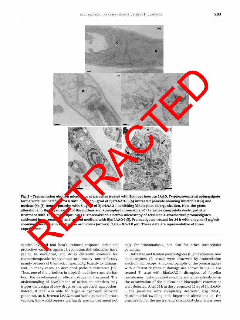
Fig. 3 – Transmission electron microscope of parasites treated with Bothrops jararaca LAAO. Trypanosoma cruzi epimastigote
forms were incubated for 24 h with 5 and 15 mg/ml of BjarLAAO-I. (A) untreated parasite showing kinetoplast (k) and
nucleus (n); (B) treated parasite with 5 mg/ml of BjarLAAO-I exhibiting kinetoplast disorganization. Note the gross
alterations in the organization of the nuclear and kinetoplast chromatins. (C) Parasites completely destroyed after
treatment with 15 mg/ml of BjarLAAO-I. Transmission electron microscopy of Leishmania amazonensis promastigotes
cultivated in untreated (D) and treated medium with BjarLAAO-I (E). Promastigotes treated for 24 h with enzyme (5 mg/ml)
showing alterations in the flagella or nucleus (arrows). Bars = 0.5–1.0 mm. These data are representative of three
experiments.
b i o c h e m i c a l p h a r m a c o l o g y 7 6 ( 2 0 0 8 ) 2 7 9 – 2 8 8 283
ETRACTED
species involved and host’s immune response. Adequate
protective vaccines against trypanosomatid infections have
yet to be developed, and drugs currently available for
chemotherapeutic intervention are mostly unsatisfactory
mainly because of their lack of specificity, toxicity to humans,
and, in many cases, to developed parasite resistance [16].
Thus, one of the priorities in tropical medicine research has
been the development of efficient drugs for treatment. The
understanding of LAAO mode of action on parasites may
trigger the design of new drugs or therapeutical approaches.
Indeed, if one was able to target a hydrogen peroxide
generator, as B. jararaca LAAO, towards the parasitophorous
vacuole, this would represent a highly specific treatment not
R
only for leishmaniasis, but also for other intracellularparasites.
Untreated and treated promastigotes (L. amazonensis) and
epimastigotes (T. cruzi) were observed by transmission
electron microscopy. Photomicrographs of the promastigotes
with different degrees of damage are shown in Fig. 3. For
treated T. cruzi with BjarLAAO-I, disruption of flagellar
membranes, mitochondrial swelling and gross alterations in
the organization of the nuclear and kinetoplast chromatins
were detected. After 24 h in the presence of 15 mg of BjarLAAO-
I, the parasites were completely destroyed (Fig. 3A–C).
Mitochondrial swelling and important alterations in the
organization of the nuclear and kinetoplast chromatins were
Fig. 4 – Sequence of cDNA and of deduced amino acid residues from BjarLAAO-I. Amino acid residues directly sequenced
from the protein (underlined).
Table 2 – Peptide mass fingerprint of BjarLAAO obtained from tryptic peptides by MALDI-TOF-MS
m/z submitted MH+ matched Delta (Da) Start End Sequence
1165.830 1165.690 0.14 308 317 (R)IKFEPPLPPK (K)
1293.750 1293.645 0.15 86 96 (K)EGWYANLGPMR (L)
1293.730 1293.622 0.13 229 238 (K)HDDIFAYEKR (F)
1388.850 1388.693 0.15 340 351 (K)KFWEDDGIHGGK (S)
1486.830 1486.648 0.14 19 30 (R)ETDYEEFLEIAR (N)
1777.090 1776.838 0.22 86 100 (K)EGWYANLGPMRLPEK(H)a
a Met was modified by oxidation.
b i o c h e m i c a l p h a r m a c o l o g y 7 6 ( 2 0 0 8 ) 2 7 9 – 2 8 8284
RETRACTED
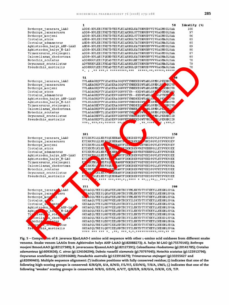
Fig. 5 – Comparison of B. jararaca BjarLAAO-I amino acid sequence with other L-amino acid oxidases from different snake
venoms. Snake venom LAAOs from Agkistrodon halys AHP-LAAO (gi:82088273); A. halys M-LAO (gi:75570145); Bothrops
moojeni BmooLAAO (gi:82127389); B. jararacussu BjussuLAAO (gi:82127391); Calosellasma rhodostoma (gi:20141785); Crotalus
adamanteus (gi:6093636); C. atrox (gi:124106294); Daboia russellii siamensis (gi:70797645); Notechis scutatus (gi:123913796);
Oxyuranus scutellatus (gi:123916680); Pseudechis australis (gi:123916679); Trimeresurus stejnegeri (gi:33355627 and
gi:82090465). Multiple sequence alignment: (*) indicates positions with fully conserved residue; (:) indicates that one of the
following high-scoring groups is conserved: K/R/Q/H, S/A, K/N/D, F/L/V/I, E/D/N/Q, T/S/A, I/M/L; (.) indicates that one of the
following ‘weaker’ scoring groups is conserved: N/R/G, G/D/N, A/V/T, Q/K/E/R, S/K/G/A, D/K/H, C/S, T/P.
b i o c h e m i c a l p h a r m a c o l o g y 7 6 ( 2 0 0 8 ) 2 7 9 – 2 8 8 285
RETRACTED

Fig. 5. (Continued )
b i o c h e m i c a l p h a r m a c o l o g y 7 6 ( 2 0 0 8 ) 2 7 9 – 2 8 8286
ETRACTED
observed by electron microscopy when L. amazonensis para-
sites were treated with 5.0 mg/ml of BjarLAAO-I (Fig. 3D and E).
B. jararaca LAAO did not induce apoptosis in the macrophages
cells, at concentrations of 1–25 mg/ml (results not shown).
Some authors have reported apoptosis-induced cell death
after incubation with L-amino acid oxidase [8,14,17–20]. The
oxidative stress induced by hydrogen peroxide could activate
heat shock proteins described in Leishmania spp., inducing
proteolytic activity inside the cell and also affecting mitochon-
drial function due to increased calcium concentrations [21].
R
A cDNA of 1452 bp was obtained, codifying a matureBjarLAAO-I with 484 amino acid residues (Fig. 4) correspon-
dent to an estimated isoelectric point and molecular weight of
5.7 and 54,771.8, respectively. The N-terminal amino acid
sequence and internal tryptic peptide sequences (Table 2),
detected and characterized by mass spectrometry, suggested
that this cDNA encodes the same enzyme purified from the
venom (Fig. 4). Fig. 5 shows the amino acid alignment of B.
jararaca LAAO and other LAAOs, indicating highly conserved
amino acid residues. The structure of LAAO from Calloselasma

Fig. 5. (Continued ).
b i o c h e m i c a l p h a r m a c o l o g y 7 6 ( 2 0 0 8 ) 2 7 9 – 2 8 8 287
ATED
rhodostoma revealed that residues 5–25 constitute one part of
the substrate-binding domain. From the comparison of the N-
terminal sequence of LAAOs, at least 13 out of 24 amino acids
are highly conserved, suggesting that these conserved amino
acids may play an important role in the substrate binding. The
cDNA-deduced amino acid sequence of snake venom LAAOs
revealed that the N-terminal sequence of these proteins
contains a highly conserved beta-alpha-beta-fold domain for
the adenylate moiety of FAD binding [1]. Pawelek and co-
workers [22] showed a high-resolution X-ray crystallographic
structure of C. rhodostoma LAAO, indicating it to be a dimer.
Each subunit consists of three domains: a FAD-binding
domain, a substrate-binding domain and a helical domain.
A deep groove is formed at the interface between the
substrate-binding and the helical domains, providing the
substrate access to the active site. According to the circular
dichroism analysis, BjarLAAO-I secondary structure is pre-
dicted to contain: 49% a-helix, 19% b-sheet, 12% b-turn and
20% random coil structure (results not shown).
Pawelek et al. [22] identified in the C. rhodostoma LAAO
structure some important residues involved in the stabiliza-
tion of the FAD molecule and in the orientation of an inhibitor
to the active site of this enzyme. The side chains of residues
Glu63, Arg71 and Glu457 make interactions with the FAD
molecule, while the cofactor dimethylbenzene ring is sur-
rounded by the hydrophobic residues Ile374, Trp420 and
Ile430. According to these authors, another potentially
essential residue of the C. rhodostoma LAAO structure is
Lys326, which coordinates a water molecule that may be
important in the hydrolytic attack on the imino intermediate.
All these residues are conserved in the B. jararaca LAAO
sequence, demonstrating the functional similarity between
BjarLAAO-I and C. rhodostoma LAAO structures.
RETR
Snake venom LAAOs (svLAAOs) share a high degree ofsequence homology among them, suggesting that additional
LAAOs from other snake venoms might also exhibit
antiviral, leishmanicide and trypanocidal activities. Further
investigations will be focused on the related molecular and
functional correlation of these enzymes. Such a study would
provide valuable information on the therapeutic develop-
ment of new generations of microbicidal drugs [23].
svLAAOs are therefore interesting multifunctional enzymes,
not only for a better understanding of the ophidian
envenomation mechanism, but also as models for poten-
tially novel therapeutic agents.
Acknowledgements
This work was supported by Fundacao de Amparo a Pesquisa
do Estado de Sao Paulo (FAPESP), Coordenacao de Aperfeicoa-
mento de Pessoal de Nıvel Superior (CAPES) and Conselho
Nacional de Desenvolvimento Cientıfico e Tecnologico (CNPq),
Brazil.
C
r e f e r e n c e s
[1] Franca SC, Kashima S, Roberto PG, Marins M, Ticli FK,Pereira JO, et al. Molecular approaches for structuralcharacterization of Bothrops L-amino acid oxidases withantiprotozoal activity: cDNA cloning, comparativesequence analysis, and molecular modeling. BiochemBiophys Res Commun 2007;355:302–6.
[2] Stabeli RG, Sant’Ana CD, Ribeiro PH, Costa TR, Ticli FK, PiresMG, et al. Cytotoxic L-amino acid oxidase from Bothrops

b i o c h e m i c a l p h a r m a c o l o g y 7 6 ( 2 0 0 8 ) 2 7 9 – 2 8 8288
A
moojeni: biochemical and functional characterization. Int JBiol Macromol 2007;41:132–40.
[3] Tempone AG, Andrade Jr HF, Spencer PJ, Lourenco CO,Rogero JR, Nascimento N. Bothrops moojeni venom killsLeishmania spp. with hydrogen peroxide generated by its L-amino acid oxidase. Biochem Biophys Res Commun2001;280:620–4.
[4] Toyama MH, Toyama DO, Passero LF, Laurenti MD, CorbettCE, Tomokane TY, et al. Isolation of a new L-amino acidoxidase from Crotalus durissus cascavella venom. Toxicon2006;47:47–57.
[5] Zhang YI, Wang JH, Lee WH, Wang Q, Liu H, Zheng YT, et al.Molecular characterization of Trimeresurus stejnegeri venomL-amino acid oxidase with potential anti-HIV activity.Biochem Biophys Res Commun 2003;309:598–604.
[6] Zieler H, Keister DB, Dvorak JA, Ribeiro JM. A snakevenom phospholipase A2 blocks malaria parasitedevelopment in the mosquito midgut by inhibitingookinete association with the midgut surface. J Exp Biol2001;204:4157–67.
[7] Deolindo P, Teixeira-Ferreira AS, Melo EJ, Arnholdt AC,Souza W, Alves EW, et al. Programmed cell death inTrypanosoma cruzi induced by Bothrops jararaca venom. MemInst Oswaldo Cruz 2005;100:33–8.
[8] Du XY, Clemetson KJ. Snake venom L-amino acid oxidases.Toxicon 2002;40:659–65.
[9] Vieira Santos MM, Sant’Ana CD, Giglio JR, Sampaio SV, DaSilva RJ, Soares AM, et al. Antitumoral effect of an L-Aminoacid oxidase isolated from Bothrops jararaca snake venom.Basic Clin Pharmacol Toxicol 2008;102:533–42.
[10] Aquino VH, Anatriello E, Goncalves PF, Da Silva EV,Vasconcelos PF, Vieira DS, et al. Molecular epidemiology ofdengue type 3 virus in Brazil and Paraguay, 2002–2004. Am JTrop Med Hyg 2006;75:710–5.
[11] Brener Z. Therapeutic activity and criterion of cure on miceexperimentally infected with Trypanosoma cruzi. Rev InstMed Trop Sao Paulo 1962;4:389–96.
[12] Izidoro LF, Ribeiro MC, Souza GR, Sant’Ana CD, HamaguchiA, Homsi-Brandeburgo MI, et al. Biochemical andfunctional characterization of an L-amino acid oxidaseisolated from Bothrops pirajai snake venom. Bioorg MedChem 2006;14:7034–43.
[13] Stabeli RG, Marcussi S, Carlos GB, Pietro RCLR, Selistre-de-Araujo HS, Giglio JR, et al. Platelet aggregation and
R
RETantibacterial effects of an L-amino acid oxidase purifiedfrom Bothrops alternatus snake venom. Bioorg Med Chem2004;12:2881–6.
[14] Sun LK, Yoshii Y, Hyodo A, Tsurushima A, Saito A,Harakuni T, et al. Apoptotic effect in the glioma cellsinduced by a specific protein extracted from Okinawa Habu(Trimeresurus flavoviridis) venom in relation to oxidativestress. Toxicol In Vitro 2003;17:169–77.
[15] Braga MD, Martins AM, Amora DN, de Menezes DB, ToyamaMH, Toyama DO, et al. Purification and biological effects ofl-amino acid oxidase isolated from Bothrops insularis venom.Toxicon 2008;51:199–207.
[16] Molyneux DH, Stiles JK. Trypanosomatid-vectorinteractions. Ann Soc Belg Med Trop 1991;71:151–66.
[17] Ali SA, Stoeva S, Abbasi A, Alam JM, Kayed R, Faigle M, et al.Isolation, structural and functional characterization of anapoptosis inducing L-amino acid oxidase from leaf-nosedviper (Eristocophis macmahoni) snake venom. Arch BiochemBiophys 2000;384:216–26.
[18] Samel M, Gunilla Ronnholm HV, Siigur J, Kalkkinen N,Siigur E. Isolation and characterization of an apoptotic andplatelet aggregation inhibiting L-amino acid oxidase fromVipera berus berus (common viper) venom. Biochim BiophysActa 2006;1764:707–14.
[19] Suhr SM, Kim DS. Identification of the snake venomsubstance that induces apoptosis. Biochem Biophys ResCommun 1996;224:134–9.
[20] Torii S, Yamane K, Mashima T, Haga N, Yamamoto K, FoxJW, et al. Molecular cloning and functional analysis ofapoxin I, a snake venom-derived apoptosis-inducing factorwith L-amino acid oxidase activity. Biochemistry2000;39:3197–205.
[21] Krobitsch S, Brandau S, Hoyer C, Schmetz C, Hubel A, Clos J.Leishmania donovani heat shock protein. Characterizationand function in amastigote stage differentiation. J BiolChem 1998;13:6488–94.
[22] Pawelek PD, Cheah J, Coulombe R, Macheroux P, Ghisla S,Vrielink A. The structure of L-amino acid oxidase revealsthe substrate trajectory into an enantiomerically conservedactive site. EMBO J 2000;19:4204–15.
[23] de Lima DC, Alvarez Abreu P, de Freitas CC, Santos DO,Borges RO, Dos Santos TC, et al. Snake venom: any clue forantibiotics and CAM? Evid Based Complement AlternatMed 2005;2:39–47.
CTED
Copyright © 2022 FDOKUMEN