Amino Acids
11
ORIGINAL ARTICLE midD-encoded ‘rhizomimosinase’ from Rhizobium sp. strain TAL1145 is a C–N lyase that catabolizes L-mimosine into 3-hydroxy-4-pyridone, pyruvate and ammonia Vishal Singh Negi • Jon-Paul Bingham • Qing X. Li • Dulal Borthakur Received: 17 November 2012 / Accepted: 19 February 2013 Ó Springer-Verlag Wien 2013 Abstract Rhizobium sp. strain TAL1145 catabolizes mimosine, which is a toxic non-protein amino acid present in Leucaena leucocephala (leucaena). The objective of this investigation was to study the biochemical and catalytic properties of the enzyme encoded by midD, one of the TAL1145 genes involved in mimosine degradation. The midD-encoded enzyme, MidD, was expressed in Esche- richia coli, purified and used for biochemical and catalytic studies using mimosine as the substrate. The reaction products in the enzyme assay were analyzed by HPLC and mass spectrometry. MidD has a molecular mass of *45 kDa and its catalytic activity was found to be optimal at 37 °C and pH 8.5. The major product formed in the reaction had the same retention time as that of synthetic 3-hydroxy-4-pyridone (3H4P). It was confirmed to be 3H4P by MS/MS analysis of the HPLC-purified product. The K m , V max and K cat of MidD were 1.27 9 10 -4 mol, 4.96 9 10 -5 mol s -1 mg -1 , and 2,256.05 s -1 , respec- tively. Although MidD has sequence similarities with aminotransferases, it is not an aminotransferase because it does not require a keto acid as the co-substrate in the degradation reaction. It is a pyridoxal-5 0 -phosphate (PLP)- dependent enzyme and the addition of 50 lM hydroxyl- amine completely inhibited the reaction. However, the supplementation of the reaction with 0.1 lM PLP restored the catalytic activity of MidD in the reaction containing 50 lM hydroxylamine. The catalytic activity of MidD was found to be specific to mimosine, and the presence of its structural analogs including L-tyrosine, L-tryptophan and L-phenylalanine did not show any competitive inhibition. In addition to 3H4P, we also identified pyruvate and ammonia as other degradation products in equimolar quan- tities of the substrate used. The degradation of mimosine into a ring compound, 3H4P with the release of ammonia indi- cates that MidD of Rhizobium sp. strain TAL1145 is a C–N lyase. Keywords MidD Rhizobium Leucaena leucocephala Mimosine 3-Hydroxy-4-pyridone Rhizomimosinase C–N lyase Abbreviations HPLC High-performance liquid chromatography PLP Pyridoxal-5 0 -phosphate 3H4P 3-Hydroxy-4-pyridone 3,4-DHP 3,4-Dihydroxypyridine 2,3-DHP 2,3-Dihydroxypyridine a-KG a-Ketoglutarate aq Aqueous Introduction Leucaena leucocephala (leucaena) is a tropical tree- legume, which is considered as promising forage for live- stock because its foliages are rich in protein, fiber, and minerals (Garcia et al. 1996). Although its foliages contain up to 18 % protein, they contain a toxic non-protein amino acid, mimosine, which limits the use of leucaena as a livestock feed (Soedarjo and Borthakur 1996). The mimosine content of leucaena foliage can be as high as 3 % Electronic supplementary material The online version of this article (doi:10.1007/s00726-013-1479-z) contains supplementary material, which is available to authorized users. V. S. Negi J.-P. Bingham Q. X. Li D. Borthakur (&) Department of Molecular Biosciences and Bioengineering, University of Hawaii at Manoa, Honolulu, HI 96822, USA e-mail: [email protected] 123 Amino Acids DOI 10.1007/s00726-013-1479-z
-
Upload
independent -
Category
Documents
-
view
1 -
download
0
Transcript of Amino Acids

ORIGINAL ARTICLE
midD-encoded ‘rhizomimosinase’ from Rhizobium sp. strainTAL1145 is a C–N lyase that catabolizes L-mimosineinto 3-hydroxy-4-pyridone, pyruvate and ammonia
Vishal Singh Negi • Jon-Paul Bingham •
Qing X. Li • Dulal Borthakur
Received: 17 November 2012 / Accepted: 19 February 2013
� Springer-Verlag Wien 2013
Abstract Rhizobium sp. strain TAL1145 catabolizes
mimosine, which is a toxic non-protein amino acid present
in Leucaena leucocephala (leucaena). The objective of this
investigation was to study the biochemical and catalytic
properties of the enzyme encoded by midD, one of the
TAL1145 genes involved in mimosine degradation. The
midD-encoded enzyme, MidD, was expressed in Esche-
richia coli, purified and used for biochemical and catalytic
studies using mimosine as the substrate. The reaction
products in the enzyme assay were analyzed by HPLC
and mass spectrometry. MidD has a molecular mass
of *45 kDa and its catalytic activity was found to be
optimal at 37 �C and pH 8.5. The major product formed in
the reaction had the same retention time as that of synthetic
3-hydroxy-4-pyridone (3H4P). It was confirmed to be
3H4P by MS/MS analysis of the HPLC-purified product.
The Km, Vmax and Kcat of MidD were 1.27 9 10-4 mol,
4.96 9 10-5 mol s-1 mg-1, and 2,256.05 s-1, respec-
tively. Although MidD has sequence similarities with
aminotransferases, it is not an aminotransferase because it
does not require a keto acid as the co-substrate in the
degradation reaction. It is a pyridoxal-50-phosphate (PLP)-
dependent enzyme and the addition of 50 lM hydroxyl-
amine completely inhibited the reaction. However, the
supplementation of the reaction with 0.1 lM PLP restored
the catalytic activity of MidD in the reaction containing
50 lM hydroxylamine. The catalytic activity of MidD was
found to be specific to mimosine, and the presence of its
structural analogs including L-tyrosine, L-tryptophan and
L-phenylalanine did not show any competitive inhibition.
In addition to 3H4P, we also identified pyruvate and
ammonia as other degradation products in equimolar quan-
tities of the substrate used. The degradation of mimosine into
a ring compound, 3H4P with the release of ammonia indi-
cates that MidD of Rhizobium sp. strain TAL1145 is a C–N
lyase.
Keywords MidD � Rhizobium � Leucaena leucocephala �Mimosine � 3-Hydroxy-4-pyridone � Rhizomimosinase �C–N lyase
Abbreviations
HPLC High-performance liquid chromatography
PLP Pyridoxal-50-phosphate
3H4P 3-Hydroxy-4-pyridone
3,4-DHP 3,4-Dihydroxypyridine
2,3-DHP 2,3-Dihydroxypyridine
a-KG a-Ketoglutarate
aq Aqueous
Introduction
Leucaena leucocephala (leucaena) is a tropical tree-
legume, which is considered as promising forage for live-
stock because its foliages are rich in protein, fiber, and
minerals (Garcia et al. 1996). Although its foliages contain
up to 18 % protein, they contain a toxic non-protein amino
acid, mimosine, which limits the use of leucaena as
a livestock feed (Soedarjo and Borthakur 1996). The
mimosine content of leucaena foliage can be as high as 3 %
Electronic supplementary material The online version of thisarticle (doi:10.1007/s00726-013-1479-z) contains supplementarymaterial, which is available to authorized users.
V. S. Negi � J.-P. Bingham � Q. X. Li � D. Borthakur (&)
Department of Molecular Biosciences and Bioengineering,
University of Hawaii at Manoa, Honolulu, HI 96822, USA
e-mail: [email protected]
123
Amino Acids
DOI 10.1007/s00726-013-1479-z

on the dry weight basis. Mimosine is toxic because it
chelates several bivalent metallic ions (Tang and Ling
1975) and also because it forms a stable complex with
pyridoxal-50-phosphate (PLP) (Lin et al. 1962). It inacti-
vates a wide variety of enzymes by limiting their metallic-
ion cofactors and PLP coenzyme. Mimosine toxicity in
animals causes various physiological abnormalities,
including enlarged thyroid glands (Hamilton et al. 1968),
infertility (Joshi 1968), fetal deformities with uterine per-
foration (Dewreede and Wayman 1970), and hair loss
(Crounse et al. 1962).
The toxic effects of mimosine on animals limit the use
of leucaena as a nutritious animal feed, and therefore, it is
important to develop leucaena plants with reduced mimo-
sine content. Identification of efficient mimosine-catabo-
lizing enzyme will be useful for developing leucaena plants
with reduced mimosine content. Previously, seedling
extract from Mimosa pudica was reported to degrade
mimosine into serine (Suda 1960). Later on a C–N lyase
enzyme was identified from leucaena seedling extract that
converts mimosine into 3,4-dihydroxypyridone (3,4-DHP),
pyruvic acid and ammonia (Smith and Fowden 1966).
Some ruminants feeding on leucaena have been reported to
contain microbes that degrade mimosine into 3H4P or 2,3-
dihydroxypyridine (2,3-DHP) (Hegarty et al. 1979).
Leucaena forms nitrogen-fixing symbiosis with some
Rhizobium strains including TAL1145 that can overcome
mimosine toxicity by degrading it, and using the degra-
dation products as a source of nutrients (Soedarjo et al.
1994). Complementation of non-mimosine-degrading
strains, such as TAL182 and CIAT899 with a TAL1145
cosmid clone, pUHR181, containing five mid genes
(midABCD and midR) resulted in the degradation of mi-
mosine in the culture (Borthakur and Soedarjo 1999). The
deduced amino acid sequences of midA, midB and midC
were found to have homologies with substrate-binding
periplasmic proteins, permeases and ATP-binding proteins,
respectively of bacterial ABC transporters; therefore, these
three genes may be involved in transport of mimosine to
Rhizobium. The gene midR has homology with transcrip-
tional activator proteins and has been shown to be involved
in transcription of mid genes. The deduced amino acid
sequence of midD was found to have high homology with
various fold type-I PLP-dependent enzymes, including
aminotransferases (up to 54 % homology) and C–S lyase
(up to 50 % homology). Based on the higher homology
with aminotransferases, the midD-encoded enzyme was
previously suggested to be an aminotransferase involved in
removal of the alanyl side chain of mimosine (Borthakur
et al. 2003). However, there was no experimental evidence
showing the catalytic action of the midD-encoded enzyme
in mimosine catabolism. In addition, the homology of
midD with both aminotransferase and C–S lyase raises the
question if the midD-encoded protein is an aminotransfer-
ase or a lyase. An enzyme from leucaena seedling extract
that has the ability to degrade mimosine was identified as a
C–N lyase that degrades mimosine by breaking the C–N
bond of mimosine (Fowden 1964). This finding along with
the homology of midD-encoded enzyme with a C–S lyase,
gives us a clue that the midD-encoded may be a C–N lyase
instead of an aminotransferase. Therefore, the objectives of
this study are to determine the catalytic role of the midD-
encoded enzyme in mimosine catabolism, to identify
whether the enzyme is an aminotransferase or a C–N lyase,
and to determine its other biochemical properties.
Materials and methods
Construction of midD-expression plasmid
The bacterial strains, plasmids, and primers used in this
study are described in Table 1. BamHI restriction site was
incorporated into midD-specific forward and reverse
primers (midD-f, and midD-r). These primers were used to
amplify the 1221-bp midD open reading frame (ORF) by
PCR from the cosmid clone pUHR263, using Phusion�
High-Fidelity DNA Polymerase (New England Biolabs,
MA, USA). The midD ORF with BamHI restriction sites at
the two ends was cloned into the BamHI site of the
expression vector pET-14b (Novagen, WI, USA) under the
control of a T7 promoter, and introduced into E. coli
JM109 (Promega, WI, USA). The resultant plasmid was
verified by sequencing and named as pET-midD. Another
plasmid, named pET-anti-midD, was also constructed by
cloning midD in the antisense direction from the T7 pro-
moter in pET14b.
Bacterial expression and purification of MidD
The expression constructs were transformed into E. coli
strain BL21(DE3) pLysS (Promega). The transformed
BL21 cells containing pET-midD were grown in LB and
induced for the expression of midD by adding isopropyl
b-D-1-thiogalactopyranoside (IPTG, Sigma-Aldrich, MO,
USA) into the LB broth medium at mid-log phase
(OD600 = 0.6) to a final concentration of 1 mM and
incubating with shaking at 210 rpm at 37 �C for 1–10 h.
To determine the optimum induction time, 200 lL induced
cultures were collected at 1, 2, 4, 6, 8 and 10 h of induc-
tion. A non-induced culture of BL21(DE3)pLysS contain-
ing pET-midD and an induced culture of BL21(DE3)pLysS
containing pET-anti-midD were used as negative controls.
The induced and non-induced cultures were harvested by
centrifugation at 4000 rpm for 10 min at 4 �C. To make
total soluble proteins, cell pellets of harvested samples
V. S. Negi et al.
123

were re-suspended in a SDS-PAGE loading buffer fol-
lowed by heating at 95 �C for 5 min. MidD protein was
purified from the cell pellets of the induced samples of
E. coli containing midD using the MagneHisTM Protein
Purification System (Promega) according to the manufac-
turer’s instructions. Total soluble proteins and purified
recombinant protein were analyzed through SDS-PAGE.
Mimosine degradation assay
In vitro enzymatic assay was performed to study the effect
of MidD on mimosine catabolism by incubating the puri-
fied MidD enzyme with 1 mM mimosine as the substrate in
0.1 M Tris–HCl. The final pH of the reaction mixtures was
set at 7.5. To test whether MidD is an aminotransferase or
not, we carried out the enzymatic reactions in the presence
and absence of 20 lM PLP and 1 mM a-ketoglutarate
(a-KG). PLP and a-KG are the key requirements for the
aminotransferases and are used as coenzyme and cosusb-
strate, respectively. For each 1,000 lL reaction, 0.016 mg
of the purified enzyme was used and the reaction was
incubated at 37 �C for 1 h unless otherwise stated. Each
reaction type was performed in three replications and the
reaction mixtures were analyzed for the amount of the
product formed through high-performance liquid chroma-
tography (HPLC) using a C18 column (4.6 9 250 mm;
Dionex acclaim 120). To determine whether MidD enzyme
is PLP-dependent or not, enzymatic assay was performed
by supplementing the reaction buffer with hydroxylamine,
an inhibitor of PLP-dependent enzymes. The hydroxyl-
amine was added at a concentration range of 0.01–50 mM
to the reaction mixture containing the enzyme and
incubated for 5 min at room temperature followed by
addition of the substrate. In a separate set of reactions, the
reaction mixture containing 50 lM hydroxylamine and the
enzyme were supplemented with 0.1–20 lM PLP after
5 min of incubation to restore the enzyme activity if it was
inhibited by hydroxylamine. The concentrations of inhibi-
tor and cofactor followed a similar trend as used by
El-Sayed (2011). For quantitative estimation of the product
formed, different concentrations of chemically synthesized
3-hydroxy-4-pyridone (synthetic 3H4P) were used as
standards. The synthetic 3H4P was kindly provided by
Dr. Behrman, Ohio State University, Columbus, OH. An
isocratic solvent system of 0.02 M o-phosphoric acid with
a flow rate of 1 mL min-1, and an UV detection photodi-
ode array (200–400 nm) were used for HPLC analysis. The
peak area of the product (3H4P) formed was obtained from
HPLC chromatogram of the reactions and the amount of
product formed was quantified from the slopes of standard
curve formed by plotting the peak area of known concen-
trations of synthetic 3H4P.
Characterization of midD-encoded enzyme
The characterization of midD-encoded enzyme was per-
formed at 37 �C in 0.1 M Tris–HCl buffer for 1 h unless
otherwise stated. For the termination of MidD-catalyzed
reactions, the enzyme was inactivated by heating the
reaction mixture at 100 �C for 3 min. The optimum pH of
the enzyme was studied by conducting mimosine degra-
dation assay at different pHs ranging from pH 3 to 12.
Similarly, the optimum temperature for the enzyme was
studied by conducting the mimosine degradation assay at
Table 1 Bacterial strains,
plasmids, and primers
a Italicized texts indicate the
BamHI restriction siteb Bold texts in primer sequence
show the homologous sequence
to midD ORF
Bacterial strains/
plasmids/primers
Description or sequence (50–30) Source/reference
E. coli strains
JM109 Host strain used for plasmid transformation Promega
BL21(DE3)pLysS Host strain used for expression of midD Promega
Plasmids
pUHR263 Cosmid clone isolated from the genomic library of
Rhizobium sp. strain TAL1145, contains mid genes
(Fox and Borthakur
2001)
pET-14b Protein expression vector with a T7 promoter and an
N-terminal His-tag sequence
Novagen
pET-midD pET-14b vector with an in-frame midD ORF in sense
orientation at its BamHI site
This study
pET-anti-midD pET-14b vector with midD ORF in antisense orientation at
its BamHI site
This study
Primers
midD-f CCTAAGGATCCGATGCACGATTTGCACCa,b This study
midD-r ACTTTGGATCCATTCATCCTTCGTGGCTGGACAAa,b This study
T7-P TAATACGACTCACTATAGGG This study
T7-T GGGTTATGCTAGTTATTGCT This study
midD-encoded ‘rhizomimosinase’ from Rhizobium sp. strain TAL1145
123
different temperatures including 4, 22, 30, 37, 45, 55, and
65 �C. The subsequent enzymatic assays were performed at
the optimum pH and temperature unless otherwise stated.
The enzyme was also studied for its thermal stability by
pre-incubating the enzyme reaction mixture in 0.1 M Tris–
HCl buffer lacking mimosine at different temperatures
(4, 37, 40, 50, 55, 60, 65, and 70 �C) for 30 min before
performing the actual mimosine degradation assay. The
reaction mixtures in the enzyme characterization study
were analyzed through HPLC for quantification of the
mimosine degradation product. The aromatic amino acids
L-tyrosine, L-tryptophan, and L-phenylalanine, which are
structural analogs of mimosine, were tested as possible
competitive inhibitors of MidD in mimosine degradation.
Each aromatic amino acid was mixed with mimosine in the
ratios of 1:1, 2:1, and 3:1 and the resultant mixtures were
studied for mimosine degradation using MidD enzyme, and
the product formed was quantified using HPLC.
Kinetic study of midD-encoded enzyme
All experiments to study kinetic properties of MidD were
performed at 37 �C and an optimum pH of 8.5 (see results).
The rate of reaction for different substrate concentrations
(0.05, 0.10, 0.15, 0.25, 0.35, 0.50 mM) was calculated at 0,
2, 3, 5, 10, 20 and 30 min. The initial velocities of the
enzyme catalysis were measured as slopes formed by
quantifying the amount of product formed at 0 and 2 min
of reaction time as the reaction was linear between 0 and
2 min. The values of initial velocities and substrate con-
centrations were then used to determine kinetic constants
Vmax, and Km using Lineweaver–Burk plot according to the
standard procedure. The mole of enzyme active sites per
mg of the enzyme ([Et]) was calculated assuming that there
is only one active site per enzyme molecule. Turnover
number (Kcat) of the enzyme was also determined using
Vmax and [Et].
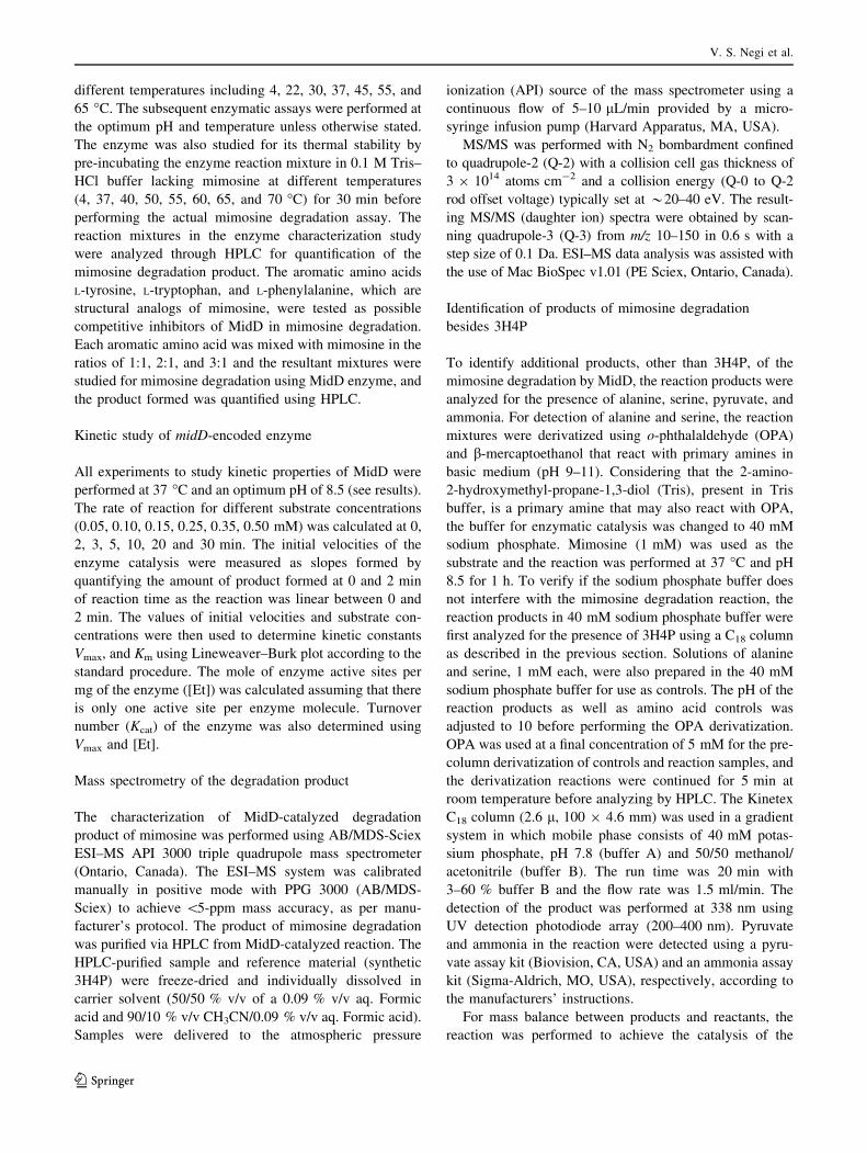
Mass spectrometry of the degradation product
The characterization of MidD-catalyzed degradation
product of mimosine was performed using AB/MDS-Sciex
ESI–MS API 3000 triple quadrupole mass spectrometer
(Ontario, Canada). The ESI–MS system was calibrated
manually in positive mode with PPG 3000 (AB/MDS-
Sciex) to achieve \5-ppm mass accuracy, as per manu-
facturer’s protocol. The product of mimosine degradation
was purified via HPLC from MidD-catalyzed reaction. The
HPLC-purified sample and reference material (synthetic
3H4P) were freeze-dried and individually dissolved in
carrier solvent (50/50 % v/v of a 0.09 % v/v aq. Formic
acid and 90/10 % v/v CH3CN/0.09 % v/v aq. Formic acid).
Samples were delivered to the atmospheric pressure
ionization (API) source of the mass spectrometer using a
continuous flow of 5–10 lL/min provided by a micro-
syringe infusion pump (Harvard Apparatus, MA, USA).
MS/MS was performed with N2 bombardment confined
to quadrupole-2 (Q-2) with a collision cell gas thickness of
3 9 1014 atoms cm-2 and a collision energy (Q-0 to Q-2
rod offset voltage) typically set at *20–40 eV. The result-
ing MS/MS (daughter ion) spectra were obtained by scan-
ning quadrupole-3 (Q-3) from m/z 10–150 in 0.6 s with a
step size of 0.1 Da. ESI–MS data analysis was assisted with
the use of Mac BioSpec v1.01 (PE Sciex, Ontario, Canada).
Identification of products of mimosine degradation
besides 3H4P
To identify additional products, other than 3H4P, of the
mimosine degradation by MidD, the reaction products were
analyzed for the presence of alanine, serine, pyruvate, and
ammonia. For detection of alanine and serine, the reaction
mixtures were derivatized using o-phthalaldehyde (OPA)
and b-mercaptoethanol that react with primary amines in
basic medium (pH 9–11). Considering that the 2-amino-
2-hydroxymethyl-propane-1,3-diol (Tris), present in Tris
buffer, is a primary amine that may also react with OPA,
the buffer for enzymatic catalysis was changed to 40 mM
sodium phosphate. Mimosine (1 mM) was used as the
substrate and the reaction was performed at 37 �C and pH
8.5 for 1 h. To verify if the sodium phosphate buffer does
not interfere with the mimosine degradation reaction, the
reaction products in 40 mM sodium phosphate buffer were
first analyzed for the presence of 3H4P using a C18 column
as described in the previous section. Solutions of alanine
and serine, 1 mM each, were also prepared in the 40 mM
sodium phosphate buffer for use as controls. The pH of the
reaction products as well as amino acid controls was
adjusted to 10 before performing the OPA derivatization.
OPA was used at a final concentration of 5 mM for the pre-
column derivatization of controls and reaction samples, and
the derivatization reactions were continued for 5 min at
room temperature before analyzing by HPLC. The Kinetex
C18 column (2.6 l, 100 9 4.6 mm) was used in a gradient
system in which mobile phase consists of 40 mM potas-
sium phosphate, pH 7.8 (buffer A) and 50/50 methanol/
acetonitrile (buffer B). The run time was 20 min with
3–60 % buffer B and the flow rate was 1.5 ml/min. The
detection of the product was performed at 338 nm using
UV detection photodiode array (200–400 nm). Pyruvate
and ammonia in the reaction were detected using a pyru-
vate assay kit (Biovision, CA, USA) and an ammonia assay
kit (Sigma-Aldrich, MO, USA), respectively, according to
the manufacturers’ instructions.
For mass balance between products and reactants, the
reaction was performed to achieve the catalysis of the
V. S. Negi et al.
123
substrate to near completion. This was done using lower
substrate concentration (500 lM mimosine) with the
excess of the enzyme (0.2 mg) in 40 mM phosphate buffer.
The final pH of the reaction was 8.5 and the reaction was
incubated for 1 h at 37 �C followed by identification and
quantification of the degradation products. The standard
curves for pyruvate, ammonia, and 3H4P were plotted, and
the products in MidD-catalyzed reaction were quantified
using the line equation from the respective standard curve.
Prediction of catalytic and PLP-binding site of MidD
To predict the catalytic and PLP-binding sites of MidD,
blastp search of MidD was performed using pdb database
as the reference. The deduced amino acid sequences of
enzymes that showed high homology with MidD were
aligned with the deduced amino acid sequence of MidD
using the ClustalW2 multiple sequence alignment program
and the conserved residues were analyzed.
Results
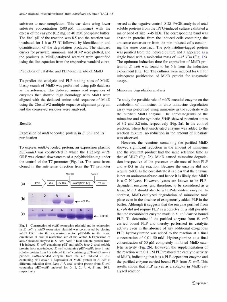
Expression of midD-encoded protein in E. coli and its
purification
To express midD-encoded protein, an expression plasmid
pET-midD was constructed in which the 1,221-bp midD
ORF was cloned downstream of a polyhistidine-tag under
the control of the T7 promoter (Fig. 1a). The same insert
cloned in the anti-sense direction from the T7 promoter
served as the negative control. SDS-PAGE analysis of total
soluble proteins from the IPTG-induced culture exhibited a
major band of size *45 kDa. The corresponding band was
absent in proteins from the induced cells containing the
antisense construct or from the non-induced cells contain-
ing the sense construct. The polyhistidine-tagged protein
was purified from the induced culture and it appeared as a
single band with a molecular mass of *45 kDa (Fig. 1b).
The optimum induction time for expression of MidD pro-
tein in E. coli was found to be 6 h from the induction
experiment (Fig. 1c). The cultures were induced for 6 h for
subsequent purification of MidD protein for enzymatic
assays.
Mimosine degradation analysis
To study the possible role of midD-encoded enzyme on the
catabolism of mimosine, in vitro mimosine degradation
assay was performed using mimosine as the substrate with
the purified MidD enzyme. The chromatograms of the
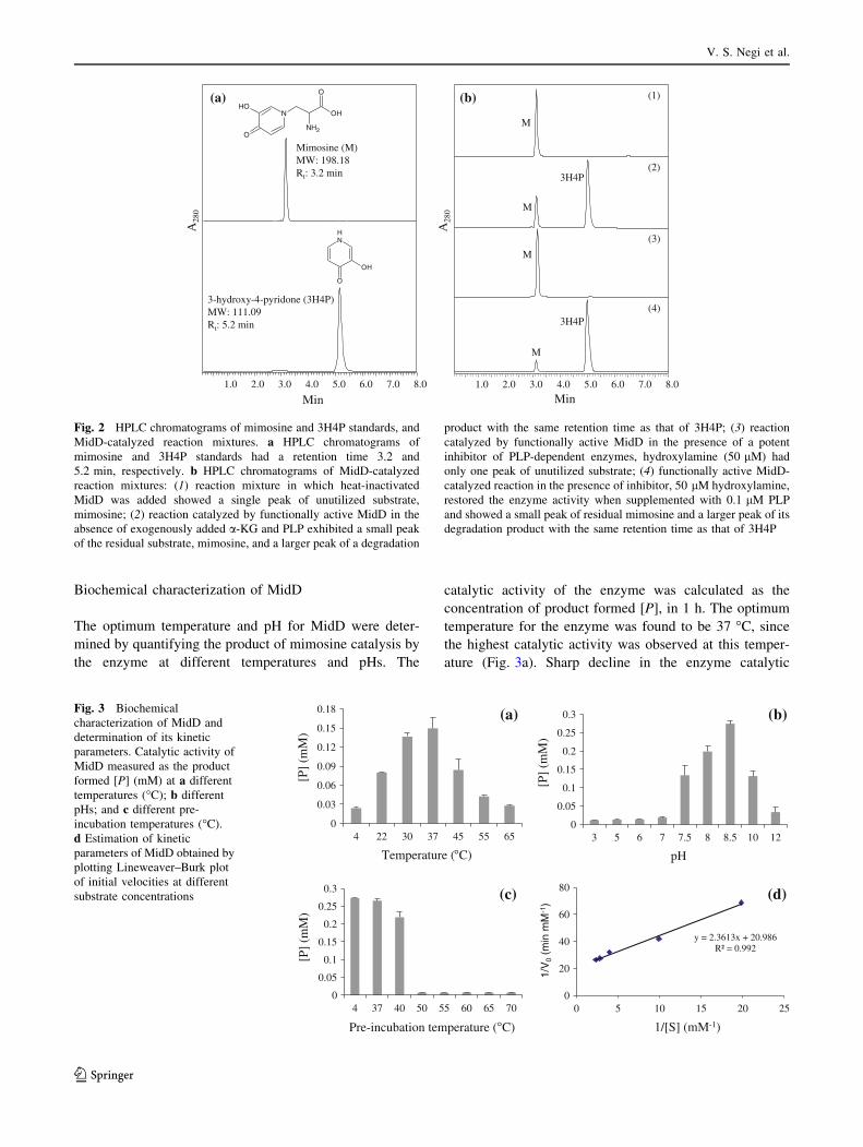
mimosine and the synthetic 3H4P showed retention times
of 3.2 and 5.2 min, respectively (Fig. 2a). In the control
reaction, where heat-inactivated enzyme was added to the
reaction mixture, no reduction in the amount of substrate
was observed.
However, the reactions containing the purified MidD
showed significant reduction in the amount of mimosine
and the resultant product had the same retention time as
that of 3H4P (Fig. 2b). MidD caused mimosine degrada-
tion irrespective of the presence or absence of both PLP
and a-KG in the reaction. Because the enzyme did not
require a-KG as the cosusbstrate it is clear that the enzyme
is not an aminotransferase and hence it is likely that MidD
is a C–N lyase. However, lyases are known to be PLP-
dependent enzymes, and therefore, to be considered as a
lyase, MidD should also be a PLP-dependent enzyme. In
contrast, MidD-catalyzed degradation of mimosine took
place even in the absence of exogenously added PLP in the
buffer. Although it suggests that the enzyme purified from
E. coli did not require PLP as a cofactor, it is still possible
that the recombinant enzyme made in E. coli carried bound
PLP. To determine if the purified enzyme from E. coli
carried bound PLP and thereby performed its catalytic
activity even in the absence of any additional exogenous
PLP, hydroxylamine was added to the reaction at a final
concentration of 0.01–50 mM. Hydroxylamine at a final
concentration of 50 lM completely inhibited MidD cata-
lytic activity (Fig. 2b). However, the supplementation of
the reaction with 0.1 lM PLP restored the catalytic activity
of MidD, indicating that it is a PLP-dependent enzyme and
the purified enzyme carried bound PLP from E. coli. This
results shows that PLP serves as a cofactor in MidD cat-
alyzed reaction.
rbs 6x-His
BamHI
T7 P midD (1221 bp) T7 ter
BamHI
(a)
45kDa(b)
45kDa(c)
Fig. 1 Construction of midD expression plasmid and its expression
in E. coli. a midD expression plasmid was constructed by cloning
midD ORF into the expression vector pET-14b in the sense
orientation at BamHI restriction site of the vector. b Expression of
midD-encoded enzyme in E. coli. Lane 1 total soluble protein from
4 h induced E. coli containing pET-anti-midD; lane 2 total soluble
protein from non-induced E. coli containing pET-midD; lane 3 total
soluble protein from 4 h induced E. coli containing pET-midD; lane 4purified midD-encoded enzyme from the 4 h induced E. colicontaining pET-midD. c Expression of MidD protein in E. coli at
different induction time. Lane 1–7, total soluble protein from E. colicontaining pET-midD induced for 0, 1, 2, 4, 6, 8 and 10 h,
respectively
midD-encoded ‘rhizomimosinase’ from Rhizobium sp. strain TAL1145
123
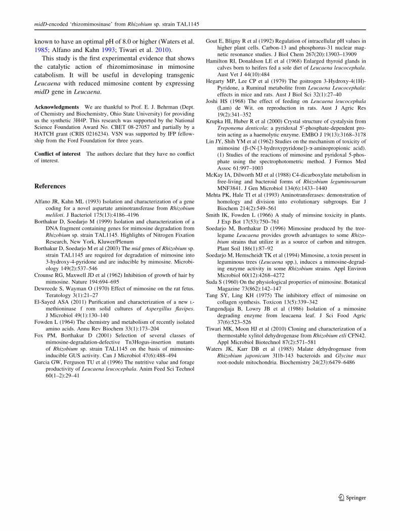
Biochemical characterization of MidD
The optimum temperature and pH for MidD were deter-
mined by quantifying the product of mimosine catalysis by
the enzyme at different temperatures and pHs. The
catalytic activity of the enzyme was calculated as the
concentration of product formed [P], in 1 h. The optimum
temperature for the enzyme was found to be 37 �C, since
the highest catalytic activity was observed at this temper-
ature (Fig. 3a). Sharp decline in the enzyme catalytic
NH
OH
O
N
O
OH
O
NH2
OH
3-hydroxy-4-pyridone (3H4P)MW: 111.09Rt: 5.2 min
Mimosine (M)MW: 198.18Rt: 3.2 min
(a)
Min1.0 2.0 3.0 4.0 5.0 6.0 7.0 8.0
A28
0
Min1.0 2.0 3.0 4.0 5.0 6.0 7.0 8.0
(1)
(2)
(3)
(4)
M
M
M
M
3H4P
3H4P
(b)
A28
0
Fig. 2 HPLC chromatograms of mimosine and 3H4P standards, and
MidD-catalyzed reaction mixtures. a HPLC chromatograms of
mimosine and 3H4P standards had a retention time 3.2 and
5.2 min, respectively. b HPLC chromatograms of MidD-catalyzed
reaction mixtures: (1) reaction mixture in which heat-inactivated
MidD was added showed a single peak of unutilized substrate,
mimosine; (2) reaction catalyzed by functionally active MidD in the
absence of exogenously added a-KG and PLP exhibited a small peak
of the residual substrate, mimosine, and a larger peak of a degradation
product with the same retention time as that of 3H4P; (3) reaction
catalyzed by functionally active MidD in the presence of a potent
inhibitor of PLP-dependent enzymes, hydroxylamine (50 lM) had
only one peak of unutilized substrate; (4) functionally active MidD-
catalyzed reaction in the presence of inhibitor, 50 lM hydroxylamine,
restored the enzyme activity when supplemented with 0.1 lM PLP
and showed a small peak of residual mimosine and a larger peak of its
degradation product with the same retention time as that of 3H4P
0
0.05
0.1
0.15
0.2
0.25
0.3
3 5 6 7 7.5 8 8.5 10 12
[P]
(mM
)
pH
(b)
0
0.03
0.06
0.09
0.12
0.15
0.18
4 22 30 37 45 55 65
[P]
(mM
)
Temperature (°C)
(a)
0
0.05
0.1
0.15
0.2
0.25
0.3
4 37 40 50 55 60 65 70
[P]
(mM
)
Pre-incubation temperature (°C)
(c)
y = 2.3613x + 20.986R² = 0.992
0
20
40
60
80
0 5 10 15 20 25
1/V
0(m
in m
M- 1
)
1/[S] (mM-1)
(d)
Fig. 3 Biochemical
characterization of MidD and
determination of its kinetic
parameters. Catalytic activity of
MidD measured as the product
formed [P] (mM) at a different
temperatures (�C); b different
pHs; and c different pre-
incubation temperatures (�C).
d Estimation of kinetic
parameters of MidD obtained by
plotting Lineweaver–Burk plot
of initial velocities at different
substrate concentrations
V. S. Negi et al.
123

activity was observed at temperature 45 �C or higher and
below 22 �C. The enzyme showed activities at a pH range
from 7.5 to 10.0, with the optimum pH of 8.5 (Fig. 3b).
The thermal stability of the enzyme was determined by pre-
incubating the reaction mixture in the absence of the sub-
strate, mimosine for 30 min at different temperatures and
then adding mimosine to start the reaction. The percent
enzyme activity for different pre-incubation temperatures
was measured with reference to the enzyme activity at pre-
incubation temperature of 4 �C. The enzyme activity at
37 �C pre-incubation temperature was found to be 97.3 %
of that of the reference, and it was reduced to 80.2 % when
the enzyme was pre-incubated at 40 �C. A sharp decline in
the enzyme activity was observed at pre-incubation tem-
peratures of 50 �C or higher (Fig. 3c). This shows that the
enzyme is fairly stable up to 40 �C.
The kinetic parameters of MidD were measured at the
optimum temperature (37 �C) and pH (8.5). The rate of
reaction for different substrate concentrations ranging from
0.05 to 0.50 mM was found to be linear from 0 to 2 min
(data not shown). Initial velocities of the enzyme for each
substrate concentration were calculated as the slope of
product formed at 0 and 2 min. The enzyme followed the
typical Michaelis–Menten kinetics. The apparent Km and
Vmax values for MidD were experimentally determined from
linear regressions by plotting Lineweaver–Burk plot
(Fig. 3d) and were found to be 1.274 9 10-4 mol and
4.9633 9 10-5 mol s-1 mg-1, respectively. Assuming one
active site per enzyme molecule, the total enzyme concen-
tration [Et], was calculated to be 0.22 9 10-7 mol mg-1.
Turnover number (Kcat) of MidD was determined to be
2,256.05 s-1.
Possible competitive inhibition of mimosine catalysis
was studied by adding the aromatic amino acids L-tyrosine,
L-tryptophan, and L-phenylalanine separately to the reac-
tion mixture containing mimosine. Despite the structural
similarities to mimosine, the presence of these amino acids
at onefold, twofold, or threefold concentration of mimosine
did not affect the amount of product formed (Fig. 4),
suggesting that these aromatic amino acids are not com-
petitive inhibitors of mimosine catalysis by MidD.
Characterization of the degradation product
of mimosine by MidD
The chromatogram of the product of mimosine degradation
by MidD was identical to that of synthetic 3H4P standard.
This suggests that mimosine is converted to 3H4P by
MidD. To confirm this correlation, its induced fragmenta-
tion pattern obtained through MS/MS of the parent ion (m/z
112.2) was compared with that of synthetic 3H4P (m/z
112.2). The resulting fragmentation patterns of synthetic
3H4P (reference sample) and mimosine-degradation
product (test sample) were found to be identical, generating
common internal structural fragments of m/z 94.0
([M–H2O]?), 65.9 ([M–H2O–CO]?), 56.0, and 39.1 amu
(Fig. 5a, b). This confirms that the degradation product of
mimosine by MidD is indeed 3H4P.
Other products of mimosine degradation
Mimosine is an aromatic compound with 8 carbons and 2
nitrogens. Identification of 3H4P as the major degradation
product of mimosine accounts for only 5 carbons and one
nitrogen. Therefore, MidD-catalyzed mimosine degrada-
tion must form other products that should account for the
remaining three carbons and one nitrogen. Previous studies
Min1.0 2.0 3.0 4.0 5.0 6.0 7.0 8.0
n = 4
n = 4
(a)
(b)
(c)
n = 4
3H4P
3H4P
3H4P
A28
0
Fig. 4 HPLC chromatograms obtained in mimosine degradation
assays in both presence and absence of structural analogs of
mimosine, including a L-tyrosine; b L-phenylalanine; and c L-trypto-
phan. Each of these structural analogs was used in three different
concentrations (1 mM, 2 mM and 3 mM) in separate reactions with
1 mM mimosine. The reaction in the absence of structural analog
served as the control. The product formed in control and test reactions
showed no difference in the amount of product formed. This shows
that none of the structural analogs competitively inhibits MidD-
catalyzed reaction. n = 4 represents 4 reactions; n1–n3: structural
analog at concentrations 1, 2 and 3 mM, respectively; n4: reaction
without structural analog. Each of the reaction had 1 mM of the
substrate
midD-encoded ‘rhizomimosinase’ from Rhizobium sp. strain TAL1145
123

with mimosine-degrading enzymes from Mimosa pudica
(Suda 1960) and leucaena (Smith and Fowden 1966) showed
serine and pyruvate, respectively as the degradation prod-
ucts. Therefore, we tested MidD-catalyzed reactions for the
presence of serine or pyruvate. We also tested the reactions
for the presence of alanine as it is also a 3-carbon amino acid
with structure similar to serine. Neither serine nor alanine
appeared in the HPLC chromatograms of MidD-catalyzed
reactions (Supplementary material, Fig. S1). However, we
successfully detected pyruvate and ammonia spectrophoto-
metrically in MidD-catalyzed reactions as mimosine degra-
dation products and quantified the products (Fig. 6). The
amounts of pyruvate, ammonia, and 3H4P produced in the
mimosine degradation reaction were found to be 87.5, 87.0,
and 82.7 %, respectively, of mimosine, indicating that these
products are formed in equimolar quantities as that of the
substrate. Therefore, the completely balanced reaction of
MidD-catalyzed mimosine-degradation can be represented
as follows:
C8H10N2O4ðmimosineÞ þ H2O
! C5H5NO2ð3H4PÞ þ C3H4O3ðpyruvateÞ þ NH3
Prediction of catalytic and PLP-binding site of MidD
The blastp analysis of MidD using pdb as reference data-
base showed homology of MidD with b C–S lyase
(gi|392935441), MetC (gi|317455161) and cystalysin
(gi|9955069) from Streptococcus anginosus, Streptococcus
mutans and Treponema denticola, respectively. The
sequence alignment showed presence of the active site
residues and PLP-binding lysine in MidD corresponding to
these residues in cystalysin of T. denticola (Fig. 7).
Discussion
In this report, we studied mimosine degradation by midD-
encoded enzyme of the root nodule bacterium Rhizobium
sp. strain TAL1145 that forms nitrogen-fixing nodules on
Leucaena. Mimosine degradation is a mechanism for
10 20 30 40 50 60 70 80 90 100 110 120 130 140
1.0e6
2.0e6
3.0e6
4.0e6
5.0e6
6.0e6
7.0e6
8.0e6
9.0e6
1.0e7
1.1e7
1.2e7
1.3e7
1.4e7
1.5e71.6e7 112.2
94.065.9
56.083.939.2 82.267.943.0 110.692.1 97.3 127.8 130.5113.4
Inte
nsity
, cps
NH
OH
O
(a)
3H4P[MH+]
m/z, amu10 20 30 40 50 60 70 80 90 100 110 120 130 140
5.0e6
1.0e7
1.5e7
2.0e7
2.5e7
3.0e7
3.5e7
4.0e7
4.5e7
5.0e7
5.2e7
Inte
nsity
, cps
112.2
94.265.9
56.066.9 84.2
69.955.0 112.839.1
Min1.0 2.0 3.0 4.0 5.0 6.0 7.0 8.0
M3H4P
A28
0 (b)
[MH+]
m/z, amu
Fig. 5 MS/MS spectra of a the synthetic 3H4P (reference sample) and b the HPLC-purified mimosine-degradation product formed in MidD-
catalyzed reaction (test sample)
0
20
40
60
80
100
Pyruvate Ammonia 3H4P
Pro
du
ct f
orm
ed (
%)
Fig. 6 The percent of product formed with respect to the substrate
used. The error bar represents the standard deviation of three
replicates
V. S. Negi et al.
123

detoxification as well as acquiring additional source of
nutrients by the bacterium. Although Rhizobium bacteroids
are provided with malate as the source of carbon by the
plant (McKay et al. 1988), in the Leucaena nodules,
mimosine may also be used as a source of carbon by the
Rhizobium bacteroids (Borthakur et al. 2003). We have
established that the Rhizobium enzyme MidD degrades
mimosine into 3H4P. MS/MS analysis using synthetic
3H4P and the purified product of mimosine degradation,
which had identical chromatogram as 3H4P, showed that
both have identical fragmentation patterns confirming that
the purified compound was 3H4P. A plant enzyme that
degrades mimosine into 3H4P is also known to be present
in the foliage of Leucaena (Smith and Fowden 1966;
Tangendjaja et al. 1986). Because the mimosine-degrading
enzyme from Leucaena has been named as mimosinase, we
used the name ‘rhizomimosinase’ to distinguish the Rhi-
zobium enzyme from the Leucaena enzyme with similar
function.
Previously, based on the sequence similarity, rhizo-
mimosinase was referred to as mimosine aminotransferase
(Borthakur et al. 2003). However, in this study, we have
shown that rhizomimosinase is not an aminotransferase
because it does not require a keto acid as the co-substrate
for accepting the amino group from mimosine. Although,
no exogenous PLP was needed for the catalytic activity of
the recombinant enzyme purified from E. coli, the presence
of hydroxylamine completely inhibited the reaction,
whereas, the addition of exogenous PLP overcame the
inhibitory effect of hydroxylamine and restored the cata-
lytic activity of rhizomimosinase, suggesting that it is a
PLP-dependent enzyme. Besides 3H4P, we also identified
pyruvate and ammonia as the products of rhizomimosi-
nase-catalyzed mimosine degradation. In addition, the
production of ammonia with the aromatic compound 3H4P
as mimosine-degradation products establishes that MidD is
a C–N ammonia lyase as the release of ammonia with the
formation of a double bond or a ring compound is the key
characteristic of C–N ammonia lyases. Based on these
results, MidD can be classified in the enzyme category with
the Enzyme Commission number EC4.3.1.
Among the lyases that showed homology with rhizo-
mimosinase, the catalytic and PLP-binding sites of cys-
talysin from T. denticola have been studied in detail
(Krupka et al. 2000). Although cystalysin is a C–S lyase,
and rhizomimosinase is a C–N lyase, both enzymes pro-
duce pyruvate and ammonia and share many conserved
residues. The Arg369, Lys238, Asp203, Tyr64 and Ala39
of cystalysin, which are conserved in all four lyases studied
and had been shown to be involved in the catalytic site in
cystalysin, correspond to Arg380, Lys248, Asp213, Tyr75,
and Ala50 in rhizomimosinase, respectively. The Lys238
of cystalysin is known to interact with PLP forming an
internal aldimine (Krupka et al. 2000). The conserved Arg
Fig. 7 Sequence alignment of MidD with b C–S lyase
(gi|392935441), MetC (gi|317455161), and cystalysin (gi|9955069)
from Streptococcus anginosus, Streptococcus mutans and Treponemadenticola, respectively. Asterisks represent identical residues and
single or double dots symbolize similar residues. The catalytic
residues of cystalysin, which are conserved in the other three lyases
are indicated by ‘filled triangle’ and the three letter code of amino
acid followed by the position of the residue. The alignment was
performed using the ClustalW2 multiple sequence alignment program
midD-encoded ‘rhizomimosinase’ from Rhizobium sp. strain TAL1145
123

corresponding to Arg369 of cystalysin has been suggested
to be the docking site for a-carboxylate of substrate (Mehta
et al. 1993; Krupka et al. 2000). Lys238, Tyr64 and Ala39
have been reported to be involved in C–S cleavage in
cystalysin (Krupka et al. 2000). Similarly, Asp203 has been
shown to be involved in charge stabilization of pyridoxal
system during the cystalysin reaction. Based on the high
homology and conserved residues at the catalytic and PLP-
binding sites between cystalysin and rhizomimosinase as
described above, it is likely that the C–N bond of mimosine
is cleaved by rhizomimosinase in a similar way as for the
C–S bond of cysteine by cystalysin (Fig. 8). In our
experiment, the purified recombinant rhizomimosinase
formed a complex with PLP in E. coli. The amino group of
mimosine may be deprotonated under the optimum pH
(8.0–8.5) to perform a nucleophilic attack on C4 of the
enzyme-bound PLP, which results in the formation of a
Schiff base (mimosine-PLP complex). Lys248, Tyr75 and
Ala50 in the rhizomimosinase catalytic site may be
involved in C–N bond cleavage of mimosine of the
mimosine-PLP complex forming 3H4P. After the release of
3H4P, reverse transaldimination reaction may take place
resulting in the formation of 2-aminoprop-2-enoate, which
can be hydrolyzed to form ammonia and pyruvate outside
the catalytic site of rhizomimosinase.
Rhizomimosinase is found to be specific for mimosine
and the presence of its structural analogs, L-tyrosine,
L-tryptophan, and L-phenylalanine did not affect the rhi-
zomimosinase’s efficiency for mimosine degradation. The
optimum pH for rhizomimosinase (8.5) is higher than the
physiological pH for Rhizobium (6.8) or plant cytoplasm
(7.4–7.5) (Gout et al. 1992). The activity of the rhizo-
mimosinase is highly reduced below pH 7.5 or above pH
10.0. Although Rhizobium strains grow best at pH 6.8,
some Rhizobium enzymes, such as xylitol dehydrogenase,
malate dehydrogenase and aspartate aminotransferase, are
N
O
OH
N+
H
CH3
CH
O-
OPO3-
N+
C COO-
CH2
H
H+
NCH2
O
OHC
O-OC
H
NH2
K248
N+
H
CH3
CHN
H
O-O
PO3-
R380
NOH
O
N
H
CH3
CH
O-O
PO3-
N+
C COO-
CH2
H
R380
H3N K248
+
A50
Y75
+K248
NOH
O
N+
H
CH3
CHNH
H
O-O
PO3-
NH
CH COO-
CH2
NHOH
O
3H4P
N+
H
CH3
CH
O-O
PO3-
N+
C COO-
CH2
HNH2K248
K248
+
N+
H
CH3
CHN
H
O-O
PO3-
+ NC
O-OC
CH2
H
H
K248
+
N+
H
CH3
CHN
H
O-O
PO3-
CH3 COO-
O
+ N
H
H
H
Pyruvate Ammonia
H2O
(b)(a)
(e)
(c) (d)
K248H2N
(f)(g)
Fig. 8 Proposed molecular mechanism of mimosine degradation
catalyzed by rhizomimosinase. The rhizomimosinase enzyme is
indicated as a shaded area, and the substrate and products are shown
in red. The proposed mechanism is based on the high homology and
conserved catalytic residues between rhizomimosinase and cystalysin.
Lys248 of rhizomimosinase binds with PLP forming internal aldimine
(a). Nucleophilic attack on C4 of internal aldimine by deprotonated
amino group of mimosine (b) produces geminal substrate diamine (c).
The amino group of Lys248 is detached from C4 of geminal substrate
diamine, forming external substrate diamine (d). Nucleophilic attack
by the amino group of Lys248 of rhizomimosinase on the H of Ca of
mimosine followed by electrophilic aromatic substitution of the
attached PLP results in quinonoid intermediate (e). Electron transfer
from the amino group of Lys248 creates positive charge on the amino
group of Lys248. Interactions of Ala50, Tyr75 and Lys248 of
rhizomimosinase with the quinonoid intermediate cleave the C–N
bond of mimosine. This results in the release of 3H4P as the first
degradation product and formation of PLP-aminoacrylate intermedi-
ate (f). Reverse transaldimination reaction results in the formation of
internal aldimine and 2-aminoprop-2-enoate; the later is hydrolyzed
to form ammonia and pyruvate as additional degradation products of
mimosine (g) (color figure online)
V. S. Negi et al.
123
known to have an optimal pH of 8.0 or higher (Waters et al.
1985; Alfano and Kahn 1993; Tiwari et al. 2010).
This study is the first experimental evidence that shows
the catalytic action of rhizomimosinase in mimosine
catabolism. It will be useful in developing transgenic
Leucaena with reduced mimosine content by expressing
midD gene in Leucaena.
Acknowledgments We are thankful to Prof. E. J. Behrman (Dept.
of Chemistry and Biochemistry, Ohio State University) for providing
us the synthetic 3H4P. This research was supported by the National
Science Foundation Award No. CBET 08-27057 and partially by a
HATCH grant (CRIS 0216234). VSN was supported by IFP fellow-
ship from the Ford Foundation for three years.
Conflict of interest The authors declare that they have no conflict
of interest.
References
Alfano JR, Kahn ML (1993) Isolation and characterization of a gene
coding for a novel aspartate aminotransferase from Rhizobiummeliloti. J Bacteriol 175(13):4186–4196
Borthakur D, Soedarjo M (1999) Isolation and characterization of a
DNA fragment containing genes for mimosine degradation from
Rhizobium sp. strain TAL1145. Highlights of Nitrogen Fixation
Research, New York, Kluwer/Plenum
Borthakur D, Soedarjo M et al (2003) The mid genes of Rhizobium sp.
strain TAL1145 are required for degradation of mimosine into
3-hydroxy-4-pyridone and are inducible by mimosine. Microbi-
ology 149(2):537–546
Crounse RG, Maxwell JD et al (1962) Inhibition of growth of hair by
mimosine. Nature 194:694–695
Dewreede S, Wayman O (1970) Effect of mimosine on the rat fetus.
Teratology 3(1):21–27
El-Sayed ASA (2011) Purification and characterization of a new L-
methioninase f rom solid cultures of Aspergillus flavipes.
J Microbiol 49(1):130–140
Fowden L (1964) The chemistry and metabolism of recently isolated
amino acids. Annu Rev Biochem 33(1):173–204
Fox PM, Borthakur D (2001) Selection of several classes of
mimosine-degradation-defective Tn3Hogus-insertion mutants
of Rhizobium sp. strain TAL1145 on the basis of mimosine-
inducible GUS activity. Can J Microbiol 47(6):488–494
Garcia GW, Ferguson TU et al (1996) The nutritive value and forage
productivity of Leucaena leucocephala. Anim Feed Sci Technol
60(1–2):29–41
Gout E, Bligny R et al (1992) Regulation of intracellular pH values in
higher plant cells. Carbon-13 and phosphorus-31 nuclear mag-
netic resonance studies. J Biol Chem 267(20):13903–13909
Hamilton RI, Donaldson LE et al (1968) Enlarged thyroid glands in
calves born to heifers fed a sole diet of Leucaena leucocephala.
Aust Vet J 44(10):484
Hegarty MP, Lee CP et al (1979) The goitrogen 3-Hydroxy-4(1H)-
Pyridone, a RuminaI metabolite from Leucaena Leucocephala:
effects in mice and rats. Aust J Biol Sci 32(1):27–40
Joshi HS (1968) The effect of feeding on Leucaena leucocephala(Lam) de Wit. on reproduction in rats. Aust J Agric Res
19(2):341–352
Krupka HI, Huber R et al (2000) Crystal structure of cystalysin from
Treponema denticola: a pyridoxal 50-phosphate-dependent pro-
tein acting as a haemolytic enzyme. EMBO J 19(13):3168–3178
Lin JY, Shih YM et al (1962) Studies on the mechanism of toxicity of
mimosine (b-(N-[3-hydroxypyridone])-a-aminopropionic acid).
(1) Studies of the reactions of mimosine and pyridoxal 5-phos-
phate using the spectrophotometric method. J Formos Med
Assoc 61:997–1003
McKay IA, Dilworth MJ et al (1988) C4-dicarboxylate metabolism in
free-living and bacteroid forms of Rhizobium leguminosarumMNF3841. J Gen Microbiol 134(6):1433–1440
Mehta PK, Hale TI et al (1993) Aminotransferases: demonstration of
homology and division into evolutionary subgroups. Eur J
Biochem 214(2):549–561
Smith IK, Fowden L (1966) A study of mimsine toxicity in plants.
J Exp Bot 17(53):750–761
Soedarjo M, Borthakur D (1996) Mimosine produced by the tree-
legume Leucaena provides growth advantages to some Rhizo-bium strains that utilize it as a source of carbon and nitrogen.
Plant Soil 186(1):87–92
Soedarjo M, Hemscheidt TK et al (1994) Mimosine, a toxin present in
leguminous trees (Leucaena spp.), induces a mimosine-degrad-
ing enzyme activity in some Rhizobium strains. Appl Environ
Microbiol 60(12):4268–4272
Suda S (1960) On the physiological properties of mimosine. Botanical
Magazine 73(862):142–147
Tang SY, Ling KH (1975) The inhibitory effect of mimosine on
collagen synthesis. Toxicon 13(5):339–342
Tangendjaja B, Lowry JB et al (1986) Isolation of a mimosine
degrading enzyme from leucaena leaf. J Sci Food Agric
37(6):523–526
Tiwari MK, Moon HJ et al (2010) Cloning and characterization of a
thermostable xylitol dehydrogenase from Rhizobium etli CFN42.
Appl Microbiol Biotechnol 87(2):571–581
Waters JK, Karr DB et al (1985) Malate dehydrogenase from
Rhizobium japonicum 3I1b-143 bacteroids and Glycine maxroot-nodule mitochondria. Biochemistry 24(23):6479–6486
midD-encoded ‘rhizomimosinase’ from Rhizobium sp. strain TAL1145
123