Functionalized chitosan based nano-filter membranes for pH-controlled separation of amino acids
36
Accepted Manuscript Functionalized chitosan based nano-filter membranes for pH-controlled sepa‐ ration of amino acids Tina Chakrabarty, Amit K. Thakur, Vinod K. Shahi PII: S1383-5866(13)00076-2 DOI: http://dx.doi.org/10.1016/j.seppur.2013.01.054 Reference: SEPPUR 11044 To appear in: Separation and Purification Technology Received Date: 9 October 2012 Revised Date: 31 January 2013 Accepted Date: 31 January 2013 Please cite this article as: T. Chakrabarty, A.K. Thakur, V.K. Shahi, Functionalized chitosan based nano-filter membranes for pH-controlled separation of amino acids, Separation and Purification Technology (2013), doi: http:// dx.doi.org/10.1016/j.seppur.2013.01.054 This is a PDF file of an unedited manuscript that has been accepted for publication. As a service to our customers we are providing this early version of the manuscript. The manuscript will undergo copyediting, typesetting, and review of the resulting proof before it is published in its final form. Please note that during the production process errors may be discovered which could affect the content, and all legal disclaimers that apply to the journal pertain.
-
Upload
independent -
Category
Documents
-
view
1 -
download
0
Transcript of Functionalized chitosan based nano-filter membranes for pH-controlled separation of amino acids

Accepted Manuscript
Functionalized chitosan based nano-filter membranes for pH-controlled sepa‐
ration of amino acids
Tina Chakrabarty, Amit K. Thakur, Vinod K. Shahi
PII: S1383-5866(13)00076-2
DOI: http://dx.doi.org/10.1016/j.seppur.2013.01.054
Reference: SEPPUR 11044
To appear in: Separation and Purification Technology
Received Date: 9 October 2012
Revised Date: 31 January 2013
Accepted Date: 31 January 2013
Please cite this article as: T. Chakrabarty, A.K. Thakur, V.K. Shahi, Functionalized chitosan based nano-filter
membranes for pH-controlled separation of amino acids, Separation and Purification Technology (2013), doi: http://
dx.doi.org/10.1016/j.seppur.2013.01.054
This is a PDF file of an unedited manuscript that has been accepted for publication. As a service to our customers
we are providing this early version of the manuscript. The manuscript will undergo copyediting, typesetting, and
review of the resulting proof before it is published in its final form. Please note that during the production process
errors may be discovered which could affect the content, and all legal disclaimers that apply to the journal pertain.

1
Functionalized chitosan based nano-filter membranes for pH-controlled separation of amino acids Tina Chakrabarty, Amit K. Thakur, Vinod K. Shahi*
Electro-Membrane Processes Division, Central Salt & Marine Chemicals Research Institute, Council of Scientific & Industrial Research (CSIR), G. B. Marg, Bhavnagar-364002 (Gujarat) INDIA Tel: +91-278-2569445; Fax: +91-278-2567562/2566970; E-mail: [email protected]; [email protected]
2
ABSTRACT
pH-controlled separation of AAs was achieved by electro-membrane cell (EMC) with three
compartments (central, comp. 1 and comp. 2). EMC is based on the principles of
electrodialysis (ED) using indigenous cation-exchange and anion-exchange nanofilter (C-NF
and A-NF, respectively). Water permeabilities (1.1-1.5 mh-1 bar-1) and molecular cut-off
(MWCO) values (400–500 Da) for C-NF and A-NF, were suggested their nanofilter (NF)
nature. Electrochemical properties of these membranes confirmed their suitability for EMC.
Chronopotentiometric studies revealed the electro-transport of positively charged lysine
(LYS+) and negatively charged glutamic acid (GLU-) across C-NF and A-NF, respectively, at
pH: 6.1 under applied voltage. While glycine at its pI (pH: 6.1) remained immobile. Thus,
separation of AAs (ternary mixture) by iso-electric point (pI) focusing of one component was
proposed. Under optimized experimental conditions (i.e. at 4.0V constant applied voltage,
pH: 6.1 for 0.05M equi-molar LYS-GLU-GLY mixture), about 4.0 k.Whr/kg of AA
separated energy consumption, 82% current efficiency and 90% product recovery showed the
economic and technical feasibility of EMC for industrial exploitation.
Keywords: Electro-driven separations; Charged nanofilter membranes, Amino acids;
Chronopotentiometry; Iso-electric point.

3
1. Introduction
Chitosan is amino polysaccharide of technological importance and found diversified
applications as molecular biology and biopolymer [1,2]. Because of its hydrophilic nature
(presence of amino and hydroxyl groups), chitosan based membranes are widely reported for
pervaporation, drug delivery, ultrafiltration and bio-separation [3,4]. However, chitosan
membranes are highly swollen in water and losses physical structure. Efforts were rendered
to prepare cross-linked chitosan-polymer or chitosan-inorganic composite membranes, to
control the membrane stability and selectivity [1,5,6]. Further, acidic or basic modification of
chitosan expected to results charged membrane. Preparation of this membrane can be
achieved in aqueous media, while pore dimension can be tailored by gelation in non-aqueous
media.
Amino acids (AAs) are produced from molasses and raw sugar by a fermentation process,
and utilized as an additive in food products, chemical and pharmaceutical industries.
However, separation of AAs (molecular weight ranging between 100-300 Da) in down-
stream processing is challenging [7]. Electrodialysis (ED) was used to separate the AAs by
varying pH either by buffer or water splitting by bipolar membrane [8,9]. Method for
purification of amino acid containing solutions by electrodialysis also has been described in
U.S. Patent 6,551,803 [10]. But, desalting (purification) of AAs was difficult because salt ion
also migrated along with AA. Further, ion-exchange membranes are dense in nature and low
electro-migration of AA across ion-exchange membrane, jeopardize the method. Bazinet et al
reported electrodialysis with ultrafiltration membrane for the separation of peptides, proteins
and chitosan oligomers [11-15]. The cell configuration proposed in this manuscript is also
similar configuration reported by Bazinet et al, but filtration membranes were replaced by
special charged nanofilter membranes (C-NF and A-NF) with 400–500 Da MWCO.

4
To differentiate between AAs based on their molecular size and charge at different pH,
we developed C-NF and A-NF and used them as a replacement of cation- and anion-
exchange membranes (CEM and AEM, respectively) for one cell pair in electrodialysis cell.
In the proposed EMC, modified chitosan based C-NF and A-NF membranes were used to
achieve fractionation/separation of AAs in the three separating channels namely cationic,
anionic and uncharged AAs, simultaneous. Because of two screening parameters (size and
charge) and polarity of electrical driving force, high resolution for AA separation was
achieved. Designed EMC was tested for pH-controlled separation of GLU, LYS, GLY, as a
model case.
2. Working principle of EMC
Iso-electric values (pI) for proteins or AAs have been extensively used for their separation
from fermentation broth and selectivity depends on the net charge of the molecules at pH
other than pI and nature of membranes [14,16,17]. Cell configuration of EMC is similar to
the cell configuration of EDUF proposed by Bazinet et al., but filtration membranes were
replaced by special charged nanofilter membranes (C-NF and A-NF) with 400–500 Da
MWCO. Schematic diagram of EMC for the separation of AAs using A-NF and C-NF
membranes has been depicted in Fig. 1. Commercially available CEM and AEM were used to
separate the electrode chambers, and prevent the AA molecules to approach the electrodes or
denaturation by electrodes reaction.
Thus, at pH>pI; GLU (pI=3.22) existed as negatively charged (GLU―) because of
deprotonation, and at pH<pI it was as positively charged (GLU+) due to protonation,
according to following scheme.
↔ H3N
+ R COOH H
3N
+ R COO
- H
2N R COO
- ↔
Cationic (<pI) Zwetterionic (pI), Anionic (>pI)

5
Similarly, LYS exists as LYS+ at pH below than 9.59 (pI of LYS), while GLY was neutral at
pH: 6.10 (pI). Thus, at pH: 6.10, mixture of these three AAs existed as GLU―, LYS+, and
GLYo in the CC.
At pH: 6.1, equi-molar mixture of GLU, LYS and GLY fed through CC. Under the
influence of applied potential gradient, LYS+ migrated through C-NF towards cathode from
CC to comp. 1. While GLU− migrated towards anode from CC to comp. 2 across A-NF, and
GLYo remained in CC. This fact was validated by the mass balance of different AAs in three
streams. Thus, high-purity of AAs may be obtained by EMC with high resolution, in
continuous manner in the buffer medium.
3. Experimental
3.1. Materials and membrane preparation
Chitosan (100% deacetylation), poly(vinyl alcohol) (PVA; Mw: 125,000 Da), phosphorous
acid (99% purity), tetraethylorthosilicate (TEOS) were received from Sigma Aldrich
chemicals and used without further purification. CH3I, GLU, LYS, GLY,NaCl, HCHO,
glacial CH3COOH, CH3OH, H2SO4, HCl, and NaOH (AR grade, S.D. Fine Chemicals, India)
were used as received.
Synthesis of N-methylene phosphonic chitosan (PC) and quaternized chitosan (QC) were
prepared by the method reported earlier [18]. One part of chitosan solution (2% (w/v) in
glacial acetic acid (1% (v/v)) and 1 part of phosphorous acid (by weight) were drop-wise
added in water under continuous stirring (1 h). Reaction temperature was raised to 70 oC and
1 part of formaldehyde (36.5 wt%) was also added drop-wise under maintained temperature
(6 h). After removal of 75% (v/v) solvent, obtained viscous solution was precipitated with
acetone, and dried under vacuum at 50 oC. For the synthesis of QC, 2.5% (w/v) chitosan was
dissolved in 42% methanol under constant stirring for 1 h at 30 oC. Then 5% NaCl (w/v) and

6
5% (v/v) CH3I was added at intervals of 4 h in 2:1:1 proportion and mixture was stirred at 70
◦C for 12 h. Quaternized chitosan was then precipitated with acetone, washed and dried under
vacuum at 50 oC.
C-NF and A-NF membranes were prepared by dissolving 20 g PVA (50% w/w in all
cases; used as a plasticizer) and 16 g of PC or QC (40% w/w) in 100 cm3 of water. Solution
was stirred for 2 h, then 4 g of TEOS (10% w/w) was drop wise added under constant
stirring. The pH of solution was adjusted to 2.0 using dilute HCl. Resultant clear solution was
again stirred for 1 h and air bubbles were removed under vacuum. Solution was casted as thin
film of desired thickness on the glass plate covered with poly(ethylene) (HDPE) sheet.
Obtained thin film was dried for5.0 h at 30 ◦C. Afterwards, these films were gelated in the
methanol at 30oC. Further, dried thin films were immersed in a solution containing
formaldehyde (54.1 g), sodium sulfate (150.0 g), sulfuric acid (125.0 g), and water (470.0 g)
for1h at 80 oC to affect the membrane cross-linking by formal reaction. Washed and
equilibrated membranes were stored in double distilled water. Schematic structures of C-NF
and A-NF are presented in Fig. S1 (supporting information).
CEM and AEM used in this work were developed by CSIR-CSMCRI, Bhavnagar, India
[19,20] and based on interpolymer of polyethylene and styrene–divinylbenzene copolymer.
Thin films were obtained by blow film extrusion, and converted to the CEM by sulfonation
with chlorosulfonic acid and AEM by subsequently choloromethylation and amination. These
membranes are commercial and their numerous versatile ED applications were demonstrated
because of high ionic conductivity as well as chemical and mechanical stabilities [21].
3.2. Membrane characterizations
Procedures for measuring water content, conductivity, ion-exchange capacity (IEC) and
counter-ion transport numbers have been described in section S1 (supporting information).
Contact angle values for C-NF and A-NF in water were measured with a tensiometer (DCAT
7
21, Data physics, Filderstadt, Germany). The chronopotentiometric responses of C-NF and
A-NF membranes were recorded in equilibrium with NaCl or AAs solutions using Perspex
cell as reported earlier [22]. Cell contained two compartments separated by the circular ion-
exchange membrane (12.5 cm2). A constant current was applied across the membrane using
two dimensionally stable titanium electrodes coated with precious metal oxide, with the help
of potentiostat/galvanostat (Auto Lab, Model PGSTAT 30 (EcoChemie, B.V. Utrecht, The
Netherlands)). Electrolyte or AA solution of known concentration was placed in two
compartments of the cell (50 and 150 cm3, respectively). Variation in potential with time
under static conditions was recorded. In all these studies, the direction of the current was set
in such a way that the counter-ion should move vertically upward from the outer to the inner
compartment with minimal perturbations caused by natural convection. Two saturated
calomel electrodes were used for the measurement of the potential difference across the ion-
exchange membrane under static conditions. The solutions of both the compartments were
vigorously stirred between two successive experiments to ensure the return of equilibrium
conditions in two solution–membrane interfacial zones.
3.3. EMC for separation of amino acids
EMC was made up of poly vinylchloride (PVC), in which C-NF and A-NF were used to
separate the separating cambers: central compartment (CC), comp. 1 and comp. 2 (Fig. 2).
Parallel-cum-series flow arrangement was used for the separation of ternary AAs (GLU,
GLY and LYS) from their mixture. Expanded TiO2 sheets coated with a triple precious metal
oxide (titanium–ruthenium–platinum; 6.0 μm thickness) of 1.5 mm thickness and 8.0×10⁻³
m2 area (received from Titanium Tantalum Products (TITAN, Chennai, India)) were used as
cathode and anode. Feed of each compartment (500 cm3) was moved in recirculation mode
with a constant flow rate (60 cm3 h−1) with the help of peristaltic pumps. The whole setup
was operated in ambient conditions (303K) without any additional temperature control. The

8
0.01 M Na2SO4 solution was recirculated into the EW chambers. The AA solution (GLU,
GLY or LYS separately or their mixture) of known concentration (0.02–0.06M) in NaAc
buffer (0.01 M) at known pH was fed into the central compartment, while NaAc buffer (0.01
M) was fed into compartments 1 and 2. DC power supply (Aplab, India, model L1285) was
used to apply the constant potential across the electrodes and current variation was recorded
with time using a multimeter. Under the influence of applied potential gradient, AA with net
negative charge (AA-) was transported from CC to comp. 2, across A-NF, while AA with net
positive charge (AA+) was transported from CC to comp. 1. Solution conductivity and pH
changes of each compartment were regularly monitored. Concentration of AA solution was
determined by UV-visible spectrophotometer (Shimadzu, Japan) at λmax208 nm fixed
wavelength. In case AA mixture solution, concentration was analysed by high performance
liquid chromatography (HPLC) after pre-column derivitization using dansyl chloride. A
mixture of acetonitrile and acetate buffer (pH= 4.5; 0.045 M) was used as the mobile phase
flowing through a C18 reverse phase column. The solvent gradient was 20% to 60%
acetonotrile over 30 min. In all cases an equal volume of AAs solution (separately or their
mixture) was taken for simplicity to study the feasibility of their separation [23]. All the data
have been reported as an average value of three successive measurements.
4. Results and discussion
4.1. Membrane properties
The physicochemical and electrochemical properties of CEM and AEM are presented in
Table S1 (Supporting information).These membranes exhibited good water content, IEC and
counter-ion transport number in the membrane phase under operating conditions. Properties
of these membranes are comparable with the best-known ion-exchange membrane. CEM and
AEM showed 2.12×10⁻² and 1.13×10⁻² S cm-1 conductivity in the equilibration with 0.10 M
NaCl solution, respectively.

9
Both membranes (C-NF and A-NF) were characterized by measuring their water content,
ion-exchange capacity (IEC), contact angle, membrane conductivity and counter-ion
transport numbers in the membrane phase (Table 1). Furthermore, IEC data were used with
advantage for the estimation of surface charge concentration in the membrane matrix [18].
These informations suggested acidic nature for C-NF, and alkaline nature for A-NF. Charged
natures of these membranes were also revealed by membrane conductivity and counter-ion
transport number. Data suggested that C-NF behaved as cation selective while A-NF behaved
as anion selective membrane. Contact angle values (close to 90◦) for C-NF and A-NF
membranes also suggested their hydrophilic nature and suitability for their application in
protein separations.
Water permeabilities of C-NF and A-NF were varied between 1.1-1.5 mh-1bar-1.
Considering the dense nature of these membranes, solvent fluxes were anticipated in
nanofiltration (NF) range [24]. Molecular weight cut-off (MWCO) values, defined as the
MW of probes (sugars or polyethylene glycol) solute that is retained for 90%, of 400–500 Da
further confirmed the NF-nature of C-NF and A-NF membranes (Fig. 3). Based on these
characterizations, charged and nanofilter nature for C-NF and A-NF was concluded [25].
Also, their good conductivity suggested their suitability for use in EMC under AAs
environment.
4.2. Chronopotentiometric responses of C-NF and A-NF
Detailed description of membrane chronopotentiometry analysis is presented in section S2
(supporting information). The chronopotentiometric responses of the C-NF and A-NF in
contact with NaCl solution (0.10 M) were recorded as a function of applied current density
(1.0-1.5 mA cm−2) (Fig. S2 (A and B) supporting information). C-NF and A-NF membranes
showed 3.536, and 3.952, respectively, Iτ1/2 value under equilibration with 0.10 M NaCl
solution (detailed descriptions are included in section S3 (supporting information). Also, Iτ1/2

10
values were fairly constant and independent of I at a given concentration of electrolyte. The
Iτ1/2 value was comparatively high for A-NF than C-NF, which confirmed rapid polarization
in the latter case due to easy transport and high mobility of the counter-ion in the membrane
phase.
For the ion-exchange membrane, transport property depends on the interfacial character of
the membrane and electrolytic environment viz. concentration and pH of electrolyte solutions
in which the membrane is operated. In EMC, separation of AAs was achieved at IEP of GLY
(pH: 6.1), and GLU- electro-migrated across A-NF towards anode, while LYS+ across C-NF
towards cathode. Chronopotentiometric responses (Et–t), for C-NF in equilibrium with 0.10
M LYS and A-NF in equilibrium with 0.10 M GLU at pH: 6.1 were recorded at different
current densities (Fig. 4(A&B)). After a potential jump due to the uncompensated Ohmic
resistance, the potential difference reaches a plateau after about 3–6 s (depending upon
applied current densities) for both membranes (C-NF and A-NF). In Fig. 4A, at I = 1.00mA
cm-2, the inflection in 0.10 M GLU (pH: 6.1) was at 6 s, which is turned sharp at higher
current density. The plots further indicated that the initial potential (E0) and height of the
inflection (ΔE) increases with I, for both type of membranes. One of the most striking
features noted that E0/I values for both types of membranes was found to be constant under
AA environment. A subsequent increase in current density altered the potential at the initial
stage across the membrane and shifted the inflection point close to y-axis. This results in the
quick polarization of the membrane, occurs due to depletion of counter-ions (GLU- and
LYS+) due to its electro-migration across A-NF or C-NF, respectively. Thus,
chronopotentiometric studies confirmed electro-migration of GLU- and LYS+, across A-NF
and C-NF at pH: 6.1.
For C-NF in equilibration with LYS+, Iτ1/2 value was 2.502, while GLU- across A-NF
showed 2.634. Reciprocal of Iτ1/2 measures the ionic transport across the membrane in a given

11
electrolytic environment. Iτ1/2 values for membranes in equilibration with AAs solutions were
relatively less in compare with strong electrolyte. At pH: 6.1, GLY remained zwitter-ion
(GLY0) and thus unable to migrate. Thus, C-NF and A-NF, used in EMC are suitable for
electro-transport of GLU-and LYS+.
4.3. Electro-transport of GLU or LYS
Separation of AAs mixture (GLY, LYS and GLU) was achieved in a 5 compartments
EMC, in which electrode chambers were separated by CEM and AEM. AAs transport
occurred through C-NF and A-NF membranes (Fig. 2). The 0.01 M Na2SO4 solution was
recirculated into the electrodechambers. Experiments were carried out at different applied
voltage (3.0-5.0 V) using AAs solutions (GLU, GLY or LYS, separately or their mixture in
NaAc buffer (0.01 M)) as initial feed of CC. While NaAc buffer (0.01 M) was initially fed
into compartments 1 and 2.
Variation in current density with time during electro-transport of LYS+ from CC to comp.
1 across C-NF or migration of GLU- from CC to comp. 2 across A-NF, at 4.0 V applied
voltage have been depicted in Fig. 5, as a representative case. It is important to record that
current density varied with time under similar fashion for different applied voltage (3.0 and
5.0 V). At pH: 6.1, GLYo remained in CC and current was unchanged (Fig. 5). Initially
current was relatively low because absence of AA in Comp. 1or 2 and thus high resistance.
Further, under similar experimental conditions current increased with applied voltage (Figure
5(B).Thus the observed current of the EMC varied due to variation in the electro-migration of
AA. Since, AA was separated from electrodes by IEMs, this avoids their degradation. At high
applied voltage (4 or 5V) intensity of current increased because of enhanced electro-
migration of GLU- or LYS+ across A-NF and C-NF, respectively.
Electro-transport of LYS+ or GLU- across C-NF or A-NF at pH: 6.1were further checked
by regular concentration monitoring by UV-visible spectrophotometer. Possibility of electro-
12
transport of LYS+ to catholyte and GLU- to anolyte was completely ruled out because
presence of AEM adjacent to cathode and CEM adjacent to anode. This was further verified
by absence of and AA in electrode compartments. Under the influence of the applied
potential (below limiting current density), electro-transport of LYS+ from CC to comp. 1 and
GLU- from CC to comp. 2 was also verified by the variation of pH of all three streams (CC,
Comp. 1 and comp. 2) with time (Fig. 6). The pH of CC almost remained constant. While pH
of comp. 2 increased, may be due to enrichment of GLU−. Similarly pH of comp. 1 reduced
because enrichment of LYS+. This phenomenon resulted concentration bulid-up of LYS+ in
comp. 1, and GLU- in comp. 2, while GLYo remained in the CC. As a reference, in EMC
water splitting was checked by feeding NaCl solution (0.1 M) in CC and variation in pH of
comp. 1, 2 and CC, supported water splitting under applied potential (3-5V) same duration as
their other tests.
Separation process in the EMC occurs on the principal of electrodialysis [26]. In the EMC
one cell pair of CEM and AEM has been replaced with C-NF and A-NF with 400–500 Da
MWCO. C-NF and A-NF allowed relatively easy electro-migration of charged AA species
under applied voltage. At pI of GLY (pH: 6.1), GLU exists as GLU- (above pI), and LYS
exists as LYS+ (below pI). Under applied potential gradient GLU- migrated towards anode
across A-NF (from CC to comp. 2), while LYS+ migrated towards cathode across C-NF
(from CC to comp. 1). Respective charged nature of C-NF and A-NF facilitated their
migration under applied voltage. Also amphoteric GLYo remained in CC and this enables the
electro-separation of different electrically charged AA species.
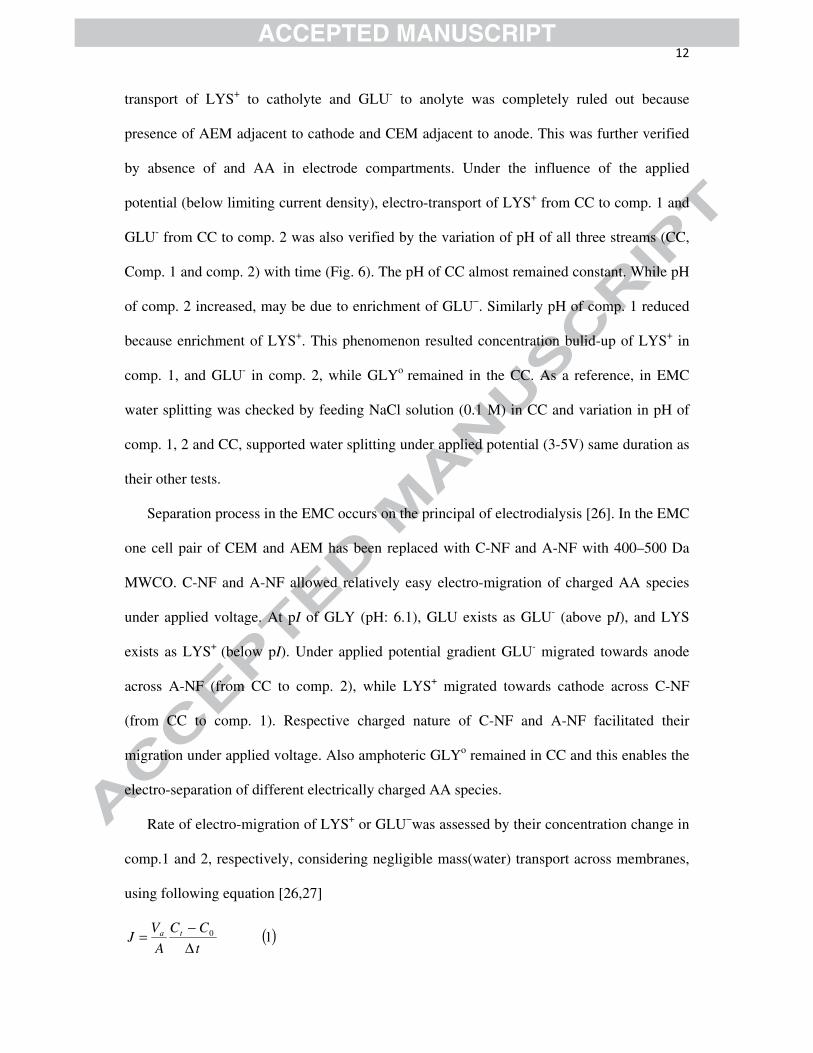
Rate of electro-migration of LYS+ or GLU−was assessed by their concentration change in
comp.1 and 2, respectively, considering negligible mass(water) transport across membranes,
using following equation [26,27]
( )10
t
CC
A
VJ ta
Δ−=
13
WhereC0 and Ct is the initial and final concentration of GLU−/LYS+ in comp. 1 (molm−3), t is
the time allowed (s), Va the total volume of solution in each compartment (0.50 × 10⁻³ m3),
and A is the effective membrane area. Flux of LYS+ and GLU−at 4.0 V applied voltage,
initially increased linearly and after attending maxima decreased under similar fashion (Fig.
7(A)). Initial concentrations of LYS+/GLU−in comp. 1 or 2, were almost zero. At the start of
the process, the concentrations of LYS+ and GLU−were reduced in CC, and increased in
comp. 1 and 2, respectively. The concentration of LYS+/GLU− in CC approached a minimum,
while enhanced in the comp. 1 and 2, up to 230 min. beyond, respective flux values were
reduced because of less availability of charged AA species in CC. After complete recovery of
GLU- and LYS+ from CC to comp. 2 or 1, respectively, their flux values approached to
minimum. Concentration of GLYo in CC remained almost same with close to 100% recovery
(Fig.7(B)). This confirms immobile nature of GLYo under the influence of electrical gradient
across C-NF or A-NF. Further, flux of GLU- from CC to comp. 2, increased with applied
voltage (Fig. S3, supporting informations). Effect of AAs concentration on process efficiency
of EMC was also explored by varying concentrations in CC and observing flux of desired AA
in comp. 1 or comp. 2. Data suggested that flux was enhanced with increase in feed AA
concentration.
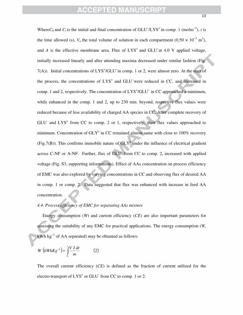
4.4. Process efficiency of EMC for separating AAs mixture
Energy consumption (W) and current efficiency (CE) are also important parameters for
assessing the suitability of any EMC for practical applications. The energy consumption (W,
kWh kg−1 of AA separated) may be obtained as follows:
( ) ( )∫=−t
m
dtIVkWhKgW
0
1 2
The overall current efficiency (CE) is defined as the fraction of current utilized for the
electro-transport of LYS+ or GLU- from CC to comp. 1 or 2:

14
Recovery of product (GLU− or LYS+) is an important parameter to examine the economic
feasibility of any process, and may be defined as:
( )3100cov00
×=FF
TPTP
CC
VCeryreAA
Where C and V denote concentration and volume, subscript 0 and T denote initial or at time
T, and subscript P and F denote product and feed. An electrochemical process must not only
be technically feasible, but should also be inexpensive with low energy consumption and high
recovery of product. To evaluate the technical and economic feasibility of the process, W, CE
(%) and recovery (%) data under different experimental conditions are presented in Table 2.
Electro-transport of LYS+ associated with 3.8 kWhr/kg of energy consumption, 96.8% CE
and 99.1% recovery. While electro-transport of GLU- was separately achieved with 3.3
kWhr/kg of energy consumption, 63.4% CE and 94.4% recovery. 98.9% of GLYo remained
in CC was recovered. These data indicate energy efficient EMC with high product recovery.
Further, Sandeaux et al reported about 60-90% CE for extraction of amphoteric amino acids
by electrodialysis [9]. Also, Nijmeijer et al reported about 24% CE and 5.1 kWhr/kg of
energy consumption for the separation of different AAs by electrodialysis with ultrafilter
membrane (EDUF) [28]. Thus, reported efficiency values in this manuscript are comparable
with the data reported in the literature.
EMC processes efficiency data for protein separation were also recorded at relatively high
applied voltage (4.0-6.0 V) (Table 3). Relatively high W and low CE may be explained by: (i)
electrode reaction and water splitting at electrodes; (ii) simultaneously transport of solvent
(water) induced by amino acid ions. Factors (i) and (ii) are responsible for enhanced energy
consumption and reduced CE with applied potential. These parameters, along with the initial
feed concentration of AA in CC, strongly affected the performance of EMC.
Also, after several successive experiments, the electrochemical properties of C-NF and A-
NF showed about ±1.0% deterioration in terms of membrane properties and process

15
efficiency. Thus any type of fouling was completely ruled out. Based on these observations, it
can be concluded that it is possible to separate LYS+, GLU- and GLYo at pH: 6.1in NaAC
buffer solution by EMC.
The separation of LYS, GLU and GLY (equi-molar, each) from their mixture was carried
out using the EMC at constant applied voltage (4.0 V) at pH: 6.1. Flux values for LYS+ and
GLU- from CC to comp. 1and comp. 2, respectively (Fig. S3, supporting information) were
depicted as a function time. Recovery of different AA under experimental conditions (Fig. 8)
was close to 100% (depended on AA mixture feed concentration). Electro-migration of LYS+
and GLU-across C-NF and A-NF towards cathode and anode, respectively, strongly depend
on their charged nature (pH), charge on membranes and applied voltage.
Proposed EMC is an important tool for separating AAs with close molecular weights by
focusing on their iso-electric values. In this process no appreciable membrane fouling or AAs
denaturation was observed. Because of different charged nature (pI) of AAs or other
biomolecules, their efficient separation can be obtained. In this particular case the difference
in the pI values of GLU and LYS was quite high, but the method could be applied in cases
where pI values are very similar, by very careful control of pH. In case of complex medium
with many AAs or peptides, reported process will be suitable for separation or extraction of
AA based on their pI values. Due to very low MWCO values for C-NF and A-NF, these
membranes are not suitable for the separation of molecules with high molecular weights
(such as peptides). Furthermore, reported process can be used with advantage for the
separating AAs from peptides.
5. Conclusions
EMC has been designed using indigenous charged NF membranes (C-NF and A-NF), for
the separation of AAs with different pI values but similar molecular weights. Water
permeabilities of C-NF and A-NF were found to be vary between (1.1-1.5 mh-1bar-1) and their

16
MWCO values (400–500 Da) suggested charged NF nature. Electrochemical properties of
these membranes suggested their suitability for EMC based on iso-electric focusing. Electro-
transport of LYS+ and GLU− across C-NF and A-NF respectively was studied under given
experimental conditions. Electrostatic interaction between the zwitterionic AA molecules and
membrane surface charge density plays important. Simultaneous electro-transport of LYS+
across C-NF and GLU−across A-NF was observed, under experimental conditions. Also,
GLYo, because of its zwitterionic nature, was immobile under applied electrical gradient and
remained in the feed compartment (CC). Thus, it was possible to separate the AA mixture by
iso-electric focusing. Also, due to relatively low energy consumption (4.0kWhr/kg of AA
separated) high CE (82%), separation of AAs (LYS-GLU-GLY) is technically feasible.
Under optimized experimental conditions (i.e. at4.0V constant applied voltage, pH: 6.1 for
0.05 M equi-molar LYS-GLU-GLY mixture), close to 90% product recovery showed the
economic feasibility of EPMC for industrial exploitation.
Also from data it is clearly evident AAs separation can be efficiently achieved by EMC
due to difference in their p Ivalues, in which applied voltage facilitates separation because the
pH of the solution does not have to change but only the applied voltage, which can be
controlled well. In conclusion it is considered that EMC offers a more techno-economically
efficient method for the production of chemicals. Further, the proposed route offers the
possibility to couple production with separation by electrodialysis.
Acknowledgments
We acknowledge the Analytical Science Division, CSMCRI, Bhavnagar for instrumental
support.

17
References
1. M.N.V. Ravi Kumar, R.A.A. Muzzarelli, C. Muzzarelli, H. Sashiwa, J. Domb,
Chitosan chemistry and pharmaceutical perspectives, Chem. Rev. 104 (2004) 6017-
6084.
2. H. Yi, L.Q. Wu, W.E. Bentley, R. Ghodssi, G.W. Rubloff, J.N. Culver, G.F. Payne,
Biofabrication with chitosan, Biomacromolecules 6 (2005) 2881-2894.
3. Y.L. Liu, C.Y. Hsu, Y.H. Su, J.Y. Lai, Chitosan−Silica Complex Membranes from
Sulfonic Acid Functionalized Silica Nanoparticles for Pervaporation Dehydration of
Ethanol−Water Solutions, Biomacromolecules 6 (2005) 368-373.
4. D. Anjali Devi, B. Smitha, S. Sridhar, T.M. Aminabhavi, Pervaporation separation of
isopropanol/water mixtures through crosslinked chitosan membranes, J. Membr. Sci.
262 (2005) 91-99.
5. T. Uragami, S. Yamamoto, T. Miyata, Dehydration from alcohols by polyion complex
cross-linked chitosan composite membranes during evapomeation,
Biomacromolecules 4 (2003) 137-144.
6. T. Uragami, T. Katayama, T. Miyata, H. Tamura, T. Shiraiwa, A. Higuchi,
Dehydration of an ethanol/water azeotrope by novel organic−inorganic hybrid
membranes based on quaternized chitosan and tetraethoxysilane, Biomacromolecules
5 (2004) 1567-1574.
7. A.G. Garem, J. Daufin, L. Maubois, J. Leonil, Selective separation of amino acids
with a charged inorganic nanofiltration membrane: effect of physicochemical
parameters on selectivity, Biotechnol. Bioeng. 54 (1997) 291-302.
8. H. Grib, L. Bonnal, J. Sandeaux, R. Sandeaux, C. Gavach, N. Mameri, Extraction of
amphoteric amino acids by an electromembrane process. pH and electrical state
18
control by electrodialysis with bipolar membranes, J. Chem. Technol. Biotechnol. 73
(1998) 64-70.
9. J. Sandeaux, R. Sandeaux, C. Gavach, H. Grib, T. Sadat, D. Belhocine, N. Mameri,
Extraction of amino acids from protein hydrolysates by electrodialysis, J. Chem.
Technol. Biotechnol. 71 (1998) 267-273.
10. F. Andreas, M. Christoph, M. Jurgen, Method for purification of amino acid
containing solution by electrodialysis, US patent 6551803.
11. M. Aider, S. Brunet, L. Bazinet, Effect of solution flow velocity and electric field
strength on chitosan oligomer electro-migration kinetics and their separation in an
electrodialysis with ultrafiltration membrane (EDUF) system, Sep. Puri. Technol. 69
(2009) 63–70.
12. E. A. Doyen, L. Saucier, L. Beaulieu, Y. Pouliot, L. Bazinet, Electro-separation of an
antibacterial peptide fraction from snow crab by-products hydrolysate by
electrodialysis with ultrafiltration membranes, Food Chemistry 132 (2012) 1177–
1184.
13. M. Aider, S. Brunet, L. Bazinet, Electro-separation of chitosan oligomers by
electrodialysis with ultrafiltration membrane (EDUF) and impact on electrodialytic
parameters, J. Membr. Sci. 309 (2008) 222–232.
14. G.J.F. Poulin, J. Amiot, L. Bazinet, Simultaneous separation of acid and basic
bioactive peptides by electrodialysis with ultrafiltration membrane, J. Biotechnol. 123
(2006) 314–328.
15. M. Aider, S. Brunet, L. Bazinet, Effect of pH and cell configuration on the selective
and specific electrodialytic separation of chitosan oligomers, Sep. Puri. Technol. 63
(2008) 612–619.

19
16. A. Saxena, G.S. Gohil, V.K. Shahi, Electrochemical membrane reactor: Single-step
separation and ion substitution for the recovery of lactic acid from lactate salts, Ind.
Eng. Chem. Res. 46 (2007) 1270- 1276.
17. L. Firdaousa, P. Dhulster, J. Amiot, A.L. Gaudreaua, D. Kapelc, R. Lutind, F.L.P.
Vézinae, L. Bazinet, Concentration and selective separation of bioactive peptides
from an alfalfa white protein hydrolysate by electrodialysis with ultrafiltration
membranes, J. Membr. Sci. 329 (2009) 60-67.
18. A. Saxena, A. Kumar, V.K. Shahi, Preparation and characterization of N-methylene
phosphonic and quaternized chitosan composite membranes for electrolyte
separations, J. Colloid Interf. Sci. 303 (2006) 484-493.
19. P.K. Narayanan, S.K. Adhikary, W.P. Harkare, K.P. Govindan, Indian Patent
(1987)160880.
20. V.K. Shahi, S.K. Thampy, R. Rangarajan, Preparation and electrochemical
characterization of sulfonated interpolymer of polyethylene and styrene
divinylbenzene copolymer membranes, React, Funct. Polym.46 (2000) 39- 47.
21. V.K. Shahi, B.S. Makwana, D.K. Gohil, S.K.Thampy, C.R.K. Reddy, R. Rangarajan,
P.K. Ghosh, Indian Patent No. (2002) 194985.
22. V.K. Shahi, S.K. Thampy, R. Rangarajan, Chronopotentiometric studies on dialytic
properties of glycine across ion-exchange membranes, J. Membr. Sci. 203 (2002) 43-
51.
23. M. Minagawa, A. Tanioka, P. Ramirez, S. Mafe, Amino acid transport through cation
exchange membranes: Effects of pH on interfacial transport, J. Colloid Interf. Sci. 188
(1997) 176- 182.
24. R.B. Schoch, A. Bertsch, P. Renaud, pH-controlled diffusion of proteins with
different pI values across a Nanochannel on a Chip, Nano Letters 6 (2006) 543-547.

20
25. A.L. Zydney, N.S. Pujar, Protein transport through porous membranes: effects of
colloidal interactions, Colloid Surface. 138 (1998) 133- 143.
26. S.S. Yi, Y.C. Lu, G.S. Luo, An in situ coupling separation process of electro-
electrodialysis with back-extraction, J. Membr. Sci. 255 (2005)57-65.
27. D.E.A. Bribiesca, M. A. Farias, G. Pourcelly, L. Bazinet, Effect of concentrate
solution pH and mineral composition of a whey protein diluate solution on membrane
fouling formation during conventional electrodialysis, J. Membr. Sci. 280 (2006)
790–80.
28. O.M.K. Readi, M. Girones, K. Nijmeijer, seperation of complex mixtures of amino
acids for biorefinery applications using electrodialysis, J. Membr. Sci. 429 (2013)
338-348.

21
Nomenclature:
T Absolute temperature (K)
i Applied current density ( mA cm-2)
V Applied potential (V)
ρ Cross-linking density
I Current (A)
η Current efficiency
m Weight (kg)
A Effective membrane area ( m2)
W Energy consumption (K Wh Kg-1)
φ Front factor
ilim1 First limiting current density (mA cm-2)
F Faraday’s constant (96,500 Cmol−1)
R Gas constant (8.314 Joule mole-1 K-1)
Ct, C0 Co and Ct are the initial and final concentration of
acid in CC (mol m-3)
(ΔE) The height of the inflection
A Membrane area ( m2)
d Membrane density
Δx Membrane thickness (cm)
κm Membrane conductivity (S cm-1)
Ps Membrane permselectivity
J Water dissociation flux (mol m-2 s-1)
n Stoichiometric number (n = 1 in this case)
Δt Time allowed for electro-membrane process (s)
Va Total volume of solution in each compartment

22
Table 1
ameasured in the
equilibration with 0.001 mol dm-3NaCl solution.
b Estimated from membrane potential measurements using NaCl solutions of0.01/0.001 mol/dm3 concentration across the membrane.
Membranes Properties
C-NF A-NF
Membrane thickness (mm) 0.20 0.20
Water content (%) 42.76 24.85
IEC (mequiv./gm) 0.202 0.199
Contact angle 76.70o 77.34o
Membrane conductivity (mS cm-1) a 2.28 2.13
Counter-ion transport number 0.86 0.83
Surface charge concentration
(m mol dm-3)
0.169 0.147

23
Table 2
AA CE, % W, kWh kg-1 of AA electro-transported
Recovery, %
LYS+ 96.8 3.8 99.1
GLY0 --- --- 98.9
GLU- 63.4 3.3 94.4

24
Table 3
Recovery, %
Applied voltage, V.
CE, %
W, kWh kg-1 of AA electro-transported
LYS+ GLU- GLY0
4.0 87.4 3.8 99.1 94.4 98.9
5.0 82.3 4.0 99.1 95.2 99.0
6.0 79.6 4.5 99.2 96.8 99.0
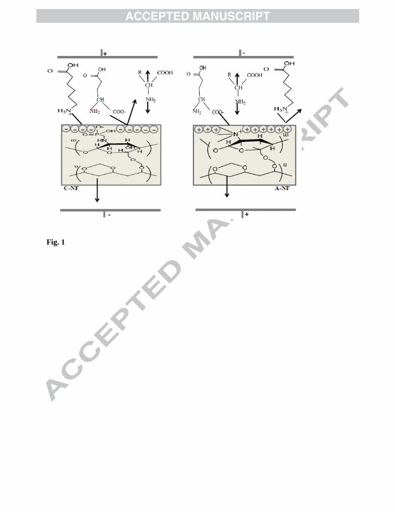
Fig. 1

Fig. 2.

Fig. 3
35
45
55
65
75
85
95
105
0 200 400 600 800 1000 1200
C-NF
A-NF
Molecular weight
% S
olu
tre
reje
ctio
n

Fig. 4.
2.5
3
3.5
4
4.5
5
5.5
0 5 10 15 20 t (s)
E (
V)
1.00 mA cm-2
1.25 mA cm-2
1.50 mA cm-2 A
3
3.5
4
4.5
5
5.5
6
0 4 8 12 16 20 t (s)
E (
V) 1.00 mA cm-2
1.25 mA cm-2
1.50 mA cm-2 B

Fig. 5.
0
10
20
30
40
50
60
50 100 150 200 250
Cu
rren
t, m
A
Time, min
A
GLU
LYS
GLY
0
10
20
30
40
50
60
50 100 150 200 250
Cu
rren
t. m
A
Time, min
B 5.0 V
4.0 V
3.0 V
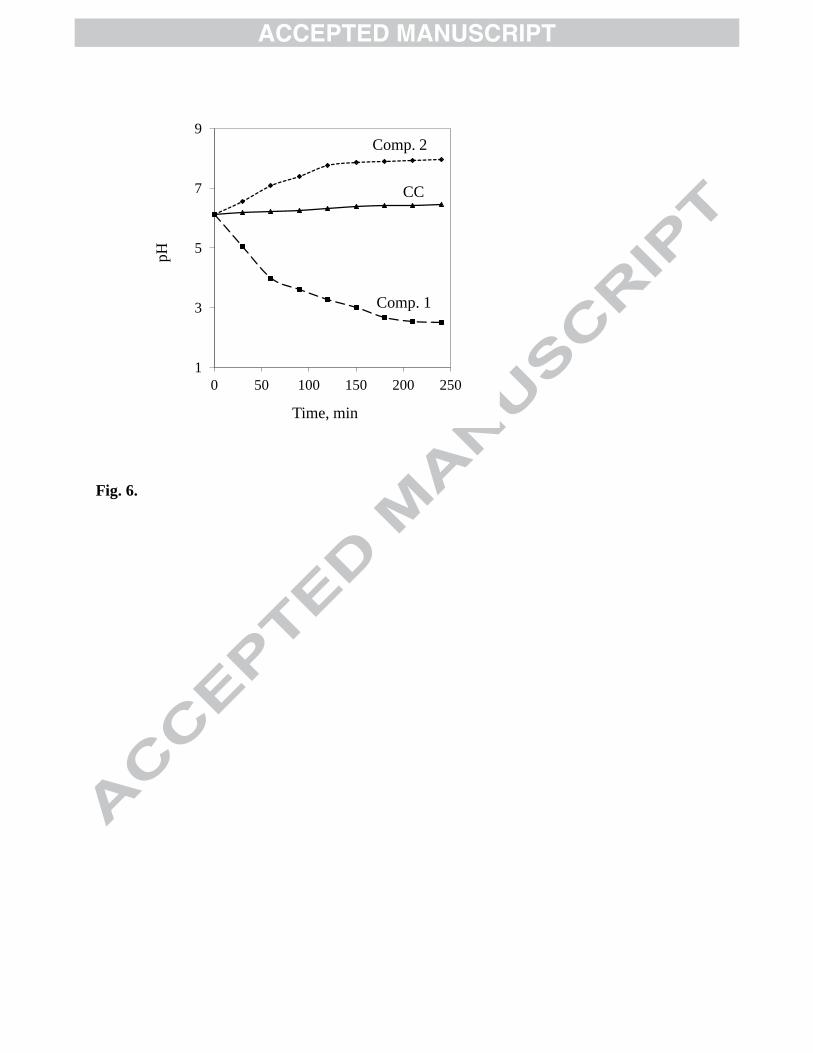
Fig. 6.
1
3
5
7
9
0 50 100 150 200 250
pH
Time, min
Comp. 1
CC
Comp. 2

Fig. 7.
0
4
8
12
16
0 100 200 300 400
J X
10
-8, m
ol
m-2
s-1
Time, min
GLU
LYS
A
94
95
96
97
98
99
100
0 100 200 300 400
Rec
over
y, %
Time, min
GLY
B

Fig. 8.

35
Table 1. Physicochemical and electrochemical properties of C-NF and A-NF
Table 2. CE, W and % recovery for different AAs separately on EMC, feed solution: 0.05 M,
separately; pH: 6.1, at 4.0V applied voltage
Table 3. CE, W and % recovery values for separation of AAs mixed solution (0.05M each)
(LYS+, GLYo, and GLU-) at pH: 6.1 as feed of CC, by EMC at different applied voltage
Fig. 1. Principle for separation of proteins using C-NF and A-NF membranes under electrical
gradient.
Fig. 2. Schematic presentation of EMC for AAs separations.
Fig. 3. MWCO values obtained from the rejection (more than 90%) curves for sugars and
poly (ethylene glycol) of different molecular weights
Fig. 4. Chronopotentiograms of: (A) A-NF in equilibration 0.10 M GLU; and (B) C-NF, in
equilibration with 0.10 M LYS solution at pH: 6.1 and different applied current density.
Fig. 5. Variation of current with time at: (A) 4.0 V applied voltage and pH: 6.1 during
electro-transport of LYS+ across C-NF; GLU- across A-NF, and CC fed with GLY (0.05 M
each separately); (B) at different applied voltage for electro transport of GLU- (0.05 M)
across A-NF.
Fig. 6. Variation of pH with time (outputof CC, comp. 1 and 2); feed of CC: AAs mixed
solution (LYS, GLU and GLY (0.05 M each) at pH:6.1) at 4.0 V applied voltage
Fig.7. Variation of Flux with time for: (A) LYS and GLU (from central comp. to comp. 1 and
comp. 2, respectively); (B) recovery of GLY in CC, at 0.05 M feed concentration (each), 4.0
V applied voltage, pH: 6.1.
36
Fig. 8. Recovery of AAs for different concentrations of feed mixture at pH: 6.1 and 4.0 V
applied voltage.
1
Highlights
Charge based pH-controlled separation of amino acids.
Indigenously prepared highly selective cationic and anionic charged nano-filter
membranes, used in electro-membrane cell.
Chronopotentiometric studies revealed the electro-transport and selectivity in separation.