JPR ddi nano
9
This article appeared in a journal published by Elsevier. The attached copy is furnished to the author for internal non-commercial research and education use, including for instruction at the authors institution and sharing with colleagues. Other uses, including reproduction and distribution, or selling or licensing copies, or posting to personal, institutional or third party websites are prohibited. In most cases authors are permitted to post their version of the article (e.g. in Word or Tex form) to their personal website or institutional repository. Authors requiring further information regarding Elsevier’s archiving and manuscript policies are encouraged to visit: http://www.elsevier.com/authorsrights
Transcript of JPR ddi nano

This article appeared in a journal published by Elsevier. The attachedcopy is furnished to the author for internal non-commercial researchand education use, including for instruction at the authors institution
and sharing with colleagues.
Other uses, including reproduction and distribution, or selling orlicensing copies, or posting to personal, institutional or third party
websites are prohibited.
In most cases authors are permitted to post their version of thearticle (e.g. in Word or Tex form) to their personal website orinstitutional repository. Authors requiring further information
regarding Elsevier’s archiving and manuscript policies areencouraged to visit:
http://www.elsevier.com/authorsrights

Author's personal copy
Original Article
Development, characterization and evaluation ofantiretroviral drug e Didanosine loaded serumalbumin nanocarriers for an antiretroviral therapy
Josephine Leno Jenita Joseph a,*, Vijaya Chockalingam b, Wilson Barnabas a
aDepartment of Pharmaceutics, Dayananda Sagar College of Pharmacy, Bangalore 560078, Karnataka, IndiabDepartment of Pharmaceutics, Ultra College of Pharmacy, Madurai, Tamil Nadu, India
a r t i c l e i n f o
Article history:
Received 6 August 2013
Accepted 15 August 2013
Available online 6 September 2013
Keywords:
Didanosine
Bovine serum albumin
Polysorbate 80
Biodistribution studies
a b s t r a c t
Aim: Didanosine, a nucleoside reverse transcriptase inhibitor, is used to treat HIV infection
in patients with or without acquired immunodeficiency syndrome. The objective of this
study is to prepare didanosine loaded bovine serum albumin nanoparticles by desolvation
technique and coated with 1% polysorbate 80 to improve antiretroviral therapy.
Method: Bovine serum albumin nanoparticles containing didanosine were prepared by des-
olvation techniqueandcross linkedwith8%v/v glutaraldehydesolution. Ethanol andmannitol
were used as a desolvating agent and cryoprotectant respectively. The formulated nano-
particles were characterized, evaluated andwere subjected to stability studies over a period of
threemonths. Biodistribution studies were investigated for the best formulation (D1).
Results: Nanoparticle size was averaged below 270 nmwith 0.2 PDI and zeta potential was in
the range of �23.0 to �36.6. Encapsulation efficiency ranges from 66 to 85.71% and % drug
loading ranges from 9.48 to 28.34. Cumulative percent drug release was in range of 60e80%
and release kinetics suggested that drug release was Fickian diffusion controlled. The sta-
bility studies over period of 3 months confirmed the stability of BSA nanoparticles. Bio-
distribution studiesdemonstrated that thedrug level inmacrophageorgans canbeenhanced
by coating of nanoparticles with 1% polysorbate 80.
Conclusion: The method adopted is simple and able to prepare stable, spherical shaped
nanoparticles which exhibit slow and sustained release.
Copyright ª 2013, JPR Solutions; Published by Reed Elsevier India Pvt. Ltd. All rights
reserved.
1. Introduction
Globally each year about 5 million people contract the virus
and over 3 million, including 500,000 children, die of acquired
immune deficiency syndrome (AIDS). HIV is concentrated in
specific anatomic sites such as central nervous system,
lymphoid organs and also testicles, female genital tract.1 Al-
bumin is emerging as a versatile protein carrier for thera-
peutic, diagnostic agent, drug targeting and for improving the
pharmacokinetic profile of drugs. In addition, it is likely that
endogenous albumin and abundant plasma protein, with the
half-life of 19 days in the blood circulation, may play an
* Corresponding author. Tel.: þ91 9448429214.E-mail address: [email protected] (J.L. Jenita Joseph).
Available online at www.sciencedirect.com
journal homepage: www.elsevier .com/locate/ jopr
j o u rn a l o f p h a rma c y r e s e a r c h 7 ( 2 0 1 3 ) 7 1 2e7 1 9
0974-6943/$ e see front matter Copyright ª 2013, JPR Solutions; Published by Reed Elsevier India Pvt. Ltd. All rights reserved.http://dx.doi.org/10.1016/j.jopr.2013.08.018
Author's personal copy
important role for improving the drug targeting properties of
many novel drugs.2 Albumin based nanocarriers drug delivery
systems are superior to traditional medicine with respect to
achieve targeted delivery, steady drug release, reduced dosing
frequency and therapeutic impact. These targeting capabil-
ities of nanocarriers have overcome many of the anatomical
and physiological barriers and deliver the drugs locally at the
HIV-infected sites thereby improving the HIV therapy.3 Even if
not providing a way to cure HIV/AIDS, the ability of a nano-
technology based systems improve drug therapy in infected
patients as demonstrated by in vitro and animal in vivo studies.
Ongoing efforts are being made to develop polymeric nano-
carriers capable of delivering active molecules specifically to
the intended target organ.4 The pharmacokinetic profile of
various therapeutic classes of antiretroviral drugs (ARV’s) can
be modifying through their incorporation into nanodelivery
systems.
There are 7 classes of FDA-approved antiretroviral agents
(ARV) and more than 25 individual drugs.5 Majority of the ARV
drugs are marketed as conventional dosage forms such as
tablets, capsules and suspensions are not able to deliver the
drug to brain due to thenature of the bloodebrain barrier (BBB).
It contains some significant drawbacks like short half-life, low
bioavailability, poor permeability and undesirable side effects.6
Didanosinewas theseconddrugapprovedbytheUSFDAfor the
treatment of patients infected with the human immunodefi-
ciency virus (HIV) in 1991. It has chosen as a model drug and
which act as chain terminators to HIV reverse transcriptase.
The most serious adverse events associated with didanosine
treatmenthavebeenperipheralneuropathy,pancreatitis, lactic
acidosis7 and also have poor gastrointestinal tolerability, un-
dergoes hepatic first pass metabolism, low oral bioavailability
(35e40%), short biological half-life (30 min e 4 h), low plasma
protein binding and narrow therapeutic index. These problems
can be overcome by formulating nanoparticles for sustained or
prolonged and targeted drug delivery.
Hence considering the importance of treating HIV, an
attempt was made to prepare didanosine loaded albumin
nanoparticles in a particular range which is suitable for the
drug delivery system that will increase bioavailability, dosing
frequency and also allow sustained drug delivery. The effect
of manufacturing conditions such as pH, BSA concentration
and agitation speed was also extensively investigated.
2. Materials and methods
2.1. Materials
Bovine serum albumin (BSA) (fraction V, with purity of 98%)
was purchased from Himedia laboratories Ltd. (Mumbai,
India). Didanosine (ddi) was received as a gift sample from the
Strides Arcolabs Ltd. (Bangalore, India). Mannitol, polysorbate
80, sodium hydroxide and glutaraldehyde and all other
chemicals were commercially supplied by Sigma Aldrich.
2.2. Method of preparation
Albumin nanoparticles were prepared by a desolvation
method.8 Different ratio of BSA powder (1%, 1.5%, 2%, 2.5% &
3%) was dissolved in distilled water; subsequently, pH was
adjusted to 8 by 0.1 M NaOH using digital pH meter under
constant stirring at 500 rpm. Didanosine was mixed, incu-
bated for 1 h at room temperature. Ethanol was added drop-
wise at a rate of 1 ml/min into the BSA solutions as a
desolvating agent until the solutions became just turbid.
Thereafter 30min of the desolvation process, 100 ml of an 8% v/
v aqueous solution of glutaraldehyde was added to induce
particle cross linking. This process was performed during
stirring over a time period of 3 h at room temperature. The
nanosuspensions were purified by two cycle centrifugation at
20,000 rpm for 30min and then subjected to freeze drying after
adding 2% (w/v) mannitol as a cryoprotectant for 8 h to obtain
fine powder of nanoparticles. The dried nanoparticles ob-
tained were then transferred to vials and were stored at 4 �C.Coating was done for D1 immediately after cross linking by
adding 1% polysorbate 80 andwas incubated for 30min, as per
the procedure described by Amit Bansal et al. Finally the
nanosuspensions was centrifuged and lyophilized with 2%
mannitol.
3. Nanoparticle characterization
3.1. Compatibility study
Compatibility of ddi and BSA, were analyzed using FT-IR
(Fourier transform infrared) spectroscopy, Shimadzu Corpo-
ration, Japan by the potassium bromide disc method (1:100).
The DSC (differential scanning calorimetry, MettlereToledo
star 822 systems, Switzerland) thermogram of drug and
lyophilized nanoparticles gives information regarding the
physical properties and melting point of the drug.
3.2. Surface morphology
Scanning electron microscopy was performed to characterize
the surface morphology of the prepared nanoparticles to
detect their morphological character of nanoparticles. This
was done by placing freeze dried nanoparticles on brass stub
then were gold-coated to render them electrically conductive
and examined under the Scanning Electron Microscope at
20 kV (JSM 6100 JEOL, Tokyo, Japan).
3.3. Determination of particle size and zeta potential
The particle size and zeta potential of didanosine albumin
nanoparticles was determined by dynamic light scattering,
using a Malvern system, with vertically polarized supplied by
Helium/Neon laser (red laser) operated at 4 mM, 633 nm. The
samples were dispersed in distilled water and taken in clear
disposable zeta cell. The experiments were performed with
non-invasive backscatter technology at a temperature of
25.0 � 0.1 �C at a detection angle of 173� to the incident beam.
3.4. Determination of % drug entrapment efficiency and% drug loading
Freshly prepared nanosuspensions were centrifuged at
20,000 rpm for 30 min and the amount of unincorporated
j o u r n a l o f p h a rm a c y r e s e a r c h 7 ( 2 0 1 3 ) 7 1 2e7 1 9 713

Author's personal copy
didanosine in supernatant liquid was measured. The %
entrapment efficiency (EE) and % drug loading were calculated
according to the following formula.
% EE ¼ ½Amount of drug actually present in nanoparticles=
amount of drug actually added� � 100
% Drug loading¼½ðTotal amount of drug added� amount of
unbound drug in supernatant liquidÞ=total amount of drug added� � 100
3.5. In vitro drug release studies
The dug release studies were carried out by dialysis method. A
knownquantity of nanoparticles equivalent to 10mgof the drug
was taken in a cellulose dialysis bag (molecular weight cut off
5kDa,Himedia, India)andadded5mlofpH7.4phosphatebuffer.
Thedialysisbagwas thensuspended inaflaskcontaining100ml
of phosphate buffer (pH 7.4) on amagnetic stirrer at 37� 0.5� at100 rpm. 5 ml quantity of sample was withdrawn at different
time periods and same volume of dissolution medium was
replaced in the flask tomaintain sink condition. Thewithdrawn
samples were filtered and then the filtrate was diluted with
phosphate buffer (pH 7.4). The samples were analyzed for drug
releasebymeasuringtheabsorbanceat249nmusingUVevisible
spectrophotometer.The invitrodrugreleasestudieswerecarried
out in triplicate for each formulation.
3.6. Release kinetics
The in vitro release data of all the formulation were fitted with
various kineticsmodels such as zero order, first order, Higuchi
model and KorsmeyerePeppas,9 in order to predict kinetics
and mechanism of drug release. The release constant was
calculated from the slope of plots and regression coefficient
(r2), diffusion exponent (n) was determined.
3.7. Stability studies
The stability study of freeze dried nanoparticles was carried
out for D1 (1:2) to assess the stability of drug in nanoparticles.
For this purpose the samples were taken in borosilicate vials
and sealed and the vials were stored in room temperature
(25�e30 �C) and refrigerator (3�e5 �C) over a period of 3
months. After specified period 0, 1, 2 and 3 months, the
samples were checked for their physical appearance and drug
content by UV spectrophotometer, as well as chemical sta-
bility by Fourier transform infrared (FTIR) studies.
3.8. Biodistribution studies
The biodistribution studies8 of ddi loaded albumin nano-
particles were carried out on healthy adult Wistar rats weigh-
ing 200e250 g and after obtaining approval from the local
animal ethics committee and CPCSEA (DSCP/PH.D PHARM/
IAEC/49/2010-2011). All animals were provided with proper
care, food, water ad libitum and were maintained under well
ventilated in large spacious cages throughout the study. The
rats were divided randomly into three groups with three ani-
mals per group and they were fasted at least 12 h before
experimentation. Group 1 was injected with ddi (which was
dispersed inwater for injection) into the tail vein of rats, Group
2 was received ddi loaded albumin nanoparticles and Group 3
was administered polysorbate 80 coated albumin nano-
particles. All the formulations were given in a dose level
equivalent to 20mg/kg body weight.7 One hour after injection,
the ratswere sacrificedby euthanized andorgans such as liver,
lung, kidney, lymph nodes, spleen, brain and blood were iso-
lated. The organs were washed with clean buffer saline and
absorbed dry with filter paper and then weighed. Prior to the
analysis organs homogenateswere prepared andwas digested
with 10% v/v trichloroacetic acid andwas treatedwith 10ml of
acetonitrile to extract didanosine. Didanosine content in the
various organswas estimated by reverse-phase HPLCmethod.
4. Results and discussions
4.1. Nanoparticle preparation
BSA nanoparticles were prepared and loaded with didanosine
by desolvation techniques with ethanol as it does not require
an increase in temperature. Initially this method was opti-
mized with different parameters like stirring speed, pH and
volume of glutaraldehyde solution to get an optimized for-
mula (data not given). This optimized method was able to
produce smooth, spherical, stable, white colored free flowing
nanoparticles. Furthermore the drug loaded nanoparticles
were characterized and evaluated.
4.2. Compatibility study
The FT-IR spectra illustrated that the characteristic peaks of
ddi, BSA and nanoparticles whereas the characteristic peaks
of nanoparticles (Fig. 1) remain samewith slightmodifications
due to other excipients present in the formulations. The DSC
thermogram of drug and lyophilized nanoparticles are shown
in Fig. 2. DSC curves showed that endothermic peak at
193.8 �C, 282.9 �C in didanosine and 77.6 �C, 193.6 �C in
nanoparticles and represented the didanosine melting point.
From DSC profiles, it was concluded that the didanosine was
present in the formulated nanoparticles in the amorphous
state and might have dispersed uniformly in the polymer.
4.3. % EE and % drug loading
% EE and % drug loading depending on the drug polymer ratio
are shown in Table 1. The % EE was decreased with respect to
drug polymer mass ratio due to limited affinity of the drug
molecule to the macromolecular material. In a nanocarrier
system the drug loading is important to determine the
amount of drug substance required for the injection. The %
drug loading was found to be high to low with increase con-
centration of BSA due to the concentration of ddi was kept
constant and was 28.34 � 0.23 to 9.48 � 0.83.
4.4. Particle size and zeta potential
The morphological properties and surface appearance of ddi
loaded BSA nanoparticles has observed using scanning
j o u rn a l o f p h a rma c y r e s e a r c h 7 ( 2 0 1 3 ) 7 1 2e7 1 9714

Author's personal copy
electron microscopy and demonstrated that nanoparticles
were spherical, smooth surface. Fig. 3a and b depicts the SEM
image and particle size distribution of ddi loaded nano-
particles. The mean particle size of ddi loaded nanoparticles
were found to be ranged between 194.8 and 268 nm with
polydispersity index was in the range of 0.121e0.281.The
mean zeta potential was found to be �23.0 to �36.6 which
indicates high degrees of stability due to inter particle re-
pulsions and are shown in Table 2.
4.5. In vitro drug release
Fig. 4 shows the comparative graph of cumulative percentage
ddi release profiles from nanoparticles and was observed
Fig. 1 e FTIR spectra of didanosine, BSA, physical mixture and formulated nanoparticles.
Fig. 2 e DSC thermograms of didanosine and formulated nanoparticles.
j o u r n a l o f p h a rm a c y r e s e a r c h 7 ( 2 0 1 3 ) 7 1 2e7 1 9 715

Author's personal copy
burst release of ddi within 1 h from nanoparticles due to the
dissociation of entrapped drug close to the surface layer of
nanoparticles. Later the drug release was observed the slow
and sustained manner over 24 h. In D1% cumulative ddi
release was found to be high due higher drug loading and
lower polymer concentration than in D5 which showed %
cumulative ddi release was low and also observed lesser burst
effect.
Table 1e Percent EE and% drug loading of ddi loaded BSAnanoparticles.
Formulation code D:P % EE % Drug loading
D1 1:2 85.71 � 0.39 28.34 � 0.23
D2 1:3 79.81 � 0.58 19.95 � 0.35
D3 1:4 76.56 � 0.68 15.37 � 0.61
D4 1:5 75.75 � 0.19 12.62 � 0.29
D5 1:6 66.38 � 0.33 9.48 � 0.83
Ps 80 coated D6 1:2 83.29 � 1.53 26.4 � 0.05
Fig. 3 e a. SEM image of ddi loaded BSA nanoparticles (D1). b. Particle size distribution of formulated ddi loaded
nanoparticles (D1).
Table 2 e Physicochemical characterization of ddi loadedBSA nanoparticles.
Formulation code Particle size PDI Zeta potential
D1 194.8 0.281 �36.6
D2 201.4 0.121 �23.0
D3 211.5 0.197 �32.9
D4 223.6 0.276 �36.1
D5 268.0 0.243 �30.8
Ps 80 coated D6 196.7 0.387 �40.2
j o u rn a l o f p h a rma c y r e s e a r c h 7 ( 2 0 1 3 ) 7 1 2e7 1 9716
Author's personal copy
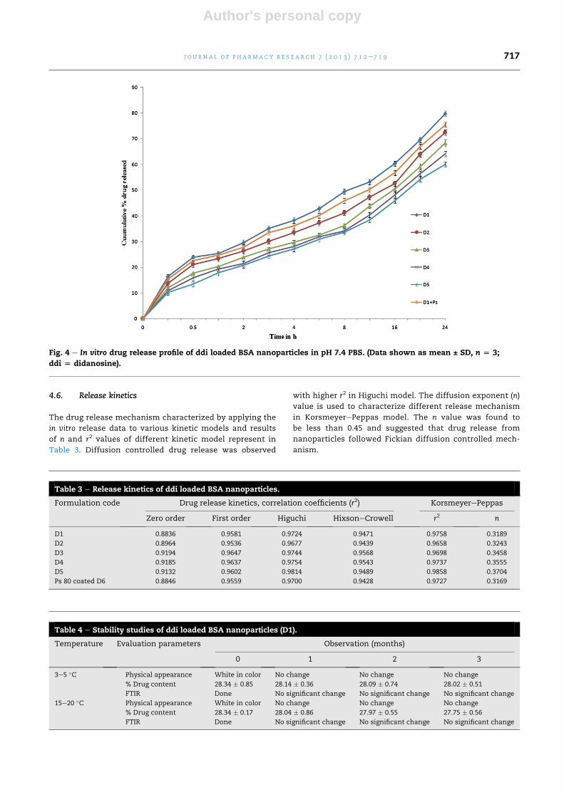
4.6. Release kinetics
The drug release mechanism characterized by applying the
in vitro release data to various kinetic models and results
of n and r2 values of different kinetic model represent in
Table 3. Diffusion controlled drug release was observed
with higher r2 in Higuchi model. The diffusion exponent (n)
value is used to characterize different release mechanism
in KorsmeyerePeppas model. The n value was found to
be less than 0.45 and suggested that drug release from
nanoparticles followed Fickian diffusion controlled mech-
anism.
Fig. 4 e In vitro drug release profile of ddi loaded BSA nanoparticles in pH 7.4 PBS. (Data shown as mean ± SD, n [ 3;
ddi [ didanosine).
Table 3 e Release kinetics of ddi loaded BSA nanoparticles.
Formulation code Drug release kinetics, correlation coefficients (r2) KorsmeyerePeppas
Zero order First order Higuchi HixsoneCrowell r2 n
D1 0.8836 0.9581 0.9724 0.9471 0.9758 0.3189
D2 0.8964 0.9536 0.9677 0.9439 0.9658 0.3243
D3 0.9194 0.9647 0.9744 0.9568 0.9698 0.3458
D4 0.9185 0.9637 0.9754 0.9543 0.9737 0.3555
D5 0.9132 0.9602 0.9814 0.9489 0.9858 0.3704
Ps 80 coated D6 0.8846 0.9559 0.9700 0.9428 0.9727 0.3169
Table 4 e Stability studies of ddi loaded BSA nanoparticles (D1).
Temperature Evaluation parameters Observation (months)
0 1 2 3
3e5 �C Physical appearance White in color No change No change No change
% Drug content 28.34 � 0.85 28.14 � 0.36 28.09 � 0.74 28.02 � 0.51
FTIR Done No significant change No significant change No significant change
15e20 �C Physical appearance White in color No change No change No change
% Drug content 28.34 � 0.17 28.04 � 0.86 27.97 � 0.55 27.75 � 0.56
FTIR Done No significant change No significant change No significant change
j o u r n a l o f p h a rm a c y r e s e a r c h 7 ( 2 0 1 3 ) 7 1 2e7 1 9 717
Author's personal copy
4.7. Stability studies
The results of stability studies are shown in Table 4. The
physical as well as chemical characteristics of the formulation
were not affected at both temperature 3e5 �C and 15e25 �Cduring 3months storage. Therewere no significant changes in
drug content and FTIR spectra. From the above results the
developed nanoparticles are stable at various temperatures.
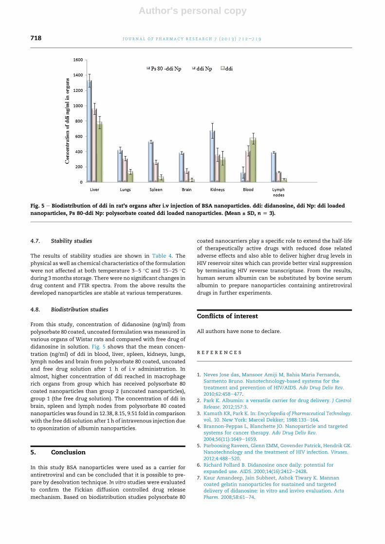
4.8. Biodistribution studies
From this study, concentration of didanosine (ng/ml) from
polysorbate 80 coated, uncoated formulationwasmeasured in
various organs of Wistar rats and compared with free drug of
didanosine in solution. Fig. 5 shows that the mean concen-
tration (ng/ml) of ddi in blood, liver, spleen, kidneys, lungs,
lymph nodes and brain from polysorbate 80 coated, uncoated
and free drug solution after 1 h of i.v administration. In
almost, higher concentration of ddi reached in macrophage
rich organs from group which has received polysorbate 80
coated nanoparticles than group 2 (uncoated nanoparticles),
group 1 (the free drug solution). The concentration of ddi in
brain, spleen and lymph nodes from polysorbate 80 coated
nanoparticles was found in 12.38, 8.15, 9.51 fold in comparison
with the free ddi solution after 1 h of intravenous injection due
to opsonization of albumin nanoparticles.
5. Conclusion
In this study BSA nanoparticles were used as a carrier for
antiretroviral and can be concluded that it is possible to pre-
pare by desolvation technique. In vitro studies were evaluated
to confirm the Fickian diffusion controlled drug release
mechanism. Based on biodistribution studies polysorbate 80
coated nanocarriers play a specific role to extend the half-life
of therapeutically active drugs with reduced dose related
adverse effects and also able to deliver higher drug levels in
HIV reservoir sites which can provide better viral suppression
by terminating HIV reverse transcriptase. From the results,
human serum albumin can be substituted by bovine serum
albumin to prepare nanoparticles containing antiretroviral
drugs in further experiments.
Conflicts of interest
All authors have none to declare.
r e f e r e n c e s
1. Neves Jose das, Mansoor Amiji M, Bahia Maria Fernanda,Sarmento Bruno. Nanotechnology-based systems for thetreatment and prevention of HIV/AIDS. Adv Drug Deliv Rev.2010;62:458e477.
2. Park K. Albumin: a versatile carrier for drug delivery. J ControlRelease. 2012;157:3.
3. Kamath KR, Park K. In: Encyclopedia of Pharmaceutical Technology.vol. 10. New York: Marcel Dekker; 1988:133e164.
4. Brannon-Peppas L, Blanchette JO. Nanoparticle and targetedsystems for cancer therapy. Adv Drug Deliv Rev.2004;56(11):1649e1659.
5. Parboosing Raveen, Glenn EMM, Govender Patrick, Hendrik GK.Nanotechnology and the treatment of HIV infection. Viruses.2012;4:488e520.
6. Richard Pollard B. Didanosine once daily: potential forexpanded use. AIDS. 2000;14(16):2412e2428.
7. Kaur Amandeep, Jain Subheet, Ashok Tiwary K. Mannancoated gelatin nanoparticles for sustained and targeteddelivery of didanosine: in vitro and invivo evaluation. ActaPharm. 2008;58:61e74.
Fig. 5 e Biodistribution of ddi in rat’s organs after i.v injection of BSA nanoparticles. ddi: didanosine, ddi Np: ddi loaded
nanoparticles, Ps 80-ddi Np: polysorbate coated ddi loaded nanoparticles. (Mean ± SD, n [ 3).
j o u rn a l o f p h a rma c y r e s e a r c h 7 ( 2 0 1 3 ) 7 1 2e7 1 9718

Author's personal copy
8. Bansal Amit, Deepak Kapoor N, Kapil Rishi, Chhabra Neha,Dhawan Sanju. Design and development of paclitaxel loadedbovine serum albumin nanoparticles for brain targeting. ActaPharm. 2011;61:141e156.
9. Suvakanta Dash, Padala Narasimha Murthy, Lilakanta Nath,Prasanta Chowdhury. Kinetic modeling on drug release fromcontrolled drug delivery systems. Acta Pol Pharm.2010;67(3):217e223.
j o u r n a l o f p h a rm a c y r e s e a r c h 7 ( 2 0 1 3 ) 7 1 2e7 1 9 719