Vibration Control of Resonant Vibratory Feeders With Electromagnetic Excitation
J Physiol 593.4 (2015) pp 825–841 825
The
Jou
rnal
of
Phys
iolo
gy
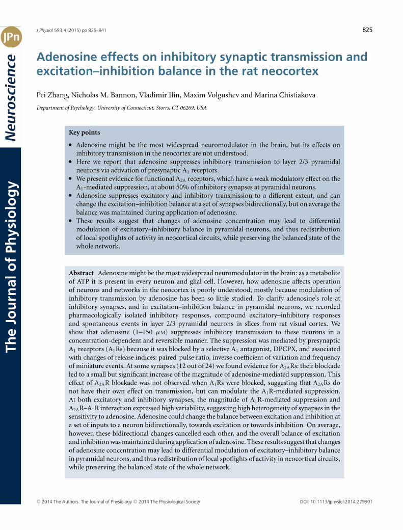
Neuroscience Adenosine effects on inhibitory synaptic transmission and
excitation–inhibition balance in the rat neocortex
Pei Zhang, Nicholas M. Bannon, Vladimir Ilin, Maxim Volgushev and Marina Chistiakova
Department of Psychology, University of Connecticut, Storrs, CT 06269, USA
Key points
� Adenosine might be the most widespread neuromodulator in the brain, but its effects oninhibitory transmission in the neocortex are not understood.
� Here we report that adenosine suppresses inhibitory transmission to layer 2/3 pyramidalneurons via activation of presynaptic A1 receptors.
� We present evidence for functional A2A receptors, which have a weak modulatory effect on theA1-mediated suppression, at about 50% of inhibitory synapses at pyramidal neurons.
� Adenosine suppresses excitatory and inhibitory transmission to a different extent, and canchange the excitation–inhibition balance at a set of synapses bidirectionally, but on average thebalance was maintained during application of adenosine.
� These results suggest that changes of adenosine concentration may lead to differentialmodulation of excitatory–inhibitory balance in pyramidal neurons, and thus redistributionof local spotlights of activity in neocortical circuits, while preserving the balanced state of thewhole network.
Abstract Adenosine might be the most widespread neuromodulator in the brain: as a metaboliteof ATP it is present in every neuron and glial cell. However, how adenosine affects operationof neurons and networks in the neocortex is poorly understood, mostly because modulation ofinhibitory transmission by adenosine has been so little studied. To clarify adenosine’s role atinhibitory synapses, and in excitation–inhibition balance in pyramidal neurons, we recordedpharmacologically isolated inhibitory responses, compound excitatory–inhibitory responsesand spontaneous events in layer 2/3 pyramidal neurons in slices from rat visual cortex. Weshow that adenosine (1–150 μM) suppresses inhibitory transmission to these neurons in aconcentration-dependent and reversible manner. The suppression was mediated by presynapticA1 receptors (A1Rs) because it was blocked by a selective A1 antagonist, DPCPX, and associatedwith changes of release indices: paired-pulse ratio, inverse coefficient of variation and frequencyof miniature events. At some synapses (12 out of 24) we found evidence for A2ARs: their blockadeled to a small but significant increase of the magnitude of adenosine-mediated suppression. Thiseffect of A2AR blockade was not observed when A1Rs were blocked, suggesting that A2ARs donot have their own effect on transmission, but can modulate the A1R-mediated suppression.At both excitatory and inhibitory synapses, the magnitude of A1R-mediated suppression andA2AR–A1R interaction expressed high variability, suggesting high heterogeneity of synapses in thesensitivity to adenosine. Adenosine could change the balance between excitation and inhibition ata set of inputs to a neuron bidirectionally, towards excitation or towards inhibition. On average,however, these bidirectional changes cancelled each other, and the overall balance of excitationand inhibition was maintained during application of adenosine. These results suggest that changesof adenosine concentration may lead to differential modulation of excitatory–inhibitory balancein pyramidal neurons, and thus redistribution of local spotlights of activity in neocortical circuits,while preserving the balanced state of the whole network.
C© 2014 The Authors. The Journal of Physiology C© 2014 The Physiological Society DOI: 10.1113/jphysiol.2014.279901
826 P. Zhang and others J Physiol 593.4
(Received 20 June 2014; accepted after revision 28 November 2014; first published online 12 December 2014)Corresponding author: M. Volgushev: University of Connecticut, Department of Psychology, 406 Babbidge Road, Unit1020, Storrs, CT 06269-1020, USA. Email: [email protected]
Abbreviations A1R, adenosine receptor type 1; A2AR, adenosine receptor type 2A; Ado, adenosine; APV,D-(−)-2-amino-5-phosphonopentanoic acid; DPCPX, 8-cyclopentyl-1,3-dipropylxanthine (A1R antagonist); mEPSP,miniature excitatory postsynaptic potential; mIPSP, miniature inhibitory postsynaptic potential; PPR, paired-pulseratio; SCH-58261, 2-(2-furanyl)-7-(2-phenylethyl)-7H-pyrazolo[4,3-e][1,2,4]triazolo[1,5-c]pyrimidin-5-amine (A2ARantagonist).
Introduction
Adenosine is a potent neuromodulator, and as anATP metabolite it is abundant in the brain. Neuronsand astrocytes release adenosine and ATP in anactivity-dependent manner (Pascual et al. 2005; Wall& Dale, 2008; Halassa et al. 2009; Lovatt et al. 2012).In the extracellular space, adenosine-phosphates arebroken down to form adenosine (Dunwiddie et al.1997). Extracellular concentration of adenosine is thusan indicator of activity and energy demand: as activityand energy expenditure rise from physiological topathological, levels of adenosine increase (Fredholm,2014). Consequently, adenosine is implicated in amultitude of functions associated with physiological andpathological alterations of brain activity. These functionsinclude regulation of the sleep–wake cycle and corticalslow oscillations (Bjorness & Greene, 2009; Halassa et al.2009), cognition, learning and memory (Fuxe et al. 2007;Halassa et al. 2009; Kadowaki Horita et al. 2013) as well asthe neuroprotective response to traumatizing events suchas hypoxia, ischaemia and excitotoxicity (de Mendoncaet al. 2000; Dunwiddie & Masino, 2001; Cunha, 2005;Gomes et al. 2011). Moreover, potential therapeutic effectsof adenosine receptor activation (Fredholm, 2010) havebeen recently explored for management of neurologicaldiseases including epilepsy (Fedele et al. 2006; Dale &Frenguelli, 2009; Masino et al. 2011), Parkinson’s disease(Hurley et al. 2000; Kitagawa et al. 2007) and schizophrenia(Deckert et al. 2003; Hong et al. 2005).
Out of four types of G-protein coupled adenosinereceptors (A1, A2A, A2B, and A3), the A1Rs and A2ARsare the most abundant in the brain (Fastbom et al. 1987;Svenningsson et al. 1997; Chaudhuri et al. 1998; Cremeret al. 2011). A1Rs are widespread throughout the brain,including the cerebral cortex (Dixon et al. 1996; Fredholmet al. 2001). A1R activation has a generally suppressiveeffect on excitatory transmission and cell excitability inthe hippocampus (Dunwiddie & Haas, 1985; Thompsonet al. 1992; Scanziani et al. 1992; Wu & Saggau, 1994;Gundlfinger et al. 2007) and neocortex (Murakoshi et al.2001; Kerr et al. 2013; van Aerde et al. 2013; Bannonet al. 2014). A2ARs are expressed most densely within thestriatum, nucleus accumbens, and olfactory bulb (Lopes
et al. 1999; Murakoshi et al. 2001; Schiffmann et al. 2007),but are also present in the neocortex (Cunha et al. 1996;Dixon et al. 1996; Sebastiao & Ribeiro, 1996; Fredholmet al. 2001; Lopes et al. 2004). Only few studies havefound a facilitatory effect of A2AR activation on excitatorysynaptic transmission in the hippocampus (Lopes et al.2002; Dias et al. 2012) and neocortex (Bannon et al.2014). Actions of A2ARs at cortical synapses are poorlyunderstood, partially because A2ARs often do not directlymodulate neural activity, but rather interact with receptorsfor other neuromodulators (Sebastiao & Ribeiro, 2009).Expression of A2AR in only a portion of synapses, andthe small magnitude of the effects further complicatethe study of A2AR-regulation of cortical synaptictransmission.
In the cortex, the effects of adenosine on synaptictransmission have been studied mostly at excitatorysynapses. Regulation of inhibitory synapses by adenosinehas received little attention, partially because early studiesin the hippocampus found that adenosine suppressesexcitatory, but not inhibitory transmission (Lambert& Teyler, 1991; Yoon & Rothman, 1991; Brundege &Dunwiddie 1996). Recent work in the neocortex provideda patchy picture: adenosine had no effect on inhibitorytransmission to layer 2/3 pyramids in the prefrontalcortex of rats (Mathew et al. 2008), but did suppressinhibitory transmission in mice, to layer 5 pyramidsin somatosensory cortex (Kruglikov & Rudy, 2008) andCajal–Retzius neurons in immature visual cortex (Kirmseet al. 2008). It is not clear from these sparse data whetherthe effects of adenosine on inhibitory transmission arerestricted to specific cell types, brain regions, or evenspecies.
In the study reported here we asked: How doesadenosine modulate inhibitory transmission to layer 2/3pyramidal neurons in rat visual cortex? Does it change theexcitatory–inhibitory balance? Which receptors mediateadenosine‘s effects on inhibitory transmission? To addressthese questions, we made whole-cell recordings from layer2/3 pyramidal neurons in slices, and measured the effectsof bath application of adenosine and antagonists of A1 andA2A receptors on pharmacologically isolated inhibitorysynaptic responses, spontaneous events and compoundexcitatory–inhibitory responses.
C© 2014 The Authors. The Journal of Physiology C© 2014 The Physiological Society
J Physiol 593.4 Adenosine modulates inhibition in the neocortex 827
Methods
Slice preparation
All experimental procedures used in this study are incompliance with the US National Institutes of Healthregulations and were approved by the Institutional AnimalCare and Use Committee of the University of Connecticut.Details of slice preparation and recording were similar tothose used in previous studies (Volgushev et al. 2000; Leeet al. 2012; Bannon et al. 2014). Wistar rats (16–28 days old,Charles River, Wilmington, MA, USA or Harlan, SouthEaston, MA USA) were anaesthetized with isoflurane,decapitated, and the brain was quickly removed andplaced into an ice-cold oxygenated artificial cerebrospinalfluid solution (ACSF), containing (in mM): 125 NaCl,25 NaHCO3, 25 glucose, 3 KCl, 1.25 NaH2PO4, 2 CaCl2,1 MgCl2, bubbled with 95% O2–5% CO2, pH 7.4. Coronalslices (350 μm thick) containing the visual cortex wereprepared from the right hemisphere. Slices were allowedto recover for at least one hour at room temperature. Forrecording, individual slices were transferred to a recordingchamber mounted on an Olympus BX-50WI microscopeequipped with infrared differential interference contrast(IR-DIC) optics. In the recording chamber slices wereperfused with oxygenated ACSF at 28–32°C.
Intracellular recording in brain slices
Layer 2/3 pyramidal cells from visual cortex were selectedfor recording in the whole cell configuration. In total,we included in the analysis results from n = 93 cells forsynaptic stimulation experiments and n = 27 cells forminiature/spontaneous activity experiments.
Identification of pyramidal neurons using DIC micro-scopy was reliable, as demonstrated in our pre-vious work with biocytin labelling and morphologicalreconstruction of recorded neurons (Volgushev et al.2000). Intracellular pipette solution contained (in mM):130 potassium gluconate, 20 KCl, 10 Hepes, 10 sodiumphosphocreatine, 4 Mg-ATP, 0.3 Na2-GTP, (pH 7.4 withKOH). All drugs were bath applied. For experimentsrecording GABAergic postsynaptic potentials, theglutamatergic blockers 6,7-dinitroquinoxaline-2,3-dione(DNQX, 10 μM; Sigma, St Louis, MO, USA) and(2R)-amino-5-phosphonopentanoate (APV, 20 μM;Tocris, Bristol, UK) were applied to block AMPA receptorsand NMDA receptors, respectively. For experimentsmeasuring miniature EPSPs (mEPSPs), tetrodotoxin(TTX, 0.1–0.5 μM; Tocris) was added to the extracellularsolution at least 25 min before recordings were started.TTX was dissolved in water to make a 0.5 mM stock beforebeing added to the bath. In some experiments (wherementioned), picrotoxin (100 μM; Sigma) was used to
block inhibitory transmission. Picrotoxin was dissolvedin the ACSF directly. Adenosine (Sigma) was dissolvedin ACSF to make a 1 mM stock before being applied tothe bath. 8-Cyclopentyl-1,3-dipropylxanthine (DPCPX;Sigma) was dissolved in >99.9% DMSO to make a1 mM stock. SCH-58261 (Tocris) was dissolved in >99.9%DMSO to make a 1 mM stock. The final concentration ofDMSO in bath was <0.05%.
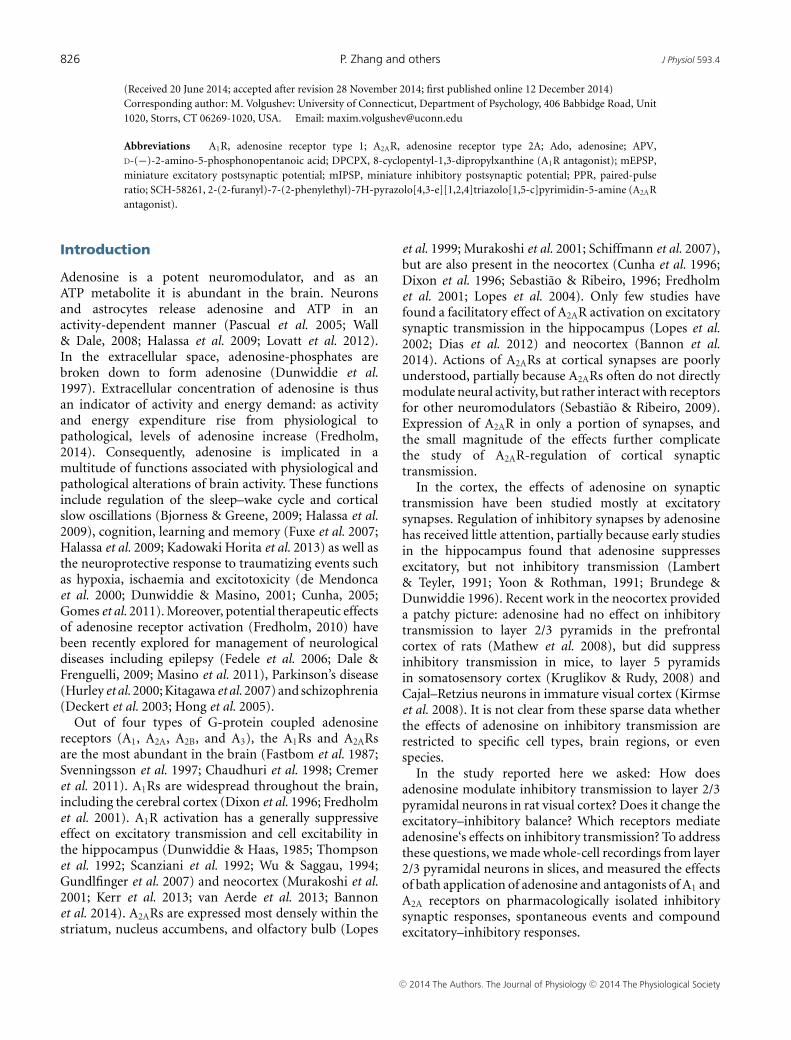
With the ionic composition of extracellular andpipette solutions used in this study, the equilibriumpotential for chloride, and thus reversal potential ofGABAA receptor-mediated postsynaptic potentials (PSPs)is expected to be approximately at −50 mV. Indeed,reversal potential of pharmacologically isolated (DNQXand APV in the bath) inhibitory postsynaptic currents(IPSCs) was between −50 mV and −55 mV (Fig. 1Cand D). Thus at resting membrane potential (typicallybetween −65 and −75 mV) synaptic stimulation induceddepolarizing GABAergic IPSPs and inward IPSCs. TheGABAergic nature of evoked responses was confirmedby their blockade by PTX (100 μM) in n = 8experiments (Fig. 1B, lower trace). In all experiments,synaptic responses were recorded at resting membranepotential (around −70 mV) and at depolarized potentials(� −45 mV) (Fig. 1B, top trace). Only responses thatexpressed reversal were considered GABAergic and usedfor the analysis.
Synaptic stimulation
Two pairs of stimulating electrodes (S1 and S2) wereplaced in layer 4, below the layer 2/3 recording site,either in layer 4 (Fig. 1A) or in layer 5. In both cases,we used a paired-pulse stimulation protocol with a50 ms inter-pulse interval. Paired stimuli were appliedto S1 and S2 in alternating sequence once per 7.5 s; asa result each input was stimulated with paired pulsesevery 15 s. Layer 4 stimulation (Fig. 1A) was used inexperiments with pharmacologically isolated IPSPs orIPSCs. This stimulation was similar to that used in ourprevious study of adenosine’s effects on excitatory trans-mission (Bannon et al. 2014), thus facilitating comparisonof results. Stimulation intensity was adjusted to evokeminimal responses. Layer 5 stimulation was used to inducecompound excitatory/inhibitory postsynaptic currents.These responses were recorded in voltage clamp mode atdifferent holding potentials (−40 mV, −50 mV, −60 mVand −70 mV) and used for calculation of excitatoryand inhibitory conductances (see below). Stimulationintensity was adjusted to obtain clearly biphasic responsesat Vhold = −40 mV. Stimulation intensity in theseexperiments was typically �5–10 stronger than thatin experiments with pharmacologically isolated IPSPsevoked by stimulation in layer 4.
C© 2014 The Authors. The Journal of Physiology C© 2014 The Physiological Society
828 P. Zhang and others J Physiol 593.4
Data analysis
Data analysis was made by using custom-writtenprograms in MatLab environment (MathWorks, Natick,MA, USA). All inputs included in the analysis fulfilled thecriteria of (1) stability of IPSP/IPSC amplitudes duringthe control period, (2) stability of the membrane potentialthroughout the recording, and (3) stability of the onsetlatency and kinetics of the rising slope of the responses.IPSP (IPSC) amplitudes were measured as the differencebetween the mean membrane potential (current) duringtwo time windows (0.5–2 ms width), the first windowplaced before the response onset and the second windowplaced on the rising slope of the postsynaptic response,just before the peak (see examples in Figs 1C and 3A).The amplitude of the response to the second pulse in thepaired-pulse stimulation protocol was measured usingwindows of the same duration, but shifted by the lengthof the inter-pulse interval (50 ms).
Excitatory and inhibitory conductances duringcompound responses were calculated by solving theconductance model equation (Monier et al. 2008). Thefollowing equations were used:
I inj = CmdVm
dt+ I inh + Iexc + I leak, (1)
where Iinj denotes the injected current, Cm the membranecapacitance, Iexc the excitatory current produced bychanges of excitatory conductance during the compoundresponses, Iinh the current produced by changes ofinhibitory conductance, Ileak the leak current. In voltageclamp mode Ileak equals to the holding current beforethe stimulus application, and was directly measured ina window placed before the stimulus. When applied tovoltage clamp measurement, the derivative terms are zero,thus eqn (1) yields:
I inj = I inh + Iexc + I leak, (2)
where excitatory and inhibitory currents are defined by:
Iexc = G exc(Vhold − E exc), (3)
I inh = G inh(Vhold − E inh), (4)
where Gexc denotes the excitatory conductance, Vhold theholding potential, Eexc the reversal potential of excitatorycomponent (0 mV), Ginh the inhibitory conductance, Einh
the reversal potential of inhibitory component (−50 mV).Substituting Iexc and Iinh in eqn (2) using eqns (3) and (4)leads to:
I 1inj(t) = G exc(t)
(V1
hold − E exc
)
+G inh(t)(V1
hold − E inh
) + I leak.(5)
For calculation of the time course of conductancechanges, for each time point t a linear system consisting
of four sets of eqn (5) each containing two unknownsGexc and Ginh was composed for four different holdingpotentials (V1
hold . . . V4hold):
I 1inj(t) = G exc(t)
(V1
hold − E exc
)
+ G inh(t)(V1
hold − E inh
) + I leak,
. . .
I 4inj(t) = G exc(t)
(V4
hold − E exc
)
+G inh(t)(V4
hold − E inh
) + I leak.
The system was solved using internal MatLab functionlinsolve. Solution of the system of equations for eachtime point t gave us time course of conductance changes,Gexc(t) and Ginh(t). These traces were used to calculate theamplitudes of excitatory and inhibitory conductances.
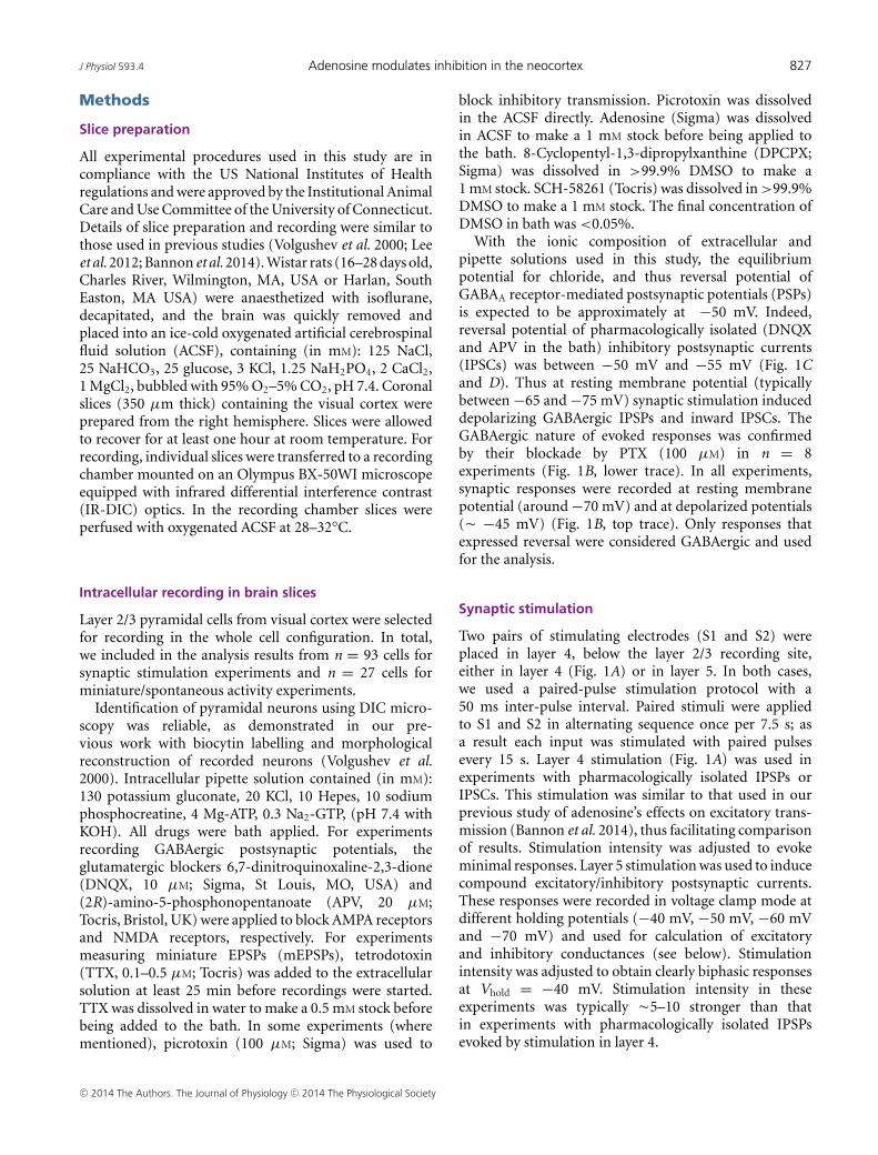
The amplitudes of excitatory and inhibitoryconductance were calculated as the mean conductancevalue in the time window placed over the peak of theGexc(t) and Ginh(t) (Figs 2A and 9A). We have used twoapproaches to validate the conductance measurements.First, we have reconstructed total current responsesat the recorded holding potential using the estimatedconductances, and compared the reconstructed responsesto those recorded. The reconstructed (black traces inFig. 2A and C) and recorded (red traces in Fig. 2A andC) traces overlap almost completely, demonstratingthat reconstructed responses reproduced the recordedresponses well. Second, we have calculated conductancesusing pharmacologically isolated IPSCs recorded in thepresence of 10 μM DNQX and 20 μM APV. As expected,calculated conductances showed a strong inhibitory butnegligible excitatory component (Fig. 2C and D).
For statistical analysis we used Student’s t tests orone-way ANOVAs with post hoc comparisons (Dunnett’sand Tukey’s HSD). Error bars represent the standard errorof the mean (± SEM).
Results
Adenosine reduces the amplitude of evoked IPSPsand increases the paired-pulse ratio
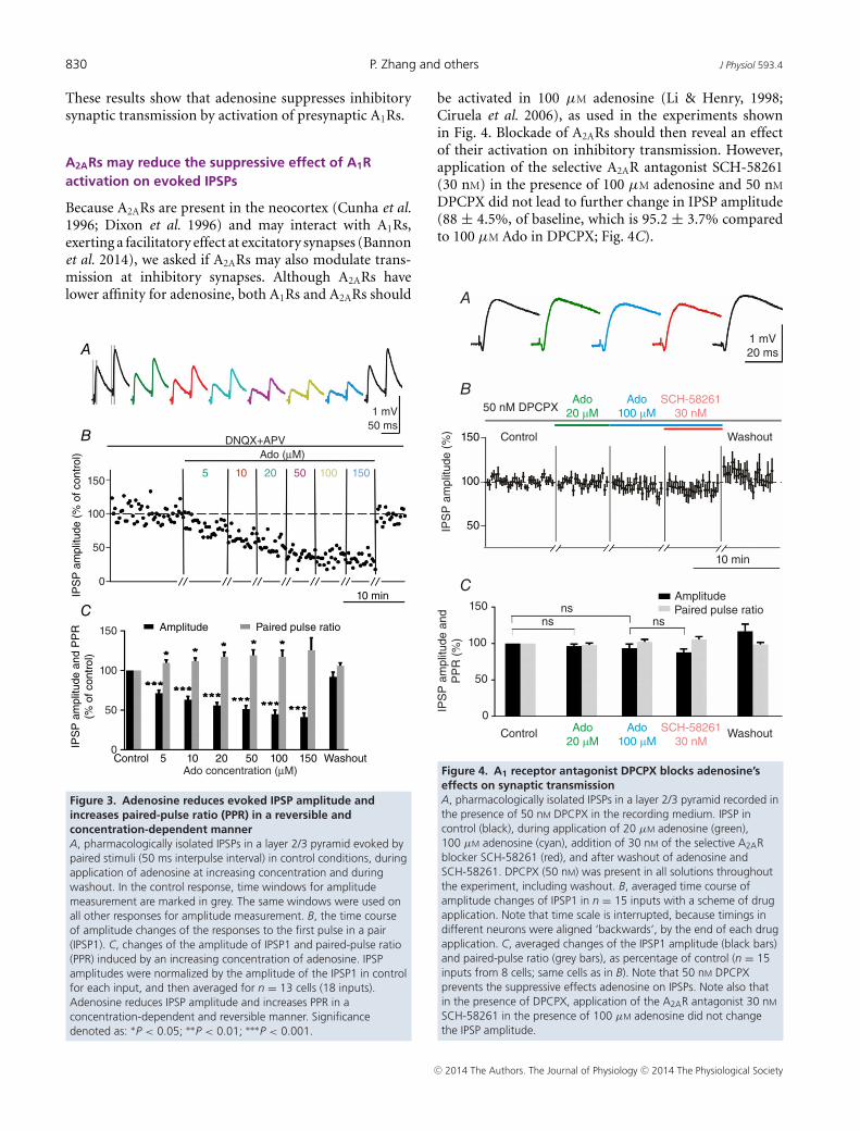
To examine the effects of adenosine on inhibitory synaptictransmission to layer 2/3 pyramidal neurons we recordedIPSPs evoked by paired-pulse electric stimuli in controlconditions and during bath application of adenosineat different concentrations. To facilitate comparison ofthe effects of adenosine on inhibitory transmissionwith results of our previous study on excitatory trans-mission (Bannon et al. 2014), we used the sameexperimental setting, and bath applied adenosine atincreasing concentrations, from 5 μM to 150 μM (Fig. 3Aand B). Already with the lowest tested concentration of5 μM, adenosine induced a clear decrease of the IPSP
C© 2014 The Authors. The Journal of Physiology C© 2014 The Physiological Society
J Physiol 593.4 Adenosine modulates inhibition in the neocortex 829
amplitude to 71.3 ± 3.9% of baseline (P < 0.001) (Fig. 3C).Increasing the concentration of adenosine in the bath ledto a progressive reduction of the IPSP amplitude. At 20 μM,adenosine had a robust effect, reducing the IPSP amplitudeto 55.9 ± 3.7% of baseline (Fig. 3C, P < 0.001). Thisconcentration was selected for further experiments.
The reduction in IPSP amplitude during applicationof adenosine was accompanied by an increase in thepaired-pulse ratio (PPR; Fig. 3C). The PPR is an indexof release that is inversely related to the release probability(Stevens, 1993; Voronin, 1993; Dobrunz & Stevens, 1997;Murthy et al. 1997; Oleskevich et al. 2000; Zucker &Regehr, 2002). The PPR significantly increased duringapplication of 5 μM adenosine to 109.7 ± 3.8% of base-line (P < 0.05). Higher concentrations of adenosine led tostronger increases of the PPR, to 117.1 ± 6.0% at 20 μM
and 119.7 ± 7.2% at 50 μM. The increasing paired-pulseratio that accompanies the decrease of IPSP amplitudeduring adenosine application suggests that adenosine hada presynaptic effect on inhibitory transmission, decreasingrelease probability at inhibitory synapses.
Cell
S2S1
–30
–45 mV
A
C
B
D
–75 mV
100
50
0
Am
plit
ud
e,
pA
–50
–70 –50Holding potential (mV)
–30–100
DNQX + APV
DNQX + APV
DNQX + APV + PTX
50 pA10 ms
0.5 mV20 ms
–40–50–60–70–80
Holding(mV)
Figure 1. Pharmacologically isolated inhibitory responses inlayer 2/3 pyramidal neuronsA, a scheme of location of recording and stimulation electrodes in aslice of rat visual cortex. Recordings were made from layer 2/3pyramidal neurons. The two bipolar stimulation electrodes, S1 andS2, were placed in layer 4 below the recording site. B,pharmacologically isolated IPSPs (10 µM DNQX and 20 µM APV inthe recording medium) reverse between −45 mV and −75 mV andare blocked by the GABAA antagonist picrotoxin (PTX, 100 µM). C,series of IPSCs recorded at different holding potentials from −30 mVto −80 mV. Vertical grey bars show time windows for amplitudemeasurement. D, IPSC amplitude plotted against holding potential,average data for n = 10 cells. The reversal potential of IPSC isbetween −50 mV and −55 mV, which corresponds to the chlorideequilibrium potential which is around −50 mV with our extracellularand intracellular solutions.
A1R blockade prevents effects of adenosine onevoked IPSPs
Which receptors mediate these effects of adenosine oninhibitory synaptic transmission? A1Rs are expressedat high levels in the neocortex (Dixon et al. 1996;Fredholm et al. 2001). Our previous data show thatsuppression of excitatory transmission by adenosine ismediated by A1Rs (Bannon et al. 2014). However, giventhe unclear effect of adenosine on inhibitory trans-mission (Yoon & Rothman, 1991; Sebastiao & Ribeiro,1996; Kruglikov & Rudy, 2008; Mathew et al. 2008),it is necessary to elucidate the roles of A1Rs andA2ARs during adenosine application. Therefore, we firstexamined whether A1Rs are involved in suppression ofinhibitory transmission by adenosine. We applied 20 μM
adenosine in the presence of the selective A1R antagonistDPCPX (50 nM) in the bath (Fig. 4A and B). In thepresence of DPCPX, the adenosine-induced reductionin IPSP amplitude was completely abolished: the IPSPamplitude remained at 96.6 ± 2.7% of 4baseline (Fig. 4C,P > 0.05). Increasing the adenosine concentration to100 μM still did not lead to a reduction in IPSP amplitude(93.1 ± 5.5%, Fig. 4C, P > 0.05). There were also nosignificant changes in the paired-pulse ratios of IPSPs in20 μM and 100 μM adenosine in the presence of DPCPX.
–40 mV
–50 mV
–60 mV
–70 mV
200 pA
10 nS Gexc
Ginh
20 ms
50 pA
2 nS
20 ms
A
B
C
D
Figure 2. Calculation of excitatory and inhibitory conductanceduring compound responses and isolated IPSCsA, compound responses recorded at holding potentialsfrom −40 mV to −70 mV. Magenta traces are recorded responses.Black traces are responses that were reconstructed using calculatedexcitatory and inhibitory conductance from B. B, excitatory (Gexc)and inhibitory (Ginh) conductance estimated using responses from A.Green and blue vertical bars in A and B show time windows forcalculating amplitudes of excitatory and inhibitory conductances. C,pharmacologically isolated IPSCs (10 µM DNQX and 20 µM APV)recorded at holding potentials from −40 mV to – 70 mV. D,excitatory (Gexc) and inhibitory (Ginh) conductance estimated usingresponses from C. Note that excitatory conductance is negligible.Conventions in C and D are as in A and B.
C© 2014 The Authors. The Journal of Physiology C© 2014 The Physiological Society
830 P. Zhang and others J Physiol 593.4
These results show that adenosine suppresses inhibitorysynaptic transmission by activation of presynaptic A1Rs.
A2ARs may reduce the suppressive effect of A1Ractivation on evoked IPSPs
Because A2ARs are present in the neocortex (Cunha et al.1996; Dixon et al. 1996) and may interact with A1Rs,exerting a facilitatory effect at excitatory synapses (Bannonet al. 2014), we asked if A2ARs may also modulate trans-mission at inhibitory synapses. Although A2ARs havelower affinity for adenosine, both A1Rs and A2ARs should
150
DNQX+APVAdo (µM)
1 mV50 ms
5 10 20 50 100 150
100
50
0
150
100
50
0
Ado concentration (µM)5 10
Amplitude
C
B
A
IPS
P a
mplit
ude a
nd P
PR
(% o
f co
ntr
ol)
IPS
P a
mplit
ude (
% o
f co
ntr
ol)
Paired pulse ratio
10 min
20 50 100 150 WashoutControl
Figure 3. Adenosine reduces evoked IPSP amplitude andincreases paired-pulse ratio (PPR) in a reversible andconcentration-dependent mannerA, pharmacologically isolated IPSPs in a layer 2/3 pyramid evoked bypaired stimuli (50 ms interpulse interval) in control conditions, duringapplication of adenosine at increasing concentration and duringwashout. In the control response, time windows for amplitudemeasurement are marked in grey. The same windows were used onall other responses for amplitude measurement. B, the time courseof amplitude changes of the responses to the first pulse in a pair(IPSP1). C, changes of the amplitude of IPSP1 and paired-pulse ratio(PPR) induced by an increasing concentration of adenosine. IPSPamplitudes were normalized by the amplitude of the IPSP1 in controlfor each input, and then averaged for n = 13 cells (18 inputs).Adenosine reduces IPSP amplitude and increases PPR in aconcentration-dependent and reversible manner. Significancedenoted as: ∗P < 0.05; ∗∗P < 0.01; ∗∗∗P < 0.001.
be activated in 100 μM adenosine (Li & Henry, 1998;Ciruela et al. 2006), as used in the experiments shownin Fig. 4. Blockade of A2ARs should then reveal an effectof their activation on inhibitory transmission. However,application of the selective A2AR antagonist SCH-58261(30 nM) in the presence of 100 μM adenosine and 50 nM
DPCPX did not lead to further change in IPSP amplitude(88 ± 4.5%, of baseline, which is 95.2 ± 3.7% comparedto 100 μM Ado in DPCPX; Fig. 4C).
150
100
50IPS
P a
mplit
ude (
%)
150 nsns ns
10 min
Washout
Washout
1 mV20 ms
Control
Control
50 nM DPCPX
A
B
C
Ado20 µM
Ado100 µM
SCH-5826130 nM
Ado20 µM
Ado100 µM
SCH-5826130 nM
AmplitudePaired pulse ratio
100
50
0
IPS
P a
mplit
ude a
nd
PP
R (
%)
Figure 4. A1 receptor antagonist DPCPX blocks adenosine’seffects on synaptic transmissionA, pharmacologically isolated IPSPs in a layer 2/3 pyramid recorded inthe presence of 50 nM DPCPX in the recording medium. IPSP incontrol (black), during application of 20 µM adenosine (green),100 µM adenosine (cyan), addition of 30 nM of the selective A2ARblocker SCH-58261 (red), and after washout of adenosine andSCH-58261. DPCPX (50 nM) was present in all solutions throughoutthe experiment, including washout. B, averaged time course ofamplitude changes of IPSP1 in n = 15 inputs with a scheme of drugapplication. Note that time scale is interrupted, because timings indifferent neurons were aligned ‘backwards’, by the end of each drugapplication. C, averaged changes of the IPSP1 amplitude (black bars)and paired-pulse ratio (grey bars), as percentage of control (n = 15inputs from 8 cells; same cells as in B). Note that 50 nM DPCPXprevents the suppressive effects adenosine on IPSPs. Note also thatin the presence of DPCPX, application of the A2AR antagonist 30 nM
SCH-58261 in the presence of 100 µM adenosine did not changethe IPSP amplitude.
C© 2014 The Authors. The Journal of Physiology C© 2014 The Physiological Society
J Physiol 593.4 Adenosine modulates inhibition in the neocortex 831
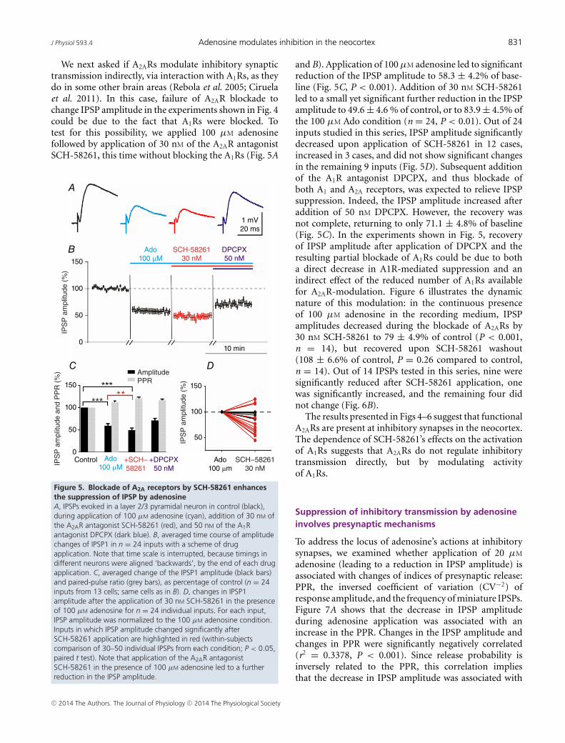
We next asked if A2ARs modulate inhibitory synaptictransmission indirectly, via interaction with A1Rs, as theydo in some other brain areas (Rebola et al. 2005; Ciruelaet al. 2011). In this case, failure of A2AR blockade tochange IPSP amplitude in the experiments shown in Fig. 4could be due to the fact that A1Rs were blocked. Totest for this possibility, we applied 100 μM adenosinefollowed by application of 30 nM of the A2AR antagonistSCH-58261, this time without blocking the A1Rs (Fig. 5A
B
A
150
Ado100 µM
SCH-5826130 nM
DPCPX50 nM
Ado100 µM
Control Ado100 µm
+SCH−58261
SCH−5826130 nM
+DPCPX50 nM
100
50
010 min
1 mV20 ms
C D
150
100
50
150
100
50
0
IPS
P a
mplit
ude (
%)
IPS
P a
mplit
ude a
nd P
PR
(%
)
IPS
P a
mplit
ude (
%)
AmplitudePPR
Figure 5. Blockade of A2A receptors by SCH-58261 enhancesthe suppression of IPSP by adenosineA, IPSPs evoked in a layer 2/3 pyramidal neuron in control (black),during application of 100 µM adenosine (cyan), addition of 30 nM ofthe A2AR antagonist SCH-58261 (red), and 50 nM of the A1Rantagonist DPCPX (dark blue). B, averaged time course of amplitudechanges of IPSP1 in n = 24 inputs with a scheme of drugapplication. Note that time scale is interrupted, because timings indifferent neurons were aligned ‘backwards’, by the end of each drugapplication. C, averaged change of the IPSP1 amplitude (black bars)and paired-pulse ratio (grey bars), as percentage of control (n = 24inputs from 13 cells; same cells as in B). D, changes in IPSP1amplitude after the application of 30 nM SCH-58261 in the presenceof 100 µM adenosine for n = 24 individual inputs. For each input,IPSP amplitude was normalized to the 100 µM adenosine condition.Inputs in which IPSP amplitude changed significantly afterSCH-58261 application are highlighted in red (within-subjectscomparison of 30–50 individual IPSPs from each condition; P < 0.05,paired t test). Note that application of the A2AR antagonistSCH-58261 in the presence of 100 µM adenosine led to a furtherreduction in the IPSP amplitude.
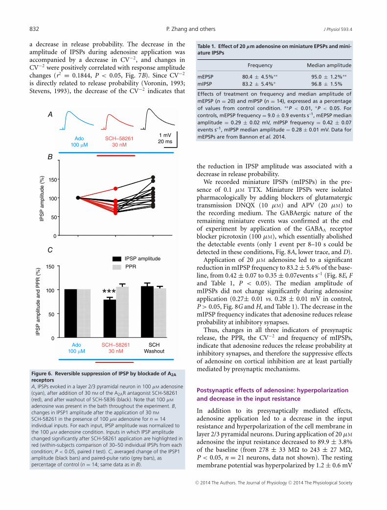
and B). Application of 100 μM adenosine led to significantreduction of the IPSP amplitude to 58.3 ± 4.2% of base-line (Fig. 5C, P < 0.001). Addition of 30 nM SCH-58261led to a small yet significant further reduction in the IPSPamplitude to 49.6 ± 4.6 % of control, or to 83.9 ± 4.5% ofthe 100 μM Ado condition (n = 24, P < 0.01). Out of 24inputs studied in this series, IPSP amplitude significantlydecreased upon application of SCH-58261 in 12 cases,increased in 3 cases, and did not show significant changesin the remaining 9 inputs (Fig. 5D). Subsequent additionof the A1R antagonist DPCPX, and thus blockade ofboth A1 and A2A receptors, was expected to relieve IPSPsuppression. Indeed, the IPSP amplitude increased afteraddition of 50 nM DPCPX. However, the recovery wasnot complete, returning to only 71.1 ± 4.8% of baseline(Fig. 5C). In the experiments shown in Fig. 5, recoveryof IPSP amplitude after application of DPCPX and theresulting partial blockade of A1Rs could be due to botha direct decrease in A1R-mediated suppression and anindirect effect of the reduced number of A1Rs availablefor A2AR-modulation. Figure 6 illustrates the dynamicnature of this modulation: in the continuous presenceof 100 μM adenosine in the recording medium, IPSPamplitudes decreased during the blockade of A2ARs by30 nM SCH-58261 to 79 ± 4.9% of control (P < 0.001,n = 14), but recovered upon SCH-58261 washout(108 ± 6.6% of control, P = 0.26 compared to control,n = 14). Out of 14 IPSPs tested in this series, nine weresignificantly reduced after SCH-58261 application, onewas significantly increased, and the remaining four didnot change (Fig. 6B).
The results presented in Figs 4–6 suggest that functionalA2ARs are present at inhibitory synapses in the neocortex.The dependence of SCH-58261’s effects on the activationof A1Rs suggests that A2ARs do not regulate inhibitorytransmission directly, but by modulating activityof A1Rs.
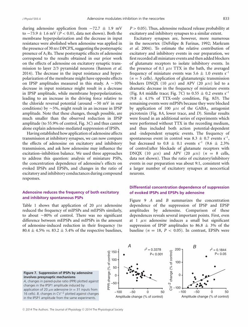
Suppression of inhibitory transmission by adenosineinvolves presynaptic mechanisms
To address the locus of adenosine’s actions at inhibitorysynapses, we examined whether application of 20 μM
adenosine (leading to a reduction in IPSP amplitude) isassociated with changes of indices of presynaptic release:PPR, the inversed coefficient of variation (CV−2) ofresponse amplitude, and the frequency of miniature IPSPs.Figure 7A shows that the decrease in IPSP amplitudeduring adenosine application was associated with anincrease in the PPR. Changes in the IPSP amplitude andchanges in PPR were significantly negatively correlated(r2 = 0.3378, P < 0.001). Since release probability isinversely related to the PPR, this correlation impliesthat the decrease in IPSP amplitude was associated with
C© 2014 The Authors. The Journal of Physiology C© 2014 The Physiological Society
832 P. Zhang and others J Physiol 593.4
a decrease in release probability. The decrease in theamplitude of IPSPs during adenosine application wasaccompanied by a decrease in CV−2, and changes inCV−2 were positively correlated with response amplitudechanges (r2 = 0.1844, P < 0.05, Fig. 7B). Since CV−2
is directly related to release probability (Voronin, 1993;Stevens, 1993), the decrease of the CV−2 indicates that
150
Ado100 µM
SCH−5826130 nM
Ado100 µM
SCH−5826130 nM
SCHWashout
IPSP amplitude
PPR
1 mV20 ms
A
B
C
100
50IPS
P a
mplit
ude (
%)
IPS
P a
mplit
ude a
nd P
PR
(%
)
0
150
100
50
0
Figure 6. Reversible suppression of IPSP by blockade of A2AreceptorsA, IPSPs evoked in a layer 2/3 pyramidal neuron in 100 µM adenosine(cyan), after addition of 30 nM of the A2AR antagonist SCH-58261(red), and after washout of SCH-5836 (black). Note that 100 µM
adenosine was present in the bath throughout the experiment. B,changes in IPSP1 amplitude after the application of 30 nM
SCH-58261 in the presence of 100 µM adenosine for n = 14individual inputs. For each input, IPSP amplitude was normalized tothe 100 µM adenosine condition. Inputs in which IPSP amplitudechanged significantly after SCH-58261 application are highlighted inred (within-subjects comparison of 30–50 individual IPSPs from eachcondition; P < 0.05, paired t test). C, averaged change of the IPSP1amplitude (black bars) and paired-pulse ratio (grey bars), aspercentage of control (n = 14; same data as in B).
Table 1. Effect of 20 µm adenosine on miniature EPSPs and mini-ature IPSPs
Frequency Median amplitude
mEPSP 80.4 ± 4.5%∗∗ 95.0 ± 1.2%∗∗
mIPSP 83.2 ± 5.4%∗ 96.8 ± 1.5%
Effects of treatment on frequency and median amplitude ofmEPSP (n = 20) and mIPSP (n = 14), expressed as a percentageof values from control condition. ∗∗P < 0.01, ∗P < 0.05. Forcontrols, mEPSP frequency = 9.0 ± 0.9 events s–1, mEPSP medianamplitude = 0.29 ± 0.02 mV, mIPSP frequency = 0.42 ± 0.07events s–1, mIPSP median amplitude = 0.28 ± 0.01 mV. Data formEPSPs are from Bannon et al. 2014.
the reduction in IPSP amplitude was associated with adecrease in release probability.
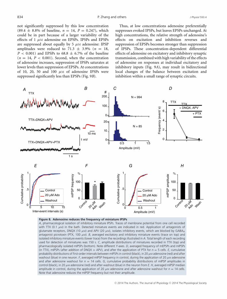
We recorded miniature IPSPs (mIPSPs) in the pre-sence of 0.1 μM TTX. Miniature IPSPs were isolatedpharmacologically by adding blockers of glutamatergictransmission DNQX (10 μM) and APV (20 μM) tothe recording medium. The GABAergic nature of theremaining miniature events was confirmed at the endof experiment by application of the GABAA receptorblocker picrotoxin (100 μM), which essentially abolishedthe detectable events (only 1 event per 8–10 s could bedetected in these conditions, Fig. 8A, lower trace, and D).
Application of 20 μM adenosine led to a significantreduction in mIPSP frequency to 83.2 ± 5.4% of the base-line, from 0.42 ± 0.07 to 0.35 ± 0.07events s–1 (Fig. 8E, Fand Table 1, P < 0.05). The median amplitude ofmIPSPs did not change significantly during adenosineapplication (0.27± 0.01 vs. 0.28 ± 0.01 mV in control,P > 0.05, Fig. 8G and H, and Table 1). The decrease in themIPSP frequency indicates that adenosine reduces releaseprobability at inhibitory synapses.
Thus, changes in all three indicators of presynapticrelease, the PPR, the CV−2 and frequency of mIPSPs,indicate that adenosine reduces the release probability atinhibitory synapses, and therefore the suppressive effectsof adenosine on cortical inhibition are at least partiallymediated by presynaptic mechanisms.
Postsynaptic effects of adenosine: hyperpolarizationand decrease in the input resistance
In addition to its presynaptically mediated effects,adenosine application led to a decrease in the inputresistance and hyperpolarization of the cell membrane inlayer 2/3 pyramidal neurons. During application of 20 μM
adenosine the input resistance decreased to 89.9 ± 3.8%of the baseline (from 278 ± 33 M� to 243 ± 27 M�,P < 0.05, n = 21 neurons, data not shown). The restingmembrane potential was hyperpolarized by 1.2 ± 0.6 mV
C© 2014 The Authors. The Journal of Physiology C© 2014 The Physiological Society
J Physiol 593.4 Adenosine modulates inhibition in the neocortex 833
during adenosine application from −72.7 ± 1.9 mVto −73.9 ± 1.6 mV (P < 0.01, data not shown). Both themembrane hyperpolarization and the decrease in inputresistance were abolished when adenosine was applied inthe presence of 30 nM DPCPX, suggesting the postsynapticpresence of A1Rs. These postsynaptic effects of adenosinecorrespond to the results obtained in our prior workon the effects of adenosine on excitatory synaptic trans-mission to layer 2/3 pyramidal neurons (Bannon et al.2014). The decrease in the input resistance and hyper-polarization of the membrane might have opposite effectson IPSP amplitudes measured in this study. A �10%decrease in input resistance might result in a decreasein IPSP amplitude, while membrane hyperpolarization,leading to an increase in the driving force relative tothe chloride reversal potential (around −50 mV in ourconditions) by �5%, might result in an increase in IPSPamplitude. Note that these changes, though possible, aremuch smaller than the observed reduction in IPSPamplitude (to 55.9% of control, Fig. 3C) and thus cannotalone explain adenosine-mediated suppression of IPSPs.
Having established how application of adenosine affectstransmission at inhibitory synapses, we can now comparethe effects of adenosine on excitatory and inhibitorytransmission, and ask how adenosine may influence theexcitation–inhibition balance. We used three approachesto address this question: analysis of miniature PSPs,the concentration dependence of adenosine’s effects onevoked IPSPs and EPSPs, and changes in the ratio ofexcitatory and inhibitory conductances during compoundresponses.
Adenosine reduces the frequency of both excitatoryand inhibitory spontaneous PSPs
Table 1 shows that application of 20 μM adenosinereduced the frequency of mIPSPs and mEPSPs similarly,to about �80% of control. There was no significantdifference between mEPSPs and mIPSPs in the amountof adenosine-induced reduction in their frequency (to80.4 ± 4.5% vs. 83.2 ± 5.4% of the respective baselines,
P > 0.05). Thus, adenosine reduced release probability atexcitatory and inhibitory synapses to a similar extent.
Excitatory synapses are, however, more numerousin the neocortex (DeFelipe & Farinas, 1992; Markramet al. 2004). To estimate the relative contribution ofexcitatory and inhibitory events in our preparation, wefirst recorded all miniature events and then added blockersof glutamate receptors to isolate inhibitory events. Inthe presence of 0.1 μM TTX in the bath, the averagedfrequency of miniature events was 5.6 ± 1.0 events s–1
(n = 5 cells). Application of glutamatergic transmissionblockers DNQX (10 μM) and APV (20 μM) led to adramatic decrease in the frequency of miniature events(Fig. 8A middle trace; Fig. 7C) to 0.55 ± 0.2 events s–1
(9.8 ± 4.1% of TTX-only condition, Fig. 8D). Theremaining events were mIPSPs because they were blockedby application of 100 μM of the GABAA antagonistpicrotoxin (Fig. 8A, lower trace, and D). Similar resultswere found in an additional series of experiments whichwere performed without TTX in the recording medium,and thus included both action potential-dependentand -independent synaptic events. The frequency ofspontaneous event in control was 8.3 ± 0.7 events s–1,but decreased to 0.8 ± 0.1 events s–1 (9.6 ± 2.3%of control)after blockade of glutamate receptors withDNQX (10 μM) and APV (20 μM) (n = 8 cells,data not shown). Thus the ratio of excitatory/inhibitoryevents in our preparation was about 9:1, consistent witha larger number of excitatory synapses at neocorticalneurons.
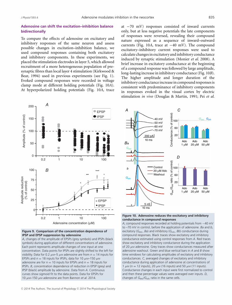
Differential concentration dependence of suppressionof evoked IPSPs and EPSPs by adenosine
Figure 9 A and B summarizes the concentrationdependence of the suppression of IPSP and EPSPamplitudes by adenosine. Comparison of thesedependences reveals several important points. First, evenat 1 μM adenosine induces a small but significantsuppression of IPSP amplitudes to 86.8 ± 3% of thebaseline (n = 18, P < 0.05). In contrast, EPSPs were
150
A Br2 = 0.3378
P< 0.001r2 = 0.1844
P< 0.05
100
50
0
−50
200
100
0
−100−100 −50
Amplitude change (% of control) Amplitude change (% of control)
PP
R c
hange (
% o
f co
ntr
ol)
CV
−2 c
hange (
% o
f co
ntr
ol)
0 50 −100 −50 0 50
Figure 7. Suppression of IPSPs by adenosineinvolves presynaptic mechanismsA, changes in paired-pulse ratio (PPR) plotted againstchanges in the IPSP1 amplitude induced byapplication of 20 µM adenosine (n = 31 inputs from16 cells). B, changes in CV−2 plotted against changesin the IPSP1 amplitude from the same experiments.
C© 2014 The Authors. The Journal of Physiology C© 2014 The Physiological Society
834 P. Zhang and others J Physiol 593.4
not significantly suppressed by this low concentration(89.4 ± 8.8% of baseline, n = 14, P = 0.247), whichcould be in part because of a larger variability of theeffects of 1 μM adenosine on EPSPs. IPSPs and EPSPsare suppressed about equally by 5 μM adenosine: IPSPamplitudes were reduced to 71.3 ± 3.9% (n = 18,P < 0.001) and EPSPs to 68.8 ± 6.7% of the baseline(n = 14, P < 0.001). Second, when the concentrationof adenosine increases, suppression of IPSPs saturates atlower levels than suppression of EPSPs. At concentrationsof 10, 20, 50 and 100 μM of adenosine IPSPs weresuppressed significantly less than EPSPs (Fig. 9B).
Thus, at low concentrations adenosine preferentiallysuppresses evoked IPSPs, but leaves EPSPs unchanged. Athigh concentrations, the relative strength of adenosine’seffects on excitation and inhibition reverses andsuppression of EPSPs becomes stronger than suppressionof IPSPs. These concentration-dependent differentialeffects of adenosine on excitatory and inhibitory synaptictransmission, combined with high variability of the effectsof adenosine on responses at individual excitatory andinhibitory inputs (Fig. 9A), may result in bidirectionallocal changes of the balance between excitation andinhibition within a small range of synaptic circuits.
200
TTXTTX
+ DNQX, APV
+ PTX
Fre
quency
(ev
ent/ s
)
8
6
4
2
0
%
100
50
0
%
100
50
0
Contro
l
Control
1
.5
00 0.5 1
Washout
20 µM Ado
20 µM
Ado Was
h
Contro
l
20 µM
Ado Was
h
N = 994
N = 85
mlP
SP
am
plit
ude
mlP
SP
fre
quency
Amplitude (mV)
Amplitude (mV)
Cu
mula
tive p
robabili
ty
Control
0
1
TTXA
E F G H
B C D
TTX+DNQX+APV
TTX+DNQX+APV+PTX
10 ms
1 mV
1 s
.5
05 10 15
Washout
20 µM Ado
Inter-event intervals (s)
Cu
mula
tive p
robabili
ty
100
0
20
10
00 0.5 1 1.5
Figure 8. Adenosine reduces the frequency of miniature IPSPsA, pharmacological isolation of inhibitory miniature IPSPs. Traces of membrane potential from one cell recordedwith TTX (0.1 µM) in the bath. Detected miniature events are indicated in red. Application of antagonists ofglutamate receptors, DNQX (10 µM) and APV (20 µM), isolates inhibitory events, which are blocked by GABAA
antagonist picrotoxin (PTX, 100 µM). B, averaged excitatory and inhibitory miniature events (trace on top) andisolated inhibitory miniature events (lower trace) from the recordings illustrated in A. Total length of each recordingused for detection of miniatures was 150 s. C, amplitude distributions of miniatures recorded in TTX (top) andpharmacologically isolated mIPSPs (bottom). Note different Y-axes. D, averaged frequency of mEPSPs and mIPSPs(in TTX), mIPSPs (after addition of DNQX + APV), and after the application of PTX for n = 5 cells. E, cumulativeprobability distributions of first-order intervals between mIPSPs in control (black), in 20 µM adenosine (red) and afterwashout (blue) in one neuron. F, averaged mIPSP frequency in control, during the application of 20 µM adenosineand after adenosine washout for n = 14 cells. G, cumulative probability distributions of mIPSP amplitudes incontrol (black), in 20 µM adenosine (red) and after washout (blue) in the neuron from E. H, averaged mIPSP medianamplitude in control, during the application of 20 µM adenosine and after adenosine washout for n = 14 cells.Note that adenosine reduces the mIPSP frequency but not their amplitude.
C© 2014 The Authors. The Journal of Physiology C© 2014 The Physiological Society
J Physiol 593.4 Adenosine modulates inhibition in the neocortex 835
Adenosine can shift the excitation–inhibition balancebidirectionally
To compare the effects of adenosine on excitatory andinhibitory responses of the same neuron and assesspossible changes in excitation–inhibition balance, weused compound responses containing both excitatoryand inhibitory components. In these experiments, weplaced the stimulation electrodes in layer 5, which allowedrecruitment of a more heterogeneous population of pre-synaptic fibres than local layer 4 stimulation (Kirkwood &Bear, 1994) used in previous experiments (see Fig. 1).Evoked compound responses were recorded in voltageclamp mode at different holding potentials (Fig. 10A).At hyperpolarized holding potentials (Fig. 10A, trace
200 A
B
150
100
50
0
Adenosine concentration (μM)
Am
plit
ude r
educt
ion
(% o
f co
ntr
ol)
Am
plit
ude (
% o
f co
ntr
ol)
100
75
50
25
0
0.2 1 10 100
0.2 1 10 100
EPSP
IPSP
EPSP
IPSP
Figure 9. Comparison of the concentration dependence ofIPSP and EPSP suppression by adenosineA, changes of the amplitude of EPSPs (grey symbols) and IPSPs (blacksymbols) during application of different concentrations of adenosine.Each point represents amplitude changes of one input at oneconcentration. Data points for IPSPs are slightly shifted to the left forvisibility. Data for 0.2 µM–5 µM adenosine are from n = 14 inputs forEPSPs and n = 18 inputs for IPSPs; data for 10 µM–150 µM
adenosine are for n = 10 inputs for EPSPs and n = 18 inputs forIPSPs. B, concentration dependence of reduction in EPSP (grey) andIPSP (black) amplitude by adenosine. Data from A. Continuouscurves show sigmoid fit to the data points. Data for EPSPs for10 µM–150 µM adenosine are from Bannon et al. 2014.
at −70 mV) responses consisted of inward currentsonly, but at less negative potentials the late componentsof responses were reversed, revealing their compoundnature expressed as a sequence of inward–outwardcurrents (Fig. 10A, trace at −40 mV). The compoundexcitatory–inhibitory current responses were used tocalculate changes in excitatory and inhibitory conductanceinduced by synaptic stimulation (Monier et al. 2008). Abrief increase in excitatory conductance at the beginningof a compound response was followed by large amplitude,long-lasting increase in inhibitory conductance (Fig. 10B).The higher amplitude and longer duration of theinhibitory conductance increase in compound responses isconsistent with predominance of inhibitory componentsin responses evoked in the visual cortex by electricstimulation in vivo (Douglas & Martin, 1991; Pei et al.
100
Gexc
Ginh
60
20
Ado1 µM
Ado20 µM
Ado50 µM
100
A C
D
60
20
−40 mV−50 mV−60 mV−70 mV
200 pA
Control
Ba
b20 µM Ado Washout
5 nS10 ms
Gex
c/G
inh r
atio
(%
of
con
trol)
10 ms
Co
nd
uct
an
ce (
% o
f co
ntr
ol)
Gexc Ginh
Ado1 µM
Ado20 µM
Ado50 µM
Figure 10. Adenosine reduces the excitatory and inhibitoryconductance in compound responsesA, compound responses recorded at holding potentials from −40 mVto –70 mV in control, before the application of adenosine. Ba and b,excitatory (Gexc, Ba) and inhibitory (Ginh, Bb) conductance duringcompound responses. Black traces show excitatory and inhibitoryconductance estimated using control responses from A. Red tracesshow excitatory and inhibitory conductance during the applicationof 20 µM adenosine. Grey traces show conductances measured afteradenosine washout. Green and blue vertical bars in A and B showtime windows for calculating amplitudes of excitatory and inhibitoryconductances. C, averaged changes of excitatory and inhibitoryconductance during application of adenosine at concentrations of1 µM (n = 12 inputs), 20 µM (16 inputs) and 50 µM (11 inputs).Conductance changes in each input were first normalized to controland then these percentage values were averaged over inputs. D,changes of Gexc/Ginh ratio in the same cells.
C© 2014 The Authors. The Journal of Physiology C© 2014 The Physiological Society
836 P. Zhang and others J Physiol 593.4
1991; Borg-Graham et al. 1998; Chung & Ferster, 1998).Application of 20 μM adenosine led to a clear reductionin both excitatory and inhibitory conductances (Fig. 10Band C). In n = 16 responses, excitatory conductancewas reduced to 59.1 ± 5.6% of control (P < 0.001)and inhibitory conductance was reduced to 66.8 ± 4.8%of control (Fig. 10C, P < 0.01). These changes werereversible. After washout of adenosine, excitatory andinhibitory conductances partially recovered towards theiroriginal values (94 ± 8.6 and 97.2 ± 6.3% of control,respectively). As a measure of the excitation–inhibitionbalance we used the ratio of excitatory to inhibitoryconductance. In 12 out of 16 responses, application of20 μM adenosine led to significant changes in the balancebetween excitation and inhibition. The ratio of excitatoryto inhibitory conductance was significantly (P < 0.05)increased in six responses and decreased in another sixresponses. In the remaining four responses the ratio didnot change. Interestingly, the bidirectional changes of thebalance in individual responses ‘cancelled’ each other, andon the average there was no significant change of theexcitation–inhibition balance (93.6 ± 10.3% of control,P > 0.05, Fig. 10D).
Because of the different concentration dependences ofadenosine’s suppression of excitatory and inhibitory trans-mission (Fig. 9), we expected that the excitation/inhibitionratio measured in compound responses will alsochange in concentration-dependent way. Indeed, 1 μM
adenosine, while suppressing both inhibitory andexcitatory conductances, changed their ratio in individualneurons, but not on average (99.5 ± 10.9% of control,n = 12). In contrast, application of 50 μM adenosineresulted in a small but significant shift of the averagebalance towards inhibition (85.7±6.8%, P<0.05, n=11).
Thus, adenosine may indeed change the balance ofexcitation and inhibition at specific sets of synapses,such as those contributing to compound responses inour experiments. These changes may occur in bothdirections: either excitatory or inhibitory inputs may besuppressed disproportionally, leading to a local shift in thebalance towards excitation or towards inhibition. Whenaveraged over large number of synapses, however, thebalance of excitation and inhibition remains unchangedby moderate (20 μM or less) adenosine concentrations,allowing the maintenance of large scale cortical networksin the balanced state, despite changing concentrations ofadenosine.
Discussion
The results of the present study demonstrate thefollowing points. (1) Adenosine suppresses inhibitorysynaptic transmission to layer 2/3 pyramids in rat visualcortex via activation of presynaptic A1 receptors; (2)
In half of cases (12/24) blockade of A2A receptorsled to a small but significant further increase of thissuppression, and this action required operational A1
receptors. This provides evidence for functional A2ARsat a portion of inhibitory terminals. (3) Adenosine maychange the balance between excitation and inhibitionin a concentration-dependent way: at low concentration(1 μM) it starts to suppress inhibition while havingheterogeneous effects on excitation, but at higherconcentrations (10 μM and higher) excitatory trans-mission is suppressed more strongly than inhibitory.Moreover, while the ‘local’ balance between excitatoryand inhibitory inputs to an individual neuron maychange in both directions, the overall balance averagedover all neurons is maintained under varying adenosineconcentrations.
Suppression of cortical inhibitory transmission byadenosine
Prior studies revealed highly diverse effects of adenosineon inhibitory transmission in the cortex. Results from thehippocampus consistently show that adenosine had noeffect on inhibitory transmission, although it did suppressexcitatory transmission (Lambert & Teyler, 1991; Yoon &Rothman, 1991; Brundege & Dunwiddie, 1996). In theneocortex, the picture is more complex: adenosine had noeffect on inhibitory transmission to layer 2/3 pyramidsin prefrontal cortex of rats (Mathew et al. 2008), butdid suppress inhibitory transmission in mice, to layer 5pyramids in somatosensory cortex (Kruglikov & Rudy,2008) and Cajal–Retzius neurons in immature visualcortex (Kirmse et al. 2008). Our results provide threelines of evidence for adenosine-mediated suppressionof inhibitory synapses at layer 2/3 pyramidal neuronsin rat visual cortex. Adenosine reduced the frequencyof spontaneous and miniature IPSPs, decreased theamplitude of pharmacologically isolated evoked IPSPsand decreased inhibitory conductance in compound PSPs.Although sparse, these results allow us to exclude at leastsome general dichotomies. The adenosine sensitivity ofinhibition in the neocortex is now demonstrated in bothrat and mouse, and thus is not species specific, and itoccurs in pyramidal neurons from both layer 2/3 and layer5, and thus is not generally restricted to supragranular orinfragranular layers. At the same time, the area specificityof adenosine’s effects on inhibition remains to beelucidated: existing evidence shows adenosine-mediatedsuppression of inhibition in sensory areas (Kirmse et al.2008; Kruglikov & Rudy, 2008; and our present results) butnot in layer 2/3 neurons from prefrontal cortex (Mathewet al. 2008).
Our results show that adenosine-mediated suppressionof inhibitory transmission to layer 2/3 pyramids is
C© 2014 The Authors. The Journal of Physiology C© 2014 The Physiological Society
J Physiol 593.4 Adenosine modulates inhibition in the neocortex 837
presynaptic, because it is associated with changes inrelease indices – paired-pulse ratio (PPR), inversedcoefficient of variation (CV−2) and frequency of mini-ature events – and is mediated by A1 receptors. Theseresults are consistent with documented mechanisms ofadenosine-suppression at other inhibitory synapses in theneocortex (Kirmse et al. 2008; Kruglikov & Rudy, 2008) aswell as with the wealth of data for excitatory synapses inthe neocortex (Murakoshi et al. 2001; Mathew et al. 2008;Kerr et al. 2013; Bannon et al. 2014) and hippocampus(Dunwiddie & Haas, 1985; Lambert & Teyler, 1991; Yoon& Rothman, 1991; Scanziani et al. 1992; Thompson et al.1992; Wu & Saggau, 1994; Brundege & Dunwiddie, 1996).
Evidence for functional A2A receptors at inhibitorysynapses in the neocortex
The results of the present study demonstrate the existenceof functional A2A receptors at inhibitory synapses at layer2/3 pyramids of the visual cortex. Application of a specificantagonist of A2A receptors, SCH-58261, in the presenceof 100 μM adenosine led to a further decrease in IPSPamplitude in half of all cases (12/24). However, this effectwas not present if adenosine and SCH-58261 were appliedin the presence of the A1R antagonist DPCPX. We inter-pret these results as follows. Activation of A2ARs doesnot have a direct effect on synaptic transmission, but itdiminishes the suppressive effect of A1Rs on inhibitorytransmission. A high concentration of adenosine activatesboth A1Rs, which suppress the transmission, and A2ARs,which interact with A1Rs, diminishing the suppression ata subset of inhibitory synapses. Blockade of A2ARs leads todisinhibition of A1Rs, revealing the full suppressive effectof A1R on synaptic transmission. Because A1Rs modulatesynaptic transmission via presynaptic mechanisms, theabove scenario implies that A2ARs are also located pre-synaptically. These results and their interpretation areconsistent with the hypothesis suggested by Sebastiao &Ribeiro (2009) that A2A receptors are less related to directmodulation of synaptic transmission, but rather inter-act with receptors for other modulators, and ‘fine-tune’synaptic function by modulating the effect of modulators.Support for these ideas comes from experiments onsynaptosomes showing that A2ARs modulate the affinityof A1Rs to ligands (Lopes et al. 1999), and from thefinding that A1Rs and A2ARs can form heteromers inmembranes of striatal neurons, with activation of A2ARsreducing affinity of A1Rs through direct receptor–receptorinteraction (Ciruela et al. 2006, 2011; Ferre et al.2007). Finally, present results for inhibitory synapses areconsistent with our recent finding of functional A2ARsat a portion of excitatory neocortical synapses (Bannonet al. 2014).
Specificity and heterogeneity of adenosine effects oncortical synaptic transmission
Published data show great variety in the magnitude ofadenosine’s effects on transmission at cortical synapses.Factors contributing to this variability might includedifferences between synapses in the expression of theadenosine receptors, the ratio of A1 and A2A receptors,their affinity with and efficiency in intracellular cascadestriggered by receptor activation, as well as the balancebetween local pathways for adenosine production, releaseand utilization. Experimental results show that each ofthese factors may be specific to the region of the brain,type of neuron and synapse, but also may change with age.
A case for region-specific expression of adenosinereceptors is provided by inhibitory transmission, which isnot affected by adenosine in the hippocampus (Lambert& Teyler, 1991; Yoon & Rothman, 1991; Brundege &Dunwiddie, 1996) and layer 2/3 pyramids from prefrontalcortex (Mathew et al. 2008), but can be suppressed byadenosine in sensory areas of the neocortex (Kirmseet al. 2008; Kruglikov & Rudy, 2008; and our pre-sent results). Results from hippocampus and layer 2/3pyramids from prefrontal cortex also provide a clear casefor synapse-type specificity of expression of adenosinereceptors only at excitatory synapses, which are suppressedby adenosine, but not at inhibitory synapses, which areinsensitive to adenosine (Lambert & Teyler, 1991; Yoon &Rothman, 1991; Brundege & Dunwiddie, 1996; Mathewet al. 2008). Notably, an example from a brain area inwhich the effects of adenosine have been investigated ingreat detail, the basal forebrain, shows that even excitatoryand inhibitory synapses at neighboring neurons may beeither sensitive to adenosine or not (Yang et al. 2013). Inthe basal forebrain, adenosine reduced the frequency ofspontaneous and miniature IPSCs in GABAergic neuronswith large H-currents, but not in GABAergic neurons withsmall H-currents and not in cholinergic neurons. Thefrequency of excitatory events was reduced by adenosinein all these neurons (Yang et al. 2013).
The results of the present study show, in agreementwith published data, that even at synapses of thesame type investigated under the same experimentalconditions, saturating concentrations of adenosine maysuppress transmission to a very different degree. Thisholds both for excitatory and for inhibitory synapses(e.g. Fig. 9). We interpret these results as evidence fora differential, synapse-specific, level of expression ofadenosine receptors. By the same logic, the highly variablemagnitude of the additional suppression of synaptic trans-mission by the blockade of A2A receptors in the presenceof a high adenosine concentration (Fig. 5 for inhibitorysynapses, and Fig. 9 in Bannon et al. 2014 for excitatorysynapses), suggests a variable ratio of A1/A2A receptorexpression. The possibility of area-specific and cell-type
C© 2014 The Authors. The Journal of Physiology C© 2014 The Physiological Society

838 P. Zhang and others J Physiol 593.4
specific expression of adenosine receptors in corticalneurons is further supported by reports on postsynapticeffects of adenosine. In somatosensory and prefrontalcortex, adenosine application led to hyperpolarization anda decrease in input resistance in layer 5 pyramids and insome layer 3 pyramids, but not in layer 2 pyramids andnot in inhibitory neurons (van Aerde et al. 2013). In thevisual cortex, adenosine changed the membrane potentialand input resistance of layer 2/3 pyramids (Bannon et al.2014; and present results), but no such effect was reportedin layer 5 pyramids (Murakoshi et al. 2001).
Finally, activity and maturation of the pathways foradenosine production, release and utilization, whichdetermine the activity-dependent dynamics of adenosineconcentration at a synapse, might be an important factorfor determining the ultimate effect of adenosine onsynaptic transmission. A recent study shows that in ratsomatosensory cortex, excitatory synapses between layer5 pyramids are subject to a tonic adenosine-mediatedsuppression, because application of the A1R antagonist8-CPT, which relieves synapses from the suppression,leads to an increase in the EPSP amplitude (Kerr et al.2013). Moreover, the magnitude of the 8-CPT effectincreases between postnatal days 17 and 32, indicatingthat the tonic adenosine-mediated suppression increaseswith age. Consistent with this interpretation, we haveobserved that in layer 2/3 pyramids from visual cortex,the A1R antagonist DPCPX leads to a stronger increasein the frequency of miniature EPSPs in slices fromP27–28 than in slices from P19–22 rats (N.B., P.Z.,M.V. unpublished observations). The interplay betweenactivity-dependent release of adenosine and availability oflocal mechanisms for its utilization may be an importantfactor determining the heterogeneity of local dynamics ofadenosine concentration and its effects on synaptic trans-mission in vivo.
One clear conclusion from the broad diversity ofadenosine effects considered above is that adenosine’seffects on synaptic transmission cannot be generalizedacross brain structures or cell types, and moredata on characterization of adenosine actions atspecific connections are necessary for revealing possibleorganizing principles of regulation of cortical function byadenosine.
Modulation of the balance between excitation andinhibition by adenosine
Cortical networks operate in a balanced regime (Wehr& Zador, 2003; Okun & Lampl, 2008; Ozeki et al. 2009;Dorrn et al. 2010; Sun et al. 2010). Thus, for networkoperation, not just the magnitude of a change at excitatoryor inhibitory synapses is important, but a change of thebalance between excitation and inhibition.
Our results expose two interesting aspects ofthe modulation of excitation–inhibition balance byadenosine. The first is the concentration dependenceof adenosine effects on excitation–inhibition balance.At low concentration adenosine preferentially suppressesevoked IPSPs, but has heterogeneous effect on EPSPs,so that on average excitation does not change. Becauseincreases in adenosine concentration are activity related,a functional role for lessening inhibition around the locusof weak activation might help maintain this ‘low profile’activity, e.g. for detection of threshold signals. At highconcentrations of adenosine the suppression of EPSPs isstronger than suppression of IPSPs. A high concentrationof adenosine is a correlate of strong activation, andstronger suppression of excitation by adenosine maybe one of the mechanisms preventing excessive activityin the system. Thus, the concentration dependenceof adenosine’s effects on the excitation–inhibitionbalance endorses the adenosine-feedback with homeo-static function: protecting weak activity from anexcessive inhibition, but also restricting excitation duringexcessively strong activation.
The second feature, exposed by the analysis ofcompound responses, is that adenosine may changethe balance in a subset of excitatory and inhibitorysynapses in both directions, either towards excitation ortowards inhibition. Notably, despite these local changes,the overall balance of excitation and inhibition a cellreceives does not change. In experiments with compoundresponses, although adenosine did change the balancein individual experiments, the average over all cells andinputs did not change significantly. This conclusion issupported by results of analysis of miniature events.The frequency of both miniature IPSPs and miniatureEPSPs was changed to the same extent (to �80% ofcontrol) by adenosine application. Thus, adenosine maychange the excitation–inhibition balance in a selected setof synapses, without compromising the overall balancedstate of a network. The set of synapses subject to selectivemodulation may be defined by activity or by structuralfeatures, such as glial islands or individual glial cellswhich control the adenosine concentration over localregions including 300–600 dendrites (Fellin, 2009; Halassaet al. 2009). By selective up- or down-regulation of localexcitation–inhibition balance, adenosine has the potentialto modulate the spread of activity in neuronal networks.
Conclusions and outlook: how adenosine regulatessynaptic transmission and excitation–inhibitionbalance in the neocortex
The following conclusions on adenosine’s effects onsynaptic transmission in the cortex can be drawn from the
C© 2014 The Authors. The Journal of Physiology C© 2014 The Physiological Society
J Physiol 593.4 Adenosine modulates inhibition in the neocortex 839
results of the present study and the wealth of publishedwork.
The most pronounced and common effect of adenosineon neocortical synapses is suppression of transmitterrelease mediated by presynaptic A1 receptors. Thissuppression has been invariably found at all excitatorysynapses in the cortex studied so far, indicating that pre-synaptic A1 receptors are expressed at all excitatory pre-synaptic terminals in the cortex. In contrast, only a fractionof inhibitory connections are affected by adenosine, andthus presumably adenosine receptors are expressed in onlya fraction of inhibitory presynaptic terminals. Elucidatingguiding principles of this selective expression and itsfunctional role remains a challenge for future studies.
At both excitatory and A1R-expressing inhibitorysynapses, the suppressive effect of A1 receptors can bepartially counteracted by A2ARs. The effects of A2ARswere small relative to the magnitude of A1-mediatedsuppression, but were clearly present in half of all cases(12/24). Our results show that an effect of A2AR blockadewas observed only when A1Rs were functional, but notwhen A1Rs were blocked. This suggests that at bothexcitatory and inhibitory synapses, A2ARs do not havetheir own effect on transmission, but can modulatethe A1R-mediated suppression. This interpretation isconsistent with the hypothesis by Sebastiao & Ribeiro(2009) on the role of A2ARs as modulators of theeffects of other neuromodulators. Interestingly, we foundevidence for A2ARs in only a fraction of excitatory andinhibitory synapses. Understanding which factors governthe expression of A2A receptors at some, but not at someother synapses is another challenge for future studies.
A salient feature of adenosine’s effects is high variabilityof the magnitude of suppression in different experiments,even in a homogeneous population of synapses. This holdsboth for the magnitude of A1R-mediated suppression, and,when present, the effect of A2AR–A1R interaction. Themost straightforward interpretation of this heterogeneityis the variable level of expression of adenosine receptors atpresynaptic fibres. Understanding the rules and functionalrole of this heterogeneous expression, especially atinhibitory synapses remains an open challenge.
The heterogeneity of the adenosine sensitivity ofcortical synapses sets the stage for possible differentiallocal modulation of the excitatory–inhibitory balancein cortical circuits by adenosine. In combination withthe concentration dependence of adenosine effects onthe excitation–inhibition balance, this provides adenosinewith an ability to combine activity-dependent fine-tuningof synaptic transmission with a homeostatic function,keeping activity in cortical networks within the operatingrange.
References
Bannon NM, Zhang P, Ilin V, Chistiakova M & Volgushev M(2014). Modulation of synaptic transmission by adenosinein layer 2/3 of the rat visual cortex in vitro. Neuroscience 260,171–184.
Bjorness TE & Greene RW (2009). Adenosine and sleep. CurrNeuropharmacol 7, 238–245.
Borg-Graham LJ, Monier C & Fregnac Y (1998). Visual inputevokes transient and strong shunting inhibition in visualcortical neurons. Nature 393, 369–373.
Brundege JM & Dunwiddie TV (1996). Modulation ofexcitatory synaptic transmission by adenosine released fromsingle hippocampal pyramidal neurons. J Neurosci 16,5603–5612.
Chaudhuri A, Cohen RZ & Larocque S (1998). Distribution ofadenosine A1 receptors in primary visual cortex ofdeveloping and adult monkeys. Exp Brain Res 123, 351–354.
Chung S & Ferster D (1998). Strength and orientation tuningof the thalamic input to simple cells revealed by electricallyevoked cortical suppression. Neuron 20, 1177–1189.
Ciruela F, Casado V, Rodrigues RJ, Lujan R, Burgueno J,Canals M, Borycz J, Rebola N, Goldberg SR, Mallol J et al.(2006). Presynaptic control of striatal glutamatergicneurotransmission by adenosine A1–A2A receptorheteromers. J Neurosci 26, 2080–2087.
Ciruela F, Gomez-Soler M, Guidolin D, Borroto-Escuela DO,Agnati LF, Fuxe K & Fernandez-Duenas V (2011).Adenosine receptor containing oligomers: their role in thecontrol of dopamine and glutamate neurotransmission inthe brain. Biochim Biophys Acta 1808, 1245–1255.
Cremer CM, Lubke JH, Palomero-Gallagher N & Zilles K(2011). Laminar distribution of neurotransmitter receptorsin different reeler mouse brain regions. Brain Struct Funct216, 201–218.
Cunha RA (2005). Neuroprotection by adenosine in the brain:From A1 receptor activation to A2A receptor blockade.Purinergic Signal 1, 111–134.
Cunha RA, Johansson B, Constantino MD, Sebastiao AM &Fredholm BB (1996). Evidence for high-affinity binding sitesfor the adenosine A2A receptor agonist [3H]CGS 21680 inthe rat hippocampus and cerebral cortex that are differentfrom striatal A2A receptors. Naunyn Schmiedebergs ArchPharmacol 353, 261–271.
Dale N & Frenguelli BG (2009). Release of adenosine and ATPduring ischemia and epilepsy. Curr Neuropharmacol 7,160–179.
de Mendonca A, Sebastiao AM & Ribeiro JA (2000). Adenosine:does it have a neuroprotective role after all? Brain Res BrainRes Rev 33, 258–274.
Deckert J, Brenner M, Durany N, Zochling R, Paulus W,Ransmayr G, Tatschner T, Danielczyk W, Jellinger K &Riederer P (2003). Up-regulation of striatal adenosine A2A
receptors in schizophrenia. Neuroreport 14, 313–316.DeFelipe J & Farinas I (1992). The pyramidal neuron of the
cerebral cortex: morphological and chemical characteristicsof the synaptic inputs. Prog Neurobiol 39, 563–607.
C© 2014 The Authors. The Journal of Physiology C© 2014 The Physiological Society

840 P. Zhang and others J Physiol 593.4
Dias RB, Ribeiro JA & Sebastiao AM (2012). Enhancement ofAMPA currents and GluR1 membrane expression throughPKA-coupled adenosine A2A receptors. Hippocampus 22,276–291.
Dixon AK, Gubitz AK, Sirinathsinghji DJ, Richardson PJ &Freeman TC (1996). Tissue distribution of adenosinereceptor mRNAs in the rat. Br J Pharmacol 118, 1461–1468.
Dobrunz LE & Stevens CF (1997). Heterogeneity of releaseprobability, facilitation, and depletion at central synapses.Neuron 18, 995–1008.
Dorrn AL, Yuan K, Barker AJ, Schreiner CE & Froemke RC(2010). Developmental sensory experience balances corticalexcitation and inhibition. Nature 465, 932–936.
Douglas RJ & Martin KA (1991). A functional microcircuit forcat visual cortex. J Physiol 440, 735–769.
Dunwiddie TV, Diao L & Proctor WR (1997). Adeninenucleotides undergo rapid, quantitative conversion toadenosine in the extracellular space in rat hippocampus.J Neurosci 17, 7673–7682.
Dunwiddie TV & Haas HL (1985). Adenosine increasessynaptic facilitation in the in vitro rat hippocampus: evidencefor a presynaptic site of action. J Physiol 369, 365–377.
Dunwiddie TV & Masino SA (2001). The role and regulation ofadenosine in the central nervous system. Annu Rev Neurosci24, 31–55.
Fastbom J, Pazos A & Palacios JM (1987). The distribution ofadenosine A1 receptors and 5’-nucleotidase in the brain ofsome commonly used experimental animals. Neuroscience22, 813–826.
Fedele DE, Li T, Lan JQ, Fredholm BB & Boison D (2006).Adenosine A1 receptors are crucial in keeping an epilepticfocus localized. Exp Neurol 200, 184–190.
Fellin T (2009). Communication between neurons andastrocytes: relevance to the modulation of synaptic andnetwork activity. J Neurochem 108, 533–544.
Ferre S, Ciruela F, Quiroz C, Lujan R, Popoli P, Cunha RA,Agnati LF, Fuxe K, Woods AS, Lluis C & Franco R (2007).Adenosine receptor heteromers and their integrative role instriatal function. ScientificWorldJournal 7, 74–85.
Fredholm BB (2010). Adenosine receptors as drug targets. ExpCell Res 316, 1284–1288.
Fredholm BB (2014). Adenosine – a physiological orpathophysiological agent? J Mol Med (Berl) 92, 201–206.
Fredholm BB, AP IJ, Jacobson KA, Klotz KN & Linden J (2001).International Union of Pharmacology. XXV. Nomenclatureand classification of adenosine receptors. Pharmacol Rev 53,527–552.
Fuxe K, Ferre S, Genedani S, Franco R & Agnati LF (2007).Adenosine receptor–dopamine receptor interactions in thebasal ganglia and their relevance for brain function. PhysiolBehav 92, 210–217.
Gomes CV, Kaster MP, Tome AR, Agostinho PM & Cunha RA(2011). Adenosine receptors and brain diseases:neuroprotection and neurodegeneration. Biochim BiophysActa 1808, 1380–1399.
Gundlfinger A, Bischofberger J, Johenning FW, Torvinen M,Schmitz D & Breustedt J (2007). Adenosine modulatestransmission at the hippocampal mossy fibre synapse viadirect inhibition of presynaptic calcium channels. J Physiol582, 263–277.
Halassa MM, Florian C, Fellin T, Munoz JR, Lee SY, Abel T,Haydon PG & Frank MG (2009). Astrocytic modulation ofsleep homeostasis and cognitive consequences of sleep loss.Neuron 61, 213–219.
Hong CJ, Liu HC, Liu TY, Liao DL & Tsai SJ (2005).Association studies of the adenosine A2a receptor (1976T >
C) genetic polymorphism in Parkinson’s disease andschizophrenia. J Neural Transm 112, 1503–1510.
Hurley MJ, Mash DC & Jenner P (2000). Adenosine A2A
receptor mRNA expression in Parkinson’s disease. NeurosciLett 291, 54–58.
Kadowaki Horita T, Kobayashi M, Mori A, Jenner P & Kanda T(2013). Effects of the adenosine A2A antagonist istradefyllineon cognitive performance in rats with a 6-OHDA lesion inprefrontal cortex. Psychopharmacology (Berl) 230,345–352.
Kerr MI, Wall MJ & Richardson MJ (2013). Adenosine A1
receptor activation mediates the developmental shift at layer5 pyramidal cell synapses and is a determinant of maturesynaptic strength. J Physiol 591, 3371–3380.
Kirkwood A & Bear MF (1994). Hebbian synapses in visualcortex. J Neurosci 14, 1634–1645.
Kirmse K, Dvorzhak A, Grantyn R & Kirischuk S (2008).Developmental downregulation of excitatory GABAergictransmission in neocortical layer I via presynaptic adenosineA1 receptors. Cereb Cortex 18, 424–432.
Kitagawa M, Houzen H & Tashiro K (2007). Effects of caffeineon the freezing of gait in Parkinson’s disease. Mov Disord 22,710–712.
Kruglikov I & Rudy B (2008). Perisomatic GABA release andthalamocortical integration onto neocortical excitatory cellsare regulated by neuromodulators. Neuron 58, 911–924.
Lambert NA & Teyler TJ (1991). Adenosine depressesexcitatory but not fast inhibitory synaptic transmission inarea CA1 of the rat hippocampus. Neurosci Lett 122, 50–52.
Lee CM, Stoelzel C, Chistiakova M & Volgushev M (2012).Heterosynaptic plasticity induced by intracellulartetanization in layer 2/3 pyramidal neurons in rat auditorycortex. J Physiol 590, 2253–2271.
Li H & Henry JL (1998). Adenosine A2 receptor mediation ofpre- and postsynaptic excitatory effects of adenosine in rathippocampus in vitro. Eur J Pharmacol 347, 173–182.
Lopes LV, Cunha RA, Kull B, Fredholm BB & Ribeiro JA(2002). Adenosine A2A receptor facilitation of hippocampalsynaptic transmission is dependent on tonic A1 receptorinhibition. Neuroscience 112, 319–329.
Lopes LV, Cunha RA & Ribeiro JA (1999). Cross talk betweenA1 and A2A adenosine receptors in the hippocampus andcortex of young adult and old rats. J Neurophysiol 82,3196–3203.
Lopes LV, Halldner L, Rebola N, Johansson B, Ledent C, ChenJF, Fredholm BB & Cunha RA (2004). Binding of theprototypical adenosine A2A receptor agonist CGS 21680 tothe cerebral cortex of adenosine A1 and A2A receptorknockout mice. Br J Pharmacol 141, 1006–1014.
Lovatt D, Xu Q, Liu W, Takano T, Smith NA, Schnermann J,Tieu K & Nedergaard M (2012). Neuronal adenosine release,and not astrocytic ATP release, mediates feedback inhibitionof excitatory activity. Proc Natl Acad Sci USA 109,6265–6270.
C© 2014 The Authors. The Journal of Physiology C© 2014 The Physiological Society

J Physiol 593.4 Adenosine modulates inhibition in the neocortex 841
Markram H, Toledo-Rodriguez M, Wang Y, Gupta A,Silberberg G & Wu C (2004). Interneurons of the neocorticalinhibitory system. Nat Rev Neurosci 5, 793–807.
Masino SA, Li T, Theofilas P, Sandau US, Ruskin DN,Fredholm BB, Geiger JD, Aronica E & Boison D (2011). Aketogenic diet suppresses seizures in mice through adenosineA1 receptors. J Clin Invest 121, 2679–2683.
Mathew SS, Pozzo-Miller L & Hablitz JJ (2008). Kainatemodulates presynaptic GABA release from two vesicle pools.J Neurosci 28, 725–731.
Monier C, Fournier J & Fregnac Y (2008). In vitro and in vivomeasures of evoked excitatory and inhibitory conductancedynamics in sensory cortices. J Neurosci Methods 169,323–365.
Murakoshi T, Song SY, Konishi S & Tanabe T (2001). MultipleG-protein-coupled receptors mediate presynaptic inhibitionat single excitatory synapses in the rat visual cortex. NeurosciLett 309, 117–120.
Murthy VN, Sejnowski TJ & Stevens CF (1997). Heterogeneousrelease properties of visualized individual hippocampalsynapses. Neuron 18, 599–612.
Okun M & Lampl I (2008). Instantaneous correlation ofexcitation and inhibition during ongoing andsensory-evoked activities. Nat Neurosci 11, 535–537.
Oleskevich S, Clements J & Walmsley B (2000). Releaseprobability modulates short-term plasticity at a rat giantterminal. J Physiol 524, 513–523.
Ozeki H, Finn IM, Schaffer ES, Miller KD & Ferster D (2009).Inhibitory stabilization of the cortical network underliesvisual surround suppression. Neuron 62, 578–592.
Pascual O, Casper KB, Kubera C, Zhang J, Revilla-Sanchez R,Sul JY, Takano H, Moss SJ, McCarthy K & Haydon PG(2005). Astrocytic purinergic signaling coordinates synapticnetworks. Science 310, 113–116.
Pei X, Volgushev M, Vidyasagar TR & Creutzfeldt OD (1991).Whole cell recording and conductance measurements in catvisual cortex in-vivo. Neuroreport 2, 485–488.
Rebola N, Rodrigues RJ, Lopes LV, Richardson PJ, Oliveira CR& Cunha RA (2005). Adenosine A1 and A2A receptors areco-expressed in pyramidal neurons and co-localized inglutamatergic nerve terminals of the rat hippocampus.Neuroscience 133, 79–83.
Scanziani M, Capogna M, Gahwiler BH & Thompson SM(1992). Presynaptic inhibition of miniature excitatorysynaptic currents by baclofen and adenosine in thehippocampus. Neuron 9, 919–927.
Schiffmann SN, Fisone G, Moresco R, Cunha RA & Ferre S(2007). Adenosine A2A receptors and basal gangliaphysiology. Prog Neurobiol 83, 277–292.
Sebastiao AM & Ribeiro JA (1996). Adenosine A2
receptor-mediated excitatory actions on the nervous system.Prog Neurobiol 48, 167–189.
Sebastiao AM & Ribeiro JA (2009). Tuning and fine-tuning ofsynapses with adenosine. Curr Neuropharmacol 7, 180–194.
Stevens CF (1993). Quantal release of neurotransmitter andlong-term potentiation. Cell 72 (Suppl.), 55–63.
Sun YJ, Wu GK, Liu BH, Li P, Zhou M, Xiao Z, Tao HW &Zhang LI (2010). Fine-tuning of pre-balanced excitation andinhibition during auditory cortical development. Nature465, 927–931.
Svenningsson P, Hall H, Sedvall G & Fredholm BB (1997).Distribution of adenosine receptors in the postmortemhuman brain: an extended autoradiographic study. Synapse27, 322–335.
Thompson SM, Haas HL & Gahwiler BH (1992). Comparisonof the actions of adenosine at pre- and postsynaptic receptorsin the rat hippocampus in vitro. J Physiol 451, 347–363.
van Aerde KI, Qi G & Feldmeyer D (2013). Cell type-specificeffects of adenosine on cortical neurons. Cereb Cortex doi:10.1093/cercor/bht274.
Volgushev M, Balaban P, Chistiakova M & Eysel UT (2000).Retrograde signalling with nitric oxide at neocorticalsynapses. Eur J Neurosci 12, 4255–4267.
Voronin LL (1993). On the quantal analysis of hippocampallong-term potentiation and related phenomena of synapticplasticity. Neuroscience 56, 275–304.
Wall M & Dale N (2008). Activity-dependent release ofadenosine: a critical re-evaluation of mechanism. CurrNeuropharmacol 6, 329–337.
Wehr M & Zador AM (2003). Balanced inhibition underliestuning and sharpens spike timing in auditory cortex. Nature426, 442–446.
Wu LG & Saggau P (1994). Adenosine inhibits evoked synaptictransmission primarily by reducing presynaptic calciuminflux in area CA1 of hippocampus. Neuron 12, 1139–1148.
Yang C, Franciosi S & Brown RE (2013). Adenosine inhibits theexcitatory synaptic inputs to Basal forebrain cholinergic,GABAergic, and parvalbumin neurons in mice. Front Neurol4, 77.
Yoon KW & Rothman SM (1991). Adenosine inhibitsexcitatory but not inhibitory synaptic transmission in thehippocampus. J Neurosci 11, 1375–1380.
Zucker RS & Regehr WG (2002). Short-term synaptic plasticity.Annu Rev Physiol 64, 355–405.
Additional information
Competing interests
The authors declare no competing interests.
Author contributions
All authors contributed to the conception and design of theexperiments, the collection, analysis and interpretation of dataand drafting the article or revising it critically for importantintellectual content. All authors have approved the final versionof the manuscript.
Funding
This work was supported by the grant R01MH087631 from theNIH and Startup funds from the University of Connecticut toM.V. M.V. was partially supported by Humboldt Research Awardfrom the Alexander von Humboldt-Stiftung.
Acknowledgements
We are grateful to Dr Stanislav Volgushev for advice on statisticalanalyis.
C© 2014 The Authors. The Journal of Physiology C© 2014 The Physiological Society
Copyright © 2022 FDOKUMEN