Translating Adenosine A24 Receptor Biology into Novel ...
118
# -1 AD Award Number: DAMD17-02-P-1125 TITLE: Translating Adenosine A24 Receptor Biology into Novel Therapies for Parkinson's Disease PRINCIPAL INVESTIGATOR: Michael Schwarzschild, M.D,, Ph.D. CONTRACTING ORGANIZATION: Massachusetts General Hospital Boston, MA 02114 REPORT DATE: October 2002 TYPE OF REPORT: Final Proceedings PREPARED FOR: U.S. Army Medical Research and Materiel Command Fort Detrick, Maryland 21702-5012 DISTRIBUTION STATEMENT: Approved for Public Release; Distribution Unlimited The views, opinions and/or findings contained in this report are those of the author(s) and should not be construed as an official Department of the Army position, policy or decision unless so designated by Other documentation. 20040112 103
-
Upload
khangminh22 -
Category
Documents
-
view
1 -
download
0
Transcript of Translating Adenosine A24 Receptor Biology into Novel ...

# -1
AD
Award Number: DAMD17-02-P-1125
TITLE: Translating Adenosine A24 Receptor Biology into Novel Therapies for Parkinson's Disease
PRINCIPAL INVESTIGATOR: Michael Schwarzschild, M.D,, Ph.D.
CONTRACTING ORGANIZATION: Massachusetts General Hospital Boston, MA 02114
REPORT DATE: October 2002
TYPE OF REPORT: Final Proceedings
PREPARED FOR: U.S. Army Medical Research and Materiel Command Fort Detrick, Maryland 21702-5012
DISTRIBUTION STATEMENT: Approved for Public Release; Distribution Unlimited
The views, opinions and/or findings contained in this report are those of the author(s) and should not be construed as an official Department of the Army position, policy or decision unless so designated by Other documentation.
20040112 103

REPORT DOCUMENTATION PAGE
Form Approved 0MB No. 074-0188
Public reporting burden for this collection of infomiation is estimated to average 1 hour per response, including the tinne for reviewing instructions, searching existing data sources, gathering and maintaining the data needed and completing and reviewing this collection of information. Send comments regarding this burden estimate or any other aspect of this collection of infonratlon, including suggestions for reducing this burden to Washington Headquarters Services, Directorate for Information Operations and Reports, 1215 Jefferson Davis Highway, Suite 1204, Arlington, VA 22202-4302, and to the Office of Management and Budget. Papenirork Reduction t^jed (0704-0188), Washington, DC 20503
1. AGENCY USE ONLY (Leave blank)
2. REPORT DATE
October 2002 3. REPORT TYPE AND DATES COVERED Final Proceedings(25 Sep 2002 - 27 Sep 2002)
4. TITLE AND SUBTITLE
Translating Adenosine A24 Receptor Biology into Novel Therapies for Parkinson's Disease
6. AUTHOR(S)
Michael A. Schwarzschild, M.D., Ph.D.
7. PERFORMING ORGANIZATION NAME{S) AND ADDRESS(ES)
Massachusetts General Hospital Boston, MA 02114
E-Mall: [email protected]
9. SPONSORING / MONITORING AGENCY NAME(S) AND ADDRESS(ES)
U.S. Army Medical Research and Materiel Command Fort Detrick, Maryland 21702-5012
5. FUNDING NUMBERS
DAMD17-02-P-1125
8. PERFORMING ORGANIZATION REPORT NUMBER
10. SPONSORING / MONITORING AGENCY REPORT NUMBER
11. SUPPLEMENTARY NOTES
Original contains color plates: ALL DTIC reproductions will be in black and white
12a. DISTRIBUTION / AVAILABILITY STATEMENT
Appr;oved for Public Release; Distribution Unlimited 12b. DISTRIBUTION CODE
13. ABSTRACT (leiaximum 200 Words)
Recent advances in the phannacology, neurotoxicology and epidemiology of the adenosine A2A receptor have provided evidence fliat A2A receptor antagonists (including caffeine) may offer therapeutic benefits in Parkinson's disease (PD) at multiple levels. Not only does A2A receptor blockade reduce the symptomatic psychomotor slowing characteristic of PD, but based on recent preclinical data on rodents and non-human primates A2A receptor blockade potentially can attenuate neurotoxin-induced dopaminergic neuron loss and the development of maladaptive (dyskinetic) responses to chronic dopaminergic therapy. The conference and post-conference publication have been organized to systematically explore the role of the A2A receptor in PD through sequential themes leading firom A2AR,
neurobiology to the development of clmical trials for A2A antagonists in PD. The purpose of our post-conference publication — a special supplement issue of the journal Neurology, — is to broadfy disseminate the information generated by the conference to a wide audience of basic and clinical neuroscientists in academics, government and industry. Given this journal's hi^ profile and direct distribution of 20,000 as well as PubMed indexing, the publication will markedly enhance the dissemination of information coming out of the conference.
14. SUBJECT TERMS Parldnson's disease, adenosine, neurotoxin, caffeine, neuroprotection, translational research, antagonists
15. NUMBER OF PAGES 119
16. PRICE CODE
17. SECURITY CLASSIFICATION OF REPORT
Unclassified
18. SECURITY CLASSIFICATION OF THIS PAGE
Unclassified
19. SECURITY CLASSIFICATION OF ABSTRACT
Unclassified
20. LIMITATION OF ABSTRACT
Unlimited NSN 7540-01-280-5500 Standard Form 298 (Rev. 2-89)
Prescribed by ANSI Std. Z39-18 298-102


JPPLEMENT TO
EUROLOGY
DLUME 61
JMBER 11
IPPLEMENT 6
ECEMBER 9, 2003
NEUROLOGY
TRANSLATING ADENOSINE
A2A RECEPTOR BIOLOGY
INTO NOVEL THERAPIES
FOR PARKINSON'S DISEASE
( Michael A. Schwarzschild, MD, PhD Jiang-Fan Chen, MD, PhD Thomas N. Chase, MD Guest Editors
Neurology supplements are not peer-reviewed. Information contained in Neurology supplements represents the opinions of the authors and is not endorsed by nor does it reflect the views of the American Academy of Neurology, Editorial Board Editor- in-Chief, or Associate Editors of Neurology.
^^ LippiNcoTT WILLIAMS & WILKINS BESt AVAILABLE COPY

SUPPLEMENT TO
NEUROLOGY
VOLUME 61 NUMBER 11 SUPPLEMENT 6 DECEMBER 9, 2003
Supplement to Neurology (ISSN: 0148-5717). Neurology is published twice monthly for the American Academy of Neurology, 1080 Montreal Avenue, St. Paul, MN 55116, by Lippincott Wilhams & Wilkins, 16522 Hunters Green Parkway, Hagerstown, MD 21740-2116. Business and production offices are located at 530 Walnut Street, Philadelphia, PA 19106-3621. Periodicals postage paid at Hagerstown, MD and at additional mailing offices. Copyright © 2003 by AAN Enterprises, Inc.
Address for nonmember subscription information, orders, or change of address (except Ja- pan, India, Bangladesh, Sri Lanka, Nepal and Pakistan): 16522 Hunters Green Parkway, Hagerstown, MD 21740-2116; tel: 1-800-638-3030, fax: 301-223-2400; in Maryland, call collect 301-223-2300. In Japan, contact LWW Igaku-Shoin Ltd., 3-23-14 Hongo, Bunkyo-ku, Tokyo 113-0033, Japan; tel: 81-3- 5689-5400, fax: 81-3-5689-5402. In India, Bangladesh, Sri Lanka, Nepal and Pakistan, contact Globe PubUcation Pvt. Ltd., B-13, 3'-'' Floor, A Block, Shopping Complex, Naraina Vihar, Ring Road, New Delhi 110028, India; tel: 91-11-579-3211, fax: 91-11-579-8876.
Academy members should send address changes and subsequent inquiries to the American Academy of Neurology, 1080 Montreal Avenue, St. Paul, MN 55116 (or call 651-695-1940).
Annual subscription rates worldwide: $347.00 Individual Domestic, $397.00 Individual Interna- tional, $595.00 Institutional Domestic, $595.00 Institutional International. (The Canadian GST tax of 7% will be added to the subscription price of all orders shipped to Canada. Lippincott WiUiams & Wilkins' GST Identification Number is 895524239. Publications Mail Agreement #1119672.) Subscrip- tions outside the United States must be prepaid. Subscriptions outside North America must add $56.00 for air-freight delivery. Prices subject to change without notice. Copies will be replaced without charge if the pubhsher receives a request within 90 days of the mailing date, both in the U.S. and worldwide. Visit us on-line at www.lww.com.
Permission to photocopy articles: This pubhcation is protected by copyright. Permission to repro- duce copies of articles for noncommercial use may be obtained from the Copyright Clearance Center, 222 Rosewood Drive, Danvers, MA 01923, tel: 978-750-8400, fax: 978-750-4470, visit us onUne at www.copjTight.com.
Advertising sales representatives: Kelly Adamitis, tel: 215-521-8402, e-mail: [email protected]; Al Lucchesi, tel; 215-521-8409, e-mail: [email protected]; both at Lippincott WiUiams & Wilkins, 530 Walnut Street, Philadelphia, PA 19106-3621. In Europe: The Point of Difference, Ltd., 417A Kingston Rd, London SW20 8SJ, UK; tel: -F44-(0)-20-8542-320, fax: -^44-(0)-20-8543-3810, e-mail: pointofdif(@BTinternet.com.
Professional notices available: Contact Danni Morinish, Lippincott WiUiams & Wilkins, 530 Wal- nut Street, Philadelphia, PA 19106-3621, tel: 215-521-8405, fax: 215-521-8411, e-mail: [email protected].
Reprints available: Contact Marjorie Rayfield, Lippincott Williams & Wilkins, 351 West Camden Street, Baltimore, MD 21201-2436, tel: 410-528-8521, fax: 410-528-4264, e-maU: [email protected]. In Europe: Rachel Day, Lippincott WUhams & Wilkins, 241 Borough High Street, London SEl 1GB, UK, tel: -F44-(0)-20-7940-7534, fax: +44-(0)-20-7940-7510, e-maU: [email protected].
Special projects office: Contact Carol Bak, Lippincott WiUiams & Wilkins, 351 West Camden Street, Baltimore, MD 21201-2436, tel: 410-528-4163, fax: 410-528-4305, e-maU: [email protected]. In Europe: EUie Ostime, Lippincott Williams & Wilkins, 241 Borough High Street, London SEl 1GB, UK, tel: -h44-(0)-20-7940-7562, fax: -l-44-(0)-20-7940-7510, e-maU: [email protected].
Postmaster: Send address changes to Neurology, P.O. Box 1550, Hagerstown, MD 21740.

NEUROLOGY www.neuroIogy.org www.aan.com
December 9, 2003, Volume 61, Supplement 6
Dedication
SI J. Stephen Fink (1950-2002) Jiang-Fan Chen, Thomas N. Chase, and Michael A. Schwarzschild
Introduction
S3 A2A antagonists for PD: A prime example of translational neuroscience Michael A. Schwarzschild, Jiang-Fan Chen, and Thomas N. Chase
Keynote Address
S5 Adenosine-dopamine interactions: Development of a concept and some comments on therapeutic possibilities Bertil B. Fredholm and Per Svenningsson
Articles
I. THE BIOLOGY OF A^^ RECEPTORS IN THE BASAL GANGLIA
S10 Introduction: Exciting news about A2A receptors Per Svenningsson and Bertil B. Fredholm
SI 2 Anatomy of adenosine A2A receptors in brain: Morphological substrates for integration of striatal function Diane L. Rosin, Barbara D. Hettinger, Amy Lee, and Joel Linden
SI 9 Receptor heteromerization in adenosine A2A receptor signaling: Relevance for striatal function and Parkinson's disease K. Fuxe, L.F. Agnati, K. Jacobsen, J. Hillion, M. Canals, M. Torvinen, B. Tinner-Staines, W. Staines, D. Rosin, A. Terasmaa, P. Popoli, C. Leo, V. Vergoni, C. Liuis, F. Ciruela, R. Franco, and S. Ferre
S24 A2A receptor and striatal cellular functions: Regulation of gene expression, currents, and synaptic transmission S.N. Schiffmann, D. Dassesse, P. d'Alcantara, C. Ledent, S. Swillens, and Michele Zoli
II. A2A RECEPTOR MODULATION OF MOTOR SYSTEMS FOR SYMPTOMATIC THERAPY IN PD
S30 Introduction: Adenosine A2A receptor modulation of motor systems for symptomatic therapy in Parkinson's disease David G. Standaert
S32 A2A antagonists as novel non-dopaminergic therapy for motor dysfunction in PD Peter Jenner
S39 Adenosine A2A and dopamine receptor interactions in basal ganglia of dopamine denervated rats Anna R. Carta, Annalisa Pinna, Elisabetta Tronci, and Micaela Morelli
S44 Modulation of GABAergic transmission in the striatopallidal system by adenosine A2A receptors: A potential mechanism for the antiparkinsonian effects of AJA antagonists Akihisa Mori and Tomomi Shindou
III. A^A RECEPTORS IN NEUROPROTECTION OF DOPAMINERGIC NEURONS
S49 Introduction: A2A receptors in neuroprotection of dopaminergic neurons Felicita Pedata, Anna Marie Pugliese, Alessia Melani, and Marco Gianfriddo
S51 Caffeinated clues from epidemiology of Parkinson's disease Alberto Ascherio and Honglei Chen

S55 Neuroprotection by caffeine and more specific A2A receptor antagonists in animal models of Parkinson's disease Michael A. Schwarzschild, Kui Xu, Emin Oztas, Jacobus P, Petzer, Kay Castagnoli, Neal Castagnoli, Jr., and Jiang-Fan Chen
S62 Monoamine oxidase B inhibition and neuroprotection: Studies on selective adenosine A2A receptor antagonists Neal Castagnoli, Jr., Jacobus P. Petzer, Salome Steyne, Kay Castagnoli, Jiang-Fan Chen, Michael A. Schwarzschild, and Cornells J. Van der Schyf
S69 Modulation of glutamate release and excitotoxicity by adenosine A2A receptors P. Popoli, C. Frank, M.T. Tebano, R.L. Potenza, A. Pintor, M.R. Domenici, V. Nazzicone, and A. Pezzola, and R. Reggio
IV. ADENOSINE A^^ RECEPTORS IN NONLOCOMOTOR FEATURES OF PARKINSON'S DISEASE
S72 Introduction: Adenosine AJA receptors in nonlocomotor features of Parkinson's disease
Ennio Ongini
S74 Adenosine AJA receptors in neuroadaptation to repeated dopaminergic stimulation: Implications for the treatment of dyskinesia in Parkinson's disease Jiang-Fan Chen, Silva Fredduzzi, Elena Bastia, Liqun Yu, Rosario Moratalla, Ennio Ongini, and
Michael A. Schwarzschild
S82 Adenosine AJA receptors and depression Malika El Yacoubi, Jean Costentin, and Jean-Marie Vaugeois
S88 Potential for antipsychotic and psychotomimetic effects of AJA receptor modulation 5coff M. Weiss, Emma Whawell, Rebecca Upton, and Colin T. Dourish
594 Minireview: Sleep regulation in adenosine AJA receptor-deficient mice Yoshihiro Urade, Naomi Eguchi, Wei-Min Qu, Mie Sakata, Zhi-Li Huang, Jiang-Fan Chen, Michael A. Schwarzschild, J. Stephen Fink, and Osamu Hayaishi
V. TRANSLATIONAL PROGRESS IN PURSUIT OF THERAPEUTIC A^^ ANTAGONISTS
S97 Industry Forum: The adenosine A2A receptor selective antagonist KW6002: Research and development toward a novel nondopaminergic therapy for Parkinson's disease
Hiroshi Kase
SI 01 Discovery of nonxanthine adenosine A2A receptor antagonists for the treatment of Parkinson's disease S.M. Weis, K. Benwell, LA. Cliffe, R.J. Cillespie, A.R. Knight, J. Lerpiniere, A. Misra, R.M. Pratt, D. Revell, R. Upton, and C.T. Dourish
SI 07 Translating AJA antagonist KW6002 from animal models to parkinsonian patients T.N. Chase, F. Bibbiani, W. Bara-Jimenez, T. Dimitrova, and J.D. Oh-Lee
This supplement to Neurology was produced by Lippincott Williams & Wilkins, Balti- more, Maryland. The United States Department of Army — Neurotoxin Exposure Treatment Research Program sponsored its publication.
This supplement to based upon an international research conference entitled, Translat- ing Adenosine A2A Receptor Biology into Novel Therapies for Parkinson's Disease, held in Boston, USA, September 25-27, 2002 and co-chaired by Drs. Michael Schwarzschild and Tom Chase. The conference was sponsored by the National Institute of Neurological Disorders and Stroke, with additional support from the National Institute on Aging, the National Institute of Environmental Health Sciences, the Department of the Army (NETRP), Kyowa Hakko Kogyo, Aventis, Vernalis, Schering-Plough, Biogen, Merck, Roche, Pfizer.
NEUROLOGY 61 December 2003

Dedication
J. Stephen Fink, MD, PhD (1950-2002)
Our friend, our colleague, and a pioneer of research on the adenosine AgA receptor in the service of im- proved neurotherapeutics. Dr. J. Stephen Fink, died December 30, 2002 (figure). Steve originally accepted an invitation to chair a session at this symposium but later reluctantly informed us he would be unable to participate because of the recently diagnosed glio- blastoma that would claim his life only 3 months later. His death deeply saddens us and is a painful loss to the adenosine receptor and Parkinson's dis- ease (PD) research communities. Steve was a gifted scientist who seamlessly combined his basic and clin- ical research skills to advance our knowledge of the A2A receptor and PD.
Steve trained as a movement disorder specialist at Massachusetts General Hospital and became an in- ternational leader in clinical research of PD. Despite his growing success and responsibilities, he main- tained his clinical practice and was beloved by his patients for his gracious caring manner. He was also an outstanding neuroscientist with considerable ex- pertise in molecular analysis of neuropsychiatric dis- orders, particularly disorders of the basal ganglia. His contributions to adenosine ASA receptor research began when he cloned (at approximately the same time as did Dr. Parmentier's group in Belgium) the rat A2A receptor. His work, along with others', next showed that A2A receptors are colocalized with dopa- mine Dg receptors in striatopallidal neurons (at ap- proximately the same time as Dr. Schiffmann in Belgium). This body of work estabhshed a strong anatomic basis for adenosine-dopamine interaction, an important step leading to the development of the concept of A2A antagonists as a novel treatment strategy for PD.
After the cloning and CNS localization of AgA re- ceptors, Steve quickly embarked on a series of phar- macologic studies to explore the potential of A2A antagonists as a new antiparkinsonian treatment strategy. Together with several other laboratories, his laboratory's work demonstrated that AgA antago- nists could synergize with dopamine D2 agonists to enhance motor function and c-fos expression in ani- mal models of PD. His work also helped demonstrate cross-talk between the A2A receptor and the dopa- mine Di receptor.
He also contributed to the concept of A2A antago- nists for PD therapy through his important role in initiating a research project in late 1994 to generate A2A receptor knockout mice in his laboratory. Later,
J. Stephen Fink, MD, PhD (1950-2002)
while pursuing an opportunity in industry to ad- vance a neurotransplantation treatment for PD, he continued to contribute his expertise to the behav- ioral and neurochemical characterization of the A2A receptor knockout mice, which highlighted the poten- tial of A2A receptor antagonists to serve as neuropro- tectants in ischemia, Huntington's disease, and PD.
At the beginning of 2000, Steve accepted the posi- tion of chair of the Department of Neurology at Boston University School of Medicine. He was genuinely ex- cited about returning to basic science and clinical re- search on the A2A receptor in the development of improved treatments for neurologic disorders. One of his immediate goals was to establish an active research center at the department with a strong emphasis on A2A receptor neurobiology. Even during his illness.
Copyright © 2003 by AAN Enterprises, Inc. SI

Steve continued to direct an AgA receptor project. Al- though we knew his time remaining would be far too short, his sudden passing left us stunned once again.
Steve's strong support and encouragement were instrumental in launching our adenosine research. That his death occurred at a time when the transla- tion of AgA receptor biology into neurotherapeutic application is accelerating only serves to amplify the loss to our research community. For those of us who were privileged to work with and know Steve, this
was particularly painful. His demeanor was defined by exceptional poise, warmth, and enthusiasm that live on in the memories of his students, colleagues, and patients. Steve is deeply missed as a superb scientist and a kind man.
Jiang-Fan Chen, MD, PhD Thomas Chase, MD
Michael Schwarzschild, MD, PhD
S2 NEUROLOGY 61(Suppl 6) December 2003
Introduction
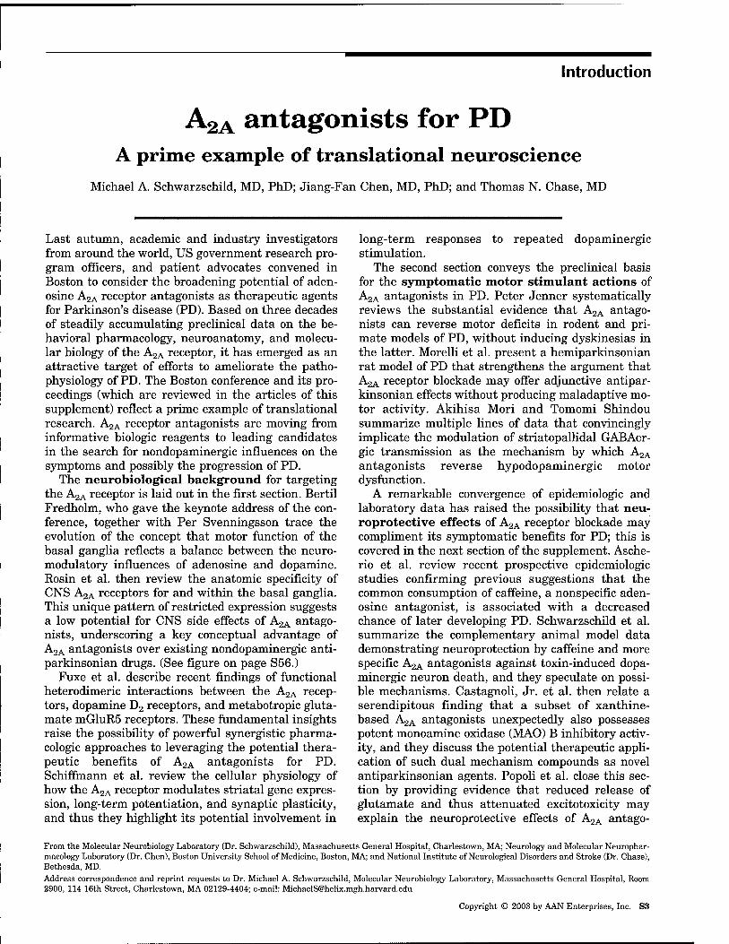
A2A antagonists for PD A prime example of translational neuroscienee
Michael A. Schwarzschild, MD, PhD; Jiang-Fan Chen, MD, PhD; and Thomas N. Chase, MD
Last autumn, academic and industry investigators from around the world, US government research pro- gram officers, and patient advocates convened in Boston to consider the broadening potential of aden- osine A2A receptor antagonists as therapeutic agents for Parkinson's disease (PD). Based on three decades of steadily accumulating preclinical data on the be- havioral pharmacology, neuroanatomy, and molecu- lar biology of the A2A receptor, it has emerged as an attractive target of efforts to ameliorate the patho- physiology of PD. The Boston conference and its pro- ceedings (which are reviewed in the articles of this supplement) reflect a prime example of translational research. A2A receptor antagonists are moving from informative biologic reagents to leading candidates in the search for nondopaminergic influences on the symptoms and possibly the progression of PD.
The neurobiological background for targeting the A2A receptor is laid out in the first section. Bertil Fredholm, who gave the keynote address of the con- ference, together with Per Svenningsson trace the evolution of the concept that motor function of the basal ganglia reflects a balance between the neuro- modulatory influences of adenosine and dopamine. Rosin et al. then review the anatomic specificity of CNS A2A receptors for and within the basal ganglia. This unique pattern of restricted expression suggests a low potential for CNS side effects of A2A antago- nists, underscoring a key conceptual advantage of A2A antagonists over existing nondopaminergic anti- parkinsonian drugs. (See figure on page S56.)
Fuxe et al. describe recent findings of functional heterodimeric interactions between the A2A recep- tors, dopamine D2 receptors, and metabotropic gluta- mate mGluR5 receptors. These fundamental insights raise the possibility of powerful sjmergistic pharma- cologic approaches to leveraging the potential thera- peutic benefits of A2A antagonists for PD. Schiffmann et al. review the cellular physiology of how the A2A receptor modulates striatal gene expres- sion, long-term potentiation, and synaptic plasticity, and thus they highlight its potential involvement in
long-term responses to repeated dopaminergic stimulation.
The second section conveys the preclinical basis for the symptomatic motor stimulant actions of A2A antagonists in PD. Peter Jenner systematically reviews the substantial evidence that A2A antago- nists can reverse motor deficits in rodent and pri- mate models of PD, without inducing dyskinesias in the latter. Morelli et al. present a hemiparkinsonian rat model of PD that strengthens the argument that A2A receptor blockade may offer adjunctive antipar- kinsonian effects without producing maladaptive mo- tor activity. Akihisa Mori and Tomomi Shindou summarize multiple lines of data that convincingly implicate the modulation of striatopallidal GABAer- gic transmission as the mechanism by which A2A antagonists reverse hypodopaminergic motor dysfunction.
A remarkable convergence of epidemiologic and laboratory data has raised the possibility that neu- roprotective effects of A2A receptor blockade may compliment its symptomatic benefits for PD; this is covered in the next section of the supplement. Asche- rio et al. review recent prospective epidemiologic studies confirming previous suggestions that the common consumption of caffeine, a nonspecific aden- osine antagonist, is associated with a decreased chance of later developing PD. Schwarzschild et al. summarize the complementary animal model data demonstrating neuroprotection by caffeine and more specific A2A antagonists against toxin-induced dopa- minergic neuron death, and they speculate on possi- ble mechanisms. Castagnoli, Jr. et al. then relate a serendipitous finding that a subset of xanthine- based A2A antagonists unexpectedly also possesses potent monoamine oxidase (MAO) B inhibitory activ- ity, and they discuss the potential therapeutic appli- cation of such dual mechanism compounds as novel antiparkinsonian agents. Popoli et al. close this sec- tion by providing evidence that reduced release of glutamate and thus attenuated excitotoxicity may explain the neuroprotective effects of A2A antago-
From the Molecular Neurobiology Laboratory (Dr. Schwarzschild), Massachusetts General Hospital, Charlestown, MA; Neurology and Molecular Neurophar- macology Laboratory (Dr. Chen), Boston University School of Medicine, Boston, MA; and National Institute of Neurological Disorders and Stroke (Dr. Chase), Bethesda, MD. Address correspondence and reprint requests to Dr. Michael A. Schwarzschild, Molecular Neurobiology Laboratory, Massachusetts General Hospital, Room 2900, 114 16th Street, Charlestown, MA 02129-4404; e-mail: [email protected]
Copyright © 2003 by AAN Enterprises, Inc. S3

nists across a range of neurotoxicity models of neuro- degenerative disease.
The supplement places the aforementioned anti- parkinsonian effects of AgA antagonists into the con- text of their expected effects on other important aspects of CNS physiology and PD pathophysi- ology. Chen et al. elaborate on the critical role played by the AgA receptor in the potentially mal- adaptive behavioral and neurochemical responses to repeated dopaminergic stimulation. Conversely, A2A antagonists may be able to prevent or reverse the maladaptive basal gangha plasticity underlying the occasionally disabling dyskinetic complications of long-term levodopa therapy in patients with PD. Vaugeois et al. make the case for antidepressant ef- fects of specific A2A antagonists (in contrast to caf- feine). Despite the caveats inherent in modeling depression in mice, their findings have major impli- cations for the management of depression in the gen- eral population and for those with PD (which is associated with a high incidence of depression). Weiss et al. take on the important question of AgA antagonist effects in models of psychosis. Although the established opposing effects of AgA and D2 recep- tors have supported the idea that AgA agonists may possess antipsychotic potential, AgA antagonists do not necessarily induce psychotomimetic side effects. Urade et al. highUght the role of the AgA receptor in sleep and, conversely, the possibility that AgA antag- onists may display arousal activity.
Finally, the supplement presents perspectives on the translational steps from development of lead compounds for human use to early clinical trials. Hiroshi Kase recounts one company's major contributions to the conceptual and pharmaceutical development of currently the most advanced A2A an- tagonist candidate for PD, which has now success- fully moved to phase II chnical trials. Dourish et al. present another company's discovery and progress in preclinical development of unique nonxanthine A2A antagonists targeted to PD. Chase et al. close the supplement with an overview of how an AgA antago- nist has advanced steadily from rodent to monkey to human studies, with the latter clinical trials validat- ing the preclinical evidence for antiparkinsonian benefits. These perspectives reflect the overarching
Figure. Boston's Leonard P. Zakim Bunker Hill Bridge under construction in November 2001, photographed by Don Eyles.
translational theme of the conference and this sup- plement, which have adopted the fitting symbol of Boston's nearly completed Lenny Zakim Bunker Hill Bridge. The striking images (figure and throughout) by the photographer Don Eyles capture the construc- tion of Boston's new landmark gateway. They also provide a metaphor for the building of a bridge that spans our knowledge of AgA receptor neurobiology and the promise of improved therapeutics for pa- tients with PD.
S4 NEUROLOGY BKSuppl 6) December 2003

Keynote Address
Adenosine-dopamine interactions Development of a concept and some comments on
therapeutic possibilities Bertil B. Fredholm, PhD; and Per Svenningsson, MD, PhD
Abstract—This brief review presents a personal perspective on the historical development of the current knowledge about the biologically important concept of functional antagonism between adenosine AgA and dopamine D2 receptors in caudate-putamen, accumbens, and tuberculum olfactorium. In the 1970s, studies of dopamine actions suggested an unexpected role of adenosine. Developments during the next decade substantiated this finding and demonstrated that a subform of adenosine Ag receptors was enriched in the basal ganglia. Cloning of adenosine receptors provided better tools for cellular localization and showed that AgA receptors are closely associated with Dg receptors. Distinct functional interactions at several levels were discovered, and there is now strong evidence that AgA receptors are tonically active and modified by dopamine acting at Dg receptors. Development of selective antagonists and knockout mice have highlighted the potential usefulness of AgA antagonists in decreasing symptoms and progression of Parkinson's disease—something that has also been vindicated by careful epidemiologic studies. There are issues of efficacy and potential side effects that need to be resolved, but the future looks bright.
NEUROLOGY 2003;61(Suppl 6):S5-S9
After the demonstration by Arvid Carlsson that do- pamine is an important transmitter in the basal gan- glia, with a particular role in Parkinson's disease (PD), the finding from Greengard's groups that dopa- mine can stimulate adenylate cyclase attracted much attention. This attention led to the discovery that adenosine might be intimately involved.
In 1974, Kjell Fuxe and Urban Ungerstedt show^ed, using animals with unilateral 6-OH- dopamine lesions, that theophylline could itself in- duce the same type of rotation behavior that was induced by drugs that directly or indirectly stimu- lated dopamine receptors and that it could markedly enhance dopamine-mediated effects.^ In that study, the effect was interpreted as secondary to blockade of phosphodiesterase (PDE) and was therefore taken as evidence for an important role of cyclic adenosine monophosphate (cAMP) as a mediator of dopamine actions. However, in a follow-up study in which I (B.F.) was involved, several different PDE inhibitors were examined, and it was found that the potency of the drugs to induce rotation fitted much better with their potency as adenosine antagonists (or enhanc- ers) than with their potency as PDE inhibitors.^ To- gether these studies showed that methylxanthines, probably by blocking adenosine receptors, could po- tentially be used as treatment for patients with PD.
Studies in two laboratories of dopamine-
stimulated adenylyl cyclase in the brain also showed that methylxanthines could decrease "basal" enzyme activity and that adenosine could stimulate it.*'^ This was observed in dopamine-rich areas of the brain, including caudate-putamen and tuberculum olfacto- rium, but not in other brain areas. This finding sug- gested that these parts of the brain might have a different set of adenosine receptors than other brain areas (figure. A).
This contention received support during the fol- lowing decade as methods to study receptors using binding techniques were developed. The first studies used relatively nonselective radioligands but phar- macologic means to discriminate between multiple binding sites.^"^ Later studies used a rather selective ligand for A2A receptors, including CGS 21680."'-i* Altogether these studies vindicated the belief that a special form of adenosine receptors, the A2A receptor, is enriched in dopamine-rich areas of the brain and that this offers a rationale for examining the role of adenosine in mediating or modulating behaviors and traits traditionally associated with dopamine.
The availability of more selective adenosine recep- tor agonists and antagonists also reinforced the idea that behavioral consequences of A^A. receptor- and dopamine receptor-mediated effects tended to be opposite.^^-i®
The interactions between adenosine and dopamine
From the Department of Physiology and Pharmacology, Karolinska Institutet, Stockholm, Sweden. Address correspondence and reprint requests to Dr. Bertil B. Fredholm, Department of Physiology and Pharmacology, Karolinska Institutet, S-171 77 Stockholm, Sweden; e-mail: [email protected]
Copyright © 2003 by AAN Enterprises, Inc. S5

/ AC ^-
Figure. Graphic description of the development of our knowledge of A2A-D2 interactions. Illustration (A) shows the situation in the 1970s; (B) depicts the knowledge around 1991; (C) shows the same in the late 1990s; and (D) demonstrates our current understanding. For further details, see text.
receptors in the striatum continued to be studied at the biochemical level. It was demonstrated that there were interactions between adenosine AgA re- ceptors at several levels. The AgA receptor, being coupled to a member of the G3 family of G proteins, and the Dg receptor, coupled to a Gi protein, would interact negatively at the level of second messengers and beyond. Binding studies revealed that high- affinity binding of Dg agonists could be reduced by stimulation of adenosine AgA receptors-^" This find- ing suggested that there were interactions directly between the receptors (figure, B), an issue that has been forcefully pursued by Fuxe et al. (see Fuxe et al., page S19).
The next major conceptual advance was the clon- ing of several adenosine receptors. Four novel mem- bers of the G protein-coupled receptor family were cloned from a canine thyroid library.^i Of these, one was the adenosine AgA receptor,^^ and another was the canine A^ receptor.^s Qnce these first structures were obtained, the same receptors were soon cloned from other mammals, including humans. Further- more, the adenosine A2B receptor was cloned.^* More surprisingly, a fourth adenosine receptor, denoted A3, was cloned, first as an orphan^^ and later as a bona fide methylxanthine-insensitive adenosine receptor.^^
These findings not only conclusively proved that S6 NEUROLOGY 61(Suppl 6) December 2003
there are two distinct adenosine A2 receptors but also provided a set of novel tools that proved useful. In situ hybridization was used to pinpoint the cells that express AgA receptors in the brain. Using in- creasingly sophisticated methods, it was proven that the bulk of AgA expression is confined to one set of neurons in the striatum, namely those GABAergic out- put neurons that constitute the so-called indirect pathway.2^-^3 These cells also express the bulk of the dopamine Dg receptors. Hence, the link between AgA and dopamine Dg receptors was further strengthened.
Techniques with a cellular resolution were also used to try to determine the roles of adenosine AgA receptors in the intact striatum. This was based on early findings showing that expression of immediate early genes (lEGs) could be used to pinpoint changes in neuronal activity or signal transduction.^^ We ob- served that stimulatory doses of caffeine and selec- tive A2A receptor antagonists caused a decrease in the expression of lEGs, known to be regulated by the cAMP/CREB cascade, in striatopallidal neurons (fig- ure, C).3'^>3'^ These and subsequent studies^''-*" provide strong evidence that adenosine, via AgA receptors, exerts a robust tonic activation on the cAMP/CREB/ lEG cascade in striatopallidal neurons. Moreover, this result also provided evidence that multiple Dg receptor-mediated effects of dopamine can be attrib-

uted to the antagonism of this adenosine-mediated activation of striatopallidal neurons.
To increase our understanding of the interactions of adenosine and dopamine at the signal transduc- tion level, we proposed a collaborative project with Paul Greengard to study the effects of adenosine A2A selective compounds and caffeine on the phosphory- lation of dopamine and cAMP phosphoprotein of 32 kDa (DARPP-32). DARPP-32 is highly enriched in all striatal GABAergic medium-sized projection neurons and is an important mediator of dopaminergic sig- naling.*! Its function is determined by its relative phosphorylation state at several different threonine/ serine residues, of which the most studied is a pro- tein kinase A (PKA) site at Thr34. When this residue is phosphorylated, it converts DARPP-32 into an in- hibitor of protein phosphatase-1, which in turn regu- lates the activity of multiple transcription factors, including CREB, ion channels, and ionotropic recep- tors (figure, D). In initial studies conducted in brain slices prepared from striatum, it was found that CGS 21680 potently increases phosphorylation at Thr34.''^ This effect was additive to that of SKF81297, a selec- tive Di agonist, and could be counteracted by quinpi- role, a selective D2 agonist.*^ This result identified adenosine, via AgA receptors, as a key regulator of the phosphorylation state of DARPP-32 in striatopal- lidal neurons. Subsequently, we developed a method to reliably detect DARPP-32 phosphorylation in vivo and could demonstrate that the A2A antagonist used, SCH 58261, significantly counteracted the increase in DARPP-32 phosphorylation that was observed af- ter treatment with selective D2 receptor antago- nists.''* Likewise, the ability of D2 antagonists to increase DARPP-32 phosphorylation was dramati- cally reduced in A2A receptor knockout mice. There- fore, these data provided further support for the notion that adenosine acting on A2A receptors is an important mediator for establishing a basal cAMP level, which is necessary for many effects of dopam- ine's action via D2 receptors. To address the involve- ment of DARPP-32 in the behavioral actions of caffeine and selective adenosine A2A receptor com- pounds, we administered such compounds to DARPP-32 knockout mice and studied effects on lo- comotor behavior. As expected from the biochemical data, we found that the ability of CGS 21680 to in- duce hypolocomotion was attenuated in DARPP-32 knockout mice.'*^ Similarly, the ability of caffeine and SCH 58261 to induce hj^jerlocomotion was attenu- ated in DARPP-32 knockout mice. In this article, an additional effect of AgA receptors on DARPP-32 phos- phorylation was shown, namely that A2A agonism via cAMP-dependent mechanisms increases the phosphorylation of Thr34-DARPP-32 but decreases the phosphorylation at Thr75-DARPP-32. Con- versely, caffeine and SCH 58261 increase phosphory- lation at Thr75-DARPP-32. This site has recently been shown to be phosphorylated by Cdk5, and when this happens, DARPP-32 is converted into an inhibi- tor of PKA.*^ Therefore, by increasing the phosphor-
ylation of Thr75-DARRP-32, caffeine and selective A2A receptor antagonists will further increase the inhibition of PKA. This feed-forward mechanism, which is also used by Dg receptor agonists, will po- tentiate the inhibitory influence of adenosine on the cAMP/PKA/CREB/IEG signaling pathway in striato- pallidal neurons (see figure, D).
In parallel with the development of an increas- ingly clear understanding of the biochemical and mo- lecular underpinning of the adenosine-dopamine interactions, there has been extensive work on the effectiveness of adenosine AgA antagonists in various experimental models of PD. Extensive review of these results is beyond the scope of this commentary (see Chase et al., page S107; Kase et al., page S97; Jenner et al., page S32; and Weiss et al., page SlOl). However, a recent study showed that A2A receptor antagonism could reduce not only symptoms of PD but also the loss of dopamine neurons induced by l-methyl-4-phenyl-l,2,3,6-tetrahydropyridine (MPTP).*^ Furthermore, it was shown that persistent L-dopa effects require A2A receptors.*^
Therefore, the results of many studies over the years have strongly developed the concept that A2A and D2 receptors interact in such a way that A2A antagonists could prove useful in treating patients with PD. However, there are concerns. One potential concern is related to tolerance. It is well known that some actions of caffeine develop rapid tolerance.*^''^" However, caffeine effects in PD models do not,^^ and there is also no tolerance to selective A2A antagonists in models that show tolerance to caffeine.^^
Another, and perhaps more serious, concern is re- lated to the fact that A2A receptors regulate other things in addition to activity in striatopallidal neu- rons. It has long been known that adenosine regu- lates platelet activation,^^'^* and now we know that A2A receptors are responsible for this.^^'^^ Similarly, A2A receptors are critically important in regulating neutrophil leukocjrte activity^' and activity of macro- phages.®^ Even more importantly, A2A receptors reg- ulate inflammatory reactions in general.^''•^° Therefore, long-term blockade of adenosine A2A re- ceptors may cause undesirable peripheral morbidity.
A potential way to attack this problem was af- forded when it was discovered that A2A receptors in striatum are coupled to G^jf proteins,®^ whereas on platelets, neutrophils, and lymphocytes, Gg mediates the A2A effects. If it proves possible to find agents that selectively affect AgA-Gojf, more selective drugs may be found.
Already in the early 1970s Fuxe and Ungerstedt suggested that methylxanthines might be used for management of PD. Since then, substantial progress has been made in understanding why, and now the prospect looks good that A2A antagonists may prove of value as part of the therapeutic armamentarium.®^ The potential may be particularly strong in com- pounds that have more than one potentially benefi- cial action.®^
December 2003 NEUROLOGY 61(Suppl 6) S7

References 1. Kebabian JW, Greengard P. Dopamine-sensitive adenyl cyclase; possi-
ble role in synaptic transmission. Science 1971;174:1346-1349. 2. Fuxe K, Ungerstedt U. Action of caffeine and theophyllamine on super-
sensitive dopamine receptors: considerable enhancement of receptor re- sponse to treatment with DOPA and dopamine receptor agonists. Med Biol 1974;52:48-54.
3. Fredholm BB, Fuxe K, Agnati L. Effect of some phosphodiesterase inhibitors on central dopamine mechanisms. Eur J Pharmacol 1976;38: 31-38.
4. Fredholm BB. Activation of adenylate cyclase from rat striatum and tuberculum olfactorium by adenosine. Med Biol 1977;55:262-267.
5. Fremont J, Perez M, Bockaert J. Adenosine-sensitive adenylate cyclase in rat striatal homogenates and its relationship to dopamine- and Ca2-l-sensitive adenylate cyclases. Mol Pharmacol 1977;13:662-670.
6. Bruns RF, Lu GH, Pugsley TA. Characterization of the Aj adenosine receptor labeled by [^H]-NECA in rat striatal membranes. Mol Pharma- col 1986;29:331-346.
7. Lee KS, Reddington M. Autoradiographic evidence for multiple CNS binding sites for adenosine derivatives. Neuroscience 1986;19:535-549.
8. Reddington M, Erfurth A, Lee KS. Heterogeneity of binding sites for N-ethyIcarboxamido-[3H]-adenosine in rat brain: effects of N-ethyl- maleimide. Brain Res 1986;399:232-239.
9. Bruns RF, Davis RE, Ninteman FW, Poschel BPH, Wiley JN, Heffner TG. Adenosine antagonists as pharmacological tools. In: Paton DM, ed. Adenosine and Adenine Nucleotides. Physiology and Pharmacology. Basingstoke: Taylor & Francis, 1988:39-49.
10. Bridges AJ, Bruns RF, Ortwine DF, Priebe SR, Szotek DL, Trivedi BK. N6-t2-(3,5-dimethoxyphenyl)-2-(2-methylphenyl)ethyl!adenosineandits uronamide derivatives. Novel adenosine agonists with both high affin- ity and high selectivity for the adenosine A2 receptor. J Med Chem 1988;31:1282-1285.
11. Alexander SP, Reddington M. The cellular localization of adenosine receptors in rat striatum. Neuroscience 1989;28:645-651.
12. Jarvis MF, Jackson RH, Williams M. Autoradiographic characterization of high affinity adenosine Aj receptors in the rat brain. Brain Res 1989;484:111-118.
13. Jarvis MF, Schulz R, Hutchison AJ, Do UH, Sills MA, Williams M. [3H1CGS 21680, a selective A2 adenosine receptor agonist directly la- bels A2 receptors in rat brain. J Pharmacol Exp Ther 1989;251:888- 893.
14. Parkinson FE, Fredholm BB. Autoradiographic evidence for G-protein coupled Aa-receptors in rat neostriatum using pH]-GGS 21680 as a ligand. Naunyn Schmiedebergs Arch Pharmacol 1990;342:85-89.
15. Fredholm BB, Herrera-Marschitz M, Jonzon B, Lindstrom K, Unger- stedt U. On the mechanism by which methylxanthines enhance apomorphine-induced rotation behaviour in the rat. Pharmacol Bio- chem Behav 1983;19:535-541.
16. Heffner TG, Wiley JN, Williams AE, Bruns RF, Coughenour LL, Downs DA. Comparison of the behavioral effects of adenosine agonists and dopamine antagonists in mice. Psychopharmacology 1989;98:31-37.
17. Brown SJ, Gill R, Evenden JL, Iversen SD, Richardson PJ. Striatal A2 receptor regulates apomorphine-induced turning in rats with unilateral dopamine denervation. Psychopharmacology 1991;103:78-82.
18. Jiang H, Jackson-Lewis V, Muthane U, et al. Adenosine receptor antag- onists potentiate dopamine receptor agonist-induced rotational behav- ior in 6-hydroxydopamine-lesioned rats. Brain Res 1993;613:347-351.
19. Popoli P, Pfizzola A, de Carolis AS. Modulation of striatal adenosine Al and A2 receptors induces rotational behaviour in response to dopami- nergic stimulation in intact rats. Eur J Pharmacol 1994;257:21-25.
20. Ferr(S S, von Euler G, Johansson B, Fredholm BB, Fuxe K. Stimulation of high-affinity adenosine Ag receptors decreases the affinity of dopa- mine Dj receptors in rat striatal membranes. Proc Natl Acad Sci USA 1991;88:7238-7241.
21. Libert F, Parmentier M, Lefort A, et al. Selective amplification and cloning of four new members of the G protein-coupled receptor family. Science 1989;244:569-572.
22. Maenhaut C, Van Sande J, Libert F, et al. RDC8 codes for an adenosine A2 receptor with physiological constitutive activity. Biochem Biophys Res Commun 1990;173:1169-1178.
23. Libert F, Schiffmann SN, Lefort A, et al. The orphan receptor cDNA RDC7 encodes an Al adenosine receptor. EMBO J 1991;10:1677-1682.
24. Stehle JH, Rivkees SA, Lee JJ, Weaver DR, Deeds JD, Reppert SM. Molecular cloning and expression of the cDNA for a novel A2-adenosine receptor subtype. Mol Endocrinol 1992;6:384-393.
25. Meyerhof W, Miiller-Brechlin R, Richter D. Molecular cloning of a novel putative G-protein coupled receptor expressed during rat spermiogene- sis. FEES Lett 1991;284:155-160.
26. Zhou QY, Li C, Olah ME, Johnson RA, Stiles GL, Civelli O. Molecular cloning and characterization of an adenosine receptor: the A3 adenosine receptor. Proc Natl Acad Sci USA 1992;89:7432-7436.
27. Schiffmann SN, Jacobs 0, Vanderhaeghen JJ. Striatal restricted aden- osine A2 receptor (RDC8) is expressed by enkephalin but not by sub- stance P neurons: an in situ hybridization histochemistry study. J Neurochem 1991;57:1062-1067.
29.
30
38
39
28. Schiffmann SN, Libert F, Vassart G, Vanderhaeghen JJ. Distribution of adenosine A2 receptor mRNA in the human brain. Neurosci Lett 1991; 130:177-181. Fink JS, Weaver DR, Rivkees SA, et al. Molecular cloning of the rat Aj adenosine receptor: selective co-expression with Dg dopamine receptors in rat striatum. Brain Res Mol Brain Res 1992;14:186-195. Johansson B, Ahlberg S, van der Ploeg I, et al. Effect of long term caffeine treatment on Aj and Aj adenosine receptor binding and on mRNA levels in rat brain. Naunyn Schmiedebergs Arch Pharmacol 1993;347:407-414.
31. Pollack AE, Harrison MB, Wooten GF, et al. Differential locahzation of A2a adenosine receptor mRNA with Dl and D2 dopamine receptor mRNA in striatal output pathways following a selective lesion of stria- tonigral neurons. Brain Res 1993;631:161-166.
32. Johansson B, Georgiev V, Fredholm BB. Distribution and postnatal ontogeny of adenosine AJA receptors in rat brain: comparison with dopamine receptors. Neuroscience 1997;80:1187-1207.
33. Svenningsson P, Le Moine C, KuU B, Sunahara R, Bloch B, Fredholm BB. Cellular expression of adenosine AgA receptor messenger RNA in the rat central nervous system with special reference to dopamine in- nervated areas. Neuroscience 1997;80:1171-1185.
34. Sheng M, Greenberg ME. The regulation and function of c-fos and other immediate early genes in the nervous system. Neuron 1990;4:477-485.
35. Svenningsson P, Nomikos GG, Fredholm BB. Biphasic changes in loco- motor behavior and in expression of mRNA for NGFI-A and NGFI-B in rat striatum following acute caffeine administration. J Neurosci 1995; 15:7612-7624.
36. Svenningsson P, Nomikos GG, Ongini E, Fredholm BB. Antagonism of adenosine ASA receptors underlies the behavioural activating effect of caffeine and is associated with reduced expression of messenger RNA for NGFI-A and NGFI-B in caudate-putamen and nucleus accumbens. Neuroscience 1997;79:753-764.
37. Boegman RJ, Vincent SR. Involvement of adenosine and glutamate receptors in the induction of c-fos in the striatum by haloperidol. Syn- apse 1996;22:70-77. Pinna A, Wardas J, Cozzolino A, Morelli M. Involvement of adenosine A2A receptors in the induction of C-Fos expression by clozapine and haloperidol. Neuropsychopharmacology 1999;20:44-51. Svenningsson P, Fourreau L, Bloch B, Fredholm BB, Gonon F, Le Moine C. Opposite tonic modulation of dopamine and adenosine on c-fos mRNA expression in striatopallidal neurons. Neuroscience 1999;89: 827-837.
40. Chen JF, Moratalla R, Impagnatiello F, et al. The role of the D(2) dopamine receptor (D(2)R) in A(2A) adenosine receptor (A(2A)R)- mediated behavioral and cellular responses as revealed by A(2A) and D(2) receptor knockout mice. Proc Natl Acad Sci USA 2001;98:1970- 1975.
41. Greengard P. The neurobiology of slow synaptic transmission. Science 2001;294:1024-1030.
42. Svenningsson P, Lindskog M, Rognoni F, Fredholm BB, Greengard P, Fisone G. Activation of adenosine A2A and dopamine Dj receptors stim- ulates cyclic AMP-dependent phosphorylation of DARPP-32 in distinct populations of striatal projection neurons. Neuroscience 1998;84:223- 228.
43. Lindskog M, Svenningsson P, Fredholm BB, Greengard P, Fisone G. Activation of dopamine Dg receptors decreases DARPP-32 phosphoryla- tion in striatonigral and striatopalhdal projection neurons via different mechanisms. Neuroscience 1999;88:1005-1008.
44. Svenningsson P, Lindskog M, Ledent C, et al. Regulation of the phos- phorylation of the dopamine- and cAMP-regulated phosphoprotein of 32 kDa in vivo by dopamine Dj, dopamine D2 and adenosine A^A receptors. Proc Natl Acad Sci USA 2000;97:1856-1860.
45. Lindskog M, Pozzi L, Svenningsson P, et al. The stimulant action of caffeine is mediated by an increase in the state of phosphorylation at the Cdk5 site of DARPP-32. Nature 2002;418:774-778.
46. Bibb JA, Snyder GL, Nishi A, et al. Phosphorylation of DARPP-32 by CdkS modulates dopamine signalhng in neurons. Nature 1999;402: 669-671.
47. Chen JF, Xu K, Petzer JP, et al. Neuroprotection by caffeine and AC2A) adenosine receptor inactivation in a model of Parkinson's disease. J Neurosci 2001;21:RC143.
48. Fredduzzi S, Moratalla R, MonopoU A, et al. Persistent behavioral sensitization to chronic L-DOPA requires A2A adenosine receptors. J Neurosci 2002;22:1054-1062.
49. Fredholm BB, Battig K, Holmfe J, NehUg A, Zvartau E. Actions of caffeine in the brain with special reference to factors that contribute to its widespread use. Pharmacol Rev 1999;51:83-133.
50. Svenningsson P, Nomikos GG, Fredholm BB. The stimulatory action and the development of tolerance to caffeine is associated with alter- ations in gene expression in specific brain regions. J Neurosci 1999;19: 4011-4022.
51. Xu K, Xu YH, Chen JF, Schwarzschild MA. Caffeine's neuroprotection against l-methyl-4-phenyl-l,2,3,6-tetrahydropyridine toxicity shows no
S8 NEUROLOGY 61(Suppl 6) December 2003

tolerance to chronic caffeine administration in mice. Neurosci Lett 2002;322:13-16.
52. Halldner L, Lozza G, Lindstrom K, Fredholm BB. Lack of tolerance to motor stimulant effects of a selective adenosine A2A receptor antago- nist. Eur J Pharmacol 2000;406:345-354.
53. Haslam RJ, Cusack NJ. Blood platelet receptors for ADP and for aden- osine. In: Burnstock G, ed. Purinergic Receptors. London: Chapman and Hall, 1981:221-285.
54. Sollevi A, Torssell L, Fredholm BB, Settergren G, Blomback M. Adeno- sine spares platelets during cardiopulmonary bypass in man without causing systemic vasodilatation. Scand J Thorac Cardiovasc Surg 1985; 19:155-159.
55. Ledent C, Vaugeois JM, Schiffmann SN, et al. Aggressiveness, hypoal- gesia and high blood pressure in mice lacking the adenosine A2A recep- tor. Nature 1997;388:674-678.
56. Gessi S, Varani K, Merighi S, Ongini E, Borea PA. A(2A) adenosine receptors in human peripheral blood cells. Br J Pharmacol 2000;129: 2-11.
57. Cronstein BN. Adenosine, an endogenous anti-inflammatory agent. J Appl Physio! 1994;76:5-13.
58. Leibovich SJ, Chen JF, Pinhal-Enfield G, et al. Synergistic up- regulation of vascular endothelial growth factor expression in murine macrophages by adenosine A(2A) receptor agonists and endotoxin. Am J Pathol 2002;160:2231-2244.
59. Okusa MD, Linden J, Macdonald T, Huang L. Selective A2A adenosine receptor activation reduces ischemia-reperfusion injury in rat kidney. Am J Physiol 1999;277:F404-412,
60. Ohta A, Sitkovsky M. Role of G-protein-coupled adenosine receptors in downregulation of inflammation and protection from tissue damage. Nature 2001;414:916-920.
61. KuU B, Svenningsson P, Fredholm BB. Adenosine A2A receptors are co-localized with and activate G^if in rat striatum. Mol Pharmacol 2000; 58:771-777.
62. Schwarzschild MA, Chen JF, Ascherio A. Caffeinated clues and the promise of adenosine A(2A) antagonists in PD. Neurology 2002;58: 1154-1160.
63. Chen JF, Steyn S, Staal R, et al. 8-(3-Chlorostyryl)caffeine may attenu- ate MPTP neurotoxicity through dual actions of MAO inhibition and A2A receptor antagonism. J Biol Chem 2002;277:36040-36044.
Don Eyies April 2000
December 2003 NEUROLOGY 61(Suppl 6) S9

Articles
I. The biology of A2A receptors in the basal ganglia
Exciting news about AgA receptors Per Svenningsson, MD, PhD; and Bertil B. Fredholm, PhD
Our understanding of the fundamental actions of adenosine AgA receptors has increased significantly during the past years, and this was well illustrated by the lectures in the session "The Biology of AgA Receptors in the Basal GangUa." The existence of a strong interaction between adenosine AgA and dopa- mine Dg receptors is now firmly estabUshed at the an- atomic, biochemical, and functional levels. In each of the lectures, novel evidence for important interactions between adenosine AgA receptors and excitatory gluta- matergic neurotransmission was presented.
Using A2A receptor-selective antibodies and immu- nohistochemistry at the light and electron microscopic levels, Rosin et al. have performed detailed anatomic work that unambiguously shows that AgA receptors are highly enriched in medium-sized spiny GABAergic stri- atal neurons.1'2 Within these neurons, AgA receptors are found in most cellular compartments, i.e., den- drites, terminals of axon collaterals, and in soma. How- ever, from a quantitative standpoint, a pronounced subcellular enrichment of AgA receptors is found in dendrites and dendritic spines, which form asymmetric synapses. These synapses receive input from glutama- tergic terminals and are of excitatory nature. This postsynaptic localization of AgA receptors implies that A2A receptors may play an important role in the regu- lation of synaptic plasticity. The excitatory glutamater- gic inputs to striatum are derived predominantly from the cerebral cortex and thalamus. In an ongoing effort. Rosin et al. are trying to define the anatomic origin of the excitatory glutamatergic inputs that innervate the A2A receptor-containing dendritic spines. For this pur- pose, colabehng studies with A2A receptor antibodies and VGLUTl, which is located on cortical terminals, or VGLUT2, which is located on thalamic terminals, are performed (see Rosin et al., page S12). It is anticipated that detailed information of the synaptology of the A2A receptor-containing neurons will be available in the near future.
Fuxe and Schiffmann presented evidence for the functional importance of A2A receptors in modulating excitatory glutamatergic neurotransmission. By com- bining biochemical and anatomic techniques, Fuxe et
al.3 have demonstrated that AgA receptors form het- erodimers with metabotropic glutamate 5 receptors (mGluR5). Interestingly, mGluRS receptors have been shown to be highly concentrated postsynaptically at excitatory synapses and thus exhibit a subcellular dis- tribution similar to AgA receptors. The heterodimeriza- tion between A2A receptors and mGluRS receptors is specific and cannot be found between AgA receptors and mGluRl receptors. Studies in cell lines transiently transfected with A2A receptors and mGluRS receptors have shown that costimulation of these receptors has a synergistic action on mitogen-activated protein kinase (MAPK) activation and c-fos gene transcription. The precise mechanisms underlying this synergy remain to be clarified, but it appears not to involve cychc adeno- sine monophosphate (cAMP) or Ca^+ accumulation (see Fuxe et al., page S19).
Schiffmann et al. have extensively used adenosine A2A receptor knockout mice to study the influence of A2A receptors on electrophysiologic properties of stri- atal neurons and on gene regulation. Using patch- clamp methodology and brain slices, Schiffmann et al. have now demonstrated that long-term potentia- tion in the ventral part of the striatum, nucleus ac- cumbens, is significantly attenuated in A2A receptor knockout mice.* Similar results were obtained when striatal slices from wild-type mice were treated with the selective AgA receptor antagonist ZM241385 (see Schiffman et al., page S24). This finding adds further evidence that AgA receptors are tonically activated by endogenous adenosine. Moreover, this effect on synaptic plasticity can be viewed as a functional cor- relate to the anatomic finding of Rosin et al., who found that ASA receptors are enriched in dendritic spines that receive excitatory inputs.
As expected from previous work using caffeine and A2A receptor antagonists, a reduction of immedi- ate early gene and neuropeptide expression has been found throughout the striatum in AgA receptor knockout mice.^ This is probably not the result of a developmental deficit because AgA receptors are first expressed late in ontogeny. Importantly, these changes are not confined to striatopallidal neurons
From the Department of Physiology and Pharmacology, Karolinska Institutet, Stockholm, Sweden. Address correspondence and reprint requests to Dr. Bertil B. Fredholm, Department of Physiology and Pharmacology, Karolinska Institutet, SE-171 77, Stockholm, Sweden; e-mail: [email protected]
SIO Copyright © 2003 by AAN Enterprises, Inc.

but also involve striatonigral neurons and nonstria- tal regions. This finding clearly demonstrates that AgA receptors, despite being restrictively expressed on striatopallidal neurons, strongly influence the physiology of neurons that do not express AgA recep- tors. The extremely tight coupling between neuronal populations in the striatum has been described and provides a good explanation for these findings. It is of course also known that activity in striatal outputs af- fects many other brain regions. Therefore, there is no discrepancy between the fact that A2A receptors have a restricted distribution and the fact that affecting them will influence many parts of the brain.
References
1. Rosin DL, Robeva A, Woodard RL, Guyenet PG, Linden J. Immunohisto- chemical localization of adenosine AJA receptors in the rat central ner- vous system. J Comp Neurol 1998;401:163-186.
2. Hettinger BD, Lee A, Linden J, Rosin DL. Ultrastructural localization of adenosine AJA receptors suggests multiple cellular sites for modulation of GABAergic neurons in rat striatum. J Comp Neurol 2001;431:331-346.
3. Ferre S, Karcz-Kubicha M, Hope BT, et al. Synergistic interaction between adenosine A2A and glutamate mGlu5 receptors: implications for striatal neuronal function. Proc Natl Acad Sci USA 2002;99:11940-11945.
4. d'Alcantara P, Ledent C, Swillens S, Schiffmann SN. Inactivation of adenosine AJA receptor impairs long term potentiation in the accumbens nucleus without altering basal synaptic transmission. Neuroscience 2001;107:455-464.
5. Dassesse D, Massie A, Ferrari R, et al. Functional striatal hypodopamin- ergic activity in mice lacking adenosine A(2A) receptors. J Neurochem 2001;78:183-198.
Don Eyies November 2000
December 2003 NEUROLOGY 61(Suppl 6) SIX

Anatomy of adenosine AgA receptors in brain
Morphological substrates for integration of striatal function Diane L. Rosin, PhD; Barbara D. Hettinger, PhD; Amy Lee, PhD; and Joel Linden, PhD
Abstract—AaA adenosine receptors (AZARS) are expressed with the greatest abundance in the striatum and other nuclei of the basal ganglia. The segregated expression of AZARS on the GABAergic striatopallidal medium spiny neurons, where A2AR and Dg dopamine receptor mRNAs are colocahzed, and the opposing functional interaction between adenosine and dopamine suggest that A2ARS may be an important therapeutic target. To further explore the role of A2ARS in the synaptic organization of the basal gangha, the authors developed an antibody directed against the purified AZAR- Immunohisto- chemical studies in rat brain showed dense labeling of the neuropil in the striatum, nucleus accumbens, and olfactory tubercles with lighter labeling of terminals in the globus pallidus (GP), where ASAR transcript is not detected. Stimulation of A2ARS on GP terminals may facilitate GABAergic signaling and contribute to the overactivation observed in Parkinson's disease (PD). Analysis at the ultrastructural level allowed a more detailed characterization of the mechanism(s) of A2A-mediated control of striatal output. In the striatum, terminals expressing A2ARS accounted for 25% of the labeled elements. These presynaptic receptors may facilitate excitatory glutamatergic, inhibitory GABAergic, and possibly cholin- ergic striatal transmission. However, the majority of striatal A2AR immunoreactivity was found on postsynaptic elements including dendrites of striatopallidal neurons, in which A2AR and GABA immunoreactivity is colocalized. Activation of these receptors may promote GABAergic signaling in striatopallidal output neurons and their local axon collaterals in the striatum. Many of the A2A-labeled dendrites were contacted by terminals forming asymmetric (excitatory) possibly glutamatergic synapses. Using the vesicular glutamate transporters (VGLUTs) as markers of glutamatergic termi- nals, the authors have found that VGLUTl-immunoreactive(ir) terminals make asymmetric contacts on AgA-ir spines and'spine heads in the striatum, suggesting that regulation of striatal output by A2AR stimulation may involve facilitation of the cortical glutamatergic excitatory input to striatopallidal neurons. These ultrastructural findings suggest several pathways through which AgA receptor blockade may act to dampen the elevated striatopallidal GABAergic signaling that occurs in PD. NEUROLOGY 2003;61(Suppl 6):S12-S18
Adenosine AgA receptors (AgARs), which are highly expressed in the basal ganglia, have become a target of therapeutic interest for a number of diseases, in- cluding Parkinson's disease (PB)^ mostly because of their discrete anatomic localization and biochemi- cal interaction with dopamine Dg receptors." The progressive degeneration of nigrostriatal neurons that occurs in PD results in a loss of dopaminergic input to striatal output neurons and enhanced stria- topallidal GABAergic signaling, resulting in the mo- tor disturbances that are characteristic of the disease. With declining levels of dopamine, which normally regulates the direct (striatonigral) and in- direct (striatopallidal) output pathways of the basal ganglia, an imbalance occurs in the outflow of inhib- itory GABAergic projection neurons. Replacement of dopaminergic input by administration of exogenous L-dopa has been the primary therapeutic strategy for the management of PD for decades. The finding that
From the Departments of Pharmacology (Dr. Rosin), Cardiovascular Medicine (Drs. Hettinger, Lee, and Linden), and IWolecular Physiology and Biological Physics (Dr. Linden), University of Virginia Health Sciences Center, Charlottesville, VA. The current affiliation for Dr. Lee is Department of Pharmacology, Emory University School of Medicine, Atlanta, GA. Supported by NIH grants NS10783, HL37942, HL60003, and HL07284; American Heart Association VHA 9960193U; and grants from the Scottish Rite Schizophrenia Research Council and Tourette Syndrome Association Permanent Research Fund. Address correspondence and reprint requests to Dr. Diane L. Rosin, Department of Pharmacology, University of Virginia Health System, P.O. Box 800735, 1300 Jefferson Park Avenue, Charlottesville, VA 22908-0735; e-mail: [email protected]
S12 Copyright © 2003 by AAN Enterprises, Inc.
the mRNAs for AgA and Dg receptors are colocalized in striatopallidal neurons,'*'^ where they are physi- cally and biochemically poised to mediate adenosine- dopamine antagonistic interactions,"'^ has focused attention on blocking ASARS as another potential means for resetting the motor imbalance in PD. We have taken the approach of studying the subcellular localization of AZARS to enhance understanding of adenosine's role as a neuromodulator in the basal ganglia. This article will briefly review the anatomy of ASARS in striatum and will integrate our recent findings in the context of striatal circuitry with a wealth of published data on AgARs to suggest a spec- ulative model for multiple mechanisms by which these receptors may modulate striatal function.
Distribution of AgA receptors in CNS. Begin- ning with some of the earliest studies of the distribu- tion of ABARS in the brain, studies of receptor

CTX
Thalamus
GABAergic Interneurons GABA GABA GABA.SST
/~\ Calretinin Parv albumin NPY/NOS
*K}
striatopallidal GABA.Enk.Dj.AjA
G^Gr- GPe
glu) CTX
Thalamus
^ SN
Figure 1. Working model for the morphologic basis of A2A action in basal ganglia: potential sites for modulation of striatal function. A simplified drawing of the striatal output neurons (GABAergic projec- tion neurons of the indirect pathway that express enkephalin [enk], dopa- mine D2 receptors [DJ, and A2A adenosine receptors [AgARs] and of the direct pathway that express sub- stance P [SP], dopamine Dj receptors [Dj], and adenosine Aj receptors [AJ) and the four interneuronal cell types (GABAergic interneurons, clas- sified based on their histochemical profiles as labeled, and cholinergic interneurons that express acetylcho- line [ACh]) illustrates the known ex- trinsic inputs and established connections in the striatum. Putative and postulated sites of subcellular
localization ofA2ARs are given based on our ultrastructural findings and biochemical, physiologic, and anatomic results in the literature. The diagram is not meant to show all the neurochemical messengers and their receptors in the striatum but emphasizes the role of the A2ARS in modulating the output pathways of the striatum. CTX = cortex; DA = dopamine; glu = glutamate; GPe = globus pallidus, external segment; mGluRS = metabotropic glutamate receptor; NOS = neuronal nitric oxide synthetase; NPY = neuropeptide Y; SN = substantia nigra; SST = somatostatin.
UADA, Or,U,,A,
SN
© A,.-AR
o Dj-DA
mGluR5
binding have shown that A2ARS are abundantly ex- pressed in striatum. Membrane binding and Ugand binding autoradiographic studies in brain sHces us- ing agonist and antagonist radioUgands have demon- strated high levels of A2ARS in the striatum, nucleus accumbens, olfactory tubercles, and globus pallidus (GP) of rat and human brain.''"!^ Lower levels of ex- trastriatal binding sites have been revealed in some studies,"'^^ but these will not be discussed here. We have corroborated these findings immunohistochemi- cally using an antibody directed against purified re- combinant human A2AR-"' The antibody epitope has been mapped to a region of the receptor in the third intracellular loop that is conserved in multiple spe- cies, including humans, rats, and mice. No specific immunoreactivity is found in brains derived from mice in which the A2AR gene has been deleted. In wild-t3T)e mice and rats, dense A2AR-like immunore- activity was detected in the neuropil of the striatum, nucleus accumbens, olfactory tubercles, and portions of the extended amygdala. Lighter labeling was found in the nucleus of the solitary tract and in the GP, presumably in terminals of vagal afferents and striatal projection neurons, respectively.
In situ hybridization studies describing the distri- bution of A2AR mRNA in brain^'^""^^ have demon- strated that A2AR mRNA in striatum is found almost exclusively in medium spiny neurons that also ex- press preproenkephalin" and dopamine D2 receptor mRNA^'i^'^'''^^ and is not colocalized (or only to a lim- ited extent) in striatal neurons that express sub- stance P and dopamine D^ receptor mRNA. AgAR mRNA has not been detected in GP or on striatal
GABAergic interneurons, but the expression in cho- linergic interneurons, and similarly the ability of A2ARS to regulate acetylcholine (ACh) release, has been controversial.^-^^-^^'^* The segregated expression of A2ARS in the striatopallidal pathway and the colocal- ization with dopamine Dg receptors have formed the basis for a large body of work on adenosine-dopamine interactions in striatum and potential applications to neurologic and neuropsychiatric disorders.^'^^"^^
A2ARS and striatal circuitry. The localization of A2ARS in the brain within the context of striatal cir- cuitry is illustrated in figure 1. This working hypo- thetical model proposes potential sites of regulation of striatal function by A2ARS and is based on our findings and published results from other groups. A complete survey of all the relevant data on A2ARS
that support this diagram is beyond the scope of this article, although reviews can be found elsewhere.^'^^ Figure 1 also draws on a wealth of studies that have defined the anatomy of the striatum^^-^^ and is meant to focus on the primary types of striatal neurons, the sources of extrinsic and intrinsic input, and the tar- gets of the projection neurons, but it does not illus- trate the extended feedback circuitry of the basal ganglia. The majority of cells in the striatum (95%) are the medium-sized GABAergic spiny neurons, which can be divided into two subpopulations—the striatopallidal and striatonigral neurons—based on their projection patterns and neurochemical pheno- type, as shown in figure 1. Cholinergic and GABAer- gic interneurons constitute the remaining striatal neurons, and the GABAergic interneurons can be di-
December 2003 NEUROLOGY 61(Suppl 6) S13

Ultrastructural findings Suggested interpretations
Unlabeled (or VGLUT1) terminal, asymmetric synapse (excitatory) on A2P, dendrite
Unlabeled terminal, symmetric synapse (inhibitory) on Aj^ dendrite
Aj;^ terminal, asymmetric synapse (excitatory) on unlabeled dendrite
A2A terminal, symmetric synapse (inhibitory) on unlabeled dendrite
Double labeled terminal, symmetric synapse
^^ (inhibitory) on GABA- dendrite
AjA-terminal, symmetric synapse (inhibitory) on double labeled-dendrite
O Unlabeled © ^^21
Cortical glutamatergic inputs to striatopallidal neurons. Synergistic interaction between postsynaptic A^f^-ARs and gluRs,
Terminals of GABAergic or cholinergic inter- neurons, local axon collaterals of GABAergic projection neurons or DAergic nigrostriatal neurons contacting striatopallidal neurons.
Cortical glutamatergic inputs. A2 stimulation of glu release.
-AR
Terminals of cholinergic interneurons or local axon collaterals of GABAergic striatopallidal neurons. A2;^-AR stimulation of GABA or ACh release.
Terminals of local axon collaterals of GABAergic striatopallidal neurons on GABA- ergic striatopallidal or sfriatonigral neurons. AjA-AR stimulation of GABA release.
Striatopallidal neurons contacted by terminals of local axon collaterals of striatopallidal neurons or cholinergic interneurons. Aj^^-AR stimulation of GABA or ACh release.
GABA "
Figure 2. Summary of ultrastruc- tural studies ofA2A adenosine recep- tors (Ag^R) and GABA localization in striatum.
vided into three subtj^ies based on the coexpression of other neurotransmitters and neuromodulators. AJARS
are highly expressed in the striatopalUdal neurons within the striatum. A somewhat lower level of expres- sion in the GP can likely be attributed to receptors on terminals of the striatopallidal neurons. Localization of AgA^s on cholinergic interneurons and terminals of corticostriatal neurons in the striatum, although con- troversial, would be consistent with a role for adeno- sine in regulation of ACh and glutamate release in the striatum. Each of these sites will be discussed in more detail in reference to our ultrastructural findings.
Ultrastructural localization: AgA^s and GABA. To further elucidate mechanisms by which stimula- tion of A2ARS can modulate the activity of striatal neurons, we examined the ultrastructural localiza- tion of A2ARS in the striatum.^*^ Our initial experi- ments revealed that ASARS are found predominantly at postsynaptic sites in the striatum (70% of labeled profiles were dendrites and dendritic spines, and 3% were soma) but that presynaptic (23%) and glial (3%) receptors are also present. The somatodendritic lo- calization of receptors most likely represents GABAergic striatopallidal neurons. To determine an- atomic substrates for AgAR-GABA interactions, we then examined the colocalization of AgA^ and GABA. The majority of double-labeled profiles were den- drites (77%) with double-labeled soma (11%) and ax- ons (11%) accounting for the remainder.
Examination of synaptic relationships in the single- and double-labeling experiments revealed that the majority of contacts involving AaA^-labeled profiles contained asymmetric synapses, which are indicative of synapses formed with excitatory presjoi- aptic elements. These contacts included unlabeled
S14 NEUROLOGY 61(Suppl 6) December 2003
terminals synapsing on ASAR dendrites, spines, and soma and A2AR terminals contacting unlabeled so- matodendritic sites. These results suggest that A2ARS are crucially poised at the cellular level to modulate the excitatory glutamatergic cortical input to the striatum. Symmetric or inhibitory contacts, although fewer in number, were also observed and could originate from extrinsic D2 dopamine inputs or local axon terminals of cholinergic or GABAergic striatal interneurons or GABAergic projection neu- rons. These two main categories of symmetric and asymmetric synapses can be further divided accord- ing to the identity of the labeled elements as shown in figure 2. In figure 2, we summarize our findings and assign possible interpretations for each observed synaptic relationship. Some of the key points of fig- ure 2 will be discussed below. The increase in neuro- nal activity in the striatopallidal pathway and the decrease in the output from the GP produced by A2AR activation in the basal ganglia are likely the sum total of the effects at presynaptic and postsyn- aptic sites (i.e., on soma, dendrites, or nerve termi- nals). Whether directly at presynaptic terminals or indirectly by increased impulse flow, we speculate that facilitation of neurotransmitter release by A2AR
activation, secondary to stimulating cyclic adenosine monophosphate (cAMP) accumulation and activating protein kinase A, plays prominently in the integra- tion of A2AR effects.
Several studies using different techniques have shown variously that A2AR stimulation either in- creases or decreases GABA release in the GP or striatum.3''-*^ I^Q reason for this discrepancy re- mains unresolved. Although it seems unlikely that activation of one receptor subtype could have inhibi- tory and stimulatory effects on neurotransmitter re-

Figure 3. Electron micrographs showing glutamatergic (vesicular glutamate transporter [VGLUT] 1-containing) terminals (Glu-T) la- beled with silver-intensified immuno- gold making asymmetric synapses (arrows) on A2A-immunoreactive dendrites (A; A2A-D) and dendritic spines (B, C; A2A-S) containing im- munoperoxidase reaction product. Scale = 0.3 iim.
lease, the integration of the physiologic effects of activation of receptors at multiple sites may account for inconsistent observations. We found AgAR-labeled terminals in GP (Rosin et al., unpublished observa- tions) that are likely terminals of striatopallidal neu- rons where A2AR activation facilitates GABAergic transmission.^'''^^'*^*'' By contrast we found A2ARS (or the colocalization of ASARS and GABA) in striatum at somatodendritic sites and in terminals forming sym- metric synapses on GABAergic dendrites or soma indicative of prejunctional and postjunctional sites of A2AR-inediated regulation of GABA release in stria- tum. One may predict that activation of presynaptic A2ARS should facilitate GABA release in the striatum as it does in the GP. At postsynaptic sites stimulation of striatal somatodendritic A2ARS could directly stimu- late the activity of GABAergic neurons, which could lead to enhanced GABA release from terminals in the GP and from terminals of local axon collaterals in the striatum. Inhibitory effects of locally released GABA on other medium spiny neurons could subsequently lead to decreased GABA release from local axon collaterals of the affected neurons. These conflicting effects may account for inconsistent experimental observations. Therefore, the output of the medium spiny neuron af- ter A2AR activation would likely involve an integration of signals, including direct effects on striatopallidal neurons and inhibitory input from recurrent collater- als. Ultrastructural evidence for synaptic connections between striatal projection neurons'*'^*'' is now sup- ported by physiologic evidence for a functional role of lateral inhibition in striatal output.*^
Ultrastructural localization: AgARs and gluta- mate. Our ultrastructural findings that many A2A- immunoreactive (ir) dendritic profiles in striatum receive asymmetric synaptic contacts suggest this is a site for A2AR modulation of excitatory input (likely the corticostriatal glutamatergic neurons) to striatal GABAergic neurons. Recent studies demonstrated a
functional synergistic interaction between A2ARS and both NMDA receptors^^-^i and metabotropic gluta- mate receptors (mGluRS),*^^ including evidence for A2AR/inGluR5 heteromerization.^2,53 These studies support the hypothesis that excitatory neurons con- tact striatopallidal neurons at subcellular domains containing A2ARS that act to facilitate glutamatergic transmission. The observation of AgA^ stimulation of glutamate release^* supports the presence of presyn- aptic A2ARS on glutamatergic terminals. Therefore, we were prompted to directly examine an adenosine- glutamate interaction in striatum by dually labeling for A2ARS and glutamate terminals.
The histologic identification of glutamate cell bod- ies and terminals using specific reagents has been vastly simplified by the discovery of three unique proteins identified as vesicular glutamate transport- ers (VGLUT).5^"i The majority of known glutamater- gic neurons in brain express either VGLUTl or VGLUT2,58.6i.62 whereas VGLUT3 was found in small populations of cells in the cortex, hippocampus, stri- atum, and raphe.^"'^^"* It is not clear yet if each of these different transporters serves a unique function in glutamate neurons or if they identify different subt3T)es of glutamate neurons (aside from their an- atomic localization).
Using antibodies directed against these transport- ers as markers of glutamatergic terminals, we have begun to examine at the ultrastructural level the notion that regulation of striatal output by A2A re- ceptor stimulation may involve enhancement of exci- tatory input from the cortex to striatal projection neurons. Our preliminary studies show that VGLUTl-labeled terminals make asymmetric syn- apses on AaA-labeled dendrites in the striatum (fig- ure 3A). This is the first direct anatomic evidence for A2AR modulation of excitatory glutamate input to striatopallidal neurons. Particularly interesting is the observation that some of these VGLUTl termi- nals synapse on the head of AgA-ir spines (figure 3, B
December 2003 NEUROLOGY 61(Suppl 6) S15

and C), in keeping with the idea that this excitatory input originates from the cortex. This interpretation is consistent with the highly specific topography of synaptic inputs onto medium spiny neurons.^^ Specif- ically, cortical afferents form asymmetric synapses predominantly on the head of striatal spines at the distal portion of the dendritic tree. Excitatory signal can be transmitted directly to the dendritic shaft unopposed, or it may be modulated by an inhibitory signal in cases where dopaminergic afferents syn- apse on the neck of the same spine. The juxtaposi- tion of ASARS with cortical and nigral afferents in the latter case would situate AaA^s ideally for integrat- ing glutamatergic and dopaminergic input to GABAergic striatal neurons.
Although it is known that the striatum receives a topographic distribution of inputs from specific areas of the cortex, there is no evidence yet for whether these excitatory neurons show any phenot3T)ic differ- ences (e.g., if there is a segregation of neurons based on expression of vesicular glutamate transporter). And although terminals from all three types of VGLUT-expressing neurons have been found in stri- atumj^S'^" it is not known which types contact striato- pallidal neurons, much less AaAR-containing somatodendritic domains of striatopallidal neurons. Given the differential distribution of each of the VGLUTs, it will ultimately be important to deter- mine if striatal output neurons receive input prefer- entially from neurons expressing one of these transporters or if terminals from all three VGLUT- expressing neuronal phenot5T)es make synaptic con- tact on A2AR-containing cellular domains.
Multiple mechanisms for integration of striatal function. The ability of adenosine to alter basal ganglia function likely depends on the localization of A2ARS at multiple sites that modulate the inputs to and output activity of GABAergic striatopallidal pro- jection neurons. Returning to our working model, we summarize our ultrastructural findings and examine multiple mechanisms by which AgA^s could modu- late striatal function (see figure 1). We use this model to discuss five key sites of localization (as identified numerically in figure 1; numbers in paren- theses below refer to these numbered sites) and to suggest interpretations and physiologic relevance of these findings, as follows.
1. Presynaptic AgA^s are occasionally found on ex- citatory terminals forming asymmetric syn- apses on unlabeled dendrites (12% of all A2AR- containing synaptic contacts). These could be glutamatergic corticostriatal terminals contact- ing GABAergic projection neurons (lA, D) or GABAergic interneurons (IB, C) where ASARS could stimulate glutamate release.
2. More prominent is the observation that somato- dendritic A2ARS are frequently found postsyn- aptically to unlabeled terminals forming asymmetric synapses (83% of all A2AR-
S16 NEUROLOGY 61(Suppl 6) December 2003
containing synaptic contacts), suggesting a site for modulation of excitatory input (possibly glu- tamatergic corticostriatal neurons) to GABAer- gic striatopallidal neurons. Results showing VGLUTl-labeled terminals contacting ASAR dendrites, and specifically AgAR spines, al- though still preliminary, provide additional evi- dence for glutamatergic input from cortex to AgAR-expressing striatal neurons and provide the morphologic basis for a synergistic interac- tion of AZARS with mGluR5 receptors postsyn- aptic to glutamate terminals.
3. The prevalence of somatodendritic A2ARS in ul- trastructural studies (-70% of all AgAR-labeled profiles)—either in single-labeled profiles or colocalized with GABA—is consistent with the localization of A2ARS on GABAergic striatopaUi- dal neurons. Receptors have been found posts5Tiaptically to inhibitory (symmetric) input from unlabeled or GABA-labeled terminals that could originate from GABAergic interneurons (3A, C), cholinergic interneurons (3B), local axon collaterals from striatopallidal (3D) or striatonigral (3F) neurons, or dopaminergic in- puts from substantia nigra (3E). The latter case would, of course, form the basis for sites of an- tagonistic interactions between AgARs and do- pamine D2 receptors.
4. Presynaptic AZARS are occasionally found on terminals forming symmetric synapses (-3% of all A2AR-containing synaptic contacts) with GABA-labeled or unlabeled soma or dendritic profiles. Single-labeled A2AR-containing termi- nals could originate from cholinergic interneu- rons (although this has been a controversial topic) where A2ARS could modulate the release of ACh onto GABAergic projection neurons (4A, B). More likely is the possibility that A2ARS are found presynaptically at terminals of GABAer- gic striatopallidal local axon collaterals where they could enhance the release of GABA onto either type of GABAergic projection neuron, re- sulting in functional inhibition (4C, D).
5. A2AR-expressing GABAergic neurons in the striatum that project to the GP express AgARs at their terminals in the GP where they could enhance the release of GABA.
Although some of these points remain to be proven experimentally, it is hoped that this model can provide a framework for development of hypoth- eses and strategies for future studies on the role of adenosine in modulating striatal function.
References 1. Ferre S, O'Connor WT, Snaprud P, Ungerstedt U, Fuxe K. Antagonistic
interaction between adenosine A2A receptors and dopamine D2 recep- tors in the ventral striopallidal system. Implications for the treatment of schizophrenia. Neuroscience 1994;63:765-773.
2. Richardson PJ, Kase H, Jenner PG. Adenosine A2A receptor antagonists as new agents for the treatment of Parkinson's disease. Trends Phar- macol Sci 1997;18:338-344.

3. Ferr^ S, Fredholm BB, Morelli M, Popoli P, Fuxe K. Adenosine- 31. dopamine receptor-receptor interactions as an integrative mechanism in the basal ganglia. Trends Neurosci 1997;20:482-487. 32.
4. Fink JS, Weaver DR, Rivkees SA, et al. Molecular cloning of the rat A2 33. adenosine receptor: selective co-expression with D2 dopamine receptors in rat striatum. Brain Res Mol Brain Res 1992;14:186-195.
5. Schiffmann SN, Jacobs O, Vanderhaeghen JJ. Striatal restricted aden- 34. osine A2 receptor (RDC8) is expressed by enkephalin but not by sub- stance P neurons: an in situ hybridization histochemistry study. 35. J Neurochem 1991;57:1062-1067.
6. Ferre S, Fuxe K, von Euler G, Johansson B, Fredholm BB. Adenosine- 36. dopamine interactions in the brain. Neuroscience 1992;51:501-512. Re- view.
7. Jarvis MF, Williams M. Direct autoradiographic localization of adeno- sine A2 receptors in the rat brain using the A2-se!ective agonist, 37. [3H1CGS 21680. Eur J Pharmacol 1989;168:243-246.
8. Parkinson FE, Fredholm BB. Autoradiographic evidence for G-protein coupled A2-receptors in rat neostriatum using [3H1-GGS 21680 as a 38. hgand. Naunyn Schmiedebergs Arch Pharmacol 1990;342:85-89.
9. Wan W, Sutherland GR, Geiger JD. Binding of the adenosine A2 recep- tor hgand [3H]CGS 21680 to human and rat brain: evidence for multi- 39. pie affinity sites. J Neurochem 1990;55:1763-1771.
10. Martinez-Mir MI, Probst A, Palacios JM. Adenosine A2 receptors: selec- tive locahzation in the human basal ganglia and alterations with dis- ease. Neuroscience 1991;42:697-706. 40.
11. Nonaka H, Mori A, Ichimura M, et al. Binding of (3H1KF17837S, a selective adenosine A2 receptor antagonist, to rat brain membranes. Mol Pharmacol 1994;46:817-822. 41.
12. Svenningsson P, Hall H, Sedvall G, Fredholm BB. Distribution of aden- osine receptors in the postmortem human brain: an extended autoradio- graphic study. Synapse 1997;27:322-335. 42.
13. Fredholm BB, Lindstrom K, Dionisotti S, Ongini E. PH]SCH 58261, a selective adenosine AJA receptor antagonist, is a useful ligand in auto- radiographic studies. J Neurochem 1998;70:1210-1216.
14. Moreau.JL, Huber G. Central adenosine A2A receptors: an overview. 43. Brain Res Rev 1999;31:65-82.
15. Svenningsson P, Le Moine C, Fisone G, Fredholm BB. Distribution, biochemistry and function of striatal adenosine AJA receptors. Prog Neurobiol 1999;59:355-396.
16. Rosin DL, Robeva A, Woodard RL, Guyenet PG, Linden J. Immunohis- 44. tochemical localization of adenosine A2A receptors in the rat central nervous system. J Comp Neurol 1998;401:163-186.
17. Schiffmann SN, Libert F, Vassart G, Vanderhaeghen JJ. Distribution of 45. adenosine A2 receptor mRNA in the human brain. Neurosci Lett 1991; 130:177-181.
18. Augood SJ, Emson PC. Adenosine A2a receptor mRNA is expressed by enkephalin cells but not by somatostatin cells in rat striatum: a co- 46. expression study. Brain Res Mol Brain Res 1994;22:204-210.
19. Dixon AK, Gubitz AK, Sirinathsinghji DJS, Richardson PJ, Freeman TC. Tissue distribution of adenosine receptor mRNAs in the rat. Br J Pharmacol 1996;118:1461-1468. 47.
20. Svenningsson P, Le Moine C, KuU B, Sunahara R, Bloch B, Fredholm BB. Cellular expression of adenosine A2A receptor messenger RNA in the rat central nervous system with special reference to dopamine in- nervated areas. Neuroscience 1997;80:1171-1185. 48.
21. Svenningsson P, Le Moine C, Aubert I, Burbaud P, Fredholm BB, Bloch B. Cellular distribution of adenosine A(2A) receptor mRNA in the pri- mate striatum. J Comp Neurol 1998;399:229-240. 49,
22. Schiffmann SN, Vanderhaeghen J-J. Adenosine A2 receptors regulate the gene expression of striatopallidal and striatonigral neurons. J Neu- rosci 1993;13:1080-1087. 50,
23. Fredholm BB, Svenningsson P. Striatal adenosine AJA receptors— where are they? What do they do? Trends Pharmacol Sci 1998;19:46-47.
24. Richardson PJ, Kase H, Jenner PG. Striatal adenosine A2A receptors— 51, where are they? What do they do? Reply. Trends Pharmacol Sci 1998; 19:47-48.
25. Ferrfi S. Adenosine-dopamine interactions in the ventral striatum— 52, implications for the treatment of schizophrenia. Psychopharmacology (Berl) 1997;133:107-120.
26. Fuxe K, Ferre S, Zoli M, Agnati LF. Integrated events in central dopa- mine transmission as analyzed at multiple levels. Evidence for in- 53, tramembrane adenosine A2A/dopamine D2 and adenosine Al/dopamine Dl receptor interactions in the basal ganglia. Brain Res Brain Res Rev 1998;26:258-273. Review. 54
27. Chen JF, Moratalla R, Impagnatiello F, et al. The role of the D2 dopa- mine receptor (D2R) in A2A adenosine receptor (AjARVmediated behav- ioral and cellular responses as revealed by AJA and ^2 receptor 55 knockout mice. Proc Natl Acad Sci USA 2001;98:1970-1975.
28. Franco R, Ferre S, Torvinen M, et al. Adenosine/dopamine receptor- receptor interactions in the central nervous system. Drug Dev Res 56, 2001;52:296-302.
29. Smith AD, Bolam JP. The neural network of the basal ganglia as revealed by the study of synaptic connections of identified neurones. Trends Neurosci 1990;13:259-265. 57
30. Gerfen CR. The neostriatal mosaic: multiple levels of compartmental organization. Trends Neurosci 1992;15:133-139.
Graybiel AM. Neurotransmitters and neuromodulators in the basal gangha. Trends Neurosci 1990;13:244-254. Graybiel AM. The basal gangha. Trends Neurosci 1995;18:60-62. Kawaguchi Y, Wilson CJ, Augood SJ, Emson PC. Striatal interneu- rones: chemical, physiological and morphological characterization. Trends Neurosci 1995;18:527-535. Kawaguchi Y. Neostriatal cell subtypes and their functional roles. Neu- rosci Res 1997;27:1-8. Review. Bolam JP, Hanley JJ, Booth PAC, Bevan MD. Synaptic organisation of the basal ganglia. J Anat 2000;196:527-542. Hettinger BD, Lee A, Linden J, Rosin DL. Ultrastructural localization of adenosine A(2A) receptors suggests multiple cellular sites for modu- lation of GABAergic neurons in rat striatum. J Comp Neurol 2001;431: 331-346. Mayfield RD, Suzuki F, Zahniser NR. Adenosine A2a receptor modula- tion of electrically evoked endogenous GABA release from shces of rat globus pallidus. J Neurochem 1993;60:2334-2337. Kirk IP, Richardson PJ. Adenosine A2a receptor-mediated modulation of striatal [3H]GABA and [3Hlacetylchohne release. J Neurochem 1994; 62:960-966. Mayfield RD, Larson G, Orona RA, Zahniser NR. Opposing actions of adenosine Ago and dopamine D2 receptor activation on GABA release in the basal gangha: Evidence for an P^JDi receptor interaction in globus pallidus. Synapse 1996;22:132-138. Kurokawa M, Kirk IP, Kirkpatrick KA, Kase H, Richardson PJ. Inhibi- tion by KF17837 of adenosine A2A receptor-mediated modulation of striatal GABA and ACh release. Br J Pharmacol 1994;113:43-48. Mori A, Shindou T, Ichimura M, Nonaka H, Kase H. The role of adeno- sine Aja receptors in regulating GABAergic synaptic transmission in striatal medium spiny neurons. J Neurosci 1996;16:605-611. Mori A. Adenosine A2A receptor-mediated modulation of GABAergic synaptic transmission in the striatopallidal system-proposed action mechanism of adenosine AJA receptor antagonists for a novel therapy in Parkinson's disease. Neurology 2003;61(Suppl 6):S44-S48. Ochi M, Koga K, Kurokawa M, Kase H, Nakamura J, Kuwana Y. Systemic administration of adenosine A2A receptor antagonist reverses increased GABA release in the globus pallidus of unilateral 6-hydroxydopamine-lesioned rats: a microdialysis study. Neuroscience 2000;100:53-62. Shindou T, Mori A, Kase H, Ichimura M. Adenosine AgA receptor en- hances GABAA-mediated IPSCs in the rat globus palhdus. J Physiol (Lond) 2001;532:423-434. Wilson CJ, Groves PM. Fine structure and synaptic connections of the common spiny neuron of the rat neostriatum: a study employing intra- cellular inject of horseradish peroxidase. J Comp Neurol 1980;194:599- 615. Somogyi P, Bolam JP, Smith AD. Monosynaptic cortical input and local axon collaterals of identified striatonigral neurons. A light and electron microscopic study using the Golgi-peroxidase transport-degeneration procedure. J Comp Neurol 1981;195:567-584. Yung KKL, Smith AD, Levey Al, Bolam JP. Synaptic connections be- tween spiny neurons of the direct and indirect pathways in the neostri- atum of the rat: evidence from dopamine receptor and neuropeptide immunostaining. Eur J Neurosci 1996;8:861-869. Tunstall MJ, Oorschot DE, Kean A, Wickens JR. Inhibitory interactions between spiny projection neurons in the rat striatum. J Neurophysiol 2002;88:1263-1269. Norenberg W, Wirkner K, Illes P. Effect of adenosine and sorne of its structural analogues on the conductance of NMDA receptor channels in a subset of rat neostriatal neurones. Br J Pharmacol 1997;122:71-80. Wirkner K, Assmann H, Koles L, et al. Inhibition by adenosine AJA
receptors of NMDA but not AMPA currents in rat neostriatal neurons. Br J Pharmacol 2000;130:259-269. Chase TN, Bibbiani F, Bara-Jimenez W, Dimitrova T, Oh-Lee JD. Translating AJA antagonist KW6002 from animal models to parkinso- nian patients. Neurology 2003;61(suppl 6): S107-S111. Ferr^ S, Karcz-Kubicha M, Hope BT, et al. Synergistic interaction be- tween adenosine A2A and glutamate mGlu5 receptors: implications for striatal neuronal function. Proc Natl Acad Sci USA 2002;99:11940- 11945. Fuxe K, Agnati LF, Jacobsen K, et al. Receptor heteromerization in adenosine A2A receptor signaling; Relevance for striatal function and Parkinson's disease. Neurology 2003;61(suppl 6):S19-S23. Popoli P, Betto P, Reggie R, Ricciarello G. Adenosine AJA receptor stimulation enhances striatal extracellular glutamate levels in rats. Eur J Pharmacol 1995;287:215-217. Bellocchio EE, Reimer RJ, Fremeau RT Jr, Edwards RH. Uptake of glutamate into synaptic vesicles by an inorganic phosphate transporter. Science 2000;289:957-960. Bellocchio EE, Hu H, Pohorille A, Chan J, Pickel VM, Edwards RH. The localization of the brain-specific inorganic phosphate transporter sug- gests a specific presynaptic role in glutamatergic transmission. J Neu- rosci 1998;18:8648-8659. Bai LQ, Xu H, Collins JF, Ghishan FK. Molecular and functional anal- ysis of a novel neuronal vesicular glutamate transporter. J Biol Chem 2001;276:36764-36769.
December 2003 NEUROLOGY 61(Suppl 6) S17

58. Fremeau RT Jr, Troyer MD, Pahner I, et al. The expression of vesicular glutamate transporters defines two classes of excitatory synapse. Neu- ron 2001;31:247-260.
59. Takamori S, Rhee JS, Rosenmund C, Jahn R. Identification of a vesicu- lar glutamate transporter that defines a glutamatergic phenotype in neurons. Nature 2000;407:189-194.
60. Gras C, Herzog E, Bellenchi GC, et al. A third vesicular glutamate transporter expressed by cholinergic and serotoninergic neurons. J Neurosci 2002;22:5442-5451.
61. Herzog E, Bellenchi GC, Gras C, et al. The existence of a second vesic- ular glutamate transporter specifies subpopulations of glutamatergic neurons. J Neurosci 2001;21:NIL1-NIL6.
62. Varoqui H, Schafer MKH, Zhu HM, Weihe E, Erickson JD. Identifica- tion of the differentiation-associated Na+/P, transporter as a novel ve- sicular glutamate transporter expressed in a distinct set of glutamatergic synapses. J Neurosci 2002;22:142-155.
63. Schafer MKH, Varoqui H, Defamie N, Weihe E, Erickson JD. Molecular cloning and functional identification of mouse vesicular glutamate transporter 3 and its expression in subsets of novel excitatory neurons. J Biol Chem 2002;50734-50748.
64. Fremeau RT Jr, Burman J, Qureshi T, et al. The identification of vesicular glutamate transporter 3 suggests novel modes of signaling by glutamate. Proc Natl Acad Sci USA 2002;99:14488-14493.
Don Eyies January 2001
S18 NEUROLOGY 61{Suppl 6) December 2003

Receptor heteromerization in adenosine A2A receptor signaling
Relevance for striatal function and Parkinson's disease K. Fuxe, MD, PhD; L.F. Agnati, MD, PhD; K. Jacobsen; J. Hilhon, PhD; M. Canals, BSc; M. Torvinen, PhD;
B. Tinner-Staines; W. Staines, PhD; D. Rosin, PhD; A. Terasmaa, MSc; P. Popoh, MD; G. Leo, PhD; V. Vergoni, PhD; C. Lluis, PhD; F. Ciruela, PhD; R. Franco, PhD; and S. Ferre, MD, PhD
Abstract—Recently evidence has been presented that adenosine A2A and dopamine D2 receptors form functional hetero- meric receptor complexes as demonstrated in human neuroblastoma cells and mouse fibroblast Ltk^ cells. These A2A/D2 heteromeric receptor complexes undergo coaggregation, cointernalization, and codesensitization on D2 or AgA receptor agonist treatments and especially after combined agonist treatment. It is hypothesized that the AgA/Dg receptor heteromer represents the molecular basis for the antagonistic k^jj^^. receptor interactions demonstrated at the biochemical and behavioral levels. Functional heteromeric complexes between AJA and metabotropic glutamate 5 receptors (mGluR5) have also recently been demonstrated in HEK-293 cells and rat striatal membrane preparations. The A2A/inGluR5 receptor heteromer may account for the synergism found after combined agonist treatments demonstrated in different in vitro and in vivo models. Dg, AgA, and mGluRS receptors are found together in the dendritic spines of the striatopalUdal GABA neurons. Therefore, possible D2/A2A/niGluR5 multimeric receptor complexes and the receptor interactions within them may have a major role in controlling the dorsal and ventral striatopalUdal GABA neurons involved in Parkinson's disease and in schizophrenia and drug addiction, respectively.
NEUROLOGY 2003;61(Suppl 6):S19-S23
The A2A/D2 heteromeric receptor complex. Evidence has accumulated that intramembrane an- tagonistic receptor-receptor interactions between adenosine A2A and dopamine D2 receptors exist in dorsal and especially in the ventral striatum as stud- ied in biochemical and receptor autoradiographic experiments.!■* Both receptors are located on the dendritic spines of the striatopalUdal GABA neu- rons.®'^ In 2002, it became possible to demonstrate the existence of A2A/D2 heteromeric receptor com- plexes in membrane preparations from human D2 receptor (long-form) stably transfected SH-SY5Y neuroblastoma cells and from mouse fibroblast Ltk~ cells stably transfected with human D2 (long-form) receptors and transiently cotransfected with the A2A receptor double tagged with hemagglutinin.'' It was observed that the A2A/D2 heteromeric receptor com- plexes existed in the absence of exogenous A2A and D2 receptor agonists and therefore represented con- stitutive heteromers. Experiments using biolumines- cence resonance energy transfer (BRET) and fluorescence resonance energy transfer (FRET) tech- niques indicate that the A^^f^z heteromeric receptor
complex represents a heterodimer (Canals et al., un- published observations).
Based on these observations, it is likely that the A2A receptor agonist-induced reduction of D2 recep- tor affinity, mainly involving the high-affinity state, is caused by an activation of the A2A receptor in the heteromeric receptor complex causing a conforma- tional change in the binding pocket of the D2 recep- tor.^ A2A receptor activation also leads to a reduction of the G-protein coupling of the D2 receptor as seen from an antagonism of the GTP-induced cross- regulation of the D2 receptor with a disappearance of the high-affinity state.^ These events result in a re- duction of D2 receptor signaling as inferred from the ability of AgA receptor agonists to counteract the re- duction of adenylyl cyclase activity' and the changes in intracellular Ca^^ levels^i" induced by D2 recep- tors. Conversely, D2 receptor activation antagonizes A2A receptor signaling by a Gj-mediated inhibition of the A2A/Go]f-activated adenylyl cyclase, which seems to be particularly pronounced in the dorsal stria- tum." The existence of the K^p/B^ heteromeric recep- tor complex is probably also the molecular
From the Department of Neuroscience (Drs. Fuxe, Torvinen, and Terasmaa), Karolinska Institute, Stockholm, Sweden; Biomedical Sciences (Drs. Agnati, Leo, and Vergoni), University of Modena, Italy; Department of Anatomy (Dr. Staines, K. Jacobsen and B. Tinner-Staines), University of Ottawa, Canada; Stroke Branch (Dr. Hillion), NINDS, NIH, Bethesda, MD; Department of Biochemistry and Molecular Biology (Drs. Lluis, Ciruela, and Franco, M. Canals), University of Barcelona, Spain; Department of Pharmacology (Dr. Rosin), University of Virginia Health Sciences Center, Charlottesville, VA; Department of Pharmacology (Dr. Popoli), Istituto Superiore di Sanita, Rome, Italy; and Pre-cUnical Pharmacology Section (Dr. Ferre), Behavioral Neuroscience Branch, NIDA, NIH, IRP, Baltimore, MD. Supported by EU QLG3-CT-2001-01056, Swedish Research Council 14X-00715, Spanish Ministry of Science and Technology BIO1999-0601-C02-02, SAF2002-03293, and SAF2001-3474, Intramural Research Program 1089 RI, ItaUan Ministry of Health (ISS, Italy), Scottish Rite Schizophrenia Research Council, and NIH HL37942 and HL28785. Address correspondence and reprint requests to Dr. Kjell Fuxe, Department of Neuroscience, Karolinska Institutet, Retzius vag 8, A2:4, S17177, Stockholm, Sweden; e-mail: [email protected]
Copyright © 2003 by AAN Enterprises, Inc. S19

mechanism underlying the demonstrated coaggrega- tion, cointernaHzation, and codesensitization of A2A and Dg receptors on A2A or Dg receptor agonist treat- ments.' It is of substantial interest that combined Dg and A2A receptor agonist treatment markedly en- hances the cointernaHzation and codesensitization of the A2A and D2 receptors. Therefore, the A-z^f^i het- eromeric receptor complex may make A2A/D2 recep- tor cotrafficking possible.
Relevance of the A2A/D2 heteromeric receptor complex for PD and its management. Based on the above, it seems likely that the recently demon- strated antiparkinsonian actions of A2A receptor an- tagonists in humans (see Chase, page S107) are to a substantial degree caused by blocking the action of endogenous adenosine on A2A receptors of the A2A/D2 receptor heteromer, leading to enhancement of D2 receptor signaling.^^ This may permit the reduction of the L-dopa dose and thus reduces the development of the L-dopa-induced dyskinesias related to a change in the phenotypic character of striatal GABAergic neurons with overexpression of pro- dynorphin and glutamic acid decarboxylase (GAD) mRNA levels>2 (see Chen et al., page S74).
Carta et al.'" (see Carta et al., page S39) have shown that combined treatment with an A2A recep- tor antagonist and a low dose of L-dopa did not pro- duce the possibly deleterious long-term increases in GAD, dynorphin, and enkephalin mRNA levels. By contrast, repeated treatment with a higher dose of L-dopa alone (which produced the same acute motor stimulant effect as did the combination of L-dopa plus A2A antagonist) led to a significant increase in striatal GAD, dynorphin, and enkephalin expression. This absence of striatal gene inductions with re- peated L-dopa plus A2A antagonist was correlated with a stable turning response, in contrast to the sensitized turning response that developed after re- peated treatment with L-dopa alone in this hemipar- kinsonian model in rats." It is of note that A2A receptor antagonists alone produce antiparkinsonian effects without dyskinesias in parkinsonian monkeys."-'*' It has also been indicated that long- term L-dopa therapy requires A2A receptors for per- sistent behavioral sensitization as studied in A2A receptor knockout mice.''
The loss of inhibition of AgA receptor signaling by the reduced D2 receptor signaling in patients with Parkinson's disease (PD) adds to the parkinsonian s5miptoms, and thus antiparkinsonian actions of A2A receptor antagonists can be related not only to an enhancement of D2 receptor signaling but also to the blockade of increased A2A receptor signaling of the hypodopaminergic state."i" A2A receptor antagonists can counteract motor inhibitory and cataleptic ef- fects after genetic and pharmacologic disruptions of D2 receptor-mediated transmission.i^'^"
Data on A2A/D2 receptor cotrafficking' suggest that increased A2A/D2 receptor cointernaHzation in response to long-term L-dopa therapy, in combination S20 NEUROLOGY 61(Suppl 6) December 2003
with increased striatal adenosine tone,' may contrib- ute to the deterioration of the therapeutic action of L-dopa. Simply stated, the desensitization may result from a decreased membrane presence of the D2 receptor.
There is also the possibility that A2A receptor an- tagonists can show neuroprotective activity because a higher coffee and caffeine intake was associated with reduced risk for PD.^^'^^ A2A receptor antago- nists may reduce excitotoxicity^" because, for exam- ple, the stimulation of striatal glutamate release by metabotropic glutamate 5 receptor (mGluRS) ago- nists involves A2A receptors.^*
In our opinion, an important aspect to be further investigated to reach a better understanding of the sensitization/desensitization process of the D2 recep- tors may reside in the stoichiometry of the A2A/D2 receptor heterodimers vs the A2A and D2 receptor monomers and homodimers present at the plasma membrane level. In particular, long-term L-dopa therapy may induce internalization of A2A and D2 receptors when associated as heterodimers, whereas it may not affect A2A receptors existing as monomers or homodimers.' Therefore, the relative amount of Dg receptors in the two forms may be of importance in controlling the manifold aspects (efficacy and po- tency of D2 signaling trafficking, sensitization, and desensitization) of the neuronal response to long- term L-dopa therapy.
The AgA/mGluRS heteromeric receptor com- plex. Our interest in A2A/inGluR5 receptor interac- tions started with the demonstration that A2A and group I mGluR receptor agonists could synergisti- cally reduce affinity of Dg receptor in striatal mem- branes.^^ Recently it has been possible to show the existence of heteromeric receptor complexes between A2A receptors and the group I mGluR receptor sub- t3rpe mGluR5 in coimmunoprecipitation experiments in HEK-293 cells cotransfected with Flag-A2A and hemagglutinin mGluRS receptors and in rat striatal membrane preparations involving native A2A and mGluR5 receptors.^^ In contrast, there was a lack of coimmunoprecipitation between mGluRS and mGluRip (an isoform of another group I mGluR re- ceptor) receptors. In agreement, it was found that the A2A and mGluRS receptors were colocalized on the membrane surface of cotransfected HEK-293 cells as shown with confocal laser microscopy after transient cotransfections. Furthermore, in prelimi- nary fluorescence microscopy experiments with opti- cal sectioning techniques (using the exhaustive photon reassignment process) we have found evi- dence for a strong A2A/inGluR5 receptor colocaliza- tion in rat striatal primary cultures (figure). The detailed quantitative analysis of these results is in preparation (only the images corresponding to the pictures in the figure have been analyzed). It is pres- ently unknown if adapter proteins, such as Homer proteins or the Shank family of scaffold proteins, link mGluRS and A2A receptors together in the het-

Figure. Colocalization of A2A and metabotropic glutamate 5 (mGluRB) receptor immunoreactivities in the soma (A) and soma and dendrites (B) of striatal neurons in primary cultures (day 15 in vitro). Cryopreserved primary striatal neurons were obtained from QBM Cell Science (R-Cp-502; Ottawa, Canada). Cells were stored in liquid nitrogen until use. Cells were immunostained with rabbit anti-mGluR5 receptor (1:100; Upstate Biotechnology, Lake Placid, NY) and mouse anti- ASA receptor (1:1000) antibodies.^" Goat antirabbit Alexa-Fluor 488 (1:400; Molecular Probes, Eugene, OR) and goat anti- mouse CY3 (1:400; Sigma Chemical Co., St. Louis, MO) conjugated antibodies were used as secondary antibodies. The immunostaining procedure is the same as described in detail elsewhere for permeabilized cells'*^ (the A^A receptor anti- body is directed against an intracellular epitope"") with some modifications. Cells were analyzed by double immunofluo- rescence with confocal-like microscopy. Superimposition of the red (CY3) and green (Alexa-Fluor 488) images reveals the AzAlmGluRB receptor colocalization in yellow (magnification bar, 100 )i.m). The "Boolean colocalization" (overlap of the field areas between A^A and mGluRS receptors) at soma level was equal to approximately 33% of the entire soma field area, whereas the "yellow colocalization" (overlap of the field areas between A^A and mGluRS receptors where red and green emission showed similar intensity of emission) at soma level was equal to approximately 4.5% of the Boolean colo- calization. This yellow colocalization most probably represents the area with the highest ratio of A2AlmGluR5 receptor heteromeric complexes vs A2A and mGluRS homomeric (or monomeric) receptor complexes (for detailed description of the method, see elsewhere^^).
eromeric receptor complex. The Homer proteins can bind to the C-terminal part of mGluRS receptor and produce their clustering, and the Shank proteins link together the mGluRS with the NMDA receptors. The AgA/mGluRS receptor heteromer was also present in the absence of exogenous agonists and appeared to be preformed, like the AgA/Dg receptor heteromer.
In the HEK-293 cells, it was possible to give a functional correlate to the AgA/mGluRS heteromeric receptor complex.^^ Therefore, AgA/mGluRS receptor coactivation produced a synergistic interaction at the level of extracellular signal-regulated kinase 1/2 (ERK) phosphorylation and c-fos expression. These and other results suggested that the AgA/mGluRS heteromeric receptor complex is involved in striatal neuron plasticity, including long-term potentiation and depression.^" This S3niergism may be brought about by independent signals interacting at the level of the mitogen-activated protein kinase (MAPK) cas- cade.^'' There is also the possibility that on coactiva- tion of the heteromeric receptor complex, a multireceptor complex may be assembled with recep- tor tyrosine kinases or nonreceptor tyrosine kinase Src, leading to ERK activation.^^-^^ Another possible mechanism involved in the synergism between AgA and mGluRS receptors could be the modulation of
mGluRS receptor desensitization, which has been demonstrated for NMDA/mGluRS and group II mGluR/mGluRS receptor interactions.^"'"* In these cases, the modulation seems to depend on the activa- tion of phosphatase 2B, which reverses the agonist- induced protein kinase C-mediated desensitization of mGluRS receptors.""'"* However, this mechanism would imply synergistic interactions at the second- messenger level (Ca^"^ mobilization),"""* which could not be demonstrated in AgA/mGluRS receptor- cotransfected HEK-293 cells.^"
A similar AgA/mGluRS receptor synergism could be demonstrated in the rat striatum for c-fos expres- sion correlated with a synergistic AgA/mGluRS- mediated counteraction of phencyclidine-induced motor activity.^'' It is well known that this motor activity depends on Dg receptor activity and has so far been blocked only by a high degree of Dg receptor blockade. Therefore, the results suggest that K2pJ mGluRS receptor costimulation can override a strong Dg receptor-mediated transmission at the behavioral level. Long-term, but not short-term, treatment with an mGluRS receptor antagonist can reverse the aki- netic deficit in a model of PD."^ It seems possible that this can in part be caused by an internalization and downregulation of the A^JmGluRb heteromeric re-
December 2003 NEUROLOGY 61(Suppl 6) S21

ceptor complex removing the Dg receptor from inhibi- tion of its signaling. In addition, there may also exist an acute mGluRS receptor antagonist-induced coun- teraction of D2 receptor blockade with haloperidol as seen from reduced rigidity and catalepsy'"' that may in part be exerted at the network level (e.g., at the level of the subthalamic nigral glutamate system).
The possible existence of A2A^2/niGluR5 multi- meric receptor complexes. The AgA/mGluRS heteromeric receptor complexes appear to be prefer- entially located at dendritic spines of striatopallidal GABAergic neurons^"-'''' like the A2A/D2 heteromeric receptor complexes. Based on the aforementioned ob- servations, it seems a reasonable hypothesis that there exist A2A/D2/niGluR5 multimeric receptor com- plexes in the dendritic spines of the striatopallidal GAB A neurons.
Observations supporting this hypothesis:
1. A2A and group I mGluR synergistically reduced the affinity of the high-affinity state of the stri- atal D2 receptors in membrane preparations.^^
2. Group I mGluR activation synergistically poten- tiated the ability of the A2A receptor agonist CGS21680 to counteract Dg receptor agonist (quinpirole)-induced contralateral rotational behavior.^°
3. The mGluRS receptor agonist CHPG reduced the affinity of the high-affinity state of the D2 receptor, an action potentiated by CGS21680.^^
4. CHPG inhibited contralateral rotational behav- iors induced by quinpirole, an effect potentiated by CGS21680.38
5. The mGluR5 receptor agonist CHPG in the nu- cleus accumbens increased GABA release in the ventral pallidum, an action strongly potentiated by coperfusion with CGS21680.39
6. Coperfusion with quinpirole counteracted the increases in ventral pallidal GABA levels by CGS21680 and CHPG.^"
These results are compatible with the existence of 23. A2A/D2/™GHUR5 multimeric complexes that are im- portant in regulation of the dorsal and ventral stria- topallidal GABA neurons and are of high relevance for management of PD (dorsal system) and schizo- phrenia and drug dependence (ventral system).
References 1. FerriS S, von Euler G, Johansson B, Fredholm BB, Fuxe K. Stimulation
of high affinity adenosine A-2 receptors decreases the affinity of dopa- mine D-2 receptors in rat striatal membranes. Proc Natl Acad Sci USA 1991;88:7238-7241.
2. Ferrd S, Fredholm BB, Morelli M, Popoli P, Fuxe K. Adenosine- dopamine receptor-receptor interactions as an integrative mechanism in the basal ganglia. Trends Neurosci 1997;20:482-487.
3. Ferr^ S, Fuxe K. Dopamine denervation leads to an increase in the membrane interaction between adenosine A2 and dopamine D2 recep- tors in the neostriatum. Brain Res 1992;594:124-130.
4. Diaz-Cabiale Z, Hurd Y, Guidolin D, et al. Adenosine AJA agonist COS 21680 decreases the affinity of dopamine Dj receptors for dopamine in human striatum. Neuroreport 2001;12:1831-1834.
5. Yung KK, Bolam JP, Smith AD, Hersch SM, Ciliax BJ, Levey AI. Immunocytochemical localization of D, and D2 dopamine receptors in
S22 NEUROLOGY 61(Suppl 6) December 2003
the basal ganglia of the rat: light and electron microscopy. Neuroscience 1995;65:709-730.
6. Hettinger BD, Lee A, Linden J, Rosin DL. Ultrastructural localization of adenosine AJA receptors suggests multiple cellular sites for modulation of GABAergic neurons in rat striatum. J Comp Neurol 2001;431:331-346.
7. Hillion J, Canals M, Torvinen M, et al. Coaggregation, cointernaUzation and codesensitization of adenosine A2A receptors and dopamine D2 re- ceptors. J Bid Chem 2002;277:18091-18097.
8. Salim H, Ferre S, Dalai. A et al. Activation of adenosine Aj and A2A receptors modulates dopamine D2 receptor-induced responses in stably transfected human neuroblastoma cells. J Neurochem 2000;74:432-439.
9. Ferre S, Snaprud P, Fuxe K. Opposing actions of an adenosine Aj receptor agonist and a GTP analogue on the regulation of dopamine D2 receptors in rat neostriatal membranes. Eur J Pharmacol 1993;244: 311-315.
10. Yang S-N, Dasgupta S, Lledo P-M, Vincent J-D, Fuxe K. Reduction of dopamine D2 receptor transduction by activation of adenosine A2A re- ceptors in stably A2A/D2 (long-form) receptor co-transfected mouse fi- broblast cell lines: studies on intracellular calcium levels. Neuroscience 1995;68:729-736.
11. Fuxe K, Stromberg I, Popoli P, et al. Adenosine receptors and Parkin- son's disease. Relevance of antagonistic adenosine and dopamine recep- tor interactions in the striatum. Adv Neurol 2001;86:345-353.
12. Cenci MA, Lee CS, Bjorklund A. L-DOPA-induced dyskinesia in the rat is associated with striatal overexpression of prodynorphin- and glu- tamic acid decarboxylase mRNA. Eur J Neurosci 1998;8:2694-2706.
13. Carta AR, Pinna A, Cauli 0, Morelli M. Differential regulation of GAD67, enkephalin and dynorphin mRNAs by chronic-intermittent L-dopa and AJA receptor blockade plus L-dopa in dopamine-denervated rats. Synapse 2002;44:166-174.
14. Grondin R, Bedard PJ, Hadj Tahar A, Gregoire L, Mori A, Kase H. Antiparkinsonian effect of a new selective adenosine A2A receptor an- tagonist in MPTP-treated monkeys. Neurology 1999;52:1673-1677.
15. Kanda T, Jackson MJ, Smith LA, et al. Adenosine A2A antagonist: a novel antiparkinsonian agent that does not provoke dyskinesia in par- kinsonian monkeys. Ann Neurol 1998;43:507-513.
16. Kanda T, Jackson MJ, Smith LA, et al. Combined use of the adenosine A(2A) antagonist KW-6002 with L-DOPA or with selective Dj or D2 dopamine agonists increases antiparkinsonian activity but not dyskine- sia in MPTP-treated monkeys. Exp Neurol 2000;162:321-327.
17. Fredduzzi S, Moratalla R, Monopoli A, et al. Persistent behavioral sensitization to chronic L-DOPA requires A2A adenosine receptors. J Neurosci 2002;22:1054-1062.
18. Morelli M, Wardas J. Adenosine A2A receptor antagonists: potential therapeutic and neuroprotective effects in Parkinson's disease. Neuro- tox Res 2001;3:545-556.
19. Chen JF, Moratalla R, Impagnatiello F, et al. The role of the D(2) dopamine receptor (D(2)R) in A(2A) adenosine receptor (A(2A)R)-
mediated behavioral and cellular responses as revealed by A(2A) and D(2) receptor knockout mice. Proc Natl Acad Sci USA 2001;98:1970- 1975.
20. Aoyama S, Kase H, Borrelli E. Rescue of locomotor impairment in dopamine D2 receptor-deficient mice by an adenosine A2A receptor antagonist. J Neuroscience 2000;20:5848-5852.
21. Ross GW, Abbott RD, Petrovitch H, et al. Association of coffee and caffeine intake with the risk of Parkinson disease. JAMA 2000;283: 2674-2679.
22. Schwarzschild MA, Chen JF, Ascherio A. Caffeinated clues and the promise of adenosine A(2A) antagonists in PD. Neurology 2002;58: 1154-1160. Morelli M, Pinna A. Interaction between dopamine and adenosine AJA
receptors as a basis for the treatment of Parkinson's disease. Neurol Sci 2001;22:71-72.
24. Pintor A, Pezzola A, Reggie R, Quarta D, Popoli P. The mGlu5 receptor agonist CHPG stimulates striatal glutamate release: possible involve- ment of A2A receptors. Neuroreport 2000;11:3611-3614.
25. Ferre S, Popoli P, Rimondini R, Reggio R, Kehr J, Fuxe K. Adenosine A2A and group I metabotropic glutamate receptors synergistically mod- ulate the binding characteristics of dopamine Da receptors in the rat striatum. Neuropharmacology 1999;38:129-140.
26. Ferre S, Karcz-Kubicha M, Hope BT, et al. Synergistic interaction be- tween adenosine AgA and glutamate mGluR5 receptors: Implications for striatal neuronal function. Proc Natl Acad Sci USA 2002;99:11940- 11945.
27. Sweatt JD. The neuronal MAP kinase cascade: a biochemical signal integration system subserving synaptic plasticity and memory. J Neu- rochem 2001;76:1-10.
28. Maudsley S, Pierce KL, Zamah M, et al. The p2-adrenergic receptor mediates extracellular signal-regulated kinase activation via assembly of a multi-receptor complex with the epidermal growth factor receptor. J Biol Chem 2000;275:9572-9580.
29. Luttrell LM, Roudabush FL, Choy EW, et al. Activation and targeting of extracellular signal-regulated kinases by p-arrestin scaffolds. Proc Natl Acad Sci USA 2001;98:2449-2454.
30. Schulte G, Fredholm BB. Human adenosine Ad), A(2A), A(2B), and A(3) receptors expressed in Chinese hamster ovary cells mediate the phos-
phorylation of extracellular-regulated kinase. Mol Pharmacol 2000;58: 477-482.
31. Marinissen MJ, Gurkind JS. G-protein-coupled receptors and signaling networks: emerging paradigms. Trends Pharmacol Sci 2001;22;368- 376.
32. Hall RA, Premont RT, Lefkowitz RJ. HeptaheKcal receptor signaling: beyond the G protein paradigm. J Cell Biol 1999;145:927-932.
33. Cho K, Bashir ZI. Cooperation between mglu receptors: a depressing mechanism? Trends Neurosci 2002:25:405-411.
34. Alagarsamy S, Marino MJ, Rouse ST, Gereau RW 4th, Heinemann SF, Conn PJ. Activation of NMDA receptors reverses desensitization of mGluR5 in native and recombinant systems. Nat Neurosci 1999;2:234- 240.
35. Breysse N, Baunez C, Spooren W, Gasparini F, Amalric M. Chronic but not acute treatment with a metabotropic glutamate 5 receptor antago- nist reverses the akinetic deficits in a rat model of parkinsonism. J Neurosci 2002;22:5669-5678.
36. Ossowska K, Konieczny J, Wolfarth S, Wieronska J, Pile A. Blockade of the metabotropic glutamate receptor subtype 5 (mGluRS) produces antiparkinsonian-like effects in rats. Neuropharmacology 2001;41:413- 420.
37. Smith Y, Charara A, Hanson JE, Paquet M, Levey AI. GABA(B) and group I metabotropic glutamate receptors in the striopallidal complex in primates. J Anat 2000;196;555-576.
38. Popoli P, Pezzola A, Torvinen M, et al. The selective mGlu(5) receptor agonist CHPG inhibits quinpirole-induced turning in 6-hydroxy- dopamine-lesioned rats and modulates the binding characteristics of dopamine D(2) receptors in the rat striatum: interactions with adeno- sine A(2A) receptors. Neuropsychopharmacology 2001;25:505-513.
39. Diaz-Cabiale Z, Vivo M, Del Arco A, et al. Metabotropic glutamate mGIu5 receptor-mediated modulation of the ventral striopallidal GABA pathway. Interactions with adenosine A2A and dopamine Dg receptors. Neurosci Lett 2002;324:154-158.
40. Rosin DL, Robeva A, Woodard RL, Guyenet PG, Linden J. Immunohis- tochemical localization of adenosine AJA receptors in the rat central nervous system. J Comp Neurol 1998;401:163-186. Gines S, Hillion J, Torvinen M, et al. Dopamine Dj and adenosine Ai receptors form functionally interacting heteromeric complexes. Proc Natl Acad Sci USA 2000;97:8606-8611. Torvinen M, Gin^s S, Hillion J, et al. Interactions among adenosine deami- nase, adenosine Aj receptors and dopamine Dj receptors in stably cotrans- fected fibroblast cells and neurons. Neuroscience 2002;3:709-719.
41
42,
December 2003 NEUROLOGY BKSuppl 6) S23

AgA receptor and striatal cellular functions
Regulation of gene expression, currents, and synaptic transmission
S.N. Schiffmann, MD, PhD; D. Dassesse, PhD; P. d'Alcantara, PhD; C. Ledent, PhD; S. Swillens, PhD; and Michele ZoH, MD, PhD
Abstract—A2A receptor is highly coexpressed with enkephahn and Dg receptor in striatopalHdal neurons. Ag^ antagonists acutely enhance motor behavior in animal models of Parkinson's disease (PD) and are therefore considered potential PD therapeutic agents. Analysis of gene expression regulation using pharmacologic tools or AgA receptor-deficient mice (ASA' ') shows that the K^A receptor positively and tonically controls the expression of enkephalin and immediate early genes in striatopalhdal neurons. Because this regulation strictly mirrors the effect of Dg receptor, these data strongly support the hypothesis that AgA antagonists reduce the activity of striatopalhdal neurons in models of PD. However, analysis oi k^^' mice suggests additional effects of AgA receptor in the control of striatal physiology. Unexpectedly, these animals exhibited a reduction in exploratory activity and a 50% reduction in substance P expression. This was associated with a 45% decrease in the striatal extracellular dopamine concentration, suggesting that chronic absence of AgA receptor results in a functional hypodopaminergic state in the striatum. The AJA receptor controls inhibitory synaptic transmission negatively in the striatum and positively in the globus pallidus; this further supports the efficacy of AgA antagonists in reducing the activity of striatopalhdal neurons in PD. The k^^ receptor does not modulate basal a-amino-3-hydroxy-5-methyl-4- isoxazole proprionic acid (AMPA)-mediated excitatory corticoaccumbal synaptic transmission during normal physiologic conditions. However, genetic inactivation or pharmacologic blockade of the AgA receptor significantly reduced long-term potentiation (LTP) at this synapse. Therefore, this receptor is implicated in the induction of corticoaccumbal LTP, an effect that could be related to its involvement in long-term behavioral sensitization to repeated dopaminergic treatment.
NEUROLOGY 2003;61(Suppl 6):S24-S29
Medium-sized spiny neurons of the striatum give rise to the output pathw^ays of the basal ganglia sys- tem. Two distinct populations of these efferent GABAergic neurons have been recognized. Neurons projecting to the globus pallidus express enkephalin and form the striatopalHdal pathway, whereas neu- rons projecting to the substantia nigra pars reticu- lata and entopeduncular nucleus express substance P and dynorphin and form the striatonigral path- way. ^ Although still controversial, dopamine recep- tors appear to be largely segregated in these two subpopulations with D^ receptor expressed by stria- tonigral and D2 receptor mostly expressed by striato- palHdal neurons.^" Striatonigral and striatopalHdal pathways have been recognized as excitatory and inhibitory, respectively, to thalamocortical activity,* although this scheme has been refined more recent- ly.'^ Adenosine AgA receptors are particularly abun- dant in the striatum in most mammalian species studied so farj^'^"-'" although they are also expressed at a much lower level in other brain areas."'^^ In the
striatum, A2A receptor expression is restricted to enkephalin- and Dg receptor-containing striatopalH- dal neurons,2'3.« and this coexpression supports most, if not all, functional interactions between these receptors.
Regulation of striatal gene expression. Neuro- nal immediate early gene (lEG) expression is rapidly and strongly induced in response to a variety of stim- uli. Quantitative assessment of lEG expression is frequently used as a neuroanatomic tool to examine neuronal activity because this expression is consid- ered a robust indicator of changes in neuronal activ- ity in most systems." The products of lEGs mostly function as transcription factors to activate or re- press other genes and are critical in mediating stimulus-induced neuronal plasticity.
In the striatum, neuronal activity and ultimately neuropeptide and lEG expression are controlled through complex interactions between many neuro- transmitters and their receptors. Among these, the
From the Laboratory of Neurophysiology (Drs. Schiffmann, Dassesse, and d'Alcantara), IRIBHM, and School of Medicine (Drs. d'Alcantara, Ledent, and Swillens), University Libre de Bruxelles, Belgium; and Dipartimento di Scienze Biomediche, sezione di Fisiologia (Dr. Zoli), Universita di Modena e Reggio Emilia, Italy. Supported by the Queen Elisabeth Medical Foundation (FMRE-Neurobiology 99-01 and 02-04, Belgium), Fund for Medical Scientific Research (FRSM- Belgium 3.4551.98/3.4507.02), the Foundation Alice et David Van Buuren, and European Community (QLRT-2000-01056 in the 5th program). Address correspondence and reprint requests to Dr. S.N. Schiffmann, Laboratory of Neurophysiology, Universite Libre de Bruxelles, Campus Erasme, CP 601, 808 route de Lennik, 1070 Brussels, Belgium; e-mail: [email protected]
S24 Copyright © 2003 by AAN Enterprises, Inc.

dopaminergic nigrostriatal pathway differentially in- fluences gene expression in both striatal subpopula- tions. Reduction in dopamine input into the striatum decreases substance P expression in striatonigral neurons, whereas it increases enkephahn expression in striatopallidal neurons; these alterations can be reversed by activation of D^ and Dg receptors, re- spectively.i Conversely, expression of lEG (i.e., c-fos and zif-268 or NGFI-A) is increased or induced in striatonigral neurons through activation of D^ recep- tors and in striatopallidal neurons after blockade of D2 receptors or dopamine depletion."^^ D^ antago- nists and D2 agonists, respectively, counteract these inductions in lEG expression and may even reduce their constitutive expression.^''
A2A receptor and neuropeptide gene expres- sion. The reversal of the dopamine depletion- induced increase in enkephalin gene expression by D2 agonists,! ^^g A2A/D2 coexpression in striatopalh- dal neurons,2''''' and the opposite actions of these receptors on signaling cascades have strongly sug- gested a putative role for A2A receptors in the control of enkephalin gene expression. More precisely, it was expected that blockade of A2A receptors on striato- paUidal neurons might counteract the increase in enkephahn expression induced by dopamine deple- tion. This hypothesis was first strongly supported by the observation that a 15-day treatment with caffeine, a nonspecific adenosine receptor antagonist, reduced the level of enkephalin expression in the ipsilateral striatum of unilaterally 6-hydroxydopamine (6- OHDAHesioned rats (figure l).^ The specificity of this effect on striatopaUidal neurons was suggested by the absence of any change in substance P expression, which remained strongly decreased (see figure I).'' The involvement of the AgA receptor in the control of enkephalin expression was supported further using more selective AgA antagonists. Molecules such as CGS15943A or KF17837 were able to counteract the dopamine depletion- or D2 receptor blockade-induced increase in enkephalin expression."^^ Although strongly suggestive, the pharmacologic analysis of AgA receptor function is hindered by the partial spec- ificity and efficacy, as well as poor solubility and brain penetration, of available adenosinergic ligands. Therefore, these questions were further addressed by disrupting the A2A receptor gene in mice.^^-^i Sup- porting a role of A2A receptor activation in the con- trol of gene expression in striatopallidal neurons, enkephalin expression is slightly but significantly re- duced in mice lacking this receptor (A2A''") (figure 2)20,22-24 Ti^jg decreased expression mainly occurs in the lateral striatum.22.23 Interestingly, specific A2A antagonist^'' or genetic inactivation of A2A receptor^^ counteracted the increase in enkephalin expression detected in the striatum of D2 receptor-deficient mice (D2"'') and reversed the parkinsonian-like motor be- havior observed in these mice.^^ In the same way, deficiency in AgA receptor strongly but partially re- duced the ability of haloperidol to induce enkephalin
/\ Saline 6-OHDA 6-OHDA + caffeine
gpti4^^ '^^''
Figure 1. Enkephalin (A) and substance P (B) expression detected by in situ hybridization in the striatum of saline- treated rats (left) and of 6-hydroxydopamine (6-OHDA)- lesioned animals subsequently treated with saline (middle) or with caffeine (right). The 6-OHDA-lesion-induced in- crease in enkephalin mRNA was partially reversed by caf- feine (A), which did not affect the decrease in substance P mRNA (B).
expression and catalepsy.^* Altogether, these data demonstrate that postsynaptic AgA receptors on striatopallidal neurons positively regulate the ex- pression of enkephalin, in balance with the negative action of D2 receptor. They also strongly suggest that this control is at least partly independent of the do- pamine D2 receptor.
Available data on control of substance P expres- sion are rather puzzling. AgA antagonists did not modify substance P expression in rat^" or wild-type mice,^'' whereas they increased substance P expres-
A +/+
Enk
A -'-
i'V -■ ►'■' ''■ - IV'. ^-■'1
SP
Figure 2. Expression of enkephalin mRNA and substance P mRNA was significantly reduced in the striatum of ASA'' "i^ce (right) compared with the wild-type mice (left). Reprinted from Neuroscience, vol 107, DAlcantara et al, pp 455-464, Copyright 2001, with permission from Elsevier Science.
December 2003 NEUROLOGY 61(Suppl 6) S25

sion in Da"^' mice.^'^ Moreover, substance P expression was strongly decreased in A2A"'" mice (see figure 2) 20,22,23 jjj agreement with this latter observation, caffeine used at a dose acting principally on A2A re- ceptor (see below) also decreased substance P expres- sion.^^ Further studies are required to explain these discrepancies. Undoubtedly, a complex interplay ex- ists between neurotransmitters such as dopamine, acetylcholine, and glutamate, whose release is regu- lated by Ai and AgA presynaptic receptors.
A2A receptor and immediate early gene expres- sion. First clues for a role of adenosine receptors in the regulation of lEG expression came from data showing that a high dose of caffeine dramatically induced their expression in striatum.^'' Because some lEG, such as zif-268, are constitutively expressed at a significant level in the striatum, it was further demonstrated that caffeine exerts a biphasic effect. Similar to its biphasic action on locomotor behavior (stimulatory at low doses and inhibitory at high dos- es), caffeine decreased striatal lEG expression at low doses and increased it at high doses.^"-^^ Interest- ingly, blockade of AgA receptors by specific molecules (SCH58261) led to locomotor stimulation and inhibi- tion of lEG expression in striatopallidal neurons.^''■^^ This strongly suggests that the blockade of A2A re- ceptors underlies the behavioral activation by low dose of caffeine through a decreased activity of these striatopallidal neurons. Several pharmacologic stud- ies extended this concept by showing that A2A ago- nists (CGS21680) stimulated c-fos expression in striatal neurons of 6-OHDA-lesioned rats,^^ that the reserpine-induced increase in c-fos expression was reduced by coapplication of D2 agonist and A2A an- tagonists,^" and that the haloperidol-induced or 6-OHDA-induced increase in c-fos mRNA expression in striatopallidal neurons was reduced by SGH58261.3132 This role of AgA receptor was defini- tively demonstrated in A2A"'' mice, in which the low- dose effects of caffeine (decreased lEG expression and stimulated locomotor behavior) were abolished, whereas the high-dose effects persisted.^''-^^'^^ Alto- gether, these data demonstrate that the A2A receptor exerts positive tonic regulation of lEGs in striatopal- lidal neurons, whereas the D2 receptor exerts nega- tive tonic regulation.
Conversely, the increase in lEG expression and the depressant effect of high-dose caffeine were mim- icked by the Ai antagonist HFCPX^^-^^'"' and have therefore been attributed to blockade of A^ receptor. This increase in lEG expression occurred in both neuronal subpopulations through different mecha- nisms involving an increase in release of neurotrans- mitters such as dopamine and acetylcholine.''*
Finally, it is noteworthy that the basal level of lEG expression in A2A"'' mice was strongly decreased not only in striatopallidal neurons but also in all striatal neurons and in the cerebral cortex and hip- pocampus. ^^-^^ This decrease is related to a reduction in dopamine release^'*-^'^ and leads to the hypothesis S26 NEUROLOGY 6 KSuppI 6) December 2003
of a functional hypodopaminergic activity in these mice.^^ This is also supported by a paradoxical reduc- tion in their locomotor behavior^'''^'*'^^'^^ and by the attenuated effect of psychostimulant agents.^''
Regulation of currents and synaptic transmis- sion. An intricate balance between excitatory and inhibitory inputs controls the activity of striatal neu- rons. These inputs can be modulated during the short term or long term by many neurotransmitters. Although the regulation of synaptic transmission and neuronal ion currents by A^ receptor has been thoroughly described in most brain areas, including the striatum,^''^^ fewer data are available regarding the action of A2A receptors. Three types of actions have been considered so far: 1) the regulation of in- hibitory synaptic transmission in the striatum and the globus pallidus; 2) the regulation of the NMDA current; and 3) the modulation of excitatory synaptic transmission and long-term plasticity at the cortico- striatal synapse.
A2A receptor and inhibitory synaptic transmis- sion. Using patch-clamp whole-cell recording in brain slices, it has been shown that an A2A agonist can reduce GABAA-mediated inhibitory postsynaptic currents (IPSCs) evoked in medium-sized spiny neu- rons by an intrastriatal stimulation.^^ This effect was counteracted by A2A antagonists and mimicked by cyclic adenosine monophosphate (cAMP) analogs. Moreover, because CGS21680 decreased the fre- quency of miniature IPSCs without modification of their amplitude, the effect was interpreted to be me- diated at a presynaptic site'*" by either recurrent ter- minals of these projection neurons (feedback circuit) or terminals of GABAergic interneurons (feed- forward circuit). However, these data are in opposi- tion to the previous demonstration that stimulation of the A2A receptor stimulates GABA release in the striatum.*" Conversely, the same patch-clamp ap- proach revealed a stimulating effect of A2A agonists on GABAA-mediated IPSCs evoked in principal neu- rons of the globus pallidus.*^*^ This effect also involved the cAMP pathway and occurred presynaptically on terminals of striatopallidal neurons. This latter effect confirms the positive regulation of GABA release in the globus pallidus by the A2A receptor in vitro and in vivo.*"*^-*^ Because it is difficult to assume that two terminals of the same neuron are regulated in opposite directions by an agonist acting at the same receptor, the inhibitory effect observed in the striatum probably occurs at terminals of GABAergic interneurons rather than on the recurrent collaterals of spiny neurons. Nevertheless, this latter hypothesis could not be defin- itively ruled out if we assume that a disynaptic path- way including recurrent collaterals and interneurons could also be involved. Although further work is needed to reconcile all data, they converge to the con- cept that activation of A2A receptor in the striatum or the globus pallidus increases the inhibitory action of striatopallidal neurons on their targets.

30
Time (min)
• Control o ZM241385
III
llJiU
I 40 20 30
Time (mIn)
-1 r 50 60
Figure 3. Long-term plasticity evoked by high-frequency stimulation of the corticoaccumbal synaptic pathway is regu- lated by AzA receptor. The level of long-term potentiation of the field excitatory postsynaptic potential (EPSP) slope was decreased in slices from A^A'' mice compared with wild-type mice (left) and in ZM241385-treated wild-type slices com- pared with wild-type slices recorded in control conditions (right). Reprinted from Neuroscience, vol 107, DAlcantara et al, pp 455-464, Copyright 2001, with permission from Elsevier Science.
A2A receptor and NMDA-AMPA glutamate currents. AgA receptor activation reduced the am- plitude of NMDA-mediated inward currents in a sub- population of spiny neurons."^ This postsynaptic effect is independent of the cAMP-protein kinase A pathway but appears to involve a phospholipase C-induced stimulation of the Ca^^/calmodulin ki- nase II pathway.*'^ To our knowledge, stimulation of this cascade by A2A receptors has not been reported elsewhere;*^ instead, NMDA currents are increased on activation of the cAMP-protein kinase A path- way.*^ In the same paradigm, an AgA agonist did not modify a-amino-3-hydroxy-5-methyl-4-isoxazole pro- prionic acid (AMPA)-induced inward currents.*''^°
A2A receptor and excitatory synaptic transmis- sion. Al receptor activation strongly depresses cor- ticostriatal or corticoaccumbal transmission through a presynaptic mechanism.^'''^® In contrast, activation of A2A receptors does not modify the excitatory postsynaptic currents (EPSCs) or excitatory postsyn- aptic potentials (EPSPs) recorded during basal con- ditions by patch-clamp or extracellular field recording.^'''^^'™" Note that in these conditions (in- cluding Mg^"" in the bath), the EPSP fully depends on AMPA receptor activation. The lack of A2A effect was demonstrated not only by using pharmacologic tools on slices from wild-type animals^'^-^^'^^'^i but also by the observation that there was no difference be- tween wild-type and AgA"'' mice in this condition." Because long-term interactions between A^ and A2A receptors in controlling synaptic transmission have been described in other systems, different protocols were considered in which the drugs were applied at different times. However, they all failed to show any effect of A2A receptors on the basal synaptic trans-
mission.si Recently, it was shown that A2A receptors are able to modulate this basal excitatory synaptic transmission only on slices recorded in the presence of 4-aminopyridine (see Popoli et al., page S69). This observation is consistent with in vivo microdialysis studies suggesting that adenosine acting through the A2A receptor stimulates glutamate release in the striatum.^2'^^
Because a specific transmitter pathway may regu- late synaptic plasticity even if it does not appear to control basal synaptic transmission, the role of A2A receptors in long-term potentiation (LTP) of the AMPA receptor-mediated EPSP was analyzed in the accumbens nucleus.^^ LTP could be elicited in wild- tjT)e and AgA"'" mice. However, LTP appeared to be quantitatively modulated by the A2A receptor path- way because the level of potentiation was reduced in A2A"'" mice and in slices of wild-type mice in which the A2A receptor pathway was blocked (figure 3).^^ The involvement of protein kinase A was supported by a reduced level of potentiation in slices of wild- type mice treated with an inhibitor of this enzyme. Although this latter experiment is suggestive of a postsynaptic site of action, the involvement of pre- synaptic regulation of dopamine or glutamate re- lease is not ruled out. Therefore, adenosine acting at the A2A receptor is implicated in events directly or indirectly related to LTP induction in the accum- bens, whereas it is barely involved in the regulation of basal AMPA receptor-mediated excitatory synap- tic transmission. Because long-term synaptic plastic- ity and particularly the equilibrium between long- term depression (LTD) and LTP in the nucleus accumbens are supposed to play important roles in motor learning and reward processes, the regulation of LTP induction by adenosine A2A receptor should
December 2003 NEUROLOGY 61(Suppl 6) S27

have important and specific behavioral conse- quences. Several observations in the literature sup- port this hypothesis. First, adenosine acting at A2A receptors inhibits central reward processes, particu- larly during the adaptation associated with chronic cocaine-induced neuronal activation.^^ In addition, A2A receptors are required for the persistence of be- havioral sensitization to repeated L-dopa therapy.'^'^
Conclusions. Roles and mechanisms of action of A2A receptor in basal ganglia have been extensively studied in the past decade through the analysis of A2A receptor-mediated regulation of striatal gene ex- pression, ion currents and synaptic transmission, and plasticity using pharmacologic tools and knock- out mice. These studies provide major clues for tar- geting this receptor in PD therapy. The main locus of the A2A receptor-mediated action is the striatopalli- dal neuron. The ability of the A2A receptor to regu- late enkephalin and lEG expression in this neuron and to modulate its inhibitory input and output con- stitutes the basis for the efficacy of A2A antagonists in models of PD.
However, inactivation of the A2A receptor gene in mice leads to additional alterations in striatal and extrastriatal gene expression and in locomotor activ- ity that were unexpected based on the A2A/D2 coex- pression in striatopallidal neurons. These alterations may be related to a functional hypodopaminergic state; therefore, further work is needed to address this question with regard to long-term treatment with A2A antagonists.
A2A receptors are also involved in the induction of long-term synaptic plasticity in the accumbens. Therefore, specific behavioral consequences of A2A receptor blockade are expected because synaptic plasticity in the nucleus accumbens is involved in motor learning and reward processes. Although these consequences remain to be identified, relation- ships with the mechanisms of persistence of behav- ioral sensitization to repeated L-dopa therapy or with long-term side effects of L-dopa therapy, such as dys- kinesia, could be suspected.
References 1. Gerfen CR, Engber TM, Mahan LC, et al. Dl and D2 dopamine
receptor-regulated gene expression of striatonigral and striatopallidal neurons. Science 1990;250:1429-1432.
2. Fink JS, Weaver DR, Rivkees SA, et al. Molecular cloning of the rat A2 adenosine receptor: selective co-expression with D2 dopamine receptors in rat striatum. Brain Res Mol Brain Res 1992;14:186-195.
3. Schiffmann SN, Vanderhaeghen JJ. Adenosine A2 receptors regulate the gene expression of striatopallidal and striatonigral neurons. J Neu- rosci 1993;13:1080-1087.
4. Albin RL, Young AB, Penney JB. The functional anatomy of basal ganglia disorders. Trends Neurosci 1989;12:366-375.
5. Chesselet MF, Delfs JM. Basal ganglia and movement disorders: an update. Trends Neurosci 1996;19:417-422.
6. Schiffmann SN, Jacobs 0, Vanderhaeghen JJ. Striatal restricted aden- osine A2 receptor (RDC8) is expressed by enkephalin but not by sub- stance P neurons: an in situ hybridization histochemistry study. J Neurochem 1991;57:1062-1067.
7. Schiffmann SN, Libert F, Vassart G, Dumont JE, Vanderhaeghen JJ. A cloned G protein-coupled protein with a distribution restricted to stria- tal medium-sized neurons. Possible relationship with Dl dopamine re- ceptor. Brain Res 1990;519:333-337.
8. Schiffmann SN, Libert F, Vassart G, Vanderhaeghen JJ. Distribution of adenosine A2 receptor mRNA in the human brain. Neurosci Lett 1991; 130:177-181.
9. Svenningsson P, Le Moine C, Aubert I, Burbaud P, Fredholm BE, Bloch B. Cellular distribution of adenosine A2A receptor mRNA in the pri- mate striatum. J Comp Neurol 1998;399:229-240.
10. Svenningsson P, Le Moine C, Fisone G, Fredholm BB. Distribution, biochemistry and function of striatal adenosine A2A receptors. Prog Neurobiol 1999;59:355-396.
11. Dixon AK, Gubitz AK, Sirinathsinghji DJ, Richardson PJ, Freeman TC. Tissue distribution of adenosine receptor mRNAs in the rat. Br J Phar- macol 1996;118:1461-1468.
12. Rosin DL, Robeva A, Woodard RL, Guyenet PG, Linden J. Immunohis- tochemical localization of adenosine A2A receptors in the rat central nervous system. J Comp Neurol 1998;401:163-186.
13. Hughes P, Dragunow M. Induction of immediate-early genes and the control of neurotransmitter-regulated gene expression within the ner- vous system. Pharmacol Rev 1995;47:133-178.
14. Young ST, Porrino LJ, ladarola MJ. Cocaine induces striatal c-fos- immunoreactive proteins via dopaminergic Dl receptors. Proc Natl Acad Sci USA 1991;88:1291-1295.
15. Robertson GS, Fibiger HC. Neuroleptics increase c-fos expression in the forebrain: contrasting effects of haloperidol and clozapine. Neuroscience 1992;46:315-328.
16. Mailleux P, Zhang F, Vanderhaeghen JJ. The dopamine Dl receptor antagonist SCH-23390 decreases the mRNA levels of the transcription factor zif268 (krox-24) in adult rat intact striatum—an in situ hybrid- ization study. Neurosci Lett 1992;147:182-184.
17. Dixon AK, Crawford SP, Richardson PJ. Adenosine Al and A2A recep- tor mediated control of proenkephalin and substance P mRNA expres- sion in the rat striatum. Drug Dev Res 1996;37:151.
18. Richardson PJ, Kase H, Jenner PG. Adenosine A2A receptor antago- nists as new agents for the treatment of Parkinson's disease. Trends Pharmacol Sci 1997;18:338-344.
19. Dixon AK, Richardson PJ. Control of gene expression in basal ganglia by adenosine receptors. In: Kase H, Richardson PJ, Jenner P, eds. Adenosine Receptors and Parkinson's Disease. New York: Academic Press, 2000:149-170.
20. Ledent C, Vaugeois JM, Schiffmann SN, et al. Aggressiveness, hypoal- gesia and high blood pressure in mice lacking the adenosine A2a recep- tor. Nature 1997;388:674-678.
21. Chen JF, Huang Z, Ma J, et al. A(2A) adenosine receptor deficiency attenuates brain injury induced by transient focal ischemia in mice. J Neurosci 1999;19:9192-9200.
22. Dassesse D, Ledent C, Parmentier M, Schiffmann SN. Acute and chronic caffeine administration differentially alters striatal gene ex- pression in wild-type and adenosine A(2A) receptor-deficient mice. Syn- apse 2001;42:63-76.
23. Dassesse D, Massie A, Ferrari R, et al. Functional striatal hypodopam- inergic activity in mice lacking adenosine A(2A) receptors. J Neurochem 2001;78:183-198.
24. Chen JF, Moratalla R, Impagnatiello F, et al. The role of the D(2) dopamine receptor (D(2)R) in A(2A) adenosine receptor (A(2A)R)- mediated behavioral and cellular responses as revealed by A(2A) and D(2) receptor knockout mice. Proc Natl Acad Sci USA 2001;98:1970- 1975.
25. Aoyama S, Kase H, Borrelli E. Rescue of locomotor impairment in dopamine D2 receptor-deficient mice by an adenosine A2A receptor antagonist. J Neurosci 2000;20:5848-5552.
26. Nakajima T, Daval JL, Morgan PP, Post RM, Marangos PJ. Adenosin- ergic modulation of caffeine-induced c-fos mRNA expression in mouse brain. Brain Res 1989;501:307-314.
27. Svenningsson P, Nomikos GG, Fredholm BB. Biphasic changes in loco- motor behavior and in expression of mRNA for NGFI-A and NGFI-B in rat striatum following acute caffeine administration. J Neurosci 1995; 15:7612-7624.
28. Svenningsson P, Nomikos GG, Ongini E, Fredholm BB. Antagonism of adenosine A2A receptors underlies the behavioural activating effect of caffeine and is associated with reduced expression of messenger RNA for NGFI-A and NGFI-B in caudate-putamen and nucleus accumbens. Neuroscience 1997;79:753-764.
29. Morelli M, Pinna A, Wardas J, Di Chiara G. Adenosine A2 receptors stimulate c-fos expression in striatal neurons of 6-hydroxydopamine- lesioned rats. Neuroscience 1995;67:49-55.
30. Pollack AE, Fink JS. Adenosine antagonists potentiate D2 dopamine- dependent activation of Fos in the striatopallidal pathway. Neuro- science 1995;68:721-728.
31. Pinna A, Wardas J, Cozzolino A, Morelli M. Involvement of adenosine A2A receptors in the induction of c-fos expression by clozapine and haloperidol. Neuropsychopharmacology 1999;20:44-51.
32. Svenningsson P, Fourreau L, Bloch B, Fredholm BB, Gonon F, Le Moine C. Opposite tonic modulation of dopamine and adenosine on c-fos gene expression in striatopallidal neurons. Neuroscience 1999;89:827- 837.
33. El Yacoubi M, Ledent C, Menard JF, Parmentier M, Costentin J, Vau- geois JM. The stimulant effects of caffeine on locomotor behaviour in
S28 NEUROLOGY 61(Suppl 6) December 2003

35.
36,
38
mice are mediated through its blockade of adenosine A(2A) receptors. Br J Pharmacol 2000;129;1465-1473.
34. Dassesse D, Vanderwinden JM, Goldberg I, Vanderhaeghen JJ, Schiff- mann SN. Caffeine-mediated induction of c-fos, zif-268 and arc expres- sion through Al receptors in the striatum: different interactions with the dopaminergic system. Eur J Neurosci 1999;11:3101-3114. Chen JF, Moratalla R, Turner T, et al. Deficiency of the A2A adenosine receptor attenuates striatal dopamine release as well as spontaneous and amphetamine-induced locomotion. Soc Neurosci Abs 1998;24: 824.11. Abstract. Chen J, Beilstein M, Xu Y, et al. Selective attenuation of psychostimulant- induced behavioral responses in mice lacking A(2A) adenosine receptors. Neuroscience 2000;97:195-204.
37. Lovinger DM, Choi S. Activation of adenosine Al receptors initiates short-term synaptic depression in rat striatum. Neurosci Lett 1995;199: 9-12. Flagmeyer I, Haas HL, Stevens DR. Adenosine Al receptor-mediated depression of corticostriatal and thalamostriatal glutamatergic synaptic potentials in vitro. Brain Res 1997;778:178-185.
39. Mori A, Shindou T, Ichimura M, Nonaka H, Kase H. The role of adeno- sine A2a receptors in regulating GABAergic synaptic transmission in striatal medium spiny neurons. J Neurosci 1996;16:605-611.
40. Mayfield RD, Suzuki F, Zahniser NR. Adenosine A2a receptor modula- tion of electrically evoked endogenous GABA release from slices of rat globus pallidus. J Neurochem 1993;60:2334-2337. Shindou T, Mori A, Kase H, Ichimura M. Adenosine A(2A) receptor enhances GABA(A)-mediated IPSCs in the rat globus pallidus. J Physiol 2001;532:423-434. Shindou T, Nonaka H, Richardson PJ, Mori A, Kase H, Ichimura M. Presynaptic adenosine A(2A) receptors enhance GABAergic synaptic transmission via a cyclic AMP dependent mechanism in the rat globus pallidus. Br J Pharmacol 2002;136:296-302. Ferre S, O'Connor WT, Fuxe K, Ungerstedt U. The striopallidal neuron: a main locus for adenosine-dopamine interactions in the brain. J Neu- rosci. 1993;13:5402-5406.
44. Mayfield DR, Larson G, Orona RA, Zahniser NR. Opposing actions of adenosine A2a and dopamine D2 receptor activation on GABA release in the basal ganglia: evidence for an A2a/D2 receptor interaction in globus palUdus. Synapse 1996;22:132-138.
41
42
43,
45. Ochi M, Koga K, Kurokawa M, Kase H, Nakamura J, Kuwana Y. Systemic administration of adenosine A(2A) receptor antagonist re- verses increased GABA release in the globus pallidus of unilateral 6-hydroxydopamine-lesioned rats: a microdialysis study. Neuroscience 2000;100:53-62.
46. Norenberg W, Wirkner K, Illes P. Effect of adenosine and some of its structural analogues on the conductance of NMDA receptor channels in a subset of rat neostriatal neurones. Br J Pharmacol 1997;122:71-80.
47. Wirkner K, Assmann H, Koles L, et al. Inhibition by adenosine A(2A) receptors of NMDA but not AMPA currents in rat neostriatal neurons. Br J Pharmacol 2000;130:259-269.
48. Fredholm BB, IJzerman AP, Jacobson KA, Klotz KN, Linden J. Inter- national Union of Pharmacology. XXV. Nomenclature and classification of adenosine receptors. Pharmacol Rev 2001;53:527-552.
49. Colwell CS, Levine MS. Excitatory synaptic transmission in neostriatal neurons: regulation by cyclic AMP-dependent mechanisms. J Neurosci 1995;15:1704-1713. Gerevich Z, Wirkner K, Illes P. Adenosine A2A receptors inhibit the N-methyl-D-aspartate component of excitatory synaptic currents in rat striatal neurons. Eur J Pharmacol 2002;451:161-164. d'Alcantara P, Ledent C, Swillens S, Schiffmann SN. Inactivation of adenosine A2A receptor impairs long term potentiation in the accum- bens nucleus without altering basal synaptic transmission. Neuro- science 2001;107:455-464. Corsi C, Melani A, Bianchi L, Pepeu G, Pedata F. Striatal A2A adeno- sine receptors differentially regulate spontaneous and K+-evoked glu- tamate release in vivo in young and aged rats. Neuroreport 1999;10: 687-691.
53. Popoli P, Pintor A, Domenici MR, et al. Blockade of striatal adenosine A2A receptor reduces, through a presynaptic mechanism, quinolinic acid-induced excitotoxicity: possible relevance to neuroprotective inter- ventions in neurodegenerative diseases of the striatum. J Neurosci 2002;22:1967-1975.
54. Baldo BA, Koob GF, Markou A. Role of adenosine A2 receptors in brain stimulation reward under baseline conditions and during cocaine with- drawal in rats. J Neurosci 1999;19:11017-11026.
55. Fredduzzi S, Moratalla R, Monopoh A, et al. Persistent behavioral sensitization to chronic L-DOPA requires A2A adenosine receptors. J Neurosci 2002;22:1054-1062.
50
51
52,
December 2003 NEUROLOGY BKSuppl 6) S29

II: AgA receptor modulation for symptomatic therapy in
Parkinson's disease Adenosine AgA receptor modulation of motor systems in PD
David G. Standaert, MD, PhD
The modern era of treatment for Parkinson's disease (PD) began with the work of Cotzias et al.,^ who demonstrated that sustained improvement in the symptoms of the disease could be obtained by long- term therapy with L-dopa. Although L-dopa remains the most effective agent available for the manage- ment of PD, it was not long before the shortcomings of this approach became apparent. The most common problems are related to the development of "motor complications:" wearing off and dyskinesias. These are observed in at least 40% of patients with PD after 4 to 6 years of treatment with L-dopa and in up to 70% of patients after 10 years of treatment.^ With time, the magnitude of beneficial effect that can be achieved with L-dopa therapy decreases, and motor complications become a major source of disability.^
The pathogenesis of motor complications is not well understood,^ but the existing evidence suggests a complex interplay between the pharmacologic agents administered and adaptive responses of neu- ral circuits. Degeneration of dopaminergic neurons seems to be important; persons who do not have PD but receive L-dopa for other indications do not de- velop motor complications. The intermittent stimula- tion of dopaminergic receptors produced by oral administration of medications is thought to be a key factor.'^ Experiments in patients using controlled ad- ministration of dopaminergic drugs support the view that such intermittent stimulation results in persis- tent changes in brain function.''^ The downstream mechanisms for these adaptive changes likely in- clude modification of striatal NMDA glutamate re- ceptors"" and changes in dopaminergic signaling, opioid neuropeptides, and other mechanisms."
Clinicians have adopted a variety of strategies in attempt to prevent or reduce the occurrence of motor complications of L-dopa. Two recent large clinical tri- als have suggested that initial therapy with the do- pamine agonist drugs pramipexole or ropinirole, which have longer plasma half-lives than L-dopa, may be associated with reduced development of wearing off and dyskinesias.^^■'■■' However, the benefi- cial effect of these drugs may be less than that of
L-dopa, and some have questioned whether the meth- odologies of these studies are adequate to warrant a change in clinical practice.^" Other approaches to the management of motor complications have been the use of inhibitors of catechol-0-methyl-transferase, an important pathway for the catabolism of L-dopa, ^^ and the use of amantadine, a drug with actions at NMDA glutamate receptors and other properties.^^ Intractable motor complications are one of the main indications for surgical intervention, using pal- lidotomyi' or implanted deep brain stimulators.i** On the whole, however, none of the currently available approaches are satisfactory, and motor complications remain a major problem for patients with PD.
It is hoped that drugs acting on the A2A receptor (AJAR) may fill this unmet need by either permitting management of PD without inducing motor compli- cations or attenuating motor complications once they develop. In the following articles. Dr. Jenner dis- cusses the role of AgARs in the circuitry of the basal ganglia and the efficacy of an A2AR antagonist as symptomatic therapy in rodent and primate models of parkinsonism. Morelli et al. describe the ability of an A2A antagonist to augment the effectiveness of dopaminergic treatment in a rodent model without induction of sensitization or the associated down- stream alterations in neuropeptide expression. Mori and Shindou provide evidence for a novel mechanism of A2AR action in the basal ganglia through presyn- aptic inhibition of GABAergic transmission. All of these are important steps toward a better under- standing of the role of A2ARS in the function of the basal ganglia and the eventual clinical use of drugs acting on AgARs in patients with PD.
References 1. Cotzias GC, Papavasiliou PS, Gellene R. Modification of parkinson-
ism—chronic treatment with L-DOPA. N Engl J Med 1969;280:337- 345.
2. Ahlskog JE, Muenter MD. Frequency of levodopa-related dyskinesias and motor fluctuations as estimated from the cumulative literature. Mov Disord 2001;16:448-458.
3. Rajput AH, Fenton ME, Birdi S, et al. Clinical-pathological study of levodopa complications. Mov Disord 2002;17:289-296.
4. Obeso JA, Rodriguez-Oroz MC, Rodriguez M, DeLong MR, Olanow CW. Pathophysiology of levodopa-induced dyskinesias in Parkinson's dis-
From the Massachusetts General Hospital, Harvard Medical School, Boston, MA.
Address correspondence and reprint requests to Dr. David G. Standaert, Massachusetts General Hospital, Center for Aging, Genetics and Neurodegenera- tion, 114 16th Street, CNYl 14-2004, Charlestown, MA 02129; e-mail: [email protected]
S30 Copyright © 2003 by AAN Enterprises, Inc.

5.
6.
ease: problems with the current model. Ann Neurol 2000;47(suppl 1): S22-32; discussion S32-24. Nutt JG. Clinical pharmacology of levodopa-induced dyskinesia. Ann Neurol 2000;47(suppl 1):S160-164; discussion S164-166. Blanchet PJ, Papa SM, Metman LV, Mouradian MM, Chase TN. Modula- tion of levodopa-induced motor response complications by NMDA antago- nists in Parkinson's disease. Neurosci Biobehav Rev 1997;21:447-453.
7. Mouradian MM, Heuser IJE, Baronti F, Chase TN. Modification of central dopaminergic mechanisms by continuous levodopa therapy for advanced Parkinson's disease. Ann Neurol 1990;27:18-23.
8. Chase TN. Levodopa therapy: consequences of nonphysiologic replace- ment of dopamine. Neurology 1998;50(suppl 5):S17-S25.
9. Dunah AW, Standaert DG. Dopamine Dl receptor-dependent traffick- ing of striatal NMDA glutamate receptors to the postsynaptic mem- brane. J Neurosci 2001;21:5546-5558. Chase TN, Oh JD. Striatal dopamine- and glutamate-mediated dys- regulation in experimental parkinsonism. Trends Neurosci 2000; 23(suppl):S86-91.
11. Bezard E, Brotchie JM, Gross CE. Pathophysiology of levodopa-induced dyskinesia: potential for new therapies. Nat Rev Neurosci 2001;2:577- 588.
10,
12. Parkinson Study Group. Pramipexole vs levodopa as initial treatment for Parkinson disease: a randomized controlled trial. JAMA 2000;284: 1931-1938.
13. Rascol O, Brooks DJ, Korczyn AD, De Deyn PP, Clarke CE, Lang AE. A five-year study of the incidence of dyskinesia in patients with early Parkinson's disease who were treated with ropinirole or levodopa. 056 Study Group. N Engl J Med 2000;342:1484-1491.
14. Albin RL, Frey KA. Initial agonist treatment of Parkinson disease: a critique. Neurology 2003;60:390-394.
15. Parkinson Study Group. Entacapone improves motor fluctuations in levodopa-treated Parkinson's disease patients. Ann Neurol 1997;42: 747-755. Luginger E, Wenning GK, Bosch S, Poewe W. Beneficial effects of amantadine on L-dopa-induced dyskinesias in Parkinson's disease. Mov Disord 2000;15:873-878. Lang AE, Duif J, Saint-Cyr JA, et al. Posteroventral medial pal- Udotomy in Parkinson's disease. J Neurol 1999;246:S28-S41. Limousin P, Krack P, PoUak P, et al. Electrical stimulation of the subthalamic nucleus in advanced Parkinson's disease. N Engl J Med 1998;339:1105-1111.
16.
17,
18
Don Eyies February 2001
December 2003 NEtJROLOGY 61(Suppl 6) S31

AgA antagonists as novel non- dopaminergic therapy for motor
dysfunction in PD Peter Jenner, DSc
Abstract—The future management of Parkinson's disease (PD) requires pharmacologic agents that do not lose efficacy with disease progression or induce dyskinesia and that are free of other dopaminergic side effects. AgA receptor antago- nists may provide an opportunity to introduce nondopaminergic management of PD. A2A receptors are selectively locahzed in basal ganglia and to the indirect output pathway where they control GABA and acetylcholine release. The AgA antagonists are effective in rodent models of PD, reversing motor deficits in haloperidol-treated, l-methyl-4-phenyl- 1,2,3,6-tetrahydropyridine (MPTP)-treated, or reserpinized mice, and potentiating L-dopa-induced rotation in 6-hydroxy- dopamine-lesioned rats without inducing dyskinesia. Importantly, the selective AgA antagonist KW6002 reverses motor disability and increases locomotor activity in MPTP-treated primates without provoking dyskinesia established by previ- ous exposure to L-dopa. In addition, KW6002 shows additive antiparkinsonian activity with L-dopa and the D2 agonist quinpirole in MPTP-treated primates without enhancing the intensity of dyskinesia. The data available suggest that A^A
antagonists, such as KW6002, may be effective as monotherapy for the management of PD and that they will also produce additional benefit when administered in combination with L-dopa or dopamine agonist therapy.
NEUROLOGY 2003;61(Suppl 6):S32-S38
The current management of Parkinson's disease (PD) is based on dopaminergic therapy aimed at re- versing the effects of striatal dopamine depletion in- duced by the destruction of the nigrostriatal pathway.i"^ Symptomatic management of PD with L-dopa and dopamine agonist drugs dominates ther- apy and is highly effective in managing the early stages of the disorder. However, the introduction of dopaminergic drugs is associated with acute side ef- fects, such as nausea, vomiting, and h3^otension, and with a series of long-term, treatment-related compHcations that increase in severity with disease progression. These include a loss of drug efficacy ("wearing-off and "on-off phenomena), the onset of dyskinesias, and the occurrence of psychosis. There are also aspects of PD, such as postural instability, that do not respond to dopamine replacement ther- apy. In addition, all current therapies are symptom- atic and do not significantly modify disease progression. As a consequence, there is a therapeutic need for novel pharmacologic approaches to the man- agement of PD. New approaches should produce agents that are antiparkinsonian throughout the course of the disease without a loss of drug efficacy and that do not prime the basal ganglia for the ap- pearance of dyskinesia. They should not provoke the expression of involuntary movements in those pa- tients already exposed to L-dopa or induce psychosis. There should be ameliorative effects on nonmotor symptoms that do not respond to current therapy without the occurrence of acute peripheral side ef-
fects. An additional advantage would be an effect in slowing or stopping disease progression by producing a neuroprotective or neurorestorative action.
A major asset in the search for novel therapeutic strategies for the management of PD is the availability of the l-methyl-4-phenyl-l,2,3,6-tetrahydropyridine (MPTP)-treated primate model of the disorder. MPTP- treated primates show a selective degeneration of ni- gral dopaminergic cells leading to caudate-putamen dopamine deficiency and the onset of most of the cardi- nal symptoms of PD.'*'^ Such animals exhibit akinesia or bradykinesia, rigidity, and postural abnormalities, although they seldom show the rest tremor character- istic of PD and instead exhibit postural tremor. The MPTP-treated primate is only a partial model of PD because there is no loss of serotonin (5HT)-containing neurons in the raphe nuclei or noradrenaline- containing neurons in the locus ceruleus. Lewy body pathology is not found in the substantia nigra, and the loss of dopaminergic neurons is not progressive. How- ever, the model has the advantage of being responsive to all current therapies used to manage PD, and be- cause of the marked degree of nigral denervation ex- hibited by these animals, they rapidly develop dyskinesia in response to repeated L-dopa treatment.^-^ Once primed with L-dopa to exhibit involuntary move- ments, MPTP-treated primates exhibit the same dyski- nesias when subsequently acutely challenged with L-dopa or dopamine agonist drugs that occur in pa- tients with PD. MPTP-treated primates also exhibit many of the other motor complications associated with
From the Neurodegenerative Diseases Research Centre, Guy's, King's and St. Thomas' School of Biomedical Sciences, King's College, London, UK. Address correspondence and reprint requests to Dr. Peter Jenner, Neurodegenerative Disease Research Centre, GKT School of Biomedical Sciences, King's College, London SEl lUL, UK; e-mail: [email protected]
S32 Copyright © 2003 by AAN Enterprises, Inc.

PD, and repeated treatment can lead to the onset of wearing-off, on-off, freezing episodes, and beginning-of- dose and end-of-dose deterioration^'!"" However, the greatest advantage of the MPTP model is that so far it has been entirely predictive of drug action in humans. In this respect, it is extremely useful in evaluating potential new therapeutic strategies for PD before the initiation of clinical trials.
One novel therapeutic approach to PD is to target nondopaminergic systems within basal ganglia that lie beyond the damaged nigrostriatal pathway. ^^-^^ This approach has been used to investigate the role of novel neuronal targets within the striatum and other regions of basal ganglia. A number of potential therapeutic agents have been described, including agents acting on glutamate, cannabinoid, opiate, and a2-adrenergic receptors and nicotinic and muscarinic receptors. These offer the potential for antiparkinso- nian activity in the absence of some of the major side effects of PD currently encountered during long-term management of the illness. In particular, the identi- fication of adenosine A2A receptors in basal ganglia as a potential means of modif3dng motor behavior through a nondopaminergic mechanism may have important therapeutic consequences for the future management of PD.^^-^^
A2A receptors and basal ganglia. The current concept of basal ganglia dysfunction, the onset of motor symptoms in PD, and the occurrence of L- dopa-induced dyskinesia center on changes that oc- cur in striatal output pathways.^^ In particular, attention has focused on the indirect striatal output pathway from the striatum to the external segment of the globus pallidus (GPe), which comprises me- dium spiny GABAergic output neurons that bear Dg receptors on their cell bodies.^"'^'^ The projection to GPe connects with another GABAergic pathway pro- jecting to the subthalamic nucleus, which in turn makes contact with glutamatergic neurons project- ing to the internal globus pallidus (GPi) and hence to the thalamus and motor cortex. The indirect striatal output pathway appears overactive in PD as a result of removal of inhibitory dopaminergic tone.^^'^* In L- dopa-induced dyskinesia, one concept is that the in- direct pathway becomes underactive, although the experimental data available are equivocal. ^^ How- ever, pharmacologic manipulation of the indirect out- put pathway may offer control of the major S3Tnptoms of PD while alleviating a common motor side effect, namely dyskinesia.
Three major classes of adenosine receptors exist: Ai, A2 (including AgA and Ags), and A3 receptors.^^-^" The A2A subtype appears of potential significance to the management of PD because A2A receptor mRNA is enriched in the striatum compared with other brain regions.^'-^^ A number of potent ligands inter- acting with the A2A receptor have been identified, including the xanthine derivatives KF17837 and KW6002.32-35 These molecules have nanomolar affin- ity for the rat striatal A2A receptor and are approxi-
mately 70-fold selective for this site over A^ receptors. Using a range of radioactive ligands selec- tive for the A2A receptor, a high concentration of AgA receptor protein has been demonstrated within the basal ganglia in vitro and in vivo.^''"^^ A2A receptors are found in high density in the striatum, although they also occur in the nucleus accumbens and GPe. Within the striatum, A2A receptors are localized to the medium spiny GABAergic neurons of the indirect output pathway, thereby labeling an important tar- get in the pathophysiology of PD.^"
A2A receptors are not only localized to the cell bodies of the indirect output pathway to GPe but also are present on recurrent collaterals projecting back to the striatum and are found on its terminals in GPe.3 In contrast, A2A receptors are not present on the direct striatal GABAergic output pathway that innervates GPi and the substantia nigra zona reticu- lata (SNr).*" Biochemical and electrophysiologic studies have shown that the AgA receptor suppresses GABAergic transmission and release in the striatum but enhances it in GPe.^^*^ In the striatum, ultra- structural localization studies suggest that AgA re- ceptors can modulate GABAergic transmission at multiple cellular sites.** A2A receptors are also present on cholinergic interneurons in the striatum, at least as shown by the presence of mRNA using reverse transcriptase (RT)-PCR, and they stimulate the release of acetylcholine through protein kinase A- and C-mediated mechanisms.^^'''^-*'' An interac- tion between A2A and Dg receptors exists with A2A receptors exerting an opposing effect on D2 receptor- mediated events that may be mediated through an A2A-D2 receptor dimerization** (see also Fuxe, page S19). The nature of this interaction is disputed with views ranging from a dominant role of D2 receptors over A2A function, to AgA receptors acting at least partially independently of Dg receptors, to AgA recep- tors controlling' dopamine pathways.*^-^^ There may also be a complex interaction between A2A receptors and glutamate transmission that may involve com- mon signaling pathways.^^''^^ Adenosine may play an important role in modulating excitatory input to the striatum.**
A2A adenosine antagonists, motor activity, and striatal output pathways. Current dopaminergic drugs used for the management of PD are effective in rodent and primate models of the disorder. Similarly, AgA antagonists exhibit the potential for the man- agement of the motor symptoms of PD. For example, A2A antagonists, including KW6002 and KF17837, increase locomotor activity in MPTP-treated or re- serpinized mice and reverse haloperidol-induced catalepsy.^*-^'' Importantly, KW6002 and the selective A2A antagonist SCH 58261 potentiate rotational be- havior produced by L-dopa or dopamine agonist drugs in the unilateral 6-hydroxydopamine (6- OHDA)-lesioned rat.^^'^^ Tolerance to this effect was not observed after repeated SCH 58261 treatment.^^ Although short-term treatment with KW6002 did not
December 2003 NEUROLOGY 61(Suppl 6) S33

8 10 12
Time (h) Vehicle 0.5 1.0 2.S S.0 10.0 SO.O 100.0
KW-6002(mg/kg,po)
Figure 1. Effect of the A^A receptor antagonist KW6002 on locomotor activity in l-methyl-4-phenyl-l,2,3,6- tetrahydropyridine (MPTP)-treated common marmosets. (A) Time course of the effect ofKW6002 (10.0 mglkg, orally, 0.3% Tween 80, 10% sucrose solution) on locomotor activity. Each point represents the mean locomotor count/1 h (± SEM, n = 4). Open circles show the vehicle treatment group. Closed circles show the KW6002 treatment group. (B) Dose-response effects ofKW6002 (0.5 to 100 mglkg, orally) on locomotor activity. Each column represents the mean locomotor count/12 h (± SEM, n = 4). *p < 0.05 compared with control (vehicle treatment group). Reprinted from Adenosine Receptors and Parkinson's Disease, vol 1, Jenner et al., "Actions of Adenosine Antagonists," pp. 211-227, Copyright 2000, with permis- sion from Elsevier.
initiate rotational behavior when administered alone, it does reduce motor disability with long-term administration. Another AgA antagonist, CSC, was shown to reverse but not prevent the decrease in duration of rotational behavior observed with long- term administration of L-dopa to 6-OHDA-lesioned rats.^° AgA antagonists are also effective in rodent models associated with L-dopa-induced dyskinesia. In 6-OHDA-lesioned rats rendered dyskinetic by previous treatment with L-dopa, KW6002 was able to relieve motor disability without eliciting involuntary movements."! Although KW6002 produced an addi- tive reduction in motor disability with L-dopa, it did not worsen dyskinesia. The behavioral sensitization to L-dopa seen in 6-OHDA-lesioned wild-type mice seems to depend on the presence of AgA receptors because it does not occur in AgA knockout mice.''^ Neurochemical studies also support actions of A2A antagonists such as KW6002 on basal ganglia func- tion in rodents that are relevant to PD. For example, KW6002 reverses the increase in GABA levels in the GPe caused by 6-OHDA-induced denervation of the striatum in the rat."^ In addition, KW6002 reverses the 6-OHDA-lesion-induced increase in preproen- kephalin A mRNA expression in the indirect output pathway, although it did not do so when coadminis- tered with L-dopa.'''■" Interestingly, 6-OHDA lesion- ing decreases striatal adenosine levels and increases A2A receptor mRNA expression.^^ In AgA receptor knockout mice, L-dopa failed to reverse 6-OHDA- induced reduction in striatal dynorphin levels seen
S34 NEUROLOGY 61(Suppl 6) December 2003
in wild-type mice.'*^ This suggests that ASA receptors play a role in controlling the effects of L-dopa on the direct striatal output pathway, even though they are not expressed on its cell bodies.
Evaluation in the MPTP-treated primate has pro- vided the clearest indication of potential antiparkin- sonian activity of AgA receptor antagonists in humans. We have previously examined the antipar- kinsonian activity of KW6002 in the MPTP-treated common marmoset that is known to be responsive to L-dopa and a range of dopamine agonist drugs.''* In these animals, KW6002 produced a dose-related in- crease in motor activity after its oral administration (figure 1). The drug was effective with a range of doses, but no further increase in activity was ob- served above the 10 mg/kg-dose level. The increase in locomotor activity produced by KW6002 differed from that produced by L-dopa or dopamine agonist drugs. KW6002 produced an approximate doubling of basal activity, whereas dopaminergic compounds produced marked hyperactivity indicative more of side effects than of therapeutic profile. The pattern and components of movement were close to that seen in normal marmosets and did not appear driven as occurs with L-dopa and dopamine agonists. The effect of KW6002 was of long duration (>10 hours), exceed- ing that produced by L-dopa and most dopamine ago- nist compounds. In addition, KW6002 markedly reduced motor disability in MPTP-treated common marmosets, although not to the same degree seen with L-dopa (figure 2). The effect was again maximal

B 8001
u o u
in
2^
in
e
10 11
Time (h) Vehicle 0.5 1.0 2.5 5.0 10.0 50.0 100.0
KW-6002 (mg/kg, po)
Figure 2. Effect ofKW6002 on motor disability in l-methyl-4-phenyl-l,2,3,6-tetrahydropyridine (MPTP)-treated common marmosets. (A) Time course of the effect ofKW6002 (10.0 mg/kg, orally) on motor disability. Each point represents the mean disability score/10 min (± SEM, n = 4). Open circles show the vehicle treatment group. Closed circles show the KW6002 treatment group. (B) Dose-response effects ofKW6002 (0.5 to 100 mg/kg, orally) on motor disability. Each col- umn represents the mean disability score/10 h (± SEM, n = 4). *p < 0.05 compared with control (vehicle treatment group). Reprinted from Adenosine Receptors and Parkinson's Disease, vol 1, Jenner et al., "Actions of Adenosine Antago- nists," pp. 211-227, Copyright 2000, with permission from Elsevier.
at 10 mg/kg, and when KW6002 was administered at this dose for 28 days, there was no evidence of toler- ance in either the increase in motor activity or the reversal of disability (figure 3). Confirmation that the effects of KW6002 are mediated through AgA re- ceptors was shown by the ability of the adenosine AgA agonist APEC to inhibit the improvement in mo- tor function produced by KW6002 in MPTP-treated primates while not itself affecting motor activity.""-^^ These data suggest that long-term administration of KW6002 as monotherapy to patients with PD would lead to sustained antiparkinsonian activity. In addi- tion, KW6002 treatment was not associated with dopa- minergic side effects, such as nausea and vomiting, and no other adverse effects were observed.
So far the antiparkinsonian profile of ICW6002 is not qualitatively different from that of L-dopa or do- pamine agonists in MPTP-treated monkeys and would not represent a major therapeutic advance. However, examination of KW6002 in MPTP-treated primates that had been previously exposed to L-dopa and routinely showed dyskinesia on short-term chal- lenge with L-dopa revealed a significant difference from dopaminergic therapies (figure 4). Therefore, the administration of KW6002 at a maximal antipar- kinsonian dose did not provoke involuntary move- ments in these animals on either short-term challenge or repeated administration for 28 days.^"-^"* Subsequent short-term challenge with L-dopa again induced dyskinesia, showing that KW6002 does not alter the priming for involuntary movements but
avoids their expression. This finding contrasts with the effects of dopamine agonists, all of which induce dyskinesia identical to that produced by L-dopa in this model. In normal monkeys exhibiting dyskinesia as a result of long-term high-dose L-dopa treatment, the occurrence of involuntary movements was associ- ated with an increase in striatal AgA receptor mRNA, providing one explanation as to why A2A antagonists might not provoke dyskinesia.^^
A2A antagonists such as KW6002 can increase lo- comotor activity, reverse motor disability, fail to pro- voke established dyskinesia, and show no tolerance on repeated administration. Such a profile fulfills many of the criteria required of novel therapeutic strategies for the management of PD.
When used as monotherapy, dopamine agonist drugs are effective in reversing motor symptoms in the early stages of the management of PD, but this effect wanes with time, and L-dopa therapy is inevi- tably introduced. Subsequently, the effect of L-dopa decreases after long-term treatment and with dis- ease progression. Therefore, it is important to deter- mine whether an adenosine A2A antagonist, such as KW6002, provides additional benefit when used in conjunction with dopaminergic agents. As a conse- quence, we examined the effects of coadministration of KW6002 with L-dopa and with the Dg agonist drug quinpirole in MPTP-treated primates.^'' In initial ex- periments, KW6002 was shown to acutely increase locomotor activity and reverse motor disability as previously demonstrated. However, when motor
December 2003 NEUROLOGY 61(Suppl 6) S35

6000 n
2 5000 a 9 e u
•I k e
■M e E o u
e H
4000-
3000
2000
1000
Vehicle 1 7 14 21 28 Day
I 1 KW-6002 (10 mg/kg/daily) KW-6002 (10 mg/kg/daUy)
Figure 3. Effect of long-term administration ofKW6002 for 21 days on the (A) locomotor activity and (B) motor disabil- ity ofl-methyl-4-phenyl-l,2,3,6-tetrahydropyridine (MPTP)-treated common marmosets. KW6002 (10 mglkg, orally) was administered once per day. (A) Each column represents the mean locomotor count/12 h (± SEM, n = 4); (B) each column represents the mean disability score/10 h (± SEM, n = 4). *p < 0.05 compared with control (vehicle treatment group).^ Reprinted from Adenosine Receptors and Parkinson's Disease, vol 1, Jenner et al, "Actions of Adenosine Antagonists,"pp. 211-227, Copyright 2000, with permission from Elsevier.
•o
28 %^^r
KW-6002 (10 mg/kg, po/daily)
Figure 4. Effect ofKW6002 administered daily for 21 days on dyskinesia in l-methyl-4-phenyl-l,2,3,6- tetrahydropyridine (MPTP)-treated common marmosets primed with L-dopa to exhibit dyskinesia. The animals previously received 21 days of long-term L-dopa (10 mg/kg, orally, in 10% sucrose twice daily) plus henserazide (2.5 mg/kg, orally, twice daily) for induction of dyskinesia. Each column represents the mean maximum dyskinesia score for four animals. *p < 0.05 compared with control (vehicle treatment group). *p < 0.05 compared with L-do- pa-treated group. Reprinted from Adenosine Receptors and Parkinson's Disease, vol 1, Jenner et al, "Actions of Adenosine Antagonists," pp. 211-227, Copyright 2000, with permission from Elsevier.
S36 NEUROLOGY 61(Suppl 6) December 2003
function was examined 24 or 48 hours after KW6002 administration, no residual drug effects were ob- served. When KW6002 was combined with quinpi- role, an additive antiparkinsonian effect was observed immediately after drug administration. However, when quinpirole was readministered 24 or 48 hours after KW6002, there was a potentiation of the effect of quinpirole at a time when KW6002 alone was ineffective. Similarly, when KW6002 was ad- ministered in combination with L-dopa, an additive effect was observed immediately after drug treat- ment. But again, when L-dopa was readministered 24 hours after KW6002 treatment, a similar en- hancement of the motor effects of L-dopa was ob- served, although this did not occur at the 48-hour time point. When KW6002 was administered in com- bination with L-dopa daily for 5 days to MPTP- treated common marmosets previously exposed to L-dopa and exhibiting dyskinesias, there was no en- hancement of the dyskinetic response to L-dopa, and there was even a suggestion of a decreased intensity of involuntary movements.
The results obtained in the MPTP-treated com- mon marmoset are not restricted to one primate spe- cies. In other studies carried out in MPTP-treated c3Tiomolgus monkeys, KW6002 also produced an in- crease in locomotor activity, which was additive with the effects of L-dopa.''^"'' In addition, the effect of KW6002 on parkinsonian disabihty was not associ- ated with the expression of involuntary movements

in animals primed with L-dopa to establish dyskine- sia. Similarly, administration of KW6002 was not associated with an increase in the dyskinetic move- ments produced by L-dopa.
This finding suggests that in patients with PD, KW6002 will produce an additive antiparkinsonian effect with L-dopa or dopamine agonist drugs without the problem of enhancement of involuntary move- ments. This profile is consistent with the goal of improved pharmacologic control of PD.
Conclusions. The selective localization of A2A re- ceptors to the indirect striatal output pathway and the manipulation of GABAergic function by A2A an- tagonists such as KW6002 suggest a potential role in the management of PD. The available data show KW6002 to have properties consistent with use as early monotherapy based on its ability to reverse motor symptoms in MPTP-treated primates. In addi- tion, KW6002 should be effective as adjunct therapy to L-dopa in the middle-to-late stages of PD. The actions of A2A antagonist on the s3Tnptoms of PD should not be associated with the expression of dys- kinesia when used as monotherapy or with any in- crease in the intensity of involuntary movements when used in conjunction with L-dopa or dopamine agonist drugs. Other findings suggest that KW6002 may also prevent or delay the priming process that leads to the induction of involuntary movements in- duced by long-term L-dopa administration in other- wise drug-naive MPTP-treated primates (see Chase, page S107). Because KW6002 produces additive ef- fects when administered with L-dopa, it should allow a reduction in dosage of L-dopa and hence a reduc- tion in dopaminergic side effects. Importantly, A2A antagonists may also be effective in the management of neuropsychiatric disease, particularly anxiety and depression, because some A2A receptors are localized to limbic areas and the hippocampus and amygda- la.''" KW6002 would then be effective against the mo- tor symptoms of PD and the high incidence of anxiety and depression associated with the disorder. Finally, AgA antagonists may act as neuroprotective agents because they are able to protect the nigrostri- atal pathway against 6-OHDA and MPTP toxicity and so may influence the progression of PD (see PopoH et al., page S69)."-'s To date, the cUnical effi- cacy of A2A receptor antagonists in PD remains to be proven, but early clinical evaluation of KW6002 has shown it to be effective in the control of motor sjTnp- toms (see Chase et al., page S107; Kase et al., page S97). Further clinical evaluation is now required to establish the full potential of this class of agents as one of the first nondopaminergic agents for the man- agement of PD.
References 1. Jankovic JJ. Therapeutic strategies in Parkinson's disease. In: Jankovic
JJ, Tolosa E, eds. Parkinson's Disease and Movement Disorders. Phila- delphia: Lippincott Williams & Wilkins, 2002:116-151.
2. Obeso JA, Olanow CW, Nutt JG. Levodopa motor complications in Par- kinson's disease. Trends Neurosci 2000;23:S2-S7.
3. Quinn NP. Classification of fluctuations in patients with Parkinson's disease. Neurology 1998;51:S25-S29.
4. Bums RS, Chiueh CC, Markey SP, Ebert MH, Jacobowitz DM, Kopin IJ. A primate model of parkinsonism: selective destruction of dopami- nergic neurons in the pars compacta of the substantia nigra by N-methyl-4-phenyl-l,2,3,6-tetrahydropyridine. Proc Natl Acad Sci USA 1983;80:4546-4550.
5. Jenner P, Rupniak NM, Rose S, et al. l-Methyl-4-phenyl-l,2,3,6- tetrahydropyridine-induced parkinsonism in the common marmoset. Neurosci Lett 1984;50:85-90.
6. Bedard PJ, Di Paolo T, Falardeau P, Boucher R. Chronic treatment with L-DOPA, but not bromocriptine induces dyskinesia in MPTP- parkinsonian monkeys. Correlation with [3H]spiperone binding. Brain Res 1986;379:294-299.
7. Clarke CE, Sambrook MA, Mitchell IJ, Grossman AR. Levodopa- induced dyskinesia and response fluctuations in primates rendered par- kinsonian with l-methyl-4-phenyl-l,2,3,6-tetrahydropyridine (MPTP). J Neurol Sci 1987;78:273-280.
8. Pearce RK, Jackson M, Smith L, Jenner P, Marsden CD. Chronic L-DOPA administration induces dyskinesias in the l-methyl-4-phenyl- 1,2,3,6-tetrahydropyridine-treated common marmoset (Callithrix Jac- chus). Mov Disord 1995;10:731-740.
9. Schneider JS. Levodopa-induced dyskinesias in parkinsonian monkeys: relationship to extent of nigrostriatal damage. Pharmacol Biochem Be- hav 1989;34:193-196.
10. Blanchet PJ, Grondin R, Bedard PJ. Dyskinesia and wearing-off follow- ing Dl agonist treatment in drug-naive l-methyl-2-pheriyl-l,2,3,6- tetrahydropyridine-lesioned primates. Mov Disord 1996;11:91-94,
11. Kuoppamaki M, Al Barghouthy G, Jackson M, et al. Beginning-of-dose and rebound worsening in MPTP-treated common marmosets treated with levodopa. Mov Disord 2002;17:1312-1317.
12. Brotchie JM. Adjuncts to dopamine replacement: a pragmatic approach to reducing the problem of dyskinesia in Parkinson's disease. Mov Dis- ord 1998;13:871-876.
13. Jenner P. Paithophysiology and biochemistry of dyskinesia: clues for the development of non-dopaminergic treatments. J Neurol 2000;247(suppl 2):43-50.
14. Jenner P. The relevance of adenosine A2a antagonists to the treatment of Parkinson's disease: concluding remarks. In: Kase H, Richardson PJ, Jenner P, eds. Adenosine Receptors and Parkinson's Disease. San Di- ego: Academic Press, 2000:257-263.
15. Kase H. New aspects of physiological and pathophysiological functions of adenosine A2A receptor in basal ganglia. Biosci Biotechnol Biochem 2001;65:1447-1457. Review.
16. Ongini E, Fredholm BB. Pharmacology of adenosine A2A receptors. Trends Pharmacol Sci 1996;17:364-372.
17. Richardson PJ, Kase H, Jenner PG. Adenosine A2A receptor antago- nists as new agents for the treatment of Parkinson's disease. Trends Pharmacol Sci 1997;18:338-344.
18. Svenningsson P, Moine C, Fisone G, Fredholm BB. Distribution, bio- chemistry and function of striatal adenosine A2a receptors. Prog Neu- robiol 1999;59:355-396.
19. Obeso JA, Rodriguez-Oroz MC, Rodriguez M, et al. Pathophysiology of the basal ganglia in Parkinson's disease. Trends Neurosci 2000;23:S8- 19.
20. Albin RL, Young AB, Penney JB. The functional anatomy of basal ganglia disorders. Trends Neurosci 1989;12:366-375.
21. Grossman AR. Primate models of dyskinesia: the experimental ap- proach to the study of basal ganglia-related involuntary movement disorders. Neuroscience 1987;21:1-40.
22. DeLong MR. Primate models of movement disorders of basal ganglia origin. Trends Neurosci 1990;13:281-285.
23. Penney JB Jr, Young AB. Striatal inhomogeneities and basal ganglia function. Mov Disord 1986;1:3-15.
24. Obeso JA, Rodriguez-Oroz MC, Rodriguez M, DeLong MR, Olanow CW. Pathophysiology of levodopa-induced dyskinesias in Parkinson's dis- ease: problems with the current model. Ann Neurol 2000;47:S22-S32.
25. Fredholm BB, Abbracchio MP, Burnstock G, et al. Nomenclature and classification of purinoceptors. Pharmacol Rev 1994;46:143-156.
26. Olah ME, Stiles GL. Adenosine receptor subtypes: characterization and therapeutic regulation. Annu Rev Pharmacol "Toxicol 1995;35:581-606.
27. Augood SJ, Emson PC. Adenosine A2a receptor mRNA is expressed by enkephalin cells but not by somatostatin cells in rat striatum: a co- expression study. Brain Res Mol Brain Res 1994;22:204-210.
28. Dixon AK, Gubitz AK, Sirinathsinghji DJ, Richardson PJ, Freeman TC. Tissue distribution of adenosine receptor mRNAs in the rat. Br J Phar- macol 1996;118:1461-1468.
29. Fink JS, Weaver DR, Rivkees SA, et al. Molecular cloning of the rat A2 adenosine receptor: selective co-expression with D2 dopamine receptors in rat striatum. Brain Res Mol Brain Res 1992;14:186-195.
30. Schiffmann SN, Libert F, Vassart G, Vanderhaeghen JJ. Distribution of adenosine A2 receptor mRNA in the human brain. Neurosci Lett 1991; 130:177-181.
31. Svenningsson P, Le Moine C, Aubert I, Burbaud P, Fredholm BB, Bloch B. Cellular distribution of adenosine A2A receptor mRNA in the pri- mate striatum. J Gomp Neurol 1998;399:229-240.
December 2003 NEUROLOGY 61(Suppl 6) S37

32. Nonaka H, Ichimura M, Takeda M, et al. KF17837 ((E)-8-(3,4- dimethoxystyryl)-l,3-dipropyl-7-methylxanthine), a potent and selec- tive adenosine A2 receptor antagonist. Eur J Pharmacol 1994;267:335- 341.
33. Nonaka H, Mori A, Ichimura M, et al. Binding of I3H1KF17837S, a selective adenosine A2 receptor antagonist, to rat brain membranes. Mol Pharmacol 1994;46:817-822.
34. Shimada J, Kioke N, Nonaka H, et al. Adenosine A2a antagonists with potent anti-cataleptic activity. Nio Med Chem Lett 1997;7:2349-2352.
35. Shimada J, Suzuki F, Nonaka H, Ishii A, Ichikawa S. (E)-l,3-dialkyl-7- methyl-8-(3,4,5-trimcthoxystyryl)xanthines. Potent and selective aden- osine A2 antagonists. J Med Chem 1992;35;2342-2345.
36. El Yacoubi M, Ledent C, Parmentier M, Ongini E, Costentin J, Vau- geois JM. In vivo labelling of the adenosine A2A receptor in mouse brain using the selective antagonist [3H1SCH 58261. Eur J Neurosci 2001;14:1567-1570.
37. Jarvis MF, Williams M. Direct autoradiographic localization of adeno- sine A2 receptors in the rat brain using the A2-selective agonist, I3HJCGS 21680. Eur J Pharmacol 1989;168:243-246.
38. Rosin DL, Robeva A, Woodard RL, Guyenet PG, Linden J. Immunohis- tochemical localization of adenosine A2A receptors in the rat central nervous system. J Comp Neurol 1998;401:163-186.
39. Svenningson P, Hall H, Sedvall G, Fredholm BB. Distribution of aden- osine receptors in the post mortem human brain: an extended autora- diographic study. Synapse 1997;27:322-335.
40. Schiffmann SN, Jacobs O, Vanderhaeghen JJ. Striatal restricted aden- osine A2 receptor (RDC8) is expressed by enkephalin but not by sub- stance P neurons: an in situ hybridization histochemistry study. J Neurochem 1991;57:1062-1067.
41. Kurokawa M, Kirk IP, Kirkpatrick KA, Kase H, Richardson PJ. Inhibi- tion by KF17837 of adenosine A2A receptor-mediated modulation of striatal GABA and ACh release. Br J Pharmacol 1994;113:43-48.
42. Mori A, Shindou T, Ichimura M, Nonaka H, Kase H. The role of adeno- sine A2a receptors in regulating GABAergic synaptic transmission in striatal medium spiny neurons. J Neurosci 1996;16:605-611.
43. Shindou T, Mori A, Kase H, Ichimura M. Adenosine A2a receptor en- hances GABA(A)-mediated IPSCs in the rat globus pallidus. J Physiol 2001;532:423-434.
44. Hottinger BD, Lee A, Linden J, Rosin DL. Ultrastructural localization of adenosine A2A receptors suggests multiple cellular sites for modula- tion of GABAergic neurons in rat striatum. J Comp Neurol 2001;431: 331-346.
45. Gubitz AK, Widdovfson L, Kurokawa M, Kirkpatrick KA, Richardson PJ. Dual signalling by the adenosine A2a receptor involves activation of both N- and P-typo calcium channels by different G proteins and pro- tein kinases in the same striatal nerve terminals. J Neurochem 1996; 67:374-381.
46. Kurokawa M, Koga K, Kase H, Nakamura J, Kuwana Y. Adenosine A2a receptor-mediated modulation of striatal acetylcholine release in vivo. J Neurochem 1996;66:1882-1888.
47. Richardson PJ, Kurokawa M. Regulation of neurotransmitter release in basal ganglia by adenosine receptor agonist and antagonists in vitro and in vivo. In: Richardson PJ, Kase H, Jenner PG, eds. Adenosine Receptors and Parkinson's Disease. San Diego: Academic Press, 2000: 129-148.
48. Franco R, Ferre S, Agnati L, et al. Evidence for adenosine/dopamine receptor interactions: indications for heteromerization. Neuropsychop- harmacology 2000;23:S50-S59.
49. Aoyama S, Kase H, Borrelli E. Rescue of locomotor impairment in dopamine D2 receptor-deficient mice by an adenosine A2A receptor antagonist. J Neurosci 2000;20:5848-5852.
50. Chen J-F, Moratalla R, Impagnatiello F, et al. The role of the D2 dopamine receptor (D2R) in A2a adenosine receptor (A2aR)-mediated behavioral and cellular responses as revealed by A2a and D2 receptor knockout mice. Proc Natl Acad Sci USA 2001;98:1970-1975.
51. Dassesse D, Massie A, Ferrari R, et al. Functional striatal hypodopam- inergic activity in mice lacking adenosine A(2A) receptors. J Neurochem 2001;78:183-198.
52. Zahnisor NR, Simosky JK, Mayfield RD, et al. Functional uncoupling of adenosine A(2A) receptors and reduced response to caffeine in mice lacking dopamine D2 receptors. J Neurosci 2000;20:5949-5957.
53. Nash JE, Brotchie JM. A common signaling pathway for striatal NMDA and adenosine A2a receptors: implications for the treatment of Parkin- son's disease. J Neurosci 2000;20:7782-7789.
54. Hauber W, Neuscheler P, Nagel J, Muller CE. Catalepsy induced by a blockade of dopamine Dl or D2 receptors was reversed by a concomi- tant blockade of adenosine A(2A) receptors in the caudate-putamen of rats. Eur J Neurosci 2001;14:1287-1293.
55. Shiozaki S, Ichikawa S, Nakamua J, Kuwana Y. Effects of Adenosine receptors. In: Kase H, Richardson PJ, Jenner P, eds. Adenosine Recep- tors and Parkinson's Disease. San Diego: Academic Press, 2000:193- 210.
56. Shiozaki S, Ichikawa S, Nakamura J, Kitamura S, Yamada K, Kuwana Y. Actions of adenosine A2A receptor antagonist KW-6002 on drug- induced catalepsy and hypokinesia caused by reserpine or MPTP. Psy- chopharmacology (Berl) 1999;147:90-95.
57. Fenu S, Pinna A, Ongini E, Morelli M. Adenosine A2A receptor antag- onism potentiates L-DOPA-induced turning behaviour and c-fos expres- sion in 6-hydroxydopamine-lesioned rats. Eur J Pharmacol 1997;321: 143-147.
58. Koga K, Kurokawa M, Ochi M, Nakamura J, Kuwana Y. Adenosine A(2A) receptor antagonists KF17837 and KW-6002 potentiate rotation induced by dopaminergic drugs in hemi-Parkinsonian rats. Eur J Phar- macol 2000;408:249-255.
59. Pinna A, Corsi C, Carta AR, Valentini V, Pedata F, Morelli M. Modifi- cation of adenosine extracellular levels and adenosine A(2A) receptor mRNA by dopamine denervation. Eur J Pharmacol 2002;446:75-82.
60. Kanda T, Jenner P. Actions of adenosine antagonists in primate model of Parkinson's disease. In: Kase H, Richardson PJ, Jenner P, eds. Aden- osine Receptors and Parkinson's Disease. San Diego: Academic Press, 2000:211-227.
61. Lundblad M, Vaudano E, Cenci MA. Cellular and behavioural effects of the adenosine A2a receptor antagonist KW-6002 in a rat model of 1-DOPA-induced dyskinesia. J Neurochem 2003;84:1398-1410.
62. Fredduzzi S, Moratalla R, Monopoli A, et al. Persistent behavioral sensitization to chronic L-DOPA requires A2A adenosine receptors. J Neurosci 2002;22:1054-1062.
63. Ochi M, Koga K, Kurokawa M, Kase H, Nakamura J, Kuwana Y. Systemic administration of adenosine A2a receptor antagonist reverses increased GABA release in the globus pallidus of unilateral 6-hydroxydopamine-lesioned rats: a microdialysis study. Neuroscience 2000;100:53-62.
64. Kanda M, Jackson MM, Smith LA, et al. Adenosine A2a antagonist: a novel antiparkinsonian agent that does not provoke dyskinesia in par- kinsonian monkeys. Ann Neurol 1998;43:507-513.
65. Kanda T, Tashiro T, Kuwana Y, Jenner P. Modulation of motor function by adenosine A2a receptors in parkinsonian common marmosets. Neu- roreport 1998;9:2857-2860.
66. Zeng BY, Pearce RK, MacKenzie GM, Jenner P. Alterations in pre- proenkephalin and adenosine-2a receptor mRNA, but not preprotachy- kinin mRNA correlate with occurrence of dyskinesia in normal monkeys chronically treated with L-DOPA. Eur J Neurosci 2000;12: 1096-1104.
67. Kanda T, Jackson MJ, Smith LA, et al. Combined use of the adenosine A(2A) antagonist KW-6002 with L-DOPA or with selective Dl or D2 dopamine agonists increases antiparkinsonian activity but not dyskine- sia in MPTP-treated monkeys. Exp Neurol 2000;162:321-327.
68. Grondin R, Bedard PJ, Hadj TA, Gregoire L, Mori A, Kase H. Antipar- kinsonian effect of a new selective adenosine A2a receptor antagonist in MPTP-treated monkeys. Neurology 1999;52:1673-1677.
69. Tahar AH, Grondin R, Gregoire L, Bedard PJ. Selective adenosine A2a receptor antagonism as an alternative therapy for Parkinson's disease. A study in nonhuman primates. In: Kase H, Richardson PJ, Jenner P, eds. Adenosine Receptors and Parkinson's Disease. San Diego: Aca- demic Press, 2000:229-263.
70. Augood SJ, Emson PC, Standaert DG. Localisation of adenosine recep- tors in brain and periphery. In: Kase H, Richardson PJ, Jenner P, eds. Adenosine Receptors and Parkinson's Disease. San Diego: Academic Press, 2000:17-30.
71. Chen JF, Steyn S, Staal R, et al. 8-(3-Chlorostyryl)caffeine may attenu- ate MPTP neurotoxicity through dual actions of monoamine oxidase inhibition and A2A receptor antagonism. J Biol Chem 2002;277:36040- 36044.
72. Chen JF, Xu K, Petzer JP, et al. Neuroprotection by caffeine and A(2A) adenosine receptor inactivation in a model of Parkinson's disease. J Neurosci 2001;21:RC143.
73. Ikeda K, Kurokawa M, Aoyama S, Kuwana Y. Neuroprotection by aden- osine A2A receptor blockade in experimental models of Parkinson's disease. J Neurochem 2002;80:262-270.
74. Morelli M, Wardas J. Adenosine A2a receptor antagonists: potential therapeutic and neuroprotective effects in Parkinson's disease. Neuro- tox Res 2001;3:545-556.
75. Wardas J. Neuroprotective role of adenosine in the CNS. Pol J Pharma- col 2002;54:313-326.
S38 NEUROLOGY 61(Suppl 6) December 2003

Adenosine AgA and dopamine receptor interactions in basal ganglia of dopamine
denervated rats Anna R. Carta, PhD; Annalisa Pinna, PhD; Elisabetta Tronci, PhD; and Micaela Morelh, PhD
Abstract—In the unilateral 6-hydroxydopamine-lesioned rat model of Parkinson's disease, blockade of AgA receptors facilitates L-dopa-induced turning behavior by antagonism of A2A transmission, which is increased after dopamine depletion. After long-term intermittent administration of doses that produced the same effect on turning behavior, SCH 58261 (5 mg/kg) + L-dopa (3 mg/kg) induced a stable turning behavior, whereas L-dopa (6 mg/kg) alone induced a sensitized turning behavior. Behavioral studies were correlated to changes in dynorphin and enkephalin mRNAs in the striatum and in glutamic acid decarboxylase 67 (GAD67) mRNA in the striatum, globus pallidus, and substantia nigra. The expression of dynorphin and, to a lesser extent, enkephalin mRNAs was increased in the lesioned striatum of rats that received long-term L-dopa treatment but not in rats that received long-term SCH 58261 + L-dopa treatment. Similarly, GAD67 mRNA was increased in the striatum and globus pallidus by long-term L-dopa administration but not by long-term SCH 58261 -f- L-dopa administration. GAD67 mRNA was strongly reduced in the lesioned substantia nigra after long-term L-dopa treatment, whereas the reduction of GAD67 mRNA was less marked after SCH 58261 + L-dopa treatment. By increasing L-dopa turning behavior, AgA receptor antagonism allows the utilization of doses of L-dopa that do not produce sensitization of turning behavior, an effect correlated with the dyskinetic potential of dopamine agonist drugs. Moreover, the combination of SCH 58261 -I- L-dopa produces little or no change in the striatal, pallidal, and nigral expression of markers correlated with dopamine agonist dyskinetic potential.
NEUROLOGY 2003;61(Suppl 6):S39-S43
Adenosine transmission, via A2A receptors, plays an important role in the modulation of motor behavior. ^'^ Several studies have reported a direct antagonistic dopamine-adenosine interaction on striatopallidal neurons, where A2A receptors are co- expressed with dopamine D2 receptors.^-^^ Moreover, indirect interactions between A2A and D^ receptors have been shown to affect behavior and c-fos lnduction.1^-1^
Studies in animal models of Parkinson's disease (PD) have shown that modulation of adenosine neu- rotransmission may be of interest for the manage- ment of PD. Early studies in the 6-hydroxydopamine (6-OHDA)-lesioned rat model of PD showed that A^/ A2A antagonists, such as caffeine, induced contralat- eral turning behavior when administered alone and increased the turning behavior induced by dopamine agonists.!''" Synthesis of potent and selective adeno- sine A2A receptor antagonists offered the possibility to test the usefulness of these drugs for the management of PD. Selective compounds such as SCH 58261, KF17837, and KW6002 decreased haloperidol-induced catalepsy and reserpine-induced akinesia in intact ani- mals and potentiated the anticataleptic effect of L-dopa.i^"^2 A study by our group showed that blockade of A2A receptors with SCH 58261 increased the con- tralateral rotation and Fos-like immunoreactivity in-
duced in the striatum and globus pallidus (GP) by a threshold dose of L-dopa.^a^" Sunilarly, SCH 58261 po- tentiated turning behavior induced by D^ and D2 dopa- mine agonists, suggesting that A2A receptors modulate dopamine transmission mediated by D^ and D2 recep- tors."'^^ These data were confirmed by studies in l-methyl-4-phenyl-l,2,3,6-tetrahydropyridine (MPTP)- treated primates, showing that AgA Eintagonists coun- teract the motor deficits induced by degeneration of dopaminergic neurons.^^'^^
A recent study by our group reported that over- activity of A2A transmission after dopamine neuron degeneration might contribute to the motor impair- ments that characterize PD. This suggests that, by eliminating AgA receptor-mediated negative tone, A2A antagonists may be beneficial for the manage- ment of PD.! Currently, A2A antagonists are among the most promising compounds for the management ofPD.
Effect of long-term SCH 58261 treatment on turning behavior. A major problem related to L-dopa management of PD is the development of mo- tor fluctuations and dyskinesias, which limit the long-term use of this drug. We showed that in 6-OHDA-lesioned rats, administration of the AgA re- ceptor antagonist SCH 58261 for 1, 7, or 14 days
From the Department of Toxicology and Center of Excellence for Neurobiology of Dependence (Drs. Carta, Pinna, and Morelli) and CNR, Institute of Neurogenetic and Neuropharmacology (Drs. Pinna and Morelli), University of Cagliari, Italy. Address correspondence and reprint requests to Dr. Micaela Morelli, Department of Toxicology, Via Ospedale 72, 09124 Cagliari, Italy; e-mail: [email protected]
Copyright © 2003 by AAN Enterprises, Inc. S39

induced a similar enhancement of L-dopa turning be- havior, suggesting that tolerance did not develop during this treatment period.^^ Similarly, another A2A antagonist, KW6002, retained its activity during a 21-day treatment period.^^ A recent study con- ducted in our laboratory in unilaterally 6-OHDA- lesioned rats showed that treatment with L-dopa (6 mg/kg) or with SCH 58261 (5 mg/kg) plus a thresh- old dose of L-dopa (3 mg/kg), although producing similar contralateral turning after the first adminis- tration, resulted in turning behaviors of different in- tensity during long-term intermittent treatment. Whereas L-dopa (6 mg/kg) alone induced a sensitized turning behavior during the course of treatment, SCH 58261 plus L-dopa (3 mg/kg) produced a stable turning response.^^ In previous studies, the sensi- tized turning response induced by long-term L-dopa administration was correlated with the dyskinetic potential of this drug;^^ therefore, the lack of sensiti- zation during treatment with SCH 58261 plus L-dopa was indicative of a low dyskinetic potential.
These data are consistent with results obtained in mice, showing that the lack of A2A receptor
GAD67 DYN ENK
^2A prevents L-dopa-induced behavioral sensitization in 6-OHDA-lesioned mice.^"
Effect of long-term SCH 58261 treatment on striatal gene expression. Alterations in the ex- pression of dynorphin, enkephalin, and glutamic acid decarboxylase 67 (GAD67) mRNAs after dopamine denervation have been suggested to reflect an imbal- ance in the activity state of striatal output neurons, which is believed to underlie motor impairment in pj) 31-33 ^g foj. dopaminergic neuron degeneration, L-
dopa-induced dyskinesia in experimental models of PD is associated with alterations in striatal mRNA levels, including overexpression of dynorphin and GAD67 mRNA on striatal output neurons.''*-^''
Based on these observations, we evaluated whether the prediction of low dyskinetic potential for SCH 58261 plus L-dopa correlated with long-term changes in basal ganglia activity markers. For this purpose, we compared the effect of long-term inter- mittent administration of SCH 58261 (5 mg/kg) + L-dopa (3 mg/kg) with L-dopa (6 mg/kg) alone on the striatal mRNA levels for dynorphin, enkephalin, and GAD67.39 mRNA levels were evaluated by in situ hybridization 3 days after discontinuation of treat- ment. As reported by previous studies,''!'^^ GAD67 and enkephalin mRNA levels were significantly in- creased, and dynorphin mRNA decreased, in the stri- atum ipsilateral to the 6-OHDA lesion, suggesting an imbalance in the activity of striatal efferent neurons with the indirect striatopallidal pathway rendered hyperactive over the direct striatonigral pathway in accordance with the current model of PD.
GAD67, dynorphin, and enkephalin mRNA levels were significantly increased in the lesioned striatum by L-dopa (6 mg/kg) treatment as compared with the vehicle. Moreover, an increase in enkephalin mRNA level was observed in the intact striatum after long-
VEHICLE
L-DOPA (6 mg/kg)
SCH 58261 + L-DOPA (3mg/kg)
Figure 1. In situ hybridization autoradiograms showing mRNA expression for glutamic acid decarboxylase 67 (GAD67), dynorphin (dyn), and enkephalin (enk) in striata from 6-hydroxydopamine (6-OHDA)-lesioned rats treated long term (19 days, twice a day) with the vehicle, L-dopa (6 mg/kg), or SCH 58261 (5 mg/kg) + L-dopa (3 mg/kg). Rats were killed 3 days after the last injection.
term L-dopa (6 mg/kg) treatment.^'' By contrast, long- term treatment with SCH 58261 plus L-dopa (3 mg/kg) did not modify GAD67, dynorphin, and enkephalin mRNAs as compared with the vehicle (figures 1 and 2).
Therefore, long-term intermittent administration of the A2A antagonist SCH 58261 plus L-dopa (3 mg/ kg) to 6-OHDA-lesioned rats produced long-term changes in the striatum different from those pro- duced by long-term intermittent L-dopa (6 mg/kg) alone. Modifications in GAD67 and peptides mRNA suggest that long-term L-dopa (6 mg/kg) treatment induces adaptive changes in neuronal activity in dynorphin-containing striatonigral neurons and enkephalin-containing striatopallidal neurons. Adap- tive changes in striatal efferent neurons are consid- ered to play an important role in the genesis of L-dopa-induced dyskinesia, and overactivity of stria- tonigral neurons has been often correlated with dys- kinetic movements-^"'"'' Our data, by showing an increased activity of striatonigral and striatopallidal neurons, suggest that both neuronal populations may participate in the onset of dyskinesia, although the changes observed in the direct striatonigral path- way are much larger than those observed in the indi- rect striatopallidal pathway and are specifically observed only in the lesioned striatum.
Long-term administration of SCH 58261 plus L-dopa (3 mg/kg) did not produce modifications in striatal GAD67, dynorphin, and enkephalin mRNAs. Therefore, the absence of a sensitized turning behav- ioral response during long-term intermittent treat- ment with SCH 58261 plus L-dopa (3 mg/kg) appears to correlate with the absence of long-term adaptive
S40 NEUROLOGY 61(Suppl 6) December 2003

^ 6-OHDA + vehicle
□ 6-OHDA+D0PA(3mg)
n 6-OHDA + DOPA (6mg)
■ 6-OHDA + SCH58261 (5mg)
■ 6-OHDA + SCH58261 (5mg) + DOPA (3mg)
250
>. 200 « S 150'
I 100-1
50
GAD67 mRNA
A A u 200
150-
100
50-
DYNmRNA
A
il 250
200-
150-
100-
50-
0-
ENK mRNA
Figure 2. Effect of drug treatments on glutamic acid decarboxylase 67 (GAD67), dynorphin (dyn), and enkephalin (enk) mRNA levels. Mean of gray values (mean ± SEM) from the dorsolateral striatum of 6-hydroxydopamine (6-0HDA)-le- sioned rats treated long term (19 days, twice a day) with vehicle, L-dopa (6 mg/kg), or SCH 58261 (5 mg/kg) + L-dopa (3 mg/kg). "^p < 0.05 vs 6-OHDA + vehicle; ^ p < 0.05 vs L-dopa (6 mg/kg).
changes in striatal neurons, an effect that may be related to the failure of this treatment to produce dyskinetic effects.
Effect of long-term SCH 58261 treatment on globus pallidus and substantia nigra gene ex- pression. GP and substantia nigra reticulata (SNr) play a crucial role in parkinsonian s5Tnptoms and the dyskinetic responses observed after long- term treatment with L-dopa. Therefore, we investi- gated changes in neuronal activity in these areas after treatment with SCH 58261 and L-dopa using the same experimental paradigm described previ- ously. For this purpose, we evaluated changes at the single-cell level for GAD67 mRNA as a marker of neuronal activity.
In GP and SNr ipsilateral to the 6-OHDA lesion, the number of GAD67 mRNA silver grains per cell
was significantly increased compared with the intact side (table).
Treatment with L-dopa (6 mg/kg) significantly en- hanced GAD67 mRNA levels in the GP ipsilateral to the lesion as compared with the vehicle (table). In contrast, the lower dose of L-dopa (3 mg/kg), SCH 58261 (5 mg/kg), or SCH 58261 (5 mg/kg) plus low- dose L-dopa (3 mg/kg) failed to modify GAD67 mRNA in the GP as compared with 6-OHDA-lesioned rats treated with the vehicle (table).
Long-term intermittent treatment with L-dopa (6 mg/kg) and, to a lesser extent, with SCH 58261 (5 mg/kg) plus L-dopa (3 mg/kg) reduced the amount of GAD67 mRNA labeling in the SNr ipsilateral to the lesion as compared with the vehicle treatment (ta- ble). Low-dose L-dopa (3 mg/kg) or SCH 58261 (5 mg/kg) alone did not produce any change in GAD67
Table Effect of drug treatment on GAD67 mRNA levels in the globus pallidus and substantia nigra and on turning behaviour after the first and last drug administration. Drugs were chronically injected for 19 days, twice a day. In each area, silver grains above each cell stained with thionin were counted and normalized to the total number of cells analyzed. Data from each group were averaged and expressed as a percentage of the unlesioned + vehicle group. "P < 0.01 vs. unlesioned + vehicle; 'p < 0.01 vs. 6-OHDA + vehicle, "p < 0.05 vs. 6-OHDA + vehicle; ''p < 0.05 vs. r' administration day".
n Globus pallidus, % Substantia nigra, %
Total turns
Experimental group day 1 day 19
Unlesioned + vehicle 4 100.00 ± 12.56 100.00 ± 5.98 0±0 0±0
6-OHDA + vehicle 4 128.02 ± 5.6" 118.47 ± 7.64" 0±0 0±0'
6-OHDA + L-dopa (6 mg/kg) 4 147.10 ± 5.3"'= 90.63 ± 1.65'= 359 ± 56 769 ± 123'^
6-OHDA + L-dopa (3 mg/kg) 4 130.58 ± 9.43" 108.51 ± 12.03 23 ± 10 159 ± 79
6-OHDA + SCH 4 130.25 ± 1.58" 107.46 ± 1.58 0±0 0±0
6-OHDA + SCH + L-dopa 4 125.23 ± 10.39 104.51 ± 6.54'= 366 ± 64 492 ± 83 (3 mg/kg)
December 2003 NEUROLOGY 61(Suppl 6) S41

mRNA in the SNr as compared with unlesioned rats and 6-OHDA-lesioned vehicle-treated rats.
The GAD67 isoform of the GABA-synthesizing en- zyme is involved in general metabolic activity and is considered an index of GABAergic neuron activity.*" The observed increase in GAD67 mRNA in the GP and SNr after 6-OHDA lesion, in agreement with previous reports,*!** suggests that changes in the activity of afferent neurons to GP and SNr may take place after dopamine denervation, such as an in- creased excitatory input from the subthalamic nucleus.*^
Neuronal activity in the GP, which is part of the indirect pathway, was altered by long-term L-dopa (6 mg/kg) treatment as shown by the increase in GAD67 mRNA levels. Recent studies reported that in parkinsonian patients, the electrical activity of the external GP neurons was altered after L-dopa thera- py 46,47 A study by Boraud et al.*^ reported that only dyskinetic doses of L-dopa were able to alter the elec- trical activity of pallidal neurons in MPTP-lesioned primates. Overactivation of GABAergic GP neurons may in turn inhibit the SNr, which may play a criti- cal role in dyskinetic movements.
Nigral expression of GAD67 mRNA was strongly inhibited by long-term L-dopa (6 mg/kg) administra- tion, an effect consistent with the reported inhibition of GABAergic efferents in primate models of L-dopa- induced dyskinesia.*!'*^ Together, these results sug- gest that long-term L-dopa administration may produce profound modifications in direct and indirect striatal efferent pathways, resulting in an excessive inhibition of the SNr, which would in turn correlate with dyskinetic movements.
SCH 58261 plus L-dopa (3 mg/kg) did not affect GAD67 mRNA in the GP and decreased GAD67 mRNA levels in the SNr to a lesser degree than L-dopa alone (6 mg/kg). Therefore, our results sug- gest that the degree of SNr inhibition could play an important role in dyskinetic movements. This conclu- sion is supported by studies reporting that an inhibi- tion of the SNr is produced by L-dopa treatment regardless of the presence of dyskinetic movements, although the inhibition appears to be greatest in the presence of dyskinetic movements.*®''^
Together, these results suggest that whereas im- provement of motor disabilities requires a decrease in neuronal activity in the SNr, which is overactive after dopamine denervation, dyskinetic movements are correlated with changes at different levels of basal ganglia circuitry, such as altered activity of striatal and paUidal neurons, resulting in an exces- sive inhibition of SNr efferent neurons.
AgA and dopamine receptors produce modifica- tions at the level of transcription factors contained in indirect and direct striatal efferent neurons, which modulate long-term adaptive responses of these neu- rons.^^'^^-^^'^^ By potentiating the behavioral effects of L-dopa, SCH 58261 allows the administration of smaller doses of L-dopa, which do not produce those adaptive changes in basal ganglia neurons that may S42 NEUROLOGY 61(Suppl 6) December 2003
cause dyskinesia. Dyskinesia and alterations in gene expression are correlated with the dosage and ad- ministration regimen of L-dopa.s*-^* Accordingly, in the present study, low-dose L-dopa (3 mg/kg) alone failed to induce significant changes in gene expres- sion in any brain area.
Conclusions. The present study shows that long- term intermittent treatment with L-dopa (6 mg/kg) results in profound alterations in neuronal activity in different nuclei of the basal ganglia circuitry, in- cluding striatum, GP, and SNr. SCH 58261 + L-dopa (3 mg/kg) fails to alter the activity of striatal and pallidal neurons, resulting in a more physiologic in- hibition of SNr neurons, which would account for the improvement in motor disabilities and the absence of dyskinetic potential reported for AgA antagonists in the models of PD.25-27
References 1. Pinna A, Corsi C, Carta AR, Valentini V, Pedata F, Morelli M. Modifi-
cation of adenosine extracellular levels and adenosine A(2A) receptor mRNA by dopamine denervation. Eur J Pharmacol 2002;446:75-82.
2. Durcan MJ, Morgan PF. Evidence for adenosine Ag receptor involve- ment in the hypomobility effects of adenosine analogues in mice. Eur J Pharmacol 1989;168:285-290.
3. Griebel G, Saffroy-Spittler M, Misslin R, Remmy D, Vogel E, Bour- guignon JJ. Comparison of the behavioral effects of an adenosine Al/ A2-receptor antagonist, CGS 5943A, and an Al-selective antagonist, DPCPX. Psychopharmacology 1991;103:541-544.
4. Hauber W, Munkle M. Stimulation of adenosine AgA receptors in the rat striatum induces catalepsy that is reversed by antagonists of N-methyl-D-aspartate receptors. Neurosci Lett 1995;196:205-208.
5. Morelli M, Acquas E, Ongini E. Dopamine-adenosine interactions. In: Dopamine in the CNS IL Berlin, Heidelberg: Springer-Verlag, 2002: 135-150.
6. Chen JF, Moratalla R, Standaert D, et al. The role of the Dj dopamme receptor (DaR) in ASA adenosine receptor (A2AR)-mediated behavioral and cellular responses as revealed by AJA and D2 receptor knockout mice. Proc Natl Acad Sci USA 2001;98:1970-1975.
7. Ferr6 S, Fredholm BB, Morelli M, Popoh P, Fuxe K. Adenosine- dopamine receptor-receptor interactions as an integrative mechanism in the basal ganglia. Trend Neurosci 1997;20:482-486.
8. Fink JS, Weaver DR, Rivkees SA, et al. Molecular cloning of the rat A2 adenosine receptor: selective co-expression with D2 dopamine receptors in rat striatum. Mol Brain Res 1992;14:186-190.
9. Hilhon J, Canals M, Torvinen M, et al. Coaggregation, cointernahzation and codesensitization of adenosine AJA receptors and dopamine D2 re- ceptors. J Biol Chem 2002;277:18091-18097.
10. Schiffmann SN, Jacobs 0, Vanderhaeghen JJ. The striatal restricted adenosine A2 receptor (RDC8) is expressed by enkephahn but not by substance P neurons. An in situ hybridization histochemistry study. J Neurochem 1991;57:1062-1067.
11. Svenningsson P, Le Moine C, Kull B, Sunahara R, Bloch B, Fredholm BB. Cellular expression of adenosine AgA receptor messenger RNA in the rat central nervous system with special reference to dopamine in- nervated areas. Neuroscience 1997;80:1171-1185.
12. Le Moine C, Svenningsson P, Fredholm BB, Bloch B. Dopaniine- adenosine interactions in the striatum and globus paUidum: inhibition of striatopallidal neurons through either Dj or A2A receptors enhances Di receptor-mediated effects on c-fos expression. J Neurosci 1997;17: 8038-8048.
13. Morelli M, Fenu S, Pinna A, Di Chiara G. Adenosine Ag receptors interact negatively with dopamine Dj and D2 receptors in unilaterally 6-hydroxydopamine-lesioned rats. Eur J Pharmacol 1994;251:21-25.
14. Pinna A, Di Chiara G, Wardas J, MoreUi M. Blockade of AJA adenosine receptors positively modulates turning behaviour and c-Fos expression induced by Dj agonists in dopamine-denervated rats. Eur J Neurosci 1996;8:1176-1181.
15. Pollack AE, Fink JS. Synergistic interaction between an adenosine antagonist and a Di dopamine agonist on rotational behavior and stri- atal c-Fos induction in 6-hydroxydopamine-lesioned rats. Brain Res 1996;743:124-130.
16. Jiang H, Jackson-Lewis V, Muthane U, et al. Adenosine receptor antag- onists potentiate dopamine receptor agonist-induced rotational behav- ior in 6-hydroxydopamine-lesioned rats. Brain Res 1993;613:347-351.

17. Richardson PJ, Kase H, Jenner PG. Adenosine AJA receptor antagonists as new agents for the treatment of Parkinson's disease. Trends Phar- macol Sci 1997;18:338-344.
18. Kanda T, Shiozaki S, Shimada J, Suzuki F, Nakamura J. KF17837: a novel selective adenosine A2A antagonist with anticataleptic activity. Eur J Pharmacol 1994;256;263-268.
19. Mandhane SN, Chopde CT, Ghosh AK. Adenosine AJA receptors modu- late haloperidol-induced catalepsy in rats. Eur J Pharmacol 1997;328: 135-141.
20. Monopoh A, Impagnatiello F, Bastia E, Fredduzzi S, Ongini E. Anti- parkinsonian effects of selective A2A adenosine receptor antagonists in relevant rodent models. Drug Dev Res 2000;50:70-87.
21. Shiozaki S, Ichikawa S, Nakamura J, Kitamura S, Yamada K, Kuwana Y. Action of adenosine AJA antagonist KW-6002 on drug-induced cata- lepsy and hypokinesia caused by reserpine or MPTP. Psychopharmacol- ogy 1999;147:90-95.
22. Wardas J, Konieczny J, Lorenc-Koci E. SCH 58261, an adenosine recep- tor antagonist, counteracts parkinsonian-like muscle rigidity in rats. Synapse 2001;41:160-171.
23. Fenu S, Pinna A, Ongini E, Morelli M. Adenosine A2A receptor antago- nism potentiates L-DOPA-induced turning behaviour and c-fos expres- sion in 6-hydroxydopamine-lesioned rats. Eur J Pharmacol 1997;321: 143-147.
24. Morelli M, Wardas J. Adenosine AJA receptor antagonist: potential therapeutic and neuroprotective effects in Parkinson's disease. Neuro- tox Res 2001;3:545-556.
25. Grondin R, Bedard PJ, Hadj Tahar A, Gr^goire L, Mori A, Kase H. Antiparkinsonian effect of a new selective adenosine AJA receptor an- tagonist in MPTP-treated monkeys. Neurology 1999;52:1673-1677.
26. Kanda T, Jackson MJ, Smith LA, et al. Combined use of the adenosine A(2A) antagonist KW-6002 with L-DOPA or with selective Dl or D2 dopamine agonists increases antiparkinsonian activity but not dyskine- sia in MPTP-treated monkeys. Exp Neurol 2000;162:321-327.
27. Pinna A, Fenu S, Morelli M. Motor stimulant effects of the adenosine A2A receptor antagonist SCH 58261 do not develop tolerance after re- peated treatments in 6-hydroxydopamine-lesioned rats. Synapse 2001; 39:233-238.
28. Kanda T, Jackson MJ, Smith LA, et al. Adenosine A2A antagonist: a novel antiparkinsonian agent that does not provoke dyskinesia in par- kinsonian monkeys. Ann Neurol 1998;43:507-513.
29. Henry B, Grossman AR, Brotchie JM. Characterisation of enhanced behavioral responses to L-DOPA following repeated administration in the 6-hydroxydopamine-lesioned rat model of Parkinson's disease. Exp Neurol 1998;151:334-342.
30. Fredduzzi S, Moratalla R, Monopoli A, et al. Persistent behavioral sensitization to chronic L-DOPA requires A2A adenosine receptors. J Neurosci 2002;22:1054-1062.
31. Gerfen CR, Engber TM, Mahan LC, et al. Dj and D2 dopamine receptor- regulated gene expression of striatonigral and striatopallidal neurons. Science 1990;250:1429-1432.
32. Levy R, Herrero MT, Ruberg M, et al. Effects of nigrostriatal denerva- tion and L-dopa therapy on the GABAergic neurons of the striatum in MPTP-treated monkeys and Parkinson's disease: an in situ hybridiza- tion study of GAD 67 mRNA. Eur J Neurosci 1995;7:1199-1209.
33. Soghomonian J-J, Gonzales C, Chesselet MF. Messenger RNAs encod- ing glutamate-decarboxylases are differentially affected by nigrostriatal lesions in subpopulations of striatal neurons. Brain Res 1992;576:68- 78.
34. Cenci MA, Lee CS, Bjorklund A. L-DOPA-induced dyskinesia in the rat is associated with striatal overexpression of prodynorphin- and glu- tamic acid decarboxylase mRNA. Eur J Neurosci 1998;10:2694-2706.
35. Engber TM, Susel Z, Kuo S, Gerfen CR, Chase TN. Levodopa replace- ment therapy alters enzyme activities in striatum and neuropeptide content in striatal output regions of 6-hydroxydopamine lesioned rats. Brain Res 1991;552:113-118.
36. Henry B, Grossman AR, Brotchie JM. Effect of repeated L-DOPA, bro- mocriptine, or lisuride administration on preproenkephalin-A and preproenkephalin-B mRNA levels in striatum of the 6-hidroxydopamine- lesioned rat. Exp Neurol 1999;155:204-220.
37. Soghomonian J-J, Pedneault S, Blanchet PJ, Goulet M, Di Paolo T, Bedard PJ. L-DOPA regulates glutamate decarboxylase mRNA levels in MPTP-treated monkeys. Mol Brain Res 1996;39:237-240.
38. Zeng B-Y, Jolkkonen J, Jenner P, Marsden CD. Chronic L-DOPA treat- ment differentially regulates gene expression of glutamate decarboxyl- ase, preproenkephalin and preprotachykinin in the striatum of 6-hydroxydopamine-lesioned rat. Neuroscience 1995;66:19-28.
39. Carta AR, Pinna A, Cauli O, Morelli M. Differential regulation of GAD67, enkephalin and dynorphin mRNAs by chronic-intermittent L-dopa and A2A receptor blockade plus L-dopa in dopamine denervated rats. Synapse 2002;44:166-174.
40. Esclapez M, Tillakaratne NJK, Kaufman DL, Tobin AJ, Houser CR. Comparative localization of two forms of glutamic acid decarboxylase and their mRNAs in rat brain supports the concept of functional differ- ences between the forms. J Neurosci 1994;14:1834-1855.
41. Herrero M-T, Levy R, Ruberg M, et al. Consequence of nigrostriatal denervation and L-dopa therapy on the expression of glutamic acid decarboxylase messenger RNA in the pallidum. Neurology 1996;47: 219-224.
42. Soghomonian J-J, Chesselet M-F. Effect of nigrostriatal lesions on the levels of messenger RNAs encoding two isoforms of glutamate decarbox- ylase in the globus pallidus and entopeduncular nucleus of the rat. Synapse 1992;11:124-133.
43. Soghomonian J-J, Pedneault S, Audet G, Parent A. Increased gluta- mate decarboxylase mRNA levels in the striatum and pallidum of MPTP-treated primates. J Neurosci 1994;14:6256-6265.
44. Vila M, Herrero MT, Levy R, et al. Consequences of nigrostriatal dener- vation on the 7-aminobutyric acidic neurons of substantia nigra pars reticulata and superior coUiculus in parkinsonian syndromes. Neurol- ogy 1996;46:802-809.
45. Delfs JM, Ciaramitaro VM, Parry TJ, Chesselet MF. Subthalamic nu- cleus lesion: widespread effects on changes in gene expression induced by nigrostriatal dopamine depletion in rats. J Neurosci 1995;15:6562- 6575.
46. Lozano AM, Lang AE, Levy R, Hutchison W, Dostrovsky J. Neuronal recordings in Parkinson's disease patients with dyskinesias induced by apomorphine. Ann Neurol 2000;47:S141-S146.
47. Hutchinson WD, Levy R, Dostrovsky JO, Lozano AM, Lang AE. Effects of apomorphine on globus pallidus neurons in parkinsonian patients. Ann Neurol 1997;42:767-775.
48. Boraud T, Bezard E, Bioulac B, Gross CE. Dopamine agonist-induced dyskinesias are correlated to both firing pattern and frequency alter- ations of pallidal neurons in the MPTP-treated monkey. Brain 2001; 124:546-557.
49. Papa SM, Desimone R, Fiorani M, Oldfield EH. Internal globus pallidus discharge is nearly suppressed during levodopa-induced dyskinesias. Ann Neurol 1999;46:732-738.
50. Boraud T, Bezard E, Guehl D, Bioulac B, Gross C. Effects of L-DOPA on neuronal activity of the globus pallidus extemalis (GPe) and globus pallidus intemalis (GPi) in the MPTP-treated monkey. Brain Res 1998; 787:157-160.
51. Filion M, Tremblay L, Bedard PJ. Effects of dopamine agonists on the spontaneous activity of globus pallidus neurons in monkeys with MPTP-induced parkinsonism. Brain Res 1991;547:152-161.
52. Obeso JA, Rodriguez-Oroz MC, Rodriguez M, et al. Pathophysiology of the basal ganglia in Parkinson's disease. Trends Neurosci 2000;23:S8- S19.
53. Morelli M, Pinna A, Wardas J, Di Chiara G. Adenosine A2 receptors stimulate c-fos expression in striatal neurons of 6-hydroxydopamine- lesioned rats. Neuroscience 1995;67:49-55.
December 2003 NEUROLOGY 61(Suppl 6) S43

Modulation of GABAergic transmission in the striatopallidal system by adenosine
A2A receptors A potential mechanism for the antiparkinsonian effects of
A2A antagonists Akihisa Mori, PhD; and Tomomi Shindou, MSc
Abstract—The selective localization of adenosine AgA receptors to the striatopallidal system suggested a new therapeutic approach to the management of Parkinson's disease (PD). The results of behavioral studies using AgA receptor-specific agents in l-methyl-4-phenyl-l,2,3,6-tetrahydropyridine (MPTP)-treated monkeys highlight the therapeutic potential of A2A antagonists as a novel treatment for PD. However, little is known about the role of AgA receptors in basal ganglia function or their pathophysiologic role in PD. Recently, the authors found that presynaptic AgA receptors modulate GABAergic synaptic transmission in the striatum and globus palhdus (GP), suggesting an AgA receptor-mediated dual modulation of the striatopallidal system. Striatal AgA receptors may increase the excitabiUty of medium spiny neurons (MSNs) by modulating an intrastriatal GABAergic network. In addition, pallidal modulation occurs at striatopallidal MSN terminals located at the GP, enhancing GABA release onto GP projection neurons and directly suppressing their activity. Blockade of these modulatory functions by AgA antagonists could counteract excessive striatopalhdal neuronal activity provoked by striatal dopamine depletion in patients with PD, leading to a reversal of parkinsonian motor deficits.
NEUROLOGY 2003;61(SuppI 6):S44-S48
There is a grovi^ing body of evidence suggesting that the A2A receptor is a potential target for novel anti- parkinsonian therapy. 1 AgA receptors are primarily located within the caudate-putamen (striatum), nu- cleus accumbens, and olfactory tubercle. This con- centration within the basal ganglia suggests a specific functional role for these receptors in neuro- nal communication in these areas, in particular, a role in motor behavior. Recent advances in neuro- science together with development of A2A receptor- selective agents have contributed to increased knowledge about adenosine and the AgA receptor. Behavioral studies show that AgA antagonists allevi- ate the motor dysfunction seen in several parkinso- nian animal models (e.g., l-methyl-4-phenyl-l,2,3,6- tetrahydropyridine [MPTP]-treated monkeys) but also reveal distinctive features of A2A antagonists.^-^
The striatal GABAergic medium spiny neurons (MSNs), which represent more than 90% of striatal neurons, are the only projection neurons in the stri- atum. The MSNs receive massive glutamatergic in- puts from the cortex and thalamus and send indirect (striatopallidal MSNs) and direct (striatonigral MSNs) GABAergic projections to the major output nuclei of the basal ganglia: the globus pallidus (GP) and the substantia nigra pars reticulata.*-*' They also receive intrastriatal GABAergic and cholinergic in-
puts and nigrostriatal dopaminergic modulatory in- puts. Importantly, AgA receptors are highly expressed in striatopallidal MSNs but not in stria- tonigral MSNs.'
The striatopallidal indirect pathway is of particu- lar interest in Parkinson's disease (PD) because it becomes overactive in this disease state. The neuro- nal basis of parkinsonism is thought to be, at least in part, a consequence of an imbalance between the two major output pathways, with the striatonigral neu- rons being relatively underactive in patients with PD.** The specific localization of A2A receptors to the striatopallidal system suggests that these receptors contribute to the generation of parkinsonian symp- toms and offers a novel target for PD therapy. How- ever, little is known regarding the physiologic and pathophysiologic function of A2A receptors in this system.
To study how AgA receptors work in the basal ganglia, we used a patch-clamp method in slice prep- arations to investigate GABAergic synaptic trans- mission at the single-neuron level. The method enables us to identify the neuronal targets of the A2A receptor-related agents more precisely. These studies revealed dual modulation of GABAergic synaptic transmission in the striatopallidal system by presyn- aptic A2A receptors.
From the Pharmaceutical R&D Division (Dr. Mori), Kyowa Hakko Kogyo Co., Ltd., Tokyo, Japan; and Pharmaceutical Research Institute (T. Shindou), Kyowa Hakko Kogyo Co., Ltd., Shizuoka, Japan. Address correspondence and reprint requests to Dr. Akihisa Mori, Pharmaceutical R&D Division, Kyowa Hakko Kogyo Co., Ltd., 1-6-1 Ohtemachi, Chiyodaku, Tokyo 100, Japan; e-mail: moriaS'kyowa.co.jp
S44 Copyright © 2003 by AAN Enterprises, Inc.

CGS21680 CGS21680 + KF17837
Wash llOOpA
'"^Control 20ms
llOOpA
-Control 20ms
^CGS21680 CGS21680 + KF17837
Control Control lOOpAL
10ms
Figure 1. Effects of A2A receptor activation on evoked GABAergic inhibitory postsynaptic currents (IPSCs) onto medium spiny neurons (MSNs) (A) and globus pallidus (GP) neurons (B). The A2A agonist CGS 21680 (1 ymollL) suppressed evoked IPSCs onto MSNs (A, left) and enhanced evoked IPSCs onto GP neurons (B, left). The A2A antagonist KF17837 (1 and 0.5 ymollL) blocked CGS 21680-induced phenomena in MSNs (A) and GP neurons (B). (A) is repro- duced from Mori et aU and reprinted with permission from Elsevier and (B) is reproduced with permission from Shin- dou et aZ."
Suppression of intrastriatal GABAergic trans- mission by striatal adenosine AgA receptors. To study GABAergic transmission onto MSNs, we adapted the high-resolution whole-cell patch-clamp recording technique to rat striatal slice preparations, directly vi- sualizing these cells." The evoked GABA^ receptor- mediated inhibitory postsynaptic currents (IPSCs) were elicited by focal stimulation under conditions in which excitatory inputs were blocked so that the effects of selective adenosine agents on GABA-mediated inhi- bition could be observed without complications caused by excitatory transmission. At 1 |xmol/L, the A2A receptor-selective agonist CGS 21680 significantly re- duced the average amplitude of evoked IPSCs. The suppression of IPSCs by CGS 21680 was concentration dependent and saturated at 1 fimol/L. The A2A receptor-selective antagonist KF17837 at 1 jjimol/L completely blocked the inhibitory effect of 1 |xmol/L CGS 21680 (figure lA). In contrast, two distinct A^ receptor-selective antagonists, DPCPX and KF15372, failed to block the CGS 21680-induced suppression of IPSCs. CGS 21680 also suppressed evoked GABAA receptor-mediated inhibitory postsjniaptic potentials (IPSPs). These results demonstrate the existence of A2A receptor-mediated modulation of striatal GABAer- gic synaptic transmission onto MSNs.
Analysis of spontaneous miniature synaptic events revealed that the A2A receptor-mediated sup- pression of GABAergic transmission onto MSNs oc- curs presynaptically. In the presence of tetrodotoxin to block the propagation of action potentials to the terminals, spontaneous GABA release from presyn-
aptic terminals can be detected as spontaneous min- iature IPSCs (mlPSCs). The frequency of mlPSCs correlates with the number of GABA quanta released from presynaptic terminals, whereas mlPSC ampli- tude correlates with the activity of postsynaptic GABA channels. CGS 21680 decreased the mean mlPSC frequency without changing the mean mlPSC amplitude or the amplitude distributions. These results indicate that CGS 21680 reduced the quantal release of GABA from presynaptic termi- nals, thus demonstrating that presynaptic, but not postsynaptic, AgA receptors suppress GABAergic synaptic transmission onto MSNs. The electrophysi- ologic findings are consistent with neurochemical re- sults using striatal synaptosome preparations^"'" and are reproduced in studies by Chergui et al.^^
Activation of pallidal adenosine AgA receptors enhances GABAergic synaptic transmission onto GP GABAergic projection neurons. In contrast to the striatum, pallidal A2A receptor activa- tion was found to enhance GABAergic IPSCs onto GP neurons.i3 The enhancement by CGS 21680 (fig- ure IB) was dose dependent within the range of 0.3 to 3.0 jjLmol/L and was completely blocked by A2A receptor-selective antagonists KF17837 (see figure IB) and ZM241385. To investigate whether the AgA receptor-mediated modulation is presynaptic or postsjTiaptic, paired-pulse facilitation (PPF)" and mlPSC analysis were used in GP neurons. The en- hancement of evoked IPSCs by CGS 21680 was ac- companied by a reduction in the PPF ratio and an increase in mlPSC frequency without affecting mlPSC mean amplitude and amplitude distribution. These observations indicate that pallidal AgA receptor-mediated enhancement of GABAergic syn- aptic transmission is attributable to an increased probability of presynaptic GABA release and not to increased postsynaptic GABAA receptor activity.
GP projection neurons have been electrophysi- ologically^^'^*' and immunohistochemically"-^^ identi- fied as GABAergic neurons. The GP neurons give rise to widespread intranuclear axon collaterals and also receive massive GABAergic inputs from the stri- atum.i^'^" Therefore, the recorded IPSCs in the study were attributable to activation of GABAergic inputs from the striatum or from local axon collaterals of GP neurons. Interestingly, recent immunohisto- chemical studies indicated that A2A receptors them- selves exist in the GP,^^ whereas A2A receptor mRNA is present in striatopallidal MSNs but has not been detected in the GP.^^-^" Thus, IPSCs affected by paUi- dal A2A receptor activation might be occurring at GABAergic synapses on axon terminals of striatopal- lidal MSNs (i.e., output of indirect pathway), al- though further investigation is necessary to demonstrate this definitively.
Striatal and pallidal modulation of GABAergic transmission by AgA receptors: differences and functional implications. Striatal modulation:
December 2003 NEUROLOGY 61(Supp! 6) S45

Glutamatergic Cortical Pyramidal Neuron
Cortex
mtrastriatal GABAergic Inputs (A orB)
Strlatopallldai GABA/ENK MSN
Striatum
Adenosine striatum.
Recurrent axon ^collaterals of MSN
GP neuron
GABAergic /^^ZM interneurons
'\2AR/GABA
• ; GABA
/)>A2A receptors (A2AR)
GABAergic GP neuron
ToSTN
MSN
Striatum. GP neuron
Figure 2. Schematic diagram of the corticostriatal and striatopallidal path- ways, representing the A2A receptor- mediated dual modulation of GABAergic synaptic transmission in the striatopallidal system. Adenosine via presynaptic A2A receptors suppresses GABA release from intrastriatal GABAergic inputs to medium spiny neurons (MSNs) and enhances GABA release from striatopallidal MSNs to globus pallidus (GP) neurons. GABAer- gic inputs to MSNs are divided into two types: (A) from recurrent axon collater- als, and (B) from GABAergic interneu- rons. ENK = enkephalin; STN = subthalamic nucleus.
A2A receptor-mediated modulation of excitability of MSNs via intrastriatal GABAergic network. In the striatum, GABAergic inputs are the major regulators of MSN excitabihty.26 Two possible GABAergic in- puts to striatal MSNs are from axon collaterals of the MSNs themselves and from GABAergic interneu- rons. MSN excitability may be controlled by feedback inhibition from the axon terminals of MSN recurrent collaterals (figure 2A) or by feed-forward inhibition from GABAergic interneurons (figure 2B).^^-^'' Recur- rent axon collaterals of MSNs could produce mutual inhibition among spiny projection cells, creating a feedback inhibition circuit within the striatum. MSNs containing substance P and enkephalin are distributed uniformly within the striatum, but MSNs with the same projection sites may be aggregated into small clusters of cells.^s-so The dendrites and axons of individual MSNs are restricted to the re- spective compartment in which they are located, so that they only receive afferents from cells of the same type.^^ AgA receptor mRNA is highly expressed in striatopallidal MSNs,'''^^ and high levels of AgA receptors on the terminals of striatal axon collaterals have been detected immunohistochemically.^^'^^ Therefore, A2A receptor-mediated modulation at axon terminals of recurrent collaterals of striatopal- Hdal MSNs may relieve feedback inhibition and in- crease the excitability of these projection neurons.
GABAergic interneurons, although comprising less than 10% of total striatal neurons, have been reported to play a major role in regulating the firing activity of spiny projection cells. The main source of intrastriatal GABAA response is most likely from GABAergic interneurons rather than from MSN col- laterals because of the strong burst response in GABAergic interneurons after cortical stimulation.^^ Inputs from GABAergic interneurons onto MSNs should provide a feed-forward inhibition mechanism. If the A2A receptor is expressed on GABAergic inter-
S46 NEUROLOGY 61(Suppl 6) December 2003
neurons, they could relay ASA receptor-mediated modulation of MSNs. So far, A^A receptor mRNA has not been detected in striatal GABAergic interneu- rons;''22 however, this issue merits further investigation.
Together, activation of AgA receptors on intrastri- atal GABAergic terminals could relieve either or both feedback and feed-forward inhibition mecha- nisms, resulting in an increase in striatopallidal MSN activity. It remains to be identified which GABAergic S5ntiapses onto MSNs subserve striatal A2A receptor-mediated modulation.
Based on the current-voltage relationship of an MSN, Kita'*^ proposed a model of the electrophysi- ologic role of GABAergic inputs to MSNs seen as a large sigmoidal curve (figure 3). The response medi- ated via GABAA receptor-chloride channel com- plexes has the reversal potential of chloride near the spike threshold potentials and thus acts mainly to shunt the glutamatergic inputs. Therefore, GABA in- put plays a significant role in determining the level of the depolarizing stage of MSN, as illustrated by a downward shift of the current-voltage curve (see fig- ure 3, "with GABAA inputs"). Striatal AgA receptor- mediated relief of either or both feedback and feed- forward inhibition mechanisms could weaken the GABAergic shunting effect on glutamatergic inputs to MSNs, thereby modulating their membranes to shift to depolarizing stage (i.e., A2A receptor- mediated disinhibition in figure 3). The A2A receptor- mediated suppression rate in striatal IPSPs, approximately 20 to 30% at most,^ could be enough to depolarize an MSN. MSNs do not frequently fire but are active in maintaining subthreshold excita- tory potentials. Thus, these cells are not quiet but are busy in determining whether to fire at depolariz- ing episodes,'** and the A2A receptor-mediated disin- hibitory modulation may directly cause an increase in activity of the striatopallidal indirect pathway.

Membrane potential of MSN
Spike ^H Tlireshold (-55 mV)
-90 mV
Without GABAA inputs
Sub-tlireshold Cortical Stim.
Supra-threshold Cortical Stim.
Reversal potential of Ct
AzA receptor-mediated disinhibitiort
With GABAA inputs
I Synaptic Current
Figure 3. A simplified schematic repre- sentation of the relationship between synaptic input and membrane potential change. In the case of subthreshold cor- tical stimulation, the main synaptic driving force is an a-amino-3-hydroxy- 5-methyl-4-isoxazole proprionic acid (AMPA) Ikainate response; in the case of suprathreshold stimulation, the NMDA response becomes significant. The GABAj^ response via chloride channels has a reversal potential near the spike threshold potentials and acts mainly to shunt the glutamatergic inputs. Striatal A2A receptor-mediated modulation at- tenuates this shunting effect by sup- pressing GABA inputs onto medium
AMPA/kainate NMDA Component Component
spiny neurons (MSNs) (i.e., A2A receptor-mediated disinhibition) as illustrated by an upward shift of the current-voltage curve. From Kita,^^ modified with permission. (Copyright 1996 by the Society for Neuroscience).
The intrastriatal GABAergic network plays impor- tant roles in the temporal and spatial filtering of various and massive inputs from other striatal inter- neurons, the cortex, and other parts of the brain.^^-^^ Modulation via AgA receptors may play a critical role in tuning MSN excitability and, as a consequence, in regulating information processing through the striatum.
Pallidal modulation: A2A receptor-mediated en- hancement of activity of GABAergic nerve endings in the GP. A2A receptor-mediated modulation in the GP serves to directly enhance GABA release from nerve terminals located within the GP. Analysis of mlPSCs has demonstrated that the modulation oc- curs at sjTiaptic terminals, independent of the prop- agation of action potentials triggered by excitation of cell bodies to the terminals. mlPSC analysis using agents that regulate the cyclic adenosine monophos- phate (cAMP)-dependent pathway revealed that se- quential activation of the cAMP-dependent cascade is involved in pallidal A2A receptor-mediated GABA release. This mechanism is calcium independent and is triggered by activation of A2A receptors located at axon terminals.^^ The results have provided evidence of A2A receptor-mediated isolated modulation system at GABAergic nerve endings located at the GP. Pal- lidal A2A receptor-mediated enhancement of GABA release may directly suppress the excitability of GP projection neurons, resulting in increased neuronal activity in the subthalamic nucleus (STN).
Neurochemical evidence supporting dual AgA receptor-mediated modulations in the striato- pallidal system: pallidal GABA levels in vi- vo. In vivo rat microdialysis studies performed by Ochi et al. have verified the existence of A2A receptor-mediated dual modulation in the striatopal- lidal system.^'' The studies demonstrated that intra- striatal injection and intrapallidal infusion of the A2A agonist CGS 21680 induced a significant in- crease in pallidal GABA levels in normal rats. These
results suggest that pallidal GABA release is regu- lated via A2A receptors located in the striatum and GP. The effect of striatal A2A receptor activation on pallidal GABA levels was blocked by tetrodotoxin locally infused into the GP, whereas the effect of local activation of pallidal A2A receptors on pallidal GABA release was not. This result indicates that striatal A2A receptor modulation targets neurons in the striatum projecting to the GP, whereas pallidal A2A receptor modulation acts directly on nerve termi- nals in the GP.
Implications for the use of A2A receptor antag- onists as a novel therapy for PD. Current mod- els of the pathophysiology of PD emphasize an increase in the overall activity in the striatopallidal indirect pathway.* Therefore, A2A-inediated dual modulation could contribute to the generation of the parkinsonian state. Neurochemical studies using mi- crodialysis have verified that the dual modulation by A2A receptors occurs in vivo. 6-Hydroxydopamine (6- OHDA)-lesioned rats exhibited significantly in- creased basal GABA levels in the GP.^^ These results are consistent with the pathophysiologic hypothesis that excessive activation of the indirect pathway oc- curs in the parkinsonian state.® Excessive levels of pallidal GABA in 6-OHDA-lesioned rats were re- duced by systemic oral administration of a novel AgA receptor-selective antagonist, KW-6002, at 3 mg/kg, the pharmacologically effective dose of KW-6002 in rodent models.^®*'' This indicates that A2A receptor blockade reverses the increase in pallidal GABA lev- els in 6-OHDA-lesioned rats and suggests that the A2A-niediated modulation is involved in enhancing the excitability of the striatopallidal indirect path- way in vivo. KW-6002 exhibits antiparkinsonian ef- fects in MPTP-treated monkeys^i*^ and in patients with PD,**'*5 with a therapeutic profile distinct from those of current dopaminomimetic therapies (see also Kase et al., page S97; Chase et al., page S107).
In conclusion, A2A antagonists target the dual December 2003 NEUROLOGY 61(Suppl 6) S47

modulation of GABAergic synaptic transmission by A2A receptors in the striatopallidal system to coun- teract abnormal activity in the indirect pathway in patients with PD. Several important issues require further investigation: 1) pathophysiologic changes in the adenosinergic system (e.g., altered extracellular adenosine concentration) in those with PD; 2) the contribution of AgA receptors localized to other sub- cellular sites, in particular postsynaptic dendritic re- ceptors;" and 3) involvement of striatal AgA receptor- mediated modulation of the GABAergic network may also provide important insights into information pro- cessing in the basal ganglia.
Acknowledgment The authors thank S. Aoyama, M. Ichimura, K. Koga, Y. Kuro- kawa, Y. Kuwana, H. Nonaka, M. Ochi, (Kyowa Hakko Kogyo Co., Ltd.), P.J. Richardson (University of Cambridge, UK), P. Jenner (King's College, London, UK), and H. Kase (Kyowa Hakko Kogyo Co., Ltd.) for their collaborative research effort.
References 1. Kase H. New aspects of physiological and pathophysiological functions
of adenosine AJA receptor in basal ganglia. Biosci Biotechnol Biochem 2001;65:1447-1457.
2. Richardson PJ, Kase H, Jenner PG. Adenosine AJA receptor antagonists as new agents for the treatment of Parkinson's disease. Trends Phar- macol Sci 1997;18:338-344.
3. Richardson PJ, Kase H, Jenner P, eds. Adenosine Receptors and Par- kinson's Disease. San Diego: Academic Press, 2000.
4. Alexander GE, Crutcher MD. Functional architecture of basal ganglia circuits: neuronal substrates of processing. Trends Neurosci 1990;13: 266-271.
5. Gerfen OR. The neostriatal mosaic: multiple levels of compartmental organization in the basal ganglia. Annu Rev Neurosci 1992;15:285-320.
6. Graybiel AM. Neurotransmitters and neuromodulators in the basal ganglia. Trends Neurosci 1990;13:244-254.
7. Schiffmann SN, Jacobs 0, Vanderhaeghen JJ. Striatal restricted aden- osine A2 receptor {RDC8) is expressed by enkephalin but not by sub- stance P neurons: an in situ hybridization histochemistry study. J Neurochem 1991;57:1062-1067.
8. Wichmann T, DeLong MR. Pathophysiology of parkinsonian motor ab- normalities. In: Narabayashi H, Nagatu T, Yanagisawa N, Mizuno Y, eds. Advance in Neurology, vol 60. New York: Raven Press Ltd., 1993: 53-61.
9. Mori A, Shindou T, Ichimura M, Nonaka H, Kase H. The role of adeno- sine AjA receptors in regulating GABAergic synaptic transmission in striatal medium spiny neurons. J Neurosci 1996;16:605-611.
10. Kirk IP, Richardson PJ. Adeno-sine AJA receptor-mediated modulation of striatal (■''HIGABA and PHJacetylcholine release. J Neurochem 1994; 62:960-966.
11. Kurokawa M, Kirk IP, Kirkpatrick KA, Kase H, Richardson PJ. Inhibi- tion by KF17837 of adenosine AJA receptor-mediated modulation of striatal GABA and Ach release. Br J Pharmacol 1994;113:43-48.
12. Chergui K, Bourson A, Normand E, et al. Functional GluR6 kinate receptors in the striatum: indirect downregulation of synaptic transmis- sion. J Neurosci 2000;20:2175-2182.
13. Shindou T, Mori A, Kase H, Ichimura M. Adenosine AJA receptor en- hances GABAA-mediated IPSCs in the rat globus pallidus. J Physiol (Lend) 2001;532:423-434.
14. Isaacson JS, Walmsley B. Counting quanta: direct measurements of transmitter release at a central synapse. Neuron 1995;15:875-884.
15. Kita H, Chang HT, Kitai ST. Pallidal inputs to subthalamus: intracel- lular analysis. Brain Res 1983;264:255-265.
16. Nakanishi H, Kita H, Kitai ST. Electrical membrane properties of rat subthalamic neurons in an in vivo slice preparation. Brain Res 1987; 437:35-44.
17. Oertel WH, Mugnaini E. Immunocytochemical studies of GABAergic neurons in rat basal ganglia and their relations to other neuronal systems. Neurosci Lett 1984;47:233-238.
18. Smith Y, Parent A, Seguela P, Descarries L. Distribution of GABA- immunoreactive neurons in the basal ganglia of the squirrel monkey. J Comp Neurol 1987;259:50-64.
19. Kita H. Parvalbumin-immunopositive neurons in rat globus pallidus: a hght and electron microscope study. Brain Res 1994;657:31-41.
20. Nambu A, Llinas R. Morphology of globus palUdus neurons: its correla- tion with electrophysiology in guinea pig brain slices. J Comp Neurol 1997;377:85-94.
21. Rosin DL, Robeva A, Woodard RL, Guyenet PG, Linden J. Immunohis- tochemical localization of adenosine AJA receptors in rat central ner- vous system. J Comp Neurol 1998;401:163-186.
22. Schiffmann SN, Libert F, Vassart G, Vanderhaeghen JJ. Distribution of adenosine A2 receptor mRNA in the human brain. Neurosci Lett 1991; 130:177-181.
23. Fink JS, Weaver DR, Rivkees SA, et al. Molecular cloning of the rat A2 adenosine receptor: selective co-expression with D2 dopamine receptors in rat striatum. Mol Brain Res 1992;14:186-195.
24. Augood SJ, Emson PC. Adenosine AgA receptor mRNA is expressed by enkephalin cells but not by somatostatin cells in rat striatum: co- expression study. Mol Brain Res 1994;22:204-210.
25. Kita H. GABAergic circuits of the striatum. In: Arbuthnott GW, Emson PC, eds. Progress in Brain Research, vol 99. Amsterdam: Elsevier Sci- ence Publishers BV, 1993:51-72.
26. Kawaguchi Y, Wilson CJ, Augood SJ, Emson PC. Striatal interneurons: chemical, physiological and morphological characterization. Trends Neurosci 1995;18:527-535.
27. Koos T, Tepper JM. Inhibitory control of neostriatal projection neurons by GABAergic interneurons. Nat Neurosci 1999;2:467-472.
28. Loopuijt LD, van der Kooy D. Organization of the striatum: coUateral- ization of its efferent axons. Brain Res 1985;348:86-99.
29. Kawaguchi Y, Wilson CJ, Emson PC. Projection subtypes of rat neos- triatal matrix cells revealed by intracellular injection of biocytin. J Neurosci 1990;10:3421-3438.
30. Paskevich PA, Evans HK, Domesick VB. Morphological assessment of neuronal aggregates in the striatum of the rat. J Comp Neurol 1991; 305:361-369.
31. Kawaguchi Y, Wilson CJ, Emson PC. Intracellular recording of identi- fied neostriatal patch and matrix spiny cells in a slice preparation preserving cortical inputs. J Neurophysiol 1989;62:1052-1068.
32. Hunttinger BD, Lee A, Linden J, Rosin DL. Ultrastructural localization of adenosine AJA receptors suggests multiple cellular sites for modula- tion of GABAergic neurons in rat striatum. J Comp Neurol 2001;431: 331-356.
33. Kita H. Glutamatergic and GABAergic postsynaptic responses of stria- tal spiny neurons to intrastriatal and cortical stimulation recorded in slice preparations. Neuroscience 1996;70:925-940.
34. Kawaguchi Y. Neostriatal cell subtypes and their functional roles. Neu- rosci Res 1997;27:1-8.
35. Groves PM. A theory of the functional organization of the neostriatum and the neostriatal control of voluntary movement. Brain Res Rev 1983; 5:109-132.
36. Shindou T, Nonaka H, Richardson PJ, Mori A, Kase H, Ichimura M. Presynaptic adenosine AJA receptors enhance GABAergic synaptic transmission via a cyclic AMP dependent mechanism in the rat globus pallidus. Br J Pharmacol 2002;136:296-302.
37. Ochi M, Koga M, Kurokawa M, Kase H, Nakamura J, Kuwana Y. Systemic administration of adenosine A2A receptor antagonist reverses increased GABA release in the globus pallidus of unilateral 6-hydoroxydopamine-lesioned rats: a microdialysis study. Neuroscience 2000;100:53-62.
38. Shimada J, Koike N, Nonaka H, et al. Adenosine AJA antagonists with potent anti-cataleptic activity. Bioorg Med Chem Lett 1997;7:2349- 2352.
39. Shiozaki S, Ichikawa S, Nakamura J, Kitamura S, Yamada K, Kuwana Y. Action of adenosine AgA receptor antagonist KW-6002 on drug- induced catalepsy and hypokinesia caused by reserpine or MPTP. Psy- chopharmacology 1999;147:90-95.
40. Koga K, Kurokawa M, Ochi M, Nakamura J, Kuwana Y. Adenosine AgA receptor antagonists KF17837 and KW-6002 potentiate rotation in- duced by dopaminergic drugs in hemi-Parkinsonian rats. Eur J Phar- macol 2000;408:249-255.
41. Kanada T, Jackson MM, Smith LA, et al. Adenosine AJA antagonist: a novel antiparkinsonian agent that does not provoke dyskinesia in par- kinsonian monkeys. Ann Neurol 1998;43:507-513.
42. Grondin R, Bedard PJ, Hadj Taher A, Gregorie L, Mori A, Kase H. Antiparkinsonian effect of a new selective adenosine AJA receptor an- tagonist in MPTP-treated monkeys. Neurology 1999;52:1673-1677.
43. Kanda T, Jackson MJ, Smith LA, et al. Combined use of the adenosine A(2A) antagonist KW-6002 with L-DOPA or with selective Dl or D2 dopamine agonists increases antiparkinsonian activity but not dyskine- sia in MPTP-treated monkeys. Exp Neurol 2000;162:321-327.
44. Hubble JP, Hauser R, and the Kyowa 6002-US-OOl Study Group. A novel adenosine antagonist (KW-6002) as a treatment for advanced Parkinson's disease with motor complications. Neurology 2002;58(suppl 3):A162. Abstract.
45. Sherzai A, Bara-Jimenz W, Gillespie M, et al. Adenosine AJA antago- nist treatment of Parkinson's disease. Neurology 2002;58(suppl 3): A467. Abstract.
S48 NEUROLOGY 61(Suppl 6) December 2003

III. A2A receptors in neuroprotection of dopaminergic neurons
Felicita Pedata, PhD; Anna Maria Pugliese, PhD; Alessia Melani, PhD; and Marco Gianfriddo, BSc
An important observation that emerged from the S3miposium "A2A Receptors in Neuroprotection of Do- paminergic Neurons" is that AgA adenosine antago- nists may behave hke therapeutic drugs in Parkinson's disease (PD) through more than one mechanism. Schwarzschild et al.^ pointed out that an important emerging issue is the possibiUty that AgA antagonists, besides being useful in the care of pa- tients with PD for acutely controlling symptoms, may also be beneficial in controlling the progression of neuronal degeneration. Although the etiology of PD is still unknown, new advances in molecular neu- roscience recently led to the idea that neuronal de- generation may be stopped and that specific neuroprotective strategies are possible. One of these strategies could be a reduction in neurotoxicity in the substantia nigra or even in the striatum. Popoli et al.2 reported that the AgA-selective antagonist SCH 58261, systemically administered before the ad- ministration of the excitotoxin quinolinic acid (QA), prevents the effects of QA on motor activity, EEG changes, lesion size, striatal gliosis, and glutamate outflow. Therefore, the protective effects may be re- lated to the capability of the AgA antagonist to antag- onize the striatal increase in glutamate induced by QA. In this regard, it is important to identify the dose-related effects of AgA antagonism on the efflux of glutamate. SCH 58261 administered at intraperi- toneal doses >0.01 mg/kg does not influence QA- stimulated glutamate release. A reduction in glutamate outflow is likely protective in neurodegen- eration processes as in those induced by in vivo ische- mia: SCH 58261 administered soon after ischemia at the same intraperitoneal dose as that used by Popoli et al.^ reduces striatal glutamate outflow and ne- crotic damage.* A reduction in glutamate outflow af- ter A2A antagonist administration is likely attributable to antagonism of pres3Tiaptic striatal A2A receptors. A strong suggestion of the presence of A2A receptors on the corticostriatal glutamatergic terminal arose in the studies of Rosin et al.^ pre- sented at this meeting. However, in animal models of PD, with the aim of controlling motor disabilities, SCH 58261 is used at higher doses (1 to 5 mg/kg, intraperitoneally) than those that are protective against striatal glutamate outflow. Therefore, the question arises whether regulation of glutamate out- flow contributes to the therapeutic potential of A2A
antagonists once neuronal degeneration linked to PD has progressed. In the CNS, A2A receptors are mainly localized on the cell bodies of striatal GABA- enkephalin neurons. Antagonism of these receptors may account for regulation of glutamate outflow in different corticobasal ganglia structures. Likely, a reduction in glutamate outflow in the substantia nigra may account for protection against the progres- sive dopaminergic degeneration.^ However, it should be considered that reduction in glutamate outflow does not necessarily imply neuroprotection.' An in- crease in striatal glutamate outflow was observed in the 6-hydroxydopamine (6-OHDA) model of PD by intrastriatal administration of SCH 58261, and it may be considered favorable in acutely controlling motor disabilities of PD in relation to disturbed stri- atal excitatory transmission.* The possibility that distinct mechanisms account for neuroprotection and motor-stimulating properties was stressed in the presentation of Schwarzschild et al.^
Alberto Ascherio^ reported on stimulating epide- miologic studies that demonstrate a reduced risk of PD among caffeine consumers. This result is re- stricted to the male population. Interestingly, recent data^" report that caffeine intake is associated with a reduced risk of PD in women who never used re- placement hormones but with an increased risk in female hormone users and heavy coffee consumers. Schwarzschild et al.' discussed the possibility that the reduced risk of PD among caffeine consumers is the result of a neuroprotective effect of caffeine, as supported by the finding that caffeine, at doses com- parable with those of typical human exposure, atten- uates the loss of striatal dopamine induced in the experimental l-methyl-4-phenyl-l,2,3,6-tetrahydro- P5nridine (MPTP) model of PD. Whereas the neuro- protective effect of caffeine appears related to the block of A2A receptors,^ Castagnoli et al." reported the capability of (E)-8-styrylxanthinyl-derived AgA- selective antagonists, CSC and KW6002, to protect against MPTP-induced neurotoxicity by an A2A receptor-independent effect. CSC in particular signif- icantly inhibits monoamine oxidase (MAO) B activity in vitro, blocking the conversion of MPTP to the ac- tive toxic metabolite l-methyl-4-phenylpyridinium (MPP"*^); this pharmacologic effect was equally potent on MAO B isolated from AgA receptor knockout and wild-type mice.^^ These data again indicate that mul-
From the Department of Preclinical and Clinical Pharmacology, University of Florence, Italy. Address correspondence and reprint requests to Fehcita Pedata, Department of Preclinical and Chnical Pharmacology, University of Florence, V.le Pieraccini 6, 50139 Florence, Italy; e-mail: [email protected]
Copyright © 2003 by AAN Enterprises, Inc. S49

tiple mechanisms may account for the therapeutic potential of AgA antagonists in PD, some related to antagonism of AgA receptors and some to AgA receptor-independent inhibition of the production of toxic metabolites that may be involved in nigrostria- tal dopaminergic neuron degeneration. Schwarzs- child et al.^ underlined that the data obtained with A2A antagonists to date are sufficiently compelling to warrant reconsideration of the clinical trial design of these drugs in PD. One issue that should be explored in future studies is whether such mechanisms are neuroprotectively relevant once the disease has pro- gressed and drug therapy is underway.
References 1. Schwarzschild MA, Xu K, Oztas E, et al. Neuroprotection by caffeine
and more specific A2A receptors antagonists in a model of Parkinson's disease. Presented at the Translating Adenosine A2A Receptor Biology into Novel Therapies for Parkinson's Disease international conference; September 25-27, 2002; Boston.
2. Popoli P, Pintor A, Domenici MR, et al. A2A receptor modulation of glutamate release and excitotoxicity. Presented at the Translating Adenosine A2A Receptor Biology into Novel Therapies for Parkinson's Disease international conference; September 25-27, 2002; Boston.
3. Popoli P, Pintor A, Domenici MR, et al. Blockade of striatal adenosine AjA receptor reduces, through a presynaptic mechanism, quinolinic acid-induced excitotoxicity: possible relevance to neuroprotective inter- ventions in neurodegenerative diseases of the striatum. J Neurosci 2002;22:1967-1975.
4. Melani A, Pantoni L, Bordoni F, et al. The selective AgA receptor antag- onist SCH 58261 reduces striatal transmitter outflow, turning behavior and ischemie brain damage induced by permanent focal ischemia in the rat. Brain Res 2003;959:243-250.
5. Rosin DL, Hettinger BD, Lee A, Linden J. Anatomy of A2A receptors in the basal ganglia: morphological substrates for integration of striatal function. Presented at the Translating Adenosine AgA Receptor Biology into Novel Therapies for Parkinson's Disease international conference; September 25-27, 2002; Boston.
6. Chen JF, Xu K, Petzer JP, et al. Neuroprotection by caffeine and AJA
adenosine receptor inactivation in a model of Parkinson's disease. J Neurosci 2001;21:RC143.
7. Calabresi P, Picconi B, SauUe E, Centonze D, Hainsworth AH, Bernardi G. Is pharmacological neuroprotection dependent on reduced glutamate release? Stroke 2000;31:766-772.
8. Corsi C, Pinna A, Gianfriddo M, Melani A, Morelli M, Pedata F. AdenosineA2A receptor antagonism increases striatal glutamate outflow in dopamine-denerved rats. Eur J Pharmacol 2003;464:33-38.
9. Ascherio A. Caffeinated clues from epidemiology. Presented at the Translating Adenosine AJA Receptor Biology into Novel Therapies for Parkinson's Disease international conference; September 25-27, 2002; Boston.
10. Ascherio A, Chen H, Schwarzschild MA, Zhang SM, Colditz GA, Speizer FE. Caffeine, postmenopausal estrogen, and risk of Parkinson's disease. Neurology 2003;60:790-795.
11. Castagnoli N, Petzer JP, Steyn S, et al. Inhibition of monoaraine oxi- dase B by selective adenosine A2A receptor antagonists. Presented at the Translating Adenosine A2A Receptor Biology into Novel Therapies for Parkinson's Disease international conference; September 25-27, 2002; Boston.
12. Chen JF, Steyn S, Staal R, et al. 8-(3-Chlorostyryl)caffeine may attenu- ate MPTP neurotoxicity through dual actions of monoamine oxidase inhibition and A2A receptor antagonism. J Biol Chem 2002;277:36040- 36044.
S50 NEUROLOGY 61(Suppl 6) December 2003

Caffeinated clues from epidemiology of Parkinson's disease
Alberto Ascherio, MD, DrPH; and Honglei Chen, MD, PhD
Although there is a growing interest in the genetic determinants of Parkinson's disease (PD), the low concordance for clinical disease among monozygotic twins clearly indicates the etiologic importance of nongenetic factors.^ On top of the list of potential candidates is cigarette smoking. For many years, re- searchers have been intrigued by the strong inverse association between cigarette smoking and risk of PD, but despite extensive investigation it remains unclear whether nicotine or some other component of cigarette smoke reduces the risk of PD or whether people predisposed to PD have an early aversion to smoking.^ More recently, coffee and caffeine con- sumption have emerged as powerful predictors of risk of PD. Here we will review the epidemiologic evidence supporting this association and discuss the contribution of epidemiologic studies in clarifying the potential biologic mechanisms.
Early case-control studies of coffee and PD. Whereas the inverse association between cigarette smoking and risk of PD has been known for decades, relatively few early studies examined the potential role of coffee or caffeine consumption. Questions on coffee and tea consumption and risk of PD were in- cluded in some of the case-control studies on smok- ingSA or environmental factors^ and risk of PD, but published results were not adjusted for smoking and thus were difficult to interpret. The first smoking- adjusted analysis of coffee consumption and PD was reported by Jimenez-Jimenez et al.,^ who studied 128 patients with PD (60 women) attending a movement disorder clinic in Madrid and 256 control subjects attending the emergency room of the same hospital for non-neurologic disorders. The data supported a 30% lower risk of PD among coffee drinkers (coffee drinking was reported as yes/no only) as compared with nondrinkers in men and women, but the results were not significant and received little attention. More detailed analyses of coffee consumption and risk of PD were later conducted in case-control stud- ies in Germany'' and Sweden.^ Hellenbrand et al.^ compared the dietary habits of 342 patients with PD (118 women) recruited from nine German clinics with those of 342 control subjects from the same neighborhood or region. Only patients diagnosed in 1987 or later and aged 65 years or younger were included. The odds ratio (OR) comparing partici-
pants in the highest with those in the lowest quartile of coffee intake was 0.27 (95% CI, 0.14 to 0.52), whereas no inverse association was found with tea. The authors also reported a strong inverse associa- tion between niacin intake and risk of PD and sug- gested that the observed association between coffee consumption and risk of PD could be the result of its high niacin content. Niacin is required for the syn- thesis of the cofactor nicotinamide adenine dinucle- otide (NADH) and for the function of glutathione reductase, and the authors speculated that a defec- tive or suboptimal functioning of this and other en- z5Tiies requiring NADH might be relevant to the development of PD. However, a significant inverse association between coffee intake and risk of PD per- sisted after adjusting for niacin in addition to smok- ing and caloric intake;" further, an inverse association between niacin and risk of PD has not been confirmed in prospective studies (see below). In a separate investigation including 113 patients with PD and 263 control subjects. Fall et al.^ reported a highly significant inverse association between risk of PD and coffee and tea consumption. The multivari- ate OR comparing drinkers of five or more cups of coffee per day with nondrinkers was 0.14 (95% CI, 0.03 to 0.60). Although there are exceptions,i° overall the results of case-control investigations favored the existence of an inverse association between coffee or caffeine intake and risk of PD."
The weaknesses of case-control studies for the in- vestigation of the dietary etiology of chronic diseases are well known^^ and include the difficulty of recruit- ing an appropriate control group, the generally inac- curate assessment of diet during the relevant period that for PD would be several years before the diagno- sis, and selective differences in recall of dietary hab- its between patients and control subjects. Therefore, although the results of case-control studies suggest a potential protective effect of caffeine, recall or selec- tion bias cannot be confidently excluded.
Prospective studies of coffee, caffeine, and PD in men. These problems have been overcome in prospective investigations that took advantage of the follow-up evaluation of large cohorts of persons for many years after they reported their coffee and caf- feine consumption. One of these studies, conducted in Honolulu, comprised more than 8,000 Japanese-
From the Departments of Nutrition (Drs. Ascherio and Chen) and Epidemiology (Dr. Ascherio), Harvard School of Pubhc Health, Boston, MA. Address correspondence and reprint requests to Dr. Alberto Ascherio, Harvard School of PuWic Health, Nutrition Department, 665 Huntington Avenue, Boston, MA 02115; e-mail: [email protected]
Copyright © 2003 by AAN Enterprises, Inc. S51

American men who completed a 24-hour diet recall at enrollment (1965 to 1968) and a food frequency questionnaire 6 years later and who were followed for up to 30 years.'^ In this cohort, the age- and smoking-adjusted risk of PD was five times higher among men who reported no coffee consumption at baseline compared with men who reported a daily consumption of 28 ounces of coffee or more. This association was also present among men who never smoked; therefore, it cannot be the result of residual confounding by cigarette smoking. Further, men with higher caffeine consumption at baseline still had a lower risk of PD during the second half of the follow-up period (i.e., more than 15 years later). This last result argues against the possibility that the lower caffeine consumption among men who devel- oped PD resulted from early, unrecognized symp- toms of the disease. Analyses adjusted for niacin, alcohol, and other nutrients suggested that caffeine was responsible for this association. However, the size of the study was insufficient to examine the association between caffeine from sources other than coffee and risk of PD among non-coffee drinkers, and no information was collected on consumption of de- caffeinated coffee. Therefore, the possibility that compounds other than caffeine contributed to the ob- served inverse association could not be excluded.
Further, it was important to determine whether this association was also present in men of different ancestry and in women.
Consequently, we have addressed the association between caffeine consumption and risk of PD in two large cohorts: the Health Professionals Follow-up Study (HPFS) and the Nurses' Health Study (NHS) cohorts." These studies included data on caffeinated and decaffeinated coffee and multiple assessments of caffeine consumption during the follow-up period. The HPFS was estabhshed in 1986, when 51,529 male health professionals (dentists, optometrists, pharmacists, podiatrists, and veterinarians), aged 40 to 75 years, responded to a mailed questionnaire that included a comprehensive diet survey and ques- tions on disease history and lifestyle. The NHS co- hort was estabhshed in 1976 when 121,700 women who were registered nurses, aged 30 to 55 years, residing in 11 large states provided detailed informa- tion about their medical history and lifestyle prac- tices. ^"^ Follow-up questionnaires are mailed to participants of both studies every 2 years to update information on potential risk factors for chronic dis- eases and to ascertain whether major medical events have occurred. The caffeine analyses were based on 157 new cases of physician-diagnosed PD during 10 years of follow-up evaluation in men and on 131 new cases during 16 years of follow-up evaluation in women; a neurologist (85%) or review of the medical records (5%) confirmed most diagnoses, whereas an internist or general physician confirmed the remain- ing 10%. Intriguingly, we found an inverse associa- tion between caffeine intake and risk of PD in men but not in women. Men consuming caffeine from ei- S52 NEUROLOGY 61(Suppl 6) December 2003
ther coffee or noncoffee sources had a lower risk of PD than noncaffeine consumers. Interestingly, a 50% reduction in risk of PD was already observed among men consuming an amount of caffeine corresponding to one cup of coffee per day as compared with men consuming no caffeine. A similar result was found in the Honolulu cohort. Further, in the HPFS cohort, consumption of tea and other caffeinated beverages among men who were not regular coffee drinkers (consumption <1 cup/d) was also inversely associ- ated with risk of PD, whereas no association was found with consumption of decaffeinated coffee. Among women, neither coffee nor caffeine from any source was significantly associated with risk of PD.
The finding that the risk of PD among men con- suming caffeine is consistently lower than that of men not consuming caffeine in two large indepen- dent cohorts with several years of follow-up evalua- tion can only be explained if either caffeine consumption reduces the risk of PD or if some com- mon genetic or environmental factor predisposes to caffeine avoidance and PD.^^ The latter alternative is consistent with the proposed existence of a premor- bid parkinsonian personality characterized by re- duced novelty seeking," or of an underlying preclinical olfactory deficit that prevents the reward- ing effects related to smell of coffee or tobacco.^^ This is the same question raised by the results on ciga- rette smoking, and unfortunately it is difficult to reach a conclusive answer. However, a few further clues can be inferred from epidemiologic studies. At least for cigarette smoking, the underlying common predisposing factor is unlikely to be genetic because an inverse association between cigarette smoking and PD risk has also been observed within monozy- gotic twin pairs. 13 Further, again for smoking, this predisposing factor must already be present during adolescence because we found in the HPFS cohort that smoking at age 19 years is associated with a significantly lower risk of PD after age 50 years (un- published data). Similar evidence for caffeine is not available, although the observation that caffeine in- take at the beginning of the study predicted risk of PD between 15 and 30 years later in the Honolulu cohort suggests that any common predisposing factor must precede the diagnosis of PD by at least 15 years.1^ To further assess the temporal relationship between caffeine consumption and PD, we have plot- ted the relative consumption of caffeine among men with PD in the HPFS before and after the diagnosis (figure). An adjusted caffeine intake of 0 mg/d repre- sents an average consumption equal to that of men without PD at the same age and time; error bars indicate the 95% CI. If the lower caffeine intake among men with PD resulted from a progressive as- pect of the disease, such as the degeneration of nigro- striatal dopaminergic neurons, then we would expect a decrease over time in relative caffeine intake. In contrast, the figure shows that the adjusted cafieine consumption is relatively stable, indicating that the effect of a hypothetical common predisposing factor

« 60- m ? 40 .
« ^ 0 - , ■
B >> r ■ ■
S 5 -40 . 1 1 ^ 1 t o E -80 ■ •■ n I J ^ ■ ' '
1 ■
> -120 - J L ■^
5 -160 - Q) tf inn
-12 -8-4 0 4
years from diagnosis (men)
8
Figure. Age-adjusted difference in caffeine intake between men with Parkinson's disease (PD) and those without PD according to time from diagnosis. Using data from the Health Professionals Follow-up Study, the relative con- sumption of caffeine among men with PD was plotted against time before and after diagnosis. An adjusted caf- feine intake of 0 mg/d represents a consumption equal to the average for men without PD at the same age and time; error bars indicate the 95% CI.
would have already been completely expressed at least 8 years before the diagnosis of PD.
Studies of coffee, ca^eine, and PD in women, A further clue to the mechanisms underlying the lower risk of PD among caffeine drinkers as com- pared with nondrinkers may come from studies in women. The fact that we found no association be- tween coffee or caffeine intake and risk of PD in our large prospective study of women, despite use of re- peated and validated measures of consumption dur- ing a 16-year follow-up period/* is unlikely to be the result of chance. Further, a similar gender difference in the relation of caffeine with PD has also been found in a case-control study that relied on prospec- tively collected information on coffee consumption. ^^ These findings suggest that caffeine may have differ- ent effects in men and women, perhaps because of hormonal differences. Caffeine is largely metabolized by the CYP1A2 isoenzyme of the P450 family,^'' which also metabolizes estrogen;^! by competing for the same enzyme, exogenous estrogen in oral contra- ceptives^^ or postmenopausal hormones^^ inhibits caffeine metabolism. To address this possibility, we have examined the interaction between use of post- menopausal hormones, caffeine consumption, and risk of PD among participants in the NHS. Overall, use of postmenopausal hormones was not associated with risk of PD. However, we found that among hor- mone users, women consuming six or more cups of coffee per day had a fourfold higher risk of PD (rela- tive risk [RR], 3.92; 95% CI, 1.49 to 10.34; p = 0.006) than women who never drink coffee; in contrast, among women who never used postmenopausal hor- mones, coffee drinkers had a lower risk of PD than nondrinkers.^^ If caffeine avoidance and increased
risk of PD were caused by a premorbid personality or an olfactory deficit occurring more than 15 years be- fore the diagnosis of PD, it would remain unclear how use of postmenopausal hormones would modify this association. Thus, albeit indirectly, this interac- tion supports a biologic effect of caffeine on risk of PD. Independent confirmation in epidemiologic stud- ies and animal experiments exploring the mechanis- tic basis of this finding will be important. Meanwhile, this possible interaction should be con- sidered in the planning and interpretation of trials of estrogen supplementation or caffeine use in women with PD, particularly because our results suggest that estrogen supplementation could be harmful among women consuming high amounts of caffeine.
The fact that two of the most common addicting behaviors are inversely associated with risk of PD (results on alcohol are less clear than those on ciga- rette smoking or caffeine and have not been dis- cussed here) has often been interpreted as supporting an underl3dng predisposing cause related to resistance to addiction as the most parsimonious explanation. Nevertheless, the strength and consis- tency of these associations, the lack of a convincing and specific alternative hypothesis, and the evidence of an interaction between caffeine and postmeno- pausal hormones suggest that nicotine and caffeine could reduce the risk of PD.
References 1. Tanner CM, Ottman R, Goldman SM, et al. Parkinson disease in twins.
An etiologic study. JAMA 1999;281:341-346. 2. Morens DM, Grandinetti A, Reed D, White LR, Ross GW. Cigarette
smoking and protection from Parkinson's disease—false association or etiologic clue. Neurology 1995;45:1041-1051. Review.
3. Nefzger MD, Quadfasel FA, Karl VC. A retrospective study of smoking in Parkinson's disease. Am J Epidemiol 1968;88:149-158.
4. Baumann RJ, Jameson D, McKean HE, Haack DG, Weisberg LM. Cig- arette smoking and Parkinson disease: I. A comparison of cases with matched neighbors. Neurology 1980;30:839-843.
5. Zayed J, Ducic S, Campanella G, et al. Facteurs environnementaux dans I'etiologie de la maladie de Parkinson. Can J Neurol Sci 1990;17: 286-291.
6. Jim^nez-Jimfeez FJ, Mateo D, GimSnez-Roldan S. Premorbid smoking, alcohol consumption, and coffee drinking habits in Parkinson's disease: a case-control study. Mov Disord 1992;7:339-344.
7. Hellenbrand W, Seidler A, Robra B-P, et al. Smoking and Parkinson's disease: a case control study in Germany. Int J Epidemiol 1997;26:328-339.
8. Fall P, Fredrikson M, Axelson 0, GranSrus A. Nutritional and occupa- tional factors influencing the risk of Parkinson's disease: a case-control study in southeastern Sweden. Mov Disord 1999;14:28-37.
9. Hellenbrand W, Seidler A, Boeing H, et al. Diet and the Parkinson's disease. I: a possible role for the past intake of specific foods and food groups. Results from a self-administered food-frequency questionnaire in a case-control study. Neurology 1996;47:636-643.
10. Checkoway H, Powers K, Smith-Weller T, FrankUn GM, Longstreth WT, Swanson PD. Parkinson's disease risks associated with cigarette smoking, alcohol consumption, and caffeine intake. Am J Epidemiol 2002;155:732-738.
11. Hernan MA, Takkouche B, Caamafto-Isorna F, Gestal-Otero J-J. A meta-analysis of coffee drinking, cigarette smoking, and the risk of Parkinson's disease. Ann Neurol 2002;52:276-284.
12. Willett WC. Nutritional Epidemiology, 2nd ed. New York: Oxford Uni- versity Press, 1998.
13. Ross GW, Abbott RD, Petrovich H, et al. Association of coffee and caffeine intake with the risk of Parkinson disease. JAMA 2000;283: 2674-2679.
14. Ascherio A, Zhang SM, Hernan MA, et al. Prospective study of caffeine consumption and risk of Parkinson's disease in men and women. Ann Neurol 2001;50:56-63.
15. Colditz GA, Manson JE, Hankinson SE. The Nurses' Health Study. 20-year contribution to the understanding of health among women. J Womens Health 1997;6:49-62.
December 2003 NEUROLOGY 61(Suppl 6) S53

16. Schwarzschild MA, Chen J, Ascherio A. Caffeinated clues and the promise of adenosine AgA antagonists in Parkinson's disease. Neurology 2002;58:1154-1160.
17. Paulson G, Dadmehr N. Is there a premorbid personality typical for Parkinson's disease? Neurology 1991;41:73-76.
18. Benedetti MD, Bower JH, Maraganore DM, et al. Smoking, alcohol, and coffee consumption preceding Parkinson's disease: a case-control study. Neurology 2000;55:1350-1358.
19. Tanner CM, Goldman SM, Aston DA, et al. Smoking and Parkinson's disease in twins. Neurology 2002;58:581-588.
20. Fredholm BB, Battig K, Holm^n J, Nehlig A, Zvartau EE. Actions of
caffeine in the brain with special reference to factors that contribute to its widespread use. Pharmacol Rev 1999;51:83-133. Pollock BG, Wylie M, Stack JA, et al. Inhibition of caffeine metabohsm by estrogen replacement therapy in postmenopausal women. J Chn Pharmacol 1999;39:936-940.
22. Abernethy DR, Todd EL. Impairment of caffeine clearance by chronic use of low-dose oestrogen-containing oral contraceptives. Eur J Chn Pharmacol 1985;28:425-428. Ascherio A, Chen H, Schwarzschild MA, et al. Caffeine, postmeno- pausal estrogen, and risk of Parkinson's disease. Neurology 2003;60: 790-795.
21
23,
Don Eyies
S54 NEUROLOGY 61(Suppl 6) December 2003
November 2001

Neuroprotection by caffeine and more specific A^A receptor antagonists in
animal models of Parkinson's disease Michael A. Schwarzschild, MD, PhD; Kui Xu, MD, PhD; Emin Oztas, MD; Jacobus P. Petzer, PhD;
Kay Castagnoh; Neal Castagnoh, Jr., PhD; and Jiang-Fan Chen, MD, PhD
Abstract—^A remarkable convergence of epidemiologic and laboratory data has raised the possibility that caffeine reduces the risk of developing Parkinson's disease (PD) by preventing the degeneration of nigrostriatal dopaminergic neurons. The authors review the evidence that caffeine and more specific antagonists of the adenosine A2A receptor protect dopaminer- gic neurons in several toxin models of PD. Other studies demonstrating protection by A2A receptor inactivation in animal models of stroke, Huntington's disease, and Alzheimer's disease suggest a more global role of A2A receptors in neuronal injury and degeneration. Although the cellular and molecular mechanisms by which AgA receptors contribute to neuronal death are not yet established, several intriguing possibilities have emerged. Now with preliminary clinical data substan- tiating the antiparkinsonian symptomatic benefit of AgA receptor blockade, the prospects for a complementary neuropro- tective benefit have enhanced the therapeutic potential of AgA antagonists in PD. NEUROLOGY 2003;61(Suppl 6):S55-S61
Recent epidemiologic studies have established an as- sociation between the common consumption of coffee or other caffeinated beverages and a reduced risk of developing Parkinson's disease (PD) later in life.^ De- spite their strength, these epidemiologic investiga- tions are unable to conclusively ansvs^er the fundamental question: Does caffeine help prevent PD, or does PD or its causes help prevent the habitual use of caffeine? Although this question of causality is difficult to address in humans, animal models can offer useful clues. Here we review the evidence that caffeine and more specific antagonists of the A2A sub- t3^e of adenosine receptor are capable of protecting dopaminergic and other neurons from degeneration and death.
Neuroprotection by caffeine in a PD model. Using the well-established l-methyl-4-phenyl- 1,2,3,6-tetrahydropyridine (MPTP) model of PD, we have investigated the effect of caffeine on the demise of nigrostriatal dopaminergic neurons.^"^ Mice ex- posed to the dopamine neuron-specific toxin MPTP develop biochemical and anatomic lesions of the do- paminergic nigrostriatal system that parallel charac- teristic features of PD.^ Caffeine, when administered to mice at doses (5 to 30 mg/kg) comparable with those of typical human exposure, dose dependently reverses the loss of striatal dopamine triggered by MPTP.^ Caffeine similarly attenuated the toxin- induced loss of dihydroxyphenylacetic acid (DOPAC),
dopamine's major CNS metabolite, suggesting that caffeine is not simply altering dopamine metabolism in the remaining nigrostriatal nerve terminals. In addition to these biochemical markers of dopaminer- gic nigrostriatal function, the density of dopamine transporter (DAT) binding sites was measured as an anatomic marker of nigrostriatal innervation. Again MPTP toxicity was decreased in the presence of caf- feine, which significantly attenuated the MPTP- induced loss of striatal DAT (^H-mazindol) binding sites. Caffeine's protective influence on the dopami- nergic innervation of the striatum can be directly attributed to its ability to prevent the death of dopa- minergic neurons originating in the substantia nigra. Stereologic analysis of nigral dopaminergic (tyrosine hydroxylase-immunoreactive) neurons showed their MPTP-induced loss could be prevented by caffeine pretreatment.* The protective effect of caffeine was observed with different MPTP exposure paradigms (single and multiple doses) and in differ- ent mouse strains (C57B1/6 and 129-Steel).2
Caffeine (1,3,7-trimethylxanthine) is metabolized by demethylation, initially to the dimethylxanthines theophylline, theobromine, and paraxanthine (1,3-, 3,7- and 1,7- dimethylxanthine, respectively), with the latter predominating as the major dimethyl me- tabolite of caffeine in humans.'' Preliminary studies in mice demonstrate that theophylline and paraxan- thine, which like caffeine are nonspecific adenosine receptor antagonists at low micromolar concentra-
From the Department of Neurology (Drs. Schwarzschild, Xu, and Chen), Massachusetts General Hospital, Boston, MA; Department of Chemistry (Drs. Petzer and Castagnoh, K. Castagnoh), Virginia Polytechnic Institute and State University, Blacksburg, VA; and Department of Neurology {Drs. Oztas and Chen), Boston University School of Medicine, Boston, MA. Supported by NIH grants NS373403 and ES10804, the Bumpus Foundation, and the Paul Beeson Faculty Scholars Program. Address correspondence and reprint requests to Dr. Michael A. Schwarzschild, Center for Aging, Genetics and Neurodegeneration, Massachusetts General Hospital, 114 16th Street, Boston, MA 02129; e-mail: [email protected]
Copyright © 2003 by AAN Enterprises, Inc. S55

adenosine glutamate AMPA kainate
I en
CO
§
Figure 1. Brain expression patterns for subtypes of receptors for two neuro- transmitters known to modulate dopa- minergic neuron function. Composite distributions of specific radioligand binding to subtypes of adenosine and glutamate are shown in coronal sections from the rostral, mid, and caudal rat brain (containing striatum, hippocam- pus, and pons, respectively). Increasing density of radioligand binding to the receptors indicated is color-coded over a spectrum from white to blue to yellow to red. Most receptors that modulate dopa- minergic transmission in the striatum are widely distributed throughout the brain, whereas the A2A subtype of aden- osine receptor is largely restricted in its expression to the striatum and the un- derlying olfactory tubercle. (Adapted, with permission, from Tohyama M and Takatsuji K. Atlas of Neuroactive Sub- stances and Their Receptors in the Rat. Oxford: Oxford University Press, 1998.)
tions,8 can also attenuate MPTP toxicity.'^ In hu- mans, the serum half-life of caffeine (which is typically ingested once to several times per day) is approximately 4 hours."!" Moreover, >80% of caf- feine is metabolized to paraxanthine.'' Therefore, the finding of a protective effect of paraxanthine and caffeine in mice suggests that precise temporal pair- ing between caffeine and putative dopaminergic neu- rotoxin exposures in humans would not be critical for caffeine to reduce the risk of developing PD (if caf- feine were protective in humans).
Although the typical human pattern of frequent caffeine exposure indirectly supports the plausibility of neuroprotection against PD, it also raises the pos- sibility that tolerance would develop to this protec- tive action of caffeine. Tolerance is a characteristic feature of caffeine's psychomotor stimulant effect, i.e., it decreases after repeated exposure."'!^ To in- vestigate the possibility that the neuroprotective ef- fect of caffeine is also affected by previous exposure, we assessed the motor stimulant and neuroprotec- tive effects of caffeine in mice treated daily with caffeine or saline for more than 1 week.^ Repeated daily caffeine administration, under conditions that produced substantial locomotor tolerance, did not at- tenuate the protective effect of caffeine on MPTP- induced dopaminergic toxicity. Together, these protective effects of caffeine and its metabolites support a causal basis for the inverse relationship between caffeine consumption and the risk of subse- quently developing PD.
Neuroprotection by specific AgA antagonists in PD models. The protective effect of caffeine in a
S56 NEUROLOGY 61(Suppl 6) December 2003
mouse model of PD provides a compelling clue to the pathophysiology and the epidemiology of PD. Insight into how caffeine protects dopaminergic neurons may also lead to improved PD therapeutics aimed at slowing the underlying neurodegenerative process. A first step in pursuing this "caffeinated" clue has been the consideration of which of caffeine's known molec- ular targets may mediate its protective effect. Phar- macologic studies indicate that the CNS effects of caffeine are mediated primarily by its antagonistic actions at the A^ and AgA subtypes of adenosine re- ceptor.8 A2A adenosine receptors may be particularly relevant because their expression in the brain is largely restricted to the striatum (figure 1),^^ the ma- jor target of the dopaminergic neurons that degener- ate in patients with PD. Furthermore, their blockade or inactivation has been known to protect against excitotoxic and ischemic neuronal injury (see below).
Accordingly, we tested relatively specific AgA and A^ receptor antagonists for their ability to mimic caffeine's attenuation of MPTP toxicity. MPTP-induced nigrostri- atal lesions were attenuated by pretreatment with all A2A antagonists tested, including xanthine-based compounds 8-(3-chlorostyryl)caffeine (CSC)" and 3,7-dimethyl-l-propargylxanthine (DMPX),^ KW6002 ((£)-!,3-diethyl-8-(3,4-dimethoxystyryl)-7-methyl- 3,7-dihydro-lH-purine-2,6-dione),2i5 and those with nonxanthine structures—SCH 58261 (7-(2-phenylethyl)- 5-amino-2-(2-furyl)-pyrazolo-[4,3-e]-l,2,4-triazolo [l,5-c]pyrimidine)2 and ZM241385 (4-(2-[7-amino- 2-[2-furyl] [l,2,4]triazolo[2,3-a] [l,3,5,]triazin-5-yl amino]ethyl)phenol) (unpublished observations). The specificity of CSC with respect to its neuroprotective effect in the MPTP model has recently been called

into question with our serendipitous finding that it possesses dual independent actions of high-potency inhibition of monoamine oxidase (MAO) B and an- tagonism of the A2A receptor. "1^ Although none of the other aforementioned xanthine- or nonxanthine- based AgA antagonists possess comparable (if any) MAO B activity, the unexpected incomplete specific- ity of CSC even at low (nanomolar) concentrations highlights the pitfalls of adenosine pharmacology.
To circumvent such pharmacologic limitations and definitively address the question of whether A2A re- ceptor blockade mimics the neuroprotective effect of caffeine, mice lacking functional AgA receptors be- cause of A2A receptor gene disruption (A2A knockout mice)^'''® were assessed for their susceptibility to MPTP toxicity. MPTP-induced losses of striatal do- pamine and DAT were significantly attenuated in A2A knockout mice compared with their wild-tjrpe littermates.2 These complimentary genetic and phar- macologic approaches clearly demonstrate that A2A re- ceptor inactivation, like caffeine, reduces MPTP toxicity. By contrast, multiple concentrations of the A^ receptor antagonist 8-cyclopentyl-l,3-dipropylxanthine (CPX) showed no evidence of neuroprotection against the dopaminergic toxicity induced by multiple con- centrations of MPTP.2 Recently, the neuroprotective effect of A2A receptor blockade against dopaminergic neuron injury has been extended to another species and model of PD. The A2A antagonist KW6002 was found to prevent nigral dopaminergic neuron loss induced by 6-hydroxydopamine (6-OHDA) in rats^^ and by MPTP toxicity in mice. Together, these data suggest that caffeine can protect against dopaminer- gic neuron injury and death through its antagonistic action at the adenosine A2A receptor.
A broader neuroprotective role for A2A antago- nists. These findings implicate endogenous adeno- sine acting on the A2A receptor in the pathophysiology of nigrostriatal neuron lesions. However, this role for the A2A receptor clearly extends beyond its targeting of central dopaminergic pathways to other popula- tions of CNS neurons. For example, recent studies have demonstrated that the A2A receptor contributes to the death of striatal medium spiny neurons in rodent models of Huntington's disease (HD). At low doses, the A2A antagonist SCH 58261 attenuates striatal lesions induced by local infusion of the exci- totoxin quinolinate.i" Moreover, findings in A2A re- ceptor knockout mice show that loss of striatal neurons induced by systemically administered 3-nitroproprionic acid (a complex II inhibitor and rel- atively specific striatal neuron toxin) is markedly reduced in the absence of the A2A receptor or in the presence of the A2A antagonist CSC.^^ Of note, it is the subset of GABAergic striatal output neurons ex- pressing high levels of A2A receptor (i.e., those that project to the lateral globus pallidus) that degener- ates earliest in patients with BD,^^ and whose ab- sence may account for the involuntary choreic movements characteristic of this disorder.
In addition to the protection against striatal and nigral neuron loss offered by A2A antagonists, their ability to protect neuronal populations outside the basal ganglia is well documented. For example, local injection of an A2A antagonist can prevent the excito- toxic death of neurons in hippocampal cortex pro- duced by the ionotropic glutamate receptor agonists kainate and quinolinate.^^-^^ Wider cortical damage in a variety of ischemic stroke models can also be attenuated by ASA receptor blockers administered at the time of cerebral blood flow disruption.^*"^' Simi- larly, transient focal ischemia produces substantially less brain damage in the cortex and striatum of adult A2A receptor knockout mice compared with their wild-type littermates.^® Interestingly, a finding that focal ischemic brain injury in rats is dramati- cally attenuated by treatment with low doses of caf- feine together with ethanoP^-^" has led to a therapeutic trial of this adenosine antagonist-CNS depressant combination in humans with acute stroke.^" A potential neuroprotective effect of caffeine and more specific A2A antagonists has also been sug- gested for Alzheimer's disease (AD) based in part on the finding that these drugs can attenuate p-amyloid neurotoxicity in vitro.^^
However, it should be noted that AgA antagonists are not universally protective. Outside the CNS, the A2A receptor may generally serve to attenuate ische- mic and inflammatory tissue damage,^^-^^ such that A2A agonists (rather than antagonists) have also emerged as promising therapeutic candidates. For example, A2A agonists can attenuate ischemic car- diac and renal damage, effects that are reversed by A2A receptor blockade.^*"^*' Even within the CNS, un- der some circumstances, A2A receptor stimulation can confer neuroprotection. Administration of an A2A agonist at the time of spinal cord ischemia and reperfusion significantly reduces resultant neuronal damage.^'' The basis for protective vs pathologic ef- fects of A2A receptor activation likely relates to the variety of cellular and molecular couplings of the A2A receptor. Therefore, the safe and effective develop- ment of A2A receptor agents will rely on efforts to clarify the mechanisms of their actions.
Mechanisms of protection by AgA antagonist in PD models. How the A2A receptor or its blockade influences the death of dopaminergic neurons re- mains uncertain (table 1 and figure 2). An intuitive explanation that the high levels of striatal A2A recep- tors''^ (see figure 1) directly trigger the demise of the dopaminergic neurons innervating the striatum be- lies the cellular anatomy of the A2A receptor within the basal ganglia. The vast majority of these recep- tors are expressed on GABAergic striatopallidal out- put neurons,^'''*'' which are postsynaptic to the dopaminergic neurons that degenerate in PD. By contrast, there is little evidence for appreciable ex- pression of A2A receptors on the dopaminergic nigro- striatal neurons themselves.^"^^ Therefore, A2A receptors on nondopaminergic neurons (or even on
December 2003 NEUROLOGY 61(Suppl 6) S57

Table How A2A antagonists may protect in PD models
Mechanism
1. Global i glutamate release (I direct excitotoxicity)
2. Local i GABA release (J, indirect excitotoxicity from GPe^STN^SNc)
3. Glial cell modulation (e.g., 1 glutamate buffering)
4. Direct DA neuron protection
5. Altered toxin metabolism
Pros Cons
*A2ARS generally t glutamate release
*Explains protection of multiple neuronal populations
*Dense striatopallidal AJARS
*A2ARS t GPe GABA release *STN-^SNc excitotoxicity data
*A2A agonists i glut uptake in cultured CNS glia; modulate NOS and other activities
*Possible vesicular mechanism
*Relies on presumed low level of AHARS on excitatory neurons (or astrocytes)
*Does not explain protection at other CNS sites
*Relies on presumed low level of A2ARS on glial cells
*Based only on in vitro studies
*A2A antagonists not protective in cell cultures
*MPTP metabolites and MAO B unaffected by AgA antagonists
See text for details and figure 2 for schematic representation of mechanisms 1 to 4 and abbreviations.
non-neuronal cells) may indirectly influence the via- bility of the dopaminergic nigral neurons.
How may the blockade of postsynaptic AgA recep- tor on GABAergic striatopallidal neurons improve the survival of presynaptic dopaminergic neurons? The shortest path back to the nigrostriatal dopami-
nergic neurons may be taken by the retrograde trans-synaptic elaboration of a protective factor.*'' Although specific neurotrophic factors in striatal neurons have been hypothesized to maintain the in- tegrity of innervating dopaminergic neurons,** there is no evidence that striatal AgA receptor stimulation
Figure 2. Sites of adenosine A2A recep- tors whose blockade by caffeine could protect dopaminergic nigrostriatal neu- rons. As detailed in the text and indi- cated in this simplified schematic, A2A receptors on the nerve terminals of exci- tatory glutamatergic (Glu) neurons (1) represent a widely distributed mecha- nism for enhanced excitotoxicity (light- ning symbol) that may converge on dopaminergic (DA) nigrostriatal neu- rons. Blockade of AQA receptor- facilitated glutamate release may account for reduced excitotoxic injury to cortical and striatal neurons (not shown) and nigral neurons. A2A recep- tors are densely expressed on GABAer- gic striatopallidal neurons (2) where their facilitative effect on GABA release in the external globus pallidus (GPe) could indirectly lead to excitotoxic stim- ulation of dopaminergic nigral neurons via disinhibition of the glutamatergic projection from subthalamic nucleus (STN) to the substantia nigra pars com- pacta (SNc). Glial cells (e.g., astrocytes) can also express A2A receptors (3) and
actively regulate the environment of neurons throughout the CNS. Blockade of these receptors by caffeine may protect neighboring dopaminergic neurons, possibly by activating (disinhibiting) the glial glutamate transporter leading to lower extracellular levels of excitatory amino acids. The unsubstantiated possibility of low levels of AZA receptors on dopaminer- gic nigrostriatal neurons (4) would allow for a direct protective effect of caffeine on dopaminergic neurons. Question marks (?) reflect the uncertainty over the presence of A2A receptors on certain neuronal or glial cells in vivo. Ctx and Str refer to cortex and striatum, respectively.
S58 NEUROLOGY 61(Suppl 6) December 2003

inhibits this hypothetical retrograde neurotrophic in- fluence (or conversely that AgA antagonists enhance it).
Perhaps more realistic, even if more circuitous, is the possibility that striatal AgA receptor blockade leads to improved dopaminergic neuron survival through a polysjTiaptic feedback loop involving the A2A receptor-laden striatopallidal neurons (see figure 2, site 2). A2A receptor stimulation of GABAergic striatopallidal neurons increases extracellular GABA in the globus pallidus/^ The increased pallidal level of inhibitory transmitter in turn may reduce the ac- tivity of the GABAergic projection from the globus pallidus to the subthalamic nucleus (STN), leading to disinhibition of its glutamatergic projections.*^ One of these activated STN outputs projects to the substantia nigra pars compacta, where its enhanced release of glutamate may exert an excitotoxic effect on the dopaminergic nigrostriatal neurons.*''*^ In- creased excitatory tone applied to the dopaminergic nigral neurons in combination with their metabolic deficits induced in the MPTP model (and possibly in PD) could contribute to the cumulative injury of do- paminergic neurons.*^ Experimental blockade or re- versal of STN excitatory activity has been shown to attenuate the death of dopaminergic nigral neurons induced by e-OHDA.^"" Therefore, striatal AgA re- ceptor stimulation could exacerbate an STN- mediated excitotoxic component of dopaminergic nigral neuron degeneration, and conversely AgA an- tagonists may modify the circuit to slow the degener- ative process.
Although this circuitry model of dopaminergic neuron protection by AgA antagonists incorporates a critical role for the prominent striatal A2A receptor (see figure 1), it does not easily explain their protec- tive effects on nondopaminergic neurons residing at other CNS locations. Because evidence for a broader neuroprotective effect now extends from the hip- pocampal to frontal cortex and from nigra to stria- tum, alternative hypotheses that involve AgA receptor modulation of a generalized CNS process have become more compelling. One such mechanism is the well-established facilitation of glutamate re- lease by A2A receptor stimulation (see figure 2, site 1),^2 which has been consistently observed in the cor- tex, basal ganglia, and brainstem.^^'^^ This phenome- non likely involves AgA receptors located on glutamatergic nerve terminals because it can be ob- served in cortical synaptosomes,^" in which an indi- rect effect of striatal AgA receptors is less plausible, and in intact brain. Recent ultrastructural analysis of A2A receptor distribution has strengthened the ev- idence for its presynaptic location on glutamatergic nerve terminals.^'' Whereas A2A agonists generally enhance release or overflow of glutamate, A2A antag- onists have been found to attenuate glutamate re- lease or overflow triggered by depolarization, ischemia, or the glutamate receptor agonist quinoli- nate in an excitotoxin model of HD.^**'^*'-^! Therefore, A2A antagonist attenuation of local excitatory amino
acid release throughout the CNS may alleviate an excitotoxic component common to most models of neurotoxicity and neurodegeneration. Whether A2A antagonists attenuate MPTP-induced elevations in striatal or nigral neurotransmitters remains to be seen.
Recently, adenosinergic modulation of glial cell function has emerged as another widely distributed CNS mechanism by which AgA antagonists may lessen neuronal cell death (see figure 2, site 3). Stim- ulation of AgA receptors present on cultured astro- cytic glial cells from the cortex or brainstem was found to enhance glutamate efflux, whereas A2A blockade reduced levels of extracellular gluta- mate.''^■''^ Genetic and pharmacologic approaches suggested that A2A receptor regulation of a specific glial glutamate transporter (GLT-1) might account for this effect. Earlier studies suggested that AgA receptors could modulate other glial functions (such as nitric oxide synthase and cyclo-oxygenase activi- ties) that might play an important role in the sur- vival of their neuronal neighbors.^''■^^ Therefore, ubiquitous glial elements in the CNS may also host A2A receptor involvement in multiple models of neurodegeneration.
Other candidate mechanisms for dopaminergic neu- ron protection by A2A antagonists have been suggested that are unique to the toxin models of PD in which the protection has been demonstrated. For example, re- duced cyclic adenosine monophosphate (cAMP) in do- paminergic neurons leading to increased vesicular sequestration of l-methyl-4-phenylpyridinium (MPP"^; the active toxin metabolite of MPTP) has been pro- posed as an explanation for how AgA antagonists atten- uate neurotoxicity in the MPTP model of PD.^^ However, this proposal is primarily based on MPP"^ uptake studies in a pheochromocytoma cell line, relies on the uncertain presence of A2A receptors on dopami- nergic neurons, and does not explain A2A antagonist protection of nondopaminergic neurons. Nevertheless, the possibility of a simple direct cellular survival effect of A2A antagonists via a small number of (as yet unsub- stantiated) A2A receptors on the dopaminergic neurons themselves (see figure 2, site 4) has not been ruled out. Recent findings of protection by caffeine or more spe- cific A2A antagonists against neuronal and non- neuronal cell death in vitro support this possibility.^^""
Another important consideration is the possibility that A2A receptor blockade may protect against MPTP toxicity simply by limiting MPTP access to the CNS or its conversion by MAO B to the active toxin MPP"^. Before it was discovered that CSC pos- sesses potent MAO B inhibitory activity independent of its A2A antagonist properties,!*'^" ^^g attenuation of MPTP metabolism in striatum had suggested that A2A blockade could reduce MPTP toxicity by inhibit- ing MAO B activity (see Castagnoli et al., page S62)."^ However, caffeine and genetic inactivation of the A2A receptor did not significantly alter striatal MPTP concentration in vivo nor did they substan- tially alter MAO B activity in vitro.^"-!" Moreover,
December 2003 NEUROLOGY 61(Suppl 6) S59

the A2A antagonist KW6002 did not appreciably alter the brain levels or kinetics of MPP^ after systemic MPTP administration."^ Therefore, inhibition of MPTP metabolism or MAO B activity does not ex- plain the neuroprotective of caffeine and more spe- cific antagonists of the AgA receptor in models of PD.
Significance for PD. The demonstration that caf- feine and more specific AgA antagonists protect dopa- minergic nigrostriatal neurons in multiple animal models of PD has pathophysiologic, epidemiologic, and therapeutic significance for PD.
Understanding the neurobiology of the A2A and other adenosine receptors will provide insight into the role of endogenous adenosine in basal ganglia biology and PD pathophysiology.
Establishing the ability of caffeine to protect dopa- minergic neurons in PD models and identifying a plausible mechanism of action greatly strengthen (but do not prove) the hypothesis that a neuroprotec- tive effect of caffeine is the basis for its inverse epi- demiologic association with risk of PD."^
With A2A antagonists emerging as promising ther- apeutic candidates based on their motor-enhancing symptomatic effects,"'^'" an additional neuroprotec- tive benefit would considerably enhance their thera- peutic potential.
References 1. Ascherio A, Chon H. Caffeinatod clues in Parkinson's disease. Neurol-
ogy 2003;61(suppl 6):S51-S54. 2. Chen J-F, Xu K, Petzcr JP, et al. Neuroprotection by caffeine and AJA
adenosine receptor inactivation in a model of Parkinson's disease. J Neurosci 2001;21:RC143.
3. Xu K, Xu Y-H, Chen J-F, Schwarzschild MA. Caffeine's neuroprotection against MPTP toxicity shows no tolerance to chronic caffeine adminis- tration. Neurosci Lett 2002;322:13-16.
4. Oztas E, Kalda A, Xu K, Irrizary MC, Schwarzschild MA, Chen J. Caffeine attenuates MPTP-induced loss of dopaminergic neurons in substantia nigi-a in mice. Soc Neurosci 2002;A487.6. Abstract.
5. Xu K, Xu Y-H, Chen J-F, Schwarzschild MA. Neuroprotection by metab- olites of caffeine may prolong its neuroprotective effect in the MPTP model of PD. Soc Neurosci 2002;A487.5. Abstract.
6. Gerlach M, Riederer P. Animal models of Parkinson's disease: an em- pirical comparison with the phenomenology of the disease in man. J Neural Transm 1996;103:987-1041.
7. Benowitz NL, Jacob P III, Mayan H, Denaro C. Sympathomimetic ef- fects of paraxanthine and caffeine in humans. Clin Pharmacol Ther 1995;58:684-691.
8. Fredholm BB, Battig K, Holmen J, Nehlig A, Zvartau EE. Actions of caffeine in the brain with special reference to factors that contribute to its widespread use. Pharmacol Rev 1999;51:83-133.
9. Morgan K,J, Stults VJ, Zabik ME. Amount and dietary sources of caf- feine and saccharin intake by individuals ages 5 to 18 years. Regul Toxicol Pharmacol 1982;2:296-307.
10. Kaplan GB, Greenblatt DJ, Ehrenberg BL, Cotreau MM, Harmatz JS, Shador RI. Pharmacokinetics and psychomotor effects of caffeine. J Clin Pharmacol 1997;37:693-703.
11. Holtzman SG, Mante S, Minneman KP. Role of adenosine receptors in caffeine tolerance. J Pharmacol Exp Ther 1991;256:62-68.
12. Kaplan GB, Greenblatt DJ, Kent MA, Cotreau-Bibbo MM. Caffeine treatment and withdrawal in mice: relationships between dosage, con- centrations, locomotor activity and Al adenosine receptor binding. J Pharmacol Exp Ther 1993;266:1563-1572.
13. Svenningsson P, Le Moine C, Fisone G, Fredholm BB. Distribution, biochemistry and function of striatal adenosine A.2A receptors. Prog Neurobiol 1999;59:355-396.
14. Chen J-F, Steyn S, Staal R, et al. 8-(3-Chlorostyryl)caffeine may atten- uate MPTP neurotoxicity through dual actions of MAO Inhibition and A^A receptor antagonism. J Bid Chem 2002;277:36040-36044.
15. Ikeda K, Kurokawa M, Aoyama S, Kuwana Y. Neuroprotection by aden- osine A2A receptor blockade in experimental models of Parkinson's disease. J Neurochem 2002;80:262-270.
16. Petzer JP, Steyn S, Castagnoli KP, et al. Inhibition of monoamine oxidase B by selective adenosine A2A receptor antagonists. Bioorg Med Chem 2003;11:1299-1310.
17. Ledent C, Vaugeois J-M, Schiffmann SN, et al. Aggressiveness, hypoal- gesia and high blood pressure in mice lacking the adenosine A2AR.
Nature 1997;388:674-678. 18. Chen J-F, Huang Z, Ma J, et al. A2A adenosine receptor deficiency
attenuates brain injury induced by transient focal Ischemia in mice. J Neurosci 1999;19:9192-9200.
19. Popoli P, Pintor A, Domenici MR, et al. Blockade of striatal adenosine A2A receptor reduces, through a presynaptic mechanism, quinolinic acid-induced excitotoxicity: possible relevance to neuroprotective inter- ventions in neurodegenerative diseases of the striatum. J Neurosci 2002;22:1967-1975.
20. Fink JS, Kalda A, Ryu H, et al. Genetic and pharmacological inactiva- tion of the adenosine A2A receptor attenuates 3-nitropropionic acid- induced striatal damage. J Neurochem 2003 (in press).
21. Glass M, Dragunow M, Faull RLM. The pattern of neurodegeneration in Huntington's disease: a comparative study of cannabinoid, dopamine, adenosine and GABAA receptor alterations in the human basal ganglia in Huntington's disease. Neuroscience 2000;97:505-519.
22. Jones PA, Smith RA, Stone TW. Protection against hippocampal kai- nate excitotoxicity by intracerebral administration of an adenosine AJA
receptor antagonist. Brain Res 1998;800:328-335. 23. Stone TW, Jones PA, Smith RA. Neuroprotection by A2A receptor an-
tagonists. Drug Dev Res 2001;52:323-330. 24. Gao Y, Phillis JW. CGS 15943, an adenosine A2 receptor antagonist,
reduces cerebral ischaemic injury in the Mongolian gerbil. Life Sci 1994;55:PL61-PL65.
25. Phillis JW. The effects of selective Aj and A2„ adenosine receptor antag- onists on cerebral ischaemic injury in the gerbil. Brain Res 1995;705: 79-84.
26. von Lubitz DJKE, Lin RCS, Jacobson KA. Cerebral ischemia in gerbils: effects of acute and chronic treatment with adenosine A2A receptor agonists and antagonists. Eur J Pharmacol 1995;287:295-302.
27. Monopoli A, Lozza G, Forlani A, Mattavelli A, Ongini E. Blockade of A2A adenosine receptors by SCH 58261 results in neuroprotective ef- fects in cerebral ischaemia in rats. Neuroreport 1998;9:3955-3959.
28. Strong R, Grotta JC, Aronowski J. Combination of low dose ethanol and caffeine protects brain from damage produced by focal ischemia in rats. Neuropharmacology 2000;39:515-522.
29. Aronowski J, Strong R, Shirzadi A, Grotta JC. Ethanol plus caffeine (caffeinol) for treatment of ischemic stroke; preclinical experience. Stroke 2003;34:1246-1251.
30. Piriyawat P, Labiche LA, Burgin WS, Aronowski JA, Grotta JC. Pilot dose-escalation study of caffeine plus ethanol (caffeinol) in acute ische- mic stroke. Stroke 2003;34:1242-1245.
31. Dall'lgna OP, Porciuncula LO, Souza DO, Cunha RA, Lara DR. Neuro- protection by caffeine and adenosine A2A receptor blockade of (3-amyloid neurotoxicity. Br J Pharmacol 2003;138:1207-1209.
32. Ohta A, Sitovsky M. Role of G-protein-coupled adenosine receptors in downregulation of inflammation and protection from tissue damage. Nature 2001;414:916-920.
33. Okusa MD. A2A adenosine receptor: a novel therapeutic target in renal disease. Am J Physiol Renal Physiol 20O2;282:F10-F18.
34. Lozza G, Conti A, Ongini E, Monopoli A. Cardioprotective effects of adenosine Al and A2A receptor agonists in the isolated rat heart. Pharmacol Res 1997;35:57-64.
35. Belardinelli L, Shryock JC, Snowdy S, et al. The A2A adenosine recep- tor mediates coronary vasodilation. J Pharmacol Exp Ther 1998;284: 1066-1073.
36. Okusa MD, Linden J, Huang L, Rieger JM, Macdonald TL, Huynh LP. A2A adenosine receptor-mediated inhibition of renal injury and neutro- phil adhesion. Am J Physiol Renal Physiol 2000;279:F809-F818.
37. Cassada DC, Tribble CG, Laubach VE, et al. An adenosine A2A agonist, ATL-146e, reduces paralysis, and apoptosis during rabbit spinal cord reperfusion. J Vase Surg 2001;34:482-488.
38. Jarvis MF, Williams M. Direct autoradiographic localization of adeno- sine A2A receptors in the rat brain using the A2A-selective agonist [^HICGS 21680. Eur J Pharmacol 1989;168:243-246.
39. Schiffmann SN, Jacobs O, Vanderhaeghen JJ. The striatal restricted adenosine A2 receptor (RDC8) is expressed by enkephalin but not by substance P neurons. An in situ hybridisation histochemistry study. J Neurochem 1991;57:1062-1067.
40. Fink JS, Weaver DR, Rivkees SA, et al. Molecular cloning of the rat A2 adenosine receptor: selective co-expression with D2 dopamine receptors in rat striatum. Mol Brain Res 1992;14:186-190.
41. Dixon AK, Gubitz AK, Sirinathsinghji DJ, Richardson PJ, Freeman TC. Tissue distribution of adenosine receptor mRNAs in the rat. Br J Phar- macol 1996;118:1461-1468.
42. Hettinger BD, Lee A, Linden J, Rosin DL. Adenosine A2A receptors in rat striatum: prevalent dendritic localization post-synaptic to cat- echolaminergic terminals. Soc Neurosci 1998;24:2056.
43. Appel SH. A unifying hypothesis for the cause of amyotrophic lateral sclerosis, parkinsonism, and Alzheimer's disease. Ann Neurol 1981;10: 499-505.
S60 NEUROLOGY 61(Suppl 6) December 2003

44. Siegel GJ, Chauhan NB. Neurotrophic factors in Alzheimer's and Par- kinson's disease brain. Brain Res Rev 2000;33:199-227.
45. Mayfield RD, Larson G, Orona RA, Zahniser NR. Opposing actions of adenosine A2a and dopamine D2 receptor activation on GABA release in the basal ganglia: evidence for an A2a/D2 receptor interaction in globus paUidus. Synapse 1996;22:132-138.
46. Shindou T, Mori A, Kase H, Ichimura M. Adenosine A(2A) receptor enhances GABA(A)-mediated IPSCs in the rat globus pallidus. J Physiol 2001;532:423-434.
47. Blandini F, Nappi G, TassorelU C, Martignoni E. Functional changes of the basal ganglia circuitry in Parkinson's disease. Prog Neurobiol 2000; 62:63-88.
48. Greenamyre JT. Glutamatergic influences on the basal ganglia. Chn Neuropharmacol 2001;24:65-70.
49. Albin RL, Greenamyre JT. Alternative excitotoxic hypotheses. Neurol- ogy 1992;42:733-738.
50. Piallat B, Benazzouz A, Benabid AL. Subthalamic nucleus lesion in rats prevents dopaminergic nigral neuron degeneration after striatal 6-OHDA injection: behavioural and immunohistochemical studies. Eur J Neurosci 1996;8:1408-1414.
51. Luo J, Kaplitt MG, Fitzsimons HL, Zuzga DS, Liu Y, Oshinsky ML, During MJ. Subthalamic GAD gene therapy in a Parkinson's disease rat model. Science 2002;298:425-429.
52. Sebastiao AM, Ribeiro JA. Adenosine A2 receptor-mediated excitatory actions on the nervous system. Prog Neurobiol 1996;48:167-189.
53. O'Regan MH, Simpson RE, Perkins LM, Phillis JW. The selective ASA
agonist CGS 21680 enhances excitatory transmitter amino acid release from the ischemic rat cerebral cortex. Neurosci Lett 1992;138:169-172.
54. Castillo-Melendez M, Krstew E, Lawrence AJ, Jarrott B. Presynaptic adenosine A2a receptors on soma and central terminals of rat vagal afferent neurons. Brain Res 1994;652:137-144.
55. Popoh P, Betto P, Reggio R, Ricciarello G. Adenosine AJA receptor stimulation enhances striatal extracellular glutamate levels in rats. Eur J Pharmacol 1995;287:215-217.
56. Marchi M, Raiteri L, Kisso F, et al. Effects of adenosine Ai and AJA
receptor activation on the evoked release of glutamate from rat cerebro- cortical synaptosomes. Br J Pharmacol 2002;136:434-440.
57. Hettinger BD, Lee A, Linden J, Rosin DL. Ultrastructural localization of adenosine AgA receptors suggests multiple cellular sites for modula- tion of GABAergic neurons in rat striatum. J Comp Neurol 2001;431: 331-346.
58. Corsi C, Melani M, Bianchi L, Pedata F. Striatal A2A adenosine recep- tor antagonism differentially modifies striatal glutamate outflow in vivo in young and aged rats. Neuroreport 2000;11:1-5.
59. Pintor A, Quarta D, Pezzola A, Reggio R, Popoli P. SCH 58261 (an adenosine A(2A) receptor antagonist) reduces, only at low doses, K(+)- evoked glutamate release in the striatum. Eur J Pharmacol 2001;421: 177-180.
60. Marcoli M, Raiteri L, Bonfanti A, et al. Sensitivity to selective adeno- sine Al and A2A receptor antagonists of the release of glutamate in- duced by ischemia in rat cerebrocortical slices. Neuropharmacology 2003;45:201-210.
61. Melani A, Pantoni L, Bordoni F, et al. The selective A(2A) receptor antagonist SCH 58261 reduces striatal transmitter outflow, turning behavior and ischemic brain damage induced by permanent focal ische- mia in the rat. Brain Res 2003;959:243-250.
62. Li XX, Nomura T, Aihara H, Nishizaki T. Adenosine enhances glial gluta- mate efflux via A2a adenosine receptors. Life Sci 2001;68:1343-1350.
63. Nishizaki T, Nagai K, Nomura T, et al. A new neuromodulatory path- way with a glial contribution mediated via A(2a) adenosine receptors. Glia 2002;39:133-147.
64. Fiebich BL, Biber K, Lieb K, et al. Cyclooxygenase-2 expression in rat microglia is induced by adenosine A2a-receptors. Glia 1996;18:152-160.
65. Brodie C, Blumberg PM, Jacobson KA. Activation of the AJA adenosine receptor inhibits nitric oxide production in glial cells. FEBS Lett 1998; 429:139-142.
66. Merighi S, Mirandola P, Milani D, et al. Adenosine receptors as media- tors of both cell proliferation and cell death of cultured human mela- noma cells. J Invest Dermatol 2002;119:923-933.
67. Chen J-F, Staal R, Xu K, Bielstein M, Sonsalla PK, Schwarzschild MA. Novel neuroprotection by adenosine A2A receptor inactivation in an MPTP model of Parkinson's disease. Mov Disord 2000;15(suppl 3):37. Abstract P304.
68. Schwarzschild MA, Chen J-F, Ascherio A. Caffeinated clues and the promise of adenosine A2A antagonists in Parkinson's disease. Neurology 2002;58:1154-1160.
69. Bara-Jimenez W, Sherzai A, Dimitrova T, et al. Adenosine AJA receptor antagonist treatment of Parkinson's disease. Neurology 2003;61:293- 296.
70. Hauser RA, Hubble JP, Truong DD. Randomized trial of the adenosine A2A receptor antagonist istradefylline in advanced PD. Neurology 2003; 61:297-303.
December 2003 NEUROLOGY 61(Suppl 6) S61

Monoamine oxidase B inhibition and neuroprotection
studies on selective adenosine AgA receptor antagonists Neal Castagnoli, Jr., PhD; Jacobus P. Petzer, PhD; Salome Steyn, MS; Kay Castagnoh;
Jiang-Fan Chen, MD, PhD; Michael A. Schwarzschild, MD, PhD; and Cornehs J. Van der Schyf, DSc
Abstract—The principal therapeutic agents used in the management of Parkinson's disease (PD) enhance nigrostriatal dopaminergic flux through either replenishment of depleted dopamine stores or the action of dopaminergic agonists. Adenosine AJA receptor antagonists (e.g., KW-6002) may provide symptomatic relief in PD and perhaps also may display neuroprotective properties based on studies in the l-methyl-4-phenyl-l,2,3,6-tetrahydropyridine (MPTP) mouse model of nigrostriatal neurodegeneration. A second class of compounds that is neuroprotective in the MPTP model comprises inhibitors of the outer mitochondrial flavoenzyme monoamine oxidase B (MAO B), one of the two forms of MAO that regulate levels of brain neurotransmitter substances, including dopamine. In this article, data are presented that docu- ment the overlapping AgA antagonist and MAO B inhibitory properties of several 2-styrylxanthinyl derivatives. A limited structure-activity analysis of these compounds and structurally related analogs is provided. The results raise the possi- bility that a single structure may offer the combined benefits of two pharmacologic strategies, each with symptomatic and potential neuroprotective benefits, for the management of PD.
NEUROLOGY 2003;61(Suppl 6):S62-S68
Our interests in monoamine oxidase B (MAO B) were stimulated by the seminal discovery that this flavoenzyme catalyzes the conversion of the parkinsonism-inducing compound l-methyl-4- phenyl-l,2,3,6-tetrahydropyridine (MPTP, see figure 2 below)^'^ to a mitochondrial toxin^*^ that localizes'''^ in and eventually destroys dopaminergic nigrostria- tal neurons. The sequence of events accounting for MPTP's neurodegenerative properties has been de- tailed in several review articles.^'" Because of the importance of this subject to many of the topics ad- dressed in this supplement and to the studies sum- marized in this article, a brief review of the more salient issues related to MPTP's neurotoxicity and the role played by MAO B is presented.
MAO B inhibitors as potential neuropro- tectants. MAO A and MAO B, the two forms of this oxidase, catalyze the a-carbon oxidation of a va- riety of primary amines." For example, MAO cata- lyzes the oxidation of dopamine (1; this and following numbers in parentheses refer to numbered com- pounds in figures 1 and 2)^^ to the corresponding iminium species (2). Subsequent hydrolysis of 2 leads to aldehyde (3) that undergoes further oxida- tion to the carboxylic acid dihydroxyphenylacetic acid (DOPAC; 4) (see figure 1)."
The principal enzymes responsible for the a-carbon oxidation of xenobiotic cyclic tertiary amines such as
MPTP (5)," however, are members of the cyto- chrome P450 family of enzymes that are located pri- marily in the liver. ^^ In the case of MPTP, the resulting cyclic iminium metabolite l-methyl-4-phenyl-2,3- dihydropjT-idinium (MPDP^; 6), being cyclic, is in reversible equilibrium with the corresponding ami- noaldehyde {7)^°-'^'^ MPDP^ is a substrate of a second important hepatic enzyme, aldehyde oxidase," which catalyzes the detoxication of this chemically reactive intermediate to the corresponding stable lactam (S).^** Unexpectedly, MPTP also is an excellent sub- strate for brain MAO B. In the brain, however, alde- hyde oxidase activity, if present,''^ does not compete with a subsequent 2-electron ring oxidation of MPDP"*^ that generates the pyridinium metabolite l-methyl-4-phenylpyridinium (MPP+; 9).^'>-^°
Apparently MAO B in the human brain is located primarily in glial cells^^"^^ and not in dopaminergic neurons. Consequently, the MPTP-mediated selec- tive loss of the nigrostriatal dopaminergic cells ap- pears to depend on the active transport of MPP"^ into the terminals of these neurons, a process that is thought to be mediated by the dopamine transport- gj. 7,8,24 Once within the nerve terminals, MPP^ is localized within the inner membrane of the mito- chondrion,^^'^'' where it inhibits complex I of the elec- tron transport chain.^"^ This, in turn, leads to depletion of adenosine triphosphate (ATP) and ulti- mate neuronal cell death.^^
From the Department of Chemistry (Drs. Castagnoh, Petzer, and Van der Schyf, S. Steyn and K. Castagnoh), Virginia Polytechnic Institute and State University, Blacksburg, VA; and Department of Neurology (Drs. Chen and Schwarzschild), Massachusetts General Hospital and Harvard Medical School, Boston, MA. Address correspondence and reprint requests to Dr. Neal Castagnoli, Jr., Department of Chemistry, Virginia Polytechnic Institute and State University, Blacksburg, VA 24061-0212; e-mail: ncastagngvt.edu
S62 Copyright © 2003 by AAN Enterprises, Inc.

HO ^NH, HO \^NH2 HO ^Y \^0 H0..,^^5j^^^^<
u HO-^ ̂ HO^ \^ J^^ OH HO^ MAO
HO ^-^
E )opamine (1 ) 2 3 DOPAC (4)
Figure 1. The monoamine oxidase (MAO)-catalyzed oxidation ofdopamine (1).
MPTP has proved to be an important agent for studies directed toward understanding the basic mechanisms of neurodegeneration and in particular those molecular events that are responsible for the selective loss of nigrostriatal neurons, a hallmark of idiopathic Parkinson's disease (PD).^^ The MPTP- induced degeneration of nigrostriatal neurons in C57BL/6 mice^^ also is used extensively in studies of potential neuroprotective agents, including AgA an- tagonists,^'''^! the topic of this supplement. Typically, MPTP is administered either intraperitoneally or subcutaneously to the test animal. After 7 or more days, the striatal dopamine levels are estimated by a liquid chromatography-electrochemical detection (LC-EC) assay.32.33 The MPTP-induced lesion then can be related to the extent to which striatal dopa- mine levels have been depleted."*
The history of the discovery of MPTP's parkinsonian-inducing properties reads like a detec- tive story. MPTP is a degradation product of street heroin, a chemically unstable analog of meperidine that had been synthesized to bypass the legal au- thorities.^'^S''"' The possibility that this contaminant might be responsible for the parkinsonism observed in young drug users was established when MPTP was found to cause a parkinsonian sjmdrome in sub- human primates.»='^'38 Subsequently, the role played by MAO B in the mediation of the neurotoxicity of MPTP was documented in the monkey and the C57BL/6 mouse model by studies with (.R)-deprenyl (also known as selegiline), a selective and potent ir- reversible (mechanism-based) inactivator of MAO B. Pretreatment of susceptible animals with {R)- deprenyl prevents the neurodegenerative properties ofMPTP.29.39-«
Interestingly, this direct relationship between MAO B activity and MPTP neurotoxicity may de- scribe only part of a more complex neuroprotective mechanism. For example, Tatton*2-44 j^^s reported that doses of (i?)-deprenyl that do not inhibit MAO B still are neuroprotective in the C57BL/6 mouse model. We*5 and others*^ have obtained supporting results showing that (itD-deprenyl administration af- ter MPTP has cleared the brain*'' is still neuroprotec- tive. However, the "window of opportunity" is fairly narrow because no protection was observed when (i?)-deprenyl was administered 180 minutes post- MPTP. Evidence has been presented suggesting that the neuroprotection observed with (i?)-deprenyl re- sults from suppression of free radical damage caused by the MPP"^-mediated damage to mitochondria.*^
These data suggest that (2?)-deprenyrs neuroprotec- tive activity is not mediated exclusively by inhibition of the MAO B-catalyzed oxidation of MPTP. However, this conclusion does not rule out a neuroprotective ef- fect of MAO B inhibition that operates independently of the bioactivation of MPTP. A potentially dramatic link between MAO B inhibition and neuroprotection is the decreased risk of developing PD in tobacco smok- ers.**' Elegant PET studies have shown that brain MAO B activity in smokers can be reduced to up to 70% of the corresponding activity observed in non- smokers.'^'' The possible links between smoking, MAO inhibition, and neuroprotection currently are tmder in- vestigation in our laboratory.
Adenosine AgA receptor antagonists as poten- tial neuroprotectants. Recent studies by Chen et al."' suggest that the xanthinyl derivative caffeine also is neuroprotective. This finding is of particular interest
Liver Aldehyde Oxidase
Liver P450 ^■
Brain MAO-B
MPTP (5) MPDP+ (6)
Brain
Figure 2. The metabolic fate of l-methyl-4-phenyl-l,2,3,6-tetrahydropyridine (MPTP) (5).
December 2003 NEUROLOGY 61(Suppl 6) S63

Table 1 The /f, values for the inhibition ofMAO-B by various CEf)-8-styryl-7H-xanthinyl derivatives, including CSC (10) and KW-6002 (11)
X (or X2)
Kj value
MAO-B Compound R1/R3 XorXa ((iM) A2A (nM)
10 methyl 3-chloro 0.1 54*
11 ethyl 3,4-dimethoxy 28 2.2t
12 methyl 3-nitro 0.2 195*
13 methyl 3-fluoro 0.4 83*
14 methyl 3,4-dimethoxy 2.7 197*
15 methyl H 3.0 94*
16 ethyl 3-chloro 3.0 Not reported
17 ethyl 3,4-methylenedioxy 8.0 6.11
The reported K; values for antagonism of the A2A receptor are also listed.
* Jacobson et al., 1993 t Shimada et al, 1997 t Suzuki et al., 1996.
MAO-B = monoamine oxidase B.
because there is strong epidemiologic evidence that cof- fee drinking is inversely related to the development of
PD.'^i Furthermore, caffeine is known to increase motil- ity in humans and animals, an effect that presumably is mediated by its antagonistic activity at the AgA re- ceptor.5^ This enhancement of motility caused by caf- feine has prompted efforts to develop more potent and more selective xanthinyl-based AgA antagonists as po- tential antiparkinsonian agents.'"'''*' Two important A2A antagonists are the (iJ)-8-(3-chlorostyryl)xanthinyl and (.E)-8-(3,4-dimethoxystyryl)xanthinyl analogs CSC (10)53,56 and KW-6002 (11),« respectively. Like caf- feine, the AgA antagonist CSC also displays neuropro- tective properties in the MPTP mouse model.'^^ The observed neuroprotective properties of CSC raised the question of mechanism and led to studies that provided evidence suggesting that CSC inhibited the metabo- lism of MPTP in the C57BL/6 mouse.^^ Detailed in vitro studies established that CSC is a potent (K; = 100 nmol/L) and selective competitive inhibitor of MAO B.''^ The reported K^ value for the selective displacement of the preferred AgA Hgand ^H-CGS 21680 from the rat striatal AgA receptor for CSC is 54 nmol/L.^'^ Conse- quently, the affinities of CSC for the enzyme and the receptor appear to be comparable. These results argue that the neuroprotection observed in the MPTP mouse model, at least in part, may be caused by the CSC- mediated inhibition of MAO B.
The demonstration that CSC potently inhibits mi- tochondrial MAO B activity suggested that it might act on MAO B directly, independent of its well- established action on the A2A receptor. However, the standard mitochondrial preparations used to identify MAO B inhibition by CSC likely contain some A2A receptors, leaving open the possibility that CSC
Table 2 The K/ values for the inhibition of MAO-B by various (E)-8-styryl-7-methylxanthinyl derivatives
X (or X2)
■R^m? XorX2
Kj value
Compound MAO-B (|xM) A^A (nM)
18 methyl 3-chloro 1.5 Not reported
19 methyl 3-nitro 1.7 438*
20 methyl 3-fluoro 1.9 516*
21 methyl 3,4-dimethoxy 6.0 1100*
22 methyl H 31.0 291*
23 ethyl 3-chloro Not determined§ Not reported
24 ethyl 3,4-methylenedioxy 2.5 15t
25 ethyl 3,4-dimethoxy 63.0 23t
The reported K, values for antagonism of the AgA receptor are also listed.
* Jacobson et al., 1993. X Suzuki et al, 1996. § The Kj value could not be determined because of limited solubility.
MAO-B = monoamine oxidase B.
S64 NEUROLOGY SKSuppl 6) December 2003

Table 3 The K^ values for the inhibition ofMAO-B by (E)-2- styrylbenzimidazolyl derivatives
R
X
Kj values
Compound R X MAO-B
28 H H 53 |xM
29 H chloro 3.5 (JLM
30 H fluoro 5.3 (juM
31 methyl H 17(JLM
32 methyl chloro 1.4 (xM
33 methyl fluoro 2.6 |xM
MAO-B = monoamine oxidase B.
could indirectly inhibit MAO B through its interac- tion with the AgA receptor. To address this possibil- ity, we took advantage of an AgA receptor knockout mouse model of A2A receptor function. The effects of CSC on MAO B activity of mitochondria prepared from the brains of A2A knockout mice were compared with those of their wild-type littermates.^'' In this experiment, MAO B activity in mitochondrial prepa- rations that were devoid of AgA receptors (i.e., those from knockout mice) were fully inhibited by CSC (with a Kj of approximately 100 nmol/L). These and other data confirmed that the novel MAO B inhibi- tory action of CSC is independent of its action on A2A receptors.
Initial structure: activity correlations between A2A antagonism and MAO B inhibition. The combination of the neuroprotective and MAO B-inhibiting properties of CSC has prompted us to examine the MAO B-inhibiting properties of other A2A antagonists and related compounds to determine if such dual, independent actions are a common fea-
X(orX2)
34: R = C2H5; X2 = 3,4-di-OCH3 35: R = CH3; X2 = 3,4-di-OCH3 36: R = CH3; X = H3 37: R = C2H5; X2 = 3,4-OCH20
Figure 3. The cis-isomer 34 of KW-6002 and related structures.
■CH,
MAO -CH,
(^:r
39 . at 420 nm)
Figure 4. The monoamine oxidase (MAO)-catalyzed a-carbon oxidation of 38 to the dihydropyridinium species 39.
ture of these types of heterocyclic systems. Should this prove to be the case, it may be possible to de- velop drugs that antagonize the A2A receptor^** and inhibit MAO B^^-'^^ and thus have enhanced thera- peutic potential for the management of PD.
As part of an initial structure activity relationship (SAR) study, several (£;)-8-styrylxanthinyl analogs (tables 1 and 2; compounds 10 to 25), most of which are known AgA antagonists, were tested in vitro for their MAO B-inhibiting properties. Of particular in- terest is KW-6002 (11), an A2A antagonist currently undergoing clinical trials for the symptomatic man- agement of PD. Also included in this study are a group of (jE)-2-styrylbenzimidazolyl analogs (table 3; compounds 28 to 33) and two compounds in which the st3Tyl double bond has been reduced to give the corresponding 8-phenylethyl analogs 26 and 27 (see figure 5, below). These particular structural types were selected to evaluate the role of the xanthinyl group (compared with the benzimidazolyl group) and the potentially importanf^"* planar features of the styryl group (compared with the 8-phenylethyl group). During the course of our studies, several (E)- 8-styryl-7-methylxanthinyl derivatives with electron- donating groups on the phenyl ring of the styryl moiety (compounds 11, 14, 15, and 17) were found to undergo isomerization to the corresponding cis- isomers 34 to 37 (figure 3) when exposed to labora- tory light.^'^'"' A s3Tithetic sample of the KW-6002 cis-isomer 34 provided an opportunity to compare the MAO B-inhibiting properties of these geometric isomers.'^''
The MAO B-inhibiting properties of all of the test compounds were examined in a baboon liver mito- chondrial preparation, which expresses only MAO B
26: R = H3 27: R = CH3
Figure 5. Structures of the 2-(8-phenylethyl)xanthinyl an- alogs discussed in the text.
December 2003 NEUROLOGY 61(Suppl 6) S65

activity''*' using l-methyl-4-(l-methylpyrrol-2-yl)- 1,2,3,6-tetrahydropyridine (38) as substrate.''^ This is an excellent substrate for this type of study because the MAO B-generated dihydropyridinium metabolite 39 (figure 4) is stable under the assay conditions and has a chromophore with X„^^^ at 420 nm, a wave- length considerably longer than that of the substrate and any of the test compounds.''^'"' By measuring the initial rates of oxidation of 38 to 39 at concentrations of 38 that brackets its K„, value of 61 ixmol/L"** in the presence of varying concentrations of the test inhibi- tor, it is possible to determine the K; value for the inhibitor. In all cases, the plots of lA^ vs 1/[S] and of the calculated slopes of these plots against [I] were found to be linear.
The K; values for inhibition of MAO B by the various test compounds are summarized in tables 1 through 3. Caffeine (not shown) was found to be a weak inhibitor of MAO B, with a K; value of 6 mmol/L. Conversely, all of the 8-substituted xanthi- nyl analogs proved to be moderate to potent compet- itive inhibitors of MAO B. The 7-methyl analogs (10 to 17; see table 1) were more potent than the corre- sponding 7-H analogs (18 to 25; see table 2). As expected, CSC (10) was an exceptionally potent in- hibitor of baboon liver mitochondrial MAO B, with a Kj value of 70 nmol/L. KW-6002 (11), which also has been shown to protect against MPTP neurotoxicity in the C57BIV6 mouse^"-^' and 6-hydroxydopamine (6- OHDA) toxicity in the rat,^° was found to be a mod- erate MAO B inhibitor, with a Kj value of 28 ixmol/L. A comparable Kj value (17 jjimol/L) was obtained us- ing a C57BL/6 mouse brain mitochondrial assay and MPTP (5) as substrate."^ The extent to which MAO B inhibition may contribute to the neuroprotective prop- erties of KW-6002 is not clear at this time. Observa- tions by Ikeda et al.^" argue against such a role because neuroprotective doses of KW-6002 do not alter MPP"^ levels in the striatum when measured 1 to 6 hours after MPTP administration. This finding does not ex- clude inhibition of MAO as a possible contributor to the neuroprotective mechanism because, as discussed ear- lier, the neuroprotective properties of the potent MAO B mechanism-based inactivator (i?)-deprenyl in the MPTP mouse model also appear to involve pathways that are independent of the inhibition of MPP^ formation.''^-'*''
Two other compounds in this series (the 3-nitrostyryl [12] and 3-fluorostyryl [13] analogs) also proved to be potent inhibitors of MAO B, with K; val- ues in the nanomolar range (see table 1). Like CSC, these compounds are (£J)-8-styrylcaffeinyl analogs bearing an electronegative group at the C-3 position of the styryl ring. However, other structural modifica- tions of CSC led to decreased MAO B inhibitory activ- ity. Although all of the (.B)-2-styrylbenzimidazolyl derivatives (see table 3) exhibited MAO B inhibitory activity, they were considerably less potent than the corresponding xanthinyl derivatives. As with the xan- thinyl analogs, the corresponding 1-methyl analogs 31 to 33 consistently were more potent than the 1-H ana- see NEUROLOGY GKSuppl 6) December 2003
logs (28 to 30). The CSC analog (£:)-l-methyl-2-(3- chlorostyryDbenzimidazole (32) proved to be the most potent inhibitor, with a K; value of 1.4 jjimol/L. Based on these results, we conclude that the xanthinyl moiety is a better system for the development of exceptionally potent ]\iAO B inhibitors. Furthermore, it appears that an electron-withdrawing substituent on the styryl moi- ety of these series of compounds enhances MAO B-inhibiting activity, suggesting that an electronic ef- fect may contribute to inhibitory activity. The 2-phenylethyl analogs 26 (Kj = 183 |xmol/L) and 27 (K; = 36 |xmol/L) were considerably less potent inhibi- tors of IVIAO B than the corresponding xanthinyl ana- logs and benzimidazolyl analogs. Consequently, a planar arrangement of the arenyl groups is probably important for activity. Finally, the photochemically produced cis-isomer 34 of KW-6(X)2 was devoid of in- hibitory activity.
Conclusions. Primate and rodent data generated in models of parkinsonian motor dysfunction suggest that AgA antagonists may significantly relieve motor disabilities present in PD.^** The therapeutic poten- tial of compounds such as CSC and KW-6002 may be enhanced according to recent findings that AgA receptor inactivation also confers protection to dopa- minergic neurons in models of the neurodegenera- tion underlying PD (as reviewed elsewhere in this supplement). Our discovery that a subset of A2A an- tagonists includes compounds with potent and A2A receptor-independent MAO B inhibitor properties raises the possibility that a single structure may of- fer the combined benefits of two pharmacologic strat- egies, each with symptomatic and potential neuroprotective benefits for PD. A2A receptor antag- onism is currently under investigation in clinical tri- als designed to test for symptomatic benefit in PD, whereas MAO B inhibition, whose symptomatic ben- efits have been confirmed recently,''^ is now being assessed for its ability to slow disease progression in PD. Future studies should also take advantage of this apparent molecular coincidence to improve our fundamental understanding of MAO and adenosine receptor biology.
References 1. Chiba K, Peterson LA, Castagnoli KP, Trevor AJ, Castagnoli N Jr.
Studies on the molecular mechanism of bioactivation of the selective nigrostriatal toxin l-methyl-4-phenyl-l,2,3,6-tetrahydropyridine (MPTP). Drug Metab Dispos 1985;13:342-347.
2. Chiba K, Trevor AJ, Castagnoli N Jr. Metabolism of the neurotoxic tertiary amine, MPTP, by brain monoamine oxidase. Biochem Biophys Res Commun 1984;120:574-578. Ramsay RR, Krueger MJ, Youngster SK, Gluck MR, Casida JE, Singer TP. Interaction of l-methyl-4-phenylpyridinium ion (MPP*) and its an- alogs with the rotenone/piericidin binding site of NADH dehydrogenase. J Neurochem 1991;56:1184-1190. Vyas I, Heikkila RE, Nicklas WJ. Studies on the neurotoxicity of l-methyl-4-phenyl-l,2,3,6-tetrahydropyridine: inhibition of NAD linked substrate oxidation by its metabolite l-methyl-4-phenyl pyridinium. J Neurochem 1986;46:1501-1507. Ramsay RR, Salach JI, Singer TP. Uptake of the neurotoxin 1-methyl- 4-phenylpyridinium (MPP*) by mitochondria and its relation to inhibi- tion of the mitochondrial oxidation of NAD+-linked substrates by MPP". Biochem Biophys Res Commun 1986;134:743-748.
3.
4.

6. Nicklas WJ, Vyas I, Heikkila RE. Inhibition of NADH-linked oxidation in brain mitochondria by l-methyl-4-phenylpyridine, a metabohte of the neurotoxin l-methyl-4-phenyl-l,2,3,6-tetrahydropyridine. Life Sci 1985; 36:2503-2508.
7. Chiba K, Trevor AJ, Castagnoli N Jr. Active uptake of MPP*, a metab- olite of MPTP, by brain synaptosomes. Biochem Biophys Res Commun 1985;128:1220-1232.
8. Javitch JA, D'Amato RJ, Strittmatter SJ, Snyder SH. Parkinsonism- inducing neurotoxin l-methyl-4-phenyl-l,2,3,6-tetrahydropyridine: up- take of the metabohte N-methyl-4-phenylpyridine by dopamine neurons explains selective toxicity. Proc Natl Acad Sci USA 1985;82:2173-2177.
9. Langston JW. The impact of MPTP on Parkinson's disease research: past, present and future. In: Factor SA, Weiner WJ, eds. Parkinson's Disease—Diagnosis and Clinical Management. New York: Demos, 2002: 299-327.
10. Castagnoh N Jr, Riraoldi JM, Bloomquist J, Castagnoli KP. Potential metabohc bioactivation pathways involving cychc tertiary amines and azaarenes. Chem Res Toxicol 1997;10:924-940.
11. Waldmeier PC. Amine oxidases and their endogenous substrates. J Neural Transm Suppl 1987;23:55-72.
12. O'CarroU A, Fowler CJ, Phillips JP, Tobbia I, Tipton KF. The deamina- tion of dopamine by human brain monoamine oxidase. Specificity for the two enzyme forms in seven brain regions. Naunyn Schmiedebergs Arch Pharmacol 1983;322:198-202.
13. Gesi M, Santinami A, RuffoU R, Gonti G, Fomai F. Novel aspects of dopamine oxidative metabolism (confounding outcomes take place in of certainties). Pharmacol Toxicol 2001;89:217-224.
14. Ottoboni S, Carlson T, Castagnoh K, Trager WF, Castagnoh N Jr. Studies on the cytochrome P-450 catalyzed ring a-carbon oxidation of the nigrostriatal toxin l-methyl-4-phenyl-l,2,3,6-tetrahydropyridine (MPTP). Chem Res Toxicol 1990;3:423-427.
15. Guengerich FP. Common and uncommon cytochrome P450 reactions related to metabohsm and chemical toxicology. Chem Res Toxicol 2002; 14:611-650.
16. Wu E, Shinka T, Caldera-Munoz P, Yoshizumi H, Trevor A, Castagnoh N Jr. Metabolic studies on the nigrostriatal toxin MPTP and its MAO-B generated dihydropyridinium metabolite MPDP*. Chem Res Toxicol 1988;1:186-194.
17. Turner NA, Doyle WA, Ventom AM, Bray RC. Properties of rabbit liver aldehyde oxidase and the relationship of the enzyme to xanthine oxi- dase and dehydrogenase. Eur J Biochem 1995;232:636-657.
18. Moriwaki Y, Yamamoto T, Yamaguchi K, Takahashi S, Higashino K. Immunohistochemical localization of aldehyde and xanthine oxidase in rat tissues using polyclonal antibodies. Histochem Cell Biol 1996;105: 71-79.
19. Markey SP, Johannessen JN, Chiueh CC, Bums RS, Herkenham MA. Intraneuronal generation of a pyridinium metabolite may cause drug- induced parkinsonism. Nature 1984;311:464-469.
20. Peterson LA, Caldera P, Trevor A, Chiba K, Castagnoh N Jr. Studies on the l-methyl-4-phenyl-2,3-dihydropyridinium species 2,3-MPDP*, the monoamine oxidase catalyzed oxidation product of the nigrostriatal toxin l-methyl-4-phenyl-l,2,3,6-tetrahydropyridine (MPTP). J Med Chem 1985;28:1432-1436.
21. Di Monte DA, Royland JE, Langston JW. Astrocytes as the site for bioactivation of neurotoxins. Neurotoxicology 1996;17:697-703.
22. Takeda M, Li ZK, Hattori T. Astroghal ablation prevents MPTP- induced nigrostriatal neuronal death. Brain Res 1990;509:55-61.
23. Brooks WJ, Jarvis MF, Wagner GC. Astrocytes as the primary locus for the conversion of MPTP to MPP*. J Neural Transm 1989;76:1-12.
24. Dungigan CD, Shamoo AE. Identification of the major transport path- way for the parkinsonism inducing neurotoxin l-methyl-4- phenylpyridinium. Neuroscience 1996;75:37-41.
25. Hoppel CL, Grinblatt D, Kwok HC, et al. Inhibition of mitochondrial respiration by analogs of 4-phenylpyridine and l-methyl-4- phenylpyridinium cation (MPP*), the neurotoxic metabolite of MPTP. Biochem Biophys Res Commun 1987;148:684-693.
26. Ramsay RR, Salach JI, Dadgar J, Singer TP. Inhibition of mitochon- drial NADPH dehydrogenase by pyridine derivatives and possible rela- tion to experimental idiopathic parkinsonism. Biochem Biophys Res Commun 1986;135:269-275.
27. Chan P, DeLanney LE, Irwin I, Langston JW, Di Monte DA. Rapid ATP loss caused by l-methyl-4-phenyl-l,2,3,6-tetrahydropyridine in mouse brain. J Neurochem 1991;57:348-351.
28. Schmidt N, Ferger B. Neurochemical findings in the MPTP model of Parkinson's disease. J Neural Transm 2001;108:1263-1282.
29. Heikkila RE, Manzino L, Cabbat FS, Duvoison RC. Protection against the dopaminergic neurotoxicity of l-methyl-4-phenyl-l,2,5,6- tetrahydropyridine by monoamine oxidase inhibitors. Nature 1984;311: 467-469.
30. Ikeda K, Kurokawa M, Aoyama S, Kuwana Y. Neuroprotection by aden- osine AgA receptor blockade in experimental models of Parkinson's dis- ease. J Neurochem 2002;80:262-270.
31. Chen J-F, Xu K, Petzer JP, et al. Neuroprotection by caffeine and AJA adenosine receptor inactivation in a model of Parkinson's disease. J Neurosci 2001;21:RC143.
32. Van der Schyf CJ, Castagnoh K, Palmer S, Hazelwood L, Castagnoh N Jr. Melatonin fails to protect against long-term MPTP-induced dopa- mine depletion in mouse striatum. Neurotox Res 2000;1:261-269.
33. Hall L, Murray S, Castagnoh K, Castagnoh N Jr. Studies on 1,2,3,6- tetrahydropyridine derivatives as potential monoamine oxidase inacti- vators. Chem Res Toxicol 1992;5:625-633.
34. Beal MF. Experimental models of Parkinson's disease. Nat Rev 2001;2: 325-332.
35. Langston JW, Ballard PA, Tetrud JW, Irwin El. Chronic parkinsonism in humans due to a product of meperidine-analog synthesis. Science 1983;219:979-980.
36. Davis GC, WilUam AC, Markey SP, et al. Chronic parkinsonism second- ary to intravenous injection of meperidine analogs. Psychiatr Res 1979; 1:249-254.
37. Jenner P, Rose SP, Nomoto M, Marsden CD. MPTP-induced parkinson- ism in the common marmoset: behavioral and biochemical effects. Adv Neurol 1986;45:183-186.
38. Bums RS, Chiueh DD, Markey SP, Ebert MH, Jacobowitz DM, Kopin IJ. A primate model of parkinsonism: selective destruction of dopami- nergic neurons in the pars compacta of the substantia nigra by N-methyl-4-phenyl-l,2,3,6-tetrahydropyridine. Proc Natl Acad Sci USA 1983;80:4546-4550.
39. Kupsch A, Sautter J, Gotz ME, et al. Monoamine oxidase-inhibition and MPTP-induced neurotoxicity in the non-human primate: comparison of rasagiline (TVP 1012) with selegiline. J Neural Transm 2001;108:985- 1009.
40. Cohen G, Pasik P, Cohen B, Leist A, Mytihneou C, Yahr MD. Pargyhne and deprenyl prevent the neurotoxicity of l-methyl-4-phenyl-l,2,3,6- tetrahydropyridine (MPTP) in monkeys. Eur J Pharmacol 1984;106: 209-210.
41. Fuller RW, Henrick-Luecke SK. Deprenyl protection against striatal dopamine depletion by l-methyl-4-phenyl-l,2,3,6-tetrahydropyridine in mice. Res Commun Sub Abuse 1984;5:241-246.
42. Tatton WG, Chalmers-Redman RME. Mitochondria in neurodegenera- tive apoptosis: an opportunity for therapy? Ann Neurol Supp 1998;44: S134-141.
43. Tatton WG. Selegiline can mediate neuronal rescue rather than neuro- nal protection. Mov Disord Suppl 1993;8:20-30.
44. Tatton WG, Greenwood CE. Rescue of dying neurons a new action for deprenyl in MPTP parkinsonism. J Neurosci 1991;30:666-667.
45. Castagnoh K, Palmer S, Castagnoli N Jr. Neuroprotection by (R)- deprenyl and 7-nitroindazole in the MPTP C57BL/6 mouse model of neurotoxicity. Neurobiology 1999;7:135-149.
46. Giovanni A, Sieber BA, Heikkila RE, Sonsalla PK. Correlation between the neostriatal content of the l-methyl-4-phenylpyridinium species and dopaminergic neurotoxicity following l-methyl-4-phenyl-l,2,3,6- tetrahydropyridine administration to several strains of mice. J Pharma- col Exp Ther 1991;257:691-697.
47. Castagnoh K, Palmer S, Anderson A, Bueters T, Castagnoh N Jr. The neuronal nitric oxide synthase inhibitor 7-nitroindazole also inhibits the monoamine oxidase-B-catalyzed oxidation of l-methyl-4-phenyl- 1,2,3,6-tetrahydropyridine. Chem Res Toxicol 1997;10:364-358.
48. Wu RM, Chen RC, Chiueh CC. Effect of MAO-B inhibitors on MPP+ toxicity in vivo. Ann NY Acad Sci 2000;899:255-261.
49. Morens DM, Grandinetti A, Reed D, White LR, Ross GW. Cigarette smoking and protection from Parkinson's disease: false association or etiologic clue? Neurology 1995;45:1041-1051.
50. Fowler JS, Volkow ND, Wang GJ, et al. Inhibition of monoamine oxi- dase B in the brains of smokers. Nature 1996;379:733-736.
51. Heraan MA, Takkouche B, Caamano-Isorna F, Gestal-Otero JJ. A meta-analysis of coffee drinking, cigarette smoking, and the risk of Parkinson's disease. Ann Neurol 2002;52:276-284.
52. Fredholm BB. Are methylxanthine effects due to antagonism of endog- enous adenosine? Trends Pharmacol Sci 1980;1:129-132.
53. Miiller CE, Schobert U, Hipp J, Geis U, Frobenius W, Pawlowski M, Configurationally stable analogs of styrylxanthines as AJA adenosine receptor antagonists. Eur J Med Chem 1997;32:709-719.
54. Shimada J, Koike N, Nonaka H, et al. Adenosine A2A antagonists with potent anti-cataleptic activity. Bioorg Med Chem Lett 1997;7:2349-2352.
55. Jacobson KA, Gallo-Rodriguez C, Melman N, et al. Structure-activity relationships of 8-styrylxanthines as Aj-selective adenosine antago- nists. J Med Chem 1993;36:1333-1342.
56. Suzuki F, Shimada J, Koike N, Nakamura J, Shioazaki S, Ichikawa S, Ishii A, Nonaka H, inventors; Kyowa Hakko Kogyo Co., Ltd, assignee. Therapeutic agent for Parkinson's Disease. US patent 4 340 598. Janu- ary 16, 1996.
57. Chen J-F, Steyn S, Staal R, et al. 8-(3-Chlorostyryl)caffeine may atten- uate MPTP neurotoxicity through dual actions of MAO inhibition and A2A receptor antagonism. J Biol Chem 2002;277:36040-36044.
58. Ongini E, Monopoh A, Cacciari B, Baraldi PG. Selective adenosine A2A receptor antagonists. Farmaco 2001;56:7-90.
59. Jankovik J. Parkinson's disease therapy: tailoring choices for early and late disease, young and old patients. Clin Neurol 2000;23;252-261.
60. Rabey JM, Sagi L, Huberman M, et al. Rasagiline mesylate, a new MAO-B inhibitor for the treatment of Parkinson's disease: a double- blind study as adjunctive therapy to levodopa. Clin Neuropharmacol 2000;23:324-330.
December 2003 NEUROLOGY 61(Suppl 6) S67

61. Koller WC. Neuroprotective therapy for Parkinson's disease. Exp Neu- rol 1997;144:24-28.
62. Gnerre C, Catto M, Leonetti F, et al. Inhibition of monoamine oxidases by functionahzed coumarin derivatives: biological activities. QSARs, and 3D-QSARS. J Med Chem 2000;43:4747-4758.
63. Harfenist B, Heuser DJ, Joyner CT, et al. Selective inhibitors of mono- amine oxidase. 3. Structure-activity relationship of tricyclics bearing imidazoline, oxadiazole and tetrazole groups. J Med Chem 1996;39: 1857-1863.
64. Kneubuehler S, ThuU U, Altomare C, et al. Inhibition of monoamine oxidase-B by 5-H-Indeno[l,2-c]pyridazines: biological activities, quanti- tative structure-activity relationships (QSARs) and 3D-QSARs. J Med Chem 1995;38:3974-3883.
65. Nonaka Y, Shimada J, Nonaka H, et al. Photo-isomerization of a potent and selective adenosine A^ antagonist (£)-l,3-dipropyl-8-(3,4- dimethoxystyryl)-7-methylxanthine. J Med Chem 1993;36:3731-3733.
66. Philip J, Szulczewski DH. Photo-induced isomerization of 8-(3,4,5- trimethoxystyryDcaffeine as possible route of drug decomposition. J Pharmacol Sci 1973;62:1885-1887.
67. Petzer JP, Castagnoli KP, Steyn SJ, et al. Monoamine oxidase B inhibi- tion by selective adenosine AJA receptor antagonists. Biochem Med Chem 2003;11:1299-1310.
68. Inoue H, Castagnoli K, Van der Schyf C, Mabic S, Igarashi K, Castag- noli N Jr. Species dependent differences in the monoamine oxidase A and B catalyzed oxidation of various C-4 substituted l-methyl-4-phenyl- 1,2,3,6-tetrahydropyridinyl derivatives. J Pharmacol Exp Ther 1999; 291:856-864.
69. Nimkar SK, Anderson A, Rimoldi JM, et al Synthesis and mono- amine oxidase-B catalyzed oxidation of C-4 heteroaromatic substi- tuted 1,2,3,6-tetrahydropyridine derivatives. Chem Res Toxicol 1996; 9:1013-1022.
70. Flaherty P, Castagnoli K, Wang Y-X, Castagnoli N Jr. Synthesis and selective MAO-B inhibiting properties of l-methyI-4-(l,2,3,6- tetrahydropyridyl) carbamate derivatives, potential prodrugs of (R)- and (S)-nordeprenyl. J Med Chem 1996;39:4756-4761.
71. Parkinson Study Group. A controlled trial of rasagiline in early Parkin- son disease: the TEMPO Study. Arch Neurol 2000;59:1937-1943.
Don Eyies April 1999
S68 NEUROLOGY 61(Suppl 6) December 2003

Modulation of glutamate release and excitotoxicity by adenosine AgA receptors p. Popoli, MD; C. Frank, MD; M.T. Tebano, MD; R.L. Potenza, BSc; A. Pintor, BSc; M.R. Domenici, BSc;
V. Nazzicone; A. Pezzola; and R. Reggio
Abstract—Because an increased glutamate outflow is thought to play a crucial role in triggering excitotoxic neuronal death, drugs able to regulate glutamate release could be effective for the management of neurodegenerative diseases. In this article, the authors discuss the hypothesis that adenosine A2A receptor antagonists (AgA antagonists) may belong to the aforementioned category. In rats bilaterally lesioned with the excitotoxin quinolinic acid (QA) in the striatum, the AgA antagonist SCH 58261 significantly reduced the motor, EEG, and neuropathologic changes induced by the lesion. Such effects of SCH 58261 occurred only at low doses and were paralleled by an inhibition of QA-stimulated glutamate release. The role played by AgA antagonists in the regulation of glutamate outflow was also confirmed by preliminary results obtained in the model of paired-pulse stimulation in corticostriatal sUces. Conversely, based on data obtained in cultured striatal neurons, AgA antagonists appear unable to directly inhibit NMDA effects. In conclusion, AgA antagonists show clear neuroprotective effects in models of brain injury, although their actual therapeutic potential needs to be confirmed jn a wider range of doses and in models of neurodegenerative diseases in which presynaptic and postsynaptic effects play different relative roles. NEUROLOGY 2003;61(Suppl 6):S69-S71
Excitotoxicity is a common pathogenetic mechanism in neurodegenerative diseases either as a primum movens or a second-Une partner triggered by other mechanisms. 1 An abnormal glutamate outflow is thought to play a crucial role in triggering the cellu- lar events leading to excitotoxic neuronal death.^'^
Adenosine is an endogenous modulator that regu- lates many CNS functions. Its effects are mediated by four G protein-coupled receptors (Aj, AgA, Ags, and Ag).* AgA receptors (AgARs) positively modulate striatal glutamate outflow,^'^ suggesting that their blockade could represent a suitable approach to the management of neurodegenerative diseases. In agreement with this hypothesis, mice lacking ASARS were shown to be more resistant to ischemia- and l-methyl-4-phenyl-l,2,3,6-tetrahydropyridine (MPTP)-induced neuronal damage,'-^ whereas the pharmacologic blockade of ASARS prevented the ef- fects elicited by the excitotoxin quinolinic acid (QA) in the rat striatum.^ Particularly in the latter model, the selective AgA antagonist SCH 58261 (0.01 but not 1 mg/kg intraperitoneally) significantly reduced the effects of QA in terms of exaggerated motor activity (increased motor response to d-amphetamine), EEG changes (reduction in voltage amplitude and altered distribution in the relative EEG power), striatal glio- sis, and lesion size (figure 1).
Modulation of excitotoxic injury by AaA^s
Presynaptic effects. Because QA acts by increas- ing glutamate outflow and directly stimulating NMDA receptors (NMDARs),!" in a second set of experiments
we aimed to determine whether the neuroprotective effects of SCH 58261 were actually mediated by a pre- synaptic mechanism (i.e., inhibition of QA-induced glu- tamate release). In microdialysis experiments in naive rats, striatal perfiision -with QA (5 mmol/L through the dialysis probe) enhanced glutamate levels by ap- proximately 500%. SCH 58261, when administered in- traperitoneally at the dose of 0.01 mg/kg 20 minutes before QA perfusion, almost completely antagonized QA-stimulated glutamate outflow (figure 2). This is the same dose and administration paradigm of SCH 58261 that was effective in protecting QA-lesioned animals, confirming that the reduction of QA-stimulated gluta- mate outflow plays a major role in the effects of the drug. Although it has been observed that an increase in extracellular glutamate levels may not be a good index of excitotoxicity," a contribution of increased glu- tamate outflow in inducing excitotoxic neiu-onal death is well established. An inhibition of evoked glutamate release has been reported to parallel the neuroprotec- tive effects of some compounds.® Therefore, the modu- lation of glutamate outflow may explain the neuroprotective effects of SCH 58261, although alter- native mechanisms (e.g., a possible regulation of exces- sive microglial cell activation)!^ should also be considered (see Schwarzschild et al., page S55).
To obtain further evidence for the ability of AgA antagonists to modulate neurotransmitter release, experiments were performed using the model of paired-pulse stimulation (PPS) of the electrical re- sponse in rat corticostriatal slices. Paired-pulse mod- ification of neurotransmission is attributed to
From the Department of Pharmacology, Istituto Superiore di Sanita, Rome, Italy. Address correspondence and reprint requests to Dr. Patrizia Popoli, Department of Pharmacology, Istituto Superiore di Sanity, Viale Regina Elena 299, 00161 Rome, Italy; e-mail: [email protected]
Copyright © 2003 by AAN Enterprises, Inc. S69

s 3
gr •r -B •r T5 0) En
Is
e
120
100
80 .;
60 ■;
40
20
;■, -i
* T
0 ',:i u QA SCH0.01+QA
Figure 1. SCH 58261 exerts neuroprotective effects on quinolinic acid (QA)-induced striatal lesions. Two groups of rats were treated with either SCH 58261 (0.01 mg/kg, intraperitoneally) or vehicle 20 minutes before intrastri- atal infusion of QA (300 nmolll ixL). Four weeks after the lesion, the brains were removed, and serial 20-txm coronal sections were stained with cresyl violet and examined by light microscopy. Images were captured by a color digital camera and analyzed using the Optilab software (Graftek, Mirmande, France). For each animal, the lesion area was measured on the slides showing the largest lesion exten- sion and expressed as a percentage of the total area of the dorsal striatum. *p < 0.05 according to the nonparametric Mann-Whitney U test (see Popoli et al., 2002).
^ 600
500
•S 400
300
200
100
QA SCH .01 ♦ QA
SCH 1 + QA
Collection periods
Figure 2. An inhibition of quinolinic acid (QA)-induced glutamate outflow parallels the neuroprotective effects of SCH 58261. In animals implanted with a concentric dial- ysis probe in the striatum, the perfusion with QA (5 mmollL over 30 minutes) induced a marked increase in extracellular glutamate levels, with peak response (from 15 to 30 minutes of the perfusion period) shown here. Such an increase was prevented by SCH 58261 (0.01 mgl kg, intraperitoneally, 20 minutes before QA perfusion). Perfusion rate was 2 fxL/min. Samples were collected every 5 minutes (see Popoli et al., 2002). *p < 0.05 vs QA.
presynaptic changes in release probability.^^ Specifi- cally, manipulations increasing and decreasing neuro- transmitter release usually decrease and increase, respectively, the ratio of the second pulse response to the first pulse response (R2/R1). In this model, 100 |xmol/L 4-aminopyridine (4-AP), a K"^ channel blocker that increases presynaptic neurotransmitter release, significantly reduces R2/R1 with respect to basal condi- tions (0.68 ± 0.02 vs 1.30 ± 0.08; p < 0.05 according to Student's t-test). The coapphcation of ZM241385 (100 nmoI/L) reduced the effects of 4-AP by 40% (p < 0.05 vs 4-AP according to Student's t-test). ZM241385 on its own did not influence the R2/R1 ratio. Because in cor- ticostriatal slices the occurrence of paired-pulse facili- tation (i.e., the increase in the second vs the first pulse response normally observed in response to a PPS pro- tocol) depends on the integrity of cortical projections," the effects of 4-AP (which exerted a facilitatory influ- ence on neurotransmitter release) and ZM241385 (which inhibited the proreleasing influence of 4-AP) can be ascribed to a modulation of glutamate release. Therefore, these results confirm, in a different model, that adenosine AgA antagonists inhibit glutamate re- lease. The finding that ZM241385 does not affect PPS by itself agrees with the results of previous microdialy- sis studies showing that SCH 58261 did not influence basal extracellular glutamate levels.^i*^ This suggests that the AgA antagonist-mediated inhibition of gluta- mate release mainly occurs when glutamate outflow is somehow stimulated, which may represent a great ad- vantage for the clinical application of A2A antagonists.
S70 NEUROLOGY 61(Suppl 6) December 2003
Postsynaptic effects. To determine whether a postsynaptic component was involved in the neuro- protective effects of SCH 58261 toward QA, experi- ments were performed on primary striatal cultures. In this preparation, we have found that bath applica- tion of QA (900 |xmol/L) significantly increased intra- cellular calcium levels, an effect prevented by the NMDAR antagonist MK-801.9 Unexpectedly, bath application of SCH 58261 (15 to 200 nmol/L) tended to potentiate QA-induced calcium increase. There- fore, SCH 58261 does not act by reducing this postsynaptic (i.e., NMDAR agonistic) effect of QA. To further explore the apparent inability of A2A antago- nists to attenuate postsynaptic NMDAR-dependent effects, additional experiments were performed on striatal neurons. In these experiments, the ability of a different AgA antagonist, ZM241385, to influence NMDA-induced increases in intracellular calcium levels and neuronal cell injury (as measured by lac- tate dehydrogenase [LDH] release) was tested.
Striatal cells from 17-day-old rat embryos were dissociated, plated, and treated as previously de- scribed.^ Experiments were started 13 to 15 days after plating. Optical fluorimetric recordings with fura 2-acetoxymethyl ester (fura-2AM) were used to evaluate the intracellular calcium concentration ([Ca^"^]i). Fura-2AM solution was applied to cells over 60 minutes and then replaced by extracellular solution. The dishes were quickly placed on the mi- croscope stage, and fluorescence changes were mea- sured by a computerized analysis system recording every 6 seconds the ratio between the values of light

intensity at 340- and 380-nm stimulation. NMDA (100 |xmol/L) and ZM241385 (10, 50, and 100 nmol/L) were applied directly in the bath. To assess neuronal cell injury, the amount of LDH released from dam- aged cells in the culture medium was measured by spectrophotometric assay using a cytotoxicity detec- tion kit. By incubating the cultures (striatal neurons obtained as above and plated onto 24-well plates) with 300 |xmol/L NMDA over 60 minutes, excitotox- icity was induced, and LDH release was assessed 24 hours thereafter. ZM241385 (10 to 100 nmol/L) was added to the cultures 15 minutes before NMDA.
Bath application of NMDA (100 ixmol/L) to cul- tured striatal neurons significantly increased [Ca^^lj with respect to basal conditions, an effect that was not influenced by 10 to 100 nmol/L ZM241385. The application of 300 ixmol/L NMDA to striatal neurons induced a marked release of LDH (approximately 100% over basal levels). Again, such an effect of NMDA was not significantly affected by 10 to 100 nmol/L ZM241385 (unpublished observations). These results suggest that no direct NMDAR-blocking ef- fects are involved in the neuroprotective effects of A2AR antagonists.
A2AR blockade and neuroprotection. As men- tioned previously, although AgA antagonists seem unable to directly inhibit NMDA-dependent effects, this does not represent a true limitation in models of excitotoxicity characterized by a prominent presyn- aptic component, as in the case of QA toxicity in vivo. QA-induced neuronal death in the rat striatum was almost abolished by the removal of corticostriatal projections.1'^ Of course, the real neuroprotective po- tential of AgA antagonists may be tempered by, or perhaps even replaced by, detrimental postsynaptic effects in neurotoxicity models sustained mainly by direct NMDAK activation.
Another point deserving further investigation is that, at least in the model of striatal lesion by QA, the neuroprotective effects of SCH 58261 only oc- curred at very low doses. Although unexpected, this finding was in line with the results of previous stud- ies showing the protective effects of very low to low doses of SCH 58261 in models of brain ischemia."'^'' The most obvious explanation for the aforementioned finding would be that higher doses of SCH 58261 also block adenosine receptors other than AgA (e.g., adenosine A^ receptors, whose blockade would be detrimental in models of excitotoxicity).^" Alterna- tively, the occurrence of peripheral effects after the administration of the higher dose of SCH 58261 can be invoked. Therefore, low doses of SCH 58261 may have protective effects in excitotoxic processes by the inhibition of adenosine AaAR-stimulated glutamate release, whereas higher doses could also block aden- osine AgAR-mediated effects on blood pressure,^" eventually reducing blood flow (and then nutrient supply) to the compromised brain area and further stimulating glutamate release. Finally, because an inflammatory reaction accompanies and contributes
to brain injury^i and because A2AR activation has been reported to reduce proinflammatory events in the brain,22 jt ig conceivable that higher doses of A2AR antagonists may neutralize the potentially beneficial effects achieved through the stimulation of A2ARS by endogenous adenosine.
In conclusion, AgA^ antagonists seem to be prom- ising neuroprotective drugs, although their true therapeutic potential needs to be confirmed in a wider range of doses and in models of neurodegen- erative diseases in which presynaptic and posts3Tiap- tic effects play different relative roles.
References 1. Alexi T, Borlongan CV, FauU RLM, et al. Neuroprotective strategies for
basal ganglia degeneration: Parkinson's and Huntington's disease. Prog Neurobiol 2000;60:409-470.
2. Choi DW. Glutamate neurotoxicity and diseases of the nervous system. Neuron 1988;1:623-634.
3. Rossi DJ, Oshima T, Attwell D. Glutamate release in severe brain ischaemia is mainly by reversed uptake. Nature 2000;403:316-321.
4. Fredholm BB, Ijzerman AP, Jacobson KA, Klotz KN, Linden J. Interna- tional Union of Pharmacology. XXV. Nomenclature and classification of adenosine receptors. Pharmacol Rev 2001;53:527-552.
5. Popoli P, Betto P, Reggio R, Ricciarello G. Adenosine AJA receptor stimulation enhances striatal extracellular glutamate levels in rats. Eur J Pharmacol 1995;287:215-217.
6. Corsi C, Melani A, Bianchi L, Pepeu G, Pedata F. Striatal AgA adeno- sine receptors differentially regulate spontaneous and K'^-evoked gluta- mate release in vivo in young and aged rats. Neuroreport 1999;10:687- 691.
7. Chen JF, Huang Z, Ma J, et al. AgA adenosine receptor deficiency attenuates brain injury induced by transient focal ischemia in mice. J Neurosci 1999;19:9192-9200.
8. Chen JF, Xu K, Petzer JP, et al. Neuroprotection by caffeine and AJA
adenosine receptor inactivation in a model of Parkinson's disease. J Neurosci 2001;21:RC143.
9. Popoli P, Pintor A, Domenici MR, et al. Blockade of adenosine A^A
receptors reduces, through a pre-synaptic mechanism, quinolinic acid- induced excitotoxicity: possible relevance to neuroprotective interven- tions in neurodegenerative diseases of the striatum. J Neurosci 2002; 22:1967-1975.
10. Stone TW. Neuropharmacology of quinohnic and kinurenic acids. Phar- macol Rev 1993;45:309-379.
11. Obrenovitch TP, Urenjak J, Zilkha E, Jay TM. Excitotoxicity in neuro- logical disorders—the glutamate paradox. Int J Dev Neurosci 2000;18: 281-287.
12. Picano E, Abbracchio MP. Adenosine, the imperfect endogenous anti-is- chemic cardio-neuroprotector. Brain Res Bull 2000;52:75-82.
13. Schultz PE, Cook EP, Johnston D. Changes in paired-pulse facilitation suggest presynaptic involvement in long-term potentiation. J Neurosci 1994;14:5325-5337.
14. Cromwell HC, Buchwald NA, Levine MS. Decortication decreases paired-pulse facilitation in the neostriatal slice of the rat. Neurosci Lett 1995;192:213-217.
15. Pintor A, Quarta D, Pezzola A, Reggio R, Popoh P. SCH 58261 (an adenosine AJA receptor antagonist), reduces, only at low doses, K*- evoked glutamate release in the striatum. Eur J Pharmacol 2001;421: 177-180.
16. Orlando LR, Alsdorf SA, Penney JB Jr, Young AB. The role of group I and group 11 metabotropic glutamate receptors in modulation of striatal NMDA and quinolinic acid toxicity. Exp Neurol 2001;167:196-204.
17. Bona E, Aden U, Giland E, Fredholm BB, Hagberg H. Neonatal cere- bral hypoxia-ischemia: the effect of adenosine receptor antagonists. Neuropharmacology 1997;36:1327-1338.
18. Monopoli A, Lozza G, Forlani A, Mattavelli A, Ongini E. Blockade of adenosine A2A receptors results in neuroprotective effects in cerebral ischaemia in rats. Neuroreport 1998b;9:3955-3959.
19. Ongini E, Schubert P. Neuroprotection induced by stimulating Aj or blocking AgA adenosine receptors: an apparent paradox. Drug Dev Res 1998;45:387-393.
20. Monopoh A, Casati C, Lozza G, Forlani A, Ongini E. Cardiovascular pharmacology of the A2A adenosine receptor antagonist, SCH 58261, in the rat. J Pharmacol Exp Ther 1998;285:9-15.
21. Arvin B, Neville LF, Barone FC, Feuerstein GZ. Brain injury and in- flammation. A putative role of TNF alpha. Ann NY Acad Sci 1995;765: 62-71.
22. Mayne M, Fotheringam J, Yan H-J, et al. Adenosine A2A receptor activation reduces proinflammatory events and decreases cell death following intracerebral hemorrhage. Ann Neurol 2001;49:727-735.
December 2003 NEUROLOGY 61(Suppl 6) S71

IV. Adenosine AgA receptors in nonlocomotor features of
Parkinson's disease Introduction
Ennio Ongini, PhD
Symptoms beyond motor abnormalities. Many symptoms of Parkinson's disease (PD) are the conse- quence of motor disabiUty. However, there are chni- cal features that appear to be independent of motor impairment. For example, depression is a common problem, and patients tend to become passive and show little interest in daily activities. Sleep difficul- ties are also common in patients with PD, and sleep disruption appears to be related to disease progres- sion. Other symptoms, such as some degree of ortho- static hypotension or constipation, may depend on autonomic dysfunction. Most patients receive treat- ment with drugs that improve symptoms but also produce side effects; therefore, it is difficult to sepa- rate problems associated with the progression of the disease from those caused by medication. For exam- ple, when used long term, the most widely used drug therapy, L-dopa, leads to a variety of severe compli- cations, including dyskinesias, "on-off fluctuations in drug effectiveness, and a number of psychiatric manifestations. Moreover, L-dopa efficacy generally decreases as the disease progresses.
The ideal new drug would arrest or even reverse the progression of the disease without the complica- tions associated with existing medications. With this aim in mind, it is crucial to assess the potential of the emerging AgA receptor blockers with respect to all features of PD, not just those related to the im- pairment of the motor system. To achieve this, it is important to understand the role of the A^p, receptors in neuronal systems beyond those involved in the control of motor function.
A2A receptors mediate many effects in the CNS. The rich distribution of A2A receptors in close association with dopaminergic pathways in the stria- tum' underlies the well-documented effects of AgA receptor antagonists in a variety of motor- impairment models relevant to PD. Modulation by AgA receptors of dopamine D2 and possibly D^ recep- tors in striatal pathways may also underlie L-dopa- induced dyskinesia, as suggested by the observation that dyskinesia induced by L-dopa is attenuated in
A2A"^" animals^ (see also Chen et al., page S74). This indicates that the presence of A2A receptors is re- quired for L-dopa-induced sensitization. Similarly, A2A receptors also play a role in amphetamine- induced behavioral sensitization. These data suggest that the combination of A2A antagonist and L-dopa is less likely to lead to dyskinesia in patients receiving long-term treatment. Clearly, this adds to the poten- tial advantages of A2A receptor antagonists for man- agement of PD.
There are other behavioral effects that appear to depend on such an interaction between A2A and do- pamine receptors in the striatum.^ In laboratory an- imals, A2A receptor stimulation leads to behavioral responses mimicking those produced by neuroleptic agents that act by blocking D2 receptors. Therefore, it has been claimed that AgA agonists have potential as antipsychotic agents, although other effects, such as reduction of blood pressure, need to be addressed before this perspective becomes persuasive. Note that A2A antagonists do not produce significant be- havioral changes related to psychotomimetic effects (Weiss et al., page S88).
Although to a lesser extent, A2A receptors are also present in other brain areas' where they can modu- late physiologic and behavioral events not related to locomotion. One important effect is on the sleep- wake continuum. Adenosine appears to be involved in the regulation of sleep states, and A2A receptors have been shown to be involved.^ Blockade of A2A receptors leads to increased duration of wakefulness, an effect similar to that produced by caffeine.'' In addition, AgA'^' mice exhibit a reduction in duration of sleep phases in response to pharmacologic agents and sleep deprivation (Urade et al., page S94). It is not known how these data will impact the therapeu- tic profile in patients. Because altered sleep occurs in patients with PD, this feature of AgA antagonists needs to be considered. This is particularly true if these agents will be used in combination with L-dopa, which is known to alter sleep states.
Also of relevance to the therapeutic perspective are data showing that blockade or genetic inactiva-
From the Nicox Research Institute, Brcsso, Milan, Italy. Address correspondence and reprint requests to Dr. Ennio Ongini, Nicox Research Institute, Via Ariosto 21, 20091 Bresso, Milan, Italy; e-mail: [email protected]
S72 Copyright © 2003 by AAN Enterprises, Inc.

tion of AgA receptors results in behaviors predictive of antidepressant activity as measured in estab- lished experimental paradigms" (see El Yacoubi et al., page S82). These data suggest that AgA receptor antagonists can benefit patients with PD by enhanc- ing mood in addition to their motor effects. Although the mechanisms underlying this putative antidepres- sant effect remain largely unknown, initial evidence suggests that interaction with dopaminergic trans- mission is involved. Clearly, clinical studies are nec- essary to determine whether antiparkinsonian effects of A2A antagonists are accompanied by some degree of antidepressant activity.
A2A receptors in the periphery. Within the neu- roscience community, attention is commonly directed toward receptors located in the brain only. However, adenosine mediates a wide range of physiologic func- tions in the periphery through interactions with its receptors located on a variety of cells. It is beyond the scope of this commentary to list all known func- tions of peripheral A2A receptors; however, the fol- lowing two adenosine-mediated effects are of particular importance to the clinical use of AgA an- tagonists. First, A2A receptors are involved in vasodi- lation. Stimulation of A2A receptors produces vasodilation and a decrease in blood pressure.'' Con- versely, high doses of A2A antagonists or genetic inactivation of A2A receptors increases blood pres- sure.^'" Although these effects may not occur at doses effective in PD models, it is important to determine whether interactions occur with L-dopa, which tends to lower blood pressure. The second point regards the distribution of the A2A receptors in blood ele- ments.^" A2A receptors are present in platelets, lym- phoc3^es, and neutrophils, where they are involved in mediating platelet aggregation and controlling in- flammatory reactions. Although toxicologic studies on new drugs will clarify whether some parameters are altered during continuous drug administration, it is important to be aware of possible A2A-mediated effects produced in the periphery.
Concluding remarks. Together, the results of studies focusing on nonlocomotor activities add inter- esting features to the known potential of A2A recep- tor antagonists for treatment of patients with PD. Their ability to reduce dyskinesia via the reduction of L-dopa-induced sensitization is of key relevance to the clinical perspective. Likewise, antidepressant properties and lack of psychotomimetic action are certainly desirable additional characteristics for a new antiparkinsonian agent. Effects on sleep should receive attention to define the overall profile of the new A2A antagonists.
Still to be defined by further studies are the mech- anisms at the cellular and neuronal network level, beyond those likely to occur through interactions with dopaminergic pathways in the striatum. More- over, peripheral effects need to be studied further in view of the continuous blockade of the A2A receptors expected with long-term therapy in patients with PD. Finally, all of us eagerly await the outcome of clinical studies.
References 1. Svenningsson P, Le Moine C, Fisone G, Fredholm BB. Distribution,
biochemistry and function of striatal adenosine A2A receptors. Prog Neurobiol 1999;59:355-396.
2. Fredduzzi S, Moratalla R, Monopoli A, et al. Persistent behavioral sen- sitization to chronic L-DOPA requires AJA adenosine receptors. J Neu- rosci 2002;22:1054-1062.
3. Impagnatiello F, Bastia E, Ongini E, Monopoli A. Adenosine receptors in neurological disorders. Emerging Ther Targets 2000;4:635-664.
4. Satoh S, Matsumara H, Koike N, Tokunaga Y, Maeda T, Hayaishi O. Region-dependent difference in the sleep-promoting potency of an aden- osine A2A receptor agonist. Eur J Neurosci 1999;11:1587-1597.
5. Bertorelli R, Ferri N, Adami M, Ongini E. Effects of selective agonists and antagonists for Ai or AJA adenosine receptor on sleep-waking pat- terns in rats. Drug Dev Res 1996;37:65-72.
6. El Yacoubi M, Ledent C, Parmentier M, et al. Adenosine A2A receptor antagonists are potential antidepressants. Evidence based on pharma- cology and A2A knockout mice. Br J Pharmacol 2001;134:68-77.
7. Belardinelli L, Shryock JC, Snovfdy S, et al. The A2A adenosine receptor mediates coronary vasodilation. J Pharmacol Exp Ther 1998;284:1066- 1073.
8. Monopoli A, Casati C, Lozza G, Forlani A, Ongini E. Cardiovascular pharmacology of the A2A adenosine receptor antagonist, SCH 58261, in the rat. J Pharmacol Exp Ther 1998;285:9-15.
9. Ledent C, Vaugeois JM, Schifftnann SN, et al. Aggressiveness, hypoal- gesia and high blood pressure in mice lacking the adenosine A2A recep- tor. Nature 1997;388:674-678.
10. Gessi S, Varani K, Merighi S, Ongini E, Borea PA. AJA adenosine receptors in human peripheral blood cells. Br J Pharmacol 2000;129: 2-11.
December 2003 NEUROLOGY 61(Suppl 6) S73

Adenosine AgA receptors in neuroadaptation to repeated
dopaminergic stimulation Implications for the treatment of dyskinesias in
Parkinson's disease Jiang-Fan Chen, MD, PhD; Silva Fredduzzi, PhD; Elena Bastia, PhD; Liqun Yu, MD, PhD;
Rosario Moratalla, PhD; Ennio Ongini, PhD; and Michael A. Schwarzschild, MD, PhD
Abstract—The AgA receptor has recently attracted considerable interest as a potential target for Parkinson's disease (PD) therapy based on the motor-enhancing and neuroprotective effects of AgA antagonists in animal models of PD. The unique neuronal localization of the adenosine AgA receptor in the basal ganglia and its extensive interactions with dopaminergic and glutamatergic systems led the authors to investigate a potential role of the AgA receptor in the development of behavioral sensitization in response to repeated dopaminergic stimulation. Because dopamine-induced behavioral sensiti- zation shares several neurochemical and behavioral features with dyskinesia, characterizing this novel aspect of AgA receptor function may enhance understanding and management of dyskinesia in PD. Recent studies from several labora- tories suggest that the AgA receptor may be an important mediator of maladaptive changes in response to long-term dopamine stimulation. The authors summarize their investigation of the role of AgA receptors in two paradigms of behavioral sensitization elicited by daily treatment with either L-dopa in hemiparkinsonian mice or amphetamine in naive mice. The results demonstrate that the ASA receptor is required for the development of behavioral sensitization in response to repeated L-dopa treatment in hemiparkinsonian mice and repeated amphetamine administration in normal mice. Together with pharmacologic studies, these results raise the possibility that the maladaptive dyskinetic responses to long-term L-dopa management of PD may be attenuated by AgA receptor blockade. Potential presynaptic, postsynaptic (cellular), and trans-synaptic (network) mechanisms are discussed.
NEUROLOGY 2003;61(Suppl 6):S74-S81
Debilitating complications associated with long-term L-dopa management of PD prompt a search for alternative therapeutic strat- egies. Patients with Parkinson's disease (PD) are profoundly depleted of striatal dopamine as a result of degeneration of the nigrostriatal dopaminergic pathway. 1"^ More than 30 years after its introduction as a dopamine-replacement strategy for PD manage- ment, L-dopa remains the most effective and most commonly prescribed therapy.^-^ Despite its deserved title as the "gold standard" against which all other antiparkinsonian drugs are judged, long-term L-dopa therapy is limited by major motor complications. Long-term L-dopa treatment of patients with PD can lead to the development of debilitating abnormal mo- tor responses,*" most commonly consisting of invol- untary dystonic and chronic movements termed dyskinesia. In fact, 5 to 10 years after beginning L-dopa treatment, 60 to 80% of patients have devel-
oped some form of dyskinesia."^"* Animal model and clinical studies suggest that a profound loss of dopa- minergic neurons and long-term treatment with L-dopa are necessary for the development of dyskinesia.^-" Such shortcomings of L-dopa and re- lated dopaminergic drugs have prompted a search for alternative treatment strategies that provide symptomatic benefits while avoiding the late motor complications associated with the long-term use of L-dopa.
Adenosine AgA receptors emerge as an attrac- tive target for PD therapy. Extensive neuroana- tomic, neurochemical, and behavioral studies have led to the development of AgA receptor antagonists for symptomatic relief from the motor deficits of PD. First, the unique, selective distribution of AgA recep- tors in striatum and the colocalization of AgA recep- tors with D2 receptors provide an anatomic basis for
From the Molecular Neurobiology Laboratory, Department of Neurology (Drs. Chen, Fredduzzi, Bastia, Yu, and Schwarzschild), Massachusetts General Hospital and Harvard Medical School, Boston, MA; Molecular Neuropharmacology Laboratory, Department of Neurology (Drs. Chen and Yu), Boston University School of Medicine, Boston, MA; Schering-Plough Research Institute (Drs. Fredduzzi, Bastia, and Ongini), San Raffaele Science Park, Milan, Italy; and Cajal Institute (Dr. Moratalla), Madrid, Spain. Supported by National Institute of Health grants DA07496, NS37403, and DA13508 and grants from National Alliance for Research on Schizophrenia and Depression, National Parkinson's Foundation, Michael J. Fox Foundation for Parkinson's Research, and the Bumphus Foundation. Address correspondence and reprint requests to Dr. Jiang-Fan Chen, Department of Neurology, Boston University School of Medicine, 715 Albany Street, E301, Boston, MA 02118; e-mail: [email protected]
S74 Copyright © 2003 by AAN Enterprises, Inc.

selective modulation of the "indirect" striatopallidal pathway and thus a major dopaminergic motor path- way (see Rosin et al., page S12). Second, behavioral studies have demonstrated that the nonspecific adenosine antagonists caffeine and theophylline^^" and AgA-specific antagonists CSC,i2-i6 KW6002,"-2o and SCH 58261"'^'^"2^ enhance motor activity in ro- dent and nonhuman primate models of PD. Third, neurochemical studies have demonstrated that acti- vation of the A2A receptor reduces the binding affin- ity of D2 receptors in the striatum and antagonizes the effects of striatal D2 receptors on acetylcholine and GABA release and on immediate early gene expression."'!^'^''-^'' This antagonistic interaction be- tween A2A adenosine and dopamine receptors may be mediated by a direct A2A-D2 receptor-receptor inter- action within the membrane and by an opposing, independent functional antagonism at the levels of postreceptor signaling pathways and neural net- works.^^'^" Fourth, acting through the cyclic adeno- sine monophosphate (cAMP) pathway, A2A receptors can modulate the conductivity of NMDA receptors^^ and affect subunit phosphorylation of NMDA and a-amino-3-hydroxy-5-methyl-4-isoxazole proprionic acid (AMPA) receptors, an important neurochemical change associated with L-dopa-induced dyskinesia (see Chase et al., page S107). Finally, activation of the A2A receptor has been shown to enhance the release of several neurotransmitters in the brain, including dopamine and glutamate,^'''^^'^^ critical pro- cesses involved in dopamine-elicited behavioral sen- sitization. Together, these features highlight the potential of targeting the A2A receptor in the search for effective new management of PD s5anptoms and the dyskinesias that complicate traditional PD ther- apy. The prospects for benefit in humans are further supported by evidence that the A2A receptor remains fully expressed in the basal ganglia of patients with PD.34
Genetic inactivation of AgA receptor attenuates L-dopa-induced behavioral sensitization in hemiparkinsonian mice. In l-methyl-4-phenyl- 1,2,3,6-tetrahydropyridine (MPTP)-lesioned parkin- sonian primates, long-term exposure to therapeutic doses of L-dopa reliably elicits purposeless choreic movements, providing an excellent model of L-dopa- induced dyskinesia in PD. In contrast to repeated L-dopa treatment, repeated treatment of MPTP- lesioned monkeys with the AgA antagonist KW6002 consistently corrects their motor deficits without in- ducing dyskinesias (see Kase et al., page S97; Jenner et al., page S32). These studies have distinguished A2A antagonists from antiparkinsonian agents that directly stimulate dopaminergic transmission. For example, an A2A antagonist has shown minimal po- tential to induce dyskinesia by itself in L-dopa-naive and L-dopa-primed parkinsonian monkeys. However, whether A2A receptors can influence the develop- ment of sensitized motor responses to repeated
L-dopa administration in parkinsonian animals is en- tirely unknown.
To critically evaluate the involvement of A2A re- ceptors in the maladaptive neuroplasticity underly- ing L-dopa-induced dyskinesia, we have adapted a rodent "priming" model in which delayed but pro- gressive rotational behavioral sensitization is in- duced by repeated treatment with L-dopa in unilaterally 6-hydroxydopamine (6-OHDA)-lesioned mice.^^-^^ The rotational behavioral sensitization in- duced by repeated L-dopa treatment in rodents shares several neurochemical features with L-dopa- induced dyskinesia in the MPTP model in primates: 35,36,38,39 2) dyskiuesla and rotational behavioral sensitization are induced only after the animals have been primed with L-dopa; 2) drugs that do not induce dyskinesia in MPTP-treated primates (such as the D2 agonist bromocriptine) also do not produce rota- tional behavioral sensitization in rodents; and 3) in the rodent and primate models of PD, similar pat- terns of altered neuropeptide expression have been observed after long-term L-dopa treatment. Therefore, this rotational behavioral sensitization model in ro- dents may help to evaluate how the A2A receptor may influence development of L-dopa-induced behavioral sensitization and may help elucidate the neuronal mechanisms underlying L-dopa-induced dyskinesia.
After unilateral intrastriatal injection of 6-OHDA, wild-type and A2A receptor knockout mice were treated daily with low doses of L-dopa (1.8 mg/kg, intraperitoneally) for 3 weeks. The administration of L-dopa produced delayed but progressive contralat- eral rotational responses (figure 1) and sensitization of grooming*" behaviors in wild-type mice. In contrast, A2A receptor knockout mice failed to develop statisti- cally significant behavioral sensitization at this dosage (see figure 1). Most strikingly, any enhancement of ro- tational response to L-dopa in A2A receptor knockout mice returned to baseline (i.e., that on day 1), where it remained for the rest of the experiment.
Because development of L-dopa-induced behav- ioral sensitization is associated with a progressively shortened onset and duration of action,*i'*^ we also compared how the kinetics of L-dopa-induced re- sponses changed during the 3-week course of daily treatments in wild-type and A2A receptor knockout mice. The time to reach the peak contralateral turn- ing response to a single dose of L-dopa (1.8 mg/kg) was progressively shortened in wild-type mice (from 20 to 30 minutes on day 1, to 20 minutes on day 11, to 10 minutes on day 20). In contrast, the time to reach the peak response in AgA receptor knockout animals remained virtually unchanged throughout the treatment (-20 minutes for days 1, 11, and 20).*° Parallel with attenuated behavioral sensitization in A2A receptor knockout mice, repeated L-dopa treat- ment reversed the 6-OHDA-induced reduction of striatal dynorphin mRNA in wild-type but not in A2A receptor knockout mice (figure IB), raising the possi- bility that the A2A receptor may contribute to L- dopa-induced behavioral sensitization by facilitating
December 2003 NEUROLOGY 61(Suppl 6) S75

300
250
in 200
Z 150
Daily L-dopa
100
I e o o 60
B
13 5 7 9 11 14
Time (days)
17 Vehicle udopa (1.8)
L<jopa (2.5)
Figure 1. Ag^ receptor deficiency atten- uates L-dopa-induced behavioral sensiti- zation and striatal dynorphin expression. (A) Wild-type (fdled circles) and A2A receptor knockout (unfilled cir- cles) mice were treated with henserazide (2 mglkg, intraperitoneally) plus L-dopa (1.8 mglkg, intraperitoneally) once a day for 20 days. Contralateral rotational be- havior was evaluated for the 1-hour test period immediately after the administra- tion of L-dopa on the indicated days. Data are expressed as mean ± SEM of the net contralateral rotations (con- tralateral - ipsilateral turns). *p <
0.01; F(2,22) = 2.92; split-plot analysis of variance followed by Fisher's LSD comparison test compared with the corre- sponding knockout value; n = 10 wild-type mice and n = 12 knockout mice. (B) The dynorphin mRNA levels were deter- mined by in situ hybridization histochemistry in mice unilaterally lesioned with 6-hydroxydopamine (6-OHDA), followed by daily treatment with L-dopa (1.8 or 2.5 mglkg) or vehicle for 21 days. Dynorphin mRNA levels (OD) were quantified at the level of midstriatum and expressed as a percentage of the contralateral side (unlesioned striatum). ^p < 0.05; Stu- dent's t-test compared with the corresponding wild-type group. The numbers inside the bars indicate the animal numbers for each group. Reprinted from Fredduzzi et al'" with permission. (Copyright 2002 by the Society for Neuroscience).
adaptations within the dynorphin-expressing stria- tonigral pathway.
Therefore, the absence of long-term L-dopa-in- duced sensitization at the behavioral (contralateral rotation and grooming) and cellular (dynorphin mRNA) levels in hemiparkinsonian mice lacking AgA receptors strongly suggests that AgA receptors are required for L-dopa-induced behavioral sensitization. Furthermore, it raises the possibility that the mal- adaptive dyskinetic responses to long-term L-dopa management of PD may be attenuated by AgA recep- tor blockade. In line with this notion, Pinna et al.^^ showed that coadministration of the AgA antagonist SCH 58261 with L-dopa (3 mg/kg) stimulated motor activity that did not sensitize after repeated treat- ments, whereas repeated L-dopa on its own (at 6 mg/kg, a dose that produced the same acute motor response as the AgA antagonist/L-dopa combination) led to clear behavioral sensitization.
More direct pharmacologic evidence has strength- ened the possibility that AgA antagonists may help prevent L-dopa-induced dyskinesia. In experiments conducted by Chase et al. (see page S107), coadmin- istration of the A2A antagonist KW6002 with L-dopa or with a dopaminergic agonist blocked the progres- sive shortening of motor response duration in hemiparkinsonian rats and prevented the develop- ment of dyskinesias themselves in parkinsonian monkeys. However, a recent study reported no effect of KW6002 paired with repeated L-dopa doses on the development of dyskinetic responses in hemiparkin- sonian rats.*'* Another group similarly found no pre- ventative effect of a different AgA antagonist on the development of shortening motor responses to re- peated L-dopa administration in hemiparkinsonian rats,^'' although they did observe a complete reversal of L-dopa response shortening when the antagonist was administered after altered L-dopa responses S76 NEUROLOGY 61(Suppl 6) December 2003
were established. Despite the aforementioned dis- crepancies, these studies generally support the no- tion that AgA antagonists may limit or reverse the induction or expression of dyskinesias in PD.
Inactivation of AgA receptors also attenuates behavioral sensitization induced by repeated treatment with amphetamine. The attenuation of L-dopa-induced rotational behavioral sensitization by A2A receptor inactivation raises the question of whether the AgA receptor is required for behavioral sensitization in response to a wide range of dopami- nergic stimuli. Similar adaptive neuronal mecha- nisms (such as supersensitivity of the dopaminergic system) have been suggested to explain L-dopa- induced priming and amphetamine-induced behav- ioral sensitization,!"''!"'^ leading us to propose that A2A receptors may also play an important role in the development of amphetamine-induced behavioral sensitization. To test this hypothesis, we investi- gated how A2A receptors influence behavioral sensiti- zation induced by the psychostimulant amphetamine and by D^ and Dg receptor agonists in a locomotor sensitization paradigm.
Wild-type and AgA receptor knockout mice were treated with amphetamine (2.5 mg/kg, intraperitone- ally) daily for 8 days, and the locomotor response to amphetamine was monitored on days 1 and 8 (figure 2). Wild-type mice displayed significantly enhanced locomotor response to amphetamine on day 8 com- pared with their response to the same dose of amphet- amine on day 1 (see figure 2). By contrast, A2A receptor knockout mice did not show any enhancement of amphetamine-induced locomotion on day 8 compared with day 1 (see figure 2). In addition, the locomotor sensitization in wild-type mice and its absence in A2A receptor knockout mice persisted for at least 2 weeks after discontinuation of daily amphetamine injec-

A. Amphetamine (2.5 mg/kg)
c 1 o
I c o
E <
B.
-•- dayl AMKO
400.
inn
-°- days
200
too M ̂ oj Wtf ,' ̂ sm^
-60 -30 0 30 60
Time (min) 90 120
Time (min)
SKF81297 (2.0 mg/kg) + Quinpirole (1 mg/kg)
WT
0 30
Time (min) 0 30
Time (min)
Figure 2. Selective attenuation of amphetamine-induced behavioral sensi- tization in mice lacking A2A receptors. Wild-type and A2A receptor knockout mice were treated daily for 8 or 9 days with either amphetamine (2.5 mg/kg) (A) or coadministration of dopamine Dj agonist SKF81297 (2.0 mg/kg, intra- peritoneally) and D2 agonist quinpirole (1.0 mg/kg, intraperitoneally) (B). Am- bulation was recorded for 120 minutes after drug treatment on days 1 and 8. WT = wild-type; KO = A^^ receptor knockout. * p < 0.05 comparing day 8 with day 1 for wild-type or Ag^ receptor knockout mice; three-way analysis of variance with repeated treatments for two factors, followed by Tukey's post-hoc test; n = 7 to 8 per group. Daily treatment of amphetamine produced locomotor sensiti- zation in wild-type mice but not in A2A receptor knockout mice. Coadministration of SKF81297 and quinpirole produced enhanced locomotor activity, but the loco- motor sensitization was indistinguishable between wild-type and A2A receptor knockout mice. Reprinted from Chen et al.'"^ Reproduced with permission.
tions.'"' Pharmacologic blockade of AgA receptors also blunted the development of amphetamine-induced loco- motor sensitization in mice (Bastia et al., unpublished data). Furthermore, the lack of amphetamine-induced locomotor sensitization in Ag^ receptor knockout mice is the result of neither a nonspecific motor threshold effect nor delayed sensitization and is not attributable to sensitized stereotyped behavior of the type reflected in horizontal fine movements.^'^ In correlation with blunted amphetamine-induced locomotor sensitization in A2A receptor knockout mice, daily treatment vnth amphetamine induced an increase in dynorphin mRNA in wild-type but not in AgA receptor knockout mice.^^
Consistent with these findings, a withdrawal syn- drome that develops after long-term repeated alcohol administration was attenuated in A2A receptor knockout mice and in mice treated with the specific A2A antagonist ZM241385.*'' However, a specific A2A agonist has also been shown to attenuate metham- phetamine-induced motor sensitization.^^ Although the exact role of the A2A receptor in the development of dopamine-mediated behavioral sensitization re- mains to be clarified, these studies consistently point to an important role for this receptor in neuroadap- tive changes underljdng dopamine-mediated behav- ioral sensitization.
Attenuation of amphetamine-induced and L- dopa-induced (but not Dj or D2 agonist- induced) behavioral sensitization suggests a possible presynaptic modulation by A2A recep- tors. The attenuation of behavioral sensitization in L-
dopa-induced rotational sensitization and amphetamine-
induced locomotor sensitization suggests that A2A receptors may be required for a broad range of behav- ioral sensitization induced by repeated dopaminergic stimulation. To fiirther address this issue, we tested the effect of A2A receptor inactivation on locomotor sen- sitization by Di and D2 receptor agonists. Daily treat- ment with the Di agonist SKF81297 produced identical behavioral sensitization in wild-type and A2A receptor knockout mice.*"^ Similarly, the repeated administra- tion of the D2 agonist quinpirole (albeit using a differ- ent treatment paradigm) elicited sensitized rotational behavior in wild-type and AgA receptor knockout mice.*" Furthermore, coinjection of SKF81297 and quinpirole also resulted in indistinguishable locomotor sensitization in A2A receptor knockout and wild-type mice (see figure 2), suggesting normal D^ and D2 recep- tor responsiveness. These results demonstrated that although A2A receptor inactivation selectively attenu- ates amphetamine-induced and L-dopa-induced behav- ioral sensitization, it does not necessarily result in a general, nonspecific loss of neuroadaptive changes to dopaminergic stimulation in the brain.
The apparent selectivity of the attenuation of be- havioral sensitization for amphetamine and L-dopa (both of whose actions involve dopamine release at presynaptic terminals) led us to hypothesize that AgA receptor inactivation may prevent behavioral sensiti- zation by impairing a presynaptic mechanism. Early studies show that dopamine accumulation in striatal microdialysates^^'*^'^" is generally increased by A2A agonists and decreased by A2A antagonists, suggest- ing a presynaptic mechanism through which AgA re- ceptor inactivation could prevent psychostimulant-
December 2003 NEUROLOGY 61(Suppl 6) S77

induced sensitization. In agreement, our preliminary study showed that depolarization-eUcited dopamine release was significantly attenuated in striatal syn- aptosomes fi-om AgA receptor knockout mice.^" This notion is supported by recent in vivo data demon- strating that basal dopamine efflux in striatum, measured by microdialysis, is significantly lower in A2A receptor knockout mice compared with their wild-type littermates.'^'
In addition to dopamine, glutamate released fi-om nerve terminals in the striatum, nucleus accumbens, and ventral mesencephalon has been strongly impli- cated in the different phases of sensitization.'^^ p^^^ receptors play a well-established excitatory role in the CNS^'' attributed to their facilitative effect on glutamate release, which has been demonstrated in striatum and cortex (see Popoli, page S69).63''5^ There- fore, alterations in glutamate release caused by A2A receptor inactivation may contribute to the pheno- type of attenuated L-dopa-induced and amphetamine-induced sensitization in A2A receptor knockout mice. Moreover, tonic activation of D^ re- ceptors by psychostimulants has been shown to in- crease adenosine tone after metabohsm of cAMP at the corticostriatal terminals,'^"^-'** which may facilitate glutamate release via presynaptic A2A receptor stim- ulation.2''"'''^-'''^ We hypothesize that costimulation of dopamine (Di) receptors and glutamate receptors af- ter increased tone of adenosine in striatum is critical to the establishment of behavioral sensitization (fig- ure 3). Consequently, inactivation of A2A receptors could conceivably affect psychostimulant sensitiza- tion by attenuating the release of glutamate or dopa- mine and by disrupting their synergistic feed- forward loops.
Postsynaptic (cellular) and trans-synaptic (net- work) mechanisms may contribute to modulation of behavioral sensitization by A2A receptors. Postsynaptic (cellular) mechanisms. In addition to the aforementioned mechanism, the AgA receptor may modulate L-dopa-induced behavioral sensitiza- tion through its cellular-level interaction with the D2 receptor or glutamate receptors in striatal neurons. Among striatal neurons' multiple intracellular sig- naling cascades, the cAMP pathway in particular has also been implicated in the basal ganglia plastic- ity that underlies behavioral sensitization to dopami- nergic stimulation.'''2,4.'->,6o,6i The A2A receptor, which is positively coupled to adenylate cyclase and cAMP production through G^,^^ may influence L-dopa-in- duced neurochemical and behavioral changes by af- fecting this pathway (see figure 3). In this regard, a recent study shows that Dg receptor blockade in- duces phosphorylation of DARPP-32 protein in wild- type mice but not in AgA receptor knockout mice, indicating a critical role for AJA receptors in striatal cellular signaling involving the cAMP pathway.''^
Moreover, cellular interaction of adenosine and glutamate receptors could underlie modulation of amphetamine-induced and L-dopa-induced behav-
S78 NEUROLOGY 61(Suppl 6) December 2003
CDiiico-slriatfll Glutamalergic
Terminals
Striatum Medium Spiny Neurons
I xDo ■- behavioral LiK^ H^^ sensitization?
Nigro-striatal DopamiitGrglc Terminals
Figure 3. A hypothesis for how Ag^ receptors facilitate behavioral sensitization—adenosine-dopamine-glutamate feed-forward interactions in long-term potentiation (LTP). A schematic diagram illustrates a possible interplay among adenosine, dopamine, and glutamate systems in the development of LTP and behavioral sensitization. Tonic activation ofDj receptors by psychostimulants (am- phetamine/cocaine) has been shown to increase adenosine tone by increasing metabolism of cyclic adenosine mono- phosphate (cAMP) at corticostriatal nerve terminals.'^-'^-^'' An increased level of extracellular adenosine, acting at A2A receptors, can facilitate glutamate release from corti- costriatal nerve terminals.^''-^^-'^^ We hypothesize that the resulting enhanced costimulation of dopamine (Dj) recep- tors and glutamate receptors on striatal neurons may be critical to the development of LTP, which has been impli- cated in the behavioral sensitization of L-dopa-induced dyskinesias. '■^ Hexagon = adenosine; square = dopamine; oval = glutamate.
ioral sensitization by AgA receptors. For example, ac- tivation of A2A receptors can also modulate NMDA receptor conductance^^*''' and phosphorylation of NMDA receptors (see Chase et al., page S107). Be- cause NMDA and non-NMDA glutamate receptor an- tagonists have been shown to interfere with the development of behavioral sensitization to amphet- amine'^^ and L-dopa,<''' inactivation of AgA receptors could modulate behavioral sensitization by affecting NMDA receptors in striatal neurons. In addition, re- cent studies have implicated metabotropic glutamate receptor (mGluR) in regulating psychostimulant- induced neurochemical and behavioral effects.*''^'^'' The reinforcing and locomotor stimulant effect of co- caine was blunted in mice lacking mGluRS."'' Be- cause several studies have demonstrated a close interaction among A2A receptors, Dg receptors, and mGluR5 receptors in the basal ganglia,"''" A2A re- ceptors may modulate psychostimulant-induced sen- sitization through interaction with mGluRS and dopamine receptors.
The molecular interactions among A2A receptors, D2 receptors, and glutamate receptors may alter cel- lular mechanisms associated with behavioral sensiti- zation. Interestingly, D'Alcantara et al.^^ recently reported that genetic depletion or pharmacologic

blockade of AgA receptors attenuates long-term po- tentiation (LTP) in the nucleus accumbens without affecting basal synaptic transmission. Therefore, in- activation of AJA receptors may also modify psychostimulant-induced behavioral sensitization by impairing LTP and "striatal learning" processes.''^
Trans-synaptic (network-level) mechanisms. Fi- nally, complex interactions between the adenosine receptor and multiple neurotransmitter receptors at a network level need to be considered. Although A2A receptors are almost exclusively colocalized with Dg receptors in striatopallidal neurons, activation of the D2 or A2A receptors in striatal slices has been shown to trans-synaptically interact with D^ receptors to modulate phosphorylation of DARPP-32, an effect that was blocked by tetrodotoxin.''* These studies il- lustrate clearly a functional cross-talk between the indirect pathway (coexpressing A2A and D2 recep- tors) and the direct pathway (expressing D^ recep- tors) at the network level. Understanding such cross- talk between A2A receptors in the indirect pathway and Di receptors in the direct pathway may be criti- cal because repeated L-dopa treatment leads to an imbalance between the outputs of the direct and in- direct pathways in the basal ganglia. Specifically, L-dopa acts on the direct pathway to produce neuro- chemical changes underlying behavioral sensitiza- tion. Repeated L-dopa administration has been shown reproducibly to increase expression of mRNAs encoding the neuropeptide dynorphin in striatonigral neurons. Increases in dynorphin have been corre- lated with the development of L-dopa-induced behav- ioral sensitization,''^''^ indicating overactivity of the (D^-expressing) direct pathway in L-dopa-sensitized animals. Our finding that long-term L-dopa treat- ment reverses the 6-OHDA-induced reduction in striatal d3Tiorphin mRNA in wild-type mice but not in A2A receptor knockout mice supports this notion. However, it should be noted that behavioral sensiti- zation to direct D^ and D2 agonists (alone or in com- bination) appears similar in A2A receptor knockout and wild-type littermates, suggesting that signifi- cant alterations in D^ or D2 receptor-mediated sig- naling pathways alone are unlikely to be the basis of attenuated sensitization in the A2A receptor knock- out mice. Further studies are needed to clarify the cellular mechanism by which the AgA receptor modu- lates behavioral sensitization induced by amphet- amine and L-dopa.
Another network level interaction may involve feedback projection loops from striatum to cortex and back to striatum via glutamatergic subthalamic and GABAergic substantia nigra neurons that indirectly link striatal AgA receptors with excitatory thalamic and corticostriatal neurons. It is possible that this extensive network interaction may be subject to A2A receptor modulation during the development of L- dopa-induced dyskinesia in PD.
Therapeutic implications for PD. The lack of persistent L-dopa-induced behavioral sensitization
in A2A receptor knockout mice has implications for the development of AgA antagonists as a potential therapeutic intervention for PD. A2A antagonists are being developed as novel therapeutic agents for PD management based primarily on their well- documented capacity to enhance motor function, and they are now entering clinical phase II trials for pa- tients with PD (see Kase et al., page S97; Chase et al., page S107). The present findings suggest an ad- ditional potential benefit of A2A receptor inactivation as early adjunctive therapy with L-dopa for manage- ment of PD. Persistent rotational sensitization, grooming sensitization, and increased striatal dynor- phin mRNA levels induced by repeated L-dopa ad- ministration in hemiparkinsonian mice depend on the presence of the A2A receptor; therefore, A2A an- tagonists may attenuate the maladaptive dyskinetic responses to long-term L-dopa management of PD. In addition, we have recently demonstrated that A2A antagonists attenuate dopaminergic neurotoxicity in the MPTP model of PD in mice, raising the possibil- ity that A2A antagonists may offer neuroprotective and S3miptomatic benefits in PD managements^ (see Schwarzschild et al., page S55). Because dopaminer- gic degeneration is critical for the development of dyskinesia, AgA antagonist-mediated neuroprotection may also contribute to attenuation of L-dopa-induced dyskinesia by slowing dopaminergic degeneration. Together, these multiple potential therapeutic bene- fits of A2A antagonists (motor enhancement, neuro- protection against dopaminergic toxicity, and prevention of dyskinesia) should greatly encourage their development as a promising treatment for pa- tients with PD.
References 1. Agid Y, Cervera P, Hirsch E, et al. Biochemistry of Parkinson's disease
28 years later: a critical review. Mov Disord 1989;4:S126-144. 2. Lang AE, Lozano AM. Parkinson's disease. First of two parts. N Engl
J Med 1998;339:1044-1053. 3. Lang AE, Lozano AM. Parkinson's disease. Second of two parts. N Engl
J Med 1998;339:1130-1143. 4. Agid Y, Ahlskog E, Albanese A, et al. Levodopa in the treatment of
Parkinson's disease: a consensus meeting. Mov Disord 1999;14:911- 913.
5. Chase TN. Levodopa therapy: consequences of the nonphysiologic re- placement of dopamine. Neurology 1998;50:S17-25.
6. Obeso JA, Olanow CW, Nutt JG. Levodopa motor complications in Par- kinson's disease. Trends Neurosci 2000;23:S2-7.
7. Hagan JJ, Middlemiss DN, Sharpe PC, Poste GH. Parkinson's disease: prospects for improved drug therapy. Trends Pharmacol Sci 1997;18: 156-163.
8. Nutt JG. Motor fluctuations and dyskinesia in Parkinson's disease. Parkinsonism Relat Disord 2001;8:101-108.
9. Chase TN, Konitsiotis S, Oh JD. Striatal molecular mechanisms and motor dysfunction in Parkinson's disease. Adv Neurol 2001;86:355-360.
10. Chase TN, Oh JD. Striatal mechanisms and pathogenesis of parkinso- nian signs and motor complications. Ann Neurol 2000;47:S122-129; discussion 8129-130.
11. Jenner P. Pathophysiology and biochemistry of dyskinesia: clues for the development of non-dopaminergic treatments. J Neurol 2000;247(suppl 2):II43-50.
12. Ferre S, Fuxe K, von Euler G, Johansson B, Fredholm BB. Adenosine- dopamine interactions in the brain. Neuroscience 1992;51:501-512.
13. Fredholm BB, Battig K, Holmen J, NehUg A, Zvartau EE. Actions of caffeine in the brain with special reference to factors that contribute to its vridespread use. Pharmacol Rev 1999;51:83-133.
14. Ongini E, Fredholm BB. Pharmacology of adenosine A2A receptors. Trends Pharmacol Sci 1996;17:364-372.
December 2003 NEUROLOGY 6 KSuppl 6) S79

15. Latini S, Pazzagli M, Pepeu G, Pedata F. A2 adenosine receptors: their presence and neuromodulatory role in the central nervous system. G«n Pharmacol 1996;27:925-933.
16. Pollack AE, Fink JS. Synergistic interaction between an adenosine antagonist and a Dl dopamine agonist on rotational behavior and stri- atal c-Fos induction in 6-hydroxydopamine-lesioned rats. Brain Res 1996;743:124-130.
17. Grondin R, Bedard PJ, Hadj Tahar A, Gregoire L, Mori A, Kase H: Antiparkinsonian effect of a new selective adenosine A2A receptor an- tagonist in MPTP-treated monkeys, Neurology 1999;52:1673-1677.
18. Kanda T, Jackson MJ, Smith LA, et al. Adenosine A2A antagonist: a novel antiparkinsonian agent that does not provoke dyskinesia in par- kinsonian monkeys. Ann Neurol 1998;43:507-513.
19. Kanda T, Jackson MJ, Smith LA, et al. Combined use of the adenosine A(2A) antagonist KW-6002 with L-DOFA or with selective Dl or D2 dopamine agonists increases antiparkinsonian activity but not dyskine- sia in MPTP-treated monkeys. Exp Neurol 2000;162:321-327.
20. Kanda T, Tashiro T, Kuwana Y, Jenner P. Adenosine A2A receptors modify motor function in MPTP-treated common marmosets. Neurore- port 1998;9:2857-2860.
21. Morelli M, Pinna A. Interaction between dopamine and adenosine A2A receptors as a basis for the treatment of Parkinson's disease. Neurol Sci 2001;22:71-72.
22. Pinna A, di Chiara G, Wardas J, Morelli M. Blockade of A2a adenosme receptors positively modulates turning behaviour and c-Fos expression induced by Dl agonists in dopamine- denervated rats. Eur J Neurosci 1996;8:1176-1181.
23. Pinna A, Fenu S, Morelli M. Motor stimulant effects of the adenosine A2A receptor antagonist SCH 58261 do not develop tolerance after repeated treatments in 6- hydroxydopamine-lesioned rats. Synapse 2001;39:233-238.
24. Dunwiddie TV, Masino SA. The role and regulation of adenosine in the central nervous system. Annu Rev Neurosci 2001;24:31-55.
25. Mori A, Shindou T, Ichimura M, Nonaka H, Kase H. The role of adeno- sine A2a receptors in regulating GABAergic synaptic transmission in striatal medium spiny neurons. J Neurosci 1996;16:605-611.
26. Richardson PJ, Kase H, Jenner PG. Adenosine A2A receptor antago- nists as new agents for the treatment of Parkinson's disease. Trends Pharmacol Sci 1997;18:338-344.
27. Sebastiao AM, Ribeiro JA. Adenosine A2 receptor-mediated excitatory actions on the nervous system. Prog Neurobiol 1996;48:167-189.
28. Svenningsson P, Johansson B, Fredholm BB. Caffeine-induced expres- sion of c-fos mRNA and NGFI-A mRNA in caudate putamen and in nucleus accumbens are differentially affected by the N-methyl-D- aspartate receptor antagonist MK-801. Brain Res Mol Brain Res 1996; 35:183-189.
29. Svenningsson P, Le Moine C, Fisone G, Fredholm BB. Distribution, biochemistry and function of striatal adenosine A2A receptors. Prog Neurobiol 1999;59:355-396.
30. Chen JF, Moratalla R, Impagnatiello F, et al. The role of the D(2) dopamine receptor (D{2)R) in A(2A) adenosine receptor (A(2A)R)- mediated behavioral and cellular responses as revealed by A(2A) and D(2) receptor knockout mice. Proc Natl Acad Sci USA 2001;98:1970- 1975.
31. Norenberg W, Wirkner K, Assmann H, Richter M, Illes P. Adenosine A2A receptors inhibit the conductance of NMDA receptor channels in rat neostriatal neurons. Amino Acids 1998;14:33-39.
32. Golembiowska K, Zylewska A. Agonists of Al and A2A adenosine recep- tors attenuate methamphetamine-induced overflow of dopamine in rat striatum. Brain Res 1998;806:202-209.
33. Okada M, Mizuno K, Kaneko S. Adenosine Al and A2 receptors modu- late extracellular dopamine levels in rat striatum. Neurosci Lett 1996; 212:53-56.
34. Martinez-Mir MI, Probst A, Palacios JM. Adenosine A2 receptors: selec- tive localization in the human basal ganglia and alterations with dis- ease. Neuroscience 1991;42:697-706.
35. Carey RJ. Chronic L-dopa treatment in the unilateral 6-OHDA rat: evidence for behavioral sensitization and biochemical tolerance. Brain Res 1991;568:205-214.
36. Henry B, Grossman AR, Brotchie JM. Characterization of a rodent model in which to investigate the molecular and cellular mechanisms underlying the pathophysiology of L-dopa-induced dyskinesia. Adv Neurol 1998;78:53-61.
37. Henry B, Grossman AR, Brotchie JM. Characterization of enhanced behavioral responses to L-DOPA following repeated administration in the 6-hydroxydopamine-lesioned rat model of Parkinson's disease. Exp Neurol 1998;151:334-342.
38. Henry B, Grossman AR, Brotchie JM. Effect of repeated L-DOPA, bro- mocriptine, or lisuride administration on preproenkephalin-A and preproenkephalin-B mRNA levels in the striatum of the 6-hydroxydopamine-lesioned rat. Exp Neurol 1999;155:204-220.
39. Brotchie JM, Henry B, Hille CJ, Grossman AR. Opioid peptide precur- sor expression in animal models of dystonia secondary to dopamine- replacement therapy in Parkinson's disease. Adv Neurol 1998;78:41-52.
40. Fredduzzi 8, Moratalla R, Monopoh A, et al. Persistent behavioral sensitization to chronic L-DOPA requires A2A adenosine receptors. J Neurosci 2002;22:1054-1062.
S80 NEUROLOGY 61(Suppl 6) December 2003
44
41. Marin C, Engber TM, Bonastre M, Chase TN, Tolosa E. Effect of long- term haloperidol treatment on striatal neuropeptides: relation to ste- reotyped behavior. Brain Res 1996;731:57-62.
42. Marin C, Papa S, Engber TM, Bonastre M, Tolosa E, Chase TN. MK- 801 prevents levodopa-induced motor response alterations in parkinso- nian rats. Brain Res 1996;736:202-205.
43. Lundblad M, Vaudano E, Cenci MA. Cellular and behavioural effects of the adenosine A2a receptor antagonist KW-6002 in a rat model of 1-DOPA-induced dyskinesia. J Neurochem 2003;84:1398-1410. Chen J-F, Moratalla R, Turner T, et al. Deficiency of the A2a adenosine receptor attenuates striatal dopamine release as well as spontaneous and amphetamine-induced locomotion. Presented at the 28th annual meeting of the Society for Neuroscience; 1998; Los Angeles.
45. Nestler EJ, Aghajanian GK. Molecular and cellular basis of addiction. Science 1997;278:58-63.
46. Chen J-F, Moratalla R, Yu L-Q, et al. Inactivation of adenosine A2A receptors selectively attenuates amphetamine-induced behavioral sen- sitization. Neuropsychopharmacology 2003;28:1086-1095.
47. El Yacoubi M, Ledent C, Parmentier M, Daoust M, Costentin J, Vau- geois J. Absence of the adenosine A(2A) receptor or its chronic blockade decrease ethanol withdrawal-induced seizures in mice. Neuropharma- cology 2001;40:424-432.
48. Shimazoe T, Yoshimatsu A, Kawashimo A, Watanabe S. Roles of aden- osine A(l) and A{2A) receptors in the expression and development of methamphetamine-induced sensitization. Eur J Pharmacol 2000;388: 249-254.
49. Okada M, Kiryu K, Kawata Y, et al. Determination of the effects of caffeine and carbamazepine on striatal dopamine release by in vivo microdialysis. Eur J Pharmacol 1997;321:181-188. Zetterstrom T, Fillenz M. Adenosine agonists can both inhibit and enhance in vivo striatal dopamine release. Eur J Pharmacol 1990;180: 137-143. Dassesse D, Massie A, Ferrari R, et al. Functional striatal hypodopam- inergic activity in mice lacking adenosine A(2A) receptors. J Neurochem 2001;78:183-198.
52. Wolf ME. The role of excitatory amino acids in behavioral sensitization to psychomotor stimulants. Prog Neurobiol 1998;54:679-720. Marchi M, Raiteri L, Risso F, et al. Effects of adenosine Al and A2A receptor activation on the evoked release of glutamate from rat cerebro- cortical synaptosomes. Br J Pharmacol 2002;136:434-440. Popoli P, Betto P, Reggie R, Ricciarello G. Adenosine A2A receptor stimulation enhances striatal extracellular glutamate levels in rats. Eur J Pharmacol 1995;287:215-217. Bond A, Williams JT. A common mechanism mediates long-term changes in synaptic transmission after chronic cocaine and morphine. Neuron 1996;16:631-639. Fiorillo CD, Williams JT. Selective inhibition by adenosine of mGluR IPSPs in dopamine neurons after cocaine treatment. J Neurophysiol 2000;83:1307-1314.
57. Harvey J, Lacey MG. A postsynaptic interaction between dopamine Dl and NMDA receptors promotes presynaptic inhibition in the rat nu- cleus accumbens via adenosine release. J Neurosci 1997;17:5271-5280.
58. Manzoni 0, Pujalte D, Williams J, Bockaert J. Decreased presynaptic sensitivity to adenosine after cocaine withdrawal. J Neurosci 1998;18: 7996-8002.
59. O'Regan MH, Simpson RE, Perkins LM, Phillis JW. The selective A2 adenosine receptor agonist CGS 21680 enhances excitatory transmitter amino acid release from the ischemic rat cerebral cortex. Neurosci Lett 1992;138:169-172. Moratalla R, Elibol B, Vallejo M, Graybiel AM. Network-level changes in expression of inducible Fos-Jun proteins in the striatum during chronic cocaine treatment and withdrawal. Neuron 1996;17:147-156. Nestler EJ, Barrot M, Self DW. DeltaFosB: a sustained molecular switch for addiction. Proc Natl Acad Sci USA 2001;98:11042-11046.
62. Svenningsson P, Lindskog M, Ledent C, et al. Regulation of the phos- phorylation of the dopamine- and cAMP-regulated phosphoprotein of 32 kDa in vivo by dopamine Dl, dopamine D2, and adenosine A2A recep- tors. Proc Natl Acad Sci USA 2000;97:1856-1860. Wirkner K, Assmann H, Koles L, et al. Inhibition by adenosine AC2A) receptors of NMDA but not AMPA currents in rat neostriatal neurons. Br J Pharmacol 2000;130:259-269. Chase TN, Oh JD. Striatal dopamine- and glutamate-mediated dys- regulation in experimental parkinsonism. Trends Neurosci 2000;23: S86-91. Chiamulera C, Epping-Jordan MP, Zocchi A, et al. Reinforcing and locomotor stimulant effects of cocaine are absent in mGluRS null mu- tant mice. Nat Neurosci 2001;4:873-874.
66. Swanson CJ, Baker DA, Carson D, Worley PF, Kalivas PW. Repeated cocaine administration attenuates group I metabotropic glutamate receptor-mediated glutamate release and behavioral activation: a po- tential role for Homer. J Neurosci 2001;21:9043-9052.
67. Diaz-Cabiale Z, Vivo M, Del Arco A, et al. Metabotropic glutamate mGlu5 receptor-mediated modulation of the ventral striopallidal GABA pathway in rats. Interactions with adenosine A(2A) and dopamine D(2) receptors. Neurosci Lett 2002;324:154-158.
50
51
53
54,
55
56
60.
61
63
64,
65

68. Kearney JA, Albin RL. Adenosine A2 receptor-mediated modulation of contralateral rotation induced by metabotropic glutamate receptor acti- vation. Eur J Pharmacol 1995;287:115-120.
69. Kearney JA, Becker JB, Frey KA, Albin RL. The role of nigrostriatal dopamine in metabotropic glutamate agonist-induced rotation. Neuro- science 1998;87:881-891.
70. Kearney JA, Frey KA, Albin RL. Metabotropic glutamate agonist- induced rotation: a pharmacological, FOS immunohistochemical, and [14C)-2-deoxyglucose autoradiographic study. J Neurosci 1997;17:4415- 4425.
71. Popoli P, Pezzola A, Torvinen M, et al. The selective mGlu(5) receptor agonist CHPG inhibits quinpirole-induced turning in 6-hydroxydopamine- lesioned rats and modulates the binding characteristics of dopamine D(2) receptors in the rat striatum: interactions with adenosine A(2a) receptors. Neuropsychopharmacology 2001;25:505-513.
72. d'Alcantara P, Ledent C, Swillens S, Schiffmann SN. Inactivation of adenosine A2A receptor impairs long term potentiation in the accum- bens nucleus without altering basal synaptic transmission. Neuro- science 2001;107:455-464.
73. Picconi B, Centonze D, Hakansson K, et al. Loss of bidirectional striatal synaptic plasticity in L-DOPA-induced dyskinesia. Nat Neurosci 2003; 6:501-506.
74. Lindskog M, Svenningsson P, Fredholm BB, Greengard P, Fisone G. Activation of dopamine D2 receptors decreases DAEPP-32 phosphoryla-
tion in striatonigral and striatopallidal projection neurons via different mechanisms. Neuroscience 1999;88:1005-1008.
75. Andersson M, Hilbertson A, Cenci MA. Striatal fosB expression is caus- ally linked with 1-DOPA-induced abnormal involuntary movements and the associated upregulation of striatal prodynorphin mRNA in a rat model of Parkinson's disease. Neurobiol Dis 1999;6:461-474.
76. Andersson M, Westin JE, Cenci MA. Time course of striatal DeltaFosB- like immunoreactivity and prodynorphin mRNA levels after discontinu- ation of chronic dopaminomimetic treatment. Eur J Neurosci 2003;17: 661-666.
77. Cenci MA, Lee CS, Bjorklund A. L-DOPA-induced dyskinesia in the rat is associated with striatal overexpression of prodynorphin- and glu- tamic acid decarboxylase mRNA. Eur J Neurosci 1998;10:2694-2706.
78. Engber TM, Susel Z, Kuo S, Gerfen CR, Chase TN. Levodopa replace- ment therapy alters enzyme activities in striatum and neuropeptide content in striatal output regions of 6-hydroxydopamine lesioned rats. Brain Res 1991;552:113-118.
79. Herrero MT, Augood SJ, Hirsch EC, et al. Effects of L-DOPA on pre- proenkephalin and preprotachykinin gene expression in the MPTP- treated monkey striatum. Neuroscience 1995;68:1189-1198.
80. Chen JF, Xu K, Petzer JP, et al. Neuroprotection by caffeine and A(2A) adenosine receptor inactivation in a model of Parkinson's disease. J Neurosci 2001;21:RC143.
Don Eyies August 1999
December 2003 NEUROLOGY 61(Suppl 6) S81

Adenosine AgA receptors and depression Malika El Yacoubi, PhD; Jean Costentin, MD, PhD; and Jean-Marie Vaugeois, PhD
Abstract—Adenosine and its analogues have been shown to induce "behavioral despair" in animal models believed to be relevant to depression. Recent data have shown that selective adenosine AgA receptor antagonists (e.g., SCH 58261, ZM241385, and KW6002) or genetic inactivation of the receptor was effective in reversing signs of behavioral despair in the tail suspension and forced swim tests, two screening procedures predictive of antidepressant activity. Ag^ antagonists were active in the tail suspension test using either mice previously screened for having high immobility scores or mice that were selectively bred for their spontaneous "helplessness" in this test. At stimulant doses, caffeine, a nonselective Aj/Ag^ receptor antagonist, was effective in the forced swim test. The authors have hypothesized that the antidepressant-like effect of selective AgA antagonists is linked to an interaction with dopaminergic transmission, possibly in the frontal cortex. In support of this idea, administration of the dopamine Dg receptor antagonist haloperidol prevented antidepressant-like effects elicited by SCH 58261 in the forced swim test (putatively involving cortex), whereas it had no effect on stimulant motor effects of SCH 58261 (putatively linked to ventral striatum). The interaction profile of caffeine with haloperidol differed markedly from that of SCH 58261 in the forced swim and motor activity tests. Therefore, a clear-cut antidepressant-like effect could not be ascribed to caffeine. In conclusion, available data support the proposition that a selective blockade of the adenosine A^^ receptor may be an interesting target for the development of effective antidepressant agents. NEUROLOGY 2003;61(Suppl 6):S82-S87
Adenosine, a multifaceted neuromodulator. It is well established that, in the CNS, adenosine is a neuromodulator acting through discrete cell-surface receptors.1 Approximately 10 years ago, the first two adenosine receptors, A^ and AgA, were identified among putative G protein-coupled receptors. Later, two other receptor types, Age and Ag, were cloned.^ Adenosine itself has been established as a potential regulator of complex central functions, such as anxi- ety states,'*''* aggressiveness,'^ and sleep."
Caffeine (1,3,7-trimethylxanthine) was recognized more than 20 years ago to act as an antagonist at adenosine receptors.^ Caffeine is likely to exert its primary action through adenosine receptors because they are the only known sites that bind caffeine at low concentrations.** Caffeine has important effects on alertness and is widely consumed by people who need to stay awake.** Stimulant effects have been quantified in motor activity studies in rodents.i°"
Major depression is one of the most frequent ill- nesses, and increasing evidence suggests that it has a neurobiologic basis that includes genetic factors. Therefore, major depression may involve several neural systems within the brain, and the dopamine system is a candidate among them.i^^" Concerning the underlying functional neuroanatomy, it has been suggested that dysfunctions or imbalances at multi- ple points within limbic cortical-striatal-pallidal- thalamic circuits may be associated with major depressive syndrome.''*
Adenosine and rodent's blues. Some experimen- tal data suggest that adenosine may be involved in
the pathophysiology of mood disorders. First, the ac- tivation of central adenosine receptors, via either an- alogues of adenosine or an increase in endogenous adenosine levels, led to a behavioral state called "learned helplessness," similar to that induced by sub- mitting rats to inescapable shocks.!*^!" Second, in the mouse forced swim test," a preclinical test aimed at screening potential antidepressant agents, adenosine and its synthetic analogue 2-chloroadenosine length- ened the duration of immobihty. Dipyridamole, which is known to inhibit adenosine uptake, potentiated the adenosine effect. Conversely, the nonselective adeno- sine receptor antagonists caffeine and theophylline blocked the nucleoside-induced enhancement of immo- bility. In the same study, the tricyclic antidepressant agents imipramine and desipramine and the mono- amine oxidase (MAO) inhibitor tranylcypromine also reversed adenosine-induced immobihty. i** This prolon- gation of immobility in animals suggested that adeno- sine might be involved in the process leading to "behavioral despair."
The adenosine-dopamine connection put for- ward. Our understanding of adenosine-dopamine interactions in the basal gangliai^'^" indicates that adenosine modulates dopaminergic functions in dor- sal and ventral striatum regions, where the nigro- striatal, the mesostriatal, and the mesolimbic dopaminergic neuronal pathways terminate. Adeno- sine AgA receptors are predominantly expressed in pallidal-projecting GABAergic enkephalin-containing neurons, which also express dopamine Dg recep- toj-s 5,21,22 Svenningsson et al.^^ have provided autora-
From the UMR 6036 CNRS, IFRMP 23, U.F.R. de Medecine & Pharmacie, Rouen, France. Address correspondence and reprint requests to Dr. Jean-Marie Vaugeois, UMR 6036 CNRS, IFRMP 23, U.F.R. de Medecine & Pharmacie, 22 Boulevard Gambetta, 76183 Rouen Cedex 1, France; e-mail: [email protected]
S82 Copyright © 2003 by AAN Enterprises, Inc.

diographic evidence for the existence of extrastriatal adenosine A2A receptors throughout the thalamus and cerebral cortex in the human brain. Currently, there is interest in A2A antagonists as therapeutic agents for dopamine-mediated motor disorders such as Parkinson's disease (PD).^^-^* Conversely, in- creases in striatal dopamine D2 receptor^^ and dopa- mine neuronal transporter^^ densities measured by SPECT have been reported in patients with depres- sion as compared with control subjects. These effects may be associated with a reduction in dopaminergic function, reflected in either decreased dopamine re- lease or dopamine receptor upregulation. The dopa- mine reuptake inhibitor bupropion^^ displays potent antidepressant properties.^^ Moreover, antidepres- sant effects in patients with depression have also been reported with direct dopamine D2 agonists piribediP^ and bromocriptine,^" which are mainly used to manage PD. Therefore, adenosine receptor antagonists that modulate mesostriatal or mesocorti- colimbic dopaminergic neuronal pathways may be therapeutically beneficial for the management of depression.
In addition to the mouse forced swim test," a sec- ond test called the automated tail suspension test^^ is commonly used to screen potential antidepressant agents, which are considered effective if one dose can reduce the period of immobility in these tests. The mouse forced swim test does not regularly detect selective serotonin reuptake inhibitors,^^ which are antidepressant agents. Therefore, for investigating antidepressant agents, it is interesting to add the tail suspension test to the mouse forced swim test because selective serotonin reuptake inhibitors are effective in the former.^^ One major drawback of these two tests is that they include a motor compo- nent unrelated to the correction of the pathologic disturbance of interest, i.e., low mood. Consequently, it is critical to carry out motor activity tests in paral- lel to try to differentiate pure motor stimulant effects from the ability to increase escape-directed behavior.^^'^^'^''
With this background in mind, it appeared logical to ask whether the blockade or absence of adenosine A2A receptors would influence helplessness in animal models useful to screen potential antidepressant agents. Therefore, the behavior of A2A receptor knockout (Aoj^R"'") mice^ as compared with wild- type (A2AR* ) controls has been evaluated in two different screening tests, and the effects of selective A2A antagonists and caffeine on helplessness were also studied. Given their potentially confounding mo- tor effects in these screening procedures, it appeared important to identify whether nonspecific changes in motor activity might be associated with any reversal of the state of despair.
Antidepressant-like behavioral effects in mice lacking the A2A receptor. In the tail suspension test, the duration of immobility was significantly re- duced in adenosine A2AR ' mice as compared with
G120 en
C 90
§ 60
30 -
0
D WILD-TYPE
m A^ARKO
X
VSif.i"\
180-
120-
60-
B
*** T
m. pi
m TAIL SUSPENSION TEST FORCED SWIM TEST
Figure 1. Immobility times of A2A receptor knockout (A2A R~'~) and wild-type mice recorded in the tail suspension or forced swim tests. (A) Duration of immobility in the tail suspension test. Mean ± SEM of data from 29 mice per group. (B) Duration of immobility in the forced swim test. Mean ± SEM of data from 16 mice per group. '*p < 0.05, *?s*p < Q QQi dg compared with wild-type mice by Stu- dent's t-test. Reprinted from El Yacoubi et al.^° Repro- duced with permission.
A2AR^'* animals. In the forced swim test, ASAR"'"
animals also behaved differently from their A2AR^'^
controls because they showed a strong reduction in the time of immobility (figure 1). Classically, these results may be interpreted as a reduction in help- lessness in mice lacking the adenosine AgA receptor. It was previously shown that adenosine A2AR~'~
mice displayed a reduced locomotor activity in an open field when compared with A2AR^'''^ mice.^'"'^^>^<' On the contrary, in the forced swim and tail suspen- sion tests, their activities were enhanced as com- pared with those of A2AR^'^ mice, suggesting that the neuronal pathways underl3dng the two behav- iors are at least partly different. Furthermore, it is obvious that antidepressant-induced reduction of im- mobility cannot be explained by a nonspecific behav- ioral stimulation because many antidepressant agents tend to decrease motor activity.^''-^^ In addi- tion, direct dopamine D2 receptor agonists, known to usually reduce motor activity when administered in mice,^^ have been shown to increase mobility time in the forced swim test.^" These encouraging results ob- tained with adenosine A2AR ''" mice prompted the authors to study the effects of A2A antagonists, non- selective and selective, with the expectation that they also have antidepressant-like properties.
Caffeine does not produce antidepressant-like effects in mice. Antidepressant-like properties of A2A antagonists were initially suggested approxi- mately 10 years ago by Sarges et al.*° These authors discovered an antagonist compound, CP 66713, with a 25-fold selectivity toward A2A vs Aj^ receptor, which was active in the forced swim test. Caffeine, which at low doses causes "positive" subjective effects on mood,^ is a nonselective A-^ (Kj = 29 |jumol/L) and A2A (Kj = 48 |xmol/L) antagonist.*^ Functional studies revealed that the main targets for the stimulatory
December 2003 NEUROLOGY 6 KSuppl 6) S83

60
X
** **
6.25 12.5 25 50
CAFFEINE mg/kg
B
JL T
SCH 58261 mg/kg
Figure 2. Effects of caffeine and SCH 58261 in the mouse forced swim test. Testing was for 6 minutes. (A) Caffeine (6.25, 12.5, 25, and 50 mg/kg) or (B) SCH 58261 (1, 3, and 10 mg/kg) were acutely administered intraperitoneally to CDl mice. The test was performed 30 minutes after treatment. Mean ± SEM of data from 8 to 14 control mice and 8 mice in treated groups. ** p < 0.01, (one-way analy- sis of variance followed by Student-Newman-Keuls test).
properties of caffeine'' are the adenosine AgA recep- ^Qj.g 10,11,42 j^ range of pharmacologically relevant doses of caffeine (6.25 to 50 mg/kg) administered to CDl mice caused significant [F(4,45) = 11.47; p < 0.001] anti-immobility effects in the forced swim test (figure 2A), starting at the motor stimulant dose of 12.5 mg/kg." These data are consistent with the re- sults obtained in previous studies in mice.*"*^ How- ever, the fact that caffeine increases motor activity in mice (e.g., figure 4A) may cast doubt on the reli- ability of the forced swim test to identify this sub- stance as a potential antidepressant. Therefore, to further analyze this effect of caffeine on the forced swim test, an interaction study with the preferential dopamine Dg receptor antagonist haloperidol** was carried out. The aim was to discriminate an escape- directed behavior (i.e., a motivation to engage in ac- tive coping attempts to avoid the stressful situation) from a nonspecific locomotor stimulant effect elicited
S84 NEUROLOGY 61(Suppl 6) December 2003
240-1
160
80
□ VEHICLE
ES SCH 5826110 mgfltg
i
** X ►♦♦-
T
MALE FEMALE
240T □ VEHICLE
ffl IMIPRAMINE 30 mg/kg
160
80
JL
*** **
MALE FEMALE
Figure 3. Effects of SCH 58261 or imipramine in the tail suspension test performed in a novel genetic mouse model of depression. Outbred CDl mice were used as the founda- tion population of a line of mice that was selectively bred for its high spontaneous helplessness (immobility scores >115 seconds = helpless) in the tail suspension test. Mice of both sexes (seventh generation) were injected intraperi- toneally with 10 mg/kg SCH 58261 (upper panel) or 30 mg/kg imipramine (lower panel) 30 minutes before the test. Testing was for 6 minutes. Mean ± SEM of data from 8 to 12 control mice and 9 to 12 mice in treated groups. **p < 0.01, ***p < 0.001 (one-way analysis of variance followed by Student-Newman-Keuls test) as com- pared with vehicle-injected groups.
by 25 mg/kg caffeine. Haloperidol depressed baseline and caffeine-stimulated open-field motor activity in a dose-dependent manner (see figure 4A). Two-way analysis of variance (ANOVA) showed that there was no haloperidol X caffeine interaction [F(2,47) = 2.05; p > 0.05], indicating a parallel evolution of dose- response motor effects after haloperidol administra- tion in vehicle- and caffeine-treated mice. The statistical analysis further showed significant effects of haloperidol and caffeine. By contrast, eticlopride, another preferential dopamine Dg receptor antago- nist, was shown to block the motor stimulant effects of caffeine in rats at doses not active on motor activ- ity by themselves.^^ Finally, the anti-immobility ef- fect of caffeine in the mouse forced swim test was prevented by pretreatment with haloperidol, as indi- cated by a significant interaction [F(3,76) = 8.05; p < 0.001] between haloperidol and caffeine treat- ments (figure 4B). Hence, the motor stimulant and

^8000
a fiOOO
V 4000 '•0 •<
2000
S CAFFKINE (25 mg/kg)
A ***
i 1
^ SCH 58261 (10 mg/kg)
I I 0 100 200 0 100 200
240
•e S
II
180
120
60
B D
I ###
§
### .### W
0 so 100 200
Haloperidol (fig/kg)
50 100 200
Haloperidol (^g/kg)
Figure 4. Effects of haloperidol on stimulation of locomo- tor activity and anti-immobility response induced by caf- feine or SCH 58261. Mice were injected with saline or increasing doses of haloperidol (50, 100, and 200 ixg/kg, intraperitoneally). Fifteen minutes later, they were injected intraperitoneally with vehicle or caffeine (25 mg/kg) or SCH 58261 (10 mg/kg). Upper panels, locomotor activity test. Immediately after the second treatment, mice were introduced into the actimeters. The horizontal activity was measured for 45 minutes. Mean ± SEM of data from eight mice per group. Statistics by two-way analysis of variance (ANOVA) followed by post hoc comparisons. (A) Shows no interaction between factors haloperidol and caffeine: F(2,47) = 2.05, p > 0.05. (C) Reveals an interaction be- tween factors haloperidol and SCH 58261: F(2,47) = 4.11, p = 0.02. Lower panels, forced swim test. Mice pretreated with haloperidol or saline received vehicle or caffeine or SCH 58261 30 minutes before testing. The duration of im- mobility was recorded during the last 3 minutes of the 6-minute testing period. Mean ± SEM of data from 14 control mice and 8 to 11 mice in treated groups. Statistics by two-way ANOVA followed by post hoc comparisons. (B) Reveals an interaction between factors haloperidol and caffeine: F(3,76) = 8.05, p < 0.001. Also, (D) shows an in- teraction between factors haloperidol and SCH 58261: F(3,72) = 5.04, p < 0.01. Post hoc comparisons: **p < 0.01, ***p < 0.001 as compared with respective caffeine- or SCH 58261-untreated control groups; #p < 0.05, ##p < 0.01, ###p < 0.001 as compared with respective haloperidol-untreated control groups. Panels C and D re- printed from El Yacoubi et al.^^ Reproduced with permission.
the anti-immobility effects of caffeine were altered by similar doses of the dopamine D2 receptor antag- onist haloperidol in the present work. These results suggest that caffeine could induce a nonspecific ef- fect in the screening test used here. No genuine anti- depressant effects have yet been attributed to caffeine despite the apparent increase it produces in norepinephrine turnover and the downregulation it induces in p-adrenoceptor levels.**' Our results agree with other studies interpreting the effects of caffeine in the forced swim test as false positive.*^*''' Given the effectiveness of adenosine A2AR"''" mice and of selective Ag^ antagonists (see below) in the same screening procedures, we suggest that the antago- nism at adenosine A^ receptor elicited by caffeine could have negative effects on the behavioral out- come in the mouse forced swim test.
Selective AgA antagonists are potential antide- pressant agents. The effects of three selective AgA antagonists, SCH 58261, ZM241385 and KW6002, have been examined in screening procedures for an- tidepressant agents. SCH 58261 is a selective adeno- sine A2A receptor antagonist, with a greater selectivity profile than ZM241385.*8 The xanthine- like derivative KW6002 displays a high affinity for A2A receptor, a moderate A2A vs A^ selectivity, and is active in experimental models of PD.*^
These three selective A2A antagonists were shown to be effective in the tail suspension test after short- term treatment using CDl mice.^° The A2A antago- nists ZM241385 and SCH 58261 also decreased (by 50% for 30 mg/kg ZM241385, and by 68% for 10 mg/kg SCH 58261) the immobility time of animals screened twice as having a "high immobility" (i.e., immobility score >115 seconds) in the tail suspen- sion test.^° Finally, SCH 58261 and the tricyclic anti- depressant imipramine increased struggling time in the latter test (figure 3) performed with mice that are selectively bred for their spontaneous helpless- ness in this test^"—a genetic mouse model that may be useful to screen potential antidepressant agents.^*
Further experiments were carried out with the more selective compound SCH 58261, which does not target the adenosine A2B receptor.*^ SCH 58261 was also found to be effective in the mouse forced swim test, with significant [F(3,31) = 6.83; p = 0.001] effects fi-om 3 mg/kg, intraperitoneally (see figure 2B). Then, an interaction study with haloperidol was also carried out. In this set of two experiments performed in parallel, mice received increasing doses of subcutaneous halo- peridol 15 minutes before the administration of an effective dose (10 mg/kg, intraperitoneally) of SCH 58261 and were then assayed in either the locomotor activity (figure 4C) or forced swim (figure 4D) tests. In the locomotor activity test, there was a significant haloperidol X SCH 58261 interaction [F(2,47) = 4.11; p < 0.05]. As expected, haloperidol by itself induced a decrease in motor activity; however, the stimulant effects of SCH 58261 were not changed by the concomitant presence of haloperidol (see figure
December 2003 NEUROLOGY 61(Suppl 6) S85

4C). In the forced swim test, the two-way ANOVA also revealed a significant interaction between the two factors [F(3,72) = 5.04; p < 0.01]. Haloperidol produced no effect over the dose range used (see figure 4D). However, the effects of SCH 58261 in the forced swim test were reversed in the presence of haloperidol (50, 100, and 200 (Jig/kg, intraperitoneal- ly). It is remarkable to note that the anti-immobility effect eUcited by SCH 58261 was prevented by a low dose (0.05 mg/kg) of the dopamine Dg receptor antag- onist, demonstrating a high sensitivity of the goal- directed behavior to haloperidol (see figure 4D). This finding should be put into the context of other stud- ies showing that dopamine D2 receptor antagonists block anti-immobility effects of antidepressant agents.'^''•''2 Haloperidol did not counteract SCH 58261-induced stimulant effects when administered in the range of doses used in the present experiment (see figure 4C). It may be pointed out that several previous studies have demonstrated that A2A antag- onists are effective in reducing catalepsy induced by high doses of dopamine Dg receptor antagonists, a screening test for potential antiparkinsonian drugs.=^ Taken together, these results would fit with the hy- pothesis put forward by Svenningsson et al.*^ that adenosine and dopamine are tonically active at their respective receptors in striatum.
Therefore, dopamine transmission through Dg re- ceptors is critically involved in the anti-immobility effect elicited by SCH 58261. Dopamine transmission in the frontal cortex and nucleus accumbens has been implicated in the mechanism of action of anti- depressant agents.i'*'^'' One tentative explanation for the dissociation between the two behaviors studied here may reside in the peculiar physiology and phar- macology of dopamine neurons originating in the ventral tegmental area in the midbrain and project- ing to the prefrontal cortex. As compared with me- solimbic and nigrostriatal dopamine neurons, mesocortical dopamine neurons have a higher firing rate, and dopamine has a higher turnover rate. Fur- thermore, mild stressors activate them. In contrast to nigrostriatal dopamine neurons, mesoprefrontal dopamine neurons also may not be well suited for maintaining homeostasis because of the absence or low sensitivity of synthesis- and impulse-regulating autoreceptors.'^'^ Because of the blockade of synthesis- and impulse-regulating autoreceptors on projections to dorsal and ventral striatum, haloperidol by itself may induce a greater release of dopamine and thus higher synaptic dopamine concentration in these ar- eas, allowing a competition between haloperidol and synaptic dopamine. This could explain, in part, the lack of antagonism of SCH 58261-induced effects in the mo- tor activity test. On the contrary, the dopamine release elicited by haloperidol in the frontal cortex would be much weaker in intensity,'^'' allowing the reversal of the anti-immobility effect caused by the selective A2A an- tagonist. Direct evidence of an increased extracellular dopamine release in the prefrontal cortex of rats, as reported for reference antidepressant agents such as S86 NEUROLOGY 61(Suppl 6) December 2003
fluoxetine and clomipramine,'''' would lend support to our h3T)othesis.
The adenosine AgA receptor has been visualized by autoradiography in the prefrontal cortex of the mouse*^^ with densities equal to approximately one- tenth of that found in the striatum. The selective adenosine A2A receptor antagonist [^H] SCH 58261 was also found to label the postmortem human pre- frontal cortex, with a binding density approximately one-third of that detected in rostral putamen.^^ Nu- merous neuroanatomic studies have found differ- ences in cerebral blood flow and metabolism in the prefrontal cortex of patients with depression when compared with healthy control subjects." Therefore, it will be of interest to investigate how A2A receptor blockade produces an increase in escape-directed be- havior in screening tests. A role of A2A receptors lo- cated in the striatum cannot be completely excluded for at least two reasons. First, dopamine transmission in this structure plays an important role in determining the individual flexibiHty to manage sensory informa- tion.5« Second, dopamine D2 receptor densities are modified in the striatum of patients with depression relative to control subjects.^^
Although it is widely accepted that the tests dis- cussed here are useful to screen potential antide- pressant agents, selective AgA antagonists should be screened in other preclinical models such as learned helplessness or chronic mild stress, for instance. Ul- timately, the question of clinically significant antide- pressant action in humans will be addressed when selective A2A antagonists are evaluated in therapeu- tic trials for pathologic states such as PD^* if not for depression itself.
In conclusion, these data support the hypothesis that SCH 58261 and other AgA antagonists induce activity in the forced swim and tail suspension tests by a prolongation of escape-directed behavior rather than by a generalized motor stimulant effect. The positive effect is likely mediated by an increase in dopaminergic transmission, possibly in the frontal cortex. Therefore, selective A2A antagonists appear to be attractive targets for drug development as an- tidepressant agents.
Acknowledgment The authors thank Astra Zeneca and Schering Plough for their gift of drugs.
References 1. Dunwlddie TV, Fredholm BB. Adenosine neuromodulation. In: Jacob-
son KA, Jarvis MF, eds. Purinergic Approaches in Experimental Ther- apeutics. New York: Wiley-Liss, 1997:359-382.
2. stiles GL. Adenosine receptor subtypes: new insights from cloning and functional studies. In: Jacobson KA, Jarvis MF, eds. Purinergic Ap- proaches in Experimental Therapeutics. New York: Wiley-Liss, 1997: 29-37.
3. Jain N, Kemp N, Adeyemo 0, Buchanan P, Stone TW. Anxiolytic activ- ity of adenosine receptor activation in mice. Br J Pharmacol 1995;116: 2127-2133.
4. El Yacoubi M, Ledent C, Parmentier M, Costentin J, Vaugeois J-M. The anxiogenic-like effects of caffeine in two experimental procedures mea- suring anxiety in the mouse is not shared by selective AJA adenosine receptor antagonists. Psychopharmacology 2000;148:153-163.

5. Ledent C, Vaugeois J-M, Schiffmann SN, et al. Aggressiveness, hypoal- gesia and high blood pressure in mice lacking the adenosine AJA recep- tor. Nature 1997;388:674-678.
6. Porkka-Heiskanen T. Adenosine in sleep and wakefulness. Ann Med 1999;31:125-129.
7. Snyder SH, Katims JJ, Annau Z, Bruns RF, Daly JW. Adenosine recep- tors and behavioral actions of methylxanthines. Proc Natl Acad Sci USA 1981;78:3260-3264.
8. Fredholm BB. Adenosine, adenosine receptors and the actions of caf- feine. Pharmacol Toxicol 1995;76:93-101.
9. Fredholm BB, Battig K, Holmen J, Nehlig A, Zvartau EE. Actions of caffeine in the brain with special reference to factors that contribute to its widespread use. Pharmacol Rev 1999;51:83-133.
10. Svenningsson P, Nomikos GSG, Ongini E, Fredholm BB. Antagonism of adenosine AJA receptors underlies the behavioural activating effect of caffeine and is associated with reduced expression of messenger RNA for NGFI-A and NGFI-B in caudate-putamen and nucleus accumbens. Neuroscience 1997;79:753-764.
11. El Yacoubi M, Ledent C, Menard J-F, Parmentier M, Costentin J, Vaugeois J-M. The stimulant effects of caffeine on locomotor behaviour in mice are mediated through its blockade of adenosine AJA receptors. Br J Pharmacol 2000;129:1465-1473.
12. Willner P. Dopaminergic mechanisms in depression and mania. In: Bloom FE, Kupfer DJ, eds. Psychopharmacology: The Fourth Genera- tion of Progress. New York: Raven Press, 1995:921-931.
13. Fibiger HC. Neurobiology of depression: focus on dopamine. In: Gessa G, Fratta W, Pani L, Serra G, eds. Depression and Mania: From Neuro- biology to Treatment. New York: Raven Press, 1995:1-17.
14. Drevets WC. Functional neuroimaging studies of depression: the anat- omy of melancholia. Annu Rev Med 1998;49:341-361.
15. Minor TR, Winslow JL, Chang WC. Stress and adenosine: II. Adenosine analogs mimic the effect of inescapable shock on shuttle-escape perfor- mance in rats. Behav Neurosci 1994;108:265-276.
16. Woodson JC, Minor TR, Job RF. Inhibition of adenosine deaminase by erythro-9-(2-hydroxy-3-nonyl)adenine(EHNA)mimics the effect of ines- capable shock on escape learning in rats. Behav Neurosci 1998;112: 399-409.
17. Porsolt RD, Bertin A, Jalfre M. Behavioural despair in mice: a primary screening test for antidepressants. Arch Int Pharmacodyn Ther 1977; 229:327-336.
18. Kulkarni SK, Mehta AK. Purine nucleoside-mediated immobility in mice: reversal by antidepressants. Psychopharmacology 1985;85:460- 463.
19. Ongini E, Dionisotti S, Morelli M, et al. Neuropharmacology of the adenosine AJA receptors. Drug Dev Res 1996;39:450-460.
20. Ferrg S, Fredholm BB, Morelli M, Popoli P, Fuxe K. Adenosine- dopamine receptor-receptor interactions as an integrative mechanism in the basal ganglia. Trends Neurosci 1997;20:482-487.
21. Ongini E, Fredholm BB. Pharmacology of adenosine AJA receptors. Trends Pharmacol Sci 1996;17:364-372.
22. Svenningsson P, Hall H, Sedvall G, Fredholm BB. Distribution of aden- osine receptors in the postmortem human brain: an extended autoradio- graphic study. Synapse 1997;27:322-335.
23. Kanda T, Jackson MJ, Smith LA, et al. Adenosine AgA antagonist: a novel antiparkinsonian agent that does not provoke dyskinesia in par- kinsonian monkeys. Ann Neurol 1998;43:507-513.
24. Schwarzschild MA, Chen J-F, Ascherio A. Caffeinated clues and the promise of adenosine AJA antagonists in PD. Neurology 2002;58:1154- 1160.
25. D'haenen HA, Bossuyt A. Dopamine D2 receptors in depression mea- sured with single photon emission computed tomography. Biol Psychia- try 1994;35:128-132.
26. Laasonen-Balk T, Kuikka J, Viinamaki H, Husso-Saastamoinen M, Lehtonen J, Tiihonen J. Striatal dopamine transporter density in major depression. Psychopharmacology 1999;144:282-285.
27. Cooper BR, Hester TJ, Maxwell RA. Behavioral and biochemical effects of the antidepressant bupropion (Wellbutrin®): evidence for selective blockade of dopamine uptake in vivo. J Pharmacol Exp Ther 1980;215: 127-134.
28. Davidson JR, Connor KM. Bupropion sustained release: a therapeutic overview. J Clin Psychiatry 1998;59:25-31.
29. Post RM, Gerner RH, Carman JS, et al. Effects of a dopamine agonist piribedil in depressed patients. Arch Gen Psychiatry 1978;35:609-615.
30. Colonna L, Petit M, Lupine JP. Bromocriptine in affective disorders. J Affect Dis 1979;1:173-177.
31. St^ru L, Chermat R, Thierry B, et al. The automated tail suspension test: a computerized device which differentiates psychotropic drugs. Prog Neuropsychopharmacol Biol Psychiatry 1987;11:659-671.
32. Porsolt RD, McArthur RA, Lenfegre A. Psychotropic screening proce- dures. In: Van Haaren F, ed. Methods in Behavioral Pharmacology. Amsterdam: Elsevier, 1993:23-51.
33. Perrault G, Morel E, Zivkovic B, Sanger DJ. Activity of litoxetine and other serotonin uptake inhibitors in the tail suspension test in mice. Pharmacol Biochem Behav 1992;42:45-47.
34
35
36
37
38.
39.
40,
41
Borsini F, Pulvirenti L, Samanin R. Evidence of dopamine involvement in the effect of repeated treatment with various antidepressants in the behavioural 'despair' test in rats. Eur J Pharmacol 1985;110:253-256. Chen J-F, Huang Z, Ma J, et al. A2A adenosine receptor deficiency attenuates brain injury induced by transient focal ischemia in mice. J Neurosci 1999;19:9192-9200. Berrendero F, Castaiie A, Ledent C, Parmentier M, Maldonado R, Valverde 0. Increase of morphine withdrawal in mice lacking A^^ re- ceptors and no changes in CBi/Aja double knockout mice. Eur J Neuro- sci 2003;17:315-324. Tucker JC, File SE. The effects of tricyclic and 'atypical' antidepres- sants on spontaneous locomotor activity in rodents. Neurosci Biobehav Rev 1986;10:115-121. Boulay D, Depoortere R, Perrault G, BoreUi E, Sanger DJ. Dopamine D2 receptor knock-out mice are insensitive to the hypolocomotor and hypothermic effects of dopamine D2/D3 receptor agonists. Neurophar- macology 1999;38:1389-1396. Borsini F, Lecci A, Mancinelli A, D'aranno V, Meli A. Stimulation of dopamine D-2 but not D-1 receptors reduces immobility time of rats in the forced swimming test: implication for antidepressant activity. Eur J Pharmacol 1988;148:301-307. Sarges R, Howard HR, Browne RG, Lebel LA, Seymour PA, Koe BK. 4-Amino[l,2,4]triazolo[4,3-a]quinoxalines. A novel class of potent aden- osine receptor antagonists and potential rapid-onset antidepressants. J Med Chem 1990;33:2240-2254. Jacobson KA, Van Rhee M. Development of selective purinoceptor ago- nists and antagonists. In: Jacobson KA, Jarvis MF, eds. Purinergic Approaches in Experimental Therapeutics. New York: Wiley-Liss, 1997: 101-128.
42. Svenningsson P, Nomikos GG, Fredholm BB. Biphasic changes in loco- motor behavior and in expression of mRNA for NGFI-A and NGFI-B in rat striatum following acute caffeine administration. J Neurosci 1995; 15:7612-7624. Schechter MD, Chance WT. Non-specificity of "behavioral despair" as an animal model of depression. Eur J Pharmacol 1979;60:139-142. Seaman P. Brain dopamine receptors. Pharmacol Rev 1980;32:229-313. Garrett BE, Holtzman SG. Di and Dj dopamine receptor antagonists block caffeine-induced stimulation of locomotor activity in rats. Phar- macol Biochem Behav 1994;47:89-94. Goldberg MR, Curatolo PW, Tung CS, Robertson D. Caffeine down- regulates beta adrenoreceptors in rat forebrain. Neurosci Lett 1982;31: 47-52. West CHK, Weiss JM. Effects of antidepressant drugs on rats bred for low activity in the swim test. Pharmacol Biochem Behav 1998;61:67-79. Ongini E, Dionisotti S, Gessi S, Irenius E, Fredholm BB. Comparison of CGS 15943, ZM 241385 and SCH 58261 as antagonists at human aden- osine receptors. Naunyn Schmiedebergs Arch Pharmacol 1999;359:7-10. Shiozaki S, Ishikawa S, Nakamura J, Kitamura S, Yamada K, Kuwana Y. Actions of adenosine A2A receptor antagonist KW-6002 on drug- induced catalepsy and hypokinesia caused by reserpine or MPTP. Psy- chopharmacology 1999;147:90-95. El Yacoubi M, Ledent C, Parmentier M, et al. Adenosine A2A receptor antagonists are potential antidepressants: evidence based on pharmacol- ogy and A2A receptor knockout mice. Br J Pharmacol 2001;134:68-77. Vaugeois J-M, Odievre C, Loisel L, Costentin J. A genetic mouse model of helplessness sensitive to imipramine. Eur J Pharmacol 1996;316:R1-R2. Borsini F, Meli A. The forced swimming test: its contribution to the understanding of the mechanisms of action of antidepressants. In: Gessa GL, Serra G, eds. Dopamine and Mental Depression. Oxford: Pergamon Press, 1990:63-76.
53. Kanda T, Shiozaki S, Shimada J, Suzuki F, Nakamura J. KF17837: a novel selective adenosine A2A receptor antagonist with anticataleptic activity. Eur J Pharmacol 1994;256:263-268. Tanda G, Carboni E, Frau R, Di Chiara G. Increase of extracellular dopamine in the prefrontal cortex: a trait of drugs with antidepressant potential? Psychopharmacology 1994;115:285-288. Deutch AY, Roth RH. The determinants of stress-induced activation of the prefrontal cortical dopamine system. Prog Brain Res 1990;85:367- 403.
56. Moghaddam B, Bunney BS. Acute effects of typical and atypical anti- psychotic drugs on the release of dopamine from prefrontal cortex, nucleus accumbens, and striatum of the rat: an in vivo microdialysis study. J Neurochem 1990;54:1755-1760. Johansson B, Georgiev V, Kuosmanen T, Fredholm BB. Long-term treatment with some methylxanthines decreases the susceptibility to bicucuUine-and pentylenetetrazol-induced seizures in mice. Eur J Neu- rosci 1996;762:153-164. Cools AR. Role of the neostriatal dopaminergic activity in sequencing and selecting behavioural strategies: facilitation of processes involved in selecting the best strategy in a stressful situation. Behav Brain Res 1980;1:361-378.
43
44, 45,
46,
47,
48,
49,
50,
51
52
54
55,
57,
58,
December 2003 NEUROLOGY 6 KSuppl 6) S87

Potential for antipsychotic and psychotomimetic effects of A2A
receptor modulation Scott M. Weiss, PhD; Emma Whawell, BSc; Rebecca Upton, MSc; and Colin T. Dourish, PhD, DSc
Abstract—The discovery of antagonistic interactions between AgA adenosine receptors and Dg dopamine receptors in the ventral striatum suggested that AgA receptor activation might modulate the antipsychotic effects of dopamine receptor antagonists and could provide an opportunity for the development of AgA receptor agonists as novel antipsychotic drugs. However, there is hmited evidence from preclinical and chnical studies that AgA receptor agonists can exert antipsychotic effects. Furthermore, it remains unclear whether AgA receptor agonists possess a sufficient safety margin or whether their potent hypotensive effects or extrapyramidal side effects would limit their therapeutic utility as antipsychotic agents. The interaction between AgA receptors and Dg receptors also raises the possibility that AgA receptor antagonists, which hold considerable promise as antiparkinsonian agents, may have dose-limiting psychotomimetic side effects. Preclinical studies using selective AgA receptor antagonists suggest that this class of compound has a low propensity to elicit psychotomimetic side effects or exacerbate those induced by Dg receptor agonists.
NEUROLOGY 2003;61(Suppl 6):S88-S93
The discovery of an antagonistic interaction betw^een adenosine AgA and dopamine D2 receptors in dorsal and ventral regions of the striatum has important implications for a number of psychiatric and neuro- logic disorders. 1 Although considerable attention has been paid to the potential utility of AgA receptor an- tagonists for managing movement disorders such as Parkinson's disease (PD),^'^ there is also evidence for involvement of adenosine in the pathophysiology of schizophrenia and in mediating some of the behav- ioral effects induced by psychotomimetic drugs .*'^
The involvement of neuronal dopamine systems in schizophrenia is well established. Most clinically pre- scribed antipsychotic agents have high affinity for dopamine Dg receptors, and the "typical" antipsy- chotic drug haloperidol is a selective Dg receptor antagonist.*'* "Atypical" antipsychotic agents, such as clozapine and olanzapine, also have high affinity for Dg receptors^ and in addition have actions at a number of other receptor systems, most notably 5-HT2 receptors. Nevertheless, the best predictor of clinical efficacy for typical and atypical antipsychotic agents for managing the positive symptoms of schizophrenia is their occupancy of D2 receptors.''-^ Typical antipsychotic agents have a high propensity to induce a range of unpleasant side effects, includ- ing hypothermia, photosensitivity, sedation, and muscle spasms. In particular, long-term treatment with typical antipsychotic agents is associated with the development of tardive dyskinesias and other extrapyramidal side effects (EPS) that are thought to be the result of prolonged blockade of striatal dopa-
mine receptors. 1° Atypical antipsychotic agents can be differentiated from typical antipsychotic agents based on having a lower propensity to elicit EPS and in some cases improved efficacy in managing nega- tive symptoms of the disease, such as flattened affect and social withdrawal." It has been proposed that the improved side effect profile of atypical antipsy- chotic agents is a result of selective blockade of lim- bic dopamine receptors or blockade of 5-HT2 receptors.i^'i^
By contrast, drugs that enhance dopaminergic function, either by promoting neurotransmitter re- lease, blocking its reuptake, or through postsjTiaptic D2 receptor activation, are psychotomimetic or ag- gravate existing psychosis."-^'^ Dopaminergic drugs tjrpically used to manage PD, such as the dopamine precursor L-dopa and dopamine Dg receptor agonists, can lead to psychotomimetic side effects at higher doses. These psychotomimetic side effects limit the usefulness of some antiparkinson drugs in managing symptoms during the later stages of PD and some- times necessitate the introduction of concurrent anti- psychotic drug treatment. I''
Evidence of antagonistic interactions between adenosine A2A receptor and dopamine D2 receptor systems within the striatum has been extensively reviewed elsewhere.^-^'^ The principal lines of evi- dence supporting this interaction are 1) the discrete colocalization of A2A and D2 receptors on GABAergic striatopallidal neurons; 2) the observation that stim- ulation of striatal A2A and D2 receptors has opposing actions on pallidal GABA release; 3) the ability of
From Vernalis Research Ltd., Wokingham, UK. Address correspondence and reprint requests to Dr. Scott M. Weiss, Vernalis Research, Oakdene Court, 613 Reading Road, Wokingham, RG41 5UA, UK; e-mail: [email protected] Dr. Dourish reported equity or ownership in Vernalis in excess of $10,000/year.
S88 Copyright © 2003 by AAN Enterprises, Inc.

■A-2A receptor activation to reduce the affinity of dopa- mine agonists for the D2 receptor; 4) the observation that dopamine depletion, dopamine receptor block- ade, and A2A receptor activation produce similar pat- terns of expression of immediate early genes such as c-fos in striatopallidal neurons, whereas activation of D2 receptors has the opposite effect; and 5) the con- trasting effects of A2A receptor and Dg receptor acti- vation on locomotor activity and catalepsy in animal studies.
Many of the actions of AgA receptor activation mimic the effects of dopamine Dg receptor blockade, and as such, A2A receptor agonists may provide a novel therapeutic approach for the management of schizophrenia. This hypothesis prompts consider- ation of whether A2A receptor antagonists, like Dg receptor agonists, may induce psychoses or may ex- acerbate the psychotomimetic effects of dopamine agonists used for the management of PD.
Role of A2A receptors in the ventral striatum. The rationale for the use of AgA receptor agonists as novel antipsychotic agents comprises neurochemical evidence of heterogeneous A2A-D2 receptor interac- tions in dorsal and ventral regions of the striatum, and behavioral data generated in rodent as- says.*'^'^^'i^ It is well established that A2A receptors play an important role in regulating the neuronal activity of striatal GABAergic efferents, in particu- lar, the enkephalin-containing neurons of the so- called "indirect" striatopallidal pathway, where A2A and D2 receptors are colocalized. GABAergic striato- pallidal neurons project from dorsal and ventral re- gions of the striatum and are components of anatomically and functionally differentiated circuit- ry.^ The ventral striatum comprises the ventrome- dial caudate putamen, olfactory tubercle, and nucleus accumbens and receives dopaminergic affer- ents principally from the ventral tegmental area and glutamatergic input from the medial prefrontal cor- tex, hippocampus, and amygdala. By contrast, the dorsal striatum comprises the dorsolateral portion of the caudate-putamen, which receives dopaminergic afferents primarily from the substantia nigra pars compacta and glutamatergic input from premotor ar- eas of the neocortex. In addition, the dorsal and ven- tral regions of the striatum have efferent projections to the substantia nigra pars compacta and ventral tegmental areas, respectively. Therefore, A^^ recep- tors located on striatopallidal GABAergic efferents could potentially regulate the activity of distinct cor- ticobasal ganglia-cortical circuits whose striatal out- put originates from either dorsal or ventral striatal regions. The importance of this differentiation re- lates to the functional role of dopamine in the dorsal and ventral striatum. Dopamine receptors in the ventral striatum and nucleus accumbens control ini- tiation of appetitive and aversive instrumental be- havior and are thought to mediate the therapeutic effects of antipsychotic agents. By contrast, dopa- mine receptors in the dorsal striatum play a promi-
nent role in facilitation and maintenance of consummatory behavior and mediate expression of stereotypies and catalepsy induced by D2 receptor activation or blockade, respectively.^-^'^''-^'' Therefore, a drug that preferentially antagonizes ventral Dg re- ceptor function via an action on GABAergic striato- pallidal efferents may have efficacy in managing positive symptoms of schizophrenia with a low liabil- ity to eUcit EPS.5
Preclinical evidence from animal models. The behavioral and neurochemical effects of intracere- bral infusion of A2A receptor agonists provide evi- dence for an atypical antipsychotic profile because more powerful antagonistic A2A-D2 receptor interac- tions are apparent within the ventral striatum than within the dorsal striatum.^^'^* It has been recog- nized for some time that A2A receptor agonists share many of the behavioral properties of dopamine D2 receptor antagonists in rodents. For example, both classes of compound reduce spontaneous locomotor activity, attenuate the motor effects of the psycho- stimulants amphetamine and phencyclidine (PCP) and the dopamine receptor agonist apomorphine, and induce catalepsy at higher doses or when in- fused into the striatum.*'6.i9,25-27 Tj^e ability of novel compounds to block amphetamine-, apomorphine-, and PCP-induced motor stimulation is often used as evidence of potential antipsychotic efficacy,i^'^^ whereas induction of catalepsy is considered indica- tive of potential for induction of EPS.^'' The behav- ioral profile of selective AgA receptor agonists in these assays is consistent with known antipsychotic drugs. However, the literature is unclear as to whether A2A agonists have the same propensity to induce EPS as D2 receptor antagonists, or possess an atypical antipsychotic profile. Rimondini et al.* have reported that CGS 21680 displays a profile similar to clozapine, being more potent at reversing amphetamine- and PCP-induced locomotor effects than inducing catalepsy in rats. Similar findings have been reported in primate studies, in which CGS 21680 was shown to reverse apomorphine-induced stereotypy in dopamine agonist-sensitized Cebus apella monkeys at doses that did not elicit EPS.^° In contrast, studies comparing the potency of CGS 21680 to reduce locomotion and block apomorphine- induced climbing with its ability to induce catalepsy in rodents have concluded that the profile of CGS 21680 is similar to classic D2 receptor antagonists rather than atypical antipsychotic agents.^^'^^ Mea- sures of antipsychotic efficacy and catalepsy can be confounded by nonspecific motor impairment. CGS 21680 potently inhibits locomotor activity when given systemically, but it is not known whether this effect is mediated by central or peripheral A2A recep- tor activation. Activation of peripheral A2A receptors produces h3T)otension;^2 a sudden decrease in blood pressure and the associated reflex tachycardia could impair the ability of an animal to maintain a cata- leptic posture or respond to a psychostimulant drug.
December 2003 NEUROLOGY 61(Suppl 6) S89

Table Effects of adenosine receptor ligands on prepulse inhibition in rats
Drug Dose*
Caffeine, nonselective antagonist
Theophylline, nonselective antagonist
KW-6002, AaA antagonist
CPA, Ai agonist
CGS-21680, A2A agonist
Rat strain Results
10 S-D No intrinsic effect on PPI."'
20-40 S-D Reduced PPI/^ Potentiated APO (0.5 mg/kg) disruption of PPI. Effects on APO blocked by Aj agonist but not by AJA agonist.''^
0.1-30 S-D No intrinsic effect on PPI.« No effect on disruption of PPI induced by peregolide.''^
0.05-0.5 Wistar No intrinsic effect on PPI (0.5 mg/kg). Dose dependently reversed deficits in PPI induced by PCP (4 mg/kg s.c.).'^^
0.05 jjLg S-D No intrinsic effect on PPI. Reversed deficits in PPI induced by APO (2 mg/kg i.p.).3'=
0.5 Wistar No intrinsic effect on PPI. No effect on deficits in PPI induced by APO (1 mg/kg) or amphetamine (5 mg/kg). Reversed deficits in PPI induced by PCP (4 mg/kg s.c.).'"'
*Doses are mg/kg unless otherwise indicated.
S-D = Sprague-Dawley; PPI = prepulse inhibition; APO = apomorphine; PCP = phencyclidine; s.c. = subcutaneous; i.p. = intraperitoneal.
Although systemically administered CGS 21680 has been shown to block the effects of amphetamine and PCP at different doses, the possibility that these ap- parent antagonistic effects are a consequence of non- specific rather than antipsychotic actions cannot be entirely excluded. Some of these issues could be re- solved by examining the profile of A2A receptor ago- nists in more complex antipsychotic models that assess attentional processes and do not depend on simple measures of motor stimulation and depression.
It is well established that patients with schizo- phrenia show impairments in attention and cogni- tion, which are linked to disruptions of sensorimotor gating. Patients with schizophrenia appear to be un- able to filter irrelevant or intrusive sensory informa- tion and as a consequence may experience sensory inundation or "flooding," which impairs their ability to discriminate and process salient information within their environment."'' Prepulse inhibition (PPI) of the startle reflex is an operational model of senso- rimotor gating that measures attenuation of the startle reflex elicited by a sudden intense stimulus when it is preceded by a stimulus of weaker intensity (a prepulse).''^ Deficits in PPI are observed in pa- tients with schizophrenia and with other conditions in which sensorimotor gating is impaired, such as Huntington's disease, Tourette's syndrome, and obsessive-compulsive disorder. Psychotomimetic drugs such as dopamine receptor agonists, amphet- amine, and PCP also disrupt PPI in animals and humans, effects that can be reversed by typical and atypical antipsychotic drugs."* PPI is not a model of schizophrenia per se, but rather it is a measure of the sensorimotor gating deficits observed in patients with schizophrenia or induced by drugs, which can be used to assess antipsychotic and psychotomimetic drug effects. S90 NEUROLOGY 61(Suppl 6) December 2003
A number of studies have examined the effects of adenosine AgA receptor activation on PPI (table). An early report found that intra-accumbens infusion of the selective A2A receptor agonist CGS 21680 had no intrinsic effect on PPI but blocked the disruption of PPI caused by the dopamine agonist apomorphine."^ In contrast, a later study that examined the effects of a range of doses of CGS 21680 given systemically observed no effect on apomorphine- or amphetamine- induced disruptions of PPL"^ However, this study reported that a high dose (0.5 mg/kg) of CGS 21680 attenuated the impairment of PPI caused by the NMDA receptor antagonist PCP. The apparent dis- crepancy between the effects of CGS 21680 given directly into the nucleus accumbens and adminis- tered systemically on apomorphine disruption of PPI is also difficult to reconcile. The brain penetration of CGS 21680 after systemic administration is not well characterized. Although the ability of CGS 21680 to reduce locomotor activity and baseline startle reflex in these studies is interpreted by the authors as evi- dence of central adenosine receptor activation, these effects may be secondary to peripherally mediated hypotension induced by the drug (see above). The profile of CGS 21680 in the PPI model can be distin- guished from that of typical and atypical antipsy- chotic drugs by the failure of the A^A receptor agonist to block the effects of apomorphine and am- phetamine. However, in parallel with atypical anti- psychotic drugs such as clozapine and olanzapine, CGS 21680 reverses NMDA receptor antagonist- induced deficits in PPI."'' It is important to note that reversal of the effects of PCP by CGS 21680 was only apparent at a dose of CGS 21680 that impaired loco- motor activity and disrupted the baseline startle re- flex. This confounds the interpretation of these results because they may reflect motor impairment induced by CGS 21680 rather than a specific

J:.
I
Figure. Contrasting effects of apomor- phine and KW6002 on prepulse inhibi- tion of the acoustic startle reflex in male Sprague-Dawley rats. Apomor- phine (0.5 mg/kg) significantly in- creased startle reactivity and impaired prepulse inhibition. By contrast, KW6002 (1 to 10 mg/kg) had no effect on either parameter. Testing consisted of random presentation of either (A) a startle stimulus (120-dB tone for 40 ms) or (B) a prepulse stimulus (76-dB tone
for 20 ms) followed 100 ms later by the startle stimulus. Mean startle amplitudes for 12 startle trials [A] and 12 prepulse trials [B] were determined for each animal, as was percentage prepulse inhibition (%PPI), using the formula (1 — [B]I[A]) X 100. Data were analyzed using one-way analysis of variance. Apomorphine was administered subcutaneously (0.5 mg/kg in 0.01% ascorbate) 20 minutes before testing. KW6002 was administered intraperitoneally (1 to 10 mg/kg in 8% Tween 80) 20 minutes before testing. *p < 0.05 vs vehicle treatment.
1000
900
800
700
600
500
100
300
200
100
0 ill Veh Apo
(0.5) Veh KW
(1)
Treatment (mgfkg)
KW (3)
KW (10)
Veh Apo (0.5)
Veh KW (1)
Treatment (mg/kg)
KW (3)
KW (10)
antipsychotic-like effect on sensorimotor gating. However, the ability of AgA receptor activation to reverse the behavioral effects of PCP in the PPI model and in locomotor studies* may reflect a spe- cific interaction betvi^een Ag^ and NMDA receptors. This interaction is further substantiated by evidence that NMDA receptor antagonists reverse the cata- lepsy produced by A2A receptor activation.^^ The mechanism by which these two receptor systems in- teract has not been established, but it may involve effects on neurotransmitter release or receptor sig- naling within the striatum. Direct infusion of AgA receptor agonists enhances spontaneous outflow of glutamate release in the striatum in young rats but not in older rats.^®-^^ It has also been postulated that A2A receptors and NMDA receptors may share a common signaling pathway at the level of the striatum.*"
A number of studies have also characterized the effects of selective A2A receptor antagonists on PPI. Initial studies using the nonselective adenosine re- ceptor antagonists caffeine and theophylline pro- duced conflicting results. Caffeine had no effect on PPI in a study that demonstrated a robust response to amphetamine.*^ However, a subsequent study found that theophylline (10 mg/kg), at a dose that had no intrinsic effect, potentiated the disruption to PPI caused by a submaximal dose of apomorphine. This effect appeared to be A^ receptor mediated be- cause it was abolished by coadministration of a selec- tive Ai agonist but not by a selective A2A agonist.*^ The lack of effect of A2A receptor blockade on PPI has been confirmed in studies using the selective A2A receptor antagonist KW6002. This compound was in- active in PPI across a wide range of doses when given alone (figure) and had no effect on the disrup- tion of PPI induced by the D2 receptor agonist pergolide.*^
Clinical studies. There have been no reports to date of the effects of selective A2A receptor agonists or antagonists in patients with schizophrenia. There- fore, clinical reports are limited to studies using non-
selective adenosine ligands. It is well established that consumption of the nonselective adenosine an- tagonist caffeine is high in patients with schizophre- nia, and this has prompted a considerable effort to establish whether a causal relationship exists be- tween caffeine intake and schizophrenia.** Long- term intake of high doses of caffeine can lead to a condition called caffeinism, which is manifested as increased anxiety, arousal, and increased risk of de- pression in some patients.*^ Case reports of psychotic symptoms such as paranoia and delusions after ex- cessive caffeine intake are rare but do exist.*^ One small double-blind, placebo-controlled study reported a worsening of psychotic symptoms in patients with schizophrenia after short-term administration of high doses of caffeine, but this negative effect was accompanied by improvements in mood and social involvement.*'' However, a number of other clinical studies have failed to find a significant effect of caf- feine intake on symptoms in this patient popula- tion.*®*^ The lack of consensus regarding caffeine's effects on patients with schizophrenia suggests caf- feine consumption is unlikely to contribute to the manifestation of symptoms of the disorder. Further- more, psychotropic doses of caffeine are widely con- sumed in food substances and are generally well tolerated.
Clinical experience of the effects of A2A receptor activation is more limited because of the lack of se- lective brain-penetrant adenosine receptor agonists. The adenosine reuptake inhibitor dipyridamole, which increases extracellular adenosine concentra- tions by inhibiting its transport into cells, has been examined in a small trial of 30 patients with schizo- phrenia.^" This double-blind study compared patients receiving neuroleptic treatment alone (haloperidol 20 mg/d) with those receiving haloperidol (20 mg/d) plus a dipyridamole supplement (75 mg/d) during an 8-week period. The study found that the combination of dipyridamole and haloperidol was superior to hal- operidol alone in reducing positive symptoms of schizophrenia. The combination therapy had no ad- ditional benefit over haloperidol alone in managing
December 2003 NEUROLOGY 61(Suppl 6) S91

negative symptoms of the disorder and therefore would appear to offer no advantage compared with atypical antipsychotic drugs. This study did not ex- amine the tolerability of the combined therapy of dipyridamole and haloperidol for either cardiovascu- lar side effects or EPS. The nonspecific pharmacol- ogy of dipyridamole, which combines nonselective adenosine receptor activation with potent inhibition of several phosphodiesterases,'^i also limits the conclu- sions that can be drawn regarding the mechanism re- sponsible for the effects of dipyridamole in this study.
Conclusions. There is a compelling neuroana- tomic and neurochemical rationale for the involve- ment of adenosine receptors in the etiology of schizophrenia and in the induction of psychotomi- metic effects induced by certain classes of psycho- tropic drugs. Attempts to elucidate the potential for central A2A receptor activation for the management of schizophrenia using animal models have been hampered by the nonspecific side effects and phar- macokinetic properties of the available adenosine ligands. These include motor impairment, hypoten- sion, and poor brain penetration of AgA receptor ago- nists such as CGS 21680. Peripheral side effects may also factor against the development of centrally act- ing A2A receptor agonists for managing schizophre- nia and related disorders. Irrespective of the limitations imposed by side effects of AgA receptor agonists, the behavioral profile of CGS 21680 sys- temically injected in the PPI paradigm was not con- sistent with either Dg receptor agonists or atypical antipsychotic agents. Furthermore, unlike Dg recep- tor agonists, the A2A receptor antagonist KW6002 had no intrinsic effect on PPI and failed to potentiate the effects of Dg receptor agonist pergolide. Data from PPI studies suggest that AgA receptor antago- nists are unlikely to induce psychotomimetic effects when given alone or to exacerbate the psychotomi- metic effects of dopamine agonists. These findings are encouraging for the development of A2A receptor antagonists to manage PD either as monotherapy or in combination with dopamine agonists.^^
References 1. Svenningsson P, Le Moine C, Fisone G, Fredholm BB. Distribution,
biochemistry and function of striatal adenosine A2A receptors. Prog Neurobiol 1999;59:355-396.
2. Kanda T, Jackson MS, Smith LA, et al. Adenosine AJA antagonist: a novel antiparkinsonian agent that does not provoke dyskinesia in par- kinsonian monkeys. Ann Neurol 1998;43:507-513.
3. Richardson PJ, Kase H, Jenner PG. Adenosine A2A receptor antagonists as new agents for the treatment of Parkinson's disease. Trends Phar- macol Sci 1997;18:338-344.
4. Rimondini R, Ferre S, Ogren SO, Fuxe K. Adenosine A2A agonists: a potential new type of atypical antipsychotic. Neuropsychopharmacology 1997;17:82-91.
5. Ferre S. Adenosine-dopamine interactions in the ventral striatum: im- plications for the treatment of schizophrenia. Psychopharmacology 1997;133:107-120.
6. George SR, Watanabe M, Di Paolo T, Falardeau P, Labrie F, Seeman P. The functional state of the dopamine receptor in the anterior pituitary is the high affinity form. Endocrinology 1985;117:690-697.
7. Strange PG. Antipsychotic drugs: importance of dopamine receptors for mechanisms of therapeutic actions and side effects. Pharmacol Rev 2001;53:119-133.
S92 NEUROLOGY 61(Suppl 6) December 2003
8. Creese I, Hurt DR, Snyder SH. Dopamine receptor binding predicts clinical and pharmacological potencies of antischizophrenic drugs. Sci- ence 1976;192:481-483.
9. Seeman P. Atypical antipsychotics: mechanism of action. Can J Psychi- atry 2002;47:27-38.
10. Jenner P, Marsden CD. Neuroleptics and tardive dyskinesia. In: Coyte JT, Enna SJ, eds. Neuroleptics: Neurochemical, Behavioral and Clinical Perspectives. New York: Raven Press, 1983:223-253.
11. Tandon R, Jibson MD. Efficacy of newer generation antipsychotics in the treatment of schizophrenia. Psychoneuroendocrinology 2003; 28(suppl l):9-26.
12. Westerink BH. Can antipsychotic drugs be classified by their effects on a particular group of dopamine neurons in the brain? Eur J Pharmacol 2002;455:1-18.
13. Kapur S, Remington G. Atypical antipsychotics: new directions and new challenges in the treatment of schizophrenia. Annu Rev Med 2001; 52:503-517.
14. Seeman P. Dopamine receptors and the dopamine hypothesis of schizo- phrenia. Synapse 1987;1:133-152.
15. Welters EC. Intrinsic and extrinsic psychosis in Parkinson's disease. J Neurol 2001;248(suppl 3):III22-27.
16. HoUister LE. Drug-induced psychiatric disorders and their manage- ment. Med Toxicol 1986;1:428-448.
17. Welters EC. Dopaminomimetic psychosis in Parkinson's disease pa- tients: diagnosis and treatment. Neurology 1999;52:S10-13.
18. Ferre S, O'Connor WT, Snaprud P, Ungerstedt U, Fuxe K. Antagonistic interaction between adenosine AgA receptors and dopamine Dg recep- tors in the ventral striopallidal system. Implications for the treatment of schizophrenia. Neuroscience 1994;63:765-773.
19. Kafka SH, Corbett R. Selective adenosine AJA receptor/dopamine Da receptor interactions in animal models of schizophrenia. Eur J Pharma- col 1996;295:147-154.
20. Robbins TW, Everitt B. Functions of dopamine in the dorsal and ven- tral striatum. Semin Neurosci 1992;4:119-127.
21. Robertson GS, Fibiger HC. Neuroleptics increase c-fos expression in the forebrain: contrasting effects of haloperidol and clozapine. Neuroscience 1992;46:315-328.
22. Salamone JD. The involvement of nucleus accumbens dopamine in ap- petitive and aversive motivation. Behav Brain Res 1994;61:117-133.
23. Ogren SO. The behavioural pharmacology of typical and atypical anti- psychotic drugs. In: Csernansky JG, ed. Handbook of Experimental Pharmacology (Antipsychotics). Berlin: Springer, 1996:225-265.
24. Barraco RA, Martens KA, Parizon M, Normile HJ. Adenosine A2A re- ceptors in the nucleus accumbens mediate locomotor depression. Brain Res Bull 1993;31:397-404.
25. Zarrindast MR, Modabber M, Sabetkasai M. Influences of different adenosine receptor subtypes on catalepsy in mice. Psychopharmacology 1993;113:257-261.
26. Heffner TG, Wiley JN, Williams AE, Bruns RF, Coughenour LL, Downs DA. Comparison of the behavioral effects of adenosine agonists and dopamine antagonists in mice. Psychopharmacology 1989;98:31-37.
27. Rimondini R, Ferre S, Gimenez-Llort L, Ogren SO, Fuxe K. Differential effects of selective adenosine Aj and A2A receptor agonists on dopamine receptor agonist-induced behavioural responses in rats. Eur J Pharma- col 1998;347:153-158.
28. Javitt DC, Zukin SR. Recent advances in the phencyclidine model of schizophrenia. Am J Psychiatry 1991;148:1301-1308.
29. Hoffman DC, Donovan H. Catalepsy as a rodent model for detecting antipsychotic drugs with extrapyramidal side effect liability. Psychop- harmacology (Beri) 1995;120:128-133.
30. Andersen MB, Fuxe K, Werge T, Gerlach J. The adenosine A2A receptor agonist CGS 21680 exhibits antipsychotic-like activity in Cebus apella monkeys. Behav Pharmacol 2002;13:639-644.
31. Hauber W, Munkle M. Motor depressant effects mediated by dopamine D2 and adenosine AJA receptors in the nucleus accumbens and the caudate-putamen. Eur J Pharmacol 1997;323:127-131.
32. Nekooeian AA, Tabrizchi R. Effects of adenosine A2A receptor agonist, CGS 21680, on blood pressure, cardiac index and arterial conductance in anaesthetized rats. Eur J Pharmacol 1996;307:163-169.
33. Braff DL, Geyer MA, Swerdlow NR. Human studies of prepulse inhibi- tion of startle: normal subjects, patient groups, and pharmacological studies. Psychopharmacology (Beri) 2001;156:234-258.
34. Geyer MA, Krebs-Thomson K, Braff DL, Swerdlow NR. Pharmacologi- cal studies of prepulse inhibition models of sensorimotor gating deficits in schizophrenia: a decade in review. Psychopharmacology 2001;156: 117-154.
35. Hauber W, Koch M. Adenosine AJA receptors in the nucleus accumbens modulate prepulse inhibition of the startle response. Neuroreport 1997; 8:1515-1518.
36. Sills TL, Azampanah A, Fletcher PJ. The adenosine A2A agonist CGS 21680 reverses the reduction in prepulse inhibition of the acoustic star- tle response induced by phencyclidine, but not by apomorphine and amphetamine. Psychopharmacology (Beri) 2001;156:187-193.
37. Hauber W, Munkle M. Stimulation of adenosine A2A receptors in the rat striatum induces catalepsy that is reversed by antagonists of N-methyl-D-aspartate receptors. Neurosci Lett 1995;196:205-208.

38. Popoli P, Betto P, Reggio R, Ricciarello G. Adenosine A2A receptor stimulation enhances striatal extracellular glutamate levels in rats. Eur J Pharmacol 1995;287:215-217.
39. Corsi C, Melani A, Bianchi L, Pepeu G, Pedata F. Striatal A2A adenosine receptors differentially regulate spontaneous and K+-evoked glutamate release in vivo in young and aged rats. Neuroreport 1999;10;687-691.
40. Nash JE, Brotchie JM. A common signaling pathway for striatal NMDA and adenosine A2a receptors: implications for the treatment of Parkin- son's disease. J Neurosci 2000;20:7782-7789.
41. Bakshi VP, Geyer MA, Taaid N, Swerdlow NR. A comparison of the effects of amphetamine, strychnine and caffeine on prepulse inhibition and latent inhibition. Behav Pharmacol 1995;6:801-809.
42. Koch M, Hauber W. Regulation of sensorimotor gating by interactions of dopamine and adenosine in the rat. Behav Pharmacol 1998;9:23-29.
43. Whawell E, Upton R, Mirza N, Wyatt A, Weiss SM. The effects of the adenosine AJA receptor antagonist, KW-6002, on pre-pulse inhibition in the rat. J Psychopharm 2001;S15:A50.
44. Hughes JR, McHugh P, Holtzman S. Caffeine and schizophrenia. Psy- chiatr Serv 1998;49:1415-1417.
45. Clementz GL, Dailey JW. Psychotropic effects of caffeine. Am Earn Physician 1988;37:167-172.
46. Mackay DC, Rollins JW. Caffeine and caffeinism. J R Nav Med Serv 1989;75:65-67.
47. Lucas PB, Pickar D, Kelsoe J, Rapaport M, Pato C, Hommer D. Effects of the acute administration of caffeine in patients with schizophrenia. Biol Psychiatry 1990;28:35-40.
48. Mayo KM, Falkowski W, Jones CA. Caffeine: use and effects in long- stay psychiatric patients. Br J Psychiatry 1993;162:543-545.
49. Koczapski A, Paredes J, Kogan C, Ledwidge B, Higenbottam J. Effects of caffeine on behavior of schizophrenic inpatients. Schizophr Bull 1989; 15:339-344.
50. Akhondzadeh S, Shasavand E, Jamilian H, Shabestari O, Kamalipour A. Dipyridamole in the treatment of schizophrenia: adenosine- dopamine receptor interactions. J Clin Pharm Ther 2000;25:131-137.
51. Soderling SH, Bayuga SJ, Beavo JA. Cloning and characterization of a cAMP-specific cyclic nucleotide phosphodiesterase. Proc Natl Acad Sci USA 1998;95:8991-8996.
52. Sills TL, Azampanah A, Fletcher PJ. The adenosine A^ receptor agonist N6-cyclopentyladenosine blocks the disruptive effect of phencyclidine on prepulse inhibition of the acoustic startle response in the rat. Eur J Pharmacol 1999;369:325-329.
December 2003 NEUROLOGY 61(Suppl 6) S93

Sleep regulation in adenosine A2A receptor-deficient mice
Yoshihiro Urade, PhD; Naomi Eguchi, PhD; Wei-Min Qu, MD, PhD; Mie Sakata, PhD; Zhi-Li Huang, MD, PhD; Jiang-Fan Chen, MD, PhD; Michael A. Schwarzschild, MD, PhD;
J. Stephen Fink, MD, PhD; and Osamu Hayaishi, MD, PhD
Adenosine in sleep regulation. Adenosine is proposed to be an endogenous sleep-promoting sub- stance based on the results of a variety of pharmaco- logic and behavioral experiments.^ For example, sleep is induced in rats after administration of meta- bolically stable adenosine analogues, such as N^-L-(phenylisopropyl)-adenosine, adenosine-5'-N- ethylcarboxamide, and cyclohexyladenosine,^'' which are agonists for adenosine A^ receptor (A^R) or AgA receptors (A2ARS). Caffeine is considered to inhibit sleep by acting as an antagonist of adenosine receptor.* Adenosine content is increased in the basal forebrain, one of the sleep centers, after sleep deprivation and is proposed to be a sleep substance accumulating in the brain during prolonged wakefulness.'^ Most previous studies on sleep regulation by adenosine have focused on the AiR-mediated pathway^'' because A^R is widely distributed in the CNS, whereas AgAR is localized mainly in the striatum, nucleus accumbens, and olfac- tory bulb. However, we found that A2AR is also impor- tant in sleep regulation by using several A^R and Az^R agonists, including N6-cyclopentyladenosine (CPA) and 2-(4-(2-carboxyethyl)phenylethylamino)-adenosine-5'- A^-ethylcarboxamideadenosine (CGS 21680).'-i2 COS 21680 is highly selective for the A2AR (KJ = 14 nmol/L), having a much lower affinity for the A^R (K; = 2,600 nmol/L), whereas CPA is selective for the A^R (K; = 0.6 nmol/L) and has a lower affinity for the A^A^
(K; = 462 nmol/L).i3"
Effects of A2AR agonists and antagonists on sleep regulation in rats. We investigated the molecular mechanism of induction of non-REM (NREM) sleep by prostaglandin (PG) Dg, which is also known as a potent endogenous sleep-promoting substance.1°''' In the course of this study, Satoh et al.'' found that PGDg-induced NREM sleep, which is also termed slow-wave sleep, was inhibited by pre- treatment of rats with A2AR antagonist KF17837 and also demonstrated that A2AR agonist CGS 21680 induced NREM sleep and REM sleep, which is also termed paradoxical sleep or active sleep. These results taken together indicated that A2AR is involved in the PGDg-induced sleep and probably also in the regula- tion of physiologic sleep. Induction of NREM and REM
sleep was also observed using other A2AR-selective ago- nists, such as 2-(4-(2-(2-aminoethylaminocarbonyl) ethyl) phenylethylamino)-5'-A7^-ethylcarboxamidoaden- osine (APEC) or Ai/AaAR-nonselective agonist 5'-N- ethylcarboxamidoadenosine (NECA).**'" In contrast, A^R-selective agonists, such as cyclohexyladenosine and CPA, were almost completely inactive for sleep induction.''-'' The sleep-promoting potency of CGS 21680 varied depending on the infusion location: the most effective site was the subarachnoid space of the rostral basal forebrain.^" Subarachnoid infusion of CGS 21680 (fi-om 0.02 to 20 pmol/min) to this region for 6 hours during the night (the active period of rats) in- duced NREM and REM sleep in a dose-dependent manner.''-^ When CGS 21680 was infiised at a flow rate of 20 pmol/min for 36 hours, it induced sleep remark- ably during the first night but became ineffective 18 hours afl;er the beginning of infiision." Instead, contin- ued CGS 21680 infusion resulted in a reboimd of wake- fulness, and rats showed almost complete insomnia during the first and second days after infusion, proba- bly because of desensitization of A2AR-"
Sleep regulation in AgA^ knockout mice. To further explore the involvement of adenosine recep- tor subtypes in sleep regulation, we compared sleep regulation between wild-type mice and A2AR knock- out mice^** of the inbred C57BL/6 strain. Typical re- sults are shown in figures 1 and 2. When CGS 21680 was infused into the lateral ventricle of wild-type mice at 1 pmol/min for 6 hours (8 PM to 2 AM), it increased the amount of NREM sleep by 160% and REM sleep by 180% as compared with the baseline day (see figure lA). When the infusion rate was in- creased to 5 pmol/min, the amount of NREM sleep was increased 230% and REM sleep was increased 220%. Although the binding affinity of CPA for A^R is higher than that of CGS 21680 for Az^R, the ad- ministration of CPA to wild-type mice did not alter the amounts of NREM and REM sleep (see figure IB). These results suggest that activation of ASAR,
but not of AiR, is involved in the sleep-promoting effect of adenosine in wild-type mice.
Figure 2 shows the sleep-stage distribution of A2AR knockout mice before and after infusion with
From the Department of Molecular Behavioral Biology (Drs. Urade, Eguchi, Qu, Sakata, Huang and Hayaishi), Osaka Bioscience Institute, Osaka, Japan; Department of Neurology (Drs. Chen and Fink), Boston University School of Medicine, Boston, MA; and Molecular Neurobiology Laboratory (Dr. Schwarzs- child), Massachusetts General Hospital, Boston, MA. Address correspondence and reprint requests to Dr. Yoshihiro Urade, Department of Molecular Behavioral Biology, Osaka Bioscience Institute, Osaka 565-0874, Japan; e-mail: [email protected]
S94 Copyright © 2003 by AAN Enterprises, Inc.

90-1 1 pmol/min NREM 90 n
60
£i! 30
1 pmol/min NREM
-O- baseline -•-CPA
REM
o Figure 1. Sleep-stage distribution pro- duced by CGS 21680 (A) and CPA (B) infusion at a dose of 1 pmol/min into the lateral ventricle of wild-type mice. Male mice (aged 11 to 13 weeks) were maintained in a room at a constant temperature (22 ±0.5 °C) and constant
^Mp^i^i^ I I I I I I relative humidity (60 ± 2%) and on a 12:12 light/dark cycle (light on at 8 AM). With pentobarbital anesthesia (50 mg/kg, intraperitoneally), mice were implanted with EEG and EMG elec- trodes and one stainless-steel cannula for continuous infusion of drugs into the lateral ventricle, as reported previ- ously.'" After a 10-day recovery period, the mice were placed in experimental cages for adaptation, and the continu- ous infusion of saline solution into the lateral ventricle was commenced at a
20:00 2:00 8:00 14:00 20:00 speed of 1 ijL/h. After an acclimation Clock time period of 4 days, sleep-wakefulness
states were monitored for 2 days, i.e., baseline and experimental days. CGS
21680 and CPA were administered between 8 PM and 2 AM, indicated by horizontal shaded bars on the experimental day. Open and closed circles stand for the baseline and experimental day profiles, respectively. The EEG/EMG signals were recorded, and the vigilance states were classified by 4-second epochs into NREM, REM, and wakefulness using the "SleepSign" program (Kissei Comtec, Nagano, Japan). Each circle represents 2-hour amounts of NREM, REM, and wake- fulness. Values are mean ± SEM (n = 6 in each group). **p < 0.01; *p < 0.05 by paired t-test.
0) O) CD V)
o CO 0)
c o E <
0
20
10,
oj 120
80.
40
0
Wake
20:00 2:00 8:00 14:00
Clock time 20:00 20:00
CGS 21680 or CPA at an infusion rate of 5 pmol/min. During basal conditions, A2AR knockout mice showed clear circadian variations of sleep-stage distribution similar to wild-tjrpe mice, with a decrease in sleep during the dark period and an increase during the light period. When CGS 21680 was infused into the lateral ventricle of A2AR knockout mice, the sleep-
stage distribution was essentially unchanged (see figure 2A). These results demonstrate that genetic depletion of the AgA^ completely abolishes the sleep- promoting effect of CGS 21680, in agreement with the high selectivity of CGS 21680 for ASAR- When CPA was administered (5 pmol/min) to A2AR knock- out mice, the profile of their sleep-stage distribution
A 90-1 5 pmol/min
60
^ 30
2. 20
NREIVI
-O- baseline -♦-CGS21680
wr'^'^^^^^=!^
to
10-
QJ fBfBfM^^ 11,111
120i
NREIVI
E 80 <
40
""'"""''■'■'" Wake § -r''"'"' •'""■ Wake
20:00 2:00 8:00 14:00 20:00
Clock time
0 20:00 2:00 8:00 14:00
Clock time 20:00
Figure 2. Sleep-stage distribution pro- duced by CGS 21680 (A) and CPA (B) infusion at a dose of 5 pmol/min into the lateral ventricle ofA2jJi knockout mice. Open and closed circles stand for the baseline and experimental day pro- files, respectively. Values are mean ± SEM (n = Sin CGS 21680 group; n = 6 in CPA group).
December 2003 NEUROLOGY 61(Suppl 6) S95

was unchanged (see figure 2B). These results sug- gest that the genetic deficiency of A2AR is not com- pensated by AjR in terms of sleep regulation.
PGDg-induced sleep and rebound sleep after sleep deprivation of ASAR knockout mice. In earlier experiments, when PGDg was infused into the lateral ventricle of wild-type mice at a dose of 50 pmol/min for 6 hours during the night, it induced an increase in NREM sleep beginning the first hour af- ter starting the infiision and persisting until 4 hours postinfusion. During the period from 3 hours after starting the infusion to 4 hours after ending the in- fusion, total NREM sleep time was increased to al- most the maximum level observed during the day with normal conditions.^" The PGD2 infusion also caused a D type of prostanoid (DP) receptor- dependent increase in the extracellular adenosine concentration in the subarachnoid space of the basal forebrain.2° To clarify the involvement of ASAR in PGDg-induced sleep, we infused PGDg into the lat- eral ventricle of ASAR knockout mice at a dose of 50 pmol/min for 6 hours during the night and deter- mined the amounts of NREM and REM sleep. Dur- ing the 6-hour infusion and continuing for 4 hours postinfusion, PGDg induced an increase in NREM sleep in ASAR knockout mice of approximately 40% of the baseline day levels. This increase was signifi- cantly less than that seen in PGDa-infused wild-type mice, in which time spent in NREM sleep increased to 90% of the baseline day level, indicating that the A2AR deficiency attenuates the somnogenic effect of PGD2 but that PGDg-induced sleep is partially inde- pendent of A2AR-
PGD2 is also involved in the homeostasis of NREM sleep after sleep deprivation because NREM sleep rebound after sleep deprivation was sup- pressed in PGD synthase knockout mice.^' We then analyzed the recovery sleep after sleep deprivation of wild-type and A2AR knockout mice for 6 hours. Wild- type mice showed remarkably increased amounts of NREM and REM sleep during the recovery period for 6 hours after sleep deprivation,^! whereas the A2AR
knockout mice did not show such a clear rebound in the amount of NREM sleep, suggesting that A2AR is also essential for NREM sleep rebound after sleep deprivation.
Conclusion and implications for the therapeu- tic use of A2A antagonists for PD. A2AR is in- volved in the regulation of NREM and REM sleep because infusion of ASAR agonist CGS 21680 into the lateral ventricle increased the amounts of NREM sleep and REM sleep in wild-type mice but not at all in the A2AR knockout mice. In contrast, A^R agonist CPA did not affect the sleep profile in either wild- type mice or A2AR knockout mice, suggesting that A2AR is the main adenosine receptor subtype in- volved in mediating the sleep-promoting effect of adenosine. The amounts of PGDg-induced NREM
sleep and NREM sleep rebound after sleep depriva- tion were significantly lower in A2AR knockout mice than in wild-tjrpe mice, indicating that A2AR is also important for the somnogenic effect of PGD2 and NREM sleep homeostasis after sleep deprivation. To- gether, these results support an important role of A2A receptors in sleep regulation and encourage the development of A2A agonists as potential somnogenic drugs and A2A antagonists as potential arousal agents. In addition, these findings highlight the need to carefully monitor the sleep-modifying effects of A2A antagonists as they are evaluated for their ther- apeutic potential for the management of Parkinson's disease.
References 1. Porkka-Heiskanen M, Alanko L, Kalinchuk A, Stenberg D. Adenosine
and sleep. Sleep Med Rev 2002;6:321-332. 2. Radulovacki M, Miletich RS, Green RD. N" (L-phenylisopropyl) adeno-
sine (L-PHA) increases slow-wave sleep (Sg) and decreases wakefulness in rats. Brain Res 1982;246:178-180.
3. Radulovacki M, Virus RM, Djuricic-Nedelson M, Green RD. Adenosine analogs and sleep in rats. J Pharmacol Exp Ther 1984;228:268-274.
4. Fredholm BB. Adenosine receptors in the central nervous system. News Physiol Sci 1995;10:122-128.
5. Porkka-Heiskanen T, Strecker RE, Thakkar M, Bjorkum AA, Greene RW, McCarley RW. Adenosine: a mediator of the sleep-inducing effects of prolonged wakefulness. Science 1997;276:1265-1268.
6. Strecker RE, Morairty S, Thakkar MM, et al. Adenosinergic modulation of basal forebrain and preoptic/anterior hypothalamic neuronal activity in the control of behavioral state. Behav Brain Res 2000;115:183-204.
7. Satoh S, Matsumura H, Suzuki F, Hayaishi O. Promotion of sleep mediated by the Aa^-adenosine receptor and possible involvement of this receptor in the sleep induced by prostaglandin Da in rats. Proc Natl Acad Sci USA 1996;93:5980-5984.
8. Satoh S, Matsumura H, Suzuki F, Hayaishi O. Evidence that Aja- adenosine receptors in the rostral basal forebrain are involved in the regulation of sleep. In: Okada Y, ed. The Role of Adenosine in the Nervous System. Amsterdam: Elsevier Science, 1997:207-213.
9. Satoh S, Matsumura H, Hayaishi O. Involvement of adenosine AgA receptor in sleep promotion. Eur J Pharmacol 1998;351:155-162.
10. Satoh S, Matsumura H, Koike N, Tokunaga Y, Maeda T, Hayaishi O. Region-dependent difference in the sleep-promoting potency of an aden- osine A2A receptor agonist. Eur J Neurosci 1999;11:1587-1597.
11. Gerashchenko D, Okano Y, Urade Y, Inou^ S, Hayaishi O. Strong rebound of wakefulness follows prostaglandin Dj- or adenosine Ag^ receptor agonist-induced sleep. J Sleep Res 2000;9:81-87.
12. Scammell TE, Gerashchenko DY, Mochizuki T, et al. An adenosine Aja agonist increases sleep and induces Fos in ventrolateral preoptic neu- rons. Neuroscience 2001;107:653-663.
13. Jacobson KA, Nikodijevic O, Padgett WL, Gallo-Rodriguez C, Maillard M, Daly JW. 8-(3-Chlorostyryl)caffeine (CSC) is a selective A2- adenosine antagonist in vitro and in vivo. FEBS Lett 1993;323:141-144.
14. Kim HO, Ji XD, Siddiqi SM, Olah ME, Stiles GL, Jacobson KA. 2-Substitution of N6-benzyladenosine-5'-uronamides enhances selectiv- ity for A3 adenosine receptors. J Med Chem 1994;37:3614-3621.
15. Pinzar E, Kanaoka Y, Inui, Eguchi N, Urade Y, Hayaishi O. Prostaglan- din D synthase gene is involved in the regulation of non-rapid eye movement sleep. Proc Natl Acad Sci USA 2000;97:4903-4907.
16. Hayaishi 0, Urade Y. Prostaglandin Dg in sleep-wake regulation: re- cent progress and perspectives. Neuroscientist 2002;8:12-15.
17. Hayaishi O. Molecular genetic studies on sleep-wake regulation, with special emphasis on the prostaglandin Dg system. J Appl Physiol 2002; 92:863-868.
18. Chen JF, Huang Z, Ma J, et al. A(2A) adenosine receptor deficiency attenuates brain injury induced by transient focal ischemia in mice. J Neurosci 1999;19:9192-9200.
19. Huang ZL, Qu WM, Li WD, et al. Arousal effect of orexin A depends on activation of the histaminergic system. Proc Natl Acad Sci USA 2001; 98:9965-9970.
20. Mizoguchi A, Eguchi N, Kimura K, et al. Dominant localization of prostaglandin D receptors on arachnoid trabecular cells in mouse basal forebrain and their involvement in the regulation of non-rapid eye movement sleep. Proc Natl Acad Sci USA 2001;98:11674-11679.
21. Eguchi N, Pinzar E, Kuwahata Y, et al. Sleep in transgenic and gene- knockout mice for lipocalin-type prostaglandin D synthase. In: Ish- imura Y, Nozaki M, Yamamoto S, Shimizu T, Narumiya S, Mitani F, eds. Oxygenases, Oxidases and Lipid Mediators, International Congress Series, vol 1233C. Amsterdam: Elsevier Science, 2003:429-433.
S96 NEUROLOGY 61(Suppl 6) December 2003

Industry forum: Progress in pursuit of therapeutic AgA antagonists
The adenosine AgA receptor selective antagonist KW6002: Research and development toward a novel nondopaminergic
therapy for Parkinson's disease Hiroshi Kase, PhD
Abstraclr—Research and development of the adenosine AgA receptor selective antagonist KW6002 have focused on developing a novel nondopaminergic therapy for Parkinson's disease (PD). Salient pharmacologic features of KW6002 were investigated in several animal models of PD. In rodent and primate models, KW6002 provides symptomatic relief from parkinsonian motor deficits without provoking dyskinesia or exacerbating existing dyskinesias. The major target neurons of the AgA receptor antagonist were identified as GABAergic striatopallidal medium spiny neurons. A possible mechanism of AgA receptor antagonist action in PD has been proposed based on the involvement of striatal and paUidal presynaptic AgA receptors in the "dual" modulation of GABAergic synaptic transmission. Experiments with dopamine Dg receptor knockout mice showed that AgA receptors can function and anti-PD activities of AgA antagonists can occur independent of the dopaminergic system. Chnical studies of KW6002 in patients with advanced PD with L-dopa-related motor complications yielded promising results with regard to motor symptom relief without motor side effects. The development of KW6002 represents the first time that a concept gleaned from AgA biologic research has been apphed successfully to "proof of concept" chnical studies. The selective AgA antagonist should provide a novel nondopaminergic approach to PD therapy. NEUROLOGY 2003;61(Suppl 6):S97-S100
Since KW6002 was identified as a potent and selec- tive antagonist for adenosine A2A receptors in the early 1990s, research and development of the com- pound have focused on its potential utility and ad- vantages as a novel nondopaminergic therapy for the management of Parkinson's disease (PD).^ A number of animal models of PD were used to evaluate its efficacy, side effects, and characteristic features as differentiated from dopaminergic drugs. Physiologic and pathophysiologic functions of A2A receptors were intensively studied in the basal ganglia-thalamocor- tical circuits in terms of behavioral control to provide basic concepts for the development of AgA antago- nists for PD.12
Biochemical studies. The original adenosine re- ceptor antagonists were xanthines such as caffeine and theophylline,^ which have little or no selectivity for adenosine receptor subtypes or phosphodiesterases. The xanthine derivative KF17837 [(£:)-l,3-dipropyl-8- (3,4-dimethoxystyryl)-7-methyl-3,7-dihydo-lff-purine- 2,6-dione] was synthesized, and its biochemical and pharmacologic properties were identified as the first adenosine A2A "selective" antagonist.*" Subsequently, the diethyl analog KW6002 [(£)-l,3-diethyl-8-(3,4- dimethoxystyryl)-7-methyl-3,7-dihydo-li/-purine- 2,6- dione] was found to be similar to KF17837 in A2A
receptor binding but vvdth a much improved pharmaco- logic profile in vivo.^'' Consequently, KW6002 had high affinity for AgA receptors in rats and humans with Kj values of 2.2 and 12 nmol/L, respectively.® The selectiv- ity for A2A receptor over A^ receptors was 60-fold in rats and 800-fold in himians. It showed little or no affinity (IC50 >10 |xmol/L) for subtypes of dopamine, noradrenaline, serotonin, and acetylcholine receptors. [^^C] KW6002 administered orally was selectively dis- tributed in striatum, nucleus accumbens, olfactory tu- bercle, and globus pallidus,^ all brain areas where A^A
receptors are enriched.^""
Animal model studies. Studies with animal mod- els of PD revealed nondopaminergic features of KW6002. In rodent models of PD, KW6002 showed anti-PD activities as follows:
1. It prominently attenuated the neuroleptic- induced catalepsy in mice and rats.^^
2. It antagonized akinesia in l-methyl-4-phenyl- 1,2,3,6-tetrahydropyridine (MPTP)-treated mice.^^
3. It potentiated and prolonged the duration of apomorphine-induced turning behavior in 6-hydroxydopamine (6-0HDA)-treated rats.^^
From Kyowa Hakko Kogyo Co. Ltd., Tokyo, Japan. Address correspondence and reprint requests to Dr. Hiroshi Kase, Kyowa Hakko Kogyo Co., e-mail: [email protected]
Ltd., 1-6-1 Ohtemachi, Chiyodaku, Tokyo 100-8185, Japan;
Copyright © 2003 by AAN Enterprises, Inc. S97

In MPTP-treated common marmosets (Callithrix jacchus), orally administrated KW6002 showed symptomatic relief of PD as follows:
1. It modestly increased locomotor activity coupled to a significant reversal of motor disability."
2. Abnormal movement such as stereotypy, nau- sea or vomiting, or other obvious peripheral side effects did not accompany the effect."
3. It potentiated the antiparkinsonian effects of L-dopa without exacerbating its dyskinesia- inducing action."
4. It did not provoke dyskinesia in MPTP-treated marmosets previously exposed to L-dopa, which have dyskinesia in response to all dopaminergic drugs."
5. It did not exhibit tolerance or tachyphylaxis af- ter long-term treatment."
6. It produced additive effects of anti-PD activity when administered in combination with L-dopa or dopamine agonists.^'^
These results with MPTP-treated marmosets were reproduced with MPTP-treated cynomolgus monkeys (Macaca fascicularis)}'^ In particular, KW6002 led to mild symptomatic improvement in PD symptoms coupled with an inability to provoke established dys- kinesias. These studies showed that KW6002 had biochemical and pharmacologic properties distinct from dopaminergic agents such as L-dopa and dopa- mine agonists.
»^2A receptor-mediated mechanisms. Using whole-cell patch-clamp recording in brain slicesi''"^^ and in vivo microdialysis methods,^" the major target neurons of A2A receptor-mediated modulation were identified as GABAergic striatopallidal medium spiny neurons (MSNs).^ These striatal projection neurons may receive A2A receptor-mediated regula- tion in two distinct modes. The main loci of this adenosine AgA receptor-mediated dual modulation in the striatopallidal system^" are as follows. 1) In the striatum, A2A receptors control excitability of the projection neurons through the intrastriatal GABAergic feedback and feed-forward inhibition net- work.i'' Major elements regulating the excitability of MSNs in the striatum are GABAergic inputs, which come from axon collaterals of the MSNs themselves and GABAergic interneurons. AgA receptors on the axon terminals of these GABAergic neurons sup- press GABA release, resulting in an increase in MSN excitability via relief of intrastriatal GABAergic in- hibitory inputs onto the MSNs. This AgA receptor- mediated modulation is further postulated to be involved in information processing within the stria- tum.21 2) In the globus pallidus (GP), A2A receptor activation enhances GABA release from nerve termi- nals and may suppress excitability of GP projection neurons, which project to the subthalamic nucleus (STN).'^ This enhancement is mediated by a sequen- tially activated cyclic adenosine monophosphate S98 NEUROLOGY 61(Suppl 6) December 2003
, (cAMP)-dependent cascade, which triggers GABA re- lease by a calcium-independent mechanism. ^^
This "dual modulation" via presynaptic AgA recep- tors in the striatum and GP is involved in the anti- parkinsonian activities of A2A receptor antagonists (figure).1 In PD and MPTP-treated primates, after destruction of the nigrostriatal dopaminergic path- way, the most relevant alteration is hyperactivity in the striatopallidal pathway. Such hyperactivity is at- tributed to an imbalance between the direct stria- tonigral pathway and the indirect striatopallidal pathway and gives rise to the parkinsonian state (see figure, middle).22.23 Note that A2A receptors are specifically expressed on a subpopulation of MSNs, the striatopallidal MSNs, but not on the striatoni- gral MSNs."'^* Therefore, A2A receptor antagonists selectively block the dual modulation mechanism in the striatopallidal system, leading to suppression of excessive activation in the striatopallidal MSNs (see figure, right). This may shift the striatopallidal/ striatonigral neuronal imbalance toward the normal state, resulting in recovery of motor function.
The effect of A2A antagonists is independent of dopamine D2 receptors,^^ which are colocalized with A2A receptors in striatopallidal MSNs.^*' D2 receptor knockout mice (D2R"'") presented a locomotor pheno- type with analogies to PD and significantly altered levels of neuropeptide genes expressed in striatal MSNs.2' No difference in the distribution and level of expression of A2A receptor mRNA and the binding properties of the receptor was found between D2R"''" and wild-type mice, indicating that the absence of D2 receptor had no influence on AgA receptor proper- ties.2'^ Blockade of A2A receptors with KW6002 re- established locomotor activity and coordination of movement in DgR''' mice and decreased the levels of striatal enkephalin expression to those in normal mice.^'^ The results indicate that AgA and Dg recep- tors have antagonistic but independent activities in controlling neuronal and motor function in the basal ganglia. Independent functioning of A2A receptors was confirmed by studies using AgA and D2 receptor knockout mice.^^
Clinical studies. Based on the foregoing biologic research, we proposed the following clinical develop- ment concept for KW6002:
Adenosine A2A receptor selective antagonist for PD therapy.
Effective for monotherapy and combination therapy.
Provide a novel nondopaminergic drug therapy. A dopamine replacement strategy, using the dopa-
mine precursor L-dopa, has been the most widely used therapy for PD and provides a dramatic benefit to virtually all patients. However, long-term use of L-dopa is often accompanied by motor complications, including involuntary movements associated with the drug's peak effects (peak-dose dyskinesia) and shortening of the therapeutic response (wearing-off). Once established, motor complications are not typi-

Normal Parkinson's disease Parlcinson's disease plus AgA antagonist
CORTEX
I Gtutamate Glulamate
h CORTEX
Glutamate Qlutamale
STRIATUM A2A SP
•ENK DYhJ
GP
Glutamate Gluta'mate Glutamale
STRIATUM A2A SP ENK DYN
Dopamino W-
GABAI GASA^^ SNc) QABA
STN 'Si
SNr/GPi Glutamale
•mALAMUS
CORTEX
Glutamale 11
Glutamale
STRIATUM 5A2A SP
m DYN
GABA
J
Figure. Schematic of the proposed mechanism of antiparkinsonian activity of A2A receptor antagonists via the dual mod- ulation of striatopallidal medium spiny neurons. (A) Normal condition, (B) Parkinson's disease (PD), and (C) treatment with ASA antagonists in PD. (A) In the normal condition, inhibitory input from the striatonigral direct pathway and dis- inhihitory input along the striatopallidal indirect pathway are well balanced. At the striatal level, dopamine appears to facilitate transmission along the direct pathway and inhibit transmission along the indirect pathway; these two opposite effects are mediated by dopamine Dj and D2 receptors, respectively. Adenosine provides facilitatory control over the indi- rect pathway via adenosine A2A receptors in the striatum and globus pallidus (GP) by the dual modulation mechanism. (B) Degeneration of nigrostriatal dopaminergic neurons depletes striatal dopamine in PD. The loss of striatal dopamine is thought to disinhibit striatal spiny projection neurons at the origin of the indirect pathway, which leads to a marked sup- pressed activity of the GP and therefore disinhibition of the subthalamic nucleus (STN). The resulting imbalance between the activity in the direct and indirect pathways leads to the alterations in the internal GP (GPi) and substantia nigra pars reticulata (SNr). Bradykinesia and akinesia observed in PD are postulated to result from increased GABAergic inhi- bition of thalamic neurons, owing to excessive excitatory drive from the STN to the GPi and SNr. (C) A2A antagonists block the dual modulation of the striatopallidal medium spiny neurons by adenosine, resulting in recovery ofGP activity. This relieves excessive excitatory drive from the STN to the GPi and SNr, thereby normalizing the balance between the direct and indirect pathways.
cally controlled with manipulation of L-dopa and other dopaminergic drugs. Features of AgA biology derived from KW6002 studies in animal models and its action mechanism suggested that its clinical de- velopment could offer a preferable nondopaminergic alternative for the control of L-dopa-related motor complications.
Two placebo-controlled, double-blind, "proof of concept" studies of KW6002 were conducted in pa- tients with advanced PD and L-dopa-related motor response complications such as wearing-off and peak-dose dyskinesia by the Kyowa 6002-US-OOl study group and by Dr. Chase at the National Insti- tutes of Health, both sponsored by Kyowa Pharma- ceutical Inc., USA (see Chase et al., page SIO?).^^^^ Both studies demonstrated the following features of KW6002:
KW6002 was well tolerated. Off time was significantly reduced compared with
placebo.^^ KW6002 potentiated the antiparkinsonian re-
sponse at a low dose of infused L-dopa.^" Dyskinesia severity was unchanged^^; or reduced
compared with optimal dose of L-dopa.''"
Studies with KW6002 are ongoing to validate its therapeutic efficacy, safety, advantages, and benefits to patients with PD.
In conclusion, the adenosine A2A receptor selective antagonist KW6002 has been developed as a novel nondopaminergic drug for the management of PD. In particular, therapeutic efficacy and potential advan- tages of the A2A antagonist have been demonstrated, for the first time, in proof of concept studies for pa- tients with advanced PD and L-dopa motor complica- tions. It may be effective in extending relief for the debilitating symptoms of PD by reducing off time and other complications associated with existing treatments, like dyskinesia. For the mechanism of action of AgA antagonists in PD, involvement of adenosine A2A receptor-mediated dual modulation in the striatopallidal system has been proposed. Ad- enosinergic function via A2A receptors, which can work independently of the dopaminergic system, may play a critical role in the physiology and patho- physiology of the basal ganglia.
Finally, neuroprotective effects of KW6002 should be noted. Protective effects of KW6002 against dopa- minergic neurodegeneration have been demon-
December 2003 NEUROLOGY 61(Suppl 6) S99

strated in 6-OHDA-lesioned rats and MPTP-treated mice.'^i The results imply that adenosine A2A recep- tor antagonists not only have an advantage in the symptomatic treatment of patients with PD but also have a therapeutic potential to delay or stop disease progression.
Acknowledgment This review is based on collaborative research with S. Aoyama, M. Ichimura, K. Ikeda, A. Ishii, T. Kanda, K. Koga, N. Koike, M. Kuro- kawa, Y. Kuwana, A. Mori, J. Nakamura, H. Nonaka, M. Ochi, M. Saki, J. Shimada, T. Shindou, S. Shiozaki, F. Suzuki, M. Takeda, K. Yanagawa (Kyowa Hakko Kogyo, Co., Ltd., Japan), P.J. Richardson (University of Cambridge, UK), P. Jenner (King's College London, UK), P. Bedard (Laval University Research Center, Ccinada), E. Bor- relli (IGBMC Strasbourg, France), R.A. Hauser (University of South Florida and Tampa General Healthcare, FL), and the KW-6002 US-001 Study Group, T.N. Chase (National Institute of Neurological Disorders and Stroke, Bethesda, MD), and H. Kase.
References 1. Kase H. New aspects of physiological and pathophysiological functions
of adenosine AJA receptor in basal ganglia. Biosci Biotechnol Biochem 2001;65:1447-1457.
2. Kase H, Richardson P, Jenner P, ed. Adenosine Receptors and Parkin- son's Disease. San Diego: Academic Press, 2000.
3. Fredholm BB, Battig K, Holmen J, Nehlig A, Zvartau E. Action of caffeine in the brain with special reference to dependence. Pharmacol Rev 1999;51:83-133.
4. Shimada J, Suzuki F, Nonaka H, Isihii A, Ichikawa S. (£)-l,3-dialkyl-7- methly-8-(3,4,5-trimethoxystyryl)xanthines. Potent and selective adeno- sine A2 antagonists. J Med Chem 1992;35:2342-2345.
5. Nonaka H, Ichimura M, Takeda M, Nonaka Y, Shimada J, Kase H. KF17837 ((£)-8-(3,4,5-trimethoxystyryl)-l,3-dipropyl-7-methly-xanthine, a potent and selective adenosine AJA receptor antagonist. Eur J Pharmacol 1994;267:335-341.
6. Richardson PJ, Kase H, Jenner PG. Adenosine AJA receptor antagonists as new agents for the treatment of Parkinson's disease. Trends Phar- macol Sci 1997;18:338-344.
7. Shimada J, Koike N, Nonaka H, et al. Adenosine AJA antagonists with potent anti-cataleptic activity. Bio Med Chem Lett 1997;7:2349-2352.
8. Nonaka H, Saki M, Ichimura M, Kase H. Biochemical characterization of a novel adenosine AJA receptor antagonist, KW-6002. Seikagaku 2002;74:P663. Abstract.
9. Aoyama S, Koga K, Mori A, et al. Distribution of adenosine AJA receptor antagonist KW-6002 and its effect on gene expression in the rat brain. Brain Res 2002;953:119-125.
10. Jarvis MF, Williams M. Direct autographic localization of adenosine A2 receptors in rat brain using the A2 selective agonist PH]CGS21680. Eur J Pharmacol 1989;168:24-26.
11. Schiffmann SN, Jacobs 0, Vanderhaegen JJ. Striatal restricted A2 re- ceptor (RDC8) is expressed by enkephalin but not by substance P neu- rons: an in situ hybridization histochemistry study. J Neurochem 1991; 57:1062-1067.
12. Shiozaki S, Ichikawa S, Nakamura J, Kitamura S, Yamada K, Kuwana Y. Action of adenosine A2A receptor antagonist KW-6002 on drug- induced catalepsy and hypokinesia caused by reserpine or MPTP. Psy- chopharmacology 1999;147:90-95.
13. Koga K, Kurokawa M, Ochi M, Nakamura J, Kuwana Y. Adenosine A2A receptor antagonists KF17837 and KW-6002 potentiate rotation in- duced by dopaminergic drugs in hemi-parkinsonian rats. Eur J Phar- macol 2000;408:249-255.
14. Kanda T, Jackson MJ, Smith LA, et al. Adenosine A2A antagonist: a novel antiparkinsonian agent that does not provoke dyskinesia in par- kinsonian monkeys. Ann Neurol 1998;43:507-513.
15. Kanda T, Jackson MJ, Smith LA, et al. Combined use of the adenosine A2A antagonist KW-6002 with L-DOPA or with selective Dj or D2 dopa- mine agonists increases antiparkisonian activity but not dyskinesia in MPTP-treated monkeys. Exp Neurol 2000;162:321-327.
16. Grondin R, Bedard PJ, Tahar AH, Gregoire L, Mori A, Kase H. Antipar- kinsonian effect of a new selective adenosine A2A receptor antagonist in MPTP-treated monkeys. Neurology 1999;52:1673-1677.
17. Mori A, Shindou T, Ichimura M, Nonaka H, Kase H. The role of adeno- sine A2A receptors in regulating GABAergic synaptic transmission in striatal medium spiny neurons. J Neurosci 1996;16:605-611.
18. Shindou T, Mori A, Kase H, Ichimura M. Adenosine AJA receptor en- hances GABAA-mediated IPSCs in the rat globus pallidus. J Physiol 2001;532:423-434.
19. Shindou T, Nonaka H, Richardson P, Mori A, Kase H, Ichimura M. Presynaptic adenosine A2A receptor enhances GABAergic synaptic transmission via cAMP dependent mechanism in the rat globus palli- dus. Br J Pharmacol 2002;136:296-302.
20. Ochi M, Koga K, Kurokawa M, Kase H, Nakamura J, Kuwana Y. Systemic adenosine A2A receptor antagonist decreases GABA release from rat globus pallidus increased by nigrostriatal lesions: a microdi- alysis study. Neuroscience 2000;100:53-62.
21. Mori A, Shindou T. Adenosine A2A receptor-mediated dual modulation of GABAergic synaptic transmission in the striatopallidal system and its implication in action mechanism of A2A antagonists for a novel therapy in Parkinson's disease. Neurology 2002;(suppl):S44-S48.
22. DeLong MR. Primate model of movement disorders of basal ganglia origin. Trends Neurosci 1990;13:281-285.
23. Obeso JA, Rodriguez-Oroz MC, Rodriguez M, et al. Pathophysiology of the basal ganglia in Parkinson's disease. Trends Neurosci 2000; 23(suppl):S8-S19.
24. Rosin DL, Robeva A, Woodard RL, Guyenet PG, Linden J. Immunohis- tochemical localization of adenosine A2A receptors in the rat central nervous system. J Comp Neurol 1998;401:163-189.
25. Aoyama S, Kase H, Borrelli E. Rescue of locomotor impairment in dopamine D2 receptor-deficient mice by an adenosine AJA receptor an- tagonist. J Neurosci 2000;20:5848-5852.
26. Gerfen CR, Engber TM, Mahan LC, et al. Dj and Dg dopamine receptor- regulated gene expression of striatonigral and striatopallidal neurons. Science 1990;250:1429-1432.
27. Baik JH, Picetti R, Saiardi A, et al. Parkinsonian-like locomotor impair- ment in mice lacking dopamine D2 receptors. Nature 1995;377:424- 428.
28. Chen JF, Xu K, Petzer JP, et al. The role of the D2 dopamine receptor (DjR) in A2A adenosine receptor (A2AR)-mediated behavioral and cellu- lar responses as revealed by A2A and D2 receptor knockout mice. Proc Natl Acad Sci USA 2001;98:1970-1975.
29. Hubble J, Hauser J, Kyowa 6002-US-OOl Study Group. A novel adeno- sine antagonist (KW-6002) as a treatment for advanced Parkinson's disease (PD) with motor compUcations. Neurology 2002;54(suppl 6):S21. Abstract.
30. Sherzai A, Bara-Jimenez A, Gillespie M, et al. Adenosine A2A antago- nist treatment of Parkinson's disease. Neurology 2002;54(suppl 6):P06. Abstract.
31. Ikeda K, Kurokawa M, Aoyama S, Kuwana Y. Neuroprotection by aden- osine A2A receptor blockade in experimental models of Parkinson's dis- ease. J Neurochem 2002;80:262-270.
SlOO NEUROLOGY 61(Suppl 6) December 2003

Discovery of nonxanthine adenosine AgA receptor antagonists for the treatment of
Parkinson's disease S.M. Weiss, PhD; K. Beriwell, BSc; LA. Cliffe, PhD; R.J. Gillespie, PhD; A.R. Knight, PhD; J. Lerpiniere, MSc; A. Misra, BSc; R.M. Pratt, PhD; D. Revell, MSc; R. Upton, MSc; and
C.T. Dourish, PhD, DSc
Abstract—During a program to investigate the biochemical basis of side effects associated with the antimalarial drug mefloquine, the authors made the unexpected discovery that the (-)-(i2,iS)-enantiomer of the drug is a potent adenosine AgA receptor antagonist. Although the compound was ineffective in in vivo animal models of central adenosine receptor function, it provided a unique nonxanthine adenosine AgA receptor antagonist lead structure and encouraged the initiation of a medicinal chemistry program to develop novel adenosine AgA antagonists for the management of Parkinson's disease (PD). The authors have synthesized and screened more than 2,000 chemically diverse and novel adenosine AgA antago- nists. Early examples from two distinct chemical series are the thieno[3,2-dy]pyrimidine VER-6623 and the purine compounds VER-6947 and VER-7835, which have high affinity at adenosine AgA receptors (K; values 1.4, 1.1, and 1.7 nmolTL, respectively) and act as competitive antagonists. In particular, VER-6947 and VER-7835 demonstrate potent in vivo activity reversing the locomotor deficit caused by the Dg receptor antagonist haloperidol, with minimum effective doses comparable with that of KW6002 (0.3 to 1 mg/kg). In conclusion, the authors have discovered potent, selective, and in vivo active nonxanthine adenosine AgA antagonists that have considerable promise as a new therapy for PD.
NEUROLOGY 2003;61(Suppl 6):S101-S106
From malaria to a new class of AgA antagonists. The endogenous neuromodulator adenosine plays an important role in regulating a number of physiologic functions within the nervous system. In the CNS, adenosine is colocalized v^rith a variety of neurotrans- mitters in presynaptic terminals from which it is released after depolarization to modulate neuronal activity.' Adenosine binds to specific cell surface G protein-coupled receptors, which have been classified into four distinct subtypes: A^, AgA, Ags, and Ag.^-^ A^ receptors are widely distributed throughout the brain, where they regulate neurotransmitter release and neuronal firing by activating potassium chan- nels. In contrast, the distribution of A2A receptors is heterogeneous with high levels of expression in the nucleus accumbens, olfactory tubercle, and striatum, where A2A receptors colocalize with dopamine Dg re- ceptors.^'^ When considered with the important role of dopamine in the control of motor activity and in the etiology and management of Parkinson's disease (PD), this observation suggested that adenosine AgA receptors could be a novel target for drugs to manage movement disorders.* There is now accumulating preclinical and clinical evidence that A2A antagonists may provide a novel therapy for PD with a lower risk of dyskinesias.""^
Mefloquine (Lariam, Roche Pharmaceuticals, Nutley, NJ; (i2*,S*)-(±)-a-2-piperidinyl-2,8-bis(trifluoromethyl)-4-
quinolinemethanol) is an antimalarial drug chemi- cally related to quinine that is effective against multidrug resistant strains of Plasmodium falcipa- rum, the protozoan parasite responsible for malaria. Although generally well tolerated, a number of clini- cal reports have emerged that suggest that meflo- quine is associated with infrequent but severe neuropsychiatric side effects, which include dis- turbed sleep, heightened anxiety, panic attacks, de- pression, psychosis, and seizures.^" The mechanism responsible for these effects is not known.
Mefloquine is an asymmetric molecule that is mar- keted as a racemic mixture consisting of equal parts of (-)-(E,S)-mefloquine and (+)-(S,i?)-mefloquine enanti- omers (figure 1).'^ Both enantiomers are reported to possess equal antimalarial potency against P. falcipa- rum in vitro'^ and have been shown to penetrate the brain.^'^" Therefore, either one or both of the enanti- omers could account for the adverse CNS effects of the racemate. In an attempt to identify the mecha- nism responsible for the neuropsychiatric effects of mefloquine, both enantiomers were assessed in a se- ries of receptor binding, enzyme, and functional tests. In addition, mefloquine enantiomers were as- sessed for overt in vivo behavioral effects in rats after short- and long-term dosing.
Unexpectedly, the results of these studies re- vealed that the (-)-(i?,S)-enantiomer of mefloquine is
From Vemalis Research Ltd., Wokingham, UK. Address correspondence and reprint requests to Dr. Scott M. Weiss, Vemalis Research, Oakdene Court, 613 Reading Road, Wokingham, RG41 5UA, UK; e-mail: [email protected] Dr. Dourish reported equity or ownership in Vernalis in excess of $10,000/year.
Copyright © 2003 by AAN Enterprises, Inc. SlOl

HO,, HH
T N CF3 CF,
(+)-(S, R)-mefloquine (-HR,S)-mefloquine
VER-6623
N N NH,
,0
N' N NH,
0
VER-6947
J o
VER-7835
Figure 1. Chemical structures of the (+)-(S,'R) and (-)- (R,S)-enantiomers of mefloquine and the Vernalis A2A re- ceptor antagonists VER-6623, VER-6947, and VER-7835.
a potent and moderately selective A2A antagonist. In an initial examination of the effects of mefloquine on 81 receptors and enzymes, the only significant inter- action identified was displacement of high-affinity binding to bovine striatal adenosine receptors. In subsequent binding studies using membranes from cells expressing human adenosine receptor subtypes, (-)-(i?,S)-mefloquine was found to possess high affin-
ity for Ai and A2A receptors, with Kj values of 255 and 61 nmol/L, respectively, and limited affinity for either the A2B or A3 receptor subtypes (Kj >6 |xmol/ L). The binding profile of racemic mefloquine was comparable with that of (-)-(i?,S')-mefloquine, whereas (-f-)-(S,i?)-mefloquine was found to have low affinity for adenosine receptors (table). Functional activity was determined in cells by assessing Ca^"^ mobilization using a fluorescence imaging plate reader (FLIPR).!"* Because activation of adenosine re- ceptors does not produce a Ca^^ signal, cell lines that coexpress adenosine receptors and the promis- cuous G protein Ga^g were employed for use in FLIPR. In FLIPR studies, (-)-(i?,S)-mefloquine and racemic mefloquine were found to be competitive an- tagonists. Schild analysis revealed pAa values at the A2A receptor of 6.96 for (-)-(jR,S)-mefloquine and 6.61 for racemic mefloquine, whereas at the A^ receptor, these compounds were found to possess pA2 values of 6.40 and 6.21, respectively.
However, (-)-(i?,S)-mefloquine was ineffective in in vivo animal models, and intracerebral microdialysis studies revealed that this was probably because of the poor ability of the compound (which is highly li- pophihc) to partition to the extracellular compartment and bind to receptors on the cell surface. Nevertheless, this discovery provided a unique nonxanthine A2A an- tagonist lead structure; therefore, we initiated a me- dicinal chemistry program to develop novel AgA antagonists for the management of PD.
Table Comparison of the binding affinities of ZM-241385, SCH-58261, and KW-6002 with mefloquine and its enantiomers and novel compounds arising from the Vernalis A2A receptor antagonist program
Receptor hA2A hAi hAjB hAg
Radioligand PH]-CGS21680 [^H]-DPCPX PH1-ZM241385 [i=^I]-ABMECA
Ligand Concentration 20 nmolA. 4 nmol/L 20 nmol/L 0.05 nmol/L
Compound
ZM-241385 1.56 ± 0.10 774 ± 68.3 (496) 75.3 ± 4.17 (48) 743 ± 43.8 (476)
SCH-58261 4.98 ± 1.29 725 ± 525 (145) 1,115 ± 186 (224) 1,205 ± 313 (242)
KW-6002 35.9 ± 4.76 2,830 ± 663 (79) 1,801 ± 451 (50) > 3,000 (>84)
racemic mefloquine 104 ± 19 675 ± 167 (6) 8,219 ± 4,079 (79) 6,377 ± 1,794(61)
{+ )-(S,i?)-mefloquine 6,553 ± 1,122 14,044 ± 3,533 (2) 26,995 ± 14,625 (4) 5,883 ± 1,618 (1)
(-)-(/?,S)-mefloquine 61 ±6 255 ± 19(4) 7,072 ± 3,409(116) 6,941 ± 2,159 (114)
VER-6409 2.3 ± 0.0 366 ± 40 (156) 1,560 ± 78 (665) 1,759 ± 32 (750)
VER-6440 3.6 ± 0.3 448 ± 55 (123) 1,831 ± 307 (503) 1,741 ± 323 (478)
VER-6489 1.3 ± 0.6 303 ± 33 (227) 1,032 ± 55 (773) 2,083 ± 10 (1,560)
VER-6623 1.4 ± 0.3 208 ± 15 (151) 865 ± 150(629) 476 ± 47 (346)
VER-6947 1.1 ± 0.2 17 ± 3 (15) 112 ± 22 (100) 1,472 ± 142 (1,307)
VER-7130 1.2 ± 0.3 70 ± 3 (57) 202 ± 10 (164) 2,375 ± 38 (1,925)
VER-7146 2.2 ± 0.0 231 ± 26 (104) 2,156 ± 428 (975) 1,958 ± 236 (886)
VER-7448 3.2 ± 0.4 518 ± 24 (164) 2,408 ± 21 (764) 1,584 ± 245 (503)
VER-7835 1.7 ± 0.2 170 ± 24 (99) 141 ± 55 (81) 1,931 ± 151(1,116)
VER-8177 3.8 ± 0.6 2,299 ± 31 (598) 2,313 ± 27 (602) 2,883 ± 1 (750)
Assays used human receptors stably expressed in cell lines. Values represent mean Kj ± SEM of at least three values. For Aj, AgB, and A3 receptors, the numbers in parentheses are selectivity ratios for the AgA receptor.
S102 NEUROLOGY 61(Suppl 6) December 2003

Discovery of novel nonxanthine A2A antago- nists. Chemistry strategy. The initial aims of the medicinal chemistry program were to increase selec- tivity for the A2A receptor and to discover novel com- pounds with improved physicochemical properties and in vivo activity.
The design of novel ligands was facilitated by the construction of receptor homology models based on the x-ray crystal structure of bovine rhodopsin for all four adenosine receptor subt3TDes.i^ A large number (>2,000) of chemically diverse novel compounds were synthesized using an iterative medicinal chemistry approach and by the construction of small compound libraries by parallel synthesis.
Initial modification of close analogues of the lead structure, including an investigation of the impact of stereochemical changes, removal of either or both chiral centers, and simplification of the chiral side chain, led to compounds with improved physicochem- ical properties and in particular with reduced li- pophilicity.^"'^^ Replacement of the highly lipophilic trifluoromethyl substituents with a range of other groups to reduce lipophilicity was also investigated.
Modifications to the core ring system led to the discovery of a series of novel thieno[3,2-dy]pjTimi- dines with significantly improved affinity for the A2A receptor.^2 Many compounds in this series show good selectivity, and several are active in vivo. Further chemistry led to the discovery of a second series of thieno[3,2-dy]p5Timidines, many of which are potent and highly selective with improved in vivo activity.^^
To further optimize physicochemical properties, we synthesized a range of compounds incorporating additional modifications to the core ring structure. This approach led to the discovery of a series of novel purines with improved "drug-like" properties.^^ This series contains many potent and selective com- pounds, including a number, such as VER-6947, that show potent in vivo activity.
Biologic screening program. Radioligand binding assays were carried out for all four human adenosine receptor subt5^es. A^ receptors were labeled using [^H]-DPCPX;25 A2A receptors were labeled using PH]-CGS 21680;26 AgB receptors were labeled usingpH]-ZM241385;^'' and A3 receptors were labeled using [-^^^Il-AB-MECA.^s Nonspecific binding was de- fined with 10 jimol/L CHA, CGS 21680, ZM241385, and IB-MECA, respectively. Novel compounds were tested in duplicate at 11 concentrations between 10"^^ and 10"^ mol/L, and inhibition curves were fitted by nonlinear regression (Prism, GraphPad Software Inc., San Diego, CA). K; values were calcu- lated by the method of Cheng and Prusoff .^^ All values are the mean of at three separate values. Confirmation of functional antagonism of compounds was determined in cells by assessing Ca^^ mobiliza- tion using FLIPR as described previously for studies with mefloquine.
In vivo efficacy of compounds was determined us- ing a range of models in rodents and primates. Ini- tial in vivo screening was conducted in mice and rats
by assessing the ability of novel compounds to re- verse locomotor deficits induced by the dopamine re- ceptor antagonist haloperidol. Subsequently, active compounds were assessed for antiparkinsonian effi- cacy in rats unilaterally lesioned with 6-hydroxydo- pamine (6-OHDA).
In vitro profile of novel A^A antagonists. A large number (>2,000) of chemically diverse and novel A2A antagonists have been synthesized and screened. The ligand-binding profiles of some early examples of these molecules are illustrated in the table. All of the compounds are potent A^^ receptor ligands with Kj values of <4 nmol/L and have recep- tor affinities similar to or greater than the reference standards, ZM241385, SCH 58261, and KW6002. A number of the compounds are also highly selective for A2A receptors and are > 100-fold less potent in displacing binding to A^, A2B, and A3 receptors. For example, VER-6623 has a K^ value of 1.4 nmol/L at A2A receptors and is 100-fold, 600-fold, and 300-fold selective over A]_, A2B and A3 receptors, respectively. This selectivity profile compares favorably with com- petitor compounds, particularly KW6002, which has a Kj of 31 nmol/L at AgA receptors and is 79-fold, 50-fold, and 84-fold selective over A^, A2B, and A3 receptors, respectively.
In FLIPR studies, all Vernalis compounds listed in the table acted as competitive antagonists, pro- ducing dose-related shifts to the right in the dose- response curve of the AgA agonist CGS 21680 (10 |jLmol/L) without depressing its maximal response, and exhibited no appreciable agonist activity in the assay when tested alone (EC50 values >10 ixmol/L). Schild analysis was used to calculate pA2 values of 8.76 for VER-6623, 9.57 for VER-6947, and 9.32 for VER-7835 that compare favorably with KW6002, which has a pAg of 8.36 at A2A receptors.
In vivo profile of novel A^A antagonists. A tem- porary form of parkinsonism can be induced in ani- mals and humans by using agents that block central dopaminergic neurotransmission, such as through blockade of dopamine receptors by dopamine recep- tor antagonists (e.g., haloperidol) or by depleting do- pamine storage or inhibiting dopamine release (e.g., by reserpine). Animals treated with haloperidol or reserpine show an impairment of movement, which can range from a mild reduction in locomotor activity to a state of catalepsy, whereas poisoning with these agents leads to parkinsonism in humans^". These ef- fects mirror some of the motor impairments observed in patients with PD, which are also attributable to loss of striatal dopamine. The validity of these mod- els has been established by experiments that show that reference antiparkinsonian agents, such as L-dopa, reverse the loss of movement caused by dopa- mine receptor antagonists and reserpine. Recent studies have also demonstrated the ability of A2A antagonists to reverse haloperidol- and reserpine- induced catalepsy.^^''^
December 2003 NEUROLOGY 6 KSuppl 6) S103

3500 3000 -
2500
2000
1500 1000
500 ]
* * *
LilU 3500
3000
2500 2000
1500 1000
500
3500 3000
2500
2000
1500 1000
03 1 10 V 0.3 10 V 03 1 10 30
3500
3000 2500
2000
1500
1000
500
V 03 1 10 30 V 0.03 0.1 0.3 1 3 10
3500 3000
2500
2000
1500
1000
500 bill V 0.3 10
Figure 2. Reversal oflocomotor motor deficits in mice treated with the D^ receptor antagonist haloperidol (0.2 mglkg, intraperitoneally) by the A^A receptor antagonists (A) KW6002, (B) CGS 15943, (C) SCH 58261, (D) VER-6947, and (E) VER-7835 but not by the Aj receptor antagonist (F) DPCPX. All doses are expressed as mglkg, intraperitoneally. Behav- ioral scores represent time (s) spent active during a 60-minute test session. *p < 0.05 vs vehicle treatment group.
The primary screen used to assess the in vivo AgA antagonist properties of novel compounds was rever- sal of the reduction in locomotor activity induced by the dopamine Dg receptor antagonist haloperidol in mice. This assay provides a relatively high- throughput pharmacodynamic model to rapidly as- sess bioavailability, duration of action, and brain penetration of novel A2A antagonists. In addition, the assay requires only small quantities of material for testing (-10 mg), thus permitting assessment of com- pounds originating from small-scale parallel synthe- sis chemistry.
To validate the model as a screen for the identifi- cation of A2A antagonist activity in vivo, experiments were carried out with nonselective and selective adenosine receptor antagonists. The selective ASA antagonists SCH 58261 (0.3 to 30 mg/kg, intraperito- neally) and KW6002 (0.3 to 10 mg/kg, intraperitone- ally), the nonselective AJ/ASA antagonist CGS 15943 (0.3 to 10 mg/kg, intraperitoneally), and the A^ selec- tive antagonist DPCPX (0.3 to 10 mg/kg, intraperito- neally) were assessed for their ability to reverse the motor depressant effects of haloperidol (0.2 mg/kg, intraperitoneally) in outbred albino mice during a 60-minute test session. Haloperidol-induced h3T)olo- comotion was dose-dependently reversed by selective and nonselective AgA antagonists, with a rank order of potency of KW6002 > CGS 15943 > SCH 58261. The Ai selective antagonist DPCPX was ineffective in the assay (figure 2). The screening of novel com- pounds from our medicinal chemistry program identi- fied a number of compounds that had potent AgA antagonist effects in the haloperidol hypolocomotion model. All of the Vemalis compounds hsted in the ta- S104 NEUROLOGY 61(Suppl 6) December 2003
ble, with the exception of VER-8177, displayed moder- ate to good in vivo efficacy in the assay. In particular, VER-6947 and VER-7835 dose-dependently reversed the decrease in locomotion induced by haloperidol and had potencies similar to KW6002, with minimum effec- tive doses in the range of 0.3 to 1 mg/kg (see figure 2).
To assess the potential therapeutic efficacy of novel compounds in a rodent model of PD, the 6-OHDA rotation assay was used.^^ Rats were le- sioned unilaterally by injection of the catecholamine neurotoxin 6-OHDA into the medial forebrain bun- dle. After a recovery period of 2 weeks, animals were administered the dopamine agonist apomorphine (0.3 mg/kg, subcutaneously), and the number of rota- tions made in an ipsilateral and contralateral direc- tion was recorded using an automated device during a 60-minute test session. Apomorphine testing was conducted at weekly intervals for at least 3 weeks, and those animals that displayed at least 100 con- tralateral rotations during the test session were se- lected for further adenosine receptor antagonist studies. The AgA antagonists KW6002 and VER-6947 enhanced the contralateral rotation response to a subthreshold dose of L-dopa (figure 3). Interestingly, unlike dopamine agonists or L-dopa, both adenosine receptor antagonists tested produced small increases in ipsilateral rotation, with the effects of KW6002 reaching statistical significance. Ipsilateral rotation in this model can be indicative of a presynaptic effect on dopamine release and is observed, for example, after administration of amphetamine."^ However, unlike amphetamine, which produces little or no con- tralateral rotation, the effects of KW6002 and VER- 6947 on contralateral rotations were approximately

B
L-DOPA -i-KW-eOOa 4-VER-6947 0.3mg/kg 3mg/kg lOmg/kg
10 20 30 40 50 60
Time (min)
L-DOPA + KW-6002 + VER-6947 0.3mg/kg 3mg/kg lOmg/kg
10 20 30 40 50 60
Time (min)
Figure 3. Effects ofh-dopa alone (M) and given in combination with the aden- osine receptor antagonists KW6002 (A) and VER-6947 (^) on turning behavior in rats unilaterally lesioned with the neurotoxin 6-hydroxydopamine. (A) Total contralateral rotation scores, (B) time course for contralateral rotations, (C) total ipsilateral rotation scores, and (D) time course for ipsilateral rotation scores. For total scores, *p < 0.05 vs L- dopa-treated group.
an order of magnitude greater than their correspond- ing effects on ipsilateral rotation. Furthermore, the effects of both adenosine receptor antagonists on ip- silateral rotation were considerably smaller in mag- nitude than those induced by amphetamine in our hands (data not shown). Although it is possible to interpret these data as evidence of a direct effect of A2A receptor blockade on dopamine release, the pro- file of A2A antagonists in this model is clearly differ- ent from that of amphetamine. Because other investigators using this model have not commonly reported A2A antagonist-induced ipsilateral rotations in 6-OHDA-lesioned animals, the robustness of these findings will depend on replication in subse- quent studies. The fact that these findings are not more widely observed suggests that A2A antagonist- induced ipsilateral rotations depend on subtle meth- odologic factors in this assay, such as previous exposure to dopamine agonists and the degree of do- pamine receptor sensitization induced before behav- ioral testing.
Summary and conclusions. In summary, the discovery that the (-)-(jR,S)-enantiomer of the anti- malarial mefloquine is a potent and moderately se- lective adenosine AgA antagonist provided a unique nonxanthine lead structure for a medicinal chemis-
try program to develop novel A2A antagonists. We have established and validated a comprehensive in vitro and in vivo screening program and synthesized and screened more than 2,000 chemically diverse and novel adenosine A2A antagonists. VER-6623, VER-6947, and VER-7835 are early examples of po- tent, selective adenosine A2A antagonists that have been identified from this program. VER-6947 and VER-7835 have good in vivo activity, with minimum effective doses of 0.3 to 1 mg/kg in a mouse haloper- idol hj^olocomotion model. Furthermore, equipotent doses of VER-6947 and KW6002 were shown to in- duce comparable increases in contralateral rotations in rats unilaterally lesioned with 6-OHDA.
References 1. Abbracchio MP, Cattabeni F. Brain adenosine receptors as targets for
therapeutic intervention in neurodegenerative diseases. Ann NY Acad Sci 1999;890:79-92.
2. Fredholm BB, Abbracchio MF, Burnstock G, et al. Nomenclature and classification of purinoceptors. Pharmacol Rev 1994;46:143-156.
3. Klotz KN, Hessling J, Hegler J, et al. Comparative pharmacology of human adenosine receptor subtypes—characterization of stably trans- fected receptors in CHO cells. Naunyn Schmiedebergs Arch Pharmacol 1998;357:1-9.
4. Richardson PJ, Kase H, Jenner PG. Adenosine A2A receptor antagonists as nevp agents for the treatment of Parkinson's disease. Trends Phar- macol Sci 1997;18:338-344.
5. Jarvis MF, Williams M. Direct autoradiographic localization of adeno- sine A2 receptors in the rat brain using the A2-selective agonist, PHJCGS 21680. Eur J Pharmacol 1989;168:243-246.
December 2003 NEUROLOGY 61(Suppl 6) S105

9
10
Kanda T, Jackson MS, Smith LA, et al. Adenosine ASA antagonist: a novel antiparkinsonian agent that does not provoke dyskinesia in par- kinsonian monkeys. Ann Neurol 1998;43:507-513. Hubble JP, Hauser R. A novel adenosine antagonist (KW-6002) as a treatment for advanced Parkinson's disease vsfith motor complications. Neurology 2002;58:A162. Sherzai A, Bara-Jiminez W, Gillespie M, Favit A, Bibbiani F, Morris MJ, Mouradian MM, Chase T. Adenosine AJA antagonist treatment of Parkinson's disease. Neurology 2002;58:A467. Barrett PJ, Emmins PD, Clarke PD, Bradley DJ. Comparison of ad- verse events associated with use of mefloquine and combination of chloroquine and proguanil as antimalarial prophylaxis: postal and tele- phone survey of travellers. BMJ 1996;313:525-528. Palmer KJ, Holliday SM, Brogden RN. Mefloquine. A review of its antimalarial activity, pharmacokinetic properties and therapeutic effi- cacy. Drugs 1993;45:430-475.
11. Pous E, Gascon J, Obach J, Corachan M. Mefloquine-induced grand mal seizure during malaria chemoprophylaxis in a non-epileptic subject. Trans R Soc Trop Med Hyg 1995;89:434.
12. Ruff TA, Sherwen SJ, Donnan GA. Seizure associated with mefloquine for malaria prophylaxis. Med J Aust 1994;161:453.
13. van Riemsdijk MM, van der Klauw MM, van Heest JA, et al. Neuro- psychiatric effects of antimalarials. Eur J Clin Pharmacol 1997;52:1-6.
14. Weinke T, Trautmann M, Held T, et al. Neuropsychiatric side effects after the use of mefloquine. Am J Trop Med Hyg 1991;45:86-91. Baudry S, Pham YT, Baune B, et al. Stereoselective passage of meflo- quine through the blood-brain barrier in the rat. J Pharm Pharmacol 1997;49:1086-1090. Basco LK, Gillotin C, Gimenez F, Farinotti R, Le Bras J. Iri vitro activity of the enantiomers of mefloquine, halofantrine and enpiroline against Plasmodium falciparum. Br J Clin Pharmacol 1992;33:517-520. Pham YT, Nosten F, Farinotti R, White NJ, Gimenez F. Cerebral up- take of mefloquine enantiomers in fatal cerebral malaria. Int J Chn Pharmacol Ther 1999;37:58-61. Porter RH, Benwell KR, Lamb H, et al. Functional characterization of agonists at recombinant human 5-HT2A, 5- HT2B and 5-HT2C receptors in CHO-Kl cells. Br J Pharmacol 1999;128:13-20. Palczewski K, Kumasaka T, Hori T, et al. Crystal structure of rhodop- sin: a G protein-coupled receptor. Science 2000;289:739-745.
20. Gillespie RJ, Lerpiniere J, Gaur S, Adams DR, Knutsen LJS, Ward SE. Preparation of 4-quinolinemethanol Derivatives as Purine Receptor An- tagonists. PCT Int. Appl. WO 0013681. Guildford, UK: Cerebrus Phar- maceuticals Limited, 2000:1-76. Gillespie RJ, Lerpiniere J, Giles PR, Adams DR, Knutsen LJS, Cliffe LA. Preparation of 4-quinolinemethanol Derivatives as Purine Receptor
15,
16,
17
18
19
21
Antagonists. (II). PCT Int. Appl. WO 0013682. Guildford, UK: Cerebrus Pharmaceuticals Limited, 2000:1-73.
22. Gillespie RJ, Giles PR, Lerpiniere J, Dawson CE, Bebbington D. Thieno- and Furopyrimidine Derivatives as AgA-receptor Antagonists. PCT Int. Appl. WO 0102409. Wokingham, UK: Vernalis Research Lim- ited, 2001:1-88.
23. Gillespie RJ, Lerpiniere J, Dawson CE, Gaur S, Pratt RM. Preparation of thieno[3,2-d]pyrimidines and furano[3,2-d]pyrimidines as Purinergic Receptor Antagonists. PCT Int. Appl. WO 0255524. Wokingham, UK: Vernalis Research Limited, 2002:1-91.
24. Gillespie RJ, Lerpiniere J, Dawson CE, et al. Preparation of Purine Derivatives as Purinergic Receptor Antagonists. PCT Int. Appl. WO 0255521. Wokingham, UK: Vernalis Research Limited, 2002:1-111.
25. Lohse MJ, Klotz KN, Lindenborn-Fotinos J, Reddington M, Schwabe U, Olsson RA. 8-Cyclopentyl-l,3-dipropylxanthine (DPCPX)—a selective high affinity antagonist radioligand for Aj adenosine receptors. Naunyn Schmiedebergs Arch Pharmacol 1987;336:204-210.
26. Jarvis MF, Schulz R, Hutchison AJ, Do UH, Sills MA, Williams M. PHJCGS 21680, a selective A2 adenosine receptor agonist directly la- bels A2 receptors in rat brain. J Pharmacol Exp Ther 1989;251:888- 893.
27. Ji XD, Jacobson KA. Use of the triazolotriazine [^HIZM 241385 as a radioligand at recombinant human AJB adenosine receptors. Drug Des Discov 1999;16:217-226.
28. Olah ME, Gallo-Rodriguez C, Jacobson KA, Stiles GL. '25J.4. aminobenzyl-5'-N-methylcarboxamidoadenosine, a high affinity radioli- gand for the rat A3 adenosine receptor. Mol Pharmacol 1994;45;978-
982. 29. Cheng Y, Prusoff WH. Relationship between the inhibition constant
(Kl) and the concentration of inhibitor which causes 50 per cent inhibi- tion (150) of an enzymatic reaction. Biochem Pharmacol 1973;22:3099- 3108.
30. Bezard E, Imbert C, Gross CE. Experimental models of Parkinson's disease: from the static to the dynamic. Rev Neurosci 1998;9:71-90.
31. Shiozaki S, Ichikawa S, Nakamura J, Kitamura S, Yamada K, Kuwana Y. Actions of adenosine A2A receptor antagonist KW-6002 on drug- induced catalepsy and hypokinesia caused by reserpine or MPTP. Psy- chopharmacology (Berl) 1999;147:90-95.
32. Mandhane SN, Chopde CT, Ghosh AK. Adenosine Ag receptors modu- late haloperidol-induced catalepsy in rats. Eur J Pharmacol 1997;328: 135-141.
33. Ungerstedt U, Arbuthnott GW. Quantitative recording of rotational behavior in rats after 6-hydroxy-dopamine lesions of the nigrostriatal dopamine system. Brain Res 1970;24:485-493.
S106 NEUROLOGY 6 KSuppl 6) December 2003

Translating AgA antagonist KW6002 from animal models to parkinsonian patients
T.N. Chase, MD; F. Bibbiani, MD; W. Bara-Jimenez, MD; T. Dimitrova, MD; and J.D. Oh-Lee, PhD
Abstract—Improving the translation of novel findings from basic laboratory research to better therapies for neurologic disease constitutes a major challenge for the neurosciences. This brief review of aspects of the development of an adenosine AgA antagonist for use in the management of Parkinson's disease (PD) illustrates approaches to some of the relevant issues. Adenosine AgA receptors, highly expressed on striatal medium spiny neurons, signal via kinases whose aberrant activation has been linked to the appearance of parkinsonian signs after dopaminergic denervation and to the motor response complications produced by dopaminomimetic therapy. To assess the ability of A^^ receptor blockade to normahze certain of these kinases and thus benefit motor dysfunction, the palHative and prophylactic effects of the selective antagonist KW6002 were first evaluated in rodent and primate models. In hemiparkinsonian rats, KW6002 reversed the intermittent L-dopa treatment-induced, protein kinase A-mediated hyperphosphorylation of striatal a-amino- 3-hydroxy-5-methyl-4-isoxazole proprionic acid receptor GluRl S845 residues and the concomitant shortening in motor response duration. In l-methyl-4-phenyl-l,2,3,6-tetrahydropyridine (MPTP)-lesioned monkeys, coadministration of KW6002 with daily apomorphine injections acted prophylactically to prevent dyskinesia onset. These and related preclin- ical observations guided the design of a limited, randomized, controlled, proof-of-concept study of the AgA antagonist in patients with moderately advanced PD. Although KW6002 alone or in combination with a steady-state IV infusion of optimal-dose L-dopa had no effect on parkinsonian severity, the drug potentiated the antiparkinsonian response to low-dose L-dopa with fewer dyskinesias than produced by optimal-dose L-dopa alone. KW6002 also safely prolonged the efficacy half-time of L-dopa. The results suggest that drugs capable of selectively blocking adenosine AgA receptors could confer therapeutic benefit to L-dopa-treated parkinsonian patients and warrant further evaluation in phase II studies. They also illustrate a strategy for successfully bridging a novel approach to PD therapy from an evolving research concept to pivotal clinical trials.
NEUROLOGY 2003;61(Suppl 6):S107-S111
Progress in basic neuroscience research has acceler- ated dramatically during the past decade as a result of the increasing support accorded this field. But translation of the findings of these laboratory efforts to the benefit of patients with neurologic disease has not always kept pace. The sluggish transfer of novel therapeutic strategies from bench to bedside may be a key contributor to the dry developmental pipelines currently plaguing much of the pharmaceutical in- dustry.i Despite a threefold increase in investment, total new drug applications approved by the Food and Drug Administration have fallen to the lowest levels in 10 years. The decrease in new molecular entities and priority drug approvals is even steeper. Although a number of factors contribute to this slow- down, the problem can be reduced by a tighter focus on essential preclinical assessments, based on heu- ristic mechanistic hypotheses and validated animal model results, and the rapid use of small-scale, proof-of-concept clinical studies. Additionally, labora- tory observations not only help identify interventions with a relatively high probability of success but also clinical findings can be equally important in direct- ing further laboratory efforts. Our recent studies of a selective adenosine receptor antagonist illustrate
some of these principles and emphasize the critical need for a symbiotic relationship between basic and clinical research.
In the movement disorders field, inadequacies surrounding the use of currently available treat- ments for patients with middle- and late-stage Par- kinson's disease (PD) have long motivated the search for alternative therapeutic approaches. Recently, these novel strategies have been increasingly based on the hypothesis that motor dysfunction in PD re- sults, in part, because of reactive changes occurring in striatal medium spiny neurons as a consequence of the nonphysiologic stimulation of their dopaminer- gic receptors.^ Medium spiny neurons, the prepon- derant nerve cell in the corpus striatum, receive axonal terminals from the degenerating nigrostriatal dopamine (DA) system and glutamatergic projections from the cerebral cortex." In turn, they project to the output nuclei of the basal ganglia and thus are well situated to regulate information flow into and through these structures.* In rodent models of PD, spiny neuron function appears to change as dopami- nergic innervation decreases.^'' Additional spiny neu- ron changes occur when their denervated DA receptors are subjected to the intermittent high-
From the Experimental Therapeutics Branch (Drs. Chase, Bibbiani, Bara-Jimenez, and Dimitrova), National Institute of Neurological Disorders and Stroke, National Institutes of Health, Bethesda, MD; and the Psychology Department (Dr. Oh-Lee), Central Michigan University, Mount Pleasant, MI. Address correspondence and reprint requests to Dr. T.N. Chase, Experimental Therapeutic Branch, Building 10, Room 5C103, National Institute of Neurological Disorders and Stroke, NIH, Bethesda, MD 20892-1406.
Copyright © 2003 by AAN Enterprises, Inc. S107

Figure. Schematic representation of dendritic spine of striatal me- dium spiny neuron to illustrate sig- naling pathways possibly contributing to the regulation of ionotropic glutamatergic receptors by dopamine (DA) and Ag^ recep- tors. DA receptors can signal via cyclic adenosine monophosphate (cAMP) and Ca^^-mediated cas- cades (via protein kinase A [PKA] and protein kinase C [PKCJI CaMKII, respectively) to modify the phosphorylation state of nearby a-amino-5-hydroxy-3-methyl-4- isoxazolyl propionate (AMPA) and NMDA receptors. By this means, the aberrant activation of certain of these spiny neuron pathways, first because of DA receptor denervation and subsequently because of their intermittent stimulation, may affect the synaptic strength of corticostria- tal input. As a result, spiny neuron
output may change in ways that favor the clinical appearance of parkinsonism and motor response complications. Adeno- sine A2A receptors also appear able to signal through PKC and PKA. Accordingly, A2A antagonists may be able to attenu- ate the activation of these kinases by the nonphysiologic stimulation of dopaminergic receptors and thus inhibit the hyperphosphorylation of glutamatergic receptor subunits. Representative serine phosphorylation sites on AMPA, GluRl, and NMDA NRl subunits are labeled. Filled lines indicate direct pathways supported by earlier experimental data; open lines indicate indirect pathways suggested by our recent studies.
intensity stimulation associated with standard do- paminomimetic regimens.^' Soon the animals manifest alterations in motor response to dopami- nergic agents, mimicking those that appear in pa- tients with later-stage PD.^ DA receptor denervation and intermittent stimulation appear to influence sig- naling kinases and phosphatases within spiny neu- rons that regulate the phosphorylation state and thus the synaptic efficacy of coexpressed ionotropic glutamatergic receptors.^'^ Resultant changes in cor- tical glutamatergic input then modify striatal GABAergic output in ways that compromise motor function.
Pharmacologic agents that inhibit pathologic events in spiny neurons occurring as a consequence of the nonphysiologic stimulation of their DA recep- tors could have the potential for ameliorating associ- ated motor dysfunction. Recent observations suggest that drugs capable of interacting with striatal ad- enosinergic receptors may act in this manner. The A2A subtype of adenosine receptor is abundantly ex- pressed on medium spiny neurons.^''" AgA receptors signal, in part, through activation of protein kinase C (PKC) and protein kinase A (PKA) (figure)."i* Serine and threonine kinases appear to be aber- rantly activated in rat models of parkinsonism and motor complications^^'"' and are capable of modulat- ing the phosphorylation state of ionotropic glutama- tergic receptors."'^"^ Conceivably, A2A receptor blockade may alleviate motor abnormalities in pa- tients with PD by attenuating the hyperphosphoryla-
tion of striatal ionotropic glutamatergic receptors of the a-amino-5-hydroxy-3-methyl-4-isoxazolyl propi- onate (AMPA) and NMDA subtypes associated with naturally occurring dopaminergic denervation and iatrogenic intermittent stimulation.
Recent observations in rodent and primate models of PD^^"^'* have provided increasing support for this possibility. A selective A2A receptor antagonist, KW6002,2^ for example, has been reported to have antiparkinsonian activity when given alone and to potentiate the antiparkinsonian response to coad- ministered L-dopa without exacerbating dyskinesias in l-methyl-4-phenyl-l,2,3,6-tetrahydropyridine (MPTP)-lesioned nonhuman primates.^^"^'^ Moreover, coadministration of the A2A antagonist SCH 58261 or genetic knock out of the A2A receptor has been shown to attenuate L-dopa-induced rotational behav- ioral sensitization in hemiparkinsonian rodents.^^•^'' Therefore, it is conceivable that A2A receptor block- ade by KW6002 may be beneficial in PD by mitigat- ing changes in the phosphorylation state of certain striatal glutamate receptor subunits. Moreover, as- suming KW6002 acts in this manner, it should be effective when administered either palliatively or prophylactically. These possibilities are readily test- able in validated animal models and, if positive, would provide a cogent basis for initiating studies of A2A antagonist therapy in patients with PD. Related efforts by the drug manufacturer are reviewed else- where in this supplement (see Kase, page S97).
8108 NEUROLOGY 61(Suppl 6) December 2003

KW6002 effects on motor function in parkinso- nian rats. Our initial explorations of the foregoing considerations were conducted in male Sprague- Dawley rats that evidenced hemiparkinsonism after unilateral 6-hydroxydopamine (6-OHDA) nigrostria- tal system lesioning.^' Systemic administration of low-dose KW6002 (1 mg/kg, orally; Castagnoli et al., personal observation) given alone did not produce rotational behavior in lesioned animals that had been treated only briefly (3 days) with L-dopa; how- ever, high-dose KW6002 (5 and 10 mg/kg) did in- crease the number and duration of contralateral rotations (p < 0.01 compared with 0- and 1-mg/kg KW6002 groups).
Like patients with PD, parkinsonian rats manifest the progressive shortening in the duration of their motor response to intermittent L-dopa treatment that gives rise to response fluctuations of the wearing-off type.^ In our studies, twice-daily L-dopa treatment for 22 days attenuated the duration of the response to L-dopa (p < 0.001). On day 23 of L-dopa treatment, the administration of KW6002 (1 mg/kg and higher), but not of the vehicle, normalized the motor response shortening induced by long-term L-dopa therapy (p < 0.05). Moreover, coadministra- tion of KW6002 (1.0 mg/kg), but not the vehicle, acted prophylactically to block the progressive reduc- tion in response duration occurring in rats receiving long-term L-dopa treatment. Accordingly, animals given KrW6002 together with L-dopa had a substan- tially longer response to L-dopa than those receiving the vehicle with L-dopa after 21 days of coadminis- tration (p < 0.01).
Therefore, the prophylactic and palliative admin- istration of KW6002 appeared to ameliorate the shortened motor response to L-dopa associated with the long-term intermittent stimulation produced by dopamine precursor treatment. In 6-OHDA-lesioned rats, a relatively low dose of KW6002 was sufficient to prevent or reverse the shortened response dura- tion. Similarly, a recent report has shown that the AgA receptor antagonist 8-(3-chlorostryryl) caffeine reverses L-dopa-induced motor alterations in parkin- sonian rats.^^ Unlike typical dopaminomimetic agents, the same KW6002 dose failed to elicit an antiparkinsonian response (i.e., induce contralateral turning) when given alone. As previously reported,^'' KW6002 monotherapy did show antiparkinsonian activity at higher doses. Clearly, the effects of A2A receptor blockade differ from those typically associ- ated with dopamine agonists or antagonists.
KW6002 effects on P-GluRl-immunopositive neurons in rat striatum. The twice-daily admin- istration of L-dopa to hemiparkinsonian rats en- hanced the phosphorylation of striatal AMPA receptor GluRl subunits at serine residue S845.^' By L-dopa treatment day 21, P-GluRl immunoreactivity appeared far more pronounced on the side ipsilateral to the lesion than on the contralateral side. KW6002 treatment, but not vehicle treatment, before short-
term L-dopa administration decreased the increased serine phosphorylation of GluRl subunits produced by L-dopa therapy. Attenuation of the increased S845 phosphorylation of striatal GluRl subunits by KW6002 occurred at a dose (1 mg/kg) that was effec- tive in preventing or reversing the shortened motor response to L-dopa. These findings were corroborated by immunohistochemical analysis indicating that hemiparkinsonian rats treated twice daily with L-dopa for 21 days followed by a single L-dopa chal- lenge 30 minutes before they were killed evidenced an increase in striatal P-GluRl immunoreactivity. The number of S845 P-GluRl-positive neurons in the dorsolateral striatum ipsilateral to the 6-OHDA lesion was higher than on the intact side in these animals (p < 0.05). An increase in P-GluRl-express- ing striatal cells did not occur in animals that re- ceived 21 days of twice-daily L-dopa plus KW6002 treatment followed by L-dopa challenge.
Results from these rodent model studies appear consistent with our hypothesis that the nonphysi- ologic stimulation of DA receptors on striatal spiny neurons activates signaling kinases capable of modu- lating the phosphorylation state and thus the synap- tic efficacy of coexpressed ionotropic glutamatergic receptors (see figure). More specifically, the en- hanced phosphorylation of glutamatergic receptor subunits in rat striatal neurons in association with the response alterations produced by L-dopa may in- volve the activation of PKA signaling cascades.^^ Adenosine A2A receptors are highly expressed on the dendrites of medium spiny neurons^-^" where they could activate PKA.^^" Therefore, it is possible that A2A antagonists, by inhibiting this kinase, may at- tenuate the aberrant hyperphosphorylation of these corticostriatal glutamatergic receptors. These results are consistent with the recent demonstration of the NMDA-mediated inward current by A2A recep- tors.^^'^° Under such circumstances, prophylactic and palliative effects on L-dopa-associated response com- plications would be anticipated. Moreover, the ap- parent participation of AMPA subtype glutamate receptor sensitization in the pathogenesis of L-dopa- induced motor response alterations is consistent with earlier observations indicating the beneficial ef- fects of AMPA receptor antagonists on motor dys- function in parkinsonian rodents and nonhuman primates.^''^^ Based on these considerations, it was decided to proceed with studies in the phenomeno- logically more informative primate model of PD.
KW6002 effects on motor function in parkinso- nian monkeys. Adult male cynomolgus (Macaca fascicularis) monkeys were rendered parkinsonian by the subcutaneous administration of MPTP and later given KW6002 monotherapy.^' At a dose of 90 mg/kg, parkinsonian severity decreased (p < 0.01), but no dyskinesias appeared. Daily injections of apo- morphine begun 2 days after initiation of placebo or KW6002 treatment completely suppressed parkinso- nian signs and induced abnormal involuntary move-
December 2003 NEUROLOGY eiCSuppl 6) S109

ments in placebo-treated monkeys within 7 to 10 days. Once induced, dyskinesia intensity peaked within 2 to 3 days and thereafter could be stably reproduced by the same apomorphine dose. In con- trast, KW6002 plus apomorphine-treated animals did not evidence any dyskinesias. After 19 days, KW6002 (and placebo) treatment was withdrawn, whereas daily apomorphine injections were contin- ued. Within 10 to 12 days, animals previously treated with KW6002 manifested dyskinesias for the first time. Once again, dyskinesia severity peaked within 2 or 3 days and remained stable when elicited by the same dose of apomorphine. In both groups, the eventual severity of dyskinesias was similar.
In parkinsonian monkeys, KW6002 monotherapy has an amelioratory effect on parkinsonian signs and can potentiate the antiparkinsonian action of do- paminomimetic agents without exacerbating associ- ated dyskinesias.23.24 ^g have further found that KW6002 acts prophylactically to prevent onset of DA agonist-induced dyskinesias. This prevention of mo- tor complication onset appears to be a true protective effect because the time to the appearance of apomorphine-induced dyskinesias after KW6002 withdrawal approximated the time to dyskinesia on- set when the DA agonist was given alone.
KW6002 effects on motor function in PD pa- tients. Taken together, the results of the preclini- cal studies reviewed here indicate that selective AgA receptor blockade should benefit motor dysfunction in patients with PD, possibly as a result of limiting the hyperphosphorylation of striatal glutamatergic receptor subunits that attend dopaminergic denerva- tion and subsequent intermittent stimulation. To ex- peditiously evaluate this possibility, we proceeded to design and conduct a proof-of-concept clinical study of KW6002. The drug, which had been found by its manufacturer (Kyowa Pharmaceutical Inc., Prince- ton, NJ) to have a benign preclinical toxicologic pro- file, was administered during controlled conditions to volunteers with moderately advanced PD.^'' Eleven patients with PD completed this 6-week, increasing- dose evaluation.
When given as monotherapy, KW6002 had no ef- fect on the severity of parkinsonian signs at a daily dose of 40 or 80 mg. Similarly, neither dose of KW6002 altered parkinsonian or dyskinesia scores in patients who were receiving an optimal dose of L-dopa (always coadministered with carbidopa) by steady-state IV infusion. Conversely, KW6002 poten- tiated the antiparkinsonian action of low-dose L-dopa (which alone produced no antiparkinsonian re- sponse) at the 40- and 80-mg doses (both p < 0.05). At the higher KW6002 dose, the degree of antipar- kinsonian response to low-dose L-dopa did not differ from that produced by optimal-dose L-dopa alone. All cardinal parkinsonian signs improved, most notably resting tremor (p < 0.02). Choreiform dyskinesias were 50% less severe during treatment with low-dose L-dopa plus high-dose KW6002 than with optimal- SllO NEUROLOGY 61(Suppl 6) December 2003
dose L-dopa alone ip < 0.05). KW6002 also prolonged the duration of antiparkinsonian action of optimal- dose L-dopa, with efficacy half-time values increasing by an average of approximately 75% at the best dose of KW6002 (p < 0.05). In our study, KW6002 alone or with L-dopa appeared safe and generally well tol- erated. Specifically, notwithstanding the cardiovas- cular effects reported in preclinical studies of some A2A antagonists,^^ no consistent change in heart rate or blood pressure was observed.
Therefore, KW6002 potentiated the antiparkinso- nian action of low-dose L-dopa without exacerbating dyskinesias. Unlike standard therapies for this dis- order, the addition of KW6002 to L-dopa produced substantially fewer dyskinesias than an equivalent antiparkinsonian dose of L-dopa given alone. More- over, although KW6002 coadministration benefited all primary parkinsonian signs, again unlike typical dopaminomimetic agents, the amelioratory effect on resting tremor predominated. KW6002 also pro- longed the antiparkinsonian action of L-dopa to a clinically significant degree. Therefore, the addition of KW6002 to L-dopa therapy would be expected to decrease fluctuations of the wearing-off type.^ The tendency for dyskinesias to be less severe when KW6002 is coadministered with L-dopa may in part reflect the reduced amount of the DA precursor re- quired to achieve the same antiparkinsonian re- sponse. Conversely, the associated prolongation in efficacy half-time suggests a direct effect of KW60()2 on mechanisms subserving motor complications in patients with PD as appears to be the case in animal models of this disorder.
Concluding observations. KW6002 effects ob- served in parkinsonian animals and patients with PD likely reflect an inhibitory action at striatal A2A receptors.S'lO'^s Previous studies support the view that the output of striatal spiny neurons undergoes characteristic alterations after exposure of their DA receptors to the nonphysiologic conditions attending denervation or intermittent stimulation.^ Either sit- uation triggers internal signaling cascades capable of modifying the phosphorylation state and thus the synaptic efficacy of nearby glutamatergic receptors.'^ Resultant changes, presumably including an in- crease in cortical excitation, affect spiny neuron fir- ing rates and patterns in ways that degrade motor function.35.36 ]^o^ j^ appears that signaling between striatal dopaminergic and glutamatergic receptors may be influenced by AgA receptor-mediated mecha- nisms and thus that pharmacologic blockade of stri- atal A2A receptors could serve as a novel approach to the amelioration of parkinsonian signs with a de- creased risk for motor compUcations (see figure). More generally, the ability of one type of neurotrans- mitter receptor to influence the synaptic strength of others via signaling cascades that can be modulated by additional receptor types could serve as a molecu- lar basis for synaptic integration in dendrites. Dur- ing such circumstances, direct pharmacologic

targeting of these signaling enzymes could provide a new strategy for the management of brain disease.
The salutatory effects of KW6002 in our patients with PD were predicted by the results of prefatory animal model studies of AgA antagonists.^"-^^ There- fore, the present clinical observations appear to pro- vide additional validation for results obtained in the 6-OHDA rat and the MPTP monkey models. They also illustrate the critical benefits to clinical trial design conferred by the mechanistic and behavioral insights provided by relevant animal model evalua- tions and by bridging proof-of-concept studies in a small number of carefully chosen and meticulously monitored target subjects. In view of the benign safety and tolerability assessments of KW6002, ad- vancing to clinical trials at the phase IIB level ap- pears warranted; similarly, our findings should encourage the development of other AgA antagonists now in preclinical evaluation. At a more general level, the aforementioned events illustrate how fo- cused preclinical studies linked with carefully de- signed proof-of-concept evaluations can mitigate certain of the rate-limiting impediments to expedi- tious neurologic drug development. The time from our initial laboratory studies to clinical trial comple- tion was less than 3 years. Steps to further optimize this translational process are essential if the full benefits of current neuroscience research are to re- dound to the benefit of those in greatest medical need.
References 1. Choi DW. Exploratory clinical testing of neuroscience drugs. Nat Neu-
rosci 2002;5(suppl 1): 1023-1025. 2. Chase TN, Oh JD. Striatal mechanisms and pathogenesis of parkinso-
nian signs and motor complications. Ann Neurol 2000;47(suppl 1): S122-129.
3. Kotter R. Postsynaptic integration of glutamatergic and dopaminergic signals in the striatum. Prog Neurobiol 1994;44:163-196.
4. Smith AD, Bolam JP. The neural network of the basal ganglia as revealed by the study of synaptic connections of identiiied neurones. Trends Neurosci 1990;13:259-265.
5. Dunah AW, Wang Y, Yasuda RP, et al. Alterations in subunit expres- sion, composition, and phosphorylation of striatal N-methyl-D- aspartate glutamate receptors in a rat 6-hydroxydopamine model of Parkinson's disease. Mol Pharmacol 2000;57:342-352.
6. Chase TN, Oh JD. Striatal dopamine- and glutamate-mediated dys- regulation in experimental parkinsonism. Trends Neurosci 2000; 23(suppl):S86-91.
7. Westin JE, Andersson M, Lundblad M, Cenci MA. Persistent changes in striatal gene expression induced by long-term L-DOPA treatment in a rat model of Parkinson's disease. Eur J Neurosci 2001;14:1171-1176.
8. Papa SM, Engber TM, Kask AM, Chase TN. Motor fluctuations in levodopa treated parkinsonian rats: relation to lesion extent and treat- ment duration. Brain Res 1994;662:69-74.
9. Hettinger BD, Lee A, Linden J, Rosin DL. Ultrastructural localization of adenosine A2A receptors suggests multiple cellular sites for modula- tion of GABAergic neurons in rat striatum. J Comp Neurol 2001;431: 331-346.
10. Hillion J, Canals M, Torvinen M, et al. Coaggregation, cointernaliza- tion, and codesensitization of adenosine A2A receptors and dopamine D2 receptors. J Biol Chem 2002;277:18091-18097.
11. Fredholm BB, Arslan G, Halldner L, Kull B, Schulte G, Wasserman W. structure and function of adenosine receptors and their genes. Naunyn Schmiedebergs Arch Pharmacol 2000;362:364-374.
12. Cunha RA, Ribeiro JA. Adenosine A2A receptor facilitation of synaptic transmission in the CAl area of the rat hippocampus requires protein
kinase C but not protein kinase A activation. Neurosci Lett 2000;289: 127-130.
13. Shindou T, Nonaka H, Richardson PJ, Mori A, Kase H, Ichimura M. Presynaptic adenosine A2A receptors enhance GABAergic synaptic transmission via a cyclic AMP dependent mechanism in the rat globus palUdus. Br J Pharmacol 2002;136:296-302.
14. Cheng HC, Shih HM, Chern Y. Essential role of cAMP-response element-binding protein activation by A2A adenosine receptors in res- cuing the nerve growth factor-induced neurite outgrowth impaired by blockage of the MAPK cascade. J Biol Chem 2002;277:33930-33942.
15. Oh JD, Del Dotto P, Chase TN. Protein kinase A inhibitor attenuates motor response alterations in the hemi-parkinsonian rat. Neurosci Lett 1997;228:5-8.
16. Oh JD, Chase TN. Glutamate-mediated striatal dysregulation and the pathogenesis of motor response complications in Parkinson's disease. Amino Acids 2002;23:133-139.
17. Swope SL, Moss SJ, Raymond LA, Huganir RL. Regulation of ligand- gated ion channels by protein phosphorylation. Adv Second Messenger Phosphoprotein Res 1999;33:49-78.
18. Carvalho AL, Duarte CB, Carvalho AP. Regulation of AMPA receptors by phosphorylation. Neurochem Res 2000;25:1245-1255.
19. Fuxe K, Ferre S, Zoli M, Agnati LF. Integrated events in central dopa- mine transmission as analyzed at multiple levels. Evidence for in- tramembrane adenosine A2A/dopamine D2 and adenosine Al/dopamine Dl receptor interactions in the basal ganglia. Brain Res Brain Res Rev 1998;26:258-273.
20. Koga K, Kurokawa M, Ochi M, Nakamura J, Kuwana Y. Adenosine A(2A) receptor antagonists KF17837 and KW-6002 potentiate rotation induced by dopaminergic drugs in hemi-Parkinsonian rats. Eur J Phar- macol 2000;408:249-255.
21. Morelli M, Pinna A. Interaction between dopamine and adenosine A2A receptors as a basis for the treatment of Parkinson's disease. Neurol Sci 2001;22:71-72.
22. Fredduzzi S, Moratalla R, Monopoli A, et al. Persistent behavioral sensitization to chronic L-DOPA requires A2A adenosine receptors. J Neurosci 2002;22:1054-1062.
23. Grondin R, Bedard PJ, Hadj Tahar A, Gregoire L, Mori A, Kase H. Antiparkinsonian effect of a new selective adenosine A2A receptor an- tagonist in MPTP-treated monkeys. Neurology 1999;52:1673-1677.
24. Kanda T, Jackson MJ, Smith LA, et al. Combined use of the adenosine A(2A) antagonist KW-6002 with L-DOPA or with selective Dl or D2 dopamine agonists increases antiparkinsonian activity but not dyskine- sia in MPTP-treated monkeys. Exp Neurol 2000;162:321-327.
25. Kase H. New aspects of physiological and pathophysiological functions of adenosine A2A receptor in basal ganglia. Biosci Biotechnol Biochem 2001;65:1447-1457.
26. Carta AR, Pinna A, CauU 0, MorelU M. Differential regulation of GAD67, enkephalin and dynorphin mRNAs by chronic-intermittent L-dopa and A2A receptor blockade plus L-dopa in dopamine-denervated rats. Synapse 2002;44:166-174.
27. Bibbiani F, Oh JD, Petzer JP, et al. AJA Antagonist prevents dopamine agonist-induced motor complications in animal models of PD. Exp Neu- rol 2003: in press.
28. Bove J, Marin C, Bonastre M, Tolosa E. Adenosine A2A antagonism reverses levodopa-induced motor alterations in hemiparkinsonian rats. Synapse 2002;46:251-257.
29. Norenberg W, Wirkner K, Illes P. Effect of adenosine and some of its structural analogues on the conductance of NMDA receptor channels in a subset of rat neostriatal neurones. Br J Pharmacol 1997;122:71-80.
30. Wirkner K, Assmann H, Koles L, et al. Inhibition by adenosine A(2A) receptors of NMDA but not AMPA currents in rat neostriatal neurons. Br J Pharmacol 2000;130:259-269.
31. Konitsiotis S, Blanchet PJ, Verhagen L, Lamers E, Chase TN. AMPA receptor blockade improves levodopa-induced dyskinesia in MPTP mon- keys. Neurology 2000;54:1589-1595.
32. Marin C, Jimenez A, Bonastre M, et al. LY293558, an AMPA glutamate receptor antagonist, prevents and reverses levodopa-induced motor al- terations in Parkinsonian rats. Synapse 2001;42:40-47.
33. Bara-Jimenez W, Dimitrova T, Sherzai A, et al. Adenosine A2a receptor blockade treatment of Parkinson's disease. Neurology 2003;61:293-296.
34. Thomas T, St Lambert JH, Dashwood MR, Spyer KM. Localization and action of adenosine A2a receptors in regions of the brainstem important in cardiovascular control. Neuroscience 2000;95:513-518.
35. Chen MT, Morales M, Woodward DJ, Hoffer BJ, Janak PH. In vivo extracellular recording of striatal neurons in the awake rat following unilateral 6-hydroxydopamine lesions. Exp Neurol 2001;171:72-83.
36. Boraud T, Bezard E, Bioulac B, Gross CE. From single extracellular unit recording in experimental and human Parkinsonism to the devel- opment of a fimctional concept of the role played by the basal ganglia in motor control. Prog Neurobiol 2002;66:265-283.
December 2003 NEUROLOGY 61(Suppl 6) Sill