Regeneration of Reinnervated Rat Soleus Muscle Is Accompanied by Fiber Transition Toward a Faster...
13
ARTICLE Regeneration of Reinnervated Rat Soleus Muscle Is Accompanied by Fiber Transition Toward a Faster Phenotype Luca Mendler, Sa ´ ndor Pinte ´ r, Mo ´ nika Kiricsi, Zsuzsanna Baka, and La ´ szlo ´ Dux Institute of Biochemistry (LM,ZB,LD) and Department of Traumatology (SP), Faculty of General Medicine, and Department of Biochemistry (MK), University of Szeged, Szeged, Hungary SUMMARY The functional recovery of skeletal muscles after peripheral nerve transection and microsurgical repair is generally incomplete. Several reinnervation abnormalities have been described even after nerve reconstruction surgery. Less is known, however, about the regenerative capacity of reinnervated muscles. Previously, we detected remarkable morphological and motor endplate alterations after inducing muscle necrosis and sub- sequent regeneration in the reinnervated rat soleus muscle. In the present study, we comparatively analyzed the morphometric properties of different fiber populations, as well as the expression pattern of myosin heavy chain isoforms at both immunohistochemical and mRNA levels in reinnervated versus reinnervated-regenerated muscles. A dramatic slow-to- fast fiber type transition was found in reinnervated soleus, and a further change toward the fast phenotype was observed in reinnervated-regenerated muscles. These findings suggest that the (fast) pattern of reinnervation plays a dominant role in the specification of fiber phenotype during regeneration, which can contribute to the long-lasting functional impair- ment of the reinnervated muscle. Moreover, because the fast II fibers (and selectively, a certain population of the fast IIB fibers) showed better recovery than did the slow type I fibers, the faster phenotype of the reinnervated-regenerated muscle seems to be actively maintained by selective yet undefined cues. (J Histochem Cytochem 56:111–123, 2008) KEY WORDS rat soleus reinnervation notexin regeneration myosin heavy chain fiber type transition THE DIFFERENT TYPES of muscle fibers are highly depen- dent on their innervation pattern (Pette and Staron 1990; Pette and Vrbova ´ 1992). Loss of innervation resulting from neuromuscular diseases or accidents leads to mus- cle inactivity, causing general morphological and phys- iological deterioration of the affected fibers (Gutmann and Zelena 1962; Borisov et al. 2001). Although myo- fibers are able to recover from denervation atrophy after the nerve–muscle contacts have been reestablished, mus- cle function, even after successful surgical repair, often fails to recover completely (Mackinnon and Dellon 1988; Hare et al. 1992; Kalliainen et al. 2002). Reinnervation requires a fine interplay between nerve and muscle involving the regeneration of the injured nerve, the proper guidance of the regenerated axons to their target muscles, and muscle recovery from the denervation phenotype. Regarding nerve regenera- tion and guidance, several abnormalities have been de- scribed, such as axon loss after reconstructive surgery (Mackinnon et al. 1991; Lloyd et al. 2007), partial cross-reinnervation caused by foreign axons (Bodine- Fowler et al. 1997; Gramsbergen et al. 2000), or a high proportion of polyneurally innervated motor endplates (Ijkema-Paassen et al. 2002). In muscle recovery, micro- array analysis has revealed that reinnervation may directly influence the molecular repertoire of myofibers by altering the expression of several factors involved in either neural regulation or fiber type determination (Zhou et al. 2006). It is known that the expression of slow myosin heavy chain (MHC) type I is absolutely dependent on slow-type nerve activity (Esser et al. 1993; Schiaffino and Reggiani 1996). In contrast, the fast-type innervation pattern maintains different fast isoforms (MHCIIA, IIX, and IIB). However, denerva- tion or muscle inactivity has a similar effect on fast- isoform expression, inducing slow-to-fast shift with a concomitant increase in hybrid fibers in slow-type mus- cles (Ohira et al. 2006; Patterson et al. 2006). Cross- Correspondence to: Luca Mendler MD, PhD, Institute of Bio- chemistry, Faculty of General Medicine, University of Szeged, Do ´mte ´r 9, 6720 Szeged, Hungary. E-mail: [email protected] Received for publication July 24, 2007; accepted September 25, 2007 [DOI: 10.1369/jhc.7A7322.2007]. The Journal of Histochemistry & Cytochemistry C The Histochemical Society, Inc. 0022-1554/07/$3.30 111 Volume 56(2): 111–123, 2008 Journal of Histochemistry & Cytochemistry http://www.jhc.org by guest on December 26, 2014 jhc.sagepub.com Downloaded from
-
Upload
independent -
Category
Documents
-
view
0 -
download
0
Transcript of Regeneration of Reinnervated Rat Soleus Muscle Is Accompanied by Fiber Transition Toward a Faster...
ARTICLE
Regeneration of Reinnervated Rat Soleus Muscle Is Accompaniedby Fiber Transition Toward a Faster Phenotype
Luca Mendler, Sandor Pinter, Monika Kiricsi, Zsuzsanna Baka, and Laszlo Dux
Institute of Biochemistry (LM,ZB,LD) and Department of Traumatology (SP), Faculty of General Medicine, andDepartment of Biochemistry (MK), University of Szeged, Szeged, Hungary
SUMMARY The functional recovery of skeletal muscles after peripheral nerve transectionand microsurgical repair is generally incomplete. Several reinnervation abnormalities havebeen described even after nerve reconstruction surgery. Less is known, however, aboutthe regenerative capacity of reinnervated muscles. Previously, we detected remarkablemorphological and motor endplate alterations after inducing muscle necrosis and sub-sequent regeneration in the reinnervated rat soleus muscle. In the present study, wecomparatively analyzed the morphometric properties of different fiber populations, as wellas the expression pattern of myosin heavy chain isoforms at both immunohistochemical andmRNA levels in reinnervated versus reinnervated-regenerated muscles. A dramatic slow-to-fast fiber type transition was found in reinnervated soleus, and a further change toward thefast phenotype was observed in reinnervated-regenerated muscles. These findings suggestthat the (fast) pattern of reinnervation plays a dominant role in the specification of fiberphenotype during regeneration, which can contribute to the long-lasting functional impair-ment of the reinnervated muscle. Moreover, because the fast II fibers (and selectively, acertain population of the fast IIB fibers) showed better recovery than did the slow type Ifibers, the faster phenotype of the reinnervated-regenerated muscle seems to be activelymaintained by selective yet undefined cues. (J Histochem Cytochem 56:111–123, 2008)
KEY WORDS
rat
soleus
reinnervation
notexin
regeneration
myosin heavy chain
fiber type transition
THE DIFFERENT TYPES of muscle fibers are highly depen-dent on their innervation pattern (Pette and Staron 1990;Pette and Vrbova 1992). Loss of innervation resultingfrom neuromuscular diseases or accidents leads to mus-cle inactivity, causing general morphological and phys-iological deterioration of the affected fibers (Gutmannand Zelena 1962; Borisov et al. 2001). Although myo-fibers are able to recover from denervation atrophy afterthe nerve–muscle contacts have been reestablished, mus-cle function, even after successful surgical repair, oftenfails to recover completely (Mackinnon and Dellon1988; Hare et al. 1992; Kalliainen et al. 2002).
Reinnervation requires a fine interplay betweennerve and muscle involving the regeneration of theinjured nerve, the proper guidance of the regeneratedaxons to their target muscles, and muscle recovery from
the denervation phenotype. Regarding nerve regenera-tion and guidance, several abnormalities have been de-scribed, such as axon loss after reconstructive surgery(Mackinnon et al. 1991; Lloyd et al. 2007), partialcross-reinnervation caused by foreign axons (Bodine-Fowler et al. 1997; Gramsbergen et al. 2000), or a highproportion of polyneurally innervated motor endplates(Ijkema-Paassen et al. 2002). In muscle recovery, micro-array analysis has revealed that reinnervation maydirectly influence the molecular repertoire of myofibersby altering the expression of several factors involvedin either neural regulation or fiber type determination(Zhou et al. 2006). It is known that the expression ofslow myosin heavy chain (MHC) type I is absolutelydependent on slow-type nerve activity (Esser et al.1993; Schiaffino and Reggiani 1996). In contrast, thefast-type innervation pattern maintains different fastisoforms (MHCIIA, IIX, and IIB). However, denerva-tion or muscle inactivity has a similar effect on fast-isoform expression, inducing slow-to-fast shift with aconcomitant increase in hybrid fibers in slow-type mus-cles (Ohira et al. 2006; Patterson et al. 2006). Cross-
Correspondence to: Luca Mendler MD, PhD, Institute of Bio-chemistry, Faculty of GeneralMedicine, University of Szeged, Dom ter9, 6720 Szeged, Hungary. E-mail: [email protected]
Received for publication July 24, 2007; accepted September 25,2007 [DOI: 10.1369/jhc.7A7322.2007].
TheJournal
ofHistoch
emistry&
Cytoch
emistry
C The Histochemical Society, Inc. 0022-1554/07/$3.30 111
Volume 56(2): 111–123, 2008
Journal of Histochemistry & Cytochemistry
http://www.jhc.org
by guest on December 26, 2014jhc.sagepub.comDownloaded from

reinnervation studies have also proved the plasticityof muscles by adapting the fiber phenotype to theproperties of the newly innervating motoneurons(Vrbova et al. 1995).
Reinnervation has been shown to change the MHCcomposition of the target muscles at both mRNA andprotein levels, although the extent and quality of altera-tions were highly dependent on the experimental pro-cedure and the type of muscles examined (Bodine andPierotti 1996; Huey and Bodine 1996; Sterne et al.1997; Ijkema-Paassen et al. 2001,2005; Wang et al.2002; Zhou et al. 2006). In contrast to mixed- or fast-type rat hindlimb muscles, which are characterized byan effective muscle recovery after reinnervation, weand others described long-lasting motor endplate andmorphological abnormalities in the slow-type soleusafter sciatic nerve transection and immediate repair byautologous graft (Ijkema-Paassen et al. 2001,2005;Pinter et al. 2003). Moreover, a slow-to-fast fiber typetransition was also observed, which seemed to be stableeven 60 weeks after nerve repair (Ijkema-Paassen et al.2001,2005). Because satellite cells, the main source ofpostnatal muscle regeneration and growth (Bischoff1994), have been described to be activated yet not fullydifferentiated in reinnervated rat muscles (Zhou et al.2006), we hypothesized that the impairment of themuscle regenerative machinery may also be involved inthe incomplete recovery of the soleus muscle.
To prove this hypothesis, we induced muscle ne-crosis and subsequent regeneration in the reinnervatedsoleus muscle 3 months after nerve repair by usingnotexin, the purified toxin of the Australian tiger snake,and followed the morphological changes during thecourse of muscle regeneration until day 28. Notexin de-stroys myofibers and motor endplate structures, leavingsatellite cells and axons essentially intact (Harris et al.1975,2000; Davis et al. 1991; Grubb et al. 1991).Motor endplates are reestablished on the surface of theregenerating muscle fibers in a few days, again buildingcontacts to nerve terminals in intact innervation (Harriset al. 2000). This leads to the normalization of thenerve–muscle interaction and to an almost completemuscle recovery, regaining or even strengthening theinnervation-dependent slow character of soleus byday 28 (Whalen et al. 1990; Sesodia and Cullen 1991,Lefaucheur and Sebille 1995, Zador et al. 1996).
As opposed to normal regeneration, we foundremarkable morphological alterations and motor end-plate abnormalities throughout the regeneration pro-cess in reinnervated soleus (Pinter et al. 2003), whichcorroborated our hypothesis about the abnormalitiesof the regenerative machinery. To gain more insightinto the underlying mechanisms, we performed in thepresent study morphometric, immunohistochemical,and mRNA analyses to investigate whether reinner-vated soleus is able to reproduce its original fiber type
pattern and size distribution after necrosis and regener-ation. Here we show that the reinnervated fast-typesoleus, unable to recover its original (slow) character,became even faster after muscle regeneration. This find-ing indicates that the (fast) pattern of reinnervationplays a dominant role in the specification of musclephenotype during regeneration, which can also contrib-ute to the long-lasting functional impairment of thereinnervated muscle.
Materials and Methods
Animals and Experimental Design
Adult male Wistar rats (270–340 g, n521), obtained from theUniversity of Szeged, Domaszek, Hungary, were used for theexperiments. In the first group of animals (defined as re-innervated group, n57), the left sciatic nerve was exposed atthe proximal part of the thigh by splitting the gluteal muscleunder intraperitoneal Nembutal anesthesia (0.5% Nembutal,1 ml/100 g body weight). For denervation and immediaterepair, a 12-mm nerve segment was resected and used as anautologous nerve graft in a reversed position to mimic a clin-ically relevant nerve reconstruction method (applying dif-ferent peripheral nerves, such as sural nerve, to repair nerveinjuries) and to avoid growth of axons through small sidebranches from the nerve graft (Mackinnon et al. 1991;Gramsbergen et al. 2000, Ijkema-Paassen et al. 2002; Pinteret al. 2003). The affected muscles first became denervatedand atrophied, but after several weeks, reinnervation tookplace (Ijkema-Paassen et al. 2002; Pinter et al. 2003). Threemonths after the operation, the reinnervated soleus musclesand the contralateral untreated ones were removed. In thesecond group of rats (defined as reinnervated-regeneratedgroup, n58), sciatic nerve transection and immediate repairwere carried out as described for the reinnervated group.However, 3 months after nerve repair, muscle necrosis wasinduced in the reinnervated soleus by intramuscular injectionof 20 mg notexin as described previously (Zador et al. 1996).On the 28th day of regeneration (4 months after nerve re-pair), the reinnervated-regenerated soleus muscles (and theircontralateral counterparts) were removed. A third group ofunoperated, age-matched rats (defined as normal group, 540–580 g, n56) served as controls. All muscles were weighed,frozen in isopentane cooled by liquid nitrogen, and kept at280C until further processing for morphological, immuno-histochemical, and mRNA analysis. Animals were sacrificedwith an overdose of Na-pentobarbital. All animal experimentswere approved by the Institutional Animal Care and UseCommittee at the University of Szeged in accordance with theU.S. National Institutes of Health guidelines for animal care.
Morphological and Morphometric Analysis of Muscles
For morphological analysis, cryostat sections of 15-mm thick-ness taken from the midbelly region of soleus muscles werestained with hematoxylin-eosin. In addition, motor endplateswere visualized according to Tago’s standard method (Tagoet al. 1986) based on their acetylcholinesterase (AChE) ac-tivity. Total fiber number was counted in normal, reinner-vated, and reinnervated-regenerated muscles analyzing the
TheJournal
ofHistoch
emistry&
Cytoch
emistry
112 Mendler, Pinter, Kiricsi, Baka, Dux
by guest on December 26, 2014jhc.sagepub.comDownloaded from
whole cross section of each muscle (three muscles for eachgroup) with the help of Olympus DP Soft software, Version3.2. (Soft Imaging System GmbH; Munster, Germany). Withthe same software, mean fiber cross-sectional area (CSA) wascalculated by measuring fiber CSA in at least three to fourmicroscopic fields for each muscle (at least 300 fibers permuscle and three muscles for each group). Due to the highvariability of fiber CSA in the different areas of muscle sec-tions in both reinnervated and reinnervated-regeneratedmuscles, the microscopic fields were carefully taken for CSAanalysis to represent the characteristic fiber populations andtheir proper ratio.
MHC Immunohistochemistry
For fiber type analysis, serial cryosections of 15-mm thicknessof normal, reinnervated, or reinnervated-regenerated soleusmuscles were subjected to MHC immunohistochemistry(Mendler et al. 1998). Sections were blocked in 5% non-fatmilk powder/PBS, then incubated with mouse monoclonalantibodies BA-D5 (purified, 1:50), sc-71 (1:25), BF-F3(1:10), and sc-75 (1:15) specific for MHCI (slow oxidative),MHCIIA (fast oxidative), MHCIIB (fast glycolytic), andMHCII (all types of fast fibers), respectively (DSMZ [Na-tional Resource Centre for Biological Material], Germany;Schiaffino et al. 1989). After incubation with the peroxidase-conjugated secondary antibody (rabbit anti-mouse; Dako,Denmark), immunocomplexes were visualized with diamino-benzidine staining with (sc-75) or without (BA-D5, sc-71, andBF-F3) nickel enhancement (in the presence of 0.15% nickelammonium sulfate). Control sections were immunostainedwithout primary antibodies. The number and proportion ofdifferent fiber types were calculated by counting total fibernumber and fibers expressing different MHC isoforms withthe help of Olympus DP Soft software, Version 3.2. (SoftImaging System GmbH). Because of the lack of antibodiesspecific to MHCIIX, the number of pure (non-hybrid) IIXfibers was estimated according to the relative proportion ofII, IIA, and IIB fibers (IIX 5 II 2 IIA 2 IIB); however, thecalculation of IIX fiber subtypes (pure and hybrid) could notbe established. The proportion of I/II hybrid fibers was esti-mated as follows: I/II (%) 5 type I (%) 1 type II (%) 2 100(%). The size (CSA) of slow MHCI– and fast MHCII–expressing fibers was measured on muscle sections immuno-
stained with the MHCII-specific sc-75 antibody, as describedabove. In addition, the CSA of IIB fibers was analyzed on mus-cle sections stained with the MHCIIB-specific BF-F3 antibody.
RT-PCR Analysis
Total RNA was isolated according to the acid-guanidinium-thiocyanate-phenol-chloroform method (Chomczynski andSacchi 1987), and reverse transcription was performedfrom normal (n53), reinnervated (n54), and reinnervated-regenerated (n55) muscles as previously described (Mendleret al. 2000). cDNAs for MHCI, MHCIIA, MHCIIX, andMHCIIB were amplified at 94C for 1 min, at the appropriateannealing temperature for 1 min, and at 72C for 1 min using2 U/tube Taq polymerase (Table 1) (Zador et al. 1996;Jaschinski et al. 1998; Girgenrath et al. 2005). We appliedtwo different sets of primers for MHCIIB amplification, bothof which produced essentially the same amplification pattern,outlined in the Results section. For quantification, we used the197 bp–long PCR fragment of MHCIIB (Jaschinski et al.1998). After polyacrylamide gel electrophoresis, specificPCR products stained with ethidium bromide were quantifiedwith the help of Quantity One software (Bio-Rad Laborato-ries; Hercules, CA). For the normalization of MHC mRNAlevels, the transcript level of the glyceraldehyde-3-phosphatedehydrogenase (GAPDH) was proposed, which however,showed significant alterations in the different groups. Thus,total RNA gel electrophoresis was carried out, followed bythe quantification of 28S rRNA, which served as an appro-priate internal control because of its relative stability in theexamined muscles.
Statistical Analysis
Each group of rats, for either protein or mRNA analysis,consisted of three to five animals. Data are reported asmean 6 SEM. Statistical differences between groups wereanalyzed using either unpaired t-tests or one-way ANOVA,followed by a Newman-Keuls posttest (GraphPad Software,Inc.; San Diego, CA). For CSA measurements, 300–1200-fiber CSAs were summarized from three muscles per animalgroup. If fiber size distribution data violated the assumptionsof normality and variance, they were also compared using aKruskal-Wallis one-way ANOVA test, and differences be-tween individual groups were analyzed by Dunn’s posttest.
Table 1 PCR conditions of different MHC isoforms and GAPDH
Amplified productPrimer sequence (5¶–3¶)(forward and reverse) Fragment size (bp) Cycle number Temperature (C) Reference
MHCI acagaggaagacaggaagaacctac 288 17 94-60-72 Jaschinski et al. 1998gggcttcacaggcatccttag
MHCIIA tatcctcaggcttcaagatttg 309 18 94-55-72 Jaschinski et al. 1998taaatagaatcacatggggaca
MHCIIX cgcgaggttcacaccaaa 121 18 94-58-72 Jaschinski et al. 1998tcccaaagtcgtaagtacaaaatgg
MHCIIB ctgaggaacaatccaacgtc 197 20 94-55-72 Jaschinski et al. 1998ttgtgtgatttcttctgtcacctgaagagccgagaggttcacac 108 20 94-55-72 Girgenrath et al. 2005caggacagtgacaaagaacgtc
GAPDH tcctgcaccaccaactgcttagcc 378 16 94-60-72 Zador et al. 1996tagcccaggatgccctttagtggg
TheJournal
ofHistoch
emistry&
Cytoch
emistry
MHC Alterations in Reinnervated–Regenerated Soleus 113
by guest on December 26, 2014jhc.sagepub.comDownloaded from
Results
Muscle Weight, Fiber Number, and Mean Fiber CSA
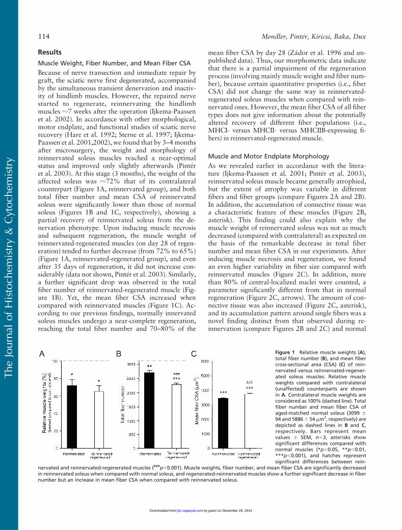
Because of nerve transection and immediate repair bygraft, the sciatic nerve first degenerated, accompaniedby the simultaneous transient denervation and inactiv-ity of hindlimb muscles. However, the repaired nervestarted to regenerate, reinnervating the hindlimbmuscles z7 weeks after the operation (Ijkema-Paassenet al. 2002). In accordance with other morphological,motor endplate, and functional studies of sciatic nerverecovery (Hare et al. 1992; Sterne et al. 1997; Ijkema-Paassen et al. 2001,2002), we found that by 3–4monthsafter microsurgery, the weight and morphology ofreinnervated soleus muscles reached a near-optimalstatus and improved only slightly afterwards (Pinteret al. 2003). At this stage (3 months), the weight of theaffected soleus was z72% that of its contralateralcounterpart (Figure 1A, reinnervated group), and bothtotal fiber number and mean CSA of reinnervatedsoleus were significantly lower than those of normalsoleus (Figures 1B and 1C, respectively), showing apartial recovery of reinnervated soleus from the de-nervation phenotype. Upon inducing muscle necrosisand subsequent regeneration, the muscle weight ofreinnervated-regenerated muscles (on day 28 of regen-eration) tended to further decrease (from 72% to 65%)(Figure 1A, reinnervated-regenerated group), and evenafter 35 days of regeneration, it did not increase con-siderably (data not shown, Pinter et al. 2003). Similarly,a further significant drop was observed in the totalfiber number of reinnervated-regenerated muscle (Fig-ure 1B). Yet, the mean fiber CSA increased whencompared with reinnervated muscles (Figure 1C). Ac-cording to our previous findings, normally innervatedsoleus muscles undergo a near-complete regeneration,reaching the total fiber number and 70–80% of the
mean fiber CSA by day 28 (Zador et al. 1996 and un-published data). Thus, our morphometric data indicatethat there is a partial impairment of the regenerationprocess (involving mainly muscle weight and fiber num-ber), because certain quantitative properties (i.e., fiberCSA) did not change the same way in reinnervated-regenerated soleus muscles when compared with rein-nervated ones. However, the mean fiber CSA of all fibertypes does not give information about the potentiallyaltered recovery of different fiber populations (i.e.,MHCI- versus MHCII- versus MHCIIB-expressing fi-bers) in reinnervated-regenerated muscle.
Muscle and Motor Endplate Morphology
As we revealed earlier in accordance with the litera-ture (Ijkema-Paassen et al. 2001; Pinter et al. 2003),reinnervated soleus muscle became generally atrophied,but the extent of atrophy was variable in differentfibers and fiber groups (compare Figures 2A and 2B).In addition, the accumulation of connective tissue wasa characteristic feature of these muscles (Figure 2B,asterisk). This finding could also explain why themuscle weight of reinnervated soleus was not as muchdecreased (compared with contralateral) as expected onthe basis of the remarkable decrease in total fibernumber and mean fiber CSA in our experiments. Afterinducing muscle necrosis and regeneration, we foundan even higher variability in fiber size compared withreinnervated muscles (Figure 2C). In addition, morethan 80% of central-localized nuclei were counted, aparameter significantly different from that in normalregeneration (Figure 2C, arrows). The amount of con-nective tissue was also increased (Figure 2C, asterisk),and its accumulation pattern around single fibers was anovel finding distinct from that observed during re-innervation (compare Figures 2B and 2C) and normal
Figure 1 Relative muscle weights (A),total fiber number (B), and mean fibercross-sectional area (CSA) (C) of rein-nervated versus reinnervated-regener-ated soleus muscles. Relative muscleweights compared with contralateral(unaffected) counterparts are shownin A. Contralateral muscle weights areconsidered as 100% (dashed line). Totalfiber number and mean fiber CSA ofaged-matched normal soleus (3099 694 and 58866 54 mm2, respectively) aredepicted as dashed lines in B and C,respectively. Bars represent meanvalues 6 SEM, n53, asterisks showsignificant differences compared withnormal muscles (*p,0.05, **p,0.01,***p,0.001), and hatches representsignificant differences between rein-
nervated and reinnervated-regenerated muscles (###p,0.001). Muscle weights, fiber number, and mean fiber CSA are significantly decreasedin reinnervated soleus when compared with normal soleus, and regenerated-reinnervated muscles show a further significant decrease in fibernumber but an increase in mean fiber CSA when compared with reinnervated soleus.
TheJournal
ofHistoch
emistry&
Cytoch
emistry
114 Mendler, Pinter, Kiricsi, Baka, Dux
by guest on December 26, 2014jhc.sagepub.comDownloaded from

regeneration (Whalen et al. 1990). In the morphologicalsense, these data are indicative of an altered, less-synchronous, and less-effective regeneration process inreinnervated soleus. To gain information about theinnervation state of the affected muscles, we performedAChE staining of motor endplates as well (Figures 2D–2F; Pinter et al. 2003). In reinnervated muscles, weconfirmed the findings of others (Ijkema-Paassen et al.2002) who found highly variable motor endplates,several of which were characterized by abnormal (frag-mented or granular) morphology (Figure 2E, arrow-heads; see also inset). In reinnervated-regeneratedmuscles (i.e., on day 28 of regeneration), we observedmotor endplates with similar morphological abnormal-ities coupled several times to smaller fibers (Figure 2F,arrowheads; Pinter et al. 2003). To summarize, ourmorphometric and morphological data revealed bothquantitative and qualitative alterations in the regenera-tion process of reinnervated-regenerated muscles whencompared with reinnervated and normal ones.
MHC Composition Based onImmunohistochemical Analysis
To analyze the possible effects of reinnervation andsubsequent regeneration on MHC composition, serialcrysections were immunostained with a set of mono-
clonal antibodies to distinguish and count (pure andhybrid) fibers expressing different MHC isoforms.
Reinnervated Muscles. In contrast to normal soleus,which contains 80–90% of slow type I fibers (notshown; Delp and Duan 1996), their number dramati-cally decreased toz33% after reinnervation (Figures 3Aand 4A), and the amount of fast fibers significantlyincreased from z10–20% to 85% (Figures 3B and 4A).Although similar data have already been published inthe literature (Bodine and Pierotti 1996; Huey andBodine 1996; Ijkema-Paassen et al. 2002), the differen-tial immunohistochemical analysis of the MHCII iso-forms has not been carried out to date. We found thatmost of the fast II fibers were IIA fibers (44%) (Fig-ures 3C and 4A), and no fibers expressed the fastestMHCIIB isoform (Figures 3D and 4A). The number ofpure (not hybrid) IIX fibers was estimated according tothe difference between II and IIA fibers, which provedto be z40% (Figures 3B–3D and 4A). Another notablechange was the increased number (z20%) of I/II hybridfibers expressing both slow MHCI and fast MHCII(Figures 3A–3C and 4B) compared with those in normalsoleus (less than 8%; Staron et al. 1999). Most of the I/IIhybrids present in reinnervated soleus seemed to expressMHCIIA as the fast isoform (Figures 3A and 3C). Inaddition, we observed variable staining intensity of the
Figure 2 Morphology of the reinnervated and reinnervated-regenerated soleus muscles. Depicted are sections stained with eitherhematoxylin-eosin (HE) (A–C) or with Tago’s method (for AChE activity of motor endplates) (D–F). Compared with normal age-matched soleus(A,D), reinnervated muscle fibers are atrophied to different degrees (see small and larger fibers) (B). Accumulation of connective tissue(B, asterisk) and several endplates of abnormal morphology (E, arrowheads; see also a different area on inset) are evident. Regenerated-reinnervated soleus is characterized by an extremely variable fiber size, with a significant number of centrally located nuclei (C, arrows), byabundant connective tissue (C, asterisk) also around fibers, and by a remarkable number of abnormal (fragmented or granular) endplates(F, arrowheads). Bar 5 100 mm.
TheJournal
ofHistoch
emistry&
Cytoch
emistry
MHC Alterations in Reinnervated–Regenerated Soleus 115
by guest on December 26, 2014jhc.sagepub.comDownloaded from

TheJournal
ofHistoch
emistry&
Cytoch
emistry
116 Mendler, Pinter, Kiricsi, Baka, Dux
by guest on December 26, 2014jhc.sagepub.comDownloaded from
individual I/IIA hybrid fibers (Figures 3A and 3C, andinsets), which may indicate a dynamic transition of type Ito type IIA fibers, similar to the stepwise transformationof slow fibers toward fast ones in inactivity and cross-reinnervation studies (Pette and Vrbova 1992). More-over, spatial grouping of the same MHC-expressingfibers was clearly detectable in all of the examinedsections (Figures 3A–3C), which is a phenomenon seenafter reinnervation (Ijkema-Paassen et al. 2001; Wanget al. 2002).
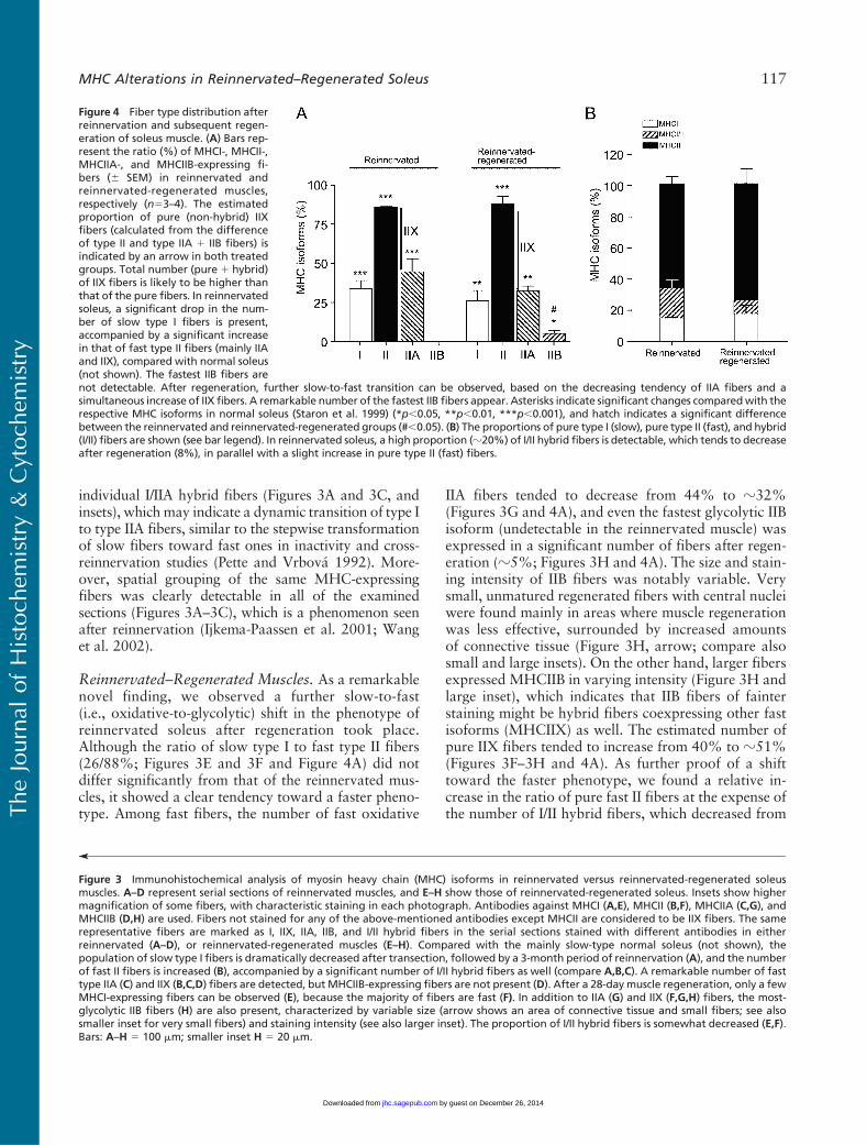
Reinnervated–Regenerated Muscles. As a remarkablenovel finding, we observed a further slow-to-fast(i.e., oxidative-to-glycolytic) shift in the phenotype ofreinnervated soleus after regeneration took place.Although the ratio of slow type I to fast type II fibers(26/88%; Figures 3E and 3F and Figure 4A) did notdiffer significantly from that of the reinnervated mus-cles, it showed a clear tendency toward a faster pheno-type. Among fast fibers, the number of fast oxidative
IIA fibers tended to decrease from 44% to z32%(Figures 3G and 4A), and even the fastest glycolytic IIBisoform (undetectable in the reinnervated muscle) wasexpressed in a significant number of fibers after regen-eration (z5%; Figures 3H and 4A). The size and stain-ing intensity of IIB fibers was notably variable. Verysmall, unmatured regenerated fibers with central nucleiwere found mainly in areas where muscle regenerationwas less effective, surrounded by increased amountsof connective tissue (Figure 3H, arrow; compare alsosmall and large insets). On the other hand, larger fibersexpressed MHCIIB in varying intensity (Figure 3H andlarge inset), which indicates that IIB fibers of fainterstaining might be hybrid fibers coexpressing other fastisoforms (MHCIIX) as well. The estimated number ofpure IIX fibers tended to increase from 40% to z51%(Figures 3F–3H and 4A). As further proof of a shifttoward the faster phenotype, we found a relative in-crease in the ratio of pure fast II fibers at the expense ofthe number of I/II hybrid fibers, which decreased from
Figure 4 Fiber type distribution afterreinnervation and subsequent regen-eration of soleus muscle. (A) Bars rep-resent the ratio (%) of MHCI-, MHCII-,MHCIIA-, and MHCIIB-expressing fi-bers (6 SEM) in reinnervated andreinnervated-regenerated muscles,respectively (n53–4). The estimatedproportion of pure (non-hybrid) IIXfibers (calculated from the differenceof type II and type IIA 1 IIB fibers) isindicated by an arrow in both treatedgroups. Total number (pure 1 hybrid)of IIX fibers is likely to be higher thanthat of the pure fibers. In reinnervatedsoleus, a significant drop in the num-ber of slow type I fibers is present,accompanied by a significant increasein that of fast type II fibers (mainly IIAand IIX), compared with normal soleus(not shown). The fastest IIB fibers are
’
Figure 3 Immunohistochemical analysis of myosin heavy chain (MHC) isoforms in reinnervated versus reinnervated-regenerated soleusmuscles. A–D represent serial sections of reinnervated muscles, and E–H show those of reinnervated-regenerated soleus. Insets show highermagnification of some fibers, with characteristic staining in each photograph. Antibodies against MHCI (A,E), MHCII (B,F), MHCIIA (C,G), andMHCIIB (D,H) are used. Fibers not stained for any of the above-mentioned antibodies except MHCII are considered to be IIX fibers. The samerepresentative fibers are marked as I, IIX, IIA, IIB, and I/II hybrid fibers in the serial sections stained with different antibodies in eitherreinnervated (A–D), or reinnervated-regenerated muscles (E–H). Compared with the mainly slow-type normal soleus (not shown), thepopulation of slow type I fibers is dramatically decreased after transection, followed by a 3-month period of reinnervation (A), and the numberof fast II fibers is increased (B), accompanied by a significant number of I/II hybrid fibers as well (compare A,B,C). A remarkable number of fasttype IIA (C) and IIX (B,C,D) fibers are detected, but MHCIIB-expressing fibers are not present (D). After a 28-day muscle regeneration, only a fewMHCI-expressing fibers can be observed (E), because the majority of fibers are fast (F). In addition to IIA (G) and IIX (F,G,H) fibers, the most-glycolytic IIB fibers (H) are also present, characterized by variable size (arrow shows an area of connective tissue and small fibers; see alsosmaller inset for very small fibers) and staining intensity (see also larger inset). The proportion of I/II hybrid fibers is somewhat decreased (E,F).Bars: A–H 5 100 mm; smaller inset H 5 20 mm.
not detectable. After regeneration, further slow-to-fast transition can be observed, based on the decreasing tendency of IIA fibers and asimultaneous increase of IIX fibers. A remarkable number of the fastest IIB fibers appear. Asterisks indicate significant changes comparedwith therespective MHC isoforms in normal soleus (Staron et al. 1999) (*p,0.05, **p,0.01, ***p,0.001), and hatch indicates a significant differencebetween the reinnervated and reinnervated-regenerated groups (#,0.05). (B) The proportions of pure type I (slow), pure type II (fast), and hybrid(I/II) fibers are shown (see bar legend). In reinnervated soleus, a high proportion (z20%) of I/II hybrid fibers is detectable, which tends to decreaseafter regeneration (8%), in parallel with a slight increase in pure type II (fast) fibers.
TheJournal
ofHistoch
emistry&
Cytoch
emistry
MHC Alterations in Reinnervated–Regenerated Soleus 117
by guest on December 26, 2014jhc.sagepub.comDownloaded from

20% to z8% (Figure 4B). Although several type II fi-bers were probably hybrids coexpressing different fastisoforms (MHCIIA/IIX, MHCIIX/IIB, or MHCIIA/IIX/IIB), we could not distinguish them, owing to the lackof a specific IIX antibody. To summarize our new find-ings, the regeneration process of the reinnervated soleus(reinnervation-regeneration model), in contrast to theuniformly slow-fiber composition of soleus muscle afternormal regeneration, resulted in an even faster musclephenotype with a notable presence of different popula-tions of IIB fibers.
Mean Fiber CSA and Frequency Distribution of MHCI,MHCII, and MHCIIB Fibers
Because fiber size could be indicative of the innervation(atrophy) level of fibers, we aimed at analyzing whetherthere are different fiber populations in reinnervated-regenerated muscles compared with reinnervated ones.In normal slow-type soleus, CSAs of type I fibers weresomewhat larger than those of type II fibers (5909 655 mm2 and 5295 6 79 mm2, respectively; Table 2;Delp and Duan 1996). In reinnervated muscles com-pared with normal muscles, type I fibers became smaller(4945 6 110 mm2; Table 2). However, the frequencydistribution revealed two main populations, one witha close-to-normal CSA and the other with a remark-ably smaller CSA (Figure 5A, black bars, two peaksat z5500 mm2 and 3000–3500 mm2, respectively,n5318). These findings suggest that the larger fibersexpressing MHCI were perfectly reinnervated by slowaxons maintaining their original size, whereas smallerMHCI fibers may represent different populations,which have been either denervated but not transformedyet, or reinnervated by foreign (slow or fast) axons.Type II fibers seemed to be uniformly smaller-sized(2998 6 31 mm2; Table 2; Figure 5B, black bars,n51079) with less than 9% of fibers smaller than1750 mm2 (Table 2; Figure 5B), indicating that most ofthem might be reinnervated.
Table 2 Mean fiber CSA of MHCI, MHCII and MHCIIB fibersin normal, reinnervated, and reinnervated-regeneratedsoleus muscles
Fiber type Normal ReinnervatedReinnervated-regenerated
MHCI 5909 6 55a 4945 6 110a,b 3300 6 80a,b,c
MHCII (all) 5295 6 79 2998 6 31b 3915 6 75b,c
MHCII (.1750 mm2) 3145 6 29b 4480 6 74b,c
MHCIIB (all) 3088 6 185a,b,c
MHCIIB (.1750 mm2) 5326 6 180a,b,c
aSignificant difference between MHCI and II (and IIB) fibers within therespective animal group.bSignificant difference compared to respective fiber type in normal muscles.cSignificant difference between the respective fiber types in reinnervated andreinnervated-regenerated muscles.Values are expressed as mean 6 SEM (mm2).
Figure 5 Frequency distribution of MHCI (A), MHCII (B), andMHCIIB(C) fiber CSA in reinnervated versus reinnervated-regenerated mus-cles. CSA of MHCI- and MHCII-expressing fibers were measured onboth reinnervated and reinnervated-regenerated muscle sectionsstained with the sc-75 antibody, andMHCIIB fibers were analyzed onreinnervated-regenerated sections stained with the BF-F3 antibody.(A) MHCI fibers in reinnervated muscles (n5318) show two peakson the histogram, representing two main fiber populations char-acterized by a smaller and a larger CSA, respectively, whereas inreinnervated-regenerated muscles, type I fibers (n5339) are gener-ally smaller, with only one peak of distribution. (B) MHCII fibers inreinnervated muscles (n51079) are smaller, with a narrow one-peakhistogram, whereas in reinnervated-regenerated soleus (n5865),they are somewhat larger and marked by a wide distribution,starting at very small fibers on one tail and ending up in larger fiberson the other tail. (C) The histogram of IIB fibers in reinnervated-regenerated muscles shows that about half of the fiber populationis very small (,1750 mm2), whereas the other half is larger anddistributed almost evenly between 2000–8500 mm2.
TheJournal
ofHistoch
emistry&
Cytoch
emistry
118 Mendler, Pinter, Kiricsi, Baka, Dux
by guest on December 26, 2014jhc.sagepub.comDownloaded from
In reinnervated-regenerated muscles, the CSA ofMHCI-expressing fibers became generally smaller, char-acterized by only one peak of a narrow-type distribution(3300 6 80 mm2; Table 2; Figure 5A, hatched bars,n5339), showing that the recovery of slow fibers wasimpaired or delayed in the regeneration process. By con-trast, MHCII fibers in reinnervated-regenerated soleusbecame larger than those in reinnervated soleus (3915675 mm2; Table 2; Figure 5B, hatched bars, n5865), andalso larger than the regenerated MHCI fibers (Table 2;compare Figures 5A and 5B, hatched bars). The histo-gram of type II fibers showed a wide distribution profilestarting at very small fibers on one tail and ending up inlarger fibers on the other tail. The small fibers withcharacteristic central nuclei (CSA,1750 mm2) make upz17% of type II fibers and probably represent, at leasta part of them, denervated unmatured fibers not ableto fully differentiate in the absence of innervation. In-deed, as revealed by the size distribution of IIB fibers inreinnervated-regenerated muscles (3088 6 185 mm2;Table 2; Figure 5C), about half of the fiber populationwas relatively small (CSA,1750mm2), suggesting that anotable part of the smaller type II fibers expressedMHCIIB, a default isoform in the denervated regener-ated fibers (see also Figure 3H, small inset; Jerkovic et al.
1997). However, in the other half of the IIB fibers(CSA.1750 mm2), mean fiber CSA was remarkably in-creased when compared with the similar population ofMHCII fibers (5326 6 180 mm2 and 4480 6 74 mm2,respectively; Table 2). This interesting finding clearlyindicates at least two populations of MHCIIB fibers(see also Figure 3H, small and large insets): a smallone with denervation phenotype and a large one,which seems to be relatively hypertrophized comparedwith other type II fibers. In summary, we found inreinnervated-regenerated muscles that with the excep-tion of the IIB-expressing small denervated fibers, fasttype II fibers showed better recovery than did slow type Ifibers at day 28 of regeneration, and a selective hyper-trophy was seen in a certain population of IIB fibers.
mRNA Levels of MHC Isoforms Detected by RT-PCR
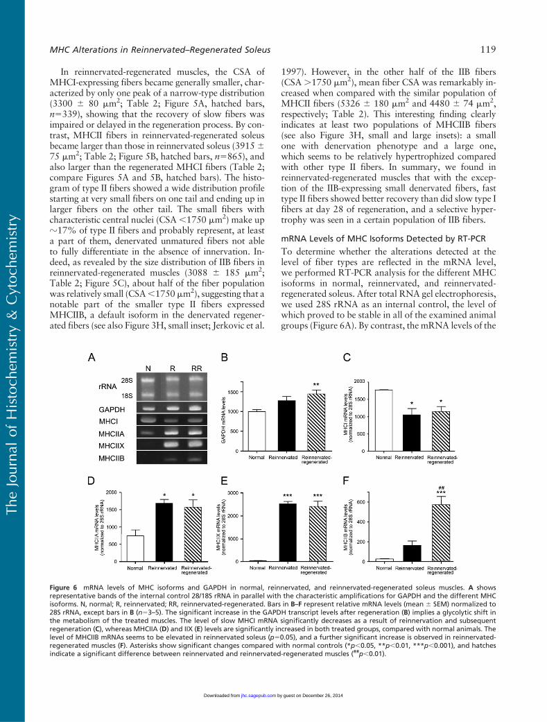
To determine whether the alterations detected at thelevel of fiber types are reflected in the mRNA level,we performed RT-PCR analysis for the different MHCisoforms in normal, reinnervated, and reinnervated-regenerated soleus. After total RNA gel electrophoresis,we used 28S rRNA as an internal control, the level ofwhich proved to be stable in all of the examined animalgroups (Figure 6A). By contrast, the mRNA levels of the
Figure 6 mRNA levels of MHC isoforms and GAPDH in normal, reinnervated, and reinnervated-regenerated soleus muscles. A showsrepresentative bands of the internal control 28/18S rRNA in parallel with the characteristic amplifications for GAPDH and the different MHCisoforms. N, normal; R, reinnervated; RR, reinnervated-regenerated. Bars in B–F represent relative mRNA levels (mean 6 SEM) normalized to28S rRNA, except bars in B (n53–5). The significant increase in the GAPDH transcript levels after regeneration (B) implies a glycolytic shift inthe metabolism of the treated muscles. The level of slow MHCI mRNA significantly decreases as a result of reinnervation and subsequentregeneration (C), whereas MHCIIA (D) and IIX (E) levels are significantly increased in both treated groups, compared with normal animals. Thelevel of MHCIIB mRNAs seems to be elevated in reinnervated soleus (p50.05), and a further significant increase is observed in reinnervated-regenerated muscles (F). Asterisks show significant changes compared with normal controls (*p,0.05, **p,0.01, ***p,0.001), and hatchesindicate a significant difference between reinnervated and reinnervated-regenerated muscles (##p,0.01).
TheJournal
ofHistoch
emistry&
Cytoch
emistry
MHC Alterations in Reinnervated–Regenerated Soleus 119
by guest on December 26, 2014jhc.sagepub.comDownloaded from
glycolytic enzyme GAPDH, another internal controlcandidate, increased significantly after regeneration,indicating a glycolytic shift in the metabolism of thetreated muscles (Figures 6A and 6B). In normal soleus,MHCI is known to be the predominant form; however,a significant decrease took place after reinnervation andsubsequent regeneration (Figures 6A and 6C). In con-trast, MHCIIA mRNA levels increased significantlywith both treatments, compared with normal rats (Fig-ures 6A and 6D), and a similar significant increase inMHCIIX expression was also observed in each treatedgroup (Figures 6A and 6E). In contrast with the immu-nohistochemical alterations, no significant difference inMHCIIA or MHCIIX mRNA levels was found in thereinnervated versus reinnervated-regenerated groups,which is reasonable if one takes into account that totalRNA was isolated from a whole muscle instead of per-forming single-fiber analysis. However, in accordancewith immunohistochemical results, MHCIIB tran-script levels were virtually not detectable in normalsoleus, but a clear elevation (p50.05) was seen in re-innervated muscles and a further highly significantincrease took place in reinnervated-regenerated soleus(Figures 6A and 6F). These findings suggest that IIBfibers could be detectable with immunohistochemistry(in reinnervated-regenerated soleus) only whenMHCIIBtranscript levels were significantly elevated, whereasMHCIIB protein expression could not be visualized inindividual fibers when the transcript levels were lower(in normal and reinnervated soleus). Our results arein agreement with the predominant transcriptionalregulation of MHC isoforms described in slow-typeskeletal muscle under several conditions (Schiaffinoand Reggiani 1996; Baldwin andHaddad 2001; Pandorfet al. 2006).
Discussion
The functional impairment of skeletal muscles afternerve injury, despite optimized surgery, is an intriguingmedical problem (Mackinnon and Dellon 1988). Wehypothesized that in addition to several reinnervationdisturbances, the regenerative capacity of the reinner-vated muscle is also altered, as indicated previously bymorphological and motor endplate abnormalities(Pinter et al. 2003). Indeed, here we provide evidencethat fiber type composition of the reinnervated-regenerated rat soleus muscle is faster than that weobserved in reinnervated soleus. This finding suggeststhat the (fast) pattern of reinnervation plays a dominantrole in the specification of fiber phenotype duringregeneration, which can also contribute to the long-lasting functional impairment of the reinnervated mus-cle. Moreover, because the fast II fibers (and selectively,a certain population of the fast IIB fibers) showed betterrecovery (larger CSA) than did slow type I fibers, the
faster phenotype of the reinnervated-regenerated mus-cle seems to be actively maintained by selective yet un-defined cues.
We applied a rat model of sciatic nerve transectionand immediate repair by graft to analyze the regener-ative machinery of reinnervated muscles (Pinter et al.2003). In reinnervated soleus, our previous morpho-logical findings, as well as the present results on theslow-to-fast fiber type transition, were consistent withthose of another group (Ijkema-Paassen et al. 2001),who used a very similar rat model and described time-dependent abnormalities without a significant im-provement in muscle recovery, even after 60 weeks ofreinnervation (Ijkema-Paassen et al. 2005). In the pres-ent study, however, we performed a more detailedanalysis of the slow, hybrid, and fast fibers at bothimmunohistochemical and mRNA levels, examiningalso the size (atrophy state) of the different fiber pop-ulations, because these data are necessary for judgingthe reinnervation level of fibers before the induction ofregeneration. Our new results, namely the significantlyhigh number of I/II hybrids, as well as the increase in thenumber of both MHCIIA- or MHCIIX-expressing fi-bers, accompanied by respective changes in their mRNAlevels, suggest a gradual slow-to-fast fiber switch inreinnervated soleus, which is regulated predominantly atthe mRNA level (Schiaffino and Reggiani 1996; Baldwinand Haddad 2001; Pandorf et al. 2006).
The explanation for fiber type transition in reinner-vated soleus might be either the partial denervationof certain fiber populations because of axon loss(Mackinnon et al. 1991; Lloyd et al. 2007) and/or therandom reinnervation of muscles by axons of differ-ent motoneuron pools (Bodine-Fowler et al. 1997;Gramsbergen et al. 2000; Ijkema-Paassen et al. 2001).In accordance with the literature (Ijkema-Paassen et al.2001), we found that the total fiber number signifi-cantly decreased in reinnervated soleus when comparedwith normal soleus, which suggests that at least onepopulation of denervated fibers, delayed in reinnerva-tion, has been eliminated. However, the question re-mains: do all denervated fibers undergo completeelimination (by apoptosis or other mechanisms); arethey replaced by new myofibers originating from pro-liferating satellite cells to some extent; or alternatively,do they exist for a longer time in the denervated stage(Tews et al. 1997; Borisov and Carlson 2000; Borisovet al. 2001,2005; Tews 2002). In fact, Patterson et al.(2006) did not find any decrease in fiber number after50-day denervation of rat soleus muscle, or any sign ofthe presence of regenerating fibers. On the basis of theirfindings, denervated fibers may exist for a longer time,expressing mainly fast MHC isoforms and formingseveral I/II hybrid fibers. However, we suggest thatonly a small proportion of fibers was still denervated3 months after repair, as revealed by the difference in
TheJournal
ofHistoch
emistry&
Cytoch
emistry
120 Mendler, Pinter, Kiricsi, Baka, Dux
by guest on December 26, 2014jhc.sagepub.comDownloaded from

muscle weights and fiber diameter of denervated versusreinnervated soleus (Pinter et al. 2003), by the relativelysmall number of very small fibers in reinnervated mus-cles, and by the notably high proportion of musclefibers recovering from notexin-induced necrosis. Thus,random reinnervation and the respective muscle ac-tivity pattern is a more likely explanation for fiber typeand size transitions detected in reinnervated soleus(Bodine-Fowler et al. 1997; Gramsbergen et al. 2000;Ijkema-Paassen et al. 2002). It has been reported thatonly 14% of the original (slow) soleus motoneuronsfind their correct target following nerve repair, indicat-ing that most of the reinnervating axons come fromdifferent motoneuron pools (Bodine-Fowler et al.1997). In fact, slow fibers of normal soleus are remark-ably larger than those of other hindlimb muscles andare also larger than most fast fibers, except IIB fibers(Delp and Duan 1996). We described here two pop-ulations of slow fibers, the larger one probably re-innervated by the original slow axons, and the smallerone, in which fibers may represent either denervatedfibers on the way to transformation, or fibers reinner-vated by foreign slow or (mismatched) fast axons.These mechanisms may even explain our results on theincreased number of I/II hybrid fibers, a fiber pool thatmay match the one found to be polyneurally innervatedin a similar animal model (Ijkema-Paassen et al. 2002).Similarly, the smaller yet remarkably uniform popula-tion of fast II fibers seems to be the consequence ofa gradual fast transformation maintained by foreignaxons reinnervating the soleus muscle. However, wecould not detect the fastest IIB fibers in the reinnervatedsoleus. The reason might be that 3 months of re-innervation was not enough for a complete MHCI–MHCIIB fiber conversion and even longer reinnervationtime would be required for this process (Ijkema-Paassenet al. 2001,2005). On the other hand, even if not all thefast fibers were reinnervated, it seems that denervationdoes not favor the expression of those fast isoforms thatare not present originally in the affected muscle, like theIIB fibers in soleus (Patterson et al. 2006).
Intriguingly, reinnervated fast-type muscles, incontrast with slow-type soleus, almost regained theirnormal phenotype after a transient change towardthe slow character (Ijkema-Paassen et al. 2001; Wanget al. 2002; Zhou et al. 2006). The different reactionsof slow- and fast-type muscles upon reinnervationmay be explained by their different functions, becausesoleus is more dependent on the amount of mechanicalactivity (force generation), compared with fast-typemuscles (Ohira et al. 2006). On the other hand, afew data support the hypothesis that the intrinsic(innervation-independent) properties of a particularmuscle, e.g., different satellite cell populations andtheir functions, play a major role in the specificationof fiber type, even after the removal of neural input
(Patterson et al. 2006). If this is the case, alterations ofthe muscle regenerative capacity, influenced by bothintrinsic and innervation-dependent mechanisms, canalso contribute to the impaired phenotype of the re-innervated soleus.
The main question of our study was whether theregenerative machinery of the reinnervated soleuscan reproduce the original muscle phenotype in bothqualitative and quantitative senses. In the presentstudy, we clarified that the total fiber number wassignificantly lower after 28 days of regeneration inreinnervated-regenerated muscles, compared with re-innervated ones. The “missing” fibers may represent afiber population in reinnervated muscles that has notbeen properly reinnervated and that therefore couldnot maintain the subsequent growth of regeneratedfibers. Otherwise, the mean fiber CSA of the existingfibers was even larger in the reinnervated-regeneratedmuscles than that in the reinnervated soleus (butsmaller than that in normal soleus), indicating thatthe recovery of certain fiber populations was preferredafter regeneration. Indeed, we detected here that eventhough slow-type regenerating fibers did not reachthe size characteristic of those in reinnervated soleus,fast fibers were generally larger in reinnervated-regenerated soleus, compared with reinnervated mus-cles. Moreover, a clear transition toward a fasterphenotype was observed, and even the fastest IIB fi-bers were present after regeneration. Because no re-verse change occurred toward a slower phenotypeeven by day 35 (data not shown), it is unlikely that allthese abnormalities are simple consequences of a de-layed regeneration process resulting from additionalinnervation problems. Although notexin destroys mo-tor endplates, several endplates were re-established attheir original sites in both normally innervated andreinnervated soleus by day 5, and the axons seemed torecover by the end of regeneration (Harris et al. 2000;Pinter et al. 2003). Accordingly, the axon populationreinnervating the soleus muscle after nerve repairshould be essentially the same even after necrosis andsubsequent regeneration.
Therefore, other explanations should exist for thephenotype shift accompanied by the appearance of IIBfibers during the regeneration process. In fact, it is theMHCIIB and IIX mRNAs and proteins that areexpressed in the first few days of normal regeneration,at the time when destroyed motor endplates have notyet been re-established. After the recovery of motorendplates and slow-type innervation, both MHCIIXand IIB mRNAs and proteins decrease gradually inparallel with the increase in slow-type MHCI expres-sion. In accordance with these data, denervated slow-type soleus expressed exclusively the fast MHCIIX andIIB mRNAs in the regeneration process (Esser et al.1993; Jerkovic et al. 1997). Other reports also suggest
TheJournal
ofHistoch
emistry&
Cytoch
emistry
MHC Alterations in Reinnervated–Regenerated Soleus 121
by guest on December 26, 2014jhc.sagepub.comDownloaded from

that IIB fibers develop in the absence of neural stim-ulation (Butler-Browne et al. 1982; Schiaffino andReggiani 1996; Fink et al. 2003). Thus, the relativelysmall MHCIIB-expressing fibers, often located in theconnective tissue–rich areas in our experiments, cer-tainly represent denervated fibers that could not ac-complish MHC transitions and growth. However, thelarger part of the MHCII-expressing fibers, andespecially about half of the IIB fibers, reached a sig-nificantly larger size after regeneration, suggesting anactive (fast-type) innervation of them. Thus, our resultsregarding fast-fiber transition can be interpreted as theimpairment of the physiological fast-to-slow conver-sion during the regeneration process either due to de-nervation (small II and IIB fibers) or mainly, due toactive maintenance of the (larger) fast fibers at theexpense of the (smaller) slow ones. The latter finding isintriguing, particularly because it is the slow-fiber pop-ulation that becomes dominant in normal soleus muscleregenerating from notexin-induced necrosis (Whalenet al. 1990). It cannot be ruled out that intrinsic musclecharacteristics, such as a subpopulation of satellite cellsdetermined to produce mainly slow-type or oxidativefibers (Kalhovde et al. 2005), are selectively impaired inreinnervated soleus, which in turn hampers the properreaction to slow-type innervation, causing a delayedregeneration. This possibility might be tested in addi-tional experiments at later stages of regeneration (i.e.,several weeks after notexin treatment) to see whetherthe difference in the size of slow versus fast fibers wouldbe still apparent. Another explanation for size differ-ences might be that because of different cues, fast-typeaxons or their collaterals become more effective inreinnervated-regenerated muscles than do slow-typeones. Some recent reports have shown a definite role ofactive (fast-type) innervation in the expression of theMHCIIB isoform (Di Maso et al. 2000; Germinarioet al. 2002). Moreover, from the view of several neuro-trophic factors influencing peripheral nerve regenera-tion, neurotrophin-3 and neurotrophin-4 have beendescribed to favor selectively the axon regeneration offast (IIB) and slow motoneuron pools, respectively,playing an important role in determining the correctmuscle phenotype (Sterne et al. 1997; Kingham andTerenghi 2006). Further investigations are needed toexplore the significance of neurotrophins, especially ofneurotrophin-3 in supporting the recovery of fast IIB(and II) fibers in the altered regeneration of the re-innervated soleus.
In conclusion, the morphological, fiber type, and sizealterations detected in reinnervated-regenerated soleuscould be the consequence of a complex process involv-ing both innervation-dependent and/or -independentmolecular mechanisms, the exploration of which maycontribute to a more effective therapy for the func-tionally impaired reinnervated muscle.
Acknowledgments
This work was supported by grants from Medical Scien-tific Board, Hungary (ETT) 421/2003, ETT 008/2006, andRegional Science Center of the University of Szeged, Hungary(RET) 08/2004 National Technological Development Board,Hungary (OMFB)-0066/2005. We thank Balashazi Istvannefor the technical assistance in animal experiments.
Literature Cited
Baldwin KM, Haddad F (2001) Effects of different activity and in-activity paradigms on myosin heavy chain gene expression instriated muscle. J Appl Physiol 90:345–357
Bischoff R (1994) The satellite cell and muscle regeneration. InEngel AG, Franzini-Armstrong C., eds. Myology, vol. 1, Basic andclinical. 2nd ed. Columbus, McGraw-Hill, Inc., 97–118
Bodine SC, Pierotti DJ (1996) Myosin heavy chain mRNA andprotein expression in single fibers of the rat soleus followingreinnervation. Neurosci Lett 215:13–16
Bodine-Fowler S, Meyer RS, Moskovitz A, Abrams R, Botte MJ(1997) Inaccurate projection of rat soleus motoneurons: a com-parison of nerve repair techniques. Muscle Nerve 20:29–37
Borisov AB, Carlson BM (2000) Cell death in denervated skeletalmuscle is distinct from classical apoptosis. Anat Rec 258:305–318
Borisov AB, Dedkov E, Carlson BM (2001) Interrelations of myo-genic response, progressive atrophy of muscle fibers, and cell deathin denervated skeletal muscle. Anat Rec 264:203–218
Borisov AB, Dedkov EI, Carlson BM (2005) Abortive myogenesis indenervated skeletal muscle: differentiative properties of satellitecells, their migration, and block of terminal differentiation. AnatEmbryol (Berl) 209:269–279
Butler-Browne GS, Bugaisky LB, Cuenoud S, Schwartz K, WhalenRG (1982) Denervation of newborn rat muscle does not blockthe appearance of adult fast myosin heavy chain. Nature 299:830–833
Chomczynski P, Sacchi N (1987) Single-step method of RNA iso-lation by acid-guanidium thiocyanate-phenol-chloroform extrac-tion. Anal Biochem 162:156–159
Davis CE, Harris JB, Nicholson LV (1991) Myosin isoform transi-tions and physiological properties of regenerated and re-innervatedsoleus muscles of the rat. Neuromuscul Disord 1:411–421
Delp MD, Duan C (1996) Composition and size of type I, IIA, IID/X,and IIB fibers and citrate synthase activity of rat muscle. J ApplPhysiol 80:261–270
Di Maso NA, Haddad F, Zeng M, McCue SA, Baldwin KM (2000)Role of denervation in modulating IIb MHC gene expression inresponse to T3 plus unloading state. J Appl Physiol 88:682–689
Esser K, Gunning P, Hardeman E (1993) Nerve-dependent and–independent patterns of mRNA expression in regenerating skeletalmuscle. Dev Biol 159:173–183
Fink E, Fortin D, Serrurier B, Ventura-Clapier R, Biggard AX (2003)Recovery of contractile and metabolic phenotypes in regeneratingslow muscle after notexin-induced or crush injury. J Muscle ResCell Motil 24:421–429
Germinario E, Esposito A, Megighian A, Midrio M, Biral D, Betto R,Danieli-Betto D (2002) Early changes of type 2B fibers after dener-vation of rat EDL skeletal muscle. J Appl Physiol 92:2045–2052
Girgenrath S, Song K, Whittemore LA (2005) Loss of myostatinexpression alters fiber-type distribution and expression of myosinheavy chain isoforms in slow- and fast-type skeletal muscle.Muscle Nerve 31:34–40
Gramsbergen A, Ijkema-Paassen J, Meek MF (2000) Sciatic nervetransection in the adult rat: abnormal EMG patterns during loco-motion by aberrant innervation of hindleg muscles. Exp Neurol161:183–193
Grubb BD, Harris JB, Schofield IS (1991) Neuromuscular transmis-sion at newly formed neuromuscular junctions in the regeneratingsoleus muscle of the rat. J Physiol 441:405–421
Gutmann E, Zelena J (1962) Morphological changes in the dener-vated muscle. In Gutmann E., ed., The DenervatedMuscle. Prague,
TheJournal
ofHistoch
emistry&
Cytoch
emistry
122 Mendler, Pinter, Kiricsi, Baka, Dux
by guest on December 26, 2014jhc.sagepub.comDownloaded from

Prague Publishing House of the Czechoslovak Academy ofSciences, 341–371
Hare GM, Evans PJ, Mackinnon SE, Best TJ, Bain JR, Szalai JP,Hunter DA (1992) Walking track analysis: a long-term assessmentof peripheral nerve recovery. Plast Reconstr Surg 100:251–258
Harris JB, Grubb BD, Maltin CA, Dixon R (2000) The neurotoxicityof the venom phospholipases A2, notexin and taipoxin. ExpNeurol 161:517–526
Harris JB, Johnson MA, Karlsson E (1975) Pathological responses ofrat skeletal muscle for a single subcutaneous injection of a toxinisolated from the venom of the Australian tiger snake, Notechisscutatus scutatus. Clin Exp Pharmacol Physiol 2:383–404
Huey KA, Bodine SC (1996) Altered expression of myosin mRNAand protein in rat soleus and tibialis anterior following reinner-vation. Am J Physiol 271:C2016–C2026
Ijkema-Paassen J, Meek MF, Gramsbergen A (2001) Muscle differ-entiation after sciatic nerve transection and reinnervation in adultrats. Ann Anat 183:369–377
Ijkema-Paassen J, MeekMF, Gramsbergen A (2002) Reinnervation ofmuscles after transection of the sciatic nerve in adult rats. MuscleNerve 25:891–897
Ijkema-Paassen J, Meek MF, Gramsbergen A (2005) Long-termreinnervation effects after sciatic nerve lesions in adult rats. AnnAnat 187:113–120
Jaschinski F, Schuler M, Peuker H, Pette D (1998) Changes in myosinheavy chain mRNA and protein levels of rat muscle during forcedcontractile activity. Am J Physiol 274:C365–370
Jerkovic R, Argentini C, Serrano-Sanchez A, Cordonnier C, CalabriaE, Schiaffino S, Lomo T (1997) Early myosin switching inducedby nerve activity in regenerating slow skeletal muscle. Cell StructFunct 22:147–153
Kalhovde JM, Jerkovic R, Sefland I, Cordonnier C, Calabria E,Schiaffino S, Lomo T (2005) “Fast” and “slow” muscle fibers inhindlimb muscles of adult rats regenerate from intrinsically dif-ferent satellite cells. J Physiol 562:847–857
Kalliainen LK, Jejurikar SS, Liang LW, Urbanchek MG, Kuzon WM(2002) A specific force deficit exists in skeletal muscle after partialdenervation. Muscle Nerve 25:31–38
Kingham PJ, Terenghi G (2006) Bioengineered nerve regenerationand muscle reinnervation. J Anat 209:511–526
Lefaucheur JP, Sebille A (1995) The cellular events of injured muscleregeneration depend on the nature of the injury. NeuromusculDisord 5:501–509
Lloyd BM, Luginbuhl RD, Brenner MJ, Rocque BG, Tung TH,Myckatyn TM, Hunter DA, et al. (2007) Use of motor nerve ma-terial in peripheral nerve repair with conduits. Microsurgery 27:138–145
Mackinnon SE, Dellon AL (1988) Surgery of the Peripheral Nerve.New York, Thieme
Mackinnon SE, Dellon AL, O’Brien JP (1991) Changes in nerve fibernumbers distal to a nerve repair in the rat sciatic nerve model.Muscle Nerve 14:1116–1122
Mendler L, Szakonyi G, Zador E, Gorbe A, Dux L, Wuytack F (1998)Expression of sarcoplasmic/endoplasmic reticulumCa21ATPases inthe rat extensor digitorum longus (EDL) muscle regenerating fromnotexin-induced necrosis. J Muscle Res Cell Motil 19:777–785
Mendler L, Zador E, Ver Heyen M, Dux L, Wuytack F (2000) Myo-statin in regenerating rat muscles and in myogenic cell cultures.J Muscle Res Cell Motil 21:551–563
Ohira Y, Yoshinaga T, Ohara M, Kawano F, Wang XD, Higo Y,
Terada M, et al. (2006) The role of neural and mechanicalinfluences in maintaining normal fast and slow muscle properties.Cells Tissues Organs 182:129–142
Pandorf CE, Haddad F, Roy RR, Qin AX, Edgerton VR, Baldwin KM(2006) Dynamics of myosin heavy chain gene regulation in slowskeletal muscle. J Biol Chem 281:38330–38342
Patterson MF, Stephenson GMM, Stephenson DG (2006) Denerva-tion produces different single fiber phenotypes in fast- and slow-twitch hindlimb muscles of the rat. Am J Physiol Cell Physiol291:518–528
Pette D, Staron R (1990) Cellular and molecular diversities of mam-malian skeletal muscle fibers. Rev Physiol Biochem Pharmacol116:1–76
Pette D, Vrbova G (1992) Adaptation of mammalian skeletal musclefibers to chronic electrical stimulation. Rev Physiol Biochem Phar-macol 120:116–202
Pinter S, Mendler L, Dux L (2003) Neural impacts on the re-generation of skeletal muscles. Acta Biochim Pol 50:1229–1237
Schiaffino S, Gorza L, Sartore S, Saggin L, Ausoni S, Vianello M,Gundersen K, et al. (1989) Three myosin heavy chain isoformsin type 2 skeletal muscle fibres. J Muscle Res Cell Motil 10:197–205
Schiaffino S, Reggiani C (1996) Molecular diversity of myofibrillarproteins: gene regulation and functional significance. Physiol Rev76:371–424
Sesodia S, Cullen MJ (1991) The effect of denervation on the mor-phology of regenerating rat soleus muscles. Acta Neuropathol(Berl) 82:21–32
Staron RS, Kraemer WJ, Hikida RS, Fry AC, Murray JD, CamposGER (1999) Fiber type composition of four hindlimb muscles ofadult Fisher 344 rats. Histochem Cell Biol 111:117–123
Sterne GD, Coulton GR, Browb RA, Green CJ, Terenghi G (1997)Neurotrophin-3-enhanced nerve regeneration selectively improvesrecovery of muscle fibers expressing myosin heavy chains 2b. J CellBiol 139:709–715
Tago H, Kimura H, Maeda T (1986) Visualization of detailedacetylcholinesterase: fiber and neuron staining in rat brain by asensitive histochemical procedure. J Histochem Cytochem 34:1431–1438
Tews DS (2002) Apoptosis and muscle fibre loss in neuromusculardisorders. Neuromuscul Disord 12:613–622
Tews DS, Goebel HH, Scheider I, Gunkel A, Stennert E, Neiss WF(1997) DNA-fragmentation and expression of apoptosis-relatedproteins in experimentally denervated and reinnervated rat facialmuscle. Neuropathol Appl Neurobiol 23:141–149
Vrbova G, Gordon T, Jones R (1995) Nerve-muscle Interaction.London, Chapman&Hall
Wang L, Copray S, Brouwer N, Meek M, Kernell D (2002) Regionaldistribution of slow-twitch muscle fibers after reinnervation inadult rat hindlimb muscles. Muscle Nerve 25:805–815
Whalen RG, Harris JB, Butler-Browne GS, Sesodia S (1990)Expression of myosin isoforms during notexin-induced regenera-tion of rat soleus muscles. Dev Biol 141:24–40
Zador E, Mendler L, Ver Heyen M, Dux L, Wuytack F (1996)Changes in mRNA levels of the sarcoplasmic/endoplasmic-reticulum Ca21-ATPase isoforms in the rat soleus muscle regen-erating from notexin-induced necrosis. Biochem J 320:107–113
Zhou Z, Cornelius CP, Eichner M, Bornemann A (2006)Reinnervation-induced alterations in rat skeletal muscle. Neuro-biol Dis 23:595–602
TheJournal
ofHistoch
emistry&
Cytoch
emistry
MHC Alterations in Reinnervated–Regenerated Soleus 123
by guest on December 26, 2014jhc.sagepub.comDownloaded from