Heme Oxygenase 1 expression after traumatic brain injury ...
Upload
khangminh22Category
view
1download
0
Oxygen Activation and Radical Transformations in Heme Proteinsand MetalloporphyrinsXiongyi Huang†,‡ and John T. Groves*,†
†Department of Chemistry, Princeton University, Princeton, New Jersey 08544, United States‡Department of Chemistry, California Institute of Technology, Pasadena, California 91125, United States
ABSTRACT: As a result of the adaptation of life to an aerobic environment, nature hasevolved a panoply of metalloproteins for oxidative metabolism and protection againstreactive oxygen species. Despite the diverse structures and functions of these proteins,they share common mechanistic grounds. An open-shell transition metal like iron orcopper is employed to interact with O2 and its derived intermediates such as hydrogenperoxide to afford a variety of metal−oxygen intermediates. These reactiveintermediates, including metal-superoxo, -(hydro)peroxo, and high-valent metal−oxospecies, are the basis for the various biological functions of O2-utilizing metalloproteins.Collectively, these processes are called oxygen activation. Much of our understanding ofthe reactivity of these reactive intermediates has come from the study of heme-containing proteins and related metalloporphyrin compounds. These studies not onlyhave deepened our understanding of various functions of heme proteins, such as O2storage and transport, degradation of reactive oxygen species, redox signaling, and biological oxygenation, etc., but also havedriven the development of bioinorganic chemistry and biomimetic catalysis. In this review, we survey the range of O2 activationprocesses mediated by heme proteins and model compounds with a focus on recent progress in the characterization andreactivity of important iron−oxygen intermediates. Representative reactions initiated by these reactive intermediates as well assome context from prior decades will also be presented. We will discuss the fundamental mechanistic features of thesetransformations and delineate the underlying structural and electronic factors that contribute to the spectrum of reactivities thathas been observed in nature as well as those that have been invented using these paradigms. Given the recent developments inbiocatalysis for non-natural chemistries and the renaissance of radical chemistry in organic synthesis, we envision that newenzymatic and synthetic transformations will emerge based on the radical processes mediated by metalloproteins and theirsynthetic analogs.
CONTENTS
1. Introduction 24922. Key Intermediates Involved in O2 Activation by
Heme Proteins and Model Compounds 24932.1. Oxy−Ferrous (Superoxoferric) Intermediates 2493
2.1.1. Structural Features 24932.1.2. Nature of Fe−O2 Bonding 24932.1.3. Metal-Dioxygen Intermediates of Metal-
loporphyrins 24942.2. Ferric-(hydro)peroxo Intermediates 2496
2.2.1. Ferric-(hydro)peroxo Intermediates ofHeme Proteins 2496
2.2.2. Ferric-(hydro)peroxo Intermediates ofMetalloporphyrins 2496
2.3. Heterolytic O−O Bond Cleavage of Ferric−Hydroperoxo Intermediates 2498
2.3.1. O−O Bond Cleavage of cpd 0 toGenerate Cpd I in Heme Proteins 2498
2.3.2. O−O Bond Cleavage in Metal-PeroxoPorphyrin Complexes 2499
2.4. Compound I: Oxoiron(IV) Porphyrin CationRadicals 2502
2.4.1. Compounds I of Heme Proteins 2502
2.4.2. Compounds I of Iron Porphyrins 25022.4.3. Manganese Porphyrin Analogues of
Compound I 25052.5. Compound II and Its Protonation State−
ferryl or Hydroxoiron(IV) 25062.6. Preventing Protein Oxidative Damage by
Cpd I 25083. C−H Functionalization Reactions Mediated by
Oxoiron(IV) Porphyrin Cation Radicals (Com-pound I) 25083.1. Factors that Affect Oxoiron(IV) Porphyrin
Cation Radical Reactivity for HAT 25093.1.1. Thermodynamic Properties of Cpd I 25093.1.2. Effect of Axial Ligands and Porphyrin
Peripheral Substituents on Cpd I Re-activity 2510
3.1.3. Correlation of Thermodynamic DrivingForce with Rate Constants of HAT 2510
3.1.4. Two-State and Multistate Reactivity 2511
Special Issue: Oxygen Reduction and Activation in Catalysis
Received: June 21, 2017Published: December 29, 2017
Review
pubs.acs.org/CRCite This: Chem. Rev. 2018, 118, 2491−2553
© 2017 American Chemical Society 2491 DOI: 10.1021/acs.chemrev.7b00373Chem. Rev. 2018, 118, 2491−2553
This is an open access article published under an ACS AuthorChoice License, which permitscopying and redistribution of the article or any adaptations for non-commercial purposes.

3.1.5. Quantum Tunneling in Hydrogen AtomTransfer 2513
3.2. Intermediacy of Substrate Radicals−OxygenRebound 2514
3.3. Redirecting Substrate Radicals to ReactionPathways Other than Oxygen Rebound 2517
3.3.1. Decarboxylation Mediated by P450OleTJE 2517
3.3.2. P450-Catalyzed Desaturation Reactions 25183.3.3. Oxidative Rearrangement Catalyzed by
PntM (CYP161C2) 25193.3.4. Mn-Catalyzed C−H Halogenations 2520
3.4. Oxygen-Atom Transfer Reactions Catalyzedby Compound I 2521
3.5. O−O Bond Formation Mediated by Oxome-talloporphyrins 2523
3.5.1. Oxygen Evolution from Oxometal Com-plexes 2523
3.5.2. Chlorite Dismutase 25253.6. Single Electron Transfer (SET) Reactions of
Cpd I 25274. Reactions Mediated by Heme−Oxygen Inter-
mediates Other than Cpd I 25284.1. Homolytic O−O Bond Cleavage of Peroxides
by Heme Proteins 25284.2. Reactions Initiated by Ferric-Superoxo Inter-
mediates 25314.3. O2 Reduction Catalyzed by Heme/Cu Termi-
nal Oxidases and Related Model Com-pounds 2532
5. Conclusions and Outlook 2536Author Information 2537
Corresponding Author 2537ORCID 2537Notes 2537Biographies 2537
Acknowledgments 2537Abbreviations 2537References 2537
1. INTRODUCTION
The highly exergonic four-electron reduction of oxygen towater has an enthalpy change of −80 kcal/mol. Thebiochemical processes that mediate this reduction provide theenergy that sustains all aerobic organisms on earth.1 Paradoxi-
cally, however, the utilization of O2-derived energy in aerobicbiology represents a significant challenge due to thethermodynamic stability of O2 in its triplet ground state.1−5
The first one-electron reduction of O2 to superoxide ion isendergonic by 7.8 kcal/mol, which makes the stepwise, one-electron processes unfavorable (Figure 1). Direct 2e− reactionsbetween ground-state O2 (S = 1) and many closed-shell organicsubstrates are spin forbidden unless a stepwise route viadiradical intermediates is available. To overcome thesedifficulties of exploiting O2, nature has evolved families ofenzymes to transport O2 and to catalyze, manipulate, andcontrol its reduction. A majority of these enzymes containtransition metals such as iron, copper, and sometimesmanganese with unpaired d-electrons.6 These open-shellsystems with variable oxidation and electronic states canfacilitate the activation of triplet ground-state oxygen andparticipate at different stages of O2 reduction (Figure 1). Thestudy of these processes, known collectively as oxygenactivation, is a central theme in biology and has driven thedevelopment of metallobiochemistry and bioinorganic chem-istry since their inception.Heme-containing proteins have assumed a key position in
our understanding of O2-activation mediated by metal-loproteins. The general catalytic cycle for oxygen reduction isshown in Figure 2, which starts with the binding of O2 to aferrous heme to afford a ferric-superoxo adduct, followed byreduction to ferric−hydroperoxo, and high-valent, iron−oxo(ferryl) intermediates via O−O bond scission. The richchemistry that occurs between the heme iron and dioxygenprovides the basis for the diverse roles of heme proteins in O2
metabolism, including O2 transport (myoglobin and hemoglo-bin), reduction of O2 to water (cytochrome c oxidases),degradation of reactive oxygen species (catalases andperoxidases), and catalysis of a variety of oxygenation reactionsof organic substrates such as steroid hydroxylation, amonooxygenation typical of cytochrome P450s, and dioxyge-nation reactions (heme dioxygenases).7 The use of metal-cofactors to effect serial reductive activation of O2 is not limitedto heme proteins but represents a general strategy employed bynature to harness the oxidation power of dioxygen. Thereaction sequence and reactive intermediates similar to thatshown in Figure 2a are widely present in a variety of O2-activating metalloenzymes such as nonheme iron oxygenasesand copper-containing oxygenases and oxidases (Figure2b).6,8−11 Accordingly, in spite of their diverse structures and
Figure 1. Reduction sequence of O2 and representative metalloenzymes related to O2 activation. Reduction potentials shown are versus normalhydrogen electrode (NHE).5
Chemical Reviews Review
DOI: 10.1021/acs.chemrev.7b00373Chem. Rev. 2018, 118, 2491−2553
2492

functions, these metalloproteins share many common mecha-nistic grounds in terms of O2 activation and catalysis.1,6,12
In this interpretive review, we will survey the range of O2activation processes mediated by heme proteins and relatedmodel compounds with a focus on recent progress in thecharacterization and reactivity of important iron−oxygenintermediates through mid-2017. Key aspects of backgroundmaterial in the field will also be presented to provide contextand perspective. The underlying structural and electronicfactors contributing to O2 activation chemistry will bedelineated. The topics of O2-activation and oxidative trans-formations mediated by heme proteins and metalloporphyrinshave been extensively reviewed previously.3,4,7,13−32 While mostof these reviews focus on either heme proteins or syntheticmodels, this review will provide a comprehensive perspective ofthe spectrum of oxidative transformations mediated by theiron−oxygen species in both heme proteins and analogoussynthetic model compounds. We will discuss the fundamentalmechanistic features of these transformations and how a varietyof oxidative pathways are developed from a commonmechanistic foundation. For model compounds, we will mainlyfocus on metalloporphyrins. Other porphyrinoid ligands, suchas corrolazines, porphyrazines, and phthalocyanines will not bediscussed in detail. Readers can refer to several recent reviewson these families of model compounds.29,33−35
2. KEY INTERMEDIATES INVOLVED IN O2ACTIVATION BY HEME PROTEINS AND MODELCOMPOUNDS
2.1. Oxy−Ferrous (Superoxoferric) Intermediates
2.1.1. Structural Features. The first step of O2 activationby heme proteins is the binding of dioxygen to the heme iron,forming an oxy−ferrous intermediate (or ferric-superoxointermediate, see discussions in section 2.1.2). Heme proteinsexhibit varied O2 binding features. For instance, O2 transportproteins like hemoglobin and myoglobin have very high O2association rate constants (107 M−1 s−1) and moderate to highdissociation rate constants (0.1−20 s−1).36,37 Heme-containingNO or CO sensor proteins are tuned to coordinate thesetighter-binding molecules and show little binding of O2 even in
O2-saturated solutions.38,39 In P450s, O2 binds to ferrous hemewith rate constants around 106 M−1 s−1,40 and the resultingferric-superoxo intermediate is prone to undergo autoxidationto the ferric state if it is not further reduced to form the ferrichydroperoxo compound 0.41 It is important to note that hemein the ferric state has a very weak oxygen-affinity and will notbind oxygen at ambient temperatures. It is the ease of electrontransfer from iron to O2 that is important for the formation ofthe ferric-superoxo intermediate and thus O2 binding. Forinstance, highly electron-withdrawing Fe(II) porphyrin such asFe(TPFPBr8) is very inert toward dioxygen (TPFPBr8 = β-pyrrole brominated 5,10,15,20-tetrakis(pentafluorophenyl)-porphyrin).42
The structural and electronic properties of the ferric-superoxo intermediate are crucial for understanding the O2activation in heme proteins. In the 1930s, Pauling and Coryellfound that oxygenated hemoglobin (oxy-Hb) was diamagneticand proposed that dioxygen was bound to heme in a bent, end-on mode.43,44 This geometric feature was confirmed by anoxy−ferrous model compound synthesized by Collman and bycrystal structures of oxy-Mb and oxy-Hb solved by Phillips andShaanan, respectively.45−47 Following these pioneering efforts,crystal structures of ferric-superoxo intermediates have beensolved for a number of heme proteins (Figure 3).48−52 Allreported structures reveal an end-on coordination of dioxygento the iron with a Fe−O bond distance around 1.8 Å, an O−Obond distance ranging from 1.22 to 1.30 Å, and an Fe−O−Oangle ranging from 110° to 160°.
2.1.2. Nature of Fe−O2 Bonding. The electronic structureof Fe−O2 heme complexes is remarkable because it forms aspecies with a singlet electronic ground state arising from thecombination of high-spin (S = 2) ferrous heme and triplet O2.The nature of Fe−O2 bonding has been a subject of activedebate for decades.53 There are three models to explain Fe−O2bonding (Figure 4).54 In Pauling’s model, an S = 0 state wasassigned to both a dioxygen and a ferrous center.43,44 Incontrast, Weiss proposed that the Fe−O2 species would be bestdescribed as a ferric-superoxide complex in which a low-spin (S= 1/2) ferric center is antiferromagnetically coupled to adoublet superoxide anion O2
−.55 In a third model initiallysuggested by McClure and refined by Goddard, an
Figure 2. (a) Interaction of dioxygen with heme and heme−oxygen intermediates in heme proteins. (b) Representative metal−oxygen intermediatesin metalloproteins (PHM: peptidylglycine α-hydroxylating monooxygenase).
Chemical Reviews Review
DOI: 10.1021/acs.chemrev.7b00373Chem. Rev. 2018, 118, 2491−2553
2493

intermediate-spin (S = 1) ferrous center antiferromagneticallycoupled to 3O2 in a 3-center-4-electron bond affords the singletFe−O2 species (“Ozone model”).56,57
Mossbauer studies have lent support to the Weiss model.The Mossbauer parameters of oxyhemoglobin (isomer shift δ =0.2−0.3 mm/s, quadrupole splitting ΔEQ = 2.00−2.20 mm/s),first measured by Lang and Marshali, are consistent with a low-spin ferric center.58 Sharrock and co-workers have reportedsimilar parameters for oxygenated reduced P450cam (δ = 0.31mm/s and ΔEQ = 2.15 mm/s at 4.2K).59 The Weiss model isfurther supported by resonance Raman spectroscopic studies, inwhich a ferric center was indicated by the oxidation statemarker band of oxyHb (ν = 1377 cm−1).60−62
Through density functional theory (DFT) and valence bond(VB) approaches, Shaik has shown that Fe−O2 contains adative σ(Fe−O) interaction between vacant 3dz
2 and π*(O2−)
and a rather weak π interaction between dyz(Fe) andπ⊥*(O2).
53 The H-bonding from a distal histidine residue(His64) in oxy-Mb and the electrostatic polarization fromprotein backbone stabilizes the negative charges on O2.Although Fe−O2 primarily follows the bonding scheme ofthe Weiss model, the VB analysis showed a mixing of the othertwo models, whose contributions depend on protein hosts andactive site environments, making Fe−O2 difficult to describeaccording to canonical oxidation state formalisms. Recent Fe K-edge X-ray absorption spectroscopy (XAS) studies by Sarangiet al. have shown that oxy-Hb is best described as a ferric
superoxide species (Weiss model) in solution. However, incrystalline oxy-Hb, the Fe K pre-edge feature indicates Fe2+ S =0 character, consistent with the Pauling model. This surprisingchange of electronic configuration with environment supportsthe multiconfigurational description.63 The impact of localenvironment on the Fe−O2 bonding was also revealed by aniron L-edge XAS study of a ferric-superoxo “picket fence”porphyrin with 1-methylimidazole (1-MeIm) axial ligand,Fe(TpivPP)(1-MeIm)O2 (TpivPP = meso-tetra(α,α,α,α−O-pivalamidophenyl)porphyrin, Figure 5b).64 The results showeda strong π-interaction between dyz(Fe) and π⊥*(O2) asopposed to the weak π-interaction calculated for oxy-Mb.This difference is likely caused by the much weaker hydrogenbonding to peroxo distal oxygen (N···O distance ∼3.9 Åcompared to 2.67 Å in oxy-myoglobin) and the lack of activesite polarization in this model compound.
2.1.3. Metal-Dioxygen Intermediates of Metallopor-phyrins. Studies of biomimetic synthetic iron porphyrins havegreatly aided in the understanding of O2 binding and activationin heme proteins. An early study by Traylor et al. showed thatthe O2 affinity of imidazole-ligated iron(II) porphyrin (1,Figure 5a) was over 3800 times stronger than that of a pyridine-ligated one (2, Figure 5a), whereas both complexes showedsimilar CO binding affinities.65 This difference was thought tooriginate from the large π basicity of imidazole, which couldbetter stabilize the weak O2 ligand. Much progress has beenmade with sterically protected porphyrin scaffolds like “picket−fence” porphyrins, which prevent the irreversible bimolecularcondensation of Fe(II) porphyrins to form μ-oxo-bridgeddiiron(III) complexes upon reacting with O2. This strategy wasfirst implemented by Collman and led to the characterization ofthe first Fe−O2 porphyrin compound in the 1970s.45,66 Thismodel binds O2 reversibly with affinity similar to those of Hband Mb. More importantly, the Fe−O2 intermediate preparedby Collman is diamagnetic and showed a bent end-on bindinggeometry of dioxygen just as postulated by Pauling. With asimilar approach, many synthetic Fe−O2 complexes have beensynthesized and characterized with varied O2 binding affinitiestuned by ligand environments.22,24 Very recently, the Scheidt
Figure 4. Three models for Fe−O2 bonding in the ferric-superoxointermediate. Adapted from ref 64. Copyright 2013 AmericanChemical Society.
Figure 3. Structural parameters of ferric-superoxo intermediates of representative heme proteins (oxy-myoglobin, PDB: 1A6M; HRP compound III,PDB: 1H57; P450cam oxygen complex, PDB: 1DZ8). aData presented are for the β subunit of oxyhemoglobin in structure 1HHO.
Chemical Reviews Review
DOI: 10.1021/acs.chemrev.7b00373Chem. Rev. 2018, 118, 2491−2553
2494
and Schulz groups have used multitemperature X-raycrystallography and Mossbauer spectroscopy to study threeferric-superoxo complexes based on “picket fence” porphyrinTpivPP with 1-methyl-, 1-ethyl-, or 2-methylimidazoles as axialligand, respectively.67 Their results showed a low energy for therotation of the Fe−O2 unit. An 80K structure of Fe(TpivPP)-(O2) (3, Figure 5b) with 2-methylimidazole (2-MeHIm) axialligand allowed them to accurately determine the position ofboth oxygen atoms, whose structural details were vailed inprevious studies because of the high thermal motion and O2disorder. They obtained a Fe−O distance of 1.811(5) Å, O−Obond lengths of 1.281(12) Å, Fe−O−O angle of 118.2°, and anoff-axis tilt of Fe−O bond of 6.2°.Heme-containing metal−organic frameworks (MOFs) have
recently been used as a platform to study the Fe−O2 adductbecause of the spatial separation of heme centers in the MOFscaffold and the gas-absorption properties of MOFs. Thisapproach allows for solvent−free generation of ferric-superoxointermediates. A rare 5-coordinate heme−O2 complex could beobtained by reacting a Zr-based porphyrinic MOF PCN-224FeII (PCN stands for porous coordination network) withO2 at −78 °C.
68 The species revealed a characteristic η1 end-onbinding mode of O2 with a Fe−O distance of 1.79(1) Å, O−Odistance of 1.15(4), and an Fe−O−O angle of 118(4)o. PCN-224FeII showed a low O2-binding enthalpy of −8 kcal/mol,about half of those of other heme model compounds withimidazole axial ligands. This weak O2 affinity suggests theimportance of electron-donation from axial ligands (σ-donationin the case of imidazole) for the formation of Fe−O2complexes.Transition-metals other than iron have also been explored for
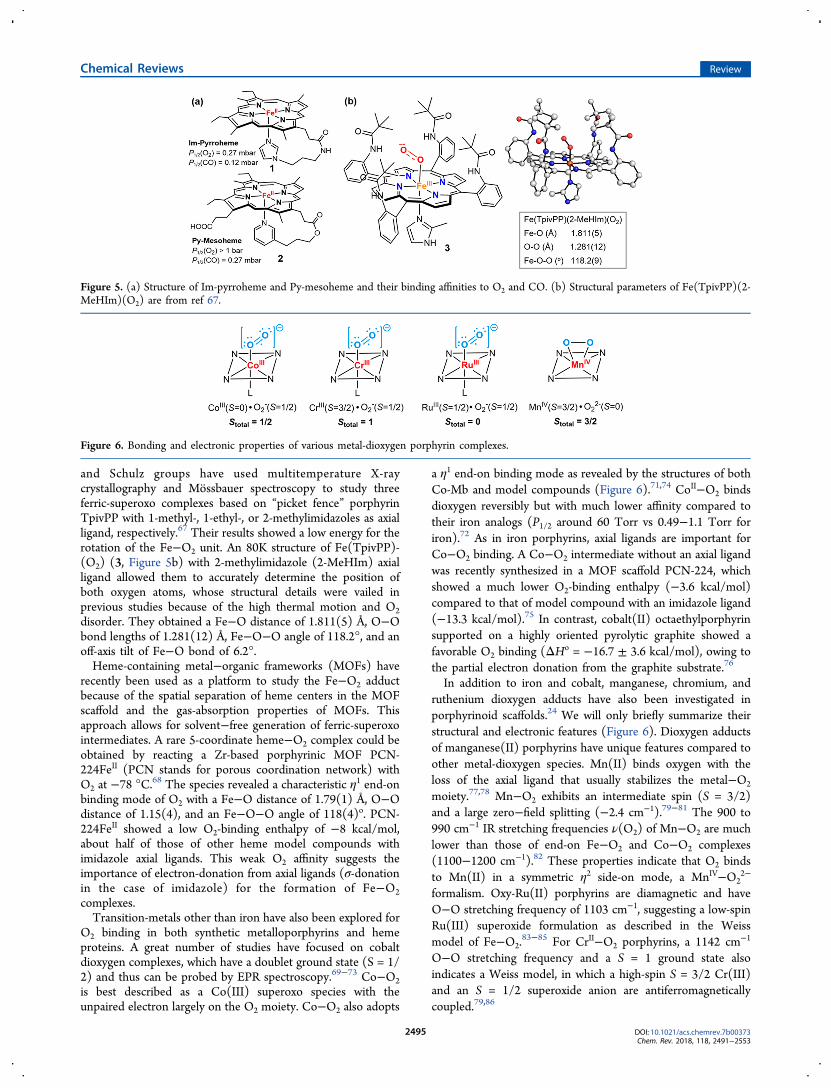
O2 binding in both synthetic metalloporphyrins and hemeproteins. A great number of studies have focused on cobaltdioxygen complexes, which have a doublet ground state (S = 1/2) and thus can be probed by EPR spectroscopy.69−73 Co−O2is best described as a Co(III) superoxo species with theunpaired electron largely on the O2 moiety. Co−O2 also adopts
a η1 end-on binding mode as revealed by the structures of bothCo-Mb and model compounds (Figure 6).71,74 CoII−O2 bindsdioxygen reversibly but with much lower affinity compared totheir iron analogs (P1/2 around 60 Torr vs 0.49−1.1 Torr foriron).72 As in iron porphyrins, axial ligands are important forCo−O2 binding. A Co−O2 intermediate without an axial ligandwas recently synthesized in a MOF scaffold PCN-224, whichshowed a much lower O2-binding enthalpy (−3.6 kcal/mol)compared to that of model compound with an imidazole ligand(−13.3 kcal/mol).75 In contrast, cobalt(II) octaethylporphyrinsupported on a highly oriented pyrolytic graphite showed afavorable O2 binding (ΔHo = −16.7 ± 3.6 kcal/mol), owing tothe partial electron donation from the graphite substrate.76
In addition to iron and cobalt, manganese, chromium, andruthenium dioxygen adducts have also been investigated inporphyrinoid scaffolds.24 We will only briefly summarize theirstructural and electronic features (Figure 6). Dioxygen adductsof manganese(II) porphyrins have unique features compared toother metal-dioxygen species. Mn(II) binds oxygen with theloss of the axial ligand that usually stabilizes the metal−O2
moiety.77,78 Mn−O2 exhibits an intermediate spin (S = 3/2)and a large zero−field splitting (−2.4 cm−1).79−81 The 900 to990 cm−1 IR stretching frequencies ν(O2) of Mn−O2 are muchlower than those of end-on Fe−O2 and Co−O2 complexes(1100−1200 cm−1).82 These properties indicate that O2 bindsto Mn(II) in a symmetric η2 side-on mode, a MnIV−O2
2−
formalism. Oxy-Ru(II) porphyrins are diamagnetic and haveO−O stretching frequency of 1103 cm−1, suggesting a low-spinRu(III) superoxide formulation as described in the Weissmodel of Fe−O2.
83−85 For CrII−O2 porphyrins, a 1142 cm−1
O−O stretching frequency and a S = 1 ground state alsoindicates a Weiss model, in which a high-spin S = 3/2 Cr(III)and an S = 1/2 superoxide anion are antiferromagneticallycoupled.79,86
Figure 5. (a) Structure of Im-pyrroheme and Py-mesoheme and their binding affinities to O2 and CO. (b) Structural parameters of Fe(TpivPP)(2-MeHIm)(O2) are from ref 67.
Figure 6. Bonding and electronic properties of various metal-dioxygen porphyrin complexes.
Chemical Reviews Review
DOI: 10.1021/acs.chemrev.7b00373Chem. Rev. 2018, 118, 2491−2553
2495

2.2. Ferric-(hydro)peroxo Intermediates
2.2.1. Ferric-(hydro)peroxo Intermediates of HemeProteins. The binding of dioxygen to ferrous heme facilitatesits reductive activation. In many hemoproteins such as nitricoxide synthase and cytochrome P450s, an electron and a protonare transferred to the oxy−ferrous complex to afford a ferric−hydroperoxo intermediate [Fe(III)−O−OH], also known ascompound 0 (cpd 0). Distinct from most heme proteins,cytochrome c oxidase (CcO) utilizes a copper cofactorcomplexed with active site residues, including an unusual His-Tyr cross-link, to facilitate the O2 activation. This arrangementleads to the formation of an analogous ferric-peroxo-cupricintermediate. O2 activation by CcO is discussed in detail insection 4.3 of this review.Cpd 0 species have proven to be short-lived, posing
significant challenges for their characterization. Cryogenicradiolytic reduction, as first demonstrated by Davydov andSymons,87−89 has proven to be very useful for the study of cpd0.90 This method utilizes the hydrated electrons generatedthrough radiolysis to reduce the ferrous-dioxygen intermediatein frozen solvent matrices. The combination of radiolyticcryoreduction with spectroscopic methods, especially EPR andENDOR spectroscopies employed by Hoffman et al., haveafforded critical insight into the electronic and structuralproperties of cpd 0.91 By controlling the cryogenic conditions,ferric-peroxo anion species [Fe(III)−O−O−] could be obtainedand its protonation at the distal oxygen to form the conjugateacid [Fe(III)−O−O−H, cpd 0] upon thermal annealing couldbe monitored by EPR spectroscopy. It is now known that cpd 0has a low-spin ferric, S = 1/2 ground state. The g1 value of cpd0 was found to be indicative of the protonation state of thedistal oxygen, with a g1 > 2.30 commonly observed for theferric−hydroperoxo state.92 The thermal annealing experimentsalso showed that the ease of proton delivery varied widelyacross heme proteins. For instance, Mb ferric-peroxo is stablenear 100 K, whereas the ferric-peroxo anion of P450cam isreadily protonated above 55 K.93−95 This ease of protonationmay be related to the presence of the proton relay channel inP450cam. Very recently, Kincaid et al. have characterized ferric-(hydro)peroxo intermediates of lactoperoxidase (LPO) viacryoradiolysis/resonance Raman (rR) spectroscopy.96 Underalkaline conditions (pH 8.2), the ferric-peroxo intermediate isthe major form for LPO and no protonation occurred viathermal annealing. Under acidic conditions (pH 5.6), however,a mixture of LPO-peroxo and LPO−hydroperoxo intermediateswere formed. Intriguingly, upon thermal annealing to 170 K,LPO-peroxo did not convert to the hydroperoxo species, andits rR intensity decreased substantially, whereas LPO−hydro-peroxo was more stable with peak intensities almost unchanged.Radiolytic cryoreduction has also enabled the structuralcharacterization of the fleeting ferric-(hydro)peroxo specieswith X-ray crystallography. In 2007, Schlichting et al. reported acrystal structure of compound 0 by controlled radiolyticreduction of ferrous-dioxygen intermediate of chloroperoxidase(CPO).97 Cpd 0 of CPO showed an O−O distance of 1.5 Å, along Fe−O distance of 1.9 Å, and an Fe−O−O angle of 131°(Figure 7a). Soon afterward, Unno and co-workers reported thecharacterization of a ferric peroxoanion compound ofmyoglobin, which showed a Fe−O distance of 1.85 Å, Fe−O−O angle of 120°, and a short O−O distance of 1.33 Å(Figure 7b), which distinguishes it from an O-protonatedhydroperoxo state.98 Peroxymyoglobin was also characterized
by Andersson et al. in 2008 and similar structural features werereported.99
2.2.2. Ferric-(hydro)peroxo Intermediates of Metal-loporphyrins. In parallel with the progress with hemeproteins, synthesis and characterization of cpd 0 analogs in
Figure 7. (a) Crystal structure of CPO-0 (PDB: 2J5M) (left) and theQM/MM optimized structure of doublet CPO-0 (generated withmolecular coordinates provided in ref 97). (b) Crystal structure of theferric-peroxo intermediate of Mb (PDB: 2Z6T) and the correspondingQM/MM optimized structure (doublet ground state) (generated withmolecular coordinates provided in ref 98).
Figure 8. (a) Formation of a high-spin side-on η2-peroxo ferricintermediate (6). (b) Formation of low-spin end-on η1-hydroperoxoferric intermediate (8). (c) Conversion of a high-spin side-on η2-peroxo ferric intermediate (10) into low-spin end-on η1-hydroperoxoferric intermediate (12). Adapted with permission from ref 106.Copyright 2009 Wiley-VCH.
Chemical Reviews Review
DOI: 10.1021/acs.chemrev.7b00373Chem. Rev. 2018, 118, 2491−2553
2496

model compounds have also been actively explored. Valentineand co-workers reported the first preparation of a ferricporphyrin peroxide complex Fe(TPP)O2
− (6, Figure 8) bytreating FeIII(TPP)Cl (4) with 2 equiv of KO2 or FeII(TPP)(5) with 1 equiv of KO2 in aprotic solvents.100,101 This speciesexhibited a characteristic EPR spectrum of a rhombic high-spinFe(III) (S = 5/2) complex with a broad and intense resonanceat g ∼ 4.2, which is in contrast to the low-spin S = 1/2 stateobserved for typical cpd 0 species (g values ∼2). Similarcomplexes can also be formed by 1e− electrochemical reductionof ferrous dioxygen intermediates.102 The infrared O−Ostretching vibration of 6 at 806 cm−1 and the lack of an axialbasic ligand suggested that this complex is a side-on η2-peroxoferric species.101 Several η1 end-on ferric−hydroperoxo modelswere synthesized by Tajima et al. via addition of H2O2 to ferricporphyrins at low temperatures and subsequently characterizedby UV−vis and EPR spectroscopies at 77 K.103−105 The EPRspectra of these complexes showed a low-spin ferricintermediate with g values ∼2, similar to those observed forcpd 0 in heme proteins. In Tajima’s conditions to prepare η1
end-on ferric-peroxo complexes (11), methanol was used as acosolvent, and hydroxide or imidazole was added as the axialligand (Figure 8b), in contrast to the conditions of preparingthe side-on η2-peroxo ferric species 10, in which an aprotic-solvent was used and no axial ligand was present. Recently, theimpact of axial ligands and solvent on ferric-peroxo bondingmode has been extensively studied by Naruta et al. Theysuccessfully synthesized both ferric−peroxo and ferric−hydro-peroxo complexes of FeII(TMPIm), an iron porphyrin complexbearing a covalently linked imidazole axial ligand (9, Figure8).106,107 The ferric-peroxo complex 10 was characterized to bea rare seven-coordinate, side-on ferric-peroxo species with axialassociation of the covalently linked imidazole. An EPR signalfor 10 at g ∼ 4.2 and an rR O−O stretching vibration at 807cm−1, which is characteristic for a high-spin η2-peroxo−ferricheme species, supports this formulation. Addition of methanolto 10 led to the formation of a low spin end-on hydro-peroxoferric complex, 12, as judged by changes in the EPRspectra from g ∼ 4.2 to a new set of signals at g = 2.31, 2.19,and 1.95. The identity of 12 was further confirmed by a 4 cm−1
upshift of ν(O−O) in MeOD solvent. The Mossbauerspectrum of 12 showed an isomer shift of 0.25 mm/s and a
quadrupole splitting of 2.16 mm/s, which is in agreement withthose of the previously characterized low-spin hydroperoxo−heme species.108
Addition of methanol to a 6-coordinated ferric-peroxocomplex [(TMP)FeIII(O2
2−)] (13, Figure 9), however, led toa direct decomposition of 13. This striking difference highlightsthe importance of the imidazole axial ligand in stabilizing the
ferric−hydroperoxo species. Soon afterward, the synthesis of anend-on ferric-peroxo anion intermediate was successfullyachieved by the same group with a TMPIm ligand containinga bulky xanthene substituent hanging over the porphyrin (14,Figure 9).109 The EPR spectrum of 14 is characteristic of a low-spin ferric complex with g values of 2.27, 2.16, and 1.96,consistent with those of end-on ferric-peroxo intermediates inheme proteins prepared via radiolytic reduction. Addition ofmethanol to this species made a new low-spin ferric complexwith g values of 2.32, 2.19, and 1.95 and ν(O−O) of 807 cm−1.The small increase in the spread of g values and a g1 > 2.3 areindicative of an end-on ferric−hydroperoxo complex.
The effect of the second coordination sphere on theformation of ferric hydroperoxo intermediates was recentlyexplored by the same group with an iron(II) porphyrincontaining both a covalently appended proton donor and anaxial imidazole ligand (15, Figure 10).110 This complex canbind oxygen to afford the ferric-superoxo intermediate 17.Intriguingly, 17 can be reduced by 15 to afford a FeIII-hydroperoxo intermediate 18 presumably via a PCET pathway.This process would be inhibited by converting the carboxylicacid moiety on xanthene to an ester. The correspondingiron(II) porphyrin 16 can still form a ferric−hydroperoxointermediate 21 upon reacting with cobaltocene (CoCp2) via aPCET pathway. Compared to 21, 18 showed a higher ν(Fe−O) (579 cm−1 vs 576 cm−1) and a lower ν(O−O) (807 cm−1 vs811 cm−1) compared to 21, indicating that the intramolecularH-bond interaction enhances the Fe−O but weakens the O−Obonding.Ferric hydroperoxo intermediates have also been investigated
using electrochemical methods.102,110−114 The use of electrodesmodified with iron porphyrins was used to probe for reactiveiron porphyrin intermediates under electrocatalytic condi-tions.111,114 The electrochemical methods also represent apotentially “cleaner” way to generate iron porphyrinintermediates by circumventing the need of reducing agents.A recent example is the preparation of [FeIII(TPFPP)(O2)]
−
and [(MeIm)FeIII(TPFPP)(OOH)] intermediates via electro-chemical reduction of the corresponding ferric-superoxointermediate [FeIII(TPFPP)(O2
•‑)] (TPFPP = 5,10,15,20-tetrakis(pentafluorophenyl)porphyrin).112
Figure 9. Chemical structure of [(TMP)FeIII(O22−)] (13) and an end-
on ferric-peroxo anion species containing a bulky TMPIm ligand (14).
Figure 10. Impact of the second coordination sphere on the formationof a ferric-(hydro)peroxo intermediate. Adapted with permission fromref 110. Copyright 2016 Royal Society of Chemistry.
Chemical Reviews Review
DOI: 10.1021/acs.chemrev.7b00373Chem. Rev. 2018, 118, 2491−2553
2497

2.3. Heterolytic O−O Bond Cleavage ofFerric−Hydroperoxo Intermediates
2.3.1. O−O Bond Cleavage of cpd 0 to Generate Cpd Iin Heme Proteins. The main role of the ferric−hydroperoxospecies (cpd 0) in O2 activation is to generate the keyoxoiron(IV) porphyrin cation radical intermediate, which is thecanonical compound I (cpd I). Historically, the termscompound I and compound II (the one-electron-reductionproduct of cpd I) stem from very early spectroscopic studies ofthe reaction of horseradish peroxidase (HRP) with hydrogenperoxide. In 1937, using an ocular spectroscope, Keilin andMann observed a red intermediate with a Soret band near 420nm upon addition of H2O2 to HRP.
115 Four year later, Theorellidentified a more short-lived green intermediate formed initiallyin the reaction between HRP and H2O2 (thus termed “cpdI”),116 which then converted into the red species (termed “cpdII”) observed by Keilin and Mann. Similar intermediates werealso observed by Stern and Chance for catalase.117−119 Jonesand Dunford suggested the presence of an HRP-H2O2
precursor complex in preequilibrium with HRP and H2O2
(termed “cpd 0”) formed prior to the cpd I generation basedon exacting kinetic measurements of this reaction.120−123
It is now known that cpd 0 is an Fe(III)−OOH intermediateand is generated via H2O2 binding to the resting ferric heme.The subsequent cpd I formation involves the protonation ofFe(III)−OOH (cpd 0) at the distal oxygen and the subsequentheterolytic scission of the O−O bond to release water. Cpd 0generation and the ensuing O−O bond cleavage in peroxidasesare facilitated by residues at distal pocket acting as acid−basecatalysts, as proposed first by Poulos and Kraut.124 Thisproposal originated from their studies of cytochrome cperoxidase (CcP), in which a highly conserved histidine/arginine (His52/Arg48) diad in the distal pocket was found tobe crucial for cpd I generation.124,125 It has been suggested thatupon binding of H2O2 to the ferric heme, His52 acts as ageneral base catalyst to deprotonate the proximal oxygen toform cpd 0 and then act as a general acid catalyst to protonatethe distal oxygen of cpd 0, whereas Arg48 stabilized the leavingoxygen through hydrogen bonding. Recently, this mechanisticpicture was further elaborated by Raven and Moody usingneutron cryo-crystallography to visualize the crucial protons(deuterons) at the active site.126 Surprisingly, their resultsshowed that His52 remained protonated after the formation ofCcP cpd I, suggesting that a second proton was delivered tocpd 0, presumably by proton relay through the H-bonded water
Figure 11. Cpd I generation in cytochrome c peroxidase (CcP), PDB: 4CVJ.
Figure 12. Proposed mechanism for generation of cpd I from cpd 0. The following structures are used: (a) Cpd 0 of CPO (CPO-0) fromLeptoxyphium fumago (PDB: 2J5M) and the active site structure of MroUPO (PDB: 5FUJ); (b) P450BSβ from Bacillus subtilis complexed with a fattyacid substrate (PDB: 1IZO); (c) crystal structure of peroxygenase from Agrocybe aegerita (PDB: 2YP1); and (d) oxygen complex of P450cam fromPseudomonas putida (PDB: 1DZ8).
Chemical Reviews Review
DOI: 10.1021/acs.chemrev.7b00373Chem. Rev. 2018, 118, 2491−2553
2498

network, for the O−O bond heterolysis of [Fe(III)−O−OH](Figure 11).127 In addition to the regulation of proton deliveryto the peroxo distal oxygen to create a better leaving group (the“pull” effect), the O−O bond cleavage is further facilitated bythe σ-donation (electron push) from an axial histidine ligand byproviding electron density to the antibonding O−O orbital.128
The low-spin electronic configuration of Fe−OOH is thoughtto facilitate O−O bond cleavage via interaction of the filled dxzorbital on iron with the low-lying σ*(O−O) orbital.129 This“push-pull” effect is a general mechanistic feature for the O−Obond to generate cpd I in heme proteins.128,130
The presence of residues acting as acid−base catalysts iscommon in heme proteins that utilize H2O2 to generatecompound I. For instance, the histidine/asparagine residue pairfound in catalases functions similarly to the His/Arg pair inperoxidases to promote compound I generation,131 whilechloroperoxidase (CPO) and unspecific peroxygenases (UPO,also sometimes termed APO) employ a Glu/His or Glu/Argacid−base pair. In CPO, as well as the UPO from M. rotula,glutamate hydrogen bonds to a histidine that stabilizes thenegative charge built on the glutamate during O−O bondcleavage (Figure 12a),132,133 whereas in UPO from A. aegerita,glutamate hydrogen bonds to an arginine to achieve thisstabilization effect (Figure 12c).133,134 CYP152, a family ofhydrogen peroxide-utilizing P450 peroxygenases, lack the keyhistidine or glutamate residue to promote O−O cleavage.Instead, these proteins utilize the carboxyl group from a fattyacid substrate as an acid−base catalyst to rearrange the protonsof H2O2 and aid the compound I generation (Figure12b).135,136 An alternative pathway for cpd I generationinvolves a O−O homolytic cleavage of the ferric-H2O2 adductfollowed by a hydrogen-atom abstraction (HAT) from theFe(IV)−OH intermediate by the incipient hydroxyl radi-cal.137−139 Recent computational studies of P450SPα(CYP152B1) by Shaik et al. indicated that the carboxyl groupof the fatty acid substrate keeps the HO· radical toward the H−O bond of the Fe(IV)−OH intermediate and facilitates theHAT and cpd I generation (dashed bracket in Figure 12b).Inspired by the role of substrate carboxyl group in CYP152,Watanabe et al. have used short-alkyl-chain fatty acids ascocatalysts and decoy molecules to expand the substrate scopeof CYP152 to include a variety of substrates that lack thecarboxyl acid groups.140,141 In this case, short-alkyl-chain fattyacids served as the proton donor required for O−O bondactivation.Unlike peroxidases, most P450s generate their compound 0
through reduction of iron-bound dioxygen, initially generating aferric-peroxo anion species. Therefore, two protons are neededto produce cpd I, one for protonating the distal oxygen togenerate cpd 0 and the other for facilitating O−O bondcleavage to generate water.25 As revealed by the crystalstructure of P450cam, these protons are delivered through ahydrogen bonding network of a water channel involving the keyresidue Thr252 that hydrogen bonds to the distal oxygen andforms an extended hydrogen bonding network with watermolecules in the active site (Figure 12d).49,50 This threonine ishighly conserved in most P450 enzymes.142 Mutation of thissite to alanine does not affect the formation of cpd 0 butdiminishes the hydroxylation reactivity, affording H2O2 and theferric heme, a so-called uncoupling process.21,94
The conversion of O2-dependent P450s into peroxygenasesthat can utilize hydrogen peroxide as a cosubstrate and terminaloxidant has generated considerable current interest. The low
cost and high step-economy of using H2O2 as well as theincreased process efficiency derived from not having to recyclereducing cofactors (e.g., NAD(P)H) make such peroxygenase-P450s highly attractive as biocatalysts.140 Protein engineeringtechniques such as directed evolution have been extensivelyused to obtain variants that can efficiently utilize H2O2 formonooxygenation reactions.143,144 The mutation of a conservedphenylalanine (F87 in P450BM3, Figure 13) within a substraterecognition site to a smaller residue (alanine, glycine, or valine)has been shown to be crucial for the introduction ofperoxygenation activity into O2-dependent P450s.144,145
Recently, Watanabe et al. found that the mutation of thehighly conserved threonine (T268 in P450BM3, Figure 13) intoglutamic acid substantially enhanced the peroxygenase activityof a number of P450s, including P450BM3, P450cam, andCYP119.146 The glutamic acid mutation is thought to mimicthe role of the substrate carboxyl group in CYP152 for O−Obond activation.In addition to O−O bond heterolytic cleavage of iron-peroxo
intermediates to generate cpd I, some heme proteins caninteract with alkyl hydroperoxides to form alkoxyl radicals andferryl intermediates (cpd II). A representative example is alleneoxide synthase, which converts fatty acid hydroperoxides intoallene oxides. This reaction is discussed in detail in section 4.1.
2.3.2. O−O Bond Cleavage in Metal-Peroxo PorphyrinComplexes. Metal-mediated O−O bond cleavage wasextensively explored in model metalloporphyrin compounds.15
In particular, acylperoxy-iron(III) porphyrins have affordedmany insights to the mechanistic details of this step.147−151 Ithas been shown that the addition of m-chloroperbenzoic acid(mCPBA) to the solution of HO−FeIII(TMP) (22, Figure 14a)led to the formation of acylperoxy-iron(III) complex 23. Thisspecies undergoes facile acid-catalyzed O−O bond heterolysisto afford cpd I 24 with a first-order rate constant (khetero) of 6.1× 10⁻³ s−1 (−48 °C) and an Ea = 4 ± 0.4 kcal/mol, ΔH‡ = 3.6± 0.4 kcal/mol, and ΔS‡ > −25 cal mol−1 K−1.149,151 Innonpolar solvents and the absence of acids, however, O−Obond homolysis occurred to afford an unusual iron(III)porphyrin N-oxide species, 26, and a diacylperoxide asproducts, whereas a ferryl species, 25, was formed for alkylacylperoxy-iron(III) complexes due to more rapid decarbox-ylation of the alkyl acyl radicals. The impact of the porphyrinmeso-substituents and axial ligands on O−O bond cleavage havebeen investigated by Watanabe et al.152 They found that bothelectron-donating meso-substituents and imidazole axial ligandsenhanced the O−O heterolytic cleavage (Figure 14b). Forinstance, the khetero of the most electron-donating [(mCPB)-FeIII(TTMPP)] was found to be ∼55−fold faster than that of(mCPB)FeIII(TDMPP) (mCPB = m-chloroperbenzoate). Thepresence of 1-methylimidazole (1-MeIm) axial ligand increased
Figure 13. Key mutation sites (F87 and T268) for the induction ofperoxygenase activity in P450BM3 (PDB: 1BVY).
Chemical Reviews Review
DOI: 10.1021/acs.chemrev.7b00373Chem. Rev. 2018, 118, 2491−2553
2499

khetero by over 50-fold for [(mCPB)FeIII(TDMPP)]. The effectsof axial ligands were also investigated for O−O bond homolysisin toluene, in which imidazole ligation showed a much less rateenhancement (2.5-fold for 1-MeIm).More recently, acylperoxy-iron(III) porphyrin intermediates
have been further studied with different porphyrin ligandscaffolds and axial ligands under both stoichiometric and
catalytic conditions.14,153−157 It was found that the rates ofoxygen atom transfer reactions mediated by acylperoxy-iron(III) porphyrins were orders of magnitude slower thanthose of corresponding cpd I species,153,156−158 demonstratingthat cpd I is the active intermediate for the oxygenationreactivity. Woggon and van Eldik et al. synthesized an iron(III)porphyrin with covalently appended sulfonate axial ligand (27,Figure 15).153 Upon reacting with mCPBA, this complex
generated cpd I with khetero of 2.4 ± 0.1 s−1 (238 K). The fasterkhetero determined in this case was attributed to the high polarityof acetonitrile solvent. Nocera et al. studied the impact of thesecondary coordinating environment on O−O bond cleavagewith so-called “hangman” iron porphyrins containing either acarboxylic acid (HPX-CO2H) or a methyl ester (HPX-CO2Me)functional group, respectively (Figure 15).155 Both complexesafforded cpd I upon mCPBA oxidation in CH2Cl2 with similarrates, whereas in toluene, the O−O homolysis was inhibited forFeIII(HPX-CO2H), and only FeIII(HPX-CO2Me) afforded theO−O homolysis product porphyrin N-oxide. This resultsuggests that the presence of the acidic group facilitates the2e− pathway for O−O bond cleavage in the same manner asperoxidases. The nature of the leaving group O−X within theperoxo unit [Fe−O−OX] also affected the preference between2e− and 1e− pathways. Alkyl hydroperoxides such as tBuOOHand cumene hydroperoxide, which have weaker alkoxy leavinggroups, were shown to follow a homolytic O−O bond scissionpathway upon reacting with Fe(III) porphyrins.159,160
The O−O bond cleavage of ferric−hydroperoxo intermedi-ates are mainly studied indirectly in oxidation reac-tions.148,150,161−163 It has been shown that iron(III) porphyrinscould react with H2O2 in protic solvents to afford porphyrincation radical intermediate.164,165 Recently, van Eldik studiedthe reaction between H2O2 and an iron(III) octa-anionicporphyrin complex (30, Figure 16).166 They found that cpd Iwas formed at pH < 9, whereas one-electron oxidizedoxoiron(IV) intermediate was observed at pH > 9. At pH =
Figure 14. (a) O−O bond heterolysis and homolysis of acylperoxy-iron(III) porphyrins. (b) Effect of axial ligands and meso-substituents on O−Obond heterolytic cleavage of acylperoxy-iron(III) porphyrins.
Figure 15. Chemical structures of compound 27, FeIII(HPX-CO2H)(28), and FeIII(HPX-CO2Me) (29).
Figure 16. Chemical structures of compound 30, Fe(TPFPP)(OOH) (31), and [Fe(TPFPP)(OH)(OOH)]− (32).
Chemical Reviews Review
DOI: 10.1021/acs.chemrev.7b00373Chem. Rev. 2018, 118, 2491−2553
2500

8, the rate constants for the coordination and dissociation ofH2O2 was determined to be k1 = 2259 ± 149 M−1 s−1, k−1 = 1.1± 0.1 s−1, and O−O heterolytic cleavage rate constant wasmeasured to be ∼0.18 s−1. In another study, the same groupalso synthesized two different ferric−hydroperoxo intermedi-ates: five-coordinate high-spin [FeIII(TPFPP)(OOH)] and six-coordinate low-spin [FeIII(TPFPP)(OH)(OOH)]− (31 and 32,Figure 16).167 These two species displayed very different modesof O−O bond cleavage in which the former underwent O−Ohomolysis to afford an oxoiron(IV) porphyrin complex and thelatter formed cpd I via O−O heterolysis.Manganese-peroxo porphyrin complexes have afforded a
particularly informative example of metal-mediated O−O bondscission. Groves and co-workers have prepared anacylperoxomanganese(III) porphyrin [(mCPB)MnIII(TMP)](33, Figure 17a) by both direct acylation of manganese-dioxygen intermediate and direct addition of peroxyacid salts tomanganese(III) porphyrins.168 Warming a CH2Cl2 solution of33 from −78 °C to −30 °C led to the formation of anoxoMn(V) intermediate 34. Weiss et al. prepared a manganese-(III)-peroxycarbonate intermediate 35 by treating manganese-
dioxygen complex with CO2 at −70 °C, which readilyconverted to oxoMn(V) species 36 by elevating the temper-ature to −35 °C (Figure 17b).169 Bruice et al. studied the effectof axial ligands on oxygen transfer from various percarboxylicacids and alkyl hydroperoxides to manganese(III) porphyrins.They found that the nitrogen base ligation lead to more than a100-fold increase in O−O bond cleavage rate and subsequentoxygen transfer.170 Recently, Groves et al. have determined theactivation parameters of O−O bond heterolysis for a water-soluble manganese porphyrin cpd 0, [MnIII(OH)(OOH)-(TDMImP)] (37, Figure 17c).171 That work determined thatthe optimal pH was in the range of 8.5−11.5 for the reactionbetween H2O2 and MnIII(TDMImP). The H2O2 oxidation wassignificantly slowed outside this range. Significantly, O−O bondcleavage was pH-independent over that range, proceeding witha first-order rate constant of 66 ± 12 s−1. The activationparameters, ΔH‡ = 4.2 ± 0.2 kcal/mol and ΔS‡ = −36 ± 1 calmol−1 K−1, revealed a remarkably low enthalpic barrier for thisreaction. The pH independence suggests a concerted “push-pull” mechanism for O−O cleavage, in which the axial OHligand is partially deprotonated to become more electron-
Figure 17. (a) Conversion of [(mCPB)MnIII(TMP)] into oxoMn(V) intermediate 34. (b) Conversion of manganese(III)-peroxycarbonate 35 intooxoMn(V) intermediate 36. (c) “Push-pull” mechanism for the O−O bond heterolysis of [MnIII(OH)(OOH)(TDMImP)].
Table 1. Structural and Spectroscopic Parameters of Representative Compound I Intermediates of Heme Proteins
entry Stotal J (cm−1) D (cm−1) |J|/D δ (mm/s) ΔEQ (mm/s) d(FeIVO) (Å) ref
HRP-I 3/2 |J| ≤ 2 26 ∼0.1 0.08 1.25 1.64a 173Catalase-I 3/2 ∼10 ∼25 0.4 − − − 176LiP-Ib 3/2 ∼10 ∼33 0.29 − − − 174APX-Ic 3/2 − − 0.28 − − − 175CPO-I 1/2 −37 36 1.02 0.15 1.02 1.661d 179CYP119A1-I 1/2 −52d 40d 1.30 0.11 0.90 1.670d 182SeCYP119−1 1/2 − − 1.43 0.07 1.49 − 201CYP158A2*-Ie 1/2 − − − 0.13 0.90 1.669d 260P450cam-I 1/2 − − 1.38 0.11 0.90 − 180
aFrom ref 187. bLiP = lignin peroxidase. cAPX = ascorbate peroxidase. dFrom ref 181. eCYP158A2 * refers to CYP158A2 Y352F mutant.
Chemical Reviews Review
DOI: 10.1021/acs.chemrev.7b00373Chem. Rev. 2018, 118, 2491−2553
2501

donating (“push”) and the terminal oxygen of hydroperoxo ispartially protonated to release a water molecule to the medium(“pull” effect) to afford the product trans-dioxomanganese(V)species (Figure 17c).
2.4. Compound I: Oxoiron(IV) Porphyrin Cation Radicals
2.4.1. Compounds I of Heme Proteins. Compound I(cpd I) is the primary intermediate that gives rise to the diverseoxidative reactivities of most heme proteins. The electronicstructure of cpd I is best described as an S = 1 ferryl coupled toan S = 1/2 porphyrin π cation radical, which can afford eitherdoublet or quartet states.172,173 The net spin of cpd I is thusdetermined by the exchange coupling parameter (J) betweenthe ferryl and the porphyrin cation radical (Table 1). Weakferromagnetic coupling (J = 0−10 cm−1) and a net spin S = 3/2have been observed for cpd I of several peroxidases andcatalase,173−178 whereas for heme-thiolate proteins like CPOand P450s strong antiferromagnetic coupling (|J| > 40 cm−1)was seen and a total spin of S = 1/2 was observed using EPRand Mossbauer spectroscopy.18,179−182 The two-oxidizing-equivalent intermediate in cytochrome c peroxidase (CcP)obtained after H2O2 oxidation, known as compound ES, hasunusual features compared to those of cpd I of most hemeproteins. The UV spectrum of CcP-ES lacks the characteristic690 nm absorbance of the porphyrin cation radical, and its EPRspectrum exhibited a unique free radical signal (g⊥ = 2.006, g∥ =2.034).183 It is recognized now that CcP compound ES is aferryl porphyrin intermediate like compound II with anuncoupled protein radical at a tryptophan residue near theaxial histidine (Figure 11).184
While cpd I of horseradish peroxidase and chloroperoxidasehave been well characterized for decades,179,185−187 the directobservation and characterization of a cytochrome P450 cpd I(P450-I) was much more challenging. Best known for theirroles in drug metabolism and oxidative tailoring in biosyntheticpathways, P450s catalyze remarkably selective aliphatic C−Hfunctionalization reactions.16 The oxygen rebound mechanism(Figure 18), first proposed by Groves in the 1970s,188−190
invoked the oxoiron(IV) porphyrin cation radical, compound I,as the “active oxygen species.” Compound I was proposed toactivate aliphatic C−H substrates via hydrogen atom transfer(HAT) to the ferryl oxygen. Subsequently, the incipientsubstrate radical recombines with the Fe(IV)−OH ofcompound II (oxygen rebound) to afford hydroxylationproducts. Because of the exceptional reactivity of P450
enzymes, efforts to understand P450-I have attracted sustainedattention for four decades. Further, these pursuits haveprovided valuable insights for the development of new catalystsfor selective aliphatic C−H functionalization.The main obstacles for studying P450-I included its very
short lifetime and low yield.18,191−193 Chloroperoxidase cpd I(CPO-I) has long been used as an analog of P450-I because italso contains an axial cysteine thiolate. However, CPO-I canonly hydroxylate weak benzylic C−H bonds.194 This lack ofreactivity for C−H activation led to proposals of other possibleintermediates such as an oxoFe(V) electromer or Fe(III)−OOH for P450 reactivity.195−197 The long-sought P450-I wasfinally elucidated in 2010.182 Rittle and Green greatly improvedthe yield of cpd I of a thermophilic cytochrome P450, CYP119.Spectroscopic studies of CYP119-I revealed a broad UVabsorbance around 690 nm typical of a porphyrin cationradical (Figure 19a) and a Mossbauer isomer shift of 0.11 mm/s and quadrupole splitting of 0.90 mm/s signaling iron(IV). Anantiferromagnetic coupling between S = 1 ferryl and porphyrincation radical with a |J/D| of 1.3 was determined by EPRspectroscopy. These data clearly showed that CYP119-I is anantiferromagnetically coupled (S = 1/2) oxoiron(IV) porphyrincation radical. Unpaired electron density of the axial thiolatewas also suggested. More importantly, CYP119-I was found tobe highly reactive for C−H hydroxylation with apparentsecond-order rate constants ranging from 104 to 107 M−1 s−1
(Figure 19b). Following the characterization of CYP119-I,compounds I of CYP158A2 Y352F variant and P450cam havealso been characterized.180,181 All these P450 compounds I havesimilar spectroscopic features and exhibit high reactivity towardalkyl C−H bonds.Unspecific peroxygenases (UPO, initially termed APO for
aromatic peroxygenase) comprise a large and newly discoveredfamily of P450-like, heme thiolate hydroxylases of fungalorigin.198 These enzymes are showing considerable promise asbiocatalysts with high reactivity toward even aliphatic hydro-carbons.199 Compound I of an unspecific peroxygenase fromthe common edible mushroom Agrocybe aegerita (AaeUPO)was recently characterized by Wang and Groves et al.200 UPOsare very significant and unusual because they can oxidize strongC−H bonds with H2O2 as oxidant and do not require anycofactors. AaeUPO-I displays similar UV−vis features asCYP119-I and fast rate constants for the hydroxylation ofstrong C−H bonds (up to 100 kcal/mol) in the range of 10−105 M−1 s−1 (Figure 19c and 19d).200
Very recently, Green et al. reported the characterization of aselenocysteine-ligated cpd I of CYP119 (SeCYP119-I).201
Given the structural similarity and the difference in electronicproperties between selenocysteine and cysteine, SeCYP119-Iprovides a good opportunity to study the effect of electron-donation of axial ligands on cpd I reactivity. Compared toCPO-I and CYP119-I, SeCYP119-I exhibits a larger |J/D| value(|J/D| = 1.43) and a smaller isomer shift δ = 0.07 mm/s,suggesting a stronger electron-donation from selenolate [ R-Se−] axial ligand. Reactivity studies showed that CYP119-I andSeCYP119-I were formed at similar rates (5 × 10⁶ M−1 s−1 and7 × 106 M−1 s−1 for CYP119 and SeCY119, respectively) upontreating corresponding ferric enzymes with mCPBA. ButSeCYP119-I was more reactive toward C−H bonds thanCYP119-I, leading to a significantly less accumulation ofSeCYP119-I during reactions with substrates.
2.4.2. Compounds I of Iron Porphyrins. The firstsynthesis and characterization of an oxoiron(IV) porphyrin
Figure 18. Oxygen rebound mechanism for C−H hydroxylationcatalyzed by cytochrome P450s.
Chemical Reviews Review
DOI: 10.1021/acs.chemrev.7b00373Chem. Rev. 2018, 118, 2491−2553
2502

cation radical model compound was achieved by our group in1981.202 Treatment of a sterically hindered iron porphyrin,FeIII(TMP)Cl (39, Figure 20a) with mCPBA in CH2Cl2/MeOH (4:1) at −78 °C produced a green compoundformulated as [FeIV(O)TMP•+]L (40, Figure 20a). This specieshad a Soret band at 406 nm and a broad absorption at 645 nm,typical of a porphyrin cation radical. EXAFS data showed ashort Fe−O bond length of 1.6 Å. A ferryl iron−oxygen stretch,ν(FeO) of 801 cm−1 (L = Cl) and 828 cm−1 (L = MeOH),was obtained by resonance Raman spectroscopy.203 Magneticsusceptibility and EPR studies showed a total spin of 3/2 withstrong ferromagnetic coupling between the ferryl and porphyrincation radical (J > 40 cm−1). These features stand in sharpcontrast to the antiferromagnetic coupling observed in P450cpd I.204 This green species [OFeIVTMP•+] was found to bereactive in oxygen atom transfer reactions.205 The direct
oxidation of norbornene by [OFeIVTMP•+] in the presenceof H2
18O led to the formation of an epoxide product with 99%18O incorporation, indicating facile ferryl−oxygen exchangewith water.A number of compound I analogues were synthesized with
varied substitutions at meso- and pyrrole-β positions of theporphyrin ligand. These studies have been summarized in anumber of reviews.15,26,27,31,32,206−208 In general, both meso-and pyrrole-β substituents affect cpd I electronic structures viaaltering the energy of porphyrin SOMO/HOMO orbitals a1uand a2u (Figure 21a). In unsubstituted porphyrins, a2u is thehigher-energy orbital and is singly occupied in cpd I. Since a2uhas a large spin density at meso-positions (as well as the pyrrolenitrogens), the increase of electron-deficiency of meso-substituents will substantially stabilize a2u.
27 For instance, themost electron-withdrawing pentafluorophenyl derivative,
Figure 19. (a) UV−vis transients observed upon 1:1 mixing of 20 μM ferric CYP119 with 40 μM mCPBA. Maximum yield of CYP119-I was ∼70%at 35 ms. (b) Observed first-order decay rates vs camphor concentration. (c) UV−vis transients observed upon 1:1 mixing of 13 μM ferric AaeUPOwith 25 μM mCPBA. Maximum yield of AaeAPO-I was ∼70% at 30 ms. (d) Observed first-order decay rates vs p-ethylbenzoic acid concentration.(a) and (b) were adapted with permission from ref 182. Copyright 2010 American Association for the Advancement of Science. (c) and (d) wereadapted from ref 200. Copyright 2012 American Chemical Society.
Figure 20. (a) The synthesis of the first model cpd I (TMP•+)FeIVO and its structural and electronic parameters. (b) Depiction of the C−Hactivation mode of (TMP•+)FeIVO and its Mossbauer spectrum, which indicates high-valent iron. Adapted from ref 205. Copyright 1983American Chemical Society.
Chemical Reviews Review
DOI: 10.1021/acs.chemrev.7b00373Chem. Rev. 2018, 118, 2491−2553
2503

FeIV(O)(TPFPP•+), has an a1u porphyrin radical configuration(Figure 21b). Fujii et al. showed that compounds I with pyrroleβ-substituents are generally a1u porphyrin radicals, and theelectronic properties of pyrrole β-substituents were shown tohave similar influences on a1u and a2u (Figure 21c). The natureof the porphyrin radical (a1u or a2u) has a large impact on itsexchange interaction with iron center, in which largeferromagnetic coupling (J > 30 cm−1) was observed for a2uradicals due to large pyrrole nitrogen orbital coefficients and, by
contrast, weak antiferromagnetic coupling (J < 10 cm−1) for a1uradicals.209,210
Although these cpd I analogs display a range of oxygentransfer reactions, their second-order rate constants for C−Hactivation (k) are generally less than 10 M−1 s−1.211 Thesevalues for the model compounds are 3 to 6 orders of magnitudeslower than those recently measured for P450-I or UPO-I. Ahighly reactive synthetic compound I was reported by our
group (Figure 22).212 This species was generated by reacting acationic iron(III) porphyrin complex FeIII(4-TMPyP) withmCPBA under rapid-mixing conditions. The resulting[FeIV(O)(4-TMPyP•+)] could oxidize a range of C−H bondswith second-order rate constants, ranging from (2.85 ± 0.3) ×106 M−1 s−1 for xanthene to (7.5 ± 0.1) × 103 M−1 s−1 forethylbenzoic acid. The rate constant of ethylbenzoic acidoxidation is comparable to those of CYP119 and CPO cpd I forethylbenzene oxidation.213 It was suggested that stabilization ofthe porphyrin a2u HOMO and the increased electron deficit atthe metal core due to the cationic porphyrin contribute to the
very high activity of [FeIV(O)(4-TMPyP•+)]. The significanceof these findings is that no unusual properties of the proteinarchitecture need to be invoked to explain high rates of C−Hbond cleavage by heme proteins.Very recently, Sorokin and co-workers have reported the
synthesis of a series of dimeric oxoiron(IV) cation radicalspecies [(L)FeIVN−FeIV(L•+)O] by treating μ-nitridodiiron porphyrins or phthalocyanines (Pc) with peroxides ormCPBA (42 and 43, Figure 23).214,215 A high-valent μ-nitrido
Figure 21. (a) Orbital diagrams of porphyrin a1u and a2u orbitals. (b) Effect of meso-substituents on the electronic structure of cpd I. (c) Effect ofpyrrole-β substituents on the electronic structure of cpd I. (b and c) were adapted from ref 27. Copyright 2002 Elsevier B. V.
Figure 22. Observed rate constant kobs at 673 nm vs xantheneconcentration for its oxidation by 41 at pH 4.7, 14.5 °C, yieldingsecond-order rate constant k3 = (2.8 ± 0.3) × 106 M−1 s−1. Inset:correlation of k3 with C−H bond dissociation energy. Adapted fromref 212. Copyright 2009 American Chemical Society.
Figure 23. Structures of μ-nitrido diiron porphyrins and phthalocya-nines (Pc) and formation of cpd I 44 from 42 and mCPBA.
Chemical Reviews Review
DOI: 10.1021/acs.chemrev.7b00373Chem. Rev. 2018, 118, 2491−2553
2504

diiron−oxo porphyrin cation radical intermediate [(TPP)-FeIVN−FeIV(TPP•+)O] was successfully isolated andcharacterized (44, Figure 23).216 This species has a greencolor similar to other porphyrin cation radicals and has a half-life of ∼20 min at −80 °C. The single narrow EPR signal at g =2.001 suggested an S = 1/2 state with the unpaired electronlocalized on the porphyrin macrocycle. In line with the EPRresults, magnetic susceptibility measurements revealed a strongantiferromagnetic coupling between two iron centers with−JFe−Fe > 600 cm−1, which leads to a SFeFe = 0. The Mossbauerspectrum showed a very small isomer shift (0.00(1) mm/s) anda moderate quadrupole splitting (0.75(2) mm/s), alsoconsistent with iron(IV). These high-valent μ-nitrido diiron−oxo complexes have exceptional activity for C−H activation.217
Under catalytic conditions, they were shown to activate C−Hbonds of methane (BDE = 104 kcal/mol) to afford formic acidwith total turnover numbers (TTN) up to ∼220.218 Recentcomputational studies showed that the activation barriers of[(L)FeIVN−FeIV(L•+)O] type intermediates for methaneoxidation are over 10 kcal/mol lower than that of a model P450cpd I.219 The calculation also suggests that the donor propertiesof the bridged nitrido group dramatically increased the basicityof the oxo group, which is crucial for the high reactivity of thisclass of compounds.2.4.3. Manganese Porphyrin Analogues of Compound
I. Synthetic manganese porphyrin analogues of compound Ihave long been known to have high reactivity toward C−Hbonds.220 While relatively stable oxoMn(IV) porphyrin specieshave been isolated and well-characterized,221,222 the directcharacterization of oxoMn(V) porphyrin complexes has beenvery challenging due to their high reactivity. The presence ofreactive oxoMn(V) porphyrin complexes has been usuallyimplicated from reactivity patterns and 18O-exchange intoproducts from water.223−225 The first Mn porphyrin compoundI (MnPor-I) to be kinetically evaluated was generated bytreating MnIIITMPyP with a peroxyacid such as mCPBA usingrapid-mixing stopped-flow techniques (45 to 46, Figure 24).226
MnPor-I 46 rapidly converted to previously characterizedoxoMn(IV) via 1e− reduction with a first-order rate constant of5.7 s−1. Unlike compound I of iron porphyrins, MnPor-I wasdiamagnetic, displaying a well-resolved and unshifted protonNMR spectrum and lacked the characteristic broad absorbancebetween 650−700 nm of porphyrin cation radical, suggestingMnPor-I was an oxoMn(V) species.227 46 catalyzed very fast
olefin epoxidation with a second-order rate constant of 6.5 ×10⁵ M−1 s−1. A 35% 18O incorporation into the epoxidationproducts was also observed when carrying out the reaction inthe presence of H2
18O, which is in accordance with an aqua−
oxo interconversion via prototropy as suggested by Meunier(47 to 48, Figure 24).223,225 By contrast, manganese-substitutedheme proteins, such as Mn-HRP, form only manganese(IV)species and a protein radical.228 Apparently, the reductionpotential of oxoMn(V)protoporphyrin IX is too high to betolerated by the more redox sensitive protein.A related oxoMn(V) porphyrin was synthesized by treating
cationic [MnIII(TF4TMAP)](CF3SO3)5 (49, Figure 25) withtwo equivalents of alkaline hydrogen peroxide.229 This speciestransformed olefins to epoxides with good yields (∼50%). Inline with the 18O exchange experiment of 46, the epoxidationmediated by 49 with H2
16O2 in H218O water led to a 45% 18O
incorporation. Newcomb et al. prepared a series of oxoMnV
porphyrins (Figure 25b) in organic solvents via laser flashphotolysis (LFP) of corresponding MnIII perchlorate porphyrincomplexes.230 The second-order rate constants for olefinepoxidation by these complexes were in the range from 104
to 106 M−1 s−1, consistent with that of water-solubleoxoMnV(TMPyP). For oxoMnV(TPFPP), the second-orderrate constant for ethylbenzene oxidation was determined to be1.2 × 105 M−1 s−1, more than 105 times faster than the
Figure 24. Generation of a cpd I manganese porphyrin analogue and the oxo-aqua interconversion of oxoMn(V) intermediate.
Figure 25. (a) Chemical structure of MnV(O)(TF4TMAP). (b)Generation of oxoMnV intermediates via laser flash photolysis (LFP).(c) A dinuclear oxoMnV intermediate 52.
Chemical Reviews Review
DOI: 10.1021/acs.chemrev.7b00373Chem. Rev. 2018, 118, 2491−2553
2505

corresponding iron complex.211 A dinuclear oxoMnV porphyrincomplex has been described by Naruta et al. This species wasprepared from the oxidation of a manganese(III) porphyrindimer with mCPBA in the presence of excess tetrabutylammo-nium hydroxide (nBu4OH) (Figure 25c).231 Interestingly,decomposition of this oxoMnV dimer produced oxygen. Anumber of oxoMnV complexes have been prepared with otherporphyrinoid ligands such as corroles and corrolazines, asrecently reviewed by Goldberg et al.29
In addition to these oxoMnV complexes, an unusual trans-dioxoMn(V) porphyrin structure was deduced from the pH-dependent halide oxidation mediated by oxoMnV porphyrins.Apparent second-order rate constants spanned a range of 5orders of magnitude from ∼107 M−1 s−1 at pH 5.2 to ∼102 M−1
s−1 at pH 9.232 This profound pH dependence was analyzed toresult from two acid−base equilibria between the trans-dioxoMn(V), oxo−hydroxo Mn(V), and oxo-aqua Mn(V)species, which displayed increasing oxygen-atom-transferactivities along the sequence. Proton ionizations with pKa1 <5 and pKa2 = 7.7 were estimated for oxo-aqua species from theNernst eq (Figure 26a). The synthesis and characterization ofsuch trans-dioxoMn(V) porphyrin complexes were reported byGroves and Spiro. These complexes were synthesized bytreating Mn(III) porphyrins with H2O2 in the presence ofexcess nBu4OH (Figure 26b).233,234 These compoundsexhibited Raman νsym(OMnVO) values of 741−744cm−1, which were decreased by 21−26 cm−1 and 39−44cm−1, respectively, upon single and double 18O substitution onOMnVO moiety. Significantly, νassym(OMnVO) val-ues detected by IR were predictably different, confirming thelinear, triatomic structure of the dioxoMn(V) unit (Figure 26c).A similar MnV(O) intermediate was synthesized by Nam et al.and was assigned as a six coordinate trans-oxohydroxo Mn(V)complex.235 However, a Mn−O distance of 1.68 Å determinedby EXAFS indicates a double rather than a triple bond betweenMn and oxygen. It is very likely that this species is also a trans-dioxoMn(V) complex.
On the basis of the νsym and νassym data obtained forcompound 54, the Mn−O bond force constant (F) andstretch−stretch constant (k) were determined to be 454 and67.2 N/m, respectively. Using the recently reported EXAFSbond length of 54 and available data of various terminalmonooxo-manganese complexes,235 a linear correlation wasfound between F−1/3 and Mn−O bond lengths (Figure 26d), inline with predictions of Badger’s rule.236,237 A similarcorrelation between F−1/3 and Fe−O bond lengths haspreviously been reported by Green et al. for oxoiron(IV)porphyrins.236 Although trans-dioxo metal porphyrins havebeen characterized for second- and third-row transition metalssuch as RuVI and OsVI,238−242 manganese remains the onlyknown first-row transition metal that can form a trans-dioxospecies. The isolated trans-dioxoMn(V) porphyrins were lessreactive toward typical substrates but could be activated viadecreasing the pH. This pH-dependent reactivity supports thepreviously proposed acid−base equilibria between the trans-dioxoMn(V), oxo−hydroxo Mn(V), and oxo-aqua Mn(V)species and their increasing reactivity for oxidation reac-tions.227,243 DFT calculations indicated that this trendcorrelated with corresponding energy gaps between unreactivesinglet ground state and higher-energy triplet and quintetstates.244
2.5. Compound II and Its Protonation State−ferryl orHydroxoiron(IV)
The one-electron-reduction of oxoiron(IV) porphyrin cationradicals, cpd I, affords a ferryl porphyrin (cpd II), which alsohas an iron(IV) center (S = 1), similar to that of cpd I butlacking the porphyrin cation radical. The protonation state ofcpd II has generated considerable research interest because ofits importance in determining cpd I reactivity (see section3.1).245 HRP-II, Mb-II, and CcP compound ES were among theearliest cpd II or cpd II-like species to be spectroscopicallycharacterized.115,122,173,246−248 The first synthetic ferryl por-phyrin model compound was described by Balch et al.249 X-rayabsorption measurements of these intermediates showed a Fe−
Figure 26. (a) pH-dependent reactivity of Mn(III) porphyrins for halide oxidation and the acid-base equilibria between the trans-dioxoMn(V), oxo−hydroxo Mn(V), and oxo-aqua Mn(V) species. (b) Synthesis of a trans-dioxoMn(V) porphyrin 54. (c) Characterization of 54 via rR and IRspectroscopies. (d) Mn−O bond length vs 1/F1/3(Mn−O). (c) and (d) were adapted from ref 233. Copyright 2007 American Chemical Society.
Chemical Reviews Review
DOI: 10.1021/acs.chemrev.7b00373Chem. Rev. 2018, 118, 2491−2553
2506

O bond lengths in the range of 1.64−1.70 Å, indicating a FeO unit.248,250−252 The FeO structure is also consistent withthe resonance Raman studies in which Fe(IV)−O stretchingvibrations were detected in the range from 740 to 870 cm−1.236
These results, however, contradicted data obtained with X-raycrystallography, in which much larger (>1.80 Å) Fe−O bonddistances were determined.48,253,254 This discrepancy arosemainly from the radiolytic reduction caused by the hydratedelectrons generated by the X-ray irradiation during datacollection. For instance, Poulos and co-workers found thatthe Fe−O distance of CcP-ES changed linearly with theradiation dosage. The Fe−O distance measured at high-X-raydoses was 0.17 Å longer than that measured at low doses (1.9 Åvs 1.73 Å).255 Structures obtained by nonionizing neutron cryo-crystallography also support a Fe(IV)O structure for CcP-ESwith a Fe−O distance around 1.6 Å.126 Since HRP-II, Mb-II,and CcP-ES exist in Fe(IV)O state with pH as low as 4.5,their conjugate acid (Fe(IV)−OH) would have to be highlyacidic (Table 2).245 Recently, a more accurate measurement ofthe Mb-II pKa was performed by Green et al. An upper limit of2.7 for pKa(Mb-II) was determined by probing Mb-II over awide pH range (3.9−9.5) with a combination of EXAFS,Mossbauer, and resonance Raman spectroscopies.256 Noevidence of a protonated Mb-II was obtained.The cpd II of chloroperoxidase (CPO) stands in sharp
contrast to HRP-II, Mb-II, and CcP-ES. The EXAFS measure-ments by Green, Dawson, and Gray revealed a Fe−O length of1.82 Å at pH 5.5 and pH 6.7.250 The visible spectrum of CPO-II remained unchanged from pH 3−7, suggesting a pKa(CPO-II) ≥ 8.2 (assuming CPO-II is 95% protonated at pH = 6.9).The unusual basicity of CPO-II is explained by the strongelectron donation by the axial thiolate, which leads to a more
basic ferryl oxygen. This “push” effect of a cysteine axial ligandsuggests that cpd II of other heme-thiolate proteins might alsobe basic.257−259 In 2013, Green and co-workers studied theprotonation event of a P450 cpd II (CYP158-II) using rapid-mixing pH-jump experiments coupled with a variety ofspectroscopic techniques. Both deprotonated and protonatedforms of CYP158-II were well-characterized, and a remarkablybasic pKa of 12 was determined.260 The ferryl protonation ledto an elongation of Fe−O bond from 1.68 to 1.84 Å and ashortening of Fe−S bond from 2.36 to 2.27 Å along with anincrease in quadrupole splitting and isomer shift, consistentwith the generation of a Fe(IV)−OH species. The pKa(cpd II)of another P450, CYP119, was also determined in the samestudy. Unlike CYP158, wild-type CYP119 did not accumulatecpd II. Mutational studies have shown that Y352 is importantfor generation of CYP158-II, as it can serve as an electronsource to reduce cpd I to cpd II. In CYP119, a leucine residue(L316) is in the position corresponding to Y352 in CYP158.The redox inactive leucine might prevent efficient cpd Ireduction to generate cpd II. Indeed, an L316Y mutation wasfound to substantially increase the yield of CYP119-II. Despitehaving a different active site environment, CYP119 displayed avery similar pKa(cpd II) to that of CYP158. Very recently, theprotonation of cpd II of a heme-thiolate peroxygenase,AaeAPO, has been investigated by Groves and co-workers.261
A pKa of 10.0 was obtained from pH-jump double-mixingstopped−flow experiments.Very recently, Green et al. reported a pKa of 13.1 for cpd II of
Helicobacter pylori catalase (HPC).262 The high pKa value ofHPC-II suggests that the tyrosinate axial ligand of catalase iselectron-donating enough to maintain a highly basic ferryl, eventhough it forms a hydrogen bond with a conserved arginine
Table 2. Structural and Spectroscopic Parameters of Representative Compound II of Heme Proteinsa
entry d(FeIV−O)b (Å) δ (mm/s) ΔEQ (mm/s) pKa(FeIVO−H) ref
HRP-II 1.70 ± 0.02 (6.0) 0.03 (6.9) 1.61 (6.9) ≤3.5 245Mb-II 1.66 (3.9) 0.07 (3.9) 1.58 (3.9) ≤2.7 256CcP-ES 1.67 (6.0) 0.05 (7.0) 1.55 (7.0) ≤4 245Catalase-II 1.78 (5.3) 0.02 (5) 2.28 (5) 13.1 262CPO-II 1.82 (6.5) 0.10 (6.5)c 2.06 (6.5)c ≥8.2 250 259,CYP119-II − 0.12 (8.0) 1.94 (8.0) 12.2 260CYP158-II 1.84 (9.0) 0.10 (9.0) 2.05 (9.0) 12.0 260
apHs are shown in parentheses. bFe−O bond distances obtained from EXAFS studies. cParameters for the major component. A minor component ispresent with δ = 0.11 mm/s and ΔEQ = 1.59 mm/s.
Figure 27. (a) Basicities of model hydroxo- and oxoiron(IV) porphyrin complexes. The second protonation provides an additional 5 kcal/moldriving force for HAT. (b) Acidities of several MnIV−OH complexes.
Chemical Reviews Review
DOI: 10.1021/acs.chemrev.7b00373Chem. Rev. 2018, 118, 2491−2553
2507

residue. Another case of a basic cpd II without a thiolate axialligand was recently reported by Raven and Moody. Theyshowed that cpd II of ascorbate peroxidase (APX-II) exists asFeIV−OH.263 A neutron diffraction analysis of APX-II preparedvia soaking APX crystals in m-CPBA solution revealed a longFe−O bond distance of 1.84 Å, apparently confirming an earlierX-ray determination for that protein by the same authors.264
The cause of protonation in APX-II and the protonation stateof compound II of other peroxidases remains to be determined.The basicity of metal−oxo moieties in model compounds has
been much less studied. Recently, Boaz and Groves investigatedthe protonation of cpd II of several water-soluble sulfonatediron porphyrins.265 A two-proton equilibrium with an apparentpKa
obs = 5.5 was determined for the protonation of [OFeIV(TMPS)(OH2)] (56, Figure 27a). The resulting bis-aquacomplex is an iron(III) porphyrin cation radical species 55 thatis at the same net oxidation state. The absence of theaccumulation of monoprotonated species 57 indicated that thefirst ferryl protonation with pKa1 (∼4) must produce a muchmore basic intermediate, pKa2 ∼ 7. The increase in basicity isapparently due to a change in iron oxidation state from aprotonated ferryl to a hydroxoferric porphyrin cation radicalthrough an electromeric equilibrium after the first protonation.The effect of this two-proton electromeric equilibrium wasshown to have a significant effect on the C−H scissionreactivity of the corresponding compound I analog, [OFeIV(TMPS•+)(OH2)]. In terms of the Bordwell equation, sucha two-proton equilibrium leads to an increase in the overalldriving force of 5−6 kcal/mol because two O−H bonds arebeing formed at the transition state (defined as D(OH2) inFigure 27a), with one proton coming from the substrate andthe other from the medium.Several oxoMn(IV) porphyrins have been synthesized and
characterized in the presence of strong base.221,222 Fujii et al.successfully prepared and characterized a series of MnIVOand MnIV−OH complexes with salen ligands via directdeprotonation of corresponding MnIII−OH2 salen cationradical complexes.266,267 But the pKa’s of these Mn−OHspecies have yet to be reported. Since manganese is lesselectronegative than iron, Mn(IV)−OH should be less acidicthan Fe(IV)−OH. There are several acidity data reported forMnIV−OH complexes with corrolazine or nonheme ligands(Figure 27b).268 For instance, a lower limit around 15 wasestimated for a MnIV−OH corrolazine complex 58 (Figure27b).269 A similar MnIV−OH pKa (∼15) was also estimated fora nonheme MnIV−OH species 59.270 Busch et al. synthesized adihydroxomanganese(IV) complex 60 based on a cross-bridgedcyclam ligand. pKa of 6.86 and 10 were determined for the firstand the second deprotonation, respectively.271
2.6. Preventing Protein Oxidative Damage by Cpd I
Compounds I of P450s and UPOs have remarkable activity andcan activate very inert C−H bonds in substrates. It is not acoincidence that they also exhibit the highest compound IIbasicities among heme proteins. As discussed in detail insection 3.1.1, the thermodynamic driving force for C−Hactivation by cpd I depends on the reduction potential of cpd Iand the basicity of cpd II. According to the Bordwellequation,272−274 an increase of one pK unit in cpd II basicitywould enhance the O−H bond strength of cpd II by 1.37 kcal/mol. Therefore, the high cpd II basicity of P450s and UPOswould allow them to break strong C−H bonds with relativelylow reduction potentials, which, at least in part, prevents the
oxidative damage of protein superstructure by cpd I. Thepresence of chains of near-by tyrosine and tryptophan residuescould provide further protection of enzymes during theseoxidative transformations.275,276 These redox-active residues
have been proposed to transport oxidizing equivalents of cpd Ito protein surfaces. This protection machinery could beimportant for P450 catalysis, in which compounds I that arenot consumed due to inefficient substrate oxidation could beeffectively quenched. For instance, a five-residue Tyr/Trp chainhas been identified in CYP11A1, which includes residuesTrp87, Trp231, Tyr90, Tyr93, and Tyr94 (Scheme 1). Trp87 iscritical for triggering the protection machinery. Once Trp87gets oxidized by cpd I to form a cation radical, the oxidizingequivalent on Trp87 will rapidly transit through Trp231 andTyr90 to surface-exposed Tyr93 and Tyr94. Kinetic simulationsof CYP11A1 have indicated a ∼3 μs survival time for cpd Ibefore its oxidizing equivalents are transferred to the proteinsurface.275 In this regard, C−H activation by cpd I could beviewed as an inner sphere process that competes with a slower,long-range outer-sphere electron transfer.
3. C−H FUNCTIONALIZATION REACTIONS MEDIATEDBY OXOIRON(IV) PORPHYRIN CATION RADICALS(COMPOUND I)
The majority of reactions mediated by cpd I are initiated viahydrogen atom transfer (HAT). These reactions not onlyinclude aliphatic hydroxylation as introduced above but alsoencompass a broad range of oxidative transformations such asdesaturation, decarboxylation, C−C bond scission, and a varietyof rearrangement reactions. Like C−H hydroxylation, thesealternative reaction pathways involve an initial hydrogen atomtransfer (HAT) via cpd I and the generation of an intermediatesubstrate radical. A common mechanistic feature shared bythese reactions is the redirection of substrate to other reactionpathways leading to nonoxygenated products.277−279 Thisstrategy represents a general approach to develop novelcatalytic systems for developing novel C−H functionalizationreactions. In this section, we will first discuss fundamentalaspects of HAT and oxygen rebound steps in cpd I-mediatedreactions, as well as various factors that affect these two steps.Further, we will introduce several representative examples ofnonconventional C−H activation transformations catalyzed byP450s and metalloporphyrins. In particular, we will delineate
Scheme 1. Chain of Redox Active Trp/Tyr Residues inCYP11A1 (PDB: 3N9Y)
Chemical Reviews Review
DOI: 10.1021/acs.chemrev.7b00373Chem. Rev. 2018, 118, 2491−2553
2508

how substrate radical intermediates are controlled to achieve
different types of reactions.
3.1. Factors that Affect Oxoiron(IV) Porphyrin CationRadical Reactivity for HAT
3.1.1. Thermodynamic Properties of Cpd I. For a givenC−H bond, the driving force of HAT reactions will bedetermined by the free energy of the O−H bond formed in cpdII [DO−H(cpd II)]. Using thermochemical cycles to estimatebond dissociation free energies from measurable pKa values andoxidation potentials has a long history,280 which was system-atically developed by Bordwell and extended to metal−oxospecies by Mayer.272−274 In accordance with the Bordwellequation (eqs 1 and 2, Scheme 2).272−274 The bonddissociation free energies of an X−H bond can be derivedfrom the X−H pKa and the X
− oxidation potential using a Hesscycle. Thus, an increase in cpd II ferryl basicity [higher pKa forFe(IV)O−H] would lead to a stronger O−H bond formed incpd II and a more thermodynamic driving force (1.37 kcal/molper pKa unit) for the C−H abstraction by cpd I. For instance,the more electron-donating cysteine (thiolate) axial ligand inP450s can increase the basicity of compound II by over 7 pKunits compared to the histidine-ligated myoglobin and
horseradish peroxidase.19,260,281 Considering the pKa term inthe Bordwell equation, this change in basicity corresponds to atleast a 9 kcal/mol increase in O−H BDE of compound II.260,261
The reduction potential of cpd I to cpd II also plays animportant role in determining cpd I reactivity. E′(cpd I/cpd II)of horseradish peroxidase (HRP) has been determined to be∼0.95 V versus NHE at pH 6.0 (Table 3).282 Intriguingly, HRPshowed almost identical reduction potentials for cpd I and cpdII. It is much harder to determine E′(cpd I/cpd II) for P450sand CPO because of the short lifetimes of these intermediates.Instead, the two-electron reduction potential E′(cpd I/ferric)has usually been measured, which, in turn, can be used toestimate E′(cpd I/cpd II) with certain assumptions. Forinstance, assuming a similar redox potential for CPO-I andCPO-II, Green, Dawson, and Gray estimated the redoxpotential of CPO-I to be 1.3 ± 0.1 V at pH 6, which is 0.35V higher than that of HRP, corresponding to an ∼8 kcal/molincrease in DOH(cpd II).250 This enhancement together withthe contribution of a basic CPO-II led to an overall over 11kcal/mol increase in DOH(CPO-II) compared to DOH(HRP-II).
Very recently, Wang and Groves were able to determine thethermodynamic properties of the heme-thiolate peroxygenaseAaeAPO-I.283 This approach utilized the rapid, reversibleoxygen atom transfer between cpd I and halide ions todetermine the two-electron cpd I reduction potential. Themeasured E′(cpd I/ferric) was in the range of 1.12−1.32 Vversus NHE from pH 7.0 to 3.0. From plots of the rate for C−H abstraction versus substrate C−H BDE, the DOH(CPO-II)was estimated to be 100 kcal/mol. This value leads to anE′(cpd I/cpd II) of 1.4 V and E′(cpd II/ferric) of 0.8 V at pH7.0 (Figure 28). This estimate was further supported by thereaction of nitroxyl radicals with AaeAPO-I. 3-Carboxyproxyl(Eo ∼ 0.9 V) reacted rapidly with APO-I to form APO-II, whichdecayed much more slowly under these conditions. Thispronounced reactivity difference shows that APO-II is a muchmilder oxidant. By contrast, the two analogous reductionpotentials of HRP-I are both around 0.9 V. Thus, APO hasincreased the reduction potential of APO-I to perform thedifficult C−H scission at the expense of APO-II, which needsonly to interact with the incipient substrate radical. Withnitroxyl radical as a selective reductant for APO-I, APO-II couldbe produced in high yield and the pKa has been determined tobe 10.0 via rapid-mixing, pH-jump spectrophotometry. Withthese values, the DOH(APO-II) was calculated to be around 100kcal/mol, consistent with the estimate above determined fromkinetic measurements.
Scheme 2. Determination of D(O−H) from the One-Electron Reduction Potential of Compound I and the pKa ofCompound IIa
aCG is a constant depending on the solvent and electrode (57.6 kcal/mol for aqueous solution and the normal hydrogen electrode). WhenE′(cpd I) at a given pH is determined, eq 2 can be used to calculateD(O−H). D(O−H) obtained via eq 1 and eq 2 is the bonddissociation free energy (BDFE). If the entropic difference betweenprotonated and unprotonated cpd II is negligible, the same equationscan be used to calculate the bond dissociation enthalpies (BDE) ofO−H bonds using constant CH (55.8 kcal/mol for aqueous solutionand normal hydrogen electrode).274
Table 3. pKa and BDEOH of Cpd II and Reduction Potentialsof Cpd I
entryE′(cpd I/cpd II)
vs NHE pKa(cpd II) BDEOH(cpd II) ref
HRP 0.95 V (pH 6.0) ≤3.5 ∼87 kcal/mol 282CPO 1.3 ± 0.1 V (pH 6.0) ≥8.2 ∼98 ± 4
kcal/mol250
AaeUPO 1.4 V (pH 7.0) 10.0 ∼100 kcal/mol 283
Figure 28. Selective reduction of APO-I to APO-II by a nitroxylradical with Eo = 0.9 V. Redox potentials are obtained from ref 283
Chemical Reviews Review
DOI: 10.1021/acs.chemrev.7b00373Chem. Rev. 2018, 118, 2491−2553
2509

3.1.2. Effect of Axial Ligands and Porphyrin Periph-eral Substituents on Cpd I Reactivity. It is clear from theabove discussion that axial ligands can have a huge impact oncpd I reactivity. Such effects have been observed in a number ofmodel compound studies. For example, a 650-fold rateenhancement was reported for cyclohexene hydroxylation bychanging the axial ligand of FeIII(TPFPP)L from CF3SO3
− toCl−.284 Subsequent DFT calculations showed that this ligandreplacement was predicted to cause a 6 kcal/mol increase inFeIVO−H BDE, and that the main attribute of this BDEenhancement is the increase in cpd II basicity.285 The axialligand effect on C−H hydroxylation was also investigated forFeIV(O)(TMP•+) with para-substituted pyridine N-oxide (p-Y-PyO) axial ligands.285 In this system, the hydroxylationreactivity increased with the increasing electron-donating abilityof para-substituent Y. A good correlation between calculatedactivation energies and FeIVO−H acidity was obtained, which,
again, suggested that axial ligand could affect cpd I reactivity viatuning cpd II basicity.While the increase in cpd II ferryl basicity via introducing
electron-donating axial ligands has now been observed, theeffect of axial ligands on cpd I reduction potential [Eo(cpd I)] isless clear. A number of theoretical calculations have shown thatEo(cpd I) decreased with increasing electron-donation fromaxial ligand and that cpd I with negatively charged ligands suchas F− and Cl− exhibited lower Eo(cpd I) than those with neutral
ligands such as imidazole.286−289 However, Fujii et al. measuredthe reduction potential of a series FeIV(O)(TMP•+)L andfound an opposite trend (Figure 29). With L = ClO4
− as areference, a 0.08−0.16 V positive shift of Eo(cpd I) wasdetermined for anionic axial ligands such as Cl−, F−, CH3CO2
−,and benzoate, whereas a negative shift (−0.08 to −0.12 V) wasfound for neutral imidazole-type ligands.290 The cause of thisdiscrepancy between theory and experiments is not clear at thiswriting and further investigations are needed.Synthetic porphyrin ligands almost always contain sub-
stitutions at the pyrrole β-carbon and/or the meso-porphyrinpositions. These substitutions, as discussed in previous sections,are important for the reactivity of synthetic metalloporphyrins.They can prevent undesired side reactions that diminish thereactivity of metalloporphyrin catalysts during the oxidativereactions, such as the formation of μ-oxo dimers and thedestruction of porphyrin ligands. As discussed in section 2.4.2,the electronic properties of meso- and pyrrole-β substituentssubstantially affect the energy of porphyrin a1u and a2u π−orbitals, thus changing the singly occupied orbitals of porphyrinradicals of compounds I. Although the electronic structure ofthe porphyrin radical (a1u or a2u) apparently does not affect cpdI reactivity very much,27 the introduction of electron-with-drawing groups at both positions were shown to greatlyincrease the reduction potential of cpd I (Figure 29)290,291 andthus increase the thermodynamic driving force of HAT.209,290
Indeed, the second order-rate constants for aliphatic hydrox-ylation mediated by an electron-deficient cpd I FeIV(O)-
(TPFPP•+) were 2−10 times larger than those obtained withFeIV(O)(TMP•+).211
3.1.3. Correlation of Thermodynamic Driving Forcewith Rate Constants of HAT. The thermodynamic drivingforce of HAT via cpd I is the net free-energy change resultingfrom the C−H bond that is broken and the O−H bond that isformed at the ferryl oxygen. In many C−H activation reactionsmediated by iron−oxo intermediates, an empirical Brønsted-Evans−Polanyi (BEP) correlation has been observed,292 inwhich the free-energy barriers (ΔG‡), and thus the rates ofhydrogen atom transfer (HAT) are negatively correlated withbond dissociation energies (BDE) of the scissile C−H bonds(eq 3, Scheme 3a).273,293 We use BDE here instead of bonddissociation free energies for simplicity because comparativedata is more readily available and the differences are small. Theslope α (Brønsted coefficient, 0 < α < 1) is interpreted toreflect the position of hydrogen along the reaction coordinatein the linear transition state [Fe−O---H---C]‡, with α = 0
Figure 29. Redox potentials (vs SCEa) of cpd I models in CH2Cl2 at−60 °C. aUnder these conditions, the redox potential (E1/2) offerrocene was 0.460 V at −60 °C and 0.528 V at 20 °C. bValues inparentheses were obtained from differential pulse voltammetry. cInCH3CN at −35 °C. dIn CH2Cl2/CH3CN (1:1). Adapted from ref 290.Copyright 2011 American Chemical Society.
Scheme 3. (a) Brønsted−Evans−Polanyi (BEP) Correlationfor HAT Reactions and (b) Cross Correlation of HATReactions
Chemical Reviews Review
DOI: 10.1021/acs.chemrev.7b00373Chem. Rev. 2018, 118, 2491−2553
2510

referring to a reactant-like (“early”) transition state with astrong C−H bond character and α = 1 for a product-like(“late”) transition state with a strong O−H bond feature.294
While being broadly applied to a variety of HAT processes, theBEP relationship is less successful at correlating reactants withdisparate molecular features. For instance, phenol O−H bondshave similar BDEs to benzylic C−H bonds but show muchfaster rates for HAT to iron−oxo intermediates.261 Suchlimitations of the BEP equation have led Mayer et al. to applythe Marcus cross correlation to analyze HAT, in analogy toMarcus electron transfer theory.295,296 Thus, for a HATreaction, AH + B· → BH + A· (eq 4, Scheme 3b), the reactionrate for HAT (kAH/B) is estimated from the equilibriumconstant Keq of the reaction and the two self-exchange rateconstants (kAH/A and kBH/B) for the individual reactants A·/AHand B·/BH (eq 5 and 6). Since the rates of the self-exchangereactions can be experimentally determined in favorable cases(e.g., via NMR line broadening), Mayer’s method offers apowerful approach to predict kinetic properties of HAT fromintrinsic properties of individual reactants.273 With this method,it was found that the self-exchange rate constants of phenolswere ∼108 larger than those of alkanes, which matched theobserved difference in their HAT rate constants.295 The originof the faster phenol rates is likely due to the fact that a phenol isa good hydrogen bond donor while an aliphatic C−H proton isnot.BDEs (or BDFEs) are purely thermodynamic properties and
should not necessarily mirror the structural and entropy effectsthat affect transition-state energies of HAT events. As recentlysummarized by Bietti et al. for aliphatic C−H activation byalkoxyl radicals, structural effects (such as polar effects,stereoelectronic effects, and strain-release) and medium effectscan have very large impacts on the reactivity of C−H bonds.297
Thus, the thermodynamically weakest C−H bond is notnecessarily the most reactive one. This phenomenon has alsobeen observed in C−H activation reactions of heme proteinsand metalloporphyrins. A good example is the deactivation of αC−H bonds near electron-withdrawing groups such as ketones,esters, and nitriles in metalloporphyrin-catalyzed C−Hhydroxylations and halogenations,298 although these bondshave lower BDEs than other reactants. This is a typical polareffect. Another factor that can affect rates of HAT is theentropic contribution to the free-energy barriers. Entropyeffects would be most evident for C−H activation of substrateswith weak C−H BDEs. These reactions will have earlytransition states with the C−H bonds elongated to a minimalextent. Thus, activation enthalpies (ΔH‡) are insensitive to C−H BDEs in this scenario, and overall free-energy barriers will bedetermined by the entropy term TΔS‡. Tanko et al. havemeasured rate constants of C−H abstraction by tBuO·, forwhich the O−H bond formed during HAT has a BDE of 104kcal/mol.299 A good BEP correlation was found for C−H BDEsgreater than 92 kcal/mol, whereas for C−H BDEs below 92kcal/mol, a flat log kH versus BDE plot was observed. Furtheranalysis showed similar ΔH‡ values (∼2 kcal/mol) for the flatregion, and the entropy contribution outweighs that of enthalpyfor most hydrogen abstractions by tBuO·.We have found a similar nonlinear BEP correlation for C−H
hydroxylation by the fungal heme-thiolate peroxygenaseAaeAPO compound I (AaeAPO-I) (Figure 30).200 A Brønstedcoefficient of about 0.4 was determined for log kH versus BDEfor substrates with C−H bonds stronger than 90 kcal/mol,indicating a nearly symmetrical [Fe−O---H---C]‡ transition
state in this region. But for BDE < 90 kcal/mol, the rateconstant was not sensitive to C−H BDEs. Since the observedrate constants in the flat region were shown to be slower thanboth the active site substrate exchange and the diffusion limit,HAT by AaeAPO-I for C−H BDEs less than 90 kcal/mol isentropy-controlled and is more dependent on factors such asorientation, accessibility, and trajectory toward the transitionstate. These results are consistent with a FeIVO−H BDE inAaeAPO-II of ∼100 kcal/mol. The entropy effect was alsofound to affect the chemoselectivity of oxygenation reactionscatalyzed by metalloporphyrins. Fujii et al. determined theactivation parameters for epoxidation and allylic hydroxylationfor cyclohexene oxidation mediated by FeIV(O)(TMP•+)Cl. Byvarying temperatures between 193 and 233 K, they determinedthe enthalpy and entropy of activation (ΔH‡ and ΔS‡) to be 8.0± 0.4 kcal/mol, −24.0 ± 1.6 cal mol−1 K−1 and 4.6 ± 0.6 kcal/mol and −40.3 ± 2.1 cal mol−1 K−1 for epoxidation and allylichydroxylation, respectively.300 On the basis of these values, thecontribution of the entropy term −TΔS‡ would exceed that ofenthalpy term in the free energy of activation (ΔG‡) for allylicoxidation at temperatures above 114 K, while the entropy term
provides the major contribution to ΔG‡ for epoxidation attemperatures above 334 K.
3.1.4. Two-State and Multistate Reactivity. Comparedto simple organic radicals such as tBuO·, a distinct feature ofparamagnetic transition metal complexes is the existence ofmultiple spin states that can be close in energy. Thus, a givenreaction may occur along different spin surfaces, and a higher-
Figure 30. Nonlinear BEP correlation for C−H hydroxylation byAaeAPO-I. Adapted from ref 200. Copyright 2012 American ChemicalSociety.
Figure 31. (a) Schematic illustration of two-state reactivity (TSR). (b)Two-state reactivity for C−H activation via an iron porphine catalyst(energies in kcal/mol, obtained from ref 305).
Chemical Reviews Review
DOI: 10.1021/acs.chemrev.7b00373Chem. Rev. 2018, 118, 2491−2553
2511

energy spin state could have the lowest-energy transition state.Such a situation can lead to spin crossover along the reactioncoordinate from reactants to products. This concept was firstproposed by Shaik, Schroder, and Schwarz and termed “two-state reactivity” (TSR) or “multistate reactivity” (MSR) (Figure
31a).301−303 HAT by compound I was one of the first systemsto be studied for TSR in transition metal catalysis (Figure 31b).DFT calculations of cpd I of iron porphine revealed that thequartet and doublet spin states, wherein the porphyrin cationradical is either ferromagnetically or antiferromagneticallycoupled to the S = 1 ferryl, were very close in energy andhad comparable activation barriers for C−H activation,suggesting important contributions of both spin states forHAT.304 QM/MM studies also showed similar mechanisticfeatures for HAT by P450 compound I.305,306
C−H activation mediated by oxoMn(V) porphyrinsrepresents another example of potential TSR in metal-loporphyrin-mediated reactions (Figure 32).226,227 DFTcalculations showed that the singlet ground state of oxoMn-(V),243 which is known from the experiment,227,233,243 had avery high energy barrier for C−H activation.244,307,308 Thetriplet spin state, which is calculated to be only 3.4 kcal/molhigher than the singlet ground state, had the lowest-energytransition state (5.3 kcal/mol relative to the singlet groundstate). The energy of the quintet state is much higher (11.1kcal/mol) than the singlet state and is likely not accessible,despite the low activation energy of this spin state (2.2 kcal/mol). Therefore, during C−H activation, the unreactive singletoxoMn(V) would undergo spin-crossing to the triplet energysurface.Shaik recently introduced the concept of exchange-enhanced
reactivity (EER) as a general principle to evaluate the preferredspin state for a given reaction state especially catalyzed by 3dmetals.309,310 The main idea is that transition states with anincreased number of unpaired spin-identical electrons at metalcenter would be stabilized by enhanced electron-exchange.Such an exchange can be assessed by multiplying the exchangeinteraction of two unpaired electrons (Kdd) with the number ofsuch interactions (n). The gained exchange-energy (ΔKdd)versus the orbital promotion energy (ΔEorb) needed for
promoting an electron into a higher-lying orbital woulddetermine the preferred spin state. This concept can be utilizedto analyze the preference for the triplet state in oxoMn(V)-mediated C−H activation in Figure 32.311 The transition stateon the S = 0 surface is an open shell singlet with no spin-exchange interactions, while for the triplet surface, threeexchange interactions are present. The overall stabilizationeffect in the triplet state can be estimated by 3Kdd − ΔEorb(dxy→ dxz/yz), in which ΔEorb(dxy → dxz/yz) is the energy needed fororbital excitation from dxy to dxz/yz. Although these energieshave not been determined for manganese porphyrins, the Kdd of11.1 kcal/mol and ΔEorb(dxy → dxz/yz) of 26.4 kcal/mol havebeen estimated for an analogous oxoMn(V) corrolazine species,indicating a net stabilization of ∼6.9 kcal/mol. This stabilizationenergy is comparable to the calculated energy barrier differencebetween singlet and triplet pathways for this species.311
The kinetic isotope effect (KIE) for hydrogen/deuteriumHAT has been proposed to be a good mechanistic probe toprobe the reactive spin state of a reaction. Theoreticalcalculations have shown that different spin states can displayvery different KIEs.312−314 For instance, in the demethylationreaction of para-substituted N,N-dimethylaniline (p-Y-DMA), apositive linear correlation was found between experimental KIEvalues (KIEexp) and the Hammett substituent parameter ofpara-substituent Y.315−317 The calculation showed that only thelow-spin doublet state showed this trend, while the calculatedKIEs of high-spin quartet state (KIEHS) were much larger thanthe experimental values and were not sensitive to the para-substituent.318 This result suggested that the low spin state isthe primary state for cpd I reactivity of N-demethylation. Afurther analysis of KIEexp using a blend of KIELS and KIEHSshowed that for Y = H and Cl, the reaction occurred exclusivelyon the LS surface, while for Y = CN and NO2, the reactionoccurred through both states with 70/30% and 50/50%,respectively.The nature of the axial ligand can also impact the relative
energies of different spin states and the spin-state crossing. Forinstance, the HAT activity of oxoMnV porphyrins with axialligand X (X = H2O, OH
−, and O2−) has shown to increase inthe following order: H2O > OH− > O2−.227,233 DFTcalculations indicated that this trend correlated with corre-sponding energy gaps between unreactive singlet ground stateand higher-energy triplet and quintet states.244 In a relatedstudy of less reactive oxoMnV corrolazine (Cz) complexes, deVisser and Goldberg showed that fluoride and cyanide axialligands increased the rate of HAT by 103−105-fold compared tothat with no axial ligand.319 Computational studies showed 12and 17 kcal/mol increases in the O−H BDE of Mn(IV)−OHfor fluoride and cyanide axial ligands, respectively. The origin ofthe rate-enhancement exerted by axial ligands is a matter ofdebate. A computational study by Shaik indicated that fluorideand cyanide also stabilized the triplet transition state by 14−16kcal/mol, due to a reduction in the σxy → π*xz/yz excitationenergy and an increase in exchange coupling of unpaired delectrons in the triplet transition state.311 However, DFT andCASSCF calculations by the Goldberg and de Visser groups onMnV(O)(Cz)-mediated sulfoxidation of thioanisole indicatedsmall spin−orbit coupling constants, suggesting a singlet-to-triplet spin-state crossing before the rate-limiting step isunlikely.320 In support of this statement, only the singlet spinstate can computationally reproduce a V-shaped patternobserved in a Hammett analysis with para-substitutedthioanisoles, in which a negative slope was obtained for
Figure 32. Energy profile for C−H activation mediated by anoxoMn(V) porphyrin complex.
Chemical Reviews Review
DOI: 10.1021/acs.chemrev.7b00373Chem. Rev. 2018, 118, 2491−2553
2512

electro-donating para-substituents but a positive slope wasobserved for electron-withdrawing substituents.320
In addition to increasing the reduction potential of cpd I, theporphyrin peripheral substituents can also affect cpd I reactivityvia regulating the spin state crossover. Groves et al. havestudied the influence of two cationic meso-substituents 2-N-methylpyridyl (2-TMPyP) and 4-N-methylpyridyl (4-TMPyP)on oxygen transfer reactivity of oxoMn(V) porphyrins.227
Surprisingly, the less electron-deficient [MnV(O)(4-TMPyP)],with the N-methyl pyridium ions farther from the porphyrinring, was 10−103 times more reactive than MnV(O)(2-TMPyP). Computational studies showed that the 4-pyridylisomer [MnV(O)(4-TMPyP)] had a smaller low-spin-high-spinpromotion energy and was therefore kinetically favored in atwo-state energy profile involving spin state crossing from the
starting singlet oxoMn(V) species.243 Furthermore, the meso-substituents were also shown to affect the acid−base equilibriainvolving the axial water ligand. As discussed in section 2.4.3,MnV(O)(4-TMPyP) and MnV(O)(2-TMPyP) have threedifferent prototropic forms in water depending on theprotonation state of the axial ligand, oxo-aqua, oxo−hydroxo,and trans-dioxo.232 The reactivity of these three states increasedin the following order: trans-dioxo < oxo−hydroxo < oxo-aqua.The less electron-deficient MnV(O)(4-TMPyP) exhibitedhigher basicity for the axial aqua and hydroxo ligands (Figure33). Thus, at the same pH, the kinetically more reactive
protonated species are more accessible for MnV(O)(4-TMPyP), leading to the observed higher reactivity.227
3.1.5. Quantum Tunneling in Hydrogen Atom Trans-fer. Hydrogen is a small particle and as such can show quantumtunneling effects.321 As a result, hydrogen can “tunnel” throughthe transition state barrier and thus increase the rate of HAT(Figure 34a). This energy reduction (ΔΔE‡
tunneling) associatedwith proton tunneling is commonly between 1−4 kcal/mol.322
The kinetic hydrogen isotope effect is a useful tool to probe thetunneling phenomenon in HAT. There are three criteriaproposed first by Kreevoy and co-workers with respect toorganic radicals that are widely used for assessing a tunnelingprocess: (1) a KIE significantly larger than 6.4 at 20 °C; (2) anactivation energy difference between deuterium transfer andhydrogen transfer (Ea(D) − Ea(H)) larger than >5.0 kJ/mol(1.2 kcal/mol); and (3) a ratio of pre-exponential factors AH/AD < 0.7.323,324
Tunneling effects have been found in HAT processesmediated by a variety of metalloenzymes and related metal−oxygen model compounds.325−328 Many of these systems arenonheme iron-containing enzymes and/or their modelcompounds.325−329 In contrast, there are few examples oftunneling effects in cytochrome P450s and metalloporphyrins.Although KIEs (10−32) larger than usually adopted theoreticallimit (∼7) for nontunneling reactions have been reported forseveral P450-catalyzed reactions,330−334 the extent of hydrogentunneling in these reactions is not conclusive because of thedifficulties in determining the activation energies and pre-exponential factors of these systems. In P450 modelcompounds, an early example of a possible tunneling eventwas reported by Sorokin and Khenkin.335 They determined aKIE value of ∼22 (20 °C) for cyclohexane oxidation catalyzedby Fe(TMP)Cl/NaOCl in benzene. They further determined a[Ea(D) − Ea(H)] of 4.5 ± 0.5 kcal/mol and A(H)/A(D) =0.01, indicating the presence of a tunneling effect. Similareffects have also been observed by Meunier et al. in a study ofintramolecular KIEs of adamantane-1,3-d2 hydroxylationcatalyzed by a variety of metalloporphyrins.333 For anFe(TMP)Cl/NaOCl system, they observed a KIE of 9 (10°C), [Ea(D) − Ea(H)] of 1.83 kcal/mol, and A(H)/A(D) of
Figure 33. pKa of the hydroxo ligand of [MnV(O)(OH)(4-TMPyP)]and [MnV(O)(OH)(2-TMPyP)].
Figure 34. (a) Quantum tunneling (wavy arrow) leads to an effective energy barrier (ΔE‡eff) 1−4 kcal/mol below TS. (b) Reactivity pattern for[FeIV(O)(TMC)(L)]z+ in HAT from alkanes (red line) and oxygen-transfer to phosphines (back line). Figure is adapted from ref 328. Copyright2015 American Chemical Society.
Chemical Reviews Review
DOI: 10.1021/acs.chemrev.7b00373Chem. Rev. 2018, 118, 2491−2553
2513

0.35. Recently, large KIE values indicative of tunneling effectswere also reported by Newcomb et al. for benzyl alcoholoxidation mediated by FeIV(O)(TMP•+)(ClO4
−)336 and byFujii et al. for C−H(D) oxidations of xanthene and tetralinpromoted by FeIV(O)(TMP•+)(L) (L = NO3
−, Cl−, imida-zole).337 It is worth noting that the KIE and tunneling effectsare very dependent on the position of transition state along thereaction coordinate, which will be dramatically affected by theproperties of ligands, metal centers, solvents, and sub-strates.326,327,338 For instance, while most studies of tunnelingeffect have been conducted on iron porphyrins with electron-donating tetramesityl porphyrin ligand, manganese porphyrinsand electron-withdrawing iron porphyrins like Fe(TDCPP)Clexhibited much smaller KIEs (3−7) and catalyzed the reactionsthrough classical HAT pathways.333,335−337
H-tunneling could potentially contribute to the reactivitydifference between hydrogen atom transfer (HAT) and oxygenatom transfer (OAT), as OAT reactions will not be affected byH-tunneling effect. For example, in a series of nonhemeiron(IV)−oxo tetramethylcyclam (TMC) complexes withdifferent axial ligands L ([FeIV(O)(TMC)(L)]z+ (z = 1, 2)),the reactivity for HAT increased with the electron-donatingability of L, while oxygen atom transfer (OAT) showed anopposite trend (Figure 34b).339−341 This reactivity differencewas attributed to a combined effect of spin inversion probabilityand quantum tunneling.328,342 In addition to decreasing thetriplet/quintet energy gap and facilitating spin crossover, themost electron-donating thiolate axial ligand (compound 63,Figure 34b) also decreased the energy barrier by 3.4 kcal/molvia a quantum tunneling effect, in comparison to the 1.4 kcal/mol obtained with the CH3CN axial ligand. Such a quantumtunneling effect was absent in oxygen atom transfer reactionsand is the main attribute of the reactivity difference betweenHAT and OAT. The reactivity difference between HAT andOAT reactions were also explored for cyclohexene oxidationmediated by FeIV(O)(TMP•+)L. It was found that ratio ofepoxidation/allylic oxidation products decreased dramaticallywith decreasing temperatures.343 This trend is further studiedby Fukuzumi and Nam et al.344 They found that whileepoxidation was preferred at higher temperatures (213−233K), the allylic hydroxylation took over at lower temperatures(193−213 K). This switchover was accompanied by a drasticincrease in KIEs for allylic hydroxylation (1.4 at 233 K vs 13 at193 K). The large difference in activation energy [Ea(D) −Ea(H) = 5.0 kcal/mol] and a small ratio of exponential factors
(AH/AD ∼ 4 × 10⁻⁵) suggest a significant contribution fromtunneling for the allylic hydroxylation pathway.The enhancement of HAT reactivity by H-tunneling is also
manifested in an unusually reactive hydroxoiron(III) porphyr-azine complex 65 (Figure 35) reported very recently by Gaoand Groves.345 This hydroxoferric species abstracts benzylicC−H bonds with rate constants of 2.22 × 10³ M−1 s−1 forxanthene and 81 M−1 s−1 for dihydroanthracene. These rateconstants are 5−6 orders of magnitude faster than other typicalFeIII−OH complexes and more reactive than many ferrylcomplexes (Figure 35a).345 65 displayed an unusually highreduction potential of 0.84 V versus NHE and a very smallactivation enthalpy of 6.1 ± 0.3 kcal/mol for C−H scission. Alarge tunneling component was inferred from the observeddeuterium isotope parameters (Figure 35b),327,346,347 KIE of20.2, Ea(D) − Ea(H) = 2.5 ± 0.4 kcal/mol, and A(H)/A(D) =0.28 ± 0.14 for xanthene oxidation, contributing to the unusualactivity of 65.
3.2. Intermediacy of Substrate Radicals−Oxygen Rebound
In P450-catalyzed hydroxylations, hydrogen atom transfer(HAT) has been shown to generate substrate radicals withshort lifetimes. The results can be best understood as leading toan active site caged substrate radical [Fe(IV)−OH ·R] thatsubsequently rebounds to the FeIV−OH intermediate to affordhydroxylation products. The intermediacy of substrate radicalsin such processes was first implicated in model reactions relatedto Fenton’s reagent, a mixture of Fe2+ and H2O2. The use ofcyclohexanol (66) and trans,trans-3,5-dideuterocyclohexanol(67) as diagnostic substrates clearly indicated a radicalepimerization process after HAT (Scheme 4a).188 Later, similarradical epimerization or rearrangement processes were found inthe hydroxylation of exo,exo,exo,exo-2,3,5,6-tetradeuteronorbor-nane (68) and selectively deuterated cyclohexene (69) byphenobarbital-induced P450LM2 (CYP2B4) as well as reactionsmediated by synthetic iron porphyrins (Scheme 4b).189,348
These results clearly indicated a stepwise, nonconcertedmechanism for C−H hydroxylation mediated by cpd I.Additional insights regarding this stepwise radical C−H
cleavage event were obtained using mechanistically diagnosticsubstrates and radical clock probes, which form radicals thatundergo characteristic stereochemical/structural changes atknown rates.349 A highly informative diagnostic clock is thesimple hydrocarbon norcarane, which will afford differentrearranged products for pathways involving radical and cationicintermediates (Scheme 5).350 Thus, the 2-norcaranyl radical
Figure 35. (a) Correlation of C−H HAT rate constants with O−H BDE for a variety of metal−oxo and metal−hydroxo oxidants at 298 K. The blackdashed line passes through permanganate, t-butylperoxyl, and t-butoxyl radicals for reference. The red ■ is (PyPz)FeIII−OH. Reportedhydroxometal(III) complexes (green ▲), Fe(IV)O complexes (blue ▲) and nitroxyl radicals HBTO and PINO (magenta ◆) are also shown forcomparison. (b) A highly reactive hydroxo-iron(III) porphyrazine complex 65 for HAT. Adapted from ref 345. Copyright 2017 American ChemicalSociety.
Chemical Reviews Review
DOI: 10.1021/acs.chemrev.7b00373Chem. Rev. 2018, 118, 2491−2553
2514

(72 in Scheme 5) will ring-open to a kinetically favoredhomoallylic radical (73), whereas formation of the thermody-namically more stable cyclohepten-4-yl cation (75) is thefavored rearrangement pathway for the 2-norcaranyl cation(74). Other small-ring systems, such as bicyclo[2.1.0]pentaneand a variety of substituted cyclopropanes are also commonradical clocks for the study of HAT in P450s and modelsystems.16 Radical clocks provide a facile way to measure theaverage lifetime of radical intermediates from the ratio ofunrearranged products (U) to rearranged products (R)(Scheme 5). It is now known that P450s and synthetic iron
porphyrins generally have very short radical lifetimes, tens tohundreds of picoseconds, with corresponding rebound rateconstants between 1010 and 1011 s−1.16 A similar time frame isalso measured for hydroxylation catalyzed by several nonhemesystems such as soluble methane monooxygenase (sMMO) andtoluene 4-monooxygenase (T4moH).351−356 Recently, withnorcarane radical clock, the rebound rate of AaeUPO wasdetermined to be 1.06 × 10¹¹ s−1 (radical lifetime: 9.4 ps),which is much faster than the values of other functionalP450s.199 Notably, substrate radical lifetimes in the picosecondtime regime provide time for many molecular vibrations androtations in an enzyme active site or solution solvent cage.However, there would not be time for such a short-lived radicalto diffuse away from the active site.Direct study of the radical rebound step is difficult because of
the fast recombination rate between substrate radicals andFeIV−OH species (1010−1011 s−1) and the difficulty of
preparing discrete FeIV−OH intermediates. Very recently,Green and Goldberg et al. have successfully prepared a stablemonomeric FeIV−OH complex with a sterically hinderedtris(2,4,6-triphenyl)phenyl corrole (ttppc) ligand.357 Thiscomplex (denoted as [FeIV(ttppc)(OH)]) exhibits a Fe−Obond distance of 1.857 Å, a resonance Raman vibration ν(Fe−O) of 555 cm−1, a small Mossbauer isomer shift (0.13 mm/s)and a large quadrupole splitting (2.21 mm/s). These featuresare characteristic of a FeIV−OH species. Treating [FeIV(ttppc)-(OH)] with stable radicals like the trityl radical (Ph3C·) led tothe formation of the hydroxylation product (Ph3COH) andFeIII(ttppc) (Scheme 6). Kinetic measurements of this radicalrecombination step with a series of para-substituted tritylradicals afforded second-order rate constants (k), ranging from12.6 to 357 M−1 s−1. A Hammett analysis with σ+para parametershowed a small slope (ρ = −0.55). A Marcus cross-relationbetween (RT/F) ln(k) and redox potentials of substituted tritylradicals (Eox[(p-X-C6H4)3C
+/•]) also yielded a small slope(−0.15). These results clearly indicate that, despite the lowoxidation potential of trityl radicals, their recombination with[FeIV(ttppc)(OH)] follows a concerted pathway with smallcharge separation, as opposed to a stepwise mechanism in
Scheme 4. (a) C−H Hydroxylation of Cyclohexanol andDideuterocyclohexanol Revealed a Step-Wise RadicalMechanism for C−H Hydroxylation Mediated by Fe2+/H2O2and (b) Radical Epimerization and Rearrangement WereAlso Observed for Hydroxylation Catalyzed by P450s andModel Compounds
Scheme 5. Radical and Cation Rearrangement of Norcaraneand Radical Lifetimes of Representative P450s andNonheme Oxygenases As Determined by the MeasuredRatios of Rearranged (R) and Unrearranged (U) Products
Scheme 6. Radical Recombination between Trityl Radicalsand [FeIV(ttppc)(OH)]. Adapted from ref 357. Copyright2017 American Chemical Society.
Chemical Reviews Review
DOI: 10.1021/acs.chemrev.7b00373Chem. Rev. 2018, 118, 2491−2553
2515

which the carbon-centered radical is first oxidized to cationsand then recombines with hydroxide.
After HAT, substrate radicals R· in the resulting [FeIV−OH ·R] cage are not static. Factors such as tightness of the cage, easeof solvent reorganization, the rate of radical rebound, and thenature of the substrate radical can all dramatically affect thebehavior of R·.13 An instructive manifestation of the interplaybetween these factors is the C−H hydroxylations catalyzed bynonheme diiron hydroxylase AlkB, in which the use of threeclock substrates with very different rearrangement rates (kr)showed similar R/U ratios and led to drastically differentapparent radical lifetimes, all in the low nanosecond timeregime (1−200 ns) (Scheme 7).358,359 The cause of thisenigmatic behavior is a competition between the relatively slowradical rebound and diffusive radical cage escape to generate asolvent-separated radical pair (RPss). If the rate of radical cageescape (ke) is comparable to that of radical rebound kR andlarger than the radical rearrangement rates krearr, then R/Uratios will be insensitive to the differences in krearr but onlyreflect the competition between cage escape and radicalrebound ke/kR. Significantly, measured radical lifetimes forAlkB hydroxylases were found to be in the nanosecond timeregime, much longer than those obtained for heme proteinssuch as P450 and APO. An embodiment of the cage escapeeffect in AlkB system is a decrease in radical lifetimes byincreasing the substrate concentration. Higher substrateconcentration would lead to more substrate molecules presentin the long and hydrophobic substrate channel in AlkB,360
which could displace and reorganize the water in the active siteand create a crowded environment that reduces the radical cageescape.In addition to the environment of the radical cage, the nature
of the metal center can also dramatically affect the radicallifetime. For instance, manganese porphyrin-catalyzed hydrox-ylation exhibited long radical lifetime (ns), whereas norearranged product was observed for ruthenium porphyrin-catalyzed reactions.220,361 The former might relate to a lowerreduction potential of MnIV−OH compared to FeIV−OH, andthe latter might be caused by an unusually high electrophilicnature of RuVO.362 Like HAT, radical rebound can alsooccur along different spin surfaces and are subject to spin
crossover events (Figure 36). DFT calculations for methanehydroxylation by a simplified heme cpd I showed that therebound barrier on the quartet transition state is much higherthan that on the doublet spin state due to the difference infrontier orbital interactions.304 In the quartet state, the unpairedelectron of the radical goes into a higher-energy 3dz
2 orbital,whereas in the doublet state, the electron transfers into a lower-energy singly occupied dxz (or dyz) orbital.Groves et al. have found a significant impact of the donor
properties of the axial ligands on rates of radical rebound.Manganese porphyrins with anionic electron-donating ligandssuch as OH− and F− were found to afford mainly radicalrearranged products in hydroxylation of norcarane, whereasradical unrearranged products prevailed in reactions catalyzedby Mn porphyrins with neutral axial ligands such as pyridineand CH3CN (Scheme 8).298,363 This effect is reminiscent ofaxial ligand effect on HAT. Electron-donating ligands woulddiminish the reduction potential of MnIV−OH and destabilizefrontier orbitals (3dz
2 and dxz/dyz), leading to a higher reboundbarrier. The deceleration of rebound would facilitate theescaping of substrate radicals from the radical cage. Indeed, alarge amount of radical dimerization products were observed inxanthene hydroxylation by manganese porphyrins in thepresence of F− with yield dependent on the concentration ofF−.The rebound step in the hydroxylation of deuterated
ethylbenzene catalyzed by iron and manganese porphyrinshas been explored recently.363 The results showed that moreelectron-deficient porphyrins displayed less stereochemicalinversion at the benzylic hydroxylation site (Figure 37). Theextent of stereochemical inversion was found to be sensitive tothe metal center, with only 8−20% retention of configurationfor Cl−MnV(O)(Por), while Cl−FeIV(O)(Por•+) showed 80−90% retention. Significantly, F−FeIV(O)(Por•+) afforded only60−70% retention, showing that the donor properties of theligand can also affect iron porphyrin stereochemistry. Theseresults are also consistent with the general idea that oxygenrebound is faster for stronger oxidants and more able tocompete with radical cage escape. The radical cage escape eventwas recently explored in nonheme systems by Nam andShaik.364 It was determined that such processes occurred in a
Scheme 7. Oxidation of Radical Clocks with DifferentRearrangement Rate Constantsa
aThe similar ratios of rearranged/unrearranged products (R/U) resultfrom the competition between cage escape and rebound of substrateradicals. Adapted with permission from ref 358. Copyright 2008 Wiley-VCH.
Figure 36. Oxygen rebound of different spin states (energies in kcal/mol, obtained from ref 305).
Chemical Reviews Review
DOI: 10.1021/acs.chemrev.7b00373Chem. Rev. 2018, 118, 2491−2553
2516

number of nonheme metal−oxo systems such as MnIVO,FeIVO, CrIVO, FeVO, and RuIVO. DFT calculations revealedsmall dissociation energies and relatively large rebound barriersfor these systems.3.3. Redirecting Substrate Radicals to Reaction PathwaysOther than Oxygen Rebound
3.3.1. Decarboxylation Mediated by P450 OleTJE. P450OleTJE, found in Jeotgalicoccus sp., converts Cn fatty acids toCn−1 terminal alkenes through a desaturative decarboxylation(eq 9, Figure 38).365 This enzyme has attracted considerableinterest for its potential applications in biofuel production andfine chemical synthesis.366−369 P450 OleTJE belongs to theP450 peroxygenase family CYP152. Other notable members inthis family are P450SPα (CYP152B1) and P450BSβ(CYP152A1),135,140,370 which have 36% and 40% amino acid
identity to P450 OleTJE, respectively. All three enzymes utilizean active-site arginine and the carboxylate group provided bythe substrate to effect the heterolytic cleavage of H2O2 togenerate compound I. Despite the similarity in H2O2 activationand active site structures to P450 OleTJE, P450SPα and P450BSβcatalyze hydroxylations of fatty acid substrates rather thandecarboxylation (Figure 38).135,136,277,370 P450SPα hydroxylatesonly at the Cα position (eq 10), whereas P450BSβ hydroxylatesboth the Cα and Cβ positions of fatty acid substrates (eq 11).The decarboxylation reaction catalyzed by P450 OleTJE is
initiated by a hydrogen atom transfer (HAT) from the Cβ
position, as indicated by the large primary kinetic isotope effect(KIE ≥ 8.1) for fully deuterated fatty acid substrates.371 Thisfinding indicates that the oxygen rebound that normally occursafter HAT in P450 catalysis is inhibited in P450 OleTJE. Thesubstrate radical is thus redirected to decarboxylationpresumably via a single electron transfer to cpd II and asubsequent C−C cleavage with concomitant loss of CO2(Figure 39). It is worth noting that a similar Cβ-H scissionstep has also been identified as the key step for thedecarboxylative desaturation catalyzed by UndA, a nonhemeiron oxidase that can synthesize medium-chain 1-alkenes fromfatty acids.372,373 Very recently, Makris and co-workersobserved OleT compound I and compound II using stopped-flow spectroscopic techniques by employing deuteratedsubstrates to slow the initial HAT step.374 Intriguingly, OleTcompound II was remarkably stable, having a lifetime ofhundreds of milliseconds after its preparation from compound Ivia substrate C−H abstraction. The unusual stability of OleT-IIsupports the proposal that this enzyme imposes an inhibition ofoxygen rebound, which would normally occur with rateconstants in the range of 1010−1011 s−1 for heme-thiolateproteins. What causes this remarkable slow-down of the radicalrebound process is still not entirely clear. But the fact that theamount of hydroxylated side products varied substantially withfatty acid chain length suggests that the binding and thepositioning of substrates play a crucial role in affecting thereaction pathway.369,375 Evidence in favor of rebound inhibitionby substrate−protein interactions has recently been providedby the observation that the radical clock probe norcarane is asubstrate for OleTJE.
376 Further, the radical lifetime for thenorcaranyl radical intermediate was typical for other P450proteins, in the picosecond time regime. Apparently, the radicalintermediate of a small hydrocarbon substrate cannot berestrained by the active site architecture and rebound proceedsnormally.Li et al. have recently examined several key active site
residues of OleTJE and found that Arg254, His85, and Ile170are all required for the decarboxylation activity (Figure 40).377
The roles of these residues are thought to be the formation of asalt-bridge with the substrate carboxyl group (Arg245), actingas a proton donor (His85), and regulating substrate positioningin the active site (Ile170). As discussed in section 2.3.1, theactive site structure of the CYP152 family of enzymes is similar
Scheme 8. Effect of Axial Ligands on Radical Rebound to MnIV−OH Porphyrins
Figure 37. Stereochemistry of the rebound process for iron andmanganese porphyrins. Adapted from ref 363. Copyright 2017American Chemical Society.
Figure 38. Native reactions catalyzed by P450 OleTJE (PDB: 4L40),P450SPα (PDB: 3AWM), and P450BSβ (PDB: 1IZO). All threeenzymes have a conserved arginine residue that can form a salt bridgewith the carboxylate group of fatty acid substrates.
Chemical Reviews Review
DOI: 10.1021/acs.chemrev.7b00373Chem. Rev. 2018, 118, 2491−2553
2517

to that of AaeUPO in that both proteins have an arginine in thedistal pocket that forms a salt bridge with a nearby carboxylatethat facilitates hydrogen peroxide binding and O−O bondscission. In AaeUPO, the carboxyl group is provided by aglutamate residue, while in CYP152 the carboxyl group is fromthe fatty acid substrate. Makris et al. recently found that amutation of Pro246 to Asp in OleT circumvents therequirement for fatty acid substrates for H2O2 activation.367
The resulting OleT P246D variant is not active toward nativefatty acids but showed high activity for a broad range ofhydrocarbon substrates.
3.3.2. P450-Catalyzed Desaturation Reactions. Inaddition to obtaining olefins via decarboxylative elimination,P450 has also shown capabilities for forming CC bonds viadehydrogenation. An early example is the formation of Δ4-valproic acid (Δ4-VPA) during the metabolism of theantiepileptic drug valproic acid (VPA) (Figure 41a).378 Otherrepresentative substrates that undergo P450-catalyzed desatura-tion include warfarin, ethyl carbamate, and retinol.279,379 Thefact that purified P450 catalyzed both desaturation andhydroxylation in most cases and the alcohol and desaturationproducts did not interconvert indicates a bifurcation of theusual rebound mechanism. Furthermore, almost identical valuesfor the primary kinetic isotope effects were determined forhydroxylation and desaturation products of VPA.380 On thebasis of these results, it is proposed that the substrate radicalresulting from the initial hydrogen-atom transfer (HAT) by cpdI will undergo a second HAT or proton-coupled electrontransfer process (PCET) by compound II at C−H bondsvicinal to the carbon-centered radical, affording the desaturationproduct and H2O (Figure 41a). This mechanism is reminiscentof the desaturation pathway of fatty acid desaturases, in which a
Figure 39. Proposed mechanism for the oxidative decarboxylation catalyzed by OleTJE.
Figure 40. Key active site residues of OleTJE (PDB: 4L40).
Figure 41. (a) P450-catalyzed hydroxylation and desaturation of valproic acid (VPA) and the proposed mechanism. (b) Proposed mechanism offatty acid desaturases and the hydroxylation and desaturation of norcarane catalyzed by nonheme diiron hydroxylase AlkB.
Chemical Reviews Review
DOI: 10.1021/acs.chemrev.7b00373Chem. Rev. 2018, 118, 2491−2553
2518

bis-μ-oxo diiron(IV) cluster mediates two sequential HAT toform CC bonds in long-chain fatty acyl derivatives (Figure41b).381,382 A recent mechanistic study of the histidine-richnonheme diiron enzyme AlkB using diagnostic substratesnorcarane and deuterated norcarane revealed H atomabstraction occurred at both C2 and C3 (Figure 41b).359
While the C2 radical leads to the formation of 2-norcaranol andthe radical-rearranged homoallylic alcohol product, desatura-tion and hydroxylation products were obtained from the C3radical with similarly large KIEs (∼20). From these results, abifurcation of the reaction pathways leading from the Fe−OH/R· intermediate appears to be a common feature for bothheme- and nonheme-catalyzed desaturation reactions.The factors that control the hydroxylation/desaturation
pathways have been studied via computational methods.383,384
In general, the desaturation pathway was found to be lessexothermic than the hydroxylation pathway. The energybarriers of two reaction channels varied depending on thespin state and electronic state of the FeIVOH intermediate, theproperties of the substrate radicals, and the active siteenvironment. For instance, comparable energy barriers werecalculated for desaturation and oxygen rebound on low-spindoublet surface, whereas the desaturation step is moreunfavorable along quartet surfaces.383,384 Shaik also suggestedthat FeIV−OH species can also exist as a FeIII−OH porphyrincation radical electromer, whose energy barrier for thedesaturation pathway is 8−20 kcal/mol higher than that ofoxygen rebound.384 The desaturation pathway also requires theapproach of FeIV−OH to C−H bonds vicinal to the substrateradical. If this step is restricted by the substrate/enzymeinteraction then the desaturation pathway will be inhibited. Inthe case of camphor, gas-phase calculations showed similarbarriers for desaturation and oxygen rebound on both doubletand quartet surfaces, however, the desaturation pathway wasover 15 kcal/mol higher than oxygen rebound in the enzymeactive site. An energy scan along the reaction coordinaterevealed that the tight binding of camphor in the active site
disfavored the reactive conformation that leads to thedesaturation product.384
3.3.3. Oxidative Rearrangement Catalyzed by PntM(CYP161C2). CYP161C2 catalyzes the conversion of pentale-nolactone F (77) to pentalenolactone (78), a sesquiterpenoidantibiotic synthesized in Streptomyces arenae (Figure 42a).385
This transformation features an oxidative cationic rearrange-ment, which is very unusual in P450-mediated transformationsbut similar to the cation rearrangement pathways typicallyencountered in terpene cycloases. The reaction involves aninitial hydrogen atom abstraction of Hsi at C1 followed by anelectron-transfer oxidation of the substrate radical bycompound II. The carbocation intermediate can then undergo1,2-migration of a methyl group with a subsequent loss of aproton at C3 (Figure 42a). Cane et al. have studied themechanism of PntM using X-ray crystallography, kineticmeasurements, site-directed mutagenesis, and product anal-yses.386 The inhibition of the rebound step in PntM could arisefrom the active site residues that stabilize the carbon cationintermediate. Examination of substrate-bound crystal structureof PntM revealed three active-site residues (F232, M77, andM81) that are in close proximity to C1, C2, and C3 and areunique among all known P450s (Figure 42b). However,mutation of these sites still afforded the pentalenolactone 78 asthe only product, albeit with lower activity. Another possiblecause of the inhibition of the rebound step is steric hindrance atthe carbon-centered radical. After hydrogen abstraction at C1−Hsi, the subsequent rebound step can be retarded by methyland vinylidene groups at the si face of C1, whereas the outer-sphere 1e− transfer would not be affected by the steric bulkinessat C1. The hydroxylation of 1-deoxypentalenic acid (79, Figure42), which is structurally analogous to pentalenolactone F, byCYP105D7 provides additional support for this hypothesis.CYP105D7 abstracts the hydrogen at the re side of C1 followedby a normal oxygen rebound process (Figure 42b).387
Compared to the si face, C1 re face has much smaller steric
Figure 42. (a) Proposed mechanism for the oxidative rearrangement of pentalenolactone F catalyzed by PntM. (b) Active site structures of PntMwith bound pentalenolactone F (PDB: 5L1O). (c) Steric environment of C1 si and re sides of pentalenolactone F and hydroxylation of 1-deoxypentalenic acid by CYP105D7.
Chemical Reviews Review
DOI: 10.1021/acs.chemrev.7b00373Chem. Rev. 2018, 118, 2491−2553
2519

hindrance with only a hydrogen atom and a methyl group atneighboring carbons (Figure 42c).3.3.4. Mn-Catalyzed C−H Halogenations. In the early
studies of Mn porphyrin-catalyzed hydroxylations, chlorinationside products were observed. The chlorine source in those sidereactions was determined to be the chloride ligand on the Mnporphyrin catalysts.220 Radical clock studies indicated anunusually long radical lifetime for the chlorination side product(∼25 ns). A lifetime this long suggests that the substrate radicalintermediate may be freely diffusing. A detailed mechanisticstudy later revealed that radical lifetimes in Mn porphyrin-catalyzed hydroxylation can be controlled by axial ligands, asnoted above, in which the rebound can be slowed down byanionic electron-donating axial ligands.298,388 Another impor-tant finding was that a Mn(IV)-X type of intermediate (X =halogens) , such as the wel l -character ized t rans -difluoromanganese(IV) porphyrins, can trap independentlygenerated alkyl radicals to yield halogenation products in aprocess analogous to the oxygen rebound (Figure 43a).389
These two mechanistic findings enabled the development of aseries of aliphatic C−H halogenation reactions catalyzed bymanganese porphyrin or manganese salen complexes (Figure43, panels b and c).298,390−394 The catalyst-dependentregioselectivity and large primary kinetic isotope effect (KIE
> 5) unambiguously indicated the involvement of oxoMn(V)intermediate in the C−H activation step. The radical nature ofthe halogen transfer step was implied by a large amount ofradical rearranged products observed with the probe substratenorcarane. Furthermore, moderate enantioselectivity (20%−70%) was observed when a chiral manganese salen catalyst wasused (Scheme 9a). Kagan analysis has shown a linearrelationship between enantiomer excess (ee) of products andee of manganese salen chiral catalysts.394 These results indicatethat halogen transfer proceeded through a catalyst-boundhalogen intermediate, namely Mn(IV)-X.What determines the bifurcation of substrate radicals to
halogen rebound instead of oxygen rebound in this case?Clearly the nature of metal center is important, as switchingfrom manganese porphyrins to iron porphyrins afforded mainlyoxygenation products.363 In addition, hydroxylation productsprevailed if more electron-withdrawing manganese porphyrinssuch as manganese 5,10,15,20-tetrapentafluorophenylporphyrin(TPFPP) were used. These results are consistent with adeceleration of oxygen rebound, as both the iron center andelectron-withdrawing porphyrins were known to cause a fasteroxygen recombination. This analysis is consistent with radicalclock analysis, as all C−H halogenations exhibit unusually longradical lifetimes (2−30 ns) (Scheme 9b). Although the identityof the Mn(IV)-X intermediate is still not fully elucidated,especially the nature of the axial ligand trans to the halogen.DFT calculations showed that halogen rebound of alkyl radicalsto Mn(IV)-X has low energy barriers, comparable to or evenlower than the oxygen rebound.389,391 The facile halogentransfer is also indicated by the successful application of C−Hfluorination for 18F labeling.390 Unlike normal catalyticconditions, [18F]fluoride is present only in minuscule amounts(pmol−nmol) and other reagents (substrate, oxidant, andcatalysts) are all in vast excess. Thus, Mn(IV)-18F intermediatesmust be competing effectively with a large amount of Mn(IV)−OH intermediates to trap alkyl radicals via fluorine transfer.Therefore, the observed high 18F conversion into targetmolecules indicates a preferential radical rebound to Mn-(IV)−F.
Figure 43. (a) Formation of fluorination products via trapping of in situ generated alkyl radicals by MnIV(TMP)F2. (b) Concept of manganese-catalyzed C−H halogenation reactions via heteroatom rebound. (c) Mn-catalyzed C−H halogenation and pseudohalogenation reactions.
Scheme 9. (a) Enantioselective C−H Halogenation-Catalyzed by a Chiral Manganese Salen Catalyst and (b)Radical lifetimes of Mn-Catalyzed C−H HalogenationReactions
Chemical Reviews Review
DOI: 10.1021/acs.chemrev.7b00373Chem. Rev. 2018, 118, 2491−2553
2520

These Mn-catalyzed halogenations are reminiscent of C−Hchlorinations catalyzed by α-ketoglutarate (αKG)-dependentnonheme iron halogenases, in which a chloroferryl intermediate(Cl−FeIVO) has been detected spectroscopically. Thischloroferyl intermediate abstracts a C−H bond from substrates,and substrate radicals recombine with the chlorine rather thanthe hydroxyl group cis to the chloride in the reboundintermediate, Cl−FeIII−OH (Figure 44a).395−401 It has beenshown by Bollinger and Krebs that substrate orientation iscrucial for the preference for chlorine rebound.395,396,402,403
Computational and structural studies revealed that the oxogroup repositions itself from an axial position to the equatorialplane defined by the chloride and α-KG (Figure 44, panels aand b).395,396,399 This geometry facilitates π pathways for boththe C−H abstraction and the subsequent radical recombinationstep. Solomon et al. analyzed the frontier molecular orbitals ofCl−FeIII−OH. Although the Fe−OH bond is intrinsicallyweaker, the dπ*(Fe−OH) was found to be 4.5 kcal/mol higherin energy than dπ*(Fe−Cl), leading to a lower energy barrierfor chlorine rebound (Figure 44c).404 Further, their results
showed that the chlorine rebound to a substrate radical is bestdescribed as an asynchronous electron transfer from thesubstrate radical to iron followed by late ligand transfer tothe substrate in the same single transition state, instead of adirect ligand radical transfer. This description should apply aswell for the fluorine transfer step in manganese porphyrin-catalyzed fluorinations, as the generation of fluorine radicals isunlikely due to the enormously high one-electron oxidationpotential of fluoride.405
3.4. Oxygen-Atom Transfer Reactions Catalyzed byCompound I
In addition to activating aliphatic C−H bonds, P450s and theirmodel compounds could also catalyze a myriad of oxygen-atomtransfer reactions. These reactions, including alkene epoxida-tion, aromatic hydroxylation, and N- and S-oxidation, have beenextensively reviewed.25,26,92,306,379 Herein we will only brieflydiscuss their mechanistic features. O-, S-, and N-dealkylations,although not belonging to OAT reactions, will also be discussedin this section for comparison with corresponding OATreactions.Alkene epoxidations catalyzed by P450s are widely present in
natural product biosynthesis and drug metabolism.406 Also, theinfamous epoxidation of benzpyrene by CYP1A1 is animportant initial step in the etiology of human cancer.407 Themechanism of this oxygen transfer event appears to begin withan electrophilic ferryl addition to a CC bond to form an alkylradical intermediate, which undergoes a subsequent radicalrebound step to forge the epoxide ring (Figure 45a). DFTcalculations have indicated that the addition and rebound stepsoccur nonsynchronously in a single transition state or in astepwise fashion with a rate-determining C−O bond formingstep followed by a low-energy-barrier ring closurestep.306,408,409 In several alkene oxidation reactions, a cationicintermediate, presumably generated via single electron transferfrom alkyl radical to the ferryl center, was inferred from theformation of aldehydes, ketone, or heme N-alkylated sideproducts.17,92,146,410,411 The ketone or aldehyde products weregenerated via a 1,2-hydride shift, while heme N-alkylationproducts were likely to be formed via direct electrophilic attackof the carbocation from a pyrrole nitrogen of the porphyrinligand.412 Very recently, Hammer, Arnold, and co-workers haveharnessed the side activity of P450s for aldehyde formation todevelop the first olefin anti-Markovnikov oxygenase (aMOx)via directed evolution (Figure 45b).413 aMOx contains 12
Figure 44. (a) Mechanism of C−H chlorination catalyzed by αKGnonheme halogenases. (b) Active site structure of a nonhemehalogenase, WelO5 (PDB: 5IQT), with a bound 12-epi-FisherindoleU substrate. (c) Orbital diagram of dπ*(Fe−OH) and dπ*(Fe−Cl).
Figure 45. (a) Proposed mechanism for alkene epoxidation catalyzed by P450s. (b) Olefin anti-Markovnikov oxidation catalyzed by an engineeredcytochrome P450 (aMOx). The homology model of wild-type P450LA1 is shown with mutation sites highlighted by red sphere. Adapted withpermission from ref 413. Copyright 2017 American Association for the Advancement of Science.
Chemical Reviews Review
DOI: 10.1021/acs.chemrev.7b00373Chem. Rev. 2018, 118, 2491−2553
2521

mutations from its parent P450LA1, a P450 from therhodobacterium Labrenzia aggregata, and exhibits high anti-Markovnikov oxidation activity (up to 4500 TTN) andaldehyde/epoxide selectivity (up to 16:1). Furthermore,aMOx can convert α-methylstyrene to (S)-2-phenylpropional-dehyde with 91:9 enantiomeric ratio (er), representing the firstcatalytic asymmetric aldehyde synthesis via direct anti-Markovnikov oxidation. This result combined with adeuterium-labeling experiment indicate a mechanism involvingan initial oxo-transfer followed by a highly ordered 1,2-hydridemigration. It is still not clear how the 12 mutations in aMOxbenefit the anti-Markovnikov activity. Some of these mutationsare in the distal pocket and might help lock the substrateconformation for 1,2-hydride transfer. Intriguingly, onemutation (M391L) is in the highly conserved proximal cysteineloop, which indicates that the proximal heme ligand might alsoplay an important role for anti-Markovnikov activity, as changesin Cys loop are known to affect the electron donation ability ofproximal cysteine thiolate.181
Aromatic hydroxylation is a paradigmatic cpd I-mediatedreaction that has attracted decades of research interest, becauseof its importance in drug metabolism and the potentialapplications in organic synthesis. The mechanism involves aninitial addition of cpd I to the arene π-system to form atetrahedral σ-complex (Figure 46).414−419 While manymechanistic studies support a direct electrophilic attack ofcpd I to arenes (step a, Figure 46),417−421 a recent study byFujii et al. using a Marcus-type analysis showed a significantcontribution of an electron transfer process in the rate-limitingstep. The results were interpreted to indicate a mechanisminvolving a fast electron transfer equilibrium between cpd I andthe arene, which is coupled to a subsequent C−O bondformation (steps b and c, Figure 46).422 The tetrahedral σ-complex could adopt two forms: a cationic species with a ferriccenter and a radical form with a ferryl center. DFT calculationssuggest that two electromers are admixed in the σ-complex.417−420 A common mechanistic feature of P450-mediated aromatic hydroxylation is the migration of theipso−hydrogen to the adjacent carbon (“NIH shift”) and thepartial retention of this hydrogen in the final product.30,423,424
There are several proposed mechanisms for the hydrogenmigration and the formation of the hydroxylation product. The“NIH shift” could occur directly at the cationic σ-complex andled to the formation of a ketone product, which undergoesketo−enol tautomerization to form the product with partialretention of the migrated hydrogen. This mechanism is
generally supported by DFT calculations,25 showing that thehydrogen migration can proceed through a 1,2-hydride shift(step d, Figure 46),419 or through a proton-shuttle mechanismmediated by the pyrrole nitrogen (step e).417,418 In analternative mechanism, an arene oxide intermediate is formedfrom the σ-complex (step f), which then converts into thehydroxylation product via “NIH shift” and tautomeriza-tion.30,423,424 There are also examples in which the completeloss of the ipso−hydrogen was observed, indicating theformation of aromatic ring via direct deprotonation of theipso−hydrogen without migration or that the keto−enolrearrangement is mediated by the active site architecture.425,426
Molecules containing O-, S-, N-alkyl groups undergodealkylation, which are commonly seen in xenobiotic anddrug metabolism.427,428 Dealkylation reactions are triggered bythe formation of α-hydroxylation intermediates, whichdecompose to afford aldehyde and dealkylated products.25
There are two mechanisms proposed for the generation of α-hydroxylation intermediates: H atom-transfer (HAT) andsingle-electron transfer (SET) followed by proton transfer(PT) (Figure 47a). The O-dealkylation reactions generallyfollow an HAT mechanism as evidenced by the large primary
Figure 46. Proposed mechanisms for aromatic hydroxylation.
Figure 47. Proposed mechanisms for (a) O-, S-, and N-dealkylation.(b) S- and N-oxidation.
Chemical Reviews Review
DOI: 10.1021/acs.chemrev.7b00373Chem. Rev. 2018, 118, 2491−2553
2522
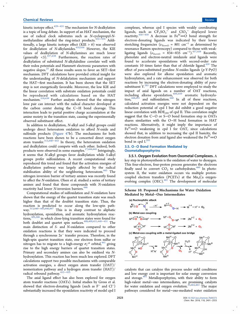
kinetic isotope effect.429−431 The mechanism for N-dealkylationis a topic of long debate. In support of an HAT mechanism, theuse of radical clock substrates such as N-cyclopropyl-N-methylaniline afforded the ring-intact products.432−434 Addi-tionally, a large kinetic isotope effect (KIE > 6) was observedfor dealkylation of N-alkylamides.435,436 However, the KIEvalues of dealkylation of N-alkylamines are much lower(generally <3).437,438 Furthermore, the reaction rates ofdealkylation of substituted N-alkylalniline correlate well withtheir redox potentials and Hammett electronic parameters withnegative slopes.25 All these results seem to favor an SET−firstmechanism. DFT calculations have provided critical insight forthe understanding of N-delakylation mechanism and supportthe HAT−first mechanism. It has been shown that the SETstep is not energetically favorable. Moreover, the low KIE andthe linear correlation with substrate oxidation potentials couldbe reproduced with calculations based on the HATmechanism.318,439 Further analysis showed that the nitrogenlone pair can interact with the radical character developed atthe carbon center during the C−H bond cleavage. Thisinteraction leads to partial positive charge accumulation at theamine moiety in the transition state, causing the experimentallyobserved substituent effect.In addition to dealkylation, N-alkyl and S-alkyl groups could
undergo direct heteroatom oxidation to afford N-oxide andsulfoxide products (Figure 47b). The mechanisms for bothreactions have been shown to be a concerted direct oxygenatom transfer.313,440−443 In theory, the heteroatom oxidationand dealkylation could compete with each other. Indeed, bothproducts were observed in some examples.17,444,445 Intriguingly,it seems that N-alkyl groups favor dealkylation while S-alkylgroups prefer sulfoxidation. A recent computational studyreproduced this trend and found that the activation energies ofdealkylation pathway was largely affected by the radicalstabilization ability of the neighboring heteroatom.443 Thenitrogen inversion barrier of tertiary amines was recently foundto affect the N-oxidation. Olsen et al. studied a series of tertiaryamines and found that those compounds with N-oxidationreactivity had lower N-inversion barriers.446
Computational studies of sulfoxidation and N-oxidation haveshown that the energy of the quartet transition state was muchhigher than that of the doublet transition state. Thus, thereaction is predicted to occur along the low-spin path-way.313,440−443,446,447 This is in sharp contrast to aliphatichydroxylation, epoxidation, and aromatic hydroxylation reac-tions,305,306 in which close-lying transition states were found forboth doublet and quartet spin surfaces.408,409,421,448−451 Themain distinction of S- and N-oxidation compared to otheroxidation reactions is that they were indicated to proceedthrough a synchronous 2e− transfer process. Therefore, in thehigh-spin quartet transition state, one electron from sulfur ornitrogen has to migrate to a high-energy σz2* orbital,441 givingrise to the high energy barriers of quartet transition states.Primary and secondary amines can also be oxidized via N-hydroxylation. This reaction has been much less explored. DFTcalculations support two possible mechanisms with comparableactivation energies, a direct oxygen atom transfer (OAT)/isomerization pathway and a hydrogen atom transfer (HAT)/radical rebound pathway.452−455
The axial ligand effect has also been explored for oxygenatom transfer reactions (OATs). Initial studies by Gross et al.showed that electron-donating ligands (such as F− and Cl−)substantially increased the epoxidation reactivity of model cpd I
complexes, whereas cpd I species with weakly coordinatingligands, such as CF3SO3
− and ClO4− displayed lower
reactivity.456−458 A decrease in FeIVO bond strength forelectron-donating ligands was indicated by their lowerstretching frequencies (νFeO = 801 cm−1 as determined byresonance Ramon spectroscopy) compared to those with weak-ligating ligands (νFeO = 834−835 cm−1).457,458 Recently,phenolate and electron-neutral imidazole axial ligands werefound to accelerate epoxidations with second-order rateconstants 10 times faster than that of chloride ligand.459 Theeffect of para-substituted pyridine N-oxides ligands (p-Y-PyO)were also explored for alkene epoxidation and aromatichydroxylation, and a rate enhancement was observed for bothreactions by increasing the electron-donating ability of para-substituent Y.285 DFT calculations were employed to study theimpact of axial ligands on a number of OAT reactions,including alkene epoxidation,409,449 aromatic hydroxyla-tion,448,451 and sulfoxidation.440 In all of these reactions,calculated activation energies were not dependent on thereduction potential of cpd I but did exhibit a good negativelinear correlation with BDEOH of cpd II. This correlation mightsuggest that the C−O or S−O bond formation step in OATsshares similarities with the O−H bond formation in HATreactions. Alternatively, it might imply the importance ofFeIVO weakening in cpd I for OAT, since calculationsshowed that, in addition to increasing the cpd II basicity, theelectron donation from axial ligand also weakened the FeIVObond in cpd I.285
3.5. O−O Bond Formation Mediated byOxometalloporphyrins
3.5.1. Oxygen Evolution from Oxometal Complexes. Akey step in photosynthesis is the oxidation of water to dioxygen.This four-electron, four-proton process generates the electronsfinally used to convert CO2 to carbohydrates.460 In photo-system II, the water oxidation occurs via multiple proton-coupled electron transfers (PCETs) at the Mn4Ca oxygen-evolving complex (OEC).461 The development of molecular
catalysts that can catalyze this process under mild conditionsand low energy cost is important for solar energy conversionand storage.461 Metalloporphyrins, with their ability to formhigh-valent metal−oxo intermediates, are promising catalystsfor water oxidation and oxygen evolution.33,462,463 The majorpathways considered for metal−oxo-mediated water oxidation
Scheme 10. Proposed Mechanisms for Water OxidationMediated by Metal−Oxo Intermediates
Chemical Reviews Review
DOI: 10.1021/acs.chemrev.7b00373Chem. Rev. 2018, 118, 2491−2553
2523

include nucleophilic attack of water/hydroxide by oxoMn(V)and the coupling of two metal−oxo (or μ-oxo) units (Scheme10). The former is formally the microscopic reverse of the O−O bond cleavage for cpd I generation, while the couplingbetween two μ-oxo units in the latter pathway will afford a (μ-η2:η2-peroxo)dimetal complex similar to the oxygen adduct ofhemocyanine.464−466 In a third mechanism, the O−O bond isformed via coupling of a metal-bound oxide with a metal-ligatedμ-oxo bridge.467−470 This mechanism is mainly proposed for O2evolution of photosystem II OEC and related polynuclearmetal−oxygen complexes.An early example of a water−oxidation catalyst based on
metalloporphyrins is a dimeric manganese porphyrin complex81 synthesized by Naruta et al. (Figure 48a).471 This speciescan catalyze the anodic water oxidation in aqueous acetonitrilesolution containing nBu4NOH with a potential range of 1.2−2.0V (vs Ag/Ag+) and Faradaic efficiency of 5−17%. An dimericoxoMnV intermediate 52 was later prepared by treating theMn(III) dimer complex with mCPBA as a potential reactiveintermediate for water oxidation (Figure 48a).231 This species isvery stable under basic conditions but decomposes to Mn(III)and dioxygen upon neutralization with acid. Åkermark, Sun,and co-workers reported a monomeric manganese porphyr-inoid system for water oxidation (Figure 48b).472 A manganesecorrole complex 83 was shown to efficiently mediate theoxygen evolution at a low potential ∼0.8 V versus Ag/AgNO3.An oxoMnV intermediate 84 was prepared from 83. Theaddition of hydroxide 84 led to the formation of O2 presumablyvia the nucleophilic attack of OH−. A Mn(IV)-peroxointermediate 86 was detected during this process, which waslikely formed from the further oxidation of the Mn(III)-hydroperoxo intermediate 85. Nam also reported the formationof a MnIV-peroxo intermediate upon adding base to aoxoMn(V) corrole complex, MnV(O)(TFMPC) (TFMPC =5,10,15-tris(3,5-trifluoromethylphenyl)corrolato trianion).473
This step is reversible as the addition of acid to peroxo-MnIV(TFMPC) could regenerate the oxoMn(V) intermediate.In addition to manganese, cobalt is another base metal that
has been extensively studied for water oxidation. Nocera et al.reported the first cobalt porphyrinoid system for wateroxidation (87, Scheme 11a) using a cobalt “hangman” β-octafluoro corrole complex 87. When immobilized in Nafionfilms, 87 can catalyze water oxidation at 1.4 V versus Ag/AgClat pH 7 with turnover frequency (TOF) of 0.81 s−1.474 Sakai etal. reported several Co(III) porphyrins, including Co(TPP),Co(TMPyP), and Co(TPPS), for water oxidation via photon-initiation using RuII(bpy)3
2+/Na2S2O8 (Scheme 11b). The
maximum TOFs were in the range of 0.16−0.35 s−1 at pH11.475 A second-order dependence of the initial rate on catalystconcentration was observed, suggesting a bimolecular oxyl−oxyl coupling pathway for O−O bond formation. Thedegradation of the catalyst has been observed presumably dueto the highly oxidizing sulfate radical anions and singledioxygen. More recently, the same group developed a
fluorinated Co(III) porphyrin catalyst CoFPS for wateroxidation with improved stability and efficiency (TOF = 1.1s−1).476 In contrast to their previous studies, a first-ordercatalyst concentration dependence was observed for CoFPS,suggesting a nucleophilic attack pathway for the water oxidationreaction catalyzed by CoFPS.Groves and Wang developed a series of electron-deficient
cationic cobalt porphyrins for highly efficient electrocatalytic
Figure 48. (a) Water oxidation mediated by a dimeric manganese porphyrin complex. Adapted from ref 231. Copyright 2004 Wiley-VCH. (b) Wateroxidation catalyzed by a manganese corrole complex 83. Adapted from ref 472. Copyright 2009 American Chemical Society.
Scheme 11. (a) Cobalt Hangman Corrole Complex forWater Oxidationa and (b) Several Cobalt PorphyrinsUtilized for Water Oxidation
aReprinted from ref 474. Copyright 2011 American Chemical Society.
Figure 49. Proposed mechanism for water oxidation catalyzed by Co-porphyrins (B = buffer anions). Adapted with permission from ref 477.Copyright 2013 National Academy of Sciences.
Chemical Reviews Review
DOI: 10.1021/acs.chemrev.7b00373Chem. Rev. 2018, 118, 2491−2553
2524

water oxidation.477 For CoIII(TDMImP) (88, Figure 49), at apotential of 1.3 V versus Ag/AgCl at pH 7, the O2 formationrate was determined to be 170 nmol cm−2 min−1 (kobs = 1.4 ×10³ s−1) with a near 90% Faradaic efficiency. The authorsconducted detailed mechanistic studies of these cobalt-basedwater−oxidation systems. They identified that a CoIV−Oporphyrin cation radical species P•+CoIV−O (92, Figure 49)is the active intermediate for water oxidation. A lineardependence between catalytic current and catalyst concen-tration suggested that the rate-limiting O−O bond formationoccurred at a single cobalt site. Intriguingly, the buffer anionwas identified to play multiple important roles in the catalyticcycle. First, it acts as a proton acceptor in the conversion ofP•+CoIII−OH to P•+CoIV−O, which is found to be a singlePCET process with a slope of −54 mV pKa
−1 for a plot of onsetpotential of water oxidation versus buffer pKa. Furthermore, itacts as a base to deprotonate water in the rate-limiting O−Oformation step, as indicated by a linear correlation between(icat/iwater)
2 versus buffer base concentration and a KIE of 2.8measured in H2O versus in D2O. Finally, an inhibition of wateroxidation was observed at high buffer concentrations,presumably resulting from the formation of an inactive bis-coordinated Co(III) intermediate 91 with buffer anions. Theunchanged intensity and UV−vis spectroscopic features of 88after bulk electrolysis, the absence of an inhibition effect ofEDTA, and the clean electrode surface with no sign ofheterogeneous cobalt phase clearly exclude the possibility offorming heterogeneous metal oxide films at electrode surface,which is known to be a potential issue for homogeneouswater−oxidation catalysts.478 More recently, Cao et al. reporteda nickel-based water−oxidation catalyst with a cationicporphyrin ligand.479 A TOF of 0.67 s−1 was observed at apotential of 1.32 V (vs NHE). The reactive intermediate of thissystem is proposed to be a Ni(III)−oxyl radical species. As inthe case of analogous cobalt cationic porphyrins, buffer basewas shown to participate in the rate-determining O−O bond
formation step. An inhibition effect of buffer base at highconcentrations was also observed.
3.5.2. Chlorite Dismutase. The conversion of chlorite tochloride and dioxygen is another reaction that might involvethe O−O bond formation mediated by cpd I. This process isimportant for the microbial perchlorate respiration, as itremoves the toxic end-product chlorite.480 In nature, thedecomposition of chlorite is catalyzed by chlorite dismutase(Cld), a heme-containing protein with a histidine proximalligand.481 The active site structure of Cld is shown in Figure 50,which is featured by an arginine positioned above the heme andthe lack of other hydrogen-bonding residues that are essentialfor O−O bond heterolytic cleavage.482 The proposedmechanism for functional Cld involves an initial formation ofa ferric−chlorito intermediate (FeIII−OClO), analogous tocompounds 0 in P450s and peroxidases (Figure 50c).483 Asubsequent O−Cl bond heterolytic cleavage will generate cpd Iand hypochlorite, which will undergo nucleophilic attack at cpdI to generate Cl− and O2.
484 Alternatively, the homolysis of Cl−O bond of FeIII−OClO will afford compound II (cpd II) andchlorine monoxide radical (ClO·. Cl− and O2 are formed via arebound process between ClO· and cpd II. The lattermechanism is analogous to the mechanism proposed for NOoxidation catalyzed by nitric oxide dioxygenase (see section4.2). The distal arginine can adopt either “in” and “out”conformations (Figure 50a and 50b),482 which is proposed toplay an important role in positioning the transiently formedhypochlorite to ensure its efficient capturing by cpd I (or cpdII).485−487 There are two phylogenetically distinct class of Clds(clades 1 and 2). Clade 1 Clds (including NdCld and DaCldshown in Figure 50) are pentameric and hexameric, whereasclade 2 Clds are dimeric. Very recently, Obinger and co-workers have carried out detail structural and kinetic studies ofa dimeric clade 2 Cld from Cyanothece sp. PCC7425 (CCld).488
The spectral features of various intermediates of dimeric CCldappeared to be much better resolved than those of clade 1 Clds,which allowed for the presteady-state kinetic studies of CCld.
Figure 50. (a) Active site of chlorite dismutase (Cld) from Candidatus Nitrospira defluvii (NdCld) with an imidazole ligand (PDB: 3NN1).Catalytically important arginine (Arg173) exhibits an “out” conformation in this structure. (b) Active site of Cld from Dechloromonas aromatica(DaCld) with a nitrite ligand (PDB: 3Q09). Catalytically important arginine (Arg183) exhibits an “in” conformation in this structure. (c) Theproposed mechanism for chlorite dismutase.
Chemical Reviews Review
DOI: 10.1021/acs.chemrev.7b00373Chem. Rev. 2018, 118, 2491−2553
2525
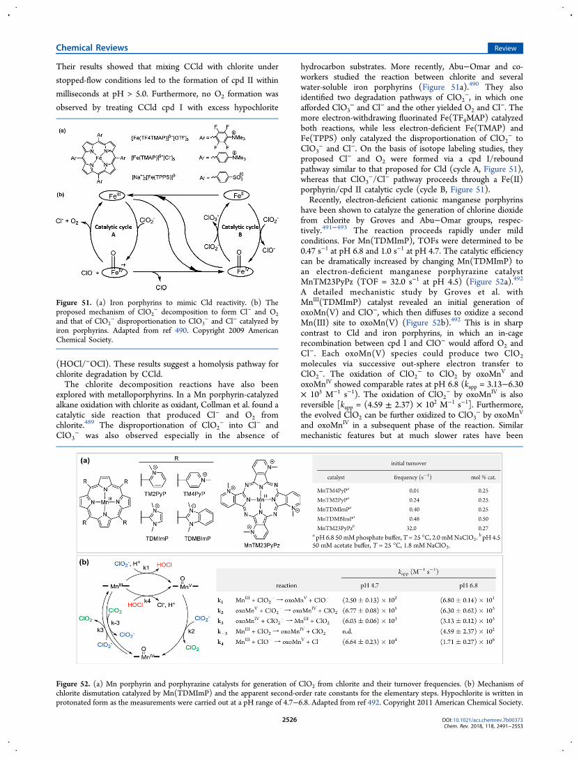
Their results showed that mixing CCld with chlorite under
stopped-flow conditions led to the formation of cpd II within
milliseconds at pH > 5.0. Furthermore, no O2 formation was
observed by treating CCld cpd I with excess hypochlorite
(HOCl/−OCl). These results suggest a homolysis pathway forchlorite degradation by CCld.The chlorite decomposition reactions have also been
explored with metalloporphyrins. In a Mn porphyrin-catalyzedalkane oxidation with chlorite as oxidant, Collman et al. found acatalytic side reaction that produced Cl− and O2 fromchlorite.489 The disproportionation of ClO2
− into Cl− andClO3
− was also observed especially in the absence of
hydrocarbon substrates. More recently, Abu−Omar and co-workers studied the reaction between chlorite and severalwater-soluble iron porphyrins (Figure 51a).490 They alsoidentified two degradation pathways of ClO2
−, in which oneafforded ClO3
− and Cl− and the other yielded O2 and Cl−. Themore electron-withdrawing fluorinated Fe(TF4MAP) catalyzedboth reactions, while less electron-deficient Fe(TMAP) andFe(TPPS) only catalyzed the disproportionation of ClO2
− toClO3
− and Cl−. On the basis of isotope labeling studies, theyproposed Cl− and O2 were formed via a cpd I/reboundpathway similar to that proposed for Cld (cycle A, Figure 51),whereas that ClO3
−/Cl− pathway proceeds through a Fe(II)porphyrin/cpd II catalytic cycle (cycle B, Figure 51).Recently, electron-deficient cationic manganese porphyrins
have been shown to catalyze the generation of chlorine dioxidefrom chlorite by Groves and Abu−Omar groups, respec-tively.491−493 The reaction proceeds rapidly under mildconditions. For Mn(TDMImP), TOFs were determined to be0.47 s−1 at pH 6.8 and 1.0 s−1 at pH 4.7. The catalytic efficiencycan be dramatically increased by changing Mn(TDMImP) toan electron-deficient manganese porphyrazine catalystMnTM23PyPz (TOF = 32.0 s−1 at pH 4.5) (Figure 52a).492
A detailed mechanistic study by Groves et al. withMnIII(TDMImP) catalyst revealed an initial generation ofoxoMn(V) and ClO−, which then diffuses to oxidize a secondMn(III) site to oxoMn(V) (Figure 52b).492 This is in sharpcontrast to Cld and iron porphyrins, in which an in-cagerecombination between cpd I and ClO− would afford O2 andCl−. Each oxoMn(V) species could produce two ClO2molecules via successive out-sphere electron transfer toClO2
−. The oxidation of ClO2− to ClO2 by oxoMnV and
oxoMnIV showed comparable rates at pH 6.8 (kapp = 3.13−6.30× 10³ M−1 s−1). The oxidation of ClO2
− by oxoMnIV is alsoreversible [kapp = (4.59 ± 2.37) × 102 M−1 s−1]. Furthermore,the evolved ClO2 can be further oxidized to ClO3
− by oxoMnV
and oxoMnIV in a subsequent phase of the reaction. Similarmechanistic features but at much slower rates have been
Figure 51. (a) Iron porphyrins to mimic Cld reactivity. (b) Theproposed mechanism of ClO2
− decomposition to form Cl− and O2and that of ClO2
− disproportionation to ClO3− and Cl− catalyzed by
iron porphyrins. Adapted from ref 490. Copyright 2009 AmericanChemical Society.
Figure 52. (a) Mn porphyrin and porphyrazine catalysts for generation of ClO2 from chlorite and their turnover frequencies. (b) Mechanism ofchlorite dismutation catalyzed by Mn(TDMImP) and the apparent second-order rate constants for the elementary steps. Hypochlorite is written inprotonated form as the measurements were carried out at a pH range of 4.7−6.8. Adapted from ref 492. Copyright 2011 American Chemical Society.
Chemical Reviews Review
DOI: 10.1021/acs.chemrev.7b00373Chem. Rev. 2018, 118, 2491−2553
2526

described by Abu−Omar et al. for a Mn(TF4TMAP)/NaClO2system.494
3.6. Single Electron Transfer (SET) Reactions of Cpd I
In addition to participating in hydrogen atom transfer (HAT)and oxygen atom transfer (OAT) reactions, compound I canalso display single electron transfer (SET) reactivity, which iscommonly seen in heme-containing peroxidases. Unlike mostP450s, peroxidase compound I is generated directly from theresting ferric heme and H2O2 or organic hydroperoxides via the“peroxide shunt” mechanism. Peroxidase cpd I can carry outtwo discrete single-electron transfers to two substrate moleculesand regenerate the ferric heme state (Scheme 12). This three-step mechanism was first proposed by Dunford et al. for theoxidation of ferrocyanide by H2O2 and HRP.246 The resultingsubstrate radicals can undergo further transformations.495 Thus,substrates of heme peroxidases are generally good one-electrondonors, such as electron-rich aromatics, ascorbate, ferrocyto-chrome c, etc.496 Even strongly oxidizing Mn3+ can be producedfrom Mn2+ in this way by manganese peroxidase.Heme peroxidases are not the only enzymes that utilize H2O2
to form compound I. Unspecific peroxygenases (UPO) andchloroperoxidase (CPO) employ a similar “peroxide shunt”pathway to generate compound I to catalyze HAT and OAT
reactions.497 As discussed in section 3.1, a major cause of thisreactivity difference is the much larger O−H BDEs of UPO-IIand CPO-II (98−100 kcal/mol) compared to that of HRP-II,originating from the elevated reduction potentials of UPO-Iand CPO-I and the decreased acidities of UPO-II and CPO-II.Furthermore, the 1e− reactions in peroxidases are facilitated bythe proper positioning of substrates.498 The local proteinenvironment of peroxidases hinders the approach of substratesto the FeIVO moiety. Therefore, many substrates bind in theheme periphery and participate in a hydrogen-bonding networkwith heme propionate and active site residues (Figure 53).499
This network can serve as a relay to mediate the proton andelectron transfer.500,501 Alternatively, substrates can be oxidizedat a remote site through a chain of redox-active amino acids vialong-range electron transfer.502−505
In some peroxidases, the porphyrin cation radical form ofcompound I is transient and will rapidly transfer one electron to
a nearby redox-active residue like tyrosine or tryptophan. Oneexample is the compound ES of cytochrome c peroxidase(CcP), which is an iron(IV)-oxo intermediate with anuncoupled Trp191 cation radical near the proximal histidine.184
Although the Trp191 radical in CcP just serves as a storage forone oxidation equivalent, in some heme proteins, such radicalscan directly mediate reactions. For instance, in prostaglandin Hsynthase (PGHS), the compound I formed via the normalperoxide activation pathway will oxidize a tyrosine (Tyr385)about 10 Å away to give a tyrosyl radical, which abstracts ahydrogen atom from arachidonic acid (Figure 54).506,507 Theresulting substrate radical will further react with two O2molecules to give product PGG2 and finally prostaglandin H2(PGH2). This reaction also represents an example of substrateactivation for O2 reactions in heme proteins.2
Another example of using protein radicals for substrateactivation is the synthesis of heme b by coprohemedecarboxylase (HemQ).508 HemQ catalyzes a H2O2-dependentdecarboxylation of two propionates in coproheme (94, Scheme13). Recent mechanistic studies by DuBois et al. showed thatTyr145, which hydrogen bonds with one of the cleavedpropionates, is first converted to a tyrosyl radical by coprohemecompound I (95, Scheme 13).509,510 The Tyr145 radical thenabstracts a hydrogen from the heme propionate grouppresumably at the Cβ position (96). The resulting carbon-centered radical transfers one electron through porphyrin toFe(IV) and reduces it to iron(III) with loss of CO2 (97 → 98).The decomposition of the second propionate is thought tofollow a similar mechanism, but the identity of the mediator ofCβ-H cleavage is not clear.
Scheme 12. Heme Peroxidase Generates Cpd I with H2O2and Catalyzes Two Discrete Single Electron Transfer (SET)Reactions to Substrates
Figure 53. Examples of substrate binding to the heme periphery ofperoxidases and forming hydrogen bonds with heme propionates andactive site residues. (a) Ascorbate peroxidase (APX, PDB: 1OAF). (b)Manganese peroxidase (MnP, PDB: 1YYD).
Figure 54. Conversion of arachidonic acid (AA) into PGG2 by PGHS.
Chemical Reviews Review
DOI: 10.1021/acs.chemrev.7b00373Chem. Rev. 2018, 118, 2491−2553
2527

4. REACTIONS MEDIATED BY HEME−OXYGENINTERMEDIATES OTHER THAN CPD I
In addition to cpd I, other heme−oxygen intermediates
generated during O2 activation also exhibit rich redox
chemistries that are harnessed by heme proteins for a variety
of oxidative transformations, giving rise to the diverse biological
functions of heme proteins. In this section, we analyze several
representative reactions catalyzed by heme proteins and
metalloporphyrins to delineate their mechanistic features and
to discuss how the fundamental properties of heme−oxygenintermediates and their active site environments impact
reaction outcomes.
4.1. Homolytic O−O Bond Cleavage of Peroxides by HemeProteins
In addition to generating compound I via O−O bondheterolysis, organic peroxides can also react with ferric hemesvia single-electron-transfer processes, which will oxidize theferric heme to ferryl with concurrent homolytic cleavage of O−O bond. These transformations play important roles in thesynthesis of important bioactive molecules, especially oxy-lipins.511 Natural sources of peroxides include fatty acidhydroperoxides formed via O2 trapping of substrate allylicradicals.512 Subsequent O−O bond homolysis leads to oxyradicals that can undergo a plethora of rearrangements, bondcleavages, and other transformations.
Scheme 13. HemQ-Catalyzed Synthesis of Heme b from Coproheme IIIa
aAdapted from ref 509. Copyright 2017 American Chemical Society.
Scheme 14. (a) Synthesis of Prostacyclin from PGH2 Mediated by Prostacyclin Synthase and (b) Synthesis of Thromboxane A2(TXA2) Catalyzed by Thromboxane A Synthase
Chemical Reviews Review
DOI: 10.1021/acs.chemrev.7b00373Chem. Rev. 2018, 118, 2491−2553
2528

One example is the synthesis of prostacyclin fromprostaglandin H2 (PGH2) via prostacyclin synthase (Scheme14a).507,513,514 This transformation starts with the interactionbetween endoperoxide of PGH2 and ferric heme center ofprostacyclin synthase (104, Scheme 14a), leading to thehomolytic O−O bond cleavage of endoperoxide to afford theC9-alkoxyl radical and ferryl heme intermediate (105). The C9-alkoxyl radical then add to the C5−C6 alkene to forge the newfive-membered heterocycle and C5 radical (106), which is lateroxidized to carbocation by the ferryl heme to regenerate theC5−C6 alkene moiety via loss of a proton (107). Thehomolytic cleavage of endoperoxide of PGH2 is also used in thesynthesis of thromboxane A2 (TXA2) mediated by thrombox-ane A synthase (Scheme 14b).515−517 In this reaction, the ferricheme center interacts with O9 and afforded C11-alkoxyl radical(109 and 110, Scheme 14b), which is later rearranged to anallylic radical at C12 (111). Subsequent electron transfer andring cyclization would afford TXA2.In another instructive example, 13S-hydroperoxyoctadeca-
trienoic acid [13(S)-HPOT] can be activated by the CYP74family of enzymes, affording three different products: 12,13(S)-allene oxide, etherolenic acid, and an analogous hemiacetal(Figure 55a).518,519 The first step in all of these threetransformations is the homolysis of an iron-bound O−Obond to afford an epoxyallylic radical and a cysteine-ligatedFeIV−OH intermediate similar to compound II in P450monooxygenases. The reactions then diverge depending onhow the incipient substrate radical interacts with the FeIV−OHintermediate. In allene oxide synthase (AOS), the C11epoxyallylic radical is oxidized by electron transfer to FeIV−OH to generate a substrate-derived carbocation intermediate,
which eliminates a proton to form the new CC bond. Inhydroperoxide lyase (HPL), by contrast, the radical undergoesrearrangement to form a radical at C13, which then recombineswith FeIV−OH via an oxygen-rebound mechanism. In thedivinyl ether synthase (DES) pathway, the product is thoughtto be formed via a hydrogen atom abstraction from C14 byFeIV−OH or via an electron-transfer process followed by cationrearrangement and proton elimination. Sequence analysis andmutagenesis studies revealed that, in all three enzymes, anasparagine positioned directly above heme is important formediating the O−O bond homolysis.520 The crystal structureof Arabidopsis thaliana AOS (AtAOS) complexed with 12R,13S-vernolic acid, a mimic of the epoxyallylic intermediate, showedthat C11 is in proximity to F137 (Figure 55b), whereas in HPLand DES, a similar position is occupied by a leucine. Thearomatic ring of F137 is thought to stabilize the C11carbocation and thus facilitate the single electron transfer inthe AOS pathway. In support of this rationale, a F137Lmutation induced HPL activity for AOS.It is intriguing to compare the structural difference between
CYP74 and P450 hydroxylases, since CYP74 has a typical P450protein fold but does not perform O−O bond heterolysis. Inaddition to the critical Asn residue (N321) located above theheme, there are two more structural features that distinguishCYP74 from P450 hydroxlyases.520 As can be seen from thecrystal structure of AtAOS, the highly conserved threonine inthe proton relay channel of P450 hydroxylases, which is crucialfor O−O bond heterolysis, is replaced by an isoleucine (I328)(Figure 55, panels c and d). Further, there is a nine-residueinsert in the heme-binding loop of AOS, which seems tocontribute to the long Fe−S bond (2.4 Å) of AOS. A long Fe−
Figure 55. (a) Reactions catalyzed by the CYP74 family of enzymes. The mechanisms of all three pathways involve a common epoxyallylic radicalintermediate. (b) Active site structure of Arabidopsis thaliana AOS complexed with vernolic acid (PDB: 2RCL). (c) Key N321 and I328 residues ofAtAOS (PDB: 3CLI). The cysteine loop region is highlighted in red. (d) Key threonine residue of P450BM3 (PDB: 1BVY). The cysteine loop regionis highlighted in yellow.
Chemical Reviews Review
DOI: 10.1021/acs.chemrev.7b00373Chem. Rev. 2018, 118, 2491−2553
2529

S distance has been shown to decrease the electron-donatingability of cysteine thiolate, which, in turn, would lead to ahigher Fe reduction potential and a more facile SET reactivityof compound II.181
In addition to CYP74, there is another enzyme that can alsocatalyze the synthesis of allene oxides via homolytic cleavage offatty acid hydroperoxides. This enzyme is structurally similar to
catalase and thus termed catalase-related allene oxide synthase(cAOS).521 cAOS converts 8R-hydroperoxyarachidonic acid(8R-HPETE) into corresponding 8,9-epoxy allene oxide in thebiosynthesis of preclavulone A. The crystal structure compar-ison between cAOS and catalase showed very similar active sitestructures, including the proximal heme-ligating tyrosine andthe critical distal histidine and asparagine that are important for
Figure 56. (a) Reactions catalyzed by cAOS and the active site structure of cAOS (PDB: 1U5U). (b) Active site structure of human catalase (PDB:1QQW) and the mechanism of O−O bond activation in catalase-compound 0.
Figure 57. (a) Previously proposed base-abstraction mechanism followed by either Criegee rearrangement or dioxetane formation. Adapted from ref539. Copyright 2011 American Chemical Society. (b) A recently proposed mechanism involves the formation of an FeIVO intermediate and a 2,3-indoline epoxide intermediate. Adapted from ref 550. Copyright 2016 American Chemical Society.
Chemical Reviews Review
DOI: 10.1021/acs.chemrev.7b00373Chem. Rev. 2018, 118, 2491−2553
2530

the O−O bond activation in catalase (Figure 56). A keydifference is identified at a residue adjacent to the distalhistidine, which is normally a valine in catalases but is changedto a threonine (T66) in cAOS.522 The histidine in cAOShydrogen bonds with both T66 and asparagine. This hydrogenbonding network could function to dampen H2O2 activation incAOS by inhibiting the hydrogen bonding between histidineand the hydroperoxo moiety of FeIII−OOH (cpd 0). Indeed, aT66V mutation greatly increased the catalase activity of cAOS,albeit with short lifetime due to the heme degradation. cAOS isthought to proceed through a similar epoxy allylic radicalintermediate. As in the case of CYP74, products other thanallene oxide were also observed in reactions catalyzed by cAOSand its relatives.523−528
4.2. Reactions Initiated by Ferric-Superoxo Intermediates
In most heme proteins, ferric-superoxo complexes either serveas stable complexes, such as in oxymyoglobin and gas sensingproteins such as heme-nitric oxide/oxygen (H-NOX) bindingproteins,529−531 or rapidly convert to high-valent iron−oxointermediates via reduction and O−O bond scission.Tryptophan 2,3-dioxygenase (TDO) and indoleamine 2,3-dioxygenase (IDO), however, utilize ferric-superoxo intermedi-ates to initiate the oxidation of substrates.532,533 TDO and IDOare histidine-ligated heme-containing dioxygenases involved in
tryptophan oxidation.534−536 TDO employs dioxygen to cleavethe 2,3-bond of L-tryptophan to form N-formyl kynurenine,whereas IDO catalyzes the same reaction but has a muchbroader substrate scope, including a variety of indoleaminederivatives such as D-tryptophan, melatonin, and serotonin.IDO has recently gained substantial research interest because ofits immunomodulatory potential for cancer treatment.537,538
The initially proposed mechanism for TDO and IDO-catalyzed tryptophan oxygenation involved a histidine-catalyzedproton abstraction of the indole N−H, followed by electro-philic attack at C3 of indoleamine by the distal oxygen of ferric-superoxo intermediate (Figure 57a).539 But this mechanism isnot consistent with later findings showing that N-methyl-tryptophan was an active substrate and that human IDO lackedthe proposed proton-abstracting histidine.540−542 DFT calcu-lations indicated high energy barriers for the formation of eithera dioxetane intermediate or a Criegee-type rearrangement.543
Further studies by Yeh et al. using resonance Raman and UV−vis spectroscopies revealed the presence of a ferryl speciesduring catalytic turnover of IDO.544−546 Similar ferryl speciescan also be generated by treating IDO and TDO usingH2O2.
541,547−549 On the basis of these results, a newmechanism has been proposed (Figure 57b), in which theferric-superoxo complexes directly attacks the indoleamine ringvia either electrophilic addition or radical addition to afford an
Figure 58. (a) Proposed mechanism for NO oxidation catalyzed by NOD. (b) Proposed mechanism for tryptophan nitration catalyzed by P450TxtE.
Chemical Reviews Review
DOI: 10.1021/acs.chemrev.7b00373Chem. Rev. 2018, 118, 2491−2553
2531

indoline-2,3-epoxide intermediate and a ferryl compound II.Subsequent reactions between the epoxide and Fe(IV)O arecurrently speculative but do lead to the formation ofkynurenine type product and regenerate the ferrous heme.While the ferryl intermediate of IDO accumulates under steady-state conditions, TDO cpd II has yet to be observed duringturnover. Rather, the [Fe(II)−O2, L-Trp] ternary complexaccumulates under catalytic conditions. Raven et al. haverecently suggested that this difference is caused by a change ofthe rate-limiting step from compound II decay in IDO tooxygen addition to formation of the 2,3-epoxide intermediate inTDO.550
Another enzyme that utilizes an ferric-superoxo intermediatefor oxygenation is nitric oxide dioxygenase (NOD).551 Asshown in Figure 58a, NO first reacts with an ferric-superoxointermediate to form a proposed ferric-peroxynitrite inter-mediate. This intermediate then undergoes homolytic cleavageto generate NO2 and compound II.552 The recombinationbetween NO2 and compound II yields nitrate and ferric heme.This recombination process is reminiscent of the oxygenrebound of carbon-centered radicals in P450s. The study ofperoxynitrite decomposition mediated by met-myoglobinoffered critical insight into the radical rebound mechanism,especially the radical cage escape effect.553,554 Unlike carbon-centered radicals, the diffusion of NO2 can be easily detectedvia fluorogenic probes such as fluorescein. Kinetic simulationsrevealed that 10% of the NO2 escaped from the radical cage[MbFeIVO ·NO2] formed after peroxynitrite decomposition.Cytochrome P450 TxtE from Streptomyces scabies also employs
a ferric-superoxo intermediate to generate NO2 from NO,which is then used to nitrate L-tryptophan to form 4-nitro-L-tryptophan.555,556 The proposed mechanism involves theformation of a ferric-peroxynitrite intermediate by the trappingof NO by the ferric-superoxo intermediate (Figure 58b). TheNO2 generated after peroxynitrite decomposition can then addto the aromatic ring, which is further oxidized by compound IIto afford the nitration product. An alternative mechanisminvolves heterolytic cleavage to generate nitronium ion (NO2
+)for direct electrophilic nitration.555
4.3. O2 Reduction Catalyzed by Heme/Cu TerminalOxidases and Related Model Compounds
Heme/Cu terminal oxidases such as cytochrome c oxidases(CcO) catalyze the 4e−/4H+ reduction of O2 as a final step inrespiration with concomitant membrane proton-translocationleading to ATP synthesis.557 The active site structure of CcO isunusual compared to many heme proteins as it contains abinuclear heme a3/CuB center (BNC) with a Fe−Cu separationof around 5 Å (Figure 59a). CuB is coordinated to threehistidine residues, one of which is covalently cross-linked to atyrosine residue.558−560 During O2 reduction by CcO, electronsfrom cyt c are delivered one at a time to BNC via a dicoppercomplex (CuA) and a low-spin heme a. The mechanism of CcOaction has been extensively studied. The catalytic cycle involvesfour consecutive proton and electron transfer steps andmultiple intermediates are involved. Figure 59b showed asimplified mechanism of CcO-catalyzed oxygen reduction.561
The Fe(III)−Cu(II) resting state of BNC (compound O) is
Figure 59. (a) Cofactor arrangement in a cytochrome c oxidase (PDB: 2YEV) and the structure of binuclear heme a3/CuB center (BNC). (b)General catalytic cycle of CcO and important intermediates involved (R, A, PM, F, O, and E). This mechanism is produced according to the catalyticcycles shown in refs 561 and 562.
Chemical Reviews Review
DOI: 10.1021/acs.chemrev.7b00373Chem. Rev. 2018, 118, 2491−2553
2532

converted to an E state and then to a Fe(II)−Cu(I) reducedstate (R) via two one-electron reductions and two protontransfer steps. The ferrous heme a3 in R will bind oxygenrapidly (∼1.4 × 10⁸ M−1 s−1), generating a low-spin end-onFe−O2 intermediate (A) similar to other oxygen-binding hemeproteins. O−O bond cleavage in A, which can be viewed as anO−O hemolysis in a bridged Fe(III)−O−O−Cu(II) inter-mediate, yields a four-electron oxidized intermediate PM,containing a ferric heme a, a Fe(IV)O, Cu(II)−OH, and a
tyrosyl radical. Four subsequent 1e− reductions and protontransfers will lead sequentially through the F, O and E states,poising the protein for another oxygen-binding event. It isworth noting that in Figure 59b, we only showed intermediatesformed after both electron- and proton-transfer steps.Furthermore, depending on the status of copper ligation andredox potentials of copper center, the cross-linked tyrosinecould exist as tyrosyl radical with Cu(II) center being reducedto Cu(I).561,562 Each of these reduction/protonation steps
Figure 60. (a) The proposed ferric-peroxo-cupric intermediate for the generation of PM state. (b) A CN-bound CcO might share similar structuralfeatures to the putative ferric-peroxo-cupric intermediate (PDB: 3AG4).
Scheme 15. Formation of FeIII-μ-oxo-CuII Complexes by Treating Fe(II) Porphyrin/CuI Complex with O2
Figure 61. (a) A heme-peroxo-copper model compound based on “capped” porphyrin with an appended TACN ligand for copper binding. (b)Different peroxo bonding mode for heme-peroxo-copper complexes. (c) Several representative heme-peroxo-copper model compounds.
Chemical Reviews Review
DOI: 10.1021/acs.chemrev.7b00373Chem. Rev. 2018, 118, 2491−2553
2533

(scalar protons) are also coupled to one vectorial protonpumped across the membrane, thus maintaining the protongradient required for ATPase phosphorylation.561
While many aspects of the CcO mechanism have beenclarified, the details of the O−O cleavage to generatecompound PM and the role of the CuB His-Tyr unit in thisstep remains elusive. As noted above, a putative ferric peroxointermediate (FeIII−O−O−CuII) is thought to be generated via1e− reduction of ferric-superoxo intermediate by Cu(I) center(Figure 60a).558 A subsequent proton-coupled electron transfer(PCET) from tyrosine to FeIII−O−O−CuII could promote theO−O cleavage and generate the ferryl center and tyrosylradical. Yoshikawa et al. have examined the binding of diatomicmolecules (CO, NO, and CN−) to the reduced bovine CcO viaX-ray structural analysis.563 Compared to CO- and NO-boundstructures, CN− binding leads to the displacement anddissociation of a copper-bound histidine and the coordinationof cyanide nitrogen to the copper. Furthermore, a watermolecule is incorporated to form a hydrogen bond with bothCN− and covalently ligated tyrosine (Tyr244) (Figure 60b).These structural features were thought to be highly relevant tothe ferric-superoxo intermediate of CcO, where O2 is negativelypolarized similarly to CN−. The hydrogen-bonding network inthe active site could provide a channel for PCET from Tyr244to the peroxo moiety. Another intriguing feature of this CN-bound CcO structure is the significant bending of the Fe−C−Nangle (96°). This bent structure is in line with previousresonance Raman characterizations and might be caused by theweak π-accepting ability of cyanide, which makes it easier toadopt a bent geometry compared to the isoelectronic Fe-COcomplexes.564−567
Model compounds have offered critical insights to the O−Oheterolytic cleavage in CcO.23,568 The study of CcO modelcompounds has led to the development of electrocatalyticsystems for the four-electron reduction of O2 to water, whichwould have potential applications in fuel cells.20,33,569−571
These systems have been reviewed in detail very recently.33
Common strategies for preparing CcO models include treatingferrous porphyrins with covalently appended copper(I) with O2or adding Cu(I) complexes to preformed, discrete ferric peroxo
porphyrins. In early attempts, μ-oxo heme−FeIII−O−CuIIspecies were obtained, presumably from the disproportionationof the corresponding complexes.23 The oxygen of FeIII-μ-oxo-CuII species are basic and can undergo reversible protonation(Scheme 15). A pKa of 8 has been reported for [(F8TPP)Fe
III-(OH)-CuII(TMPA)]2+ (F8TPP = tetrakis(2,6-difluorophenyl)-porphyrinate, TMPA = tris(2-pyridylmethyl)amine).572
The first well-characterized heme-peroxo-copper adduct wasreported by Collman with a complex “capped” porphyrinscaffold with an appended triazacyclononane (TACN) ligandfor copper binding (114, Figure 61a).573 This species has a highspin (S = 2) ground state with a ν(O−O) of 758 cm−1. Morerecently, a variety of heme-peroxo-copper adducts werecharacterized and shown to display a rich coordinationchemistry. The peroxo moiety can adopt μ-η2:η1, μ-η1:η1, orμ-η2:η2 bonding modes depending on the precise copper ligandenvironment and heme axial ligand (Figure 61, panels b andc).20,568,574 Karlin has described a μ-η2:η1 heme-peroxo-coppercompound obtained by treating a ferric peroxo complex[(F8TPP)Fe
III(O22−)]− with a tetracoordinated CuII complex
CuII(TMPA) (115, Figure 61c).575,576 The compound showeda high spin (S = 2) electronic state with a ν(O−O) of 804cm−1. Naruta has successfully obtained the crystal structure of aμ-η2:η1 heme-peroxo-copper adduct, [(TMP)FeIII-(O2)-(5MeTPA)CuII)BPh4 (116, Figure 61c).
577 The μ-η2:η1 peroxocore showed two Fe−O bond distances of 2.031(4) and1.890(6) , respect ive ly . L ike [(F8TPP)Fe
I I I - (O2)-CuII(TMPA)]+, [(TMP)FeIII-(O2)-Cu
II(5MeTPA)]BPh4 isEPR silent with an S = 2 spin state resulting from a strongantiferromagnetic coupling between high spin FeIII (S = 5/2)and CuII (S = 1/2).The peroxo bonding mode can be regulated by copper
coordination and axial ligand of iron porphyrins. For example,addition of a tridentate Cu(II) complex Cu(AN) (AN = 3,3′-iminobis(N ,N-dimethylpropylamine)) to [(F8TPP)-FeIII(O2
2−)]− afforded a μ-η2:η2 peroxo coordination (117,Figure 61c), whereas adding imidazole-type axial ligand to[[(F8TPP)Fe
III-(O2)-CuII(AN)] shifted the peroxo coordina-
tion to the μ-η1:η1 mode (118, Figure 61c).578−581 The end onμ-η1:η1 heme-peroxo-copper complex is diamagnetic with a low
Figure 62. Reactions between FeIII-superoxide-CuI complexes and phenols.
Chemical Reviews Review
DOI: 10.1021/acs.chemrev.7b00373Chem. Rev. 2018, 118, 2491−2553
2534

spin S = 1/2 Fe(III) antiferromagnetically coupled to Cu(II) (S= 1/2).581,582 Very recently, Karlin et al. reported a [FeIII-(O2
2−)-CuII(MeTHF)3]+ (MeTHF = 2-methyltetrahydrofuran)
complex (119, Figure 61c). The weak coordination of theMeTHF ether oxygen allows for facile ligand exchange,providing ready access to a variety of heme-peroxo-CuII
species.583 In addition to the heme-peroxo-CuII intermediate,isoelectronic FeIII-superoxide-CuI complexes were synthesizedusing picket fence porphyrins.584,585 These FeIII-superoxide-CuI
complexes are relatively stable at room temperature, suggesting
that CuI could also play a nonredox role in stabilizing irondioxygen adducts.
The reactivity of CcO models toward phenols was alsoexplored. Collman et al. found that an FeIII-superoxide-CuI
complex could react with phenol both intra- and intermolec-ularly to afford a ferryl species, a tyrosyl radical, and Cu(II)−OH,586,587 a process that resembles the transition from CcO Astate to PM state (Figure 62). Solomon and Karlin have testedthe reactivity of both low- and high-spin heme-peroxo-coppercomplexes by treating them with phenols (Figure 63).578 Whilehigh-spin [(F8TPP)Fe
III-(O22−)-CuII(AN)]+ was not reactive
toward phenols, the low-spin [(DCHIm)(F8TPP)FeIII-(O2
2−)-CuII(AN)]+ (DCHIm = dicyclohexyl imidazole) reacted withphenols readily, even at −80 °C, to afford a ferryl intermediate,Cu(II)−OH, and a tyrosyl radical.578
The impact of copper complex structure and the nature ofthe proton source on the O−O bond activation step wasrecently investigated.588 Two FeIII-peroxo-CuII complexes withtridentate and tetradentate copper complexes, respectively (LS-3DCHIm and LS-4DCHIm), were prepared and examined fortheir reactivity toward different acids or electron donordecamethylferrocene (Fc*) (Figure 64). While LS-4DCHImalone was not reactive toward Fc*, the addition of 4-nitrophenol led to the formation of an [LS-4DHCIm···4-NO2-phenol] association complex, which underwent facile O−O bond cleavage via proton-coupled electron transfer from 4-nitrophenol and Fc* to peroxo moiety. Intriguingly, undersimilar conditions, LS-3DCHIm was not reactive. Furtherexamination showed that LS-3DCHIm could not form theassociation complex with phenol presumably due to a morecompressed and sterically inaccessible peroxo core. Strong acidslike triflic acid led to the direct decomposition of both LS-3DCHIm and LS-4DHCIm and the release of hydrogenperoxide, regardless of the steric environment of peroxo group.
Figure 63. Comparison of reactions with phenols between low-spinand high-spin FeIII-peroxo-CuII complexes.
Figure 64. (a) Phenol association will promote the O−O bond cleavage of LS-4DCHIm in the presence of electron donor Fc*. (b) LS-3DCHImcould not form the phenol association complex and is not reactive toward Fc*. DFT calculation showed a more compressed structure of LS-3DCHIm. Adapted from ref 588. Copyright 2017 American Chemical Society.
Chemical Reviews Review
DOI: 10.1021/acs.chemrev.7b00373Chem. Rev. 2018, 118, 2491−2553
2535

In addition to small molecule models of CcO, redesignedmetalloproteins that mimic the reactivity of CcO representsanother powerful approach to study its mechanism.589 Lu et al.found that the introduction of two mutations (L29H andF43H) would create a copper binding site in the heme pocketof myoglobin (Figure 65a).590 The resulting copper-boundmyoglobin (CuBMb) showed heme oxygenase reactivity thatdegrades heme to verdoheme. In 2012, Lu and Wang generateda CuBMb with a cross-linked Tyr-His ligand via geneticincorporation of the unnatural amino acid imiTyr at the F33position (Figure 65). The imiTyr-CuBMb exhibited enhancedoxidase reactivity (2/min).591 In the same year, Lu et al. foundthat the introduction of a tyrosine mutation to CuBMb at F33also improved O2 reduction activity (24/min). Intriguingly, theobserved oxidase activity is not copper-dependent. Asubsequent study showed that a ferric−hydroperoxo specieswas formed for F33Y−CuBMb upon cryoreduction at 77K,whereas a peroxo−ferric state was formed for wild-type Mb.592
The X-ray crystal structure of F33Y−CuBMb revealed anextensive H-bond network with two water molecules (W1 andW2), in which W1 is H-bonded to H43 and Y33 and W2 isstabilized by H-bonds to W1 and H29 (Figure 65b).593 Thishydrogen-bonding network could polarize the O−O bond andfacilitate its reduction and protonation. Very recently, Lu andco-workers studied the effect of nonheme metals on oxidaseactivity of the repurposed Mb.594 While Zn-Mb formed a stableferric-superoxo intermediate, both Cu-Mb and Fe-Mb showedoxidase reactivity with Cu-Mb almost three times more reactivethan Fe-Mb. The higher reactivity of CuI is attributed to thehigher CuII/CuI potential and an enhanced weakening of O−Obond through dπ-pπ back bonding, as revealed by the longerO−O bond length (1.391 Å) in Cu-Mb than that in Fe-Mb(1.351 Å).
5. CONCLUSIONS AND OUTLOOKThe central goal of this review was to survey the range of redoxreactions mediated by heme proteins. Parallel developmentswith model metalloporphyrins and computational approachesare also reviewed and have served both to elucidate the natureof reactive enzymatic intermediates and to discover newchemistry, some of which is unknown in the repertoire of hemeprotein reactions. Another goal was to provide context for therecent advances with key discoveries from the formative yearsof the field. This coverage took inspiration from a course wedeveloped at Princeton University called Metals in Biology:From Stardust to DNA. It is interesting to consider that hemeand heme proteins apparently evolved long before oxygen was
present in the environment in significant quantities. From thisperspective, it is clear that oxygen binding and oxygenactivation are relatively recent innovations in biology.Accordingly, it is likely that access to hydrogen peroxideallowed heme proteins to be repurposed by exploring the richchemistry made possible by the ferryl group as well as ferric andiron(IV) hydroxides. In this context, heme and nonheme ironproteins are essentially similar, tied together by the sequentialreduction of oxygen to superoxide, hydrogen peroxide, andultimately water. As we have seen in the examples discussedabove, the porphyrin ring can be oxidized at ∼1 V versus NHEto a porphyrin cation radical. Accordingly, there is a directmechanistic analogy between heme iron and nonheme diironproteins such as sMMO and AlkB, wherein the secondoxidizing equivalent resides on the second iron rather thanthe porphyrin ligand. There are also nonheme, monoironsystems such as TauD and naphthalene dioxygenase thatexploit Fe(IV)/Fe(II) and Fe(III)/Fe(V) couples, respectively,in their substrate oxidations. Studies of heme protein andmetalloporphyrin reaction mechanisms and the insights gainedfrom that work have in recent years led to significantdevelopments in drug design. Clopidogrel (Plavix) and similardrugs are effective at lowering the risk of heart attack and strokedue to the nature of their oxidative metabolism by livercytochrome P450s. Likewise, inhibitors of P450 aromataseproteins, such as anastrazole, are emerging as key agents for thetreatment of certain human breast cancers. New methods ofC−H functionalization, such as C−H fluorination with fluorideion-manganese systems, and their applications have offerednovel and practical routes to more efficacious drugs and to thedevelopment of potential PET imaging agents. More generally,the recent upsurge of radical chemistry in organic synthesis hasprovided an explosion of methods for generating substrateradicals under mild conditions.595−597 One can well imaginethat a merger of these radical-generating processes (such asphotoredox catalysis and electrocatalysis) with metalloenzymesand their model systems could lead to the development ofentirely new synthetic transformations that are inaccessibletoday. These developments are further strengthened by recentadvancements in biocatalysis for non-natural transformations,in which existing enzymes or proteins can be adapted andrepurposed to catalyze abiological chemistry via directedevolution.598,599 We envision that an orchestration betweenmetalloenzymes/metal−oxygen systems and these emergingfields in catalysis have the potential to push the frontiers ofbiocatalysis and organic synthesis as well as the conceptualunderpinnings of the field that continue to provide fresh air.
Figure 65. (a) Crystal structure of oxy-Mb (PDB: 1A6M). (b) Crystal structure and the hydrogen bonding network in the active site of oxy−F33Y−CuBMb (PDB: 5HAV).
Chemical Reviews Review
DOI: 10.1021/acs.chemrev.7b00373Chem. Rev. 2018, 118, 2491−2553
2536

AUTHOR INFORMATIONCorresponding Author
*E-mail: [email protected].
ORCID
Xiongyi Huang: 0000-0001-7156-8881John T. Groves: 0000-0002-9944-5899Notes
The authors declare no competing financial interest.
Biographies
Xiongyi Huang received his B.S. degree in chemistry in 2010 fromUniversity of Science and Technology of China (USTC), where heperformed undergraduate research in computational chemistry underthe guidance of Prof. Yao Fu and Prof. Jing Shi. He received his Ph.D.in 2016 at Princeton University with Prof. John T. Groves. His Ph.D.work mainly focused on developing new synthetic strategies for theconstruction of aliphatic C−halogen, C−F, and C−N bonds usingbiomimetic manganese catalysts. While at Princeton, Xiongyi was arecipient of the HHMI International Student Research Fellowship(2013−2015) and the 2016 Chinese Government Award forOutstanding Students Abroad. He is currently an NIH Ruth L.Kirschstein postdoctoral fellow in the laboratory of Prof. Frances H.Arnold at Caltech, where he works on developing new enzymaticreactions via directed evolution.
John T. Groves is the Hugh Stott Taylor Chair of Chemistry atPrinceton University. He received an S.B. in Chemistry at MIT and aPh.D. in Organic Chemistry at Columbia University, where he workedwith Ronald Breslow. Groves’ early academic career was at theUniversity of Michigan where he was director of the Michigan Centerfor Catalysis and Surface Science and received the Phi LambdaUpsilon Award for Teaching and Leadership. Moving to PrincetonUniversity as Professor of Chemistry, he became Department Chair in1988. He has received the Arthur C. Cope Scholar Award, the AlfredBader Award in Bioorganic and Bioinorganic Chemistry, the IraRemsen Award and the Award in Inorganic Chemistry, all from theAmerican Chemical Society. He was co-Chair of the ACS-NSFCommittee and Workshop on the Molecular Basis of Life Processes.He is a fellow of the American Academy of Arts and Sciences, theAmerican Association for the Advancement of Science, the JapanSociety for the Promotion of Science, and the Royal Society ofChemistry. Groves is a member of the U.S. National Academy ofSciences. International awards include the Grand Prix, Maison de laChimie (Paris), the Frontiers in Biological Chemistry Award from theMax Planck Institute (Mulheim), and the Hans Fischer Award inPorphyrin Chemistry. Groves’ research has focused on the roles ofmetal−oxo species in chemical and biological oxidations.
ACKNOWLEDGMENTSWe are deeply indebted to our co-workers at PrincetonUniversity and at the University of Michigan in the early years,for their imaginative and energetic contributions to the workfrom our laboratories described herein. We are also grateful forthe continuous support of this research by federal grantingagencies. Research directed toward C−H oxidative functional-ization was supported by the U.S. National Science FoundationAwards CHE-1148597 and CHE-1464578 and by the Centerfor Catalytic Hydrocarbon Functionalization, an EnergyFrontier Research Center, U.S. Department of Energy, Officeof Science, Basic Energy Sciences, under Award DESC0001298.Cytochrome P450 and APO enzymology has been supported
by the National Institutes of Health (2R37 GM036298). X.H.thanks Merck, Inc., the Howard Hughes Foundation, and NIH(Award F32GM125231) for fellowship support.
ABBREVIATIONS
AOS allene oxide synthaseAPX ascorbate peroxidaseCcO cytochrome c oxidaseCld chlorite dismutaseCpd 0 compound 0Cpd I compound ICpd II compound IIDES divinyl ether synthaseHAT hydrogen atom transferHb hemoglobinHRP horseradish peroxidaseMb myoglobinOAT oxygen atom transferTPFPBr8 2,3,7,8,12,13,17,18-octabromo-5,10,15,20-tetrakis-
(pentafluorophenyl)porphyrinTpivPP 5,10,15,20-tetra(α,α,α,α-O-pivalamidophenyl)-
porphyrinTPP 5,10,15,20-tetraphenylporphyrinTMP 5,10,15,20-tetramesitylporphyrinTPFPP 5,10,15,20-tetrakis(pentafluorophenyl)porphyrinTDMPP meso-tetrakis(2,6-dimethylphenyl)porphyrinTTMPP meso-tetrakis(3,4,5-trimethoxypbenyl)porphyrin4-TMPyP meso-tetrakis(4-N-methylpyridyl)porphyrin2-TMPyP meso-tetrakis(2-N-methylpyridyl)porphyrinTF4TMAP meso-tetrakis(2,3,5,6-tetrafluoro-N,N,N-trimethyl-
4-aniliniumyl)porphinato dianionTFMPC 5,10,15-tris(3,5-trifluoromethylphenyl)corrolato
trianionPHM peptidylglycine α-hydroxylating monooxygenaseTDO tryptophan 2,3-dioxygenaseIDO indoleamine 2,3-dioxygenase
REFERENCES(1) Kovaleva, E. G.; Lipscomb, J. D. Versatility of Biological Non-Heme Fe(II) Centers in Oxygen Activation Reactions. Nat. Chem. Biol.2008, 4, 186−193.(2) Pau, M. Y. M.; Lipscomb, J. D.; Solomon, E. I. SubstrateActivation for O2 Reactions by Oxidized Metal Centers in Biology.Proc. Natl. Acad. Sci. U. S. A. 2007, 104, 18355−18362.(3) Groves, J. T. The Bioinorganic Chemistry of Iron in Oxygenasesand Supramolecular Assemblies. Proc. Natl. Acad. Sci. U. S. A. 2003,100, 3569−3574.(4) Groves, J. T. High-Valent Iron in Chemical and BiologicalOxidations. J. Inorg. Biochem. 2006, 100, 434−447.(5) Sheng, Y.; Abreu, I. A.; Cabelli, D. E.; Maroney, M. J.; Miller, A.-F.; Teixeira, M.; Valentine, J. S. Superoxide Dismutases andSuperoxide Reductases. Chem. Rev. 2014, 114, 3854−3918.(6) Decker, A.; Solomon, E. I. Dioxygen Activation by Copper, Hemeand Non-Heme Iron Enzymes: Comparison of Electronic Structuresand Reactivities. Curr. Opin. Chem. Biol. 2005, 9, 152−163.(7) Poulos, T. L. Heme Enzyme Structure and Function. Chem. Rev.2014, 114, 3919−3962.(8) Mbughuni, M. M.; Chakrabarti, M.; Hayden, J. A.; Bominaar, E.L.; Hendrich, M. P.; Munck, E.; Lipscomb, J. D. Trapping andSpectroscopic Characterization of an FeIII-Superoxo Intermediate froma Nonheme Mononuclear Iron-Containing Enzyme. Proc. Natl. Acad.Sci. U. S. A. 2010, 107, 16788−16793.(9) Itoh, S. Mononuclear Copper Active-Oxygen Complexes. Curr.Opin. Chem. Biol. 2006, 10, 115−122.
Chemical Reviews Review
DOI: 10.1021/acs.chemrev.7b00373Chem. Rev. 2018, 118, 2491−2553
2537

(10) Mirica, L. M.; Ottenwaelder, X.; Stack, T. D. P. Structure andSpectroscopy of Copper−Dioxygen Complexes. Chem. Rev. 2004, 104,1013−1046.(11) Elwell, C. E.; Gagnon, N. L.; Neisen, B. D.; Dhar, D.; Spaeth, A.D.; Yee, G. M.; Tolman, W. B. Copper−Oxygen Complexes Revisited:Structures, Spectroscopy, and Reactivity. Chem. Rev. 2017, 117, 2059−2107.(12) Costas, M.; Mehn, M. P.; Jensen, M. P.; Que, L. DioxygenActivation at Mononuclear Nonheme Iron Active Sites: Enzymes,Models, and Intermediates. Chem. Rev. 2004, 104, 939−986.(13) Huang, X.; Groves, J. T. Beyond Ferryl-Mediated Hydrox-ylation: 40 Years of the Rebound Mechanism and C−H Activation.JBIC, J. Biol. Inorg. Chem. 2017, 22, 185−207.(14) van Eldik, R. Fascinating Inorganic/Bioinorganic ReactionMechanisms. Coord. Chem. Rev. 2007, 251, 1649−1662.(15) Oszajca, M.; Franke, A.; Brindell, M.; Stochel, G.; van Eldik, R.Redox Cycling in the Activation of Peroxides by Iron Porphyrin andManganese Complexes. ‘Catching’ Catalytic Active Intermediates.Coord. Chem. Rev. 2016, 306, 483−509.(16) Ortiz de Montellano, P. R. Hydrocarbon Hydroxylation byCytochrome P450 Enzymes. Chem. Rev. 2010, 110, 932−948.(17) Ortiz de Montellano, P. R.; De Voss, J. J. Oxidizing Species inthe Mechanism of Cytochrome P450. Nat. Prod. Rep. 2002, 19, 477−493.(18) Krest, C. M.; Onderko, E. L.; Yosca, T. H.; Calixto, J. C.; Karp,R. F.; Livada, J.; Rittle, J.; Green, M. T. Reactive Intermediates inCytochrome P450 Catalysis. J. Biol. Chem. 2013, 288, 17074−17081.(19) Yosca, T. H.; Ledray, A. P.; Ngo, J.; Green, M. T. A New Lookat the Role of Thiolate Ligation in Cytochrome P450. JBIC, J. Biol.Inorg. Chem. 2017, 22, 209−220.(20) Collman, J. P.; Boulatov, R.; Sunderland, C. J.; Fu, L. FunctionalAnalogues of Cytochrome c Oxidase, Myoglobin, and Hemoglobin.Chem. Rev. 2004, 104, 561−588.(21) Denisov, I. G.; Makris, T. M.; Sligar, S. G.; Schlichting, I.Structure and Chemistry of Cytochrome P450. Chem. Rev. 2005, 105,2253−2278.(22) Jones, R. D.; Summerville, D. A.; Basolo, F. Synthetic OxygenCarriers Related to Biological-Systems. Chem. Rev. 1979, 79, 139−179.(23) Kim, E.; Chufan, E. E.; Kamaraj, K.; Karlin, K. D. SyntheticModels for Heme−Copper Oxidases. Chem. Rev. 2004, 104, 1077−1134.(24) Momenteau, M.; Reed, C. A. Synthetic Heme-DioxygenComplexes. Chem. Rev. 1994, 94, 659−698.(25) Shaik, S.; Cohen, S.; Wang, Y.; Chen, H.; Kumar, D.; Thiel, W.P450 Enzymes: Their Structure, Reactivity, and Selectivity-Modeled byQM/MM Calculations. Chem. Rev. 2010, 110, 949−1017.(26) Costas, M. Selective C-H oxidation catalyzed by metal-loporphyrins. Coord. Chem. Rev. 2011, 255, 2912−2932.(27) Fujii, H. Electronic Structure and Reactivity of High-Valent OxoIron Porphyrins. Coord. Chem. Rev. 2002, 226, 51−60.(28) Fukuzumi, S.; Kojima, T.; Lee, Y.-M.; Nam, W. High-ValentMetal-Oxo Complexes Generated in Catalytic Oxidation ReactionsUsing Water as an Oxygen Source. Coord. Chem. Rev. 2017, 333, 44−56.(29) Sahu, S.; Goldberg, D. P. Activation of Dioxygen by Iron andManganese Complexes: A Heme and Nonheme Perspective. J. Am.Chem. Soc. 2016, 138, 11410−11428.(30) Sono, M.; Roach, M. P.; Coulter, E. D.; Dawson, J. H. Heme-Containing Oxygenases. Chem. Rev. 1996, 96, 2841−2888.(31) Watanabe, Y.; Nakajima, H.; Ueno, T. Reactivities of Oxo andPeroxo Intermediates Studied by Hemoprotein Mutants. Acc. Chem.Res. 2007, 40, 554−562.(32) Nam, W. High-Valent Iron(IV)−Oxo Complexes of Heme andNon-Heme Ligands in Oxygenation Reactions. Acc. Chem. Res. 2007,40, 522−531.(33) Zhang, W.; Lai, W.; Cao, R. Energy-Related Small MoleculeActivation Reactions: Oxygen Reduction and Hydrogen and OxygenEvolution Reactions Catalyzed by Porphyrin- and Corrole-BasedSystems. Chem. Rev. 2017, 117, 3717−3797.
(34) Sorokin, A. B. Phthalocyanine Metal Complexes in Catalysis.Chem. Rev. 2013, 113, 8152−8191.(35) Baglia, R. A.; Zaragoza, J. P. T.; Goldberg, D. P. BiomimeticReactivity of Oxygen-Derived Manganese and Iron PorphyrinoidComplexes. Chem. Rev. 2017, asap, 11713320 10.1021/acs.chem-rev.7b00180.(36) Marti, M. A.; Crespo, A.; Capece, L.; Boechi, L.; Bikiel, D. E.;Scherlis, D. A.; Estrin, D. A. Dioxygen Affinity in Heme ProteinsInvestigated by Computer Simulation. J. Inorg. Biochem. 2006, 100,761−770.(37) Scott, E. E.; Gibson, Q. H.; Olson, J. S. Mapping the Pathwaysfor O2 Entry into and Exit from Myoglobin. J. Biol. Chem. 2001, 276,5177−5188.(38) Tsai, A.-L.; Berka, V.; Martin, E.; Olson, J. S. A “Sliding ScaleRule” for Selectivity among NO, CO, and O2 by Heme ProteinSensors. Biochemistry 2012, 51, 172−186.(39) Tsai, A. L.; Martin, E.; Berka, V.; Olson, J. S. How Do Heme-Protein Sensors Exclude Oxygen? Lessons Learned from Cytochromec ′, Nostoc puntiforme Heme Nitric Oxide/Oxygen-Binding Domain,and Soluble Guanylyl Cyclase. Antioxid. Redox Signaling 2012, 17,1246−1263.(40) Peterson, J. A.; Ishimura, Y.; Griffin, B. W. Pseudomonas putidaCytochrome P450: Characterization of an Oxygenated Form of theHemoprotein. Arch. Biochem. Biophys. 1972, 149, 197−208.(41) Zangar, R. C.; Davydov, D. R.; Verma, S. Mechanisms ThatRegulate Production of Reactive Oxygen Species by CytochromeP450. Toxicol. Appl. Pharmacol. 2004, 199, 316−331.(42) Grinstaff, M.; Hill, M.; Labinger, J.; Gray, H. Mechanism ofCatalytic Oxygenation of Alkanes by Halogenated Iron Porphyrins.Science 1994, 264, 1311−1313.(43) Pauling, L.; Coryell, C. D. The Magnetic Properties andStructure of Hemoglobin, Oxyhemoglobin and Carbonmonoxyhemo-globin. Proc. Natl. Acad. Sci. U. S. A. 1936, 22, 210−216.(44) Pauling, L. Nature of the Iron-Oxygen Bond in Oxy-haemoglobin. Nature 1964, 203, 182−183.(45) Collman, J. P.; Gagne, R. R.; Reed, C. A.; Robinson, W. T.;Rodley, G. A. Structure of an Iron(II) Dioxygen Complex; A Modelfor Oxygen Carrying Hemeproteins. Proc. Natl. Acad. Sci. U. S. A.1974, 71, 1326−1329.(46) Phillips, S. E. V. Structure and Refinement of Oxymyoglobin at1.6 Å Resolution. J. Mol. Biol. 1980, 142, 531−554.(47) Shaanan, B. The Iron-Oxygen Bond in Human Oxy-haemoglobin. Nature 1982, 296, 683−684.(48) Berglund, G. I.; Carlsson, G. H.; Smith, A. T.; Szoke, H.;Henriksen, A.; Hajdu, J. The Catalytic Pathway of HorseradishPeroxidase at High Resolution. Nature 2002, 417, 463−468.(49) Schlichting, I.; Berendzen, J.; Chu, K.; Stock, A. M.; Maves, S.A.; Benson, D. E.; Sweet, R. M.; Ringe, D.; Petsko, G. A.; Sligar, S. G.The Catalytic Pathway of Cytochrome P450cam at Atomic Resolution.Science 2000, 287, 1615−1622.(50) Nagano, S.; Poulos, T. L. Crystallographic Study on theDioxygen Complex of Wild-Type and Mutant Cytochrome P450cam.Implications for the Dioxygen Activation Mechanism. J. Biol. Chem.2005, 280, 31659−31663.(51) Nagano, S.; Cupp-Vickery, J. R.; Poulos, T. L. Crystal Structuresof the Ferrous Dioxygen Complex of Wild-Type CytochromeP450eryF and Its Mutants, A245S and A245T: Investigation of theProton Transfer System in p450eryF. J. Biol. Chem. 2005, 280, 22102−22107.(52) Vojtechovsky, J.; Chu, K.; Berendzen, J.; Sweet, R. M.;Schlichting, I. Crystal Structures of Myoglobin-Ligand Complexes atNear-Atomic Resolution. Biophys. J. 1999, 77, 2153−2174.(53) Chen, H.; Ikeda-Saito, M.; Shaik, S. Nature of the Fe−O2
Bonding in Oxy-Myoglobin: Effect of the Protein. J. Am. Chem. Soc.2008, 130, 14778−14790.(54) Bren, K. L.; Eisenberg, R.; Gray, H. B. Discovery of theMagnetic Behavior of Hemoglobin: a Beginning of BioinorganicChemistry. Proc. Natl. Acad. Sci. U. S. A. 2015, 112, 13123−13127.
Chemical Reviews Review
DOI: 10.1021/acs.chemrev.7b00373Chem. Rev. 2018, 118, 2491−2553
2538

(55) Weiss, J. J. Nature of the Iron-Oxygen Bond in Oxy-haemoglobin. Nature 1964, 202, 83−84.(56) Goddard, W. A.; Olafson, B. D. Ozone Model for Bonding of anO2 to Heme in Oxyhemoglobin. Proc. Natl. Acad. Sci. U. S. A. 1975, 72,2335−2339.(57) McClure, D. S. Electronic Structure of Transition-MetalComplex Ions. Radiat. Res., Suppl. 1960, 2, 218−242.(58) Lang, G.; Marshali, W. Mossbauer Effect in Some HaemoglobinCompounds. Proc. Phys. Soc., London 1966, 87, 3−34.(59) Sharrock, M.; Debrunner, P. G.; Schulz, C.; Lipscomb, J. D.;Marshall, V.; Gunsalus, I. C. Cytochrome P450cam and Its Complexes,MoSsbauer Parameters of the Heme Iron. Biochim. Biophys. Acta,Protein Struct. 1976, 420, 8−26.(60) Das, T. K.; Couture, M.; Ouellet, Y.; Guertin, M.; Rousseau, D.L. Simultaneous Observation of the O-O and Fe-O2 Stretching Modesin Oxyhemoglobins. Proc. Natl. Acad. Sci. U. S. A. 2001, 98, 479−484.(61) Spiro, T. G.; Strekas, T. C. Resonance Raman Spectra of HemeProteins. Effects of Oxidation and Spin State. J. Am. Chem. Soc. 1974,96, 338−345.(62) Yamamoto, T.; Palmer, G.; Gill, D.; Salmeen, I. T.; Rimai, L.The Valence and Spin State of Iron in Oxyhemoglobin as Inferredfrom Resonance Raman Spectroscopy. J. Biol. Chem. 1973, 248, 5211−5213.(63) Wilson, S. A.; Green, E.; Mathews, I. I.; Benfatto, M.; Hodgson,K. O.; Hedman, B.; Sarangi, R. X-Ray Absorption SpectroscopicInvestigation of the Electronic Structure Differences in Solution andCrystalline Oxyhemoglobin. Proc. Natl. Acad. Sci. U. S. A. 2013, 110,16333−16338.(64) Wilson, S. A.; Kroll, T.; Decreau, R. A.; Hocking, R. K.;Lundberg, M.; Hedman, B.; Hodgson, K. O.; Solomon, E. I. Iron L-Edge X-ray Absorption Spectroscopy of Oxy-Picket Fence Porphyrin:Experimental Insight into Fe−O2 Bonding. J. Am. Chem. Soc. 2013,135, 1124−1136.(65) Chang, C. K.; Traylor, T. G. Proximal Base Influence on theBinding of Oxygen and Carbon Monoxide to Heme. J. Am. Chem. Soc.1973, 95, 8477−8479.(66) Collman, J. P. Synthetic Models for the Oxygen-BindingHemoproteins. Acc. Chem. Res. 1977, 10, 265−272.(67) Li, J.; Noll, B. C.; Oliver, A. G.; Schulz, C. E.; Scheidt, W. R.Correlated Ligand Dynamics in Oxyiron Picket Fence Porphyrins:Structural and Mossbauer Investigations. J. Am. Chem. Soc. 2013, 135,15627−15641.(68) Anderson, J. S.; Gallagher, A. T.; Mason, J. A.; Harris, T. D. AFive-Coordinate Heme Dioxygen Adduct Isolated within a Metal−Organic Framework. J. Am. Chem. Soc. 2014, 136, 16489−16492.(69) Basolo, F.; Hoffman, B. M.; Ibers, J. A. Synthetic OxygenCarriers of Biological Interest. Acc. Chem. Res. 1975, 8, 384−392.(70) Collman, J. P.; Brauman, J. I.; Doxsee, K. M.; Halbert, T. R.;Hayes, S. E.; Suslick, K. S. Oxygen Binding to Cobalt Porphyrins. J.Am. Chem. Soc. 1978, 100, 2761−2766.(71) Kitagawa, T.; Ondrias, M. R.; Rousseau, D. L.; Ikedasaito, M.;Yonetani, T. Evidence for Hydrogen-Bonding of Bound Dioxygen tothe Distal Histidine of Oxycobalt Myoglobin and Hemoglobin. Nature1982, 298, 869−871.(72) Wang, M. Y. R.; Hoffman, B. M.; Shire, S. J.; Gurd, F. R. N.Oxygen Binding to Myoglobins and Their Cobalt Analogs. J. Am.Chem. Soc. 1979, 101, 7394−7397.(73) Brucker, E. A.; Olson, J. S.; Phillips, G. N.; Dou, Y.; IkedaSaito,M. High Resolution Crystal Structures of the Deoxy, Oxy, andAquomet Forms of Cobalt Myoglobin. J. Biol. Chem. 1996, 271,25419−25422.(74) Li, J.; Noll, B. C.; Oliver, A. G.; Scheidt, W. R. StructuralInsights into Ligand Dynamics: Correlated Oxygen and Picket Motionin Oxycobalt Picket Fence Porphyrins. J. Am. Chem. Soc. 2012, 134,10595−10606.(75) Gallagher, A. T.; Kelty, M. L.; Park, J. G.; Anderson, J. S.;Mason, J. A.; Walsh, J. P. S.; Collins, S. L.; Harris, T. D. DioxygenBinding at a Four-Coordinate Cobaltous Porphyrin Site in a Metal-
Organic Framework: Structural, EPR, and O2 Adsorption Analysis.Inorg. Chem. Front. 2016, 3, 536−540.(76) Friesen, B. A.; Bhattarai, A.; Mazur, U.; Hipps, K. W. SingleMolecule Imaging of Oxygenation of Cobalt Octaethylporphyrin at theSolution/Solid Interface: Thermodynamics from Microscopy. J. Am.Chem. Soc. 2012, 134, 14897−14904.(77) Weschler, C. J.; Hoffman, B. M.; Basolo, F. Synthetic OxygenCarrier. Dioxygen Adduct of a Manganese Porphyrin. J. Am. Chem. Soc.1975, 97, 5278−5280.(78) Gonzalez, B.; Kouba, J.; Yee, S.; Reed, C. A.; Kirner, J. F.;Scheidt, W. R. Manganese(II) Porphyrins - Synthesis, Structures, andPreference for 5-Coordination. J. Am. Chem. Soc. 1975, 97, 3247−3249.(79) Reed, C. A.; Kouba, J. K.; Grimes, C. J.; Cheung, S. K.Manganese(II) and Chromium(II) Porphyrin Complexes - Synthesisand Characterization. Inorg. Chem. 1978, 17, 2666−2670.(80) Hoffman, B. M.; Weschler, C. J.; Basolo, F. The DioxygenAdduct of Meso-Tetraphenylporphyrinmanganese(II), a SyntheticOxygen Carrier. J. Am. Chem. Soc. 1976, 98, 5473−5482.(81) Hoffman, B. M.; Szymanski, T.; Brown, T. G.; Basolo, F.Dioxygen Adducts of Several Manganese(II) Porphyrins - Electron-Paramagnetic Resonance Studies. J. Am. Chem. Soc. 1978, 100, 7253−7259.(82) Watanabe, T.; Ama, T.; Nakamoto, K. Matrix-Isolation Infrared-Spectra of (Octaethylporphyr inato)Manganese(II) and(Phthalocyaninato)Manganese(II) and Their Dioxygen Adducts.Inorg. Chem. 1983, 22, 2470−2472.(83) Collman, J. P.; Brauman, J. I.; Fitzgerald, J. P.; Sparapany, J. W.;Ibers, J. A. Reversible Binding of Dinitrogen and Dioxygen by aRuthenium Picnic-Basket Porphyrin. J. Am. Chem. Soc. 1988, 110,3486−3495.(84) Paulson, D. R.; Addison, A. W.; Dolphin, D.; James, B. R.Preparation of Ruthenium(II) and Ruthenium(III) Myoglobin and theReaction of Dioxygen, and Carbon Monoxide, with Ruthenium(II)Myoglobin. J. Biol. Chem. 1979, 254, 7002−7006.(85) Farrell, N.; Dolphin, D. H.; James, B. R. Reversible Binding ofDioxygen to Ruthenium(II) Porphyrins. J. Am. Chem. Soc. 1978, 100,324−326.(86) Cheung, S. K.; Grimes, C. J.; Wong, J.; Reed, C. A.Chromium(II) Porphyrins and an Irreversible Dioxygen Complex. J.Am. Chem. Soc. 1976, 98, 5028−5030.(87) Blumenfeld, L. A.; Davydov, R. M.; Fel, N. S.; Magonov, S. N.;Vilu, R. O. Studies on Conformational-Changes of MetalloproteinsInduced by Electrons in Water-Ethylene Glycol Solutions at Low-Temperatures - Cytochrome c. FEBS Lett. 1974, 45, 256−258.(88) Blumenfeld, L. A.; Davydov, R. M.; Magonov, S. N.; Vilu, R. O.Studies on Conformational-Changes of Metalloproteins Induced byElectrons in Water-Ethylene-Glycol Solutions at Low-Temperatures -Hemoglobin. FEBS Lett. 1974, 49, 246−248.(89) Symons, M. C. R.; Petersen, R. L. Electron Capture at the Iron-Oxygen Centre in Single Crystals of Oxymyoglobin Studied byElectron Spin Resonance Spectroscopy. Biochim. Biophys. Acta, ProteinStruct. 1978, 535, 241−246.(90) Denisov, I. G.; Makris, T. M.; Sligar, S. G. In Cytochrome P450,Part C; Johnson, E. F., Waterman, M. R., Eds.; Elsevier AcademicPress: San Diego, 2002; Vol. 357.(91) Davydov, R.; Hoffman, B. M. Active Intermediates in HemeMonooxygenase Reactions as Revealed by Cryoreduction/Annealing,EPR/ENDOR Studies. Arch. Biochem. Biophys. 2011, 507, 36−43.(92) Denisov, I. G.; Sligar, S. G. In Cytochrome P450: Structure,Mechanism, and Biochemistry; Ortiz de Montellano, P. R., Ed.; SpringerInternational Publishing: Cham, 2015.(93) Davydov, R.; Chemerisov, S.; Werst, D. E.; Rajh, T.; Matsui, T.;Ikeda-Saito, M.; Hoffman, B. M. Proton Transfer at HeliumTemperatures During Dioxygen Activation by Heme Monooxyge-nases. J. Am. Chem. Soc. 2004, 126, 15960−15961.(94) Davydov, R.; Makris, T. M.; Kofman, V.; Werst, D. E.; Sligar, S.G.; Hoffman, B. M. Hydroxylation of Camphor By-Reduced Oxy-Cytochrome P450cam: Mechanistic Implications of EPR and ENDOR
Chemical Reviews Review
DOI: 10.1021/acs.chemrev.7b00373Chem. Rev. 2018, 118, 2491−2553
2539

Studies of Catalytic Intermediates in Native and Mutant Enzymes. J.Am. Chem. Soc. 2001, 123, 1403−1415.(95) Denisov, I. G.; Makris, T. M.; Sligar, S. G. Formation and Decayof Hydroperoxo-Ferric Heme Complex in Horseradish PeroxidaseStudied by Cryoradiolysis. J. Biol. Chem. 2002, 277, 42706−42710.(96) Mak, P. J.; Thammawichai, W.; Wiedenhoeft, D.; Kincaid, J. R.Resonance Raman Spectroscopy Reveals pH-Dependent Active SiteStructural Changes of Lactoperoxidase Compound 0 and Its FerrylHeme O−O Bond Cleavage Products. J. Am. Chem. Soc. 2015, 137,349−361.(97) Kuhnel, K.; Derat, E.; Terner, J.; Shaik, S.; Schlichting, I.Structure and Quantum Chemical Characterization of Chloroperox-idase Compound 0, a Common Reaction Intermediate of DiverseHeme Enzymes. Proc. Natl. Acad. Sci. U. S. A. 2007, 104, 99−104.(98) Unno, M.; Chen, H.; Kusama, S.; Shaik, S.; Ikeda-Saito, M.Structural Characterization of the Fleeting Ferric Peroxo Species inMyoglobin: Experiment and Theory. J. Am. Chem. Soc. 2007, 129,13394−13395.(99) Hersleth, H. P.; Hsiao, Y. W.; Ryde, U.; Gorbitz, C. H.;Andersson, K. K. The Crystal Structure of PeroxymyoglobinGenerated Through Cryoradiolytic Reduction of Myoglobin Com-pound Iii During Data Collection. Biochem. J. 2008, 412, 257−264.(100) Burstyn, J. N.; Roe, J. A.; Miksztal, A. R.; Shaevitz, B. A.; Lang,G.; Valentine, J. S. Magnetic and Spectroscopic Characterization of anIron Porphyrin Peroxide Complex - Peroxoferrioctaethylporphyrin(1-). J. Am. Chem. Soc. 1988, 110, 1382−1388.(101) McCandlish, E.; Miksztal, A. R.; Nappa, M.; Sprenger, A. Q.;Valentine, J. S.; Stong, J. D.; Spiro, T. G. Reactions of Superoxide withIron Porphyrins in Aprotic Solvents. a High Spin Ferric PorphyrinPeroxo Complex. J. Am. Chem. Soc. 1980, 102, 4268−4271.(102) Welborn, C. H.; Dolphin, D.; James, B. R. One-ElectronElectrochemical Reduction of a Ferrous Porphyrin Dioxygen Complex.J. Am. Chem. Soc. 1981, 103, 2869−2871.(103) Tajima, K. A Possible Model of a Hemoprotein Hydrogen-Peroxide Complex. Inorg. Chim. Acta 1989, 163, 115−122.(104) Tajima, K.; Shigematsu, M.; Jinno, J.; Ishizu, K.;Ohyanishiguchi, H. Generation of Fe(III)OEP-Hydrogen PeroxideComplex (OEP = Octaethylporphyrinato) by Reduction of Fe(II)-OEP-O2 with Ascorbic Acid Sodium Salt. J. Chem. Soc., Chem.Commun. 1990, 144−145.(105) Tajima, K.; Oka, S.; Edo, T.; Miyake, S.; Mano, H.; Mukai, K.;Sakurai, H.; Ishizu, K. Optical-Absorption and Epr Studies on a 6-Coordinate Iron(III)-Tetramesitylporphyrin-Hydrogen PeroxideComplex Having a Nitrogenous Axial Ligand. J. Chem. Soc., Chem.Commun. 1995, 0, 1507−1508.(106) Liu, J. G.; Ohta, T.; Yamaguchi, S.; Ogura, T.; Sakamoto, S.;Maeda, Y.; Naruta, Y. Spectroscopic Characterization of a Hydro-peroxo-Heme Intermediate: Conversion of a Side-On Peroxo to anEnd-On Hydroperoxo Complex. Angew. Chem., Int. Ed. 2009, 48,9262−9267.(107) de Visser, S. P.; Valentine, J. S.; Nam, W. A Biomimetic FerricHydroperoxo Porphyrin Intermediate. Angew. Chem., Int. Ed. 2010, 49,2099−2101.(108) Garcia-Serres, R.; Davydov, R. M.; Matsui, T.; Ikeda-Saito, M.;Hoffman, B. M.; Huynh, B. H. Distinct Reaction Pathways Followedupon Reduction of Oxy-Heme Oxygenase and Oxy-Myoglobin asCharacterized by Mossbauer Spectroscopy. J. Am. Chem. Soc. 2007,129, 1402−1412.(109) Liu, J. G.; Shimizu, Y.; Ohta, T.; Naruta, Y. Formation of anEnd-On Ferric Peroxo Intermediate upon One-Electron Reduction ofa Ferric Superoxo Heme. J. Am. Chem. Soc. 2010, 132, 3672−3673.(110) Nagaraju, P.; Ohta, T.; Liu, J.-G.; Ogura, T.; Naruta, Y. TheSecondary Coordination Sphere Controlled Reactivity of a Ferric-Superoxo Heme: Unexpected Conversion to a Ferric HydroperoxoIntermediate by Reaction with a High-Spin Ferrous Heme. Chem.Commun. 2016, 52, 7213−7216.(111) Sengupta, K.; Chatterjee, S.; Dey, A. Catalytic H2O2
Disproportionation and Electrocatalytic O2 Reduction by a Functional
Mimic of Heme Catalase: Direct Observation of Compound 0 andCompound I in Situ. ACS Catal. 2016, 6, 1382−1388.(112) Oliveira, R.; Zouari, W.; Herrero, C.; Banse, F.; Schollhorn, B.;Fave, C.; Anxolabehere-Mallart, E. Characterization and SubsequentReactivity of an Fe-Peroxo Porphyrin Generated by ElectrochemicalReductive Activation of O2. Inorg. Chem. 2016, 55, 12204−12210.(113) Costentin, C.; Dridi, H.; Saveant, J.-M. Molecular Catalysis ofO2 Reduction by Iron Porphyrins in Water: Heterogeneous versusHomogeneous Pathways. J. Am. Chem. Soc. 2015, 137, 13535−13544.(114) Sengupta, K.; Chatterjee, S.; Samanta, S.; Dey, A. DirectObservation of Intermediates Formed During Steady-State Electro-catalytic O2 Reduction by Iron Porphyrins. Proc. Natl. Acad. Sci. U. S.A. 2013, 110, 8431−8436.(115) Keilin, D.; Mann, T. On the Haematin Compound ofPeroxidase. Proc. R. Soc. London, Ser. B 1937, 122, 119−133.(116) Theorell, H. Crystalline Peroxidase. Enzymologia 1942, 10,250−252.(117) Stern, K. G. On the Mechanism of Enzyme Action: A study ofthe Decomposition of Monoethyl Hydrogen Peroxide by Catalse andof an Intermediate Enzyme Substrate Compound. J. Biol. Chem. 1936,114, 473−494.(118) Chance, B.; Rosted, C. O.; Sillen, L. G.; Linnasalmi, A.;Laukkanen, P. An Intermediate Compound in the Catalase-HydrogenPeroxide Reaction. Acta Chem. Scand. 1947, 1, 236−267.(119) Nicholls, P. Classical Catalase: Ancient and Modern. Arch.Biochem. Biophys. 2012, 525, 95−101.(120) Jones, P.; Dunford, H. B. On the Mechanism of Compound IFormation from Peroxidases and Catalases. J. Theor. Biol. 1977, 69,457−470.(121) Baek, H. K.; Van Wart, H. E. Elementary Steps in theFormation of Horseradish Peroxidase Compound I: Direct Observa-tion of Compound 0, a New Intermediate with a HyperporphyrinSpectrum. Biochemistry 1989, 28, 5714−5719.(122) Dunford, H. B.; Stillman, J. S. On the Function andMechanism of Action of Peroxidases. Coord. Chem. Rev. 1976, 19,187−251.(123) Jones, P.; Dunford, H. B. The Mechanism of Compound IFormation Revisited. J. Inorg. Biochem. 2005, 99, 2292−2298.(124) Poulos, T. L.; Kraut, J. The Stereochemistry of PeroxidaseCatalysis. J. Biol. Chem. 1980, 255, 8199−8205.(125) Poulos, T. L.; Freer, S. T.; Alden, R. A.; Edwards, S. L.;Skoglund, U.; Takio, K.; Eriksson, B.; Xuong, N.; Yonetani, T.; Kraut,J. The Crystal Structure of Cytochrome c Peroxidase. J. Biol. Chem.1980, 255, 575−580.(126) Casadei, C. M.; Gumiero, A.; Metcalfe, C. L.; Murphy, E. J.;Basran, J.; Concilio, M. G.; Teixeira, S. C. M.; Schrader, T. E.; Fielding,A. J.; Ostermann, A.; et al. Neutron Cryo-Crystallography Capturesthe Protonation State of Ferryl Heme in a Peroxidase. Science 2014,345, 193−197.(127) Groves, J. T.; Boaz, N. C. Fishing for Peroxidase Protons.Science 2014, 345, 142−143.(128) Dawson, J. Probing Structure-Function Relations in Heme-Containing Oxygenases and Peroxidases. Science 1988, 240, 433−439.(129) Usharani, D.; Zazza, C.; Lai, W.; Chourasia, M.; Waskell, L.;Shaik, S. A Single-Site Mutation (F429H) Converts the Enzyme CYP2B4 into a Heme Oxygenase: A QM/MM Study. J. Am. Chem. Soc.2012, 134, 4053−4056.(130) Dawson, J. H.; Sono, M. Cytochrome P450 and Chloroper-oxidase: Thiolate-Ligated Heme Enzymes. Spectroscopic Determi-nation of Their Active-Site Structures and Mechanistic Implications ofThiolate Ligation. Chem. Rev. 1987, 87, 1255−1276.(131) Fita, I.; Rossmann, M. G. The Active Center of Catalase. J. Mol.Biol. 1985, 185, 21−37.(132) Sundaramoorthy, M.; Terner, J.; Poulos, T. L. The CrystalStructure of Chloroperoxidase: a Heme Peroxidase−CytochromeP450 Functional Hybrid. Structure 1995, 3, 1367−1378.(133) Yi, X.; Conesa, A.; Punt, P. J.; Hager, L. P. Examining the Roleof Glutamic Acid 183 in Chloroperoxidase Catalysis. J. Biol. Chem.2003, 278, 13855−13859.
Chemical Reviews Review
DOI: 10.1021/acs.chemrev.7b00373Chem. Rev. 2018, 118, 2491−2553
2540

(134) Piontek, K.; Strittmatter, E.; Ullrich, R.; Grobe, G.; Pecyna, M.J.; Kluge, M.; Scheibner, K.; Hofrichter, M.; Plattner, D. A. StructuralBasis of Substrate Conversion in a New Aromatic Peroxygenase:Cytochrome P450 Functionality with Benefits. J. Biol. Chem. 2013,288, 34767−34776.(135) Lee, D. S.; Yamada, A.; Sugimoto, H.; Matsunaga, I.; Ogura,H.; Ichihara, K.; Adachi, S.; Park, S. Y.; Shiro, Y. Substrate Recognitionand Molecular Mechanism of Fatty Acid Hydroxylation byCytochrome P450 from Bacillus Subtilis. Crystallographic, Spectro-scopic, and Mutational Studies. J. Biol. Chem. 2003, 278, 9761−9767.(136) Belcher, J.; McLean, K. J.; Matthews, S.; Woodward, L. S.;Fisher, K.; Rigby, S. E. J.; Nelson, D. R.; Potts, D.; Baynham, M. T.;Parker, D. A.; et al. Structure and Biochemical Properties of the AlkeneProducing Cytochrome P450 OleTJE (CYP152L1) from theJeotgalicoccus sp. 8456 Bacterium. J. Biol. Chem. 2014, 289, 6535−6550.(137) Wang, B.; Li, C.; Dubey, K. D.; Shaik, S. QuantumMechanical/Molecular Mechanical Calculated Reactivity NetworksReveal How Cytochrome P450cam and Its T252A Mutant SelectTheir Oxidation Pathways. J. Am. Chem. Soc. 2015, 137, 7379−7390.(138) Cho, K.-B.; Derat, E.; Shaik, S. Compound I of Nitric OxideSynthase: The Active Site Protonation State. J. Am. Chem. Soc. 2007,129, 3182−3188.(139) Ramanan, R.; Dubey, K. D.; Wang, B.; Mandal, D.; Shaik, S.Emergence of Function in P450-Proteins: A Combined QuantumMechanical/Molecular Mechanical and Molecular Dynamics Study ofthe Reactive Species in the H2O2-Dependent Cytochrome P450SPα andIts Regio- and Enantioselective Hydroxylation of Fatty Acids. J. Am.Chem. Soc. 2016, 138, 6786−6797.(140) Shoji, O.; Watanabe, Y. Peroxygenase Reactions Catalyzed byCytochromes P450. JBIC, J. Biol. Inorg. Chem. 2014, 19, 529−539.(141) Shoji, O.; Fujishiro, T.; Nakajima, H.; Kim, M.; Nagano, S.;Shiro, Y.; Watanabe, Y. Hydrogen Peroxide Dependent Monooxyge-nations by Tricking the Substrate Recognition of CytochromeP450BSβ. Angew. Chem., Int. Ed. 2007, 46, 3656−3659.(142) Kumar, D.; Altun, A.; Shaik, S.; Thiel, W. Water as Biocatalystin Cytochrome P450. Faraday Discuss. 2011, 148, 373−383.(143) Li, Q.-S.; Ogawa, J.; Schmid, R. D.; Shimizu, S. EngineeringCytochrome P450 BM3 for Oxidation of Polycyclic AromaticHydrocarbons. Appl. Environ. Microbiol. 2001, 67, 5735−5739.(144) Cirino, P. C.; Arnold, F. H. A Self-Sufficient Peroxide-DrivenHydroxylation Biocatalyst. Angew. Chem., Int. Ed. 2003, 42, 3299−3301.(145) Li, Q. S.; Ogawa, J.; Shimizu, S. Critical Role of the ResidueSize at Position 87 in H2O2-Dependent Substrate HydroxylationActivity and H2O2 Inactivation of Cytochrome P450BM3. Biochem.Biophys. Res. Commun. 2001, 280, 1258−1261.(146) Shoji, O.; Fujishiro, T.; Nishio, K.; Kano, Y.; Kimoto, H.;Chien, S.-C.; Onoda, H.; Muramatsu, A.; Tanaka, S.; Hori, A.; et al. ASubstrate-Binding-State Mimic of H2O2-Dependent Cytochrome P450Produced by One-Point Mutagenesis and Peroxygenation of Non-Native Substrates. Catal. Sci. Technol. 2016, 6, 5806−5811.(147) Traylor, T. G.; Lee, W. A.; Stynes, D. V. Model CompoundStudies Related to Peroxidases. Mechanisms of Reactions of Heminswith Peracids. J. Am. Chem. Soc. 1984, 106, 755−764.(148) Lee, W. A.; Bruice, T. C. Homolytic and Heterolytic Oxygen-Oxygen Bond Scissions Accompanying Oxygen Transfer to Iron(III)Porphyrins by Percarboxylic Acids and Hydroperoxides. a MechanisticCriterion for Peroxidase and Cytochrome P-450. J. Am. Chem. Soc.1985, 107, 513−514.(149) Groves, J. T.; Watanabe, Y. Oxygen Activation by Metal-loporphyrins Related to Peroxidase and Cytochrome P450. DirectObservation of the Oxygen-Oxygen Bond Cleavage Step. J. Am. Chem.Soc. 1986, 108, 7834−7836.(150) Traylor, T. G.; Xu, F. A Biomimetic Model for Catalase: theMechanisms of Reaction of Hydrogen Peroxide and Hydroperoxideswith Iron(III) Porphyrins. J. Am. Chem. Soc. 1987, 109, 6201−6202.(151) Groves, J. T.; Watanabe, Y. Reactive Iron PorphyrinDerivatives Related to the Catalytic Cycles of Cytochrome P450 and
Peroxidase. Studies of the Mechanism of Oxygen Activation. J. Am.Chem. Soc. 1988, 110, 8443−8452.(152) Yamaguchi, K.; Watanabe, Y.; Morishima, I. DirectObservation of the Push Effect on the Oxygen-Oxygen Bond Cleavageof Acylperoxoiron(III) Porphyrin Complexes. J. Am. Chem. Soc. 1993,115, 4058−4065.(153) Hessenauer-Ilicheva, N.; Franke, A.; Meyer, D.; Woggon, W.-D.; van Eldik, R. Low-Temperature Rapid-Scan Detection of ReactiveIntermediates in Epoxidation Reactions Catalyzed by a New EnzymeMimic of Cytochrome P450. J. Am. Chem. Soc. 2007, 129, 12473−12479.(154) Seo, M. S.; Kamachi, T.; Kouno, T.; Murata, K.; Park, M. J.;Yoshizawa, K.; Nam, W. Experimental and Theoretical Evidence forNonheme Iron(III) Alkylperoxo Species as Sluggish Oxidants inOxygenation Reactions. Angew. Chem., Int. Ed. 2007, 46, 2291−2294.(155) Soper, J. D.; Kryatov, S. V.; Rybak-Akimova, E. V.; Nocera, D.G. Proton-Directed Redox Control of O−O Bond Activation by HemeHydroperoxidase Models. J. Am. Chem. Soc. 2007, 129, 5069−5075.(156) Franke, A.; Fertinger, C.; van Eldik, R. Which Oxidant Is ReallyResponsible for P450 Model Oxygenation Reactions? A KineticApproach. Angew. Chem., Int. Ed. 2008, 47, 5238−5242.(157) Han, A.-R.; Jin Jeong, Y.; Kang, Y.; Yoon Lee, J.; Sook Seo, M.;Nam, W. Direct Evidence for an Iron(IV)-Oxo Porphyrin π-CationRadical as an Active Oxidant in Catalytic Oxygenation Reactions.Chem. Commun. 2008, 1076−1078.(158) Fertinger, C.; Hessenauer-Ilicheva, N.; Franke, A.; van Eldik, R.Direct Comparison of the Reactivity of Model Complexes forCompounds 0, I, and II in Oxygenation, Hydrogen-Abstraction, andHydride-Transfer Processes. Chem. - Eur. J. 2009, 15, 13435−13440.(159) Nam, W.; Choi, H. J.; Han, H. J.; Cho, S. H.; Lee, H. J.; Han,S.-Y. Use of 2-Methyl-1-Phenylpropan-2-yl Hydroperoxide (Mpph) asa Mechanistic Probe for the Heterolytic Versus Homolytic O-O BondCleavage of Tert-Alkyl Hydroperoxide by Iron(III) PorphyrinComplex. Chem. Commun. 1999, 387−388.(160) Almarsson, O.; Bruice, T. C. A Homolytic Mechanism of O-OBond Scission Prevails in the Reactions of Alkyl Hydroperoxides withan Octacationic Tetraphenylporphinato-Iron(III) Complex in Aque-ous Solution. J. Am. Chem. Soc. 1995, 117, 4533−4544.(161) Nam, W.; Goh, Y. M.; Lee, Y. J.; Lim, M. H.; Kim, C.Biomimetic Alkane Hydroxylations by an Iron(III) PorphyrinComplex with H2O2 and by a High-Valent Iron(IV) Oxo PorphyrinCation Radical Complex. Inorg. Chem. 1999, 38, 3238−3240.(162) Nam, W.; Lim, M. H.; Oh, S. Y.; Lee, J. H.; Lee, H. J.; Woo, S.K.; Kim, C.; Shin, W. Remarkable Anionic Axial Ligand Effects ofIron(III) Porphyrin Complexes on the Catalytic Oxygenations ofHydrocarbons by H2O2 and the Formation of Oxoiron(IV) PorphyrinIntermediates by M-Chloroperoxybenzoic Acid. Angew. Chem., Int. Ed.2000, 39, 3646−3649.(163) Yang, S. J.; Nam, W. Water-Soluble Iron Porphyrin Complex-Catalyzed Epoxidation of Olefins with Hydrogen Peroxide and tert-Butyl Hydroperoxide in Aqueous Solution. Inorg. Chem. 1998, 37,606−607.(164) Traylor, T. G.; Kim, C.; Richards, J. L.; Xu, F.; Perrin, C. L.Reactions of Iron(III) Porphyrins with Oxidants. Structure-ReactivityStudies. J. Am. Chem. Soc. 1995, 117, 3468−3474.(165) Traylor, T. G.; Xu, F. Mechanisms of Reactions of Iron(III)Porphyrins with Hydrogen Peroxide and Hydroperoxides: Solvent andSolvent Isotope Effects. J. Am. Chem. Soc. 1990, 112, 178−186.(166) Brausam, A.; Eigler, S.; Jux, N.; van Eldik, R. MechanisticInvestigations of the Reaction of an Iron(III) Octa-Anionic PorphyrinComplex with Hydrogen Peroxide and the Catalyzed Oxidation ofDiammonium-2,2′-azinobis(3-ethylbenzothiazoline-6-sulfonate). Inorg.Chem. 2009, 48, 7667−7678.(167) Franke, A.; Fertinger, C.; van Eldik, R. Axial Ligand and Spin-State Influence on the Formation and Reactivity of Hydroperoxo−Iron(III) Porphyrin Complexes. Chem. - Eur. J. 2012, 18, 6935−6949.(168) Groves, J. T.; Watanabe, Y.; McMurry, T. J. Oxygen Activationby Metalloporphyrins. Formation and Decomposition of an
Chemical Reviews Review
DOI: 10.1021/acs.chemrev.7b00373Chem. Rev. 2018, 118, 2491−2553
2541

Acylperoxymanganese(III) Complex. J. Am. Chem. Soc. 1983, 105,4489−4490.(169) Schappacher, M.; Weiss, R. Formation of Manganese(IV)-Oxo-Porphyrin Derivatives by Decomposition of Peroxycarbonate Com-plexes. Inorg. Chem. 1987, 26, 1189−1190.(170) Yuan, L. C.; Bruice, T. C. Influence of Nitrogen Base Ligationand Hydrogen Bonding on the Rate Constants for Oxygen Transferfrom Percarboxylic Acids and Alkyl Hydroperoxides to (meso-Tetraphenylporphinato)Manganese(III) Chloride. J. Am. Chem. Soc.1986, 108, 1643−1650.(171) Jin, N.; Lahaye, D. E.; Groves, J. T. A ″Push-Pull″ Mechanismfor Heterolytic O-O Bond Cleavage in Hydroperoxo ManganesePorphyrins. Inorg. Chem. 2010, 49, 11516−11524.(172) Schulz, C. E.; Devaney, P. W.; Winkler, H.; Debrunner, P. G.;Doan, N.; Chiang, R.; Rutter, R.; Hager, L. P. Horseradish PeroxidaseCompound I: Evidence for Spin Coupling Between the Heme Ironand a ‘Free’ Radical. FEBS Lett. 1979, 103, 102−105.(173) Schulz, C. E.; Rutter, R.; Sage, J. T.; Debrunner, P. G.; Hager,L. P. Mossbauer and Electron Paramagnetic Resonance Studies ofHorseradish Peroxidase and Its Catalytic Intermediates. Biochemistry1984, 23, 4743−4754.(174) Khindaria, A.; Aust, S. D. EPR Detection and Characterizationof Lignin Peroxidase Porphyrin π-Cation Radical. Biochemistry 1996,35, 13107−13111.(175) Patterson, W. R.; Poulos, T. L.; Goodin, D. B. Identification ofa Porphyrin.pi. Cation Radical in Ascorbate Peroxidase Compound I.Biochemistry 1995, 34, 4342−4345.(176) Benecky, M. J.; Frew, J. E.; Scowen, N.; Jones, P.; Hoffman, B.M. Epr and Endor Detection of Compound I from Micrococcuslysodeikticus Catalase. Biochemistry 1993, 32, 11929−11933.(177) Green, M. T. The Structure and Spin Coupling of CatalaseCompound I: A Study of Noncovalent Effects. J. Am. Chem. Soc. 2001,123, 9218−9219.(178) Green, M. T. Imidazole-Ligated Compound I Intermediates:The Effects of Hydrogen Bonding. J. Am. Chem. Soc. 2000, 122, 9495−9499.(179) Rutter, R.; Hager, L. P.; Dhonau, H.; Hendrich, M.; Valentine,M.; Debrunner, P. Chloroperoxidase Compound I: Electron Para-magnetic Resonance and Mossbauer Studies. Biochemistry 1984, 23,6809−6816.(180) Yosca, T. H.; Green, M. T. Preparation of Compound I inP450cam: The Prototypical P450. Isr. J. Chem. 2016, 56, 834−840.(181) Krest, C. M.; Silakov, A.; Rittle, J.; Yosca, T. H.; Onderko, E.L.; Calixto, J. C.; Green, M. T. Significantly Shorter Fe-S Bond inCytochrome p450-I Is Consistent with Greater Reactivity Relative toChloroperoxidase. Nat. Chem. 2015, 7, 696−702.(182) Rittle, J.; Green, M. T. Cytochrome P450 Compound I:Capture, Characterization, and C-H Bond Activation Kinetics. Science2010, 330, 933−937.(183) Coulson, A. F.; Yonetani, T. Oxidation of Cytochrome cPeroxidase with Hydrogen Peroxide: Identification of the ″Endoge-nous Donor″. Biochem. Biophys. Res. Commun. 1972, 49, 391−398.(184) Sivaraja, M.; Goodin, D.; Smith, M.; Hoffman, B. Identificationby Endor of trp191 as the Free-Radical Site in Cytochrome cPeroxidase Compound Es. Science 1989, 245, 738−740.(185) Dolphin, D.; Forman, A.; Borg, D. C.; Fajer, J.; Felton, R. H.Compounds I of Catalase and Horse Radish Peroxidase: π-CationRadicals. Proc. Natl. Acad. Sci. U. S. A. 1971, 68, 614−618.(186) Palcic, M. M.; Rutter, R.; Araiso, T.; Hager, L. P.; Dunford, H.B. Spectrum of Chloroperoxidase Compound I. Biochem. Biophys. Res.Commun. 1980, 94, 1123−1127.(187) Chance, B.; Powers, L.; Ching, Y.; Poulos, T.; Schonbaum, G.R.; Yamazaki, I.; Paul, K. G. X-Ray Absorption Studies ofIntermediates in Peroxidase Activity. Arch. Biochem. Biophys. 1984,235, 596−611.(188) Groves, J. T.; Vanderpuy, M. Stereospecific AliphaticHydroxylation by Iron-Hydrogen Peroxide - Evidence for a StepwiseProcess. J. Am. Chem. Soc. 1976, 98, 5290−5297.
(189) Groves, J. T.; McClusky, G. A.; White, R. E.; Coon, M. J.Aliphatic Hydroxylation by Highly Purified Liver MicrosomalCytochrome P450. Evidence for a Carbon Radical Intermediate.Biochem. Biophys. Res. Commun. 1978, 81, 154−160.(190) Groves, J. T.; McClusky, G. A. Aliphatic Hydroxylation viaOxygen Rebound. Oxygen Transfer Catalyzed by Iron. J. Am. Chem.Soc. 1976, 98, 859−861.(191) Egawa, T.; Shimada, H.; Ishimura, Y. Evidence for CompoundI Formation in the Reaction of Cytochrome-P450cam with m-Chloroperbenzoic Acid. Biochem. Biophys. Res. Commun. 1994, 201,1464−1469.(192) Spolitak, T.; Dawson, J. H.; Ballou, D. P. Reaction of FerricCytochrome p450cam with Peracids: Kinetic Characterization ofIntermediates on the Reaction Pathway. J. Biol. Chem. 2005, 280,20300−20309.(193) Kellner, D. G.; Hung, S.-C.; Weiss, K. E.; Sligar, S. G. KineticCharacterization of Compound I Formation in the ThermostableCytochrome P450 CYP119. J. Biol. Chem. 2002, 277, 9641−9644.(194) van Rantwijk, F.; Sheldon, R. A. Selective Oxygen TransferCatalysed by Heme Peroxidases: Synthetic and Mechanistic Aspects.Curr. Opin. Biotechnol. 2000, 11, 554−564.(195) Chandrasena, R. E. P.; Vatsis, K. P.; Coon, M. J.; Hollenberg,P. F.; Newcomb, M. Hydroxylation by the Hydroperoxy-Iron Speciesin Cytochrome P450 Enzymes. J. Am. Chem. Soc. 2004, 126, 115−126.(196) Newcomb, M.; Aebisher, D.; Shen, R. N.; Chandrasena, R. E.P.; Hollenberg, P. F.; Coon, M. J. Kinetic Isotope Effects ImplicateTwo Electrophilic Oxidants in Cytochrome P450-Catalyzed Hydrox-ylations. J. Am. Chem. Soc. 2003, 125, 6064−6065.(197) Wang, Q.; Sheng, X.; Horner, J. H.; Newcomb, M.Quantitative Production of Compound I from a Cytochrome P450Enzyme at Low Temperatures. Kinetics, Activation Parameters, andKinetic Isotope Effects for Oxidation of Benzyl Alcohol. J. Am. Chem.Soc. 2009, 131, 10629−10636.(198) Hofrichter, M.; Ullrich, R. Oxidations Catalyzed by FungalPeroxygenases. Curr. Opin. Chem. Biol. 2014, 19, 116−125.(199) Peter, S.; Kinne, M.; Wang, X.; Ullrich, R.; Kayser, G.; Groves,J. T.; Hofrichter, M. Selective Hydroxylation of Alkanes by anExtracellular Fungal Peroxygenase. FEBS J. 2011, 278, 3667−3675.(200) Wang, X. S.; Peter, S.; Kinne, M.; Hofrichter, M.; Groves, J. T.Detection and Kinetic Characterization of a Highly Reactive Heme-Thiolate Peroxygenase Compound I. J. Am. Chem. Soc. 2012, 134,12897−12900.(201) Onderko, E. L.; Silakov, A.; Yosca, T. H.; Green, M. T.Characterization of a Selenocysteine-Ligated P450 Compound IReveals Direct Link between Electron Donation and Reactivity. Nat.Chem. 2017, 9, 623−628.(202) Groves, J. T.; Haushalter, R. C.; Nakamura, M.; Nemo, T. E.;Evans, B. J. High-Valent Iron-Porphyrin Complexes Related toPeroxidase and Cytochrome P450. J. Am. Chem. Soc. 1981, 103,2884−2886.(203) Pennerhahn, J. E.; Mcmurry, T. J.; Renner, M.; Latosgrazynsky,L.; Eble, K. S.; Davis, I. M.; Balch, A. L.; Groves, J. T.; Dawson, J. H.;Hodgson, K. O. X-Ray Absorption Spectroscopic Studies of HighValent Iron Porphyrins - Horseradish-Peroxidase Compound-I andCompound-II and Synthetic Models. J. Biol. Chem. 1983, 258, 2761−2764.(204) Boso, B.; Lang, G.; Mcmurry, T. J.; Groves, J. T. Mossbauer-Effect Study of Tight Spin Coupling in Oxidized Chloro-5,10,15,20-Tetra(Mesityl)Porphyrinatoiron(III). J. Chem. Phys. 1983, 79, 1122−1126.(205) Groves, J. T.; Nemo, T. E. Aliphatic Hydroxylation Catalyzedby Iron Porphyrin Complexes. J. Am. Chem. Soc. 1983, 105, 6243−6248.(206) Hohenberger, J.; Ray, K.; Meyer, K. The Biology andChemistry of High-Valent Iron−Oxo and Iron−Nitrido Complexes.Nat. Commun. 2012, 3, 720.(207) Jung, C. The Mystery of Cytochrome P450 Compound I: aMini-Review Dedicated to Klaus Ruckpaul. Biochim. Biophys. Acta,Proteins Proteomics 2011, 1814, 46−57.
Chemical Reviews Review
DOI: 10.1021/acs.chemrev.7b00373Chem. Rev. 2018, 118, 2491−2553
2542

(208) Dolphin, D.; Traylor, T. G.; Xie, L. Y. Polyhaloporphyrins:Unusual Ligands for Metals and Metal-Catalyzed Oxidations. Acc.Chem. Res. 1997, 30, 251−259.(209) Fujii, H. Effects of the Electron-Withdrawing Power ofSubstituents on the Electronic Structure and Reactivity in Oxoiron-(IV) Porphyrin π-Cation Radical Complexes. J. Am. Chem. Soc. 1993,115, 4641−4648.(210) Fujii, H.; Yoshimura, T.; Kamada, H. ESR Studies of A1u andA2u Oxoiron(IV) Porphyrin π-Cation Radical Complexes. SpinCoupling between Ferryl Iron and A1u/A2u Orbitals. Inorg. Chem.1996, 35, 2373−2377.(211) Pan, Z. Z.; Zhang, R.; Newcomb, M. Kinetic Studies ofReactions of Iron(IV)-Oxo Porphyrin Radical Cations with OrganicReductants. J. Inorg. Biochem. 2006, 100, 524−532.(212) Bell, S. R.; Groves, J. T. A Highly Reactive P450 ModelCompound I. J. Am. Chem. Soc. 2009, 131, 9640−9641.(213) Sheng, X.; Horner, J. H.; Newcomb, M. Spectra and KineticStudies of the Compound I Derivative of Cytochrome P450 119. J.Am. Chem. Soc. 2008, 130, 13310−13320.(214) Afanasiev, P.; Sorokin, A. B. μ-Nitrido Diiron MacrocyclicPlatform: Particular Structure for Particular Catalysis. Acc. Chem. Res.2016, 49, 583−593.(215) Afanasiev, P.; Kudrik, E. V.; Millet, J.-M. M.; Bouchu, D.;Sorokin, A. B. High-Valent Diiron Species Generated from N-BridgedDiiron Phthalocyanine and H2O2. Dalton Trans. 2011, 40, 701−710.(216) Kudrik, E. V.; Afanasiev, P.; Alvarez, L. X.; Dubourdeaux, P.;Clemancey, M.; Latour, J.-M.; Blondin, G.; Bouchu, D.; Albrieux, F.;Nefedov, S. E.; et al. An N-Bridged High-Valent Diiron−Oxo Specieson a Porphyrin Platform That Can Oxidize Methane. Nat. Chem.2012, 4, 1024−1029.(217) Sorokin, A. B.; Kudrik, E. V.; Bouchu, D. Bio-InspiredOxidation of Methane in Water Catalyzed by N-Bridged DiironPhthalocyanine Complex. Chem. Commun. 2008, 2562−2564.(218) Isci, U.; Faponle, A. S.; Afanasiev, P.; Albrieux, F.; Briois, V.;Ahsen, V.; Dumoulin, F.; Sorokin, A. B.; de Visser, S. P. Site-SelectiveFormation of an Iron(IV)-Oxo Species at the More Electron-Rich IronAtom of Heteroleptic μ-Nitrido Diiron Phthalocyanines. Chem. Sci.2015, 6, 5063−5075.(219) Quesne, M. G.; Senthilnathan, D.; Singh, D.; Kumar, D.;Maldivi, P.; Sorokin, A. B.; de Visser, S. P. Origin of the EnhancedReactivity of μ-Nitrido-Bridged Diiron(IV)-Oxo Porphyrinoid Com-plexes over Cytochrome P450 Compound I. ACS Catal. 2016, 6,2230−2243.(220) Groves, J. T.; Kruper, W. J.; Haushalter, R. C. HydrocarbonOxidations with Oxometalloporphinates. Isolation and Reactions of a(Porphinato)manganese(V) Complex. J. Am. Chem. Soc. 1980, 102,6375−6377.(221) Czernuszewicz, R. S.; Su, Y. O.; Stern, M. K.; Macor, K. A.;Kim, D.; Groves, J. T.; Spiro, T. G. Oxomanganese(IV) PorphyrinsIdentified by Resonance Raman and Infrared-Spectroscopy - WeakBonds and the Stability of the Half-Filled t2g Subshell. J. Am. Chem.Soc. 1988, 110, 4158−4165.(222) Groves, J. T.; Stern, M. K. Synthesis, Characterization, andReactivity of Oxomanganese(IV) Porphyrin Complexes. J. Am. Chem.Soc. 1988, 110, 8628−8638.(223) Bernadou, J.; Fabiano, A.-S.; Robert, A.; Meunier, B. ″RedoxTautomerism″ in High-Valent Metal-Oxo-Aquo Complexes. Origin ofthe Oxygen Atom in Epoxidation Reactions Catalyzed by Water-Soluble Metalloporphyrins. J. Am. Chem. Soc. 1994, 116, 9375−9376.(224) Groves, J. T.; Marla, S. S. Peroxynitrite-Induced DNA StrandScission Mediated by a Manganese Porphyrin. J. Am. Chem. Soc. 1995,117, 9578−9579.(225) Pitie, M.; Bernadou, J.; Meunier, B. Oxidation at Carbon-1′ ofDNA Deoxyriboses by the Mn-TMPyP/KHSO5 System Results froma Cytochrome P-450-type Hydroxylation Reaction. J. Am. Chem. Soc.1995, 117, 2935−2936.(226) Groves, J. T.; Lee, J. B.; Marla, S. S. Detection andCharacterization of an Oxomanganese(V) Porphyrin Complex by
Rapid-Mixing Stopped-Flow Spectrophotometry. J. Am. Chem. Soc.1997, 119, 6269−6273.(227) Jin, N.; Groves, J. T. Unusual Kinetic Stability of a Ground-State Singlet Oxomanganese(V) Porphyrin. Evidence for a Spin StateCrossing Effect. J. Am. Chem. Soc. 1999, 121, 2923−2924.(228) Nick, R. J.; Ray, G. B.; Fish, K. M.; Spiro, T. G.; Groves, J. T.Evidence for a Weak Mn:O Bond and a Non-Porphyrin Radical inManganese-Substituted Horseradish Peroxidase Compound I. J. Am.Chem. Soc. 1991, 113, 1838−1840.(229) Nam, W.; Kim, I.; Lim, M. H.; Choi, H. J.; Lee, J. S.; Jang, H.G. Isolation of an Oxomanganese(V) Porphyrin Intermediate in theReaction of a Manganese(III) Porphyrin Complex and H2O2 inAqueous Solution. Chem. - Eur. J. 2002, 8, 2067−2071.(230) Zhang, R.; Horner, J. H.; Newcomb, M. Laser Flash PhotolysisGeneration and Kinetic Studies of Porphyrin−Manganese−OxoIntermediates. Rate Constants for Oxidations Effected by Porphyr-in−MnV−Oxo Species and Apparent Disproportionation EquilibriumConstants for Porphyrin−MnIV−Oxo Species. J. Am. Chem. Soc. 2005,127, 6573−6582.(231) Shimazaki, Y.; Nagano, T.; Takesue, H.; Ye, B.-H.; Tani, F.;Naruta, Y. Characterization of a Dinuclear MnVO Complex and ItsEfficient Evolution of O2 in the Presence of Water. Angew. Chem., Int.Ed. 2004, 43, 98−100.(232) Jin, N.; Bourassa, J. L.; Tizio, S. C.; Groves, J. T. Rapid,Reversible Oxygen Atom Transfer Between an Oxomanganese(V)Porphyrin and Bromide: a Haloperoxidase Mimic with EnzymaticRates. Angew. Chem., Int. Ed. 2000, 39, 3849−3851.(233) Jin, N.; Ibrahim, M.; Spiro, T. G.; Groves, J. T. Trans-DioxoManganese(V) Porphyrins. J. Am. Chem. Soc. 2007, 129, 12416−12417.(234) Gross, Z. The Groves−Spiro Dioxomanganese(V) Story.Angew. Chem., Int. Ed. 2008, 47, 2737−2739.(235) Song, W. J.; Seo, M. S.; DeBeer George, S.; Ohta, T.; Song, R.;Kang, M. J.; Tosha, T.; Kitagawa, T.; Solomon, E. I.; Nam, W.Synthesis, Characterization, and Reactivities of Manganese(V)-OxoPorphyrin Complexes. J. Am. Chem. Soc. 2007, 129, 1268−1277.(236) Green, M. T. Application of Badger’s Rule to Heme and Non-Heme Iron−Oxygen Bonds: An Examination of Ferryl ProtonationStates. J. Am. Chem. Soc. 2006, 128, 1902−1906.(237) Badger, R. M. The Relation Between the InternuclearDistances and Force Constants of Molecules and Its Application toPolyatomic Molecules. J. Chem. Phys. 1935, 3, 710−714.(238) Ho, C.; Leung, W.-H.; Che, C.-M. Kinetics of C-H Bond andAlkene Oxidation by Trans-Dioxoruthenium(VI) Porphyrins. J. Chem.Soc., Dalton Trans. 1991, 2933−2939.(239) Leung, W. H.; Che, C. M. High-Valent Ruthenium(IV) and-(VI) Oxo Complexes of Octaethylporphyrin. Synthesis, Spectroscopy,and Reactivities. J. Am. Chem. Soc. 1989, 111, 8812−8818.(240) Groves, J. T.; Quinn, R. Models of Oxidized Heme Proteins.Preparation and Characterization of a Trans-Dioxoruthenium(VI)Porphyrin Complex. Inorg. Chem. 1984, 23, 3844−3846.(241) Che, C. M.; Poon, C. K.; Chung, W. C.; Gray, H. B. Synthesisand Characterization of Osmium Porphyrins. Inorg. Chem. 1985, 24,1277−1278.(242) Che, C. M.; Chung, W. C.; Lai, T. F. Synthesis, Reactivity, andX-Ray Structural Characterization of Trans-Dioxoosmium(VI) Por-phyrin Complexes. Inorg. Chem. 1988, 27, 2801−2804.(243) De Angelis, F.; Jin, N.; Car, R.; Groves, J. T. ElectronicStructure and Reactivity of Isomeric Oxo-Mn(V) Porphyrins: Effectsof Spin-State Crossing and pKa Modulation. Inorg. Chem. 2006, 45,4268−4276.(244) Balcells, D.; Raynaud, C.; Crabtree, R. H.; Eisenstein, O. ARational Basis for the Axial Ligand Effect in C-H Oxidation byMnO(porphyrin)(X)(+) (X = H2O, OH
−, O2−) from a DFT Study.
Inorg. Chem. 2008, 47, 10090−10099.(245) Behan, R. K.; Green, M. T. On the Status of FerrylProtonation. J. Inorg. Biochem. 2006, 100, 448−459.
Chemical Reviews Review
DOI: 10.1021/acs.chemrev.7b00373Chem. Rev. 2018, 118, 2491−2553
2543

(246) Dunford, H. B.; Hasinoff, B. B. Kinetics of the Oxidation ofFerrocyanide by Horseradish Peroxidase Compounds I and II.Biochemistry 1970, 9, 4930−4939.(247) Sitter, A. J.; Reczek, C. M.; Terner, J. Observation of theFe(IV)O Stretching Vibration of Ferryl Myoglobin by ResonanceRaman Spectroscopy. Biochim. Biophys. Acta, Protein Struct. Mol.Enzymol. 1985, 828, 229−235.(248) Chance, M.; Powers, L.; Poulos, T.; Chance, B. Cytochrome cPeroxidase Compound ES Is Identical with Horseradish PeroxidaseCompound I in Iron-Ligand Distances. Biochemistry 1986, 25, 1266−1270.(249) Chin, D.-H.; La Mar, G. N.; Balch, A. L. Mechanism ofAutoxidation of Iron(II) Porphyrins. Detection of a Peroxo-BridgedIron(III) Porphyrin Dimer and the Mechanism of Its ThermalDecomposition to the Oxo-Bridged Iron(III) Porphyrin Dimer. J. Am.Chem. Soc. 1980, 102, 4344−4350.(250) Green, M. T.; Dawson, J. H.; Gray, H. B. Oxoiron(IV) inChloroperoxidase Compound II Is Basic: Implications for P450Chemistry. Science 2004, 304, 1653−1656.(251) Penner-Hahn, J. E.; Smith Eble, K.; McMurry, T. J.; Renner,M.; Balch, A. L.; Groves, J. T.; Dawson, J. H.; Hodgson, K. O.Structural Characterization of Horseradish Peroxidase Using ExafsSpectroscopy. Evidence for FeO Ligation in Compounds I and II. J.Am. Chem. Soc. 1986, 108, 7819−7825.(252) Chance, M.; Powers, L.; Kumar, C.; Chance, B. X-RayAbsorption Studies of Myoglobin Peroxide Reveal FunctionalDifferences Between Globins and Heme Enzymes. Biochemistry1986, 25, 1259−1265.(253) Bonagura, C. A.; Bhaskar, B.; Shimizu, H.; Li, H.;Sundaramoorthy, M.; McRee, D. E.; Goodin, D. B.; Poulos, T. L.High-Resolution Crystal Structures and Spectroscopy of Native andCompound I Cytochrome c Peroxidase. Biochemistry 2003, 42, 5600−5608.(254) Hersleth, H.-P.; Dalhus, B.; Gorbitz, C.; Andersson, K. An IronHydroxide Moiety in the 1.35 Å Resolution Structure of HydrogenPeroxide Derived Myoglobin Compound II at pH 5.2. JBIC, J. Biol.Inorg. Chem. 2002, 7, 299−304.(255) Meharenna, Y. T.; Doukov, T.; Li, H.; Soltis, S. M.; Poulos, T.L. Crystallographic and Single-Crystal Spectral Analysis of thePeroxidase Ferryl Intermediate. Biochemistry 2010, 49, 2984−2986.(256) Yosca, T. H.; Behan, R. K.; Krest, C. M.; Onderko, E. L.;Langston, M. C.; Green, M. T. Setting an Upper Limit on theMyoglobin Iron(IV)Hydroxide pKa: Insight into Axial Ligand Tuningin Heme Protein Catalysis. J. Am. Chem. Soc. 2014, 136, 9124−9131.(257) Behan, R. K.; Hoffart, L. M.; Stone, K. L.; Krebs, C.; Green, M.T. Evidence for Basic Ferryls in Cytochromes P450. J. Am. Chem. Soc.2006, 128, 11471−11474.(258) Stone, K. L.; Behan, R. K.; Green, M. T. Resonance RamanSpectroscopy of Chloroperoxidase Compound Ii Provides DirectEvidence for the Existence of an Iron(IV)-Hydroxide. Proc. Natl. Acad.Sci. U. S. A. 2006, 103, 12307−12310.(259) Stone, K. L.; Hoffart, L. M.; Behan, R. K.; Krebs, C.; Green, M.T. Evidence for Two Ferryl Species in Chloroperoxidase CompoundII. J. Am. Chem. Soc. 2006, 128, 6147−6153.(260) Yosca, T. H.; Rittle, J.; Krest, C. M.; Onderko, E. L.; Silakov,A.; Calixto, J. C.; Behan, R. K.; Green, M. T. Iron(IV)hydroxide pKa
and the Role of Thiolate Ligation in C-H Bond Activation byCytochrome P450. Science 2013, 342, 825−829.(261) Wang, X. S.; Ullrich, R.; Hofrichter, M.; Groves, J. T. Heme-Thiolate Ferryl of Aromatic Peroxygenase Is Basic and Reactive. Proc.Natl. Acad. Sci. U. S. A. 2015, 112, 3686−3691.(262) Yosca, T. H.; Langston, M. C.; Krest, C. M.; Onderko, E. L.;Grove, T. L.; Livada, J.; Green, M. T. Spectroscopic Investigations ofCatalase Compound II: Characterization of an Iron(IV) HydroxideIntermediate in a Non-thiolate-Ligated Heme Enzyme. J. Am. Chem.Soc. 2016, 138, 16016−16023.(263) Kwon, H.; Basran, J.; Casadei, C. M.; Fielding, A. J.; Schrader,T. E.; Ostermann, A.; Devos, J. M.; Aller, P.; Blakeley, M. P.; Moody,
P. C. E.; et al. Direct Visualization of a Fe(IV)−OH Intermediate in aHeme Enzyme. Nat. Commun. 2016, 7, 13445.(264) Gumiero, A.; Metcalfe, C. L.; Pearson, A. R.; Raven, E. L.;Moody, P. C. E. Nature of the Ferryl Heme in Compounds I and II. J.Biol. Chem. 2011, 286, 1260−1268.(265) Boaz, N. C.; Bell, S. R.; Groves, J. T. Ferryl Protonation inOxoiron(IV) Porphyrins and Its Role in Oxygen Transfer. J. Am.Chem. Soc. 2015, 137, 2875−2885.(266) Kurahashi, T.; Kikuchi, A.; Tosha, T.; Shiro, Y.; Kitagawa, T.;Fujii, H. Transient Intermediates from Mn(salen) with StericallyHindered Mesityl Groups: Interconversion between MnIV-Phenolateand MnIII-Phenoxyl Radicals as an Origin for Unique Reactivity. Inorg.Chem. 2008, 47, 1674−1686.(267) Kurahashi, T.; Kikuchi, A.; Shiro, Y.; Hada, M.; Fujii, H.Unique Properties and Reactivity of High-Valent Manganese−Oxoversus Manganese−Hydroxo in the Salen Platform. Inorg. Chem. 2010,49, 6664−6672.(268) Chen, Z.; Yin, G. The Reactivity of the Active Metal Oxo andHydroxo Intermediates and Their Implications in Oxidations. Chem.Soc. Rev. 2015, 44, 1083−1100.(269) Lansky, D. E.; Goldberg, D. P. Hydrogen Atom Abstraction bya High-Valent Manganese(V)−Oxo Corrolazine. Inorg. Chem. 2006,45, 5119−5125.(270) Parsell, T. H.; Yang, M.-Y.; Borovik, A. S. C−H Bond Cleavagewith Reductants: Re-Investigating the Reactivity of MonomericMnIII/IV−Oxo Complexes and the Role of Oxo Ligand Basicity. J.Am. Chem. Soc. 2009, 131, 2762−2763.(271) Yin, G.; Danby, A. M.; Kitko, D.; Carter, J. D.; Scheper, W. M.;Busch, D. H. Understanding the Selectivity of a Moderate OxidationCatalyst: Hydrogen Abstraction by a Fully Characterized, ActivatedCatalyst, the Robust Dihydroxo Manganese(IV) Complex of a BridgedCyclam. J. Am. Chem. Soc. 2007, 129, 1512−1513.(272) Bordwell, F. G. Equilibrium Acidities in Dimethyl SulfoxideSolution. Acc. Chem. Res. 1988, 21, 456−463.(273) Mayer, J. M. Understanding Hydrogen Atom Transfer: FromBond Strengths to Marcus Theory. Acc. Chem. Res. 2011, 44, 36−46.(274) Warren, J. J.; Tronic, T. A.; Mayer, J. M. Thermochemistry ofProton-Coupled Electron Transfer Reagents and its Implications.Chem. Rev. 2010, 110, 6961−7001.(275) Gray, H. B.; Winkler, J. R. Hole Hopping Through Tyrosine/Tryptophan Chains Protects Proteins from Oxidative Damage. Proc.Natl. Acad. Sci. U. S. A. 2015, 112, 10920−10925.(276) Winkler, J. R.; Gray, H. B. Electron Flow Through BiologicalMolecules: Does Hole Hopping Protect Proteins from OxidativeDamage? Q. Rev. Biophys. 2015, 48, 411−420.(277) Guengerich, F. P.; Munro, A. W. Unusual Cytochrome P450Enzymes and Reactions. J. Biol. Chem. 2013, 288, 17065−17073.(278) Guengerich, F. P.; Waterman, M. R.; Egli, M. Recent StructuralInsights into Cytochrome P450 Function. Trends Pharmacol. Sci. 2016,37, 625−640.(279) Kramlinger, V. M.; Nagy, L. D.; Fujiwara, R.; Johnson, K. M.;Phan, T. T. N.; Xiao, Y.; Enright, J. M.; Toomey, M. B.; Corbo, J. C.;Guengerich, F. P. Human Cytochrome P450 27C1 Catalyzes 3,4-Desaturation of Retinoids. FEBS Lett. 2016, 590, 1304−1312.(280) Breslow, R.; Balasubramanian, K. The pKa of Triphenylcyclo-propene. Electrochemical Determination of an Inaccessible Equili-brium Constant. J. Am. Chem. Soc. 1969, 91, 5182−5183.(281) Groves, J. T. Enzymatic C-H Bond Activation: Using Push toGet Pull. Nat. Chem. 2014, 6, 89−91.(282) Hayashi, Y.; Yamazaki, I. The Oxidation-Reduction Potentialsof Compound I/Compound II and Compound II/Ferric Couples ofHorseradish Peroxidases A2 and C. J. Biol. Chem. 1979, 254, 9101−9106.(283) Wang, X. S.; Peter, S.; Ullrich, R.; Hofrichter, M.; Groves, J. T.Driving Force for Oxygen-Atom Transfer by Heme-Thiolate Enzymes.Angew. Chem., Int. Ed. 2013, 52, 9238−9241.(284) Song, W. J.; Ryu, Y. O.; Song, R.; Nam, W. Oxoiron(IV)Porphyrin π-Cation Radical Complexes with a Chameleon Behavior in
Chemical Reviews Review
DOI: 10.1021/acs.chemrev.7b00373Chem. Rev. 2018, 118, 2491−2553
2544

Cytochrome P450 Model Reactions. JBIC, J. Biol. Inorg. Chem. 2005,10, 294−304.(285) Kang, Y.; Chen, H.; Jeong, Y. J.; Lai, W.; Bae, E. H.; Shaik, S.;Nam, W. Enhanced Reactivities of Iron(IV)-Oxo Porphyrin π-CationRadicals in Oxygenation Reactions by Electron-Donating AxialLigands. Chem. - Eur. J. 2009, 15, 10039−10046.(286) Castro, L.; Buhl, M. Calculations of One-Electron RedoxPotentials of Oxoiron(IV) Porphyrin Complexes. J. Chem. TheoryComput. 2014, 10, 243−251.(287) Rydberg, P.; Sigfridsson, E.; Ryde, U. On the Role of the AxialLigand in Heme Proteins: a Theoretical Study. JBIC, J. Biol. Inorg.Chem. 2004, 9, 203−223.(288) Dey, A.; Jiang, Y.; Ortiz de Montellano, P.; Hodgson, K. O.;Hedman, B.; Solomon, E. I. S K-edge XAS and DFT Calculations onCytochrome P450: Covalent and Ionic Contributions to the Cysteine-Fe Bond and Their Contribution to Reactivity. J. Am. Chem. Soc. 2009,131, 7869−7878.(289) de Visser, S. P. Trends in Substrate Hydroxylation Reactionsby Heme and Nonheme Iron(IV)-Oxo Oxidants Give Correlationsbetween Intrinsic Properties of the Oxidant with Barrier Height. J. Am.Chem. Soc. 2010, 132, 1087−1097.(290) Takahashi, A.; Kurahashi, T.; Fujii, H. Redox Potentials ofOxoiron(IV) Porphyrin π-Cation Radical Complexes: Participation ofElectron Transfer Process in Oxygenation Reactions. Inorg. Chem.2011, 50, 6922−6928.(291) Sugimoto, H.; Tung, H. C.; Sawyer, D. T. The Formation,Characterization, and Reactivity of the Oxene Adduct of [Tetrakis(2,6-Dichlorophenyl)Porphinato]Iron(III) Perchlorate in Acetonitrile.Model for the Reactive Intermediate of Cytochrome P450. J. Am.Chem. Soc. 1988, 110, 2465−2470.(292) Evans, M. G.; Polanyi, M. Inertia and Driving Force ofChemical Reactions. Trans. Faraday Soc. 1938, 34, 11−24.(293) Panov, G. I.; Parfenov, M. V.; Parmon, V. N. The Brønsted−Evans−Polanyi Correlations in Oxidation Catalysis. Catal. Rev.: Sci.Eng. 2015, 57, 436−477.(294) Mayr, H.; Ofial, A. R. The Reactivity−Selectivity Principle: AnImperishable Myth in Organic Chemistry. Angew. Chem., Int. Ed. 2006,45, 1844−1854.(295) Roth, J. P.; Yoder, J. C.; Won, T.-J.; Mayer, J. M. Application ofthe Marcus Cross Relation to Hydrogen Atom Transfer Reactions.Science 2001, 294, 2524−2526.(296) Warren, J. J.; Mayer, J. M. In Proton-Coupled Electron Transfer:A Carrefour of Chemical Reactivity Traditions; The Royal Society ofChemistry, 2012.(297) Salamone, M.; Bietti, M. Tuning Reactivity and Selectivity inHydrogen Atom Transfer from Aliphatic C−H Bonds to AlkoxylRadicals: Role of Structural and Medium Effects. Acc. Chem. Res. 2015,48, 2895−2903.(298) Liu, W.; Groves, J. T. Manganese Catalyzed C-HHalogenation. Acc. Chem. Res. 2015, 48, 1727−1735.(299) Finn, M.; Friedline, R.; Suleman, N. K.; Wohl, C. J.; Tanko, J.M. Chemistry of the t-Butoxyl Radical: Evidence that Most HydrogenAbstractions from Carbon are Entropy-Controlled. J. Am. Chem. Soc.2004, 126, 7578−7584.(300) Takahashi, A.; Kurahashi, T.; Fujii, H. Activation Parametersfor Cyclohexene Oxygenation by an Oxoiron(IV) Porphyrin π-CationRadical Complex: Entropy Control of an Allylic HydroxylationReaction. Inorg. Chem. 2007, 46, 6227−6229.(301) Schroder, D.; Shaik, S.; Schwarz, H. Two-State Reactivity as aNew Concept in Organometallic Chemistry. Acc. Chem. Res. 2000, 33,139−145.(302) Shaik, S.; Danovich, D.; Fiedler, A.; Schroder, D.; Schwarz, H.Two-State Reactivity in Organometallic Gas-Phase Ion Chemistry.Helv. Chim. Acta 1995, 78, 1393−1407.(303) Usharani, D.; Wang, B.; Sharon, D. A.; Shaik, S. In Spin Statesin Biochemistry and Inorganic Chemistry; John Wiley & Sons, Ltd:Chichester, UK, 2015.(304) Ogliaro, F.; Harris, N.; Cohen, S.; Filatov, M.; de Visser, S. P.;Shaik, S. A Model “Rebound” Mechanism of Hydroxylation by
Cytochrome P450: Stepwise and Effectively Concerted Pathways, andTheir Reactivity Patterns. J. Am. Chem. Soc. 2000, 122, 8977−8989.(305) Shaik, S.; de Visser, S. P.; Ogliaro, F.; Schwarz, H.; Schroder,D. Two-State Reactivity Mechanisms of Hydroxylation and Epox-idation by Cytochrome P450 Revealed by Theory. Curr. Opin. Chem.Biol. 2002, 6, 556−567.(306) Shaik, S.; Kumar, D.; de Visser, S. P.; Altun, A.; Thiel, W.Theoretical Perspective on the Structure and Mechanism ofCytochrome p450 Enzymes. Chem. Rev. 2005, 105, 2279−2328.(307) Balcells, D.; Raynaud, C.; Crabtree, R. H.; Eisenstein, O. C-HOxidation by Hydroxo Manganese(V) Porphyrins: a DFT Study.Chem. Commun. 2009, 1772−1774.(308) Balcells, D.; Raynaud, C.; Crabtree, R. H.; Eisenstein, O. TheRebound Mechanism in Catalytic C-H Oxidation by MnO(TPP)Clfrom DFT Studies: Electronic Nature of the Active Species. Chem.Commun. 2008, 744−746.(309) Usharani, D.; Janardanan, D.; Li, C. S.; Shaik, S. A Theory forBioinorganic Chemical Reactivity of Oxometal Complexes andAnalogous Oxidants: The Exchange and Orbital-Selection Rules. Acc.Chem. Res. 2013, 46, 471−482.(310) Shaik, S.; Chen, H.; Janardanan, D. Exchange-EnhancedReactivity in Bond Activation by Metal-Oxo Enzymes and SyntheticReagents. Nat. Chem. 2011, 3, 19−27.(311) Janardanan, D.; Usharani, D.; Shaik, S. The Origins ofDramatic Axial Ligand Effects: Closed-Shell MnVO Complexes UseExchange-Enhanced Open-Shell States to Mediate Efficient HAbstraction Reactions. Angew. Chem., Int. Ed. 2012, 51, 4421−4425.(312) Li, C. S.; Wu, W.; Kumar, D.; Shaik, S. Kinetic Isotope Effect Isa Sensitive Probe of Spin State Reactivity in C-H Hydroxylation ofN,N-Dimethylaniline by Cytochrome P450. J. Am. Chem. Soc. 2006,128, 394−395.(313) Li, C.; Wu, W.; Cho, K.-B.; Shaik, S. Oxidation of TertiaryAmines by Cytochrome P450Kinetic Isotope Effect as a Spin-StateReactivity Probe. Chem. - Eur. J. 2009, 15, 8492−8503.(314) Klinker, E. J.; Shaik, S.; Hirao, H.; Que, L. A Two-StateReactivity Model Explains Unusual Kinetic Isotope Effect Patterns inC−H Bond Cleavage by Nonheme Oxoiron(IV) Complexes. Angew.Chem., Int. Ed. 2009, 48, 1291−1295.(315) Manchester, J. I.; Dinnocenzo, J. P.; Higgins; Jones, J. P. ANew Mechanistic Probe for Cytochrome P450: An Application ofIsotope Effect Profiles. J. Am. Chem. Soc. 1997, 119, 5069−5070.(316) Dinnocenzo, J. P.; Karki, S. B.; Jones, J. P. On Isotope Effectsfor the Cytochrome P450 Oxidation of Substituted N,N-Dimethylani-lines. J. Am. Chem. Soc. 1993, 115, 7111−7116.(317) Karki, S. B.; Dinnocenzo, J. P.; Jones, J. P.; Korzekwa, K. R.Mechanism of Oxidative Amine Dealkylation of Substituted N,N-Dimethylanilines by Cytochrome P450: Application of Isotope EffectProfiles. J. Am. Chem. Soc. 1995, 117, 3657−3664.(318) Wang, Y.; Kumar, D.; Yang, C.; Han, K.; Shaik, S. TheoreticalStudy of N-Demethylation of Substituted N,N-Dimethylanilines byCytochrome P450: The Mechanistic Significance of Kinetic IsotopeEffect Profiles. J. Phys. Chem. B 2007, 111, 7700−7710.(319) Prokop, K. A.; de Visser, S. P.; Goldberg, D. P. UnprecedentedRate Enhancements of Hydrogen-Atom Transfer to a Manganese(V)−Oxo Corrolazine Complex. Angew. Chem., Int. Ed. 2010, 49, 5091−5095.(320) Yang, T.; Quesne, M. G.; Neu, H. M.; Cantu Reinhard, F. G.;Goldberg, D. P.; de Visser, S. P. Singlet versus Triplet Reactivity in anMn(V)−Oxo Species: Testing Theoretical Predictions AgainstExperimental Evidence. J. Am. Chem. Soc. 2016, 138, 12375−12386.(321) Kohen, A.; Klinman, J. P. Enzyme Catalysis: Beyond ClassicalParadigms. Acc. Chem. Res. 1998, 31, 397−404.(322) Truhlar, D. G.; Gao, J.; Alhambra, C.; Garcia-Viloca, M.;Corchado, J.; Sanchez, M. L.; Villa, J. The Incorporation of QuantumEffects in Enzyme Kinetics Modeling. Acc. Chem. Res. 2002, 35, 341−349.(323) Kim, Y.; Kreevoy, M. M. The Experimental Manifestations ofCorner-Cutting Tunneling. J. Am. Chem. Soc. 1992, 114, 7116−7123.
Chemical Reviews Review
DOI: 10.1021/acs.chemrev.7b00373Chem. Rev. 2018, 118, 2491−2553
2545

(324) Kwart, H. Temperature Dependence of the Primary KineticHydrogen Isotope Effect as a Mechanistic Criterion. Acc. Chem. Res.1982, 15, 401−408.(325) Kwon, Y. H.; Mai, B. K.; Lee, Y. M.; Dhuri, S. N.; Mandal, D.;Cho, K. B.; Kim, Y.; Shaik, S.; Nam, W. Determination of SpinInversion Probability, H-Tunneling Correction, and Regioselectivity inthe Two-State Reactivity of Nonheme Iron(IV)-Oxo Complexes. J.Phys. Chem. Lett. 2015, 6, 1472−1476.(326) Klinman, J. P. The Role of Tunneling in Enzyme Catalysis ofC−H Activation. Biochim. Biophys. Acta, Bioenerg. 2006, 1757, 981−987.(327) Layfield, J. P.; Hammes-Schiffer, S. Hydrogen Tunneling inEnzymes and Biomimetic Models. Chem. Rev. 2014, 114, 3466−3494.(328) Mandal, D.; Ramanan, R.; Usharani, D.; Janardanan, D.; Wang,B.; Shaik, S. How Does Tunneling Contribute to Counterintuitive H-Abstraction Reactivity of Nonheme Fe(IV)O Oxidants with Alkanes?J. Am. Chem. Soc. 2015, 137, 722−733.(329) Mandal, D.; Shaik, S. Interplay of Tunneling, Two-StateReactivity, and Bell−Evans−Polanyi Effects in C−H Activation byNonheme Fe(IV)O Oxidants. J. Am. Chem. Soc. 2016, 138, 2094−2097.(330) Zhang, Y.; Lin, H. Quantum Tunneling in Testosterone 6β-Hydroxylation by Cytochrome P450: Reaction Dynamics CalculationsEmploying Multiconfiguration Molecular−Mechanical Potential En-ergy Surfaces. J. Phys. Chem. A 2009, 113, 11501−11508.(331) Krauser, J. A.; Guengerich, F. P. Cytochrome P450 3A4-catalyzed Testosterone 6β-Hydroxylation Stereochemistry, KineticDeuterium Isotope Effects, and Rate-limiting Steps. J. Biol. Chem.2005, 280, 19496−19506.(332) Nelson, S. D.; Trager, W. F. The Use of Deuterium IsotopeEffects to Probe the Active Site Properties, Mechanism of CytochromeP450-Catalyzed Reactions, and Mechanisms of Metabolically Depend-ent Toxicity. Drug Metab. Dispos. 2003, 31, 1481−1497.(333) Sorokin, A.; Robert, A.; Meunier, B. Intramolecular KineticIsotope Effects in Alkane Hydroxylations Catalyzed by Manganese andIron Porphyrin Complexes. J. Am. Chem. Soc. 1993, 115, 7293−7299.(334) White, R. E.; Miller, J. P.; Favreau, L. V.; Bhattacharyya, A.Stereochemical Dynamics of Aliphatic Hydroxylation by CytochromeP450. J. Am. Chem. Soc. 1986, 108, 6024−6031.(335) Sorokin, A. B.; Khenkin, A. M. The Contribution of Tunnellingto High Values of Kinetic Isotope Effect in Aliphatic Hydroxylation bya Cytochrome P450 Model. J. Chem. Soc., Chem. Commun. 1990, 45−46.(336) Pan, Z.; Horner, J. H.; Newcomb, M. Tunneling in C−HOxidation Reactions by an Oxoiron(IV) Porphyrin Radical Cation:Direct Measurements of Very Large H/D Kinetic Isotope Effects. J.Am. Chem. Soc. 2008, 130, 7776−7777.(337) Cong, Z.; Kinemuchi, H.; Kurahashi, T.; Fujii, H. FactorsAffecting Hydrogen-Tunneling Contribution in Hydroxylation Re-actions Promoted by Oxoiron(IV) Porphyrin π-Cation RadicalComplexes. Inorg. Chem. 2014, 53, 10632−10641.(338) Meisner, J.; Kastner, J. Atom Tunneling in Chemistry. Angew.Chem., Int. Ed. 2016, 55, 5400−5413.(339) Bukowski, M. R.; Koehntop, K. D.; Stubna, A.; Bominaar, E. L.;Halfen, J. A.; Munck, E.; Nam, W.; Que, L. A Thiolate-LigatedNonheme Oxoiron(IV) Complex Relevant to Cytochrome P450.Science 2005, 310, 1000−1002.(340) Rohde, J.-U.; Que, L. Axial Coordination of CarboxylateActivates the Non-heme FeIVO Unit. Angew. Chem., Int. Ed. 2005,44, 2255−2258.(341) Sastri, C. V.; Lee, J.; Oh, K.; Lee, Y. J.; Lee, J.; Jackson, T. A.;Ray, K.; Hirao, H.; Shin, W.; Halfen, J. A.; et al. Axial Ligand Tuning ofa Nonheme Iron(IV)−Oxo Unit for Hydrogen Atom Abstraction.Proc. Natl. Acad. Sci. U. S. A. 2007, 104, 19181−19186.(342) Hirao, H.; Que, L.; Nam, W.; Shaik, S. A Two-State ReactivityRationale for Counterintuitive Axial Ligand Effects on the C-HActivation Reactivity of Nonheme Fe(IV)O Oxidants. Chem. - Eur.J. 2008, 14, 1740−1756.
(343) Groves, J. T.; Gross, Z. In Bioinorganic Chemistry: An InorganicPerspective of Life; Kessissoglou, D. P., Ed.; Springer: Netherlands,1995.(344) Gupta, R.; Li, X. X.; Cho, K. B.; Guo, M.; Lee, Y. M.; Wang, Y.;Fukuzumi, S.; Nam, W. Tunneling Effect That Changes the ReactionPathway from Epoxidation to Hydroxylation in the Oxidation ofCyclohexene by a Compound I Model of Cytochrome P450. J. Phys.Chem. Lett. 2017, 8, 1557−1561.(345) Gao, H.; Groves, J. T. Fast Hydrogen Atom Abstraction by aHydroxo Iron(III) Porphyrazine. J. Am. Chem. Soc. 2017, 139, 3938−3941.(346) Lewandowska-Andralojc, A.; Grills, D. C.; Zhang, J.; Bullock,R. M.; Miyazawa, A.; Kawanishi, Y.; Fujita, E. Kinetic and MechanisticStudies of Carbon-to-Metal Hydrogen Atom Transfer Involving Os-Centered Radicals: Evidence for Tunneling. J. Am. Chem. Soc. 2014,136, 3572−3578.(347) Slaughter, L. M.; Wolczanski, P. T.; Klinckman, T. R.; Cundari,T. R. Inter- and Intramolecular Experimental and CalculatedEquilibrium Isotope Effects for (Silox)2(
tBu3SiND)TiR + RH (Silox= tBu3SiO): Inferred Kinetic Isotope Effects for Rh/D Addition toTransient (Silox)2Ti = NSitBu3. J. Am. Chem. Soc. 2000, 122, 7953−7975.(348) Groves, J. T.; Subramanian, D. V. Hydroxylation byCytochrome P450 and Metalloporphyrin Models. Evidence for AllylicRearrangement. J. Am. Chem. Soc. 1984, 106, 2177−2181.(349) Griller, D.; Ingold, K. U. Free-Radical Clocks. Acc. Chem. Res.1980, 13, 317−323.(350) Rozhkova-Novosad, E. A.; Chae, J. C.; Zylstra, G. J.; Bertrand,E. M.; Alexander-Ozinskas, M.; Deng, D. Y.; Moe, L. A.; van Beilen, J.B.; Danahy, M.; Groves, J. T.; et al. Profiling Mechanisms of AlkaneHydroxylase Activity in vivo Using the Diagnostic Substrate Norcarane.Chem. Biol. 2007, 14, 165−172.(351) Baik, M. H.; Newcomb, M.; Friesner, R. A.; Lippard, S. J.Mechanistic Studies on the Hydroxylation of Methane by MethaneMonooxygenase. Chem. Rev. 2003, 103, 2385−2419.(352) Brazeau, B. J.; Austin, R. N.; Tarr, C.; Groves, J. T.; Lipscomb,J. D. Intermediate Q from Soluble Methane MonooxygenaseHydroxylates the Mechanistic Substrate Probe Norcarane: Evidencefor a Stepwise Reaction. J. Am. Chem. Soc. 2001, 123, 11831−11837.(353) Jin, Y.; Lipscomb, J. D. Probing the Mechanism of C-HActivation: Oxidation of Methylcubane by Soluble MethaneMonooxygenase from Methylosinus Trichosporium OB3b. Biochemis-try 1999, 38, 6178−6186.(354) Jin, Y.; Lipscomb, J. D. Mechanistic Insights into C-HActivation from Radical Clock Chemistry: Oxidation of SubstitutedMethylcyclopropanes Catalyzed by Soluble Methane Monooxygenasefrom Methylosinus Trichosporium OB3b. Biochim. Biophys. Acta,Protein Struct. Mol. Enzymol. 2000, 1543, 47−59.(355) Austin, R. N.; Buzzi, K.; Kim, E.; Zylstra, G. J.; Groves, J. T.Xylene Monooxygenase, a Membrane-Spanning Non-Heme DiironEnzyme That Hydroxylates Hydrocarbons via a Substrate RadicalIntermediate. J. Biol. Inorg. Chem. 2003, 8, 733−740.(356) Moe, L. A.; Hu, Z. B.; Deng, D. Y.; Austin, R. N.; Groves, J. T.;Fox, B. G. Remarkable Aliphatic Hydroxylation by the Diiron EnzymeToluene 4-Monooxygenase in Reactions with Radical or CationDiagnostic Probes Norcarane, 1,1-Dimethylcyclopropane, and 1,1-Diethylcyclopropane. Biochemistry 2004, 43, 15688−15701.(357) Zaragoza, J. P. T.; Yosca, T. H.; Siegler, M. A.; Moenne-Loccoz, P.; Green, M. T.; Goldberg, D. P. Direct Observation ofOxygen Rebound with an Iron-Hydroxide Complex. J. Am. Chem. Soc.2017, 139, 13640−13643.(358) Austin, R. N.; Luddy, K.; Erickson, K.; Pender-Cudlip, M.;Bertrand, E.; Deng, D.; Buzdygon, R. S.; van Beilen, J. B.; Groves, J. T.Cage Escape Competes with Geminate Recombination during AlkaneHydroxylation by the Diiron Oxygenase AlkB. Angew. Chem., Int. Ed.2008, 47, 5232−5234.(359) Cooper, H. L.; Mishra, G.; Huang, X.; Pender-Cudlip, M.;Austin, R. N.; Shanklin, J.; Groves, J. T. Parallel and CompetitivePathways for Substrate Desaturation, Hydroxylation, and Radical
Chemical Reviews Review
DOI: 10.1021/acs.chemrev.7b00373Chem. Rev. 2018, 118, 2491−2553
2546

Rearrangement by the Non-Heme Diiron Hydroxylase AlkB. J. Am.Chem. Soc. 2012, 134, 20365−20375.(360) Bertrand, E.; Sakai, R.; Rozhkova-Novosad, E.; Moe, L.; Fox, B.G.; Groves, J. T.; Austin, R. N. Reaction Mechanisms of Non-HemeDiiron Hydroxylases Characterized in Whole Cells. J. Inorg. Biochem.2005, 99, 1998−2006.(361) Cooper, H. L. R.; Groves, J. T. Molecular Probes of theMechanism of Cytochrome P450. Oxygen Traps a Substrate RadicalIntermediate. Arch. Biochem. Biophys. 2011, 507, 111−118.(362) Ogliaro, F.; de Visser, S. P.; Groves, J. T.; Shaik, S. ChameleonStates: High-Valent Metal−Oxo Species of Cytochrome P450 and ItsRuthenium Analogue. Angew. Chem., Int. Ed. 2001, 40, 2874−2878.(363) Liu, W.; Cheng, M.-J.; Nielsen, R. J.; Goddard, W. A.; Groves,J. T. Probing the C−O Bond-Formation Step in Metalloporphyrin-Catalyzed C−H Oxygenation Reactions. ACS Catal. 2017, 7, 4182−4188.(364) Cho, K. B.; Hirao, H.; Shaik, S.; Nam, W. To Rebound orDissociate? This Is the Mechanistic Question in C-H Hydroxylation byHeme and Nonheme Metal-Oxo Complexes. Chem. Soc. Rev. 2016, 45,1197−1210.(365) Rude, M. A.; Baron, T. S.; Brubaker, S.; Alibhai, M.; DelCardayre, S. B.; Schirmer, A. Terminal Olefin (1-Alkene) Biosynthesisby a Novel P450 Fatty Acid Decarboxylase from Jeotgalicoccus Species.Appl. Environ. Microbiol. 2011, 77, 1718−1727.(366) Wang, J.-b.; Lonsdale, R.; Reetz, M. T. Exploring SubstrateScope and Stereoselectivity of P450 Peroxygenase OleTJE in Olefin-Forming Oxidative Decarboxylation. Chem. Commun. 2016, 52, 8131−8133.(367) Hsieh, C. H.; Makris, T. M. Expanding the Substrate Scopeand Reactivity of Cytochrome P450 OleT. Biochem. Biophys. Res.Commun. 2016, 476, 462−466.(368) Wise, C. E.; Grant, J. L.; Amaya, J. A.; Ratigan, S. C.; Hsieh, C.H.; Manley, O. M.; Makris, T. M. Divergent Mechanisms of Iron-Containing Enzymes for Hydrocarbon Biosynthesis. JBIC, J. Biol. Inorg.Chem. 2017, 22, 221−235.(369) Dennig, A.; Kuhn, M.; Tassoti, S.; Thiessenhusen, A.; Gilch, S.;Bulter, T.; Haas, T.; Hall, M.; Faber, K. Oxidative Decarboxylation ofShort-Chain Fatty Acids to 1-Alkenes. Angew. Chem., Int. Ed. 2015, 54,8819−8822.(370) Fujishiro, T.; Shoji, O.; Nagano, S.; Sugimoto, H.; Shiro, Y.;Watanabe, Y. Crystal Structure of H2O2-Dependent CytochromeP450SPα With Its Bound Fatty Acid Substrate: Insight into theRegioselective Hydroxylation of Fatty Acids at the Alpha Position. J.Biol. Chem. 2011, 286, 29941−29950.(371) Grant, J. L.; Hsieh, C. H.; Makris, T. M. Decarboxylation ofFatty Acids to Terminal Alkenes by Cytochrome P450 Compound I. J.Am. Chem. Soc. 2015, 137, 4940−4943.(372) Kourist, R. A New Class of Enzymes Discovered: A Non-Heme Oxidase Produces Medium-Chain 1-Alkenes. Angew. Chem., Int.Ed. 2015, 54, 4156−4158.(373) Rui, Z.; Li, X.; Zhu, X.; Liu, J.; Domigan, B.; Barr, I.; Cate, J. H.D.; Zhang, W. Microbial Biosynthesis of Medium-Chain 1-Alkenes bya Nonheme Iron Oxidase. Proc. Natl. Acad. Sci. U. S. A. 2014, 111,18237−18242.(374) Grant, J. L.; Mitchell, M. E.; Makris, T. M. Catalytic Strategyfor Carbon-Carbon Bond Scission by the Cytochrome P450 OleT.Proc. Natl. Acad. Sci. U. S. A. 2016, 113, 10049−10054.(375) Zachos, I.; Gaβmeyer, S. K.; Sieber, V.; Hollmann, F.; Kourist,R.; Bauer, D. Photobiocatalytic Decarboxylation for Olefin Synthesis.Chem. Commun. 2015, 51, 1918−1921.(376) Hsieh, C. H.; Huang, X.; Amaya, J. A.; Rutland, C. D.; Keys, C.L.; Groves, J. T.; Austin, R. N.; Makris, T. M. The Enigmatic P450Decarboxylase OleT Is Capable of, but Evolved To Frustrate, OxygenRebound Chemistry. Biochemistry 2017, 56, 3347−3357.(377) Fang, B.; Xu, H.; Liu, Y.; Qi, F.; Zhang, W.; Chen, H.; Wang,C.; Wang, Y.; Yang, W.; Li, S. Mutagenesis and Redox PartnersAnalysis of the P450 Fatty Acid Decarboxylase OleTJE. Sci. Rep. 2017,7, 44258.
(378) Rettie, A.; Rettenmeier, A.; Howald, W.; Baillie, T.Cytochrome P450-Catalyzed Formation of Delta 4-VPA, a ToxicMetabolite of Valproic Acid. Science 1987, 235, 890−893.(379) Guengerich, F. P. Common and Uncommon CytochromeP450 Reactions Related to Metabolism and Chemical Toxicity. Chem.Res. Toxicol. 2001, 14, 611−650.(380) Rettie, A. E.; Boberg, M.; Rettenmeier, A. W.; Baillie, T. A.Cytochrome P450-Catalyzed Desaturation of Valproic Acid in vitro.Species Differences, Induction Effects, and Mechanistic Studies. J. Biol.Chem. 1988, 263, 13733−13738.(381) Buist, P. H. Fatty Acid Desaturases: Selecting the Dehydrogen-ation Channel. Nat. Prod. Rep. 2004, 21, 249−262.(382) Behrouzian, B.; Buist, P. H. Fatty Acid Desaturation: Variationson an Oxidative Theme. Curr. Opin. Chem. Biol. 2002, 6, 577−582.(383) Ji, L.; Faponle, A. S.; Quesne, M. G.; Sainna, M. A.; Zhang, J.;Franke, A.; Kumar, D.; van Eldik, R.; Liu, W.; de Visser, S. P. DrugMetabolism by Cytochrome P450 Enzymes: What Distinguishes thePathways Leading to Substrate Hydroxylation Over Desaturation?Chem. - Eur. J. 2015, 21, 9083−9092.(384) Lai, W.; Chen, H.; Cohen, S.; Shaik, S. Will P450camHydroxylate or Desaturate Alkanes? QM and QM/MM Studies. J.Phys. Chem. Lett. 2011, 2, 2229−2235.(385) Zhu, D.; Seo, M.-J.; Ikeda, H.; Cane, D. E. Genome Mining inStreptomyces. Discovery of an Unprecedented P450-CatalyzedOxidative Rearrangement That Is the Final Step in the Biosynthesisof Pentalenolactone. J. Am. Chem. Soc. 2011, 133, 2128−2131.(386) Duan, L.; Jogl, G.; Cane, D. E. The Cytochrome P450-Catalyzed Oxidative Rearrangement in the Final Step of Pentaleno-lactone Biosynthesis: Substrate Structure Determines Mechanism. J.Am. Chem. Soc. 2016, 138, 12678−12689.(387) Cane, D. E.; Oliver, J. S.; Harrison, P. H. M.; Abell, C.;Hubbard, B. R.; Kane, C. T.; Lattman, R. Biosynthesis of Pentaleneneand Pentalenolactone. J. Am. Chem. Soc. 1990, 112, 4513−4524.(388) Liu, W.; Groves, J. T. Manganese Porphyrins Catalyze SelectiveC-H Bond Halogenations. J. Am. Chem. Soc. 2010, 132, 12847−12849.(389) Liu, W.; Huang, X. Y.; Cheng, M. J.; Nielsen, R. J.; Goddard,W. A.; Groves, J. T. Oxidative Aliphatic C-H Fluorination withFluoride Ion Catalyzed by a Manganese Porphyrin. Science 2012, 337,1322−1325.(390) Huang, X.; Liu, W.; Ren, H.; Neelamegam, R.; Hooker, J. M.;Groves, J. T. Late Stage Benzylic C-H Fluorination with F-18 Fluoridefor PET Imaging. J. Am. Chem. Soc. 2014, 136, 6842−6845.(391) Huang, X. Y.; Bergsten, T. M.; Groves, J. T. Manganese-Catalyzed Late-Stage Aliphatic C-H Azidation. J. Am. Chem. Soc. 2015,137, 5300−5303.(392) Liu, W.; Huang, X.; Groves, J. T. Oxidative Aliphatic C-HFluorination with Manganese Catalysts and Fluoride Ion. Nat. Protoc.2013, 8, 2348−2354.(393) Liu, W.; Groves, J. T. Manganese-Catalyzed Oxidative BenzylicC-H Fluorination by Fluoride Ions. Angew. Chem., Int. Ed. 2013, 52,6024−6027.(394) Huang, X.; Zhuang, T.; Kates, P. A.; Gao, H.; Chen, X.;Groves, J. T. Alkyl Isocyanates via Manganese-Catalyzed C-HActivation for the Preparation of Substituted Ureas. J. Am. Chem.Soc. 2017, 139, 15407−15413.(395) Martinie, R. J.; Livada, J.; Chang, W. C.; Green, M. T.; Krebs,C.; Bollinger, J. M.; Silakov, A. Experimental Correlation of SubstratePosition with Reaction Outcome in the Aliphatic Halogenase, SyrB2. J.Am. Chem. Soc. 2015, 137, 6912−6919.(396) Wong, S. D.; Srnec, M.; Matthews, M. L.; Liu, L. V.; Kwak, Y.;Park, K.; Bell, C. B., III; Alp, E. E.; Zhao, J.; Yoda, Y.; et al. Elucidationof the Fe(IV)O Intermediate in the Catalytic Cycle of theHalogenase SyrB2. Nature 2013, 499, 320−323.(397) Blasiak, L. C.; Vaillancourt, F. H.; Walsh, C. T.; Drennan, C. L.Crystal Structure of the Non-Haem Iron Halogenase SyrB2 inSyringomycin Biosynthesis. Nature 2006, 440, 368−371.(398) Vaillancourt, F. H.; Yin, J.; Walsh, C. T. SyrB2 in SyringomycinE Biosynthesis Is a Nonherne FeII Alpha-Ketoglutarate- and O2-
Chemical Reviews Review
DOI: 10.1021/acs.chemrev.7b00373Chem. Rev. 2018, 118, 2491−2553
2547

Dependent Halogenase. Proc. Natl. Acad. Sci. U. S. A. 2005, 102,10111−10116.(399) Mitchell, A. J.; Zhu, Q.; Maggiolo, A. O.; Ananth, N. R.;Hillwig, M. L.; Liu, X.; Boal, A. K. Structural Basis for Halogenation byIron- and 2-Oxo-Glutarate-Dependent Enzyme WelO5. Nat. Chem.Biol. 2016, 12, 636−640.(400) Hillwig, M. L.; Zhu, Q.; Ittiamornkul, K.; Liu, X. Discovery of aPromiscuous Non-Heme Iron Halogenase in Ambiguine AlkaloidBiogenesis: Implication for an Evolvable Enzyme Family for Late-StageHalogenation of Aliphatic Carbons in Small Molecules. Angew. Chem.,Int. Ed. 2016, 55, 5780−5784.(401) Wong, C.; Fujimori, D. G.; Walsh, C. T.; Drennan, C. L.Structural Analysis of an Open Active Site Conformation of NonhemeIron Halogenase CytC3. J. Am. Chem. Soc. 2009, 131, 4872−4879.(402) Kulik, H. J.; Blasiak, L. C.; Marzari, N.; Drennan, C. L. First-Principles Study of Non-heme Fe(II) Halogenase SyrB2 Reactivity. J.Am. Chem. Soc. 2009, 131, 14426−14433.(403) Matthews, M. L.; Neumann, C. S.; Miles, L. A.; Grove, T. L.;Booker, S. J.; Krebs, C.; Walsh, C. T.; Bollinger, J. M. SubstratePositioning Controls the Partition Between Halogenation andHydroxylation in the Aliphatic Halogenase, SyrB2. Proc. Natl. Acad.Sci. U. S. A. 2009, 106, 17723−17728.(404) Srnec, M.; Solomon, E. I. Frontier Molecular OrbitalContributions to Chlorination versus Hydroxylation Selectivity inthe Non-Heme Iron Halogenase SyrB2. J. Am. Chem. Soc. 2017, 139,2396−2407.(405) Furuya, T.; Kamlet, A. S.; Ritter, T. Catalysis for Fluorinationand Trifluoromethylation. Nature 2011, 473, 470−477.(406) Thibodeaux, C. J.; Chang, W.-C.; Liu, H.-W. EnzymaticChemistry of Cyclopropane, Epoxide, and Aziridine Biosynthesis.Chem. Rev. 2012, 112, 1681−1709.(407) Alexandrov, K.; Cascorbi, I.; Rojas, M.; Bouvier, G.; Kriek, E.;Bartsch, H. CYP1A1 and GSTM1 Genotypes Affect Benzo[a]PyreneDna Adducts in Smokers’ Lung: Comparison with Aromatic/Hydrophobic Adduct Formation. Carcinogenesis 2002, 23, 1969−1977.(408) de Visser, S. P.; Ogliaro, F.; Harris, N.; Shaik, S. Multi-StateEpoxidation of Ethene by Cytochrome P450: a Quantum ChemicalStudy. J. Am. Chem. Soc. 2001, 123, 3037−3047.(409) Kumar, D.; Karamzadeh, B.; Sastry, G. N.; de Visser, S. P.What Factors Influence the Rate Constant of Substrate Epoxidation byCompound I of Cytochrome P450 and Analogous Iron(IV)-OxoOxidants? J. Am. Chem. Soc. 2010, 132, 7656−7667.(410) Gross, Z.; Nimri, S.; Barzilay, C. M.; Simkhovich, L. ReactionProfile of the Last Step in Cytochrome P450 Catalysis Revealed byStudies of Model Complexes. JBIC, J. Biol. Inorg. Chem. 1997, 2, 492−506.(411) Groves, J. T.; Gross, Z.; Stern, M. K. Preparation andReactivity of Oxoiron(IV) Porphyrins. Inorg. Chem. 1994, 33, 5065−5072.(412) de Visser, S. P.; Kumar, D.; Shaik, S. How Do Aldehyde SideProducts Occur During Alkene Epoxidation by Cytochrome P450?Theory Reveals a State-Specific Multi-State Scenario Where the High-Spin Component Leads to All Side Products. J. Inorg. Biochem. 2004,98, 1183−1193.(413) Hammer, S. C.; Kubik, G.; Watkins, E.; Huang, S.; Minges, H.;Arnold, F. H. Anti-Markovnikov Alkene Oxidation by Metal-Oxo−Mediated Enzyme Catalysis. Science 2017, 358, 215−218.(414) Jerina, D. M.; Daly, J. W.; Witkop, B.; Zaltzman-Nirenberg, P.;Udenfriend, S. 1,2-Naphthalene Oxide as an Intermediate in theMicrosomal Hydroxylation of Naphthalene. V. Biochemistry 1970, 9,147−156.(415) Korzekwa, K. R.; Swinney, D. C.; Trager, W. F. IsotopicallyLabeled Chlorobenzenes as Probes for the Mechanism of CytochromeP450 Catalyzed Aromatic Hydroxylation. Biochemistry 1989, 28,9019−9027.(416) Rietjens, I. M. C. M.; Soffers, A. E. M. F.; Veeger, C.; Vervoort,J. Regioselectivity of Cytochrome P450 Catalyzed Hydroxylation ofFluorobenzenes Predicted by Calculated Frontier Orbital SubstrateCharacteristics. Biochemistry 1993, 32, 4801−4812.
(417) Bathelt, C. M.; Ridder, L.; Mulholland, A. J.; Harvey, J. N.Mechanism and Structure-Reactivity Relationships for AromaticHydroxylation by Cytochrome P450. Org. Biomol. Chem. 2004, 2,2998−3005.(418) de Visser, S. P.; Shaik, S. A Proton-Shuttle MechanismMediated by the Porphyrin in Benzene Hydroxylation by CytochromeP450 Enzymes. J. Am. Chem. Soc. 2003, 125, 7413−7424.(419) Bathelt, C. M.; Ridder, L.; Mulholland, A. J.; Harvey, J. N.Aromatic Hydroxylation by Cytochrome P450: Model Calculations ofMechanism and Substituent Effects. J. Am. Chem. Soc. 2003, 125,15004−15005.(420) Shaik, S.; Milko, P.; Schyman, P.; Usharani, D.; Chen, H.Trends in Aromatic Oxidation Reactions Catalyzed by CytochromeP450 Enzymes: A Valence Bond Modeling. J. Chem. Theory Comput.2011, 7, 327−339.(421) Cantu Reinhard, F. G.; Sainna, M. A.; Upadhyay, P.; Balan, G.A.; Kumar, D.; Fornarini, S.; Crestoni, M. E.; de Visser, S. P. ASystematic Account on Aromatic Hydroxylation by a CytochromeP450 Model Compound I: A Low-Pressure Mass Spectrometry andComputational Study. Chem. - Eur. J. 2016, 22, 18608−18619.(422) Asaka, M.; Fujii, H. Participation of Electron Transfer Processin Rate-Limiting Step of Aromatic Hydroxylation Reactions byCompound I Models of Heme Enzymes. J. Am. Chem. Soc. 2016,138, 8048−8051.(423) Jerina, D. M.; Daly, J. W. Arene Oxides: A New Aspect of DrugMetabolism. Science 1974, 185, 573−582.(424) Ortiz de Montellano, P. R. In Cytochrome P450: Structure,Mechanism, and Biochemistry; Ortiz de Montellano, P. R., Ed.; SpringerInternational Publishing: Cham, 2015.(425) Preston, B. D.; Miller, J. A.; Miller, E. C. Non-Arene OxideAromatic Ring Hydroxylation of 2,2′,5,5′-Tetrachlorobiphenyl as theMajor Metabolic Pathway Catalyzed by Phenobarbital-Induced RatLiver Microsomes. J. Biol. Chem. 1983, 258, 8304−8311.(426) Tomaszewski, J. E.; Jerina, D. M.; Daly, J. W. DeuteriumIsotope Effects During Formation of Phenols by Hepatic Mon-oxygenases. Evidence for an Alternative to the Arene Oxide Pathway.Biochemistry 1975, 14, 2024−2031.(427) Zanger, U. M.; Schwab, M. Cytochrome P450 Enzymes inDrug Metabolism: Regulation of Gene Expression, Enzyme Activities,and Impact of Genetic Variation. Pharmacol. Ther. 2013, 138, 103−141.(428) Guengerich, F. P. Enzymatic Oxidation of XenobioticChemicals. Crit. Rev. Biochem. Mol. Biol. 1990, 25, 97−153.(429) Fukuto, J. M.; Kumagai, Y.; Cho, A. K. Determination of theMechanism of Demethylenation of (Methylenedioxy)Phenyl Com-pounds by Cytochrome P450 Using Deuterium Isotope Effects. J. Med.Chem. 1991, 34, 2871−2876.(430) Ichinose, R.; Kurihara, N. Deuterium and Tritium IsotopeEffects on the Oxidative Demethylation Rate of Methoxychior in RatLiver Microsomes. J. Pestic. Sci. 1986, 11, 231−236.(431) Miwa, G. T.; Walsh, J. S.; Lu, A. Y. Kinetic Isotope Effects onCytochrome P-450-Catalyzed Oxidation Reactions. J. Biol. Chem.1984, 259, 3000−3004.(432) Roberts, K. M.; Jones, J. P. Anilinic N-Oxides SupportCytochrome P450-Mediated N-Dealkylation through Hydrogen-AtomTransfer. Chem. - Eur. J. 2010, 16, 8096−8107.(433) Shaffer, C. L.; Morton, M. D.; Hanzlik, R. P. N-Dealkylation ofan N-Cyclopropylamine by Horseradish Peroxidase. Fate of theCyclopropyl Group. J. Am. Chem. Soc. 2001, 123, 8502−8508.(434) Shaffer, C. L.; Morton, M. D.; Hanzlik, R. P. Enzymatic N-Dealkylation of an N-Cyclopropylamine: An Unusual Fate for theCyclopropyl Group. J. Am. Chem. Soc. 2001, 123, 349−350.(435) Constantino, L.; Rosa, E.; iley, J. The MicrosomalDemethylation of N,N-Dimethylbenzamides: Substituent and KineticDeuterium Isotope Effects. Biochem. Pharmacol. 1992, 44, 651−658.(436) Hall, L. R.; Hanzlik, R. P. Kinetic Deuterium Isotope Effects onthe N-Demethylation of Tertiary Amides by Cytochrome P-450. J.Biol. Chem. 1990, 265, 12349−12355.
Chemical Reviews Review
DOI: 10.1021/acs.chemrev.7b00373Chem. Rev. 2018, 118, 2491−2553
2548

(437) Guengerich, F. P.; Yun, C. H.; Macdonald, T. L. Evidence for a1-Electron Oxidation Mechanism in N-Dealkylation of N,N-Dialkylani-lines by Cytochrome P450 2B1. Kinetic Hydrogen Isotope Effects,Linear Free Energy Relationships, Comparisons with HorseradishPeroxidase, and Studies with Oxygen Surrogates. J. Biol. Chem. 1996,271, 27321−27329.(438) Miwa, G. T.; Walsh, J. S.; Kedderis, G. L.; Hollenberg, P. F.The Use of Intramolecular Isotope Effects to Distinguish BetweenDeprotonation and Hydrogen Atom Abstraction Mechanisms inCytochrome P-450- and Peroxidase-Catalyzed N-DemethylationReactions. J. Biol. Chem. 1983, 258, 14445−14449.(439) Li, C.; Wu, W.; Kumar, D.; Shaik, S. Kinetic Isotope Effect is aSensitive Probe of Spin State Reactivity in C−H Hydroxylation ofN,N-Dimethylaniline by Cytochrome P450. J. Am. Chem. Soc. 2006,128, 394−395.(440) Kumar, D.; Sastry, G. N.; de Visser, S. P. Effect of the AxialLigand on Substrate Sulfoxidation Mediated by Iron(IV)−OxoPorphyrin Cation Radical Oxidants. Chem. - Eur. J. 2011, 17, 6196−6205.(441) Shaik, S.; Wang, Y.; Chen, H.; Song, J.; Meir, R. Valence BondModelling and Density Functional Theory Calculations of Reactivityand Mechanism of Cytochrome P450 Enzymes: Thioether Sulfox-idation. Faraday Discuss. 2010, 145, 49−70.(442) Li, C.; Zhang, L.; Zhang, C.; Hirao, H.; Wu, W.; Shaik, S.Which Oxidant Is Really Responsible for Sulfur Oxidation byCytochrome P450? Angew. Chem., Int. Ed. 2007, 46, 8168−8170.(443) Rydberg, P.; Ryde, U.; Olsen, L. Sulfoxide, Sulfur, andNitrogen Oxidation and Dealkylation by Cytochrome P450. J. Chem.Theory Comput. 2008, 4, 1369−1377.(444) Afzelius, L.; Hasselgren Arnby, C.; Broo, A.; Carlsson, L.;Isaksson, C.; Jurva, U.; Kjellander, B.; Kolmodin, K.; Nilsson, K.;Raubacher, F.; et al. State-of-the-art Tools for Computational Site ofMetabolism Predictions: Comparative Analysis, Mechanistical Insights,and Future Applications. Drug Metab. Rev. 2007, 39, 61−86.(445) Seto, Y.; Guengerich, F. P. Partitioning Between N-Dealkylation and N-Oxygenation in the Oxidation of N,N-Dialkylaryl-amines Catalyzed by Cytochrome P450 2B1. J. Biol. Chem. 1993, 268,9986−9997.(446) Rydberg, P.; Jørgensen, M. S.; Jacobsen, T. A.; Jacobsen, A.-M.;Madsen, K. G.; Olsen, L. Nitrogen Inversion Barriers Affect the N-Oxidation of Tertiary Alkylamines by Cytochromes P450. Angew.Chem., Int. Ed. 2013, 52, 993−997.(447) Li, C.; Zhang, L.; Zhang, C.; Hirao, H.; Wu, W.; Shaik, S.Which Oxidant is Really Responsible for Sulfur Oxidation byCytochrome P450? Angew. Chem., Int. Ed. 2008, 47, 8148−8148.(448) de Visser, S. P.; Tahsini, L.; Nam, W. How Does the AxialLigand of Cytochrome P450 Biomimetics Influence the Regioselec-tivity of Aliphatic versus Aromatic Hydroxylation? Chem. - Eur. J. 2009,15, 5577−5587.(449) Kumar, D.; Latifi, R.; Kumar, S.; Rybak-Akimova, E. V.; Sainna,M. A.; de Visser, S. P. Rationalization of the Barrier Height for p-Z-styrene Epoxidation by Iron(IV)-Oxo Porphyrin Cation Radicals withVariable Axial Ligands. Inorg. Chem. 2013, 52, 7968−7979.(450) Sainna, M. A.; Kumar, S.; Kumar, D.; Fornarini, S.; Crestoni,M. E.; de Visser, S. P. A Comprehensive Test Set of Epoxidation RateConstants for Iron(IV)-Oxo Porphyrin Cation Radical Complexes.Chem. Sci. 2015, 6, 1516−1529.(451) Kumar, D.; Sastry, G. N.; de Visser, S. P. Axial Ligand EffectOn The Rate Constant of Aromatic Hydroxylation By Iron(IV)−OxoComplexes Mimicking Cytochrome P450 Enzymes. J. Phys. Chem. B2012, 116, 718−730.(452) Ji, L.; Schuurmann, G. Model and Mechanism: N-Hydroxylation of Primary Aromatic Amines by Cytochrome P450.Angew. Chem., Int. Ed. 2013, 52, 744−748.(453) Ripa, L.; Mee, C.; Sjo, P.; Shamovsky, I. Theoretical Studies ofthe Mechanism of N-Hydroxylation of Primary Aromatic Amines byCytochrome P450 1A2: Radicaloid or Anionic? Chem. Res. Toxicol.2014, 27, 265−278.
(454) Seger, S. T.; Rydberg, P.; Olsen, L. Mechanism of the N-Hydroxylation of Primary and Secondary Amines by CytochromeP450. Chem. Res. Toxicol. 2015, 28, 597−603.(455) Lonsdale, R.; Fort, R. M.; Rydberg, P.; Harvey, J. N.;Mulholland, A. J. Quantum Mechanics/Molecular Mechanics Model-ing of Drug Metabolism: Mexiletine N-Hydroxylation by CytochromeP450 1A2. Chem. Res. Toxicol. 2016, 29, 963−971.(456) Gross, Z.; Nimri, S. A Pronounced Axial Ligand Effect on theReactivity of Oxoiron(IV) Porphyrin Cation Radicals. Inorg. Chem.1994, 33, 1731−1732.(457) Czarnecki, K.; Nimri, S.; Gross, Z.; Proniewicz, L. M.; Kincaid,J. R. Direct Resonance Raman Evidence for a Trans Influence on theFerryl Fragment in Models of Compound I Intermediates of HemeEnzymes. J. Am. Chem. Soc. 1996, 118, 2929−2935.(458) Gross, Z. The Effect of Axial Ligands on the Reactivity andStability of the Oxoferryl Moiety in Model Complexes of Compound Iof Heme-Dependent Enzymes. JBIC, J. Biol. Inorg. Chem. 1996, 1,368−371.(459) Takahashi, A.; Kurahashi, T.; Fujii, H. Effect of Imidazole andPhenolate Axial Ligands on the Electronic Structure and Reactivity ofOxoiron(IV) Porphyrin π-Cation Radical Complexes: Drastic Increasein Oxo-Transfer and Hydrogen Abstraction Reactivities. Inorg. Chem.2009, 48, 2614−2625.(460) Barber, J. Photosynthetic Energy Conversion: Natural andArtificial. Chem. Soc. Rev. 2009, 38, 185−196.(461) McEvoy, J. P.; Brudvig, G. W. Water-Splitting Chemistry ofPhotosystem II. Chem. Rev. 2006, 106, 4455−4483.(462) Blakemore, J. D.; Crabtree, R. H.; Brudvig, G. W. MolecularCatalysts for Water Oxidation. Chem. Rev. 2015, 115, 12974−13005.(463) Karkas, M. D.; Verho, O.; Johnston, E. V.; Akermark, B.Artificial Photosynthesis: Molecular Systems for Catalytic WaterOxidation. Chem. Rev. 2014, 114, 11863−12001.(464) Solomon, E. I.; Heppner, D. E.; Johnston, E. M.; Ginsbach, J.W.; Cirera, J.; Qayyum, M.; Kieber-Emmons, M. T.; Kjaergaard, C. H.;Hadt, R. G.; Tian, L. Copper Active Sites in Biology. Chem. Rev. 2014,114, 3659−3853.(465) Tolman, W. B. Making and Breaking the Dioxygen O-O Bond:New Insights from Studies of Synthetic Copper Complexes. Acc. Chem.Res. 1997, 30, 227−237.(466) Halfen, J. A.; Mahapatra, S.; Wilkinson, E. C.; Kaderli, S.;Young, V. G.; Que, L.; Zuberbuhler, A. D.; Tolman, W. B. ReversibleCleavage and Formation of the Dioxygen O-O Band Within aDicopper Complex. Science 1996, 271, 1397−1400.(467) Cox, N.; Retegan, M.; Neese, F.; Pantazis, D. A.; Boussac, A.;Lubitz, W. Electronic Structure of the Oxygen-Evolving Complex inPhotosystem II Prior to O-O Bond Formation. Science 2014, 345,804−808.(468) Siegbahn, P. E. M. Structures and Energetics for O2 Formationin Photosystem II. Acc. Chem. Res. 2009, 42, 1871−1880.(469) Askerka, M.; Brudvig, G. W.; Batista, V. S. The O2-EvolvingComplex of Photosystem II: Recent Insights from QuantumMechanics/Molecular Mechanics (QM/MM), Extended X-ray Ab-sorption Fine Structure (EXAFS), and Femtosecond X-ray Crystallog-raphy Data. Acc. Chem. Res. 2017, 50, 41−48.(470) Suga, M.; Akita, F.; Sugahara, M.; Kubo, M.; Nakajima, Y.;Nakane, T.; Yamashita, K.; Umena, Y.; Nakabayashi, M.; Yamane, T.;et al. Light-Induced Structural Changes and the Site of OO BondFormation in PSII Caught by XFEL. Nature 2017, 543, 131−135.(471) Naruta, Y.; Sasayama, M.-a.; Sasaki, T. Oxygen Evolution byOxidation of Water with Manganese Porphyrin Dimers. Angew. Chem.,Int. Ed. Engl. 1994, 33, 1839−1841.(472) Gao, Y.; Åkermark, T.; Liu, J.; Sun, L.; Åkermark, B.Nucleophilic Attack of Hydroxide on a MnV Oxo Complex: AModel of the O−O Bond Formation in the Oxygen Evolving Complexof Photosystem II. J. Am. Chem. Soc. 2009, 131, 8726−8727.(473) Kim, S. H.; Park, H.; Seo, M. S.; Kubo, M.; Ogura, T.; Klajn, J.;Gryko, D. T.; Valentine, J. S.; Nam, W. Reversible O−O BondCleavage and Formation between Mn(IV)-Peroxo and Mn(V)-OxoCorroles. J. Am. Chem. Soc. 2010, 132, 14030−14032.
Chemical Reviews Review
DOI: 10.1021/acs.chemrev.7b00373Chem. Rev. 2018, 118, 2491−2553
2549

(474) Dogutan, D. K.; McGuire, R.; Nocera, D. G. ElectocatalyticWater Oxidation by Cobalt(III) Hangman β-Octafluoro Corroles. J.Am. Chem. Soc. 2011, 133, 9178−9180.(475) Nakazono, T.; Parent, A. R.; Sakai, K. Cobalt Porphyrins asHomogeneous Catalysts for Water Oxidation. Chem. Commun. 2013,49, 6325−6327.(476) Nakazono, T.; Parent, A. R.; Sakai, K. Improving SingletOxygen Resistance during Photochemical Water Oxidation by CobaltPorphyrin Catalysts. Chem. - Eur. J. 2015, 21, 6723−6726.(477) Wang, D.; Groves, J. T. Efficient Water Oxidation Catalyzed byHomogeneous Cationic Cobalt Porphyrins with Critical Roles for theBuffer Base. Proc. Natl. Acad. Sci. U. S. A. 2013, 110, 15579−15584.(478) Artero, V.; Fontecave, M. Solar Fuels Generation andMolecular Systems: Is It Homogeneous or Heterogeneous Catalysis?Chem. Soc. Rev. 2013, 42, 2338−2356.(479) Han, Y.; Wu, Y.; Lai, W.; Cao, R. Electrocatalytic WaterOxidation by a Water-Soluble Nickel Porphyrin Complex at NeutralpH with Low Overpotential. Inorg. Chem. 2015, 54, 5604−5613.(480) Coates, J. D.; Achenbach, L. A. Microbial PerchlorateReduction: Rocket-Fuelled Metabolism. Nat. Rev. Microbiol. 2004, 2,569−580.(481) DuBois, J. L.; Ojha, S. In Sustaining Life on Planet Earth:Metalloenzymes Mastering Dioxygen and Other Chewy Gases; Kroneck,P. M. H., Sosa Torres, M. E., Eds.; Springer International Publishing:Cham, 2015.(482) Schaffner, I.; Hofbauer, S.; Krutzler, M.; Pirker, K. F.;Furtmuller, P. G.; Obinger, C. Mechanism of Chlorite Degradation toChloride and Dioxygen by the Enzyme Chlorite Dismutase. Arch.Biochem. Biophys. 2015, 574, 18−26.(483) Lee, A. Q.; Streit, B. R.; Zdilla, M. J.; Abu-Omar, M. M.;DuBois, J. L. Mechanism of and Exquisite Selectivity for O−O BondFormation by the Heme-Dependent Chlorite Dismutase. Proc. Natl.Acad. Sci. U. S. A. 2008, 105, 15654−15659.(484) Mayfield, J. A.; Blanc, B.; Rodgers, K. R.; Lukat-Rodgers, G. S.;DuBois, J. L. Peroxidase-Type Reactions Suggest a Heterolytic/Nucleophilic O−O Joining Mechanism in the Heme-DependentChlorite Dismutase. Biochemistry 2013, 52, 6982−6994.(485) Celis, A. I.; Geeraerts, Z.; Ngmenterebo, D.; Machovina, M.M.; Kurker, R. C.; Rajakumar, K.; Ivancich, A.; Rodgers, K. R.; Lukat-Rodgers, G. S.; DuBois, J. L. A Dimeric Chlorite Dismutase ExhibitsO2-Generating Activity and Acts as a Chlorite Antioxidant in Klebsiellapneumoniae MGH 78578. Biochemistry 2015, 54, 434−446.(486) Hofbauer, S.; Gruber, C.; Pirker, K. F.; Sundermann, A.;Schaffner, I.; Jakopitsch, C.; Oostenbrink, C.; Furtmuller, P. G.;Obinger, C. Transiently Produced Hypochlorite Is Responsible for theIrreversible Inhibition of Chlorite Dismutase. Biochemistry 2014, 53,3145−3157.(487) Sundermann, A.; Reif, M. M.; Hofbauer, S.; Obinger, C.;Oostenbrink, C. Investigation of Ion Binding in Chlorite Dismutasesby Means of Molecular Dynamics Simulations. Biochemistry 2014, 53,4869−4879.(488) Schaffner, I.; Mlynek, G.; Flego, N.; Puhringer, D.; Libiseller-Egger, J.; Coates, L.; Hofbauer, S.; Bellei, M.; Furtmuller, P. G.;Battistuzzi, G.; et al. Molecular Mechanism of Enzymatic ChloriteDetoxification: Insights from Structural and Kinetic Studies. ACSCatal. 2017, 7, 7962−7976.(489) Slaughter, L. M.; Collman, J. P.; Eberspacher, T. A.; Brauman,J. I. Radical Autoxidation and Autogenous O2 Evolution inManganese−Porphyrin Catalyzed Alkane Oxidations with Chlorite.Inorg. Chem. 2004, 43, 5198−5204.(490) Zdilla, M. J.; Lee, A. Q.; Abu-Omar, M. M. ConcertedDismutation of Chlorite Ion: Water-Soluble Iron-Porphyrins As FirstGeneration Model Complexes for Chlorite Dismutase. Inorg. Chem.2009, 48, 2260−2268.(491) Umile, T. P.; Groves, J. T. Catalytic Generation of ChlorineDioxide from Chlorite Using a Water-Soluble Manganese Porphyrin.Angew. Chem., Int. Ed. 2011, 50, 695−698.
(492) Umile, T. P.; Wang, D.; Groves, J. T. Dissection of theMechanism of Manganese Porphyrin-Catalyzed Chlorine DioxideGeneration. Inorg. Chem. 2011, 50, 10353−10362.(493) Hicks, S. D.; Petersen, J. L.; Bougher, C. J.; Abu-Omar, M. M.Chlorite Dismutation to Chlorine Dioxide Catalyzed by a Water-Soluble Manganese Porphyrin. Angew. Chem., Int. Ed. 2011, 50, 699−702.(494) Hicks, S. D.; Xiong, S.; Bougher, C. J.; Medvedev, G. A.;Caruthers, J.; Abu-Omar, M. M. Mechanistic Study of a ManganesePorphyrin Catalyst for On-Demand Production of Chlorine Dioxide inWater. J. Porphyrins Phthalocyanines 2015, 19, 492−499.(495) de Montellano, P. R. O. In Biocatalysis Based on HemePeroxidases: Peroxidases as Potential Industrial Biocatalysts; Torres, E.,Ayala, M., Eds.; Springer: Berlin, 2010.(496) Raven, E. L. In Encyclopedia of Biophysics; Roberts, G. C. K.,Ed.; Springer: Berlin, 2013.(497) Hofrichter, M.; Kellner, H.; Pecyna, M. J.; Ullrich, R. InMonooxygenase, Peroxidase and Peroxygenase Properties and Mechanismsof Cytochrome P450; Hrycay, E. G., Bandiera, S. M., Eds.; SpringerInternational Publishing: Cham, 2015.(498) Kwon, H.; Moody, P. C. E.; Raven, E. L. In Heme Peroxidases;The Royal Society of Chemistry: Cambridge, UK, 2016.(499) Macdonald, I. K.; Badyal, S. K.; Ghamsari, L.; Moody, P. C. E.;Raven, E. L. Interaction of Ascorbate Peroxidase with Substrates: AMechanistic and Structural Analysis. Biochemistry 2006, 45, 7808−7817.(500) Efimov, I.; Badyal, S. K.; Metcalfe, C. L.; Macdonald, I.;Gumiero, A.; Raven, E. L.; Moody, P. C. E. Proton Delivery to FerrylHeme in a Heme Peroxidase: Enzymatic Use of the GrotthussMechanism. J. Am. Chem. Soc. 2011, 133, 15376−15383.(501) Guallar, V. Heme Electron Transfer in Peroxidases: ThePropionate e-Pathway. J. Phys. Chem. B 2008, 112, 13460−13464.(502) Ruiz-Duenas, F. J.; Pogni, R.; Morales, M.; Giansanti, S.; Mate,M. J.; Romero, A.; Martinez, M. J.; Basosi, R.; Martinez, A. T. ProteinRadicals in Fungal Versatile Peroxidase: Catalytic Tryptophan Radicalin Both Compound I and Compound II and Studies on W164Y,W164H, and W164S Variants. J. Biol. Chem. 2009, 284, 7986−7994.(503) Gray, H. B.; Winkler, J. R. Long-Range Electron Transfer. Proc.Natl. Acad. Sci. U. S. A. 2005, 102, 3534−3539.(504) Saez-Jimenez, V.; Baratto, M. C.; Pogni, R.; Rencoret, J.;Gutierrez, A.; Santos, J. I.; Martínez, A. T.; Ruiz-Duenas, F. J.Demonstration of Lignin-To-Peroxidase Direct Electron Transfer: aTransient-State Kinetics, Directed Mutagenesis, EPR, and NMR Study.J. Biol. Chem. 2015, 290, 23201−23213.(505) Harvey, P. J.; Palmer, J. M. Oxidation of Phenolic Compoundsby Ligninase. J. Biotechnol. 1990, 13, 169−179.(506) Tsai, A.-L.; Kulmacz, R. J. Prostaglandin H Synthase: Resolvedand Unresolved Mechanistic Issues. Arch. Biochem. Biophys. 2010, 493,103−124.(507) Rouzer, C. A.; Marnett, L. J. Mechanism of Free RadicalOxygenation of Polyunsaturated Fatty Acids by Cyclooxygenases.Chem. Rev. 2003, 103, 2239−2304.(508) Dailey, H. A.; Gerdes, S.; Dailey, T. A.; Burch, J. S.; Phillips, J.D. Noncanonical Coproporphyrin-Dependent Bacterial Heme Biosyn-thesis Pathway That Does Not Use Protoporphyrin. Proc. Natl. Acad.Sci. U. S. A. 2015, 112, 2210−2215.(509) Celis, A. I.; Gauss, G. H.; Streit, B. R.; Shisler, K.; Moraski, G.C.; Rodgers, K. R.; Lukat-Rodgers, G. S.; Peters, J. W.; DuBois, J. L.Structure-Based Mechanism for Oxidative Decarboxylation ReactionsMediated by Amino Acids and Heme Propionates in CoprohemeDecarboxylase (HemQ). J. Am. Chem. Soc. 2017, 139, 1900−1911.(510) Streit, B. R.; Celis, A. I.; Shisler, K.; Rodgers, K. R.; Lukat-Rodgers, G. S.; DuBois, J. L. Reactions of Ferrous CoprohemeDecarboxylase (HemQ) with O2 and H2O2 Yield Ferric Heme b.Biochemistry 2017, 56, 189−201.(511) Andreou, A.; Brodhun, F.; Feussner, I. Biosynthesis ofOxylipins in Non-Mammals. Prog. Lipid Res. 2009, 48, 148−170.
Chemical Reviews Review
DOI: 10.1021/acs.chemrev.7b00373Chem. Rev. 2018, 118, 2491−2553
2550

(512) Brash, A. R. Mechanistic Aspects of CYP74 Allene OxideSynthases and Related Cytochrome P450 Enzymes. Phytochemistry2009, 70, 1522−1531.(513) Jahn, U.; Galano, J.-M.; Durand, T. Beyond ProstaglandinsChemistry and Biology of Cyclic Oxygenated Metabolites Formed byFree-Radical Pathways from Polyunsaturated Fatty Acids. Angew.Chem., Int. Ed. 2008, 47, 5894−5955.(514) Das, S.; Chandrasekhar, S.; Yadav, J. S.; Gree, R. RecentDevelopments in the Synthesis of Prostaglandins and Analogues.Chem. Rev. 2007, 107, 3286−3337.(515) Ullrich, V.; Brugger, R. Prostacyclin and ThromboxaneSynthase: New Aspects of Hemethiolate Catalysis. Angew. Chem., Int.Ed. Engl. 1994, 33, 1911−1919.(516) Smith, W. L.; Urade, Y.; Jakobsson, P.-J. Enzymes of theCyclooxygenase Pathways of Prostanoid Biosynthesis. Chem. Rev.2011, 111, 5821−5865.(517) Audran, G.; Bremond, P.; Marque, S. R. A.; Siri, D.; Santelli, M.Energetics of the Biosynthesis of Prostanes from Arachidonate.Tetrahedron 2015, 71, 6920−6927.(518) Grechkin, A. N. Hydroperoxide Lyase and Divinyl EtherSynthase. Prostaglandins Other Lipid Mediators 2002, 68−69, 457−470.(519) Hughes, R. K.; De Domenico, S.; Santino, A. PlantCytochrome CYP74 Family: Biochemical Features, EndocellularLocalisation, Activation Mechanism in Plant Defence and Improve-ments for Industrial Applications. ChemBioChem 2009, 10, 1122−1133.(520) Lee, D.-S.; Nioche, P.; Hamberg, M.; Raman, C. S. StructuralInsights into the Evolutionary Paths of Oxylipin Biosynthetic Enzymes.Nature 2008, 455, 363−368.(521) Oldham, M. L.; Brash, A. R.; Newcomer, M. E. The Structureof Coral Allene Oxide Synthase Reveals a Catalase Adapted forMetabolism of a Fatty Acid Hydroperoxide. Proc. Natl. Acad. Sci. U. S.A. 2005, 102, 297−302.(522) Mashhadi, Z.; Newcomer, M. E.; Brash, A. R. The Thr−HisConnection on the Distal Heme of Catalase-Related Hemoproteins: AHallmark of Reaction with Fatty Acid Hydroperoxides. ChemBioChem2016, 17, 2000−2006.(523) Brash, A. R.; Hughes, M. A.; Hawkins, D. J.; Boeglin, W. E.;Song, W. C.; Meijer, L. Allene Oxide and Aldehyde Biosynthesis inStarfish Oocytes. J. Biol. Chem. 1991, 266, 22926−22931.(524) Gao, B.; Boeglin, W. E.; Zheng, Y.; Schneider, C.; Brash, A. R.Evidence for an Ionic Intermediate in the Transformation of FattyAcid Hydroperoxide by a Catalase-related Allene Oxide Synthase fromthe Cyanobacterium Acaryochloris marina. J. Biol. Chem. 2009, 284,22087−22098.(525) Teder, T.; Lohelaid, H.; Boeglin, W. E.; Calcutt, W. M.; Brash,A. R.; Samel, N. A Catalase-Related Hemoprotein in Coral IsSpecialized for Synthesis of Short-Chain Aldehydes: Discovery ofP450-Type Hydroperoxide Lyase Activity in a Catalase. J. Biol. Chem.2015, 290, 19823−19832.(526) Brash, A. R.; Niraula, N. P.; Boeglin, W. E.; Mashhadi, Z. AnAncient Relative of Cyclooxygenase in Cyanobacteria Is a Linoleate10S-Dioxygenase That Works in Tandem with a Catalase-relatedProtein with Specific 10S-Hydroperoxide Lyase Activity. J. Biol. Chem.2014, 289, 13101−13111.(527) Hung, R.; Lee, S.; Bennett, J. W. Fungal Volatile OrganicCompounds and Their Role in Ecosystems. Appl. Microbiol. Biotechnol.2015, 99, 3395−3405.(528) Schneider, C.; Niisuke, K.; Boeglin, W. E.; Voehler, M.; Stec,D. F.; Porter, N. A.; Brash, A. R. Enzymatic Synthesis of aBicyclobutane Fatty Acid by a Hemoprotein−Lipoxygenase FusionProtein from the Cyanobacterium Anabaena PCC 7120. Proc. Natl.Acad. Sci. U. S. A. 2007, 104, 18941−18945.(529) Plate, L.; Marletta, M. A. Nitric Oxide-Sensing H-NOXProteins Govern Bacterial Communal Behavior. Trends Biochem. Sci.2013, 38, 566−575.(530) Cary, S. P. L.; Winger, J. A.; Derbyshire, E. R.; Marletta, M. A.Nitric Oxide Signaling: No Longer Simply on or Off. Trends Biochem.Sci. 2006, 31, 231−239.
(531) Shimizu, T.; Huang, D.; Yan, F.; Stranava, M.; Bartosova, M.;Fojtíkova, V.; Martínkova, M. Gaseous O2, NO, and CO in SignalTransduction: Structure and Function Relationships of Heme-BasedGas Sensors and Heme-Redox Sensors. Chem. Rev. 2015, 115, 6491−6533.(532) Millett, E. S.; Efimov, I.; Basran, J.; Handa, S.; Mowat, C. G.;Raven, E. L. Heme-Containing Dioxygenases Involved in TryptophanOxidation. Curr. Opin. Chem. Biol. 2012, 16, 60−66.(533) Geng, J.; Liu, A. Heme-Dependent Dioxygenases inTryptophan Oxidation. Arch. Biochem. Biophys. 2014, 544, 18−26.(534) Hayaishi, O.; Rothberg, S.; Mehler, A. H.; Saito, Y. Studies onOxygenases; Enzymatic Formation of Kynurenine from Tryptophan. J.Biol. Chem. 1957, 229, 889−896.(535) Yamamoto, S.; Hayaishi, O. Tryptophan Pyrrolase of RabbitIntestine. D- and L-Tryptophan-Cleaving Enzyme or Enzymes. J. Biol.Chem. 1967, 242, 5260−5266.(536) Hirata, F.; Hayaishi, O.; Tokuyama, T.; Senoh, S. In Vitro andin vivo Formation of Two New Metabolites of Melatonin. J. Biol. Chem.1974, 249, 1311−1313.(537) Mellor, A. L.; Munn, D. H. Ido Expression by Dendritic Cells:Tolerance and Tryptophan Catabolism. Nat. Rev. Immunol. 2004, 4,762−774.(538) Prendergast, G. C. Immune Escape as a Fundamental Trait ofCancer: Focus on Ido. Oncogene 2008, 27, 3889−3900.(539) Efimov, I.; Basran, J.; Thackray, S. J.; Handa, S.; Mowat, C. G.;Raven, E. L. Structure and Reaction Mechanism in the HemeDioxygenases. Biochemistry 2011, 50, 2717−2724.(540) Chauhan, N.; Thackray, S. J.; Rafice, S. A.; Eaton, G.; Lee, M.;Efimov, I.; Basran, J.; Jenkins, P. R.; Mowat, C. G.; Chapman, S. K.;et al. Reassessment of the Reaction Mechanism in the HemeDioxygenases. J. Am. Chem. Soc. 2009, 131, 4186−4187.(541) Geng, J.; Dornevil, K.; Liu, A. Chemical Rescue of the DistalHistidine Mutants of Tryptophan 2,3-Dioxygenase. J. Am. Chem. Soc.2012, 134, 12209−12218.(542) Lu, C.; Lin, Y.; Yeh, S.-R. Inhibitory Substrate Binding Site ofHuman Indoleamine 2,3-Dioxygenase. J. Am. Chem. Soc. 2009, 131,12866−12867.(543) Chung, L. W.; Li, X.; Sugimoto, H.; Shiro, Y.; Morokuma, K.Density Functional Theory Study on a Missing Piece in Understandingof Heme Chemistry: The Reaction Mechanism for Indoleamine 2,3-Dioxygenase and Tryptophan 2,3-Dioxygenase. J. Am. Chem. Soc.2008, 130, 12299−12309.(544) Lewis-Ballester, A.; Batabyal, D.; Egawa, T.; Lu, C.; Lin, Y.;Marti, M. A.; Capece, L.; Estrin, D. A.; Yeh, S.-R. Evidence for a FerrylIntermediate in a Heme-Based Dioxygenase. Proc. Natl. Acad. Sci. U. S.A. 2009, 106, 17371−17376.(545) Booth, E. S.; Basran, J.; Lee, M.; Handa, S.; Raven, E. L.Substrate Oxidation by Indoleamine 2,3-Dioxygenase: EVIDENCEFOR A COMMON REACTION MECHANISM. J. Biol. Chem. 2015,290, 30924−30930.(546) Sachiko, Y.; Keiko, Y.; Yumi, H.; Masaki, H.; Hiroshi, S.;Yoshitsugu, S.; Appelman, E. H.; Takashi, O.Identification of the Fe−O2 and the FeO Heme Species for Indoleamine 2,3-Dioxygenaseduring Catalytic Turnover. Chem. Lett. 2010, 39, 36−37 10.1246/cl.2010.36.(547) Freewan, M.; Rees, M. D.; Plaza, T. S.; Glaros, E.; Lim, Y. J.;Wang, X. S.; Yeung, A. W.; Witting, P. K.; Terentis, A. C.; Thomas, S.R. Human Indoleamine 2,3-Dioxygenase Is a Catalyst of PhysiologicalHeme Peroxidase Reactions: Implications for the Inhibition ofDioxygenase Activity by Hydrogen Peroxide. J. Biol. Chem. 2013,288, 1548−1567.(548) Lu, C.; Yeh, S.-R. Ferryl Derivatives of Human Indoleamine2,3-Dioxygenase. J. Biol. Chem. 2011, 286, 21220−21230.(549) Fu, R.; Gupta, R.; Geng, J.; Dornevil, K.; Wang, S.; Zhang, Y.;Hendrich, M. P.; Liu, A. Enzyme Reactivation by Hydrogen Peroxidein Heme-based Tryptophan Dioxygenase. J. Biol. Chem. 2011, 286,26541−26554.(550) Basran, J.; Booth, E. S.; Lee, M.; Handa, S.; Raven, E. L.Analysis of Reaction Intermediates in Tryptophan 2,3-Dioxygenase: A
Chemical Reviews Review
DOI: 10.1021/acs.chemrev.7b00373Chem. Rev. 2018, 118, 2491−2553
2551

Comparison with Indoleamine 2,3-Dioxygenase. Biochemistry 2016, 55,6743−6750.(551) Gardner, P. R.; Gardner, A. M.; Martin, L. A.; Salzman, A. L.Nitric Oxide Dioxygenase: an Enzymic Function for Flavohemoglobin.Proc. Natl. Acad. Sci. U. S. A. 1998, 95, 10378−10383.(552) Gardner, P. R. Nitric Oxide Dioxygenase Function andMechanism of Flavohemoglobin, Hemoglobin, Myoglobin and TheirAssociated Reductases. J. Inorg. Biochem. 2005, 99, 247−266.(553) Su, J.; Groves, J. T. Mechanisms of Peroxynitrite Interactionswith Heme Proteins. Inorg. Chem. 2010, 49, 6317−6329.(554) Su, J.; Groves, J. T. Direct Detection of the Oxygen ReboundIntermediates, Ferryl Mb and NO2, in the Reaction of metMyoglobinwith Peroxynitrite. J. Am. Chem. Soc. 2009, 131, 12979−12988.(555) Barry, S. M.; Kers, J. A.; Johnson, E. G.; Song, L.; Aston, P. R.;Patel, B.; Krasnoff, S. B.; Crane, B. R.; Gibson, D. M.; Loria, R.; et al.Cytochrome P450−Catalyzed L-Tryptophan Nitration in ThaxtominPhytotoxin Biosynthesis. Nat. Chem. Biol. 2012, 8, 814−816.(556) Dodani, S. C.; Kiss, G.; Cahn, J. K. B.; Su, Y.; Pande, V. S.;Arnold, F. H. Discovery of a Regioselectivity Switch in Nitrating P450sGuided by Molecular Dynamics Simulations and Markov Models. Nat.Chem. 2016, 8, 419−425.(557) Yoshikawa, S.; Shimada, A. Reaction Mechanism ofCytochrome c Oxidase. Chem. Rev. 2015, 115, 1936−1989.(558) Yoshikawa, S.; Shinzawa-Itoh, K.; Nakashima, R.; Yaono, R.;Yamashita, E.; Inoue, N.; Yao, M.; Fei, M. J.; Libeu, C. P.; Mizushima,T.; et al. Redox-Coupled Crystal Structural Changes in Bovine HeartCytochrome c Oxidase. Science 1998, 280, 1723−1729.(559) Ostermeier, C.; Harrenga, A.; Ermler, U.; Michel, H. Structureat 2.7 Å Resolution of the Paracoccus Denitrificans Two-SubunitCytochrome C Oxidase Complexed with an Antibody Fv Fragment.Proc. Natl. Acad. Sci. U. S. A. 1997, 94, 10547−10553.(560) Lyons, J. A.; Aragao, D.; Slattery, O.; Pisliakov, A. V.;Soulimane, T.; Caffrey, M. Structural Insights into Electron Transfer incaa3-Type Cytochrome Oxidase. Nature 2012, 487, 514−518.(561) Blomberg, M. R. A.; Siegbahn, P. E. M. How Cytochrome cOxidase Can Pump Four Protons per Oxygen Molecule at HighElectrochemical Gradient. Biochim. Biophys. Acta, Bioenerg. 2015, 1847,364−376.(562) Sharma, V.; Wikstrom, M. The Role of the K-Channel and theActive-Site Tyrosine in the Catalytic Mechanism of Cytochrome cOxidase. Biochim. Biophys. Acta, Bioenerg. 2016, 1857, 1111−1115.(563) Muramoto, K.; Ohta, K.; Shinzawa-Itoh, K.; Kanda, K.;Taniguchi, M.; Nabekura, H.; Yamashita, E.; Tsukihara, T.; Yoshikawa,S. Bovine Cytochrome c Oxidase Structures Enable O2 Reduction withMinimization of Reactive Oxygens and Provide a Proton-PumpingGate. Proc. Natl. Acad. Sci. U. S. A. 2010, 107, 7740−7745.(564) Hirota, S.; Ogura, T.; Shinzawa-Itoh, K.; Yoshikawa, S.;Kitagawa, T. Observation of Multiple CN-Isotope-Sensitive RamanBands for CN- Adducts of Hemoglobin, Myoglobin, and Cytochromec Oxidase: Evidence for Vibrational Coupling between the Fe−C−NBending and Porphyrin In-Plane Modes. J. Phys. Chem. 1996, 100,15274−15279.(565) Pinakoulaki, E.; Vamvouka, M.; Varotsis, C. Resonance RamanDetection of the Fe2+−C−N Modes in Heme−Copper Oxidases: AProbe of the Active Site. Inorg. Chem. 2004, 43, 4907−4910.(566) Pinakoulaki, E.; Vamvouka, M.; Varotsis, C. The Active SiteStructure of Heme a3
3+-C⋮N-CuB2+ of Cytochrome aa3 Oxidase asRevealed from Resonance Raman Scattering. J. Phys. Chem. B 2003,107, 9865−9868.(567) Kim, Y.; Babcock, G. T.; Surerus, K. K.; Fee, J. A.; Dyer, R. B.;Woodruff, W. H.; Oertling, W. A. Cyanide Binding and Active SiteStructure in Heme-Copper Oxidases: Normal Coordinate Analysis ofIron-Cyanide Vibrations of a3
2+ CN− Complexes of Cytochromes ba3and aa3. Biospectroscopy 1998, 4, 1−15.(568) Hematian, S.; Garcia-Bosch, I.; Karlin, K. D. Synthetic Heme/Copper Assemblies: Toward an Understanding of Cytochrome cOxidase Interactions with Dioxygen and Nitrogen Oxides. Acc. Chem.Res. 2015, 48, 2462−2474.
(569) Mukherjee, S.; Mukherjee, A.; Bhagi-Damodaran, A.;Mukherjee, M.; Lu, Y.; Dey, A. A Biosynthetic Model of Cytochromec Oxidase as an Electrocatalyst for Oxygen Reduction. Nat. Commun.2015, 6, 8467.(570) Halime, Z.; Kotani, H.; Li, Y.; Fukuzumi, S.; Karlin, K. D.Homogeneous Catalytic O2 Reduction to Water by a Cytochrome cOxidase Model with Trapping of Intermediates and MechanisticInsights. Proc. Natl. Acad. Sci. U. S. A. 2011, 108, 13990−13994.(571) Collman, J. P.; Devaraj, N. K.; Decreau, R. A.; Yang, Y.; Yan,Y.-L.; Ebina, W.; Eberspacher, T. A.; Chidsey, C. E. D. A Cytochromec Oxidase Model Catalyzes Oxygen to Water Reduction Under Rate-Limiting Electron Flux. Science 2007, 315, 1565−1568.(572) Fox, S.; Nanthakumar, A.; Wikstrom, M.; Karlin, K. D.;Blackburn, N. J. XAS Structural Comparisons of ReversiblyInterconvertible Oxo- and Hydroxo-Bridged Heme-Copper OxidaseModel Compounds. J. Am. Chem. Soc. 1996, 118, 24−34.(573) Collman, J. P.; Herrmann, P. C.; Boitrel, B.; Zhang, X.;Eberspacher, T. A.; Fu, L.; Wang, J.; Rousseau, D. L.; Williams, E. R.Synthetic Analog for the Oxygen Binding Site in Cytochrome cOxidase. J. Am. Chem. Soc. 1994, 116, 9783−9784.(574) Chufan, E. E.; Puiu, S. C.; Karlin, K. D. Heme−Copper/Dioxygen Adduct Formation, Properties, and Reactivity. Acc. Chem.Res. 2007, 40, 563−572.(575) Chufan, E. E.; Karlin, K. D. An Iron−Peroxo PorphyrinComplex: New Synthesis and Reactivity Toward a Cu(II) ComplexGiving a Heme−Peroxo−Copper Adduct. J. Am. Chem. Soc. 2003, 125,16160−16161.(576) del Río, D.; Sarangi, R.; Chufan, E. E.; Karlin, K. D.; Hedman,B.; Hodgson, K. O.; Solomon, E. I. Geometric and Electronic Structureof the Heme−Peroxo−Copper Complex [(F8TPP)Fe
III−(O2)−CuII(TMPA)](ClO4). J. Am. Chem. Soc. 2005, 127, 11969−11978.(577) Chishiro, T.; Shimazaki, Y.; Tani, F.; Tachi, Y.; Naruta, Y.;Karasawa, S.; Hayami, S.; Maeda, Y. Isolation and Crystal Structure ofa Peroxo-Bridged Heme−Copper Complex. Angew. Chem. 2003, 115,2894−2897.(578) Halime, Z.; Kieber-Emmons, M. T.; Qayyum, M. F.; Mondal,B.; Gandhi, T.; Puiu, S. C.; Chufan, E. E.; Sarjeant, A. A. N.; Hodgson,K. O.; Hedman, B.; et al. Heme−Copper−Dioxygen Complexes:Toward Understanding Ligand-Environmental Effects on the Coordi-nation Geometry, Electronic Structure, and Reactivity. Inorg. Chem.2010, 49, 3629−3645.(579) Chufan, E. E.; Mondal, B.; Gandhi, T.; Kim, E.; Rubie, N. D.;Moenne-Loccoz, P.; Karlin, K. D. Reactivity Studies on FeIII−(O2)−CuII Compounds: Influence of the Ligand Architecture and CopperLigand Denticity. Inorg. Chem. 2007, 46, 6382−6394.(580) Kim, E.; Helton, M. E.; Wasser, I. M.; Karlin, K. D.; Lu, S.;Huang, H.-w.; Moenne-Loccoz, P.; Incarvito, C. D.; Rheingold, A. L.;Honecker, M.; et al. Superoxo, μ-Peroxo, and μ-Oxo Complexes fromHeme/O2 and Heme-Cu/O2 Reactivity: Copper Ligand Influences inCytochrome c Oxidase Models. Proc. Natl. Acad. Sci. U. S. A. 2003,100, 3623−3628.(581) Kieber-Emmons, M. T.; Li, Y.; Halime, Z.; Karlin, K. D.;Solomon, E. I. Electronic Structure of a Low-Spin Heme/Cu PeroxideComplex: Spin-State and Spin-Topology Contributions to Reactivity.Inorg. Chem. 2011, 50, 11777−11786.(582) Kieber-Emmons, M. T.; Qayyum, M. F.; Li, Y.; Halime, Z.;Hodgson, K. O.; Hedman, B.; Karlin, K. D.; Solomon, E. I.Spectroscopic Elucidation of a New Heme/Copper DioxygenStructure Type: Implications for O−O Bond Rupture in Cytochromec Oxidase. Angew. Chem., Int. Ed. 2012, 51, 168−172.(583) Garcia-Bosch, I.; Adam, S. M.; Schaefer, A. W.; Sharma, S. K.;Peterson, R. L.; Solomon, E. I.; Karlin, K. D. A “Naked” FeIII-(O2)-Cu
II
Species Allows for Structural and Spectroscopic Tuning of Low-SpinHeme-Peroxo-Cu Complexes. J. Am. Chem. Soc. 2015, 137, 1032−1035.(584) Collman, J. P.; Sunderland, C. J.; Berg, K. E.; Vance, M. A.;Solomon, E. I. Spectroscopic Evidence for a Heme−Superoxide/Cu(I)Intermediate in a Functional Model of Cytochrome c Oxidase. J. Am.Chem. Soc. 2003, 125, 6648−6649.
Chemical Reviews Review
DOI: 10.1021/acs.chemrev.7b00373Chem. Rev. 2018, 118, 2491−2553
2552

(585) Liu, J.-G.; Naruta, Y.; Tani, F. A Functional Model of theCytochrome c Oxidase Active Site: Unique Conversion of a Heme−μ-peroxo−CuII Intermediate into Heme− superoxo/CuI. Angew. Chem.,Int. Ed. 2005, 44, 1836−1840.(586) Collman, J. P.; Decreau, R. A.; Sunderland, C. J. Single-Turnover Intermolecular Reaction Between a Fe-Superoxide-CuCytochrome C Oxidase Model and Exogeneous Tyr244 Mimics.Chem. Commun. 2006, 3894−3896.(587) Collman, J. P.; Decreau, R. A.; Yan, Y.; Yoon, J.; Solomon, E. I.Intramolecular Single-Turnover Reaction in a Cytochrome c OxidaseModel Bearing a Tyr244 Mimic. J. Am. Chem. Soc. 2007, 129, 5794−5795.(588) Adam, S. M.; Garcia-Bosch, I.; Schaefer, A. W.; Sharma, S. K.;Siegler, M. A.; Solomon, E. I.; Karlin, K. D. Critical Aspects of Heme−Peroxo−Cu Complex Structure and Nature of Proton Source DictateMetal−Operoxo Breakage versus Reductive O−O Cleavage Chem-istry. J. Am. Chem. Soc. 2017, 139, 472−481.(589) Nastri, F.; Chino, M.; Maglio, O.; Bhagi-Damodaran, A.; Lu,Y.; Lombardi, A. Design and Engineering of Artificial Oxygen-Activating Metalloenzymes. Chem. Soc. Rev. 2016, 45, 5020−5054.(590) Sigman, J. A.; Kim, H. K.; Zhao, X.; Carey, J. R.; Lu, Y. TheRole of Copper and Protons in Heme-Copper Oxidases: Kinetic Studyof an Engineered Heme-Copper Center in Myoglobin. Proc. Natl.Acad. Sci. U. S. A. 2003, 100, 3629−3634.(591) Liu, X.; Yu, Y.; Hu, C.; Zhang, W.; Lu, Y.; Wang, J. SignificantIncrease of Oxidase Activity through the Genetic Incorporation of aTyrosine−Histidine Cross-Link in a Myoglobin Model of Heme−Copper Oxidase. Angew. Chem., Int. Ed. 2012, 51, 4312−4316.(592) Miner, K. D.; Mukherjee, A.; Gao, Y.-G.; Null, E. L.; Petrik, I.D.; Zhao, X.; Yeung, N.; Robinson, H.; Lu, Y. A Designed FunctionalMetalloenzyme that Reduces O2 to H2O with Over One ThousandTurnovers. Angew. Chem., Int. Ed. 2012, 51, 5589−5592.(593) Petrik, I. D.; Davydov, R.; Ross, M.; Zhao, X.; Hoffman, B.; Lu,Y. Spectroscopic and Crystallographic Evidence for the Role of aWater-Containing H-Bond Network in Oxidase Activity of anEngineered Myoglobin. J. Am. Chem. Soc. 2016, 138, 1134−1137.(594) Bhagi-Damodaran, A.; Michael, M. A.; Zhu, Q.; Reed, J.;Sandoval, B. A.; Mirts, E. N.; Chakraborty, S.; Moenne-Loccoz, P.;Zhang, Y.; Lu, Y. Why Copper is Preferred over Iron for OxygenActivation and Reduction in Haem-Copper Oxidases. Nat. Chem.2016, 9, 257−263.(595) Yan, M.; Lo, J. C.; Edwards, J. T.; Baran, P. S. Radicals:Reactive Intermediates with Translational Potential. J. Am. Chem. Soc.2016, 138, 12692−12714.(596) Prier, C. K.; Rankic, D. A.; MacMillan, D. W. C. Visible LightPhotoredox Catalysis with Transition Metal Complexes: Applicationsin Organic Synthesis. Chem. Rev. 2013, 113, 5322−5363.(597) Studer, A.; Curran, D. P. Catalysis of Radical Reactions: ARadical Chemistry Perspective. Angew. Chem., Int. Ed. 2016, 55, 58−102.(598) Renata, H.; Wang, Z. J.; Arnold, F. H. Expanding the EnzymeUniverse: Accessing Non-Natural Reactions by Mechanism-GuidedDirected Evolution. Angew. Chem., Int. Ed. 2015, 54, 3351−3367.(599) Prier, C. K.; Arnold, F. H. Chemomimetic Biocatalysis:Exploiting the Synthetic Potential of Cofactor-Dependent Enzymes ToCreate New Catalysts. J. Am. Chem. Soc. 2015, 137, 13992−14006.
Chemical Reviews Review
DOI: 10.1021/acs.chemrev.7b00373Chem. Rev. 2018, 118, 2491−2553
2553
Copyright © 2022 FDOKUMEN