Immunotherapies for Acute Myeloid Leukemia - MDPI
164
Immunotherapies for Acute Myeloid Leukemia Printed Edition of the Special Issue Published in Journal of Clinical Medicine www.mdpi.com/journal/jcm Jochen Greiner Edited by
-
Upload
khangminh22 -
Category
Documents
-
view
2 -
download
0
Transcript of Immunotherapies for Acute Myeloid Leukemia - MDPI
Imm
unotherapies for Acute Myeloid Leukem
ia • Jochen Greiner
Immunotherapies for Acute Myeloid Leukemia
Printed Edition of the Special Issue Published in Journal of Clinical Medicine
www.mdpi.com/journal/jcm
Jochen GreinerEdited by

Immunotherapies for Acute MyeloidLeukemia

Immunotherapies for Acute MyeloidLeukemia
Special Issue Editor
Jochen Greiner
MDPI • Basel • Beijing • Wuhan • Barcelona • Belgrade • Manchester • Tokyo • Cluj • Tianjin
Special Issue Editor
Jochen Greiner
Department of Internal Medicine,
Diakonie Hospital Stuttgart
Germany
Editorial Office
MDPI
St. Alban-Anlage 66
4052 Basel, Switzerland
This is a reprint of articles from the Special Issue published online in the open access journal
Journal of Clinical Medicine (ISSN 2077-0383) (available at: https://www.mdpi.com/journal/jcm/
special issues/Immuno Acute Myeloid Leukemia).
For citation purposes, cite each article independently as indicated on the article page online and as
indicated below:
LastName, A.A.; LastName, B.B.; LastName, C.C. Article Title. Journal Name Year, Article Number,
Page Range.
ISBN 978-3-03936-110-6 (Hbk) ISBN 978-3-03936-111-3 (PDF)
c© 2020 by the authors. Articles in this book are Open Access and distributed under the Creative
Commons Attribution (CC BY) license, which allows users to download, copy and build upon
published articles, as long as the author and publisher are properly credited, which ensures maximum
dissemination and a wider impact of our publications.
The book as a whole is distributed by MDPI under the terms and conditions of the Creative Commons
license CC BY-NC-ND.

Contents
About the Special Issue Editor . . . . . . . . . . . . . . . . . . . . . . . . . . . . . . . . . . . . . . vii
Preface to “Immunotherapies for Acute Myeloid Leukemia” . . . . . . . . . . . . . . . . . . . . ix
Jochen Greiner
The Important Role of Immunotherapies in Acute Myeloid LeukemiaReprinted from: J. Clin. Med. 2019, 8, 2054, doi:10.3390/jcm8122054 . . . . . . . . . . . . . . . . . 1
Hakon Reikvam, Elise Aasebø, Annette K. Brenner, Sushma Bartaula-Brevik, Ida Sofie
Grønningsæter, Rakel Brendsdal Forthun, Randi Hovland and Øystein Bruserud
High Constitutive Cytokine Release by Primary Human Acute Myeloid Leukemia Cells IsAssociated with a Specific Intercellular Communication PhenotypeReprinted from: J. Clin. Med. 2019, 8, 970, doi:10.3390/jcm8070970 . . . . . . . . . . . . . . . . . . 5
Jochen Greiner, Marlies Gotz, Donald Bunjes, Susanne Hofmann and Verena Wais
Immunological and Clinical Impact of Manipulated and Unmanipulated DLI after AllogeneicStem Cell Transplantation of AML PatientsReprinted from: J. Clin. Med. 2020, 9, 39, doi:10.3390/jcm9010039 . . . . . . . . . . . . . . . . . . 25
Weerapat Owattanapanich, Patompong Ungprasert, Verena Wais, Smith Kungwankiattichai,
Donald Bunjes and Florian Kuchenbauer
FLAMSA-RIC for Stem Cell Transplantation in Patients with Acute Myeloid Leukemia andMyelodysplastic Syndromes: A Systematic Review and Meta-AnalysisReprinted from: J. Clin. Med. 2019, 8, 1437, doi:10.3390/jcm8091437 . . . . . . . . . . . . . . . . . 47
Brent A. Williams, Arjun Law, Judit Hunyadkurti, Stephanie Desilets, Jeffrey V. Leyton and
Armand Keating
Antibody Therapies for Acute Myeloid Leukemia: Unconjugated, Toxin-Conjugated,Radio-Conjugated and Multivalent FormatsReprinted from: J. Clin. Med. 2019, 8, 1261, doi:10.3390/jcm8081261 . . . . . . . . . . . . . . . . . 61
Heleen H. Van Acker, Maarten Versteven, Felix S. Lichtenegger, Gils Roex, Diana
Campillo-Davo, Eva Lion, Marion Subklewe, Viggo F. Van Tendeloo, Zwi N. Berneman and
Sebastien Anguille
Dendritic Cell-Based Immunotherapy of Acute Myeloid LeukemiaReprinted from: J. Clin. Med. 2019, 8, 579, doi:10.3390/jcm8050579 . . . . . . . . . . . . . . . . . . 93
Krzysztof Giannopoulos
Targeting Immune Signaling Checkpoints in Acute Myeloid LeukemiaReprinted from: J. Clin. Med. 2019, 8, 236, doi:10.3390/jcm8020236 . . . . . . . . . . . . . . . . . . 107
Susanne Hofmann, Maria-Luisa Schubert, Lei Wang, Bailin He, Brigitte Neuber, Peter
Dreger, Carsten Muller-Tidow and Michael Schmitt
Chimeric Antigen Receptor (CAR) T Cell Therapy in Acute Myeloid Leukemia (AML)Reprinted from: J. Clin. Med. 2019, 8, 200, doi:10.3390/jcm8020200 . . . . . . . . . . . . . . . . . . 119
Ghazala Naz Khan, Kim Orchard and Barbara-ann Guinn
Antigenic Targets for the Immunotherapy of Acute Myeloid LeukaemiaReprinted from: J. Clin. Med. 2019, 8, 134, doi:10.3390/jcm8020134 . . . . . . . . . . . . . . . . . . 133
v

About the Special Issue Editor
Jochen Greiner, Prof. Dr., started his research career as a postdoctoral researcher at the Institute
of Internal Medicine III, University of Ulm, Germany, where he became member and head of the
Tumorimmunology Group. His research focuses on immune responses of cytotoxic T cells against
malignant cells. This includes the definition of new antigens for immunotherapeutic approaches,
the development of vaccines for the clinical treatment of haematological malignancies, especially
of acute myeloid leukemia and other haematological malignancies and solid tumors, as well as
the evaluation of immune responses against leukemias after allogeneic stem cell transplantation.
The activities of the group include in vitro T cell assays, preclinical studies, and clinical trials. He
and his group investigate immune responses and possibilities to increase T cell responses against
leukemic stem cells. He has published 74 research articles, 45 of which as first or last author
in peer-reviewed journals such as Journal of Clinical Oncology, Blood, Leukemia, and Clinical Cancer
Research. Currently, Professor Greiner is Medical Director of the Department of Hematology and
Oncology at Diakonie-Klinikum Stuttgart. He is also still active as a scientist and the head of the
Tumor Immunology Laboratory at the University of Ulm.
vii

Preface to “Immunotherapies for Acute Myeloid
Leukemia”
This series on immunotherapies in acute myeloid leukemia (AML) aims to provide readers with
new insights on established and emerging immunotherapeutic approaches for AML patients. The
therapeutic landscape in AML is rapidly changing, and several drugs have been developed and their
use has been authorized. Thus, median overall survival for AML patients has increased; however, it
remains relatively low.
Immunotherapeutic approaches might be an option to prevent disease relapse and to eliminate
leukemic cells or leukemic stem cells (LSC) that survive intensive treatment approaches. The efficacy
of immunotherapeutic approaches has become ever more evident in solid tumors, especially immune
checkpoint inhibitors that are routinely used in several solid tumor entities, but also in lymphoma. In
this Special Issue, our focus is on different strategies of immunotherapeutic approaches in AML.
Jochen Greiner
Special Issue Editor
ix


Journal of
Clinical Medicine
Editorial
The Important Role of Immunotherapies in AcuteMyeloid Leukemia
Jochen Greiner 1,2
1 Department of Internal Medicine, Diakonie Hospital, 70176 Stuttgart, Germany; [email protected] Department of Internal Medicine III, University Hospital of Ulm, 89081 Ulm, Germany
Received: 18 November 2019; Accepted: 20 November 2019; Published: 22 November 2019 ���������������
This series on immunotherapies in acute myeloid leukemia (AML) aims to give readers new insightson established but also emerging immunotherapeutic approaches for AML patients. The therapeuticlandscape in AML is rapidly changing, and several drugs have been developed and approved such asfirst and second generation FLT3 inhibitors [1–3], IDH1 and 2-inhibitors [4,5], demethylating agents,liposomal cytarabine and daunorubicin (CPX-351) [6], venetoclax [7,8] and the hedgehog pathwayinhibitor glasdegib. However, relapse after intensive chemotherapy or allogeneic hematopoietic stemcell transplantation is one of the major obstacles impeding the complete elimination of all AML cells [9].Thus, although the median overall survival for AML patients has increased, it still remains relativelylow [10].
Therefore, immunotherapeutic approaches might be an option to prevent disease relapse andto eliminate leukemic cells or leukemic stem cells (LSC) that survive intensive treatment approaches.The efficacy of immunotherapeutic approaches has become ever more evident in solid tumors,especially immune-checkpoint inhibitors that are routinely used in several solid tumor entities, butalso lymphoma [11,12].
Our focus in this special issue is different strategies of immunotherapeutic approaches in AML.Some of the immunotherapies in the treatment of AML, such as allogeneic hematopoietic stem
cell transplantation (HSCT) and donor lymphocyte infusion (DLI), have been part of routine clinicalpractice in the treatment of AML for a long time, whereas other immunotherapeutic approaches haveonly recently entered clinical practice or need to be further developed. A key aspect is the mechanismsunderlying the cure of AML patients, which are based on the graft-versus-leukemia (GvL) effect, inwhich allogeneic T cells recognize target antigens on malignant cells by T cell approaches including DLI.An effective and well-tolerated regimen for HSCT in patients with AML and MDS is the FLAMSA-RICregimen, and therefore novel data of this approach are presented in this issue [13].
It is very appropriate to utilize DLI after allogeneic HSCT to prevent relapse, to prolongprogression-free survival, to establish full donor chimerism, and to restore the GvL effect in patientswith hematological malignancies. There are different strategies to use DLI in a therapeutic setting for thetreatment of morphological relapse, and also for prophylactic use in AML/MDS and DLI administeredpreemptively. There is also the approach of antigen-directed immunogenic and specifically stimulatedand modified DLI as well as virus-specific donor T cells and third-party DLI [14].
DC-based immunotherapies also have the potential to bring about demonstrable clinical responsesin AML patients, although there has not been a complete breakthrough for this type of therapy untiltoday. Van Acker et al. have highlighted different DC strategies in AML [15].
Leukemia-associated antigens (LAAs) represent immunogenic structures to target LSC [16,17], andLAA might be relevant for the elimination of malignant cells by cytotoxic T lymphocytes. Therefore,LAAs might be a good target for specific immunotherapeutic approaches. Several LAAs have beenidentified in the context of malignant hematological diseases [16,18,19], and in clinical phase I/II peptidevaccination trials, some LAAs showed immunological as well as clinical responses [20–23].
J. Clin. Med. 2019, 8, 2054; doi:10.3390/jcm8122054 www.mdpi.com/journal/jcm1

J. Clin. Med. 2019, 8, 2054
In this special issue, we also elucidate antibody-based therapies in AML, such as T cell activatingantibodies including immune-checkpoint inhibitors and diverse monoclonal antibodies [11,12,24].Immune-checkpoint inhibitors have changed clinical treatment algorithms of malignant diseasessuch as malignant melanoma, lung cancer, as well as lymphoma. Today, immune-checkpointinhibitors are not yet established in the routine treatment of AML but should be considered asfurther immunotherapeutic options in the future, especially in the context of allogeneic stem celltransplantation [24]. Further antibody-directed approaches such as unconjugated, toxin-conjugated,radio-conjugated, and multivalent formats of antibody-based therapy, are demonstrating the potentialof a diverse leukemia-derived antibody strategy which is already established in acute lymphoblasticleukemia and are summarized in one section of this issue [25].
Chimeric antigen receptor T cells (CARs) are highly effective in the treatment of refractory andrelapsed acute lymphoblastic leukemia, to some lower extent in aggressive lymphoma, but also inmultiple myeloma [26]. However, early CAR-T cell approaches are also being tested in AML withinteresting target structures, and these strategies are described in this issue [27]. Immune responsesare complex and are also influenced by T cell cross-talk and communication by cytokines and thecommunication of leukemic cells with their microenvironment, as presented by Reikvam et al. [28] inthis issue.
All of these aspects emphasize the high potential of immunotherapeutic approaches to improvethe survival of AML patients in the future, where combination therapies utilizing immunotherapeuticdrugs could represent further innovation strategies to further improve the treatment of AML.
Conflicts of Interest: The author declares no conflict of interest.
References
1. Stone, R.M.; Mandrekar, S.J.; Sanford, B.L.; Laumann, K.; Geyer, S.; Bloomfield, C.D.; Thiede, C.; Prior, T.W.;Dohner, K.; Marcucci, G.; et al. Midostaurin plus Chemotherapy for Acute Myeloid Leukemia with a FLT3Mutation. N. Engl. J. Med. 2017, 377, 454–464. [CrossRef] [PubMed]
2. Perl, A.E.; Martinelli, G.; Cortes, J.E.; Neubauer, A.; Berman, E.; Paolini, S.; Montesinos, P.; Baer, M.R.;Larson, R.A.; Ustun, C.; et al. Gilteritinib or Chemotherapy for Relapsed or Refractory FLT3-Mutated AML.N. Engl. J. Med. 2019, 381, 1728–1740. [CrossRef] [PubMed]
3. Cortes, J.E.; Khaled, S.; Martinelli, G.; Perl, A.E.; Ganguly, S.; Russell, N.; Kramer, A.; Dombret, H.; Hogge, D.;Jonas, B.A.; et al. Quizartinib versus salvage chemotherapy in relapsed or refractory FLT3-ITD acute myeloidleukaemia (QuANTUM-R): A multicentre, randomised, controlled, open-label, phase 3 trial. Lancet Oncol.2019, 20, 984–997. [CrossRef]
4. DiNardo, C.D.; Stein, E.M.; de Botton, S.; Roboz, G.J.; Altman, J.K.; Mims, A.S.; Swords, R.; Collins, R.H.;Mannis, G.N.; Pollyea, D.A.; et al. Durable Remissions with Ivosidenib in IDH1-Mutated Relapsed orRefractory AML. N. Engl. J. Med. 2018, 378, 2386–2398. [CrossRef]
5. Stein, E.M.; DiNardo, C.D.; Pollyea, D.A.; Fathi, A.T.; Roboz, G.J.; Altman, J.K.; Stone, R.M.; DeAngelo, D.J.;Levine, R.L.; Flinn, I.W.; et al. Enasidenib in mutant IDH2 relapsed or refractory acute myeloid leukemia.Blood 2017, 130, 722–731. [CrossRef]
6. Lancet, J.E.; Uy, G.L.; Cortes, J.E.; Newell, L.F.; Lin, T.L.; Ritchie, E.K.; Stuart, R.K.; Strickland, S.A.; Hogge, D.;Solomon, S.R.; et al. CPX-351 (cytarabine and daunorubicin) Liposome for Injection Versus ConventionalCytarabine Plus Daunorubicin in Older Patients With Newly Diagnosed Secondary Acute Myeloid Leukemia.J. Clin. Oncol. 2018, 36, 2684–2692. [CrossRef]
7. DiNardo, C.D.; Pratz, K.; Pullarkat, V.; Jonas, B.A.; Arellano, M.; Becker, P.S.; Frankfurt, O.; Konopleva, M.;Wei, A.H.; Kantarjian, H.M.; et al. Venetoclax combined with decitabine or azacitidine in treatment-naive,elderly patients with acute myeloid leukemia. Blood 2019, 133, 7–17. [CrossRef]
8. Wei, A.H.; Strickland, S.A., Jr.; Hou, J.Z.; Fiedler, W.; Lin, T.L.; Walter, R.B.; Enjeti, A.; Tiong, I.S.; Savona, M.;Lee, S.; et al. Venetoclax Combined With Low-Dose Cytarabine for Previously Untreated Patients With AcuteMyeloid Leukemia: Results From a Phase Ib/II Study. J. Clin. Oncol. 2019, 37, 1277–1284. [CrossRef]
2
J. Clin. Med. 2019, 8, 2054
9. Lee, C.J.; Savani, B.N.; Mohty, M.; Gorin, N.C.; Labopin, M.; Ruggeri, A.; Schmid, C.; Baron, F.; Esteve, J.;Giebel, S.; et al. Post-remission strategies for the prevention of relapse following allogeneic hematopoietic celltransplantation for high-risk acute myeloid leukemia: Expert review from the Acute Leukemia Working Partyof the European Society for Blood and Marrow Transplantation. Bone Marrow Transplant. 2018. [CrossRef]
10. Dohner, H.; Estey, E.; Grimwade, D.; Amadori, S.; Appelbaum, F.R.; Buchner, T.; Dombret, H.; Ebert, B.L.;Fenaux, P.; Larson, R.A.; et al. Diagnosis and management of AML in adults: 2017 ELN recommendationsfrom an international expert panel. Blood 2017, 129, 424–447. [CrossRef] [PubMed]
11. Annibali, O.; Crescenzi, A.; Tomarchio, V.; Pagano, A.; Bianchi, A.; Grifoni, A.; Avvisati, G. PD-1 /PD-L1checkpoint in hematological malignancies. Leuk. Res. 2018, 67, 45–55. [CrossRef] [PubMed]
12. Gravbrot, N.; Gilbert-Gard, K.; Mehta, P.; Ghotmi, Y.; Banerjee, M.; Mazis, C.; Sundararajan, S. TherapeuticMonoclonal Antibodies Targeting Immune Checkpoints for the Treatment of Solid Tumors. Antibodies 2019,8, 51. [CrossRef] [PubMed]
13. Owattanapanich, W.; Ungprasert, P.; Wais, V.; Kungwankiattichai, S.; Bunjes, D.; Kuchenbauer, F.FLAMSA-RIC for Stem Cell Transplantation in Patients with Acute Myeloid Leukemia and MyelodysplasticSyndromes: A Systematic Review and Meta-Analysis. J. Clin. Med. 2019, 8, 1437. [CrossRef] [PubMed]
14. Greiner, J.; Götz, M.; Bunjes, D.; Hofmann, S.; Wais, V. Immunological and clinical impact of manipulatedand unmanipulated DLI after allogeneic stem cell transplantation (allo-SCT) of AML patients. J. Clin. Med.2019, submitted for publication.
15. Van Acker, H.H.; Versteven, M.; Lichtenegger, F.S.; Roex, G.; Campillo-Davo, D.; Lion, E.; Subklewe, M.;Van Tendeloo, V.F.; Berneman, Z.N.; Anguille, S. Dendritic Cell-Based Immunotherapy of Acute MyeloidLeukemia. J. Clin. Med. 2019, 8, 579. [CrossRef]
16. Anguille, S.; Van Tendeloo, V.F.; Berneman, Z.N. Leukemia-associated antigens and their relevance to theimmunotherapy of acute myeloid leukemia. Leukemia 2012, 26, 2186–2196. [CrossRef]
17. Schneider, V.; Zhang, L.; Rojewski, M.; Fekete, N.; Schrezenmeier, H.; Erle, A.; Bullinger, L.; Hofmann, S.;Gotz, M.; Dohner, K.; et al. Leukemic progenitor cells are susceptible to targeting by stimulated cytotoxic Tcells against immunogenic leukemia-associated antigens. Int. J. Cancer 2015, 137, 2083–2092. [CrossRef]
18. Greiner, J.; Schmitt, M.; Li, L.; Giannopoulos, K.; Bosch, K.; Schmitt, A.; Dohner, K.; Schlenk, R.F.; Pollack, J.R.;Dohner, H.; et al. Expression of tumor-associated antigens in acute myeloid leukemia: Implications forspecific immunotherapeutic approaches. Blood 2006, 108, 4109–4117. [CrossRef]
19. Greiner, J.; Ono, Y.; Hofmann, S.; Schmitt, A.; Mehring, E.; Gotz, M.; Guillaume, P.; Dohner, K.; Mytilineos, J.;Dohner, H.; et al. Mutated regions of nucleophosmin 1 elicit both CD4(+) and CD8(+) T-cell responses inpatients with acute myeloid leukemia. Blood 2012, 120, 1282–1289. [CrossRef]
20. Schmitt, M.; Schmitt, A.; Rojewski, M.T.; Chen, J.; Giannopoulos, K.; Fei, F.; Yu, Y.; Gotz, M.; Heyduk, M.;Ritter, G.; et al. RHAMM-R3 peptide vaccination in patients with acute myeloid leukemia, myelodysplasticsyndrome, and multiple myeloma elicits immunologic and clinical responses. Blood 2008, 111, 1357–1365.[CrossRef]
21. Rezvani, K.; Yong, A.S.; Mielke, S.; Savani, B.N.; Musse, L.; Superata, J.; Jafarpour, B.; Boss, C.; Barrett, A.J.Leukemia-associated antigen-specific T-cell responses following combined PR1 and WT1 peptide vaccinationin patients with myeloid malignancies. Blood 2008, 111, 236–242. [CrossRef] [PubMed]
22. Greiner, J.; Schmitt, A.; Giannopoulos, K.; Rojewski, M.T.; Gotz, M.; Funk, I.; Ringhoffer, M.; Bunjes, D.;Hofmann, S.; Ritter, G.; et al. High-dose RHAMM-R3 peptide vaccination for patients with acute myeloidleukemia, myelodysplastic syndrome and multiple myeloma. Haematologica 2010, 95, 1191–1197. [CrossRef][PubMed]
23. Rezvani, K.; Yong, A.S.; Mielke, S.; Jafarpour, B.; Savani, B.N.; Le, R.Q.; Eniafe, R.; Musse, L.; Boss, C.;Kurlander, R.; et al. Repeated PR1 and WT1 peptide vaccination in Montanide-adjuvant fails to inducesustained high-avidity, epitope-specific CD8+ T cells in myeloid malignancies. Haematologica 2011, 96,432–440. [CrossRef] [PubMed]
24. Giannopoulos, K. Targeting Immune Signaling Checkpoints in Acute Myeloid Leukemia. J. Clin. Med. 2019,8, 236. [CrossRef]
25. Williams, B.A.; Law, A.; Hunyadkurti, J.; Desilets, S.; Leyton, J.V.; Keating, A. Antibody Therapies for AcuteMyeloid Leukemia: Unconjugated, Toxin-Conjugated, Radio-Conjugated and Multivalent Formats. J. Clin.Med. 2019, 8, 1261. [CrossRef]
3

J. Clin. Med. 2019, 8, 2054
26. Majzner, R.G.; Mackall, C.L. Clinical lessons learned from the first leg of the CAR T cell journey. Nat. Med.2019, 25, 1341–1355. [CrossRef]
27. Hofmann, S.; Schubert, M.L.; Wang, L.; He, B.; Neuber, B.; Dreger, P.; Muller-Tidow, C.; Schmitt, M. ChimericAntigen Receptor (CAR) T Cell Therapy in Acute Myeloid Leukemia (AML). J. Clin. Med. 2019, 8, 200.[CrossRef]
28. Reikvam, H.; Aasebo, E.; Brenner, A.K.; Bartaula-Brevik, S.; Gronningsaeter, I.S.; Forthun, R.B.; Hovland, R.;Bruserud, O. High Constitutive Cytokine Release by Primary Human Acute Myeloid Leukemia Cells IsAssociated with a Specific Intercellular Communication Phenotype. J. Clin. Med. 2019, 8, 970. [CrossRef]
© 2019 by the author. Licensee MDPI, Basel, Switzerland. This article is an open accessarticle distributed under the terms and conditions of the Creative Commons Attribution(CC BY) license (http://creativecommons.org/licenses/by/4.0/).
4

Journal of
Clinical Medicine
Article
High Constitutive Cytokine Release by PrimaryHuman Acute Myeloid Leukemia Cells IsAssociated with a Specific IntercellularCommunication Phenotype
Håkon Reikvam 1,2,*, Elise Aasebø 1, Annette K. Brenner 2, Sushma Bartaula-Brevik 1,
Ida Sofie Grønningsæter 2, Rakel Brendsdal Forthun 2, Randi Hovland 3,4
and Øystein Bruserud 1,2
1 Department of Clinical Science, University of Bergen, 5020,Bergen, Norway2 Department of Medicine, Haukeland University Hospital, 5021 Bergen, Norway3 Department of Medical Genetics, Haukeland University Hospital, 5021 Bergen, Norway4 Institute of Biomedicine, University of Bergen, 5020 Bergen, Norway* Correspondence: [email protected]; Tel.: +55-97-50-00
Received: 23 May 2019; Accepted: 1 July 2019; Published: 4 July 2019���������������
Abstract: Acute myeloid leukemia (AML) is a heterogeneous disease, and this heterogeneity includesthe capacity of constitutive release of extracellular soluble mediators by AML cells. We investigatedwhether this capacity is associated with molecular genetic abnormalities, and we compared theproteomic profiles of AML cells with high and low release. AML cells were derived from 71 consecutivepatients that showed an expected frequency of cytogenetic and molecular genetic abnormalities.The constitutive extracellular release of 34 soluble mediators (CCL and CXCL chemokines, interleukins,proteases, and protease regulators) was investigated for an unselected subset of 62 patients, and theycould be classified into high/intermediate/low release subsets based on their general capacity ofconstitutive secretion. FLT3-ITD was more frequent among patients with high constitutive mediatorrelease, but our present study showed no additional associations between the capacity of constitutiverelease and 53 other molecular genetic abnormalities. We compared the proteomic profiles of twocontrasting patient subsets showing either generally high or low constitutive release. A networkanalysis among cells with high release levels demonstrated high expression of intracellular proteinsinteracting with integrins, RAC1, and SYK signaling. In contrast, cells with low release showed highexpression of several transcriptional regulators. We conclude that AML cell capacity of constitutivemediator release is characterized by different expression of potential intracellular therapeutic targets.
Keywords: acute myeloid leukemia; gene mutations; differentiation; cytokines; proteomic profile;integrin; RAC1; SYK
1. Introduction
Acute myeloid leukemia (AML) is a heterogeneous hematological malignancy characterized byclonal proliferation of a hierarchically organized leukemia cell population that arises from hematopoieticprogenitors in the bone marrow [1–3]. AML is distinguished from other related blood disorders bythe presence of at least 20% myeloblasts in the bone marrow [1–3]. However, despite this commoncharacteristic, AML is very heterogeneous [1], and patients differ, for example, with regard to geneticabnormalities [4–7], transcriptional [8] and cell cycle regulation [9], autocrine and paracrine growthregulation [10–13], as well as the cellular metabolomic [14] and proteomic profiles [15–17]. This cellpopulation heterogeneity is also reflected in the biological characteristics of AML stem cells [8,10].
J. Clin. Med. 2019, 8, 970; doi:10.3390/jcm8070970 www.mdpi.com/journal/jcm5

J. Clin. Med. 2019, 8, 970
Most relapses occur within 2–3 years after diagnosis and the overall five-year leukemia-free survivalfor younger AML patients able to receive intensive chemotherapy possibly combined with stem celltransplantation is only 45–50%, and a major cause of death is chemoresistant AML relapse thought tooriginate from remaining AML or preleukemic cells that recapitulate disease development [18–21]. Cure isnot possible for the large group of elderly/unfit patients who cannot receive such intensive therapy dueto an unacceptable high risk of severe treatment-related morbidity or treatment-related mortality [2].Thus, there is a need for identification of new therapeutic targets and development of new therapeuticstrategies that are more efficient and better tolerated [22]. Targeting of the bidirectional communicationbetween AML cells and their neighboring leukemia-supporting stromal cells is a possible approach [23–28].In a previous study investigating another patient cohort, we described that high constitutive mediatorrelease is associated with better long-term overall survival compared with low constitutive release [29].The aims of the present study were, therefore, to characterize the in vitro secretome of primary humanAML cells, to investigate possible associations between the capacity of constitutive mediator secretionand molecular genetic abnormalities, and to compare the proteomic profiles for primary AML cells withgenerally high and low capacity of releasing extracellular soluble mediators.
2. Materials and Methods
2.1. AML Patients and Preparation of Primary AML Cells
The study was approved by the Regional Ethics Committee (REK) (REK III 060.02, 10th of June2002; REK Vest 215.03, 12th of March 04; REK III 231.06, 15th of March 2007; REK Vest 2013/634,19th of March 2013; REK Vest 2015/1410, 19th of June 2015), The Norwegian Data Protection Authority02/1118-5, 22 October 2002, and The Norwegian Ministry of Health 03/05340 HRA/ASD, 16 February2004. All samples were collected after written informed consent.
The study population included 71 consecutive AML patients with high peripheral blood blastcounts (>5 × 10⁹/L) and a high percentage of leukemic blasts among peripheral blood leukocytes(Table 1). Highly enriched AML cell populations (at least 95% leukemic blasts) could thereby beprepared by density gradient separation alone (Lymphoprep, Axis-Shield, Oslo, Norway). The cellswere stored in liquid nitrogen until used in the experiments [30].
Table 1. The clinical and biological characteristics of the 71 acute myeloid leukemia (AML) patientsincluded in the study.
Age and gender Etiology
Median (years) 64 Previous chemo-radiotherapy 1Range (years) 18–90 CML 1
Females 31 Li–Fraumeni’s syndrome 1Males 40 Polycythemia vera 1
MDS 8Relapse 10de novo 49
FAB1 classification Cytogenetic abnormalities3
M0/1 26 Adverse 17M2 14 Favorable 5
M4/5 22 Intermediate 43M6 1 Normal 404
Unknown 8 Unknown 6
CD34 expression
Negative (<20%) 282
Positive (>20%) 431 The French–American–British classification. 2 The percentage of positive cells in flow cytometric analysis. 3 TheEuropean Leukemia Net classification was used [2]. 4 The 43 patients classified as intermediate cytogenetics included40 patients with normal karyotype. Abbreviations: CML, chronic myeloid leukemia; MDS, myelodysplastic syndrome.
6

J. Clin. Med. 2019, 8, 970
2.2. Mutation Profiling, Flow Cytometric Analyses, and Analysis of Global Gene Expression Profiles
Submicroscopic mutation profiling of 54 genes frequently mutated in AML was done by usingthe Illumina TruSight Myeloid Gene Panel and sequenced using the MiSeq system and reagent kitv3 (all from Illumina, San Diego, CA, USA). A detailed description of the methodology and the 54genes is given in a previous publication [31]. Fragment analysis of FLT3 exon 14–15, NPM1 exon 12,and sequencing of CEBPA were performed as described previously [32].
Immunophenotyping was performed as a part of the standard diagnostic workup using freshlyisolated cells [2], and analyses were performed by multiparametric flow cytometry (BD FACS Canto;Franklin Lakes, NJ, USA).
Our methods for analysis of global mRNA profiles have been described previously [31]. All theseanalyses were performed using the Illumina iScan Reader and based upon fluorescence detectionof biotin-labeled cRNA. For each sample, 300 ng of total RNA was reversely transcribed, amplified,and biotin-16-UTP-labeled (Illumina TotalPrep RNA Amplification Kit; Applied Biosystems/Ambion;San Diego, CA, USA). The amount and quality of the biotin-labeled cRNA was controlled by theNanoDrop spectrophotometer and Agilent 2100 Bioanalyzer (Agilent Technologies, Inc.; Santa Clara,CA, USA). Biotin-labeled cRNA (750 ng) was hybridized to the HumanHT-12 V4 Expression BeadChip.The Human HT-12 V4 BeadChip targets 47,231 probes that are mainly derived from genes in the NCBIRefSeq database (Release 38). Data from the array scanning were investigated in GenomeStudio andJ-Express 2012. All arrays within each experiment were quantile normalized before being compiledinto an expression profile data matrix.
2.3. Analysis of Constitutive Mediator Release by Primary Human AML Cells
The studies of constitutive mediator release included a consecutive subset of 46 patients from theoriginal study population (see Section 2.1 and Table 1). AML cells (1 × 10⁶/mL) were cultured for 48 hin Stem Span SFEMTM medium in flat-bottomed 24-well (2 mL/well) culture plates (Nunc Micro-Well;Sigma-Aldrich, Saint-Louis, MO, USA) before supernatants were collected and stored at −80 ◦C untilanalyzed. The levels of the following 34 mediators were determined by Luminex analyses (R&D Systems;Minnesota, MN, USA) or enzyme-linked immunosorbent assays (ELISA) (R&D Systems; Minnesota,MN, USA): (i) the chemokines CCL2-5 and CXCL1/2/5/8/10/11; (ii) the interleukins IL-1β/1RA/6/10/33;(iii) the matrix metalloproteinases MMP-1/2/9 together with the protease/protease regulators tissueinhibitor of metalloproteinases 1 (TIMP-1), Cystatin B and C, polymorphonuclear (PMN) elastase,serpin C1 and E, and CD147, plasminogen activator (PA), and complement factor D (CFD); (iv) theimmunomodulatory tumor necrosis factor-α (TNF); (v) the growth factors granulocyte-macrophagecolony-stimulating factor (GM-CSF), hepatocyte growth factor (HGF), heparin-binding EGF-likegrowth factor (HB-EGF), basic fibroblast growth factor (bFGF), and vascular endothelial growth factor(VEGF); and (vi) the soluble angiopoietin-1 receptor tyrosine kinase with immunoglobulin-like andEGF-like domain 2 (Tie-2).
2.4. Proteomic Profiling: Selection of Patients, Sample Preparation, and Proteomic Analysis
The present study is based on mutational analysis of the leukemic cells for 71 consecutive andthereby unselected AML patients with a high number and/or percentage of AML blasts in the peripheralblood (Table 2). This selection based on the peripheral blood blast level (see Section 2.1) was usedto reduce the risk of inducing molecular alterations in the leukemia cells due to more extensiveseparation procedures. The karyotyping (Table 1) as well as the mutational analyses showed anexpected frequency of both cytogenetic and molecular genetic abnormalities, suggesting that despitethe separation-dependent selection of patients, they are representative for AML in general. Constitutivecytokine release was investigated for a consecutive and thereby unselected subset of 46 patients fromthe original study population. Global proteomic profiling of enriched AML cells was performedfor 16 of the 46 patients included in the constitutive release study; and these 16 patients represent
7
J. Clin. Med. 2019, 8, 970
all patients in the secretomic cohort completing intensive antileukemic treatment with inductionchemotherapy followed by either 2–4 consolidation cycles or allogeneic stem cell transplantation asthe final consolidation. Thus, they represent an unselected subset of relatively young and fit patients(Tables S1,S2).
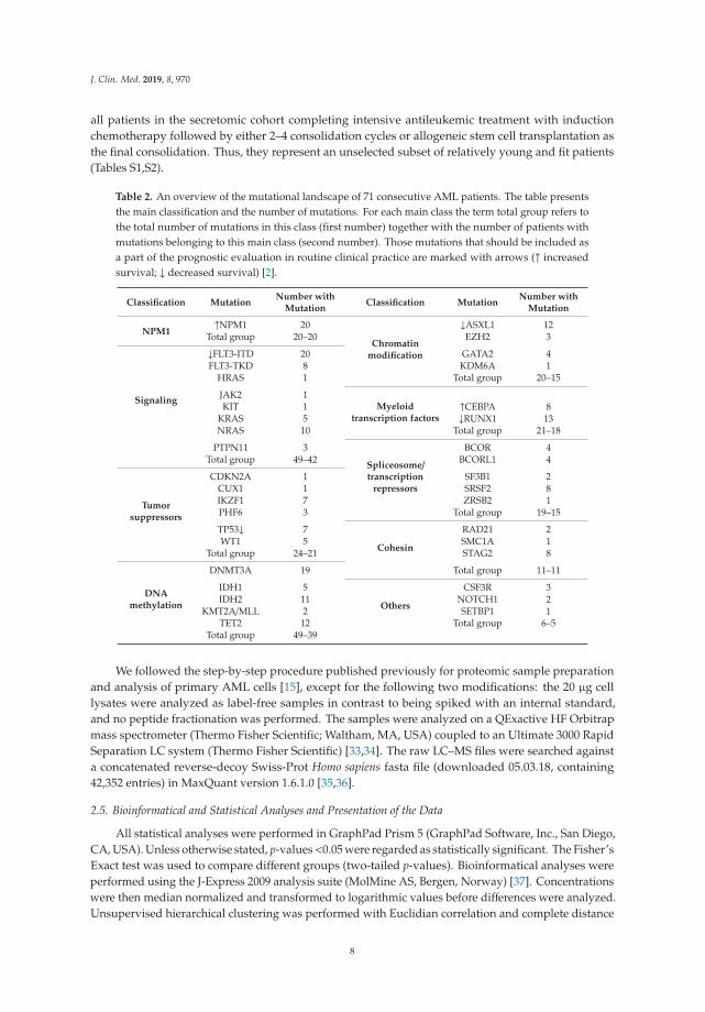
Table 2. An overview of the mutational landscape of 71 consecutive AML patients. The table presentsthe main classification and the number of mutations. For each main class the term total group refers tothe total number of mutations in this class (first number) together with the number of patients withmutations belonging to this main class (second number). Those mutations that should be included asa part of the prognostic evaluation in routine clinical practice are marked with arrows (↑ increasedsurvival; ↓ decreased survival) [2].
Classification MutationNumber with
MutationClassification Mutation
Number withMutation
NPM1↑NPM1 20
Chromatinmodification
↓ASXL1 12Total group 20–20 EZH2 3
Signaling
↓FLT3-ITD 20 GATA2 4FLT3-TKD 8 KDM6A 1
HRAS 1 Total group 20–15
JAK2 1Myeloid
transcription factorsKIT 1 ↑CEBPA 8
KRAS 5 ↓RUNX1 13NRAS 10 Total group 21–18
PTPN11 3
Spliceosome/transcription
repressors
BCOR 4Total group 49–42 BCORL1 4
Tumorsuppressors
CDKN2A 1 SF3B1 2CUX1 1 SRSF2 8IKZF1 7 ZRSB2 1PHF6 3 Total group 19–15
TP53↓ 7
Cohesin
RAD21 2WT1 5 SMC1A 1
Total group 24–21 STAG2 8
DNAmethylation
DNMT3A 19 Total group 11–11
IDH1 5
Others
CSF3R 3IDH2 11 NOTCH1 2
KMT2A/MLL 2 SETBP1 1TET2 12 Total group 6–5
Total group 49–39
We followed the step-by-step procedure published previously for proteomic sample preparationand analysis of primary AML cells [15], except for the following two modifications: the 20 μg celllysates were analyzed as label-free samples in contrast to being spiked with an internal standard,and no peptide fractionation was performed. The samples were analyzed on a QExactive HF Orbitrapmass spectrometer (Thermo Fisher Scientific; Waltham, MA, USA) coupled to an Ultimate 3000 RapidSeparation LC system (Thermo Fisher Scientific) [33,34]. The raw LC–MS files were searched againsta concatenated reverse-decoy Swiss-Prot Homo sapiens fasta file (downloaded 05.03.18, containing42,352 entries) in MaxQuant version 1.6.1.0 [35,36].
2.5. Bioinformatical and Statistical Analyses and Presentation of the Data
All statistical analyses were performed in GraphPad Prism 5 (GraphPad Software, Inc., San Diego,CA, USA). Unless otherwise stated, p-values<0.05 were regarded as statistically significant. The Fisher’sExact test was used to compare different groups (two-tailed p-values). Bioinformatical analyses wereperformed using the J-Express 2009 analysis suite (MolMine AS, Bergen, Norway) [37]. Concentrationswere then median normalized and transformed to logarithmic values before differences were analyzed.Unsupervised hierarchical clustering was performed with Euclidian correlation and complete distance
8

J. Clin. Med. 2019, 8, 970
measure for all analyses in J-Express. The Panther classification system (version PANTHER14.0) wasused to identify distinct functional classes [38].
The proteomics data processing of the raw data (i.e., filtering for reverse hits, contaminants andproteins only identified by site, and log2 transformation of label-free quantification (LFQ) intensities),and statistical analysis of two groups using Welch’s t-test was performed in Perseus version 1.6.1.1. [39].Furthermore, Z-statistics were used to find the proteins with the most abundant fold changes (FCs), i.e.,the proteins with highest or lowest FC when comparing the high-release with the low-release group andcalculating the FCs from the median log2 intensity per group as described by others [40]. Unsupervisedhierarchical clustering was performed with Euclidian correlation and complete distance measure forall analyses in J-Express [37], and gene ontology analysis in DAVID version 6.8 [41]. Gene ontology(GO) terms with false discovery rate (FDR) < 0.05, the number of proteins associated to the term,and the fold enrichment were presented. The significantly different proteins were imported to theSTRING database version 11.0 [42] to obtain protein–protein interaction networks, using experimentsand databases as interaction sources at highest confidence (0.9). The networks were imported andvisualized in Cytoscape version 3.3.0 [43]. Venny 2.1 (http://bioinfogp.cnb.csic.es/tools/venny/) wasused to create Venn diagrams.
To summarize, due to the previously described AML heterogeneity and the fact that we sometimeshave unequal numbers of quantified values of a protein in the two groups, we assumed an unequalvariation in the groups and first applied the Welch t-test to identify proteins with significantly (p < 0.05)different mean tests. Thereafter we used Z-statistics as an additional test to identify those proteinswith the most extreme/significant fold changes (fold change defined as the median intensity forhigh-release patients relative to the median intensity for low-release patients; the intensities were thenlog2-transformed).
3. Results
3.1. The Genetic Heterogeneity of AML Patients: TP53 Mutations are Associated with High-Risk Karyotypesand NPM1 Mutations are Associated with Mutations in DNA Methylation Genes
We analyzed the submicroscopic mutational profile for all 71 patients. The profile included 54frequent mutated genes in myeloid malignancies, 37 of them carried non-benign mutations in ourpatients (Figure 1). At least one mutation was detected for 69 of the 71 patients, and one of patientswithout detected mutations had a balanced translocation. The median number of mutations per patientwas 3.5 (range 0–7). The most frequently detected mutations were NPM1 exon 12 insertion and theFLT3-ITD mutation (20 patients for each), followed by mutations in the DNMT3A (19), TET2 (13),and RUNX1 (13) genes (Figure S1).
We used the same (and now generally accepted) classification of AML-associated mutationsin our present study as was used in two large previous studies, including 1540 and 200 patients,respectively [6,7]. The following mutations were detected in our patients: (i) NPM1 insertion(detected in 20 out of the 71 patients), (ii) mutations causing activation of intracellular signaling(9 genes, 42 patients), (iii) mutated tumor suppressor genes (8 genes, 21 patients), (iv) mutations ingenes involved in DNA methylation (5 genes, 39 patients) or (v) chromatin modification (3 genes,15 patients), (vi) mutations in genes encoding myeloid transcription factors (3 genes, 20 patients), (vii)mutated genes important for the spliceosome (5 genes, 15 patients), (vii) mutated genes encodingcohesion proteins (3 genes, 9 patients), and (viii) the three genes CSF3R, NOTCH1, and SETBP1 thatwere mutated in 5 patients (Table 2). The median number of different class mutations per patient was2.5 (range 0–5); 24% of the patients had mutations from two different main classes and 34% from threemain classes of mutations (Table S1).
9

J. Clin. Med. 2019, 8, 970
Figure 1. The total genomic profile and organization of mutations into defined categories; an overviewof the data for the 71 AML patients included in our study. The figure shows the somatic mutationsidentified from a 54 gene mutation panel, the mutations being classified as described previously [6,7].A majority of 69 patients had at least one detectable mutation. Risk classification of the karyotypes,morphological signs of differentiation (i.e., FAB-classification), etiology, age, and gender are presentedin the right part of the figure. The patients selected for proteomic analyses are indexed with black inthe left part of the figure.
We compared the mutational status with karyotype, French–American–British (FAB) classification(i.e., morphological differentiation), de novo versus secondary leukemia, age, and gender (Figure 1);these statistical analyses are summarized in Table S3. Firstly, we observed a highly significant associationbetween NPM1 and DNA methylation gene mutations (Fisher’s Exact test, p = 0.0015), whereas theassociation between FLT3-ITD and NPM1 mutations did not reach significance. Secondly, there was anegative association between NPM1 and myeloid transcription factor mutations (Fisher’s Exact test,p = 0.0001), and also between NPM1 and chromatin modifier mutations that occurred together onlyfor two patients. Thirdly, all patients with TP53 mutations had high-risk cytogenetic abnormalities(Fisher’s Exact test, p < 0.0001). Fourthly, NPM1 mutations were associated with morphological signs
10

J. Clin. Med. 2019, 8, 970
of differentiation, i.e., FAB classification M2/M4/M5/M6 (Fisher’s Exact test, p = 0.0233). Finally, even inthis relatively small patient cohort, we observed that no patients with TET2 mutations (13 patients)had IDH mutation (5 patients); this inverse correlation has been described in previous cohorts [6],but did not reach statistical significance in our smaller cohort. We did not detect any significantassociations between individual mutation or mutational main classes and age, gender, or AML etiology(de novo/secondary). A trend toward higher number of identified mutations in patients >65 years wasdetected, (median 4 mutations >65 years, and median 3 mutations <65 years), although did not reachstatistical significance in this patient cohort. To summarize, the frequencies of individual mutationsand the various associations are similar to what has been described previously [7,44]; the observationsthus suggest that our patient cohort of consecutive patients with relatively high peripheral blood blastcounts is representative for AML in general.
3.2. Expression of Molecular Differentiation Markers by Primary AML Cells: The Expression of the CD34 StemCell Markers Differs between Mutational Subsets
The AML cell expression of eight common differentiation markers (CD13, CD14, CD15, CD33,CD34, CD45, CD117, and HLA-DR) was available for 62 unselected AML patients. We first did anunsupervised hierarchical cluster analysis based on this expression profile (Figure S2). We could thenidentify four main patient subsets, but no single mutation or mutational class showed significantassociations with any of the four main patient clusters.
We investigated whether there were any significant correlations between the CD34 stem cellmarker and any of the other differentiation markers, but no significant associations were then detected.
We finally investigated whether any of the mutations that are used as prognostic markers inroutine clinical practice [2] showed significant correlations with the expression of single differentiationmarkers. These statistical analyses are summarized in Table S3. Firstly, NPM1 mutations showed asignificant correlation with CD33 expression (Fisher’s Exact test, p = 0.0107) and a negative associationwith CD34 expression (Fisher’s Exact test, p < 0.0001). These NPM1 associations are similar to theobservations in a previous large study of 184 unselected patients [45], and they are consistent with theobservation that NPM1 mutations are frequently associated with morphological signs of differentiation(see above). Secondly, neither FLT3-ITD nor DNMT3A mutations showed any association withCD34 expression. NPM1 mutations are frequently combined with FLT3-ITD and DNA-methylationmutations [6], but only the negative NPM1 association reached significance in our relatively smallcohort. Thirdly, patients with mutations in chromatin modifier genes showed an increased frequencyof CD34 expression by their AML cells (Fisher’s Exact test p = 0.0159). We detected the combination ofNPM1 and chromatin modifier mutations for only two patients, and this was similar to the observationsin previous studies [7]. Thus, these mutational subsets also differ in their expression of differentiationmarkers, especially CD34 expression.
3.3. AML Patients Can Be Subclassified Based on Their Constitutive Release of Extracellular Mediators,but this Capacity Shows no Association with the Mutational Profile
Primary AML cells from 46 of the patients were available for additional studies of constitutivecytokine release during in vitro culture. This patient subset represents a constitutive and therebyunselected subset among the 71 patients included in our present study. We investigated theconstitutive release of 34 soluble mediators, including several cytokines (interleukins, CCLand CXCL chemokines, immunoregulatory cytokines, growth factors), proteases, and proteaseregulators/inhibitors. A clustering analysis identified a subset of patients with generally highconstitutive mediator release; the other patients showed generally low or intermediate release (Figure 2).Neither any single mutation nor mutational main class differed significantly when comparing the threepatient subsets identified in this clustering analysis.
11
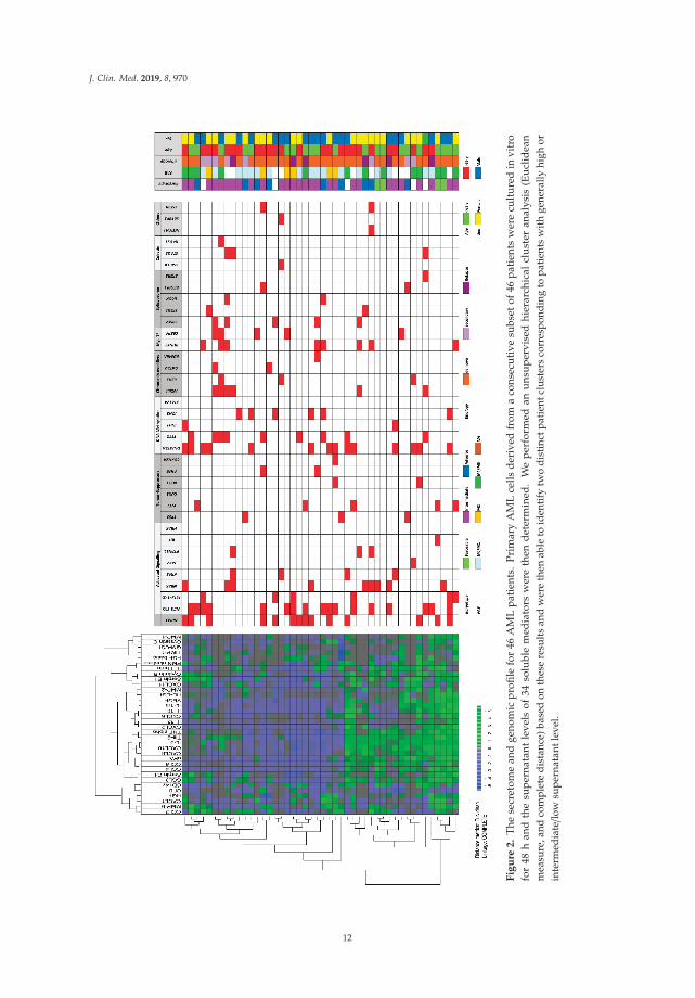
J. Clin. Med. 2019, 8, 970
Fig
ure
2.
The
secr
etom
ean
dge
nom
icp
rofi
lefo
r46
AM
Lp
atie
nts.
Pri
mar
yA
ML
cells
der
ived
from
aco
nsec
uti
vesu
bset
of46
pat
ient
sw
ere
cult
ure
din
vitr
ofo
r48
han
dth
esu
per
nata
ntle
vels
of34
solu
ble
med
iato
rsw
ere
then
det
erm
ined
.W
ep
erfo
rmed
anu
nsu
per
vise
dhi
erar
chic
alcl
ust
eran
alys
is(E
ucl
idea
nm
easu
re,a
ndco
mpl
ete
dist
ance
)bas
edon
thes
ere
sults
and
wer
eth
enab
leto
iden
tify
two
dist
inct
patie
ntcl
uste
rsco
rres
pond
ing
topa
tient
sw
ithge
nera
llyhi
ghor
inte
rmed
iate/lo
wsu
pern
atan
tlev
el.
12
J. Clin. Med. 2019, 8, 970
3.4. Comparison of Global Gene Expression Profiles for Patients with Generally High and Low ConstitutiveRelease of Extracellular Mediators
We have previously described differences in global gene expression profiles between AML cellswith generally high and low constitutive mediator release [46]. We performed a similar comparison forthe patients included in the present studies based on the differentially expressed genes, and we couldthen identify two main patient subsets based on this expression (d-score >3.5; 149 genes identified).However, these two subsets did not differ significantly with regard to the distribution of singlemutations or the overall mutational profiles of the AML cell populations (Figure S3).
3.5. Comparison of Proteomic Profiles for AML Cell Populations Showing Generally High and Low ConstitutiveRelease of Extracellular Mediators
Our proteomic analyses identified 5852 proteins, but 5586 proteins were left after leaving outprotein contaminants, reverse hits, and proteins only identified by site. Our further analyses were basedon 4350 proteins that could be detected in at least five patients for each of the two compared groups.A significant difference (p < 0.05) in protein abundance between the two groups was detected for 256 ofthese proteins (182 proteins increased in patients showing high constitutive release, 74 proteins beingincreased in the others), i.e., determined by Welch’s t-test and Z-statistics (a list of selected proteins aredescribed more in detail in Table S4 and the complete list of all 256 proteins is given in Table S5).
We first performed an unsupervised hierarchical cluster analysis (Euclidean measure, and completedistance) based on the 256 differentially expressed proteins (Figure 3). Our analysis identified twomain clusters/subsets of patients corresponding to patients with generally high and low constitutiverelease by their AML cells; only one of the high release patients clustered as an outlier. Furthermore,we performed GO term overrepresentation analyses based on the 256 differentially abundant proteins.The analysis of those proteins showing increased expression (n = 74) in patients with low constitutivemediator release and returned significantly increased GO terms, which reflected an altered regulationof nuclear functions/transcription/RNA metabolism (Table 3 and Table S4). It can be seen that a majorpart of these genes are important for transcriptional regulation/RNA expression/RNA metabolism.
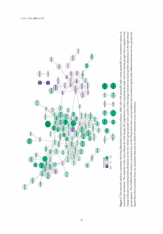
We then analyzed those proteins showing increased expression in AML cells with high constitutivecytokine release; the most significant GO-terms are listed in Table 4. When analyzing the proteins withregard to cell compartment the four largest terms (extracellular exosomes, cytosol, membrane, andcytoplasm) were only partly overlapping with regard to individual proteins and included 153 of the182 proteins that were significantly increased in high-release AML cells (Figure 4). These four GO termsreflect cytoplasmic/cytosolic structures/functions together with the terms actin filament and phagocyticvesicle membrane. One of the terms reflects metabolic functions (NADPH oxidase complex), whereasthe two last terms reflect cell surface functions/cellular communication (focal adhesion, membranerafts). Analysis of biological processes and molecular functions included several relatively small GOterms that also reflect intracellular signaling, protein interactions, or cell surface receptor signaling(Table 4). Table S4 gives a more detailed description of those proteins that were identified both inthe GO term analyses (Table 4, Figure 4) and in the network analysis (Figure 5; proteins in the largenetwork to the left in the figure with increased levels in high-secreting cells).
The proteins with increased expression in patients with generally high constitutive release arepresented in Figure 4 (all proteins included in the GO-terms GO:0070062—extracellular exosome,GO:0005829—cytosol, GO:0016020—membrane, or GO:0005737—cytoplasm); Table 3 (classification ofproteins showing p < 0.01); Table S4 (description of proteins from Table 3 with p < 0.01); and TableS5 (the complete list of all 256 differentially expressed proteins). These more detailed analyses andclassifications of individual proteins from Table 3 and Table S4 also show that AML cells showinggenerally high or low constitutive release of extracellular mediators differ especially with regard totranscriptional regulation, cell surface molecular profile, intracellular signaling, intracellular trafficking,and cell adhesion/migration.
13

J. Clin. Med. 2019, 8, 970
Fig
ure
3.
Iden
tifi
cati
onof
two
mai
npa
tien
tsub
sets
base
don
prot
eom
icd
iffer
ence
sof
AM
Lce
llsw
ith
high
and
low
cons
titu
tive
rele
ase.
Eig
htof
the
16pa
tien
tsin
clud
edin
the
prot
eom
icst
udie
sbe
long
edto
the
clus
ter
char
acte
rize
dby
gene
rally
high
cons
titut
ive
med
iato
rre
leas
ean
dth
eei
ghto
ther
ssh
owed
low/in
term
edia
tese
cret
ion
(Fig
ure
2);2
56pr
otei
nsdiff
ered
sign
ifica
ntly
betw
een
thes
etw
ogr
oups
.We
perf
orm
edan
unsu
perv
ised
hier
arch
ical
clus
ter
anal
yses
(Euc
lidea
nm
easu
re,
and
com
plet
ed
ista
nce)
base
don
the
leve
lsof
thes
epr
otei
ns,a
ndth
ele
ftpa
rtd
emon
stra
tes
the
den
dro
gram
and
heat
map
;blu
ein
dic
ates
low
prot
ein
leve
lsan
dgr
een
high
leve
ls.
Two
mai
ncl
ust
ers
wer
eth
enid
enti
fied
corr
esp
ond
ing
toth
ehi
ghan
dlo
w/in
term
edia
tese
cret
ion
pat
ient
sex
cep
tfor
one
outl
ier
pat
ient
(lef
tco
lum
n,re
dco
lor
indi
catin
ghi
ghre
leas
e).A
sex
pect
ed,t
hetw
om
ain
clus
ters
wer
ehe
tero
gene
ous
with
rega
rdto
mut
atio
nalf
requ
enci
es(m
iddl
epa
nel)
and
did
not
diff
erw
ith
rega
rdto
clin
ical
orbi
olog
ical
char
acte
rist
ics
eith
er(r
ight
pane
l).
14

J. Clin. Med. 2019, 8, 970
Ta
ble
3.
Diff
eren
tial
lyex
pres
sed
prot
eins
inpr
imar
yA
ML
cell
popu
lati
ons
wit
hhi
gh(l
eft)
and
low
(hig
h)co
nsti
tuti
vere
leas
eof
extr
acel
lula
rso
lubl
em
edia
tors
.T
hem
edia
tors
are
clas
sifi
edba
sed
onth
eir
mai
nfu
ncti
onal
char
acte
rist
ics.
The
info
rmat
ion
isba
sed
onth
eG
ene
dat
abas
ean
dse
lect
edre
fere
nces
from
the
PubM
edda
taba
se(T
able
S4).
The
prot
eins
bein
gin
crea
sed
inhi
gh-s
ecre
ting
AM
Lce
llsar
eth
ose
prot
eins
that
wer
ebo
thin
clud
edin
the
gene
onto
logy
(GO
)ter
ms
GO
:007
0062
—ex
trac
ellu
lar
exos
ome,
GO
:000
5829
—cy
toso
l,G
O:0
0160
20—
mem
bran
e,an
dG
O:0
0057
37—
cyto
pla
sm(F
igu
re4)
,and
also
inth
em
ain
inte
ract
ing
pro
tein
netw
ork
inth
ele
ftp
art
ofFi
gure
5(T
able
S4).
The
pro
tein
sbe
ing
incr
ease
din
the
low
-sec
reti
ngA
ML
cells
are
thos
ep
rote
ins
incl
ud
edth
eG
Ote
rms
GO
:000
0790
—nu
clea
rch
rom
atin
and
GO
:000
5736
—D
NA
-dir
ecte
dR
NA
poly
mer
ase
Icom
plex
(Tab
le4)
.
Main
Cla
ssifi
cati
on
Incr
ease
dP
rote
inL
ev
els
inC
ell
sw
ith
Hig
hC
on
stit
uti
ve
Rele
ase
Incr
ease
dP
rote
inL
ev
els
inC
ell
sw
ith
Lo
wC
on
stit
uti
ve
Rele
ase
Nu
cleo
som
eM
BD3
Ch
rom
ati
n,h
isto
ne,tr
an
scri
pti
on
,R
NA
TOLL
IP,N
FKB1
HIF
0,H
IST
IH2A
J,M
TA1,
SMA
RC
E1,M
EN1,
MBD
3,PO
LR1E
,C
LPX
,PO
LR1A
,PO
LR1B
DN
Are
pair
CLP
X,J
UN
D,P
OG
2O
nco
gen
eC
BL,D
BNL
Cell
cycl
ere
gu
lati
on
IL16
Intr
ace
llu
lar
sig
nali
ng
SYK
,HC
LS1,
AK
AP1
,TLR
2,TO
LLIP
,AG
TRA
P,A
NX
A2,
CEC
R1,
INPP
5D,L
PKN
,IK
BKB,
TBK
1T
yro
sin
ek
inase
SYK
,HC
LS1,
FGR
,PK
N1
SR
Cty
rosi
ne
kin
ase
sH
CLS
1,FG
R,H
CK
,P
I3K
-Ak
t-m
TO
RN
CF4
RA
C1
RA
C1,
NC
F4,R
HO
T1,A
RH
GEF
1,PK
N1,
RH
OG
,AR
HG
AP3
0,PR
EX1,
GM
IP,D
OK
2,A
KA
P1
GT
Pase
DN
M2,
AR
HG
EF1,
PKN
1,R
HO
G,A
RH
GA
P30,
PREX
1,G
MIP
,A
KA
P1,A
RH
GA
P,R
AB2
7AG
-pro
tein
cou
ple
dre
cep
tors
AR
RB2
,AR
HG
EF1,
PREX
1,G
RK
6
Ph
ag
ocy
tosi
sC
YBA
,NC
F2,N
CF4
,ELM
O2
Pro
tein
deg
rad
ati
on
CBL
,SER
PIN
A1
Intr
ace
llu
lar
traffi
ckin
gVA
MP3
,DN
M2,
PIC
ALM
,SN
X18
,AR
AP1
,AR
AP1
,TO
LLIP
,A
P1G
2,S1
00A
10,S
100A
4,TO
M1,
SDC
DP,
DN
AJC
13,E
PN1,
APH
GA
P,R
AB2
7A
Mic
rotu
bu
le,cy
tosk
ele
ton
,st
ruct
ure
DN
M2,
EPN
1,SH
3KBP
1,PK
N1,
RH
OG
,AH
NA
K,S
DC
DP,
S100
A4,
CK
AP4
,FA
M49
BC
ell
mig
rati
on
PLX
NB2
,HC
K,D
NM
2,R
HO
G,E
LMO
2,A
HN
AK
Mit
och
on
dri
a,m
eta
bo
lism
FAM
49B,
FTL,
IMPD
H1,
PDX
KC
LPX
Ly
soso
mes
CTS
H,C
TSS,
CTS
Z,L
YZ
,PSA
PC
ell
meta
bo
lism
,N
AD
PH
CK
,NC
F4C
yto
kin
esi
sFM
NL1
Extr
ace
llu
lar
matr
ix,ce
llad
hesi
on
EPN
1,SH
3KBP
1E
xtr
ace
llu
lar
med
iato
rsIL
16,T
LR2,
TOLL
IPC
ell
surf
ace
mo
lecu
les
ITG
AL,
ITG
AM
.ITG
B2,S
YK
,LIL
RB2
,PK
N1,
LPX
NIn
teg
rin
sIT
GA
L,IT
GA
M.I
TGB2
,SY
K,F
GR
,LPX
N
Via
bil
ity,
ap
op
tosi
sSH
3KBP
1,PK
N1,
AR
AP1
,TLR
2
AM
LC
BL,P
ICA
LMD
iffere
nti
ati
on
MN
DA
,NC
F1,C
ECR
1
15

J. Clin. Med. 2019, 8, 970
Fig
ure
4.
GO
-ter
ms
incl
ud
ing
sign
ifica
ntly
incr
ease
dp
rote
ins
for
AM
Lce
llsw
ith
gene
rally
high
cons
titu
tive
rele
ase
ofex
trac
ellu
lar
solu
ble
med
iato
rs.
The
over
-rep
rese
ntat
ion
anal
ysis
base
don
cellu
lar
com
par
tmen
tid
enti
fied
fou
rG
Ote
rms
wit
hFD
R<
0.05
and
incl
ud
ing
atle
ast
40p
rote
ins,
i.e.,
GO
:007
0062
—ex
trac
ellu
lar
exos
ome,
GO
:000
5829
—cy
toso
l,G
O:0
0160
20—
mem
bran
e,an
dG
O:0
0057
37—
cyto
plas
m.T
hese
four
GO
-ter
ms
wer
epa
rtly
over
lapp
ing
(onl
ysi
xp
rote
ins
incl
ud
edin
allf
our)
;tog
ethe
rth
eyin
clu
ded
153
ofth
e18
6p
rote
ins
that
wer
ein
crea
sed
inA
ML
cells
wit
hge
nera
llyhi
ghco
nsti
tuti
vere
leas
eco
mpa
red
wit
hA
ML
cells
wit
hlo
w/in
term
edia
teco
nsti
tuti
vere
leas
e.
16

J. Clin. Med. 2019, 8, 970
Ta
ble
4.
Sign
ifica
ntG
O-t
erm
s(i
.e.,
FDR<
0.05
)for
prot
eins
show
ing
sign
ifica
ntly
incr
ease
dle
vels
inA
ML
pati
ents
wit
hin
term
edia
te/lo
wan
dhi
ghco
nsti
tuti
vem
edia
tor
rele
ase.
Lo
wco
nst
itu
tiv
em
ed
iato
rre
lea
se;
list
of
sig
nifi
can
tG
O-t
erm
sP
rote
inn
um
be
rF
old
en
rich
me
nt
FD
R
Ce
llco
mp
art
me
nt
GO
:000
5654
—nu
cleo
plas
m31
2.8
2.3×1
0–5
GO
:000
0790
—nu
clea
rch
rom
atin
811
0.00
99G
O:0
0057
36—
DN
A-d
irec
ted
RN
Apo
lym
eras
eIc
ompl
ex4
800.
017
Mo
lecu
lar
fun
ctio
nG
O:0
0037
13—
tran
scri
ptio
nco
acti
vato
rac
tivi
ty9
8.5
0.01
1G
O:0
0010
54—
RN
Apo
lym
eras
eIa
ctiv
ity
478
0.01
8
Hig
hco
nst
itu
tiv
em
ed
iato
rre
lea
se;
list
of
sig
nifi
can
tG
O-t
erm
s
Bio
log
ica
lp
roce
sse
sG
O:0
0069
54—
infla
mm
ator
yre
spon
se19
5.0
6.5×1
0–5
GO
:004
5087
—in
nate
imm
une
resp
onse
204.
78.
3×1
0–5
GO
:004
8010
—va
scul
aren
doth
elia
lgro
wth
fact
orre
cept
orsi
gnal
ing
path
way
913
8.6×1
0–4
GO
:000
7229
—in
tegr
in-m
edia
ted
sign
alin
gpa
thw
ay10
109.
4×1
0–4
GO
:003
1623
—re
cept
orin
tern
aliz
atio
n7
160.
0062
GO
:000
7165
—si
gnal
tran
sduc
tion
292.
50.
015
GO
:009
8609
—ce
ll–ce
llad
hesi
on13
4.8
0.02
6C
ell
com
pa
rtm
en
tG
O:0
0700
62—
extr
acel
lula
rex
osom
e73
2.7
1.4×1
0–13
GO
:000
5829
—cy
toso
l79
2.5
5.7×1
0–13
GO
:001
6020
—m
embr
ane
482.
37.
8×1
0–5
GO
:004
3020
—N
AD
PHox
idas
eco
mpl
ex5
430.
0048
GO
:000
5737
—cy
topl
asm
781.
60.
010
GO
:003
0670
—ph
agoc
ytic
vesi
cle
mem
bran
e7
120.
026
GO
:000
5925
—fo
cala
dhes
ion
154.
00.
03G
O:0
0451
21—
mem
bran
era
ft11
5.6
0.03
8G
O:0
0058
84—
acti
nfil
amen
t7
110.
046
Mo
lecu
lar
fun
ctio
nG
O:0
0055
15—
prot
ein
bind
ing
129
1.4
5.8×1
0–6
GO
:001
7124
—SH
3do
mai
nbi
ndin
g11
8.9
5.8×1
0–4
GO
:003
5325
—To
ll-lik
ere
cept
orbi
ndin
g4
960.
0058
17
J. Clin. Med. 2019, 8, 970
Fig
ure
5.
The
netw
ork
anal
ysis
ofpr
otei
nssh
owin
gd
iffer
enti
alex
pres
sion
inpr
imar
yA
ML
cells
wit
hge
nera
llyhi
ghve
rsus
gene
rally
low
cons
titu
tive
rele
ase
ofex
trac
ellu
lar
med
iato
rs.T
hein
tens
ityof
the
colo
rre
flect
sth
efo
ldch
ange
(FC
)sig
nific
ance
whe
nco
mpa
ring
the
high
-and
low
-rel
ease
grou
ps;t
hus
ane
gativ
efo
ldch
ange
indi
cate
sin
crea
sed
prot
ein
abun
danc
ein
the
low
-rel
ease
grou
p(p
urpl
e)an
da
posi
tive
fold
chan
gein
dica
tes
incr
ease
dpr
otei
nab
unda
nce
inth
ehi
gh-r
elea
segr
oup
(gre
en).
This
STR
ING
-DB
anal
ysis
was
base
don
lyon
the
256
prot
eins
that
wer
equ
antifi
edan
dco
nsid
ered
sign
ifica
ntly
diff
eren
tbet
wee
nth
etw
ogr
oups
;the
figur
eth
ussh
ows
prot
eins
from
our
quan
tifie
dda
taan
dno
shel
lsof
inte
ract
ors
wer
eco
nsid
ered
.
18

J. Clin. Med. 2019, 8, 970
We finally did a molecular network analysis based on the 256 differentially abundant proteins,and Figure 5 shows all molecular connections identified in this analysis (those molecules without anyconnections are left out). A total of 129 proteins were included in various networks; most of themappeared in a large network linked to the nodes spleen tyrosine kinase (SYK), NCF4 (a cytosolic regulatorof superoxide-producing NADPH-oxidase), ARRB2 (regulator of G-protein-coupled receptor activity),ACTR3 (a major constituent of the ARP2/3 complex located at the cell surface and being essentialfor cell motility), and RAC1 (a GTPase belonging to the RAS superfamily of small GTP-bindingproteins). Our overrepresentation analysis showed that exosomal proteins as well as proteinsimportant for intracellular trafficking were differentially expressed; both these groups are importantfor communication from the leukemic cells to neighboring AML supporting stromal cells [47]. On theother hand, our network analysis showed that these AML cells had increased levels of several membersof a signaling pathway, including cell surface integrins (αLβ2, αMβ2) known to mediate downstreamsignaling involving SYK and SRC kinase family members (FGR, HCK) [48–51]. Toll like receptor (TLR)2 together with its downstream NFκB complex are also linked to this network [49]. Taken togetherthese observations suggest that high constitutive extracellular release of soluble mediators is only a partof a more complex cellular phenotype that is characterized by differences in the bidirectional crosstalkbetween the leukemic cells and their neighboring AML-supporting cells. This bidirectional crosstalkinvolves cytokine-mediated signaling directed from the AML cells to the stromal cells. At the sametime the stromal cells may influence the AML cells through soluble mediators or cell–cell contact withligation of cell surface molecules, followed by downstream signaling (involving kinases and G-proteininitiated signaling), and finally NFκB mediated modulation of cytokine/chemokine expression [48–52].Finally, this crosstalk involves integrins that can mediate both inside–out and outside–in effects [48].
4. Discussion
AML is a heterogeneous disease, and this can also be seen from our present studies of primaryhuman AML cells derived from a cohort of consecutive patients. In this study we focused on themolecular genetic abnormalities and the proteomic profiles of the leukemic cells [53]. Both the numberand the nature of the molecular genetic abnormalities differed between the patients (number ofdetected mutations per patients 0–7, median 3.5 mutations). The frequencies of the various mutationswere comparable to previous studies [6,7], NPM1 mutations were associated with molecular andmorphological signs of differentiation [45], and TP53 mutations were associated with adversekaryotypes [54]. Taken together, these observations suggest that we investigated a representativeAML patient population, even though we selected patients with relatively high peripheral blood blastcounts/percentages.
In the present study, we included a group of consecutive and thereby unselected AML patientswith a high percentage of leukemic blasts in peripheral blood. We used this selection of patients sothat highly enriched AML cell populations could be prepared by density gradient separation alone;the risk of inducing molecular and/or functional alterations in the AML patients by more extensivecell separation procedures was thereby avoided [55]. Our results may therefore be representativeonly for this selected subset of patients, but several observations suggest that they possibly arerepresentative for AML in general. Firstly, our patients showed an expected fraction of secondaryversus de novo AML [56,57]. Secondly, as previously described in detail patients selected according tothese criteria show a similar distribution of cytogenetic abnormalities as AML patients in general [30].Thirdly, our present study shows that the distribution of various molecular genetic abnormalities isalso similar to AML in general [6,7,44,58]. Finally, we have described in detail the selection of the 16AML patients included in our proteomic studies (see Section 3.5), and they should then be regarded asrepresentative for relatively young AML patients.
Extensive separation procedures will influence the functional characteristics of primary humanAML cells, and one would expect that in vitro incubation in culture medium would have similar effects.However, previous studies have shown that the characteristics of even long-term cultured primary
19

J. Clin. Med. 2019, 8, 970
human AML cells are associated with patient survival [59], an observation suggesting that even in vitrocultured cells will reflect functional characteristics of clinical relevance.
Distinct immunophenotype profiles may be associated with specific mutations, and search forimmunophenotype-based screening approaches have therefore been suggested [60,61]. We investigatedthe immunophenotype profiles of individual patients based on the expression of eight differentiationmarkers commonly used for classification of myeloid cells. We identified four different mainclusters/patient subsets based on this profiling, but no single mutation or mutation main classesshowed significant associations to any of these profiles. However, associations between mutations andsingle differentiation markers were observed, especially expression of the CD34 stem cell marker thatwas negatively associated with NPM1 mutations as well as FLT3-ITD and DNMT3 mutations, whereaschromatin modifier mutations were positively associated with CD34 expression. Such associationshave also been described previously [45,62]. A possible explanation for this is that single mutationsmay have a major impact on the expression of single or related markers, whereas the overall mutationalprofile has a major impact on the overall differentiation profile.
In previous studies we showed that the constitutive release of a wide range of soluble mediatorsby primary AML cells varied considerably between patients, and a subset of patients then showeda generally high release compared with other patients that either showed intermediate or lowrelease [11,52]. This capacity of constitutive mediator release was tested in a highly standardizedin vitro model. We investigated the constitutive release for a consecutive subset of our patients,and again we found that a subset of patients showed generally higher release of most mediatorscompared with the other patients. We then selected those samples that were derived before the first timeof diagnosis for all relatively young patients that completed intensive chemotherapy. We comparedthe proteomic profiles of the primary AML cells for eight patients showing high and another group ofeight patients showing generally lower mediator release.
Several proteins were differentially expressed when comparing patients with generally high andlow constitutive cytokine release. The high release patients showed high expression, especially ofproteins involved in intracellular signaling, intracellular transport/trafficking and communicationbetween cells (soluble mediators, exosomes, cell surface molecules, and intracellular mediatorsdownstream to cell surface receptors). We did not identify any of the soluble mediators when analyzingdifferentially abundant cell proteins between the two patient subsets; this is not unexpected becausethere is often not a strong correlation between cellular levels and extracellular release of solublemediators during culture [34].
The high constitutive mediator release should in our opinion be regarded as only a part of amore complex communication phenotype with neighboring non-leukemic stromal cells. In contrast,the cell populations with low constitutive release showed increased abundance proteins involved in orregulating gene transcription/RNA synthesis/RNA metabolism. A possible hypothesis may be that cellswith high constitutive release have a higher dependency on neighboring AML-supporting stromal cellsthan leukemia cells showing low constitutive release. We would emphasize that primary AML cellshave a wide range of secreted biomolecules, which can be useful in classification/prognostication andas therapeutic targets [11,52,63]. The mediators included in the present study are well-characterizedand are released at detectable levels for most patients. For these reasons they should be regardedas biologically important in the disease, but they probably represent only a part of the AML cellsecretome that is involved in the bidirectional crosstalk between leukemic and non-leukemic cells intheir common bone marrow microenvironment.
5. Conclusions
We conclude that the high constitutive extracellular release of soluble mediators by primaryhuman AML cells seems to reflect a complex functional phenotype with regard to communicationbetween AML cells and their neighboring non-leukemic stromal cells in their common bone marrowmicroenvironment. Our proteomic comparison has identified high expression in this patient subset of
20

J. Clin. Med. 2019, 8, 970
several intracellular molecules that are regarded as possible therapeutic targets in human AML. Dualtargeting of intracellular signaling and extracellular communication should therefore be considered forthese patients.
Supplementary Materials: The following are available online at http://www.mdpi.com/2077-0383/8/7/970/s1,Figure S1: Mutation profile and mutation classes. Figure S2: The immunophenotypic profile and mutationprofile. Figure S3: Transcriptomics data based on AML secretomic profile. Table S1: The biological and clinicalcharacteristics of patients in the proteomic studies. Table S2: The mutational distribution for patients included inthe proteomic studies. Table S3: Statistical comparisons of associations between various mutations and betweenmutations and signs of AML cell differentiation (morphology, CD34 expression). Table S4: Proteomic profilingbased on AML secretome. Table S5: All proteins differently expressed between high and low secretome group.
Author Contributions: Conceptualization, H.R. and Ø.B.; methodology, H.R., E.A., A.K.B., S.B.B., I.S.G., R.B.F.,and R.H.; software, H.R., E.A., and A.K.B.; validation, H.R. and Ø.B.; formal analysis, H.R., E.A., and A.K.B.;investigation, H.R., E.A., A.K.B., S.B.B., I.S.G., R.B.F., R.H., and Ø.B.; resources, H.R. and Ø.B.; data curation,H.R., E.A., and A.K.B.; writing—original draft preparation, H.R. and Ø.B.; writing—review and editing, H.R.,E.A., A.K.B., S.B.B., I.S.G., R.B.F., R.H., and Ø.B; visualization, H.R., E.A., and A.K.B.; supervision, Ø.B.; projectadministration, H.R., and Ø.B.; funding acquisition, Ø.B.
Funding: This research was funded by The Norwegian Cancer Society (DNK 100933) and Helse-Vest.
Acknowledgments: Technical support from Karen Marie Hagen and Kristin Paulsen Rye is greatly appreciated.
Conflicts of Interest: The authors declare no conflict of interest.
References
1. Döhner, H.; Weisdorf, D.J.; Bloomfield, C.D. Acute myeloid leukemia. N. Engl. J. Med. 2015, 373, 1136–1152.[PubMed]
2. Döhner, H.; Estey, E.; Grimwade, D.; Amadori, S.; Appelbaum, F.R.; Buchner, T.; Dombret, H.; Ebert, B.L.;Fenaux, P.; Larson, R.A.; et al. Diagnosis and management of aml in adults: 2017 eln recommendations froman international expert panel. Blood 2017, 129, 424–447. [PubMed]
3. Arber, D.A.; Orazi, A.; Hasserjian, R.; Thiele, J.; Borowitz, M.J.; Le Beau, M.M.; Bloomfield, C.D.; Cazzola, M.;Vardiman, J.W. The 2016 revision to the world health organization classification of myeloid neoplasms andacute leukemia. Blood 2016, 127, 2391–2405. [PubMed]
4. Schlenk, R.F.; Dohner, K.; Krauter, J.; Frohling, S.; Corbacioglu, A.; Bullinger, L.; Habdank, M.; Spath, D.;Morgan, M.; Benner, A.; et al. Mutations and treatment outcome in cytogenetically normal acute myeloidleukemia. N. Engl. J. Med. 2008, 358, 1909–1918. [PubMed]
5. Valk, P.J.; Verhaak, R.G.; Beijen, M.A.; Erpelinck, C.A.; Barjesteh van Waalwijk van Doorn-Khosrovani, S.;Boer, J.M.; Beverloo, H.B.; Moorhouse, M.J.; van der Spek, P.J.; Lowenberg, B.; et al. Prognostically usefulgene-expression profiles in acute myeloid leukemia. N. Engl. J. Med. 2004, 350, 1617–1628. [PubMed]
6. Papaemmanuil, E.; Gerstung, M.; Bullinger, L.; Gaidzik, V.I.; Paschka, P.; Roberts, N.D.; Potter, N.E.;Heuser, M.; Thol, F.; Bolli, N.; et al. Genomic classification and prognosis in acute myeloid leukemia. N. Engl.J. Med. 2016, 374, 2209–2221. [PubMed]
7. Cancer Genome Atlas Research, N.; Ley, T.J.; Miller, C.; Ding, L.; Raphael, B.J.; Mungall, A.J.; Robertson, A.;Hoadley, K.; Triche, T.J., Jr.; Laird, P.W.; et al. Genomic and epigenomic landscapes of adult de novo acutemyeloid leukemia. N. Engl. J. Med. 2013, 368, 2059–2074.
8. Eppert, K.; Takenaka, K.; Lechman, E.R.; Waldron, L.; Nilsson, B.; van Galen, P.; Metzeler, K.H.; Poeppl, A.;Ling, V.; Beyene, J.; et al. Stem cell gene expression programs influence clinical outcome in human leukemia.Nat. Med. 2011, 17, 1086–1093.
9. Brenner, A.K.; Reikvam, H.; Lavecchia, A.; Bruserud, O. Therapeutic targeting the cell division cycle 25(cdc25) phosphatases in human acute myeloid leukemia–the possibility to target several kinases throughinhibition of the various cdc25 isoforms. Molecules 2014, 19, 18414–18447.
10. Hatfield, K.J.; Reikvam, H.; Bruserud, O. Identification of a subset of patients with acute myeloid leukemiacharacterized by long-term in vitro proliferation and altered cell cycle regulation of the leukemic cells.Expert. Opin. Therap. Targets 2014, 18, 1237–1251.
21

J. Clin. Med. 2019, 8, 970
11. Brenner, A.K.; Reikvam, H.; Bruserud, O. A subset of patients with acute myeloid leukemia has leukemia cellscharacterized by chemokine responsiveness and altered expression of transcriptional as well as angiogenicregulators. Front Immunol. 2016, 7, 205. [PubMed]
12. Griessinger, E.; Anjos-Afonso, F.; Vargaftig, J.; Taussig, D.C.; Lassailly, F.; Prebet, T.; Imbert, V.; Nebout, M.;Vey, N.; Chabannon, C.; et al. Frequency and dynamics of leukemia-initiating cells during short-term ex vivoculture informs outcomes in acute myeloid leukemia patients. Cancer Res. 2016, 76, 2082–2086. [PubMed]
13. Griessinger, E.; Anjos-Afonso, F.; Pizzitola, I.; Rouault-Pierre, K.; Vargaftig, J.; Taussig, D.; Gribben, J.;Lassailly, F.; Bonnet, D. A niche-like culture system allowing the maintenance of primary human acutemyeloid leukemia-initiating cells: A new tool to decipher their chemoresistance and self-renewal mechanisms.Stem Cells Transl. Med. 2014, 3, 520–529. [PubMed]
14. Hauge, M.; Bruserud, O.; Hatfield, K.J. Targeting of cell metabolism in human acute myeloid leukemia–more thantargeting of isocitrate dehydrogenase mutations and pi3k/akt/mtor signaling? Eur. J. Haematol. 2016, 96, 211–221.[PubMed]
15. Hernandez-Valladares, M.; Aasebo, E.; Mjaavatten, O.; Vaudel, M.; Bruserud, O.; Berven, F.; Selheim, F.Reliable fasp-based procedures for optimal quantitative proteomic and phosphoproteomic analysis onsamples from acute myeloid leukemia patients. Biol. Proced. Online 2016, 18, 13. [PubMed]
16. Aasebo, E.; Mjaavatten, O.; Vaudel, M.; Farag, Y.; Selheim, F.; Berven, F.; Bruserud, O.;Hernandez-Valladares, M. Freezing effects on the acute myeloid leukemia cell proteome andphosphoproteome revealed using optimal quantitative workflows. J. Proteomics 2016, 145, 214–225. [PubMed]
17. Aasebo, E.; Vaudel, M.; Mjaavatten, O.; Gausdal, G.; Van der Burgh, A.; Gjertsen, B.T.; Doskeland, S.O.;Bruserud, O.; Berven, F.S.; Selheim, F. Performance of super-silac based quantitative proteomics forcomparison of different acute myeloid leukemia (aml) cell lines. Proteomics 2014, 14, 1971–1976. [PubMed]
18. Ossenkoppele, G.J.; Janssen, J.J.; van de Loosdrecht, A.A. Risk factors for relapse after allogeneictransplantation in acute myeloid leukemia. Haematologica 2016, 101, 20–25. [PubMed]
19. Terwijn, M.; Zeijlemaker, W.; Kelder, A.; Rutten, A.P.; Snel, A.N.; Scholten, W.J.; Pabst, T.; Verhoef, G.;Lowenberg, B.; Zweegman, S.; et al. Leukemic stem cell frequency: A strong biomarker for clinical outcomein acute myeloid leukemia. PloS ONE 2014, 9, e107587.
20. Wouters, R.; Cucchi, D.; Kaspers, G.J.; Schuurhuis, G.J.; Cloos, J. Relevance of leukemic stem cells in acutemyeloid leukemia: Heterogeneity and influence on disease monitoring, prognosis and treatment design.Expert. Rev. Hematol. 2014, 7, 791–805.
21. Majeti, R. Clonal evolution of pre-leukemic hematopoietic stem cells precedes human acute myeloid leukemia.Best Pract. Res. Clin. Haematol. 2014, 27, 229–234.
22. Stapnes, C.; Gjertsen, B.T.; Reikvam, H.; Bruserud, O. Targeted therapy in acute myeloid leukaemia: Currentstatus and future directions. Expert. Opin. Investig. Drugs 2009, 18, 433–455. [PubMed]
23. Binder, S.; Luciano, M.; Horejs-Hoeck, J. The cytokine network in acute myeloid leukemia (aml): A focus onpro- and anti-inflammatory mediators. Cytokine Growth Factor Rev. 2018, 43, 8–15. [PubMed]
24. Brenner, A.K.; Andersson Tvedt, T.H.; Bruserud, O. The complexity of targeting pi3k-akt-mtor signalling inhuman acute myeloid leukaemia: The importance of leukemic cell heterogeneity, neighbouring mesenchymalstem cells and immunocompetent cells. Molecules 2016, 21.
25. Reikvam, H.; Hatfield, K.J.; Fredly, H.; Nepstad, I.; Mosevoll, K.A.; Bruserud, O. The angioregulatorycytokine network in human acute myeloid leukemia-from leukemogenesis via remission induction to stemcell transplantation. Eur. Cytokine Netw. 2012, 23, 140–153. [PubMed]
26. Kupsa, T.; Horacek, J.M.; Jebavy, L. The role of cytokines in acute myeloid leukemia: A systematic review.Biomed. Pap. Med. Fac. Univ. Palacky Olomouc Czech Repub. 2012, 156, 291–301. [PubMed]
27. Cho, B.S.; Kim, H.J.; Konopleva, M. Targeting the cxcl12/cxcr4 axis in acute myeloid leukemia: From benchto bedside. Korean J. Intern. Med. 2017, 32, 248–257. [PubMed]
28. Bernasconi, P.; Farina, M.; Boni, M.; Dambruoso, I.; Calvello, C. Therapeutically targeting self-reinforcingleukemic niches in acute myeloid leukemia: A worthy endeavor? Am. J. Hematol. 2016, 91, 507–517.[PubMed]
29. Brenner, A.K.; Tvedt, T.H.; Nepstad, I.; Rye, K.P.; Hagen, K.M.; Reikvam, H.; Bruserud, O. Patients withacute myeloid leukemia can be subclassified based on the constitutive cytokine release of the leukemic cells;the possible clinical relevance and the importance of cellular iron metabolism. Expert. Opin. Therap. Targets2017, 21, 357–369.
22

J. Clin. Med. 2019, 8, 970
30. Bruserud, O.; Hovland, R.; Wergeland, L.; Huang, T.S.; Gjertsen, B.T. Flt3-mediated signaling in humanacute myelogenous leukemia (aml) blasts: A functional characterization of flt3-ligand effects in aml cellpopulations with and without genetic flt3 abnormalities. Haematologica 2003, 88, 416–428.
31. Reikvam, H.; Hovland, R.; Forthun, R.B.; Erdal, S.; Gjertsen, B.T.; Fredly, H.; Bruserud, O. Disease-stabilizingtreatment based on all-trans retinoic acid and valproic acid in acute myeloid leukemia-identification ofresponders by gene expression profiling of pretreatment leukemic cells. BMC Cancer 2017, 17, 630.
32. Staffas, A.; Kanduri, M.; Hovland, R.; Rosenquist, R.; Ommen, H.B.; Abrahamsson, J.; Forestier, E.;Jahnukainen, K.; Jonsson, O.G.; Zeller, B.; et al. Presence of flt3-itd and high baalc expression are independentprognostic markers in childhood acute myeloid leukemia. Blood 2011, 118, 5905–5913. [PubMed]
33. Wangen, R.; Aasebo, E.; Trentani, A.; Doskeland, S.O.; Bruserud, O.; Selheim, F.; Hernandez-Valladares, M.Preservation method and phosphate buffered saline washing affect the acute myeloid leukemia proteome.Int. J. Mol. Sci. 2018, 19, 296.
34. Aasebo, E.; Hernandez-Valladares, M.; Selheim, F.; Berven, F.S.; Brenner, A.K.; Bruserud, O. Proteomicprofiling of primary human acute myeloid leukemia cells does not reflect their constitutive release of solublemediators. Proteomes 2018, 7, 1.
35. Cox, J.; Mann, M. Maxquant enables high peptide identification rates, individualized p.P.B.-range massaccuracies and proteome-wide protein quantification. Nat. Biotechnol. 2008, 26, 1367–1372. [PubMed]
36. Cox, J.; Matic, I.; Hilger, M.; Nagaraj, N.; Selbach, M.; Olsen, J.V.; Mann, M. A practical guide to the maxquantcomputational platform for silac-based quantitative proteomics. Nat. Protoc. 2009, 4, 698–705. [PubMed]
37. Stavrum, A.K.; Petersen, K.; Jonassen, I.; Dysvik, B. Analysis of gene-expression data using j-express.Curr. Protoc. Bioinformat. 2008, Chapter 7, Unit 7.3. [CrossRef]
38. Mi, H.; Muruganujan, A.; Casagrande, J.T.; Thomas, P.D. Large-scale gene function analysis with the pantherclassification system. Nat. Protocols 2013, 8, 1551. [PubMed]
39. Tyanova, S.; Temu, T.; Sinitcyn, P.; Carlson, A.; Hein, M.Y.; Geiger, T.; Mann, M.; Cox, J. The perseuscomputational platform for comprehensive analysis of (prote)omics data. Nat. Methods 2016, 13, 731–740.
40. Arntzen, M.O.; Koehler, C.J.; Barsnes, H.; Berven, F.S.; Treumann, A.; Thiede, B. Isobariq: Software forisobaric quantitative proteomics using iptl, itraq, and tmt. J. Proteome Res. 2011, 10, 913–920.
41. Huang, D.W.; Sherman, B.T.; Tan, Q.; Collins, J.R.; Alvord, W.G.; Roayaei, J.; Stephens, R.; Baseler, M.W.;Lane, H.C.; Lempicki, R.A. The david gene functional classification tool: A novel biological module-centricalgorithm to functionally analyze large gene lists. Genome Biol. 2007, 8, R183. [CrossRef] [PubMed]
42. Szklarczyk, D.; Morris, J.H.; Cook, H.; Kuhn, M.; Wyder, S.; Simonovic, M.; Santos, A.; Doncheva, N.T.;Roth, A.; Bork, P.; et al. The string database in 2017: Quality-controlled protein-protein association networks,made broadly accessible. Nucleic Acids Res. 2017, 45, D362–D368. [CrossRef] [PubMed]
43. Shannon, P.; Markiel, A.; Ozier, O.; Baliga, N.S.; Wang, J.T.; Ramage, D.; Amin, N.; Schwikowski, B.; Ideker, T.Cytoscape: A software environment for integrated models of biomolecular interaction networks. Genome Res.2003, 13, 2498–2504. [CrossRef] [PubMed]
44. Patel, J.L.; Schumacher, J.A.; Frizzell, K.; Sorrells, S.; Shen, W.; Clayton, A.; Jattani, R.; Kelley, T.W. Coexistingand cooperating mutations in npm1-mutated acute myeloid leukemia. Leukemia Res 2017, 56, 7–12. [CrossRef][PubMed]
45. Tsykunova, G.; Reikvam, H.; Hovland, R.; Bruserud, O. The surface molecule signature of primary humanacute myeloid leukemia (aml) cells is highly associated with npm1 mutation status. Leukemia 2012, 26, 557–559.[CrossRef] [PubMed]
46. Honnemyr, M.; Bruserud, O.; Brenner, A.K. The constitutive protease release by primary human acutemyeloid leukemia cells. J. Cancer Res. Clin. Oncol. 2017, 143, 1985–1998. [CrossRef]
47. Brenner, A.K.; Nepstad, I.; Bruserud, O. Mesenchymal stem cells support survival and proliferation ofprimary human acute myeloid leukemia cells through heterogeneous molecular mechanisms. Front Immunol.2017, 8, 106. [CrossRef]
48. Johansen, S.; Brenner, A.K.; Bartaula-Brevik, S.; Reikvam, H.; Bruserud, O. The possible importance of beta3integrins for leukemogenesis and chemoresistance in acute myeloid leukemia. Int. J. Mol. Sci. 2018, 19, 251.[CrossRef]
49. Reikvam, H.; Olsnes, A.M.; Gjertsen, B.T.; Ersvar, E.; Bruserud, O. Nuclear factor-kappab signaling: Acontributor in leukemogenesis and a target for pharmacological intervention in human acute myelogenousleukemia. Crit. Rev. Oncog. 2009, 15, 1–41. [CrossRef]
23
J. Clin. Med. 2019, 8, 970
50. Schmitt, A.; Li, L.; Giannopoulos, K.; Greiner, J.; Reinhardt, P.; Wiesneth, M.; Schmitt, M. Quantitativeexpression of toll-like receptor-2, -4, and -9 in dendritic cells generated from blasts of patients with acutemyeloid leukemia. Transfusion 2008, 48, 861–870. [CrossRef]
51. Bartaula-Brevik, S.; Lindstad Brattas, M.K.; Tvedt, T.H.A.; Reikvam, H.; Bruserud, O. Splenic tyrosine kinase(syk) inhibitors and their possible use in acute myeloid leukemia. Expert Opin. Investig. Drugs 2018, 27, 377–387.[CrossRef] [PubMed]
52. Bruserud, O.; Ryningen, A.; Olsnes, A.M.; Stordrange, L.; Oyan, A.M.; Kalland, K.H.; Gjertsen, B.T.Subclassification of patients with acute myelogenous leukemia based on chemokine responsiveness andconstitutive chemokine release by their leukemic cells. Haematologica 2007, 92, 332–341. [CrossRef] [PubMed]
53. Godley, L.A. Profiles in leukemia. N. Engl. J. Med 2012, 366, 1152–1153. [CrossRef] [PubMed]54. Rucker, F.G.; Schlenk, R.F.; Bullinger, L.; Kayser, S.; Teleanu, V.; Kett, H.; Habdank, M.; Kugler, C.M.; Holzmann, K.;
Gaidzik, V.I.; et al. Tp53 alterations in acute myeloid leukemia with complex karyotype correlate with specific copynumber alterations, monosomal karyotype, and dismal outcome. Blood 2012, 119, 2114–2121. [CrossRef] [PubMed]
55. Granfeldt Ostgard, L.S.; Medeiros, B.C.; Sengelov, H.; Norgaard, M.; Andersen, M.K.; Dufva, I.H.; Friis, L.S.;Kjeldsen, E.; Marcher, C.W.; Preiss, B.; et al. Epidemiology and clinical significance of secondary and therapy-relatedacute myeloid leukemia: A national population-based cohort study. J. Clin. Oncol. 2015, 33, 3641–3649. [CrossRef][PubMed]
56. Bruserud, O.; Gjertsen, B.T.; Foss, B.; Huang, T.S. New strategies in the treatment of acute myelogenousleukemia (aml): In vitro culture of aml cells–the present use in experimental studies and the possibleimportance for future therapeutic approaches. Stem Cells 2001, 19, 1–11. [CrossRef] [PubMed]
57. Bruserud, O.; Gjertsen, B.T.; von Volkman, H.L. In vitro culture of human acute myelogenous leukemia (aml) cellsin serum-free media: Studies of native aml blasts and aml cell lines. J. Hematother. Stem Cell Res. 2000, 9, 923–932.[CrossRef]
58. Patel, J.P.; Gonen, M.; Figueroa, M.E.; Fernandez, H.; Sun, Z.; Racevskis, J.; Van Vlierberghe, P.; Dolgalev, I.;Thomas, S.; Aminova, O.; et al. Prognostic relevance of integrated genetic profiling in acute myeloid leukemia.N. Engl. J. Med. 2012, 366, 1079–1089. [CrossRef]
59. Brenner, A.K.; Aasebo, E.; Hernandez-Valladares, M.; Selheim, F.; Berven, F.; Gronningsaeter, I.S.;Bartaula-Brevik, S.; Bruserud, O. The capacity of long-term in vitro proliferation of acute myeloid leukemiacells supported only by exogenous cytokines is associated with a patient subset with adverse outcome.Cancers 2019, 11, 73. [CrossRef]
60. Angelini, D.F.; Ottone, T.; Guerrera, G.; Lavorgna, S.; Cittadini, M.; Buccisano, F.; De Bardi, M.; Gargano, F.;Maurillo, L.; Divona, M.; et al. A leukemia-associated cd34/cd123/cd25/cd99+ immunophenotype identifiesflt3-mutated clones in acute myeloid leukemia. Clin. Cancer Res. 2015, 21, 3977–3985. [CrossRef]
61. Mannelli, F.; Ponziani, V.; Bencini, S.; Bonetti, M.I.; Benelli, M.; Cutini, I.; Gianfaldoni, G.; Scappini, B.;Pancani, F.; Piccini, M.; et al. Cebpa-double-mutated acute myeloid leukemia displays a unique phenotypicprofile: A reliable screening method and insight into biological features. Haematologica 2017, 102, 529–540.[CrossRef] [PubMed]
62. Falini, B.; Mecucci, C.; Tiacci, E.; Alcalay, M.; Rosati, R.; Pasqualucci, L.; La Starza, R.; Diverio, D.; Colombo, E.;Santucci, A.; et al. Cytoplasmic nucleophosmin in acute myelogenous leukemia with a normal karyotype. N.Engl. J. Med. 2005, 352, 254–266. [CrossRef] [PubMed]
63. Reikvam, H.; Fredly, H.; Kittang, A.O.; Bruserud, O. The possible diagnostic and prognostic use of systemicchemokine profiles in clinical medicine;the experience in acute myeloid leukemia from disease developmentand diagnosis via conventional chemotherapy to allogeneic stem cell transplantation. Toxins 2013, 5, 336–362.[CrossRef] [PubMed]
© 2019 by the authors. Licensee MDPI, Basel, Switzerland. This article is an open accessarticle distributed under the terms and conditions of the Creative Commons Attribution(CC BY) license (http://creativecommons.org/licenses/by/4.0/).
24

Journal of
Clinical Medicine
Review
Immunological and Clinical Impact of Manipulatedand Unmanipulated DLI after Allogeneic Stem CellTransplantation of AML Patients
Jochen Greiner 1,2,*, Marlies Götz 2, Donald Bunjes 2, Susanne Hofmann 3 and Verena Wais 2
1 Department of Internal Medicine, Diakonie Hospital Stuttgart, 70176 Stuttgart, Germany2 Department of Internal Medicine III, University of Ulm, 89081 Ulm, Germany;
[email protected] (M.G.); [email protected] (D.B.);[email protected] (V.W.)
3 Department of Internal Medicine V, University of Heidelberg, 69120 Heidelberg, Germany;[email protected]
* Correspondence: [email protected]; Tel.: +49-731-150-6754
Received: 11 October 2019; Accepted: 17 December 2019; Published: 23 December 2019���������������
Abstract: Allogeneic stem cell transplantation (allo-SCT) is the preferred curative treatment forseveral hematological malignancies. The efficacy of allo-SCT depends on the graft-versus-leukemia(GvL) effect. However, the prognosis of patients with relapsed acute myeloid leukemia (AML)following allo-SCT is poor. Donor lymphocyte infusion (DLI) is utilized after allo-SCT in this settingto prevent relapse, to prolong progression free survival, to establish full donor chimerism and torestore the GvL effect in patients with hematological malignancies. Thus, there are different optionsfor the administration of DLI in AML patients. DLI is currently used prophylactically and in thesetting of an overt relapse. In addition, in the minimal residual disease (MRD) setting, DLI may be apossibility to improve overall survival. However, DLI might increase the risk of severe life-threateningcomplications such as graft-versus-host disease (GvHD) as well as severe infections. The transfusionof lymphocytes has been tested not only for the treatment of hematological malignancies but alsochronic infections. In this context, manipulated DLI in a prophylactic or therapeutic approach arean option, e.g., virus-specific DLI using different selection methods or antigen-specific DLI suchas peptide-specific CD8+ cytotoxic T lymphocytes (CTLs). In addition, T cells are also geneticallyengineered, using both chimeric antigen receptor (CAR) genetically modified T cells and T cell receptor(TCR) genetically modified T cells. T cell therapies in general have the potential to enhance antitumorimmunity, augment vaccine efficacy, and limit graft-versus-host disease after allo-SCT. The focus ofthis review is to discuss the different strategies to use donor lymphocytes after allo-SCT. Our objectiveis to give an insight into the functional effects of DLI on immunogenic antigen recognition for a betterunderstanding of the mechanisms of DLI. To ultimately increase the GvL potency without raising therisk of GvHD at the same time.
Keywords: allogeneic stem cell transplantation (allo-SCT); donor lymphocyte infusion (DLI);graft-versus-leukemia (GvL) effect; relapse; virus-specific T cells; α/β T depletion
1. Introduction
Donor lymphocyte infusion (DLI) holds curative potential for acute myeloid leukemia (AML)patients due to the augmentation of the graft-versus-leukemia (GvL) effect. However, DLI may causegraft-versus-host disease (GvHD), which could become life threatening. A better understanding ofspecific T cell responses against leukemic cells could escalate GvL potency without increasing the riskof GvHD.
J. Clin. Med. 2020, 9, 39; doi:10.3390/jcm9010039 www.mdpi.com/journal/jcm25

J. Clin. Med. 2020, 9, 39
DLI has been in use for approximately 30 years as a kind of adoptive T cell therapy and was firstadministered in chronic myeloid leukemia (CML) patients suffering from relapse after allogeneic stemcell transplantation (allo-SCT) [1]. DLI is a potent approach to enable remission after relapse, despitethe risk of inducing GvHD (Figure 1). The best response rates were achieved in CML (80%), while inother hematological diseases, it was less effective [2]. Delayed DLI as a T cell boost as well as gradualdose escalation with repeated DLI is utilized to date in clinical practice to reduce GvHD risk [3]. Sometechnologies are under development and could play a significant role in the future. It has become clearthat DLI can be administered at a much later time point. This allows for the manipulation of the T cellresponse, such as the selective depletion of alloreactive T cells or the introduction of molecular killswitches, which enable the termination of T cell activity in severe GvHD. These technologies could beparticularly important in a haploid setting.
Figure 1. Different modalities and conditions of unmanipulated and manipulated donor lymphocyteinfusion (DLI). Unmanipulated DLI is administered to prevent relapse in a prophylactic situation aswell as to treat relapse. This has an immunological and, in the majority of cases, also a clinical impact.As to manipulated DLI, this manipulation may take place in vivo or ex vivo. GvHD, graft-versus-hostdisease; MRD, minimal residual disease; allo-SCT, Allogeneic stem cell transplantation.
Additional new immunotherapeutic T cell approaches are genetically modified affinity enhancedT cell receptor (TCR) against several leukemia-associated antigens (LAAs). LAAs such as New Yorkesophageal squamous cell carcinoma-1 (NYESO-1) in multiple myeloma [4] or Wilms tumor antigen1 (WT1)-TCR in AML and myelodysplastic syndrome (MDS) [5–7] have been clinically investigatedand can be employed both in a autologous and in a allogeneic setting. Some were tested in clinicalphase I/II peptide vaccination trials and showed immunological as well as clinical responses. Anotherapproach includes T cell receptor (TCR)-gene modified T cells as well as chimeric antigen receptor(CAR) gene-modified T cells [8,9].
Many of these approaches may be applied to selected patients in the future, but there are severalchallenges. The best combinations and targets with high GvL potency and reduced GvHD risk have tobe selected and the ideal cytokine milieu for therapy as well as the right T cell composition have to bediscovered (Table 1).
26

J. Clin. Med. 2020, 9, 39
Ta
ble
1.
Prop
hyla
ctic
,pre
empt
ive
and
ther
apeu
tic
stud
ies
inD
LI.C
ondi
tion
san
dre
spon
sera
tes
ofst
udie
san
dco
rres
pond
ing
refe
renc
es.
Sit
ua
tio
nP
ati
en
ts,
Stu
dy
Ty
pe
Str
ate
gy
Re
spo
nse
Re
fere
nce
prop
hyla
ctic
retr
ospe
ctiv
e,m
atch
edn=
46,h
igh-
risk
AM
L/M
DS
DLI
,+12
0d
post
allo
-SC
T7-
yrO
S,67
%vs
31%
(p<
0.00
1)O
Sin
the
DLI
grou
pw
assi
gnifi
cant
lyim
prov
ed
Jedl
icko
vaet
al.B
MT,
2016
retr
ospe
ctiv
e,hi
gh-r
isk
AM
LD
LI,p
osta
llo-S
CT
OS
inD
LIgr
oup
was
impr
oved
70%
vs.4
0%(p=
0.02
7)Sc
hmid
etal
.Br
JHae
mat
ol,2
019
pree
mpt
ive
n=
105,
pros
pect
ive
stan
dard
-ris
kA
ML,
ALL
,MD
S
49lo
w-d
ose
IL-2
56D
LI
3-yr
OS:
DLI
:58%
,IL
-2:2
8%Ya
net
al.B
lood
,201
2
n=
101,
MD
S/A
ML
pree
mpt
ive
chem
othe
rapy
inco
mbi
nati
onw
ith
DLI
appl
icat
ion
CIR
,NR
Man
dD
FS39
.5%
,9.6
%,a
nd51
.7%
MO
etal
.Eur
JHae
mat
ol,2
016
pros
pect
ive
phas
eII
stud
yn=
20,
MD
S/A
ML
>10
0d
post
allo
-SC
Tfo
uraz
acyt
idin
ecy
cles
(75
mg/
m2 /
dfo
r7
days
)
hem
atol
ogic
alre
laps
ein
13pa
tien
ts(6
5%)
Plat
zbec
ker
etal
.Leu
kem
ia,2
012
ther
apeu
tic
retr
ospe
ctiv
e,39
9pa
tien
ts17
7D
LI,2
28no
DLI
2yr
OS
21%
wit
hD
LI,
9%w
itho
utD
LI(p=
0.04
)Sc
hmid
etal
.JC
linO
ncol
,200
7
n=
263,
retr
ospe
ctiv
ecy
tore
duct
ive
ther
apy,
follo
wed
byD
LIor
seco
ndH
SCT
CR
was
rein
duce
din
32%
;2
yrO
Sw
as14
%Sc
hmid
etal
.Blo
od,2
012
n=
57,p
rosp
ecti
vecy
tara
bine
-bas
edth
erap
yan
dD
LI2
yrO
S19
%Le
vine
etal
.JC
linO
ncol
,200
2
pros
pect
ive
phas
eIs
tudy
AM
Laz
acyt
idin
epo
stD
LIC
R(6/8
)G
hoba
diet
al.L
euk
Res
,201
6
retr
ospe
ctiv
e,M
DS/
AM
Laz
acyt
idin
e/D
LIO
vera
llre
spon
sew
as33
%O
Saf
ter
2yr
s29
%Sc
hroe
der
etal
.BBM
T,20
15
retr
ospe
ctiv
e,A
ML
Sora
feni
b,in
com
bina
tion
wit
hhy
pom
ethy
lati
ngag
ents
and
DLI
38%
CR
De
Frei
tas
etal
.Eur
JHae
mat
ol,
2016
Leg
end
:C
IR=
cum
ula
tive
inci
den
ceof
rela
pse
;CR=
com
ple
tere
mis
sion
;d=
day
s;D
LI=
don
orly
mp
hocy
tein
fusi
on;I
L-2=
inte
rleu
kin-
2;n=
num
ber;
NR
M=
non-
rela
pse
mor
talit
y;O
S=ov
eral
lsur
viva
l;yr=
year
.
27
J. Clin. Med. 2020, 9, 39
A better comprehension of the interaction between DLI and the corresponding targets mightfacilitate the increase in GvL potency, without escalating the GvHD risk, which is of utmost importance.
2. Understanding the Functionality of DLI—Immunological Effects of Unmanipulated DLI
Although unmanipulated DLI is commonly used in the clinical setting, the immunologicalmechanisms have to be further elucidated. In particular, the role of antigen-specific T cell responsesafter DLI and a more extensive comprehension of leukemia elimination by T cells is mandatory inestablished immunotherapies such as allo-SCT and DLI. The main goal is to increase the GvL potencywithout raising the risk of GvHD at the same time. The most applied technique is the infusion ofunmanipulated DLI after unmanipulated or in vivo T cell depleted transplantation from matchedsibling or unrelated donors in patients with AML or MDS. After allo-SCT, tissue damage is graduallyrepaired. In this process, donor dendritic cells (DCs) replace the recipient DCs within the first 6 monthsafter allo-SCT. Accordingly, the host and donor immune subsets do progressively adapt. This explainsthe clinical observation that a higher number of T cells can be administered without induction of severeGvHD (less than 105/kg body weight after 3 months, to 106/kg body weight at 6 months) [10]. Therefore,DLI should only be administered in the absence of tissue damage and inflammatory circumstances, forexample without GvHD and uncontrolled infections. To date, these infusions are not guided by thediversity of the TCR repertoire or the subsets of lymphocytes [11,12].
For a better comprehension of the immunological function of DLI and for the recognition ofimmunogenic leukemia-associated antigens (LAAs), Hofmann et al. [13] assessed the frequency anddiversity of LAA-specific cytotoxic T cells in a small patient cohort, before and after DLI. Patients werescreened for LAA-specific cytotoxic T lymphocyte (CTL) responses, number of Tregs and cytokinelevels before and after DLI. Several LAAs—among them, preferentially expressed antigen in melanoma(PRAME), WT1, receptor for hyaluronan acid-mediated motility (RHAMM), and NYESO-1—weretested for specific CTL responses before and after DLI. A significant increase in the number of LAAsrecognized by CTLs in clinical responders after DLI and an enhanced LAA diversity in T cell responseswere detected. Thus, clinical responses after allo-SCT and DLI might be dependent on an increase inthe frequency and diversity of LAA-specific T cell responses. The assumption is that several LAAsplay a role in CTL response after DLI and the increase in CTL specific LAA-detection is especiallydecisive for a successful clinical response to DLI. The diversity of antigen-specific T cells seems to havea strong influence on the GvL effect after allo-SCT and DLI. The conjunction of all these factors maycontribute to the clinical outcome of patients treated with several DLI applications.
Moreover, clinical responders showed a significant decrease in the frequency of the highlyimmunosuppressive CD4+ Tregs. The quantity of Tregs remained stable in non-responders [13].Tregs play a central role in the maintenance of self-tolerance and promote malignant cell progressionby suppressing effective antitumor immunity [14], and thus it is truly striking that clinical respondersin the analyzed patient cohort show a significant reduction of Treg. Further studies detected anassociation with a higher frequency of Treg and unfavorable clinical outcome in several other tumorentities including hematological malignancies [15–19].
These data imply that DLI may not only qualify for mono-therapeutic use but also for combinedapproaches. Thus, the reduction of Treg could improve the efficacy of other immunotherapies orimmune checkpoint inhibitors [20].
In another analysis, patients with Nucleophosmin 1 (NPM1)-mutated AML were treated withDLI after allo-SCT. The authors detected immune responses against different LAAs, especially againstNPM1-derived epitopes of the mutated region of NPM1. The detection of the immune responses waslinked to minimal residual disease (MRD) negativity, therefore suggesting a correlation of GvL andLAA-specific CTL response [21]. Interestingly, in a cohort of 25 patients with NPM1-mutated AML,the presence of CTL responses against the immunogenic region of NPM1 was associated with a longeroverall survival [22]. Due to a lack of tolerance against mutant-derived neoantigen epitopes, these arepromising targets for immunotherapy and are currently particularly in the focus for checkpoint inhibitor
28

J. Clin. Med. 2020, 9, 39
therapies. Moreover, a correlation of mutational antigen load and clinical benefit was described formelanoma and non-small-cellular lung cancer [23,24]. Neoantigens derived from the mutated regionof NPM1 are interesting targets in AML.
The cytokine milieu may influence the function of DLI. Moore et al. [25] show that interleukin-7(IL-7) and IL-2 are homeostatic cytokines for naive CD4+ and CD8+ T cells. Furthermore, in highconcentrations, IL-15 provides a setting for the directed expansion of in vitro-derived memory/effectorCD8+ T cell populations that have been adoptively transferred. Yet, IL-15 has to be further tested inphase I trials.
Based on this concept, cytokine-induced killer (CIK) cells have been developed. CIK cells arememory T lymphocytes, which have acquired CD56 expression. In several experimental allogeneicmodels, CIK cells have demonstrated in vitro and in vivo antitumor activity, direct intratumor homingfollowing intravenous administration and, more importantly, reduced GvHD activity. Finally, thestudy suggests that CIK cells may be effective in the treatment of post-transplant relapse [26]. Furtherscientific studies are necessary to improve the understanding of DLI and to increase the efficacy of DLIand DLI in combination with other drugs.
3. Clinical Impact of Unmanipulated DLI in AML
3.1. Therapeutic DLI for the Treatment of Morphological Relapse
Therapeutic DLI is well established in the treatment of clinical relapse in different hematologicalmalignancies. The response rate and survival after DLI vary from entity to entity and depend onseveral factors, such as disease characteristics and the genotype of the disease, disease burden, theproliferative rate of the disease, donor origin, as well as the clinical situation of the patient.
In 1997, Collins et al. [27] published a retrospective study with 140 patients in 25 North Americanprograms with relapsed malignancies (CML, AML, ALL, MDS and myeloma) after allo-SCT. In thisstudy, a high percentage of patients with relapsed chronic-phase CML, DLI administration resulted incomplete remission. While complete remission was observed less frequently in patients with advancedCML and acute leukemia [27]. Similar results were oberserved by Posthuma et al. [28], where DLIresulted in complete cytogenetic remission (CCR) of relapsed chronic-phase chronic myeloid leukemia(CML-CP) after allo-SCT in up to 80% of patients.
With GvHD as the main complication, in 1998, Verdonck et al. [29] evaluated the efficacy andtoxicity of different doses of donor T cells. T cell doses varied from 0.1 × 10⁷ to 33 × 107 T cells/kg bodyweight. They observed that higher T cell doses (> or = 10 × 107/kg) induced serious GvHD as well asmarrow aplasia [29].
Based on this, Posthuma et al. [28] reduced the dosage of DLI in CML patients, which wasassociated with less GvHD but also with a longer interval between treatment and CCR. Posthuma alsoobserved that DLI resulted in complete cytogenetic remission (CCR) of relapsed chronic-phase chronicmyeloid leukemia (CML-CP) after allo-SCT in up to 80% of patients. Because of the longer intervalbetween treatment and CCR, they postulated that combining alpha-interferon (alpha-IFN) with DLIwould make it feasible to decrease the dose of DLI, thereby limiting GvHD, and at the same time decreasethe interval between DLI and CCR for patients with either a hematologic or cytogenetic relapse. Thisconcept is still used today in patients with AML as well as in other hematological malignancies. Furthergenerated methods include chemotherapy, immunosuppressive medications, and the use of selectedT cell subsets and/or modified T cells (for instance, suicide gene insertion) [26,30–32]. In addition,composition approaches have been used to try to improve the outcome of the treatment with DLI. Thereare strategies to combine DLI with other drugs that stimulate the immune system and T cells such asinterferon derivates, cytokines or immune checkpoint inhibitors as well as combinations of DLI withother drugs such as hypomethylating substances and other immunomodulatory agents [30,33–35].
To evaluate the role of DLI in the treatment of relapsed AML in comparison to further strategies,Schmid et al. analyzed 399 patients retrospectively. In total, 177 patients were treated with DLI and
29

J. Clin. Med. 2020, 9, 39
228 were the controls. The survival rate two years after allo-SCT was 21% for patients receiving DLIand 9% for patients without DLI treatment (p = 0.04). Among DLI recipients, a lower tumor burden atrelapse (<35% of bone marrow blasts; p = 0.006) and favorable cytogenetics (p = 0.004) were predictivefor survival in a multivariate analysis. Two-year survival was 15% ± 3% if DLI was administered inaplasia or in active disease [36].
The European Society for Blood and Marrow Transplantation (EBMT) Acute Leukemia WorkingGroup conducted a retrospective study of AML patients in complete remission (CR) and relapse afterallo-SCT. In 32%, CR could be reinduced, but long-term survival was almost exclusively achievedafter successful induction of CR by cytoreductive therapy, followed either by DLI or by a secondallo-SCT [37].
Retrospective studies found the combination of Sorafenib with DLI in FLT3-ITD+AML with relapseafter allo-SCT to be superior to treatment with DLI alone [38,39]. De Freitsas et al. retrospectively collecteddata of Sorafenib, partially in combination with hypomethylating agents and DLI. Hematologicalresponse was documented in 12 of 13 patients (92%), and five of 13 (38%) achieved CR. GvHD wasfrequently observed in association with DLI. Therefore, Sorafenib might represent a valid treatmentoption; however, prospective and larger studies are needed [40].
In particular, the combination of DLI with hypomethylating agents seems to be a very effectivetherapy for relapsed MDS and AML patients after allo-SCT [41–43]. In a phase I study [43], a phase IIstudy [42] and several retrospective analyses [44–46], this was shown. A relevant number of the patientsincluded showed significantly improved survival rates with acceptable toxicity [41–43]. For example,in a retrospective study with azacytidine and DLI, the overall response rate was 33% and the 2 yearoverall survival (OS) was 29% [45]. Nonetheless, it has to be considered that molecular relapse alone,diagnosis of MDS and low marrow blast count at the time of relapse are associated with better OS [38].In a retrospective study, treatment with decitabine and DLI as alternative substance showed a responserate of 25%, including patients with previous azacytidine failure, and a 2 year OS of 11% [42]. There wasno significant incidence of acute GvHD (aGvHD) or chronic GvHD (cGvHD). According to these data,hypomethylating agents in combination with DLI may be considered in patients who might not beeligible for a more aggressive remission induction [38]. For long-term disease control after relapse,a second allo-SCT has to be considered [38]. Patients with an MDS relapse or AML with low diseaseburden after allo-SCT seem to benefit more from azacytidine and DLI therapy, than patients withAML [45]. There are currently no specific data on these aspects.
If possible, in the case of bulky and fast-growing disease, intensive chemotherapy should bechosen rather than hypomethylating agents, as in a retrospective analysis, chemotherapy was superior,considering OS [47].
Especially in cases of high tumor burden, conventional chemotherapy should be considered.However, chemotherapy alone generally has no curative potential in this setting. To overcome thereduced effectiveness of DLI in these circumstances, Levine et al. used a chemotherapy strategy to debulkdisease before administration of DLI. 65 patients were prospectively treated with cytarabine-basedchemotherapy, followed by DLI. In total, 27 of 57 assessable patients achieved CR. GvHD was observedin 56% of the patients. Overall survival at 2 years for the entire cohort was 19%. Patients in CR weremore likely to survive, with 1 and 2 year survival rates of 51% and 41%, respectively. In conclusion,treatment with chemotherapy before DLI can help patients with advanced myeloid relapse. However,patients with short remissions after allo-SCT are unlikely to benefit from this approach [48].
The possibility of combining DLI with chemotherapy was also evaluated in several otherstudies [49]. In the combination therapy, DLI is administered either at the time of the leukocyte nadiror after regeneration. DLI administration in the leukocyte nadir does not require sustained responsebut has a higher risk of toxicity. DLI after regeneration could reduce the GvHD risk but might notbe appropriate in some patients without sustained response. Furthermore, in a retrospective study,it was demonstrated that intensive chemotherapy administered with a second allo-SCT or DLI issuperior to chemotherapy alone in relapsed MDS after allo-SCT; OS was 32% in the immunotherapy
30

J. Clin. Med. 2020, 9, 39
group, 6% in the cyto-reductive chemotherapy only group, and 2% in the palliative care-only group(p < 0.001) [49]. Another option is chemotherapy followed by DLI and azacytidine, and for furtherinsight, a phase I study was conducted in patients with AML relapse [43]. Nonetheless, prospectivestudies are needed [38].
Another concept for the treatment of AML relapse after allo-SCT is the initiation of epigenetictherapy, interspersed with low dose DLI. Therefore, a phase I/II feasibility study of panobinostat aloneand the combination of panobinostat and decitabine prior to DLI in patients after allo-SCT with poorand very poor-risk AML was developed (Hovon 116-trial). This trial contained three dose levelsconsisting of either panobinostat (PNB) (20 mg at days 1, 4, 8, 11 of a 4 week-cycle) or PNB combinedwith decitabine (DCB, 10 or 20 mg/m2 at days 1–3 of every 4 week-cycle). DLI consisted of 1 × 10⁶ CD3T cells/kg body weight at day 90 and 3 × 10⁶ at day 180 in case of a matched sibling (sib) donor or of a70% reduced dose in case of a matched unrelated donor (MUD). In the interim analysis, 54 patientswere transplanted, and median follow up was 9 months (range: 2–25) after transplantation. In total, 41of 54 patients received PNB alone, 13 PNB/DCB (20 mg/m2), and 15 PNB/DCB (10 mg/m2). CombiningPNB with DCB at a dose of 20 mg/m2 was not feasible due to resulting cytopenia. OS at 12 monthsfrom transplantation was 81% (±7). Five patients died due to non-relapse mortality and five died dueto relapse. Relapse-free survival (RSF) at 12 months was 66% (±9). A historical HOVON control groupof very poor-risk AML CR1 recipients of allo-SCT showed an OS of 52% ± 6 at 12 months and RFS of43% ± 5. DLI could be administered in 34 patients, including 19 receiving two DLI, and nine patientsthree DLI. Out of 34 recipients of DLI, severe cGvHD occurred in five (15%) patients. Collectively,these results suggest an encouraging outcome with respect to relapse and OS in patients receiving PBNalone or PBN combined with DCB followed by DLI. An international prospective randomized study isin the pipeline [50].
At present, there are no valid data for treatment with chimeric antigen receptor (CAR) T cells forMDS and AML, but trials are ongoing.
In conclusion, therapeutic DLI is effective in AML/MDS and is currently used with or withoutother agents depending on the individual disease burden and GvHD risk (Table 1) [38]. Furthermore,the presented data suggest that chemotherapy is recommended in AML/MDS relapse after allo-SCTfor patients who most likely tolerate the toxicity and are eligible for subsequent treatment with eitherDLI or second allo-SCT [38].
3.2. Biology of Therapeutic DLI
High tumor burden, proliferative rate and relapse, predominantly caused by immune escapemechanisms, limit the efficacy of DLI [33,51,52]. In particular, NK cells provide acute control overleukemic activity. However, by tolerance induction over time, NK cells lose their antileukemicreaction [53]. Other subsets such as gamma/delta (γ/δ) T cells appear to have a prolonged anti-leukemiceffect [54].
In addition to the timing, frequency, setting and combination of DLI with other substances, thereis still an ongoing discussion about the dosage of DLI.
Donor type and setting, as well as the frequency and interval between infusions of DLI, havean influence on the adequate dose of DLI. Considering these factors, the recommended range inliterature is 0.001 × 10⁸ to 8.8 × 10⁸ CD3+-cells/kg body weight [55]. An approach with a smallerdosage for example of 0.1–1 × 10⁶ CD3+/kg body weight, in the prophylactic setting seems reasonable.The infusion is to be repeated every four to eight weeks with an increase in the dosage by half a loglevel, e.g., 1. DLI: 1 × 10⁶ CD3+/kg body weight, 2. DLI: 5 × 10⁶ CD3+/kg body weight, 3. DLI: 1 × 10⁷
CD3+/kg body weight, 4. DLI: 5 × 10⁷ CD3+/kg body weight, etc. After every DLI administration, theincidence of GvHD and remission status have to be evaluated to reduce the risk of treatment-relatedmortality [56]. In case of preemptive or therapeutic DLI, the application of a higher starting dose ispossible (5–10 × 106 CD3+/kg body weight). However, the associated higher risk of GvHD has to bekept in mind [55].
31

J. Clin. Med. 2020, 9, 39
Another aspect in this setting is the origin of DLI. Normally, DLI is collected from naïve donorsas steady state lymphocytes. When donor lymphocytes are collected during stem cell apheresis,donors are pre-treated with granulocyte colony stimulating factor (G-CSF). However, the impact ofG-CSF stimulation and the resulting composition of DLI on beneficial anti-leukemic responses andsurvival remains elusive. To evaluate the role of G-CSF-DLI, a retrospective analysis was conducted.The G-CSF-DLI patient cohort showed an improved conversion to full donor chimerism and a lowercumulative incidence of relapse or disease progression without a significantly increased cumulativeincidence of GvHD [57]. DLI were examined by flow cytometry as to their cellular components.The results showed that infusion with a lower dose of CD14+ cells (<0.33 × 10⁸/kg body weight)was an independent risk factor for the occurrence of II–IV aGvHD (HR = 0.104, p = 0.032) in humanleukocyte antigen (HLA)-identical transplant patients. In addition, a dose of CD14+ cells greater0.33 × 108/kg body weight was associated with a lower incidence of hematological relapse and longerdisease-free survival (DFS) (relapse: HR = 0.193, p = 0.007; DFS: HR = 0.259, p = 0.016). However, agreater number of CD14+ cells was an independent risk factor for II–IV aGvHD (HR = 1.758, p = 0.034)in haploidentical allo-SCT. These data show that the cell composition of DLI provides a novel approachfor the development of cellular therapies by manipulating the components of infused cells [58].
Another interesting concept first identified by Vago et al. is the potential for leukemic cells toescape immunosurveillance through loss of the mismatched HLA [59]. Therefore, uniparental HLAwould escape the immunotherapeutic effect of DLI. Currently, we do not routinely monitor for HLAloss in recurrent disease. However, this would potentially allow for more targeted utilization of DLIand possibly improve the efficacy [60].
Overall, it seems that DLI in the preemptive setting achieves a better response than DLIadministered in case of dynamic relapse [61]. Yet, DLI alone may not be the preferred strategyfor treatment of manifest relapse. Repetitive DLI can be considered based, e.g., on MRD positivity, 6–8weeks after DLI administration. The cell doses used in this setting are usually one order of magnitudehigher than in a prophylactic or preemptive situation (1 × 10⁷/kg body weight) [51]. The maincomplication of DLI is GvHD.
3.3. Prophylactic Use of DLI in AML/MDS
Relapse is the most common cause of allo-SCT failure in AML. Accordingly, DLI has been routinelyused in complete hematological remission without any sign of underlying disease, with full chimerism,for relapse prevention. The use of DLI in this setting is prophylactic. DLI application should beconsidered based on the expected risk of relapse and GvHD [38]. Generally, prophylactic DLI isadministered at approximately day 100 after allo-SCT, if the patient is not under immunosuppression,and without signs of GvHD or infections.
In some studies, immunosuppressive drugs are applied concurrently with DLI [62]. However,there are discrepancies in the different results and therefore further trials are needed [38]. In case ofpreemptive and prophylactic use, the CD3+ dosage for the first infusion varies between 1 × 10⁵/kg and1 × 106/kg body weight and is dependent on donor type and timing [38]. In the absence of GvHD,most centers administer prophylactic DLI as a single-shot intervention, but also repetitive DLI [12,56],every 4 to 12 weeks in a dose escalation by 5- to 10-fold based on response, is feasible [38].
Jedlickova et al. analyzed DLI administration in high-risk AML and MDS (46 patients) at day120 post allo-SCT with a matched control group (34 patients) in a retrospective study [12]. The OSin the DLI group compared with the control group was significantly better (7 year OS, 67% versus31% (p < 0.001)). Ten patients (22%) relapsed in spite of DLI, compared with 53% in the control group.However, non-relapse mortality was low; GvHD was the main complication in the DLI group. Finally,31/46 DLI recipients were alive and in CR at a median of 5.7 years after the first DLI.
Schmid et al. [63] described the evaluation of efficacy of prophylactic DLI in AML patients ina registry based matched-pair analysis. Patients received DLI in complete remission and controlswere matched for parameters such as age, cytogenetics, diagnosis, stage, donor, gender, conditioning
32
J. Clin. Med. 2020, 9, 39
and T cell depletion therapy. In total, 89 matched pairs were used for further analysis. There was nodifference in survival across the entire cohort, but, notably, the authors reported significantly improvedoverall survival in patients with high-risk AML. Thus, prophylactic DLI is effective and may contributeto improved outcome in high-risk AML patients [63].
Furthermore, in transplantation strategies using haploidentical donors, prophylactic DLI appearto be an option to prevent relapse with an acceptable risk of GvHD and GvHD-related mortalityin hematological malignancies [64,65]. Several other colleagues recorded improved outcome afterprophylactic DLI [12,63,66,67]. Therefore, prophylactic DLI seems to be an effective option to preventrelapse after allogeneic stem cell transplantation and the possibilities need to be further explored inclinical phase II and III studies.
FLAMSA-RIC and DLI
Leukemia relapse is a major obstacle in refractory leukemia undergoing allo-SCT. To improveoutcome in this cohort, a sequential intensified conditioning (fludarabine 30 mg/m2, high-dosecytarabine 2 g/m2, and amsacrine 100 mg/m2 from days −12 to −9 (FLAMSA-RIC)) and early rapidimmunosuppressant withdrawal was invented. At this point, we will only provide a brief summary,for more information please see separate review in this special issue.
The outcome in this special risk group treated according the FLAMSA-RIC protocol is promising.The 5 year overall survival (OS) and 3 year relapse rate was 44.6% and 33.3%, respectively. To reducethe relapse risk further, prophylactic DLI was administered. Xuan and colleagues analyzed 153refractory AML patients in a prospective study. Comparing the two groups (80 DLI versus 64 non-DLI),the relapse rate was less and OS was superior in patients receiving DLI than in those without DLIadministration (22.7% vs. 33.9%, p = 0.048; 58.1% vs. 54.9%, p = 0.043). In a multivariate analysis, DLIand cGvHD were associated with less relapse and improved OS [68].
Another prospective study with FLAMSA-RIC DLI was conducted by Michallet et al. [69] inhigh-risk AML patients. At day +120 or 30 days after discontinuation of immunosuppressive therapy,patients received three increasing doses of donor DLI. There had to be no signs of GvHD or infections.The starting DLI dose was 1 × 10⁶ CD3+ cells/kg body weight. In total, 66 AML patients were includedwith a median age of 52 years. In total, 17 patients developed cGvHD (10 limited and seven extensive),five of them after DLI, with a cumulative incidence of 48% at 2 years. Patients in CR at allo-SCTbenefited most from sequential intensified conditioning followed by DLI. However high rates of deadlyinfections were observed; therefore, the authors recommend a prophylactic anti-infectious strategy [69].
3.4. Preemptive Use of DLI in AML/MDS
Preemptive DLI is administered in case of persistent MRD or at the first signs of relapse, such asMRD positivity or a decreasing donor chimerism. As in the prophylactic setting, there should be nosigns of GvHD. Immunosuppressant drugs preferably should already have been tapered. The dosagefor DLI can be chosen slightly higher than for prophylactic DLI, according to GvHD risk and donor type(1 × 10⁵/kg and 1 × 10⁶/kg body weight), followed by repetitive DLI administration in intervals of 4–12weeks at an escalated dose schedule and increasing the cell doses by 5- to 10-fold with each infusion, ifnecessary. The timing of administration depends on reappearance of MRD or mixed chimerism. So far,there is the discussion whether DLI dosage needs to be adjusted in the setting of an unrelated, relatedor haploidentical donor. In this context, various retrospective studies have demonstrated the effificacyof preemptive DLI [56].
In a prospective analysis, 105 patients with standard-risk acute leukemia (AML, ALL or MDS)were MRD positive after allo-SCT—of which, 49 received low-dose IL-2 only, and 56 modified DLI,with or without low-dose IL-2. The cumulative risk of relapse was significantly lower and DFSwas significantly higher in patients who received DLI compared to patients who were treated onlywith IL-2 (p = 0.001 and p = 0.002, respectively; 3-J-OS: DLI: 58%, IL-2: 28%). These data suggestthat DLI administration in patients with standard-risk acute leukemia who are MRD positive after
33

J. Clin. Med. 2020, 9, 39
transplantation may improve transplantation outcomes [70]. Preemptive treatment with azacytidine inMDS and AML after allo-SCT is another aspect that needs to be evaluated. The up regulation of immunesignaling in cancer through the viral defense pathway is the rationale behind the combination therapyof DLI and demethylation substances, such as the DNA methyltransferase inhibitors azacytidine unddecitabine [71]. In a prospective phase II study, patients with a decrease in CD34+ donor chimerismto <80%, >100 days after allo-SCT received four azacytidine cycles (75 mg/m2/day for 7 days) duringcomplete hematologic remission. In total, 16 patients (80%) responded with either increasing CD34+donor chimerism up to >80% (n = 10; 50%) or stabilization (n = 6; 30%) with the absence of relapse.Eventually, hematologic relapse occurred in 13 patients (65%), but was delayed until a median of 231days (range, 56–558) after initial decrease in CD34+ donor chimerism to <80% [72].
Another prospective phase II study showed the efficacy of 5-azacytidine as well as 5-azacytidine incombination with DLI in patients with decreased chimerism or increasing MRD [73]. Based on these data,5-azacytidine may be considered in patients with AML or MDS and decreasing donor chimerism [38].Another approach in AML and MDS is DLI combined with maintenance therapies, using manipulatedDLI to enhance the GvL effificacy while reducing the risk of GvHD [38]. In conclusion, pre-emptiveazacytidine treatment can substantially prevent or delay hematologic relapse in patients with MDS orAML and MRD positivity after allo-SCT. Furthermore, a combination with DLI is possible.
The possibility of preemptive chemotherapy in combination with DLI application in patientswith MDS and AML (n = 101) was analyzed in another study. The 3 year cumulative incidences ofrelapse, non-relapse mortality, and DFS after allo-SCT were 39.5%, 9.6%, and 51.7%, respectively. Onemonth after Chemo-DLI 44 patients became MRD negative; their cumulative incidences of relapseand DFS significantly improved compared to those with persistent MRD one month after preemptiveChemo-DLI (relapse: 19.8% vs. 46.8%, p = 0.001; DFS: 69.6% vs. 46.4%, p = 0.004). Early onset MRD,persistent MRD after Chemo-DLI, and non-cGvHD after Chemo-DLI were associated with increasedrelapse and impaired DFS [74].
3.5. Biology of Preemptive/Prophylactic DLI
Nonetheless, it is still a challenge to separate GvL from GvHD and to find ways to enhance theGvL effect without inducing GvHD. Efforts have been made to reduce GvHD-associated morbidityand mortality by in vivo T cell depletion. This resulted in an impaired immune reconstitution, whichlead to an increased incidence of opportunistic infections and a decreased GvL effect. The InternationalBone Marrow Transplant Registry (IBMTR) described in a retrospective study an increased leukemiarelapse rate when the stem cell transplant was T cell depleted, underlining the importance of T cells aseffector cells in GvL [75]. In addition, it was shown that increased natural killer T cells in the graft areassociated with reduced GvHD incidence [76], whereas depletion of Tregs in DLI improves the GvLeffect but on the other hand augments the risk of GvHD [77]. Thus, prophylactic and dose-escalatedDLI was integrated in reduced intensity conditioning (RIC) protocols to reinforce the GvL effect andprevent disease relapse, however the risk of inducing GvHD remains [78].
The sensitivity of the underlying diseases to a DLI-mediated GvL effect is an additional factor.Response to DLI and DLI sensitivity was estimated by the relapse workshop of the National CancerInstitute [11]. CML, myelofibrosis and low-grade NHL were classified to be highly sensitive to DLI;AML, MDS, multiple myeloma and Hodgkin’s disease intermediately; and ALL and DLBCL only asslightly sensitive. Additionally, freshly infused DLI may have a higher potency compared to frozenDLI depending on different viabilities and compositions [11,56,79].
Similarly, Gröger et al. observed long-term efficacy of prophylactic donor lymphocyte infusionin 61 patients with multiple myeloma [80]. Prophylactic DLI used in escalated doses in a selectedcohort resulted in a low rate of grade II–IV GvHD and encouraging long-term results in these myelomapatients. These data support the relevance of a graft-versus-myeloma effect in long-term respondersafter allogeneic stem cell transplantation.
34

J. Clin. Med. 2020, 9, 39
4. Antigen-Directed Immunogenic DLI
Already in 1999, Falkenburg et al. published the idea that relapse of CML in chronic phase afterallo-SCT can be successfully treated by DLI [81]. Leukemia-reactive T cell lines that could effectivelyelicit an antileukemic response in vivo were selected and expanded in vitro. These T-lymphocyte (CTL)lines were generated from an HLA-identical donor. Three CTL lines were generated that were able tolyse the patient leukemic cells and inhibit the growth of leukemic progenitor cells. Intriguingly, theseCTL did not react with lymphocytes from donor or recipient and did not affect donor hematopoieticprogenitor cells. The three leukemia-reactive CTL lines were infused at 5 week intervals at a cumulativedose of 3.2 × 10⁹ CTL. Complete eradication of the leukemic cells was observed shortly after the thirdinfusion. The results showed that in vitro-cultured leukemia-reactive CTL lines selected for their abilityto inhibit the proliferation of leukemic progenitor cells in vitro can be successfully applied to treataccelerated phase CML after allo-SCT. Based on this study further developments in AML patients afterallo-SCT were possible.
Employing leukemia-specific enriched DLI could be another approach to improve the efficacy ofDLI against leukemic cells, thus using immunogenic DLI directed against LAAs. These approachesmay be used prospectively in selected patients to enforce GvL without inducing GvHD. The challengewill be to find the best combinations and targets to maximize the GvL- and minimize the GvHD effect,as well as the ideal cytokine milieu for therapy and the ideal T cell composition. A better understandingof the mechanisms of DLI with their targets would open doors to increase the GvL potency withoutraising the risk of GvHD at the same time.
One option to obtain LAA-specific T cells is the use of selection methods such as multimerapproaches. Wang et al. reported about CD8+ T cells purified by streptamer technology [82]. The focuswas on the immunogenic leukemia antigen WT1, the streptamer technology was employed and a60-fold increase in WT1-specific CD8+ effector T cells after positive selection by magnetic cell separationwas found. Thus, the streptamer technology allows selection of pure and antigen-specific effector Tcells. These results further suggest that the functional status of CD8+ T cells purified by the streptamertechnology is preserved and most purified cells are effector T cells. Therefore, these purified effector Tcells could be suitable to provide immediate immune protection and might be useful for adoptive Tcell transfer. However, the amount of LAA-specific T cells is low and therefore strategies for the exvivo expansion of LAA-specific T cells have to be established. Bae et al. reported such an expansionstrategy for BCMA-specific T cells in myeloma patients [83].
T cell receptor (TCR)-engineered T cells constitute another method among antigen-directed T cellapproaches. Several clinical studies have been performed or are ongoing, targeting LAA like, e.g.,WT-1 or PRAME. Tawara et al., demonstrated that WT-1 specific TCR-T cells manipulated ex vivosurvived in vivo and induced immune responses in WT-1-positive HLA-A*24:02 positive AML andMDS patients. Furthermore, moderate clinical effects such as a decrease in blast counts in blood andbone marrow have been reported [84].
Combination strategies for these antigen-directed immunotherapeutic approaches with otherimmunotherapies such as immune checkpoint inhibitors might enhance or multiplicate the immuneeffects and are effective to eliminate leukemic cells and leukemic progenitor or even stem cells.
5. Specifically Stimulated and Modified DLI
5.1. Virus-Specific Donor T Cells for Cytomegalovirus (CMV)
CMV disease constitutes a serious complication after allo-SCT. Despite improved antiviral drugtherapy used for the prophylaxis and/or treatment of CMV reactivation and disease, reactivation ofCMV after allo-SCT occurs in more than 60% of CMV-seropositive patients. CMV reactivation remains amajor cause for mortality and morbidity. Moreover, prolonged antiviral therapy can cause pronouncedside effects, particularly myelosuppression and nephrotoxicity [85,86]. A novel prophylactic drug calledletermovir showed a decrease in clinically significant CMV infection in a placebo-controlled randomized
35
J. Clin. Med. 2020, 9, 39
trial. Nevertheless, this prophylaxis is expensive and breakthrough infections, drug resistance aswell as intolerance are still an issue [86]. Beyond humoral immune response, cell-mediated immuneresponse is essential for the control of CMV infection and disease [87–90]. Studies demonstrated thatpatients are protected against CMV disease once a detectable T cell response against CMV has beenmounted [91]. For prevention and therapy of CMV disease, the adoptive transfer of unmanipulatedand virus-specific T cells has been evaluated in several clinical trials. [92–95]. The CMV-specific T cellsare mostly derived from the donor, a third-party donor or even the patient himself prior to conditioningtherapy. This specific treatment leads to virus clearance in patients after allo-SCT.
However, long-term in vitro culturing to select CMV-specific T cells is difficult and time consuming,therefore new strategies were necessary. For example, the cytokine capture assay is combined withthe Miltenyi Clini MACS system to generate CMV-specific T cells. Accordingly, it was concludedthat adoptive T cell therapy is a valid therapeutic option, which allowed patients to discontinue toxicantiviral drug therapy without further high-level reactivation of CMV.
An aggravation of GvHD was not observed. However, high-dose (>2 mg/kg body weight)corticosteroids could reduce the efficacy significantly.
5.2. Virus-Specific Donor T Cells for Epstein-Barr Virus (EBV)
EBV is widespread in all human populations and persists as a lifelong, asymptomatic infection.Post transplantation lymphoproliferative disease (PTLD) associated with EBV is a life-threatening
complication after allo-SCT [96]. In the past, the mortality from PTLD after allo-SCT was >80% [97].Chemotherapy seems not to contribute to improved survival of patients with PTLD after allo-SCT andantiviral agents are not active against PTLD [97].
Fortunately, it was shown that by use of rituximab and the adoptive T cell transfer of EBV-specificT cells in high-risk patients, PTLD could be prevented [97], whereas EBV-specific T cells (in vitrogenerated donor derived or even third-party T cells) are administered in cases with EBV-DNA-emia inorder to prevent EBV disease. If no response is achieved unselected DLI from EBV-positive donors areused in order to restore broad T cell reactivity including EBV-specific response (preemptive therapy94–100% response; therapy of PTLD 71–75% response).
Another option is the treatment of established EBV-PTLD with EBV-specific T cells from thedonor but there is the risk of a rapidly growing high-grade lymphoid tumor. In late-stage diseasewith multiorgan dysfunction at the time of T cell transfer, the results are poor [96]. In this case,the T cell therapy should be implemented as soon as possible. The main obstruction for the use ofthis approach is the limited availability of T cells and the urgency. To improve the availability ofEBV-specific T cells in such urgent clinical situations, Moosmann et al. developed a rapid protocol forthe isolation by overnight stimulation of donor blood cells with peptides derived from 11 EBV antigens,interferon-gamma surface capture and subsequent immunomagnetic separation. Therefore, protectiveEBV-specific T cell memory could be achieved after the infusion of a small number of EBV-specific Tcells [96].
Another approach is the administration of Tabelecleucel (formerly known as ATA129) in patientswith rituximab-refractory EBV-PTLD. Tabelecleucel is Atara’s off-the-shelf T cell immunotherapy indevelopment for the treatment of EBV-PTLD, as well as other EBV associated hematologic and solidtumors. To evaluate the efficacy, a global, multicenter, open-label phase 3 clinical study, called MATCHwas designed. The recruitment for MATCH (NCT03392142) is ongoing until November 2020.
5.3. Third Party DLI
Tzannou et al. [98] state the improvement of overall survival for patients treated with allo-SCTwill require efforts to decrease treatment-related mortality caused by severe viral infections. Broadantiviral protection to recipients of allo-SCT could be provided by adoptively transferred virus-specificT cells generated from eligible, third-party donors. In their study, third-party virus-specific T cellswere administered to recipients of allo-SCT with drug-refractory infections. Infusions were safe and
36
J. Clin. Med. 2020, 9, 39
virus-specific T cell tracking by epitope profiling revealed persistence of functional virus-specific Tcells of third-party origin for up to 12 weeks. In turn, Muranski et al. developed multi-virus-specificT cells not as therapeutic, but as prophylactic approach early after transplantation [99]. In a phase Istudy (NIH 14-H-0182) multi virus-specific T cells (MVSTs) with the target of immunodominant viralproteins such as CMV, EBV, human polyomavirus and adenovirus were administrated immediately(day +0 to +60) after allo-SCT. Elutriated lymphocytes from sibling donors were stimulated for 14days with seven overlapping peptide libraries (pepmixes) pulsed onto autologous DCs in presenceof IL-7, IL-15 and IL-2. Twelve patients were treated. There were no infusion toxicities detected,while minimal risk of aGvHD was observed, there was no correlation found with GvHD biomarkers.By serial CDR3 sequencing, it was shown that MVSTs contribute to the T cell repertoire. This approachsuggests efficacy in reducing viral reactivation. A phase II study is warranted [99]. Further studies arewarranted to establish third-party antiviral T cells for clinical use.
6. In Vivo and Ex Vivo Manipulation of DLI for the Reduction of Alloreactive T Cells
6.1. In Vivo T Cell Depletion
Donor T cells are not DLI in the common sense but are very important in the field of allo-SCT.Concerning in vivo T cell manipulation, there are different approaches, for example alemtuzumab,administered intravenously or administered during the transplantation itself as “campath-1H inthe bag” [100,101] as well as antithymocyte globulin (ATG) [102]. In the field of in vivo T celldepletion, the haploidentical setting is the most interesting setting [103]. Because of post-transplantcyclophosphamide (PT-Cy) [104], further advances in graft cell processing and manipulation, as wellas GvHD prophylaxis, haploidentical allo-SCT is a save option for nearly all patients with AML, sincea significant reduction of treatment-related mortality is now possible. However, there are limited datawith respect to DLI in the setting of haploidentical allo-SCT or considering early DLI with concurrentimmunosuppression [38].
6.2. Ex Vivo T Cell Depletion
Alpha/beta T cells are the main cell population responsible for the success or failure of allo-SCTor DLI. Expression of the alpha/beta (α/β) TCR characterizes most mature T cells, which allowsMHC-restricted recognition of peptides derived from non-self-proteins. The T cell repertoire afterallo-SCT is influenced by the source of the graft and infectious challenges such as CMV and EBV, as wellas GvHD and cellular intervention such as DLI. Depletion of naïve T cells from the graft is a promisingapproach to prevent GvHD while retaining a strong GvL effect [32]. Attempts are encouraging onthe one hand against hematological tumor antigens for the treatment of overt leukemia relapse andon the other hand to enable a faster immunreconstitution after allo-SCT [105]. The repertoire of α/βT cells after allo-SCT has been studied in different allo-SCT settings and is still restricted 6 monthsafter allo-SCT when compared to healthy individuals. Surprisingly cord blood grafts lead to a higherdiversity of the α/β TCR repertoire at 6 and 12 months compared to other graft sources [106].
It has been shown that the main cell population responsible for the success or failure of allo-SCTor DLI is α/β T cells [53]. Since most alloreactive α/β T cells are present in the naïve repertoire ofthe donor, recipient-derived DCs are key players in producing an appropriate T cell activation [107].DCs are derived from the hematopoietic system and therefore generate a recipient targeting immuneresponse, including the malignant population, and therefore give rise to GvL [107]. The level of crossreactivity against antigens expressed on non-hematopoietic cells determines the likelihood and severityof GvHD.
In T cell-repleted allo-SCT, it is difficult to dissect the GvL and GvHD effect [108,109]. Consequently,many current transplantation techniques remove immune cells from the graft and administer DLI at alater time point as standard part of the transplantation regimen. Both a complete immune depletion byselection of CD34+ stem cells [110], and a partial depletion of alloreactive T cells through PT-Cy [111],
37
J. Clin. Med. 2020, 9, 39
are feasible. This upfront T cell depletion is associated with a lower risk of GvHD and allows early DLIadministration for the majority of patients (e.g., 100 days) after allo-SCT. An improved segregation ofthe GvL- and GvHD effect is possible due to this approach. More recent transplantation strategies allowbetter consideration of the sophisticated variety of immune cells. These novel strategies utilize either aselective depletion of α/β T cells [112] or naïve subsets [32] to abrogate GvHD, while maintaining earlyimmune surveillance directed against infections as well as leukemia.
One strategy to eliminate alloreactive T cells and at the same time protect virus-specific memory Tcells is the ATIR101 program. is a new approach to reduce risk of GvHD after allo-SCT. It consists of asingle DLI dose with functional, mature immune cells from a haploidentical family member. Thus,protective T cells are preserved to fight relapse and infections and reduce risk of GvHD. AlloreactiveT cells are depleted ex vivo. So far, the results are promising; hence, this approach may increase thesafety of allo-SCT from a haploidentical family donor.
7. Five-Year View
The T cell repertoire after allo-SCT is not as diverse as in healthy individuals [106,113]. GvHDis associated with both an increased [106] and a decreased diversity [114]. Selective GvL reactivitycould be associated with lower diversity, lower magnitude and relatively specific tissue recognitionof hematopoiesis by alloreactive α/β T cells [115]. The contribution of the diversity of the γ/δ TCRrepertoire to the GvL after allo-SCT is not well described. The γ/δ T cell repertoire seems to beestablished quite early after 30–60 days after allo-SCT. CMV reactivation promotes the massiveexpansion of a few γ/δ T cell subtypes (belonging mainly to the delta-1 subset) resulting in a so-calledrepertoire focusing [116]. Henceforth, DLI administration for prophylactic, preemptive and overtrelapse, as well as treatment of prophylaxis of infections, or immune reconstitution might not onlydepend on the type of the disease, or the timing but also on the size of the α/β and γ/δ T cell repertoiremonitored at a given time point.
8. Conclusions
DLI after allo-SCT offers great opportunities with regard to the treatment or prophylaxis of relapseas well as preemptive treatment of a persistent MRD or decreased chimerism in AML/MDS patients.Furthermore, DLI may be administrated in the setting of treatment or prophylaxis of viral infectionsand provide substantial support with regard to immunreconstitution.
However, there are many aspects involved in the application of DLI. DLI application is a difficultintervention, whereby many factors have to be considered, for example individual patient-orientedfactors prior to application, as well as possible combinations with further therapies during and afterDLI application. Currently, there are many new developments, and this is quite necessary because DLIneeds to be improved in terms of efficacy and toxicity reduction. This remains a major challenge withthe goal to improve the outcome for AML patients after allo-SCT. Prospectively, CAR T cells may be anintriguing concept even in AML, with the possibility of leukemia rejection without eliminating healthyprogenitor and stem cells. However, there is no feasible approach yet.
In this field, of manipulated and unmanipulated DLI after allo-SCT of AML patients, it remainsmost challenging to avoid substantial risks such as severe infections and several key points, such asdosage, donor origin, as well as the clinical situation of the patient, have to be considered prior to DLIadministration. The pivotal point is to increase the GvL effect without escalating the risk of GvHD.
Author Contributions: Conceptualization, J.G. and V.W.; reviewing and editing, M.G.; validation of manuscript,D.B. and S.H., manuscript writing, J.G., V.W. and M.G.; interpretation of data D.B. and S.H. All authors have readand agreed to the published version of the manuscript.
Funding: This research received no external funding
Conflicts of Interest: The authors declare no conflict of interest.
38
J. Clin. Med. 2020, 9, 39
References
1. Kolb, H.J.; Mittermuller, J.; Clemm, C.; Holler, E.; Ledderose, G.; Brehm, G.; Heim, M.; Wilmanns, W. Donorleukocyte transfusions for treatment of recurrent chronic myelogenous leukemia in marrow transplantpatients. Blood 1990, 76, 2462–2465. [CrossRef] [PubMed]
2. Kolb, H.J.; Schattenberg, A.; Goldman, J.M.; Hertenstein, B.; Jacobsen, N.; Arcese, W.; Ljungman, P.; Ferrant, A.;Verdonck, L.; Niederwieser, D.; et al. Graft-versus-leukemia effect of donor lymphocyte transfusions inmarrow grafted patients. Blood 1995, 86, 2041–2050. [CrossRef] [PubMed]
3. Barrett, A.J.; Mavroudis, D.; Tisdale, J.; Molldrem, J.; Clave, E.; Dunbar, C.; Cottler-Fox, M.; Phang, S.;Carter, C.; Okunnieff, P.; et al. T cell-depleted bone marrow transplantation and delayed T cell add-back tocontrol acute GVHD and conserve a graft-versus-leukemia effect. Bone Marrow Transplant. 1998, 21, 543–551.[CrossRef] [PubMed]
4. Rapoport, A.P.; Stadtmauer, E.A.; Binder-Scholl, G.K.; Goloubeva, O.; Vogl, D.T.; Lacey, S.F.; Badros, A.Z.;Garfall, A.; Weiss, B.; Finklestein, J.; et al. NY-ESO-1-specific TCR-engineered T cells mediate sustainedantigen-specific antitumor effects in myeloma. Nat. Med. 2015, 21, 914–921. [CrossRef]
5. Tawara, I.M.M.; Kageyama, S.; Nishida, T.; Terakura, S.; Murata, M.; Fujiwara, H.; Akatsuka, Y.; Ikeda, H.;Miyahara, Y.; Tomura, D.; et al. Adoptive Transfer of WT1-Specific TCR Gene-Transduced Lymphocytes inPatients with Myelodysplastic Syndrome and Acute Myeloid Leukemia. Blood 2015, 126, 97. [CrossRef]
6. Perret, R.; Valliant-Saunders, K.; Cao, J.W.; Greenberg, P.D. Expanding the scope of WT1-and cyclin A1-specificTCR gene therapy for AML and other cancers. J. Immunol. 2016, 143, 145.
7. Schmitt, T.M.; Aggen, D.H.; Stromnes, I.M.; Dossett, M.L.; Richman, S.A.; Kranz, D.M.; Greenberg, P.D.Enhanced-affinity murine T-cell receptors for tumor/self-antigens can be safe in gene therapy despitesurpassing the threshold for thymic selection. Blood 2013, 122, 348–356. [CrossRef]
8. Kochenderfer, J.N.; Dudley, M.E.; Kassim, S.H.; Somerville, R.P.; Carpenter, R.O.; Stetler-Stevenson, M.;Yang, J.C.; Phan, G.Q.; Hughes, M.S.; Sherry, R.M.; et al. Chemotherapy-refractory diffuse large B-celllymphoma and indolent B-cell malignancies can be effectively treated with autologous T cells expressing ananti-CD19 chimeric antigen receptor. J. Clin. Oncol. Off. J. Am. Soc. Clin. Oncol. 2015, 33, 540–549. [CrossRef]
9. Brudno, J.N.; Somerville, R.P.; Shi, V.; Rose, J.J.; Halverson, D.C.; Fowler, D.H.; Gea-Banacloche, J.C.;Pavletic, S.Z.; Hickstein, D.D.; Lu, T.L.; et al. Allogeneic T Cells That Express an Anti-CD19 Chimeric AntigenReceptor Induce Remissions of B-Cell Malignancies That Progress After Allogeneic Hematopoietic Stem-CellTransplantation Without Causing Graft-Versus-Host Disease. J. Clin. Oncol. Off. J. Am. Soc. Clin. Oncol.2016, 34, 1112–1121. [CrossRef]
10. Yun, H.D.; Waller, E.K. Finding the sweet spot for donor lymphocyte infusions. Biol. Blood Marrow Transplant.J. Am. Soc. Blood Marrow Transplant. 2013, 19, 507–508. [CrossRef]
11. Alyea, E.P.; DeAngelo, D.J.; Moldrem, J.; Pagel, J.M.; Przepiorka, D.; Sadelin, M.; Young, J.W.; Giralt, S.;Bishop, M.; Riddell, S. NCI First International Workshop on The Biology, Prevention and Treatment ofRelapse after Allogeneic Hematopoietic Cell Transplantation: Report from the committee on prevention ofrelapse following allogeneic cell transplantation for hematologic malignancies. Biol. Blood Marrow Transplant.J. Am. Soc. Blood Marrow Transplant. 2010, 16, 1037–1069. [CrossRef]
12. Jedlickova, Z.; Schmid, C.; Koenecke, C.; Hertenstein, B.; Baurmann, H.; Schwerdtfeger, R.; Tischer, J.;Kolb, H.J.; Schleuning, M. Long-term results of adjuvant donor lymphocyte transfusion in AML afterallogeneic stem cell transplantation. Bone Marrow Transplant. 2016, 51, 663–667. [CrossRef] [PubMed]
13. Hofmann, S.; Schmitt, M.; Gotz, M.; Dohner, H.; Wiesneth, M.; Bunjes, D.; Greiner, J. Donor lymphocyteinfusion leads to diversity of specific T cell responses and reduces regulatory T cell frequency in clinicalresponders. Int. J. Cancer J. Int. du Cancer 2019, 144, 1135–1146. [CrossRef] [PubMed]
14. Tanaka, A.; Sakaguchi, S. Regulatory T cells in cancer immunotherapy. Cell Res. 2017, 27, 109–118. [CrossRef][PubMed]
15. Gjerdrum, L.M.; Woetmann, A.; Odum, N.; Burton, C.M.; Rossen, K.; Skovgaard, G.L.; Ryder, L.P.; Ralfkiaer, E.FOXP3+ regulatory T cells in cutaneous T-cell lymphomas: Association with disease stage and survival.Leukemia 2007, 21, 2512–2518. [CrossRef] [PubMed]
16. Yang, W.; Xu, Y. Clinical significance of Treg cell frequency in acute myeloid leukemia. Int. J. Hematol. 2013,98, 558–562. [CrossRef]
39

J. Clin. Med. 2020, 9, 39
17. D’Arena, G.; Laurenti, L.; Minervini, M.M.; Deaglio, S.; Bonello, L.; De Martino, L.; De Padua, L.; Savino, L.;Tarnani, M.; De Feo, V.; et al. Regulatory T-cell number is increased in chronic lymphocytic leukemia patientsand correlates with progressive disease. Leuk. Res. 2011, 35, 363–368. [CrossRef]
18. Mailloux, A.W.; Sugimori, C.; Komrokji, R.S.; Yang, L.; Maciejewski, J.P.; Sekeres, M.A.; Paquette, R.;Loughran, T.P., Jr.; List, A.F.; Epling-Burnette, P.K. Expansion of effector memory regulatory T cells representsa novel prognostic factor in lower risk myelodysplastic syndrome. J. Immunol. 2012, 189, 3198–3208. [CrossRef]
19. Idris, S.Z.; Hassan, N.; Lee, L.J.; Md Noor, S.; Osman, R.; Abdul-Jalil, M.; Nordin, A.J.; Abdullah, M. Increasedregulatory T cells in acute lymphoblastic leukaemia patients. Hematology 2016, 21, 206–212. [CrossRef]
20. Simpson, T.R.; Li, F.; Montalvo-Ortiz, W.; Sepulveda, M.A.; Bergerhoff, K.; Arce, F.; Roddie, C.; Henry, J.Y.;Yagita, H.; Wolchok, J.D.; et al. Fc-dependent depletion of tumor-infiltrating regulatory T cells co-defines theefficacy of anti-CTLA-4 therapy against melanoma. J. Exp. Med. 2013, 210, 1695–1710. [CrossRef]
21. Hofmann, S.; Gotz, M.; Schneider, V.; Guillaume, P.; Bunjes, D.; Dohner, H.; Wiesneth, M.; Greiner, J. Donorlymphocyte infusion induces polyspecific CD8(+) T-cell responses with concurrent molecular remission inacute myeloid leukemia with NPM1 mutation. J. Clin. Oncol. Off. J. Am. Soc. Clin. Oncol. 2013, 31, e44–e47.[CrossRef] [PubMed]
22. Greiner, J.; Schneider, V.; Schmitt, M.; Gotz, M.; Dohner, K.; Wiesneth, M.; Dohner, H.; Hofmann, S. Immuneresponses against the mutated region of cytoplasmatic NPM1 might contribute to the favorable clinicaloutcome of AML patients with NPM1 mutations (NPM1mut). Blood 2013, 122, 1087–1088. [CrossRef] [PubMed]
23. Snyder, A.; Makarov, V.; Merghoub, T.; Yuan, J.; Zaretsky, J.M.; Desrichard, A.; Walsh, L.A.; Postow, M.A.;Wong, P.; Ho, T.S.; et al. Genetic basis for clinical response to CTLA-4 blockade in melanoma. N. Engl. J. Med.2014, 371, 2189–2199. [CrossRef]
24. McGranahan, N.; Furness, A.J.; Rosenthal, R.; Ramskov, S.; Lyngaa, R.; Saini, S.K.; Jamal-Hanjani, M.;Wilson, G.A.; Birkbak, N.J.; Hiley, C.T.; et al. Clonal neoantigens elicit T cell immunoreactivity and sensitivityto immune checkpoint blockade. Science 2016, 351, 1463–1469. [CrossRef] [PubMed]
25. Moore, T.; Wagner, C.R.; Scurti, G.M.; Hutchens, K.A.; Godellas, C.; Clark, A.L.; Kolawole, E.M.; Hellman, L.M.;Singh, N.K.; Huyke, F.A.; et al. Clinical and immunologic evaluation of three metastatic melanoma patientstreated with autologous melanoma-reactive TCR-transduced T cells. Cancer Immunol. Immunother. CII 2018,67, 311–325. [CrossRef]
26. Rambaldi, A.; Biagi, E.; Bonini, C.; Biondi, A.; Introna, M. Cell-based strategies to manage leukemia relapse:Efficacy and feasibility of immunotherapy approaches. Leukemia 2015, 29, 1–10. [CrossRef]
27. Collins, R.H., Jr.; Shpilberg, O.; Drobyski, W.R.; Porter, D.L.; Giralt, S.; Champlin, R.; Goodman, S.A.; Wolff, S.N.;Hu, W.; Verfaillie, C.; et al. Donor leukocyte infusions in 140 patients with relapsed malignancy after allogeneicbone marrow transplantation. J. Clin. Oncol. Off. J. Am. Soc. Clin. Oncol. 1997, 15, 433–444. [CrossRef]
28. Posthuma, E.F.M.; Marijt, E.W.A.F.; Barge, R.M.Y.; van Soest, R.A.; Baas, I.O.; Starrenburg, C.W.J.I.; vanZelderen-Bhola, S.L.; Fibbe, W.E.; Smit, W.M.; Willemze, R.; et al. α-Interferon with very-low-dose donorlymphocyte infusion for hematologic or cytogenetic relapse of chronic myeloid leukemia induces rapid anddurable complete remissions and is associated with acceptable graft-versus-host disease. Biol. Blood MarrowTransplant. 2004, 10, 204–212. [CrossRef]
29. Verdonck, L.F.; Petersen, E.J.; Lokhorst, H.M.; Nieuwenhuis, H.K.; Dekker, A.W.; Tilanus, M.G.;de Weger, R.A. Donor leukocyte infusions for recurrent hematologic malignancies after allogeneic bonemarrow transplantation: Impact of infused and residual donor T cells. Bone Marrow Transplant. 1998, 22,1057–1063. [CrossRef]
30. Bao, H.; Wu, D. Current Status of Leukemia Cytotherapy-Exploitation with Immune Cells. Curr. Stem CellRes. Ther. 2017, 12, 188–196. [CrossRef]
31. Kongtim, P.; Lee, D.A.; Cooper, L.J.; Kebriaei, P.; Champlin, R.E.; Ciurea, S.O. Haploidentical HematopoieticStem Cell Transplantation as a Platform for Post-Transplantation Cellular Therapy. Biol. Blood MarrowTransplant. J. Am. Soc. Blood Marrow Transplant. 2015, 21, 1714–1720. [CrossRef] [PubMed]
32. Bleakley, M.; Heimfeld, S.; Loeb, K.R.; Jones, L.A.; Chaney, C.; Seropian, S.; Gooley, T.A.; Sommermeyer, F.;Riddell, S.R.; Shlomchik, W.D. Outcomes of acute leukemia patients transplanted with naive T cell-depletedstem cell grafts. J. Clin. Investig. 2015, 125, 2677–2689. [CrossRef] [PubMed]
40

J. Clin. Med. 2020, 9, 39
33. De Lima, M.; Porter, D.L.; Battiwalla, M.; Bishop, M.R.; Giralt, S.A.; Hardy, N.M.; Kroger, N.; Wayne, A.S.;Schmid, C. Proceedings from the National Cancer Institute’s Second International Workshop on the Biology,Prevention, and Treatment of Relapse After Hematopoietic Stem Cell Transplantation: Part III. Preventionand treatment of relapse after allogeneic transplantation. Biol. Blood Marrow Transplant. J. Am. Soc. BloodMarrow Transplant. 2014, 20, 4–13. [CrossRef]
34. Kolb, H.J. Hematopoietic stem cell transplantation and cellular therapy. HLA 2017, 89, 267–277. [CrossRef]35. Cooper, N.; Rao, K.; Goulden, N.; Amrolia, P.; Veys, P. Alpha interferon augments the graft-versus-leukaemia
effect of second stem cell transplants and donor lymphocyte infusions in high-risk paediatric leukaemias.Br. J. Haematol. 2012, 156, 550–552. [CrossRef] [PubMed]
36. Schmid, C.; Labopin, M.; Nagler, A.; Bornhauser, M.; Finke, J.; Fassas, A.; Volin, L.; Gurman, G.; Maertens, J.;Bordigoni, P.; et al. Donor lymphocyte infusion in the treatment of first hematological relapse after allogeneicstem-cell transplantation in adults with acute myeloid leukemia: A retrospective risk factors analysis andcomparison with other strategies by the EBMT Acute Leukemia Working Party. J. Clin. Oncol. Off. J. Am. Soc.Clin. Oncol. 2007, 25, 4938–4945. [CrossRef]
37. Schmid, C.; Labopin, M.; Nagler, A.; Niederwieser, D.; Castagna, L.; Tabrizi, R.; Stadler, M.; Kuball, J.;Cornelissen, J.; Vorlicek, J.; et al. Treatment, risk factors, and outcome of adults with relapsed AML afterreduced intensity conditioning for allogeneic stem cell transplantation. Blood 2012, 119, 1599–1606. [CrossRef]
38. Zeiser, R.; Vago, L. Mechanisms of immune escape after allogeneic hematopoietic cell transplantation. Blood2019, 133, 1290–1297. [CrossRef]
39. Rautenberg, C.; Nachtkamp, K.; Dienst, A.; Schmidt, P.V.; Heyn, C.; Kondakci, M.; Germing, U.; Haas, R.;Kobbe, G.; Schroeder, T. Sorafenib and azacitidine as salvage therapy for relapse of FLT3-ITD mutated AMLafter allo-SCT. Eur. J. Haematol. 2017, 98, 348–354. [CrossRef]
40. De Freitas, T.; Marktel, S.; Piemontese, S.; Carrabba, M.G.; Tresoldi, C.; Messina, C.; Lupo Stanghellini, M.T.;Assanelli, A.; Corti, C.; Bernardi, M.; et al. High rate of hematological responses to sorafenib in FLT3-ITDacute myeloid leukemia relapsed after allogeneic hematopoietic stem cell transplantation. Eur. J. Haematol.2016, 96, 629–636. [CrossRef]
41. Sommer, S.; Cruijsen, M.; Claus, R.; Bertz, H.; Wasch, R.; Marks, R.; Zeiser, R.; Bogatyreva, L.; Blijlevens, N.M.A.;May, A.; et al. Decitabine in combination with donor lymphocyte infusions can induce remissions in relapsedmyeloid malignancies with higher leukemic burden after allogeneic hematopoietic cell transplantation.Leuk. Res. 2018, 72, 20–26. [CrossRef] [PubMed]
42. Schroeder, T.; Rautenberg, C.; Kruger, W.; Platzbecker, U.; Bug, G.; Steinmann, J.; Klein, S.; Hopfer, O.;Nachtkamp, K.; Kondakci, M.; et al. Treatment of relapsed AML and MDS after allogeneic stem celltransplantation with decitabine and DLI-a retrospective multicenter analysis on behalf of the GermanCooperative Transplant Study Group. Ann. Hematol. 2018, 97, 335–342. [CrossRef] [PubMed]
43. Ghobadi, A.; Choi, J.; Fiala, M.A.; Fletcher, T.; Liu, J.; Eissenberg, L.G.; Abboud, C.; Cashen, A.; Vij, R.;Schroeder, M.A.; et al. Phase I study of azacitidine following donor lymphocyte infusion for relapsed acutemyeloid leukemia post allogeneic stem cell transplantation. Leuk. Res. 2016, 49, 1–6. [CrossRef] [PubMed]
44. Steinmann, J.; Bertz, H.; Wasch, R.; Marks, R.; Zeiser, R.; Bogatyreva, L.; Finke, J.; Lubbert, M. 5-Azacytidineand DLI can induce long-term remissions in AML patients relapsed after allograft. Bone Marrow Transplant.2015, 50, 690–695. [CrossRef] [PubMed]
45. Schroeder, T.; Rachlis, E.; Bug, G.; Stelljes, M.; Klein, S.; Steckel, N.K.; Wolf, D.; Ringhoffer, M.; Czibere, A.;Nachtkamp, K.; et al. Treatment of acute myeloid leukemia or myelodysplastic syndrome relapse afterallogeneic stem cell transplantation with azacitidine and donor lymphocyte infusions–A retrospectivemulticenter analysis from the German Cooperative Transplant Study Group. Biol. Blood Marrow Transplant. J.Am. Soc. Blood Marrow Transplant. 2015, 21, 653–660. [CrossRef] [PubMed]
46. Tessoulin, B.; Delaunay, J.; Chevallier, P.; Loirat, M.; Ayari, S.; Peterlin, P.; Le Gouill, S.; Gastinne, T.; Moreau, P.;Mohty, M.; et al. Azacitidine salvage therapy for relapse of myeloid malignancies following allogeneichematopoietic SCT. Bone Marrow Transplant. 2014, 49, 567–571. [CrossRef]
47. Motabi, I.H.; Ghobadi, A.; Liu, J.; Schroeder, M.; Abboud, C.N.; Cashen, A.F.; Stockler-Goldstein, K.E.;Uy, G.L.; Vij, R.; Westervelt, P.; et al. Chemotherapy versus Hypomethylating Agents for the Treatment ofRelapsed Acute Myeloid Leukemia and Myelodysplastic Syndrome after Allogeneic Stem Cell Transplant.Biol. Blood Marrow Transplant. J. Am. Soc. Blood Marrow Transplant. 2016, 22, 1324–1329. [CrossRef]
41
J. Clin. Med. 2020, 9, 39
48. Levine, J.E.; Braun, T.; Penza, S.L.; Beatty, P.; Cornetta, K.; Martino, R.; Drobyski, W.R.; Barrett, A.J.;Porter, D.L.; Giralt, S.; et al. Prospective trial of chemotherapy and donor leukocyte infusions for relapse ofadvanced myeloid malignancies after allogeneic stem-cell transplantation. J. Clin. Oncol. Off. J. Am. Soc.Clin. Oncol. 2002, 20, 405–412. [CrossRef]
49. Guieze, R.; Damaj, G.; Pereira, B.; Robin, M.; Chevallier, P.; Michallet, M.; Vigouroux, S.; Beguin, Y.; Blaise, D.;El Cheikh, J.; et al. Management of Myelodysplastic Syndrome Relapsing after Allogeneic HematopoieticStem Cell Transplantation: A Study by the French Society of Bone Marrow Transplantation and Cell Therapies.Biol. Blood Marrow Transplant. J. Am. Soc. Blood Marrow Transplant. 2016, 22, 240–247. [CrossRef]
50. Cornelissen, J.J.; Blaise, D. Hematopoietic stem cell transplantation for patients with AML in first completeremission. Blood 2016, 127, 62–70. [CrossRef]
51. Dietz, A.C.; Wayne, A.S. Cells to prevent/treat relapse following allogeneic stem cell transplantation. Hematol.Educ. Program Am. Soc. Hematol. Am. Soc. Hematol. Educ. Program 2017, 2017, 708–715. [CrossRef]
52. Sun, C.; Dotti, G.; Savoldo, B. Utilizing cell-based therapeutics to overcome immune evasion in hematologicmalignancies. Blood 2016, 127, 3350–3359. [CrossRef] [PubMed]
53. Orr, M.T.; Lanier, L.L. Natural killer cell education and tolerance. Cell 2010, 142, 847–856. [CrossRef] [PubMed]54. Handgretinger, R.; Schilbach, K. The potential role of gammadelta T cells after allogeneic HCT for leukemia.
Blood 2018, 131, 1063–1072. [CrossRef] [PubMed]55. Deol, A.; Lum, L.G. Role of donor lymphocyte infusions in relapsed hematological malignancies after stem
cell transplantation revisited. Cancer Treat. Rev. 2010, 36, 528–538. [CrossRef] [PubMed]56. Tsirigotis, P.; Byrne, M.; Schmid, C.; Baron, F.; Ciceri, F.; Esteve, J.; Gorin, N.C.; Giebel, S.; Mohty, M.;
Savani, B.N.; et al. Relapse of AML after hematopoietic stem cell transplantation: Methods of monitoring andpreventive strategies. A review from the ALWP of the EBMT. Bone Marrow Transplant. 2016, 51, 1431–1438.[CrossRef] [PubMed]
57. Schneidawind, C.; Jahnke, S.; Schober-Melms, I.; Schumm, M.; Handgretinger, R.; Faul, C.; Kanz, L.; Bethge, W.;Schneidawind, D. G-CSF administration prior to donor lymphocyte apheresis promotes anti-leukaemiceffects in allogeneic HCT patients. Br. J. Haematol. 2019, 186, 60–71. [CrossRef]
58. Zhao, X.S.; Wang, Y.; Yan, C.H.; Wang, J.Z.; Zhang, X.H.; Xu, L.P.; Liu, K.Y.; Huang, X.J. The cell compositionof infused donor lymphocyte has different impact in different types of allogeneic hematopoietic stem celltransplantation. Clin. Transplant. 2014, 28, 926–934. [CrossRef]
59. Vago, L.; Perna, S.K.; Zanussi, M.; Mazzi, B.; Barlassina, C.; Stanghellini, M.T.; Perrelli, N.F.; Cosentino, C.;Torri, F.; Angius, A.; et al. Loss of mismatched HLA in leukemia after stem-cell transplantation. N. Engl. J.Med. 2009, 361, 478–488. [CrossRef]
60. Goldsmith, S.R.; Slade, M.; DiPersio, J.F.; Westervelt, P.; Schroeder, M.A.; Gao, F.; Romee, R. Donor-lymphocyteinfusion following haploidentical hematopoietic cell transplantation with peripheral blood stem cell graftsand PTCy. Bone Marrow Transplant. 2017, 52, 1623–1628. [CrossRef]
61. Miyamoto, T.; Fukuda, T.; Nakashima, M.; Henzan, T.; Kusakabe, S.; Kobayashi, N.; Sugita, J.; Mori, T.;Kurokawa, M.; Mori, S.I. Donor Lymphocyte Infusion for Relapsed Hematological Malignancies afterUnrelated Allogeneic Bone Marrow Transplantation Facilitated by the Japan Marrow Donor Program. Biol.Blood Marrow Transplant. J. Am. Soc. Blood Marrow Transplant. 2017, 23, 938–944. [CrossRef] [PubMed]
62. Mo, X.D.; Zhang, X.H.; Xu, L.P.; Wang, Y.; Yan, C.H.; Chen, H.; Chen, Y.H.; Han, W.; Wang, F.R.; Wang, J.Z.;et al. Comparison of outcomes after donor lymphocyte infusion with or without prior chemotherapy forminimal residual disease in acute leukemia/myelodysplastic syndrome after allogeneic hematopoietic stemcell transplantation. Ann. Hematol. 2017, 96, 829–838. [CrossRef] [PubMed]
63. Schmid, C.; Labopin, M.; Schaap, N.; Veelken, H.; Schleuning, M.; Stadler, M.; Finke, J.; Hurst, E.; Baron, F.;Ringden, O.; et al. Prophylactic donor lymphocyte infusion after allogeneic stem cell transplantation in acuteleukaemia-a matched pair analysis by the Acute Leukaemia Working Party of EBMT. Br. J. Haematol. 2019,184, 782–787. [CrossRef] [PubMed]
64. Gao, X.N.; Lin, J.; Wang, S.H.; Huang, W.R.; Li, F.; Li, H.H.; Chen, J.; Wang, L.J.; Gao, C.J.; Yu, L.; et al. Donorlymphocyte infusion for prevention of relapse after unmanipulated haploidentical PBSCT for very high-riskhematologic malignancies. Ann. Hematol. 2019, 98, 185–193. [CrossRef] [PubMed]
42

J. Clin. Med. 2020, 9, 39
65. Cauchois, R.; Castagna, L.; Pagliardini, T.; Harbi, S.; Calmels, B.; Bramanti, S.; Granata, A.; Lemarie, C.;Maisano, V.; Legrand, F.; et al. Prophylactic donor lymphocyte infusions after haploidentical haematopoieticstem cell transplantation for high risk haematological malignancies: A retrospective bicentric analysis ofserial infusions of increasing doses of CD3(+) cells. Br. J. Haematol. 2018. [CrossRef] [PubMed]
66. Eefting, M.; Halkes, C.J.; de Wreede, L.C.; van Pelt, C.M.; Kersting, S.; Marijt, E.W.; von dem Borne, P.A.;Willemze, R.; Veelken, H.; Falkenburg, J.H. Myeloablative T cell-depleted alloSCT with early sequentialprophylactic donor lymphocyte infusion is an efficient and safe post-remission treatment for adult ALL. BoneMarrow Transplant. 2014, 49, 287–291. [CrossRef]
67. Ljungman, P.; Brand, R.; Einsele, H.; Frassoni, F.; Niederwieser, D.; Cordonnier, C. Donor CMV serologicstatus and outcome of CMV-seropositive recipients after unrelated donor stem cell transplantation: AnEBMT megafile analysis. Blood 2003, 102, 4255–4260. [CrossRef]
68. Xuan, L.; Fan, Z.; Zhang, Y.; Zhou, H.; Huang, F.; Dai, M.; Nie, D.; Lin, D.; Xu, N.; Guo, X.; et al. Sequentialintensified conditioning followed by prophylactic DLI could reduce relapse of refractory acute leukemiaafter allo-HSCT. Oncotarget 2016, 7, 32579–32591. [CrossRef]
69. Michallet, M.; Sobh, M.; Detrait, M.Y.; Labussiere-Wallet, H.; Hayette, S.; Tigaud, I.; Elhamri, M.; Gilis, L.;Lebras, L.L.; Barraco, F.; et al. Flamsa Sequential Chemotherapy Followed By Reduced Intensity Conditioningand Allogeneic Hematopoietic Transplantation for High Risk Acute Myeloid Leukemia Patients. Blood 2014,124, 3892. [CrossRef]
70. Yan, C.H.; Liu, D.H.; Liu, K.Y.; Xu, L.P.; Liu, Y.R.; Chen, H.; Han, W.; Wang, Y.; Qin, Y.Z.; Huang, X.J. Riskstratification-directed donor lymphocyte infusion could reduce relapse of standard-risk acute leukemiapatients after allogeneic hematopoietic stem cell transplantation. Blood 2012, 119, 3256–3262. [CrossRef]
71. Chiappinelli, K.B.; Strissel, P.L.; Desrichard, A.; Li, H.; Henke, C.; Akman, B.; Hein, A.; Rote, N.S.; Cope, L.M.;Snyder, A.; et al. Inhibiting DNA Methylation Causes an Interferon Response in Cancer via dsRNA IncludingEndogenous Retroviruses. Cell 2015, 162, 974–986. [CrossRef] [PubMed]
72. Platzbecker, U.; Wermke, M.; Radke, J.; Oelschlaegel, U.; Seltmann, F.; Kiani, A.; Klut, I.M.; Knoth, H.;Rollig, C.; Schetelig, J.; et al. Azacitidine for treatment of imminent relapse in MDS or AML patients afterallogeneic HSCT: Results of the RELAZA trial. Leukemia 2012, 26, 381–389. [CrossRef] [PubMed]
73. Platzbecker, U.; Schetelig, J.; Finke, J.; Trenschel, R.; Scott, B.L.; Kobbe, G.; Schaefer-Eckart, K.; Bornhauser, M.;Itzykson, R.; Germing, U.; et al. Allogeneic hematopoietic cell transplantation in patients age 60–70 yearswith de novo high-risk myelodysplastic syndrome or secondary acute myelogenous leukemia: Comparisonwith patients lacking donors who received azacitidine. Biol. Blood Marrow Transplant. J. Am. Soc. BloodMarrow Transplant. 2012, 18, 1415–1421. [CrossRef]
74. Mo, X.D.; Zhang, X.H.; Xu, L.P.; Wang, Y.; Yan, C.H.; Chen, H.; Chen, Y.H.; Han, W.; Wang, F.R.; Wang, J.Z.;et al. Salvage chemotherapy followed by granulocyte colony-stimulating factor-primed donor leukocyteinfusion with graft-vs.-host disease control for minimal residual disease in acute leukemia/myelodysplasticsyndrome after allogeneic hematopoietic stem cell transplantation: Prognostic factors and clinical outcomes.Eur. J. Haematol. 2016, 96, 297–308. [CrossRef] [PubMed]
75. Horowitz, M.M.; Gale, R.P.; Sondel, P.M.; Goldman, J.M.; Kersey, J.; Kolb, H.J.; Rimm, A.A.; Ringden, O.;Rozman, C.; Speck, B.; et al. Graft-versus-leukemia reactions after bone marrow transplantation. Blood 1990,75, 555–562. [CrossRef] [PubMed]
76. Malard, F.; Labopin, M.; Stuhler, G.; Bittenbring, J.; Ganser, A.; Tischer, J.; Michallet, M.; Kroger, N.;Schmid, C.; Huynh, A.; et al. Sequential Intensified Conditioning Regimen Allogeneic Hematopoietic StemCell Transplantation in Adult Patients with Intermediate-or High-Risk Acute Myeloid Leukemia in CompleteRemission: A Study from the Acute Leukemia Working Party of the European Group for Blood and MarrowTransplantation. Biol. Blood Marrow Transplant. J. Am. Soc. Blood Marrow Transplant. 2017, 23, 278–284. [CrossRef]
77. Maury, S.; Lemoine, F.M.; Hicheri, Y.; Rosenzwajg, M.; Badoual, C.; Cherai, M.; Beaumont, J.L.; Azar, N.;Dhedin, N.; Sirvent, A.; et al. CD4+CD25+ regulatory T cell depletion improves the graft-versus-tumor effectof donor lymphocytes after allogeneic hematopoietic stem cell transplantation. Sci. Transl. Med. 2010, 2,41ra52. [CrossRef] [PubMed]
78. Peggs, K.S.; Thomson, K.; Hart, D.P.; Geary, J.; Morris, E.C.; Yong, K.; Goldstone, A.H.; Linch, D.C.;Mackinnon, S. Dose-escalated donor lymphocyte infusions following reduced intensity transplantation:Toxicity, chimerism, and disease responses. Blood 2004, 103, 1548–1556. [CrossRef]
43

J. Clin. Med. 2020, 9, 39
79. Lemieux, J.; Jobin, C.; Simard, C.; Neron, S. A global look into human T cell subsets before and aftercryopreservation using multiparametric flow cytometry and two-dimensional visualization analysis.J. Immunol. Methods 2016, 434, 73–82. [CrossRef]
80. Groger, M.; Gagelmann, N.; Wolschke, C.; von Pein, U.M.; Klyuchnikov, E.; Christopeit, M.; Zander, A.;Ayuk, F.; Kroger, N. Long-Term Results of Prophylactic Donor Lymphocyte Infusions for Patients withMultiple Myeloma after Allogeneic Stem Cell Transplantation. Biol. Blood Marrow Transplant. J. Am. Soc.Blood Marrow Transplant. 2018, 24, 1399–1405. [CrossRef]
81. Falkenburg, J.H.; Wafelman, A.R.; Joosten, P.; Smit, W.M.; van Bergen, C.A.; Bongaerts, R.; Lurvink, E.; vander Hoorn, M.; Kluck, P.; Landegent, J.E.; et al. Complete remission of accelerated phase chronic myeloidleukemia by treatment with leukemia-reactive cytotoxic T lymphocytes. Blood 1999, 94, 1201–1208. [CrossRef][PubMed]
82. Wang, X.; Schmitt, A.; Chen, B.; Xu, X.; Mani, J.; Linnebacher, M.; Freund, M.; Schmitt, M. Streptamer-basedselection of WT1-specific CD8+ T cells for specific donor lymphocyte infusions. Exp. Hematol. 2010, 38,1066–1073. [CrossRef] [PubMed]
83. Bae, J.; Samur, M.; Richardson, P.; Munshi, N.C.; Anderson, K.C. Selective targeting of multiplemyeloma by B cell maturation antigen (BCMA)-specific central memory CD8(+) cytotoxic T lymphocytes:Immunotherapeutic application in vaccination and adoptive immunotherapy. Leukemia 2019. [CrossRef][PubMed]
84. Tawara, I.; Kageyama, S.; Miyahara, Y.; Fujiwara, H.; Nishida, T.; Akatsuka, Y.; Ikeda, H.; Tanimoto, K.;Terakura, S.; Murata, M.; et al. Safety and persistence of WT1-specific T-cell receptor gene-transducedlymphocytes in patients with AML and MDS. Blood 2017, 130, 1985–1994. [CrossRef]
85. Boeckh, M.; Murphy, W.J.; Peggs, K.S. Recent advances in cytomegalovirus: An update on pharmacologicand cellular therapies. Biol. Blood Marrow Transplant. J. Am. Soc. Blood Marrow Transplant. 2015, 21, 24–29.[CrossRef]
86. Ljungman, P.; Boeckh, M.; Hirsch, H.H.; Josephson, F.; Lundgren, J.; Nichols, G.; Pikis, A.; Razonable, R.R.;Miller, V.; Griffiths, P.D.; et al. Definitions of Cytomegalovirus Infection and Disease in Transplant Patientsfor Use in Clinical Trials. Clin. Infect. Dis. Off. Publ. Infect. Dis. Soc. Am. 2017, 64, 87–91. [CrossRef]
87. Quinnan, G.V., Jr.; Kirmani, N.; Rook, A.H.; Manischewitz, J.F.; Jackson, L.; Moreschi, G.; Santos, G.W.;Saral, R.; Burns, W.H. Cytotoxic t cells in cytomegalovirus infection: HLA-restricted T-lymphocyteand non-T-lymphocyte cytotoxic responses correlate with recovery from cytomegalovirus infection inbone-marrow-transplant recipients. N. Engl. J. Med. 1982, 307, 7–13. [CrossRef]
88. Reusser, P.; Riddell, S.R.; Meyers, J.D.; Greenberg, P.D. Cytotoxic T-lymphocyte response to cytomegalovirusafter human allogeneic bone marrow transplantation: Pattern of recovery and correlation withcytomegalovirus infection and disease. Blood 1991, 78, 1373–1380. [CrossRef]
89. Gillespie, G.M.; Wills, M.R.; Appay, V.; O’Callaghan, C.; Murphy, M.; Smith, N.; Sissons, P.; Rowland-Jones, S.;Bell, J.I.; Moss, P.A. Functional heterogeneity and high frequencies of cytomegalovirus-specific CD8(+) Tlymphocytes in healthy seropositive donors. J. Virol. 2000, 74, 8140–8150. [CrossRef]
90. Ma, C.K.K.; Clancy, L.; Simms, R.; Burgess, J.; Deo, S.; Blyth, E.; Micklethwaite, K.P.; Gottlieb, D.J.Adjuvant Peptide Pulsed Dendritic Cell Vaccination in Addition to T Cell Adoptive Immunotherapy forCytomegalovirus Infection in Allogeneic Hematopoietic Stem Cell Transplantation Recipients. Biol. BloodMarrow Transplant. J. Am. Soc. Blood Marrow Transplant. 2018, 24, 71–77. [CrossRef]
91. Meyers, J.D.; Flournoy, N.; Thomas, E.D. Risk factors for cytomegalovirus infection after human marrowtransplantation. J. Infect. Dis. 1986, 153, 478–488. [CrossRef] [PubMed]
92. Schmitt, A.; Tonn, T.; Busch, D.H.; Grigoleit, G.U.; Einsele, H.; Odendahl, M.; Germeroth, L.; Ringhoffer, M.;Ringhoffer, S.; Wiesneth, M.; et al. Adoptive transfer and selective reconstitution of streptamer-selectedcytomegalovirus-specific CD8+ T cells leads to virus clearance in patients after allogeneic peripheral bloodstem cell transplantation. Transfusion 2011, 51, 591–599. [CrossRef] [PubMed]
93. Papadopoulos, E.B.; Ladanyi, M.; Emanuel, D.; Mackinnon, S.; Boulad, F.; Carabasi, M.H.; Castro-Malaspina, H.;Childs, B.H.; Gillio, A.P.; Small, T.N.; et al. Infusions of donor leukocytes to treat Epstein-Barr virus-associatedlymphoproliferative disorders after allogeneic bone marrow transplantation. N. Engl. J. Med. 1994, 330,1185–1191. [CrossRef] [PubMed]
44
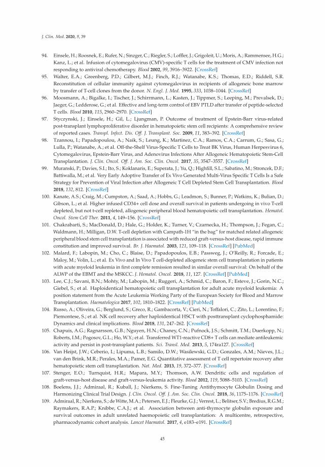
J. Clin. Med. 2020, 9, 39
94. Einsele, H.; Roosnek, E.; Rufer, N.; Sinzger, C.; Riegler, S.; Loffler, J.; Grigoleit, U.; Moris, A.; Rammensee, H.G.;Kanz, L.; et al. Infusion of cytomegalovirus (CMV)-specific T cells for the treatment of CMV infection notresponding to antiviral chemotherapy. Blood 2002, 99, 3916–3922. [CrossRef]
95. Walter, E.A.; Greenberg, P.D.; Gilbert, M.J.; Finch, R.J.; Watanabe, K.S.; Thomas, E.D.; Riddell, S.R.Reconstitution of cellular immunity against cytomegalovirus in recipients of allogeneic bone marrowby transfer of T-cell clones from the donor. N. Engl. J. Med. 1995, 333, 1038–1044. [CrossRef]
96. Moosmann, A.; Bigalke, I.; Tischer, J.; Schirrmann, L.; Kasten, J.; Tippmer, S.; Leeping, M.; Prevalsek, D.;Jaeger, G.; Ledderose, G.; et al. Effective and long-term control of EBV PTLD after transfer of peptide-selectedT cells. Blood 2010, 115, 2960–2970. [CrossRef]
97. Styczynski, J.; Einsele, H.; Gil, L.; Ljungman, P. Outcome of treatment of Epstein-Barr virus-relatedpost-transplant lymphoproliferative disorder in hematopoietic stem cell recipients: A comprehensive reviewof reported cases. Transpl. Infect. Dis. Off. J. Transplant. Soc. 2009, 11, 383–392. [CrossRef]
98. Tzannou, I.; Papadopoulou, A.; Naik, S.; Leung, K.; Martinez, C.A.; Ramos, C.A.; Carrum, G.; Sasa, G.;Lulla, P.; Watanabe, A.; et al. Off-the-Shelf Virus-Specific T Cells to Treat BK Virus, Human Herpesvirus 6,Cytomegalovirus, Epstein-Barr Virus, and Adenovirus Infections After Allogeneic Hematopoietic Stem-CellTransplantation. J. Clin. Oncol. Off. J. Am. Soc. Clin. Oncol. 2017, 35, 3547–3557. [CrossRef]
99. Muranski, P.; Davies, S.I.; Ito, S.; Koklanaris, E.; Superata, J.; Yu, Q.; Highfill, S.L.; Sabatino, M.; Stroncek, D.F.;Battiwalla, M.; et al. Very Early Adoptive Transfer of Ex Vivo Generated Multi-Virus Specific T Cells Is a SafeStrategy for Prevention of Viral Infection after Allogeneic T Cell Depleted Stem Cell Transplantation. Blood2018, 132, 812. [CrossRef]
100. Kanate, A.S.; Craig, M.; Cumpston, A.; Saad, A.; Hobbs, G.; Leadmon, S.; Bunner, P.; Watkins, K.; Bulian, D.;Gibson, L.; et al. Higher infused CD34+ cell dose and overall survival in patients undergoing in vivo T-celldepleted, but not t-cell repleted, allogeneic peripheral blood hematopoietic cell transplantation. Hematol.Oncol. Stem Cell Ther. 2011, 4, 149–156. [CrossRef]
101. Chakrabarti, S.; MacDonald, D.; Hale, G.; Holder, K.; Turner, V.; Czarnecka, H.; Thompson, J.; Fegan, C.;Waldmann, H.; Milligan, D.W. T-cell depletion with Campath-1H “in the bag” for matched related allogeneicperipheral blood stem cell transplantation is associated with reduced graft-versus-host disease, rapid immuneconstitution and improved survival. Br. J. Haematol. 2003, 121, 109–118. [CrossRef] [PubMed]
102. Malard, F.; Labopin, M.; Cho, C.; Blaise, D.; Papadopoulos, E.B.; Passweg, J.; O’Reilly, R.; Forcade, E.;Maloy, M.; Volin, L.; et al. Ex Vivo and In Vivo T cell-depleted allogeneic stem cell transplantation in patientswith acute myeloid leukemia in first complete remission resulted in similar overall survival: On behalf of theALWP of the EBMT and the MSKCC. J. Hematol. Oncol. 2018, 11, 127. [CrossRef] [PubMed]
103. Lee, C.J.; Savani, B.N.; Mohty, M.; Labopin, M.; Ruggeri, A.; Schmid, C.; Baron, F.; Esteve, J.; Gorin, N.C.;Giebel, S.; et al. Haploidentical hematopoietic cell transplantation for adult acute myeloid leukemia: Aposition statement from the Acute Leukemia Working Party of the European Society for Blood and MarrowTransplantation. Haematologica 2017, 102, 1810–1822. [CrossRef] [PubMed]
104. Russo, A.; Oliveira, G.; Berglund, S.; Greco, R.; Gambacorta, V.; Cieri, N.; Toffalori, C.; Zito, L.; Lorentino, F.;Piemontese, S.; et al. NK cell recovery after haploidentical HSCT with posttransplant cyclophosphamide:Dynamics and clinical implications. Blood 2018, 131, 247–262. [CrossRef]
105. Chapuis, A.G.; Ragnarsson, G.B.; Nguyen, H.N.; Chaney, C.N.; Pufnock, J.S.; Schmitt, T.M.; Duerkopp, N.;Roberts, I.M.; Pogosov, G.L.; Ho, W.Y.; et al. Transferred WT1-reactive CD8+ T cells can mediate antileukemicactivity and persist in post-transplant patients. Sci. Transl. Med. 2013, 5, 174ra127. [CrossRef]
106. Van Heijst, J.W.; Ceberio, I.; Lipuma, L.B.; Samilo, D.W.; Wasilewski, G.D.; Gonzales, A.M.; Nieves, J.L.;van den Brink, M.R.; Perales, M.A.; Pamer, E.G. Quantitative assessment of T cell repertoire recovery afterhematopoietic stem cell transplantation. Nat. Med. 2013, 19, 372–377. [CrossRef]
107. Stenger, E.O.; Turnquist, H.R.; Mapara, M.Y.; Thomson, A.W. Dendritic cells and regulation ofgraft-versus-host disease and graft-versus-leukemia activity. Blood 2012, 119, 5088–5103. [CrossRef]
108. Boelens, J.J.; Admiraal, R.; Kuball, J.; Nierkens, S. Fine-Tuning Antithymocyte Globulin Dosing andHarmonizing Clinical Trial Design. J. Clin. Oncol. Off. J. Am. Soc. Clin. Oncol. 2018, 36, 1175–1176. [CrossRef]
109. Admiraal, R.; Nierkens, S.; de Witte, M.A.; Petersen, E.J.; Fleurke, G.J.; Verrest, L.; Belitser, S.V.; Bredius, R.G.M.;Raymakers, R.A.P.; Knibbe, C.A.J.; et al. Association between anti-thymocyte globulin exposure andsurvival outcomes in adult unrelated haemopoietic cell transplantation: A multicentre, retrospective,pharmacodynamic cohort analysis. Lancet Haematol. 2017, 4, e183–e191. [CrossRef]
45
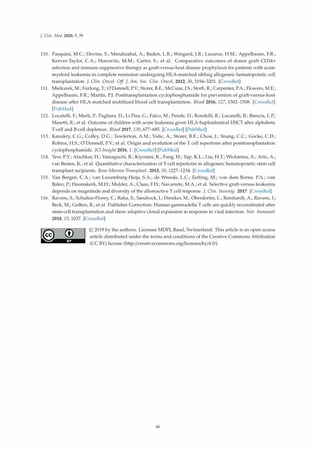
J. Clin. Med. 2020, 9, 39
110. Pasquini, M.C.; Devine, S.; Mendizabal, A.; Baden, L.R.; Wingard, J.R.; Lazarus, H.M.; Appelbaum, F.R.;Keever-Taylor, C.A.; Horowitz, M.M.; Carter, S.; et al. Comparative outcomes of donor graft CD34+selection and immune suppressive therapy as graft-versus-host disease prophylaxis for patients with acutemyeloid leukemia in complete remission undergoing HLA-matched sibling allogeneic hematopoietic celltransplantation. J. Clin. Oncol. Off. J. Am. Soc. Clin. Oncol. 2012, 30, 3194–3201. [CrossRef]
111. Mielcarek, M.; Furlong, T.; O’Donnell, P.V.; Storer, B.E.; McCune, J.S.; Storb, R.; Carpenter, P.A.; Flowers, M.E.;Appelbaum, F.R.; Martin, P.J. Posttransplantation cyclophosphamide for prevention of graft-versus-hostdisease after HLA-matched mobilized blood cell transplantation. Blood 2016, 127, 1502–1508. [CrossRef][PubMed]
112. Locatelli, F.; Merli, P.; Pagliara, D.; Li Pira, G.; Falco, M.; Pende, D.; Rondelli, R.; Lucarelli, B.; Brescia, L.P.;Masetti, R.; et al. Outcome of children with acute leukemia given HLA-haploidentical HSCT after alphabetaT-cell and B-cell depletion. Blood 2017, 130, 677–685. [CrossRef] [PubMed]
113. Kanakry, C.G.; Coffey, D.G.; Towlerton, A.M.; Vulic, A.; Storer, B.E.; Chou, J.; Yeung, C.C.; Gocke, C.D.;Robins, H.S.; O’Donnell, P.V.; et al. Origin and evolution of the T cell repertoire after posttransplantationcyclophosphamide. JCI Insight 2016, 1. [CrossRef] [PubMed]
114. Yew, P.Y.; Alachkar, H.; Yamaguchi, R.; Kiyotani, K.; Fang, H.; Yap, K.L.; Liu, H.T.; Wickrema, A.; Artz, A.;van Besien, K.; et al. Quantitative characterization of T-cell repertoire in allogeneic hematopoietic stem celltransplant recipients. Bone Marrow Transplant. 2015, 50, 1227–1234. [CrossRef]
115. Van Bergen, C.A.; van Luxemburg-Heijs, S.A.; de Wreede, L.C.; Eefting, M.; von dem Borne, P.A.; vanBalen, P.; Heemskerk, M.H.; Mulder, A.; Claas, F.H.; Navarrete, M.A.; et al. Selective graft-versus-leukemiadepends on magnitude and diversity of the alloreactive T cell response. J. Clin. Investig. 2017. [CrossRef]
116. Ravens, S.; Schultze-Florey, C.; Raha, S.; Sandrock, I.; Drenker, M.; Oberdorfer, L.; Reinhardt, A.; Ravens, I.;Beck, M.; Geffers, R.; et al. Publisher Correction: Human gammadelta T cells are quickly reconstituted afterstem-cell transplantation and show adaptive clonal expansion in response to viral infection. Nat. Immunol.2018, 19, 1037. [CrossRef]
© 2019 by the authors. Licensee MDPI, Basel, Switzerland. This article is an open accessarticle distributed under the terms and conditions of the Creative Commons Attribution(CC BY) license (http://creativecommons.org/licenses/by/4.0/).
46

Journal of
Clinical Medicine
Review
FLAMSA-RIC for Stem Cell Transplantation inPatients with Acute Myeloid Leukemia andMyelodysplastic Syndromes: A Systematic Reviewand Meta-Analysis
Weerapat Owattanapanich 1,*, Patompong Ungprasert 2, Verena Wais 3,
Smith Kungwankiattichai 1, Donald Bunjes 3 and Florian Kuchenbauer 4,5,*
1 Division of Hematology, Department of Medicine, Faculty of Medicine Siriraj Hospital, Mahidol University,Bangkok 10700, Thailand
2 Clinical Epidemiology Unit, Department of Research and Development, Faculty of Medicine Siriraj Hospital,Mahidol University, Bangkok 10700, Thailand
3 Department of Internal Medicine III, University Hospital of Ulm, 89081 Ulm, Germany4 Vancouver General Hospital, L/BMT Program of British Columbia, Vancouver, BC V5Z 1M9, Canada5 Terry Fox Laboratory, British Columbia Research Centre, Vancouver, BC V5Z 1L3, Canada* Correspondence: [email protected] (W.O.); [email protected] (F.K.)
Received: 2 July 2019; Accepted: 3 September 2019; Published: 11 September 2019���������������
Abstract: Reduced-intensity conditioning (RIC) regimens are established options for hematopoieticstem cell transplantation (HSCT) for patients with acute myeloid leukemia (AML) and myelodysplasticsyndrome (MDS). However, the efficacy of RIC regimens for patients with high-risk disease is limited.The addition of a fludarabine, amsacrine, and cytarabine (FLAMSA)-sequential conditioning regimenwas introduced for patients with high-risk MDS and AML to combine a high anti-leukemic activity withthe advantages of RIC. The current systematic literature review and meta-analysis was conductedwith the aim of identifying all cohort studies of patients with AML and/or MDS who receivedFLAMSA-RIC to determine its efficacy and toxicity. Out of 3044 retrieved articles, 12 publishedstudies with 2395 overall patients (18.1–76.0 years; 96.8% AML and 3.2% MDS; follow-up duration of0.7–145 months; 50.3% had active AML disease before HSCT) met the eligibility criteria and wereincluded in the meta-analysis. In the pooled analysis, the 1- and 3-year overall survival (OS) rateswere 59.6% (95% confidence interval (CI), 47.9–70.2%) and 40.2% (95% CI, 28.0–53.7%), respectively.The pooled 3-year OS rate of the patients who achieved CR1 or CR2 prior to HSCT was 60.1% (95%CI, 55.1–64.8%) and the percentage of those with relapse or refractory disease was 27.8% (95% CI,23.3–32.8%). The pooled 3-year leukemia-free survival (LFS) rate was 39.3% (95% CI, 26.4–53.9%).Approximately 29% of the patients suffered from grades 2–4 acute graft-versus-host disease (GVHD),while 35.6% had chronic GVHD. The pooled 1- and 3-year non-relapse mortality (NRM) rates were17.9% (95% CI, 16.1–19.8%) and 21.1% (95% CI, 18.8–23.7%), respectively. Our data indicates that theFLAMSA-RIC regimen is an effective and well-tolerated regimen for HSCT in patients with high-riskAML and MDS.
Keywords: FLAMSA; reduced-intensity; acute myeloid leukemia; myelodysplastic syndrome
1. Introduction
Reduced-intensity conditioning (RIC) regimens were initially introduced to reduce the adverseeffects associated with myeloablative conditioning (MAC) and to improve the chance of successfulhematopoietic stem cell transplantation (HSCT), especially in elderly and frail patients [1]. However,
J. Clin. Med. 2019, 8, 1437; doi:10.3390/jcm8091437 www.mdpi.com/journal/jcm47

J. Clin. Med. 2019, 8, 1437
the efficacy of RIC regimens for patients who do not achieve a complete remission (CR) is limited [2].The combination of fludarabine, amsacrine, and cytarabine (FLAMSA)-polychemotherapy with RICwas initially adopted by Schmid et al. for patients with high-risk myelodysplastic syndrome (MDS) andacute myeloid leukemia (AML) [3–5] to combine high anti-leukemic activity with the advantages of RIC.Although several variations have been published [6–16], the ‘classic’ FLAMSA-RIC regimen consistsof fludarabine, amsacrine, and cytarabine, followed by RIC with 4-Gy total body irradiation (TBI),high-dose cyclophosphamide (Cy), antithymocyte globulin (ATG), and prophylactic donor lymphocyteinfusions (DLI) if indicated. Because of initially promising data, especially in poor prognosis AMLpatients, FLAMSA-RIC was adopted by many transplantation centers, and variations that includedbusulfan (Bu) or treosulfan were established. The current systematic review and meta-analysis wasconducted with the aim of identifying all cohort studies that have investigated the efficacy and toxicityof the FLAMSA-RIC regimen and summarize their results.
2. Materials and Methods
2.1. Data Sources and Searches
Three investigators (W.O., P.U. and S.K.) independently searched for published articles indexed inMEDLINE and EMBASE databases from their inception to May 2019. The search strategy is availableas Supplementary Data 1. The references of the included studies were also manually reviewed foradditional eligible studies. This study was undertaken in accordance with the Preferred Reporting Itemsfor Systematic Reviews and Meta-Analyses (PRISMA) statement, which is available as SupplementaryData 2 [17].
2.2. Selection Criteria and Data Extraction
Studies included in this meta-analysis were cohort studies (either prospective or retrospective) ofpatients with AML and/or MDS who received FLAMSA-RIC regimens, which reported our primaryoutcomes of interest (overall survival (OS) and/or leukemic-free survival (LFS) rates). The secondaryoutcomes of interest, which included non-relapse mortality (NRM) rate, relapse rate (RR), full chimerismrate, grades 2–4 acute GVHD (aGVHD) rate, and chronic GVHD (cGVHD) rate were also collectedfor analysis but were not part of the inclusion criteria. Assessment of the eligibility of each studywas independently conducted by three investigators. In the event of opposing decisions regarding astudy’s eligibility, the study in question was reviewed by the three investigators together and the finaldetermination was reached by mutual consensus.
2.3. Definition of Treatment Response and Outcome
Complete remission (CR) was defined as bone marrow blasts of < 5%, the absence of circulatingblasts and blasts with Auer rods, the absence of an extramedullary disease, an absolute neutrophilcount (ANC) of ≥ 1.0 × 10⁹/L and a platelet count of ≥ 100 × 109/L [8]. Refractory AML was definedas failure to achieve CR following induction or salvage chemotherapy. Relapse AML was defined asrecurrence of disease after CR. Overall survival (OS) rate was defined as the percentage of patients whowere still alive at the time of interest (such as at 1 year after transplantation). Leukemia-free survival(LFS) rate was defined as the percentage of patients who were still alive and did not have leukemia atthe time of interest. Non-relapse mortality (NRM) was defined as any death without previous relapseor progression.
2.4. Statistical Analysis
All data analyses were performed using the Comprehensive Meta-Analysis program, version 2.2(Biostat, Englewood, NJ, USA). Using a standardized data extraction algorithm, two authors (W.O. andP.U.) extracted and tabulated all data from each study. The pooled rates and 95% confidence interval ofOS rate, LFS rate, NRM rate, relapse rate, full chimerism rate, aGVHD rate, and cGVHD rate were
48

J. Clin. Med. 2019, 8, 1437
calculated using the DerSimonian-Laird random-effect model with double arcsine transformation [18]. Arandom-effect model, rather than fixed-effect, was used because of the high likelihood of between-studyheterogeneity. Cochran’s Q test and I2 statistic were used to determine the between-study heterogeneity.I2 statistic quantified the proportion of total variation across studies that is due to heterogeneity ratherthan chance. An I2 value of 0% to 25% represents insignificant heterogeneity, greater than 25% butless than or equal to 50% represents low heterogeneity, greater than 50% but less than or equal to 75%represents moderate heterogeneity, and greater than 75% represents high heterogeneity [19].
3. Results
The search strategy yielded 3044 potentially relevant articles (504 articles from MEDLINE and2540 from EMBASE). After exclusion of 416 duplicated articles using the EndNote X8 software, 2628articles underwent title and abstract review. A total of 2607 articles were excluded at this stage as theydid not meet the inclusion criteria based on type of article, study design, subjects and interventionsused. A total of eighteen articles underwent full-text review and 9 of them were excluded becausethey did not report the primary outcomes of interest. Finally, 12 studies fulfilled the eligibility criteriaand were included in the meta-analysis [3,6–16]. A manual review of the bibliography of the includedstudies, and some selected review articles, did not yield any additional eligible studies. Figure 1summarizes the literature review and identification process. The main characteristics of the includedstudies are described in Table 1.
Figure 1. Flowchart of literature review process.
49

J. Clin. Med. 2019, 8, 1437
Ta
ble
1.
Base
line
pati
entc
hara
cter
isti
csof
each
incl
uded
arti
cle.
Refe
ren
ces
No
.S
ex
(M/F
)
Med
ian
Ag
e(Y
ears
,R
an
ge)
Dis
ease
sD
isease
Sta
tus
Ste
mC
ell
So
urc
eD
on
or
So
urc
eC
D34+
(×10
6
cell
s/k
g)
Stu
dy
Peri
od
Med
ian
Fo
llo
wU
p(M
on
ths,
Ran
ge)
Ty
pe
Schm
idet
al.
(200
5)[4
]75
42/3
352
.3(1
8.5–
65.8
)
50dA
ML,
15sA
ML,
10M
DS
8C
R1,
8C
R2,
49R/R
,10
MD
S
61PB
SC,
14BM
31M
RD
,30
MU
D6
MM
RD
,8M
MU
D9.
619
99–2
002
31.5
(13.
6–47
.6)
P
Saur
eet
al.(
2012
)[9
]30
20/1
049
(36–
66)
10sA
ML,
20M
DS
10un
trea
ted
AM
L,20
MD
S30
PBSC
13M
RD
,13
MU
D4
MM
UD
7.7
2003
–201
028
(7–8
1)P
Kre
jcie
tal.
(201
3)[7
]60
28/3
252
(20–
63)
50dA
ML,
10sA
ML
34C
R1,
26R/R
56PB
SC,
4BM
15M
RD
,29
MU
D,
16M
MU
D6.
320
06–2
011
37(1
0–69
)P
Schn
eida
win
det
al.(
2013
)[10
]62
34/2
855
(20–
72)
35dA
ML,
27sA
ML
62R/R
62PB
SC11
MR
D,2
2M
UD
4M
MR
D,2
5M
MU
D5.
420
05–2
012
17.5
(2.2
–77.
6)R
Bohl
etal
.(20
16)
[8]
8446/3
848
.767
dAM
L,17
sAM
L
13C
R1,
12C
R2,
59R/R
NR
NR
NR
2000
–201
2N
RR
Hol
tick
etal
.(2
016)
[6]
130
59/7
150
.9(1
9–73
)N
R47
CR
1,26
CR
2,57
R/R
127
PBSC
,3
BM42
MR
D,6
4M
UD
1M
MR
D,2
3MM
UD
7.07
2004
–201
537
(10–
125)
R
Pfre
pper
etal
.(2
016)
[11]
4425/1
952
(21–
65)
NR
44R/R
44PB
SC3
MR
D,2
7M
UD
,14
MM
UD
NR
2006
–201
334
(6–7
1)R
Rin
gden
etal
.(2
016)
[12]
267
131/
136
51.7
(19.
4–72
.5)
NR
267
R/R
256
PBSC
,11
BM77
MR
D,1
90M
UD
NR
NR
68.2
(2–1
57)
R
Mal
ard
etal
.(2
017)
[13]
265
143/
122
55(1
9–76
)
156
dAM
L,10
9sA
ML
216
CR
1,49
CR
225
1PB
SC,
14BM
74M
RD
,191
MU
DN
R20
02–2
014
46(1
–145
)R
Hei
nick
eet
al.
(201
8)[1
4]39
920
6/19
3(1
8–74
.4)
NR
305
CR
1,94
CR
237
9PB
SC,
20BM
139
MR
D,1
98M
UD
,62
MM
UD
NR
2005
–201
6(0
.7–1
21.5
)R
Shet
het
al.(
2019
)[1
5]34
817
9/16
9(4
0.1–
65)
294
dAM
L,54
sAM
L
264
CR
1,84
CR
233
0PB
SC,1
8BM
113
MR
D,1
82M
UD
,53
MM
UD
NR
2007
–201
6N
RR
Sara
ceni
etal
.(2
019)
[16]
631
336/
295
51.5
(18.
1–76
)N
R63
1R
R61
6PB
SC,1
5BM
252
MR
D,2
68M
UD
,11
1M
MU
DN
R20
05–2
016
53(4
–35)
R
Ab
bre
via
tio
ns;
BMbo
nem
arro
w;C
R1
com
plet
ere
mis
sion
afte
rfir
stin
duct
ion
ther
apy;
CR
2co
mpl
ete
rem
issi
onaf
ter
rela
pse;
dAM
Lde
novo
acut
em
yelo
idle
ukem
ia;F
Fem
ale;
MM
ale;
MD
Sm
yelo
dys
plas
tic
synd
rom
es;M
RD
mat
chre
late
dd
onor
;MM
RD
mis
mat
chre
late
dd
onor
;MM
UD
mis
mat
chun
rela
ted
don
or;M
UD
mat
chun
rela
ted
don
or;N
Rno
trep
orte
d;P
pros
pect
ivel
y;PB
SCpe
riph
eral
bloo
dst
emce
ll;R
retr
ospe
ctiv
ely;
R/R
rela
pse
and/
orre
frac
tory
dise
ases
;sA
ML
seco
ndar
yac
ute
mye
loid
leuk
emia
.
50
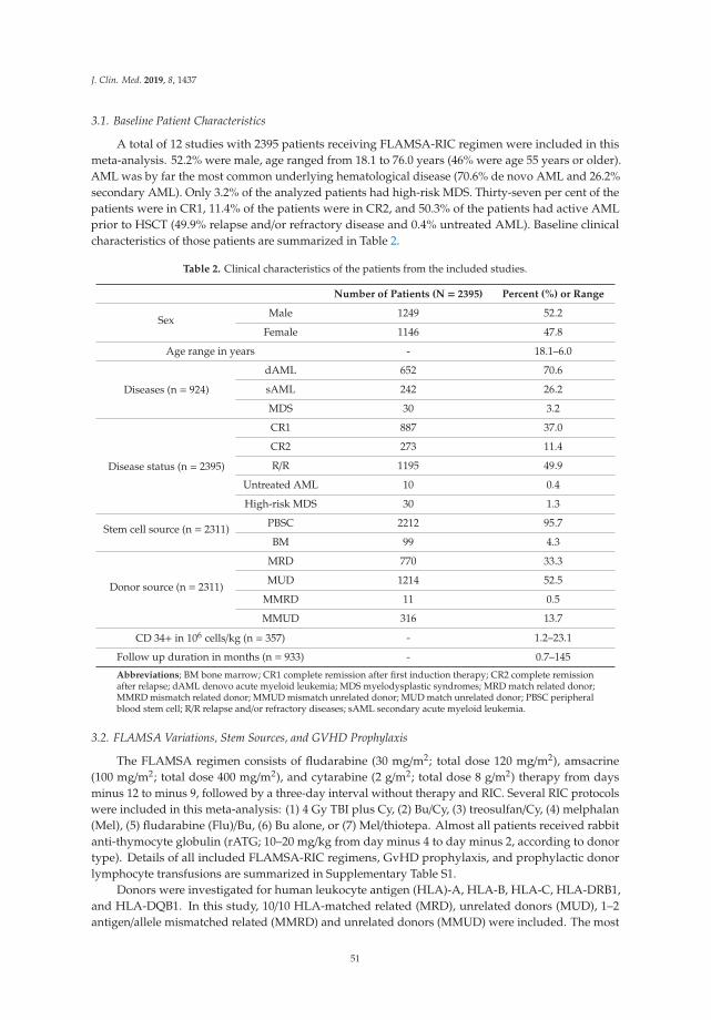
J. Clin. Med. 2019, 8, 1437
3.1. Baseline Patient Characteristics
A total of 12 studies with 2395 patients receiving FLAMSA-RIC regimen were included in thismeta-analysis. 52.2% were male, age ranged from 18.1 to 76.0 years (46% were age 55 years or older).AML was by far the most common underlying hematological disease (70.6% de novo AML and 26.2%secondary AML). Only 3.2% of the analyzed patients had high-risk MDS. Thirty-seven per cent of thepatients were in CR1, 11.4% of the patients were in CR2, and 50.3% of the patients had active AMLprior to HSCT (49.9% relapse and/or refractory disease and 0.4% untreated AML). Baseline clinicalcharacteristics of those patients are summarized in Table 2.
Table 2. Clinical characteristics of the patients from the included studies.
Number of Patients (N = 2395) Percent (%) or Range
SexMale 1249 52.2
Female 1146 47.8
Age range in years - 18.1–6.0
Diseases (n = 924)
dAML 652 70.6
sAML 242 26.2
MDS 30 3.2
Disease status (n = 2395)
CR1 887 37.0
CR2 273 11.4
R/R 1195 49.9
Untreated AML 10 0.4
High-risk MDS 30 1.3
Stem cell source (n = 2311) PBSC 2212 95.7
BM 99 4.3
Donor source (n = 2311)
MRD 770 33.3
MUD 1214 52.5
MMRD 11 0.5
MMUD 316 13.7
CD 34+ in 106 cells/kg (n = 357) - 1.2–23.1
Follow up duration in months (n = 933) - 0.7–145
Abbreviations; BM bone marrow; CR1 complete remission after first induction therapy; CR2 complete remissionafter relapse; dAML denovo acute myeloid leukemia; MDS myelodysplastic syndromes; MRD match related donor;MMRD mismatch related donor; MMUD mismatch unrelated donor; MUD match unrelated donor; PBSC peripheralblood stem cell; R/R relapse and/or refractory diseases; sAML secondary acute myeloid leukemia.
3.2. FLAMSA Variations, Stem Sources, and GVHD Prophylaxis
The FLAMSA regimen consists of fludarabine (30 mg/m2; total dose 120 mg/m2), amsacrine(100 mg/m2; total dose 400 mg/m2), and cytarabine (2 g/m2; total dose 8 g/m2) therapy from daysminus 12 to minus 9, followed by a three-day interval without therapy and RIC. Several RIC protocolswere included in this meta-analysis: (1) 4 Gy TBI plus Cy, (2) Bu/Cy, (3) treosulfan/Cy, (4) melphalan(Mel), (5) fludarabine (Flu)/Bu, (6) Bu alone, or (7) Mel/thiotepa. Almost all patients received rabbitanti-thymocyte globulin (rATG; 10–20 mg/kg from day minus 4 to day minus 2, according to donortype). Details of all included FLAMSA-RIC regimens, GvHD prophylaxis, and prophylactic donorlymphocyte transfusions are summarized in Supplementary Table S1.
Donors were investigated for human leukocyte antigen (HLA)-A, HLA-B, HLA-C, HLA-DRB1,and HLA-DQB1. In this study, 10/10 HLA-matched related (MRD), unrelated donors (MUD), 1–2antigen/allele mismatched related (MMRD) and unrelated donors (MMUD) were included. The most
51

J. Clin. Med. 2019, 8, 1437
frequent donors were MUD (52.5%), followed by MRD (33.3%), MMUD (13.7%) and MMRD (0.5%).The most frequent stem cell source (95.7%) were stem cells collected from peripheral blood. CD 34+
cell infusions ranged from 1.2 to 23.1 × 10⁶ cells/kg.GVHD prophylaxis was available for 720 patients, most of them received cyclosporine A (CyA)
plus mycophenolate mofetil (MMF; n = 573), followed by tacrolimus plus MMF (n = 112), CyA alone(n = 28), and CyA plus methotrexate (n = 7). Prophylactic donor lymphocyte transfusions were givenif patients did not show any evidence for GVHD either at day 120 or 30 days after discontinuation ofthe immunosuppression [3,7,10–12].
3.3. Survival Outcome
The follow-up period ranged from 0.7 to 145 months. The pooled 1-, 2- and 3-year OS rateswere 59.6% (95% CI, 47.9–70.2%; I2 94%; Figure 2A) [6–8,14–16], 48.4% (95% CI, 37.3–59.6%; I2
96%; Figure 2B [3,6,8,10,13–16], and 40.2% (95% CI, 28.0–53.7%; I2 96%; Figure 2C) [6,7,11,12,14–16],respectively. The pooled 1-, 2- and 3-year LFS were 57.4% (95% CI, 38.6–74.2%; I2 98%; Figure 2D) [6,14–16], 49.4% (95% CI, 38.1–60.8%; I2 95%; Figure 2E [3,6,9,13–16], and 39.3% (95% CI, 26.4–53.9%;I2 97%; Figure 2F) [6,7,12,14–16], respectively. The pooled 2- and 3-year RR were 31.3% (95% CI,21.1–43.8%; I2 96%; Figure 3A) [3,6,10,13–16] and 41.9% (95% CI, 30.9–53.8%; I2 95%; Figure 3B) [6,7,11,12,14–16], respectively.
A total of 4 studies [3,6,9,11] reported the rate of full chimerism at day +28 (defined as the presenceof > 98% of HLA belonging to the donor) and the pooled rate across the 4 studies was 82.9% (95% CI,69.7–91.1%; I2 77%) (Figure 3C).
Figure 2. Forest plots of pooled estimates (95% confidence interval (CI)) for overall survival (OS) andleukemia-free survival (LFS) after hematopoietic stem cell transplantation (HSCT); (A): 1-year OS; (B):2-year OS; (C): 3-year OS; (D): 1-year LFS; (E): 2-year LFS; (F): 3-year LFS.
52
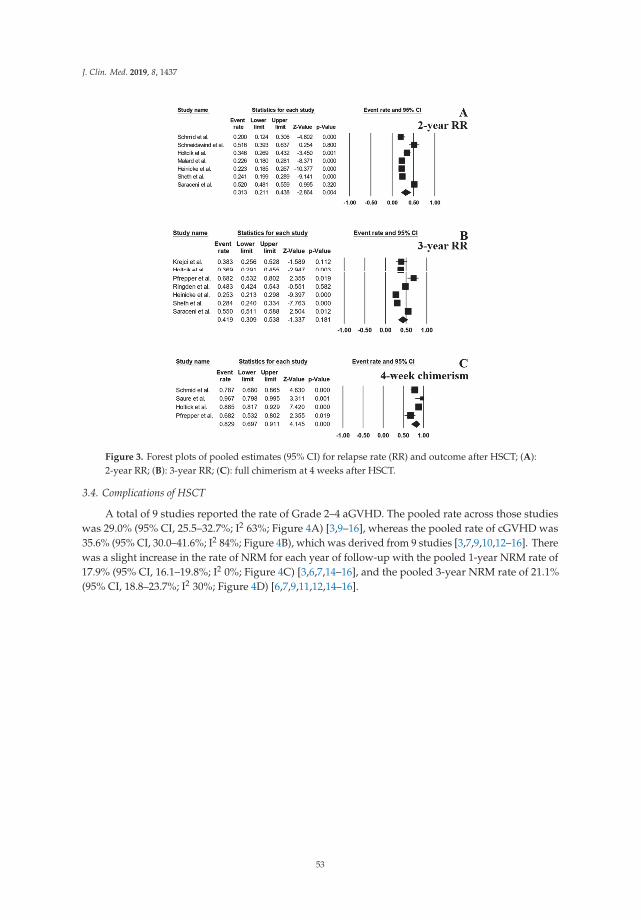
J. Clin. Med. 2019, 8, 1437
Figure 3. Forest plots of pooled estimates (95% CI) for relapse rate (RR) and outcome after HSCT; (A):2-year RR; (B): 3-year RR; (C): full chimerism at 4 weeks after HSCT.
3.4. Complications of HSCT
A total of 9 studies reported the rate of Grade 2–4 aGVHD. The pooled rate across those studieswas 29.0% (95% CI, 25.5–32.7%; I2 63%; Figure 4A) [3,9–16], whereas the pooled rate of cGVHD was35.6% (95% CI, 30.0–41.6%; I2 84%; Figure 4B), which was derived from 9 studies [3,7,9,10,12–16]. Therewas a slight increase in the rate of NRM for each year of follow-up with the pooled 1-year NRM rate of17.9% (95% CI, 16.1–19.8%; I2 0%; Figure 4C) [3,6,7,14–16], and the pooled 3-year NRM rate of 21.1%(95% CI, 18.8–23.7%; I2 30%; Figure 4D) [6,7,9,11,12,14–16].
53

J. Clin. Med. 2019, 8, 1437
Figure 4. Forest plots of pooled estimates (95% CI) for complications after HSCT; (A): Acutegraft-versus-host disease (aGVHD); (B): Chronic graft-versus-host disease (cGVHD); (C): 1-yearnon-relapse mortality (NRM); (D): 3-year NRM.
3.5. Subgroup Analysis
The pooled 3-year OS rate of the patients who achieved CR1 or CR2 prior to HSCT was 60.1%(95% CI, 55.1–64.8%; I2 48%; Figure 5A) [14,15] and 3-year LFS was 55.2% (95% CI, 51.6–58.7%; I2 0%;Figure 5B) [14,15]. As for the patients with relapse or refractory disease, the pooled 3-year OS and LFSrates were 27.8% (95% CI, 23.3–32.8%; I2 47%; Figure 5C) [11,12,16] and 23.7% (95% CI, 21.1–26.6%; I2
0%; Figure 5D) [12,16], respectively.
Figure 5. Forest plots of pooled estimates (95% CI) for outcomes of the patient’s subgroups; (A): 3-yearOS of CR1 or CR2 patients; (B): 3-year LFS of CR1 or CR2 patients; (C): 3-year OS of acute myeloidleukemia (AML) patients with relapse and/or refractory disease; (D): 3-year LFS of high risk AMLpatients with relapse and/or refractory disease.
A subgroup analysis based on the reported conditioning regimens showed that the patientsreceiving a TBI-based regimen had a pooled 3-year OS rate of 58.5% (95% CI, 47.2–68.9%; I2 82%;Figure 6A) [7,14,15] and a pooled 3-year LFS of 54.0% (95% CI, 43.6–64.1%; I2 80%; Figure 6B) [7,14,15].With regard to the patients receiving Bu-based regimens, the pooled 3-year OS rate was 52.8% (95%
54

J. Clin. Med. 2019, 8, 1437
CI, 39.8–65.3%; I2 80%; Figure 6C) [14,15] and the 3-year LFS was 48.2% (95% CI, 41.4–51.1%; I2 29%;Figure 6D) [14,15].
Figure 6. Forest plots of pooled estimates (95% CI) for outcome outcomes of the patient’s subgroups; (A):3-year OS of patients receiving total body irradiation (TBI)-based conditioning regimen; (B): 3-year LFSof patients receiving TBI-based conditioning regimen; (C): 3-year OS of patients receiving busulfan-basedconditioning regimen; (D): 3-year LFS of patients receiving busulfan-based conditioning regimen.
3.6. Sensitivity Analysis
A sensitivity analysis was performed by excluding five studies by Ringden et al. [12],Malard et al. [13], Heinicke et al. [14], Sheth et al. [15] and Saraceni et al. [16] from the pooled analysesbased on a concern over double-counting of patients. These five studies collected and reported data ofpatients who were treated at several medical centers and some centers also reported their own dataseparately (including the studies by Schmid et al. [3], Holtick et al. [6], Krejci et al. [7], Saure et al. [9],Schneidawind et al. [10], and Pfrepper et al. [11], which were included in this meta-analysis). Thepooled 3-year OS, 3-year RR, and 3-year LFS were 34.9%, 47.2% and 41.6%, respectively. The newpooled results, after exclusion of the aforementioned studies, were fairly similar to the results of theoriginal analysis as demonstrated in Supplementary Data 3.
4. Discussion
Although HSCT remains the most effective treatment option for AML, data from the NationalCancer Data Base on patients with AML aged ≥ 61 years who were diagnosed between 2003 and2012 found that only about 6% of older patients (n = 17,555) underwent HSCT [20]. RIC regimenswere subsequently introduced with the aim of reducing adverse effects and making HSCT feasiblefor elderly and fragile patients. However, RIC regimens alone may not be sufficient for patients withhigh-risk features, such as persisting disease [21]. To overcome this limitation, FLAMSA-RIC wasintroduced in 2005 and has been adopted in many countries [3]. The current study is the first systematicreview and meta-analysis to examine the efficacy and toxicity of the FLAMSA-RIC regimen for patientswith AML and MDS. Recently published trials comparing MAC vs. RIC showed controversial results.For example, in the study of Scott et al., RIC led to significantly lower TRM but higher relapse ratescompared with MAC, suggesting the use of MAC as the standard of care for fit patients with AML andMDS in CR [22]. In contrast, Bornhäuser et al. did not observe any difference between RIC and MACwith regards to relapse rate, OS and TRM for intermediate and high-risk AML patients [23]. We founda pooled 2-year OS rate of approximately 50%, which is lower than the reported results of Scott et al.(18-month OS: 76.4% MAC and 63.4% RIC) and Bornhäuser et al. (3-year OS: 61% vs. 58%), but similaror lower RR (FLAMSA: 2-year RR: 31.3%) compared to Scott et al. (18-month RR: 65.2%: MAC and45.3%: RIC) and Bornhäuser et al. (3-year RR: 28% MAC and 26% RIC). In a retrospective analysis,Eapen et al. analyzed the impact of conditioning regimes with varying intensity on outcome in 2209
55

J. Clin. Med. 2019, 8, 1437
AML and MDS patients transplanted in complete remission [24]. The authors reported a higher 3-yearRR with 46% with Flu/Bu2 and 56% with Flu/Bu2+ATG. In line with this finding, a recent retrospectiveEBMT analysis demonstrated a lower RR for FLAMSA-RIC compared to Flu/Bu2 in AML patients inCR1 and CR2 [14], suggesting that this patient group could benefit from FLAMSA-RIC. The clinicaloutcome was similar using either a TBI or Bu-based FLAMSA regimen. Considering the side-effects ofTBI, FLAMSA-Bu appears to be an effective alternative to FLAMSA-TBI.
With regard to patients with active disease in this meta-analysis, the 3-year OS and LFS rates were27.8% and 23.7%, respectively. Given, that only 46% of AML patients with induction failure respondto chemotherapy and that 52% with refractory disease die within 90 days [25], FLAMSA-RIC is areasonable treatment option for this otherwise difficult to treat patient cohort. Interestingly, Bohl et al.recently showed a correlation between tumor load and outcome for FLAMSA-RIC, underscoring therelevance of a high tumor burden as one of the strongest negative predictors for treatment outcomeafter HSCT especially in relapsed or refractory AML patients [8]. Although Goyal et al. showed thatthe exclusive presence of extramedullary disease did not impact on outcome after HSCT [26], Bohl et al.noted that that FLAMSA-RIC followed by HSCT is not effective in patients with concurrent activebone marrow and extramedullary disease [8], which led to a change of practice in our institution.
The results may also suggest that the addition of FLAMSA to RIC may help to alleviatethe aggressive behavior of the disease, which will ultimately help to improve survival outcome.Furthermore, the FLAMSA-RIC regimen did not appear to increase the risk of serious toxicity withthe comparable 1-year NRM rate to those who received RIC regimens alone (17.9% versus 28% [27],respectively), as well as the comparable rate of grade 2–4 aGVHD events (29% versus 35% [27],respectively).
The limitations of this study are mainly due to the observational nature of the included primarystudies. Varying event rates such as OS, LFS and RR among the included studies could attributedto differences in the analyzed patient cohorts and treatment protocols in each study. Furthermore,without a direct prospective head-to-head comparison, a conclusion on whether the FLAMSA-RICregimen offers a superior survival benefit compared with RIC regimens alone among patients who donot achieve CR cannot be made. Randomized controlled trials comparing the FLAMSA-RIC regimenwith RIC regimens alone are still needed. Ongoing randomized trials such as NCT01423175 andNCT00606723 comparing FLAMSA-RIC with alternative conditioning regimes will eventually proveits efficacy and toxicity as a standard conditioning protocol for high risk AML.
5. Conclusions
A FLAMSA-RIC regimen is an effective and well-tolerated regimen for HSCT in patients withAML and MDS, even among those who do not achieve complete remission.
Supplementary Materials: The following are available online at http://www.mdpi.com/2077-0383/8/9/1437/s1,Supplementary data 1: Search strategy, Supplementary data 2: The Preferred Reporting Items for SystematicReviews and Meta-Analyses statement, Supplementary data 3: Sensitivity analysis of the odds of outcomes afterHSCT; A: 3-year OS; B: 2-year relapse rate; C: 3-year relapse rate; D: 3-year LFS; E: aGVHD; F: cGVHD; G: 3-yearNRM., Table S1: FLAMSA-RIC regimen, GvHD prophylaxis, and prophylactic donor lymphocyte transfusions ineach study.
Author Contributions: All authors designed the study. W.O., P.U., S.K. designed the study, collected the data,and performed the statistical analyses. W.O. and F.K. drafted the manuscript. V.W., D.B. and F.K. made criticalrevisions to the manuscript. W.O. and F.K. revised the final manuscript. All authors read and approved thefinal manuscript.
Funding: This research received no external funding.
Conflicts of Interest: The authors declare no conflict of interest.
56

J. Clin. Med. 2019, 8, 1437
Abbreviation
Abbreviation Full Name
aGVHD acute graft-versus-host disease;AML acute myeloid leukemiaBu busulfanCI confidence intervalcGVHD chronic graft-versus-host diseaseCR complete remissionCy cyclophosphamideCyA cyclosporine AFlu fludarabineGVHD graft-versus-host diseaseHLA human leukocyte antigenHSCT hematopoietic stem cell transplantationLFS leukemia-free survivalMAC myeloablative conditioningMDS MMUD mismatched unrelated donorsMRD matched related donorMUD matched unrelated donorsNRM non-relapse mortalityOS overall survivalrATG rabbit anti-thymocyte globulinRIC reduced-intensity conditioningRR relapse rateTBI total body irradiation
References
1. Slavin, S.; Nagler, A.; Naparstek, E.; Kapelushnik, Y.; Aker, M.; Cividalli, G.; Varadi, G.; Kirschbaum, M.;Ackerstein, A.; Samuel, S.; et al. Non-MA stem cell transplantation and cell therapy as an alternative toconventional bone marrow transplantation with lethal cytoreduction for the treatment of malignant andnonmalignant hematologic diseases. Blood 1998, 91, 756–763. [PubMed]
2. Wais, V.; Kundgen, L.; Bohl, S.R.; von Harsdorf, S.; Schlenk, R.F.; Dohner, K.; Teleanu, V.; Bullinger, L.;Nguyen, T.M.; Drognitz, K.; et al. Reduced-toxicity conditioning for allogeneic hematopoietic celltransplantation in elderly or comorbid patients with AML using fludarabine, BCNU and melphalan:Disease stage at transplant determines outcome. Bone Marrow Transplant. 2018, 53, 94–96. [CrossRef][PubMed]
3. Schmid, C.; Schleuning, M.; Ledderose, G.; Tischer, J.; Kolb, H.-J. Sequential Regimen of Chemotherapy,Reduced-Intensity Conditioning for Allogeneic Stem-Cell Transplantation, and Prophylactic DonorLymphocyte Transfusion in High-Risk Acute Myeloid Leukemia and Myelodysplastic Syndrome.J. Clin. Oncol. 2005, 23, 5675–5687. [CrossRef] [PubMed]
4. Schmid, C.; Schleuning, M.; Schwerdtfeger, R.; Hertenstein, B.; Mischak-Weissinger, E.; Bunjes, D.;Harsdorf, S.V.; Scheid, C.; Holtick, U.; Greinix, H.; et al. Long-term survival in refractory acute myeloidleukemia after sequential treatment with chemotherapy and reduced-intensity conditioning for allogeneicstem cell transplantation. Blood 2006, 108, 1092–1099. [CrossRef] [PubMed]
5. Schmid, C.; Schleuning, M.; Hentrich, M.; Markl, G.E.; Gerbitz, A.; Tischer, J.; Ledderose, G.; Oruzio, D.;Hiddemann, W.; Kolb, H.-J. High antileukemic efficacy of an intermediate intensity conditioning regimenfor allogeneic stem cell transplantation in patients with high-risk acute myeloid leukemia in first completeremission. Bone Marrow Transplant. 2008, 41, 721–727. [CrossRef] [PubMed]
6. Holtick, U.; Shimabukuro-Vornhagen, A.; Chakupurakal, G.; Theurich, S.; Leitzke, S.; Burst, A.; Hallek, M.;von Bergwelt-Baildon, M.; Scheid, C.; Chemnitz, J.M. FLAMSA reduced-intensity conditioning is equallyeffective in AML patients with primary induction failure as well as in first or second complete remission.Eur. J. Haematol. 2016, 96, 475–482. [CrossRef] [PubMed]
57

J. Clin. Med. 2019, 8, 1437
7. Krejci, M.; Doubek, M.; Dušek, J.; Brychtova, Y.; Racil, Z.; Navrátil, M.; Tomiska, M.; Horky, O.; Pospisilova, S.;Mayer, J. Combination of fludarabine, amsacrine, and cytarabine followed by reduced-intensity conditioningand allogeneic hematopoietic stem cell transplantation in patients with high-risk acute myeloid leukemia.Ann. Hematol. 2013, 92, 1397–1403. [CrossRef] [PubMed]
8. Bohl, S.; Von Harsdorf, S.; Mulaw, M.; Hofmann, S.; Babiak, A.; Maier, C.P.; Schnell, J.; Hütter-Krönke, L.-M.;Scholl, K.; Wais, V.; et al. Strong impact of extramedullary involvement in high-risk AML patients withactive disease receiving the FLAMSA conditioning regimen for HSCT. Bone Marrow Transplant. 2016, 51,994–996. [CrossRef] [PubMed]
9. Saure, C.; Schroeder, T.; Zohren, F.; Groten, A.; Bruns, I.; Czibere, A.; Galonska, L.; Kondakci, M.;Weigelt, C.; Fenk, R.; et al. Upfront Allogeneic Blood Stem Cell Transplantation for Patients with High-RiskMyelodysplastic Syndrome or Secondary Acute Myeloid Leukemia Using a FLAMSA-Based High-DoseSequential Conditioning Regimen. Biol. Blood Marrow Transplant. 2012, 18, 466–472. [CrossRef]
10. Schneidawind, D.; Federmann, B.; Faul, C.; Vogel, W.; Kanz, L.; Bethge, W.A. Allogeneic hematopoietic celltransplantation with reduced-intensity conditioning following FLAMSA for primary refractory or relapsedacute myeloid leukemia. Ann. Hematol. 2013, 92, 1389–1395. [CrossRef]
11. Pfrepper, C.; Klink, A.; Behre, G.; Schenk, T.; Franke, G.N.; Jentzsch, M.; Schwind, S.; Al-Ali, H.K.;Hochhaus, A.; Niederwieser, D.; et al. Risk factors for outcome in refractory acute myeloid leukemia patientstreated with a combination of fludarabine, cytarabine, and amsacrine followed by a reduced-intensityconditioning and allogeneic stem cell transplantation. J. Cancer Res. Clin. Oncol. 2016, 142, 317–324.[CrossRef] [PubMed]
12. Ringdén, O.; Labopin, M.; Schmid, C.; Sadeghi, B.; Polge, E.; Tischer, J.; Ganser, A.; Michallet, M.; Kanz, L.;Schwerdtfeger, R.; et al. Sequential chemotherapy followed by reduced-intensity conditioning and allogeneichaematopoietic stem cell transplantation in adult patients with relapse or refractory acute myeloid leukaemia:A survey from the Acute Leukaemia Working Party of EBMT. Br. J. Haematol. 2017, 176, 431–439. [CrossRef][PubMed]
13. Malard, F.; Labopin, M.; Stuhler, G.; Bittenbring, J.; Ganser, A.; Tischer, J.; Michallet, M.; Kröger, N.;Schmid, C.; Huynh, A.; et al. Sequential Intensified Conditioning Regimen Allogeneic Hematopoietic StemCell Transplantation in Adult Patients with Intermediate- or High-Risk Acute Myeloid Leukemia in CompleteRemission: A Study from the Acute Leukemia Working Party of the European Group for Blood and MarrowTransplantation. Biol. Blood Marrow Transplant. 2017, 23, 278–284. [PubMed]
14. Heinicke, T.; Labopin, M.; Schmid, C.; Polge, E.; Socié, G.; Blaise, D.; Mufti, G.J.; Huynh, A.; Brecht, A.;LeDoux, M.-P.; et al. Reduced Relapse Incidence with FLAMSA–RIC Compared with Busulfan/Fludarabinefor Acute Myelogenous Leukemia Patients in First or Second Complete Remission: A Study from theAcute Leukemia Working Party of the European Society for Blood and Marrow Transplantation. Biol. BloodMarrow Transplant. 2018, 24, 2224–2232. [PubMed]
15. Sheth, V.; Labopin, M.; Canaani, J.; Volin, L.; Brecht, A.; Ganser, A.; Mayer, J.; Labussière-Wallet, H.;Bittenbring, J.; Shouval, R.; et al. Comparison of FLAMSA-based reduced intensity conditioning withtreosulfan/fludarabine conditioning for patients with acute myeloid leukemia: An ALWP/EBMT analysis.Bone Marrow Transplant. 2019, 54, 531–539. [CrossRef] [PubMed]
16. Saraceni, F.; Labopin, M.; Brecht, A.; Kröger, N.; Eder, M.; Tischer, J.; Labussière-Wallet, H.; Einsele, H.;Beelen, D.; Bunjes, D.; et al. Fludarabine-treosulfan compared to thiotepa-busulfan-fludarabine or FLAMSAas conditioning regimen for patients with primary refractory or relapsed acute myeloid leukemia: A studyfrom the Acute Leukemia Working Party of the European Society for Blood and Marrow Transplantation(EBMT). J. Hematol. Oncol. 2019, 12, 44.
17. Moher, D.; Liberati, A.; Tetzlaff, J.; Altman, D.G. Preferred Reporting Items for Systematic Reviews andMeta-Analyses: The PRISMA Statement. J. Clin. Epidemiol. 2009, 62, 1006–1012. [CrossRef]
18. Mathes, T.; Kuss, O. A comparison of methods for meta-analysis of a small number of studies with binaryoutcomes. Res. Synth. Methods 2018, 9, 366–381. [CrossRef]
19. Higgins, J.P.T.; Thompson, S.G.; Deeks, J.J.; Altman, D.G. Measuring inconsistency in meta-analyses. BMJ2003, 327, 557–560. [CrossRef]
20. Bhatt, V.R.; Chen, B.; Gyawali, B.; Lee, S.J. Socioeconomic and health system factors associated with lowerutilization of hematopoietic cell transplantation in older patients with acute myeloid leukemia. Bone MarrowTransplant. 2018, 53, 1288–1294. [CrossRef]
58
J. Clin. Med. 2019, 8, 1437
21. De Lima, M.; Anagnostopoulos, A.; Munsell, M.; Shahjahan, M.; Ueno, N.; Ippoliti, C.; Andersson, B.S.;Gajewski, J.; Couriel, D.; Cortes, J.; et al. Nonablative versus reduced-intensity conditioning regimens inthe treatment of acute myeloid leukemia and high-risk myelodysplastic syndrome: Dose is relevant forlong-term disease control after allogeneic hematopoietic stem cell transplantation. Blood 2004, 104, 865–872.[CrossRef] [PubMed]
22. Scott, B.L.; Pasquini, M.C.; Logan, B.R.; Wu, J.; Devine, S.M.; Porter, D.L.; Maziarz, R.T.; Warlick, E.D.;Fernandez, H.F.; Alyea, E.P.; et al. Myeloablative Versus Reduced-Intensity Hematopoietic CellTransplantation for Acute Myeloid Leukemia and Myelodysplastic Syndromes. J. Clin. Oncol. 2017,35, 1154–1161. [CrossRef] [PubMed]
23. Bornhäuser, M.; Kienast, J.; Trenschel, R.; Burchert, A.; Hegenbart, U.; Stadler, M.; Baurmann, H.;Schäfer-Eckart, K.; Holler, E.; Kröger, N.; et al. Reduced-intensity conditioning versus standard conditioningbefore allogeneic haemopoietic cell transplantation in patients with acute myeloid leukaemia in first completeremission: A prospective, open-label randomised phase 3 trial. Lancet Oncol. 2012, 13, 1035–1044. [CrossRef]
24. Eapen, M.; Brazauskas, R.; Hemmer, M.; Perez, W.S.; Steinert, P.; Horowitz, M.M.; Deeg, H.J. Hematopoieticcell transplant for acute myeloid leukemia and myelodysplastic syndrome: Conditioning regimen intensity.Blood Adv. 2018, 2, 2095–2103. [CrossRef] [PubMed]
25. Wattad, M.; Amlsg, F.T.G.-A.; Weber, D.; Döhner, K.; Krauter, J.; Gaidzik, V.I.; Paschka, P.; Heuser, M.; Thol, F.;Kindler, T.; et al. Impact of salvage regimens on response and overall survival in acute myeloid leukemiawith induction failure. Leukemia 2017, 31, 1306–1313. [CrossRef] [PubMed]
26. Goyal, S.D.; Zhang, M.J.; Wang, H.L.; Akpek, G.; Copelan, E.A.; Freytes, C.; Gale, R.P.; Hamadani, M.;Inamoto, Y.; Kamble, R.T.; et al. Allogeneic hematopoietic cell transplant for AML: No impact of pre-transplantextramedullary disease on outcome. Bone Marrow Transplant. 2015, 50, 1057–1062. [CrossRef] [PubMed]
27. Zhang, Z.-H.; Lian, X.-Y.; Yao, D.-M.; He, P.-F.; Ma, J.-C.; Xu, Z.-J.; Guo, H.; Zhang, W.; Lin, J.; Qian, J. Reducedintensity conditioning of allogeneic hematopoietic stem cell transplantation for myelodysplastic syndromeand acute myeloid leukemia in patients older than 50 years of age: A systematic review and meta-analysis.J. Cancer Res. Clin. Oncol. 2017, 143, 1853–1864. [CrossRef] [PubMed]
© 2019 by the authors. Licensee MDPI, Basel, Switzerland. This article is an open accessarticle distributed under the terms and conditions of the Creative Commons Attribution(CC BY) license (http://creativecommons.org/licenses/by/4.0/).
59


Journal of
Clinical Medicine
Review
Antibody Therapies for Acute Myeloid Leukemia:Unconjugated, Toxin-Conjugated, Radio-Conjugatedand Multivalent Formats
Brent A. Williams 1,*, Arjun Law 2, Judit Hunyadkurti 3, Stephanie Desilets 4,
Jeffrey V. Leyton 3,5,6 and Armand Keating 1
1 Cell Therapy Program, Princess Margaret Cancer Centre, Toronto, ON M5G 2C1, Canada2 Hans Messner Allogeneic Blood and Marrow Transplant Program, Princess Margaret Cancer Centre,
Toronto, ON M5G 2C1, Canada3 Département de Medécine Nucléaire et Radiobiology, Faculté de Medécine et des Sciences de la Santé,
Centre Hospitalier Universitaire de Sherbrooke (CHUS), Université de Sherbrooke,Sherbrooke, QC J1H 5N4, Canada
4 Service de Hemato-Oncologie, CHUS, Sherbrooke, QC J1H 5N4, Canada5 Sherbrooke Molecular Imaging Centre, Centre de Recherche du CHUS, Sherbrooke, QC J1H 5N4, Canada6 Institute de Pharmacologie de Sherbrooke, Université de Sherbrooke, Sherbrooke, QC J1H 5N4, Canada* Correspondence: [email protected]
Received: 1 July 2019; Accepted: 16 August 2019; Published: 20 August 2019���������������
Abstract: In recent decades, therapy for acute myeloid leukemia (AML) has remained relativelyunchanged, with chemotherapy regimens primarily consisting of an induction regimen based ona daunorubicin and cytarabine backbone, followed by consolidation chemotherapy. Patients whoare relapsed or refractory can be treated with allogeneic hematopoietic stem-cell transplantationwith modest benefits to event-free and overall survival. Other modalities of immunotherapy includeantibody therapies, which hold considerable promise and can be categorized into unconjugatedclassical antibodies, multivalent recombinant antibodies (bi-, tri- and quad-specific), toxin-conjugatedantibodies and radio-conjugated antibodies. While unconjugated antibodies can facilitate NaturalKiller (NK) cell antibody-dependent cell-mediated cytotoxicity (ADCC), bi- and tri-specific antibodiescan engage either NK cells or T-cells to redirect cytotoxicity against AML targets in a highly efficientmanner, similarly to classic ADCC. Finally, toxin-conjugated and radio-conjugated antibodies canincrease the potency of antibody therapies. Several AML tumour-associated antigens are at theforefront of targeted therapy development, which include CD33, CD123, CD13, CLL-1 and CD38and which may be present on both AML blasts and leukemic stem cells. This review focused onantibody therapies for AML, including pre-clinical studies of these agents and those that are eitherentering or have been tested in early phase clinical trials. Antibodies for checkpoint inhibition andmicroenvironment targeting in AML were excluded from this review.
Keywords: acute myeloid leukemia; AML; antibody; bi-specific antibody; therapy
1. Introduction
The discovery of a means to generate murine monoclonal antibodies by George Köhler andCésar Milstein garnered the 1984 Nobel Prize in Medicine and paved the way for a new class oftherapeutics [1]. Monoclonal antibodies (mAbs) have transformed therapy for numerous diseases,including cancer. Rituximab (anti-CD20 chimeric antibody) was the first monoclonal antibody approvedfor use in cancer and tested experimentally in a clinical trial for lymphoma in 1998 [2]. The approachto the antibody therapy of cancer has developed rapidly, leading to several general therapeutic
J. Clin. Med. 2019, 8, 1261; doi:10.3390/jcm8081261 www.mdpi.com/journal/jcm61
J. Clin. Med. 2019, 8, 1261
approaches: (1) unconjugated classical antibodies (e.g., rituximab/Rituxan), (2) toxin-conjugatedantibodies (e.g., gemtuzumab ozogamicin (GO)/Mylotarg), (3) bi- and tri-specific recombinantantibodies (e.g., blinatumomab/Blincyto), and 4) radio-conjugated antibodies (e.g., 131I-BC8/Iomab-B).Acute myeloid leukemia (AML) represents a challenging malignancy to treat, particularly in thesituation of relapsed and refractory AML (R/R AML), and antibody therapeutics have not, in general,become a standard of care for most patients. In this review, we will discuss basic aspects of AMLbiology, which inform the strategies that have been used in developing targeted antibody therapies tocomplement, enhance or replace existing standards of care. We highlighted four major approaches toantibody therapy, emphasizing mechanisms of cytotoxicity and data from both pre-clinical and clinicalstudies of specific agents. We will not cover checkpoint inhibitors that facilitate T-cell anti-tumourresponses (e.g., ipilimumab/Yervoy; anti-CTLA-4), or similar approaches to facilitate macrophageanti-tumour responses (i.e., Hu5F9-G4; anti-CD47), or antibodies not directed at cancer cells specifically,such as those that target stroma (BMS-936564/MDX1338/ulocuplumab; anti-CXCR4).
1.1. Current Standard of Care for AML
The core of most AML chemotherapy regimens consists of continuously infused cytarabine withanthracyclines such as daunorubicin [3]. The most frequently used regimen, referred to as “3 + 7”,consists of continuous infusion of cytarabine at 100 mg/m2 for seven days and rapid intravenousinjection of daunorubicin on the first three days of the treatment cycle [4]. Another subsequent trialshowed improved complete response (CR) rates, with 45 mg/m2 of daunorubicin over lower doses,particularly in younger patients [5]. Dose intensification of cytarabine and daunorubicin at 45 or90 mg/m2 for 3 days was assessed in a large prospective trial showing that adverse events were similarin both arms and a significantly higher complete remission rate was achieved with higher dosing ofdaunorubicin (67.6% vs. 57.2%). However, in this study, OS was only significantly improved in patientswith favorable or intermediate risk cytogenetics [6]. Further studies also confirmed the improvedefficacy of high dose daunorubicin (90 mg/m2), including subgroup analysis that showed superioroutcomes in patients with mutations in DNMT3A, NPM1, and MLL [7–9]. The UK National CancerResearch Institute AML17 trial compared an intermediate 60 mg/m2 dose to 90 mg/m2 and foundno differences in CR rate but a higher dose arm was associated with greater mortality at day 60 [10].Current recommendations suggest that daunorubicin doses should be ≥60 mg/m2 in all cases [11].
Several attempts have been made to improve upon the success of induction chemotherapy byadding other agents, but none have shown significant improvements in outcomes without increasingtoxicity. The topoisomerase II inhibitor, etoposide, has single agent activity against AML and hasbeen incorporated into induction or consolidation protocols depending on the risk category, age,and cardiac status of the patient [12]. However, there are no data to suggest that adding etoposideor 6-thioguanine to 3 + 7 improves outcomes [13,14]. More intense combination regimens, such asFLAG-Ida (Fludarabine, Cytarabine, Idarubicin, and Filgrastim), have higher rates of CR but areassociated with increased toxicity, resulting in no improvement in overall survival [15,16].
A risk-adapted approach may be beneficial in certain situations. AML with FLT3-ITD mutationsmay benefit from the addition of targeted therapy with tyrosine kinase inhibitors (TKIs) likemidostaurin [17]. Similarly, the addition of GO has improved outcomes in patients with favourable riskof AML with core binding factor mutations [18]. More recently, a specialized liposomal formulationcontaining cytarabine and daunorubicin (CPX-351/Vyxeos) was evaluated in patients with R/R-AML,and later, in patients with de novo high-risk or secondary AML, with improvements in survival inboth settings [19–21]. This drug is now approved for use in therapy-related AML and AML withmyelodysplasia-related changes [22,23].
Furthermore, patients unable to tolerate standard chemotherapy have benefited from venetoclax,an oral B-cell leukemia/lymphoma-2 (BCL-2) inhibitor with a 19% overall response rate and anadditional 19% demonstrating partial anti-leukemic activity [24]. A subsequent phase 1b study ofvenetoclax in patients ≥65 years of age with treatment-naive AML ineligible for standard chemotherapy
62

J. Clin. Med. 2019, 8, 1261
who received oral venetoclax in combination with the hypomethylating agents decitabine or azacytidineresulted in a 67% CR or Cri, maintained with a median duration of 11.3 months [25].
Virtually all patients who achieve CR relapse without post-remission therapy [26]. Consolidationchemotherapy with high-dose cytarabine (HiDAC) was found to be effective in preventing relapse in upto 44% of patients who did not receive allogeneic hematopoietic stem-cell transplantation (HSCT) [27].While there are significant variations in doses between centers, a dose of 3 g/m2 administered ondays 1, 3, and 5 of each course was considered optimal [28]. Finally, it has been well established thatallogeneic HSCT is the post-remission treatment of choice in eligible patients with intermediate orhigh-risk AML [29].
However, despite these therapies, five-year survival for AML patients was approximately 40% inadults [30] and 60% in children [31], with high-risk groups faring much worse (<10%) [32]. Survivaloutcomes remained particularly poor for patients over the age of 60 [33]. To improve outcomes,novel therapeutics are needed and antibody-based therapeutics have the potential to integrate withcurrently used standard-of-care regimens. However, selecting the right antibody-based treatmentstrategy in combination with complementary AML biological aspects, such as target antigens andleukemogenic stem cells, is key to providing patients with long-term leukemic-free survival.
1.2. AML Cell Surface Antigens
AML cells typically express antigens found on normal myeloid progenitor and differentiatedcells, such as macrophages and monocytes, with aberrant expression of other lineage markers. AMLexpresses the pan-leukocyte marker CD45 and other myeloid markers such as CD11b, CD13 and CD33.A review of 106 adult AML cases was conducted to assess immunophenotypic variation based onthe French-American-British (FAB) classification using a 22-antibody panel [34]. The most commonlyexpressed antigens were CD45 (97.2%), CD33 (95.3%), and CD13 (94.3%). Lymphoid-associatedantigens were expressed in approximately half of cases, with the following descending order offrequency: CD20 (17%), CD7 (16%), CD19 (9.8%), CD2 (7.5%), CD3 (6.7%), CD5 (4.8%), and CD10(2.9%). CD56, typically found on NK cells, can also be found on AML cells, but not on normal myeloidcells. CD56 expression in t(8;21) AML was associated with a higher rate of relapse [35]. These markersprovide potential therapeutic targets to exploit.
1.3. Cancer Stem Cell Hypothesis and Optimal Antigen Targets in AML
Normal hematopoietic stem cells were first discovered by Till and McCulloch [36], which led themto explore the underlying process that determined cell fate decision to self-renew or differentiate basedon stochastic or deterministic models. Ultimately, hematopoietic stem cells (HSCs) were identified asbeing enriched in the CD34 + CD38- fraction of bone marrow cells and cord blood [37] and capableof recapitulating an entire hematopoietic system in both animal and human models. The concept ofa cancer stem cell in leukemia was proposed by Bruce et al. [38] and provided an alternative to thestochastic model to explain rare tumour-initiating cells. One additional feature of this theory was thatcancer cells could differentiate along a hierarchy somewhat analogous to normal HSCs. Leukemiastem cells (LSCs) in AML were the first identified cancer stem-cell population, which were shown to beenriched in the CD34 + CD38- fraction of whole blasts, as measured by the ability to engraft in thebone marrow of immunodeficient mice [39,40]. Most subsequent studies of leukemic stem cells (LSCs)have focused on the CD34 + CD38- definition with or without addition of CD123 or CLL-1 [41–43],and these have informed the majority of therapeutic strategies to date that target LSCs. Other markersthat are expressed on LSCs and have utility in further discriminating them from HSCs include CD7,CD32, CD45RA, CD96, CD99, CD157, CD244, IL-1 receptor accessory protein (IL-1RAP) [44], and T cellimmunoglobulin mucin-3 (TIM-3), which are discussed briefly below. However, some of these antigensare expressed on normal tissues and might pose challenges to target therapeutically. CD7 and CD96were both found on T and NK cells populations [45,46], CD32 on myeloid cells and B-cells [47],CD45RA on naïve T-cells [48], CD99 on all leukocytes [49], CD244 on NK and T cells [50], CD157 on
63
J. Clin. Med. 2019, 8, 1261
all myeloid progenitors, neutrophils and macrophage [51], IL-1RAP on neutrophils, monocytes andlymphocytes [52] and TIM-3 on T, NK, macrophage and dendritic cells [53]. CD96 has been shown tobe expressed on the majority of AML LSCs, with minimal expression on HSCs making it a potentialtarget [54]. TIM-3 is expressed on LSCs in most types of AML, but is absent on HSCs and can betherapeutically targeted in AML xenograft models with TIM-3 specific antibodies [55]. CD157 has beenshown to be present on 97% of primary AML blasts, with some expression on LSC sub-populationsand a novel humanized antibody has been generated with in vitro activity in combination with NKcells [56]. CD244 has been implicated in maintaining and driving LSCs and represents an emergingtarget [57].
Finally, several lines of evidence support the clinical relevance of what is commonly referredto in the field as the leukemic stem cell (LSC) hypothesis: (1) a higher engraftment capacity in thebone marrow of murine recipients correlates with worse survival [58], (2) patients whose whole AMLsamples had a gene expression profile similar to LSCs or HSCs had worse survival and could riskstratify patients independent of known prognostic factors [59], and (3) patients with a high burden ofCD34 + CD38- AML cells at diagnosis correlated with poor survival [41,42]. These findings support thenotion that an optimal target would include a LSC population in the whole cell leukemic population.In this review, we focused primarily on LSC- and leukemic blast-associated cell-surface antigens forthe various targeting strategies, as these are optimal targets for achieving curative therapy.
1.4. Optimal Targets in AML Therapy (CD33, CD123, CD13, CLL-1 and CD38)
CD33 is a 67 kDa immunoglobulin superfamily glycoprotein that is classified as a sialic acid-bindingimmunoglobulin-like lectin (Siglec) which has two immunoreceptor tyrosine-based inhibitor motifs(ITIMs). Phosphorylation events lead to distal activation of SHP1 and SHP2. CD33 appears on earlymyelomonocytic lineage-committed cells in normal hematopoiesis. It is expressed on 99% of AMLblasts and LSCs.
CD123 is the interleukin (IL)-3 receptor alpha chain and is a type I transmembrane glycoprotein [60].The beta chain (CDw131) for the IL-3 receptor is common to the IL-5 and granulocyte monocyte-colonystimulating factor (GM-CSF) receptors [61]. When CD123 is coupled to CDw131, the binding infinityfor IL-3 increases dramatically, facilitating signal transduction from low concentrations of IL-3 [62].IL-3 is important in driving myeloid differentiation and can activate STAT5. CD123 is present on~99% of CD34 + CD38- LSCs [63] and on the majority of leukemic blasts. Importantly, CD123 is nothighly expressed on normal hematopoietic stem cells [64], making it a potential therapeutic target.CD123 is expressed on committed hematopoietic progenitor cells and mediates differentiation andproliferation. CD123 is also expressed on cells of the hematopoietic system (monocytes, neutrophils,basophils, eosinophils, megakaryocytes and erythroid precursors, mast cells, macrophages, some Blymphocytes) and non-hematopoietic tissue (Leyding cells of the testis, placenta, and brain) [60].
CD13 is a zinc-dependent metalloprotease with enzymatic activity of N-terminal amino acidcleavage from peptides [65]. It is present on normal myeloid cells and is involved in several cellularfunctions, including adhesion, differentiation, proliferation, apoptosis, and phagocytosis, and isoverexpressed on AML cells [66]. It is expressed on both blasts and LSCs. Antibodies which can bindto CD13 can not only facilitate ADCC, but also inhibit proliferation and trigger apoptosis of AML cells,but not against normal CD13-expressing blood cells [67].
C-type lectin-like receptor 1 (CLL-1) belongs to group V of the C-type lectin-like receptor family,which is calcium independent [68,69]. Based on the structure, C-type lectin and C-type-like lectinreceptors are categorized into type I and type II receptor. CLL-1 is not expressed on HSCs, but isexpressed on myeloid committed progenitors and on differentiated myeloid cells, such as peripheralblood monocytes, dendritic cells, and granulocytes [68]. However, it is expressed on LSCs [70], makingit a potentially relevant target for antibody therapeutics.
CD38 is made up of a single chain of 300 amino acids with a molecular weight of 45 kDa and isexpressed by hematopoietic and non-hematopoietic cells, including NK cells and monocytes (reviewed
64

J. Clin. Med. 2019, 8, 1261
in [71]). Other CD38+ cells include smooth and striated muscle cells, renal tubules, retinal ganglioncells, and cornea (reviewed in [72]). CD38 is involved in lymphocyte signal transduction [73] andadhesion [74]. The binding of CD38 to the ligand CD31 (PECAM-1) initiates a signaling cascadethat includes the phosphorylation of sequential intracellular targets and increases cytoplasmic Ca2+
levels, mediating different biological events, depending on the cell type (e.g., activation, proliferation,apoptosis, cytokine secretion and homing). While the absence of CD38 was used to establish classic LSCdefinitions [39], subsequent work demonstrated that LSCs can exist in the CD34+CD38+ compartmentof a significant number of primary AML samples [75]. Interestingly, CD38, a well-studied target formultiple myeloma [76], has been less focused on as a target for AML. This target is of potential interestfor AML, given that 75% of AML samples express CD38 and there are anti-CD38 mAbs approved formultiple myeloma with established safety profiles.
1.5. Additional Targets in AML (WT-1, CD15, CD25, CD30, CD45)
Several other AML target candidates that have greater limitations than the previously listedantigens have been studied and are briefly summarized here. WT1 is a zinc finger transcription factorand an oncogene in AML progression and detectable in the majority of AML samples [77], but is anintracellular protein not expressed on the cell surface. However, mutations can be detected by T-cellsthrough the presentation of mutant peptides in the context of HLA class I. This antigen presentation hasbeen exploited by a novel BiTE construct specific for CD3 and a WT1 epitope presented in the contextof HLA-A*02:01 [78]. However, this approach is limited compared with targeting tumour-associatedantigens (TAAs) and may be complicated by cross-reactivity with other antigens presented with thisHLA subtype [79]. CD15 is another antigen expressed on AML cells, but is only expressed on AMLblasts and not LSCs, and is also present on many normal myeloid cells, making it a suboptimal target.CD25 (IL-2R alpha chain), another target selected by some investigators, is present on blasts and LSCs,but is not as widely expressed as other AML antigens, and is present on other IL-2-dependent celltypes such as regulatory T-cells (Tregs), which could lead to undesirable side-effects.
CD45 is a type 1 transmembrane glycoprotein present on all hematopoietic cells [80]. It hastyrosine phosphatase activity and is involved in signal transduction with several isoforms (E3_8,CD45RO, CD45RB, CD45 RABC) derived from alternative splicing [81,82]. While CD45 is not an idealtarget, as it is expressed on all hematopoietic cells, it can be used for myeloablative conditioning priorto HSCT, which is covered in the section on radio-immunotherapy.
2. Unconjugated Antibody Therapies
Unconjugated antibody therapy relies on important structural regions, each of which have aspecific function. The basic structure of a mAb involves two identical heavy chains each with variableregions (VH) and two identical light chains each with variable regions (VL). These are joined bydisulphide bonds to create the classical antibody Y structure. The fragment crystallizable (Fc) portionhas regions that allow for cells of the innate immune system to bind by Fcγ receptors (FcγRs) or forcomplement binding. The VL and VH contain the complementarity determining regions which providemAbs with a high antigen binding affinity and specificity.
The major mechanism of action of many classical therapeutic mAbs is NK cell antibody-dependentcell-mediated cytotoxicity (ADCC), which involves exocytosis of granules containing perforin andgranzymes onto target cells (Figure 1G) [83]. Following an encounter with another cell, an NK cell formsan immunological synapse (IS) [84]. If there are sufficient activation signals on the potential target celland also a lack of inhibitory signals, a cytolytic response will be triggered. Antibody-coated targets maybind Fcγ receptor III (CD16) on NK cells and induce cytotoxicity, which can over-ride inhibitory signals.This requires cytoskeletal rearrangement and re-orientation of the granules to the IS, followed by fusioninto the synapse, and subsequent contact of granule contents with the plasma membrane of the targetcell. Granules contain perforin, which can create pores in the membranes of target cells followingcytolytic effector degranulation [85,86]. Serine proteases termed granzymes are also contained within
65

J. Clin. Med. 2019, 8, 1261
granules. Granzyme A was the first characterized family member in T cell granules [87,88]. Granzymesare facilitated entry by perforin, where they are able to initiate apoptosis by both caspase-dependentand independent pathways. Perforin is a 70-Kda protein that requires free calcium and neutral pHto optimally integrate into the target membrane. Granzyme B induces DNA damage through theactivation of caspase activity and has been shown to partially process procaspase 3, which requires therelease of other proapoptotic factors from the mitochondria to complete apoptosis [89]. By contrast,granzyme A is unable to activate caspases, but instead targets nuclear proteins directly to induce DNAsingle-stranded DNA breaks and fragmentation by a caspase-independent pathway [90].
Furthermore, antibodies may also bind to activating receptors on NK cells and engage FcγR onAML cells in the reverse position to classical ADCC, in a process termed reverse-ADCC (R-ADCC)(Figure 1J) first demonstrated by Saxena et al. [91]. R-ADDC can stimulate granule exocytosis withsimilar potency to ADCC at concentrations as low as 0.001 μg/mL by bridging an FcγR positive targetwith an NK cell [92]. Pre-treatment of NK cell lines with mAbs to the natural cytotoxicity receptors(NCRs), NKp30 and NKp44, can facilitate a several-fold enhancement of cytotoxicity against leukemiacell lines and primary AML blasts which express FcγRs [93]. Although no humanized anti-NKp30antibody has been developed yet, this approach could be used to target FcγR-positive cancers, whichincludes approximately two thirds of the primary AML samples [94].
Antibodies can facilitate antibody-dependent phagocytosis (ADCP) by macrophage andneutrophils owing to their expression of all classes of FcγRs, in contrast to NK cells, which expressonly FcγRIIIA (CD16) (reviewed in [95]). Antibody-coated cancer cells may become engulfed bymacrophages or neutrophils, leading to their destruction and also to antigen presentation that couldresult in an adaptive anti-tumour immune response. Elotuzumab (anti-SLAMF7) has been approvedfor treatment of multiple myeloma and has demonstrated ADCP as a mechanism [96] in addition toADDC and NK-cell activation.
Antibodies can also mediate cytotoxicity against cancer cells by complement dependent cytotoxicity(CDC) via the classical pathway (reviewed in [97]). Briefly, CDC involves initial binding of C1 viaits C1q subcomponent to two adjacent cell-bound antibodies, leading to the recruitment of othercomponents (C4, C2) and ultimately forming a C3 and C5 convertase. This leads to another cascadeinvolving components C5-C9 with the final formation of the membrane attack complex, which resultsin pore formation in the target cell, and ultimately, osmotic lysis.
2.1. CSL360/CSL362 (Talacotuzumab)
The murine anti-human CD123 mAb 7G3 has been modified into two versions: chimeric CSL360and humanized CSL362 (talcotuzumab). CSL360 has the variable region of 7G3 and is fused withthe backbone of a human IgG1 through genetic engineering. Unfortunately, in a phase I clinical trialof 40 relapsed and refractory AML patients, CSL360 was unable to provide therapeutic benefit inall but two cases [98]. As a consequence, CSL360 was not pursued for further clinical development.A second-generation version of this antibody, CSL362, was Fc optimized to bind CD16A on NKcells with better affinity, as well as affinity matured to better bind to CD123 by its variable region(Figure 1G). CSL362 was tested in a Phase 1 clinical trial of AML patients in first or second CR asconsolidation therapy which, at interim report, had 25 patients and was generally well tolerated,with three severe adverse events reported and no deaths from toxicity [99]. Ten patients maintaineda CR for greater than or equal to 6 months (median 34 weeks) and of six MRD-positive patients,three converted to MRD-negative. While a further Phase II study of talocotuzumab and decitabine forAML (NCT02472145) was completed by Janssen, this trial did not meet the endpoint criterion to justifythe further development of CSL362. However, these clinical results indicate that targeting CD123 hassome anti-leukemic effect, when the mAb is engineered to provide more potent ADCC activation.
66
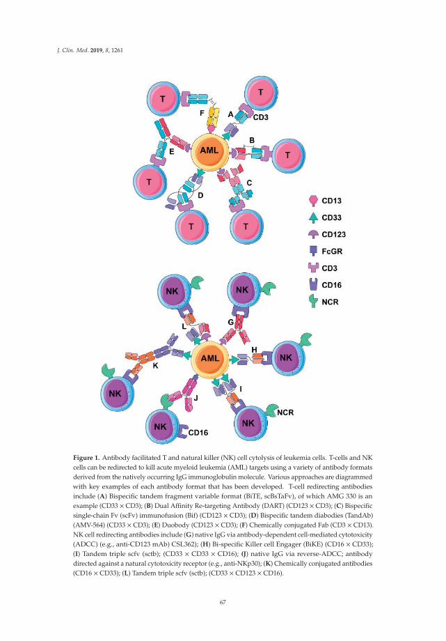
J. Clin. Med. 2019, 8, 1261
Figure 1. Antibody facilitated T and natural killer (NK) cell cytolysis of leukemia cells. T-cells and NKcells can be redirected to kill acute myeloid leukemia (AML) targets using a variety of antibody formatsderived from the natively occurring IgG immunoglobulin molecule. Various approaches are diagrammedwith key examples of each antibody format that has been developed. T-cell redirecting antibodiesinclude (A) Bispecific tandem fragment variable format (BiTE, scBsTaFv), of which AMG 330 is anexample (CD33 × CD3); (B) Dual Affinity Re-targeting Antibody (DART) (CD123 × CD3); (C) Bispecificsingle-chain Fv (scFv) immunofusion (Bif) (CD123 × CD3); (D) Bispecific tandem diabodies (TandAb)(AMV-564) (CD33 × CD3); (E) Duobody (CD123 × CD3); (F) Chemically conjugated Fab (CD3 × CD13).NK cell redirecting antibodies include (G) native IgG via antibody-dependent cell-mediated cytotoxicity(ADCC) (e.g., anti-CD123 mAb) CSL362); (H) Bi-specific Killer cell Engager (BiKE) (CD16 × CD33);(I) Tandem triple scfv (sctb); (CD33 × CD33 × CD16); (J) native IgG via reverse-ADCC; antibodydirected against a natural cytotoxicity receptor (e.g., anti-NKp30); (K) Chemically conjugated antibodies(CD16 × CD33); (L) Tandem triple scfv (sctb); (CD33 × CD123 × CD16).
67
J. Clin. Med. 2019, 8, 1261
2.2. Lintuzimab and Bl 835858
Lintuzumab (SGN-33, HuM195) is an unconjugated anti-CD33 mAb which has been testedin several clinical trials for AML (NCT00002609, NCT00002800, NCT00006084, NCT00016159,NCT00283114, NCT00502112, NCT00528333, and NCT00997243) in combination with standardinduction chemotherapy and a maintenance monotherapy in R/R AML (reviewed in [100]). Both a phase2b randomized trial [101] and a phase 3 randomized trial [102] of lintuzumab did not demonstratesurvival benefit and the agent was not pursued further but has been applied for the delivery ofradionuclides and described in a subsequent section. Another unconjugated anti-CD33 mAb, BI 836858,is Fc optimized through engineering, leading to improved NK cell-mediated ADCC relative tonative antibody Fc [103]. Several clinical trials of BI 836,858 are currently underway for R/R AML(NCT02632721 NCT01690624) and AML relapsing post HSCT (NCT03207191).
2.3. Daratumumab (Darzalex), Isatuximab
Daratumumab is a fully human IgG1 kappa mAb that targets CD38 and was generated using theHuMAb platform with human antibody transgenic mice [104]. Daratumumab was originally evaluatedin and approved for use in multiple myeloma (MM) in 2015 [105]. Daratumumab has been tested againstprimary AML targets in vitro, demonstrating apoptosis induction, ADCC and CDC as mechanisms ofcytotoxicity, as well as being shown to reduce leukemic burden in the spleen and peripheral blood,but not in bone marrow in primary AML xenograft models [106]. Its mechanism of action includes(CDC), ADCC [107] and (ADCP). Investigators at MD Anderson Cancer Center are currently evaluatingthe efficacy of daratumumab as a stand-alone treatment for R/R AML (NCT 03067571), while OhioState University is evaluating its effectiveness in combination with donor leukocyte infusions (DLI) forAML patients who have relapsed after allogeneic HSCT (NCT 03537599).
Another anti-CD38 antibody, isatuximab, has also been tested in MM, NHL, and CLL patientsin a phase 1 trial [108] and a phase 3 trial in patients with MM, which showed improvement inprogression-free survival. One advantage of isatuximab versus daratumumab is the need for lessfrequent dosing, though no head-to-head comparison of efficacy has been performed. A study ofisatumximab recently opened a phase I/II trial for pediatric patients with R/R AML and ALL for usewith combination chemotherapy (NCT 03860844).
3. Multivalent Antibody Therapies
Multivalent antibodies with, bi-, tri- and quadri-specific binding domains are engineered constructswhich combine specificities of two or more antibodies into one molecular product that is designed tobind to both a TAA and an activating receptor on the effector cells, typically a T cell or natural-killer(NK) cell. There are several different structural variants of bispecific antibodies, which, in turn,can be utilized to target various combinations of effector and tumour targets antigens [109]. The firstFDA-approved dual-binding antibody was blinatumomab, a bispecific T-cell engager (BiTE) developedby Amgen with specificities for CD3 and CD19 for treatment of acute lymphoblastic leukemia (ALL).BiTE format antibodies (tandem di-scFv) are engineered products involving combining the VL andVH domains of a monoclonal antibody into a single chain fragment variable (scFv) specific to anactivating receptor (e.g., CD3) and further linked to the scFv of an antibody specific to a target antigen(e.g., CD19). The CD19 × CD3 BiTE was able to induce remissions in relapsed and refractory ALLthat had failed other therapies [110,111] and was recently approved by the FDA for ALL patients withminimal residual disease (MRD) [112]. Because of the low molecular weight of blinatumomab, it israpidly cleared by the kidneys and excreted through the urine, requiring administration by continuousinfusion for up to several weeks [110]. Hence, for clinical use, there are challenges in cost and practicalapplications. Nonetheless, this technology is powerful, as it can be used as a general approach toredirect T-cells toward any antigen of interest. It can also be applied to engineered antibody fragments
68
J. Clin. Med. 2019, 8, 1261
with different formats than the BiTE, such as DART and Duobody, also increasing valency, such aswith tri-specific antibodies.
In general, most bi- and tri-specific engineered antibodies lack an Fc region and therefore,do not have the ability to mediate CDC and ADCC. Moreover, the engagement of NK cells to targetsby bi- or tri-specific antibodies can result in immediate degranulation, owing to their preformedgranules [113,114], whereas only a subset of primed T-cells can optimally be redirected to mediatedcytotoxicity against against cancer-cell targets [115]. Only three bi-specific antibodies have been enteredinto clinical trials for AML, which are registered on ClinicalTrials.gov (Table 1) and are discussed inmore detail with other bi-, tri- and quadri-specific formats in the pre-clinical stage of development.
Table 1. Clinical trials of bispecific antibody therapy for AML.
NCI ClinicalTrial and Phase
Target Agent(s)InclusionCriteria
Estimated Startand End Dates
Outcomes Status
NCT02715011Phase 1 CD123
JNJ-63709178CD123 × CD3
Duobody
≥18 years ofage with R/R
AML
June 2016October 2021
MTD, ORR, 1.5 yearEFS and RFS Suspended
NCT02520427Phase 1 CD33
AMG330CD33 × CD3Tandem scFv
(BiTE)
≥18 years ofage with R/R
AML
August 2015January 2020
MTD and ORRduration at 3 years Suspended
NCT02152956Phase 1 CD123
MGD006CD123 × CD3
DARTflotetuzumab
≥18 years ofage with R/R
AML
May 2014April 2020
MTD and OS at2 years Recruiting
AML: acute myeloid leukemia; R/R: felapsed/refractory; MTD: maximum tolerated dose; ORR: overall response rate(CR + CRi); OS: overall survival; RFS: relapse free survival.
3.1. Bispecific Tandem Fragment Variable Format (BiTE, scBsTaFv)
Given the success of blinatumomab for ALL, Amgen subsequently developed AMG 330, which isa CD33 × CD3 specific BiTE for treatment of AML (Figure 1A). The CD33 × CD3 BiTE can facilitateT-cell activation, expansion and in vitro lysis of primary AML cells [116,117]. Furthermore, AMG 330,in combination with infusions of activated human T cells, could suppress the growth of AML cellsin a MOLM-13 cell line xenograft model, leading to improved survival [118]. Furthermore, AMG330 could facilitate the cytotoxicity of both autologous and healthy donor allogenic T-cells againstprimary AML [119]. Recently, preliminary data for AMG330 was presented at the American Societyfor Hematology (ASH) meeting for patients with relapsed and refractory AML (NCT02520427) [120].This was a phase 1 dose escalation study with 35 patients (enrolment to 40 in progress) to evaluate thesafety, pharmacokinetics and tolerance. Four patients (10%) achieved a CR or a CR with incompleteblood count recovery treated with 120 or 240 ug/day. However, these CRs were not sustained beyond onecycle of treatment and the majority of patients discontinued treatment because of disease progression.Adverse events included cytokine release syndrome (CRS), febrile neutropenia, pneumonia, leukopenia,pyrexia, thrombocytopenia and subdural hematoma. Although this study established a tolerabledose, there was a low frequency of remission induction and short durations of remission in thefour responders.
Another bispecific antibody format termed single-chain bispecific tandem fragment variable(scBsTaFv) provides an alternative to tandem scFv fragments in the BiTE format. This approachattaches the VH of an anti-CD3 mAb to the VL of an anti-CD33 mAb with the two tandem scFvjoined by a glycine-serine linker. Further optimization of this format was resolved by rearrangingthe variable regions. A CD33 × CD3 scBsTaFv construct was able to facilitate the cytotoxicity ofallogeneic mononuclear cells (PBMC) against CD33-expressing target cells [121]. This CD33 × CD3scBsTaFv bispecific antibody was further humanized, reducing the probability of the patient mountingan immune response against the agent and was effective in picomolar concentrations, was independentof CD33 antigen density and did not redirect cytotoxicity to HSCs, as measured by a clonogenic
69

J. Clin. Med. 2019, 8, 1261
assay [122]. An additional modification was to add 4-1BB ligand (4-1BBL), a co-stimulatory moleculewhich further enhanced T cell cytotoxicity against AML cells, against CD33low targets, relative to theparental construct [123].
3.2. Dual-Affinity Retargeting (DART)
Dual-affinity retargeting (DART) molecules are generated from a VH and VL taken from two distinctmAbs in a format distinct from BiTE to target two distinct antigens (Figure 1B). Specifically, a CD123 ×CD3 DART was developed by Macrogenics (flotetuzumab), which can redirect T cells to target and killAML cells, as well as stimulate T-cell proliferation [124]. The CD123 × CD3 DART was able to suppressleukemia progression in a CD123 + GFP + CBRLuc K562 murine xenograft model as measured bybioluminescence. The preliminary results of a phase 1 clinical trial of flotetuzumab demonstrated thatit had anti-leukemic activity in 57% of a cohort of 45 patients with R/R AML/MDS (89% AML). Toxicitywas reported and included infusion-related fever, chills, tachycardia, and hypotension, which werenot severe. Importantly, the overall response rate (ORR) was 43%. Thus, the preliminary results areencouraging for DART as a potential effective targeted therapeutic for R/R AML.
3.3. Bispecific scFv Immunofusion or BIf
A novel and interesting bi-specific antibody format is the Bispecific scFv Immunofusion (BIf) withan scFv fused at the N-terminus of human IgG1 hinge region with a second scFv at the C-terminus(Figure 1C). The first described BIf was an CD123 × CD3 construct which would form pairs as ahomodimer with a tumour target binding Kd of 1.0 × 10⁻¹⁰ molar which is superior to other bispecificantibody formats and can facilitate T-cell mediated cytotoxicity in vitro at low effector:target ratios [125].Due to an intact Fc, it has a longer half-life than the lower molecular weight classical BiTE moleculesand, hence, a longer therapeutic exposure to AML cells.
3.4. Bispecific Tandem Diabodies (TandAb)
Bispecific tandem diabodies (TandAb), also termed bispecific tetravalent antibodies, have aunique structure with two homologous immunoglobulin chains running counter to each other(VHA-VLB-VHB-VLA)2 (Figure 1D) [126]. A CD30 × CD3 TandAb (AFM13) is being tested in clinicaltrials for cutaneous lymphoma (NCT 03192202—recruiting), R/R Hodgkin lymphoma (NCT 02321592(recruiting and NCT 02665650—completed). A CD33 × CD3 TandAb has been generated (AMV-564)which, in the presence of T cells, can mediate dose-dependent cytotoxicity against primary AML targetsfrom newly diagnosed and refractory or relapsed patients in vitro. AMV-564 had efficacy in treating amurine AML xenograft model [127]. A trial of AMV-564 has been initiated for R/R AML (NCT 03144245)and is sponsored by this platform’s developer, Affimed Therapeutics Inc. ( Heidelberg, Germany).
3.5. Chemically Conjugated Bispecific Antibodies
An early approach to dual target antigens was to chemically conjugate two different antibodies.A chemical conjugate of anti-CD16 and anti-CD33 monoclonal antibodies was developed which couldredirect the cytotoxicity of NK cells toward AML blasts (Figure 1K) [128]. Another variant of thisapproach was to develop a bispecific (Fab’)2 fragment derived from two different antibodies [129,130].Specifically, antibodies underwent cleavage and separation of (Fab’)2 fragments with dithiothreitolto create Fab’-SH fragments which could be recombined using a thiol-disulfide interchain reagent,ultimately producing a bispecific hybrid F(ab’)2. This approach was used to combine an anti-CD3(OKT3) and anti-CD13 mAb (Figure 1F) [131]. These anti-CD3 and anti-CD13 Fab’ fragments weremixed and reduced to form a bispecific F(ab’)2. This CD13 × CD3 Fab was able to enhance lysisof AML blasts by PMBCs. Also, this CD13 × CD3 antibody construct had some inhibition of AMLcolony-forming units (CFU), and had a lesser effect on granulocyte/macrophage CFU from normalbone marrow [131].
70

J. Clin. Med. 2019, 8, 1261
3.6. Bispecific Full-Length Antibodies (Duobody and Biclonics)
Genmab developed the DuoBody platform to develop bispecific human IgG1 antibodies.Two mAbs of different specificity, each containing single matched mutations in the third constant (CH3)domain, were produced using mammalian recombinant cell lines and are then in a bispecific antibody.A CD123 × CD3 duobody was developed from an IgG4 backbone with a silenced Fc function andtermed JNA-63709178 (Figure 1E) [132]. A phase 1 trial of a CD123 × CD3 Duobody (JNJ-63709178)sponsored by Janssen is in active recruitment of R/R AML patients (NCT 02715011). A full-lengthbispecific CLL-1 × CD3 antibody was developed by Merus using their proprietary Biclonics platformwith preclinical activity demonstrated against AML [133], with an ongoing Phase 1/2 clinical trialongoing for R/R AML in adults and newly diagnosed elderly patients with complex cytogenetics(NCT 03038230).
3.7. BiKEs and TriKEs
A CD33 × CD16 bispecific scfv was designed to activate NK cells and redirect them to lyse CD33+AML targets and also secrete cytokines (IFNγ and TNFα) [134]. This construct is structurally similar toBiTE and was termed a bispecific killer-cell engager (BiKE) (Figure 1H). The observation that CD16 canbe shed from NK cells by cleavage with ADAM17 limited this approach, but this was overcome withan ADAM17 inhibitor that could enhance NK cell cytotoxicity and cytokine secretion in the presenceof the CD33 × CD16 BiKE. The BiKE platform was also functional using NK cells from patients withmyelodysplastic syndrome, facilitating cytotoxicity against both AML targets and myeloid-derivedsuppressor cells [135].
Further modification of BiKEs was carried out to incorporate a third functional domain, specifically,to incorporate the IL-15 cytokine, and accordingly named a trispecific killer cell engager (TriKE) [136].This approach facilitated additional expansion and activation capacity of NK cells, and when comparedto the BiKE, yielded superior anti-leukemic results in mouse models of human AML. Three scFvcomponents can be inserted to create a (scFv)3 construct termed a single-chain Fv triplebody (sctb).One such construct linked two anti-CD33 scFv fragments to a single anti-CD16 scFv and wascompared with a bispecific format (bsscFv) that consisted of only a single anti-CD33 scFv (Figure 1I).The CD33 × CD33 × CD16 sctb had a greater binding affinity for CD33 compared to the affinityobtained with the CD33 × CD16 bsscFv. More importantly, the sctb had increased by ≥2-logs the NKcell cytotoxic potency against AML cells relative to bsscFv [137]. Another approach using the triplescFv involves targeting two different antigens on a single target cell (Figure 1L). Using this approach,a CD33 × CD123 × CD16 was developed and shown to facilitate superior leukemic cell killing by NKcells relative to the dual targeting of the same antigen (CD123) [138]. A clinical trial of TriKE therapy isunderway for patients with CD33 + R/R AML (NC T03214666).
4. Toxin-Conjugated Antibody Therapy for AML
Conceptually, the combination of highly potent anti-neoplastic agents and targeted antibodiesin a single antibody-drug conjugate is not a recent development [139]. The principle of attaching atargeted antibody to a cytotoxic drug or radioactive isotope (referred to as ‘payload’ or ‘warhead’)through covalent linkage has led to a number of antibody-drug conjugates (ADCs) approved for use inthe management of hematological malignancies. Although several warheads are being tested in cancer,the warheads that will be described have been used in AML.
Structurally, there are three components of equal importance in the design of an ADC, namely themAb itself, the cytotoxic agent, and the conjugation linker [140]. The mAbs can be human, humanized,or chimeric, and may be engineered to target the antigen of choice with high specificity. For optimaldrug delivery, the linker must bind with sufficient integrity to prevent premature de-conjugation,yet must release the drug once the antibody has bound to the target [141]. Linkers may be cleavableor non-cleavable. Typically, ADCs are reliant on efficient degradation in lysosomes to release the
71

J. Clin. Med. 2019, 8, 1261
payload inside the target tumour cell [140]. Each approach is associated with its unique advantages anddrawbacks and should be engineered with the target cell and the payload in mind. Finally, the drugitself must be sufficiently potent to ensure tumour killing with minimal off-target toxicity.
Calicheamicin is an anti-tumour antibiotic synthesized from Micromonospora echinospora thatinduces double-stranded DNA breaks, leading to cell death [142]. This is used in conjugationwith antibodies targeting CD33 (gemtuzumab ozogamicin/Mylotarg) in AML, or CD22 (inotuzumabozogamicin) in B-cell acute lymphoblastic leukemia (ALL). Monomethyl auristatin E (MMAE) isconjugated to an anti-CD30 antibody in brentuximab vedotin, an ADC that is FDA-approvedfor Hodgkin lymphoma [143]. Most recently, an investigational agent was developed using apyrrolobenzodiazepine (PBD) dimer to induce DNA damage in tumour cells [144]. Vadastuximabtalirine (SGN-CD33A) is a third generation ADC construct whereby an anti-CD33 antibody is conjugatedto two molecules of a pyrrolobenzodiazepine (PBD) dimer via a maleimidocaproyl valine-alaninedipeptide connecting segment [145]. The PBD dimer is released after protease cleavage and inducesDNA cross-linking, leading to target-cell apoptosis [146]. Several ADCs for AML have been tested inpublish clinical trials and are discussed below, while other novel ADC trials for AML are ongoing.
4.1. Gemtuzumab Ozogamicin (GO)
The first clinically viable ADC to be approved in hematological malignancies was gemtuzumabozogamicin (GO; Mylotarg), which targets CD33 [147]. In phase III studies of GO as monotherapy inpatients over the age of 60 with relapsed AML, an overall response rate of 30% was reported. Based onthese data, GO received accelerated FDA approval in 2000 [148]. However, a subsequent multicenterphase 3 randomized clinical trial comparing GO 6 mg/m2 on day 4 of a daunorubicin and cytarabineinduction chemotherapy protocol failed to demonstrate differences in survival. In fact, the patientsreceiving GO had a higher rate of mortality during induction due to Veno-Occlusive Disease (VOD)(5.5% death rate in the combination arm versus 1.4% in the chemotherapy alone arm) [149]. As a result,the drug was voluntarily withdrawn from the market in 2010. However, subsequent randomized trialsevaluating lower doses of GO in combination with chemotherapy demonstrated improved overallsurvival without increased toxicities such as VOD. For example, The MRC AML15 trial combinedGO at a dose of 3 mg/m2 on day 1 of conventional induction chemotherapy in 1113 patients withpreviously untreated AML and reported a significant survival benefit without increased toxicityin younger patients with favorable cytogenetics, particularly core binding factor leukemias [150].Another trial of a similar dosage regimen in older patients showed no difference in CR rates butsignificantly improved the three-year OS and relapse-free survival (RFS) with no appreciable increasein toxicity [151]. This demonstrated that lower doses of GO are effective in AML. As a result, GO wasreapproved by the FDA for the treatment of newly-diagnosed CD33-positive acute myeloid leukemia(AML) in adults and for treatment of relapsed or refractory CD33-positive AML in adults and inpediatric patients 2 years and older.
4.2. Vadastuximab Talirine (SGN33A) and IMGN779
Other ADCs that target CD33 have been developed and are being actively investigated for AMLtherapy. Early in vitro experiments and animal studies showed that SGN33A was active even inmulti-drug resistant and p53 mutated AML cell lines [152]. A phase 1 trial of SGN-CD33A in 27 older(median age 74 years) treatment naïve AML patients reported responses in 54%, with 14 patientsachieving CR/CRi and five achieving a morphological leukemia-free state [153]. MRD negativity byflow cytometry was noted in six of 13 patients for whom data were available. VOD was not reportedwith this drug. Current trials are focused on evaluating SGN-CD33A in combination with standardinduction chemotherapy. For example, a trial of SGN-CD33A in combination with decitabine orazacitidine in 24 patients with AML unfit or unwilling for conventional chemotherapy had a responserate (CR + CRi) of 73% amongst 49 evaluable patients [154]. In addition, 47% of the responding patientsachieved MRD negativity by flow cytometry. Noteworthily, the combinations were well tolerated,
72

J. Clin. Med. 2019, 8, 1261
with a 30-day mortality rate of 2%. Another CD33-directed antibody-drug conjugate IMGN779 inwhich the mAb is bound to a novel alkylating agent DGN462 was active in preclinical models [155]and a phase 1 clinical trial is currently underway in R/R AML (NCT 02674763).
4.3. Current Clinical Trials
There are many current clinical trials evaluating ADCs for AML which are registered onClinicalTrials.gov (Table 2). GO is under investigation as a single agent for R/R AML in a phase 2 trial(NCT 03374332). Given that treatment of R/R AML is challenging and would not address its role intreating minimal residual disease (MRD), a phase 2 trial of GO is being examined after standard inductionchemotherapy in patients achieving a CR who remain MRD + (NCT 03737955). Finally, another phase1/2 study is aiming to evaluate the efficacy of GO in combination with chemotherapy in the up-frontsetting (NCT 03531918). Another CD33-directed antibody-drug conjugate IMGN779, in which themAb is bound to a novel alkylating agent DGN462, was active in preclinical models [155], and a phase1 clinical trial of this agent is currently underway in R/R AML (NCT 02674763). Other currently activeclinical trials are examining the role of ADCs targeting CD30, CD123, CD71, and FLT3 in patients withAML and other hematological malignancies expressing these antigens.
Table 2. Clinical trials of toxin-conjugated antibodies for AML.
NCI Clinical Trialand Phase
Target Agent(s) Inclusion CriteriaEstimated Startand End Dates
Status
NCT03374332Phase 2 CD33 gemtuzmab
ozogamicin≥18 years of age withR/R AML
June 2019March 2021
Not yetrecruiting
NCT03737955Phase 2 CD33 gemtuzmab
ozogamicin
≥2 years of age withAML in CR with MRDafter inductionchemotherapy
November 2018August 2021 Recruiting
NCT03531918Phase 1/2 CD33
gemtuzmabozogamicin in
combination withGCLAM
≥18 years of age withuntreated “high-grade”myeloid neoplasm(≥10% Blasts in blood orBM) or AML, exludingAPL
September 2018July 2025 Recruiting
NCT02674763Phase 1 CD33 IMGN779 ≥18 years of age with
R/R AMLMarch 2016
December 2019 Recruiting
NCT03386513Phase 1 CD123 IMGN632
≥18 years of age withR/R CD123 + AML andother CD123 +malignancies
January 2018February 2021 Recruiting
NCT02864290Phase 1 FLT3 ASP1235 (AGS62P1) ≥18 years of age with
R/R AMLNovember 2016
January 2024 Recruiting
NCT03957915Phase 1 CD71 INA03
≥18 years of age R/RAML, ALL, or MPALwith ≥ 20% CD71positive blasts
September 2019November 2021
Active, notrecruiting
NCT01830777Phase 1 CD30
Brentuximab vedotinin combination with
Mitoxantrone,Etoposide, and
Cytarabine
≥18 years of age withCD30 + relapsedAML
May 2013December 2019
Active, notrecruiting
NCI: National Cancer Institute; AML: Acute Myeloid Leukemia; R/R: Relapsed/Refractory; MRD: MeasurableResidual Disease; BM: Bone Marrow; GCLAM: Granulocyte-Colony Stimulating Factor, Cladribine, Cytarabineand Mitoxantrone.
5. Radioimmunotherapy of AML
Radioimmunotherapy (RIT) for R/R AML is a scientifically sound approach that is translatableto clinical practice and can improve treatment outcome in patients. RIT utilizes mAbs labeled withradionuclides, providing continuous ionizing particle-based radiation exposure to cells expressing
73

J. Clin. Med. 2019, 8, 1261
target antigens. RIT for AML has been investigated for the past three decades, demonstrating its abilityto kill leukemic cells and deliver radiation specifically to sites harboring AML disease.
Although clinical testing is in place, important aspects such as target antigens on AML (or also onnormal hematopoietic) cells and the properties of the payload radionuclides are still being optimized.Therefore, in this section, we put into context how RIT works and demonstrated the unique propertiesof select radionuclides and what has been learned from several clinical trials. Here, we identifiedclinical trials that are currently recruiting patients or are in preparation.
Due to exquisite affinity and specificity for target antigens preferentially expressed on diseasedcells by mAbs and because radiolabeled mAbs have an increasingly well-understood pharmacokineticprofile, they target tumours with high target-to-nonspecific organ ratios [156]. Hence, radiolabeledmAbs are able to deliver ionizing radiation to disease sites more specifically than traditional totalbody irradiation (TBI). Ionizing radiation is then delivered with increased precision to AML cells andinduces cell-death by two main mechanisms—apoptosis and necrosis—as a result of unrepaired DNAstrand breaks [157]. Although RIT efficiency depends on many additional factors, this is beyond thescope of this summary and was reviewed in Desouky et al. [158].
A key RIT property is the type of radionuclide that is attached to the mAb. The nature of radiationemitted from radioisotopes that radioactively decay via α-particle or β-particle emission is different(Table 3). For example, the most relevant radionuclides for AML 225Ac and 211At decay by the emissionof α-particles, whereas 131I and 90Y decay by the emission of β-particles. These radionuclides havedifferent energies and another unique important property called linear energy transfer (LET). LET is theamount of energy that an ionizing particle transfers to material traversed per unit distance. Typically,α-particles have a range in tissue of 50–80 μm, which results in an LET of 50–230 keV/μm (Table 1).In contrast, β-particles have a considerably reduced LET of 0.1–1 keV/μm as they have a range of0.5–12 mm in tissue. This signifies that α-particles provide an increased relative biological effectiveness(RBE) relative to β-particles. RBE is a measure of the extent of damage (e.g., DNA double strandbreaks) to the cell an emitted particle produces. The RBE for α-particles is 3–7-fold increased relativeto β-particles [159].
Table 3. Radionuclides used in active clinical trials for AML.
Radionuclide T 1/2 Emission Emax (keV) Range (μm)
β-Emitting Radionuclides (LET = 0.1–1.0 keV/μm)
Iodine-131 8.02 days β and γ 610/362 2300
Yttrium-90 2.67 days β 2250 11,300
α-Emitting Radionuclides (LET = 50–230 keV/μm)
Astatine-211 7.2 h α and X 5870 and 7450/77–92 80
Actinium-225 9.92 days 4α, 2β and γ6000–8000/
198–659/218–444 90
Bismuth-213 45.59 min α and γ 8400/440 17
In general, 131I and 90Y have been employed in >95% of clinical RIT trials and represent the currentstandard to which all other radionuclides are compared [160]. Both isotopes have distinct favorableproperties. 131I emits a second ionizing particle, the γ-ray and can thus be utilized for both imagingand therapy. However, 131I-mAbs degrades rapidly if the receptor internalizes upon mAb binding.This results in the release of 131I-tyrosine in the bloodstream. In addition, the γ-rays emitted by 131Iincreases the absorbed radiation dose to tissues and pose a risk to family members and healthcareprofessionals and, hence, require patient isolation. Both of these properties increase the absorbedradiation to healthy organs. Alternatively, 90Y is an almost exclusive β-particle emitter (Table 3).Since the travel distance of β-particles is short compared to γ-rays and does not leave the patient’s body,
74

J. Clin. Med. 2019, 8, 1261
90Y is safer and more practical to work with for healthcare professionals. 90Y also emits β-particles thatare more energetic by 5-fold compared to the β-particles emitted by 131I. In addition, if 90Y-mAbs areinternalized, the isotope is retained in the cell and is not released into the bloodstream. With either case,it is vital to have knowledge that the radionuclide targets tissue harboring the leukemic cells severalfold higher than the liver, kidney, and lung—the dose limiting normal tissues. Since 90Y cannot beimaged, dosimetry relies on utilizing another isotope labeled on the mAb, typically 111-indium (111In).
A caveat for β-particle-based RIT is that the emitted β-particles often overshoot single AML cellsand ablate surrounding normal hematopoietic cells residing in the bone marrow [161] (Figure 2A,B).This often results in dose-limiting toxicity (DLT) to the bone marrow, as normal hematopoietic cells arealso highly sensitive to irradiation [162]. In contrast, the short range of emitted α-particles in theorymakes them ideal for eradicating individual leukemic cells (Figure 2A,C). However, the energy ofα-particles is several-fold higher than β-particles and thus, can also cause unwanted toxicities.
Although this is not a focus in this brief summary, short-ranged and high LET Auger electronsmay provide an approach to eradicate AML cells. Recent preclinical studies have shown that theAuger-emitter 111In labeled to mAbs targeting CD33 and CD123 and conjugated to short peptidesharboring a nuclear localization signal sequence could kill AML cell lines and primary AML cellengrafted into mice. In addition, single-photon emission computed tomography (SPECT) imagingcould be used as a companion to evaluate the targeting of AML cells in the bone marrow andextramedullary sites in these preclinical models to the γ-ray emissions by 111In [163–166].
Figure 2. Radioimmunotherapy for acute myeloid leukemia. Illustration of (A) a mAb radiolabeledwith either a (B)β-particle or (C)α-particle-emitting radionuclide and the track of the particles perfusingthe bone marrow to target AML cells. Note that the path length of β-particles is greater than forα-particles leading to β-particle-based RIT used primarily in preparative regimens to myeloablate thebone marrow prior to hematopoietic cell transplantation.
5.1. β-Particle RIT for AML
Because of the properties of β-particles and the need to overcome the limitation of normal organtolerance with current preparative regimens HSCT, The Fred Hutchinson Cancer Research Center(FHCRC) made an important breakthrough for developing an effective niche for RIT utilization.As detailed in a review by Gyurkocza and Sandmaier, successful HCT relies on the effectiveness of thepreparative or conditioning regimen administered to patients prior to transplantation [167]. The goals
75

J. Clin. Med. 2019, 8, 1261
of the regimen are to provide sufficient immunoablation to prevent the graft rejection of the donor cellsand to reduce the leukemic burden [167].
In general, myeloablative regimens consist of alkylating agents with or without TBI that ablatemarrow hematopoiesis to the point where autologous hematologic recovery does not occur. The greaterpart of regimens combines 12- to 16-Gy TBI, typically fractionated, with chemotherapy, and includescyclophosphamide, cytarabine, etoposide, melphelan, and busulphan [167]. These chemotherapeuticagents have shown the ability to simultaneously exert cytotoxic and cytostatic effects on leukemic cellsand to suppress the patient’s own immune system to reduce graft rejection. While increased doses ofTBI reduce the risk of AML relapse, this also increases treatment-related mortality (TRM), often dueto toxicity in the gastrointestinal system, the liver, and the lungs. Hence, HSCT is not an option formany older and medically infirm patients. In addition, TBI-induced malignancies and developmentimpairment in children are significant concerns.
As an alternative, non-myeloablative regimens have been developed for patients with AML thatprovide more favorable toxicity profiles yet are sufficiently immunosuppressive. There is a considerablerange in intensity of these regimens. However, the approach developed at FHCRC is relevant sincemost RIT trials have occurred there. In particular, in older patients, conventional conditioning regimensprior to HCT often leads to a high TRM [168]. A low-dose 2-Gy TBI-based preparative regimen isadministered to patients at FHCRC with the addition of 90 mg/m2 of fludarabine to prevent graftrejection and increase pre-transplantation host T-cell immunosuppression [169]. Cyclosporine andmycophenolate mofetil is also administered to increase immunosuppression.
Although high-intensity TBI doses reduce relapse rates, this is offset by increased conditioningregimen-related death [170,171]. Conversely, reducing the TBI allows for increased utilization of HCTfor patients with AML, however AML relapse rates increase [172,173]. Hence, there is a dilemma tofind an ideal regimen that allows for low toxicity yet potent myeloablation/myelosuppression beforetransplantation that improves patient survival. Thus, targeted radiation is an alternative.
The mAb BC8 is specific for CD45 and radiolabeled with 131I was developed as means tomyeloablate since the antigen is expressed on the surface of almost all hematopoietic cells, exceptmature red blood cells and platelets and is also present on AML blasts [174]. CD45 is an attractivetarget because it is expressed by most AML samples at relatively high levels (~200,000 receptors percell), and the antigen does not internalize, which is important for 131I. Because CD45 is expressed onboth normal and leukemic cells, it can be used to target the bone marrow. The strategy is to reduce theradiation dose and, hence, the overall toxicity to the patient by delivering radiation specifically to sitesharboring leukemia cells.
An early clinical trial evaluating RIT for AML combined escalating doses of radiation deliveredby the 131I-BC8 with cyclophosphamide and 12-Gy TBI in patients with advanced AML [175]. Patientsreceived a “tracer” infusion of the radiolabeled mAb weeks before HCT to allow for the mAb to penetratethe hematopoietic tissues harboring the leukemic cells and for sufficient washout from the blood andnormal organs. At ~72 h post-injection, patients were imaged with a focus on region-of-interests suchas the bone marrow (acetabulum and sacrum), spleen, liver, lungs, kidneys, and spleen. The studyshowed that 84% of patients contained radiation in the bone marrow and spleen 2.3- and 4.8-foldhigher than the liver, the normal organ receiving the highest dose due to normal mAb metabolism.The ratios of radiation delivered to the bone marrow and spleen relative to the lung and kidneyswere even greater than the liver. More importantly, this procedure could be safely performed with aconventional HCT preparative regimen.
The effectiveness of targeting radiation to specific hematopoietic/AML sites was then expandedto patients with AML in a first remission with a human leukocyte antigens (HLA)-identical familydonor [176]. In addition to 131I-BC8, patients underwent a preparative regimen consisting of busulfanand cyclophosphamide without TBI. The three-year survival among the patients in this study was63% and the TRM was 21%. In patients with unfavorable cytogenetics, the percentages of survivaland TRM were 26% and 27%, respectively. This study demonstrated that 131I-BC8 could be used to
76
J. Clin. Med. 2019, 8, 1261
intensify the amount of radiation delivered to leukemic cells to a greater extent than that deliveredto normal organs, which provided increased survival benefit without excessive TRM. As a result,the effectiveness of 131I-BC8 is now being evaluated to validate its use for AML as part of preparativeconditioning prior to HSCT. Distinct evaluations are currently active, such as testing 131I-BC8 incombination with fludarabine and 2-Gy TBI for allogeneic HCT, specifically in patients ≥50 yearsold (NCT 00008177). This trial reported outcomes in 58 patients with R/R-AML or myelodysplasticsyndrome (MDS) [177]. Treatment resulted in the MTD of 131I-BC8 delivered to the liver was 24-Gy.At day 28 post-transplantation, all the patients had a complete remission and donor-cell engraftment.Seven patients ultimately succumbed to TRM by day 100. The one-year survival estimate was 41%.Another phase 1 trial is evaluating 131I-BC8 with fludarabine and 2-Gy TBI in 15 R/R-AML patients thatwere ≥16–50 years of age (NCT 00119366) [178]. This study capped the maximum dose delivered to theliver at 43-Gy as was carried out in the trial with older AML patients out of concern for causing stromaldamage and marrow failure. In these younger patients, no cases of DLTs or TRM were observed.At ~12 months since treatment, 11 of the 15 patients survived and at five years, there were six survivingpatients. This study suggested that increased doses from 131I-BC8 are tolerable and provide even moresurvival benefit to younger adults with R/R AML. The results from these completed and active trialsshow the feasibility of using 131I-BC8 to enhance the efficacy of allogeneic HCT in both young andolder patients in a reduced conditioning setting.
One area in need of progress to extend RIT is for patients who do not have closely HLA-matchedallogeneic donors and instead are reliant on haploidentical donor engraftment, especially patients fromethnic minority groups. Orozco et al. demonstrated that 90Y-anti-CD45 RIT and cyclophosphamidewithout TBI or fludarabine before haploidentical HCT resulted in a high rate of engraftment,which remained stable for six months after transplantation in a murine mouse model [179]. An ongoingphase 2 trial is evaluating the use of 131I-BC8 in R/R AML patients receiving haploidentical donor HCT(NCT00589316). Thus far, six of eight patients have been reported to achieve complete remission byday 28 post-HSCT and all patient had 100% donor chimerism [180].
As previously described, a significant barrier to expanding 131I-BC8 as a mainstream approach totreat patients with AML is that many Nuclear Medicine Departments have difficulty properly handlinglarge quantities of 131I. Thus, a phase 1 study was undertaken to establish the safety, feasibility andoptimized dose of 90Y-BC8 in patients undergoing reduced-intensity regimen before allogeneic HCT(NCT01300572). Favorable outcome measures were reported with 90Y-BC8 for multiple myeloma andlymphoma [181,182]. A previous Phase 1 clinical trial studying 90Y conjugated to rat mAb YAML568that recognizes CD45 was evaluated in eight patients [183]. No significant administration-related sideeffects were observed. However, in order for 90Y-YAML568 to deliver preferentially increased radiationdoses to the bone marrow and spleen, the patients were required to be preloaded with cold antibody.This indicates that patients may have produced an anti-YAML immune response that causes clearanceof the mAb. Patient outcome was not reported.
90Y has also been conjugated to the mAb lintuzumab that targets CD33. CD33 is exclusivelyexpressed on myeloid cells and lymphocytes and not on all hematopoietic cells, such as CD45.Importantly, as CD33 is frequently expressed on AML cells it is an attractive target for RIT. Lintuzumabis a humanized mAb and binds CD33 with a very high affinity, as previously discussed. Two completedtrials (NCT 00002890 and NCT 00006040) have investigated the MTD of 90Y-BC8 in patients with R/RAML with and without HCT, respectively. No findings have yet been published.
5.2. α-Particle RIT for AML
The relative increased energy of α-particles, relative to β-particles, coupled with a range of onlya few cell diameters and high LET in theory makes this RIT approach ideal for the treatment ofsmall-volume disease or MRD. Hence, there has been high clinical activity to test α-particle-based RITin AML. A Phase 1 study from the 1990s evaluating 213Bi-lintuzumab administered at escalating dosesin patients with R/R AML revealed that the absorbed dose was preferential to the bone marrow and
77
J. Clin. Med. 2019, 8, 1261
spleen and was calculated to be 1000-fold higher than for β-particle-emitting radionuclides [184,185].A subsequent phase I/II trial of 213Bi-lintuzumab produced remissions in patients after partial leukemiccell reductions were achieved due to patients receiving cytarabine (NCT 00014495). The MTD of213Bi-lintuzumab in combination with cytarabine was 37 MBq/kg. Partial or complete remissions wereobserved in 34% of patients. However, no responses were achieved for patients receiving reduceddoses of 213Bi-lintuzumab. These studies provided proof-of-concept for α-particle RIT, however the45.6 min half-life of 213Bi and the requirement of an on-site generator makes its application challenging.
A phase 1 trial of 225Ac-lintuzumab was evaluated in 18 patients with R/R AML (NCT 00672165).Patients tolerated doses of 225Ac-lintuzumab up to 111 kBq/kg and peripheral AML blasts wereeliminated in 63% of patients at doses starting at 37 kBq/kg. Importantly, extramedullary toxicitieswere limited to transient grade-3 liver-function abnormalities in three patients. No evidence ofradiation-induced nephrotoxicity was observed [186]. 225Ac-lintuzumab was then tested in 40 olderpatients. Objective responses were observed in nine and six patients receiving doses of 2.0 μCi/kg and1.5 μCi/kg, respectively. Although myelosuppression was observed in all patients, the study has thusfar concluded that fractionated-dosing of 225Ac-lintuzumab can be combined safely with cytarabineand has antileukemic activity [187].
5.3. Ongoing Clinical Trials of RIT in AML
A number of ongoing clinical trials of RIT for AML are at various stages (Table 4). The SIERRAtrial (NCT 02665065) is the first randomized Phase III study comparing a conventional myeloablativeconditioning regimen plus allogeneic HCT versus 131I-BC8 plus HCT. The trial was specificallydesigned to enroll an estimated 150 patients >55 years old with R/R AML. Because the SIERRA trial israndomized, if patients in the conventional treatment arm do not respond to conventional treatment,the trial allows for crossover for patients to receive 131I-BC8 as well. Findings from the first 25% (n = 38)of patients enrolled demonstrated the feasibility of the trial. [181] At randomization, the median rangesfor bone marrow blasts in the investigational and control arms were 30% (range, 4–74%) and 26%(range, 6–97%). Eighteen of 19 patients treated with 131I-BC8 successfully engrafted with a median of13 days (range, 9–22). Fifteen patients (79%) receiving conventional therapy did not achieve completeremission, and of these, 10 crossed over to receive 131I-BC8 and were able to undergo HSCT. Thus far,hematologic and nonhematologic toxicities have been similar between arms. However, NRM wasobserved in patients who were randomized to the 131I-BC8 arm. Further updates are planned for 50%and 75% of enrolled patients.
For α-particle RIT, a multicenter phase 1/2 trial (NCT 03867682) is in the planning stages to recruitpatients ≥18 years of age with R/R AML. The trial will determine the MTD and the overall responseof 225Ac-lintuzumab when combined with the drugs venetoclax. Recently, the B-cell lymphoma 2(BCL-2) protein was demonstrated to play a central role in the survival of AML cells [188]. Venetoclax,an inhibitor of BCL-2, has recently been demonstrated in a Phase Ib dose-escalation study in ≥65 yearsof age with naive AML and ineligible for conventional treatment had favorable outcomes [189].In November 2018, the U.S. Food and Drug Administration granted an accelerated approval tovenetoclax for use in newly diagnosed AML who are 75 years or older or who have comorbidities thatpreclude use of intensive chemotherapy.
A Phase 1 trial (NCT 03441048) is currently recruiting patients≥18 years of age with naive/secondaryand R/R-AML to evaluate the therapeutic effectiveness of 225Ac-lintuzumab when combined withcladribine, cytarabine, granulocyte colony stimulating factor, and mitoxantrone (CLAG-M). BecauseCLAG-M has been shown to have impressive anti-leukemic activity and acceptable toxicity in youngand older patients with R/R AML, it is rapidly becoming a valuable addition to chemotherapeuticoptions available to patients [190,191]. The primary outcomes will be to determine the MTD and thetoxicities from the therapeutic combination.
78

J. Clin. Med. 2019, 8, 1261
Table 4. Clinical trials of RIT for AML.
NCI ClinicalTrial and Phase
Target Agent(s) Inclusion CriteriaEstimated Startand End Dates
Outcomes Status
NCT02665065(SIERRA)Phase 3
CD45131I-BC8 Fludarabine2-Gy TBI
≥55 years of agewith R/R AMLpatients
June 2015June 2020
Durable CR and OSat 1 year Recruiting
NCT03867682Phase 1/2 CD33
225Ac-lintuzumabVenetoclaxSpironolactone
≥18 years of agewith refractory R/RAML.
May 2019November 2022
MTD and completeand partialremission status at 6,12, and 24 months
Not yetrecruiting
NCT03670966Phase 1/2 CD45
211At-BC8FludarabineCyclophosphamide2-Gy TBI Haplotypetransplant
≥18 years of agewith R/RAML whohave an availablehaploindenticaldonor for a haploHSCT.
March 2019September 2024
Toxicity (GVHD,and NRM), donorchimerism, rate ofengraftment, andOS up to 100 daysand maintenance ofremission at 2 years
Recruiting
NCT03128034Phase 1/2 CD45
211At-BC8Fludarabine 2-3-GyTBI Haplotypetransplant
≥18 years of agewith R/R AML whohave an availablehaploindenticaldonor for a haploHSCT.
October 2017March 2023
Toxicity (GVHD,and NRM), donorchimerism, rate ofengraftment, andOS up to 100,remission at 2 years
Recruiting
NCT03441048Phase 1 CD45
211At-BC8 CLAG-M(cladribine,cytarabine, G-CSF,mitoxantrone)
≥18 years of agewith R/R AML
May 2018October 2020 MTD and toxicity Recruiting
AML: acute myeloid leukemia; R/R: relapsed/refractory; MTD: maximum tolerated dose; OS: overall survival;GVHD: graft versus host disease; NRM: non-relapse mortality.
FHCRC is currently recruiting patients with advanced or R/R AML who can receive 211At-BC8combined with fludarabine and 2–3-Gy TBI as a preparative regimen for HLA-matched (NCT 03128034)or related haplo-identical (NCT 03670966) allogeneic HCT. Thirty participants are estimated forrecruitment in each trial. The objectives will be to determine the MTD of 211At-BC8 and AML response.
5.4. Future of RIT for AML
Future optimization of RIT will require better quantitative methods to estimate the dose ofradiation absorbed in critical tissues which will allow for individualizing patient treatment and furtherreducing toxicity. This concept has matured in recent years and is widely known as “theranostics”.As previously described, AML patients treated with 131I-BC8 were first imaged to visualize thedistribution and estimate the absorbed radiation dose in various organs in humans. These patientswere administered the same reagent, which served as both a diagnostic and therapeutic purpose.Currently, dose-escalation with radiolabeled mAbs determines the MTD based on several toxicityparameters. Despite being in the era of precision medicine, which RIT conforms with, current protocolsstill implement a “one-dose-fits-all” approach. As the distribution of radiolabeled mAbs is variableamong patients with AML, it is vital that the accuracy for individualized organ doses be improved bybeing able to monitor and adjust dosages [192]. The development of more precise and streamlinedmethods for individualized patient dosimetric determination will increase the effectiveness of RIT forR/R AML.
It is important to note that radiolabeled mAbs undergo extensive preclinical development priorto clinical testing—a drug maturation process the National Cancer Institute (NCI) supports [193].Kunos and colleagues [193] recently summarized the launch of the NCI Cancer Therapy EvaluationProgram, which has determined that radioactive drugs are an important strategic experimentaltherapeutic approach for AML for patients with R/R AML. The NCI is developing organizationalplans for scientific review, oversight, medical monitoring, and further infrastructure elements essentialfor full radioactive drug development. Commercial partners, such as Actinium Pharmaceuticals,Inc., also realize the commercialization impact with radiolabeled antibodies and further benefit the
79

J. Clin. Med. 2019, 8, 1261
development of these agents by providing funding for clinical trials. With governmental and industrialsupport and with the radiolabeled mAbs 131I-BC8, 225Ac-lintuzumab, and 211At-BC8 currently beingtested in clinical trials, RIT is on the cusp of becoming a realistic alternative for patients with R/R AML.Certainly, there is a need for further development and optimization, such as broader availability ofα-emitters and delivering appropriate radiation dose to circumvent radioresistance while sparingradiosensitive tissues. Thus, we eagerly await the outcome of the active and recruiting trials to provideguidance on future directions for more effective clinical implementation.
6. Conclusions
In this study, we outlined the four major approaches to treat AML with antibody therapeutics.The initial development of unconjugated antibody therapy was not successful for AML possibly owingto limitations of patients’ innate immune systems to be activated by ADCC.
However, the subsequent development of Mylotarg demonstrated that antibody potency could beenhanced by conjugation to a toxin and impact clinical outcomes in subsets of patients. Another similarapproach would be to add a radiolabeled molecule capable of emitting alpha or beta particles to enhanceantibody potency. As more infrastructure and support for RIT becomes widespread, this modalitywill provide patients with relapsed or refractory AML with additional options. Finally, molecularrecombination techniques have allowed numerous antibody format variations with the possibly ofmultivalency increasing the binding affinity, specificity or activation properties over native antibodies.It is also conceivable that some or all of the four major pathways of antibody therapeutics be combinedto take advantage of the higher binding affinity that dual targeting mAbs possess and co-label themwith toxins or potent radioisotopes. Furthermore, a combination of novel antibody formats withadoptive cell therapy approaches such as CAR-T cells and NK cells presents an attractive means todevelop a multimodal therapy with the potential for better therapeutic efficacy.
Author Contributions: B.A.W. designed the review outline, wrote the sections on unconjugated antibody therapyand multivalent antibody therapy, developed and edited figures, wrote the conclusion and reviewed and editedthe entire manuscript. A.L. wrote the sections on AML standard of care and toxin-conjugated antibody therapy,and reviewed the entire manuscript. J.H. wrote and contributed to the section on radioimmunotherapy. S.D. wroteand contributed to the section on radioimmunotherapy and developed and edited figures. J.V.L. wrote andcontributed to the section on radioimmunotherapy and reviewed and edited the entire manuscript. A.K. contributedto review outline, figure development and reviewed and edited the entire manuscript.
Conflicts of Interest: The authors declare no conflict of interest.
References
1. Kohler, G.; Milstein, C. Continuous cultures of fused cells secreting antibody of predefined specificity. Nature1975, 256, 495–497. [CrossRef] [PubMed]
2. McLaughlin, P.; Grillo-López, A.J.; Link, B.K.; Levy, R.; Czuczman, M.S.; Williams, M.E.; Heyman, M.R.;Bence-Bruckler, I.; White, C.A.; Cabanillas, F.; et al. Rituximab chimeric anti-CD20 monoclonal antibodytherapy for relapsed indolent lymphoma: Half of patients respond to a four-dose treatment program. J. Clin.Oncol. 1998, 16, 2825–2833. [CrossRef] [PubMed]
3. Yates, J.W. Cytosine arabinoside (NSC-63878) and daunorubicin (NSC-83142) therapy in acute nonlymphocyticleukemia. Cancer Chemother. Rep. 1973, 57, 485–488. [PubMed]
4. Rai, K.R.; Holland, J.F.; Glidewell, O.J.; Weinberg, V.; Brunner, K.; Obrecht, J.P.; Preisler, H.D.; Nawabi, I.W.;Prager, D.; Carey, R.W.; et al. Treatment of acute myelocytic leukemia: A study by cancer and leukemiagroup B. Blood 1981, 58, 1203–1212. [PubMed]
5. Yates, J.; Glidewell, O.; Wiernik, P.; Cooper, M.R.; Steinberg, D.; Dosik, H.; Levy, R.; Hoagland, C.; Henry, P.;Gottlieb, A.; et al. Cytosine arabinoside with daunorubicin or adriamycin for therapy of acute myelocyticleukemia: A CALGB study. Blood 1982, 60, 454–462. [PubMed]
6. Fernandez, H.F.; Sun, Z.; Yao, X.; Litzow, M.R.; Luger, S.M.; Paietta, E.M.; Racevskis, J.; Dewald, G.W.;Ketterling, R.P.; Bennett, J.M.; et al. Anthracycline dose intensification in acute myeloid leukemia. N. Engl. J.Med. 2009, 361, 1249–1259. [CrossRef] [PubMed]
80

J. Clin. Med. 2019, 8, 1261
7. Löwenberg, B.; Ossenkoppele, G.J.; van Putten, W.; Schouten, H.C.; Graux, C.; Ferrant, A.; Sonneveld, P.;Maertens, J.; Jongen-Lavrencic, M.; von Lilienfeld-Toal, M.; et al. High-dose daunorubicin in older patientswith acute myeloid leukemia. N. Engl. J. Med. 2009, 361, 1235–1248.
8. Lee, J.H.; Joo, Y.D.; Kim, H.; Bae, S.H.; Kim, M.K.; Zang, D.Y.; Lee, J.L.; Lee, G.W.; Lee, J.H.; Park, J.H.; et al.A randomized trial comparing standard versus high-dose daunorubicin induction in patients with acutemyeloid leukemia. Blood 2011, 118, 3832–3841. [CrossRef] [PubMed]
9. Patel, J.P.; Gönen, M.; Figueroa, M.E.; Fernandez, H.; Sun, Z.; Racevskis, J.; Van Vlierberghe, P.; Dolgalev, I.;Thomas, S.; Aminova, O.; et al. Prognostic relevance of integrated genetic profiling in acute myeloid leukemia.N. Engl. J. Med. 2012, 366, 1079–1089. [CrossRef] [PubMed]
10. Burnett, A.K.; Russell, N.H.; Hills, R.K.; Kell, J.; Cavenagh, J.; Kjeldsen, L.; McMullin, M.F.; Cahalin, P.;Dennis, M.; Friis, L.; et al. A randomized comparison of daunorubicin 90 mg/m2 vs. 60 mg/m2 in AMLinduction: Results from the UK NCRI AML17 trial in 1206 patients. Blood 2015, 125, 3878–3885. [CrossRef][PubMed]
11. Murphy, T.; Yee, K.W.L. Cytarabine and daunorubicin for the treatment of acute myeloid leukemia. Expert OpinPharm. 2017, 18, 1765–1780. [CrossRef] [PubMed]
12. Ho, A.D.; Brado, B.; Haas, R.; Hunstein, W. Etoposide in acute leukemia. Past experience and futureperspectives. Cancer 1991, 67 (Suppl. 1), 281–284. [CrossRef]
13. Preisler, H.; Davis, R.B.; Kirshner, J.; Dupre, E.; Hoagland, H.C.; Kopel, S.; Levy, R.N.; Carey, R.; Schulman, P.Comparison of three remission induction regimens and two postinduction strategies for the treatment ofacute nonlymphocytic leukemia: A cancer and leukemia group B study. Blood 1987, 69, 1441–1449. [PubMed]
14. Bishop, J.F.; Lowenthal, R.M.; Joshua, D.; Matthews, J.P.; Todd, D.; Cobcroft, R.; Whiteside, M.G.;Kronenberg, H.; Ma, D.; Dodds, A. Etoposide in acute nonlymphocytic leukemia. Australian LeukemiaStudy Group. Blood 1990, 75, 27–32. [PubMed]
15. Clavio, M.; Gatto, S.; Beltrami, G.; Quintino, S.; Canepa, L.; Pierri, I.; Galbusera, V.; Carrara, P.; Miglino, M.;Varaldo, R.; et al. Fludarabine, ARA-C, idarubicin and G-CSF (FLAG-Ida), high dose ARA-C and early stemcell transplant. A feasable and effective therapeutic strategy for de novo AML patients. J. Exp. Clin. CancerRes. 2002, 21, 481–487. [PubMed]
16. Burnett, A.K.; Russell, N.H.; Hills, R.K.; Hunter, A.E.; Kjeldsen, L.; Yin, J.; Gibson, B.E.; Wheatley, K.;Milligan, D. Optimization of chemotherapy for younger patients with acute myeloid leukemia: Results ofthe medical research council AML15 trial. J. Clin. Oncol. 2013, 31, 3360–3368. [CrossRef]
17. Stone, R.M.; Mandrekar, S.J.; Sanford, B.L.; Laumann, K.; Geyer, S.; Bloomfield, C.D.; Thiede, C.; Prior, T.W.;Döhner, K.; Marcucci, G.; et al. Midostaurin plus Chemotherapy for Acute Myeloid Leukemia with a FLT3Mutation. N. Engl. J. Med. 2017, 377, 454–464. [CrossRef]
18. Lambert, J.; Pautas, C.; Terré, C.; Raffoux, E.; Turlure, P.; Caillot, D.; Legrand, O.; Thomas, X.; Gardin, C.;Gogat-Marchant, K.; et al. Gemtuzumab ozogamicin for de novo acute myeloid leukemia: Final efficacy andsafety updates from the open-label, phase III ALFA-0701 trial. Haematologica 2019, 104, 113–119. [CrossRef]
19. Lancet, J.E.; Cortes, J.E.; Hogge, D.E.; Tallman, M.S.; Kovacsovics, T.J.; Damon, L.E.; Komrokji, R.;Solomon, S.R.; Kolitz, J.E.; Cooper, M.; et al. Phase 2 trial of CPX-351, a fixed 5:1 molar ratio ofcytarabine/daunorubicin, vs. cytarabine/daunorubicin in older adults with untreated AML. Blood 2014, 123,3239–3246. [CrossRef]
20. Cortes, J.E.; Goldberg, S.L.; Feldman, E.J.; Rizzeri, D.A.; Hogge, D.E.; Larson, M.; Pigneux, A.; Recher, C.;Schiller, G.; Warzocha, K.; et al. Phase II, multicenter, randomized trial of CPX-351 (cytarabine:daunorubicin)liposome injection versus intensive salvage therapy in adults with first relapse AML. Cancer 2015, 121,234–242. [CrossRef]
21. Lancet, J.E.; Uy, G.L.; Cortes, J.E.; Newell, L.F.; Lin, T.L.; Ritchie, E.K.; Stuart, R.K.; Strickland, S.A.; Hogge, D.;Solomon, S.R.; et al. CPX-351 (cytarabine and daunorubicin) Liposome for Injection Versus ConventionalCytarabine Plus Daunorubicin in Older Patients with Newly Diagnosed Secondary Acute Myeloid Leukemia.J. Clin. Oncol. 2018, 36, 2684–2692. [CrossRef] [PubMed]
22. Wei, A.H.; Tiong, I.S. Midostaurin, enasidenib, CPX-351, gemtuzumab ozogamicin, and venetoclax bringnew hope to AML. Blood 2017, 130, 2469–2474. [CrossRef] [PubMed]
81
J. Clin. Med. 2019, 8, 1261
23. Krauss, A.C.; Gao, X.; Li, L.; Manning, M.L.; Patel, P.; Fu, W.; Janoria, K.G.; Gieser, G.; Bateman, D.A.;Przepiorka, D.; et al. FDA Approval Summary: (Daunorubicin and Cytarabine) Liposome for Injection forthe Treatment of Adults with High-Risk Acute Myeloid Leukemia. Clin. Cancer Res. 2019, 25, 2685–2690.[CrossRef] [PubMed]
24. Konopleva, M.; Pollyea, D.A.; Potluri, J.; Chyla, B.; Hogdal, L.; Busman, T.; McKeegan, E.; Salem, A.H.;Zhu, M.; Ricker, J.L.; et al. Efficacy and Biological Correlates of Response in a Phase II Study of VenetoclaxMonotherapy in Patients with Acute Myelogenous Leukemia. Cancer Discov. 2016, 6, 1106–1117. [CrossRef][PubMed]
25. DiNardo, C.D.; Pratz, K.; Pullarkat, V.; Jonas, B.A.; Arellano, M.; Becker, P.S.; Frankfurt, O.; Konopleva, M.;Wei, A.H.; Kantarjian, H.M.; et al. Venetoclax combined with decitabine or azacitidine in treatment-naive,elderly patients with acute myeloid leukemia. Blood 2019, 133, 7–17. [CrossRef] [PubMed]
26. Cassileth, P.A.; Harrington, D.P.; Hines, J.D.; Oken, M.M.; Mazza, J.J.; McGlave, P.; Bennett, J.M.;O’Connell, M.J. Maintenance chemotherapy prolongs remission duration in adult acute nonlymphocyticleukemia. J. Clin. Oncol. 1988, 6, 583–587. [CrossRef] [PubMed]
27. Mayer, R.J.; Davis, R.B.; Schiffer, C.A.; Berg, D.T.; Powell, B.L.; Schulman, P.; Omura, G.A.; Moore, J.O.;McIntyre, O.R.; Frei, E. Intensive postremission chemotherapy in adults with acute myeloid leukemia. Cancerand Leukemia Group B. N. Engl. J. Med. 1994, 331, 896–903. [CrossRef] [PubMed]
28. Magina, K.N.; Pregartner, G.; Zebisch, A.; Wölfler, A.; Neumeister, P.; Greinix, H.T.; Berghold, A.; Sill, H.Cytarabine dose in the consolidation treatment of AML: A systematic review and meta-analysis. Blood 2017,130, 946–948. [CrossRef]
29. Döhner, H.; Estey, E.; Grimwade, D.; Amadori, S.; Appelbaum, F.R.; Büchner, T.; Dombret, H.; Ebert, B.L.;Fenaux, P.; Larson, R.A.; et al. Diagnosis and management of AML in adults: 2017 ELN recommendationsfrom an international expert panel. Blood 2017, 129, 424–447. [CrossRef]
30. Lowenberg, B.; Downing, J.R.; Burnett, A. Acute myeloid leukemia. N. Engl. J. Med. 1999, 341, 1051–1062.[CrossRef]
31. Rubnitz, J.E. How I treat pediatric acute myeloid leukemia. Blood 2012, 119, 5980–5988. [CrossRef] [PubMed]32. Grimwade, D.; Hills, R.K.; Moorman, A.V.; Walker, H.; Chatters, S.; Goldstone, A.H.; Wheatley, K.;
Harrison, C.J.; Burnett, A.K. Refinement of cytogenetic classification in acute myeloid leukemia:Determination of prognostic significance of rare recurring chromosomal abnormalities among 5876 youngeradult patients treated in the United Kingdom Medical Research Council trials. Blood 2010, 116, 354–365.[CrossRef] [PubMed]
33. Laubach, J.; Rao, A.V. Current and emerging strategies for the management of acute myeloid leukemia in theelderly. Oncologist 2008, 13, 1097–1108. [CrossRef] [PubMed]
34. Khalidi, H.S.; Medeiros, L.J.; Chang, K.L.; Brynes, R.K.; Slovak, M.L.; Arber, D.A. The immunophenotypeof adult acute myeloid leukemia: High frequency of lymphoid antigen expression and comparison ofimmunophenotype, French-American-British classification, and karyotypic abnormalities. Am. J. Clin. Pathol.1998, 109, 211–220. [CrossRef] [PubMed]
35. Iriyama, N.; Hatta, Y.; Takeuchi, J.; Ogawa, Y.; Ohtake, S.; Sakura, T.; Mitani, K.; Ishida, F.; Takahashi, M.;Maeda, T.; et al. CD56 expression is an independent prognostic factor for relapse in acute myeloid leukemiawith t(8;21). Leuk. Res. 2013, 37, 1021–1026. [CrossRef] [PubMed]
36. McCulloch, E.A.; Till, J.E. The radiation sensitivity of normal mouse bone marrow cells, determined byquantitative marrow transplantation into irradiated mice. Radiat. Res. 1960, 13, 115–125. [CrossRef][PubMed]
37. Hao, Q.L.; Shah, A.J.; Thiemann, F.T.; Smogorzewska, E.M.; Crooks, G.M. A functional comparison of CD34+ CD38- cells in cord blood and bone marrow. Blood 1995, 86, 3745–3753. [PubMed]
38. Bruce, W.R.; Ash, C.L. Survival of Patients Treated for Cancer of the Breast, Cervix, Lung, and UpperRespiratory Tract at the Ontario Cancer Institute (Toronto) from 1930 to 1957. Radiology 1963, 81, 861–870.[CrossRef]
39. Lapidot, T.; Sirard, C.; Vormoor, J.; Murdoch, B.; Hoang, T.; Caceres-Cortes, J.; Minden, M.; Paterson, B.;Caligiuri, M.A.; Dick, J.E. A cell initiating human acute myeloid leukaemia after transplantation into SCIDmice. Nature 1994, 367, 645–648. [CrossRef]
40. Bonnet, D.; Dick, J.E. Human acute myeloid leukemia is organized as a hierarchy that originates from aprimitive hematopoietic cell. Nat. Med. 1997, 3, 730–737. [CrossRef]
82

J. Clin. Med. 2019, 8, 1261
41. Van Rhenen, A.; Feller, N.; Kelder, A.; Westra, A.H.; Rombouts, E.; Zweegman, S.; Van Der Pol, M.A.;Waisfisz, Q.; Ossenkoppele, G.J.; Schuurhuis, G.J. High stem cell frequency in acute myeloid leukemia atdiagnosis predicts high minimal residual disease and poor survival. Clin. Cancer Res. 2005, 11, 6520–6527.[CrossRef] [PubMed]
42. Witte, K.E.; Ahlers, J.; Schäfer, I.; André, M.; Kerst, G.; Scheel-Walter, H.G.; Schwarze, C.P.; Pfeiffer, M.;Lang, P.; Handgretinger, R.; et al. High proportion of leukemic stem cells at diagnosis is correlated withunfavorable prognosis in childhood acute myeloid leukemia. Pediatr. Hematol. Oncol. 2011, 28, 91–99.[CrossRef] [PubMed]
43. Van Rhenen, A.; Moshaver, B.; Kelder, A.; Feller, N.; Nieuwint, A.W.; Zweegman, S.; Ossenkoppele, G.J.;Schuurhuis, G.J. Aberrant marker expression patterns on the CD34 + CD38- stem cell compartment in acutemyeloid leukemia allows to distinguish the malignant from the normal stem cell compartment both atdiagnosis and in remission. Leukemia 2007, 21, 1700–1707. [CrossRef] [PubMed]
44. Ho, T.C.; Jordan, C.T.; LaMere, M.W.; Ashton, J.M.; O’Dwyer, K.; Mendler, J.H.; Liesveld, J.L.; Wang, E.S.;Guzman, M.L.; Calvi, L.M.; et al. A Role for IL1RAP in Acute Myelogenous Leukemia Stem Cells FollowingTreatment and Progression. Blood 2015, 126, 4266.
45. Rabinowich, H.; Pricop, L.; Herberman, R.B.; Whiteside, T.L. Expression and function of CD7 molecule onhuman natural killer cells. J. Immunol. 1994, 152, 517–526. [PubMed]
46. Blake, S.J.; Dougall, W.C.; Miles, J.J.; Teng, M.W.; Smyth, M.J. Molecular Pathways: Targeting CD96 andTIGIT for Cancer Immunotherapy. Clin. Cancer Res. 2016, 22, 5183–5188. [CrossRef] [PubMed]
47. Anania, J.C.; Chenoweth, A.M.; Wines, B.D.; Hogarth, P.M. The Human FcγRII (CD32) Family of LeukocyteFcR in Health and Disease. Front. Immunol. 2019, 10, 464. [CrossRef]
48. Seddiki, N.; Santner-Nanan, B.; Tangye, S.G.; Alexander, S.I.; Solomon, M.; Lee, S.; Nanan, R.; de SaintGroth, B.F. Persistence of naive CD45RA+ regulatory T cells in adult life. Blood 2006, 107, 2830–2838.[CrossRef]
49. Pasello, M.; Manara, M.C.; Scotlandi, K. CD99 at the crossroads of physiology and pathology. J. Cell Commun.Signal. 2018, 12, 55–68. [CrossRef]
50. Yang, B.; Wang, X.; Jiang, J.; Cheng, X. Involvement of CD244 in regulating CD4+ T cell immunity in patientswith active tuberculosis. PLoS ONE 2013, 8, e63261. [CrossRef]
51. Ortolan, E.; Augeri, S.; Fissolo, G.; Musso, I.; Funaro, A. CD157: From immunoregulatory protein to potentialtherapeutic target. Immunol. Lett. 2019, 205, 59–64. [CrossRef] [PubMed]
52. Peters, V.A.; Joesting, J.J.; Freund, G.G. IL-1 receptor 2 (IL-1R2) and its role in immune regulation. BrainBehav. Immun. 2013, 32, 1–8. [CrossRef] [PubMed]
53. Banerjee, H.; Kane, L.P. Immune regulation by Tim-3. F1000Research 2018, 7, 316. [CrossRef] [PubMed]54. Hosen, N.; Park, C.Y.; Tatsumi, N.; Oji, Y.; Sugiyama, H.; Gramatzki, M.; Krensky, A.M.; Weissman, I.L. CD96
is a leukemic stem cell-specific marker in human acute myeloid leukemia. Proc. Natl. Acad. Sci. USA 2007,104, 11008–11013. [CrossRef] [PubMed]
55. Kikushige, Y.; Shima, T.; Takayanagi, S.I.; Urata, S.; Miyamoto, T.; Iwasaki, H.; Takenaka, K.; Teshima, T.;Tanaka, T.; Inagaki, Y.; et al. TIM-3 is a promising target to selectively kill acute myeloid leukemia stem cells.Cell Stem Cell 2010, 7, 708–717. [CrossRef] [PubMed]
56. Krupka, C.; Lichtenegger, F.S.; Köhnke, T.; Bögeholz, J.; Bücklein, V.; Roiss, M.; Altmann, T.; Do, T.U.;Dusek, R.; Wilson, K.; et al. Targeting CD157 in AML using a novel, Fc-engineered antibody construct.Oncotarget 2017, 8, 35707–35717. [CrossRef] [PubMed]
57. Zhang, F.; Liu, X.; Chen, C.; Zhu, J.; Yu, Z.; Xie, J.; Xie, L.; Bai, H.; Zhang, Y.; Fang, X.; et al. CD244 maintainsthe proliferation ability of leukemia initiating cells through SHP-2/p27(kip1) signaling. Haematologica 2017,102, 707–718. [CrossRef]
58. Pearce, D.J.; Taussig, D.; Zibara, K.; Smith, L.L.; Ridler, C.M.; Preudhomme, C.; Young, B.D.; Rohatiner, A.Z.;Lister, T.A.; Bonnet, D. AML engraftment in the NOD/SCID assay reflects the outcome of AML: Implicationsfor our understanding of the heterogeneity of AML. Blood 2006, 107, 1166–1173. [CrossRef]
59. Eppert, K.; Takenaka, K.; Lechman, E.R.; Waldron, L.; Nilsson, B.; Van Galen, P.; Metzeler, K.H.; Poeppl, A.;Ling, V.; Beyene, J.; et al. Stem cell gene expression programs influence clinical outcome in human leukemia.Nat. Med. 2011, 17, 1086–1093. [CrossRef]
60. Moretti, S. CD123 (interleukin 3 receptor alpha chain). J. Biol. Regul. Homeost. Agents 2001, 15, 98–100.
83

J. Clin. Med. 2019, 8, 1261
61. Martinez-Moczygemba, M.; Huston, D.P. Biology of common beta receptor-signaling cytokines: IL-3, IL-5,and GM-CSF. J. Allergy Clin. Immunol. 2003, 112, 653–665. [PubMed]
62. Barry, S.C.; Korpelainen, E.; Sun, Q.; Stomski, F.C.; Moretti, P.A.; Wakao, H.; D’Andrea, R.J.; Vadas, M.A.;Lopez, A.F.; Goodall, G.J. Roles of the N and C terminal domains of the interleukin-3 receptor alpha chain inreceptor function. Blood 1997, 89, 842–852. [PubMed]
63. Jordan, C.T.; Upchurch, D.; Szilvassy, S.J.; Guzman, M.L.; Howard, D.S.; Pettigrew, A.L.; Meyerrose, T.;Rossi, R.; Grimes, B.; Rizzieri, D.A.; et al. The interleukin-3 receptor alpha chain is a unique marker forhuman acute myelogenous leukemia stem cells. Leukemia 2000, 14, 1777–1784. [CrossRef] [PubMed]
64. Huang, S.; Chen, Z.; Yu, J.F.; Young, D.; Bashey, A.; Ho, A.D.; Law, P. Correlation between IL-3 receptorexpression and growth potential of human CD34+ hematopoietic cells from different tissues. Stem Cells 1999,17, 265–272. [CrossRef] [PubMed]
65. Ashmun, R.A.; Look, A.T. Metalloprotease activity of CD13/aminopeptidase N on the surface of humanmyeloid cells. Blood 1990, 75, 462–469. [PubMed]
66. Mina-Osorio, P. The moonlighting enzyme CD13: Old and new functions to target. Trends Mol. Med. 2008,14, 361–371. [CrossRef] [PubMed]
67. Piedfer, M.; Dauzonne, D.; Tang, R.; N′Guyen, J.; Billard, C.; Bauvois, B. Aminopeptidase-N/CD13 is apotential proapoptotic target in human myeloid tumor cells. FASEB J. 2011, 25, 2831–2842. [CrossRef][PubMed]
68. Bakker, A.B.; van den Oudenrijn, S.; Bakker, A.Q.; Feller, N.; van Meijer, M.; Bia, J.A.; Jongeneelen, M.A.;Visser, T.J.; Bijl, N.; Geuijen, C.A.; et al. C-type lectin-like molecule-1: A novel myeloid cell surface markerassociated with acute myeloid leukemia. Cancer Res. 2004, 64, 8443–8450. [CrossRef] [PubMed]
69. Marshall, A.S.; Willment, J.A.; Lin, H.H.; Williams, D.L.; Gordon, S.; Brown, G.D. Identification andcharacterization of a novel human myeloid inhibitory C-type lectin-like receptor (MICL) that is predominantlyexpressed on granulocytes and monocytes. J. Biol. Chem. 2004, 279, 14792–14802. [CrossRef] [PubMed]
70. Ma, H.; Padmanabhan, I.S.; Parmar, S.; Gong, Y. Targeting CLL-1 for acute myeloid leukemia therapy.J. Hematol. Oncol. 2019, 12, 41. [CrossRef] [PubMed]
71. van de Donk, N.W.; Janmaat, M.L.; Mutis, T.; Lammerts van Bueren, J.J.; Ahmadi, T.; Sasser, A.K.;Lokhorst, H.M.; Parren, P.W. Monoclonal antibodies targeting CD38 in hematological malignancies andbeyond. Immunol. Rev. 2016, 270, 95–112. [CrossRef] [PubMed]
72. Malavasi, F.; Deaglio, S.; Funaro, A.; Ferrero, E.; Horenstein, A.L.; Ortolan, E.; Vaisitti, T.; Aydin, S. Evolutionand function of the ADP ribosyl cyclase/CD38 gene family in physiology and pathology. Physiol. Rev. 2008,88, 841–886. [CrossRef] [PubMed]
73. Deaglio, S.; Morra, M.; Mallone, R.; Ausiello, C.M.; Prager, E.; Garbarino, G.; Dianzani, U.; Stockinger, H.;Malavasi, F. Human CD38 (ADP-ribosyl cyclase) is a counter-receptor of CD31, an Ig superfamily member.J. Immunol. 1998, 160, 395–402. [PubMed]
74. Dianzani, U.; Funaro, A.; DiFranco, D.; Garbarino, G.; Bragardo, M.; Redoglia, V.; Buonfiglio, D.; DeMonte, L.B.; Pileri, A.; Malavasi, F. Interaction between endothelium and CD4+CD45RA+ lymphocytes.Role of the human CD38 molecule. J. Immunol. 1994, 153, 952–959. [PubMed]
75. Taussig, D.C.; Miraki-Moud, F.; Anjos-Afonso, F.; Pearce, D.J.; Allen, K.; Ridler, C.; Lillington, D.; Oakervee, H.;Cavenagh, J.; Agrawal, S.G.; et al. Anti-CD38 antibody-mediated clearance of human repopulating cellsmasks the heterogeneity of leukemia-initiating cells. Blood 2008, 112, 568–575. [CrossRef] [PubMed]
76. Lin, P.; Owens, R.; Tricot, G.; Wilson, C.S. Flow cytometric immunophenotypic analysis of 306 cases ofmultiple myeloma. Am. J. Clin. Pathol. 2004, 121, 482–488. [CrossRef] [PubMed]
77. Menssen, H.D.; Renkl, H.J.; Rodeck, U.; Maurer, J.; Notter, M.; Schwartz, S.; Reinhardt, R.; Thiel, E. Presence ofWilms’ tumor gene (wt1) transcripts and the WT1 nuclear protein in the majority of human acute leukemias.Leukemia 1995, 9, 1060–1067. [PubMed]
78. Dao, T.; Pankov, D.; Scott, A.; Korontsvit, T.; Zakhaleva, V.; Xu, Y.; Xiang, J.; Yan, S.; de Morais Guerreiro, M.D.;Veomett, N.; et al. Therapeutic bispecific T-cell engager antibody targeting the intracellular oncoprotein WT1.Nat. Biotechnol. 2015, 33, 1079–1086. [CrossRef]
79. Ataie, N.; Xiang, J.; Cheng, N.; Brea, E.J.; Lu, W.; Scheinberg, D.A.; Liu, C.; Ng, H.L. Structure of a TCR-MimicAntibody with Target Predicts Pharmacogenetics. J. Mol. Biol. 2016, 428, 194–205. [CrossRef]
80. Rheinlander, A.; Schraven, B.; Bommhardt, U. CD45 in human physiology and clinical medicine. Immunol.Lett. 2018, 196, 22–32. [CrossRef]
84

J. Clin. Med. 2019, 8, 1261
81. Hermiston, M.L.; Xu, Z.; Weiss, A. CD45: A critical regulator of signaling thresholds in immune cells.Annu. Rev. Immunol. 2003, 21, 107–137. [CrossRef] [PubMed]
82. Holmes, N. CD45: All is not yet crystal clear. Immunology 2006, 117, 145–155. [CrossRef] [PubMed]83. Arancia, G.; Malorni, W.; Donelli, G. Cellular mechanisms of lymphocyte-mediated lysis of tumor cells.
Annali Dell’Istituto Superiore di Sanita 1990, 26, 369–384. [PubMed]84. McCann, F.E.; Vanherberghen, B.; Eleme, K.; Carlin, L.M.; Newsam, R.J.; Goulding, D.; Davis, D.M. The size
of the synaptic cleft and distinct distributions of filamentous actin, ezrin, CD43, and CD45 at activating andinhibitory human NK cell immune synapses. J. Immunol. 2003, 170, 2862–2870. [CrossRef] [PubMed]
85. Podack, E.R.; Dennert, G. Assembly of two types of tubules with putative cytolytic function by clonednatural killer cells. Nature 1983, 302, 442–445. [CrossRef] [PubMed]
86. Dennert, G.; Podack, E.R. Cytolysis by H-2-specific T killer cells. Assembly of tubular complexes on targetmembranes. J. Exp. Med. 1983, 157, 1483–1495. [CrossRef] [PubMed]
87. Masson, D.; Zamai, M.; Tschopp, J. Identification of granzyme A isolated from cytotoxicT-lymphocyte-granules as one of the proteases encoded by CTL-specific genes. FEBS Lett. 1986, 208,84–88. [CrossRef]
88. Masson, D.; Tschopp, J. A family of serine esterases in lytic granules of cytolytic T lymphocytes. Cell 1987, 49,679–685. [CrossRef]
89. Sutton, V.R.; Wowk, M.E.; Cancilla, M.; Trapani, J.A. Caspase activation by granzyme B is indirect, andcaspase autoprocessing requires the release of proapoptotic mitochondrial factors. Immunity 2003, 18, 319–329.[CrossRef]
90. Beresford, P.J.; Xia, Z.; Greenberg, A.H.; Lieberman, J. Granzyme A loading induces rapid cytolysis and anovel form of DNA damage independently of caspase activation. Immunity 1999, 10, 585–594. [CrossRef]
91. Saxena, R.K.; Saxena, Q.B.; Adler, W.H. Identity of effector cells participating in the reverseantibody-dependent cell-mediated cytotoxicity. Immunology 1982, 46, 459–464. [PubMed]
92. Pende, D.; Parolini, S.; Pessino, A.; Sivori, S.; Augugliaro, R.; Morelli, L.; Marcenaro, E.; Accame, L.;Malaspina, A.; Biassoni, R.; et al. Identification and molecular characterization of NKp30, a novel triggeringreceptor involved in natural cytotoxicity mediated by human natural killer cells. J. Exp. Med. 1999, 190,1505–1516. [CrossRef] [PubMed]
93. Williams, B.A.; Wang, X.; Routy, B.; Cheng, R.; Maghera, S.; Keating, A. NK Cell Line Killing of LeukemiaCells Is Enhanced By Reverse Antibody Dependent Cell Mediated Cytotoxicity (R-ADCC) Via NKp30 andNKp44 and Target Fcγ Receptor II (CD32). Blood 2014, 124, 2444.
94. Ball, E.D.; McDermott, J.; Griffin, J.D.; Davey, F.R.; Davis, R.; Bloomfield, C.D. Expression of the three myeloidcell-associated immunoglobulin G Fc receptors defined by murine monoclonal antibodies on normal bonemarrow and acute leukemia cells. Blood 1989, 73, 1951–1956. [PubMed]
95. Weiskopf, K.; Weissman, I.L. Macrophages are critical effectors of antibody therapies for cancer. MAbs 2015,7, 303–310. [CrossRef] [PubMed]
96. Kurdi, A.T.; Glavey, S.V.; Bezman, N.A.; Jhatakia, A.; Guerriero, J.L.; Manier, S.; Moschetta, M.; Mishima, Y.;Roccaro, A.; Detappe, A.; et al. Antibody-Dependent Cellular Phagocytosis by Macrophages is a NovelMechanism of Action of Elotuzumab. Mol. Cancer 2018, 17, 1454–1463. [CrossRef] [PubMed]
97. Fishelson, Z.; Kirschfink, M. Complement C5b-9 and Cancer: Mechanisms of Cell Damage, CancerCounteractions, and Approaches for Intervention. Front. Immunol. 2019, 10, 752. [CrossRef] [PubMed]
98. He, S.Z.; Busfield, S.; Ritchie, D.S.; Hertzberg, M.S.; Durrant, S.; Lewis, I.D.; Marlton, P.; McLachlan, A.J.;Kerridge, I.; Bradstock, K.F.; et al. A Phase 1 study of the Safety, Pharmacokinetics, and Anti-leukemicActivity of the anti-CD123 monoclonal antibody, CSL360, in Relapsed, Refractory or High-Risk AcuteMyeloid Leukemia (AML). Leuk. Lymphoma 2014, 56, 1406–1415. [CrossRef] [PubMed]
99. Smith, B.D.; Roboz, G.J.; Walter, R.B.; Altman, J.K.; Ferguson, A.; Curcio, T.J.; Orlowski, K.F.; Garrett, L.;Busfield, S.J.; Barnden, M.; et al. First-in Man, Phase 1 Study of CSL362 (Anti-IL3Rα/Anti-CD123 MonoclonalAntibody) in Patients with CD123+ Acute Myeloid Leukemia (AML) in CR at High Risk for Early Relapse.Blood 2014, 124, 120.
100. Schurch, C.M. Therapeutic Antibodies for Myeloid Neoplasms-Current Developments and Future Directions.Front. Oncol. 2018, 8, 152. [CrossRef] [PubMed]
85
J. Clin. Med. 2019, 8, 1261
101. Sekeres, M.A.; Lancet, J.E.; Wood, B.L.; Grove, L.E.; Sandalic, L.; Sievers, E.L.; Jurcic, J.G. Randomized phaseIIb study of low-dose cytarabine and lintuzumab versus low-dose cytarabine and placebo in older adultswith untreated acute myeloid leukemia. Haematologica 2013, 98, 119–128. [CrossRef] [PubMed]
102. Feldman, E.J.; Brandwein, J.; Stone, R.; Kalaycio, M.; Moore, J.; O’Connor, J.; Wedel, N.; Roboz, G.J.; Miller, C.;Chopra, R.; et al. Phase III randomized multicenter study of a humanized anti-CD33 monoclonal antibody,lintuzumab, in combination with chemotherapy, versus chemotherapy alone in patients with refractory orfirst-relapsed acute myeloid leukemia. J. Clin. Oncol. 2005, 23, 4110–4116. [CrossRef] [PubMed]
103. Vasu, S.; He, S.; Cheney, C.; Gopalakrishnan, B.; Mani, R.; Lozanski, G.; Mo, X.; Groh, V.; Whitman, S.P.;Konopitzky, R.; et al. Decitabine enhances anti-CD33 monoclonal antibody BI 836858-mediated natural killerADCC against AML blasts. Blood 2016, 127, 2879–2889. [CrossRef] [PubMed]
104. Lonberg, N.; Taylor, L.D.; Harding, F.A.; Trounstine, M.; Higgins, K.M.; Schramm, S.R.; Kuo, C.C.;Mashayekh, R.; Wymore, K.; McCabe, J.G.; et al. Antigen-specific human antibodies from mice comprisingfour distinct genetic modifications. Nature 1994, 368, 856–859. [CrossRef] [PubMed]
105. Zhang, K.; Desai, A.; Zeng, D.; Gong, T.; Lu, P.; Wang, M. Magic year for multiple myeloma therapeutics:Key takeaways from the ASH 2015 annual meeting. Oncotarget 2017, 8, 10748–10759. [CrossRef] [PubMed]
106. Dos Santos, C.; Xiaochuan, S.; Chenghui, Z.; Ndikuyeze, G.H.; Glover, J.; Secreto, T.; Doshi, P.; Sasser, K.;Danet-Desnoyers, G. Anti-Leukemic Activity of Daratumumab in Acute Myeloid Leukemia Cells andPatient-Derived Xenografts. Blood 2014, 124, 2312.
107. De Weers, M.; Tai, Y.T.; Van Der Veer, M.S.; Bakker, J.M.; Vink, T.; Jacobs, D.C.; Oomen, L.A.; Peipp, M.;Valerius, T.; Slootstra, J.W.; et al. Daratumumab, a novel therapeutic human CD38 monoclonal antibody,induces killing of multiple myeloma and other hematological tumors. J. Immunol. 2011, 186, 1840–1848.[CrossRef] [PubMed]
108. Strickland, S.A.; Glenn, M.; Zheng, W.; Daskalakis, N.; Mikhael, J.R. SAR650984, a CD38 MonoclonalAntibody in Patients with Selected CD38+ Hematological Malignancies- Data From a Dose-Escalation PhaseI Study. Blood 2013, 122, 284.
109. Spiess, C.; Zhai, Q.; Carter, P.J. Alternative molecular formats and therapeutic applications for bispecificantibodies. Mol. Immunol. 2015, 67 Pt A, 95–106. [CrossRef]
110. Handgretinger, R.; Zugmaier, G.; Henze, G.; Kreyenberg, H.; Lang, P.; Von Stackelberg, A. Complete remissionafter blinatumomab-induced donor T-cell activation in three pediatric patients with post-transplant relapsedacute lymphoblastic leukemia. Leukemia 2011, 25, 181–184. [CrossRef]
111. Topp, M.S.; Kufer, P.; Gökbuget, N.; Goebeler, M.; Klinger, M.; Neumann, S.; Horst, H.A.; Raff, T.;Viardot, A.; Schmid, M.; et al. Targeted therapy with the T-cell-engaging antibody blinatumomab ofchemotherapy-refractory minimal residual disease in B-lineage acute lymphoblastic leukemia patients resultsin high response rate and prolonged leukemia-free survival. J. Clin. Oncol. 2011, 29, 2493–2498. [CrossRef][PubMed]
112. Curran, E.; Stock, W. Taking a “BiTE out of ALL”—Blinatumomab approval for MRD positive ALL. Blood2019, 16, 1715–1719. [CrossRef]
113. Krzewski, K.; Coligan, J.E. Human NK cell lytic granules and regulation of their exocytosis. Front. Immunol.2012, 3, 335. [CrossRef] [PubMed]
114. Huehls, A.M.; Coupet, T.A.; Sentman, C.L. Bispecific T-cell engagers for cancer immunotherapy. Immunol. CellBiol. 2015, 93, 290–296. [CrossRef] [PubMed]
115. Dreier, T.; Lorenczewski, G.; Brandl, C.; Hoffmann, P.; Syring, U.; Hanakam, F.; Kufer, P.; Riethmuller, G.;Bargou, R.; Baeuerle, P.A. Extremely potent, rapid and costimulation-independent cytotoxic T-cell responseagainst lymphoma cells catalyzed by a single-chain bispecific antibody. Int. J. Cancer 2002, 100, 690–697.[CrossRef] [PubMed]
116. Aigner, M.; Feulner, J.; Schaffer, S.; Kischel, R.; Kufer, P.; Schneider, K.; Henn, A.; Rattel, B.; Friedrich, M.;Baeuerle, P.A.; et al. T lymphocytes can be effectively recruited for ex vivo and in vivo lysis of AML blasts bya novel CD33/CD3-bispecific BiTE antibody construct. Leukemia 2013, 27, 1107–1115. [CrossRef] [PubMed]
117. Laszlo, G.S.; Gudgeon, C.J.; Harrington, K.H.; Dell’Aringa, J.; Newhall, K.J.; Means, G.D.; Sinclair, A.M.;Kischel, R.; Frankel, S.R.; Walter, R.B. Cellular determinants for preclinical activity of a novel CD33/CD3bispecific T-cell engager (BiTE) antibody, AMG 330, against human AML. Blood 2014, 123, 554–561. [CrossRef][PubMed]
86

J. Clin. Med. 2019, 8, 1261
118. Friedrich, M.; Henn, A.; Raum, T.; Bajtus, M.; Matthes, K.; Hendrich, L.; Wahl, J.; Hoffmann, P.; Kischel, R.;Kvesic, M.; et al. Preclinical characterization of AMG 330, a CD3/CD33-bispecific T-cell-engaging antibodywith potential for treatment of acute myelogenous leukemia. Mol. Cancer 2014, 13, 1549–1557. [CrossRef]
119. Harrington, K.H.; Gudgeon, C.J.; Laszlo, G.S.; Newhall, K.J.; Sinclair, A.M.; Frankel, S.R.; Kischel, R.; Chen, G.;Walter, R.B. The Broad Anti-AML Activity of the CD33/CD3 BiTE Antibody Construct, AMG 330, Is Impactedby Disease Stage and Risk. PLoS ONE 2015, 10, e0135945. [CrossRef]
120. Ravandi, F.; Stein, A.S.; Kantarjian, H.M.; Walter, R.B.; Paschka, P.; Jongen-Lavrencic, M.; Ossenkoppele, G.J.;Yang, Z.; Mehta, B.; Subklewe, M. A Phase 1 First-in-Human Study of AMG 330, an Anti-CD33 BispecificT-Cell Engager (BiTE®) Antibody Construct, in Relapsed/Refractory Acute Myeloid Leukemia (R/R AML).Blood 2018, 132 (Suppl. 1), 25.
121. Stamova, S.; Cartellieri, M.; Feldmann, A.; Arndt, C.; Koristka, S.; Bartsch, H.; Bippes, C.C.; Wehner, R.;Schmitz, M.; von Bonin, M.; et al. Unexpected recombinations in single chain bispecific anti-CD3-anti-CD33antibodies can be avoided by a novel linker module. Mol. Immunol. 2011, 49, 474–482. [CrossRef] [PubMed]
122. Arndt, C.; Von Bonin, M.; Cartellieri, M.; Feldmann, A.; Koristka, S.; Michalk, I.; Stamova, S.; Bornhäuser, M.;Schmitz, M.; Ehninger, G.; et al. Redirection of T cells with a first fully humanized bispecific CD33-CD3antibody efficiently eliminates AML blasts without harming hematopoietic stem cells. Leukemia 2013, 27,964–967. [CrossRef] [PubMed]
123. Arndt, C.; Feldmann, A.; Von Bonin, M.; Cartellieri, M.; Ewen, E.M.; Koristka, S.; Michalk, I.; Stamova, S.;Berndt, N.; Gocht, A.; et al. Costimulation improves the killing capability of T cells redirected to tumor cellsexpressing low levels of CD33: Description of a novel modular targeting system. Leukemia 2014, 28, 59–69.[CrossRef] [PubMed]
124. Al-Hussaini, M.; Rettig, M.P.; Ritchey, J.K.; Karpova, D.; Uy, G.L.; Eissenberg, L.G.; Gao, F.; Eades, W.C.;Bonvini, E.; Chichili, G.R.; et al. Targeting CD123 in acute myeloid leukemia using a T-cell-directeddual-affinity retargeting platform. Blood 2016, 127, 122–131. [CrossRef] [PubMed]
125. Kuo, S.R.; Wong, L.; Liu, J.S. Engineering a CD123xCD3 bispecific scFv immunofusion for the treatmentof leukemia and elimination of leukemia stem cells. Protein Eng. Des. Sel. 2012, 25, 561–569. [CrossRef][PubMed]
126. Kipriyanov, S.M.; Moldenhauer, G.; Schuhmacher, J.; Cochlovius, B.; Von der Lieth, C.W.; Matys, E.R.; Little, M.Bispecific tandem diabody for tumor therapy with improved antigen binding and pharmacokinetics11Editedby J. Karn. J. Mol. Biol. 1999, 293, 41–56. [CrossRef] [PubMed]
127. Reusch, U.; Harrington, K.H.; Gudgeon, C.J.; Fucek, I.; Ellwanger, K.; Weichel, M.; Knackmuss, S.H.;Zhukovsky, E.A.; Fox, J.A.; Kunkel, L.A.; et al. Characterization of CD33/CD3 Tetravalent Bispecific TandemDiabodies (TandAbs) for the Treatment of Acute Myeloid Leukemia. Clin. Cancer Res. 2016, 22, 5829–5838.[CrossRef]
128. Silla, L.M.; Chen, J.; Zhong, R.K.; Whiteside, T.L.; Ball, E.D. Potentiation of lysis of leukaemia cells by abispecific antibody to CD33 and CD16 (Fc gamma RIII) expressed by human natural killer (NK) cells. Br. J.Haematol. 1995, 89, 712–718. [CrossRef]
129. Nitta, T.; Yagita, H.; Azuma, T.; Sato, K.; Okumura KBispecific, F. Bispecific F (ab’)2 monomer prepared withanti-CD3 and anti-tumor monoclonal antibodies is most potent in induction of cytolysis of human T cells.Eur. J. Immunol. 1989, 19, 1437–1441. [CrossRef]
130. Oshimi, K.; Seto, T.; Oshimi, Y.; Masuda, M.; Okumura, K.O.; Mizoguchi, H. Increased lysis of patientCD10-positive leukemic cells by T cells coated with anti-CD3 Fab’ antibody cross-linked to anti-CD10 Fab’antibody. Blood 1991, 77, 1044–1049.
131. Kaneko, T.; Fusauchi, Y.; Kakui, Y.; Masuda, M.; Akahoshi, M.; Teramura, M.; Motoji, T.; Okumura, K.;Mizoguchi, H.; Oshimi, K. A bispecific antibody enhances cytokine-induced killer-mediated cytolysis ofautologous acute myeloid leukemia cells. Blood 1993, 81, 1333–1341. [PubMed]
132. Gaudet, F.; Nemeth, J.F.; McDaid, R.; Li, Y.; Harman, B.; Millar, H.; Teplyakov, A.; Wheeler, J.; Luo, J.;Tam, S.; et al. Development of a CD123xCD3 Bispecific Antibody (JNJ-63709178) for the Treatment of AcuteMyeloid Leukemia (AML). Blood 2016, 128, 2824.
133. Van Loo, P.F.; Doornbos, R.; Dolstra, H.; Shamsili, S.; Bakker, L. Preclinical Evaluation of MCLA117,a CLEC12AxCD3 Bispecific Antibody Efficiently Targeting a Novel Leukemic Stem Cell Associated Antigenin AML. Blood 2015, 126, 325.
87

J. Clin. Med. 2019, 8, 1261
134. Wiernik, A.; Foley, B.; Zhang, B.; Verneris, M.R.; Warlick, E.; Gleason, M.K.; Ross, J.A.; Luo, X.; Weisdorf, D.J.;Walcheck, B.; et al. Targeting natural killer cells to acute myeloid leukemia in vitro with a CD16× 33 bispecifickiller cell engager and ADAM17 inhibition. Clin. Cancer Res. 2013, 19, 3844–3855. [CrossRef] [PubMed]
135. Gleason, M.K.; Ross, J.A.; Warlick, E.D.; Lund, T.C.; Verneris, M.R.; Wiernik, A.; Spellman, S.; Haagenson, M.D.;Lenvik, A.J.; Litzow, M.R.; et al. CD16xCD33 bispecific killer cell engager (BiKE) activates NK cells againstprimary MDS and MDSC CD33+ targets. Blood 2014, 123, 3016–3026. [CrossRef] [PubMed]
136. Vallera, D.A.; Felices, M.; McElmurry, R.; McCullar, V.; Zhou, X.; Schmohl, J.U.; Zhang, B.; Lenvik, A.J.;Panoskaltsis-Mortari, A.; Verneris, M.R.; et al. IL15 Trispecific Killer Engagers (TriKE) Make Natural KillerCells Specific to CD33+ Targets While Also Inducing Persistence, In Vivo Expansion, and Enhanced Function.Clin. Cancer Res. 2016, 22, 3440–3450. [CrossRef] [PubMed]
137. Singer, H.; Kellner, C.; Lanig, H.; Aigner, M.; Stockmeyer, B.; Oduncu, F.; Schwemmlein, M.; Stein, C.;Mentz, K.; Mackensen, A.; et al. Effective elimination of acute myeloid leukemic cells by recombinantbispecific antibody derivatives directed against CD33 and CD16. J. Immunother 2010, 33, 599–608. [CrossRef][PubMed]
138. Kügler, M.; Stein, C.; Kellner, C.; Mentz, K.; Saul, D.; Schwenkert, M.; Schubert, I.; Singer, H.; Oduncu, F.;Stockmeyer, B.; et al. A recombinant trispecific single-chain Fv derivative directed against CD123 and CD33mediates effective elimination of acute myeloid leukaemia cells by dual targeting. Br. J. Haematol. 2010, 150,574–586. [CrossRef] [PubMed]
139. Ghose, T.; Norvell, S.T.; Guclu, A.; MacDonald, A.S. Immunochemotherapy of human malignant melanomawith chlorambucil-carrying antibody. Eur J. Cancer 1975, 11, 321–326. [CrossRef]
140. Diamantis, N.; Banerji, U. Antibody-drug conjugates–an emerging class of cancer treatment. Br. J. Cancer2016, 114, 362–367. [CrossRef] [PubMed]
141. Hedrich, W.D.; Fandy, T.E.; Ashour, H.M.; Wang, H.; Hassan, H.E. Antibody-Drug Conjugates:Pharmacokinetic/Pharmacodynamic Modeling, Preclinical Characterization, Clinical Studies, and LessonsLearned. Clin. Pharm. 2018, 57, 687–703. [CrossRef] [PubMed]
142. Zein, N.; Sinha, A.M.; McGahren, W.J.; Ellestad, G.A. Calicheamicin gamma 1I: An antitumor antibiotic thatcleaves double-stranded DNA site specifically. Science 1988, 240, 1198–1201. [CrossRef]
143. Richardson, N.C.; Kasamon, Y.L.; Chen, H.; de Claro, R.A.; Ye, J.; Blumenthal, G.M.; Farrell, A.T.; Pazdur, R.FDA Approval Summary: Brentuximab Vedotin in First-Line Treatment of Peripheral T-Cell Lymphoma.Oncologist 2019, 24, e180–e187. [CrossRef] [PubMed]
144. Gregson, S.J.; Masterson, L.A.; Wei, B.; Pillow, T.H.; Spencer, S.D.; Kang, G.D.; Yu, S.F.; Raab, H.; Lau, J.; Li, G.;et al. Pyrrolobenzodiazepine Dimer Antibody-Drug Conjugates: Synthesis and Evaluation of NoncleavableDrug-Linkers. J. Med. Chem 2017, 60, 9490–9507. [CrossRef] [PubMed]
145. Sutherland, M.S.; Walter, R.B.; Jeffrey, S.C.; Burke, P.J.; Yu, C.; Harrington, K.H.; Stone, I.; Ryan, M.C.;Sussman, D.; Zeng, W.; et al. SGN-CD33A: A Novel CD33-Directed Antibody-Drug Conjugate, UtilizingPyrrolobenzodiazepine Dimers, Demonstrates Preclinical Antitumor Activity Against Multi-Drug ResistantHuman AML. Blood 2012, 120, 3589.
146. Smellie, M.; Bose, D.S.; Thompson, A.S.; Jenkins, T.C.; Hartley, J.A.; Thurston, D.E. Sequence-selectiverecognition of duplex DNA through covalent interstrand cross-linking: Kinetic and molecular modelingstudies with pyrrolobenzodiazepine dimers. Biochemistry 2003, 42, 8232–8239. [CrossRef]
147. van der Velden, V.H.; te Marvelde, J.G.; Hoogeveen, P.G.; Bernstein, I.D.; Houtsmuller, A.B.; Berger, M.S.;van Dongen, J.J. Targeting of the CD33-calicheamicin immunoconjugate Mylotarg (CMA-676) in acutemyeloid leukemia: In vivo and in vitro saturation and internalization by leukemic and normal myeloid cells.Blood 2001, 97, 3197–3204. [CrossRef]
148. Bross, P.F.; Beitz, J.; Chen, G.; Chen, X.H.; Duffy, E.; Kieffer, L.; Roy, S.; Sridhara, R.; Rahman, A.; Williams, G.;et al. Approval summary: Gemtuzumab ozogamicin in relapsed acute myeloid leukemia. Clin. Cancer Res.2001, 7, 1490–1496.
149. Petersdorf, S.H.; Kopecky, K.J.; Slovak, M.; Willman, C.; Nevill, T.; Brandwein, J.; Larson, R.A.; Erba, H.P.;Stiff, P.J.; Stuart, R.K.; et al. A phase 3 study of gemtuzumab ozogamicin during induction andpostconsolidation therapy in younger patients with acute myeloid leukemia. Blood 2013, 121, 4854–4860.[CrossRef]
88
J. Clin. Med. 2019, 8, 1261
150. Burnett, A.K.; Hills, R.K.; Milligan, D.; Kjeldsen, L.; Kell, J.; Russell, N.H.; Yin, J.A.; Hunter, A.; Goldstone, A.H.;Wheatley, K. Identification of patients with acute myeloblastic leukemia who benefit from the addition ofgemtuzumab ozogamicin: Results of the MRC AML15 trial. J. Clin. Oncol. 2011, 29, 369–377. [CrossRef]
151. Burnett, A.K.; Russell, N.H.; Hills, R.K.; Kell, J.; Freeman, S.; Kjeldsen, L.; Hunter, A.E.; Yin, J.; Craddock, C.F.;Dufva, I.H.; et al. Addition of gemtuzumab ozogamicin to induction chemotherapy improves survival inolder patients with acute myeloid leukemia. J. Clin. Oncol. 2012, 30, 3924–3931. [CrossRef] [PubMed]
152. Sutherland, M.S.; Walter, R.B.; Jeffrey, S.C.; Burke, P.J.; Yu, C.; Kostner, H.; Stone, I.; Ryan, M.C.;Sussman, D.; Lyon, R.P.; et al. SGN-CD33A: A novel CD33-targeting antibody-drug conjugate usinga pyrrolobenzodiazepine dimer is active in models of drug-resistant AML. Blood 2013, 122, 1455–1463.[CrossRef] [PubMed]
153. Bixby, D.L.; Stein, A.S.; Fathi, A.T.; Kovacsovics, T.J.; Levy, M.Y.; Erba, H.P.; Lancet, J.E.; Jillella, A.P.;Ravandi, F.; Walter, R.B.; et al. Vadastuximab Talirine Monotherapy in Older Patients with Treatment NaiveCD33-Positive Acute Myeloid Leukemia (AML). Blood 2016, 128, 590.
154. Fathi, A.T.; Erba, H.P.; Lancet, J.E.; Stein, E.M.; Ravandi, F.; Faderl, S.; Walter, R.B.; Advani, A.; DeAngelo, D.J.;Kovacsovics, T.J.; et al. Vadastuximab Talirine Plus Hypomethylating Agents: A Well-Tolerated Regimenwith High Remission Rate in Frontline Older Patients with Acute Myeloid Leukemia (AML). Blood 2016, 128,591.
155. Whiteman, K.R.; Noordhuis, P.; Walker, R.; Watkins, K.; Kovtun, Y.; Harvey, L.; Wilhelm, A.; Johnson, H.;Schuurhuis, G.J.; Ossenkoppele, G.J.; et al. The Antibody-Drug Conjugate (ADC) IMGN779 Is Highly Activein Vitro and in Vivo Against Acute Myeloid Leukemia (AML) with FLT3-ITD Mutations. Blood 2014, 124,2321.
156. Wu, A.M.; Senter, P.D. Arming antibodies: Prospects and challenges for immunoconjugates. Nat. Biotechnol.2005, 23, 1137–1146. [CrossRef] [PubMed]
157. Eriksson, D.; Stigbrand, T. Radiation-induced cell death mechanisms. Tumour. Biol. 2010, 31, 363–372.[CrossRef] [PubMed]
158. Dai, S.P.; Xie, C.; Ding, N.; Zhang, Y.J.; Han, L.; Han, Y.W. Targeted inhibition of genome-wide DNAmethylation analysis in epigenetically modulated phenotypes in lung cancer. Med. Oncol. 2015, 32, 615.[CrossRef]
159. Aghevlian, S.; Boyle, A.J.; Reilly, R.M. Radioimmunotherapy of cancer with high linear energy transfer (LET)radiation delivered by radionuclides emitting alpha-particles or Auger electrons. Adv. Drug Deliv. Rev. 2017,109, 102–118. [CrossRef]
160. Larson, S.M.; Carrasquillo, J.A.; Cheung, N.K.; Press, O.W. Radioimmunotherapy of human tumours.Nat. Rev. Cancer 2015, 15, 347–360. [CrossRef]
161. Rizvi, S.M.; Henniker, A.J.; Goozee, G.; Allen, B.J. In vitro testing of the leukaemia monoclonal antibodyWM-53 labeled with alpha and beta emitting radioisotopes. Leuk. Res. 2002, 26, 37–43. [CrossRef]
162. Heylmann, D.; Rödel, F.; Kindler, T.; Kaina, B. Radiation sensitivity of human and murine peripheral bloodlymphocytes, stem and progenitor cells. Biochim. Biophys. Acta 2014, 1846, 121–129. [CrossRef] [PubMed]
163. Kersemans, V.; Cornelissen, B.; Minden, M.D.; Brandwein, J.; Reilly, R.M. Drug-resistant AML cells andprimary AML specimens are killed by 111In-anti-CD33 monoclonal antibodies modified with nuclearlocalizing peptide sequences. J. Nucl. Med. 2008, 49, 1546–1554. [CrossRef] [PubMed]
164. Leyton, J.V.; Gao, C.; Williams, B.; Keating, A.; Minden, M.; Reilly, R.M. A radiolabeled antibody targetingCD123(+) leukemia stem cells—initial radioimmunotherapy studies in NOD/SCID mice engrafted withprimary human AML. Leuk. Res. Rep. 2015, 4, 55–59. [CrossRef] [PubMed]
165. Leyton, J.V.; Hu, M.; Gao, C.; Turner, P.V.; Dick, J.E.; Minden, M.; Reilly, R.M. Auger electronradioimmunotherapeutic agent specific for the CD123+/CD131- phenotype of the leukemia stem cellpopulation. J. Nucl. Med. 2011, 52, 1465–1473. [CrossRef] [PubMed]
166. Leyton, J.V.; Williams, B.; Gao, C.; Keating, A.; Minden, M.; Reilly, R.M. MicroSPECT/CT imaging of primaryhuman AML engrafted into the bone marrow and spleen of NOD/SCID mice using 111In-DTPA-NLS-CSL360radioimmunoconjugates recognizing the CD123+/CD131- epitope expressed by leukemia stem cells. Leuk. Res.2014, 38, 1367–1373. [CrossRef] [PubMed]
167. Gyurkocza, B.; Sandmaier, B.M. Conditioning regimens for hematopoietic cell transplantation: One size doesnot fit all. Blood 2014, 124, 344–353. [CrossRef] [PubMed]
89
J. Clin. Med. 2019, 8, 1261
168. Wallen, H.; Gooley, T.A.; Deeg, H.J.; Pagel, J.M.; Press, O.W.; Appelbaum, F.R.; Storb, R.; Gopal, A.K. Ablativeallogeneic hematopoietic cell transplantation in adults 60 years of age and older. J. Clin. Oncol. 2005, 23,3439–3446. [CrossRef] [PubMed]
169. McSweeney, P.A.; Niederwieser, D.; Shizuru, J.A.; Sandmaier, B.M.; Molina, A.J.; Maloney, D.G.;Chauncey, T.R.; Gooley, T.A.; Hegenbart, U.; Nash, R.A.; et al. Hematopoietic cell transplantation in olderpatients with hematologic malignancies: Replacing high-dose cytotoxic therapy with graft-versus-tumoreffects. Blood 2001, 97, 3390–3400. [CrossRef]
170. Clift, R.A.; Buckner, C.D.; Appelbaum, F.R.; Bearman, S.I.; Petersen, F.B.; Fisher, L.D.; Anasetti, C.; Beatty, P.;Bensinger, W.I.; Doney, K. Allogeneic marrow transplantation in patients with acute myeloid leukemia infirst remission: A randomized trial of two irradiation regimens. Blood 1990, 76, 1867–1871.
171. Clift, R.A.; Buckner, C.D.; Appelbaum, F.R.; Sullivan, K.M.; Storb, R.; Thomas, E.D. Long-term follow-Up ofa randomized trial of two irradiation regimens for patients receiving allogeneic marrow transplants duringfirst remission of acute myeloid leukemia. Blood 1998, 92, 1455–1456. [PubMed]
172. Goyal, G. Reduced-intensity conditioning allogeneic hematopoietic-cell transplantation for older patientswith acute myeloid leukemia. Adv. Hematol. 2016, 7, 131–141. [CrossRef] [PubMed]
173. Storb, R.; Gyurkocza, B.; Storer, B.E.; Sorror, M.L.; Blume, K.; Niederwieser, D.; Chauncey, T.R.; Pulsipher, M.A.;Petersen, F.B.; Sahebi, F.; et al. Graft-versus-host disease and graft-versus-tumor effects after allogeneichematopoietic cell transplantation. J. Clin. Oncol. 2013, 31, 1530–1538. [CrossRef] [PubMed]
174. Lacombe, F.; Durrieu, F.; Briais, A.; Dumain, P.; Belloc, F.; Bascans, E.; Reiffers, J.; Boisseau, M.R.; Bernard, P.Flow cytometry CD45 gating for immunophenotyping of acute myeloid leukemia. Leukemia 1997, 11,1878–1886. [CrossRef] [PubMed]
175. Matthews, D.C.; Appelbaum, F.R.; Eary, J.F.; Fisher, D.R.; Durack, L.D.; Hui, T.E.; Martin, P.J.; Mitchell, D.;Press, O.W.; Storb, R.; et al. Phase I study of (131)I-anti-CD45 antibody plus cyclophosphamide and totalbody irradiation for advanced acute leukemia and myelodysplastic syndrome. Blood 1999, 94, 1237–1247.[PubMed]
176. Pagel, J.M.; Appelbaum, F.R.; Eary, J.F.; Rajendran, J.; Fisher, D.R.; Gooley, T.; Ruffner, K.; Nemecek, E.;Sickle, E.; Durack, L.; et al. 131I-anti-CD45 antibody plus busulfan and cyclophosphamide before allogeneichematopoietic cell transplantation for treatment of acute myeloid leukemia in first remission. Blood 2006,107, 2184–2191. [CrossRef] [PubMed]
177. Pagel, J.M.; Gooley, T.A.; Rajendran, J.; Fisher, D.R.; Wilson, W.A.; Sandmaier, B.M.; Matthews, D.C.;Deeg, H.J.; Gopal, A.K.; Martin, P.J.; et al. Allogeneic hematopoietic cell transplantation after conditioning with131I-anti-CD45 antibody plus fludarabine and low-dose total body irradiation for elderly patients with advancedacute myeloid leukemia or high-risk myelodysplastic syndrome. Blood 2009, 114, 5444–5453. [CrossRef]
178. Mawad, R.; Gooley, T.A.; Rajendran, J.G.; Fisher, D.R.; Gopal, A.K.; Shields, A.T.; Sandmaier, B.M.; Sorror, M.L.;Deeg, H.J.; Storb, R.; et al. Radiolabeled anti-CD45 antibody with reduced-intensity conditioning andallogeneic transplantation for younger patients with advanced acute myeloid leukemia or myelodysplasticsyndrome. Biol. Blood Marrow Transpl. 2014, 20, 1363–1368. [CrossRef]
179. Orozco, J.J.; Kenoyer, A.; Balkin, E.R.; Gooley, T.A.; Hamlin, D.K.; Wilbur, D.S.; Hylarides, M.D.; Frost, S.H.;Mawad, R.; O’Donnell, P.; et al. Anti-CD45 radioimmunotherapy without TBI before transplantationfacilitates persistent haploidentical donor engraftment. Blood 2016, 127, 352–359. [CrossRef]
180. Orozco, J.; Zeller, J.; Pagel, J.M. Radiolabeled antibodies directed at CD45 for conditioning prior to allogeneictransplantation in acute myeloid leukemia and myelodysplastic syndrome. Adv. Hematol. 2011, 3, 5–16.[CrossRef]
181. Agura, E.D.; Gyurkocza, B.; Nath, R.; Litzow, M.R.; Tomlinson, B.K.; Abhyankar, S.; Seropian, S.; Stiff, P.J.;Choe, H.K.; Kebriaei, P.; et al. Targeted Conditioning of Iomab-B (131I-anti-CD45) Prior to AllogeneicHematopoietic Cell Transplantation Versus Conventional Care in Relapsed or Refractory Acute MyeloidLeukemia (AML): Preliminary Feasibility and Safety Results from the Prospective, Randomized Phase 3Sierra Trial. Blood 2018, 132 (Suppl. 1), 1017.
182. Cassaday, R.D.; Press, O.W.; Pagel, J.M.; Rajendran, J.G.; Gooley, T.A.; Fisher, D.R.; Miyaoka, R.S.;Maloney, D.G.; Smith, S.D.; Till, B.G.; et al. Safety and Efficacy of Escalating Doses of 90Y-BC8-DOTA(Anti-CD45) Followed by Carmustine, Etoposide, Cytarabine, and Melphalan (BEAM) Chemotherapy andAutologous Stem Cell Transplantation (ASCT) for High- Risk Lymphoma. Biol. Blood Marrow Transpl. 2018,24, 329. [CrossRef]
90
J. Clin. Med. 2019, 8, 1261
183. Glatting, G.; Müller, M.; Koop, B.; Hohl, K.; Friesen, C.; Neumaier, B.; Berrie, E.; Bird, P.; Hale, G.;Blumstein, N.M.; et al. Anti-CD45 monoclonal antibody YAML568: A promising radioimmunoconjugate fortargeted therapy of acute leukemia. J. Nucl. Med. 2006, 47, 1335–1341. [PubMed]
184. Jurcic, J.G.; Larson, S.M.; Sgouros, G.; McDevitt, M.R.; Finn, R.D.; Divgi, C.R.; Ballangrud, Å.M.;Hamacher, K.A.; Ma, D.; Humm, J.L.; et al. Targeted alpha particle immunotherapy for myeloid leukemia.Blood 2002, 100, 1233–1239. [CrossRef] [PubMed]
185. Mehta, B.M.; Finn, R.D.; Larson, S.M.; Scheinberg, D.A. Pharmacokinetics and dosimetry of an alpha-particleemitter labeled antibody: 213Bi-HuM195 (anti-CD33) in patients with leukemia. J. Nucl. Med. 1999, 40,1935–1946.
186. Jurcic, J.G.; Rosenblat, T.L.; McDevitt, M.R.; Pandit-Taskar, N.; Carrasquillo, J.A.; Chanel, S.M.; Ryan, C.;Frattini, M.G.; Cicic, D.; Larson, S.M.; et al. Phase I Trial of the Targeted Alpha-Particle Nano-GeneratorActinium-225 (225Ac)-Lintuzumab (Anti-CD33; HuM195) in Acute Myeloid Leukemia (AML). Blood 2011,118, 6516. [CrossRef]
187. Jurcic, J.G.; Ravandi, F.; Pagel, J.M.; Park, J.H.; Smith, B.D.; Douer, D.; Levy, M.Y.; Estey, E.; Kantarjian, H.M.;Earle, D.; et al. Phase I trial of a-particle therapy with actinium-225 (225Ac)-lintuzumab (anti-CD33) andlow-dose cytarabine (LDAC) in older patients with untreated acute myeloid leukemia. J. Clin. Oncol. 2017,33, 7050. [CrossRef]
188. Lagadinou, E.D.; Sach, A.; Callahan, K.; Rossi, R.M.; Neering, S.J.; Minhajuddin, M.; Ashton, J.M.; Pei, S.;Grose, V.; O’Dwyer, K.M.; et al. BCL-2 inhibition targets oxidative phosphorylation and selectively eradicatesquiescent human leukemia stem cells. Cell Stem Cell 2013, 12, 329–341. [CrossRef]
189. DiNardo, C.D.; Rausch, C.R.; Benton, C.; Kadia, T.; Jain, N.; Pemmaraju, N.; Daver, N.; Covert, W.; Marx, K.R.;Mace, M.; et al. Clinical experience with the BCL2-inhibitor venetoclax in combination therapy for relapsedand refractory acute myeloid leukemia and related myeloid malignancies. Am. J. Hematol. 2018, 93, 401–407.[CrossRef]
190. Wierzbowska, A.; Robak, T.; Pluta, A.; Wawrzyniak, E.; Cebula, B.; Hołowiecki, J.; Kyrcz-Krzemien, S.;Grosicki, S.; Giebel, S.; Skotnicki, A.B.; et al. Cladribine combined with high doses of arabinoside cytosine,mitoxantrone, and G-CSF (CLAG-M) is a highly effective salvage regimen in patients with refractory andrelapsed acute myeloid leukemia of the poor risk: A final report of the Polish Adult Leukemia Group. Eur. J.Haematol. 2008, 80, 115–126.
191. Muppidi, M.R.; Freyer, C.W.; Ford, L.A.; Ontiveros, E.P.; Thompson, J.E.; Griffiths, E.A.; Wang, E.S. CLAG±M(cladribine, cytarabine, granulocyte colony stimulating factor ±mitoxantrone) Results in High ResponseRates in Older Patients with Secondary and Relapsed/Refractory Acute Myeloid Leukemia—A SingleInstitute Experience. Blood 2015, 126, 1341.
192. Li, T.; Ao, E.C.; Lambert, B.; Brans, B.; Vandenberghe, S.; Mok, G.S. Quantitative Imaging for TargetedRadionuclide Therapy Dosimetry—Technical Review. Theranostics 2017, 7, 4551–4565. [CrossRef] [PubMed]
193. Kunos, C.A.; Capala, J.; Ivy, S.P. Radiopharmaceuticals for Relapsed or Refractory Leukemias. Front. Oncol.2019, 9, 97. [CrossRef] [PubMed]
© 2019 by the authors. Licensee MDPI, Basel, Switzerland. This article is an open accessarticle distributed under the terms and conditions of the Creative Commons Attribution(CC BY) license (http://creativecommons.org/licenses/by/4.0/).
91


Journal of
Clinical Medicine
Review
Dendritic Cell-Based Immunotherapy of AcuteMyeloid Leukemia
Heleen H. Van Acker 1, Maarten Versteven 1, Felix S. Lichtenegger 2, Gils Roex 1,
Diana Campillo-Davo 1, Eva Lion 1, Marion Subklewe 2, Viggo F. Van Tendeloo 1,
Zwi N. Berneman 1,3 and Sébastien Anguille 1,3,*
1 Laboratory of Experimental Hematology, Vaccine & Infectious Disease Institute, Faculty of Medicine &Health Sciences, University of Antwerp, 2610 Wilrijk, Antwerp, Belgium;[email protected] (H.H.V.A.); [email protected] (M.V.);[email protected] (G.R.); [email protected] (D.C.-D.); [email protected] (E.L.);[email protected] (V.F.V.T.); [email protected] (Z.N.B.)
2 Department of Medicine III, LMU Munich, University Hospital, 80799 Munich, Germany;[email protected] (F.S.L.); [email protected] (M.S.)
3 Division of Hematology and Center for Cell Therapy & Regenerative Medicine, Antwerp UniversityHospital, 2650 Edegem, Antwerp, Belgium
* Correspondence: [email protected]; Tel.: +32-3-821-56-96
Received: 21 March 2019; Accepted: 24 April 2019; Published: 27 April 2019���������������
Abstract: Acute myeloid leukemia (AML) is a type of blood cancer characterized by the uncontrolledclonal proliferation of myeloid hematopoietic progenitor cells in the bone marrow. The outcomeof AML is poor, with five-year overall survival rates of less than 10% for the predominant groupof patients older than 65 years. One of the main reasons for this poor outcome is that the majorityof AML patients will relapse, even after they have attained complete remission by chemotherapy.Chemotherapy, supplemented with allogeneic hematopoietic stem cell transplantation in patients athigh risk of relapse, is still the cornerstone of current AML treatment. Both therapies are, however,associated with significant morbidity and mortality. These observations illustrate the need formore effective and less toxic treatment options, especially in elderly AML and have fostered thedevelopment of novel immune-based strategies to treat AML. One of these strategies involves the useof a special type of immune cells, the dendritic cells (DCs). As central orchestrators of the immunesystem, DCs are key to the induction of anti-leukemia immunity. In this review, we provide an updateof the clinical experience that has been obtained so far with this form of immunotherapy in patientswith AML.
Keywords: dendritic cells; immunotherapy; acute myeloid leukemia
1. Introduction
Acute myeloid leukemia (AML) is a highly aggressive type of leukemia characterized by theuncontrolled clonal proliferation of abnormal myeloid cells in the bone marrow [1–3]. AML primarilyaffects older people; over 50% of AML patients are older than 65 [1,2]. The past decades havewitnessed an increased incidence of AML, which is mainly due to the aging population [4]. Treatmentof AML remains challenging, although considerable advances have been made over the last 50years. In this context, an influential discovery in the treatment of AML was the cytarabine-basedchemotherapy in association with an anthracycline or related agent during the 1970s. This combinationchemotherapy regimen significantly improved the probability to induce complete remission (CR) [5] andhas consequently remained the backbone of frontline AML therapy [6]. The next major development
J. Clin. Med. 2019, 8, 579; doi:10.3390/jcm8050579 www.mdpi.com/journal/jcm93

J. Clin. Med. 2019, 8, 579
in the treatment of AML was marked by the implementation of allogeneic hematopoietic stem celltransplantation (HSCT), which transformed this disease into a potentially curable one [7].
Despite these accomplishments, the long-term outcome of adults with AML remains precarious,with a five-year overall survival (OS) rate hovering at around 25% [8]. According to data from theSurveillance, Epidemiology, and End Results (SEER) Program of the National Cancer Institute (NCI,Bethesda, MD, USA), the steady improvement in long-term survival since the mid-1970s is almostcompletely attributable to the decrease in mortality among patients younger than 65 years. By contrast,the prognosis of patients aged 65 years or older has not improved considerably over time, the latestreported five-year survival rate being <10% [8]. Considering the above observation that elderly patientsrepresent a significant and rising proportion of AML patients [4], one cannot but conclude that theoverall picture remains grim, and that the scientific and therapeutic progress made did not translateinto an equivalent improvement in long-term survival [2].
Perhaps the most important reason for this unsatisfying outcome is the high relapse-rate in leukemia,especially in elderly AML patients. Indeed, up to 80% of the patients older than 60 years will eventuallyrelapse, despite having initially achieved CR with conventional (poly)chemotherapy [9,10]. It isgenerally accepted that this relapse arises from the existence of a small reservoir of treatment-resistantleukemic (stem) cells (LSCs) that persist after chemotherapy [11], a condition known as minimalresidual disease (MRD), which may evolve to a full clinical relapse [12,13]. Allogeneic HSCT can beused effectively to clear MRD and has a positive impact on relapse rate and survival. Unfortunately,HSCT is still associated with significant morbidity and mortality, generally limiting its use to youngerpatients with fewer co-morbidities [2]. For patients with no transplant donor available or for olderpatients who are usually deemed unfit for HSCT, there is currently no standard post-remission therapyto control MRD and avoid relapse [1,4,6,14].
The above observations emphasize the need for more effective and less aggressive treatmentalternatives to improve the long-term outcome of AML, especially in elderly patients. It is within thiscontext that immunotherapy has come to the fore in recent years [12,15–17]. From the experience withallogeneic HSCT, we have learned that immune cells are indeed capable of recognizing and eliminatingAML cells—the so-called “graft-versus-leukemia” (GvL) effect [12,16]. Leukemia antigen-specificCD8+ cytotoxic T-lymphocytes (CTLs) and natural killer (NK) cells are the main immune effector cellsresponsible for attacking and killing AML cells (Figure 1) [12]. As conductors of the immunologicalorchestra, dendritic cells (DCs) are endowed with the potent and unique ability to harness theanti-leukemia activity of both immune effector cell types. It is therefore not surprising that DCs haveattracted much interest in recent years as tools for immunotherapy of AML [12,18,19]. In this review,we summarize the clinical experience that has been obtained with this form of immunotherapy in AML.
Figure 1. Dendritic cells are key to the induction of adaptive and innate anti-leukemia immunity. Dendritic
94

J. Clin. Med. 2019, 8, 579
cells (DCs) can stimulate both adaptive and innate immune responses against acute myeloid leukemia(AML) cells [12]. There exist two types of adaptive immune responses: humoral or B-cell-mediated(not shown in this figure), and cellular or T-cell-mediated immune responses. The initiation of thelatter type of immune response involves the presentation of AML-related antigens (Ag) by DCs viamajor histocompatibility complex (MHC) class I and II molecules to CD8+ cytotoxic T-lymphocytes(CTLs) and CD4+ helper T cells, respectively. If appropriately stimulated, naive CD4+ T cells (TH0) canbe polarized into T helper type 1 (TH1) cells, which in turn support the generation of antigen-specificCTLs (+). These CTLs—via their specific T-cell receptor (TCR)—are capable of recognizing AML cellsthat display the particular antigen(s) to which the CTLs are directed [12]. CTLs possess differenttools in their armamentarium to kill AML cells, such as death receptor ligands (e.g., Fas ligand (FasL)and tumor necrosis factor-related apoptosis-inducing ligand (TRAIL)) and cytolytic proteins releasedfrom intracytoplasmic granules (e.g., granzyme B and perforin) [16]. The innate arm of the immunesystem is unequivocally important for mounting effective anti-leukemia immunity [20]. Innate effectorcells, predominantly natural killer (NK) cells, are able to eradicate AML cells in a non-antigen-specific,non-MHC-restricted manner. NK cells can utilize the same cytolytic tools as CTLs [16]. In addition totheir direct cytotoxic activity, NK cells also play an immunoregulatory role by secreting cytokines such asinterferon (IFN)-γ. Through this so-called “helper” function, NK cells can support the generation of TH1and CTL responses [21,22]. Several DC-derived cytokines are known to be involved in DC-mediatedNK-cell activation, including interleukin (IL)-12 and IL-15 [23]. The latter is usually not secreted by theDCs, but “trans-presented” on the DC surface via IL-15 receptor-α [24,25].
2. Clinical Use of DCs for Immunotherapy of AML
The feasibility, safety/toxicity and immunogenicity of DC vaccination in AML has been reviewedelsewhere (see Ref. [26]) and are outside the scope of this review. In addition, our group has previouslydone a cost–benefit analysis and found that DC therapy following chemotherapy is a cost-effectivetreatment [27]. Tables 1–3 provide an overview of all DC-based clinical studies performed so farin AML. As of 31 December 2018, nearly 200 patients with AML have been treated with this formof immunotherapy.
As shown in Tables 1–3 most studies have relied on DCs derived from autologous peripheral bloodmonocytes (moDCs), although allogeneic DCs have also been used [26]. In some studies, autologousleukemic blast cells were used as precursor cells for DC generation (AML-DCs) [26]. In one clinicaltrial, AML-DCs were generated from a leukemic cell line [28]. One drawback of the use of AML-DCsis their limited yield compared to moDCs, making clinical implementations more cumbersome [29].In addition, in a head-to-head comparison between AML-DCs and moDCs, Draube and colleagues [29]found that moDCs were more effective in activating autologous leukemia-specific T cells than AML-DCs.Several arguments exist that could explain these findings. It has been postulated that AML-DCs lackthe expression of 4-1BBL, an important ligand for co-stimulation [30]. Alternatively, indoleamine2,3-dioxygenase 1 (IDO-1) expression by leukemic blasts can result in DCs with a more tolerogenicfunctionality [31]. Combined, these findings provide a preference of the use of moDCs over AML-DCs.On the other hand, AML-DCs have the advantage over moDCs in the sense that they present the fullantigen repertoire of the leukemic blasts from which they are derived, thereby obviating the need foran antigen-loading step [26]. MoDCs, by contrast, require to be loaded with one or more AML antigens.This can be done by exogenous pulsing with a peptide (e.g., Wilms’ tumor 1 [WT1] peptide) [32–36],by pulsing with apoptotic AML cells or lysates [37–40], by fusing the DCs with leukemic blasts (so-calledfusion hybrids) [41,42], or by messenger RNA (mRNA) electroporation [8,43–45]. Messenger RNAelectroporation involves the application of a brief electrical pulse to make the DC plasma membranetransiently permeable allowing the antigen-encoding mRNA to enter the cytosol. The mRNA will thenbe translated by the DCs into full-length antigenic protein. The translated antigen is further degradedinto small peptide fragments, which are presented on the DC surface via major histocompatibilitycomplex (MHC) molecules to the T cells. This technique has been used to load moDCs with one ofthe following leukemia-associated antigens: WT1, human telomerase reverse transcriptase (hTERT)
95

J. Clin. Med. 2019, 8, 579
and preferentially expressed antigen in melanoma (PRAME) [8,43–45]. mRNA electroporation is anon-viral gene transfer method; only one study implemented an (adeno)viral transduction approachfor gene transfer of the leukemia-associated antigens survivin and MUC1 [46].
Table 1. Overview of DC (dendric cell) vaccine studies for AML (acute myeloid leukemia) in thepost-HSCT setting.
DC Type(Auto/Allo)
Antigen(Loading)
Immunological Effects Clinical Effects
n = 1 [37] CD34+ DCs Apo-AML cells Positive DTH ↓ AML cell load(allogeneic) (pulsing) ↑ T-cell reactivity to DCs (morphological)
n = 1 [32] moDCs WT1235 Positive DTH Absent(allogeneic) (pulsing) ↔WT1-specific T cells
n = 1 [47] MoDCs * Unloaded Allo-MLR response to DCs Absent(allogeneic)
n = 1 [34] moDCs WT137;126;187 Absence of WT1 response Absent(allogeneic) (pulsing)
n = 2 [38] moDCs AML cell lysate Positive DTH Absent(autologous) (pulsing) ↑ T-cell reactivity to DCs
n = 19/23 [46] MoDCs ** survivin/MUC1 ND Induction of CR (13)(autologous) (adenovirus) Favorable OS (48.9% at 3 years)
n = 12 [46] MoDCs ** survivin/MUC1 ND Induction of CR (10)(autologous) (adenovirus)
Abbreviations: HSCT, hematopoietic stem cell transplantation; n, number of DC-treated patients; DC type, type ofDC used; auto, DCs from autologous origin; allo, DCs from allogeneic origin; CD34+ DCs, DCs derived from CD34+
hematopoietic progenitor cells; moDCs, monocyte-derived DCs; *, in combination with donor lymphocyte infusions(DLI); **, in combination with cytokine-induced killer cells; Antigen, antigenic material used to load DCs; loading,antigen-loading method used; Apo-AML cells, apoptotic AML cells; WT137;126;187;235, designated epitope derivedfrom Wilms’ tumor 1 (WT1) antigen; MUC1, mucin 1; DTH, delayed-type hypersensitivity test; ↑, increase;↔, steadystate; allo-MLR, allogeneic mixed lymphocyte reaction; ND, no data; ↓, decrease; CR, complete remission; (number),number of patients in whom the designated clinical effect was observed; OS, overall survival.
Table 2. Overview of DC vaccine studies for AML in an advanced disease setting.
DC Type(Auto/Allo)
Antigen(Loading)
Immunological Effects Clinical Effects
n = 1 [41] moDCs AML cells ND Disease stabilization
(allogeneic) (fusionhybrids)
n = 4 [39] moDCs‖ Apo-AML cells ↑ AML-reactive T cells (2/4) Disease stabilization (2/4)(autologous) (pulsing) ↑WT1/hTERT-specific T cells (1/1)
n = 5 [48] AML-DCs NA ↑ PRAME-specific T cells (1/3) Disease stabilization (1)(autologous) ↑ IFN-γ by CD4+ T cells (2/3) ↓ AML cell load (2)
TH1/TH2 cytokine profile (morphological)
n = 8 † [35,36] moDCs ‖ WT1 peptide ↑WT1-specific T cells Disease stabilization (3)(autologous) (pulsing) (in clinical responders) ↓ AML cell load (2)
↓ Treg cells and MDSCs (molecular)(in clinical responders)
n = 21 [49] AML-DCs ** NA ↑ CD4+ and CD8+ T cells Induction of CR (6)(autologous) ↑ TH1 cytokines Induction of PR (9)
Abbreviations: n, number of DC-treated patients; †, including two patients with acute lymphoblastic leukemia(ALL); DC type, type of DC used; auto, DCs from autologous origin; allo, DCs from allogeneic origin; moDCs,monocyte-derived DCs; ‖, in combination with systemic administration of the Toll-like receptor agonist OK432;AML-DCs, AML cell-derived DCs; **, in combination with cytokine-induced killer cells and low-dose chemotherapy(for further details, see [49]); Antigen, antigenic material used to load DCs; loading, antigen-loading method used;Apo-AML cells, apoptotic AML cells; NA, not applicable; WT1, Wilms’ tumor 1 antigen; ND, no data; ↑, increase;hTERT, human telomerase reverse transcriptase; PRAME, preferentially expressed antigen in melanoma; IFN-,interferon; TH1/TH2, T helper type 1 or 2; ↓, decrease; Treg, regulatory T cells; MDSCs, myeloid-derived suppressorcells; CR, complete remission; PR, partial remission; (number), number of patients in whom the designatedimmunological or clinical effect was observed.
96

J. Clin. Med. 2019, 8, 579
Table 3. Overview of DC vaccine studies for AML in a post-remission setting.
DC Type(Auto/Allo)
Antigen(Loading)
Immunological Effects Clinical Effects
n = 3 [33] moDCs ♦ WT1235 Positive DTH (2/3) Disease stabilization (1/3)(autologous) (pulsing) ↑WT1-specific T cells (2/2) ↓ AML cell load (1/3)
No ↑ γδ T cells (morphological)
n = 5 [43,50] moDCs WT1/PRAME Positive DTH (4) Continued CR (21, 25, 33 m) (3)(autologous) (mRNA EP) ↑ Ag-specific T cells (2)
n = 5 [51] AML-DCs NA Minimal or absent DTH Continued CR (13–16 m) (2)(autologous) ↑ AML-reactive T cells (4/4)
↑WT1-specific T cells (1/1)No ↑ Treg cells
n = 5 [40] moDCs Apo-AML cells ND Continued CR (+13 m) (1)(autologous) (pulsing)
n = 12 [28] AML-DCs NA Positive DTH Disease stabilization (1)(allogeneic) ↑WT1/PRAME-specific T cells Disease stabilization (1)
Favorable OS in patientswithout circulating blasts
n = 10/13 [44] moDCs(autologous)
WT1/PRAME/CMVpp65
(mRNA EP)
Local immune response (10)↑ Ag-specific T cells WT1 (2/10)
PRAME (4/10)CMV (9/10)
Favorable RFS (1084 days vs.396 days in matched cohort)
Prolonged RFS and OS inimmune responders
n = 17 [42] moDCs AML cells ↑ AML-reactive T cells (6) Favorable RFS (71% at 57 m)(autologous) (fusion hybrids) ↑ AML Ag-specific T cells (2)
(i.e., MUC1, WT1 or PRAME)
n = 21 [45] moDCs hTERT Positive DTH Favorable RFS (58% at 52 m)(autologous) (mRNA EP) ↑ hTERT-specfic T cells (11/19)
n = 30 [8,52] moDCs WT1 Positive DTH Induction of CMR (9)(autologous) (mRNA EP) ↑WT1-specific T cells Disease stabilization (4)
(in clinical responders) Favorable RFS in respondersNK activation (4/10) Favorable OS
Abbreviations: n, number of DC-treated patients; DC type, type of DC used; auto, DCs from autologous origin;allo, DCs from allogeneic origin; moDCs, monocyte-derived DCs; ♦, pulsed with zoledronic acid in an attempt toinduce γδ T-cell anti-leukemia immunity; AML-DCs, AML cell-derived DCs; Antigen, antigenic material used toload DCs; loading, antigen-loading method used; WT1235, designated epitope derived from Wilms’ tumor 1 (WT1)antigen; PRAME, preferentially expressed antigen in melanoma; mRNA EP, messenger RNA electroporation; NA,not applicable; Apo-AML cells, apoptotic AML cells; CMVpp65, Cytomegalovirus pp65 peptide; hTERT, humantelomerase reverse transcriptase; DTH, delayed-type hypersensitivity test; ↑, increase; Ag, antigen; Treg, regulatoryT cells; ND, no data; MUC1, mucin 1; NK, natural killer cell; ↓, decrease; CR, complete remission; CMR, completemolecular remission; RFS, relapse-free survival; OS, overall survival; (number), number of patients in whom thedesignated immunological or clinical effect was observed; (number m), follow-up time in months.
Non-specific and antigen-specific immunological effects have been obtained in a considerablenumber of DC-treated AML patients. These effects include delayed-type hypersensitivity (DTH) skintest reactions, which essentially confirms the ability of the DCs to elicit T-cell-mediated immunityin vivo [52]. Other (non-specific) signs of the immunogenicity of DC therapy that have been observedinclude: increases in CD4+ and/or CD8+ T-cell frequencies during or after DC administration [49,53],enhanced activation of CD4+ T cells, as evidenced by their increased IFN-γ production following DCtherapy [48], and elevations in plasma levels of immunostimulatory or TH1-polarizing cytokines (suchas interleukin (IL)-2) [48,49,52]. Regarding the increased IFN-γ expression, it should be mentioned thatIFN-γ can in turn have an effect on DCs. It is known that IFN-γ can induce IDO expression in subsetsof DCs [54] and that this can induce tolerance in the DCs under specific circumstances. This mightserve as an explanation why IDO-1 expression correlates with poor clinical outcome in patients withAML [55–57]. To date, studies have focused on the effects of IDO-expression in AML-blasts. Therefore,during the generation of AML-DCs derived from AML-blasts, their IDO-1 expression status might needto be taken into consideration. In contrast, the effect of IDO-expression on the phenotype and functionof moDCs has not been studied yet. Several studies listed in Tables 1–3 have provided proof-of-principlethat antigen-loaded DCs can induce leukemia antigen-specific T-cell immunity in patients with AML.
97

J. Clin. Med. 2019, 8, 579
Specific T-cell responses have been demonstrated by direct ex vivo tetramer analysis and/or followingin vitro antigenic restimulation experiments towards the following AML-related tumor antigens:WT1 [33,35,36,39,44,51–53,58,59], PRAME [44,48,53,59], hTERT [39,60,61], and MUC1 [59]. It was alsoshown that DC-induced T-cell immune responses within a single patient can be directed againstmultiple antigens (e.g., WT1 and PRAME [44,53], or WT1 and hTERT [39]) and/or multiple epitopesa particular antigen (e.g., WT137–45, WT1126–134, WT1187–195, and WT1235–243) [52]. This ability toinduce multi-antigen- or multi-epitope-specific T-cell immunity is important, as this reduces thelikelihood of tumor escape from T-cell recognition due to antigen loss (i.e., loss of expression of a singleantigen/epitope) [62] or antigenic drift (i.e., mutations leading to epitope changes resulting in failure ofthe CTLs to recognize the original epitope) [63]. Theoretically, the use of DCs loaded with multipleantigens or AML lysates could also involve a higher risk of autoimmunity, for example, towardsnon-malignant cells that also express low levels of leukemia-associated antigens [64]. In clinical trials,treatment with multiple antigen-loaded DCs are well tolerated, and no or minor autoimmune reactionsare normally observed [38]. Several studies have shown that DC therapy can also elicit leukemiaantigen-specific T cells in the bone marrow compartment [39,59], which is of special importance inview of the observation that the bone marrow is the primary site where high-avidity AML-reactiveCTLs reside [65].
In AML, DC vaccines have been applied in three different clinical settings: (a) in the context ofHSCT, usually for treatment of relapsed AML after allogeneic HSCT (Table 1); (b) in an advanced diseasesetting, for example, for patients with refractory disease or relapsed AML for whom conventionaltreatment options have been exhausted (Table 2); and (c) after chemotherapy-induced remission ofAML to prevent or delay relapse (Table 3). In the post-transplant relapse setting (Setting a), one studymerits further discussion. Here, multi-genetically modified moDCs (Ad-siSSF DCs) were manufacturedbased on an adenovirus delivering: (i) secretory flagellin, a Toll-like receptor (TLR)-5 agonist inducingDC maturation; (ii) a survivin-MUC1 fusion protein, two leukemia-associated antigens; and (iii)SOCS1 shRNA, an RNA interference moiety overriding the intracellular immune checkpoint moleculeSOCS1 [46]. Forty-eight patients with a post-transplant acute leukemia relapse (all AML, exceptfor seven patients with acute lymphoblastic leukemia) were treated with either Ad-siSSF DCs ordonor lymphocyte infusions (DLI). The vaccine was not only found to be safe but also induced athree-year OS of 48.9% compared with 27.5% in the DLI group. Thirteen out of 23 (57%) patientstreated with the Ad-siSSF DCs achieved CR versus 12 out of 25 (48%) treated with DLI. In a secondphase, 12 AML patients with early molecular relapse after HSCT were treated with Ad-siSSF DCs.Here, DC vaccination induced a CR rate of 83% (10 out of 12 patients). It should, however, be pointedout that the patients also received two subsequent infusions of cytokine-induced killer cells (CIKs),potentially contributing to the clinical effects of the DC vaccine. Moreover, all 12 patients were in early(molecular) relapse and efficacy would likely be lower in full-blown relapse [66,67]. This is supportedby a mouse model of DC-based immunotherapy of AML [68], indicating that the therapeutic utility ofDC vaccines is limited in the case of a high leukemic cell load.
In patients with relapsed/refractory AML (Setting b), clinical responses were usually limitedto temporary disease stabilizations before further progression [39,41,48] and/or transient reductionsin leukemic cell load [35,36,48]. The latter was evidenced either morphologically by demonstrationof decreases in blast counts [48], or molecularly by demonstration of decreases in WT1 tumormarker transcript levels (as measured by quantitative reverse-transcriptase polymerase chain reaction(qRT-PCR) [35,36]. WT1 is a transcription factor used as a molecular marker for the monitoringof minimal residual disease in leukemia, especially in myeloid leukemias and myelodysplasticsyndrome [69]. It is also a predictive factor of imminent relapse in AML patients, including those thatreceived allo-SCT, even when other markers are not available [70–73]. Only one study [49] reportedCR and partial remissions (PR) in the relapsed/refractory setting. It is important to note, however,that these patients also received chemotherapy and CIKs, making it difficult to draw conclusions aboutthe effectiveness of DCs as a stand-alone treatment for advanced AML. In patients with advanced AML,
98
J. Clin. Med. 2019, 8, 579
immunosuppressive cells (Treg cells and myeloid-derived suppressor cells (MDSCs)) may prevail overanti-tumor immune effector cells (CTLs and NK cells), explaining the higher likelihood of treatmentfailure when applying immunotherapy in the context of a high tumor burden [67,74].
The most obvious proof of clinical activity of DC vaccination in monotherapy has indeed beengathered in patients with low disease burden or MRD (Setting c). The concept is to administer DCvaccines as consolidation therapy to prevent or postpone relapse. Sustained and longer than usualCRs were reported in all post-remission DC vaccine studies, but the single-arm design of these studiesprecludes drawing firm conclusions on the true efficacy with respect to relapse prevention [28,40,43,44,50,51]. Nevertheless, several clinical trials have reported exceptionally long progression-free survival(PFS) times [42,44,45], indicating that DC vaccination can be an effective strategy to prevent/delayrelapse. In the study by Rosenblatt et al. [42], 17 AML patients who achieved remission afterchemotherapy, were vaccinated with moDCs fused to AML cells. This resulted in a 71% relapse-freesurvival at a median follow-up of 57 months. Moreover, the treatment was well tolerated and adverseevents were transient and minor (grade 1–2 intensity). It should, however, be noted that the selectionbias for long-term survivors requires careful interpretation of the data [42]. The achieved preventionof relapse is nonetheless remarkable when comparing to the treatment of patients aged 60–70 yearswith reduced intensity conditioning HSCT or chemotherapy alone resulting in three-year relapse-freesurvival of 68% and 19%, respectively [75]. In a study by Khoury et al. [45], 22 intermediate- or high-riskAML patients (median age of 58 years) were treated with human telomerase reverse transcriptase(hTERT)-expressing DCs. Given the central role of telomerase activity in maintaining self-renewalof leukemic stem cells, hTERT-DC vaccination may be ideally suited to target the small reservoir ofresidual leukemic stem cells that persist after chemotherapy. hTERT-DC vaccines administered inthe post-remission setting were well tolerated and, after 52 months, 58% of the patients were free ofdisease recurrence. This compares favorably to the reported three-year relapse rates of 60% and 90%for patients with intermediate- and high-risk AML, respectively [76].
The group of Dr Felix Lichtenegger and Prof Marion Subklewe from Munich, Germany,used TLR-7/8-matured DCs loaded with WT1, PRAME and CMVpp65 mRNA in 10 AML patientswho were in remission after intensive chemotherapy, but at high risk of relapse. The vaccinationproved to be safe and resulted in local inflammatory responses with dense T-cell infiltration. Increasedantigen-specific CD8+ T cells were seen in peripheral blood for all three antigens. PFS was 1084 days,comparing favorably to a closely matched cohort from a patient registry of the same study group(Table 3). Median overall survival was not reached at the end of observation. In particular, excellentsurvival was seen in the immune responders (Ref. [44] and personal communication).
Our group has also shown that DC vaccination can confer an OS benefit in remission patients withAML. In a recently completed phase II clinical trial [8], we treated 30 AML patients with autologous,WT1 mRNA-electroporated moDCs following standard induction chemotherapy; 27 of them werein CR and three were in PR. Two out of these three patients in PR were brought into CR by DCtherapy. Most patients did not have morphologically demonstrable disease prior to the start of DCtherapy but had evidence of residual disease at the molecular level (i.e., elevated WT1 transcriptlevels in blood and/or marrow, as determined by qRT-PCR). In nine patients who had an increasedlevel of the WT1 tumor marker at the start of DC therapy, WT1 transcript levels returned to normalduring DC vaccination, compatible with the induction of complete molecular remission (CMR). Five ofthese nine patients are still in CMR now more than five years after diagnosis and can be probablyconsidered as cured. Apart from induction of morphological and/or molecular remission, four patientsexperienced disease stabilization for a period of time, a situation that is highly uncommon in AMLgiven the aggressive behavior of this disease. The objective clinical response rate was 43%. PFS wassignificantly different in responders vs. non-responders. OS compared favorable to controls from theSEER and Swedish Acute Leukemia Registry, in patients ≤65 as well as >65 years, and was linked tothe induction of WT1-specific CD8+ T-cell immunity [8]. Eleven out of 30 patients were alive in CRwith a median OS from diagnosis of eight years (range 72.6–125.5 months), at the time of publication.
99
J. Clin. Med. 2019, 8, 579
These encouraging results have led us to embark on a follow-up randomized clinical trial comparingWT1 mRNA-electroporated DC vaccination with standard-of-care in the post-remission setting of AML.The study is open for inclusion (Clinicaltrials.gov identifier NCT01686334).
3. Conclusions and Future Perspectives
Taken together, it can be concluded that DC-based immunotherapy has the potential to bring aboutdemonstrable clinical responses in patients with AML. This holds particularly true in the post-remissionsetting of AML where treatment with DCs can produce durable remissions and prevent or delayrelapse in some high-risk patients. Unfortunately, not all patients experience overt clinical benefitfrom this form of immunotherapy, underscoring the need to delve further into the possible reasons fortherapeutic success or failure. In all studies listed in Tables 1–3 patients who failed to mount an immuneresponse to DC vaccination had an inferior clinical outcome as compared to immune responders,indicating that the elicitation of (anti-leukemia) immunity by DCs is required to obtain a clinicalresponse. For example, in our phase II clinical trial of WT1-targeted DC vaccination as a post-remissiontreatment for AML, only patients in whom DC vaccination elicited a poly-epitope WT1-specific CD8+
T-cell immune response experienced sustained CR [52,58]. There was also evidence in our study for acorrelation between DC-induced NK cell activation and clinical activity [52]. As becomes evident fromthe data summarized in Tables 1–3 there is also a considerable number of patients who do not mounta clinical response despite the presence of DC-induced immune changes. One possible explanationfor this observation is that the DCs currently used for immunotherapy are too weakly immunogenicto evoke clinically beneficial immune responses and/or that they do not induce the “right” type ofimmunity, i.e.:
- The immunostimulatory activity of the DCs may be too weak to induce high-avidity, long-livedleukemia-specific CTLs capable of mediating cytotoxicity of AML cells [33,39].
- The immunostimulatory activity of the DCs may be too weak to activate NK cells or γδ T-cellsand harness innate immunity against AML cells [77–82].
- The immunostimulatory activity of the DCs may be too weak to overcome the immunosuppressiveaction of Treg cells and MDSCs [35,36,51,83].
- The DCs used for therapy may favor the induction of a TH2 response over a TH1 response [84,85],which is otherwise the type of immunity that would be preferred in the setting of cancerimmunotherapy [48].
- The DCs used for therapy may favor immune tolerance and produce undesired immune effectssuch as induction of Treg cells and MDSCs [35,36,86].
These observations explain the impetus behind the many research efforts that are currentlybeing undertaken to optimize the immunostimulatory properties of DCs in order to increase thelikelihood of inducing protective anti-leukemia immunity in AML patients and, consequently, also thelikelihood of therapeutic success [87]. One of the promising next-generation DC product candidatesare IL-15-differentiated DCs [88]. In contrast to conventional IL-4 moDC vaccines, IL-15 DCs proved tobe superior antigen-presenting cells, capable of direct tumoricidal activity [89], and, via expression ofIL-15, capable of harnessing both NK cells and γδ T cells in the anti-tumor immune response [82,90].
Combining DC therapy with immune checkpoint targeting strategies, currently evoking arenaissance in the cancer immunotherapy field [91,92], is another avenue to unlock the full therapeuticpotential of DC vaccines for AML. Moving beyond the combination of DC vaccines with systemicmonoclonal antibodies, interceding programmed death (PD)-1/PD-L signaling in the DCs themselvesreinforces the DC-mediated T cell and NK cell activation and prevents Treg cell stimulation [91,92].
Another potential approach to enhance efficacy of DC therapy is combination with AML-specificmonoclonal antibodies. Leukemia-specific mAb targets include CD33, CD123 and CD56, as reviewedin [93,94]. Especially ADCC-eliciting antibodies are interesting in the context of combination with DCvaccination, as ADCC-mediated killing results in the release of tumor neoantigens that can be taken up
100
J. Clin. Med. 2019, 8, 579
and cross-presented by tumor-residing DCs [95]. However, as of now, there are no studies combiningsuch mAbs with DC therapy in AML.
Finally, there is an increasing interest to combine DC vaccination with conventional therapies,given the potential synergism between both. In our phase II clinical trial of WT1 mRNA-electroporatedDC vaccination, we observed unexpectedly high second remission rates and OS times to subsequentsalvage treatment (i.e., chemotherapy and/or allogeneic HSCT) in vaccinated patients that experiencedthe first relapse. This may indicate that DC vaccination can potentiate the response to subsequenttreatment, an observation that has also been made in the solid tumor vaccine field [67,96].Hypomethylating agents (HMAs), which are being increasingly applied in the frontline treatment ofelderly AML patients, have also shown synergistic activity with DC vaccination; one of the mechanismsunderlying this synergism involves reduction of PD-1 expression on T cells and inhibition of MDSCs [97].A phase II randomized clinical trial (Clinicaltrials.gov identifier NCT01686334) is currently ongoing toevaluate the effectiveness of combined HMA treatment and WT1 mRNA-targeted DC vaccination.
Author Contributions: Conceptualization, H.H.V.A. and S.A.; Methodology, H.H.V.A., M.V., F.S.L., G.R., D.C.-D.and S.A.; Writing—Original Draft Preparation, H.H.V.A., M.V., F.S.L. and S.A.; Writing—Review and Editing, E.L.,M.V., D.C.-D., M.S., V.F.V.T., Z.N.B. and S.A.; Visualization, S.A.; Supervision, E.L., M.S., V.F.V.T., Z.N.B. and S.A.;Project Administration, E.L.; and Funding Acquisition, E.L., V.F.V.T., Z.N.B. and S.A.
Funding: This work was supported by grants from the Fund for Scientific Research Flanders (FWO Vlaanderen,grants G.0535.18N and 1524919N), the Baillet-Latour Fund, Kom op Tegen Kanker (KoTK) and the BelgianFoundation against Cancer (Stichting tegen Kanker, grant 2016-138 FAF-C/2016/764). We also received supportfrom a Methusalem Fund from the University of Antwerp, from the Kaushik Bhansali Fund, and from the Belgianpublic utility foundations VOCATIO and Horlait-Dapsens. H.H.V.A. held a Ph.D. fellowship of the FWO (grant11ZL518N). MV holds a Doctoral Grant Strategic Basic Research of the FWO (grant 1S24517N). G.R. receives adoctoral scholarship from the University of Antwerp. D.C.D. is supported by a DOC-PRO Ph.D. grant of theSpecial Research Fund (BOF) of the University of Antwerp.
Conflicts of Interest: V.F.V.T. and Z.N.B. are co-inventors of a patent covering the messenger RNAelectroporation technique (WO/2003/000907; improved transfection of eukaryotic cells with linear polynucleotidesby electroporation). The remaining authors declare no competing financial interests.
References
1. Estey, E.; Doehner, H. Acute myeloid leukaemia. Lancet 2006, 368, 1894–1907. [CrossRef]2. Ferrara, F.; Schiffer, C.A. Acute myeloid leukaemia in adults. Lancet 2013, 381, 484–495. [CrossRef]3. De Veirman, K.; Van Valckenborgh, E.; Lahmar, Q.; Geeraerts, X.; De Bruyne, E.; Menu, E.; Van Riet, I.;
Vanderkerken, K.; Van Ginderachter, J.A. Myeloid-derived suppressor cells as therapeutic target inhematological malignancies. Front. Oncol. 2014, 4, 349. [CrossRef] [PubMed]
4. Sekeres, M.A. Treatment of older adults with acute myeloid leukemia: state of the art and current perspectives.Haematologica 2008, 93, 1769–1772. [CrossRef] [PubMed]
5. Tallman, M.S.; Gilliland, D.G.; Rowe, J.M. Drug therapy for acute myeloid leukemia. Blood 2005, 106,1154–1163. [CrossRef]
6. Rowe, J.M.; Tallman, M.S. How I treat acute myeloid leukemia. Blood 2010, 116, 3147–3156. [CrossRef]7. Peccatori, J.; Ciceri, F. Allogeneic stem cell transplantation for acute myeloid leukemia. Haematologica 2010,
95, 857–859. [CrossRef]8. Anguille, S.; Van de Velde, A.L.; Smits, E.L.; Van Tendeloo, V.F.; Juliusson, G.; Cools, N.; Nijs, G.; Stein, B.;
Lion, E.; Van Driessche, A.; et al. Dendritic cell vaccination as postremission treatment to prevent or delayrelapse in acute myeloid leukemia. Blood 2017, 130, 1713–1721. [CrossRef] [PubMed]
9. Buchner, T.; Berdel, W.E.; Haferlach, C.; Schnittger, S.; Haferlach, T.; Serve, H.; Mueller-Tidow, C.; Braess, J.;Spiekermann, K.; Kienast, J.; et al. Long-term results in patients with acute myeloid leukemia (AML): Theinfluence of high-dose AraC, G-CSF priming, autologous transplantation, prolonged maintenance, age,history, cytogenetics, and mutation status. Data of the AMLCG 1999 trial. Blood 2009, 114, 200–201.
10. Stone, R.M.; Berg, D.T.; George, S.L.; Dodge, R.K.; Paciucci, P.A.; Schulman, P.P.; Lee, E.J.; Moore, J.O.;Powell, B.L.; Baer, M.R.; et al. Postremission therapy in older patients with de novo acute myeloid leukemia:a randomized trial comparing mitoxantrone and intermediate-dose cytarabine with standard-dose cytarabine.Blood 2001, 98, 548–553. [CrossRef]
101

J. Clin. Med. 2019, 8, 579
11. Snauwaert, S.; Vandekerckhove, B.; Kerre, T. Can immunotherapy specifically target acute myeloid leukemicstem cells? Oncoimmunology 2013, 2, e22943. [CrossRef]
12. Schurch, C.M.; Riether, C.; Ochsenbein, A.F. Dendritic cell-based immunotherapy for myeloid leukemias.Front. Immunol. 2013, 4, 496. [CrossRef]
13. Hourigan, C.S.; Karp, J.E. Minimal residual disease in acute myeloid leukaemia. Nat. Rev. Clin. Oncol. 2013,10, 460–471. [CrossRef] [PubMed]
14. Vasu, S.; Kohlschmidt, J.; Mrozek, K.; Eisfeld, A.K.; Nicolet, D.; Sterling, L.J.; Becker, H.; Metzeler, K.H.;Papaioannou, D.; Powell, B.L.; et al. Ten-year outcome of patients with acute myeloid leukemia not treatedwith allogeneic transplantation in first complete remission. Blood Adv. 2018, 2, 1645–1650. [CrossRef]
15. Smits, E.L.J.M.; Berneman, Z.N.; Van Tendeloo, V.F.I. Immunotherapy of acute myeloid leukemia: currentapproaches. Oncologist 2009, 14, 240–252. [CrossRef]
16. Barrett, A.J.; Le Blanc, K. Immunotherapy prospects for acute myeloid leukaemia. Clin. Exp. Immunol. 2010,161, 223–232. [CrossRef]
17. Martner, A.; Thoren, F.B.; Aurelius, J.; Hellstrand, K. Immunotherapeutic strategies for relapse control inacute myeloid leukemia. Blood Rev. 2013, 27, 209–216. [CrossRef] [PubMed]
18. Tesfatsion, D.A. Dendritic cell vaccine against leukemia: advances and perspectives. Immunotherapy 2014, 6,485–496. [CrossRef] [PubMed]
19. Pyzer, A.R.; Avigan, D.E.; Rosenblatt, J. Clinical trials of dendritic cell-based cancer vaccines in hematologicmalignancies. Hum. Vaccin. Immunother. 2014, 10, 3125–3131. [CrossRef]
20. Rey, J.; Veuillen, C.; Vey, N.; Bouabdallah, R.; Olive, D. Natural killer and gamma delta T cells in haematologicalmalignancies: enhancing the immune effectors. Trends Mol. Med. 2009, 15, 275–284. [CrossRef]
21. Hardy, M.Y.; Kassianos, A.J.; Vulink, A.; Wilkinson, R.; Jongbloed, S.L.; Hart, D.N.; Radford, K.J. NK cellsenhance the induction of CTL responses by IL-15 monocyte-derived dendritic cells. Immunol. Cell Biol. 2009,87, 606–614. [CrossRef]
22. Pampena, M.B.; Levy, E.M. Natural killer cells as helper cells in dendritic cell cancer vaccines. Front. Immunol.2015, 6, 13. [CrossRef]
23. Chijioke, O.; Munz, C. Dendritic cell derived cytokines in human natural killer cell differentiation andactivation. Front. Immunol. 2013, 4, 365. [CrossRef] [PubMed]
24. Lucas, M.; Schachterle, W.; Oberle, K.; Aichele, P.; Diefenbach, A. Dendritic cells prime natural killer cells bytrans-presenting interleukin 15. Immunity 2007, 26, 503–517. [CrossRef] [PubMed]
25. Mortier, E.; Woo, T.; Advincula, R.; Gozalo, S.; Ma, A. IL-15Ralpha chaperones IL-15 to stable dendriticcell membrane complexes that activate NK cells via trans presentation. J. Exp. Med. 2008, 205, 1213–1225.[CrossRef] [PubMed]
26. Anguille, S.; Willemen, Y.; Lion, E.; Smits, E.L.; Berneman, Z.N. Dendritic cell vaccination in acute myeloidleukemia. Cytotherapy 2012, 14, 647–656. [CrossRef]
27. Van de Velde, A.L.; Beutels, P.; Smits, E.L.; Van Tendeloo, V.F.; Nijs, G.; Anguille, S.; Verlinden, A.;Gadisseur, A.P.; Schroyens, W.A.; Dom, S.; et al. Medical costs of treatment and survival of patients withacute myeloid leukemia in Belgium. Leuk. Res. 2016, 46, 26–29. [CrossRef] [PubMed]
28. van de Loosdrecht, A.A.; van Wetering, S.; Santegoets, S.; Singh, S.K.; Eeltink, C.M.; den Hartog, Y.; Koppes, M.;Kaspers, J.; Ossenkoppele, G.J.; Kruisbeek, A.M.; et al. A novel allogeneic off-the-shelf dendritic cell vaccinefor post-remission treatment of elderly patients with acute myeloid leukemia. Cancer Immunol. Immunother.2018, 67, 1505–1518. [CrossRef] [PubMed]
29. Draube, A.; Beyer, M.; Wolf, J. Activation of autologous leukemia-specific T cells in acute myeloid leukemia:monocyte-derived dendritic cells cocultured with leukemic blasts compared with leukemia-derived dendriticcells. Eur. J. Haematol. 2008, 81, 281–288. [CrossRef]
30. Houtenbos, I.; Westers, T.M.; Dijkhuis, A.; de Gruijl, T.D.; Ossenkoppele, G.J.; van de Loosdrecht, A.A.Leukemia-specific T-cell reactivity induced by leukemic dendritic cells is augmented by 4-1BB targeting.Clin. Cancer Res.: Off. J. Am. Assoc. Cancer Res. 2007, 13, 307–315. [CrossRef] [PubMed]
31. Curti, A.; Pandolfi, S.; Valzasina, B.; Aluigi, M.; Isidori, A.; Ferri, E.; Salvestrini, V.; Bonanno, G.; Rutella, S.;Durelli, I.; et al. Modulation of tryptophan catabolism by human leukemic cells results in the conversion ofCD25- into CD25+ T regulatory cells. Blood 2007, 109, 2871–2877. [CrossRef]
32. Kitawaki, T.; Kadowaki, N.; Kondo, T.; Ishikawa, T.; Ichinohe, T.; Teramukai, S.; Fukushima, M.;Kasai, Y.; Maekawa, T.; Uchiyama, T. Potential of dendritic-cell immunotherapy for relapse after allogeneic
102

J. Clin. Med. 2019, 8, 579
hematopoietic stem cell transplantation, shown by WT1 peptide- and keyhole-limpet-hemocyanin-pulsed,donor-derived dendritic-cell vaccine for acute myeloid leukemia. Am. J. Hematol. 2008, 83, 315–317.[CrossRef]
33. Kitawaki, T.; Kadowaki, N.; Fukunaga, K.; Kasai, Y.; Maekawa, T.; Ohmori, K.; Kondo, T.; Maekawa, R.;Takahara, M.; Nieda, M.; et al. A phase I/IIa clinical trial of immunotherapy for elderly patients with acutemyeloid leukaemia using dendritic cells co-pulsed with WT1 peptide and zoledronate. Br. J. Haematol. 2011,153, 796–799. [CrossRef]
34. Shah, N.N.; Loeb, D.M.; Khuu, H.; Stroncek, D.; Ariyo, T.; Raffeld, M.; Delbrook, C.; Mackall, C.L.; Wayne, A.S.;Fry, T.J. Induction of Immune Response after Allogeneic Wilms’ Tumor 1 Dendritic Cell Vaccination andDonor Lymphocyte Infusion in Patients with Hematologic Malignancies and Post-Transplantation Relapse.Biol. Blood Marrow Transpl. 2016, 22, 2149–2154. [CrossRef] [PubMed]
35. Ota, S.; Ogasawara, M. Vaccination of acute leukemia patients with WT1 peptide-pulsed dendritic cellsinduces immunological and clinical responses: a pilot study. Blood 2014, 124, 2319.
36. Ogasawara, M.; Ota, S. Dendritic cell vaccination in acute leukemia patients induces reduction ofmyeloid-derived suppressor cells: immunological analysis of a pilot study. Blood 2014, 124, 1113.
37. Fujii, S.; Shimizu, K.; Fujimoto, K.; Kiyokawa, T.; Tsukamoto, A.; Sanada, I.; Kawano, F. Treatmentof post-transplanted, relapsed patients with hematological malignancies by infusion of HLA-matched,allogeneic-dendritic cells (DCs) pulsed with irradiated tumor cells and primed T cells. Leuk. Lymphoma 2001,42, 357–369. [CrossRef]
38. Lee, J.J.; Kook, H.; Park, M.S.; Nam, J.H.; Choi, B.H.; Song, W.H.; Park, K.S.; Lee, I.K.; Chung, I.J.; Hwang, T.J.;et al. Immunotherapy using autologous monocyte-derived dendritic cells pulsed with leukemic cell lysatesfor acute myeloid leukemia relapse after autologous peripheral blood stem cell transplantation. J. Clin. Apher.2004, 19, 66–70. [CrossRef] [PubMed]
39. Kitawaki, T.; Kadowaki, N.; Fukunaga, K.; Kasai, Y.; Maekawa, T.; Ohmori, K.; Itoh, T.; Shimizu, A.;Kuzushima, K.; Kondo, T.; et al. Cross-priming of CD8(+) T cells in vivo by dendritic cells pulsed withautologous apoptotic leukemic cells in immunotherapy for elderly patients with acute myeloid leukemia.Exp. Hematol. 2011, 39, 424–433. [CrossRef]
40. Chevallier, P.; Saiagh, S.; Dehame, V.; Guillaume, T.; Peterlin, P.; Garnier, A.; Le Bris, Y.; Bercegeay, S.;Coulais, D.; Rambaud, M.A.; et al. A Phase I/II Study of Vaccination By Autologous Leukemic ApoptoticCorpse Pulsed Dendritic Cells for Elderly Acute Myeloid Leukemia Patients in First or Second CompleteRemission (LAM DC trial). Blood 2016, 128, 2821.
41. Massumoto, C.; Sousa-Canavez, J.M.; Leite, K.R.; Camara-Lopes, L.H. Stabilization of acute myeloid leukemiawith a dendritic cell vaccine. Hematol. Oncol. Stem Cell 2008, 1, 239–240. [CrossRef]
42. Rosenblatt, J.; Stone, R.M.; Uhl, L.; Neuberg, D.; Joyce, R.; Levine, J.D.; Arnason, J.; McMasters, M.;Luptakova, K.; Jain, S.; et al. Individualized vaccination of AML patients in remission is associated withinduction of antileukemia immunity and prolonged remissions. Sci. Transl. Med. 2016, 8, 368ra171.[CrossRef] [PubMed]
43. Bigalke, I.; Fløisand, Y.; Solum, G.; Hønnåshagen, K.; Lundby, M.; Anderson, K.; Sæbøe-Larssen, S.;Inderberg, E.-M.; Eckl, J.; Schendel, D.J.; et al. AML Patients in Minimal Residual Disease Vaccinated with aNovel Generation of Fast Dendritic Cells Expressing WT-1 and PRAME Mount Specific Immune ResponsesThat Relate to Clinical Outcome. Blood 2015, 126, 3798.
44. Lichtenegger, F.S.; Deiser, K.; Rothe, M.; Schnorfeil, F.M.; Krupka, C.; Augsberger, C.; Kohnke, T.; Bucklein, V.L.;Altmann, T.; Moosmann, A.; et al. Induction of Antigen-Specific T-Cell Responses through Dendritic CellVaccination in AML: Results of a Phase I/II Trial and Ex Vivo Enhancement By Checkpoint Blockade. Blood2016, 128, 5.
45. Khoury, H.J.; Collins, R.H., Jr.; Blum, W.; Stiff, P.S.; Elias, L.; Lebkowski, J.S.; Reddy, A.; Nishimoto, K.P.; Sen, D.;Wirth, E.D., 3rd; et al. Immune responses and long-term disease recurrence status after telomerase-baseddendritic cell immunotherapy in patients with acute myeloid leukemia. Cancer 2017, 123, 3061–3072.[CrossRef]
46. Wang, D.H.; Huang, X.F.; Hong, B.X.; Song, X.T.; Hu, L.D.; Jiang, M.; Zhang, B.; Ning, H.M.; Li, Y.H.; Xu, C.;et al. Efficacy of intracellular immune checkpoint-silenced DC vaccine. JCI Insight 2018, 3. [CrossRef]
47. Ho, V.T.; Kim, H.T.; Kao, G.; Cutler, C.; Levine, J.; Rosenblatt, J.; Joyce, R.; Antin, J.H.; Soiffer, R.J.; Ritz, J.;et al. Sequential infusion of donor-derived dendritic cells with donor lymphocyte infusion for relapsed
103
J. Clin. Med. 2019, 8, 579
hematologic cancers after allogeneic hematopoietic stem cell transplantation. Am. J. Hematol. 2014, 89,1092–1096. [CrossRef]
48. Li, L.; Giannopoulos, K.; Reinhardt, P.; Tabarkiewicz, J.; Schmitt, A.; Greiner, J.; Rolinski, J.; Hus, I.;Dmoszynska, A.; Wiesneth, M.; et al. Immunotherapy for patients with acute myeloid leukemia usingautologous dendritic cells generated from leukemic blasts. Int. J. Oncol. 2006, 28, 855–861. [CrossRef][PubMed]
49. Dong, M.; Liang, D.; Li, Y.; Kong, D.; Kang, P.; Li, K.; Ping, C.; Zhang, Y.; Zhou, X.; Zhang, Y.; et al. Autologousdendritic cells combined with cytokine-induced killer cells synergize low-dose chemotherapy in elderlypatients with acute myeloid leukaemia. J. Int. Med Res. 2012, 40, 1265–1274. [CrossRef] [PubMed]
50. Przespolewski, A.; Szeles, A.; Wang, E.S. Advances in immunotherapy for acute myeloid leukemia. FutureOncol. (Lond. Engl.) 2018, 14, 963–978. [CrossRef]
51. Roddie, H.; Klammer, M.; Thomas, C.; Thomson, R.; Atkinson, A.; Sproul, A.; Waterfall, M.; Samuel, K.; Yin, J.;Johnson, P.; et al. Phase I/II study of vaccination with dendritic-like leukaemia cells for the immunotherapyof acute myeloid leukaemia. Br. J. Haematol. 2006, 133, 152–157. [CrossRef]
52. Van Tendeloo, V.F.; Van de Velde, A.; Van Driessche, A.; Cools, N.; Anguille, S.; Ladell, K.; Gostick, E.;Vermeulen, K.; Pieters, K.; Nijs, G.; et al. Induction of complete and molecular remissions in acute myeloidleukemia by Wilms’ tumor 1 antigen-targeted dendritic cell vaccination. Proc. Natl. Acad. Sci. USA 2010, 107,13824–13829. [CrossRef]
53. de Gruijl, T.D.; Santegoets, S.; van Wetering, S.; Singh, S.K.; Hall, A.; van den Loosdrecht, A.A.; Kruisbeek, A.M.Allogeneic dendritic cell (DC) vaccination as an "off the shelf" treatment to prevent or delay relapse in elderlyacute myeloid leukemia patients: Results of phase I/IIa safety and feasibility study. J. Immunother. Cancer2013, 1, P205. [CrossRef]
54. Mellor, A.L.; Lemos, H.; Huang, L. Indoleamine 2,3-Dioxygenase and Tolerance: Where Are We Now?Front. Immunol. 2017, 8, 1360. [CrossRef]
55. Chamuleau, M.E.; van de Loosdrecht, A.A.; Hess, C.J.; Janssen, J.J.; Zevenbergen, A.; Delwel, R.; Valk, P.J.;Lowenberg, B.; Ossenkoppele, G.J. High INDO (indoleamine 2,3-dioxygenase) mRNA level in blasts of acutemyeloid leukemic patients predicts poor clinical outcome. Haematologica 2008, 93, 1894–1898. [CrossRef][PubMed]
56. Folgiero, V.; Goffredo, B.M.; Filippini, P.; Masetti, R.; Bonanno, G.; Caruso, R.; Bertaina, V.; Mastronuzzi, A.;Gaspari, S.; Zecca, M.; et al. Indoleamine 2,3-dioxygenase 1 (IDO1) activity in leukemia blasts correlates withpoor outcome in childhood acute myeloid leukemia. Oncotarget 2014, 5, 2052–2064. [CrossRef] [PubMed]
57. Mangaonkar, A.; Mondal, A.K.; Fulzule, S.; Pundkar, C.; Park, E.J.; Jillella, A.; Kota, V.; Xu, H.; Savage, N.M.;Shi, H.; et al. A novel immunohistochemical score to predict early mortality in acute myeloid leukemiapatients based on indoleamine 2,3 dioxygenase expression. Sci. Rep. 2017, 7, 12892. [CrossRef]
58. Berneman, Z.N.; Van de Velde, A.L.; Willemen, Y.; Anguille, S.; Saevels, K.; Germonpré, P.; Huizing, M.T.;Peeters, M.; Snoeckx, A.; Parizel, P.; et al. Vaccination with WT1 mRNA-electroporated dendritic cells: reportof clinical outcome in 66 cancer patients. Blood 2014, 124, 310.
59. Rosenblatt, J.; Stone, R.M.; Uhl, L.; Neuberg, D.; Vasir, B.; Somaiya, P.; Joyce, R.; Levine, J.D.; Boussiotis, V.A.;Zwicker, J.; et al. Clinical trial evaluating DC/AML fusion cell vaccination in AML patients who achieve achemotherapy-induced remission. Biol. Blood Marrow Transpl. 2014, 20, S50. [CrossRef]
60. DiPersio, J.F.; Collins, R.H., Jr.; Blum, W.; Devetten, M.P.; Stiff, P.; Elias, L.; Reddy, A.; Smith, J.A.; Khoury, H.J.Immune Responses in AML Patients Following Vaccination with GRNVAC1, Autologous RNA TransfectedDendritic Cells Expressing Telomerase Catalytic Subunit hTERT. Blood 2009, 114, 262.
61. Khoury, H.J.; Collins, R.H., Jr.; Blum, W.; Maness, L.; Stiff, P.; Kelsey, S.M.; Reddy, A.; Smith, J.A.; DiPersio, J.F.Prolonged administration of the telomerase vaccine GRNVAC1 is well tolerated and appears to be associatedwith favorable outcomes in high-risk acute myeloid leukemia (AML). Blood 2010, 116, 904.
62. Teague, R.M.; Kline, J. Immune evasion in acute myeloid leukemia: current concepts and future directions.J. Immunother. Cancer 2013, 1, 13. [CrossRef]
63. Bai, X.F.; Liu, J.; Li, O.; Zheng, P.; Liu, Y. Antigenic drift as a mechanism for tumor evasion of destruction bycytolytic T lymphocytes. J. Clin. Invest. 2003, 111, 1487–1496. [CrossRef] [PubMed]
64. Anguille, S.; Van Tendeloo, V.F.; Berneman, Z.N. Leukemia-associated antigens and their relevance to theimmunotherapy of acute myeloid leukemia. Leukemia 2012, 26, 2186–2196. [CrossRef]
104

J. Clin. Med. 2019, 8, 579
65. Melenhorst, J.J.; Scheinberg, P.; Chattopadhyay, P.K.; Gostick, E.; Ladell, K.; Roederer, M.; Hensel, N.F.;Douek, D.C.; Barrett, A.J.; Price, D.A. High avidity myeloid leukemia-associated antigen-specific CD8(+) Tcells preferentially reside in the bone marrow. Blood 2009, 113, 2238–2244. [CrossRef]
66. Gulley, J.L.; Madan, R.A.; Schlom, J. Impact of tumour volume on the potential efficacy of therapeuticvaccines. Curr. Oncol. (Tor. Ont.) 2011, 18, e150–e157. [CrossRef]
67. Anguille, S.; Smits, E.L.; Lion, E.; van Tendeloo, V.F.; Berneman, Z.N. Clinical use of dendritic cells for cancertherapy. Lancet Oncol 2014, 15, e257–e267. [CrossRef]
68. Pawlowska, A.B.; Hashino, S.; McKenna, H.; Weigel, B.J.; Taylor, P.A.; Blazar, B.R. In vitro tumor-pulsedor in vivo Flt3 ligand-generated dendritic cells provide protection against acute myelogenous leukemia innontransplanted or syngeneic bone marrow-transplanted mice. Blood 2001, 97, 1474–1482. [CrossRef]
69. Menssen, H.D.; Siehl, J.M.; Thiel, E. Wilms tumor gene (WT1) expression as a panleukemic marker.Int. J. Hematol. 2002, 76, 103–109. [CrossRef] [PubMed]
70. Trka, J.; Kalinova, M.; Hrusak, O.; Zuna, J.; Krejci, O.; Madzo, J.; Sedlacek, P.; Vavra, V.; Michalova, K.;Jarosova, M.; et al. Real-time quantitative PCR detection of WT1 gene expression in children with AML:prognostic significance, correlation with disease status and residual disease detection by flow cytometry.Leukemia 2002, 16, 1381–1389. [CrossRef] [PubMed]
71. Ogawa, H.; Tamaki, H.; Ikegame, K.; Soma, T.; Kawakami, M.; Tsuboi, A.; Kim, E.H.; Hosen, N.; Murakami, M.;Fujioka, T.; et al. The usefulness of monitoring WT1 gene transcripts for the prediction and management ofrelapse following allogeneic stem cell transplantation in acute type leukemia. Blood 2003, 101, 1698–1704.[CrossRef] [PubMed]
72. Garg, M.; Moore, H.; Tobal, K.; Liu Yin, J.A. Prognostic significance of quantitative analysis of WT1 genetranscripts by competitive reverse transcription polymerase chain reaction in acute leukaemia. Br. J. Haematol.2003, 123, 49–59. [CrossRef] [PubMed]
73. Cilloni, D.; Renneville, A.; Hermitte, F.; Hills, R.K.; Daly, S.; Jovanovic, J.V.; Gottardi, E.; Fava, M.; Schnittger, S.;Weiss, T.; et al. Real-time quantitative polymerase chain reaction detection of minimal residual disease bystandardized WT1 assay to enhance risk stratification in acute myeloid leukemia: a European LeukemiaNetstudy. J. Clin. Oncol. Off. J. Am. Soc. Clin. Oncol. 2009, 27, 5195–5201. [CrossRef] [PubMed]
74. Widen, K.; Mozaffari, F.; Choudhury, A.; Mellstedt, H. Overcoming immunosuppressive mechanisms.Ann. Oncol. Off. J. Eur. Soc. Med Oncol. / Esmo 2008, 19, vii241–vii247. [CrossRef]
75. Farag, S.S.; Maharry, K.; Zhang, M.J.; Perez, W.S.; George, S.L.; Mrozek, K.; DiPersio, J.; Bunjes, D.W.;Marcucci, G.; Baer, M.R.; et al. Comparison of reduced-intensity hematopoietic cell transplantation withchemotherapy in patients age 60-70 years with acute myelogenous leukemia in first remission. Biol. BloodMarrow Transpl. 2011, 17, 1796–1803. [CrossRef] [PubMed]
76. Byrd, J.C.; Mrozek, K.; Dodge, R.K.; Carroll, A.J.; Edwards, C.G.; Arthur, D.C.; Pettenati, M.J.; Patil, S.R.;Rao, K.W.; Watson, M.S.; et al. Pretreatment cytogenetic abnormalities are predictive of induction success,cumulative incidence of relapse, and overall survival in adult patients with de novo acute myeloid leukemia:results from Cancer and Leukemia Group B (CALGB 8461). Blood 2002, 100, 4325–4336. [CrossRef] [PubMed]
77. Lion, E.; Willemen, Y.; Berneman, Z.N.; Van Tendeloo, V.F.I.; Smits, E.L.J. Natural killer cell immune escapein acute myeloid leukemia. Leukemia 2012, 26, 2019–2026. [CrossRef] [PubMed]
78. Lion, E.; Smits, E.L.; Berneman, Z.N.; Van Tendeloo, V.F. NK cells: key to success of DC-based cancervaccines? Oncologist 2012, 17, 1256–1270. [CrossRef] [PubMed]
79. Van Acker, H.H.; Anguille, S.; Van Tendeloo, V.F.; Lion, E. Empowering gamma delta T cells with antitumorimmunity by dendritic cell-based immunotherapy. Oncoimmunology 2015, 4, e1021538. [CrossRef]
80. van Beek, J.J.; Gorris, M.A.; Skold, A.E.; Hatipoglu, I.; Van Acker, H.H.; Smits, E.L.; de Vries, I.J.; Bakdash, G.Human blood myeloid and plasmacytoid dendritic cells cross activate each other and synergize in inducingNK cell cytotoxicity. Oncoimmunology 2016, 5, e1227902. [CrossRef] [PubMed]
81. Van Acker, H.H.; Beretta, O.; Anguille, S.; De Caluwe, L.; Papagna, A.; Van den Bergh, J.M.; Willemen, Y.;Goossens, H.; Berneman, Z.N.; Van Tendeloo, V.F.; et al. Desirable cytolytic immune effector cell recruitmentby interleukin-15 dendritic cells. Oncotarget 2017, 8, 13652–13665. [CrossRef]
82. Van Acker, H.H.; Anguille, S.; De Reu, H.; Berneman, Z.N.; Smits, E.L.; Van Tendeloo, V.F.Interleukin-15-Cultured Dendritic Cells Enhance Anti-Tumor Gamma Delta T Cell Functions throughIL-15 Secretion. Front. Immunol. 2018, 9, 658. [CrossRef]
105

J. Clin. Med. 2019, 8, 579
83. van Ee, T.J.; Van Acker, H.H.; van Oorschot, T.G.; Van Tendeloo, V.F.; Smits, E.L.; Bakdash, G.; Schreibelt, G.;de Vries, I.J.M. BDCA1+CD14+ Immunosuppressive Cells in Cancer, a Potential Target? Vaccines (Basel) 2018,6, 65. [CrossRef] [PubMed]
84. Ueno, H.; Schmitt, N.; Klechevsky, E.; Pedroza-Gonzalez, A.; Matsui, T.; Zurawski, G.; Oh, S.; Fay, J.;Pascual, V.; Banchereau, J.; et al. Harnessing human dendritic cell subsets for medicine. Immunol. Rev. 2010,234, 199–212. [CrossRef] [PubMed]
85. Palucka, K.; Banchereau, J. Cancer immunotherapy via dendritic cells. Nat. Rev. Cancer 2012, 12, 265–277.[CrossRef]
86. Curti, A.; Trabanelli, S.; Onofri, C.; Aluigi, M.; Salvestrini, V.; Ocadlikova, D.; Evangelisti, C.; Rutella, S.;De Cristofaro, R.; Ottaviani, E.; et al. Indoleamine 2,3-dioxygenase-expressing leukemic dendritic cellsimpair a leukemia-specific immune response by inducing potent T regulatory cells. Haematologica 2010,95, 2022–2030. [CrossRef]
87. Anguille, S.; Lion, E.; Smits, E.; Berneman, Z.N.; van Tendeloo, V.F.I. Dendritic cell vaccine therapy for acutemyeloid leukemia: questions and answers. Hum. Vaccines 2011, 7, 579–584. [CrossRef]
88. Anguille, S.; Smits, E.L.; Cools, N.; Goossens, H.; Berneman, Z.N.; Van Tendeloo, V.F. Short-term cultured,interleukin-15 differentiated dendritic cells have potent immunostimulatory properties. J. Transl. Med. 2009,7, 109. [CrossRef] [PubMed]
89. Anguille, S.; Lion, E.; Tel, J.; de Vries, I.J.; Coudere, K.; Fromm, P.D.; Van Tendeloo, V.F.; Smits, E.L.;Berneman, Z.N. Interleukin-15-induced CD56(+) myeloid dendritic cells combine potent tumor antigenpresentation with direct tumoricidal potential. Plos ONE 2012, 7, e51851. [CrossRef]
90. Anguille, S.; Van Acker, H.H.; Van den Bergh, J.; Willemen, Y.; Goossens, H.; Van Tendeloo, V.F.; Smits, E.L.;Berneman, Z.N.; Lion, E. Interleukin-15 Dendritic Cells Harness NK Cell Cytotoxic Effector Function in aContact- and IL-15-Dependent Manner. PLoS ONE 2015, 10, e0123340. [CrossRef]
91. Versteven, M.; Van den Bergh, J.M.J.; Marcq, E.; Smits, E.L.J.; Van Tendeloo, V.F.I.; Hobo, W.; Lion, E. DendriticCells and Programmed Death-1 Blockade: A Joint Venture to Combat Cancer. Front. Immunol. 2018, 9, 394.[CrossRef]
92. Giannopoulos, K. Targeting Immune Signaling Checkpoints in Acute Myeloid Leukemia. J. Clin. Med. 2019,8, 236. [CrossRef]
93. Masarova, L.; Kantarjian, H.; Garcia-Mannero, G.; Ravandi, F.; Sharma, P.; Daver, N. Harnessing the ImmuneSystem Against Leukemia: Monoclonal Antibodies and Checkpoint Strategies for AML. Adv. Exp. Med. Biol.2017, 995, 73–95.
94. Liu, Y.; Bewersdorf, J.P.; Stahl, M.; Zeidan, A.M. Immunotherapy in acute myeloid leukemia andmyelodysplastic syndromes: The dawn of a new era? Blood Rev. 2019, 34, 67–83. [CrossRef]
95. Lee, S.C.; Srivastava, R.M.; Lopez-Albaitero, A.; Ferrone, S.; Ferris, R.L. Natural killer (NK): dendritic cell(DC) cross talk induced by therapeutic monoclonal antibody triggers tumor antigen-specific T cell immunity.Immunol. Res. 2011, 50, 248–254. [CrossRef] [PubMed]
96. Gribben, J.G.; Ryan, D.P.; Boyajian, R.; Urban, R.G.; Hedley, M.L.; Beach, K.; Nealon, P.; Matulonis, U.;Campos, S.; Gilligan, T.D.; et al. Unexpected association between induction of immunity to the universaltumor antigen CYP1B1 and response to next therapy. Clin. Cancer Res. Off. J. Am. Assoc. Cancer Res. 2005, 11,4430–4436. [CrossRef]
97. Nahas, M.R.; Stroopinsky, D.; Rosenblatt, J.; Cole, L.; Pyzer, A.R.; Anastasiadou, E.; Sergeeva, A.; Ephraim, A.;Washington, A.; Orr, S.; et al. Hypomethylating agent alters the immune microenvironment in acute myeloidleukaemia (AML) and enhances the immunogenicity of a dendritic cell/AML vaccine. Br. J. Haematol. 2019.[CrossRef]
© 2019 by the authors. Licensee MDPI, Basel, Switzerland. This article is an open accessarticle distributed under the terms and conditions of the Creative Commons Attribution(CC BY) license (http://creativecommons.org/licenses/by/4.0/).
106
Journal of
Clinical Medicine
Review
Targeting Immune Signaling Checkpoints in AcuteMyeloid Leukemia
Krzysztof Giannopoulos 1,2
1 Department of Experimental Hematooncology, Medical University of Lublin, 20-093 Lublin, Poland;[email protected] or [email protected]; Tel.: +48-81-448-66-32;Fax: +48-81-448-66-34
2 Department of Hematology, St John’s Cancer Centre, 20-093 Lublin, Poland
Received: 15 January 2019; Accepted: 5 February 2019; Published: 12 February 2019���������������
Abstract: The modest successes of targeted therapies along with the curative effects of allogeneichematopoietic stem cell transplantation (alloHSCT) in acute myeloid leukemia (AML) stimulate thedevelopment of new immunotherapies. One of the promising methods of immunotherapy is theactivation of immune response by the targeting of negative control checkpoints. The two best-knowninhibitory immune checkpoints are cytotoxic T-lymphocyte antigen-4 (CTLA-4) and the programmedcell death protein 1 receptor (PD-1). In AML, PD-1 expression is observed in T-cell subpopulations,including T regulatory lymphocytes. Increased PD-1 expression on CD8+ T lymphocytes may beone of the factors leading to dysfunction of cytotoxic T cells and inhibition of the immune responseduring the progressive course of AML. Upregulation of checkpoint molecules was observed afteralloHSCT and therapy with hypomethylating agents, pointing to a potential clinical application inthese settings. Encouraging results from recent clinical trials (a response rate above 50% in a relapsedsetting) justify further clinical use. The most common clinical trials employ two PD-1 inhibitors(nivolumab and pembrolizumab) and two anti-PD-L1 (programmed death-ligand 1) monoclonalantibodies (atezolizumab and durvalumab). Several other inhibitors are under development or inearly phases of clinical trials. The results of these clinical trials are awaited with great interest in, asthey may allow for the established use of checkpoint inhibitors in the treatment of AML.
Keywords: acute myeloid leukemia; immunotherapy; programmed cell death protein 1 receptor/programmed death-ligand 1 (PD-1/PD-L1) signaling
1. Introduction
The modest successes of targeted therapies in acute myeloid leukemia (AML) and the provenpower of the immune system to fight effectively against leukemic blasts in the context of thegraft-versus-leukemia effect, stimulate the development of new immunotherapies. One of thepromising methods of immunotherapy is the activation of immune response by the targeting ofnegative control checkpoints on the surface of immune cells or by eliminating the regulatory proteinspresent in the tumor microenvironment [1]. The homeostasis of the stimulating and inhibiting signalsof the immune response is regulated by immunological control checkpoints, which allow the activationof cytotoxic response against pathogens, while maintaining a tolerance to the organism’s own cells.
To avoid autoimmunity, the process of T-cell activation must be strictly regulated. For stimulation,T cells require at least two different signals from antigen presenting cells (APC). The first signal isthe recognition of the antigen, and more specifically its immunogenic part—an epitope presentedby the major histocompatibility complex (MHC), located on the APC and recognized by the T-cellreceptor (TCR) (Figure 1a). The second signal is the result of costimulation, mainly by the interactionof the CD28 molecule of the T-lymphocyte with CD80 (B7.1) or CD86 (B7.2) molecules, on the APC.
J. Clin. Med. 2019, 8, 236; doi:10.3390/jcm8020236 www.mdpi.com/journal/jcm107

J. Clin. Med. 2019, 8, 236
The stimulation of T cells is also closely related to other stimulatory signals sent to the cell as a resultof the combination of specific receptor pairs and their ligands, including the glucocorticoid-inducedTNF (tumor necrosis factor) receptor and its specific ligand, and also the interaction between thetransmembrane 4-1BB receptor (CD137) and its ligand, 4-1BBL (CD137L) [2–4].
(a)
(b)
Figure 1. (a) The regulation of antileukemic immune response; the activation of an immune responserequires two signals. The first is responsible for the specific recognition of antigen (peptide) locatedin the great groove of the major histocompatibility complex (MHC) by the T-cell receptor (TCR).The second is the costimulatory signal that is transmitted by the interaction of CD28 and B7 molecules.These signals are regulated by many negative receptors including the lymphocyte-activation gene 3(LAG3), OX40, programmed cell death protein 1 receptor (PD-1), CD43 as well as T-cell immunoglobulindomain and mucin domain 3 (TIM3) on the T cell that interacts with ligands on acute myeloid leukemia(AML) blasts, i.e., CD47, programmed death-ligand 1 (PD-L1) and Cytotoxic T-lymphocyte Antigen-4(CTLA-4). (b) The restoration of antileukemic immune response by the targeting of the negative controlcheckpoints. In order to restore the antileukemic immune response, two inhibitory immune checkpointmolecules might be targeted by treatment with specific monoclonal antibodies directed against thecytotoxic T-lymphocyte antigen-4 (CTLA-4), ipilimumab, and the programmed cell death protein 1,nivolumab and pembrolizumab as well as PD-L1, atezolizumab and durvalumab.
The two best-known inhibitory immune checkpoints are cytotoxic T-lymphocyte antigen-4(CTLA-4) and the programmed cell death protein 1 receptor(PD-1) (Figure 1a) [1]. On T cells,the CTLA-4 receptor inhibits T cell maturation and differentiation by competing with the costimulatoryreceptor CD28, for CD80 (B7.1) and CD86 (B7.2) [5]. Although the increased CD80 and CD86
108

J. Clin. Med. 2019, 8, 236
expressions that are associated with poor outcome were reported in AML, treatment with theanti-CTLA-4 monoclonal antibody ipilimumab, proved limited clinical activity [6–8]. Encouragingresults from trials on solid tumors have turned the attention of researchers to the potential that blockingthe PD-1 signaling pathway may have potential applications in the field of hematology–oncology.Those new therapies might target the immune synapse of patients irrespective of the PD-1 expressionand thereby could be proposed for a majority of AML patients (Figure 1b).
2. The Role of PD-1/PD-L1 Signaling Pathway
PD-1 is a surface glycoprotein cell receptor that belongs to the CD28 family. PD-1 is composed of288 amino acids, and its molecular weight ranges from 50 to 55 kDa2. PD-1 exhibits approximately31–33% homology with CTLA-4, CD28, and Inducible T-cell COStimulator (ICOS) molecules.PD-1 interactions with ligands prevent autoimmunity on the one hand, by inducing apoptosis ofautoantigen-specific T cells and on the other hand, by inhibiting regulatory T cell (Treg) apoptosis.The expressions of PD-1 on T and B cells is a consequence of the activation of the signaling pathwayTCR or the B-cell receptor (BCR), respectively [9].
The PD-1 protein is encoded by the PDCD-1 gene, located on chromosome 2 (2q.37.3) [10].PDCD-1 consists of five exons. Exon 1 encodes a leader peptide that is extracellular. Exon 2 encodesthe immunoglobulin (Ig) variable (V-like domain. Amino acid fragments (ca. 20) are located at theIgV-like domain, that separates it from the cell membrane. A transmembrane domain encapsulatedby exon 3 is anchored within the cell membrane. Exons 4 and 5 encode an intracellular domain,in which we distinguish two tyrosines, located in two amino acid motifs—proximal (tyrosine-basedmotif inhibitors—ITIM) and distal (a tyrosine immunoreceptor-based switch motif—ITSM) [11].The tyrosines mentioned above play a fundamental role in the function of PD-1 as an inhibitor [12].Under physiological conditions, PD-1 is expressed on the cells of the immune system, including matureCD4+ and CD8+ T cells, as well as on B cells and T cells during their thymus development [13,14].In addition, PD-1 expression is found on natural killer (NK) cells, some dendritic cell (DC)subpopulations, and monocytes [15,16]. In a form unrelated to the cell membrane, PD-1 may bepresent in the cytoplasm of Treg and naïve CD4+ cells. PD-1 can be regulated by various factors,including hormones, cytokines or suppressor genes, such as Phosphatase and tensin homolog (PTEN)and liver kinase B1 (LKB1) [17]. The cytokines that stimulate the expression of PD-1 are interleukin 2(IL-2), IL-7, IL-15 and IL-21. It has been shown that in the induction of PD-1 expression in T cells, thereis a significant role played by the nuclear factor of stimulated Tc1 cells (NF-ATc1). It has also beenproven that the specific inhibition of this factor, consisting in the abolition of its translocation to thenucleus, results in the reduction of PD-1 expression, and the mutation of the gene encoding NF-ATc1results in the complete lack of receptor expression [18]. The transmission of the signal through TCRafter its stimulation leads to the binding of NFAT to the promoter region of the PDCD1 gene [18].PD-1 expression in B-lymphocytes is induced by the molecules that stimulate the activation and theproliferation of these lymphocytes, including anti-IgM, anti-CD40 and lipopolysaccharide (LPS) [9].The interaction with toll-like receptors (TLRs) such as TLR2, TLR3, TLR4 and the nucleotide-bindingoligomerization domain (NOD) has a stimulating effect on the expression of PD-1 in DC. In turn,IL-4 and TLR9 act to inhibit the expression of PD-1 in DC [19]. In macrophages, PD-1 expression isstimulated by an interferon-stimulated response element (ISRE), signal transducers and activators oftranscription (STAT), including STAT1 and STAT2, and interferon α (IFNα), through ISRE [20].
The programmed death-ligand 1 (PD-L1), also referred to as B7-H1 or CD274, is a transmembranetype I glycoprotein, made up of 290 amino acids, belonging to the B7 family. This protein has twoextracellular IgV- and Ig constant (C)-like domains, wherein the IgV-like domain allows for interactionwith the analogous domain of the PD-1 receptor. The cytoplasmic domain of the PD-L1 ligand isshort, and its exact role in the transmission of intracellular signals has not yet been determined [21].The expression of PD-L1 at the mRNA level is detected in almost all cells. The expression of thePD-L1 protein on hematopoietic cells is limited primarily to antigen-presenting cells, such as dendritic
109
J. Clin. Med. 2019, 8, 236
cells, macrophages, and B28 lymphocytes. PD-L1 is also expressed in activated T cells [12]. PD-L1is also found in tissues not belonging to the immune system, including pancreatic islet cells, hepaticstellate cells, vascular endothelial cells and placental trophoblast cells [18,22]. The expression ofPD-L1 on B cells is stimulated by anti-IgM antibodies, LPS, type I and II IFNs, TNF and IL-21.In the case of T cells, the inducers of PD-L1 expression are anti-CD3 antibodies or cytokines, suchas IL-2, IL-7, IL-15, IFN and TNF. The expression of PD-L1 on macrophages is stimulated by agranulocyte-macrophage-colony-stimulating factor (GM-CSF), monocytes by IL-10, and on DC byIFN-γ, IL-4, IL-12 and GM-CSF [23].
Programmed death-ligand 2 (PD-L2), also referred to as B7-DC and CD273, is the second ligandable to attach to the PD-1 receptor [24]. PD-L2 has extracellular Ig-V- and IgC-like domains, and ashort intracellular domain. The expression of PD-L2, as compared with PD-L1, is not as commonand is limited to macrophages, DC, and some B-cell subpopulations [25–27]. The partial presence ofPD-L2 was also demonstrated on mast cells of myeloid origin, T-lymphocytes and vascular endothelialcells [28,29]. The PD-1 receptor interacts with its specific ligands—PD-L1 and PD-L2. Ligands competewith each other for binding to PD-1, but PD-L1 plays a major role in regulating the PD-1/PD-L1/PD-L2pathway. Although PD-L2 has a stronger affinity for PD-1 compared to PD-L1, the extent of expressionof this molecule is limited [30]. The expression of PD-1 on lymphocytes may be induced by the contactof the lymphocyte receptor with an antigen [1,31]. The interaction of PD-1 with ligands results inthe activation of phosphotyrosine phosphatase, containing the SH2 domain (SHP2) and the decreasein Bcl-xL expression, leading to the inhibition of phosphatidylinositol 3-kinase/serine-threonineprotein kinase—(PI3K/AKT). Functionally, elevated levels of PD-1 expression are observed ontumor-infiltrating lymphocytes (TILs) that interact with tumor cells by linking to the PD-L1 andPD-L2 ligands present on them, which in turn could lead to a lymphocyte depletion. This phenomenonleads to the inhibition of the effector functions of T cells, which lose the cytotoxic ability to killtumor cells. PD-1 also plays a significant role in T-cell adhesion, which is activated upon contactwith APC. The interaction of these cells may be impaired by PD-1-derived inhibitory signals that arenecessary for its interaction with PD-L1. This hypothesis is reinforced by experimental in vitro studiesshowing lower T-cell mobility and improved T-cell interactions with APC after blocking with PD-1or PD-L1 antibodies [32]. Furthermore, PD-1 could inhibit T-cell adhesion and the formation of theimmunological synapse [33,34].
3. PD-1/PD-L1 Expression in Leukemias
The expression of PD-1 in hematological malignancies has been the subject of many studies inrecent years. In patients with chronic lymphocytic leukemia (CLL), PD-1 expression is observed on Tlymphocytes, but also on leukemic cells [35–37]. In addition, we proved earlier that PD-1 expressionon leukemic cells in CLL patients was higher compared to the healthy group, both at the level ofthe transcript and in the form of membrane protein. However, the significance of PD-1 and PD-L1expression in the prognostic context has not been confirmed [35]. In addition, in CLL patients withan advanced disease (stage III and IV, according to the Rai classification), a higher percentage ofCD4+PD-1+ T lymphocytes was observed than in patients with a less advanced disease [38].
In AML, PD-1 expression was observed in T-cell subpopulations, including CD4+ T-effectorcells, Tregs and CD8+ T cells, both in untreated patients and in patients with a recurrent disease [39].An increased PD-1 expression on CD8+ T cells may be one of the factors leading to the dysfunctionof cytotoxic T cells and the inhibition of the immune response during the progressive course ofAML [40]. Knaus et al. [41] characterized the T-cell exhaustion in AML at diagnosis, that divergedbetween responders and non-responders upon treatment. Response to therapy correlated withthe upregulation of costimulatory T-cell signaling pathways, and the downregulation of inhibitoryT-cell signaling pathways, indicative of the restoration of T-cell function. Notably, CD8+ T-celldysfunction was, in part, reversible upon PD-1 blockade in vitro. By contrast, a similar expressionof inhibitory molecules on T cells from patients at AML diagnosis and from age-matched healthy
110
J. Clin. Med. 2019, 8, 236
controls were observed [42]. However, when observed at relapse after allogeneic hematopoieticstem-cell transplantation (alloHSCT), the PD-1 expression was significantly increased, comparedwith its expression at diagnosis, in both CD4+ and CD8+ T cells. Notably, bone marrow CD8 Tcells consisted of a higher frequency of PD-1+ cells compared with those from peripheral blood [43].These cells were also functionally deficient, as was the case in the functional model of WT-1-specificleukemia-reactive CD8+ cells from bone marrow that released lower levels of IFN-γ, granzyme B andTNF, when compared with those from peripheral blood.
Most studies suggest that in newly diagnosed AML patients, PD-L1 expression on blasts is usuallynot observed [44]. However, it might depend on the detection method, since in one study the PD-L1was expressed in 24 out of 75 AML patients [45]. The appearance of PD-L1 on AML blasts wasassociated with the negative course of the disease [44]. The PD-L1 overexpression in AML usuallyoccurred during therapy, after alloHSCT and at the relapse of the disease. The PD-L1 positive rate inthe relapsed/refractory group was higher than that in the de novo patient group (56.3% vs. 25.4%,p = 0.019). In 59 de novo patients, the complete remission (CR) rate of the PD-L1 positive group afterone course of chemotherapy, was lower than that of the PD-L1 negative group (66.7% vs. 71.4%);the CR rate of PD-L1 positive group after 2 two courses of chemotherapy, was also lower than that ofPD-L1 negative group (70% vs. 88.6%). The relapse rate and the proportion of refractory patients inPD-L1 positive group were higher than those in the PD-L1 negative group [45].
The factors that stimulate the expression of PD-L1 in AML were cytokines, particularlyIFN-γ [46,47]. In addition, we reported that TP53 might specifically modulate the immune responseto tumor antigens by regulating PD-L1 via miR-34 and blocking its expression [48]. The PD-L1expression was elevated in the AML group with TP53 mut, compared with the TP53 wt group, witha median expression of 9.1 vs. 8.3, p < 0.001. In line with this finding, Goltz et al. [49], analyzinggene methylation status in AML patients, found that low PD-L1 methylation was found in the TP53mut group. Wang et al. [50] have shown that PD-L1 was overexpressed in the AML samples andthat the expression level was reversely correlated with miR-34a expression, that directly targets the3′ untranslated region of PD-L1, thereby modulating PD-L1 expression. We also found that thehighest expression of PD-L1 was in a group with a poor prognosis, compared with favorable andintermediate groups, as defined by The Cancer Genome Atlas research network’s risk stratification.The expression of PD-L1 was also associated with the number of recurrent mutations. Possibly, anincreased number of driver mutations created more neoantigens, which in turn, modified the immunemicroenvironment and caused an increase in PD-L1 expression [51]. The expression of PD-L1 in AMLis therefore associated with adverse gene mutations that affect the microenvironment of the tumor andmay lead to an unfavorable clinical course of the disease [45].
4. Inhibition of PD-1/PD-L1 in AML
The PD-1/PD-L1/PD-L2 pathway may be inhibited by blocking the PD-1 receptor or its ligands.Blocking the PD-1 molecule itself prevents its interaction with PD-L1 and PD-L2, which is consideredthe most effective activation of the immune response (Figure 1b). By contrast, PD-L1 blockade affectsonly the PD-1/PD-L1 axis, and considering its widespread expression, the activation of the immuneresponse might be significant. Due to the limited expression of the PD-L2 ligand, it is not suitable targetfor therapies that use monoclonal antibodies [52]. Thus, in actions directed toward the PD-1/PD-L1pathway, anti-PD-1 antibodies, as well as anti-PD-L1, are used [30]. The most common clinicaltrials employ two PD-1 inhibitors (nivolumab and pembrolizumab) and two anti-PD-L1 monoclonalantibodies (atezolizumab and durvalumab) (Table 1). Several others are under development orare in early phases of clinical trials. The key mechanism of action in anti-PD-1 and anti-PD-L1monoclonal antibodies, relies on blocking PD-1 interaction with PD-L1 and/or PD-L2 ligands.To minimalize the side effects mediated by the recognition of fragment crystallizable (Fc) region,atezolizumab and durvalumab have a point mutation in the Fc domain; thus, they did not inducethe cytotoxicity of the antibody-dependent cellular cytotoxicity, nor the complement-dependent
111

J. Clin. Med. 2019, 8, 236
cytotoxicity. The upregulation of checkpoint molecules was observed after alloHSCT and therapy withhypomethylating agents, pointing to a potential clinical application in these settings [53,54]. Moreover,a higher PD-1 expression on T cells was strongly associated with leukemia relapse, post-alloHSCT [55].This was especially the case in the subpopulation of CD8+ T cells that, characterized by the expressionof two exhaustion markers, PD-1 and the T-cell immunoglobulin domain and mucin domain 3 (TIM-3),presented the strongest predictive value for leukemia relapse, post-alloHSCT. The median frequenciesof CD8+ PD-1+TIM-3+ in relapsed AML were 8.6%, compared with 0.5% in patients maintainingremission. Notably, the increase of PD-1+ TIM-3+ CD8+ T cells occurred before a clinical diagnosisof leukemia relapse, suggesting their predictive value. This study might provide an early diagnosticapproach and a therapeutic target for leukemia relapse, post-transplantation.
The expression of PD-1 is regulated by DNA methylation. The demethylation of the PD-1 promotercorrelated with an increase in PD-1 expression. The demethylation of the PD-1 promoter correlatedwith a significantly worse overall response rate (8% vs. 60%, p = 0.014), and a trend towards a shorteroverall survival (OS) (p = 0.11) was observed [56]. In a cohort of patients treated with hypomethylatingtherapy, PD-L1, PD-L2, PD-1 and CTLA4 expressions were upregulated [54]. The treatment of leukemiacells with a hypomethylating agent, decitabine, resulted in the dose-dependent upregulation of PD-L1,PD-L2, PD-1 and CTLA4. Decitabine could also increase the expression of genes involved in antigenprocessing and presentation by the respective promoter, demethylation. In a mouse model of colorectalcancer, a significantly larger inhibition of tumor growth and a prolongation of survival were observedafter treatment with a combination of PD-1 blockade and decitabine, than in mice treated withdecitabine or PD-1 blockade alone [57]. These results suggest that PD-1 signaling may be involved inresistance mechanisms to hypomethylating agents, and provide evidence that checkpoint inhibitioncould be a potential therapy for treating AML.
In recently published results from a phase II study, relapsed/refractory AML patients were treatedwith nivolumab and azacytidine [58]. The overall response rate (ORR) was 33%, including 15 (22%)patients with complete remission/complete remission with insufficient count recovery (CRi), onepatient with a partial response, and seven patients with hematologic improvements (HI) that weremaintained for >6 months. Six patients (9%) exhibited a stable disease for >6 months. The highest ORR(58%) was observed in hypomethylating agent-naive patients. Grades 2–4 immune-related adverseevents (irAE) occurred in 16 (23%) patients. Fourteen of the 16 (88%) patients with toxicities respondedto steroids, and these 14 patients were safely rechallenged with nivolumab. In this study, a total of13% of the patients had to discontinue nivolumab (all discontinuations were due to grades 3/4 irAE;no discontinuations were due to grade 2 irAE) and were subsequently kept only on azacitidine.
The preliminary results of a phase-II study (NCT02532231) of nivolumab maintenance in high-riskAML patients, who have achieved CR following induction and consolidation chemotherapy, alsoshowed promising results, with the rates for a 6- and a 12-month CR duration being 79% and 71%,respectively [59]. In a series of case reports, Albring et al. [60] reported three AML patients who,at relapse after alloHSCT, were treated with nivolumab. In one patient, the therapy led to CR; in another,it led to disease stabilization; a third patient failed to respond to nivolumab. The only side effects werethe irAE of pancytopenia and a graft-versus-host disease of the skin in one patient, as well as muscleand joint pain in another patient.
Table 1. The first agents for PD-1/PD-L1 inhibition in AML.
Monoclonal Antibody Target/Type First Registration
Nivolumab PD-1, IgG4 Melanoma—06.2015Pembrolizumab PD-1, IgG4 Melanoma—08.2014Atezolizumab PD-L1, IgG1 modified Fc region Urothelial carcinoma—05.2016Durvalumab PD-L1, IgG1 modified Fc region Urothelial carcinoma—05.2017
PD-1: programmed cell death protein 1 receptor; PD-L1: programmed death-ligand 1; AML: acute myeloid leukemia;IgG: Intravenous Gamma Globulin.
112

J. Clin. Med. 2019, 8, 236
5. Future Treatment Modalities with Checkpoint Inhibitors in AML
The era of immunotherapy in AML started several years ago when the curative potential ofalloHSCT, due to the graft-versus-leukemia effect, was discovered. Nowadays, chimeric antigenreceptor (CAR)T-cell therapy replaces the defective immune system and the checkpoint inhibitorsrestore function to the antileukemia immune response (Figure 1b). Several phases I/II clinical trials forcheckpoint inhibitors in monotherapy or combined treatment for AML have started in the last few years(Figure 2) [61,62]. The results of larger clinical trials are needed to determine the role that checkpointinhibition plays in AML. It seems that the clinical setting can be complex, starting from combinedtherapy with hypomethylating drugs for patients who are ineligible for transplantation, followedby combined therapy with other modifiers of the immune system and finally, to the augmentationof the graft-versus-leukemia effect, post-alloHSCT, in either monotherapy or polytherapy. In thisregard, the double blockade of CTLA-4 and PD-1/PD-L1 presents an interesting treatment option,in boosting antileukemic immunity. Recent results in solid tumors proved that in advanced melanoma,combined therapy was effective in 57% of patients, compared with 43% for patients treated withnivolumab monotherapy and 19% for patients in the ipilimumab monotherapy arm [63]. The firstclinical trials on the ipilimumab and nivolumab combined treatment in patients with relapsed AML,post-alloHSCT, have been initiated. Another approach is a double blockade of TIM-3 and PD-1 that isbeing evaluated in an ongoing clinical trial. Furthermore, checkpoint inhibitors might be effective incombination with cellular therapies, including chimeric antigen receptor T cells (CART) or vaccinationstrategies [64,65]. This is an evolving field since anti-PD1/PD-L1 therapy along with vaccination couldexpand the pre-existing specific T cells and induce functionally active antileukemic cytotoxic T cells.The results from clinical trials in melanoma have not provided clear support for this approach, sincesimilar clinical activity was observed irrespective of vaccination [66]. Similarly, PD-1 inhibition incombination with CART did not further enhance the expansion or persistence of CART [64]. Furtherresults from these clinical trials and others are awaited with great interest, as they may allow for theestablished use of checkpoint inhibitors in the treatment of AML.
Figure 2. The numbers of active clinical trials for checkpoints inhibitors in acute myeloid leukemia.
Figure 2 displays the number of active clinical trials for checkpoint inhibitors in AML both inmonotherapy and in combination with chemotherapy. The histogram presents trials registered onclinicaltrial.gov [62].
Funding: The studies on PD-1/PD-L1 function in AML were supported by the Polish National Science Centre,grant no 2013/10/M/NZ5/00313. The APC was funded by Medical University of Lublin grant No. 462.
Acknowledgments: Author would like to thank Dominika Iwanek and Jacek Przesmycki for technical support inthe preparation of figures.
Conflicts of Interest: The author declares no conflict of interest.
113
J. Clin. Med. 2019, 8, 236
References
1. Pianko, M.J.; Liu, Y.; Bagchi, S.; Lesokhin, A.M. Immune checkpoint blockade for hematologic malignancies:A review. Stem Cell Investig. 2017, 4, 32. [CrossRef] [PubMed]
2. Chen, J.; Jiang, C.C.; Jin, L.; Zhang, X.D. Regulation of PD-L1: A novel role of pro-survival signalling incancer. Ann. Oncol. 2016, 27, 409–416. [CrossRef] [PubMed]
3. Lee, H.W.; Park, S.J.; Choi, B.K.; Kim, H.H.; Nam, K.O.; Kwon, B.S. 4-1BB promotes the survival of CD8+T lymphocytes by increasing expression of Bcl-xL and Bfl-1. J. Immunol. 2002, 169, 4882–4888. [CrossRef][PubMed]
4. Ronchetti, S.; Zollo, O.; Bruscoli, S.; Agostini, M.; Bianchini, R.; Nocentini, G.; Ayroldi, E.; Riccardi, C.GITR, a member of the TNF receptor superfamily, is costimulatory to mouse T lymphocyte subpopulations.Eur. J. Immunol. 2004, 34, 613–622. [CrossRef]
5. Sehgal, A.; Whiteside, T.L.; Boyiadzis, M. Programmed death-1 checkpoint blockade in acute myeloidleukemia. Expert Opin. Biol. Ther. 2015, 15, 1191–1203. [CrossRef] [PubMed]
6. Costello, R.T.; Mallet, F.; Sainty, D.; Maraninchi, D.; Gastaut, J.A.; Olive, D. Regulation of CD80/B7-1 andCD86/B7-2 molecule expression in human primary acute myeloid leukemia and their role in allogenicimmune recognition. Eur. J. Immunol. 1998, 28, 90–103. [CrossRef]
7. Graf, M.; Reif, S.; Hecht, K.; Pelka-Fleischer, R.; Kroell, T.; Pfister, K.; Schmetzer, H. High expression ofcostimulatory molecules correlates with low relapse-free survival probability in acute myeloid leukemia(AML). Ann. Hematol. 2005, 84, 287–297. [CrossRef]
8. Alatrash, G.; Daver, N.; Mittendorf, E.A. Targeting immune checkpoints in hematologic malignancies.Pharmacol. Rev. 2016, 68, 1014–1025. [CrossRef]
9. Yamazaki, T.; Akiba, H.; Iwai, H.; Matsuda, H.; Aoki, M.; Tanno, Y.; Shin, T.; Tsuchiya, H.; Pardoll, D.M.;Okumura, K.; et al. Expression of programmed death 1 ligands by murine T cells and APC. J. Immunol. 2002,169, 5538–5545. [CrossRef]
10. Shinohara, T.; Taniwaki, M.; Ishida, Y.; Kawaichi, M.; Honjo, T. Structure and chromosomal localization ofthe human PD-1 gene (PDCD1). Genomics 1994, 23, 704–706. [CrossRef]
11. Ishida, Y.; Agata, Y.; Shibahara, K.; Honjo, T. Induced expression of PD-1, a novel member of theimmunoglobulin gene superfamily, upon programmed cell death. EMBO J. 1992, 11, 3887–3895. [CrossRef][PubMed]
12. Dougall, W.C.; Kurtulus, S.; Smyth, M.J.; Anderson, A.C. TIGIT and CD96: New checkpoint receptor targetsfor cancer immunotherapy. Immunol. Rev. 2017, 276, 112–120. [CrossRef] [PubMed]
13. Liang, S.C.; Latchman, Y.E.; Buhlmann, J.E.; Tomczak, M.F.; Horwitz, B.H.; Freeman, G.J.; Sharpe, A.H.Regulation of PD-1, PD-L1, and PD-L2 expression during normal and autoimmune responses. Eur. J. Immunol.2003, 33, 2706–2716. [CrossRef]
14. Nishimura, H.; Agata, Y.; Kawasaki, A.; Sato, M.; Imamura, S.; Minato, N.; Yagita, H.; Nakano, T.; Honjo, T.Developmentally regulated expression of the PD-1 protein on the surface of double-negative (CD4-CD8-)thymocytes. Int. Immunol. 1996, 8, 773–780. [CrossRef] [PubMed]
15. Kobayashi, M.; Kawano, S.; Hatachi, S.; Kurimoto, C.; Okazaki, T.; Iwai, Y.; Honjo, T.; Tanaka, Y.; Minato, N.;Komori, T.; et al. Enhanced expression of programmed death-1 (PD-1)/PD-L1 in salivary glands of patientswith Sjögren’s syndrome. J. Rheumatol. 2005, 32, 2156–2163. [PubMed]
16. Agata, Y.; Kawasaki, A.; Nishimura, H.; Ishida, Y.; Tsubata, T.; Yagita, H.; Honjo, T. Expression of the PD-1antigen on the surface of stimulated mouse T and B lymphocytes. Int. Immunol. 1996, 8, 765–772. [CrossRef][PubMed]
17. Francisco, L.M.; Salinas, V.H.; Brown, K.E.; Vanguri, V.K.; Freeman, G.J.; Kuchroo, V.K.; Sharpe, A.H. PD-L1regulates the development, maintenance, and function of induced regulatory T cells. J. Exp. Med. 2009, 206,3015–3029. [CrossRef]
18. Oestreich, K.J.; Yoon, H.; Ahmed, R.; Boss, J.M. NFATc1 regulates PD-1 expression upon T cell activation.J. Immunol. 2008, 181, 4832–4839. [CrossRef]
19. Yao, S.; Wang, S.; Zhu, Y.; Luo, L.; Zhu, G.; Flies, S.; Xu, H.; Ruff, W.; Broadwater, M.; Choi, I.H.; et al. PD-1 ondendritic cells impedes innate immunity against bacterial infection. Blood 2009, 113, 5811–5818. [CrossRef]
114
J. Clin. Med. 2019, 8, 236
20. Cho, H.Y.; Lee, S.W.; Seo, S.K.; Choi, I.W.; Choi, I.; Lee, S.W. Interferon-sensitive response element (ISRE) ismainly responsible for IFN-alpha-induced upregulation of programmed death-1 (PD-1) in macrophages.Biochim. Biophys. Acta 2008, 1779, 811–819. [CrossRef]
21. Iwai, Y.; Hamanishi, J.; Chamoto, K.; Honjo, T. Cancer immunotherapies targeting the PD-1 signalingpathway. J. Biomed. Sci. 2017, 24, 26. [CrossRef] [PubMed]
22. Stewart, R.; Morrow, M.; Hammond, S.A.; Mulgrew, K.; Marcus, D.; Poon, E.; Watkins, A.; Mullins, S.;Chodorge, M.; Andrews, J.; et al. Identification and Characterization of MEDI4736, an AntagonisticAnti-PD-L1 Monoclonal Antibody. Cancer Immunol. Res. 2015, 3, 1052–1062. [CrossRef] [PubMed]
23. Kinter, A.L.; Godbout, E.J.; McNally, J.P.; Sereti, I.; Roby, G.A.; O’Shea, M.A.; Fauci, A.S. The commongamma-chain cytokines IL-2, IL-7, IL-15, and IL-21 induce the expression of programmed death-1 and itsligands. J. Immunol. 2008, 181, 6738–6746. [CrossRef] [PubMed]
24. Messal, N.; Serriari, N.E.; Pastor, S.; Nunès, J.A.; Olive, D. PD-L2 is expressed on activated human T cellsand regulates their function. Mol. Immunol. 2011, 48, 2214–2219. [CrossRef] [PubMed]
25. Hawkes, E.A.; Grigg, A.; Chong, G. Programmed cell death-1 inhibition in lymphoma. Lancet Oncol. 2015,16, e234–e245. [CrossRef]
26. Latchman, Y.; Wood, C.R.; Chernova, T.; Chaudhary, D.; Borde, M.; Chernova, I.; Iwai, Y.; Long, A.J.;Brown, J.A.; Nunes, R.; et al. PD-L2 is a second ligand for PD-1 and inhibits T cell activation. Nat. Immunol.2001, 2, 261–268. [CrossRef] [PubMed]
27. Zhong, X.; Tumang, J.R.; Gao, W.; Bai, C.; Rothstein, T.L. PD-L2 expression extends beyond dendriticcells/macrophages to B1 cells enriched for V(H)11/V(H)12 and phosphatidylcholine binding. Eur. J. Immunol.2007, 37, 2405–2410. [CrossRef]
28. Keir, M.E.; Butte, M.J.; Freeman, G.J.; Sharpe, A.H. PD-1 and its ligands in tolerance and immunity.Annu. Rev. Immunol. 2008, 26, 677–704. [CrossRef]
29. Salmaninejad, A.; Khoramshahi, V.; Azani, A.; Soltaninejad, E.; Aslani, S.; Zamani, M.R.; Zal, M.; Nesaei, A.;Hosseini, S.M. PD-1 and cancer: Molecular mechanisms and polymorphisms. Immunogenetics 2018, 70, 73–86.[CrossRef]
30. Ohaegbulam, K.C.; Assal, A.; Lazar-Molnar, E.; Yao, Y.; Zang, X. Human cancer immunotherapy withantibodies to the PD-1 and PD-L1 pathway. Trends Mol. Med. 2015, 21, 24–33. [CrossRef]
31. Chemnitz, J.M.; Parry, R.V.; Nichols, K.E.; June, C.H.; Riley, J.L. SHP-1 and SHP-2 associate withimmunoreceptor tyrosine-based switch motif of programmed death 1 upon primary human T cell stimulation,but only receptor ligation prevents T cell activation. J. Immunol. 2004, 173, 945–954. [CrossRef] [PubMed]
32. Fife, B.T.; Pauken, K.E.; Eagar, T.N.; Obu, T.; Wu, J.; Tang, Q.; Azuma, M.; Krummel, M.F.; Bluestone, J.A.Interactions between PD-1 and PD-L1 promote tolerance by blocking the TCR-induced stop signal.Nat. Immunol. 2009, 10, 1185–1192. [CrossRef] [PubMed]
33. Patsoukis, N.; Sari, D.; Boussiotis, V.A. PD-1 inhibits T cell proliferation by upregulating p27 and p15 andsuppressing Cdc25A. Cell Cycle 2012, 11, 4305–4309. [CrossRef]
34. Zinselmeyer, B.H.; Heydari, S.; Sacristán, C.; Nayak, D.; Cammer, M.; Herz, J.; Cheng, X.; Davis, S.J.;Dustin, M.L.; McGavern, D.B. PD-1 promotes immune exhaustion by inducing antiviral T cell motilityparalysis. J. Exp. Med. 2013, 210, 757–774. [CrossRef] [PubMed]
35. Grzywnowicz, M.; Zaleska, J.; Mertens, D.; Tomczak, W.; Wlasiuk, P.; Kosior, K.; Piechnik, A.;Bojarska-Junak, A.; Dmoszynska, A.; Giannopoulos, K. Programmed death-1 and its ligand are novelimmunotolerant molecules expressed on leukemic B cells in chronic lymphocytic leukemia. PLoS ONE 2012,7, e35178. [CrossRef] [PubMed]
36. Xerri, L.; Chetaille, B.; Serriari, N.; Attias, C.; Guillaume, Y.; Arnoulet, C.; Olive, D. Programmed death1 is a marker of angioimmunoblastic T-cell lymphoma and B-cell small lymphocytic lymphoma/chroniclymphocytic leukemia. Hum. Pathol. 2008, 39, 1050–1058, Erratum in 2010, 41, 1655. [CrossRef] [PubMed]
37. Grzywnowicz, M.; Karabon, L.; Karczmarczyk, A.; Zajac, M.; Skorka, K.; Zaleska, J.; Wlasiuk, P.;Chocholska, S.; Tomczak, W.; Bojarska-Junak, A.; et al. The function of a novel immunophenotype candidatemolecule PD-1 in chronic lymphocytic leukemia. Leuk. Lymphoma 2015, 56, 2908–2913. [CrossRef] [PubMed]
38. Rusak, M.; Eljaszewicz, A.; Bołkun, Ł.; Łuksza, E.; Łapuc, I.; Piszcz, J.; Singh, P.; Dabrowska, M.;Bodzenta-Łukaszyk, A.; Kłoczko, J.; et al. Prognostic significance of PD-1 expression on peripheral bloodCD4+ T cells in patients with newly diagnosed chronic lymphocytic leukemia. Pol. Arch. Med. Wewn. 2015,125, 553–559. [CrossRef]
115

J. Clin. Med. 2019, 8, 236
39. Williams, P.; Basu, S.; Garcia-Manero, G.; Hourigan, C.S.; Oetjen, K.A.; Cortes, J.E.; Ravandi, F.; Jabbour, E.J.;Al-Hamal, Z.; Konopleva, M.; et al. The distribution of T-cell subsets and the expression of immunecheckpoint receptors and ligands in patients with newly diagnosed and relapsed acute myeloid leukemia.Cancer 2018. [CrossRef]
40. Tan, J.; Chen, S.; Lu, Y.; Yao, D.; Xu, L.; Zhang, Y.; Yang, L.; Chen, J.; Lai, J.; Yu, Z.; et al. Higher PD-1expression concurrent with exhausted CD8+ T cells in patients with de novo acute myeloid leukemia. Chin. J.Cancer Res. 2017, 29, 463–470. [CrossRef]
41. Knaus, H.A.; Berglund, S.; Hackl, H.; Blackford, A.L.; Zeidner, J.F.; Montiel-Esparza, R.; Mukhopadhyay, R.;Vanura, K.; Blazar, B.R.; Karp, J.E.; et al. Signatures of CD8+ T cell dysfunction in AML patients and theirreversibility with response to chemotherapy. JCI Insight. 2018, 3. [CrossRef] [PubMed]
42. Schnorfeil, F.M.; Lichtenegger, F.S.; Emmerig, K.; Schlueter, M.; Neitz, J.S.; Draenert, R.; Hiddemann, W.;Subklewe, M. T cells are functionally not impaired in AML: Increased PD-1 expression is only seen at time ofrelapse and correlates with a shift towards the memory T cell compartment. J. Hematol. Oncol. 2015, 8, 93.[CrossRef] [PubMed]
43. Jia, B.; Wang, L.; Claxton, D.F.; Ehmann, W.C.; Rybka, W.B.; Mineishi, S.; Rizvi, S.; Shike, H.; Bayerl, M.;Schell, T.D.; et al. Bone marrow CD8 T cells express high frequency of PD-1 and exhibit reduced anti-leukemiaresponse in newly diagnosed AML patients. Blood Cancer J. 2018, 8, 34. [CrossRef] [PubMed]
44. Annibali, O.; Crescenzi, A.; Tomarchio, V.; Pagano, A.; Bianchi, A.; Grifoni, A.; Avvisati, G. PD-1 /PD-L1checkpoint in hematological malignancies. Leuk. Res. 2018, 67, 45–55. [CrossRef] [PubMed]
45. Zhang, Z.F.; Zhang, Q.T.; Xin, H.Z.; Gan, S.L.; Ma, J.; Liu, Y.F.; Xie, X.S.; Sun, H. Expression of ProgrammedDeath Ligand-1 (PD-L1) in Human Acute Leukemia and Its Clinical Significance. Zhongguo Shi Yan Xue YeXue Za Zhi 2015, 23, 930–934. [CrossRef] [PubMed]
46. Krönig, H.; Kremmler, L.; Haller, B.; Englert, C.; Peschel, C.; Andreesen, R.; Blank, C.U. Interferon-inducedprogrammed death-ligand 1 (PD-L1/B7-H1) expression increases on human acute myeloid leukemia blastcells during treatment. Eur. J. Haematol. 2014, 92, 195–203. [CrossRef]
47. Jelinek, T.; Mihalyova, J.; Kascak, M.; Duras, J.; Hajek, R. PD-1/PD-L1 inhibitors in haematologicalmalignancies: Update 2017. Immunology 2017, 152, 357–371. [CrossRef]
48. Zajac, M.; Zaleska, J.; Dolnik, A.; Bullinger, L.; Giannopoulos, K. Expression of CD274 (PD-L1) is associatedwith unfavourable recurrent mutations in AML. Br. J. Haematol. 2018, 183, 822–825. [CrossRef]
49. Goltz, D.; Gevensleben, H.; Grünen, S.; Dietrich, J.; Kristiansen, G.; Landsberg, J.; Dietrich, D. PD-L1 (CD274)promoter methylation predicts survival in patients with acute myeloid leukemia. Leukemia 2017, 31, 738–743.[CrossRef]
50. Wang, X.; Li, J.; Dong, K.; Lin, F.; Long, M.; Ouyang, Y.; Wei, J.; Chen, X.; Weng, Y.; He, T.; et al. Tumorsuppressor miR-34a targets PD-L1 and functions as a potential immunotherapeutic target in acute myeloidleukemia. Cell Signal 2015, 27, 443–452. [CrossRef]
51. Le, D.T.; Uram, J.N.; Wang, H.; Bartlett, B.R.; Kemberling, H.; Eyring, A.D.; Skora, A.D.; Luber, B.S.;Azad, N.S.; Laheru, D.; et al. D-1 Blockade in Tumors with Mismatch-Repair Deficiency. N. Engl. J. Med.2015, 372, 2509–2520. [CrossRef] [PubMed]
52. Rotte, A.; Jin, J.Y.; Lemaire, V. Mechanistic overview of immune checkpoints to support the rational designof their combinations in cancer immunotherapy. Ann. Oncol. 2018, 29, 71–83. [CrossRef] [PubMed]
53. Liu, L.; Chang, Y.J.; Xu, L.P.; Zhang, X.H.; Wang, Y.; Liu, K.Y.; Huang, X.J. Reversal of T Cell Exhaustion bythe First Donor Lymphocyte Infusion Is Associated with the Persistently Effective Antileukemic Responses inPatients with Relapsed AML after Allo-HSCT. Biol. Blood Marrow Transplant. 2018, 24, 1350–1359. [CrossRef][PubMed]
54. Yang, H.; Bueso-Ramos, C.; DiNardo, C.; Estecio, M.R.; Davanlou, M.; Geng, Q.R.; Fang, Z.; Nguyen, M.;Pierce, S.; Wei, Y.; et al. Expression of PD-L1, PD-L2, PD-1 and CTLA4 in myelodysplastic syndromes isenhanced by treatment with hypomethylating agents. Leukemia 2014, 28, 1280–1288. [CrossRef] [PubMed]
55. Kong, Y.; Zhang, J.; Claxton, D.F.; Ehmann, W.C.; Rybka, W.B.; Zhu, L.; Zeng, H.; Schell, T.D.; Zheng, H.PD-1(hi)TIM-3(+) T cells associate with and predict leukemia relapse in AML patients post allogeneic stemcell transplantation. Blood Cancer J. 2015, 5, e330. [CrossRef] [PubMed]
56. Ørskov, A.D.; Treppendahl, M.B.; Skovbo, A.; Holm, M.S.; Friis, L.S.; Hokland, M.; Grønbæk, K.Hypomethylation and up-regulation of PD-1 in T cells by azacytidine in MDS/AML patients: A rationalefor combined targeting of PD-1 and DNA methylation. Oncotarget. 2015, 6, 9612–9626. [CrossRef] [PubMed]
116
J. Clin. Med. 2019, 8, 236
57. Yu, G.; Wu, Y.; Wang, W.; Xu, J.; Lv, X.; Cao, X.; Wan, T. Low-dose decitabine enhances the effect of PD-1blockade in colorectal cancer with microsatellite stability by re-modulating the tumor microenvironment.Cell Mol. Immunol. 2018. [CrossRef]
58. Daver, N.; Garcia-Manero, G.; Basu, S.; Boddu, P.C.; Alfayez, M.; Cortes, J.E.; Konopleva, M.;Ravandi-Kashani, F.; Jabbour, E.; Kadia, T.M.; et al. Efficacy, Safety, and Biomarkers of Response toAzacitidine and Nivolumab in Relapsed/Refractory Acute Myeloid Leukemia: A Non-randomized,Open-label, Phase 2 Study. Cancer Discov. 2018. [CrossRef]
59. Kadia, T.M.; Cortes, J.E.; Ghorab, A.; Ravandi, F.; Jabbour, E.; Daver, N.G.; Alvarado, Y.; Ohanian, M.;Konopleva, M.; Kantarjian, H.M. Nivolumab (Nivo) maintenance (maint) in high-risk (HR) acute myeloidleukemia (AML) patients. J. Clin. Oncol. 2018, 36 (Suppl. 15), 7014. [CrossRef]
60. Albring, J.C.; Inselmann, S.; Sauer, T.; Schliemann, C.; Altvater, B.; Kailayangiri, S.; Rössig, C.; Hartmann, W.;Knorrenschild, J.R.; Sohlbach, K.; et al. PD-1 checkpoint blockade in patients with relapsed AML afterallogeneic stem cell transplantation. Bone Marrow Transplant. 2017, 52, 317–320. [CrossRef]
61. Liu, Y.; Bewersdorf, J.P.; Stahl, M.; Zeidan, A.M. Immunotherapy in acute myeloid leukemia andmyelodysplastic syndromes: The dawn of a new era? Blood Rev. 2018. [CrossRef] [PubMed]
62. ClinicalTrials.gov. Available online: https://clinicaltrials.gov/ (accessed on 11 January 2019).63. Larkin, J.; Hodi, F.S.; Wolchok, J.D. Combined Nivolumab and Ipilimumab or Monotherapy in Untreated
Melanoma. N. Engl. J. Med. 2015, 373, 1270–1271. [CrossRef] [PubMed]64. Heczey, A.; Louis, C.U.; Savoldo, B.; Dakhova, O.; Durett, A.; Grilley, B.; Liu, H.; Wu, M.F.; Mei, Z.;
Gee, A.; et al. CAR T Cells Administered in Combination with Lymphodepletion and PD-1 Inhibition toPatients with Neuroblastoma. Mol. Ther. 2017, 25, 2214–2224. [CrossRef] [PubMed]
65. Gibney, G.T.; Kudchadkar, R.R.; DeConti, R.C.; Thebeau, M.S.; Czupryn, M.P.; Tetteh, L.; Eysmans, C.;Richards, A.; Schell, M.J.; Fisher, K.J.; et al. Safety, correlative markers, and clinical results of adjuvantnivolumab in combination with vaccine in resected high-risk metastatic melanoma. Clin. Cancer Res. 2015,21, 712–720. [CrossRef] [PubMed]
66. Weber, J.S.; Kudchadkar, R.R.; Yu, B.; Gallenstein, D.; Horak, C.E.; Inzunza, H.D.; Zhao, X.; Martinez, A.J.;Wang, W.; Gibney, G.; et al. Safety, efficacy, and biomarkers of nivolumab with vaccine in ipilimumab-refractory or -naive melanoma. J. Clin. Oncol. 2013, 31, 4311–4318. [CrossRef] [PubMed]
© 2019 by the author. Licensee MDPI, Basel, Switzerland. This article is an open accessarticle distributed under the terms and conditions of the Creative Commons Attribution(CC BY) license (http://creativecommons.org/licenses/by/4.0/).
117


Journal of
Clinical Medicine
Review
Chimeric Antigen Receptor (CAR) T Cell Therapy inAcute Myeloid Leukemia (AML)
Susanne Hofmann 1,*, Maria-Luisa Schubert 1, Lei Wang 1, Bailin He 1, Brigitte Neuber 1,
Peter Dreger 1,2, Carsten Müller-Tidow 1,2 and Michael Schmitt 1,2
1 Department of Internal Medicine V (Hematology/Oncology/Rheumatology), University HospitalHeidelberg, 69120 Heidelberg, Germany; [email protected] (M.-L.S.);[email protected] (L.W.); [email protected] (B.H.);[email protected] (B.N.); [email protected] (P.D.);[email protected] (C.M.-T.) [email protected] (M.S.)
2 National Center for Tumor Diseases (NCT), 69120 Heidelberg, Germany* Correspondence: [email protected]; Tel.: +49-6221-56-6614
Received: 9 January 2019; Accepted: 3 February 2019; Published: 6 February 2019���������������
Abstract: Despite high response rates after initial chemotherapy in patients with acute myeloidleukemia (AML), relapses occur frequently, resulting in a five-year-survival by <30% of the patients.Hitherto, allogeneic hemotopoietic stem cell transplantation (allo-HSCT) is the best curative treatmentoption in intermediate and high risk AML. It is the proof-of-concept for T cell-based immunotherapiesin AML based on the graft-versus-leukemia (GvL)-effect, but it also bears the risk of graft-versus-hostdisease. CD19-targeting therapies employing chimeric antigen receptor (CAR) T cells are abreakthrough in cancer therapy. A similar approach for myeloid malignancies is highly desirable.This article gives an overview on the state-of-the art of preclinical and clinical studies on suitabletarget antigens for CAR T cell therapy in AML patients.
Keywords: AML; CAR T cell; immunotherapy
1. Introduction
With conventional chemotherapy employing anthracycline and cytarabine, high completeremission (CR) rates of 60% to 80% in younger adults and 40% to 60% in older adults (>60 years)can be achieved [1,2]. Despite these successful response rates, relapse after conventional therapy iscommon, mainly due to the chemorefractoriness of leukemic stem cells [3,4]. The estimated five-yearsurvival of acute myeloid leukemia (AML) patients in the years 2008–2014 was 27.4% [5]. Until now,allogeneic hemotopoietic stem cell transplantation (allo-HSCT) was the best curative treatment optionin intermediate and high risk AML. However, allo-HSCT is not suitable for every patient and bearsthe risk of non-relapse mortality as well as relapse. Allo-HSCT and donor lymphocyte infusion (DLI)also suggest that cellular immunotherapy is effective in AML. Both allo-HSCT and DLI bear curativepotential based on the graft-versus-leukemia (GvL) effect but endow the danger of life-threateninggraft-versus-host disease (GvHD). The remaining challenge is to separate GvL from GvHD and to findways to enhance GvL without inducing GvHD. This underlines the urgent need for novel effectivetreatment options that mediate enduring eradication of the leukemic tumor burden including leukemicstem cells (LSCs).
Fueled by the success of immunotherapeutic strategies in other malignant hematologic entities,e.g., the anti-CD20 antibody rituximab in Non-Hodgkin’s-lymphoma (NHL) or the CD19-specificchimeric antigen receptor (CAR)-T-cell therapies in acute lymphoblastic leukemia (ALL) and NHL,several efforts have been made to develop antibody-based or cellular immunotherapies for AML.
J. Clin. Med. 2019, 8, 200; doi:10.3390/jcm8020200 www.mdpi.com/journal/jcm119
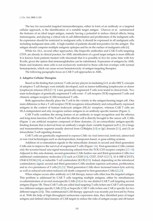
J. Clin. Med. 2019, 8, 200
The key for successful targeted immunotherapies, either in form of an antibody or a targetedcellular approach, is the identification of a suitable target antigen. Cheever et al. summarizedthe features of an ideal target antigen, namely having a potential to induce clinical effects, beingimmunogenic, and playing a critical role in cell differentiation and proliferation of the malignant cells.Its expression should be restricted to malignant cells; it should be expressed in all malignant cellsincluding malignant stem cells. A high number of patients should test positive for the antigen. Theantigen should comprise multiple antigenic epitopes and be on the surface of malignant cells [6].
While for ALL, several other approaches, like bispecific antibodies and CAR-T-cells targetingCD19, are already in clinical practice, for AML identification of a good target antigen is more difficult.It is known from patients treated with rituximab that it is possible to live for some time with fewB-cells, given the option that immunoglobulins can be substituted. Expression of antigens by AMLblasts and leukemic stem cells is not exclusively restricted to those cells but overlaps with normalhematopoiesis, which can cause severe hematotoxicity of antigen-targeting therapies.
The following paragraphs focus on CAR-T cell approaches in AML.
2. Adoptive Cellular Therapies
Based on the finding that cytotoxic T cells are key players in mediating GvL in allo-HSCT, conceptsof adoptive T cell therapy were initially developed, such as tumor-infiltrating lymphocytes or donorlymphocyte infusion (DLI) [7–9]. Later, genetically engineered T cells were tested in clinical trials. Twomain technologies of genetically engineered T cells exist—T cell receptor (TCR) engineered T cells andchimeric antigen receptor (CAR) transduced T cells.
Both approaches directly place the T cell in the vicinity to the antigen-bearing target cell. Onemain difference is that a T cell receptor (TCR) recognizes intracellularly and extracelluarly expressedantigens in the context of human leukocyte antigen (HLA)- receptors, whereas CAR T cells areHLA-independent and only recognize surface antigens in an antibody-specific manner (Figure 1).
CAR T-cells combine the strong feature of an antibody in target recognition and the effector,and long-term function of the T-cell and the effector cell is directly brought to the cancer cell. CARs(Figure 1) are artificial receptors composed of three domains, (1) an extracellular antigen-specificbinding domain that is derived from an antibody’s single chain variable fragment (scFv), (2) a hingeand transmembrane segment usually derived from CD8alpha [10] or IgG domain [11], and (3) anintracellular T-cell signaling domain.
CAR T cells are genetically engineered to express CARs via viral (retroviral, lentiviral, adenoviral)or non-viral technologies such as electroporation, transposon-based, or gene-editing systems.
Addition of co-stimulation signals to the intracellular domain in second and third generationCARs aim to improve the survival of engineered T cells (Figure 1A). First-generation CARs containonly the tyrosine-based zeta-signal-transducing subunit from the TCR/CD3 receptor complex [12–14].Adjacent to this zeta-domain, second-generation CARs harbor one and third-generation CARs twoadditional costimulatory molecules [15] such as CD28 [16], CD27, DAP-12 [17], 10 4-1BB (CD137),OX40 (CD134) [18], or inducible T cell costimulator (ICOS) [19]. Indeed, depending on the introducedcostimulatory signal, second and third generation CARs mediate superior activation, proliferation,and in vivo persistence of T cells [20,21]. Third generation CARs show increased tumor-lytic activityas well as reduced activation-induced cell death compared to first-generation CARs [22,23].
When relapse occurs after antibody or CAR therapy, tumor cells often lose the targeted antigen.This problem is addressed by CAR T cells targeting multiple antigens, either by simultaneousco-administration of several monospecific CARs [24] or by one distinct CAR T cell targeting severalantigens (Figure 1B). These CAR T cells are called dual-targeting T cells (when one CAR T cell expressestwo different antigen-specific CARs [25]) or bispecific CAR T cells (when one CAR is specific for twodifferent targets [26]). This combinatorial CAR therapy approach was recently put forward by Pernaet al. With the help of high-throughput surfaceome expression data, they identified pairs of targetantigens and defined ideal features of CAR targets to reduce the risks of antigen escape and off-tumor
120
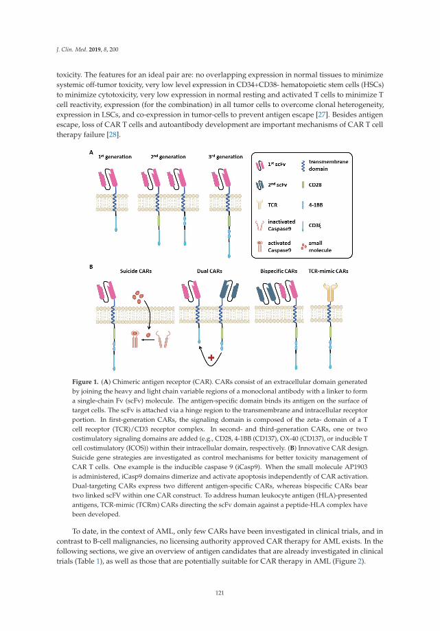
J. Clin. Med. 2019, 8, 200
toxicity. The features for an ideal pair are: no overlapping expression in normal tissues to minimizesystemic off-tumor toxicity, very low level expression in CD34+CD38- hematopoietic stem cells (HSCs)to minimize cytotoxicity, very low expression in normal resting and activated T cells to minimize Tcell reactivity, expression (for the combination) in all tumor cells to overcome clonal heterogeneity,expression in LSCs, and co-expression in tumor-cells to prevent antigen escape [27]. Besides antigenescape, loss of CAR T cells and autoantibody development are important mechanisms of CAR T celltherapy failure [28].
Figure 1. (A) Chimeric antigen receptor (CAR). CARs consist of an extracellular domain generatedby joining the heavy and light chain variable regions of a monoclonal antibody with a linker to forma single-chain Fv (scFv) molecule. The antigen-specific domain binds its antigen on the surface oftarget cells. The scFv is attached via a hinge region to the transmembrane and intracellular receptorportion. In first-generation CARs, the signaling domain is composed of the zeta- domain of a Tcell receptor (TCR)/CD3 receptor complex. In second- and third-generation CARs, one or twocostimulatory signaling domains are added (e.g., CD28, 4-1BB (CD137), OX-40 (CD137), or inducible Tcell costimulatory (ICOS)) within their intracellular domain, respectively. (B) Innovative CAR design.Suicide gene strategies are investigated as control mechanisms for better toxicity management ofCAR T cells. One example is the inducible caspase 9 (iCasp9). When the small molecule AP1903is administered, iCasp9 domains dimerize and activate apoptosis independently of CAR activation.Dual-targeting CARs express two different antigen-specific CARs, whereas bispecific CARs beartwo linked scFV within one CAR construct. To address human leukocyte antigen (HLA)-presentedantigens, TCR-mimic (TCRm) CARs directing the scFv domain against a peptide-HLA complex havebeen developed.
To date, in the context of AML, only few CARs have been investigated in clinical trials, and incontrast to B-cell malignancies, no licensing authority approved CAR therapy for AML exists. In thefollowing sections, we give an overview of antigen candidates that are already investigated in clinicaltrials (Table 1), as well as those that are potentially suitable for CAR therapy in AML (Figure 2).
121
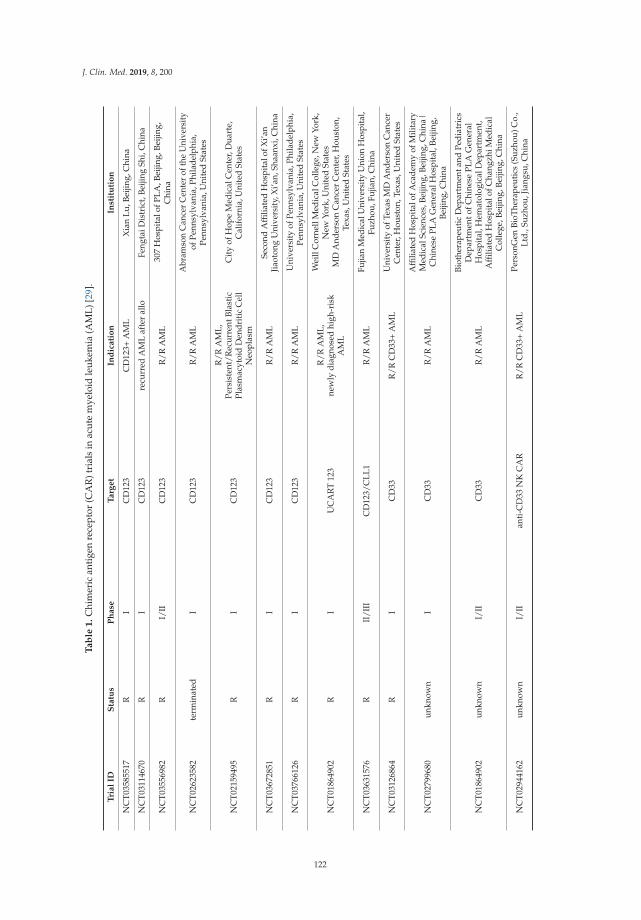
J. Clin. Med. 2019, 8, 200
Ta
ble
1.
Chi
mer
ican
tige
nre
cept
or(C
AR
)tri
als
inac
ute
mye
loid
leuk
emia
(AM
L)[2
9].
Tri
al
IDS
tatu
sP
ha
seT
arg
et
Ind
ica
tio
nIn
stit
uti
on
NC
T035
8551
7R
IC
D12
3C
D12
3+A
ML
Xia
nLu
,Bei
jing,
Chi
na
NC
T031
1467
0R
IC
D12
3re
curr
edA
ML
afte
ral
loFe
ngta
iDis
tric
t,Be
ijing
Shi,
Chi
na
NC
T035
5698
2R
I/II
CD
123
R/R
AM
L30
7H
ospi
talo
fPLA
,Bei
jing,
Beiji
ng,
Chi
na
NC
T026
2358
2te
rmin
ated
IC
D12
3R
/RA
ML
Abr
amso
nC
ance
rC
ente
rof
the
Uni
vers
ityof
Penn
sylv
ania
,Phi
lade
lphi
a,Pe
nnsy
lvan
ia,U
nite
dSt
ates
NC
T021
5949
5R
IC
D12
3
R/R
AM
L,Pe
rsis
tent
/Rec
urre
ntBl
asti
cPl
asm
acyt
oid
Den
drit
icC
ell
Neo
plas
m
Cit
yof
Hop
eM
edic
alC
ente
r,D
uart
e,C
alif
orni
a,U
nite
dSt
ates
NC
T036
7285
1R
IC
D12
3R
/RA
ML
Seco
ndA
ffilia
ted
Hos
pita
lofX
i’an
Jiao
tong
Uni
vers
ity,
Xi’a
n,Sh
aanx
i,C
hina
NC
T037
6612
6R
IC
D12
3R
/RA
ML
Uni
vers
ity
ofPe
nnsy
lvan
ia,P
hila
delp
hia,
Penn
sylv
ania
,Uni
ted
Stat
es
NC
T018
6490
2R
IU
CA
RT
123
R/R
AM
L,ne
wly
diag
nose
dhi
gh-r
isk
AM
L
Wei
llC
orne
llM
edic
alC
olle
ge,N
ewYo
rk,
New
York
,Uni
ted
Stat
esM
DA
nder
son
Can
cer
Cen
ter,
Hou
ston
,Te
xas,
Uni
ted
Stat
es
NC
T036
3157
6R
II/I
IIC
D12
3/C
LL1
R/R
AM
LFu
jian
Med
ical
Uni
vers
ityU
nion
Hos
pita
l,Fu
zhou
,Fuj
ian,
Chi
na
NC
T031
2686
4R
IC
D33
R/R
CD
33+
AM
LU
nive
rsit
yof
Texa
sM
DA
nder
son
Can
cer
Cen
ter,
Hou
ston
,Tex
as,U
nite
dSt
ates
NC
T027
9968
0un
know
nI
CD
33R
/RA
ML
Affi
liate
dH
ospi
talo
fAca
dem
yof
Mili
tary
Med
ical
Scie
nces
,Bei
jing,
Beiji
ng,C
hina
|C
hine
sePL
AG
ener
alH
ospi
tal,
Beiji
ng,
Beiji
ng,C
hina
NC
T018
6490
2un
know
nI/
IIC
D33
R/R
AM
L
Biot
hera
peut
icD
epar
tmen
tand
Pedi
atri
csD
epar
tmen
tofC
hine
sePL
AG
ener
alH
ospi
tal,
Hem
atol
ogic
alD
epar
tmen
t,A
ffilia
ted
Hos
pita
lofC
hang
zhiM
edic
alC
olle
ge,B
eijin
g,Be
ijing
,Chi
na
NC
T029
4416
2un
know
nI/
IIan
ti-C
D33
NK
CA
RR
/RC
D33
+A
ML
Pers
onG
enBi
oThe
rape
utic
s(S
uzho
u)C
o.,
Ltd.
,Suz
hou,
Jian
gsu,
Chi
na
122

J. Clin. Med. 2019, 8, 200
Ta
ble
1.
Con
t.
Tri
al
IDS
tatu
sP
ha
seT
arg
et
Ind
ica
tio
nIn
stit
uti
on
NC
T032
9144
4R
IC
D33
,CD
38C
D56
,CD
117,
CD
123,
CD
34,a
ndM
uc1
for
AM
Lan
dM
DS
R/R
AM
L,M
DS;
ALL
Zhu
jiang
Hos
pita
l,So
uthe
rnM
edic
alU
nive
rsit
y,G
uang
zhou
,Gua
ngdo
ng,
Chi
na
NC
T034
7345
7R
n.a.
sing
leC
AR
-Tor
doub
leC
AR
-Tce
llsw
ith
CD
33,C
D38
,CD
56,C
D12
3,C
D11
7,C
D13
3,C
D34
,or
Muc
lR
/RA
ML
Sout
hern
Med
ical
Uni
vers
ity
Zhu
jiang
Hos
pita
l,G
uang
dong
,Gua
ngdo
ng,C
hina
NC
T032
2267
4R
I/II
Muc
1/C
LL1/
CD
33/C
D38
/CD
56/C
D12
3A
ML
Zhu
jiang
Hos
pita
lofS
outh
ern
Med
ical
Uni
vers
ity,
Gua
ngzh
ou,G
uang
dong
,C
hina
|Sh
enzh
enG
eno-
imm
une
Med
ical
Inst
itute
,Sh
enzh
en,G
uang
dong
,Chi
na|
Yunn
anC
ance
rH
ospi
tal&
The
Thi
rdA
ffilia
ted
Hos
pita
lofK
unm
ing
Med
ical
Uni
vers
ity
&Yu
nnan
Can
cer
Cen
ter,
Kun
Min
g,Yu
nnan
,Chi
na
NC
T022
0382
5co
mpl
eted
IN
KG
2DA
ML,
MD
S-R
AEB
,and
Mul
tipl
eM
yelo
ma.
Dan
a-Fa
rber
Can
cer
Inst
itut
e,Bo
ston
,M
assa
chus
etts
,Uni
ted
Stat
es
NC
T030
1840
5R
I/II
NK
R2
(NK
G2D
)R
/RA
ML,
AM
L,M
yelo
ma
Tam
pa,F
lori
da,U
nite
dSt
ates
|Bu
ffal
o,N
ewYo
rk,U
nite
dSt
ates
|Br
usse
ls,B
elgi
um|B
russ
els,
Belg
ium
|G
hent
,Bel
gium
NC
T030
1840
5un
know
nI/
IIC
D7/
NK
92ce
llC
D7+
R/R
Leuk
emia
and
Lym
phom
aPe
rson
Gen
BioT
hera
peut
ics
(Suz
hou)
Co.
,Lt
d.,S
uzho
u,Ji
angs
u,C
hina
NC
T017
1636
4un
know
nI
Lew
isY
Mye
lom
a,A
ML,
MD
SPe
ter
Mac
Cal
lum
Can
cer
Cen
tre,
Mel
bour
ne,V
icto
ria,
Aus
tral
ia
Abb
revi
atio
ns:R
,rec
ruit
ing;
r/r
rela
psed
/ref
ract
ory;
AM
L,a
cute
mye
loid
leuk
emia
;AL
L,a
cute
lym
phob
last
icle
ukem
ia;M
DS,
mye
lod
yspl
asti
csy
ndro
me.
Not
e:Se
arch
term
:CA
R,
AM
L(a
sby
12D
ecem
ber
2018
).So
urce
:[29
].
123
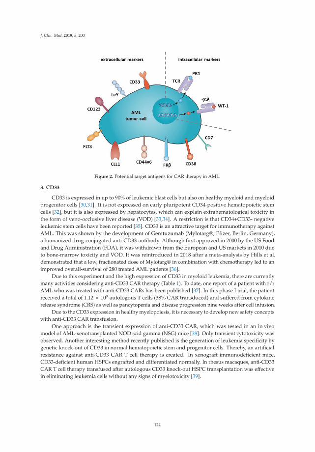
J. Clin. Med. 2019, 8, 200
Figure 2. Potential target antigens for CAR therapy in AML.
3. CD33
CD33 is expressed in up to 90% of leukemic blast cells but also on healthy myeloid and myeloidprogenitor cells [30,31]. It is not expressed on early pluripotent CD34-positive hematopoietic stemcells [32], but it is also expressed by hepatocytes, which can explain extrahematological toxicity inthe form of veno-occlusive liver disease (VOD) [33,34]. A restriction is that CD34+CD33- negativeleukemic stem cells have been reported [35]. CD33 is an attractive target for immunotherapy againstAML. This was shown by the development of Gemtuzumab (Mylotarg®, Pfizer, Berlin, Germany),a humanized drug-conjugated anti-CD33-antibody. Although first approved in 2000 by the US Foodand Drug Administration (FDA), it was withdrawn from the European and US markets in 2010 dueto bone-marrow toxicity and VOD. It was reintroduced in 2018 after a meta-analysis by Hills et al.demonstrated that a low, fractionated dose of Mylotarg® in combination with chemotherapy led to animproved overall-survival of 280 treated AML patients [36].
Due to this experiment and the high expression of CD33 in myeloid leukemia, there are currentlymany activities considering anti-CD33 CAR therapy (Table 1). To date, one report of a patient with r/rAML who was treated with anti-CD33 CARs has been published [37]. In this phase I trial, the patientreceived a total of 1.12 × 10⁹ autologous T-cells (38% CAR transduced) and suffered from cytokinerelease syndrome (CRS) as well as pancytopenia and disease progression nine weeks after cell infusion.
Due to the CD33 expression in healthy myelopoiesis, it is necessary to develop new safety conceptswith anti-CD33 CAR transfusion.
One approach is the transient expression of anti-CD33 CAR, which was tested in an in vivomodel of AML-xenotransplanted NOD scid gamma (NSG) mice [38]. Only transient cytotoxicity wasobserved. Another interesting method recently published is the generation of leukemia specificity bygenetic knock-out of CD33 in normal hematopoietic stem and progenitor cells. Thereby, an artificialresistance against anti-CD33 CAR T cell therapy is created. In xenograft immunodeficient mice,CD33-deficient human HSPCs engrafted and differentiated normally. In rhesus macaques, anti-CD33CAR T cell therapy transfused after autologous CD33 knock-out HSPC transplantation was effectivein eliminating leukemia cells without any signs of myelotoxicity [39].
124

J. Clin. Med. 2019, 8, 200
4. Lewis Y (LeY)
Lewis Y (LeY) is a carbohydrate antigen that is overexpressed by a wide variety of epithelialcancers [40] and hematological malignancies including AML [41,42] but with only limited expressionon normal tissue [43].
In 2010, Peinert and colleagues published the results of the first phase I CAR- T cell trial forrelapsed LeY-expressing AML [44]. They investigated an autologous second-generation anti-LeY CARin four patients who received up to 1.3 × 10⁹ total T cells (14–38% with anti-LeY CAR expression). Nograde three or four toxicity was observed. The best response was transient cytogenetic remission in onepatient; another patient showed a transient reduction of blasts, and two patients showed stable disease.All patients relapsed after 28 days to 23 months after adoptive cell therapy. CAR T cell persistence wasdemonstrated for up to 10 months.
5. CD123
CD123 is the transmembrane alpha chain of the interleukin 3 receptor. Due to its surface expressionand its overexpression on AML blasts and LSCs, as well as its low expression on normal hematopoeieticstem cells, CD123 qualifies as a suitable target [45,46]. However, similar to CD33 targeted therapy, theproblem of myelotoxicity in CD123 targeted therapy remains.
At the time of writing this manuscript, CD123 is being studied in 11 clinical trials for AML(Table 1).
CARs normally encode in its scFvs a VH and VL chain from one monoclonal antibody in theextracellular antigen binding domain. In an experimental AML model, hematopoietic toxicity wasshown after treatment with anti-CD123 CAR T cells. When using VH and VL chains derived fromdifferent CD123-specific mAbs for CAR engineering, one specific combination showed less lysis of thenormal hematopoietic stem cells while preserving the toxicity [47].
6. FLT3 (CD135)
FLT3-ITD mutations are found in about 20% (and FLT3-TKD in about 7%) of all AML patients [48].In a preclinical model, second-generation anti-FLT3-41BB CARs were tested [49]. Specific cytotoxicityagainst FLT3+ leukemia cell lines and primary cell lines in vitro, as well as little off-tumor cytotoxicityon normal hematopoietic stem cells, was observed. In a xenograft mouse model, prolonged survivalwas seen in FLT3+ mice that were treated with the anti-FLT3 CARs. Compared to anti-CD33 CAR Tcells, less toxicity to hematopoietic stem cells and multipotent myeloid progenitor cells and equivalenttoxicity to common myeloid progenitor and granulocyte-macrophage progenitor cells was described,suggesting a lower hematologic toxicity with anti-FLT3 CAR T cells. In a second preclinical study,second-generation 4-1BB CARs that target the FLT3-ligand (FLT3L) were tested [50]. For anti-FLT3L,little off-tumor cytotoxicity on normal hematopoietic stem and progenitor cells was observed. Axenograft mouse model also showed a significantly prolonged survival in FLT3+ leukemia bearingmice after anti-FLT3L CAR T cells [50].
7. CLL1
The myeloid surface antigen C-type lectin-like molecule 1 (CLL1 or CLEC12A) is a glycoproteinhighly expressed by the majority of AML patients. It is expressed on AML blasts and on normallydifferentiated myeloid cells. Relatively low amounts are expressed on CD34+ progenitor cells. It is notexpressed on normal hematopoietic stem cells [51]. CLL 1 therefore qualifies as a promising CAR Tcell target suggesting low “off-tumor” toxicity.
Four research groups generated anti-CLL1 CAR T cells, three second generation [52–54] and onethird generation CAR [55]. All four showed potent activity against CLL1+ AML cell lines, as well asprimary CLL1+ AML blasts in vitro and in xenograft mouse models, while sparing normal myeloid
125

J. Clin. Med. 2019, 8, 200
precursor cells. Tashiro et al. went a step further and introduced the inducible caspase-9 suicide genesystem into the CARs and could successfully control anti-CLL1 CAR T cell activity in vitro and in vivo.
8. CD44v6
CD44v6 is the isoform variant 6 of the hyaluronic acid receptor CD44, a class I membraneglycoprotein overexpressed in hematologic malignancies including AML [56] and epithelial tumors [57].It is absent in hematopoietic stem cells [58] and shows low expression levels on normal cells. Casucciand colleagues designed a second generation anti-CD44v6 CAR with cytotoxicity against AMLcells while sparing normal hematopoietic stem cells [59]. Monocytopenia was the dose limitingtoxicity in this preclinical study. To control this adverse event, the clinical-grade suicide genes,thymidine kinase [60] and the nonimmunogenic inducible Caspase 9 (iC9) [61], were coexpressed inthe anti-CD44v6 CARs with iC9 successfully eradicating the CAR T cells within hours.
9. Folate Receptor ß (FRß)
Folate receptor ß (FRß) is primarily expressed on myeloid-lineage hematopoietic cells [62] andis expressed on about 70% of primary AML cells [63]. The expression of FRß on AML blasts canbe increased by all-trans retinoic acid (ATRA) and enhanced the efficacy of folate-conjugated drugtherapy in a preclinical study [64,65]. Preclinical models showed the efficacy of anti-FRß CAR T cellsand an even better efficacy of high-affinity anti-FRß CAR T cells against AML cells in vitro and in vivowithout toxicity against healthy hematopoietic progenitor/stem cells (HPSCs) [66,67].
10. CD38
CD38 is expressed on the majority of AML blasts but not healthy human hematopoietic stemcells [68,69]. Due to the modest expression level of CD38 in AML, the combination of ATRA andsecond generation anti-CD38 CAR T cells to enhance the CD38 expression was tested [70]. In thisstudy, ATRA enhanced the cytotoxicity of anti-CD38 CAR T cells on AML cells with the augmentedCD38 expression in vitro.
11. CD7
CD7 is expressed by T cells and natural killer cells [71]; it is also expressed in over 90% oflymphoblastic T cell leukemia and lymphoma [72,73] and in about 30% of AML cases [74,75], but isabsent in normal myeloid and erythroid cells. An anti-CD7 CAR in CD7 knock-out T cells to preventfratricide can effectively eliminate CD7 + AML cell lines as well as primary AML cells while sparingnormal myeloid and erythroid progenitor cells [76–78].
12. Intracellular Targets: PR1/HLA-A2; WT1/HLA-A2
The majority of leukemia-associated-antigens and neoantigens are intracellularly processed andpresented by HLA class II molecules. To address HLA-presented antigens, TCR-mimic (TCRm) CARsdirecting the scFv domain against a peptide-HLA complex were developed. Proteinase 1 (PR1) is aHLA A2-restricted nonamer derived from the leukemia associated antigen proteinase 3 and neutrophilelastase. Both proteases are expressed in the primary azurophilic granules of neutrophils and areoverexpressed in myeloid leukemic blasts [79,80]. A second-generation CAR construct targetingHLA-A2/PR1 was preferentially cytotoxic against human AML cell lines and primary AML blastsin vitro [81]. The second TCRm CAR published targets the leukemia associated antigen Wilms tumor1 (WT1) in the context of HLA-A2 and has demonstrated efficacy in vivo in an AML mouse model [82].WT1 is overexpressed in AML, chronic myeloid leukemia (CML), and several solid tumors [83–85].Another antigen candidate is PRAME (preferentially expressed antigen in melanoma). It is a so-calledcancer-testis antigen and is therefore exclusively expressed in the testes and ovaries in healthy tissue.However, in several malignant tissues—and in about 20–40% of AML cases—it is intracellularly
126

J. Clin. Med. 2019, 8, 200
expressed and presented on the cell surface via human leukocyte antigen (HLA)-I. Chang et al.developed a TCRm human IgG1 antibody that recognizes a decamer peptide derived from PRAMEin the context with HLA-A. It showed therapeutical effectiveness against mouse xenograft models ofhuman leukemia [86]. In addition, a multicenter phase I/II clinical trial is currently testing autologousT cells that are transduced with a PRAME-specific HLA-A*02:01-restricted TCR (NCT03503968).
Whether autoimmune reactions as off-tumor toxicity occur with TCRm CAR application has to beinvestigated in further studies.
13. Safety Affairs
Relevant side effects of CARs are tumor-lysis syndrome and cytokine release syndrome, as wellas “on-target but off-tumor” toxicity. “On-target but off-tumor” toxicity occurs when the target antigenis not only expressed on the target cells but also on normal tissues. This is the case for HER2, whichis expressed in epithelial cells in the gastrointestinal, respiratory, reproductive, urinary tract, skin,breast, placenta, and normal hematopoietic cells [87]. A clinical trial investigating a third generationCAR targeting HER2 reported of one patient who developed respiratory distress within 15 min afterreceiving a single dose of 10ˆ10 CAR T cells, followed by cardiac arrest [88]. This study underlines howimportant the target antigen selection is. Other aspects of reducing toxicity, mainly cytokine storm,are the number of infused CARs as well as the use of immunosuppressive agents and an introducedcontrol mechanism into the CARs (Figure 1B). As control mechanisms, several suicide gene strategieswere investigated, including thymidine kinase gene of the herpes simplex virus thymidine kinase(HSV-TK) [89] and the inducible caspase 9 (iCasp9) [90]. An elegant approach to limit “off-tumor”toxicity is to modify the CAR scFvs affinity of the antibody [91]. For high affinity HER2 CAR T cells,it was demonstrated that in dependence of the antigen density on the surface of the target cell, ahigh-affinity CAR is reactive against a malignant (high density) but not a normal (low density) cell [91].
14. Conclusions
Although in B-cell malignancies, CAR T cells now begin to build one therapeutic column inclinical practice, the value of CAR T cell therapy for AML still has to be determined.
There are major hurdles to take, e.g., finding the right antigen with low “off-tumor” toxicity andsupplementing strategies to minimize “off-tumor” toxicity. Several attempts have already been made,such as suicidal control of CAR T cells, temporary expression of the CAR, and improvement of theaffinity of the CAR. The CAR race has started and will hopefully improve and enrich the therapeuticarmentarium against AML.
Acknowledgments: We thank M. Hinkelbein for support with editing text and references.
Conflicts of Interest: The authors declare no conflicts of interest.
References
1. Döhner, H.; Weisdorf, D.J.; Bloomfield, C.D. Acute myeloid leukemia. N. Engl. J. Med. 2015, 373, 1136–1152.[CrossRef] [PubMed]
2. Döhner, H.; Estey, E.H.; Amadori, S.; Appelbaum, F.R.; Büchner, T.; Burnett, A.K.; Dombret, H.; Fenaux, P.;Grimwade, D.; Larson, R.A.; et al. Diagnosis and management of acute myeloid leukemia in adults:Recommendations from an international expert panel, on behalf of the european leukemianet. Blood 2010,115, 453–474. [CrossRef] [PubMed]
3. Eppert, K.; Takenaka, K.; Lechman, E.R.; Waldron, L.; Nilsson, B.; van Galen, P.; Metzeler, K.H.; Poeppl, A.;Ling, V.; Beyene, J.; et al. Stem cell gene expression programs influence clinical outcome in human leukemia.Nat. Med. 2011, 17, 1086–1093. [CrossRef] [PubMed]
4. Bonnet, D.; Dick, J.E. Human acute myeloid leukemia is organized as a hierarchy that originates from aprimitive hematopoietic cell. Nat. Med. 1997, 3, 730–737. [CrossRef]
5. Available online: https://seer.cancer.gov/statfacts/html/amyl.html (accessed on 5 February 2019).
127
J. Clin. Med. 2019, 8, 200
6. Cheever, M.A.; Allison, J.P.; Ferris, A.S.; Finn, O.J.; Hastings, B.M.; Hecht, T.T.; Mellman, I.; Prindiville, S.A.;Viner, J.L.; Weiner, L.M.; et al. The prioritization of cancer antigens: A national cancer institute pilot projectfor the acceleration of translational research. Clin. Cancer Res. 2009, 15, 5323–8337. [CrossRef] [PubMed]
7. Kolb, H.J.; Mittermuller, J.; Clemm, C.; Holler, E.; Ledderose, G.; Brehm, G.; Heim, M.; Wilmanns, W. Donorleukocyte transfusions for treatment of recurrent chronic myelogenous leukemia in marrow transplantpatients. Blood 1990, 76, 2462–2465. [PubMed]
8. Kolb, H.J.; Schattenberg, A.; Goldman, J.M.; Hertenstein, B.; Jacobsen, N.; Arcese, W.; Ljungman, P.;Ferrant, A.; Verdonck, L.; Niederwieser, D.; et al. Graft-versus-leukemia effect of donor lymphocytetransfusions in marrow grafted patients. Blood 1995, 86, 2041–2050.
9. Kolb, H.J.; Schmid, C.; Barrett, A.J.; Schendel, D.J. Graft-versus-leukemia reactions in allogeneic chimeras.Blood 2004, 103, 767–776. [CrossRef]
10. Maude, S.L.; Frey, N.; Shaw, P.A.; Aplenc, R.; Barrett, D.M.; Bunin, N.J.; Chew, A.; Gonzalez, V.E.; Zheng, Z.;Lacey, S.F.; et al. Chimeric antigen receptor t cells for sustained remissions in leukemia. N. Engl. J. Med. 2014,371, 1507–1517. [CrossRef]
11. Turtle, C.J.; Hanafi, L.A.; Berger, C.; Gooley, T.A.; Cherian, S.; Hudecek, M.; Sommermeyer, D.; Melville, K.;Pender, B.; Budiarto, T.M.; et al. Cd19 car-t cells of defined cd4+:Cd8+ composition in adult b cell all patients.J. Clin. Investig. 2016, 126, 2123–2138. [CrossRef]
12. Eshhar, Z.; Waks, T.; Gross, G.; Schindler, D.G. Specific activation and targeting of cytotoxic lymphocytesthrough chimeric single chains consisting of antibody-binding domains and the gamma or zeta subunits ofthe immunoglobulin and t-cell receptors. Proc. Natl. Acad. Sci. USA 1993, 90, 720–724. [CrossRef] [PubMed]
13. Jensen, M.C.; Popplewell, L.; Cooper, L.J.; DiGiusto, D.; Kalos, M.; Ostberg, J.R.; Forman, S.J. Antitransgenerejection responses contribute to attenuated persistence of adoptively transferred cd20/cd19-specific chimericantigen receptor redirected t cells in humans. Biol. Blood Marrow Trans. 2010, 16, 1245–1256. [CrossRef][PubMed]
14. Stancovski, I.; Schindler, D.G.; Waks, T.; Yarden, Y.; Sela, M.; Eshhar, Z. Targeting of t lymphocytes toneu/her2-expressing cells using chimeric single chain fv receptors. J. Immunol. 1993, 151, 6577–6582.[PubMed]
15. Till, B.G.; Jensen, M.C.; Wang, J.; Qian, X.; Gopal, A.K.; Maloney, D.G.; Lindgren, C.G.; Lin, Y.; Pagel, J.M.;Budde, L.E.; et al. Cd20-specific adoptive immunotherapy for lymphoma using a chimeric antigen receptorwith both cd28 and 4-1bb domains: Pilot clinical trial results. Blood 2012, 119, 3940–3950. [CrossRef][PubMed]
16. Finney, H.M.; Lawson, A.D.G.; Bebbington, C.R.; Weir, A.N.C. Chimeric receptors providing both primaryand costimulatory signaling in t cells from a single gene product. J. Immunol. 1998, 161, 2791. [PubMed]
17. Wang, E.; Wang, L.C.; Tsai, C.Y.; Bhoj, V.; Gershenson, Z.; Moon, E.; Newick, K.; Sun, J.; Lo, A.; Baradet, T.;et al. Generation of potent t-cell immunotherapy for cancer using dap12-based, multichain, chimericimmunoreceptors. Cancer Immunol. Res. 2015, 3, 815–826. [CrossRef] [PubMed]
18. Hombach, A.A.; Chmielewski, M.; Rappl, G.; Abken, H. Adoptive immunotherapy with redirected t cellsproduces ccr7- cells that are trapped in the periphery and benefit from combined cd28-ox40 costimulation.Hum. Gene Ther. 2013, 24, 259–269. [CrossRef]
19. Guedan, S.; Chen, X.; Madar, A.; Carpenito, C.; McGettigan, S.E.; Frigault, M.J.; Lee, J.; Posey, A.D., Jr.;Scholler, J.; Scholler, N.; et al. Icos-based chimeric antigen receptors program bipolar th17/th1 cells. Blood2014, 124, 1070–1080. [CrossRef]
20. Zhao, Y.; Wang, Q.J.; Yang, S.; Kochenderfer, J.N.; Zheng, Z.; Zhong, X.; Sadelain, M.; Eshhar, Z.;Rosenberg, S.A.; Morgan, R.A. A herceptin-based chimeric antigen receptor with modified signaling domainsleads to enhanced survival of transduced t lymphocytes and antitumor activity. J. Immunol. 2009, 183,5563–5574. [CrossRef]
21. Savoldo, B.; Ramos, C.A.; Liu, E.; Mims, M.P.; Keating, M.J.; Carrum, G.; Kamble, R.T.; Bollard, C.M.;Gee, A.P.; Mei, Z.; et al. Cd28 costimulation improves expansion and persistence of chimeric antigenreceptor-modified t cells in lymphoma patients. J. Clin. Investig. 2011, 121, 1822–1826. [CrossRef]
22. Han, E.Q.; Li, X.L.; Wang, C.R.; Li, T.F.; Han, S.Y. Chimeric antigen receptor-engineered t cells for cancerimmunotherapy: Progress and challenges. J. Hematol. Oncol. 2013, 6, 47. [CrossRef] [PubMed]
128

J. Clin. Med. 2019, 8, 200
23. Long, A.H.; Haso, W.M.; Shern, J.F.; Wanhainen, K.M.; Murgai, M.; Ingaramo, M.; Smith, J.P.; Walker, A.J.;Kohler, M.E.; Venkateshwara, V.R.; et al. 4-1bb costimulation ameliorates t cell exhaustion induced by tonicsignaling of chimeric antigen receptors. Nat. Med. 2015, 21, 581–590. [CrossRef] [PubMed]
24. Ruella, M.; Barrett, D.M.; Kenderian, S.S.; Shestova, O.; Hofmann, T.J.; Perazzelli, J.; Klichinsky, M.;Aikawa, V.; Nazimuddin, F.; Kozlowski, M.; et al. Dual cd19 and cd123 targeting prevents antigen-lossrelapses after cd19-directed immunotherapies. J. Clin. Investig. 2016, 126, 3814–3826. [CrossRef] [PubMed]
25. Wilkie, S.; van Schalkwyk, M.C.; Hobbs, S.; Davies, D.M.; van der Stegen, S.J.; Pereira, A.C.; Burbridge, S.E.;Box, C.; Eccles, S.A.; Maher, J. Dual targeting of erbb2 and muc1 in breast cancer using chimeric antigenreceptors engineered to provide complementary signaling. J. Clin. Immunol. 2012, 32, 1059–1070. [CrossRef][PubMed]
26. Zah, E.; Lin, M.Y.; Silva-Benedict, A.; Jensen, M.C.; Chen, Y.Y. T cells expressing cd19/cd20 bispecificchimeric antigen receptors prevent antigen escape by malignant b cells. Cancer Immunol. Res. 2016, 4,498–508. [CrossRef] [PubMed]
27. Perna, F.; Berman, S.H.; Soni, R.K.; Mansilla-Soto, J.; Eyquem, J.; Hamieh, M.; Hendrickson, R.C.;Brennan, C.W.; Sadelain, M. Integrating proteomics and transcriptomics for systematic combinatorialchimeric antigen receptor therapy of aml. Cancer Cell 2017, 32, 506–519. [CrossRef] [PubMed]
28. Dotti, G.; Gottschalk, S.; Savoldo, B.; Brenner, M.K. Design and development of therapies using chimericantigen receptor-expressing t cells. Immunol. Rev. 2014, 257, 107–126. [CrossRef]
29. Available online: https://clinicaltrials.gov/ct2/results?cond=aml&term=car&cntry=&state=&city=&dist=(accessed on 12 December 2018).
30. Griffin, J.D.; Linch, D.; Sabbath, K.; Larcom, P.; Schlossman, S.F. A monoclonal antibody reactive with normaland leukemic human myeloid progenitor cells. Leuk. Res. 1984, 8, 521–534. [CrossRef]
31. Nguyen, D.H.; Ball, E.D.; Varki, A. Myeloid precursors and acute myeloid leukemia cells express multiplecd33-related siglecs. Exp. Hematol. 2006, 34, 728–735. [CrossRef]
32. Hauswirth, A.W.; Florian, S.; Printz, D.; Sotlar, K.; Krauth, M.T.; Fritsch, G.; Schernthaner, G.H.; Wacheck, V.;Selzer, E.; Sperr, W.R.; et al. Expression of the target receptor cd33 in cd34+/cd38-/cd123+ aml stem cells.Eur. J. Clin. Investig. 2007, 37, 73–82. [CrossRef]
33. Thol, F.; Schlenk, R.F. Gemtuzumab ozogamicin in acute myeloid leukemia revisited. Expert Opin. Biol. Ther.2014, 14, 1185–1195. [CrossRef] [PubMed]
34. Giles, F.J.; Kantarjian, H.M.; Kornblau, S.M.; Thomas, D.A.; Garcia-Manero, G.; Waddelow, T.A.; David, C.L.;Phan, A.T.; Colburn, D.E.; Rashid, A.; et al. Mylotarg (gemtuzumab ozogamicin) therapy is associated withhepatic venoocclusive disease in patients who have not received stem cell transplantation. Cancer 2001, 92,406–413. [CrossRef]
35. Walter, R.B.; Appelbaum, F.R.; Estey, E.H.; Bernstein, I.D. Acute myeloid leukemia stem cells andcd33-targeted immunotherapy. Blood 2012, 119, 6198. [CrossRef] [PubMed]
36. Hills, R.K.; Castaigne, S.; Appelbaum, F.R.; Delaunay, J.; Petersdorf, S.; Othus, M.; Estey, E.H.; Dombret, H.;Chevret, S.; Ifrah, N.; et al. Addition of gemtuzumab ozogamicin to induction chemotherapy in adult patientswith acute myeloid leukaemia: A meta-analysis of individual patient data from randomised controlled trials.Lancet Oncol. 2014, 15, 986–996. [CrossRef]
37. Wang, Q.-S.; Wang, Y.; Lv, H.-Y.; Han, Q.-W.; Fan, H.; Guo, B.; Wang, L.-L.; Han, W.-D. Treatment ofcd33-directed chimeric antigen receptor-modified t cells in one patient with relapsed and refractory acutemyeloid leukemia. Mol. Ther. 2015, 23, 184–191. [CrossRef]
38. Kenderian, S.S.; Ruella, M.; Shestova, O.; Klichinsky, M.; Aikawa, V.; Morrissette, J.J.; Scholler, J.; Song, D.;Porter, D.L.; Carroll, M.; et al. Cd33-specific chimeric antigen receptor t cells exhibit potent preclinical activityagainst human acute myeloid leukemia. Leukemia 2015, 29, 1637–1647. [CrossRef]
39. Kim, M.Y.; Yu, K.-R.; Kenderian, S.S.; Ruella, M.; Chen, S.; Shin, T.-H.; Aljanahi, A.A.; Schreeder, D.;Klichinsky, M.; Shestova, O.; et al. Genetic inactivation of cd33 in hematopoietic stem cells to enable car t cellimmunotherapy for acute myeloid leukemia. Cell 2018, 173, 1439–1453. [CrossRef]
40. Westwood, J.A.; Murray, W.K.; Trivett, M.; Haynes, N.M.; Solomon, B.; Mileshkin, L.; Ball, D.; Michael, M.;Burman, A.; Mayura-Guru, P.; et al. The lewis-y carbohydrate antigen is expressed by many human tumorsand can serve as a target for genetically redirected t cells despite the presence of soluble antigen in serum.J. Immunother. 2009, 32, 292–301. [CrossRef]
129

J. Clin. Med. 2019, 8, 200
41. Sakamoto, J.; Furukawa, K.; Cordon-Cardo, C.; Yin, B.W.T.; Rettig, W.J.; Oettgen, H.F.; Old, L.J.; Lloyd, K.O.Expression of Lewisa, X, and Y blood group antigens in human colonic tumors and normal tissue and inhuman tumor-derived cell lines. Cancer Res. 1986, 46, 1553.
42. Peinert, S.; Prince, H.M.; Guru, P.M.; Kershaw, M.H.; Smyth, M.J.; Trapani, J.A.; Gambell, P.; Harrison, S.;Scott, A.M.; Smyth, F.E.; et al. Gene-modified t cells as immunotherapy for multiple myeloma and acutemyeloid leukemia expressing the lewis y antigen. Gene Ther. 2010, 17, 678. [CrossRef]
43. Kobayashi, K.; Sakamoto, J.; Kito, T.; Yamamura, Y.; Koshikawa, T.; Fujita, M.; Watanabe, T.; Nakazato, H.Lewis blood group-related antigen expression in normal gastric epithelium, intestinal metaplasia, gastricadenoma, and gastric carcinoma. Am. J. Gastroenterol. 1993, 88, 919–924. [PubMed]
44. Neeson, P.; Shin, A.; Tainton, K.M.; Guru, P.; Prince, H.M.; Harrison, S.J.; Peinert, S.; Smyth, M.J.; Trapani, J.A.;Kershaw, M.H.; et al. Ex vivo culture of chimeric antigen receptor t cells generates functional cd8+ t cellswith effector and central memory-like phenotype. Gene Ther. 2010, 17, 1105. [CrossRef]
45. Jordan, C.T.; Upchurch, D.; Szilvassy, S.J.; Guzman, M.L.; Howard, D.S.; Pettigrew, A.L.; Meyerrose, T.;Rossi, R.; Grimes, B.; Rizzieri, D.A.; et al. The interleukin-3 receptor alpha chain is a unique marker forhuman acute myelogenous leukemia stem cells. Leukemia 2000, 14, 1777–1784. [CrossRef] [PubMed]
46. Jin, L.; Lee, E.M.; Ramshaw, H.S.; Busfield, S.J.; Peoppl, A.G.; Wilkinson, L.; Guthridge, M.A.; Thomas, D.;Barry, E.F.; Boyd, A.; et al. Monoclonal antibody-mediated targeting of cd123, il-3 receptor α chain, eliminateshuman acute myeloid leukemic stem cells. Cell Stem Cell 2009, 5, 31–42. [CrossRef]
47. Thokala, R.; Olivares, S.; Mi, T.; Maiti, S.; Deniger, D.; Huls, H.; Torikai, H.; Singh, H.; Champlin, R.E.;Laskowski, T.; et al. Redirecting specificity of t cells using the sleeping beauty system to express chimericantigen receptors by mix-and-matching of vl and vh domains targeting cd123+ tumors. PLoS ONE 2016, 11,e0159477. [CrossRef] [PubMed]
48. Levis, M.; Small, D. Flt3: Itdoes matter in leukemia. Leukemia 2003, 17, 1738–1752. [CrossRef]49. Chien, C.D.; Sauter, C.T.; Ishii, K.; Nguyen, S.M.; Shen, F.; Tasian, S.K.; Chen, W.; Dimitrov, D.S.; Fry, T.J.
Preclinical development of flt3-redirected chimeric antigen receptor t cell immunotherapy for acute myeloidleukemia. Blood 2016, 128, 1072.
50. Wang, Y.; Xu, Y.; Li, S.; Liu, J.; Xing, Y.; Xing, H.; Tian, Z.; Tang, K.; Rao, Q.; Wang, M.; et al. Targeting flt3 inacute myeloid leukemia using ligand-based chimeric antigen receptor-engineered t cells. J. Hematol. Oncol.2018, 11, 60. [CrossRef]
51. Bakker, A.B.H.; van den Oudenrijn, S.; Bakker, A.Q.; Feller, N.; van Meijer, M.; Bia, J.A.; Jongeneelen, M.A.C.;Visser, T.J.; Bijl, N.; Geuijen, C.A.W.; et al. C-type lectin-like molecule-1. Cancer Res. 2004, 64, 8443. [CrossRef]
52. Laborda, E.; Mazagova, M.; Shao, S.; Wang, X.; Quirino, H.; Woods, A.K.; Hampton, E.N.; Rodgers, D.T.;Kim, C.H.; Schultz, P.G.; et al. Development of a chimeric antigen receptor targeting c-type lectin-likemolecule-1 for human acute myeloid leukemia. Int. J. Mol. Sci. 2017, 18, 2259. [CrossRef]
53. Kenderian, S.S.; Habermann, T.M.; Macon, W.R.; Ristow, K.M.; Ansell, S.M.; Colgan, J.P.; Johnston, P.B.;Inwards, D.J.; Markovic, S.N.; Micallef, I.N.; et al. Large b-cell transformation in nodularlymphocyte-predominant hodgkin lymphoma: 40-year experience from a single institution. Blood 2016, 127,1960–1966. [CrossRef] [PubMed]
54. Tashiro, H.; Sauer, T.; Shum, T.; Parikh, K.; Mamonkin, M.; Omer, B.; Rouce, R.H.; Lulla, P.; Rooney, C.M.;Gottschalk, S.; et al. Treatment of acute myeloid leukemia with t cells expressing chimeric antigen receptorsdirected to c-type lectin-like molecule 1. Mol. Ther. J. Am. Soc. Gene Ther. 2017, 25, 2202–2213. [CrossRef][PubMed]
55. Wang, J.; Chen, S.; Xiao, W.; Li, W.; Wang, L.; Yang, S.; Wang, W.; Xu, L.; Liao, S.; Liu, W.; et al. Car-t cellstargeting cll-1 as an approach to treat acute myeloid leukemia. J. Hematol. Oncol. 2018, 11, 7. [CrossRef][PubMed]
56. Legras, S.; Gunthert, U.; Stauder, R.; Curt, F.; Oliferenko, S.; Kluin-Nelemans, H.C.; Marie, J.P.; Proctor, S.;Jasmin, C.; Smadja-Joffe, F. A strong expression of cd44-6v correlates with shorter survival of patients withacute myeloid leukemia. Blood 1998, 91, 3401–3413. [PubMed]
57. Günthert, U.; Hofmann, M.; Rudy, W.; Reber, S.; Zöller, M.; Hauβmann, I.; Matzku, S.; Wenzel, A.; Ponta, H.;Herrlich, P. A new variant of glycoprotein cd44 confers metastatic potential to rat carcinoma cells. Cell 1991,65, 13–24. [CrossRef]
130
J. Clin. Med. 2019, 8, 200
58. Neu, S.; Geiselhart, A.; Sproll, M.; Hahn, D.; Kuçi, S.; Niethammer, D.; Handgretinger, R. Expressionof cd44 isoforms by highly enriched cd34-positive cells in cord blood, bone marrow and leukaphereses.Bone Marrow Trans. 1997, 20, 593. [CrossRef] [PubMed]
59. Casucci, M.; Nicolis di Robilant, B.; Falcone, L.; Camisa, B.; Norelli, M.; Genovese, P.; Gentner, B.; Gullotta, F.;Ponzoni, M.; Bernardi, M.; et al. Cd44v6-targeted t cells mediate potent antitumor effects against acutemyeloid leukemia and multiple myeloma. Blood 2013, 122, 3461–3472. [CrossRef] [PubMed]
60. Ciceri, F.; Bonini, C.; Stanghellini, M.T.; Bondanza, A.; Traversari, C.; Salomoni, M.; Turchetto, L.; Colombi, S.;Bernardi, M.; Peccatori, J.; et al. Infusion of suicide-gene-engineered donor lymphocytes after familyhaploidentical haemopoietic stem-cell transplantation for leukaemia (the tk007 trial): A non-randomisedphase i-ii study. Lancet Oncol. 2009, 10, 489–500. [CrossRef]
61. Di Stasi, A.; Tey, S.K.; Dotti, G.; Fujita, Y.; Kennedy-Nasser, A.; Martinez, C.; Straathof, K.; Liu, E.; Durett, A.G.;Grilley, B.; et al. Inducible apoptosis as a safety switch for adoptive cell therapy. N. Engl. J. Med. 2011, 365,1673–1683. [CrossRef] [PubMed]
62. Shen, D.; Jiang, M.; Hao, W.; Tao, L.; Salazar, M.; Fong, H.K. A human opsin-related gene that encodes aretinaldehyde-binding protein. Biochemistry 1994, 33, 13117–13125. [CrossRef]
63. Ross, J.F.; Wang, H.; Behm, F.G.; Mathew, P.; Wu, M.; Booth, R.; Ratnam, M. Folate receptor type beta is aneutrophilic lineage marker and is differentially expressed in myeloid leukemia. Cancer 1999, 85, 348–357.[CrossRef]
64. Wang, H.; Zheng, X.; Behm, F.G.; Ratnam, M. Differentiation-independent retinoid induction of folatereceptor type beta, a potential tumor target in myeloid leukemia. Blood 2000, 96, 3529–3536. [PubMed]
65. Pan, X.Q.; Zheng, X.; Shi, G.; Wang, H.; Ratnam, M.; Lee, R.J. Strategy for the treatment of acute myelogenousleukemia based on folate receptor beta-targeted liposomal doxorubicin combined with receptor inductionusing all-trans retinoic acid. Blood 2002, 100, 594–602. [CrossRef] [PubMed]
66. Lynn, R.C.; Poussin, M.; Kalota, A.; Feng, Y.; Low, P.S.; Dimitrov, D.S.; Powell, D.J., Jr. Targeting of folatereceptor beta on acute myeloid leukemia blasts with chimeric antigen receptor-expressing t cells. Blood 2015,125, 3466–3476. [CrossRef] [PubMed]
67. Lynn, R.C.; Feng, Y.; Schutsky, K.; Poussin, M.; Kalota, A.; Dimitrov, D.S.; Powell, D.J., Jr. High-affinityfrbeta-specific car t cells eradicate aml and normal myeloid lineage without hsc toxicity. Leukemia 2016, 30,1355–1364. [CrossRef] [PubMed]
68. Terstappen, L.W.; Safford, M.; Unterhalt, M.; Konemann, S.; Zurlutter, K.; Piechotka, K.; Drescher, M.;Aul, C.; Buchner, T.; Hiddemann, W.; et al. Flow cytometric characterization of acute myeloid leukemia:Iv. Comparison to the differentiation pathway of normal hematopoietic progenitor cells. Leukemia 1992, 6,993–1000. [PubMed]
69. Konopleva, M.; Rissling, I.; Andreeff, M. Cd38 in hematopoietic malignancies. Chem. Immunol. 2000, 75,189–206. [PubMed]
70. Yoshida, T.; Mihara, K.; Takei, Y.; Yanagihara, K.; Kubo, T.; Bhattacharyya, J.; Imai, C.; Mino, T.; Takihara, Y.;Ichinohe, T. All-trans retinoic acid enhances cytotoxic effect of t cells with an anti-cd38 chimeric antigenreceptor in acute myeloid leukemia. Clin. Transl. Immunol. 2016, 5, e116. [CrossRef]
71. Rabinowich, H.; Vitolo, D.; Altarac, S.; Herberman, R.B.; Whiteside, T.L. Role of cytokines in the adoptiveimmunotherapy of an experimental model of human head and neck cancer by human il-2-activated naturalkiller cells. J. Immunol. 1992, 149, 340–349.
72. Campana, D.; van Dongen, J.J.; Mehta, A.; Coustan-Smith, E.; Wolvers-Tettero, I.L.; Ganeshaguru, K.;Janossy, G. Stages of t-cell receptor protein expression in t-cell acute lymphoblastic leukemia. Blood 1991, 77,1546–1554.
73. Campana, D.; Behm, F.G. Immunophenotyping of leukemia. J. Immunol. Methods 2000, 243, 59–75. [CrossRef]74. Tiftik, N.; Bolaman, Z.; Batun, S.; Ayyildiz, O.; Isikdogan, A.; Kadikoylu, G.; Muftuoglu, E. The importance
of cd7 and cd56 antigens in acute leukaemias. Int. J. Clin. Pract. 2004, 58, 149–152. [CrossRef]75. Miwa, H.; Nakase, K.; Kita, K. Biological characteristics of cd7(+) acute leukemia. Leuk Lymphoma 1996, 21,
239–244. [PubMed]76. Gomes-Silva, D.; Atilla, E.; Atilla, P.A.; Mo, F.; Tashiro, H.; Srinivasan, M.; Lulla, P.; Rouce, R.H.; Cabral, J.M.S.;
Ramos, C.A.; et al. Cd7 car t cells for the therapy of acute myeloid leukemia. Mol. Ther. 2018, 27, 272–280.[CrossRef]
131

J. Clin. Med. 2019, 8, 200
77. Gomes-Silva, D.; Srinivasan, M.; Sharma, S.; Lee, C.M.; Wagner, D.L.; Davis, T.H.; Rouce, R.H.; Bao, G.;Brenner, M.K.; Mamonkin, M. Cd7-edited t cells expressing a cd7-specific car for the therapy of t-cellmalignancies. Blood 2017, 130, 285–296. [CrossRef]
78. Silva, D.; Tashiro, H.; Srinivasan, M.; Brenner, M.K.; Mamonkin, M. Cd7 car for the treatment of acutemyeloid and lymphoid leukemia. Blood 2016, 128, 4555.
79. Molldrem, J.; Dermime, S.; Parker, K.; Jiang, Y.Z.; Mavroudis, D.; Hensel, N.; Fukushima, P.; Barrett, A.J.Targeted t-cell therapy for human leukemia: Cytotoxic t lymphocytes specific for a peptide derived fromproteinase 3 preferentially lyse human myeloid leukemia cells. Blood 1996, 88, 2450–2457.
80. Molldrem, J.J.; Clave, E.; Jiang, Y.Z.; Mavroudis, D.; Raptis, A.; Hensel, N.; Agarwala, V.; Barrett, A.J.Cytotoxic t lymphocytes specific for a nonpolymorphic proteinase 3 peptide preferentially inhibit chronicmyeloid leukemia colony-forming units. Blood 1997, 90, 2529–2534. [PubMed]
81. Ma, Q.; Garber, H.R.; Lu, S.; He, H.; Tallis, E.; Ding, X.; Sergeeva, A.; Wood, M.S.; Dotti, G.; Salvado, B.;et al. A novel tcr-like car with specificity for pr1/hla-a2 effectively targets myeloid leukemia in vitro whenexpressed in human adult peripheral blood and cord blood t cells. Cytotherapy 2016, 18, 985–994. [CrossRef][PubMed]
82. Rafiq, S.; Purdon, T.J.; Daniyan, A.F.; Koneru, M.; Dao, T.; Liu, C.; Scheinberg, D.A.; Brentjens, R.J. Optimizedt-cell receptor-mimic chimeric antigen receptor t cells directed toward the intracellular wilms tumor 1 antigen.Leukemia 2017, 31, 1788–1797. [CrossRef]
83. Rezvani, K.; Grube, M.; Brenchley, J.M.; Sconocchia, G.; Fujiwara, H.; Price, D.A.; Gostick, E.; Yamada, K.;Melenhorst, J.; Childs, R.; et al. Functional leukemia-associated antigen-specific memory cd8+ t cells existin healthy individuals and in patients with chronic myelogenous leukemia before and after stem celltransplantation. Blood 2003, 102, 2892–2900. [CrossRef] [PubMed]
84. Krug, L.M.; Dao, T.; Brown, A.B.; Maslak, P.; Travis, W.; Bekele, S.; Korontsvit, T.; Zakhaleva, V.; Wolchok, J.;Yuan, J.; et al. Wt1 peptide vaccinations induce cd4 and cd8 t cell immune responses in patients withmesothelioma and non-small cell lung cancer. Cancer Immunol. Immunother. 2010, 59, 1467–1479. [CrossRef][PubMed]
85. Gao, L.; Bellantuono, I.; Elsässer, A.; Marley, S.B.; Gordon, M.Y.; Goldman, J.M.; Stauss, H.J. Selectiveelimination of leukemic CD34+ progenitor cells by cytotoxic t lymphocytes specific for wt1. Blood 2000, 95,2198. [PubMed]
86. Chang, A.Y.; Dao, T.; Gejman, R.S.; Jarvis, C.A.; Scott, A.; Dubrovsky, L.; Mathias, M.D.; Korontsvit, T.;Zakhaleva, V.; Curcio, M.; et al. A therapeutic t cell receptor mimic antibody targets tumor-associated pramepeptide/hla-i antigens. J. Clin. Investig. 2017, 127, 2705–2718. [CrossRef] [PubMed]
87. Press, M.F.; Cordon-Cardo, C.; Slamon, D.J. Expression of the her-2/neu proto-oncogene in normal humanadult and fetal tissues. Oncogene 1990, 5, 953–962. [PubMed]
88. Morgan, R.A.; Yang, J.C.; Kitano, M.; Dudley, M.E.; Laurencot, C.M.; Rosenberg, S.A. Case report of a seriousadverse event following the administration of t cells transduced with a chimeric antigen receptor recognizingerbb2. Mol. Ther. 2010, 18, 843–851. [CrossRef]
89. Bonini, C.; Ferrari, G.; Verzeletti, S.; Servida, P.; Zappone, E.; Ruggieri, L.; Ponzoni, M.; Rossini, S.;Mavilio, F.; Traversari, C.; et al. Hsv-tk gene transfer into donor lymphocytes for control of allogeneicgraft-versus-leukemia. Science 1997, 276, 1719–1724. [CrossRef] [PubMed]
90. Straathof, K.C.; Pulè, M.A.; Yotnda, P.; Dotti, G.; Vanin, E.F.; Brenner, M.K.; Heslop, H.E.; Spencer, D.M.;Rooney, C.M. An inducible caspase 9 safety switch for t-cell therapy. Blood 2005, 105, 4247. [CrossRef]
91. Liu, X.; Jiang, S.; Fang, C.; Yang, S.; Olalere, D.; Pequignot, E.C.; Cogdill, A.P.; Li, N.; Ramones, M.; Granda, B.;et al. Affinity-tuned erbb2 or egfr chimeric antigen receptor t cells exhibit an increased therapeutic indexagainst tumors in mice. Cancer Res. 2015, 75, 3596–3607. [CrossRef]
© 2019 by the authors. Licensee MDPI, Basel, Switzerland. This article is an open accessarticle distributed under the terms and conditions of the Creative Commons Attribution(CC BY) license (http://creativecommons.org/licenses/by/4.0/).
132
Journal of
Clinical Medicine
Review
Antigenic Targets for the Immunotherapy of AcuteMyeloid Leukaemia
Ghazala Naz Khan 1, Kim Orchard 2 and Barbara-ann Guinn 1,*
1 Department of Biomedical Sciences, University of Hull, Hull HU7 6RX, UK; [email protected] Department of Haematology, University Hospital Southampton NHS Foundation Trust,
Southampton SO16 6YD, UK; [email protected]* Correspondence: [email protected]; Tel: +44-1482-466543
Received: 25 December 2018; Accepted: 20 January 2019; Published: 23 January 2019���������������
Abstract: One of the most promising approaches to preventing relapse is the stimulation of the body’sown immune system to kill residual cancer cells after conventional therapy has destroyed the bulk ofthe tumour. In acute myeloid leukaemia (AML), the high frequency with which patients achieve firstremission, and the diffuse nature of the disease throughout the periphery, makes immunotherapyparticularly appealing following induction and consolidation therapy, using chemotherapy, andwhere possible stem cell transplantation. Immunotherapy could be used to remove residual disease,including leukaemic stem cells from the farthest recesses of the body, reducing, if not eliminating, theprospect of relapse. The identification of novel antigens that exist at disease presentation and can actas targets for immunotherapy have also proved useful in helping us to gain a better understand ofthe biology that belies AML. It appears that there is an additional function of leukaemia associatedantigens as biomarkers of disease state and survival. Here, we discuss these findings.
Keywords: Acute myeloid leukaemia; cancer-testis antigen; human; clinical trial; immunotherapy
1. Introduction
Acute Myeloid Leukaemia (AML) is rare in children, but is more commonly observed in adultsover the age of 65. For context, in the United Kingdom (UK) there were 3126 new cases of AMLin 2015 and 2601 deaths from AML in 2016, in a population of 65 million. AML incidence hasincreased more than 30% since the 1990s and the mortality rate has increased more than 79% sincethe early 1970s (https://www.cancerresearchuk.org/health-professional/cancer-statistics/statistics-by-cancer-type/leukaemia-aml/incidence) [1]. This likely reflects the ageing population and priorexposure to treatments for cancer, radiation, benzene, and pre-conditions, such as Down Syndrome(www.nhs.uk/conditions.acute-myeloid-leukaemia) [2]. Typically, at diagnoses, the bone marrowsample comprises of about 1 × 10¹² blast cells and prognosis depends on the severity of the illnessat the point of diagnosis. Patients with AML usually present with complications of disorderedhaematopoiesis: bleeding, fatigue, refractory infections, or the clinical consequences of an extremelyhigh white blood cell count: difficulty breathing, confusion, or other symptoms of organ failure [3].We have been interested in identifying the antigens that are expressed by AML cells for three reasons.They can (i) act as targets for immunotherapy, (ii) provide new information about the biology of thedisease, and (iii) act as biomarkers for the best treatment options or survival.
Immunotherapy stimulates the body’s own immune system to recognise and kill cancer cells andpotentially protect against cancer development in the future. It is known that one of the functions ofthe immune system is to prevent tumour growth, and this is exemplified by the increased tumourfrequencies seen in immunocompromised patients following organ transplantation, those with acquiredimmune deficiency syndrome (AIDS), and in patients with severe combined immunodeficiency (SCID)
J. Clin. Med. 2019, 8, 134; doi:10.3390/jcm8020134 www.mdpi.com/journal/jcm133

J. Clin. Med. 2019, 8, 134
syndrome [4]. A range of immunotherapy strategies that engage the innate and more often the adaptiveimmune system have been developed to treat AML (recently reviewed in [5]).
Survival for patients with AML has the potential to be greatly impacted by immunotherapy.Similar to all leukaemias, AML rapidly spreads throughout the body making localised treatments usedfor solid tumours, such as radiotherapy, of no real benefit. In addition, almost all AML patients willachieve first remission where minimal residual disease (MRD) can be monitored in anticipation of anall too frequent relapse. Around 70–80% of AML patients that were aged less than 65 achieve remissionthrough chemotherapy treatment [6], but around half relapse in the absence of stem cell transplantation(SCT). During this period the immune system can recover and residual disease in difficult to reachplaces could be eliminated by immunotherapy. Indeed, we already use immunotherapy to treat AMLpatients through allo-SCT [7]. To boost this anti-tumour response, patients are given donor leukocyteinfusions (DLIs) as follow-up treatments post-transplant to maximise the chances of the transplantbeing successful. Even with SCT, over one-third of patients will relapse [8], and we know that themortality rates that are associated with SCT, though decreased with the advent of peripheral blood(PB) based haematopoietic-SCT (HSCT), still remain high. Indeed, patients are often exempted fromSCT due to a lack of a suitable donor or because they are too fragile to cope with the rigours of SCT,although reduced intensity regimens have made SCT available to a broader base of older patients [9].
We already know a lot about how the immune system works from transplantation studies forAML patients, especially around the importance of graft-versus-host disease (GvHD) to achievegraft-versus-leukaemia (GvL) through twin studies and T cell depletions [10], the boosting of GvHDthrough repeated DLI transfusions [11] and the role of reduced intensity conditioning allo-transplantsto improve the outcomes for older patients [12]. However many patients, especially those whoare ineligible to have a HSCT transplant, will relapse after first remission and require furtherchemotherapeutic treatments [13]. Ideally, patients could be treated with immunotherapy in firstremission, to delay or hopefully prevent relapse.
Currently, the median survival for AML is around one year; however, there has been a steadyincrease in the overall survival in younger patients [14]. The shift from bone marrow SCTs to PB SCTshas increased donor availability and MRD allows for the prediction of relapse and prophylactic care.However, to date, the largest improvements in survival remain due to improvements in palliative andsupportive care [3].
2. Immunotherapy
Although conventional treatments can be successful for patients with leukaemia, with five-yearsurvival rates for those patients treated with conventional chemotherapeutics (e.g., cytarabine anddaunorubicin), being at 27.4% (National Cancer Institute, https://seer.cancer.gov/statfacts/html/amyl.html) [15] in comparison to those who were treated with SCTs being at 44.1%, at five-yearspost-diagnosis [16]. The success of SCTs needs to be considered in a background of 15–25%mortality [17], due to the treatment itself. On the whole aggressive types and stages are stillparticularly challenging to diagnose and treat. The future of cancer treatment is increasingly focussedon immunotherapy [18] used in combination with conventional treatments, which is seen as the bestopportunity for personalised and more effective treatments that could significantly increase survivalrates [19], and in the case of liquid tumours, could remove residual disease at diffuse sites in the body.
The ideal immunotherapy targets should play a role in tumour progression [20], so that tumourdestruction targets those cells that are responsible for the tumours aggression as well as startinga cascade of activation induced cell death (AICD), immune stimulation in the context of ‘danger’ andinflammation, and epitope spreading. To ensure monies from National Institute of Health (NIH) grantsprioritised immunotherapeutic treatments that focussed on a limited number of antigenic targets,maximising the speed with which treatments reached clinical trials, Cheever and colleagues [21]identified 75 cancer antigens and evaluated them based on nine characteristics that were identified asbeing essential for effective treatment. p53 [22] was identified as one of the most desirable targets for
134
J. Clin. Med. 2019, 8, 134
immunotherapy as targeting p53 can kill both the evolving tumour cell population and any cancer“stem” cell that harbours this as an early stage aberration. By targeting p53, you prevent its support offurther tumour growth and genomic instability [23]. However p53, like many other antigens is foundto be expressed in solid tumours, but is absent or expressed at low frequencies in haematologicalmalignancies [24]. Indeed, of the antigens considered, those that have been found with any frequencyin AML were limited to Wilms’ Tumour protein (WT1) (3rd out of 75) and survivin (12th out of75), reflecting the authors’ need to provide a shortlist of antigens relevant to as many solid andhaematological malignancies as possible. However, the re-expression of some of the antigens listed hasbeen demonstrated through demethylation agents, such as 5′aza-2′-deoxy-cytidine, in recent studies,including, but not limited to, melanoma antigen (MAGE)-A3 (29th of 75), NY-ESO-1 (34th of 75) [25],and synovial sarcoma X breakpoint 2 (SSX2) (53rd of 75) [26].
3. The Role of Immunotherapy to Prevent or Delay Relapse in AML Patients in Remission
Treatment for leukaemia is often successful and a first remission achieved [27] however, recurrenceis seen in about 50% of younger patients and 90% of older patients [28]. MRD monitoring can predictrelapse 2–3 months prior to the development of clinical symptoms [29], enabling prophylactic treatmentto give patients the best chance of remaining in remission. The death of patients with leukaemia aregenerally due to disease relapse and patients in first complete remission who are positive for MRDprior to SCT were more likely to die (2.61 times) or relapse (4.9 times) a second time than patients whowere MRD negative [30].
Immunotherapy provides an opportunity to remove MRD from cancer patients in first remission,when the burden of disease is low and their immune system is recovering from induction andconsolidation therapies. In addition, immunotherapy can be specific to the diseased cells, unlikechemotherapy [31], and destroy leukaemic blast cells in the PB and organs throughout the body. Thereare a number of different types of antigens [32], including differentiation, mutated, overexpressed,and cancer-testis antigens (CTAs), some of which have been found in AML, including antigens frommutated genes such as Nucleophosmin 1 (NPM1), DNA methyltansferase 3A (DNMT3A), Fms RelatedTyrosine Kinase 3 (FLT3), and Ten–Eleven Translocation 2 (TET2) (recently reviewed by [33]). TheCTAs category includes some of the oldest and best characterized families, and although MAGE familymembers were not found to be expressed in presentation AML patient samples with any notablefrequency [34], helicase antigen (HAGE) and Per ARNT SIM domain containing 1 (PASD1) antigenshave been [34,35]. The differentiation antigens category is another large group of molecules thatincludes, among many others, the well-known Carcinoembryonic antigen (CEA), glycoprotein 100(gp100), melan A/melanoma antigen recognized by T cells (MART-1), prostate specific antigen (PSA),and tyrosinase antigens, but relatively few AML antigens have come from this category. The myeloiddifferentiation antigen CD65 is found at low levels in the least differentiated forms of AML (M0, M1),and usually appears as CD34 disappears during normal myeloid development, reflecting the lackof differentiation in the blast cells in these disease states. The largest group are the overexpressedantigens that include human epidermal growth factor receptor 2 (ErbB-2), human telomerase reversetranscriptase (hTERT), Mucin1 (MUC1), mesothelin, PSA, prostate specific membrane antigen (PSMA),survivin, WT1, p53 and cyclin B1, some of whom are discussed below.
4. CTAs
We are particularly interested in CTAs, whose expression is usually restricted to healthy majorhistocompatability complex (MHC) class I-deficient germline cells (reviewed by [32]). This featuremakes them appealing targets for immunotherapeutic strategies because they provide tumour-specificantigens for MHC class I-restricted CD8+ T cells [36]. Developing immunogenic cancer vaccines thattarget these antigens has become a priority in how cancer is diagnosed and treated. Boon and colleagueswere the first to clone a human tumour antigen, named MAGE-1 [37], through the analyses of responsesof cytotoxic T cells to melanoma cells. Subsequently, other CTAs were discovered by the group namely
135
J. Clin. Med. 2019, 8, 134
the B melanoma antigen (BAGE) and G antigen (GAGE) gene families. Common characteristics of CTAsinclude mostly being encoded by multigene families, often mapping to the X chromosome and havingtheir expression level epigenetically regulated with drugs, such as 5-aza-2-deoxycytidine [25,26],and although the functions of many are still unidentified, they have been shown to be involvedin tumourigenesis [36]. A large number CTAs have been discovered using serological analysis ofrecombinant cDNA expression libraries (SEREX) [38] showing much promise as biomarkers for diseaseand providing targets for immunotherapy. Examples include PASD1 in AML [35], LY6K in lung andoesophageal carcinomas [39], sperm protein 17 (Sp17) in head and neck squamous cell carcinoma [40]and transmembrane protein 31 (TMEM31) in metastatic melanoma [41]. The problem is that CTAs areoften expressed in less patients (23% for HAGE [34] and 33% for PASD1 [35]) at AML presentation ascompared with leukaemia associated antigens (LAAs), such as Survivin [42] and WT1 [43], which arefound in most patients and can act as MRD markers in their own right. However CTAs are restrictedin their expression to cancer/leukaemia cells and they offer an opportunity to circumvent the initiationof auto-immune responses that could destroy healthy tissues in vulnerable patients.
It has been increasingly apparent that immunotherapy works best when patients have a healthyimmune system and low tumour burden. This is exemplified by the increased cancer incidenceobserved in patients who have been immune suppressed by Human Immunodeficiency virus (HIV) [44],organ transplantation [45], or cancer treatments, such as radiotherapy and/or chemotherapy [46].It appears likely that immunotherapy will require use in combination with other treatments, such ashypomethylating agents i.e., SGI-110, a derivative of decitabine [47], which has been shown to leadto the re-expression of MAGE-A and NY-ESO-1 in AML blasts, or more recently treatment in a PhaseII clinical trial of AML patients with azacitidine and vorinostat, which led to an increased expressionof MAGE, renal cell carcinoma antigen (RAGE), LAGE, SSX2, and taxol resistance associated gene-3(TRAG3) in blasts, which can be recognised when presented to circulating T cells [48]. In addition,anti-CTLA4 or anti-PD-L1 have been shown to enable the memory of the immune system to recognisetumour antigens (reviewed in [49]).
There has been some suggestion of using CTAs vaccines in a preventative manner at the earlieststages before the cancer advances [50], but predicting which patients are at risk of cancer is oftenlimited to inherited cancers, which account for approximately 5% of all of those affected by cancer andpredisposing factors such as exposure to carcinogens that may or may not lead to cancer development.
5. CTAs and AML
HAGE is part of the DEAD-box RNA helicases that implies that its function may include RNAmetabolism in malignant cells [51]. It has been shown to be expressed in a number of tumour types butnot healthy tissues [52]. In 2002, Adams et al. [34] investigated the expression of 10 CTAs in presentationsamples from 26 AML and 42 CML. They found little or no expression of MAGE-A1, -A3, -A6, -A12,BAGE, GAGE, LAGE-1, NY-ESO-1 or RAGE. In contrast to previous studies of CTAs in AML, Adams etal. found that HAGE was expressed in 23% of AML patient samples by RT-PCR while it was detected in14.8% (11/74) AML patients by qPCR analysis by Chen et al. [53]. HAGE has been found to be inducedin a dose dependent manner by 5-aza-2′-deoxycytidine [54], a treatment now being used in Phase IIclinical trials to overcome T cell exhaustion that is caused by AML blast arginase II activity [48].
The PASD1 gene was identified through the immunoscreening of testes cDNA libraries [35,55]using the SEREX technique [56]. A number of investigations have demonstrated PASD1 expression inhaematological malignancies, including 4/12 (33%) AML samples [35]. In a cohort of haematologicalmalignancy derived cell lines, the sub-cellular localisation of PASD1, as determined by immunostainingwith monoclonal antibodies, was variable [57]. The detection of nuclear staining was not unexpectedand it likely reflected the presence of a nuclear localisation signal in the common region of the PASD1-1and PASD1-2 proteins and the role of PASD1 as a transcription factor [58].
Immunogenic T-cell epitopes within PASD1a and PASD1b have proved to be more difficultto identify [59,60]. In AML, Hardwick et al. [60] modified HLA-A*02:01 binding PASD1-specific
136

J. Clin. Med. 2019, 8, 134
peptides to generate effective T cell responses. One epitope, Pa14, caused limited expansion in CD8+ Tcell numbers from two of three HLA-A*02:01 positive, PASD1-positive AML patient samples. Thiscorresponds with the findings of Rezvani et al. [61], who also found AML T cells have limited capacityto respond to stimulation ex vivo. A 2–3 week limited expansion is the maximum that has beenachieved prior to AML T cell death. Reasons for the limited responses may be due to the presence ofmyeloid suppressor cells in mixed lymphocyte assays [62], interleukin-6 (IL-6) secretion by myeloidleukaemia cells [63], and/or defects in T cell populations in myeloid leukaemia patients [61,64].However, the stimulation of T cells from a colon cancer patient, by Hardwick et al, led to a substantialincrease in the number of Pa14-specific T cells to 13.6% of the CD8+ cell population after four roundsof stimulation, with Pa14-specific IFNγ responses being evidenced [60].
PASD1 expression has not been described in solid tumours although the issues around publishingnegative results [65] means that there is little record of which solid tumour have been investigated for PASD1expression. However the absence of PASD1 expression in solid tumours, including basal cell cancer [66]and ovarian cancer [67], has been published suggesting low expression where it has been described.
6. The Role of Tumour Antigens as Biomarkers for Survival
Although tumour antigens were identified for their potential to act as targets for immunotherapy,using the patient immune response for their identification, a number of subsequent studies showedthat some, but not all of these antigens could also act as biomarkers [68]. Indeed, despite their knownrole in cancer initiation and progression, some antigens with elevated expression correlated withimproved survival.
Greiner and Guinn theorised that when leukaemia cells with elevated levels of LAAs are destroyed bychemotherapy the clean-up of the dead/dying cancer cells by the immune system leads to the presentationof antigens in an immunogenic and inflammatory context, leading to improved post-treatment immuneresponses. In acute promyelocytic leukaemia (APL) patients who harbour the t(15;17) translocation, had adecreased expression of Preferentially Expressed Antigen In Melanoma (PRAME) that correlated witha shorter overall survival [69], whereas the typically favourable t(8;21) translocation was associated witha higher level of PRAME in AML M2 patients [70]. Greiner et al. [71] had shown a significant correlationbetween high G250 mRNA expression levels and a longer overall survival (p = 0.022) based on DNAmicroarray data from 116 AML patients. In addition, the SSX2 interacting protein (SSX2IP) has been foundto be a marker of improved survival in AML patients who had no cytogenetic aberrations [72], while alsobeing elevated in patients with t(15;17), associated with poor prognosis until the advent of (treatment) anddecreased in patients harbouring the more favourable t(8;21) [73]. Guinn et al. found a positive correlationbetween the expression of SSX2IP and the poor prognostic indicator FLT-3-ITD (p = 0.008, t test), but notbetween SSX2IP and other poor prognostic markers, such as cytogenetic abnormalities associated withpoor survival, white cell count, age, sex, or survival [73].
However this has not been the case with all antigens. Liberante et al. [74] suggested a ‘Goldilocks’effect of the relative levels of PRAME expression in terms of its role as a biomarker for survival. It wasfound that ‘very high’ and ‘very low’ levels of PRAME expression correlated with poor survival. Lowlevels of PRAME expression may reflect a situation where leukaemia cells are able to escape immunesurveillance, while higher levels of PRAME could reflect a higher tumour load and/or the presence ofmore aberrant leukaemia cells [74]. In addition, elevated survivin expression has been shown to correlatewith chemoresistance [42] and poor outcomes [75,76] in AML. This is more commonly the case in solidtumours, where the elevated expression of antigens tends to be associated with a worse clinical outcome,if there is an association. Examples include survivin in different solid tumours, including renal cellcarcinoma [76] and HAGE in breast cancer [77]. In addition, differences between survival and antigenexpression can vary with AML subtype, patient age, and cytogenetics perhaps reflecting the heterogeneityof AML. For example, RAGE-1 and MGEA6 were both found to have elevated expression in the lesslineage restricted forms of AML [78], while microarray analysis showed elevated SSX2IP in patients withthe t(15;17) and significantly decreased levels of SSX2IP in patients harbouring the t(8;21) [73].
137
J. Clin. Med. 2019, 8, 134
Bergmann et al. showed that high levels of WT1 mRNA in AML were associated with poor long-termoutcome [79], while others found no correlation [71,80,81]. However Bergmann’s findings reflected thesituation in non-small cell lung cancer, where low WT1 mRNA expression has been associated with poorsurvival and lymph node metastases [82]. This may demonstrate the need to further sub-group patientsbased on age or other demographics. Indeed, the expression of BCL-2 and WT1 has been associated witha reduced rate of achieving complete remission and overall survival in patients that were younger than60 years, and no effect on survival rates in patients older than 60 years [83].
7. Antigens that Have Been Shown to Play a Role in the Biological Basis of AML
A number of proteins were identified by virtue of an antibody response against them and werethen shown to have an important role in the biological basis of AML. Greiner discussed the role ofa number of LAAs in cell cycle proliferation (BAGE, BCL-2, OFA-iLRP, FLT3-ITD, G250, hTERT, PRAME,Hyaluronan-mediated motility receptor (HMMR, also known as RHAMM), proteinase 3, survivin, andWT1), meaning that immunotherapy strategies targeting them would also destroy leukaemic cells thatare proliferating abnormally under the control of overexpressed or mutated antigens.
In AML patients with the t(15;17) translocation, SSX2IP levels were associated with geneexpression of proteins involved in regulating cyclin dependent kinases (CDK) activity (p57Kip2,cdk7, cyclins D2, D3, E2, and B2), DNA replication (CDC6) and mitosis (survivin and CENPJ) [73].We also found a very significant correlation between AML patients harbouring a t(8;21) and lowcdc20 expression [73]. Boyapati et al. [84] had described a mouse model of AML M2 whose cells hada C-terminal truncated AML-ETO product and developed aneuploidy through the attenuation of thespindle checkpoint. Using microarray datasets for associations between SSX2IP and the genes involvedin spindle checkpoints described by Boyapati et al., Guinn et al. [73] found a strong correlation betweenlow-CDC20 expression, one of the substrate-targeting subunits of the anaphase-promoting complexand low-SSX2IP expression in patients harbouring a t(8;21) translocation when compared with AMLpatients without a t(8;21) translocation and normal donors.
In 2007, Denniss observed the variable expression of PASD1 in synchronised K562 cells overtime [85], but could not demonstrate an association with the phases of the cell cycle. Others also notedthat only a subset of K562 cells expressed PASD1 (around 17% of the cell population) [60,86] and theycould be reproducibly killed by PASD1-specific T cells [60]. PASD1, a homologue to the mouse CLOCKgene, has now been shown to suppress circadian rhythms. The circadian clock regulates and respondsto the physiological and environmental changes by regulating transcription in a roughly 24 h cycle.PASD1 through its interaction with CLOCK:BMAL1 reduces transcription regulation, leading to thetransformation of cells. PASD1 C-terminal CC1 domain bears homology to the essential regulatoryregion encoded by CLOCK exon 19. Using molecular mimicry, PASD1 can restrict the activation ofCLOCK exon 19 to disrupt the CLOCK:BMAL1 function, therefore supressing transcription [87].
Survivin, coded by the baculoviral IAP repeat-containing 5 (BIRC5) gene, has been shown to beinvolved in several central pathways that control cell proliferation and viability (reviewed recently byGarg et al. [88]). Of particular note, survivin is a key player of the survivin-Borealin-INCENP corecomplex that regulates important proteins that are involved in cell division, like aurora B kinaseor polo-like kinase 1 [89,90]. Several pathways, such as mTOR- and ran-GTP, are regulated bysurvivin [91,92], and survivin is involved in spindle formation and anti-apoptosis [91]. While innormal differentiated adult tissues little or no expression of survivin is found, high expressionhas been described in a number of different solid tumors and hematological malignancies [91].Attempts to antagonize survivin using antisense molecules are ongoing, including immuno-targetingby vaccination and tyrosine kinase inhibition [93–95]. Notably, a repressor of survivin recentlyproduced encouraging results in heavily pretreated cancer patients [96].
WT1 has emerged as one of the most promising targets for AML immunotherapy, because of itsoncogenic role in leukaemogenesis, its high expression in the majority of AML cells, and its ability tofunction as a tumour rejection antigen [97]. Concomitantly, many other haematological [98–100] and
138
J. Clin. Med. 2019, 8, 134
solid [99–101] tumours could benefit from WT1-directed therapy. Despite its ubiquitous expressionduring embryogenesis, WT1 expression in normal individuals is limited to renal podocytes, gonadalcells, and CD34+ bone marrow cells [102,103], where expression is significantly lower than in leukaemiacells (10–100 fold) [103], making it an excellent target for immunotherapy.
8. Clinical Trials–State-of-The-Art
As T cells are able to recognise and kill cancer cells [104], it was thought that T cell therapies wouldbe the most effective form of immunotherapy. T cells are believed to have an exquisite specificity forepitopes within tumour antigens and they are able to effectively kill cancer cells in a controlled manner.Cytotoxic T-lymphocytes (CTLs) can be stimulated through the use of dendritic cells (DCs) [105],peptide vaccines [106], DNA vaccines [107], and natural killer (NK) cells [108].
DCs are antigen presenting cells that are able to cross present by ingesting and processingextracellular antigens and presenting them on Major Histocompatability Complex (MHC) class Imolecules [109]. DC therapy involves extracting the patient’s own monocytes, maturing and activatingthem to DCs using antigens. The DCs are then injected back into the body to stimulate the immunesystem to eliminate the antigen expressing cancer cells [110].
AML cell lines were used to show that PRAME is involved in retinoic acid-regulated (RAR) cellproliferation and differentiation by inhibiting RAR signalling [111] and introducing all-trans-retinoicacid (ATRA) may be able to reverse this, especially in patients without the t(15:17) mutation.Combination treatment of targeting PRAME along with ATRA would potentially benefit patientsexpressing elevated levels of PRAME [111]. The presence of PRAME could be an indicator for relapse,as it was found to be increased, after decreasing during remission, even with multiple relapses [112].PRAME has been shown to induce specific T-cell responses in both solid tumours and leukaemia [113].However, in some patients expressing PRAME, the cytotoxic response is too weak but after treatmentwith a Histone deacetylase (HDAC) inhibitor chidamide enhanced PRAME levels are observed, withfurther improvement when chidamide is combined with the DNA demethylating agent decitabineresulting in immune cells recognizing the PRAME100–108 or PRAME300–309 peptide presented byHLA-A*02:01 [114].
Monoclonal antibodies are used to treat a number of cancers, including low-grade or follicularnon-Hodgkin's lymphoma (NHL) and chronic lymphocytic leukaemia (CLL), through treatment withrituximab, which is a CD20 specific antibody. Rituximab targets CD20 that is present on the surface ofthe B cells, including the malignant NHL and CLL cells [115].
The best strategy for the effective treatment of cancer may include a combination of conventionaland immunotherapy techniques [116], or even a combination of immunotherapy techniques, asdemonstrated in increasing numbers of mouse models [117] and clinical trials [118–120]. Subsequently,adoptive T cell therapy has been shown to be very promising with the number of cells being returned topatients [121] and their status–activated but not matured [122], being the main considerations. Chimericantigen receptors-T cells (CAR-T) are where a patients T cells are genetically engineered to expressthe CAR receptor on their surface against a specific antigen. Upon expansion, they are injected backinto the body to recognise and kill the antigen expressing cancer cells. In a recent novel study, a T-cellreceptor-mimic (TCRm) CAR, known as WT1-28z, responded to a peptide portion of the intracellularantigen WT1, as it is presented on the surface of the tumour cell in the context of HLA-A*02:01. Tcells genetically modified to recognise WT1-28z specifically targeted and lysed HLA-A*02:01+ WT1+tumours and improved the survival of mice engrafted with HLA-A*02:01+, WT1+ leukaemia cells [123].
There are a number of excellent reviews in this area of research that aim to identify anddiscuss effective immunotherapy strategies for the future (Table 1). These include cellularimmunotherapy [124], whole cell vaccines [125], multidrug resistance [126], DCs [127], oncolyticviruses [128], and nanotechnology [129]. Targeted therapeutic strategies along with ever improvingdesigns in clinical trials pave the way for further success [130].
139

J. Clin. Med. 2019, 8, 134
Ta
ble
1.
Som
eex
ampl
esof
curr
entc
linic
altr
ials
invo
lvin
gan
tige
nic
targ
ets
inac
ute
mye
loid
leuk
aem
ia(A
ML)
.
Ta
rge
tA
nti
ge
n(s
)D
esi
gn
ate
dN
am
eT
yp
eo
fIm
mu
no
the
rap
yP
ha
seF
ind
ing
sR
efs
CD
33an
dC
LL1
CD
123b
-CD
33b
cCA
RC
AR
-T/c
ellu
lar
imm
unot
hera
pyI
1pa
tient
–44
year
old
fem
ale.
Liu
stat
edth
atth
eC
D33
cCA
RT
cell
ther
apy
coul
dbe
used
asa
cond
uitt
otr
ansp
lant
,in
addi
tion
toco
nven
tion
alch
emot
hera
pyor
alon
e.[1
31]
MU
C1-
Cpl
usde
cita
bine
GO
-203
-2C
Pept
ide
inhi
biti
onof
MU
C1/
targ
eted
ther
apy
I/Ib
Com
bina
tion
coho
rt,r
espo
nse
was
achi
eved
in57
%co
mpa
red
toG
O-2
03-2
Cal
one
who
had
resi
stan
tdis
ease
.Sh
owed
trea
tmen
tis
safe
.[1
32]
Prot
eina
se3
PR1
Pept
ide
vacc
ine
I/II
PR1
vacc
ine
indu
ces
spec
ific
imm
unit
yth
atco
rrel
ates
wit
hcl
inic
alre
spon
se,i
nclu
ding
mol
ecul
arre
mis
sion
[133
]
Bcl-
2Ve
neto
clax
Smal
lmol
ecul
ein
hibi
tor
IIM
easu
rabl
ere
duct
ion
inbo
nem
arro
wbl
astc
ount
sw
asob
serv
edin
53%
ofpa
tien
ts[1
34]
WT1
galin
pepi
mut
-SPe
ptid
eva
ccin
eII
Med
ian
dise
ase-
free
surv
ival
from
CR
1w
as16
.9m
onth
s,w
here
asth
eov
eral
lsur
viva
lfro
mdi
agno
sis
ises
tim
ated
tobe
≥67.
6m
onth
s[1
35]
hTER
TA
ST-V
AC
1hT
ERT
expr
essi
ngau
tolo
gous
DC
sII
58%
deve
lope
dT-
cell
resp
onse
s,58
%pa
tien
tsin
CR
wer
efr
eeof
rela
pse
afte
r52
mon
ths,
57%
ofpa
tien
tsag
ed≥6
0al
sow
ere
free
ofre
laps
eaf
ter
54m
onth
s[1
36]
140

J. Clin. Med. 2019, 8, 134
In addition, combinations of immunotherapy could further enhance survival, reducing residualdisease where there are escape variants. Combining the antibodies anti-CTLA-4 and anti-4-1BBrevealed CD8+ immune responses against advanced MC38 tumours as well as establishment ofmemory T cells. Combination treatments reduced autoimmunity in comparison to a single antibodytherapy [137] and they often offer an opportunity to eliminate escape variants. Combination therapycould be the answer for drug resistant tumours as the resistance mechanisms of the tumour can beidentified and targeted alongside standard treatments. Two cell lines (breast and gastric cancer),resistant to sacituzumab govitecan, became susceptible to therapy through the use of an ATP-bindingcassette (ABC) transporter inhibitor that is used in combination with antibody treatment [138]. ABCtransporters can cause drug resistance by efflux-removal of the drug from the cell [139]. Promisingcombination therapies utilising antibodies include Lapatinib with trastuzumab in Her2 positive breastcancer [140], Dabrafenib and Trametinib in relapsed ovarian cancer [141], carboplatin and pemetrexedin advanced non-small cell lung cancer [142], pidilizumab and rituximab in follicular lymphoma [143],albumin-bound paclitaxel and gemcitabine in pancreatic cancer [144], nivolumab and ipilimumabin untreated metastatic melanoma [145], cisplatin and topotecan or cisplatin and gemcitabine inadvanced colon cancer [146], and bevacizumab plus oral capecitabine plus irinotecan in metastaticcolon cancer [147].
9. Summary
We have described the multiplex of insights that novel antigens have provided into how AMLdevelops and how it might be targeted by immunotherapy approaches during disease remission. Wehave not however discussed novel treatments that we felt were outside the scope of this review anddealt with in detail elsewhere. Obvious examples include CAR-T cells (recently reviewed in [148]),RNA interference (RNAi) targeting, for example, of Brd4 [149], and antibody therapies, includinganti-CD33 (recently reviewed in [150])
Poor T cells responses in AML patients [60,61] make gauging anti-tumour responses using ex vivoT cells from AML patients difficult, and expanding immune and leukaemia cells for therapy beforepatients relapse have struggled to succeed. However, the success of HSCT and DLIs has shown thecapacity of the immune system to overcome leukaemia cells when advantaged to do so.
For the monitoring of MRD and effective T cell responses, it is important that proteins specificto the disease are identified and for immunotherapy that cancer specific antigens are the targets ofimmune responses, including those enacted by B-cell responses (by definition) and their immunecounterparts (CD4+ and CD8+ T-cells among others).
The issues remain when to give vaccines against leukaemia to best impact the disease and theeffect of treatment on the immune system cannot be underestimated, especially in myeloid leukaemia.Clinical trials, for what is a relatively rare cancer, as compared to many solid tumours, include alimited number of immunotherapy treatments and perhaps a new list of prioritised tumour antigensfor haematological malignancies/leukaemia/myeloid leukaemia are required.
Whatever the way forward for AML treatment, it will undoubtedly require the combination ofSCT wherever possible, induction and consolidation therapies to achieve MRD, immune recovery, anda lot of trial and error for this heterogenous population.
Funding: This research received no external funding and the Article Processing Charges were paid for by theUniversity of Hull.
Conflicts of Interest: The authors declare no conflict of interest. The funders had no role in the design of thestudy; in the collection, analyses, or interpretation of data; in the writing of the manuscript, or in the decision topublish the results.
Abbreviations
AML: acute myeloid leukaemia; ATRA: all-trans-retinoic acid; BAGE: B melanoma antigen; CAR-T: Chimericantigen receptors-T cells; CLL: chronic lymphocytic leukaemia; CTA: cancer-testis antigen; DC: dendritic cells;DLI: donor leukocyte infusion; GAGE: G antigen; GvHD: Graft-versus-Host disease; HAGE: helicase antigen;
141
J. Clin. Med. 2019, 8, 134
HSCT: haematopoietic stem cell transplant; hTERT: human telomerase reverse transcriptase; LAA: leukaemiaassociated antigen; MAGE: Melanoma antigen; MHC: major histocompatability complex; MRD: minimal residualdisease; MUC1: Mucin1; NHL: Non-hodgkin’s lymphoma; PASD1: Per ARNT SIM domain containing 1; PB:peripheral blood; PRAME: Preferentially Expressed Antigen In Melanoma; PSA: prostate specific antigen; RAGE:Renal cell carcinoma antigen; RAR: retinoic acid-regulated SEREX: serological analysis of recombinant cDNAexpression; SSX2IP: synovial sarcoma X breakpoint 2 interacting protein; WT1: Wilms’ Tumour protein.
References
1. Cancer Research UK: Acute Myeloid Leukaemia (AML) Incidence Statistics. Available online:https://www.cancerresearchuk.org/health-professional/cancer-statistics/statistics-by-cancer-type/leukaemia-aml/incidence (accessed on 21 January 2019).
2. NHS Overview: Acute Myeloid Leukaemia. Available online: www.nhs.uk/conditions.acute-myeloid-leukaemia (accessed on 21 January 2019).
3. Showel, M.M.; Levis, M. Advances in treating acute myeloid leukemia. F1000Prime Rep. 2014, 6, 96.[CrossRef] [PubMed]
4. Penn, I. Tumors of the immunocompromised patient. Annu. Rev. Med. 1988, 39, 63–73. [CrossRef] [PubMed]5. Geiger, T.L.; Rubnitz, J.E. New approaches for the immunotherapy of acute myeloid leukemia. Discov. Med.
2015, 19, 275–284. [PubMed]6. Döhner, H.; Estey, E.H.; Amadori, S.; Appelbaum, F.R.; Büchner, T.; Burnett, A.K.; Dombret, H.; Fenaux, P.;
Grimwade, D.; Larson, R.A.; et al. Diagnosis and management of acute myeloid leukemia in adults:Recommendations from an international expert panel, on behalf of the European LeukemiaNet. Blood 2010,115, 453–474. [CrossRef] [PubMed]
7. Appelbaum, F.R. Haematopoietic cell transplantation as immunotherapy. Nature 2001, 411, 385–389.[CrossRef] [PubMed]
8. Cornelissen, J.J.; van Putten, W.L.; Verdonck, L.F.; Theobald, M.; Jacky, E.; Daenen, S.M.; van MarwijkKooy, M.; Wijermans, P.; Schouten, H.; Huijgens, P.C.; et al. Results of a HOVON/SAKK donor versusno-donor analysis of myeloablative HLA-identical sibling stem cell transplantation in first remission acutemyeloid leukemia in young and middle-aged adults: Benefits for whom? Blood 2007, 109, 3658–3666.[CrossRef] [PubMed]
9. McClune, B.L.; Weisdorf, D.J.; Pedersen, T.L.; Tunes da Silva, G.; Tallman, M.S.; Sierra, J.; Dipersio, J.;Keating, A.; Gale, R.P.; George, B.; et al. Effect of age on outcome of reduced-intensity hematopoieticcell transplantation for older patients with acute myeloid leukemia in first complete remission or withmyelodysplastic syndrome. J. Clin. Oncol. 2010, 28, 1878–1887. [CrossRef] [PubMed]
10. Marmont, A.M.; Horowitz, M.M.; Gale, R.P.; Sobocinski, K.; Ash, R.C.; van Bekkum, D.W.; Champlin, R.E.;Dicke, K.A.; Goldman, J.M.; Good, R.A.; et al. T-cell depletion of HLA-identical transplants in leukemia.Blood 1991, 78, 2120–2130. [PubMed]
11. Collins, R.H., Jr.; Shpilberg, O.; Drobyski, W.R.; Porter, D.L.; Giralt, S.; Champlin, R.; Goodman, S.A.;Wolff, S.N.; Hu, W.; Verfaillie, C.; et al. Donor leukocyte infusions in 140 patients with relapsed malignancyafter allogeneic bone marrow transplantation. J. Clin. Oncol. 1997, 15, 433–444. [CrossRef] [PubMed]
12. Estey, E.; de Lima, M.; Tibes, R.; Pierce, S.; Kantarjian, H.; Champlin, R.; Giralt, S. Prospective feasibilityanalysis of reduced-intensity conditioning (RIC) regimens for hematopoietic stem cell transplantation (HSCT)in elderly patients with acute myeloid leukemia (AML) and high-risk myelodysplastic syndrome (MDS).Blood 2007, 109, 1395–1400. [CrossRef] [PubMed]
13. Dores, G.M.; Devesa, S.S.; Curtis, R.E.; Linet, M.S.; Morton, L.M. Acute leukemia incidence and patientsurvival among children and adults in the United States, 2001–2007. Blood 2012, 119, 34–43. [CrossRef][PubMed]
14. Maynadié, M.; De Angelis, R.; Marcos-Gragera, R.; Visser, O.; Allemani, C.; Tereanu, C.; Capocaccia, R.;Giacomin, A.; Lutz, J.M.; Martos, C.; et al. Survival of European patients diagnosed with myeloidmalignancies: A HAEMACARE study. Haematologica 2013, 98, 230–238. [CrossRef] [PubMed]
15. National Cancer Institute Cancer Stat Facts: Leukaemia–Acute Myeloid Leukaemia (AML). Available online:https://seer.cancer.gov/statfacts/html/amyl.html (accessed on 21 January 2019).
16. Master, S.; Mansour, R.; Devarakonda, S.S.; Shi, Z.; Mills, G.; Shi, R. Predictors of Survival in Acute MyeloidLeukemia by Treatment Modality. Anticancer Res. 2016, 36, 1719–1727.
142

J. Clin. Med. 2019, 8, 134
17. Estey, E.; Döhner, H. Acute myeloid leukaemia. Lancet 2006, 368, 1894–1907. [CrossRef]18. Ryan, J.F.; Hovde, R.; Glanville, J.; Lyu, S.C.; Ji, X.; Gupta, S.; Tibshirani, R.J.; Jay, D.C.; Boyd, S.D.;
Chinthrajah, R.S.; et al. Successful immunotherapy induces previously unidentified allergen-specific CD4+T-cell subsets. Proc. Natl. Acad. Sci. USA 2016, 113, E1286–E1295. [CrossRef] [PubMed]
19. Schadendorf, D.; Hodi, F.S.; Robert, C.; Weber, J.S.; Margolin, K.; Hamid, O.; Patt, D.; Chen, T.T.; Berman, D.M.;Wolchok, J.D. Pooled Analysis of Long-Term Survival Data from Phase II and Phase III Trials of Ipilimumabin Unresectable or Metastatic Melanoma. J. Clin. Oncol. 2015, 33, 1889–1894. [CrossRef] [PubMed]
20. Zhang, J.Y.; Looi, K.S.; Tan, E.M. Identification of tumor-associated antigens as diagnostic and predictivebiomarkers in cancer. Methods Mol. Biol. 2009, 520, 1–10. [CrossRef]
21. Cheever, M.A.; Allison, J.P.; Ferris, A.S.; Finn, O.J.; Hastings, B.M.; Hecht, T.T.; Mellman, I.; Prindiville, S.A.;Viner, J.L.; Weiner, L.M.; et al. The prioritization of cancer antigens: A national cancer institute pilot projectfor the acceleration of translational research. Clin. Cancer Res. 2009, 15, 5323–5337. [CrossRef]
22. Soussi, T. p53 Antibodies in the sera of patients with various types of cancer: A review. Cancer Res. 2000, 60,1777–1788.
23. Bykov, V.J.N.; Eriksson, S.E.; Bianchi, J.; Wiman, K.G. Targeting mutant p53 for efficient cancer therapy.Nat. Rev. Cancer 2018, 18, 89–102. [CrossRef]
24. Padua, R.A.; Guinn, B.A.; Al-Sabah, A.I.; Smith, M.; Taylor, C.; Pettersson, T.; Ridge, S.; Carter, G.; White, D.;Oscier, D.; et al. RAS, FMS and p53 mutations and poor clinical outcome in myelodysplasias: A 10-yearfollow-up. Leukemia 1998, 12, 887–892. [CrossRef] [PubMed]
25. Almstedt, M.; Blagitko-Dorfs, N.; Duque-Afonso, J.; Karbach, J.; Pfeifer, D.; Jager, E.; Lubbert, M. TheDNA demethylating agent 5-aza-2′-deoxycytidine induces expression of NY-ESO-1 and other cancer/testisantigens in myeloid leukemia cells. Leuk. Res. 2010, 34, 899–905. [CrossRef] [PubMed]
26. Atanackovic, D.; Luetkens, T.; Kloth, B.; Fuchs, G.; Cao, Y.; Hildebrandt, Y.; Meyer, S.; Bartels, K.; Reinhard, H.;Lajmi, N.; et al. Cancer-testis antigen expression and its epigenetic modulation in acute myeloid leukemia.Am. J. Hematol. 2011, 86, 918–922. [CrossRef] [PubMed]
27. Burnett, A.K.; Goldstone, A.H.; Stevens, R.M.; Hann, I.M.; Rees, J.K.; Gray, R.G.; Wheatley, K. Randomisedcomparison of addition of autologous bone-marrow transplantation to intensive chemotherapy for acutemyeloid leukaemia in first remission: Results of MRC AML 10 trial. UK Medical Research Council Adultand Children’s Leukaemia Working Parties. Lancet 1998, 351, 700–708. [CrossRef]
28. Schlenk, R.F.; Döhner, H. Genomic applications in the clinic: Use in treatment paradigm of acute myeloidleukemia. Hematol. Am. Soc. Hematol. Educ. Program. 2013, 2013, 324–330. [CrossRef]
29. San Miguel, J.F.; Martínez, A.; Macedo, A.; Vidriales, M.B.; López-Berges, C.; González, M.; Caballero, D.;García-Marcos, M.A.; Ramos, F.; Fernández-Calvo, J.; et al. Immunophenotyping investigation of minimalresidual disease is a useful approach for predicting relapse in acute myeloid leukemia patients. Blood 1997,90, 2465–2470. [PubMed]
30. Walter, R.B.; Buckley, S.A.; Pagel, J.M.; Wood, B.L.; Storer, B.E.; Sandmaier, B.M.; Fang, M.; Gyurkocza, B.;Delaney, C.; Radich, J.P.; et al. Significance of minimal residual disease before myeloablative allogeneichematopoietic cell transplantation for AML in first and second complete remission. Blood 2013, 122,1813–1821. [CrossRef]
31. Liu, H.; Kline, J. Novel Immunotherapy to Eliminate Minimal Residual Disease in AML Patients.J. Hematol. Thromboemb. Dis. 2013, 1. Available online: https://www.omicsonline.org/open-access/novel-immunotherapy-to-eliminate-minimal-residual-disease-in-aml-patients-2329-8790.1000112.php?aid=12874 (accessed on 21 January 2019). [CrossRef]
32. Coulie, P.G.; Van den Eynde, B.J.; van der Bruggen, P.; Boon, T. Tumour antigens recognized by T lymphocytes:At the core of cancer immunotherapy. Nat. Rev. Cancer 2014, 14, 135–146. [CrossRef]
33. Saultz, J.N.; Garzon, R. Acute Myeloid Leukemia: A Concise Review. J. Clin. Med. 2016, 5, 33. [CrossRef]34. Adams, S.P.; Sahota, S.S.; Mijovic, A.; Czepulkowski, B.; Padua, R.A.; Mufti, G.J.; Guinn, B.A. Frequent
expression of HAGE in presentation chronic myeloid leukaemias. Leukemia 2002, 16, 2238–2242. [CrossRef][PubMed]
35. Guinn, B.A.; Bland, E.A.; Lodi, U.; Liggins, A.P.; Tobal, K.; Petters, S.; Wells, J.W.; Banham, A.H.;Mufti, G.J. Humoral detection of leukaemia-associated antigens in presentation acute myeloid leukaemia.Biochem. Biophys. Res. Commun. 2005, 335, 1293–1304. [CrossRef]
143
J. Clin. Med. 2019, 8, 134
36. Smith, H.A.; McNeel, D.G. The SSX family of cancer-testis antigens as target proteins for tumor therapy.Clin. Dev. Immunol. 2010, 2010, 150591. [CrossRef]
37. van der Bruggen, P.; Traversari, C.; Chomez, P.; Lurquin, C.; De Plaen, E.; Van den Eynde, B.; Knuth, A.;Boon, T. A gene encoding an antigen recognized by cytolytic T lymphocytes on a human melanoma. Science1991, 254, 1643–1647. [CrossRef]
38. Chen, Y.T.; Scanlan, M.J.; Sahin, U.; Türeci, O.; Gure, A.O.; Tsang, S.; Williamson, B.; Stockert, E.;Pfreundschuh, M.; Old, L.J. A testicular antigen aberrantly expressed in human cancers detected byautologous antibody screening. Proc. Natl. Acad. Sci. USA 1997, 94, 1914–1918. [CrossRef]
39. Ishikawa, N.; Takano, A.; Yasui, W.; Inai, K.; Nishimura, H.; Ito, H.; Miyagi, Y.; Nakayama, H.; Fujita, M.;Hosokawa, M.; et al. Cancer-testis antigen lymphocyte antigen 6 complex locus K is a serologic biomarkerand a therapeutic target for lung and esophageal carcinomas. Cancer Res. 2007, 67, 11601–11611. [CrossRef]
40. Schutt, C.A.; Mirandola, L.; Figueroa, J.A.; Nguyen, D.D.; Cordero, J.; Bumm, K.; Judson, B.L.;Chiriva-Internati, M. The cancer-testis antigen, sperm protein 17, a new biomarker and immunological targetin head and neck squamous cell carcinoma. Oncotarget 2017, 8, 100280–100287. [CrossRef]
41. Li, J.; Zou, X.; Li, C.; Zhong, J.; Chen, Y.; Zhang, X.; Qi, F.; Li, M.; Cai, Z.; Tang, A. Expression of novelcancer/testis antigen TMEM31 increases during metastatic melanoma progression. Oncol. Lett. 2017, 13,2269–2273. [CrossRef] [PubMed]
42. Invernizzi, R.; Travaglino, E.; Lunghi, M.; Klersy, C.; Bernasconi, P.; Cazzola, M.; Ascari, E. Survivinexpression in acute leukemias and myelodysplastic syndromes. Leuk. Lymphoma 2004, 45, 2229–2237.[CrossRef] [PubMed]
43. Cilloni, D.; Renneville, A.; Hermitte, F.; Hills, R.K.; Daly, S.; Jovanovic, J.V.; Gottardi, E.; Fava, M.;Schnittger, S.; Weiss, T.; et al. Real-time quantitative polymerase chain reaction detection of minimalresidual disease by standardized WT1 assay to enhance risk stratification in acute myeloid leukemia: AEuropean LeukemiaNet study. J. Clin. Oncol. 2009, 27, 5195–5201. [CrossRef]
44. Silverberg, M.J.; Chao, C.; Leyden, W.A.; Xu, L.; Horberg, M.A.; Klein, D.; Towner, W.J.; Dubrow, R.;Quesenberry, C.P., Jr.; Neugebauer, R.S.; et al. HIV infection, immunodeficiency, viral replication, and therisk of cancer. Cancer Epidemiol. Prev. Biomark. 2011, 20, 2551–2559. [CrossRef]
45. Engels, E.A.; Pfeiffer, R.M.; Fraumeni, J.F., Jr.; Kasiske, B.L.; Israni, A.K.; Snyder, J.J.; Wolfe, R.A.;Goodrich, N.P.; Bayakly, A.R.; Clarke, C.A.; et al. Spectrum of cancer risk among US solid organ transplantrecipients. JAMA 2011, 306, 1891–1901. [CrossRef] [PubMed]
46. Travis, L.B.; Gospodarowicz, M.; Curtis, R.E.; Clarke, E.A.; Andersson, M.; Glimelius, B.; Joensuu, T.;Lynch, C.F.; van Leeuwen, F.E.; Holowaty, E.; et al. Lung cancer following chemotherapy and radiotherapyfor Hodgkin’s disease. J. Natl. Cancer Inst. 2002, 94, 182–192. [CrossRef] [PubMed]
47. Srivastava, P.; Paluch, B.E.; Matsuzaki, J.; James, S.R.; Collamat-Lai, G.; Karbach, J.; Nemeth, M.J.; Taverna, P.;Karpf, A.R.; Griffiths, E.A. Immunomodulatory action of SGI-110, a hypomethylating agent, in acute myeloidleukemia cells and xenografts. Leuk. Res. 2014, 38, 1332–1341. [CrossRef]
48. Mussai, F.; Wheat, R.; Sarrou, E.; Booth, S.; Stavrou, V.; Fultang, L.; Perry, T.; Kearns, P.; Cheng, P.; Keehsan, K.;et al. Targeting the arginine metabolic brake enhances immunotherapy for leukaemia. Int. J. Cancer 2018.[CrossRef] [PubMed]
49. Ribas, A. Tumor immunotherapy directed at PD-1. N. Engl. J. Med. 2012, 366, 2517–2519. [CrossRef][PubMed]
50. Finn, O.J. The dawn of vaccines for cancer prevention. Nat. Rev. Immunol. 2018, 18, 183–194. [CrossRef]51. Riley, C.L.; Mathieu, M.G.; Clark, R.E.; McArdle, S.E.; Rees, R.C. Tumour antigen-targeted immunotherapy
for chronic myeloid leukaemia: Is it still viable? Cancer Immunol. Immunother. 2009, 58, 1489–1499. [CrossRef]52. Mathieu, M.G.; Linley, A.J.; Reeder, S.P.; Badoual, C.; Tartour, E.; Rees, R.C.; McArdle, S.E. HAGE, a
cancer/testis antigen expressed at the protein level in a variety of cancers. Cancer Immun. 2010, 10, 2.53. Chen, Q.; Lin, J.; Qian, J.; Yao, D.M.; Qian, W.; Li, Y.; Chai, H.Y.; Yang, J.; Wang, C.Z.; Zhang, M.; et al. Gene
expression of helicase antigen in patients with acute and chronic myeloid leukemia. Zhongguo Shi Yan Xue YeXue Za Zhi 2011, 19, 1171–1175.
54. Stankovic, T.; McLarnon, A.; Agathanggelou, A.; Goodyear, O.; Craddock, C.; Moss, P. EpigeneticManipulation of Cancer Testis Antigen (CTA) Expression: A Strategy for Manipulating the Graft-VersusLeukaemia Response in Patients Allografted for Haematological Malignancies. Blood 2008, 112, 600.
144
J. Clin. Med. 2019, 8, 134
55. Liggins, A.P.; Guinn, B.A.; Hatton, C.S.; Pulford, K.; Banham, A.H. Serologic detection of diffuse large B-celllymphoma-associated antigens. Int. J. Cancer 2004, 110, 563–569. [CrossRef]
56. Sahin, U.; Tureci, O.; Schmitt, H.; Cochlovius, B.; Johannes, T.; Schmits, R.; Stenner, F.; Luo, G.; Schobert, I.;Pfreundschuh, M. Human neoplasms elicit multiple specific immune responses in the autologous host.Proc. Natl. Acad. Sci. USA 1995, 92, 11810–11813. [CrossRef]
57. Cooper, C.D.; Liggins, A.P.; Ait-Tahar, K.; Roncador, G.; Banham, A.H.; Pulford, K. PASD1, a DLBCL-associated cancer testis antigen and candidate for lymphoma immunotherapy. Leukemia 2006, 20, 2172–2174.[CrossRef] [PubMed]
58. Xu, Z.S.; Zhang, H.X.; Zhang, Y.L.; Liu, T.T.; Ran, Y.; Chen, L.T.; Wang, Y.Y.; Shu, H.B. PASD1 promotesSTAT3 activity and tumor growth by inhibiting TC45-mediated dephosphorylation of STAT3 in the nucleus.J. Mol. Cell Biol. 2016, 8, 221–231. [CrossRef] [PubMed]
59. Ait-Tahar, K.; Liggins, A.P.; Collins, G.P.; Campbell, A.; Barnardo, M.; Lawrie, C.; Moir, D.; Hatton, C.;Banham, A.H.; Pulford, K. Cytolytic T-cell response to the PASD1 cancer testis antigen in patients withdiffuse large B-cell lymphoma. Br. J. Haematol. 2009, 146, 396–407. [CrossRef] [PubMed]
60. Hardwick, N.; Buchan, S.; Ingram, W.; Khan, G.; Vittes, G.; Rice, J.; Pulford, K.; Mufti, G.; Stevenson, F.;Guinn, B.A. An analogue peptide from the Cancer/Testis antigen PASD1 induces CD8+ T cell responsesagainst naturally processed peptide. Cancer Immun. 2013, 13, 16.
61. Rezvani, K.; Yong, A.S.; Tawab, A.; Jafarpour, B.; Eniafe, R.; Mielke, S.; Savani, B.N.; Keyvanfar, K.; Li, Y.;Kurlander, R.; et al. Ex vivo characterization of polyclonal memory CD8+ T-cell responses to PRAME-specificpeptides in patients with acute lymphoblastic leukemia and acute and chronic myeloid leukemia. Blood 2009,113, 2245–2255. [CrossRef]
62. Mougiakakos, D.; Jitschin, R.; von Bahr, L.; Poschke, I.; Gary, R.; Sundberg, B.; Gerbitz, A.; Ljungman, P.;Le Blanc, K. Immunosuppressive CD14+HLA-DRlow/neg IDO+ myeloid cells in patients followingallogeneic hematopoietic stem cell transplantation. Leukemia 2013, 27, 377–388. [CrossRef]
63. Buggins, A.G.; Patten, P.E.; Richards, J.; Thomas, N.S.; Mufti, G.J.; Devereux, S. Tumor-derived IL-6 maycontribute to the immunological defect in CLL. Leukemia 2008, 22, 1084–1087. [CrossRef]
64. Wendelbo, Ø.; Nesthus, I.; Sjo, M.; Paulsen, K.; Ernst, P.; Bruserud, Ø. Functional characterization ofT lymphocytes derived from patients with acute myelogenous leukemia and chemotherapy-inducedleukopenia. Cancer Immunol. Immunother. 2004, 53, 740–747. [CrossRef] [PubMed]
65. Guinn, B. The future of publishing scientific data: Is it time to accept the wider publication of null data?EC Cancer 2014, 1, 1–2.
66. Ghafouri-Fard, S.; Abbasi, A.; Moslehi, H.; Faramarzi, N.; Taba Taba Vakili, S.; Mobasheri, M.B.;Modarressi, M.H. Elevated expression levels of testis-specific genes TEX101 and SPATA19 in basal cellcarcinoma and their correlation with clinical and pathological features. Br. J. Dermatol. 2010, 162, 772–779.[CrossRef]
67. Khan, G.; Brooks, S.E.; Mills, K.I.; Guinn, B.A. Infrequent Expression of the Cancer-Testis Antigen, PASD1, inOvarian Cancer. Biomark. Cancer 2015, 7, 31–38. [CrossRef]
68. Schumacher, T.N.; Schreiber, R.D. Neoantigens in cancer immunotherapy. Science 2015, 348, 69–74. [CrossRef]69. Santamaría, C.; Chillón, M.C.; García-Sanz, R.; Balanzategui, A.; Sarasquete, M.E.; Alcoceba, M.; Ramos, F.;
Bernal, T.; Queizán, J.A.; Peñarrubia, M.J.; et al. The relevance of preferentially expressed antigen ofmelanoma (PRAME) as a marker of disease activity and prognosis in acute promyelocytic leukemia.Haematologica 2008, 93, 1797–1805. [CrossRef]
70. van Baren, N.; Chambost, H.; Ferrant, A.; Michaux, L.; Ikeda, H.; Millard, I.; Olive, D.; Boon, T.; Coulie, P.G.PRAME, a gene encoding an antigen recognized on a human melanoma by cytolytic T cells, is expressed inacute leukaemia cells. Br. J. Haematol. 1998, 102, 1376–1379. [CrossRef] [PubMed]
71. Greiner, J.; Schmitt, M.; Li, L.; Giannopoulos, K.; Bosch, K.; Schmitt, A.; Dohner, K.; Schlenk, R.F.; Pollack, J.R.;Dohner, H.; et al. Expression of tumor-associated antigens in acute myeloid leukemia: Implications forspecific immunotherapeutic approaches. Blood 2006, 108, 4109–4117. [CrossRef]
72. Guinn, B.; Greiner, J.; Schmitt, M.; Mills, K.I. Elevated expression of the leukemia-associated antigen SSX2IPpredicts survival in acute myeloid leukemia patients who lack detectable cytogenetic rearrangements. Blood2009, 113, 1203–1204. [CrossRef] [PubMed]
145

J. Clin. Med. 2019, 8, 134
73. Guinn, B.A.; Bullinger, L.; Thomas, N.S.; Mills, K.I.; Greiner, J. SSX2IP expression in acute myeloid leukaemia:An association with mitotic spindle failure in t(8;21), and cell cycle in t(15;17) patients. Br. J. Haematol. 2008,140, 250–251. [CrossRef] [PubMed]
74. Liberante, F.G.; Pellagatti, A.; Boncheva, V.; Bowen, D.T.; Mills, K.I.; Boultwood, J.; Guinn, B.A. High and low,but not intermediate, PRAME expression levels are poor prognostic markers in myelodysplastic syndromeat disease presentation. Br. J. Haematol. 2013, 162, 282–285. [CrossRef] [PubMed]
75. Carter, B.Z.; Qiu, Y.; Huang, X.; Diao, L.; Zhang, N.; Coombes, K.R.; Mak, D.H.; Konopleva, M.; Cortes, J.;Kantarjian, H.M.; et al. Survivin is highly expressed in CD34(+)38(-) leukemic stem/progenitor cells andpredicts poor clinical outcomes in AML. Blood 2012, 120, 173–180. [CrossRef]
76. Tamm, I.; Richter, S.; Oltersdorf, D.; Creutzig, U.; Harbott, J.; Scholz, F.; Karawajew, L.; Ludwig, W.D.;Wuchter, C. High expression levels of x-linked inhibitor of apoptosis protein and survivin correlate withpoor overall survival in childhood de novo acute myeloid leukemia. Clin. Cancer Res. 2004, 10, 3737–3744.[CrossRef]
77. Abdel-Fatah, T.M.; McArdle, S.E.; Johnson, C.; Moseley, P.M.; Ball, G.R.; Pockley, A.G.; Ellis, I.O.; Rees, R.C.;Chan, S.Y. HAGE (DDX43) is a biomarker for poor prognosis and a predictor of chemotherapy response inbreast cancer. Br. J. Cancer 2014, 110, 2450–2461. [CrossRef]
78. Guinn, B.A.; Gilkes, A.F.; Mufti, G.J.; Burnett, A.K.; Mills, K.I. The tumour antigens RAGE-1 and MGEA6 areexpressed more frequently in the less lineage restricted subgroups of presentation acute myeloid leukaemia.Br. J. Haematol. 2006, 134, 238–239. [CrossRef] [PubMed]
79. Bergmann, L.; Miething, C.; Maurer, U.; Brieger, J.; Karakas, T.; Weidmann, E.; Hoelzer, D. High levels ofWilms’ tumor gene (wt1) mRNA in acute myeloid leukemias are associated with a worse long-term outcome.Blood 1997, 90, 1217–1225. [PubMed]
80. Yanada, M.; Terakura, S.; Yokozawa, T.; Yamamoto, K.; Kiyoi, H.; Emi, N.; Kitamura, K.; Kohno, A.;Tanaka, M.; Tobita, T.; et al. Multiplex real-time RT-PCR for prospective evaluation of WT1 and fusiongene transcripts in newly diagnosed de novo acute myeloid leukemia. Leuk. Lymphoma 2004, 45, 1803–1808.[CrossRef]
81. Gaiger, A.; Schmid, D.; Heinze, G.; Linnerth, B.; Greinix, H.; Kalhs, P.; Tisljar, K.; Priglinger, S.; Laczika, K.;Mitterbauer, M.; et al. Detection of the WT1 transcript by RT-PCR in complete remission has no prognosticrelevance in de novo acute myeloid leukemia. Leukemia 1998, 12, 1886–1894. [CrossRef] [PubMed]
82. Hayashi, S.; Oji, Y.; Kanai, Y.; Teramoto, T.; Kitaichi, M.; Kawaguchi, T.; Okada, M.; Sugiyama, H.;Matsumura, A. Low Wilms’ tumor gene expression in tumor tissues predicts poor prognosis in patients withnon-small-cell lung cancer. Cancer Investig. 2012, 30, 165–171. [CrossRef]
83. Karakas, T.; Miething, C.C.; Maurer, U.; Weidmann, E.; Ackermann, H.; Hoelzer, D.; Bergmann, L. Thecoexpression of the apoptosis-related genes bcl-2 and wt1 in predicting survival in adult acute myeloidleukemia. Leukemia 2002, 16, 846–854. [CrossRef]
84. Boyapati, A.; Yan, M.; Peterson, L.F.; Biggs, J.R.; Le Beau, M.M.; Zhang, D.E. A leukemia fusion proteinattenuates the spindle checkpoint and promotes aneuploidy. Blood 2007, 109, 3963–3971. [CrossRef] [PubMed]
85. Denniss, F. The Protein Expression of Two Leukaemia Associated Antigens in AML: PASD1 and SSX2IP andTheir Potential as Targets for Immunotherapy. MSc Thesis, King’s College London, London, UK, 2006.
86. Denniss, F.A.; Breslin, A.; Ingram, W.; Hardwick, N.R.; Mufti, G.J.; Guinn, B.A. The leukaemia-associatedantigen, SSX2IP, is expressed during mitosis on the surface of myeloid leukaemia cells. Br. J. Haematol. 2007,138, 668–669. [CrossRef] [PubMed]
87. Michael, A.K.; Harvey, S.L.; Sammons, P.J.; Anderson, A.P.; Kopalle, H.M.; Banham, A.H.; Partch, C.L.Cancer/Testis Antigen PASD1 Silences the Circadian Clock. Mol. Cell 2015, 58, 743–754. [CrossRef]
88. Garg, H.; Suri, P.; Gupta, J.C.; Talwar, G.P.; Dubey, S. Survivin: A unique target for tumor therapy. CancerCell Int. 2016, 16, 49. [CrossRef] [PubMed]
89. Jeyaprakash, A.A.; Klein, U.R.; Lindner, D.; Ebert, J.; Nigg, E.A.; Conti, E. Structure of a Survivin-Borealin-INCENP core complex reveals how chromosomal passengers travel together. Cell 2007, 131, 271–285.[CrossRef] [PubMed]
90. Ruchaud, S.; Carmena, M.; Earnshaw, W.C. The chromosomal passenger complex: One for all and all for one.Cell 2007, 131, 230–231. [CrossRef]
91. Altieri, D.C. Survivin, cancer networks and pathway-directed drug discovery. Nat. Rev. Cancer 2008, 8, 61–70.[CrossRef] [PubMed]
146
J. Clin. Med. 2019, 8, 134
92. Xia, F.; Canovas, P.M.; Guadagno, T.M.; Altieri, D.C. A survivin-ran complex regulates spindle formation intumor cells. Mol. Cell. Biol. 2008, 28, 5299–5311. [CrossRef] [PubMed]
93. Chang, M.L.; Chen, J.C.; Alonso, C.R.; Kornblihtt, A.R.; Bissell, D.M. Regulation of fibronectin splicing insinusoidal endothelial cells from normal or injured liver. Proc. Natl. Acad. Sci. USA 2004, 101, 18093–18098.[CrossRef] [PubMed]
94. Pennati, M.; Folini, M.; Zaffaroni, N. Targeting survivin in cancer therapy. Expert Opin. Ther. Targets 2008, 12,463–476. [CrossRef] [PubMed]
95. Sung, B.; Pandey, M.K.; Ahn, K.S.; Yi, T.; Chaturvedi, M.M.; Liu, M.; Aggarwal, B.B. Anacardic acid(6-nonadecyl salicylic acid), an inhibitor of histone acetyltransferase, suppresses expression of nuclearfactor-kappaB-regulated gene products involved in cell survival, proliferation, invasion, and inflammationthrough inhibition of the inhibitory subunit of nuclear factor-kappaBalpha kinase, leading to potentiation ofapoptosis. Blood 2008, 111, 4880–4891. [PubMed]
96. Nakahara, T.; Takeuchi, M.; Kinoyama, I.; Minematsu, T.; Shirasuna, K.; Matsuhisa, A.; Kita, A.; Tominaga, F.;Yamanaka, K.; Kudoh, M.; et al. YM155, a novel small-molecule survivin suppressant, induces regressionof established human hormone-refractory prostate tumor xenografts. Cancer Res. 2007, 67, 8014–8021.[CrossRef] [PubMed]
97. Sugiyama, H. WT1 (Wilms’ tumor gene 1): Biology and cancer immunotherapy. Jpn. J. Clin. Oncol. 2010, 40,377–387. [CrossRef] [PubMed]
98. Oka, Y.; Tsuboi, A.; Murakami, M.; Hirai, M.; Tominaga, N.; Nakajima, H.; Elisseeva, O.A.; Masuda, T.;Nakano, A.; Kawakami, M.; et al. Wilms tumor gene peptide-based immunotherapy for patients with overtleukemia from myelodysplastic syndrome (MDS) or MDS with myelofibrosis. Int. J. Hematol. 2003, 78, 56–61.[CrossRef] [PubMed]
99. Tsuboi, A.; Oka, Y.; Udaka, K.; Murakami, M.; Masuda, T.; Nakano, A.; Nakajima, H.; Yasukawa, M.;Hiraki, A.; Oji, Y.; et al. Enhanced induction of human WT1-specific cytotoxic T lymphocytes with a 9-merWT1 peptide modified at HLA-A*2402-binding residues. Cancer Immunol. Immunother. 2002, 51, 614–620.[CrossRef] [PubMed]
100. Oka, Y.; Tsuboi, A.; Oji, Y.; Kawase, I.; Sugiyama, H. WT1 peptide vaccine for the treatment of cancer.Curr. Opin. Immunol. 2008, 20, 211–220. [CrossRef]
101. Oka, Y.; Tsuboi, A.; Taguchi, T.; Osaki, T.; Kyo, T.; Nakajima, H.; Elisseeva, O.A.; Oji, Y.; Kawakami, M.;Ikegame, K.; et al. Induction of WT1 (Wilms’ tumor gene)-specific cytotoxic T lymphocytes by WT1 peptidevaccine and the resultant cancer regression. Proc. Natl. Acad. Sci. USA 2004, 101, 13885–13890. [CrossRef][PubMed]
102. Inoue, K.; Ogawa, H.; Sonoda, Y.; Kimura, T.; Sakabe, H.; Oka, Y.; Miyake, S.; Tamaki, H.; Oji, Y.; Yamagami, T.;et al. Aberrant overexpression of the Wilms tumor gene (WT1) in human leukemia. Blood 1997, 89, 1405–1412.[PubMed]
103. Hosen, N.; Sonoda, Y.; Oji, Y.; Kimura, T.; Minamiguchi, H.; Tamaki, H.; Kawakami, M.; Asada, M.; Kanato, K.;Motomura, M.; et al. Very low frequencies of human normal CD34+ haematopoietic progenitor cells expressthe Wilms’ tumour gene WT1 at levels similar to those in leukaemia cells. Br. J. Haematol. 2002, 116, 409–420.[CrossRef]
104. Wolfel, T.; Klehmann, E.; Muller, C.; Schutt, K.H.; Meyer zum Buschenfelde, K.H.; Knuth, A. Lysis of humanmelanoma cells by autologous cytolytic T cell clones. Identification of human histocompatibility leukocyteantigen A2 as a restriction element for three different antigens. J. Exp. Med. 1989, 170, 797–810. [CrossRef]
105. Zizzari, I.G.; Veglia, F.; Taurino, F.; Rahimi, H.; Quaglino, E.; Belleudi, F.; Riccardo, F.; Antonilli, M.;Napoletano, C.; Bellati, F.; et al. HER2-based recombinant immunogen to target DCs through FcγRs forcancer immunotherapy. J. Mol. Med. 2011, 89, 1231–1240. [CrossRef] [PubMed]
106. Bae, J.; Smith, R.; Daley, J.; Mimura, N.; Tai, Y.T.; Anderson, K.C.; Munshi, N.C. Myeloma-specific multiplepeptides able to generate cytotoxic T lymphocytes: A potential therapeutic application in multiple myelomaand other plasma cell disorders. Clin. Cancer Res. 2012, 18, 4850–4860. [CrossRef] [PubMed]
107. Nguyen-Hoai, T.; Baldenhofer, G.; Ahmed, M.S.; Pham-Duc, M.; Gries, M.; Lipp, M.; Dörken, B.; Pezzutto, A.;Westermann, J. CCL19 (ELC) improves TH1-polarized immune responses and protective immunity ina murine Her2/neu DNA vaccination model. J. Gene Med. 2012, 14, 128–137. [CrossRef] [PubMed]
147

J. Clin. Med. 2019, 8, 134
108. Anderson, M.W.; Zhao, S.; Freud, A.G.; Czerwinski, D.K.; Kohrt, H.; Alizadeh, A.A.; Houot, R.; Azambuja, D.;Biasoli, I.; Morais, J.C.; et al. CD137 is expressed in follicular dendritic cell tumors and in classical Hodgkinand T-cell lymphomas: Diagnostic and therapeutic implications. Am. J. Pathol. 2012, 181, 795–803. [CrossRef][PubMed]
109. Nierkens, S.; Tel, J.; Janssen, E.; Adema, G.J. Antigen cross-presentation by dendritic cell subsets: One generalor all sergeants? Trends Immunol. 2013, 34, 361–370. [CrossRef] [PubMed]
110. Sabado, R.L.; Bhardwaj, N. Dendritic cell immunotherapy. Ann. N. Y. Acad. Sci. 2013, 1284, 31–45. [CrossRef][PubMed]
111. Bullinger, L.; Schlenk, R.F.; Götz, M.; Botzenhardt, U.; Hofmann, S.; Russ, A.C.; Babiak, A.; Zhang, L.;Schneider, V.; Döhner, K.; et al. PRAME-induced inhibition of retinoic acid receptor signaling-mediateddifferentiation—A possible target for ATRA response in AML without t(15;17). Clin. Cancer Res. 2013, 19,2562–2571. [CrossRef]
112. Paydas, S.; Tanriverdi, K.; Yavuz, S.; Disel, U.; Baslamisli, F.; Burgut, R. PRAME mRNA levels in cases withacute leukemia: Clinical importance and future prospects. Am. J. Hematol. 2005, 79, 257–261. [CrossRef]
113. Greiner, J.; Bullinger, L.; Guinn, B.A.; Dohner, H.; Schmitt, M. Leukemia-associated antigens are critical forthe proliferation of acute myeloid leukemia cells. Clin. Cancer Res. 2008, 14, 7161–7166. [CrossRef]
114. Yao, Y.; Zhou, J.; Wang, L.; Gao, X.; Ning, Q.; Jiang, M.; Wang, J.; Yu, L. Increased PRAME-specific CTLkilling of acute myeloid leukemia cells by either a novel histone deacetylase inhibitor chidamide alone orcombined treatment with decitabine. PLoS ONE 2013, 8, e70522. [CrossRef]
115. Yang, H.; Rosove, M.H.; Figlin, R.A. Tumor lysis syndrome occurring after the administration of rituximabin lymphoproliferative disorders: High-grade non-Hodgkin’s lymphoma and chronic lymphocytic leukemia.Am. J. Hematol. 1999, 62, 247–250. [CrossRef]
116. Peng, Z. Current status of gendicine in China: Recombinant human Ad-p53 agent for treatment of cancers.Hum. Gene Ther. 2005, 16, 1016–1027. [CrossRef] [PubMed]
117. Bose, A.; Lowe, D.B.; Rao, A.; Storkus, W.J. Combined vaccine+axitinib therapy yields superior antitumorefficacy in a murine melanoma model. Melanoma Res. 2012, 22, 236–243. [CrossRef] [PubMed]
118. Karan, D.; Van Veldhuizen, P. Combination immunotherapy with prostate GVAX and ipilimumab: Safetyand toxicity. Immunotherapy 2012, 4, 577–580. [CrossRef] [PubMed]
119. Ciccarese, C.; Nobili, E.; Grilli, D.; Casolari, L.; Rihawi, K.; Gelsomino, F.; Tortora, G.; Massari, F. The safetyand efficacy of enzalutamide in the treatment of advanced prostate cancer. Expert Rev. Anticancer Ther. 2016,16, 681–696. [CrossRef] [PubMed]
120. Daniels, G.A.; McKinney, M.; Ongkeko, W.; Wang-Rodriguez, J.; Sakamoto, K.; Elliott, R.L.; Head, J.F. A phase1 clinical trial of a PSA/IL-2/GM-CSF containing prostate cancer vaccine in PSA defined biochemicalrecurrent prostate cancer patients. J. Clin. Oncol. 2016, 34, e14584. [CrossRef]
121. Gattinoni, L.; Finkelstein, S.E.; Klebanoff, C.A.; Antony, P.A.; Palmer, D.C.; Spiess, P.J.; Hwang, L.N.; Yu, Z.;Wrzesinski, C.; Heimann, D.M.; et al. Removal of homeostatic cytokine sinks by lymphodepletion enhancesthe efficacy of adoptively transferred tumor-specific CD8+ T cells. J. Exp. Med. 2005, 202, 907–912. [CrossRef][PubMed]
122. Klebanoff, C.A.; Gattinoni, L.; Palmer, D.C.; Muranski, P.; Ji, Y.; Hinrichs, C.S.; Borman, Z.A.; Kerkar, S.P.;Scott, C.D.; Finkelstein, S.E.; et al. Determinants of successful CD8+ T-cell adoptive immunotherapy forlarge established tumors in mice. Clin. Cancer Res. 2011, 17, 5343–5352. [CrossRef]
123. Rafiq, S.; Purdon, T.J.; Daniyan, A.F.; Koneru, M.; Dao, T.; Liu, C.; Scheinberg, D.A.; Brentjens, R.J. OptimizedT-cell receptor-mimic chimeric antigen receptor T cells directed toward the intracellular Wilms Tumor 1antigen. Leukemia 2017, 31, 1788–1797. [CrossRef]
124. Smits, E.L.; Lee, C.; Hardwick, N.; Brooks, S.; Van Tendeloo, V.F.; Orchard, K.; Guinn, B.A. Clinical evaluationof cellular immunotherapy in acute myeloid leukaemia. Cancer Immunol. Immunother. 2011, 60, 757–769.[CrossRef]
125. Keenan, B.P.; Jaffee, E.M. Whole cell vaccines–past progress and future strategies. Semin. Oncol. 2012, 39,276–286. [CrossRef]
126. Curiel, T.J. Immunotherapy: A useful strategy to help combat multidrug resistance. Drug Resist. Updat. 2012,15, 106–113. [CrossRef] [PubMed]
127. Palucka, K.; Banchereau, J. Cancer immunotherapy via dendritic cells. Nat. Rev. Cancer 2012, 12, 265–277.[CrossRef] [PubMed]
148
J. Clin. Med. 2019, 8, 134
128. Guo, Z.S.; Liu, Z.; Bartlett, D.L. Oncolytic Immunotherapy: Dying the Right Way is a Key to Eliciting PotentAntitumor Immunity. Front. Oncol. 2014, 4, 74. [CrossRef] [PubMed]
129. Goldberg, M.S. Immunoengineering: How nanotechnology can enhance cancer immunotherapy. Cell 2015,161, 201–204. [CrossRef]
130. Mellman, I.; Coukos, G.; Dranoff, G. Cancer immunotherapy comes of age. Nature 2011, 480, 480–489.[CrossRef]
131. Liu, F.; Pinz, K.; Ma, Y.; Wada, M.; Chen, K.; Ma, G.; Su, Y.; Zhang, S.; He, G.; Ma, Y. First-in-humanCLL1-CD33 compound CAR Tcells as a two-pronged approach for the treatment of refactory acute myeloidleukemia. In Proceedings of the 23rd Congress of the European Hematology Association, Stockholm, Sweden,14–17 June 2018.
132. Liegel, J.; Rosenblatt, J.; Stone, R.M.; McMasters, M.; Levine, J.D.; Nahas, M.; Joyce, R.M.; Jain, S.;DeAngelo, D.J.; Garcia, J.S.; et al. Phase I/Ib Trial of the MUC1 Inhibitor GO-203-2C Alone and inCombination with Decitabine for Acute Myeloid Leukemia. Blood 2017, 130, 2659.
133. Qazilbash, M.H.; Wieder, E.; Thall, P.F.; Wang, X.; Rios, R.; Lu, S.; Kanodia, S.; Ruisaard, K.E.; Giralt, S.A.;Estey, E.H.; et al. PR1 peptide vaccine induces specific immunity with clinical responses in myeloidmalignancies. Leukemia 2017, 31, 697–704. [CrossRef]
134. Chyla, B.; Daver, N.; Doyle, K.; McKeegan, E.; Huang, X.; Ruvolo, V.; Wang, Z.; Chen, K.; Souers, A.;Leverson, J.; et al. Genetic Biomarkers of Sensitivity and Resistance to Venetoclax Monotherapy in PatientsWith Relapsed Acute Myeloid Leukemia. Am. J. Hematol. 2018. [CrossRef]
135. Maslak, P.G.; Dao, T.; Bernal, Y.; Chanel, S.M.; Zhang, R.; Frattini, M.; Rosenblat, T.; Jurcic, J.G.; Brentjens, R.J.;Arcila, M.E.; et al. Phase 2 trial of a multivalent WT1 peptide vaccine (galinpepimut-S) in acute myeloidleukemia. Blood Adv. 2018, 2, 224–234. [CrossRef]
136. Khoury, H.J.; Collins, R.H., Jr.; Blum, W.; Stiff, P.S.; Elias, L.; Lebkowski, J.S.; Reddy, A.; Nishimoto, K.P.;Sen, D.; Wirth, E.D., 3rd; et al. Immune responses and long-term disease recurrence status aftertelomerase-based dendritic cell immunotherapy in patients with acute myeloid leukemia. Cancer 2017,123, 3061–3072. [CrossRef]
137. Kocak, E.; Lute, K.; Chang, X.; May, K.F.; Exten, K.R.; Zhang, H.; Abdessalam, S.F.; Lehman, A.M.; Jarjoura, D.;Zheng, P.; et al. Combination therapy with anti-CTL antigen-4 and anti-4-1BB antibodies enhances cancerimmunity and reduces autoimmunity. Cancer Res. 2006, 66, 7276–7284. [CrossRef]
138. Chang, C.H.; Wang, Y.; Zalath, M.; Liu, D.; Cardillo, T.M.; Goldenberg, D.M. Combining ABCG2 Inhibitorswith IMMU-132, an Anti-Trop-2 Antibody Conjugate of SN-38, Overcomes Resistance to SN-38 in Breast andGastric Cancers. Mol. Cancer Ther. 2016, 15, 1910–1919. [CrossRef]
139. Borges-Walmsley, M.I.; McKeegan, K.S.; Walmsley, A.R. Structure and function of efflux pumps that conferresistance to drugs. Biochem. J. 2003, 376, 313–338. [CrossRef]
140. Baselga, J.; Bradbury, I.; Eidtmann, H.; Di Cosimo, S.; de Azambuja, E.; Aura, C.; Gómez, H.; Dinh, P.;Fauria, K.; Van Dooren, V.; et al. Lapatinib with trastuzumab for HER2-positive early breast cancer(NeoALTTO): A randomised, open-label, multicentre, phase 3 trial. Lancet 2012, 379, 633–640. [CrossRef]
141. Robert, C.; Karaszewska, B.; Schachter, J.; Rutkowski, P.; Mackiewicz, A.; Stroiakovski, D.; Lichinitser, M.;Dummer, R.; Grange, F.; Mortier, L.; et al. Improved overall survival in melanoma with combined dabrafeniband trametinib. N. Engl. J. Med. 2015, 372, 30–39. [CrossRef]
142. Zukin, M.; Barrios, C.H.; Pereira, J.R.; Ribeiro, R.e.A.; Beato, C.A.; do Nascimento, Y.N.; Murad, A.;Franke, F.A.; Precivale, M.; Araujo, L.H.; et al. Randomized phase III trial of single-agent pemetrexedversus carboplatin and pemetrexed in patients with advanced non-small-cell lung cancer and EasternCooperative Oncology Group performance status of 2. J. Clin. Oncol. 2013, 31, 2849–2853. [CrossRef]
143. Westin, J.R.; Chu, F.; Zhang, M.; Fayad, L.E.; Kwak, L.W.; Fowler, N.; Romaguera, J.; Hagemeister, F.;Fanale, M.; Samaniego, F.; et al. Safety and activity of PD1 blockade by pidilizumab in combinationwith rituximab in patients with relapsed follicular lymphoma: A single group, open-label, phase 2 trial.Lancet Oncol. 2014, 15, 69–77. [CrossRef]
144. Von Hoff, D.D.; Ervin, T.; Arena, F.P.; Chiorean, E.G.; Infante, J.; Moore, M.; Seay, T.; Tjulandin, S.A.; Ma, W.W.;Saleh, M.N.; et al. Increased survival in pancreatic cancer with nab-paclitaxel plus gemcitabine. N. Engl.J. Med. 2013, 369, 1691–1703. [CrossRef]
145. Larkin, J.; Hodi, F.S.; Wolchok, J.D. Combined Nivolumab and Ipilimumab or Monotherapy in UntreatedMelanoma. N. Engl. J. Med. 2015, 373, 1270–1271. [CrossRef]
149

J. Clin. Med. 2019, 8, 134
146. Leath, C.A.; Straughn, J.M. Chemotherapy for advanced and recurrent cervical carcinoma: Results fromcooperative group trials. Gynecol. Oncol. 2013, 129, 251–257. [CrossRef]
147. Ducreux, M.; Adenis, A.; Pignon, J.P.; François, E.; Chauffert, B.; Ichanté, J.L.; Boucher, E.; Ychou, M.;Pierga, J.Y.; Montoto-Grillot, C.; et al. Efficacy and safety of bevacizumab-based combination regimens inpatients with previously untreated metastatic colorectal cancer: Final results from a randomised phase IIstudy of bevacizumab plus 5-fluorouracil, leucovorin plus irinotecan versus bevacizumab plus capecitabineplus irinotecan (FNCLCC ACCORD 13/0503 study). Eur. J. Cancer 2013, 49, 1236–1245. [CrossRef]
148. Jurcic, J.G. Novel Immunotherapy Approaches in AML: Focus on Monoclonal Antibodies. Clin. LymphomaMyeloma Leuk. 2017, 17, S115–S119. [CrossRef]
149. Zuber, J.; Shi, J.; Wang, E.; Rappaport, A.R.; Herrmann, H.; Sison, E.A.; Magoon, D.; Qi, J.; Blatt, K.;Wunderlich, M.; et al. RNAi screen identifies Brd4 as a therapeutic target in acute myeloid leukaemia. Nature2011, 478, 524–528. [CrossRef]
150. Laing, A.A.; Harrison, C.J.; Gibson, B.E.S.; Keeshan, K. Unlocking the potential of anti-CD33 therapy in adultand childhood acute myeloid leukemia. Exp. Hematol. 2017, 54, 40–50. [CrossRef]
© 2019 by the authors. Licensee MDPI, Basel, Switzerland. This article is an open accessarticle distributed under the terms and conditions of the Creative Commons Attribution(CC BY) license (http://creativecommons.org/licenses/by/4.0/).
150

MDPISt. Alban-Anlage 66
4052 BaselSwitzerland
Tel. +41 61 683 77 34Fax +41 61 302 89 18
www.mdpi.com
Journal of Clinical Medicine Editorial OfficeE-mail: [email protected]
www.mdpi.com/journal/jcm


MDPI St. Alban-Anlage 66 4052 Basel Switzerland
Tel: +41 61 683 77 34 Fax: +41 61 302 89 18
www.mdpi.com ISBN 978-3-03936-111-3