Role of Setbp1 in Myeloid Leukemia Development - DTIC
97
i Role of Setbp1 in Myeloid Leukemia Development by Bandana Ajay Vishwakarma, M.Sc., M.Tech Dissertation submitted to the Faculty of the Molecular and Cell Biology (MCB) Graduate Program Uniformed Services University of the Health Sciences In partial fulfillment of the requirements for the degree of Doctor of Philosophy 2014
-
Upload
khangminh22 -
Category
Documents
-
view
0 -
download
0
Transcript of Role of Setbp1 in Myeloid Leukemia Development - DTIC

i
Role of Setbp1 in Myeloid Leukemia Development
by
Bandana Ajay Vishwakarma, M.Sc., M.Tech
Dissertation submitted to the Faculty of the
Molecular and Cell Biology (MCB) Graduate Program Uniformed Services University of the Health Sciences
In partial fulfillment of the requirements for the degree of Doctor of Philosophy 2014
ii
Approval Sheet

iii
ACKNOWLEDGMENTS
First and foremost, I’d like to thank my parents for always believing in me and
encouraging me to pursue my passion. To my husband and my son for their continuous
support, affection and sacrifices they made for me to fulfill my dreams. To my parents’
in-law, Parwati and Rajendra Vishwakarma, brother, and sisters who had faith in
whatever I did.
I would like to express my deep appreciation and gratitude to my mentor, Dr.
Yang Du, for his patience, motivation, enthusiasm and guidance at all steps in
accomplishing the work presented here. I am truly fortunate to have had the opportunity
to learn science from him.
Sincere thanks to my committee members Dr. Chou Zen Giam, Dr. Thomas
Darling, Dr. Andrew. L. Snow and Dr. Jonathan Keller for their valuable suggestions
and guidance.
I owe a sincere thank you to the Program Director of Molecular and Cell Biology
Program, Dr. Mary Lou Cutler, who was always available to help and guide towards the
completion of my degree.
Thanks to Dr. Ildy Katona, Chair Department of Pediatrics, for her constant
encouragement.
Thanks to Dr. Saibal Dey and Dr. Karobi Moitra for informing me about this great
university, USUHS, and helping me to settle down here for my studies.
Finally, thanks to all my lab members and friends for their help and making my
stay here, a memorable one.

iv
DEDICATION
I dedicate this dissertation to my parents, Meena and Munni Lal Sharma, my
husband, Ajay and my son, Prithviraj, for being source of inspiration throughout my life.
v
COPYRIGHT STATEMENT

vi
ABSTRACT
Title of Dissertation: Role of Setbp1 in Myeloid Leukemia Development.
Author: Bandana Ajay Vishwakarma, PhD, 2014 Uniformed Services University of the Health Sciences
Thesis directed by: Dr. Yang Du, PhD, Assistant Professor, Department of Pediatrics, Uniformed Services University of the Health Sciences
SETBP1, an AT-hook transcription factor, was first identified through its interaction
with SET. Since then it has been implicated in development of myeloid leukemias either
through overexpression or missense mutation. We have found previously that
overexpression of Setbp1 can immortalize mouse myeloid progenitors in culture through
activation of Homeobox genes, Hoxa9 and Hoxa10 both in vitro and in vivo. However, it
is not known whether activation of Setbp1 alone is sufficient to induce myeloid leukemia
development. Here we show that Setbp1 overexpression in murine bone marrow
progenitors through retroviral transduction is capable of inducing myeloid leukemia
development in irradiated recipient mice. In pre-leukemia stage, overexpression of Setbp1
enhances the self-renewal of hematopoietic stem cells (HSCs) and expands granulocyte
macrophage progenitors (GMPs). Interestingly, Setbp1 activation also causes
transcriptional repression of tumor suppressor gene Runx1 and this effect is crucial for
Setbp1-induced transformation. Runx1 repression is induced by Setbp1-mediated
recruitment of Hdac1 to Runx1 promoters and can be relieved by treatment with histone

vii
deacetylases (HDAC) inhibitors entinostat and vorinostat. Moreover, treatment with
these inhibitors caused efficient differentiation of Setbp1-induced myeloid leukemia cells
and immortalized myeloid progenitors in culture and significantly extended the survival
of mice with Setbp1-induced myeloid neoplasm, suggesting that HDAC inhibition could
be an effective strategy for treating myeloid malignancies with SETBP1 activation.
Previous observations demonstrated that overexpression of Setbp1 in mouse bone marrow
cells is capable of inducing myeloid leukemia development in mice. However, only 50%
of the mice receiving Setbp1-transduced cells developed leukemia in 10 months,
suggesting that additional cooperating mutations may be required for Setbp1-induced
leukemia development. To identify such mutations, we cloned retroviral insertions from a
total of 16 Setbp1-induced leukemias. Interestingly, two such leukemias contained
independent viral integrations at Mllt3 that activated its expression, strongly suggesting
that Mllt3 may cooperate with Setbp1 to induce leukemia development. To test this
hypothesis, we co-transduced BM progenitors with retroviruses expressing Setbp1 and
Mllt3, and compared their leukemia induction potential to cells singly infected with either
virus by transplantation into irradiated recipient mice. When aged for 6 months, only 2
out of 8 mice receiving cells singly transduced with Setbp1 virus developed leukemia and
none of the mice transplanted with Mllt3-transduced cells fell ill. In contrast, 100% of the
mice transplanted with co-transduced cells developed myeloid leukemia within 92 days,
confirming cooperation between Mllt3 and Setbp1 in inducing myeloid leukemia
development. Moreover, we also found that co-transduction induced leukemia cells
expressed significantly higher levels of Meis1 compared to leukemia cells induced by

viii
Setbp1 alone. Given that Setbp1 activates Hoxa9, which is known to cooperate with
Meis1 in leukemic transformation, this finding further suggests that Meis1 activation by
Mllt3 may be responsible for the cooperation between Setbp1 and Mllt3.
Taken together our studies indicate that Setbp1 is a novel oncogene capable of inducing
myeloid leukemia development.

ix
TABLE OF CONTENTS
TITLE PAGE ....................................................................................................................... i
APPROVAL SHEET .......................................................................................................... ii
ACKNOWLEDGMENTS ................................................................................................. iii
DEDICATION ................................................................................................................... iv
COPYRIGHT STATEMENT ............................................................................................. v
ABSTRACT ....................................................................................................................... vi
LIST OF TABLES ............................................................................................................. xi
LIST OF FIGURES .......................................................................................................... xii
CHAPTER 1: INTRODUCTION, HYPOTHEISIS AND AIMS .................................... 15
CHAPTER 2: BACKGROUND INFORMATION ............................................................ 4
Leukemic Stem Cell and its origin in myeloid leukemia ................................................ 4
Self-renewal associated signaling ................................................................................. 7
Retroviral insertional mutagenesis –a tool to identify cooperating mutation in cancer 10
SETBP1 ........................................................................................................................ 13
CHAPTER 3: Manuscript 1 .............................................................................................. 17
Abstract ......................................................................................................................... 19
Methods......................................................................................................................... 26
Figure Legends .............................................................................................................. 34

x
CHAPTER 4: Manuscript 2 .............................................................................................. 50
Abstract ......................................................................................................................... 50
Introduction ................................................................................................................... 51
Methods......................................................................................................................... 53
Results ........................................................................................................................... 56
Discussion ..................................................................................................................... 60
Figure legends. .............................................................................................................. 63
Summary ....................................................................................................................... 70
REFERENCES ............................................................................................................. 75

xi
LIST OF TABLES
Table 1- Viral integrations at genes identified in pMYs-Setbp1 virus induced leukemias 66

xii
LIST OF FIGURES
Chapter 2: BACKGROUND INFORMATION
Figure 1. Targeting leukemic stem cell to eradicate the disease ....................................... 6
Figure 2. Schematic diagram of SETBP1 protein ............................................................ 14
Figure 3. Somatic SETBP1 mutations in Ski homology domain ................................... 15
CHAPTER 3 -‐ MANUSCRIPT 2 Figure 1. Setbp1 overexpression induces myeloid leukemia development. ..................... 34
Figure 2. Overexpression of Setbp1 promotes self-renewal of HSCs and expansion of
GMPs. ....................................................................................................................... 34
Figure 3. Setbp1 directly represses Runx1 transcription through recruitment of Hdac1. . 35
Figure4. Histone H3 deacetylation is essential for Setbp1-induced Runx1 repression,
immortalization and transformation. ......................................................................... 36
Supplementary Figures ..................................................................................................... 41
CHAPTER 4 -‐ MANUSCRIPT 2 Figure1. Viral integration activates Mllt3 expression. .................................................... 63
Figure2. Co-transduction of Mllt3 and Setbp1 increases the colony forming potential of
LSK cells. .................................................................................................................. 64
Figure3. Deregulated Mllt3 expression accelerates the development of Setbp1-induced
leukemia. ................................................................................................................... 64
Figure4. Overexpression of Mllt3 with Setbp1 activates Meis1 expression .................... 65

xiii
LIST OF ABBREVIATIONS
5-FU 5-fluorouracil
ALL Acute Lymphoblastic Leukemia
AML Acute Myeloid leukemia
AUL Acute Undifferentiated Leukemia
BM Bone marrow
CGD Chronic Granulomatous Disease
CHIP Chromatin Immunoprecipitation
CML Chronic Myeloid Leukemia
CMML Chronic Myelomonocytic leukemia
CSCs Cancer Stem Cells
DOT1L DOT1-like histone H3K79 ethyltransferase
FACS Fluorescence –Activated Cell Sorting
GSK3 Glycogen Synthase Kinase 3 beta
HDAC Histone Deacetylases
Hoxa10 Homeobox protein Hox-A10
Hoxa9 Homeobox Protein Hox-A9
HSCs Hematopoietic Stem Cells
IL11 Interleukin 11
IL3 Interleukin 3
IL6 Interleukin 6
JMML Juvenile Myelomonocytic leukemia
LICs Leukemia Initiating Cells
LSCs Leukemic Stem Cells
LT- HSC Long Term HSC
LTR Long Terminal Repeat
MDR1 Multi Drug Resistance 1
MDS Myeloid Dysplastic Syndrome
Meis1 Myeloid Ecotropic Viral Integration Site 1

xiv
MLL Mixed lineage leukemia
MLLT1 Mixed Lineage Leukemia Translocated to 1
MLLT3 Mixed Lineage Leukemia Translocated to 3
MPN Myeloproliferative Neoplasm
MSCV Murine Stem Cell Virus
NOD/SCID Non Obese Diabetic/Severe Combined
immunodeficient
PCG Polycomb group of genes
PP2A Protein Phosphatase Type 2A
SCF Stem Cell Factor
SET SET nuclear proto-oncogene
Setbp1 SET binding protein 1

xv
CHAPTER 1: INTRODUCTION, HYPOTHEISIS AND AIMS
The conventional model of cancer states that all cells in a tumor have the capacity to
propagate malignancy. Alternately, the cancer stem model posits that cancer is
maintained by a small population of cells, cancer stem cells (CSCs), which can
regenerate themselves (self-renew) and also gives rise to more differentiated cells that
constitute the bulk of the disease. In leukemia, such cells with capability to self-renew are
referred to as leukemic stem cells (LSCs) or leukemia initiating cells (LICs). They are the
source of initiation and maintenance of leukemia. Targeting the self-renewal pathway of
LSCs could be a potential therapeutic approach to cure the disease. The Mechanism of
self-renewal in LSCs is still not well understood. Initially it was thought that LSCs arose
from transformation of hematopoietic stem cell (HSC), as they share a common feature:
self-renewal. However, recent studies have shown that committed progenitors which
lack self-renewal property can also acquire, through mutations, the capability to self-
renew and give rise to LSCs. Hence, characterization of the mutations conferring self-
renewal properties to committed progenitors would help to understand the mechanism of
self-renewal and provide rational targets for development of specific therapeutics.
SET-binding protein1 (Setbp1), an AT-hook transcription factor, is a gene identified in
our lab through retroviral insertional mutagenesis and was activated through viral
insertions in two different hematopoietic progenitor clones. When expressed ectopically
Setbp1 is able to immortalize myeloid progenitor cells. The morphology of the

2
immortalized cells resembles immature myeloid blast cells. Knockdown of Setbp1 in
Setbp1-immortalized cell line reduces the colony forming capability of these cells.
Homeobox genes, Hoxa9 and Hoxa10, implicated in self-renewal of LSCs, are direct
downstream targets of Setbp1. They are down regulated upon Setbp1 knock down,
suggesting that Setbp1 might regulate the Hox genes. Setbp1 also promotes self-renewal
in vivo. When co-expressed with BCR/ABL, it could transform the myeloid progenitors
and induce leukemia similar to blast crisis in recipient mice (88). SETBP1 has been
reported to be overexpressed in 27.6% of human acute myeloid leukemia and its
overexpression predicts shorter overall survival (27). Moreover, abnormal activation of
SETBP1 through overexpression or missense mutations is highly recurrent in various
myeloid malignancies (12; 69; 72; 92); however, it is unclear whether such activation
alone is able to induce leukemia development. Thus, we hypothesize that Setbp1 is a
novel oncogene which can promote self-renewal of HSC and myeloid progenitors
during myeloid leukemia development.
The specific aims to test the hypothesis proposed are as follows:
Specific Aim1: To determine the leukemogenic potential of Setbp1 and identify the
cooperating mutations through oncogenic retrovirus induced insertional
mutagenesis.
5-fluorouracil (5-FU) treated murine bone marrow cells will be infected with high titer
retrovirus carrying Setbp1 cDNA and subsequently will be transplanted into lethally
irradiated congenic recipients to examine whether the transduced stem and progenitor
cells can induce leukemia. Taking advantage of insertional mutagenesis by the Setbp1
3
expressing virus in this system, we will also identify the insertional mutations that
cooperate along with Setbp1 to contribute to leukemogenesis by cloning the retroviral
integrations present in the developed leukemia.
Specific Aim 2: To determine the cell types to which Setbp1 can promote self-
renewal.
Different myeloid progenitors and HSCs will be sorted based on cell surface markers,
using FACS, and infect with retrovirus carrying Setbp1 and then assess for the self-
renewal property in vitro and in transplanted recipients. My study will reveal the cellular
compartment in hematopoietic hierarchy which can be altered to LSCs by Setbp1.
The first step to target leukemic stem cells for therapy is to identify and understand the
role of self-renewal pathways involved in maintaining LSCs. Preliminary studies in our
laboratory have identified Setbp1 as a novel gene regulating LSC self-renewal. The
proposed studies will further characterize the capacity of Setbp1 to confer self-renewal
capacity and its leukemogenic potential. This would give us insight into the underlying
mechanism of LSC self-renewal and reveal potential therapeutic target to inhibit LSCs.

4
CHAPTER 2: BACKGROUND INFORMATION
LEUKEMIC STEM CELL AND ITS ORIGIN IN MYELOID LEUKEMIA
Human myeloid leukemias are classified into two types based on the latency of the
disease: acute myeloid leukemia (AML) and chronic myeloid leukemia (CML). LSCs
have been identified in both AML and CML and share functional properties with normal
stem cells (52; 59). Signaling pathways normally involved in regulation of stem cells are
found to be deregulated in LSCs, suggesting that stem cells can be the target of
transformation in some cancers (104). It was first demonstrated using non obese
diabetic/severe combined immunodeficient (NOD/SCID) mice that AML-LSCs arose
from primitive cells with phenotype CD34⁺ CD38⁻, termed as SCID leukemic initiating
cell (SL-IC) ;with cell surface markers similar to SCID repopulating cells or HSCs (9).
Most of the leukemic cells were unable to proliferate extensively and only a small subset
(.2-1%) could transmit the malignancy to recipient mice. The most frequent fusion
transcript AML1-ETO, associated with AML, is detected in leukemic blast cells as well as
in normal HSC of AML patients in remission, suggesting that translocation occurred in
HSC and later additional mutation in a group of HSCs or its progeny generated leukemia
(77; 119). AML1-ETO expressing HSCs were Lin⁻CD34⁺CD38⁻Thy⁺ whereas LSCs
were Lin⁻CD34⁺CD38⁻Thy⁻ signifying that subsequent mutation might have occurred in
Thy1⁻ progeny of HSCs or could have lost the expression of Thy1. A similar report was
published, where LSCs of AML are Thy1⁻ (CD90) (8). In CML, the BCR-ABL oncogenic
fusion transcript is present in all the blood lineages, suggesting translocation occurs in
HSCs (23). It has been debated that LSCs can arise only from primitive stem cell as they
5
have the machinery to self-renew and can accumulate mutations as they persist for longer
period. The restricted progenitor cells are less likely to transform as they lack in self-
renewal, proliferate for short period and require more mutations for the neoplastic
change(13; 98). However, LSCs can arise from more committed progenitors by acquiring
the capacity to self-renew and explain why phenotypic differences exist with leukemia of
same molecular abnormality (31; 41). Fusion protein MOZ-TIF2 resulting from
inv(8)(p11q13) has been implicated in AML, where MOZ a chromatin remodeling gene
and TIF2 nuclear receptor transcriptional co-activator regulate target genes through
abnormal histone acetylation(18; 35). Using retroviral gene transfer, followed by serial
replating assay and transplantation into lethally irradiated mice, it has been shown that
MOZ-TIF2 fusion protein can bestow self-renewal property to highly purified common
myeloid progenitors(CMPs) and granulocyte-monocyte progenitors (GMPs)(53), whereas
BCR-ABL and mutant form of MOZ-TIF2 could not. BCR-ABL can only confer
proliferative and survival advantage to stem and progenitor cells(32; 101) but MOZ-TIF2
have more oncogenic effect as, beside conferring self-renewal can block differentiation
and cause leukemia on transplantation. The results imply that progenitor cells require
additional mutations to acquire self-renewal property to become a LSCs. MLL-ENL and
MLL-AF9 fusion protein too can transform progenitors to LSCs (26; 64). Murine model
of acute promyelocytic leukemia (APL) , M3 subtype of AML, was used to show that
PML-RARα can confer properties of self-renewal to committed promyelocytic
progenitors supporting the concept that leukemic stem cells can arise from committed
progenitors which lack stem cell properties (116). The differing transforming ability of
the oncoprotein could be due to differing ability to induce self-renewal properties (40).

6
Higher frequencies of LSCs have also been found in several congenic transplantation
mouse models. The discrepancy between the studies may be explained by lower
engraftment efficiency in xenotransplantation models due to a different
microenvironment (95). The frequency of LSCs could also be affected by oncogenic
mutation, as different mutation causes transformation in these studies (100). Besides
unlimited self-renewal capacity, another important characteristic of LSCs in AMLs is that
it exists in quiescent non cycling stage. This characteristic explains the frequent relapse
of the disease, as conventional therapies are mostly designed to kill proliferating cells and
thus may not be able to effectively target LSCs (Fig1)(44; 102). Therefore new
treatments capable of eliminating LSCs have to be developed to cure AML.
Figure 1. Targeting leukemic stem cell to eradicate the disease Upper panel- Conventional chemotherapy kills cycling cell, while sparing LSCs
causing recurrence of the disease. Lower panel- LSCs- targeting therapeutic approach destroy LSCs. The surviving tumor cells lack self-renewal property and cannot maintain the tumor, resulting into cure of the disease.
CML, which accounts for 20% of adult leukemia, is characterized by biphasic
clinical course in which the initial chronic phase resembles a benign myeloproliferative
disorder with high level of granulocytes and progresses into blast crisis with immature
blast cells(15; 17). It is induced by expression of BCR-ABL fusion protein which

7
constitutively activates tyrosine kinase. The origin of LSC population in CML is different
in different phase. In chronic phase BCR-ABL transcript is detected in all hematopoietic
lineages except natural killer cells implying that LSCs in chronic phase arises from HSCs
(48; 62). During blast crisis a second hit in the committed progenitors give rise to new set
of self-renewing LSCs (59; 63). Imatinib mesylate (Gleevac) is a revolutionary drug in
the treatment of CML. It induces remission in patients but does not eliminate LSCs which
remain a potential threat for relapse of the disease (51; 82). As in AML, in CML too it is
necessary to target LSCs for complete cure of the disease.
SELF-RENEWAL ASSOCIATED SIGNALING
Though many genes and pathways implicated in self-renewal of HSCs are found
to be deregulated in LSCs, but there should be some different molecular requirements in
both suggesting different self-renewal program in normal and malignant stem cells which
could be exploited to develop therapies (118). There can be a possibility that the
mechanism of self-renewal might be overlapping but some unique self-renewal signatures
might be involved in induction of leukemia (67). NF-KB pathway has been seen to be
activated in LSCs rather than HSCs, revealing LSC specific phenomenon. But this is not
the only mechanism for the development of leukemia (46).
AML fusion genes AML1-ETO, MLL-ENL, MOZ-TIF2 and NUP98-HOXA9
confer self-renewal to LSCs in leukemia. Evidences indicate that the polycomb groups of
genes (PCG) are involved in both normal and leukemic hemopoiesis through epigenetic
regulation of HSCs and progenitor self-renewal and proliferation. Bmi1 , a polycomb
group of protein is essential for the self-renewal of HSCs and LSCs (68). It maintains

8
stem cell pool population by either repressing genes involved in senescence or by
inducing telomerase to prevent shortening of telomere (58). Induction of telomerase by
Bmi1 is cell specific because it failed to induce in fibroblast. Expression of Bmi1 has
been found to be higher in AML cells than in normal bone marrow cells. It regulates self-
renewal through suppression of cyclin dependent inhibitors (CDK), P16ink4a and p19Arf
(68). Besides, in Bmi1-/- mice AML is produced but cannot be serially transplanted
suggesting that Bmi1 is important for self-renewal in LSC (91). Bmi-1 has been
implicated in human AML (115) where it was upregulated in cord blood cells
transformed in vitro.
Wnt/β-catenin pathway has been implicated in different types of cancer (107). In
hematopoiesis it is required in the bone marrow niche to regulate proliferation and self-
renewal of HSCs (42). β-catenin, the downstream molecule of Wnt signaling pathway has
been shown to be necessary for HSC development as β-catenin knockout mice were
deficient in long term HSC(LT-HSC) (121) but, in adult HSC it is not indispensable for
HSC maintenance indicating a different requirement for development versus maintenance
of HSC. β-catenin is required for the maintenance of self-renewal in LSCs. Activation of
β-catenin has been shown to occur in CML-blast crisis LSCs (59). This hyper activation
of β-catenin is due to aberrant splicing of glycogen synthase kinase 3 beta (GSK3), an
inhibitor of β-catenin(1).
Notch signaling is another pathway suggested in the regulation of self-renewal.
Notch receptors are found to be activated and expressed in HSCs (38) and as

9
differentiation occurs it is downregulated. In both AML and CML LSCs, Notch has not
been demonstrated very convincingly. However, Hes1 downstream molecule of Notch 1
is upregulated in CML blast crisis. Retroviral co-expression of BCR-ABL and Hes1
resulted in aggressive acute leukemia (83).
Homeobox (Hox) genes are regulators of hematopoiesis and are downregulated
during differentiation. Mixed lineage leukemia (MLL) rearrangements accounts for 5-6%
in AML and 20% in acute lymphoblastic leukemia (ALL) (70). Fusion proteins involving
MLL: MLL-ENL, MLL-AF9 and MLL-AF4, deregulate HOXA9 and MEIS1 in AML.
Acute lymphoblastic leukemia’s with MLL rearrangement display higher expression of
HOXA7, HOXA9 and MEIS1 (6). Overexpression of Hoxa9 along with Meis1
immortalizes cells, blocks myeloid differentiation and subsequently causes AML on
transplant (65). These results indicate that HOXA9 and MEIS1 are regulators of
transformation by MLL fusion proteins. Direct involvement of HOX genes as fusion
proteins, NUP98-HOXA9 and NUP98-HOXD13 have been reported in AML (2). NUP98-
HOXA9 confers self-renewal property to GMPs in a mouse model of CML blast crisis.
(34; 71; 85)
Translocations targeting the core binding factor, RUNX1-ETO confer an
immortalization phenotype to the progenitors and can be propagated in serial
transplantation assays in vitro. Further, translocations including RUNX1-EVI1 and
RUNX1-PRDM16 and activating mutations in GATA-2 during CML progression suggest

10
that they could be also involved in regulating LSC self-renewal in CML blast crisis (28;
29; 76) .
Self-renewal of LSCs is a complex process which is still not very well
understood. Many genes and regulatory pathways have been discovered and many more
are required to be identified to have a clear idea of the mechanism involved in self-
renewal of LSCs and in the development of therapies to target LSCs towards elimination
of the disease from source.
RETROVIRAL INSERTIONAL MUTAGENESIS –A TOOL TO IDENTIFY COOPERATING
MUTATION IN CANCER
Multiple genetic and epigenetic alterations confer growth advantage to a cell
which leads to carcinogenesis (49). Several mutations involving cancer have been
identified in human and animal. These mutations are cell type specific and involve
specific genetic cooperation in the multistep evolution of cancer. Retroviral insertional
mutagenesis is a powerful forward genetic strategy to identify genes involved in
carcinogenesis /leukemogenesis (112).
Retrovirus is a RNA virus with a unique ability to integrate their genome into the
host genome. Integration of retrovirus in the genome may either activate a proto-
oncogene or inactivate tumor suppressor gene. Cell with such integrations acquire growth
advantage and is clonally selected to grow into tumor. The integrations are identified
using the provirus as a molecular tag. Proviral tagging technique has been useful to
11
discover oncogene, tumor suppressors and genes worth examining for their role in cancer
(74). Transgenic mice susceptible to cancer have been used for identification of
cooperative genetic events through retroviral mutagenesis (106). NUP98-HOXD13
transgenic mice were infected with MOL4070LTR retrovirus to study the collaborating
gene, which transformed myelodysplastic syndrome to acute leukemia (105). This
technique is time consuming as well as it requires meticulous development of a correct
transgenic model. Moreover, using replication competent virus has other drawbacks too.
Tumors from replication competent virus are oligoclonal. If two genes are mutated in the
same cancer, it is difficult to tell whether they are in the same cell. Retroviruses also
often target many genes infrequently than a few genes more frequently. The present
concept is to transfer gene using replication incompetent retrovirus into primary bone
marrow cell and transplant into myeloablated mice to analyze for oncogenesis (84). It
was thought that replication incompetent retrovirus, very rarely causes insertional
mutagenesis because they only integrate into the genome at the time of initial infection.
However, it was found during retroviral gene therapy that retrovirus carrying IL2RG
could induce T cell lymphomas in patients with SCID-XI mutation by insertionally
mutating LMO2 gene. Subsequent studies revealed that IL2RG act as oncogene when
expressed through retroviral LTR and cooperate with LMO2 to induce leukemia (47).
Bone marrow cells infected with replication defective retroviruses carrying multidrug
resistance 1(MDR1) transplanted into mice developed leukemia and had multiple
integration which likely represent cooperating cancer genes (79). When murine stem cell
retrovirus (MSCV) carrying Sox4 gene was used to infect bone marrow cells and
transplanted in lethally irradiated mice, it developed myeloid leukemia due to insertional

12
mutation in Mef2c gene(2). It was first time shown in this study that replication defective
retrovirus carrying oncogene can induce leukemia through insertional mutagenesis.
Recently, in various studies HSCs and progenitor cells have been infected with
replication defective viruses in vitro and cells, either grown in culture or transplanted into
recipient to select the transforming events and identify the genes involved in inducing
leukemia (61; 73). Retrovirus insertional mutagenesis also provides information to
understand the genetic interaction involved in the mechanism of leukemogenesis (84).
Insertional mutagenesis can also discover genes which can immortalize primary
bone marrow cells and probably be candidate genes for self-renewal in LSCs. It has been
shown in our lab that increased expression of Evi1 and Prdm16 due to viral insertions
could immortalize the myeloid progenitors which normally lack self-renewal capacity.
These cells had a phenotype similar to LSCs and could self-renew indefinitely and also
differentiate into granulocytes and macrophages (2). Deregulation of MDS1/Evi1 through
retroviral insertion has been reported to immortalize nonhuman primate myeloid
progenitors (16). Evi1 and Prdm16 are involved in various fusion proteins in human
AML, CML blast crisis and myelodysplastic syndrome (28; 29). They confer growth
advantage to myeloid progenitors through viral insertion activation during gene therapy
trial of chronic granulomatous disease (CGD) patient (20). During the screen of Evi1 and
Prdm16 in our lab, SET binding protein (Setbp1) was also identified as a retroviral
insertion site (RIS) and was activated due to insertion. Activation of Setbp1 immortalized
myeloid progenitors in the presence of SCF and IL3. SETBP1 has been reported to be
activated by vector insertions in myeloid clones in a patient for CGD gene therapy trial

13
(20). Thus, it indicates that Setbp1 can be a possible novel regulator of self-renewal in
myeloid progenitors and HSCs during myeloid leukemia development.
SETBP1
SETBP1, located on chromosome 18q21, encodes 170kda protein of 1542 amino
acids with unknown functions (75). Both mouse and human SETBP1 proteins display
90% homology and most likely have conserved functions. The peptide sequence has 3
AT-hook motifs which are conserved in both mouse and human. These AT-hook motifs
are positively charged stretch of amino acid containing the unchanging repeat Arg-Gly-
Arg-Pro (R-G-R-P) flanked by other positively charged residues and bind to AT rich
sequences in the minor groove of B-form of DNA (97). It has been demonstrated that
AT-hook containing proteins play an important role in chromatin structure, act as
transcription factor or cofactors and also serve as DNA–binding domains for other
transcription factors (5; 22). This implies that most probably SETBP1 might also have
chromatin remodeling functions. Besides AT-hook , the peptide sequence contain Ski
homology domain ( 652 – 863 AA) , six PEST sequence , three bipartite Nuclear
localization (NLS) motifs , three proline rich repeats PPLPPPPP at the C-terminus and
SET binding domain at the C-terminal end extending from 1238 -1434 AA. The three
nuclear localization signal sequence(462-477,1370-1384 and 1383-1399 amino acids)
might help in translocating the protein into the nucleus where it predominantly resides
(Fig2)(50).

14
Figure 2. Schematic diagram of SETBP1 protein
SETBP1 is ubiquitously expressed and shown to bind to SET protein through SET
binding domain. SET, a nucleophosphoprotein, implicated in leukemogenesis, inhibit
protein phosphatase 2A (PP2A) which is involved in regulation of cell proliferation,
differentiation and transformation (25). Fusion of SET with CAN/NUP214 has been
reported in AML, AUL and T- cell acute lymphoblastic leukemia (4). SET-NUP214
fusion protein binds to the promoter region of HOXA genes and elevates its expression
which contributes to the pathogenesis of T-ALL (114). MLL fusion proteins have been
found to form complex with SET and PP2A, MLL-SET-PP2A, suggesting that SET can
play a role in leukemia through MLL fusion proteins (3). As CAN and SET both are
associated with myeloid leukemogenesis, it indicates that SETBP1which binds to SET
can also play a role in leukemia.
SETBP1 has been implicated in other diseases too. In Schinzel-Giedion
syndrome, characterized by mental retardation, skeletal deformity and high occurrence of
tumor, missense mutations have been observed in highly conserved Ski homology
domain of SETBP1 [2]. Fusion of SETBP1 and NUP98 has been identified in pediatric

15
acute lymphoblastic leukemia (90). In 27.6% of elderly patients with acute myeloid
leukemia, recurrent overexpression of SETBP1 has been reported and is associated with
poor prognosis for overall survival (27). Overexpression is due to translocation involving
ETV6. Besides, it is found to be associated with other markers such as monosomy 7 and
increased expression of EVI1 (27). Activation of SETBP1 through retroviral insertion
during gene therapy in CGD patients imparts a growth advantage to myeloid progenitor
cells (20). Recently, somatic gain of function mutations identical to germline mutations
found in Schinzel-Giedion syndrome has been reported in atypical chronic myeloid
leukemia, secondary AML, chronic myelomonocytic leukemia and juvenile
myelomonocytic leukemia (Fig3). Growing evidence suggests its abnormal activation
through overexpression or missense mutations may play an important role in the
development of multiple myeloid malignancies (27; 69; 88; 92). Though SETBP1 has
been shown to be involved in leukemia and other diseases but very little is known about
the physiological function of the same and its role in the development of leukemia.
Figure 3. Somatic SETBP1 mutations in Ski homology domain. Somatic missense mutations (D868N, D868Y, S869N, G870S, and I871T) identified in the highly conserved Ski-homology domain of Setbp1 in myeloid malignancies.

16
We have shown previously shown that overexpression of Setbp1 immortalizes
myeloid progenitors in vitro and in vivo (88), suggesting that it could confer self-renewal
capability to LSCs in AML. Thus characterizing the role of Setbp1 in self-renewal of
LSCs and its leukemogenic potential will help us to understand the molecular mechanism
of LSC self-renewal.

17
CHAPTER 3: Manuscript 1
(Submitted)
Setbp1 induces leukemia development through repression of Runx1
Bandana A. Vishwakarma1, Kristbjorn O. Gudmundsson1, Hideki Makishima2, Naoko Honsono2, Nhu Nguyen1,Vijay Negi1, Kevin Oakley1, Bartlomiej Przychodzen2, Jaroslaw
P. Maciejewski2,3, and Yang Du1,3
1Department of Pediatrics, Uniformed Services University of the Health Sciences,
Bethesda, MD, USA, 2Department of Translational Hematology and Oncology Research,
Taussig Cancer Institute, Cleveland Clinic, Cleveland, OH, USA, 3These authors
contributed equally to this work.
Key word: Setbp1, Runx1, HDAC inhibitors
Running title: Setbp1 represses Runx1 transcription
Abstract: 168 words, Main text: 1413 words, Online methods: 1462 words
Corresponding authors: Yang Du, Ph.D.
Uniformed Services University of the Health Sciences,
4301 Jones Bridge Road, Bethesda, MD, USA 20814
Phone: 301-295-9714, FAX: 301-295-3898

18
E-mail: [email protected]
Jaroslaw P. Maciejewski MD., Ph.D., FACP
Taussig Cancer Institute/R40, 9500 Euclid Avenue,
Cleveland, OH, USA 44195
Phone: 216-445-5962, FAX: 216-636-2498
E-mail: [email protected]
19
ABSTRACT
Abnormal activation of SETBP1 through overexpression or missense mutations is highly
recurrent in various myeloid malignancies (27; 69; 88; 92); however, it is unclear whether
such activation alone is able to induce leukemia development. Here we show that Setbp1
overexpression in mouse bone marrow progenitors through retroviral transduction is
capable of initiating leukemia development in irradiated recipient mice. Before leukemic
transformation, Setbp1 overexpression significantly enhances the self-renewal of
hematopoietic stem cells (HSCs) and expands granulocyte macrophage progenitors
(GMPs). Interestingly, Setbp1 activation also causes transcriptional repression of tumor
suppressor gene Runx1 and this effect is crucial for Setbp1-induced transformation.
Runx1 repression is induced by Setbp1-mediated recruitment of Hdac1 to Runx1
promoters and can be relieved by treatment with histone deacetylases (HDAC) inhibitors
entinostat and vorinostat. Moreover, treatment with these inhibitors caused efficient
differentiation of Setbp1-induced myeloid leukemia cells and immortalized myeloid
progenitors in culture and significantly extended the survival of mice with Setbp1-
induced myeloid neoplasm, suggesting that HDAC inhibition could be an effective
strategy for treating myeloid malignancies with SETBP1 activation.
20
SETBP1 is a large nuclear protein first identified through its interaction with oncoprotein
SET (75). Growing evidence suggests its abnormal activation through overexpression or
missense mutations may play an important role in the development of multiple myeloid
malignancies including primary acute myeloid leukemia (AML) (27), chronic myeloid
leukemia blast crisis (CML-BC) (88), atypical chronic myeloid leukemia (92), chronic
myelomonocytic leukemia (CMML) (69), secondary AML (69), and juvenile
myelomonocytic leukemia (JMML)(69). Multiple mechanisms could contribute to the
involvement of SETBP1 in leukemia development. SETBP1 may promote inhibition of
PP2A through physical interaction with SET (27). Setbp1 can also function as an AT-
hook transcription factor to activate the transcription of oncogenes Hoxa9 and Hoxa10
(88). Overexpression of Setbp1 can promote the self-renewal of myeloid progenitors in
vitro and in vivo, further suggesting that Setbp1 could play a direct role in conferring
unlimited self-renewal capability to leukemia-initiating cells in myeloid leukemias (20;
88). However, it remains unclear whether SETBP1 is a potent oncogene capable of
inducing leukemia development and whether additional mechanism(s) may be important
for its leukemia promoting effects.
To examine the oncogenicity of SETBP1 overexpression, we transduced 5-fluorouracil
(5-FU) treated C57BL/6 mouse bone marrow progenitors with high titer pMYs retrovirus
expressing Setbp1 and GFP (pMYs-Setbp1-IRES-GFP) or empty virus (pMYs-IRES-GFP)
and subsequently transplanted transduced cells into lethally irradiated syngeneic B6-
Ly5.2 recipient mice. Interestingly, mice receiving Setbp1 virus infected cells started to
fall ill starting from about 4 months after transplantation and by 10 months over 50% of
the mice had to be euthanized due to sickness (Fig. 1b). In contrast, mice that received
21
empty virus infected cells with higher infection efficiencies remained healthy during the
same period (Fig. 1b and Supplementary Fig. 1). Moribund animals displayed enlarged
spleens and livers (Fig.1c and data not shown) and cytospin analysis of their bone
marrow and spleens revealed high prevalence of immature myeloid blasts (Fig. 1d),
suggesting the development of myeloid leukemias. This was confirmed by
histopathological examinations and flow cytometry analyses showing that over 70% of
the expanded cells are positive for both Gr-1 and negative for other lineage markers
including CD19, CD3 and Ter119 (Fig.1e and 1f). These leukemias were also
transplantable as irradiated secondary recipient mice died of the same disease within 21
days (Fig. 1b). As expected, leukemia cells expressed high levels of Setbp1 and its
targets Hoxa9 and Hoxa10 (Supplementary Fig. 2). Southern blotting analysis on
genomic DNA from the leukemic spleens using a GFP-specific probe further suggests
that these leukemias are mostly monoclonal (Supplementary Fig. 4). Cell lines can also
be readily established from these leukemia cells by culturing in the presence of SCF and
IL-3. Knockdown of Setbp1 in these leukemia cell lines dramatically reduced their
colony formation on methylcellulose (Supplementary Fig. 3), suggesting that Setbp1
overexpression is also critical for the maintenance of leukemia cells. These results
suggest that SETBP1 is a potent oncogene capable of inducing myeloid leukemia
development. The variable leukemia latencies and incomplete penetrance observed
further suggest that additional mutations are likely required for leukemic transformation.
To study the early effects of Setbp1 overexpression before leukemia development, we
analyzed the engraftment of transduced cells in the peripheral blood of recipient mice at
4, 8 and 16 weeks after transplantation. The engraftment of Setbp1 virus transduced
22
cells increased gradually over time while a gradual decline of empty virus infected cells
was detected (Fig. 2a), suggesting that Setbp1 overexpression may promote the expansion
of hematopoietic stem and progenitor cells. Lineage analysis of the donor cells further
showed dramatically increased contribution of Setbp1-expressing cells to the myeloid
lineage and concomitant reduction in their contribution to the B and T cell lineages (Fig.
2b). Consistent with this expansion of myeloid compartment, the GMP population was
significantly expanded after Setbp1 expression (Fig. 2c). The gradually increased
engraftment of Setbp1-expressing cells also suggests that Setbp1 expression may also
promote the self-renewal of HSCs. To test this notion, we transduced purified mouse
lineage-Sca1+c-kit+ (LSK) cells enriched for HSCs with the same viruses and compared
their engraftment potential by serial transplantation. In line with results using 5-FU
treated progenitors, a significantly greater engraftment by Setbp1 transduced cells than
control virus infected cells was observed starting from 8 weeks after transplantation
despite of lower transduction efficiencies by the Setbp1 virus (Fig. 2d and Supplementary
Fig. 5). Furthermore, an average of over 80-fold higher engraftment potential was
detected for Setbp1 transduced cells than control cells in secondary recipients receiving
GFP+ LSK cells purified from the primary recipients 16 weeks after transplantation (Fig.
2d). These data support the notion that increased expression of Setbp1 significantly
enhances the self-renewal capability of HSCs.
As both activation of proto-oncogenes and suppression of tumor suppressors are likely
required for cancer transformation, we were interested to learn whether Setbp1 may
additionally induce repression of tumor suppressor gene(s) besides activating proto-
oncogenes Hoxa9 and Hoxa10 during leukemia induction. Human AMLs with high

23
SETBP1 expression display significantly lower mRNA levels of tumor suppressor gene
RUNX1 compared to AMLs with low SETBP1 expression (Supplementary Fig. 6),
suggesting that SETBP1 may suppress RUNX1 expression. This regulation would also be
consistent with increased HSC self-renewal and GMP expansion associated with loss of
Runx1(43; 54; 57). In supporting this notion, Setbp1 overexpression in primary myeloid
progenitors significantly reduced Runx1 mRNA levels while its knockdown in Setbp1-
immortalized cells induced substantial increases in Runx1 mRNA and protein levels (Fig.
3a and 3b). Such repression is also critical for Setbp1-induced transformation as ectopic
Runx1 expression in Setbp1-induced BL3 and BL12 leukemia cells dramatically inhibit
their colony-forming capability (Fig. 3c). Interestingly, chromatin immunoprecipitation
(ChIP) analysis using FLAG M2 antibody in myeloid progenitors immortalized by
FLAG-tagged Setbp1 (88) showed that Setbp1 directly binds to Runx1 promoters in
myeloid progenitors (Fig. 3d)(1), further suggesting that Runx1 is a direct transcriptional
target of Setbp1. Proteins with AT-hook DNA-binding motifs are known to be important
chromatin-remodeling factors (11; 14; 40; 117). In search of potential epigenetic changes
induced by Setbp1 for the repression of Runx1, we found significant increases in histone
H3 acetylation at Runx1 promoters after Setbp1 knockdown in cells immortalized by
FLAG-tagged Setbp1 (Fig. 3e), suggesting that Setbp1 may repress Runx1 transcription
by preventing histone H3 acetylation at its promoters. In line with this notion, significant
binding of Hdac1 to Runx1 promoters can be detected by ChIP assay in these cells (Fig.
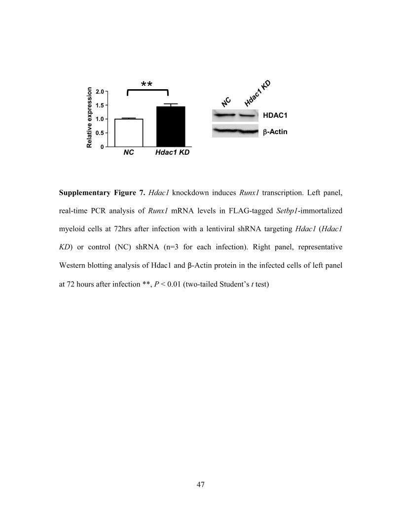
3f). This binding is also critical for Runx1 repression as Hdac1 knockdown in these cells
significantly increased Runx1 mRNA levels (Supplementary Fig. 7). Moreover,
significant reductions in Hdac1 binding to the Runx1 promoters were detected after

24
Setbp1 knockdown in the same cells (Fig. 3f). These results suggest that Setbp1 recruits
Hdac1 to the Runx1 promoters causing histone H3 deacetylation and subsequent
transcriptional repression of Runx1.
Given that Runx1 repression by Setbp1-mediated Hdac1 recruitment is required for
efficient colony formation by Setbp1-induced leukemia cells, we explored the therapeutic
potential of HDAC inhibitors for treating leukemias induced by Setbp1 activation. As
expected, Runx1 mRNA and protein levels were significantly up-regulated in Setbp1-
induced leukemia cells by treatment with HDAC inhibitors entinostat and vorinostat (Fig.
4a). Treatment with these inhibitors also completely ablated colony formation by these
leukemia cells and Setbp1-immortalized S3 cells (Fig. 4b). Cytospin analysis of treated
cells in liquid culture further suggests induction of myeloid differentiation, which was
confirmed by significantly increased expression of differentiation markers including
Cd11b, Lyz2, and Csf1r (Fig. 4c and 4d). Both HDAC inhibitors also induced identical
effects on myeloid progenitors immortalized by mutant Setbp1 carrying a recurrent
mutation in leukemia patients (Supplementary Fig. 8). Similarly, HDAC inhibitors also
caused significant growth inhibition and differentiation of primary human leukemia cells
harboring an activating SETBP1 mutation (Fig. 4e and Supplementary Fig. 9). To further
test therapeutic potential of HDAC inhibitors in vivo, we transplanted mice with 2
independent mouse myeloid leukemias induced by Setbp1 overexpression, and treated the
recipient mice with entinostat or vehicle once every three days for 21 days starting from 7
days post transplantation (Fig. 4f). While all vehicle-treated recipient mice become
moribund after 2 weeks, significant survival extensions were observed for mice treated
with entinostat. HDAC inhibitors including vorinostat and romidepsin have been
25
recently approved by FDA for the treatment of cutaneous T-cell lymphoma (39; 89; 93).
Studies have also suggested that myeloid leukemias induced by AML/ETO, PLZF/RARa,
or Hoxa9/Meis1 are sensitive to HDAC inhibitors (6; 10; 96). Our results suggest that
HDAC inhibitors are likely effective for treating human myeloid malignancies with
SETBP1 activation.
Taken together, our results establish SETBP1 activation as a ‘driver’ mutation capable of
initiating myeloid leukemia development partly through histone deacetylation mediated
transcriptional repression of RUNX1, and identify HDAC inhibition as a rational and
likely effective therapeutic strategy for various myeloid malignancies with SETBP1
activation.

26
METHODS
Mice
C57BL/6 and B6-Ly5.2 mice (7-12 weeks old; Charles River, Frederick, MD) were
maintained in the animal facility of Laboratory of Animal Medicine at Uniformed
Services University of the Health Sciences (USUHS, Bethesda, MD). All mouse
experiments were carried out according to protocols approved by the USUHS
Institutional Animal Care and Use Committee.
Patient samples
Primary human AML cells were collected after signing the informed consent, according
to the protocols approved by the Institutional Review Board of Cleveland Clinic in
accordance with the Declaration of Helsinki.
Retrovirus generation
The pMYs-Setbp1-IRES-GFP retroviral construct was described previously (88). The
murine Runx1 cDNA from pcDNA3.1-Flag-Runx1FL(60)(Addgene plasmid 14585) was
cloned into MSCV-neo using EcoRI and XhoI sites to generate MSCV-Runx1-neo. High
titer retroviruses were produced by transient transfection of Plat-E cells using Fugene-6
(Roche). Viral titer was assessed by serial dilution and infection of NIH-3T3 cells.
Retroviral transduction and bone marrow transplantation
C57BL/6 mice (7-12 weeks old) were injected intraperitoneally with 5-fluorouracil (150
mg/kg of body weight) 4 days before harvest of their bone marrow (BM) cells. The

27
harvested BM cells were grown in media [DMEM with 15% fetal bovine serum
containing SCF (100ng/ml), IL-3 (6ng/ml) and IL-6 (10ng/ml)] for 2 days to induce
proliferation of hematopoietic stem cells (HSCs). These expanded BM cells were
subsequently infected three times with high-titer retrovirus carrying Setbp1 cDNA
(pMYs-Setbp1-IRES-GFP) or GFP only (pMYs-IRES-GFP) on retronectin coated plates.
For transplantation, 0.7-1.3 x 10⁶ transduced BM cells were injected into the tail vein of
each lethally irradiated (1100 rads from 137Cs source) B6-Ly5.2 mouse along with 7.5 x
105 supporting bone marrow cells from un-irradiated B6-Ly5.2 mice. Transplanted mice
were aged and closely monitored for signs of leukemia development. Retro-orbital
bleeding was performed in 4, 8 and 16 weeks to analyze the short term and long term
engraftment of the donor cells by FACS. For secondary transplantation, 1 x 10⁶ spleen
cells from primary recipients with leukemia were injected into lethally irradiated
secondary recipients along with 7.5 x 10⁵ supporting bone marrow cells.
For serial transplantation of LSK cells, 1x10⁵ LSK cells transduced twice with pMYs-
Setbp1-IRES-GFP or pMYs-IRES-GFP virus were first transplanted into each lethally
irradiated primary B6-Ly5.2 recipient. At 4 months after primary transplantation, 5 x10²
GFP⁺ LSK cells purified from the primary recipients by FACS were transplanted into
each lethally irradiated secondary B6-Ly5.2 recipient along with supporting bone marrow.
Flow Cytometry
Flow cytometry analysis of mouse peripheral blood, bone marrow and spleen samples
were performed using BD LSRII flow cytometer. After sample collection and ACK lysis

28
of RBCs, spleen and bone marrow cells were blocked by incubation with anti-FcγR-II/III
and subsequently stained with antibodies against markers for myeloid (Gr-1, Mac-1),
erythroid (Ter-119), B (CD19) and T (CD3) lineages. Dead cells were excluded by
staining with Sytox Blue (Invitrogen). For serial transplantation of LSK cells, first
mononuclear cells were isolated from the bone marrow of C57BL/6 mice (7-12 weeks
old) by density centrifugation through lymphocyte separation medium. Lineage positive
cells were labeled by incubation with a cocktail of purified rat anti-mouse antibodies
specific to Gr-1, Mac-1, CD4, CD8, B220, CD127, and Ter-119 and were subsequently
removed by incubation with sheep anti-rat IgG conjugated magnetic beads (Invitrogen)
and exposure to a magnet. The isolated lin- cells were then stained with anti-Sca-1-APC,
and anti-c-Kit-PE antibodies and LSK cells were sorted using a FACSAria cell sorter.
The GMP (IL-7Rαˉ Sca-1ˉc-Kit⁺Fc-γR-II/IIIHiCD34⁺) population in bone marrow was
analyzed at 3 months after transplantation using 5-FU treated cells. Lin- cells were
obtained similarly as mentioned above and subsequently stained with anti-Sca-1-APC,
anti-CD34-Alexa fluor-700, anti-c-Kit-PE, and anti-FcR-II/III-PE-Cy7 and analyzed
using BD LSRII flow cytometer.
In vitro HDAC inhibitor treatment
5 x 10⁵ Setbp1-induced leukemic cells (BL3 and BL12) plated in media (IMDM, 20%
horse serum and 1x pen/strep) with SCF (50ng/ml) and IL3 (10ng/ml) were treated with
1µM of Entinostat (LC Laboratories Woburn, MA and Selleck Chemicals,Houston,TX),

29
Vorinostat (LC Laboratories) or equal volume of control DMSO for 48hrs. Treated cells
were subsequently subjected to cytospin, RNA extraction and Western blotting analysis.
For colony formation assay, 2x10⁴ BL3 and BL12 cells were plated in IMDM
methylcellulose medium supplemented with 20% horse serum, mouse SCF (50ng/ml),
IL-3 (10ng/ml) and 1µM of Entinostat, Vorinostat, or DMSO. Colony numbers were
counted after 7 days.
Primary human AML cells were cultured in IMDM medium supplemented with 10%
fetal bovine serum, SCF(10ng/ml) , IL-3 (10ng/ml), TPO (10ng/ml) and FLT3 ligand
(10ng/ml) at a density of 1 x 10⁵ cells/ml and treated with 1 µM Vorinostat or equal
volume of control DMSO for 72hrs.
In vivo entinostat treatment
Spleen cells from Setbp1-induced leukemic mice (BL12 and BL19) were transplanted
into lethally irradiated secondary recipients (1x10⁶ cells/animal) for inducing leukemia
development. Beginning from 7days after transplantation, recipient mice were injected
intraperitoneally with either 30 mg/kg of Entinostat (dissolved in 20 µl of DMSO and 180
µl of 50% polyethylene glycol) or vehicle once every 3 days for 21 days.
Lentiviral production, infection, and analysis
pLKO.1 lentiviral constructs containing shRNA were purchased from Sigma ( NC-sh,
SHC002; GFP-sh, SHC005; St. Louis, MO) and infectious lentivirus were generated as
described previously (88). Colony formation assays were performed at 48 hours after
infection using 2 x10⁴ puromycin resistant cells on IMDM methylcellulose medium

30
supplemented with 20% horse serum, mouse SCF (50ng/ml) and IL-3 (10ng/ml), and
puromycin (2 µg/ml). Colony numbers were counted after 7days.
Western blotting analysis
For Western blotting analysis the cells were washed twice with cold PBS and then whole
cell lysates were prepared by direct lysis of cell pellets in heated 2 x SDS sample buffer.
Samples were resolved on 4-12% tris-glycine gels (Life Technologies, Carlsbad, CA)
before transferring onto nitrocellulose membranes (Bio-Rad, Hercules, CA). Primary
antibodies used include anti-Setbp1 (16841-1AP, Proteintech, Chicago, IL) (88) Runx1
(19555-1-AP, Proteintech) and β-actin (MAB1501R, Millipore). Secondary antibodies
used include goat anti-rabbit (SC-2004, Santa Cruz Biotechnology, Dallas, TX) and anti-
mouse IgG-HRP (a-9044, Sigma Aldrich). Protein bands were visualized by incubation
with SuperSignal West chemiluminescent substrate (Pierce, Thermo Fisher Scientific,
Rockford, IL) and quantified using Quantity One data analysis software (Bio-Rad).
Real-time RT-PCR
For real-time RT-PCR, total RNA was extracted from cells using RNAeasy Plus mini kit
(QIAGEN). Oligo-dT-primed cDNA samples were prepared using Superscript III
(Invitrogen), and real-time PCR analysis was performed in triplicates using SYBR green
detection reagents (Invitrogen) on a 7500 real time PCR system (Applied Biosystems).
Relative changes in expression of Setbp1, Hoxa9, Hoxa10, Runx1, Cd11b, Lyz2 and Csf1r
were calculated according to the ∆∆Ct method. The cycling conditions are 50°C for 2

31
minutes followed by 95°C for 2 minutes, and then 40 cycles of 95°C for 15 seconds and
60°C for 1 minute. Gene-specific primer sequences are:
Setbp1 5’ CTG CTC ACT GTG GAG ACG ATT C 3’
5’ TTC TTA TCC AGC ACA CCA AGC TT 3’
Hoxa9 5’ TGT CTC CTC TCC CCC AAA CC 3’
5’ GAG ATG AGG CCT GGG ATTTAG A 3’
Hoxa10 5’ CCA GCC CTG GGT AAA CTT AGC 3’
5’ CATTGA CCT CAG GCC AGA CA 3’
Runx1 5’ GCA GGC AAC GAT GAA AAC TAC T 3’
5’ GCA ACT TGT GGC GGA TTT GTA 3’
β–Actin 5’ CCT CCC TGG AGA AGA GCT A 3’;
5’ TCC ATA CCC AAG AAG GAA G 3’
Rpl4 5’ ATG ATG AAC ACC GAC CTT AGC A 3’
5’ CGG AGG GCT CTT TGG ATT TC 3’
Cd11b 5’ GAA GCT GCC CCC CAA GAC 3’
5’ GGT CAA TGC ATG GAG AAA AGG 3’
Csf1r 5’ TGG ACT TCG CCC TCA GCT T 3’
5’ CCC CAG ACC CCT CAT GTT C 3’
Lyz2 5’ TGT GAG CTG CAG GGC TTT G 3’
5’ CCC ACC ACA GAG GCT GTT CT 3’
Chromatin immunoprecipitation (ChIP)
Mouse myeloid progenitors immortalized by FLAG-tagged Setbp1 were generated as
described (88). ChIP analyses were performed using ChIP-IT Express kit (Active Motif).
Immunoprecipitations were performed using FLAG M2 (Sigma Aldrich), mouse
monoclonal anti-HDAC1 antibody (10E2, #5356, Cell Signaling Technologies), rabbit
polyclonal anti-acetylated histone H3 (#39139, Active Motif) and mouse IgG (G3A1,
#5415, Cell Signaling Technologies) and rabbit IgG (#p120-101, Bethyl Laboratories).
Chromatin DNA was purified using MinElute PCR Purification Kit (QIAGEN) and
quantified by real-time PCR. The following Runx1 promoter-specific primers were used:

32
Runx1-P1 S 5’ ACAGGATCTGAAAGCCACCAA 3’
AS 5’ CCTGCCTCAGTCTTTTCTTGCT 3’
Runx1-P2 S 5’CCGTCGGTCTCCTCTATGCA 3’
AS 5’ GCCCGACCCGAGGAATT 3’
Southern blotting analysis
7 ug of DNA from leukemic spleens were digested with EcoRI, resolved on 0.75%
agarose gel, and transferred to nylon membrane using standard procedures. 32P-labeled
GFP-specific probe was synthesized by random primer labeling using the Prime-IT II kit
(Stratagene. LoJolla, CA) and hybridization was carried out in MiracleHyb buffer
(Stratagene) following manufacturer’s instructions.
Statistical analysis
Sample sizes and animal numbers were determined by previous experiences. No samples
were excluded from analyses. All data were analyzed by two-tailed Student’s t-test except
that survival curves were compared by Log-rank test. The researchers were not blinded
during sample collection and analysis.
Acknowledgements
This work was supported by National Institutes of Health (NIH) grants RO1CA-143193
(Y.D.) and RO1HL118281 (J.P.M.) and USUHS Pediatrics Grant QP86GI (Y.D.).
33
The views presented in this manuscript are those of the authors; no endorsement by the
Uniformed Services University of the Health Sciences or the Department of Defense has
been given or should be inferred.
Authorship
B.A.V. designed experiments, performed experiments, analyzed results, and wrote the
manuscript. K.O.G. and H.M. designed experiments, performed experiments, and
analyzed results. N.H., N.N., V.N., and K.O. performed experiments and analyzed
results. B.P. analyzed data. J.P.M. and Y.D. designed research, analyzed data, and wrote
the manuscript.
Conflict of interest disclosure: The authors declare no competing financial interests.
Corresponding authors:
Yang Du, Ph.D., Uniformed Services University of the Health Sciences, 4301 Jones
Bridge Road, Bethesda, MD, USA 20814. e-mail: [email protected]
Jaroslaw P. Maciejewski MD., Ph.D., FACP, Taussig Cancer Institute/R40, 9500 Euclid
Avenue, Cleveland, OH, USA 44195. E-mail: [email protected]
34
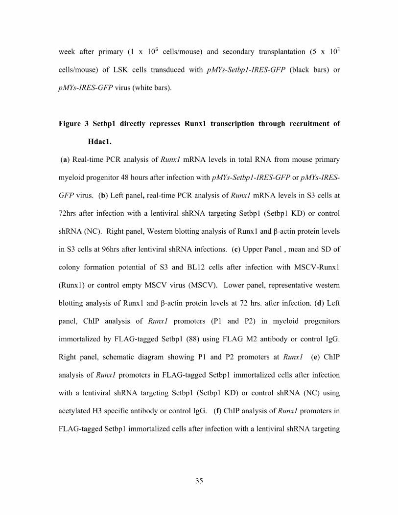
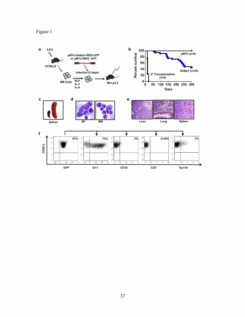
FIGURE LEGENDS
Figure 1 Setbp1 overexpression induces myeloid leukemia development.
(a) Schematic diagram of bone marrow transduction transplantation assay. (b) Survival
curves of irradiated C57BL6-Ly5.2 mice receiving bone marrow progenitors transduced
with pMYs-Setbp1-IRES-GFP or pMYs-IRES-GFP virus, or 1 x 10⁶ spleen cells from
primary leukemic mice. (c) Enlarged leukemic spleen (right) compared to a normal
spleen (left). (d) Cytospin of Spleen (SP) and bone marrow (BM) cells from leukemic
mice. (e) H&E staining showing infiltration of myeloid blasts in liver, lung and spleen of
a Setbp1-induced leukemic mouse. (f) FACS analysis of lineage specific markers on
bone marrow cells of a leukemic mouse.
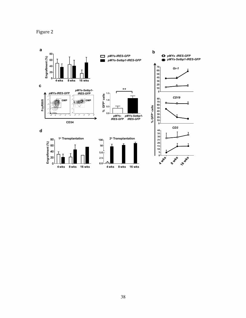
Figure 2 Overexpression of Setbp1 promotes self-renewal of HSCs and expansion of
GMPs.
(a) Engraftment of indicated transduced 5-FU treated bone marrow cells in recipient
mice analyzed by FACS analysis of percentage of GFP+ cells in peripheral blood at 4th,
8th and 16th weeks after transplantation. (b) FACS analysis of indicated lineage specific
markers on GFP+ donor cells in peripheral blood of mice receiving 5-FU treated bone
marrow cells transduced with pMYs-Setbp1-IRES-GFP or pMYs-IRES-GFP virus at 4th,
8th and 16th week after transplantation. (c) Left panel, FACS analysis of GMP
populations of GFP+ donor cells in the bone marrow of mice transplanted with 5-FU
treated bone marrow cells transduced with pMYs-Setbp1-IRES-GFP or pMYs-IRES-GFP
virus at 3 months after transplantation. Right panel, quantification of results on the left.
(d) FACS analysis of GFP+ cells in peripheral blood of recipient mice at 4th,8th and 16th
35
week after primary (1 x 10⁵ cells/mouse) and secondary transplantation (5 x 10²
cells/mouse) of LSK cells transduced with pMYs-Setbp1-IRES-GFP (black bars) or
pMYs-IRES-GFP virus (white bars).
Figure 3 Setbp1 directly represses Runx1 transcription through recruitment of
Hdac1.
(a) Real-time PCR analysis of Runx1 mRNA levels in total RNA from mouse primary
myeloid progenitor 48 hours after infection with pMYs-Setbp1-IRES-GFP or pMYs-IRES-
GFP virus. (b) Left panel, real-time PCR analysis of Runx1 mRNA levels in S3 cells at
72hrs after infection with a lentiviral shRNA targeting Setbp1 (Setbp1 KD) or control
shRNA (NC). Right panel, Western blotting analysis of Runx1 and β-actin protein levels
in S3 cells at 96hrs after lentiviral shRNA infections. (c) Upper Panel , mean and SD of
colony formation potential of S3 and BL12 cells after infection with MSCV-Runx1
(Runx1) or control empty MSCV virus (MSCV). Lower panel, representative western
blotting analysis of Runx1 and β-actin protein levels at 72 hrs. after infection. (d) Left
panel, ChIP analysis of Runx1 promoters (P1 and P2) in myeloid progenitors
immortalized by FLAG-tagged Setbp1 (88) using FLAG M2 antibody or control IgG.
Right panel, schematic diagram showing P1 and P2 promoters at Runx1 (e) ChIP
analysis of Runx1 promoters in FLAG-tagged Setbp1 immortalized cells after infection
with a lentiviral shRNA targeting Setbp1 (Setbp1 KD) or control shRNA (NC) using
acetylated H3 specific antibody or control IgG. (f) ChIP analysis of Runx1 promoters in
FLAG-tagged Setbp1 immortalized cells after infection with a lentiviral shRNA targeting
36
Setbp1 (Setbp1 KD) or control shRNA (NC) using Hdac1 specific antibody or control
IgG.
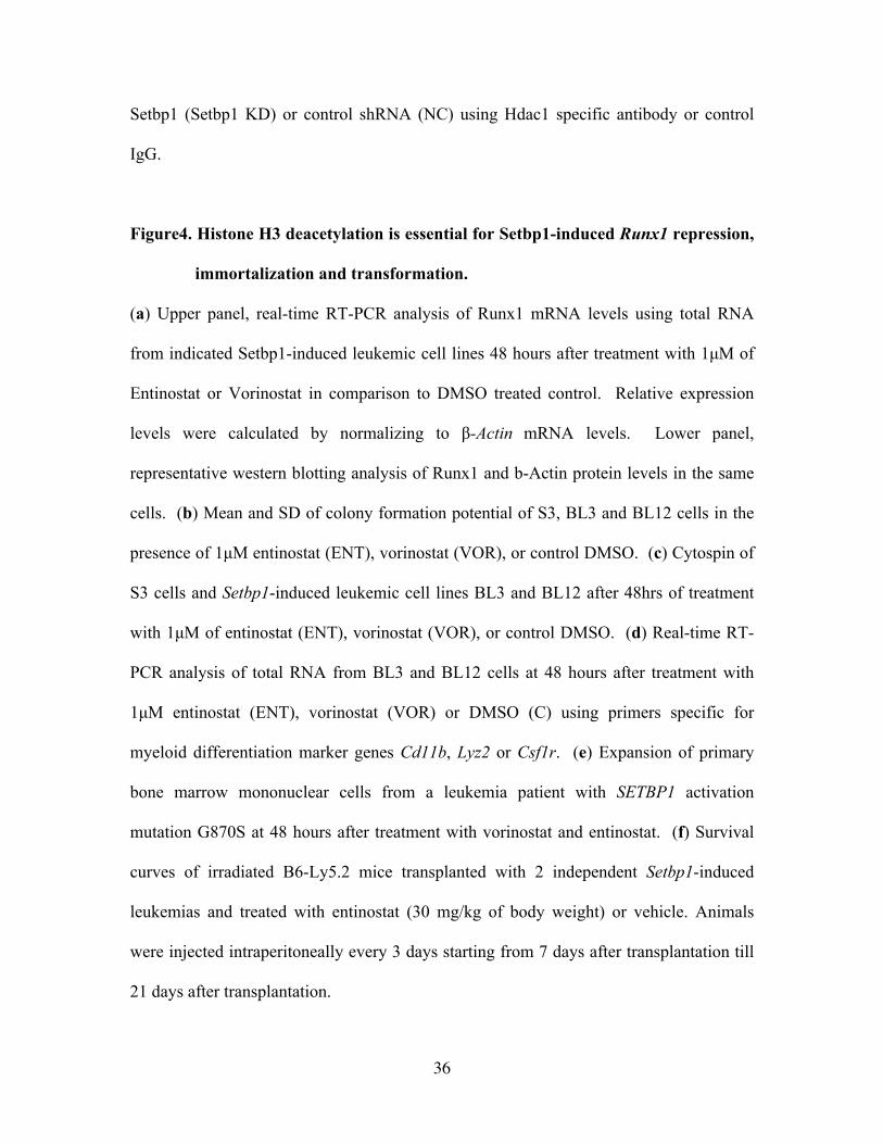
Figure4. Histone H3 deacetylation is essential for Setbp1-induced Runx1 repression,
immortalization and transformation.
(a) Upper panel, real-time RT-PCR analysis of Runx1 mRNA levels using total RNA
from indicated Setbp1-induced leukemic cell lines 48 hours after treatment with 1µM of
Entinostat or Vorinostat in comparison to DMSO treated control. Relative expression
levels were calculated by normalizing to β-Actin mRNA levels. Lower panel,
representative western blotting analysis of Runx1 and b-Actin protein levels in the same
cells. (b) Mean and SD of colony formation potential of S3, BL3 and BL12 cells in the
presence of 1µM entinostat (ENT), vorinostat (VOR), or control DMSO. (c) Cytospin of
S3 cells and Setbp1-induced leukemic cell lines BL3 and BL12 after 48hrs of treatment
with 1µM of entinostat (ENT), vorinostat (VOR), or control DMSO. (d) Real-time RT-
PCR analysis of total RNA from BL3 and BL12 cells at 48 hours after treatment with
1µM entinostat (ENT), vorinostat (VOR) or DMSO (C) using primers specific for
myeloid differentiation marker genes Cd11b, Lyz2 or Csf1r. (e) Expansion of primary
bone marrow mononuclear cells from a leukemia patient with SETBP1 activation
mutation G870S at 48 hours after treatment with vorinostat and entinostat. (f) Survival
curves of irradiated B6-Ly5.2 mice transplanted with 2 independent Setbp1-induced
leukemias and treated with entinostat (30 mg/kg of body weight) or vehicle. Animals
were injected intraperitoneally every 3 days starting from 7 days after transplantation till
21 days after transplantation.
37
Figure 1
Spleen SP BM
b
c d e
Liver SpleenLung
apMYs (n=8)
Setbp1 (n=33)2o Transplantation
(n=5)
pMYs-Setbp1-IRES-GFPor pMYs-IRES- GFP
C57BL/6
BM Cells SCFIL-3IL-6
Infection (2 days)
CD
45.2
97% 0.34%3% 7%
GFP Gr-1 CD3CD19 Ter119
73%f
5-FU
B6-Ly5.2
38
Figure 2
c
Gr1
CD19
CD3
pMYs -IRES-GFP pMYs-Setbp1-IRES-GFP
Engr
aftm
ent (
%)
d
apMYs-IRES-GFPpMYs-Setbp1-IRES-GFP
% G
FP+
cells
pMYs-IRES-GFPpMYs-Setbp1-
IRES-GFP
CD34
FcγR
III/II
pMYs-IRES-GFP
pMYs-Setbp1-IRES-GFP
**
% G
FP+
cells
b
4 wks 16 wks8 wks
Engr
aftm
ent (
%)
1o Transplantation 2o Transplantation
4 wks 16 wks8 wks 4 wks 16 wks8 wks
Gr-1
GMPGMP

39
Figure 3

40
Figure 4
Days
a
BL12
DMSO ENT VOR
S3
BL3
b
Col
ony
Form
atio
n
BL3 BL12S3
c
d
e
**
Control(n=4)
ENT(n=4)
*
Control(n=4)
ENT(n=4)
f
Rel
ativ
e C
d11b
ex
pres
sion
**** ***
BL3 BL12
Rel
ativ
e Ly
z2
expr
essi
onR
elat
ive
Csf
1r
expr
essi
on
C ENT C ENT C VOR C VOR
BL3 BL12
*******
**** **** **** ***
*************
BL3 BL12
******
Runx1
β-Actin
****
Rel
ativ
e R
unx1
expr
essi
on
DMSO
ENTVOR
Cel
l no.
(x 1
06/m
l)
0.5
0.0
1.0
1.5
2.0
2.5
10µM 1µM
****
**

41
Supplementary Figures
Supplementary Figure 1. Transduction efficiencies for 5-FU treated mouse bone
marrow progenitors. Representative transduction efficiencies in indicated transduction
groups determined by GFP fluorescence are shown. 20-50% and 55-72% infection
efficiencies were observed for pMYs-Setbp1-IRES-GFP and pMYs-IRES-GFP virus
respectively. Samples were analyzed at 48 hours after infection. Numbers represent the
percentages of GFP positive cells.
GFP
COUNT
25.7% 57%
Untransduced pMYs-Setbp1-IRES-GFP pMYs-IRES-GFP

42
Supplementary Figure 2. Increased expression of Setbp1, Hoxa9 and Hoxa10 in Setbp1-
induced myeloid leukemias Real-time RT-PCR analysis of total RNA extracted from
spleens of Setbp1-induced leukemic mice (BL3, BL4, BL12, and BL19) and control
normal bone marrow (BM) and spleen (SP) using gene-specific primers (n=3). Relative
expression levels were calculated by normalizing to Rpl4 mRNA levels in the same
sample and also wild-type bone marrow. The mean and SD of each relative expression
level is shown.
Rela
tive
Expr
essi
on

43
Supplementary Figure 3. Setbp1-induced leukemia cells are dependent on Setbp1
expression for maintenance. Upper panel, mean and SD of colony-forming potential of
Setbp1-induced leukemia cell lines BL3 and BL12 in the presence of SCF and IL-3 at 48
hours after infection with GFP-specific lentiviral shRNA (Setbp1KD) or control lentiviral
shRNA (NC). Lower panel, representative Western blotting analysis of Setbp1 and b-
Actin protein in the infected cells of the top panel at 72 hours after infection **, P < 0.01;
***, P < 0.001 (two-tailed Student’s t test)
BL3 BL12
** ***
Col
ony
Form
atio
n
β-Actin
Setbp1

44
Supplementary Figure 4. Setbp1-induced leukemias are mostly clonal. Southern
blotting analysis of viral integrations present in 10 Setbp1-induced myeloid leukemias
(BL4-9, BL11-14) using a GFP-specific probe. Seven ug of genomic DNA from each
leukemic spleen was digested with EcoRI, resulting the generation of a single GFP-
containing DNA fragment from each provirus. Each band represents an independent
integration. Same amount of genomic DNA from wild-type spleen (WT) was included as
negative control

45
Supplementary Figure 5. Transduction efficiencies for purified mouse LSK cells
Representative transduction efficiencies in indicated transduction groups were
determined by GFP fluorescence. 37-75% and 70-85% infection efficiencies were
observed for pMYs-Setbp1-IRES-GFP and pMYs-IRES-GFP virus respectively. Samples
were analyzed at 48 hours after infection. Numbers represent the percentages of GFP
positive cells
GFP
COUNT
38% 70%
Untransduced pMYs-Setbp1-IRES-GFP pMYs-IRES-GFP

46
Supplementary Figure 6. Correlation between SETBP1 and RUNX1 expression in
human AMLs. Expression array values were extracted from Oncomine dataset1. P-values
were calculated by comparisons between indicated 2 groups with AML (without RUNX1
mutations) using Mann-Whitney U test. Cut off value of high (n=42) and low / normal
(n=140) expression of SETBP1 was mean+0.5 standard deviation.
47
Supplementary Figure 7. Hdac1 knockdown induces Runx1 transcription. Left panel,
real-time PCR analysis of Runx1 mRNA levels in FLAG-tagged Setbp1-immortalized
myeloid cells at 72hrs after infection with a lentiviral shRNA targeting Hdac1 (Hdac1
KD) or control (NC) shRNA (n=3 for each infection). Right panel, representative
Western blotting analysis of Hdac1 and β-Actin protein in the infected cells of left panel
at 72 hours after infection **, P < 0.01 (two-tailed Student’s t test)
**HDAC1
β-Actin
Rel
ativ
e ex
pres
sion
0
0.5
1.0
1.5
2.0
NC Hdac1 KD

48
Supplementary Figure 8. HDAC inhibitors induced differentiation of myeloid
progenitors immortalized by SETBP1 activation mutation identified in leukemia patients.
(a) Representative cytospin of myeloid progenitor cells (DNc2 and DNc3) immortalized
by mutant Setbp1 (harboring activation mutation D868N) after 48hrs of treatment with
1µM of entinostat (ENT), vorinostat (VOR), or control DMSO. (b) Mean and SD of
colony formation potential of DNc2 (upper panel) and DNc3 (lower panel) cells in the
presence of 1µM entinostat, vorinostat or DMSO (n=3 for each treatment).
DMSO ENT VOR
DN
c2D
Nc3
0 .0
0 .5
1 .0
1 .5
Rel
ativ
e C
olon
y Fo
rmat
ion
DMSO ENT VOR
0 .0
0 .5
1 .0
1 .5
Rel
ativ
e C
olon
y Fo
rmat
ion
a b
DMSO ENT VOR

49
Supplementary Figure 9. Vorinostat induced differentiation of human myeloid leukemia
cells with SETBP1 activation mutation G870S. Representative cytospin of leukemia
cells after 72hrs of treatment with 1µM of vorinostat, or control DMSO (N=3).

50
CHAPTER 4: Manuscript 2
Mllt3 cooperates with Setbp1 in inducing myeloid transformation
Bandana A Vishwakarma1 and Yang Du1 1Department of Pediatrics, Uniformed Services University of the Health Sciences,
Bethesda, MD, USA.
ABSTRACT
We showed previously that overexpression of Setbp1 in mouse bone marrow (BM)
progenitors, through retroviral transduction is capable of inducing myeloid leukemia
development in irradiated recipient mice. However, only 50% of the mice receiving
Setbp1-transduced cells developed leukemia in 10 months, suggesting that additional
cooperating mutations may be required for Setbp1-induced leukemia development. To
identify such mutations, we cloned retroviral insertions from a total of 16 Setbp1-induced
leukemias. Interestingly, two such leukemias contained independent viral integrations at
Mllt3 that activated its expression, strongly suggesting that Mllt3 may cooperate with
Setbp1 to induce leukemia development. To test this hypothesis, we co-transduced BM
progenitors with retroviruses expressing Setbp1 and Mllt3, and compared their leukemia
induction potential to cells singly infected with either virus by transplantation into
irradiated recipient mice. When aged for 6 months, only 2 out of 8 mice receiving cells
singly transduced with Setbp1 virus developed leukemia and none of the mice
transplanted with Mllt3-transduced cells fell ill. In contrast, 100% of the mice
transplanted with co-transduced cells developed myeloid leukemia within 92 days,
confirming cooperation between Mllt3 and Setbp1 in inducing myeloid leukemia
development. Moreover, we also found that co-transduction induced leukemia cells
51
expressed significantly higher levels of Meis1 compared to leukemia cells induced by
Setbp1 alone. Given that Setbp1 activates Hoxa9, which is known to cooperate with
Meis1 in leukemic transformation, this finding further suggests that Meis1 activation by
Mllt3 may be responsible for the cooperation between Setbp1 and Mllt3. In summary,
our studies identify cooperation between Mllt3 and Setbp1 in leukemia induction by
simultaneous activation of Hoxa9 and Meis1.
Introduction
Recurrent somatic activating mutations in SETBP1 or its overexpression has been
reported in various human leukemias (12; 27; 33; 69; 92). SETBP1 is known to bind to
nuclear protein SET (75) and inhibit the activity of tumor suppressor PP2A through the
formation of heterotrimeric complex SETBP1-SET-PP2A (27). Furthermore, its
overexpression promotes self-renewal in murine myeloid progenitors through activation
of Homeobox genes, Hoxa9 and Hoxa10, both in vitro and in vivo suggesting that Setbp1
could be an oncogene (88). Recently, we showed that overexpression of Setbp1 in mouse
bone marrow progenitors through retroviral transduction, is capable of inducing myeloid
leukemia in mice. However, only 50% of the mice receiving the Setbp1- transduced cells
developed leukemia in 10 months. Given that multiple cooperating genetic and epigenetic
alterations are required for carcinogenesis (49), this result suggests that additional
cooperating mutations are required for the development of leukemia. Identification of
these cooperating mutations will be critical for designing combinatorial therapies for
Setbp1- induced leukemia.

52
Insertional mutagenesis using retroviral vectors is a powerful tool for identifying
cooperating mutations in leukemia development (37; 84; 110; 112). Oncogene of interest
is inserted into a replication incompetent retrovirus and is used for infection of bone
marrow cells. The retrovirus incorporates into the genome in a largely random fashion
and frequently causes activation of the gene in its vicinity. Cells with the viral integration
activate a cooperating gene and acquire higher growth advantage and develop faster into
leukemia. Retroviral integrations in individual tumor are cloned and sequenced to
identify the genes that cooperate with oncogene of interest to induce leukemia (2; 61).
In the present study, we attempted to identify Setbp1-cooperating partner by cloning
integrations from the leukemias induced by transplant of mouse bone marrow progenitors
with a Setbp1-expressing retroviral vector. Mixed-Lineage Leukemia Translocated to 3
(Mllt3) was identified as a potential cooperating mutation for Setbp1. Co-transduction of
Mllt3 and Setbp1 in BM progenitors accelerated the development of Setbp1-induced
leukemia. Furthermore, we found that deregulated expression of Mllt3 upregulated the
expression of Meis1 oncogene, which is known to cooperate with Hoxa9 in leukemic
transformations (65), and may be responsible for the acceleration of Setbp1-induced
leukemia. Taken together, our studies indicate cooperation between Mllt3 and Setbp1 in
myeloid leukemia induction by simultaneous activation of Hoxa9 and Meis1.

53
METHODS
Mice
C57BL/6 and B6-Ly5.2 mice (Charles River Laboratories, Fredrick, MD) were
maintained in the animal facility of the Laboratory of Animal Medicine at Uniformed
Services University of the Health Sciences (USUHS). All mouse experiments were
carried out according to protocols approved by the USUHS Institutional Animal Care and
Use Committee.
Splinkerette PCR
Viral integrations were cloned using splinkerette PCR as previously described (113).
Briefly, genomic DNA prepared from spleens of animals with Setbp1-induced leukemia
was digested with NlaIII or Mse1 and ligated to the splinkerette linker overnight. Nested
PCR was done to amplify the genomic sequence between the insertion and annealed
splinkerette, using primers specific to splinkerette and long terminal repeat (LTR) of
pMYs. PCR products were separated using 2% agarose gel and purified using Mini Elute
Columns (Qiagen, Valencia, CA). Amplified fragments were directly sequenced.
Retrovirus generation
The pMYs-Setbp1-IRES-GFP retroviral construct was generated as described previously
(88). The murine Mllt3 cDNA was amplified and cloned into MSCV-puro using XhoI and
EcoRI sites to generate MSCV-Mllt3-puro. High titer retroviruses were produced by
transient transfection of Plat-E cells using Fugene-6 (Roche, Indianapolis, IN). Viral titer
was assessed by serial dilution and infection of NIH-3T3 cells.

54
Retroviral transduction and bone marrow transplantation
C57BL/6 mice (7-12 weeks old) were injected intraperitoneally with 5-fluorouracil (150
mg/kg of body weight), 4 days before harvest of their bone marrow cells. The harvested
BM cells were grown in media [DMEM with 15% fetal bovine serum containing SCF
(100ng/ml), IL-3 (6ng/ml) and IL-6 (10ng/ml)] for 2 days to induce proliferation of
hematopoietic stem cells (HSCs). These expanded BM cells were subsequently infected
two times with high-titer retrovirus carrying Setbp1 cDNA (pMYs-Setbp1-IRES-GFP)
and MSCV-Mllt3-puro, pMYs-Setbp1-IRES-GFP or MSCV-Mllt3-puro on retronectin
coated plates. For transplantation, 0.4-0.6 x 10⁶ transduced BM cells were injected into
the tail vein of each lethally irradiated (1100 rads from 137Cs source) B6-Ly5.2 mouse
along with 7.5 x 10⁵ supporting bone marrow cells from un-irradiated B6-Ly5.2 mice.
Transplanted mice were aged and closely monitored for signs of leukemia development.
Flow Cytometry
Flow cytometry analysis of BM and spleen samples of moribund mice was performed
using BD LSRII flow cytometer. After sample collection and ACK lysis of RBCs, spleen
and bone marrow cells were blocked by incubation with anti-FcγR-II/III and
subsequently stained with antibodies against markers for myeloid (Gr-1, Mac-1),
erythroid (Ter-119), B (CD19) and T (CD4 & CD8) lineages. Dead cells were excluded
by staining with Sytox Blue (Invitrogen). For LSK cell purification, mononuclear cells
were isolated from the BM of C57BL/6 mice (7-12 weeks old) by density centrifugation
through lymphocyte separation medium. Lineage positive cells were then labeled by

55
incubation with a cocktail of purified rat anti-mouse antibodies specific to Gr-1, Mac-1,
CD4, CD8, B220, CD127, and Ter-119 and were subsequently removed by incubation
with sheep anti-rat IgG conjugated magnetic beads (Invitrogen) and exposure to a
magnet. The isolated lin- cells were then stained with anti-Sca-1-APC, and anti-c-Kit-PE
antibodies and LSK cells were sorted using a FACSAria cell sorter.
Colony Formation Assay
Colony formation assay was performed, at 72 hours after infection of LSK cells with
pMYs-Setbp1-IRES-GFP and MSCV-Mllt3-puro , pMYs-Setbp1-IRES-GFP or MSCV-
Mllt3-puro, using 3000 cells on methylcellulose medium supplemented with 15% Fetal
Bovine Serum, mouse SCF (50ng/ml) and IL-3 (6ng/ml), and IL6 (10ng/ml). LSK cells
positive for Mllt3 c-DNA were selected by puromycin, whereas Setbp1 cells were sorted
for GFP⁺. Colony numbers were counted after 7days.
Western blotting analysis
For western blotting analysis, the cells were washed twice with cold PBS and whole cell
lysates prepared by direct lysis of cell pellets in heated 2 x SDS sample buffer. Cell
lysates were resolved on 4-12% tris-glycine gels (Life Technologies) before transferring
onto nitrocellulose membranes (Bio-Rad). For protein detection following antibodies
were used: anti-Mllt3 (A300 595A-Bethyl), anti-Meis1 (ab19867-abcam) anti-Setbp1
(16841-1AP, Proteintech), anti-Hoxa9 (07-178, Millipore) and β-actin (MAB1501R,
Millipore). Secondary antibodies used include goat anti-rabbit (SC-2004, Santa Cruz) and
anti-mouse IgG-HRP (a-9044, Sigma Aldrich). Protein bands were visualized by

56
incubation with SuperSignal West chemiluminescent substrate (Pierce) and quantified
using Quantity One data analysis software (Bio-Rad).
Real-time RT-PCR
For real-time RT-PCR, total RNA was extracted from cells using RNAeasy Plus mini kit
(QIAGEN). Oligo-dT-primed cDNA samples were prepared using Superscript III
(Invitrogen), and real-time PCR analysis was performed in triplicates using SYBR green
detection reagents (Invitrogen) on a 7500 real time PCR system (Applied Biosystems).
Relative changes in expression of Hoxa9, Mllt3 and Meis1 were calculated according to
the ∆∆Ct method. The cycling conditions are 50°C for 2 minutes, followed by 95°C for 2
minutes, and then 40 cycles of 95°C for 15 seconds and 60°C for 1 minute. Gene-specific
primer sequences are:
Hoxa9 S 5’ TGT CTC CTC TCC CCC AAA CC 3’
AS 5’ GAG ATG AGG CCT GGG ATTTAG A 3’
Mllt3 S1 5’ TCCCAGACCAGGCAACAAG 3’
AS1 5’ TCGTTGTCATCAGAATGCAGATC3’
S5 5’ AACATTCAGCACTTTGTGGAGAAA 3’
AS5 5’ TTTTGGCCTAGGGAAGCTTT 3’
Meis1 S 5’GCAAAGTATGCCAGGGGAGTA 3’
AS 5’TCCTGTGTTAAGAACCGAGGG 3’ Rps29 S 5’ CAA CCG CCA CGG TCT GA 3’
AS 5’GCA CTG GCG GCA CA TGT 3’
RESULTS
Mllt3 is a common viral insertion site in Setbp1-induced leukemias
To identify genes that cooperate with Setbp1 in inducing myeloid leukemia development,
we cloned and sequenced 26 integrations from 16 Setbp1-induced leukemias by

57
splinkerette PCR (113). The integration site sequences were searched against the mouse
genome sequence through BLAT on UCSC genome browser to determine their genomic
locations (Table1). Interestingly, 2 independent leukemias (BL6 and BL20) had
insertions at Mllt3 (or Af9) gene at two different positions (Fig.1a). In BL6 the insertion
was found 0.125kb upstream of the promoter of the Mllt3 gene in the same direction as
the transcription of Mllt3, while in BL20 the insertion was present in intron 4 in the
opposite direction.
It is known that proviral integrations frequently cause activation of nearby genes due to
the strong promoter and enhancer activity of LTR (112). By Real-time RT-PCR, both
BL6 and BL20 leukemias had significantly higher expression of Mllt3 mRNA levels
compared to normal BM and other Setbp1-induced leukemias, which did not bear any
integrations at Mllt3, suggesting that both viral integrations activate Mllt3 transcription
(Fig.1b). Given that retrovirus integrate into the host genome in a largely random fashion,
frequent identification of insertions at Mllt3 in Setbp1-induced leukemias further suggests
that Mllt3 may cooperate with Setbp1 to induce myeloid leukemia development.
Overexpression of Mllt3 and Setbp1 increases the colony forming potential of LSK
cells
To test the possibility that Mllt3 and Setbp1 may cooperate in leukemia induction, serial
replating assay was performed with LinˉSca1⁺Kit⁺ (LSK) cells. LSK cells were sorted
and infected with pMys-Setbp1-IRES-GFP and PMSCV-Mllt3-puro together or only
pMys-Setbp1-IRES-GFP or PMSCV-Mllt3-puro. Mllt3 positive cells were selected using
puromycin, whereas Setbp1 cells were sorted for GFP+. 3000 cells from each group were

58
plated in methyl cellulose with cytokines SCF, IL3 and IL6 for the formation of colonies
(Fig.2a). In the primary colony assay no significant difference was observed in the
number of colonies between individually transduced LSK cells and cells co-transduced
with Mllt3 and Setbp1. In contrast, in secondary and tertiary plating there was a 50%
increase in the number of colonies with cells overexpressing both Mllt3 and Setbp1 in
comparison to LSK expressing only Setbp1(Fig.2b). Very few colonies were observed in
secondary and tertiary replating of cells infected with Mllt3 virus alone (Fig.2b). These
data suggest that overexpression of Mllt3 may cooperate with Setbp1 to promote cell
proliferation in vitro.
Overexpression of Mllt3 accelerates leukemia induced by Setbp1
To test whether cooperation between Mllt3 and Setbp1 in leukemia induction also exists
in vivo, we co-transduced 5-FU-treated bone marrow progenitors with retroviruses
expressing Mllt3 and Setbp1 and compared their leukemia inducing capability to that of
cells singly transduced with either virus in lethally irradiated B6-Ly5.2 mice (Fig.3a). To
maintain similar exposure of virus in double transduction experiments, half the viral titer
was used for each virus. 100% of the mice transplanted with double-infected cells
developed leukemia in 92 days than mice that received cells infected with pMys-Setbp1-
IRES-GFP (Fig.3b). Cytospin of cells from bone marrow and spleen of sick animals of
co-transduction group displayed blast like morphology and infiltration of myeloid cells
occurred in non-hematopoietic tissues like spleen, lungs and liver (Fig.3c&d), suggesting
development of myeloid leukemia. Flow cytometry analysis revealed that 83% of the BM
cells were GFP positive and almost all the cells expressed the myeloid marker-Mac1

59
confirming the development of myeloid leukemia (Fig.3e). In contrast, only 2 mice that
received cells infected with pMys-Setbp1-IRES-GFP and none of the mice from the Mllt3
alone cohort developed myeloid leukemia. These results show that Mllt3 does not induce
leukemia on its own, but can accelerate myeloid leukemia development induced by
Setbp1.
The acceleration of leukemia in double transduction experiment confirms that Mllt3 and
Setbp1 cooperate to induce leukemia, but it does not indicate whether the cooperation is
cell autonomous or non-cell autonomous. If the cooperation is cell autonomous, then both
the genes should be present in the same leukemia cell. If non-cell autonomous, the
cooperation would have resulted from Mllt3 and Setbp1 being overexpressed in two
different populations of cells. To verify this, individual colonies developed from single
leukemic cells generated on methylcellulose were examined for the presence of GFP and
the puromycin resistance gene in the genomic DNA. Both genes can be detected by PCR
in the genomic DNA extracted from all the colonies, suggesting that Setbp1 and Mllt3
cooperate in a cell autonomous manner to induce myeloid leukemia development
(Fig.3f).
Activation of Meis1 in Mllt3 -Setbp1 leukemia
We reported previously that overexpression of Setbp1 causes activation of Hoxa9 and
Hoxa10 in BM progenitors (88). Hoxa9 is known to cooperate with Meis1 to induce
leukemia development (65). To test the possibility that Meis1 may be activated by Mllt3
overexpression (Fig.4a), we performed real-time RT-PCR to examine Meis1 expression
in leukemias developed from co-transduction of Mllt3 and Setbp1 in comparison to
60
Setbp1-induced leukemia with integrations other than Mllt3. Meis1 mRNA was barely
detected in leukemias induced by Setbp1alone, whereas it is expressed at high levels in
Mllt3-Setbp1 leukemia cells (Fig.4a). Significantly higher levels of Meis1 protein was
also detected in the co-transduction induced leukemias than Setbp1 alone induced
leukemia. Hoxa9 mRNA and protein were also detected at higher levels in the co-
transduced leukemia. These results support that activation of Meis1 and probably
increased expression of Hoxa9 are likely responsible for cooperation between Mllt3 and
Setbp1 in leukemia induction.
DISCUSSION
Previously, we showed that overexpression of Setbp1 in bone marrow progenitors
through retroviral transduction is capable of inducing myeloid leukemia development
when transplanted in mice. However, it is likely that Setbp1 alone is insufficient to induce
leukemia, as in 10 months only 50% of the transplanted mice could develop leukemia,
suggesting that additional cooperating mutations are required for complete
transformation. Similar observations have been reported with the Hoxa9, HOXA10 genes
(108; 109) and the fusion genes CALM-AF10, NUP98-HOXD13 and NUP98-HOXA9
(19; 84; 105). This is consistent with the fact that like other cancers, acute myeloid
leukemia (AML) is also a consequence of multiple mutations (49). Therefore, we sought
to identify the mutations that might cooperate with Setbp1 in leukemic transformation, by
cloning the retroviral integrations in the Setbp1-induced leukemias.
61
We identified 26 integrations from 16 Setbp1-induced leukemias. Most of the genes
identified are novel mutations and never have been implicated in leukemia or any other
cancer. Integrations identified at some of the genes, Arhgef2, IL6, Bcl9l and Mllt3, have
been implicated in different cancers (30; 36; 45; 78). They are members of signaling
pathways which are involved in cell proliferation, apoptosis, differentiation and
migration. Evidence shows that IL6 is degerulated in AML patients and high level of IL6
represents an unfavorable prognosis (103). Apart from this, integration was found in
genes involved in splicing, vacuolar trafficking, cytoskeleton and membrane protein. An
association of the vacuolar protein sorting family of proteins has been shown in cancer.
One of the integrations, Dnm2 (Dynamin) a GTPase, involved in membrane trafficking,
has been reported as recurrent mutation in early T-cell precursor acute lymphoblastic
leukemia (45; 86). Loss of function mutation in Vps37b is found in gastric and colorectal
cancer (21).
Interestingly, 2 independent leukemias (BL6 and BL20) had integration in the Mllt3 gene.
Real-time RT-PCR analysis demonstrated that insertion at Mllt3 caused its activation,
suggesting that Mllt3 might cooperate with Setbp1 in inducing leukemia development.
MLLT3, also known as AF9, is a homologue of MLLT1 and regulates
erythrocyte/megakaryocytes lineage decision (94). During embryogenesis, it is required
for controlling embryo patterning. Af9 knockout mice are perinatal lethal and has no
effect on hematopoiesis (24). It was first identified as a fusion protein with mixed lineage
leukemia, MLL, in AML as MLL-AF9 (55) and was later shown to generate
myeloproliferative disorder, phenotypically similar to human leukemia, when expressed
in granulocyte macrophage progenitors (GMPs) in mice (64). This suggests that Mllt3

62
activation could be a potential cooperating mutation for Setbp1. To test this hypothesis,
serial replating assay was done using LSK cells, either co-transduced with Mllt3 and
Setbp1 or singly transduced with either virus. Co-expression of Mllt3 with Setbp1
increased the colony forming potential of LSK in comparison to cells infected with only
Setbp1 or Mllt3 virus. The colonies of LSK cells overexpressing Mllt3 were smaller in
size and differentiated in comparison to Setbp1 transduced or double transduced. This is
in line with the previous report, where forced expression of MLLT3 in total CD34⁺ cells
resulted in colonies smaller and differentiated than controls (94). Next, in vivo study was
done to test the leukemia induction potential of co-transduced cells in comparison with
singly transduced cells. All the mice transplanted with BM cells expressing Mllt3 and
Setbp1 fell sick with myeloid leukemia in 92 days. However, only 25% of the mice
succumbed to leukemia in Setbp1 cohort within 8 months, whereas all of the mice
remained healthy in Mllt3 alone. Deregulated expression of Mllt3 with Setbp1 accelerated
the development of Setbp1-induced leukemia.
Hoxa9 and Hoxa10 is a direct transcriptional target of Setbp1 and are activated in Setbp1-
immortalized myeloid progenitors and in the leukemic cells of Setbp1-induced leukemia
(88). Thus, Setbp1 promotes self-renewal through activation of oncogenes, Hoxa9 and
Hoxa10. In leukemia induced by co-transduction of Mllt3 and Setbp1, Hoxa9 expression
is enhanced in comparison to Setbp1-induced leukemia. Besides, Meis1 is significantly
expressed at higher levels in co-transduction induced leukemia than Setbp1 alone induced
leukemia. Hoxa9 is known to cooperate with Meis1 in leukemic transformation (65). It
could be possible that Mllt3 accelerates Setbp1-induced leukemia by activating Meis1.
Hoxa9 and Meis1 are the downstream targets of MLL rearranged leukemias and are

63
required for the transformation and survival of these leukemias (40; 56; 64; 120). It is
well established that activation of HOXA9 and MEIS1 occurs in MLL-AF9 leukemia.
Meis1 is essential for maintaining the stem like features in MLL-AF9 leukemia (66).
MLLT3 was earlier shown as a component of ENL associated protein complex (EAP), a
transcription elongation complex, which also contains DOT1L and p-TEFb (7; 80; 81).
Later it was also identified in another complex, Supra Elongation Complex, with DOT1L,
POLII elongation factors, components of p-TEFb kinase and other translocation partners
of MLL(4). DOT1L is the only H3K79 methyl transferases and is essential for
maintenance of HOXA9 and MEIS1 transcription in MLL-AF9 leukemia (87). It might be
possible that, when Mllt3 is overexpressed with Setbp1, Mllt3 initiates and maintains the
expression of Hoxa9 and Meis1 through recruitment of transcription elongation complex
at the respective locus and accelerates the development of Setbp1-induced leukemia.
Thus, our study indicates that Mllt3 cooperate with Setbp1 in the development of myeloid
leukemia through activation of Hoxa9 and Meis1. Future studies will focus on unraveling
any interaction between Mllt3 and Setbp1 in leukemic transformation and identifying
MLLT3 mutation(s) in human leukemias.
FIGURE LEGENDS.
Figure1. Viral integration activates Mllt3 expression.
(a) pMys-Setbp1-IRES-GFP integration at Mllt3 in BL6 (top panel) and BL20 (bottom
panel) leukemia. Exon-coding regions are highlighted in yellow and noncoding exons in
gray. Arrows indicate transcription start sites. The location and orientation of viral
integrations are depicted by red triangles. (b) Real-time RT-PCR analysis of Mllt3

64
expression in BM and spleen cells of leukemic mice with Mllt3 integration (BL6 and
BL20) and other Setbp1-induced leukemias (BL2, BL3, BL4, BL7, and BL12) in
comparison to wild type BM. Relative expression levels were calculated by normalizing
to Rps29 mRNA levels in the same sample and to WT BM.
Figure2. Co-transduction of Mllt3 and Setbp1 increases the colony forming potential
of LSK cells.
(a) Schematic diagram of colony formation assay. (b) Mean and SD of colony formation
potential of PMSCV-Mllt3-puro-infected, pMys-Setbp1-IRES-GFP-infected or double-
infected LSK cells in serial replating assay. (*** P < 0.001, ****P < 0,0001)
Figure3. Deregulated Mllt3 expression accelerates the development of Setbp1-
induced leukemia.
(a) Schematic diagram of bone marrow transduction transplantation assay. (b) Survival
curves of irradiated C57BL/6-Ly5.2 mice receiving pMYs-Setbp1-IRES-GFP-infected,
PMSCV-Mllt3-puro-infected or double infected bone marrow cells. (c) Cytospin of
Spleen (SP) and bone marrow (BM) cells from Mllt3 and Setbp1-induced leukemic mice;
MS1, MS2 and MS3. (d) H&E staining showing infiltration of myeloid blasts in liver,
lung and spleen of Mllt3 and Setbp1-induced leukemic mice; MS1, MS2 and MS3. (e)
FACS analysis of lineage specific markers on bone marrow cells of Mllt3 and Setbp1-
induced leukemic mouse. (f) Individual colonies cultured from BM cells of leukemic
mice carry both Mllt3 and Setbp1 gene. PCR products using primers specific to GFP and
puromycin resolved on ethidium bromide gel.

65
Figure4. Overexpression of Mllt3 with Setbp1 activates Meis1 expression
(a) Real-time RT-PCR analysis of total RNA extracted from bone marrow of wild-type
(WT BM) and spleen cells of Mllt3-Setbp1 (MS1, MS2, MS3, MS4, and MS6) and
Setbp1 (BL3, BL4, and BL12) -induced leukemic mice using primers specific for Mllt3,
Hoxa9 and Meis1. Relative expression levels were calculated by normalizing to Rps29
mRNA levels in the same sample and in WT BM. (b) Western blotting analysis of
Hoxa9, Meis1, Mllt3, Setbp1 and beta actin protein level in Mllt3-Setbp1 and Setbp1-
induced leukemias.

66
Table 1- Viral integrations at genes identified in pMYs-Setbp1 virus induced leukemias

67

68

69

70
SUMMARY
Our studies demonstrate that Setbp1 is a novel oncogene capable of inducing myeloid
leukemia development. Retrovirally directed expression of Setbp1 in murine bone
marrow progenitors induces myeloid leukemia development in mice on transplant.
Evidence shows that most likely mutations in SETBP1 occurs after the initial
establishment of the disease and SETBP1 contribute to its progression (12; 72; 92; 111).
However, our studies indicate that deregulation of Setbp1 through mutation or
overexpression could be a driver mutation during leukemia development.
Overexpression of Setbp1 induces myeloid leukemia as cytospin of BM and spleen cells
from moribund mice displayed myeloid blast like morphology, infiltration of myeloid
cells were observed in non-hematopoietic tissues and significantly more infected cells
expressed myeloid marker, Gr-1. We found that in the pre-leukemic stage,
overexpression of Setbp1 promotes the expansion of hematopoietic stem and progenitor
cells, as the engraftment of Setbp1 expressing cells increased gradually overtime, while
there was a gradual decline of cells infected with empty virus. The gradual increase in the
engraftment of the Setbp1 expressing cells suggests that Setbp1 expression may promote
self-renewal of HSCs. Overexpression of Setbp1 in hematopoietic stem cells bias their
lineage commitment to the myeloid pathway, as we observed expansion of GMP
population in mice transplanted with cells overexpressing Setbp1 in comparison to
control group.
Besides activation of oncogenes, suppression of tumor suppressor genes is also essential
for leukemic transformations. Setbp1, an AT-hook transcription factor, not only activates

71
proto-oncogenes Hoxa9 and Hoxa10 (88), but we found it suppress the expression of
tumor suppressor gene Runx1. Analysis of human AML revealed that the expression of
RUNX1 is inversely related to SETBP1 expression. A similar result was observed on
overexpression of Setbp1 in myeloid progenitors. Setbp1 overexpression inhibited the
expression of Runx1. Suppression of Runx1 is critical to Setbp1-induced transformation,
as the colony forming potential of Setbp1-induced leukemic cell line, BL3 and BL12,
decreased on ectopic expression of Runx1. Loss of Runx1 is associated with increased
HSC self-renewal and GMP expansion (43; 54; 57). CHIP analysis revealed that Setbp1
binds to Runx1 promoter and directly regulates its expression. In search of potential
epigenetic changes induced by Setbp1 for the repression of Runx1, we found a
significant increase in histone H3 acetylation at Runx1 promoters after Setbp1
knockdown in cells immortalized by FLAG-tagged Setbp1, suggesting that Setbp1 may
repress Runx1 transcription by preventing histone H3 acetylation at its promoters.
Histone deacetylases inhibit transcription by deacetylating histones (99). CHIP data
confirmed binding of Hdac1 at Runx1 promoter. This binding is also critical for Runx1
repression, as Hdac1 knockdown in these cells significantly increased Runx1 mRNA
levels. Moreover, significant reductions in Hdac1 binding to the Runx1 promoters were
detected after Setbp1 knockdown in the same cells. These results suggest that Setbp1
recruits Hdac1 to the Runx1 promoters causing histone H3 deacetylation and subsequent
transcriptional repression of Runx1.
Given that Runx1 repression by Setbp1-mediated Hdac1 recruitment is required for
efficient colony formation by Setbp1-induced leukemia cells, we explored the therapeutic
potential of HDAC inhibitors for treating leukemias induced by Setbp1 activation. HDAC
72
inhibitors, Entinostat and Vorinostat promoted differentiation of Setbp1-induced
leukemic cell line BL3 and BL12. Treatment of BL3 and BL12 cells with HDAC
inhibitor upregulated the expression of Runx1. The differentiation observed in HDAC
inhibitor treated leukemic cell line suggest that it could be through increased expression
of Runx1, as studies have shown that RUNX1 is involved in megakaryocytic and
lymphocytic differentiation(54). Chip analysis demonstrated that Setbp1 recruit Hdac1 at
the Runx1 promoter as knockdown of Sebp1 decreased the binding of Setbp1 at the
promoter of Runx1. This also increased H3 acetylation of the Runx1 promoter. This result
thus confirmed that Setbp1 is responsible for recruitment of Hdac1 at Runx1 promoter
and is involved in epigenetic modification of chromatin.
Interestingly, HDAC inhibitor, Entinostat significantly enhanced the survival of Setbp1-
induced leukemic mice, suggesting that HDAC inhibition could be a potential therapeutic
strategy for leukemia developed by Setbp1 overexpression. Vorinostat and Entinostat
both were effective in in vitro studies, but in in vivo Entinostat had pronounced effect.
Entinostat is better retained in the body because it has longer half- life in comparison to
Vorinostat and so is more effective.
Though overexpression of Setbp1 was capable of inducing myeloid leukemia in mice, but
only fifty percent of the transplanted mice fell sick in 10 months, while secondary
recipients of spleen cells from leukemic mice developed the same disease with much
shorter latency, suggesting that additional mutations may be required for Setbp1-induced
leukemic transformations. So, we cloned and sequenced 26 integrations from 16 Setbp1-
induced leukemias, to identify mutations that might have cooperated in the development
of AML. Integrations identified are members of signaling pathways involved in cell

73
proliferation, apoptosis, differentiation and migration. Apart from this, integration was
found in genes involved in splicing, vacuolar trafficking, cytoskeleton and membrane
protein. Integrations at some of the genes Arhgef2, Bcl9l, Il6 and Mllt3 have been
implicated in cancer. A common insertion site was found at Mllt3 gene, as two
independent leukemias, BL6 and BL20, had insertion in Mllt3. We selected to study the
cooperation of Mllt3 with Setbp1, as two independent leukemias had insertion in Mllt3
and it has been associated with AML as fusion protein, MLL-AF9. Besides, integration
caused activation of Mllt3, suggesting that Mllt3 could cooperate with Setbp1 in inducing
leukemia. Arhgef2 and Rbm8a are also potential candidates to study cooperation with
Setbp1, as the latency of the leukemias was very short. Beta-catenin binding protein,
Bcl9l is associated with the canonical Wnt signaling pathway, which has been implicated
in cancer and leukemia. Integration in Bcl9l also induced leukemia with latency of 168
days, suggesting that it could also be a potential cooperating partner with Setbp1 in
leukemia development.
Co transduction of Mllt3 and Setbp1 enhanced the colony forming potential of LSK cells
compared to only Setbp1 expressing LSK cells. The colonies of LSK cells overexpressing
Mllt3 were smaller in size and differentiated in comparison to Setbp1 transduced or
double transduced. This is in congruence with previous study where forced expression of
MLLT3 in total CD34⁺ cells resulted in colonies smaller and differentiated than controls.
In vivo study confirmed the cooperation, as co-transduction of Mllt3 and Setbp1
accelerated the development of Setbp1-induced myeloid leukemia. Moreover, we also
found that co-transduction induced leukemia cells expressed significantly higher level of
Meis1 compared to leukemia induced by Setbp1 alone. Hoxa9 cooperates with Meis1 in
74
leukemic transformations. Our finding thus suggests that Mllt3 accelerates Setbp1-
induced leukemia through activation of Hoxa9 and Meis1.
It remained to be addressed any protein-protein interaction between Setbp1 and Mllt3 in
regulating Hoxa9 and Meis1 expression. Besides, future study would focus on identifying
crosstalk between the signaling pathway of Mllt3 and Setbp1. Mllt3 has always been
implicated as a fusion protein with MLL in leukemia. The direct role played by Mllt3 in
inducing myeloid leukemia is unknown. So, identifying Mllt3 mutation(s) in leukemia
with Setbp1 mutation and other leukemia would be a promising area to study.

75
REFERENCES
1. Abrahamsson AE, Geron I, Gotlib J, Dao KH, Barroga CF, et al. 2009. Glycogen synthase kinase 3beta missplicing contributes to leukemia stem cell generation. Proceedings of the National Academy of Sciences of the United States of America 106:3925-9
2. Abramovich C, Humphries RK. 2005. Hox regulation of normal and leukemic hematopoietic stem cells. Current opinion in hematology 12:210-6
3. Adler HT, Nallaseth FS, Walter G, Tkachuk DC. 1997. HRX leukemic fusion proteins form a heterocomplex with the leukemia-associated protein SET and protein phosphatase 2A. The Journal of biological chemistry 272:28407-14
4. An CH, Kim YR, Kim HS, Kim SS, Yoo NJ, Lee SH. 2012. Frameshift mutations of vacuolar protein sorting genes in gastric and colorectal cancers with microsatellite instability. Human pathology 43:40-7
5. Aravind L, Landsman D. 1998. AT-hook motifs identified in a wide variety of DNA-binding proteins. Nucleic acids research 26:4413-21
6. Armstrong SA, Staunton JE, Silverman LB, Pieters R, den Boer ML, et al. 2002. MLL translocations specify a distinct gene expression profile that distinguishes a unique leukemia. Nature genetics 30:41-7
7. Bitoun E, Oliver PL, Davies KE. 2007. The mixed-lineage leukemia fusion partner AF4 stimulates RNA polymerase II transcriptional elongation and mediates coordinated chromatin remodeling. Human molecular genetics 16:92-106
8. Blair A, Hogge DE, Ailles LE, Lansdorp PM, Sutherland HJ. 1997. Lack of expression of Thy-1 (CD90) on acute myeloid leukemia cells with long-term proliferative ability in vitro and in vivo. Blood 89:3104-12
9. Bonnet D, Dick JE. 1997. Human acute myeloid leukemia is organized as a hierarchy that originates from a primitive hematopoietic cell. Nature medicine 3:730-7
10. Bots M, Verbrugge I, Martin BP, Salmon JM, Ghisi M, et al. 2014. Differentiation therapy for the treatment of t(8;21) acute myeloid leukemia using histone deacetylase inhibitors. Blood 123:1341-52
11. Bourachot B, Yaniv M, Muchardt C. 1999. The activity of mammalian brm/SNF2alpha is dependent on a high-mobility-group protein I/Y-like DNA binding domain. Molecular and cellular biology 19:3931-9
12. Bousquet M, Zhuang G, Meng C, Ying W, Cheruku PS, et al. 2013. miR-150 blocks MLL-AF9-associated leukemia through oncogene repression. Molecular cancer research : MCR 11:912-22

76
13. Bradford GB, Williams B, Rossi R, Bertoncello I. 1997. Quiescence, cycling, and turnover in the primitive hematopoietic stem cell compartment. Experimental hematology 25:445-53
14. Cairns BR, Schlichter A, Erdjument-Bromage H, Tempst P, Kornberg RD, Winston F. 1999. Two functionally distinct forms of the RSC nucleosome-remodeling complex, containing essential AT hook, BAH, and bromodomains. Molecular cell 4:715-23
15. Calabretta B, Perrotti D. 2004. The biology of CML blast crisis. Blood 103:4010-22
16. Calmels B, Ferguson C, Laukkanen MO, Adler R, Faulhaber M, et al. 2005. Recurrent retroviral vector integration at the Mds1/Evi1 locus in nonhuman primate hematopoietic cells. Blood 106:2530-3
17. Campbell LJ, Martinow A, Michael PM, White JS, Rayeroux KC, Januszewicz EH. 1999. Correlation of cytogenetics, BCR-ABL PCR studies and fluorescence in situ hybridisation (FISH) in adult acute lymphoblastic leukaemia. Australian and New Zealand journal of medicine 29:707-12
18. Carapeti M, Aguiar RC, Goldman JM, Cross NC. 1998. A novel fusion between MOZ and the nuclear receptor coactivator TIF2 in acute myeloid leukemia. Blood 91:3127-33
19. Caudell D, Harper DP, Novak RL, Pierce RM, Slape C, et al. 2010. Retroviral insertional mutagenesis identifies Zeb2 activation as a novel leukemogenic collaborating event in CALM-AF10 transgenic mice. Blood 115:1194-203
20. Cavazzini F, Bardi A, Tammiso E, Ciccone M, Russo-Rossi A, et al. 2006. Validation of an interphase fluorescence in situ hybridization approach for the detection of MLL gene rearrangements and of the MLL/AF9 fusion in acute myeloid leukemia. Haematologica 91:381-5
21. Chen L, Deshpande AJ, Banka D, Bernt KM, Dias S, et al. 2013. Abrogation of MLL-AF10 and CALM-AF10-mediated transformation through genetic inactivation or pharmacological inhibition of the H3K79 methyltransferase Dot1l. Leukemia 27:813-22
22. Cleynen I, Van de Ven WJ. 2008. The HMGA proteins: a myriad of functions (Review). International journal of oncology 32:289-305
23. Cobaleda C, Gutierrez-Cianca N, Perez-Losada J, Flores T, Garcia-Sanz R, et al. 2000. A primitive hematopoietic cell is the target for the leukemic transformation in human philadelphia-positive acute lymphoblastic leukemia. Blood 95:1007-13
24. Collins EC, Appert A, Ariza-McNaughton L, Pannell R, Yamada Y, Rabbitts TH. 2002. Mouse Af9 is a controller of embryo patterning, like Mll, whose human homologue fuses with Af9 after chromosomal translocation in leukemia. Molecular and cellular biology 22:7313-24
25. Corral J, Lavenir I, Impey H, Warren AJ, Forster A, et al. 1996. An Mll-AF9 fusion gene made by homologous recombination causes acute leukemia in chimeric mice: a method to create fusion oncogenes. Cell 85:853-61
26. Cozzio A, Passegue E, Ayton PM, Karsunky H, Cleary ML, Weissman IL. 2003. Similar MLL-associated leukemias arising from self-renewing stem cells and short-lived myeloid progenitors. Genes & development 17:3029-35

77
27. Cristobal I, Blanco FJ, Garcia-Orti L, Marcotegui N, Vicente C, et al. 2010. SETBP1 overexpression is a novel leukemogenic mechanism that predicts adverse outcome in elderly patients with acute myeloid leukemia. Blood 115:615-25
28. Cuenco GM, Ren R. 2001. Cooperation of BCR-ABL and AML1/MDS1/EVI1 in blocking myeloid differentiation and rapid induction of an acute myelogenous leukemia. Oncogene 20:8236-48
29. Cuenco GM, Ren R. 2004. Both AML1 and EVI1 oncogenic components are required for the cooperation of AML1/MDS1/EVI1 with BCR/ABL in the induction of acute myelogenous leukemia in mice. Oncogene 23:569-79
30. Cullis J, Meiri D, Sandi MJ, Radulovich N, Kent OA, et al. 2014. The RhoGEF GEF-H1 is required for oncogenic RAS signaling via KSR-1. Cancer cell 25:181-95
31. Dalerba P, Cho RW, Clarke MF. 2007. Cancer stem cells: models and concepts. Annual review of medicine 58:267-84
32. Daley GQ, Ben-Neriah Y. 1991. Implicating the bcr/abl gene in the pathogenesis of Philadelphia chromosome-positive human leukemia. Advances in cancer research 57:151-84
33. Damm F, Itzykson R, Kosmider O, Droin N, Renneville A, et al. 2013. SETBP1 mutations in 658 patients with myelodysplastic syndromes, chronic myelomonocytic leukemia and secondary acute myeloid leukemias. Leukemia 27:1401-3
34. Dash AB, Williams IR, Kutok JL, Tomasson MH, Anastasiadou E, et al. 2002. A murine model of CML blast crisis induced by cooperation between BCR/ABL and NUP98/HOXA9. Proceedings of the National Academy of Sciences of the United States of America 99:7622-7
35. Deguchi K, Ayton PM, Carapeti M, Kutok JL, Snyder CS, et al. 2003. MOZ-TIF2-induced acute myeloid leukemia requires the MOZ nucleosome binding motif and TIF2-mediated recruitment of CBP. Cancer cell 3:259-71
36. Deka J, Wiedemann N, Anderle P, Murphy-Seiler F, Bultinck J, et al. 2010. Bcl9/Bcl9l are critical for Wnt-mediated regulation of stem cell traits in colon epithelium and adenocarcinomas. Cancer research 70:6619-28
37. Du Y, Spence SE, Jenkins NA, Copeland NG. 2005. Cooperating cancer-gene identification through oncogenic-retrovirus-induced insertional mutagenesis. Blood 106:2498-505
38. Duncan AW, Rattis FM, DiMascio LN, Congdon KL, Pazianos G, et al. 2005. Integration of Notch and Wnt signaling in hematopoietic stem cell maintenance. Nature immunology 6:314-22
39. Duvic M, Talpur R, Ni X, Zhang C, Hazarika P, et al. 2007. Phase 2 trial of oral vorinostat (suberoylanilide hydroxamic acid, SAHA) for refractory cutaneous T-cell lymphoma (CTCL). Blood 109:31-9
40. Faber J, Krivtsov AV, Stubbs MC, Wright R, Davis TN, et al. 2009. HOXA9 is required for survival in human MLL-rearranged acute leukemias. Blood 113:2375-85
41. Fialkow PJ, Singer JW, Adamson JW, Vaidya K, Dow LW, et al. 1981. Acute nonlymphocytic leukemia: heterogeneity of stem cell origin. Blood 57:1068-73

78
42. Fleming HE, Janzen V, Lo Celso C, Guo J, Leahy KM, et al. 2008. Wnt signaling in the niche enforces hematopoietic stem cell quiescence and is necessary to preserve self-renewal in vivo. Cell stem cell 2:274-83
43. Growney JD, Shigematsu H, Li Z, Lee BH, Adelsperger J, et al. 2005. Loss of Runx1 perturbs adult hematopoiesis and is associated with a myeloproliferative phenotype. Blood 106:494-504
44. Guan Y, Gerhard B, Hogge DE. 2003. Detection, isolation, and stimulation of quiescent primitive leukemic progenitor cells from patients with acute myeloid leukemia (AML). Blood 101:3142-9
45. Guo Y, Xu F, Lu T, Duan Z, Zhang Z. 2012. Interleukin-6 signaling pathway in targeted therapy for cancer. Cancer treatment reviews 38:904-10
46. Guzman ML, Swiderski CF, Howard DS, Grimes BA, Rossi RM, et al. 2002. Preferential induction of apoptosis for primary human leukemic stem cells. Proceedings of the National Academy of Sciences of the United States of America 99:16220-5
47. Hacein-Bey-Abina S, von Kalle C, Schmidt M, Le Deist F, Wulffraat N, et al. 2003. A serious adverse event after successful gene therapy for X-linked severe combined immunodeficiency. The New England journal of medicine 348:255-6
48. Haferlach T, Winkemann M, Nickenig C, Meeder M, Ramm-Petersen L, et al. 1997. Which compartments are involved in Philadelphia-chromosome positive chronic myeloid leukaemia? An answer at the single cell level by combining May-Grunwald-Giemsa staining and fluorescence in situ hybridization techniques. British journal of haematology 97:99-106
49. Hanahan D, Weinberg RA. 2011. Hallmarks of cancer: the next generation. Cell 144:646-74
50. Hoischen A, van Bon BW, Gilissen C, Arts P, van Lier B, et al. 2010. De novo mutations of SETBP1 cause Schinzel-Giedion syndrome. Nature genetics 42:483-5
51. Holyoake T, Jiang X, Eaves C, Eaves A. 1999. Isolation of a highly quiescent subpopulation of primitive leukemic cells in chronic myeloid leukemia. Blood 94:2056-64
52. Hope KJ, Jin L, Dick JE. 2003. Human acute myeloid leukemia stem cells. Archives of medical research 34:507-14
53. Huntly BJ, Shigematsu H, Deguchi K, Lee BH, Mizuno S, et al. 2004. MOZ-TIF2, but not BCR-ABL, confers properties of leukemic stem cells to committed murine hematopoietic progenitors. Cancer cell 6:587-96
54. Ichikawa M, Asai T, Saito T, Seo S, Yamazaki I, et al. 2004. AML-1 is required for megakaryocytic maturation and lymphocytic differentiation, but not for maintenance of hematopoietic stem cells in adult hematopoiesis. Nature medicine 10:299-304
55. Iida S, Seto M, Yamamoto K, Komatsu H, Tojo A, et al. 1993. MLLT3 gene on 9p22 involved in t(9;11) leukemia encodes a serine/proline rich protein homologous to MLLT1 on 19p13. Oncogene 8:3085-92
56. Imamura T, Morimoto A, Takanashi M, Hibi S, Sugimoto T, et al. 2002. Frequent co-expression of HoxA9 and Meis1 genes in infant acute lymphoblastic leukaemia with MLL rearrangement. British journal of haematology 119:119-21
79
57. Jacob B, Osato M, Yamashita N, Wang CQ, Taniuchi I, et al. 2010. Stem cell exhaustion due to Runx1 deficiency is prevented by Evi5 activation in leukemogenesis. Blood 115:1610-20
58. Jacobs JJ, Kieboom K, Marino S, DePinho RA, van Lohuizen M. 1999. The oncogene and Polycomb-group gene bmi-1 regulates cell proliferation and senescence through the ink4a locus. Nature 397:164-8
59. Jamieson CH, Ailles LE, Dylla SJ, Muijtjens M, Jones C, et al. 2004. Granulocyte-macrophage progenitors as candidate leukemic stem cells in blast-crisis CML. The New England journal of medicine 351:657-67
60. Jiang H, Zhang F, Kurosu T, Peterlin BM. 2005. Runx1 binds positive transcription elongation factor b and represses transcriptional elongation by RNA polymerase II: possible mechanism of CD4 silencing. Molecular and cellular biology 25:10675-83
61. Jin G, Yamazaki Y, Takuwa M, Takahara T, Kaneko K, et al. 2007. Trib1 and Evi1 cooperate with Hoxa and Meis1 in myeloid leukemogenesis. Blood 109:3998-4005
62. Kabarowski JH, Witte ON. 2000. Consequences of BCR-ABL expression within the hematopoietic stem cell in chronic myeloid leukemia. Stem cells 18:399-408
63. Kavalerchik E, Goff D, Jamieson CH. 2008. Chronic myeloid leukemia stem cells. Journal of clinical oncology : official journal of the American Society of Clinical Oncology 26:2911-5
64. Krivtsov AV, Twomey D, Feng Z, Stubbs MC, Wang Y, et al. 2006. Transformation from committed progenitor to leukaemia stem cell initiated by MLL-AF9. Nature 442:818-22
65. Kroon E, Krosl J, Thorsteinsdottir U, Baban S, Buchberg AM, Sauvageau G. 1998. Hoxa9 transforms primary bone marrow cells through specific collaboration with Meis1a but not Pbx1b. The EMBO journal 17:3714-25
66. Kumar AR, Sarver AL, Wu B, Kersey JH. 2010. Meis1 maintains stemness signature in MLL-AF9 leukemia. Blood 115:3642-3
67. Kvinlaug BT, Chan WI, Bullinger L, Ramaswami M, Sears C, et al. 2011. Common and overlapping oncogenic pathways contribute to the evolution of acute myeloid leukemias. Cancer research 71:4117-29
68. Lessard J, Sauvageau G. 2003. Bmi-1 determines the proliferative capacity of normal and leukaemic stem cells. Nature 423:255-60
69. Makishima H, Yoshida K, Nguyen N, Przychodzen B, Sanada M, et al. 2013. Somatic SETBP1 mutations in myeloid malignancies. Nature genetics 45:942-6
70. Martin ME, Milne TA, Bloyer S, Galoian K, Shen W, et al. 2003. Dimerization of MLL fusion proteins immortalizes hematopoietic cells. Cancer cell 4:197-207
71. Mayotte N, Roy DC, Yao J, Kroon E, Sauvageau G. 2002. Oncogenic interaction between BCR-ABL and NUP98-HOXA9 demonstrated by the use of an in vitro purging culture system. Blood 100:4177-84
72. Meggendorfer M, Bacher U, Alpermann T, Haferlach C, Kern W, et al. 2013. SETBP1 mutations occur in 9% of MDS/MPN and in 4% of MPN cases and are strongly associated with atypical CML, monosomy 7, isochromosome i(17)(q10), ASXL1 and CBL mutations. Leukemia 27:1852-60

80
73. Miething C, Grundler R, Mugler C, Brero S, Hoepfl J, et al. 2007. Retroviral insertional mutagenesis identifies RUNX genes involved in chronic myeloid leukemia disease persistence under imatinib treatment. Proceedings of the National Academy of Sciences of the United States of America 104:4594-9
74. Mikkers H, Berns A. 2003. Retroviral insertional mutagenesis: tagging cancer pathways. Advances in cancer research 88:53-99
75. Minakuchi M, Kakazu N, Gorrin-Rivas MJ, Abe T, Copeland TD, et al. 2001. Identification and characterization of SEB, a novel protein that binds to the acute undifferentiated leukemia-associated protein SET. European journal of biochemistry / FEBS 268:1340-51
76. Mitani K, Ogawa S, Tanaka T, Miyoshi H, Kurokawa M, et al. 1994. Generation of the AML1-EVI-1 fusion gene in the t(3;21)(q26;q22) causes blastic crisis in chronic myelocytic leukemia. The EMBO journal 13:504-10
77. Miyamoto T, Weissman IL, Akashi K. 2000. AML1/ETO-expressing nonleukemic stem cells in acute myelogenous leukemia with 8;21 chromosomal translocation. Proceedings of the National Academy of Sciences of the United States of America 97:7521-6
78. Mizuarai S, Yamanaka K, Kotani H. 2006. Mutant p53 induces the GEF-H1 oncogene, a guanine nucleotide exchange factor-H1 for RhoA, resulting in accelerated cell proliferation in tumor cells. Cancer research 66:6319-26
79. Modlich U, Kustikova OS, Schmidt M, Rudolph C, Meyer J, et al. 2005. Leukemias following retroviral transfer of multidrug resistance 1 (MDR1) are driven by combinatorial insertional mutagenesis. Blood 105:4235-46
80. Monroe SC, Jo SY, Sanders DS, Basrur V, Elenitoba-Johnson KS, et al. 2011. MLL-AF9 and MLL-ENL alter the dynamic association of transcriptional regulators with genes critical for leukemia. Experimental hematology 39:77-86 e1-5
81. Mueller D, Bach C, Zeisig D, Garcia-Cuellar MP, Monroe S, et al. 2007. A role for the MLL fusion partner ENL in transcriptional elongation and chromatin modification. Blood 110:4445-54
82. Murnane R, Zhang XB, Hukkanen RR, Vogel K, Kelley S, Kiem HP. 2011. Myelodysplasia in 2 pig-tailed macaques (Macaca nemestrina) associated with retroviral vector-mediated insertional mutagenesis and overexpression of HOXB4. Veterinary pathology 48:999-1001
83. Nakahara F, Sakata-Yanagimoto M, Komeno Y, Kato N, Uchida T, et al. 2010. Hes1 immortalizes committed progenitors and plays a role in blast crisis transition in chronic myelogenous leukemia. Blood 115:2872-81
84. Nakamura T. 2005. Retroviral insertional mutagenesis identifies oncogene cooperation. Cancer science 96:7-12
85. Neering SJ, Bushnell T, Sozer S, Ashton J, Rossi RM, et al. 2007. Leukemia stem cells in a genetically defined murine model of blast-crisis CML. Blood 110:2578-85
86. Neumann M, Heesch S, Schlee C, Schwartz S, Gokbuget N, et al. 2013. Whole-exome sequencing in adult ETP-ALL reveals a high rate of DNMT3A mutations. Blood 121:4749-52

81
87. Nguyen AT, Taranova O, He J, Zhang Y. 2011. DOT1L, the H3K79 methyltransferase, is required for MLL-AF9-mediated leukemogenesis. Blood 117:6912-22
88. Oakley K, Han Y, Vishwakarma BA, Chu S, Bhatia R, et al. 2012. Setbp1 promotes the self-renewal of murine myeloid progenitors via activation of Hoxa9 and Hoxa10. Blood 119:6099-108
89. Olsen EA, Kim YH, Kuzel TM, Pacheco TR, Foss FM, et al. 2007. Phase IIb multicenter trial of vorinostat in patients with persistent, progressive, or treatment refractory cutaneous T-cell lymphoma. Journal of clinical oncology : official journal of the American Society of Clinical Oncology 25:3109-15
90. Panagopoulos I, Kerndrup G, Carlsen N, Strombeck B, Isaksson M, Johansson B. 2007. Fusion of NUP98 and the SET binding protein 1 (SETBP1) gene in a paediatric acute T cell lymphoblastic leukaemia with t(11;18)(p15;q12). British journal of haematology 136:294-6
91. Park IK, Morrison SJ, Clarke MF. 2004. Bmi1, stem cells, and senescence regulation. The Journal of clinical investigation 113:175-9
92. Piazza R, Valletta S, Winkelmann N, Redaelli S, Spinelli R, et al. 2013. Recurrent SETBP1 mutations in atypical chronic myeloid leukemia. Nature genetics 45:18-24
93. Piekarz RL, Frye R, Turner M, Wright JJ, Allen SL, et al. 2009. Phase II multi-institutional trial of the histone deacetylase inhibitor romidepsin as monotherapy for patients with cutaneous T-cell lymphoma. Journal of clinical oncology : official journal of the American Society of Clinical Oncology 27:5410-7
94. Pina C, May G, Soneji S, Hong D, Enver T. 2008. MLLT3 regulates early human erythroid and megakaryocytic cell fate. Cell stem cell 2:264-73
95. Quintana E, Shackleton M, Sabel MS, Fullen DR, Johnson TM, Morrison SJ. 2008. Efficient tumour formation by single human melanoma cells. Nature 456:593-8
96. Ramsey JM, Kettyle LM, Sharpe DJ, Mulgrew NM, Dickson GJ, et al. 2013. Entinostat prevents leukemia maintenance in a collaborating oncogene-dependent model of cytogenetically normal acute myeloid leukemia. Stem cells 31:1434-45
97. Reeves R, Nissen MS. 1990. The A.T-DNA-binding domain of mammalian high mobility group I chromosomal proteins. A novel peptide motif for recognizing DNA structure. The Journal of biological chemistry 265:8573-82
98. Reya T, Morrison SJ, Clarke MF, Weissman IL. 2001. Stem cells, cancer, and cancer stem cells. Nature 414:105-11
99. Ropero S, Esteller M. 2007. The role of histone deacetylases (HDACs) in human cancer. Molecular oncology 1:19-25
100. Rosen JM, Jordan CT. 2009. The increasing complexity of the cancer stem cell paradigm. Science 324:1670-3
101. Rowley JD. 1973. Identificaton of a translocation with quinacrine fluorescence in a patient with acute leukemia. Annales de genetique 16:109-12
102. Saito Y, Kitamura H, Hijikata A, Tomizawa-Murasawa M, Tanaka S, et al. 2010. Identification of therapeutic targets for quiescent, chemotherapy-resistant human leukemia stem cells. Science translational medicine 2:17ra9

82
103. Sanchez-Correa B, Bergua JM, Campos C, Gayoso I, Arcos MJ, et al. 2013. Cytokine profiles in acute myeloid leukemia patients at diagnosis: survival is inversely correlated with IL-6 and directly correlated with IL-10 levels. Cytokine 61:885-91
104. Sawyers CL, Denny CT, Witte ON. 1991. Leukemia and the disruption of normal hematopoiesis. Cell 64:337-50
105. Slape C, Hartung H, Lin YW, Bies J, Wolff L, Aplan PD. 2007. Retroviral insertional mutagenesis identifies genes that collaborate with NUP98-HOXD13 during leukemic transformation. Cancer research 67:5148-55
106. Stewart M, Mackay N, Hanlon L, Blyth K, Scobie L, et al. 2007. Insertional mutagenesis reveals progression genes and checkpoints in MYC/Runx2 lymphomas. Cancer research 67:5126-33
107. Taipale J, Beachy PA. 2001. The Hedgehog and Wnt signalling pathways in cancer. Nature 411:349-54
108. Thorsteinsdottir U, Mamo A, Kroon E, Jerome L, Bijl J, et al. 2002. Overexpression of the myeloid leukemia-associated Hoxa9 gene in bone marrow cells induces stem cell expansion. Blood 99:121-9
109. Thorsteinsdottir U, Sauvageau G, Hough MR, Dragowska W, Lansdorp PM, et al. 1997. Overexpression of HOXA10 in murine hematopoietic cells perturbs both myeloid and lymphoid differentiation and leads to acute myeloid leukemia. Molecular and cellular biology 17:495-505
110. Touw IP, Erkeland SJ. 2007. Retroviral insertion mutagenesis in mice as a comparative oncogenomics tool to identify disease genes in human leukemia. Molecular therapy : the journal of the American Society of Gene Therapy 15:13-9
111. Trimarchi T, Ntziachristos P, Aifantis I. 2013. A new player SETs in myeloid malignancy. Nature genetics 45:846-7
112. Uren AG, Kool J, Berns A, van Lohuizen M. 2005. Retroviral insertional mutagenesis: past, present and future. Oncogene 24:7656-72
113. Uren AG, Mikkers H, Kool J, van der Weyden L, Lund AH, et al. 2009. A high-throughput splinkerette-PCR method for the isolation and sequencing of retroviral insertion sites. Nature protocols 4:789-98
114. Van Vlierberghe P, van Grotel M, Tchinda J, Lee C, Beverloo HB, et al. 2008. The recurrent SET-NUP214 fusion as a new HOXA activation mechanism in pediatric T-cell acute lymphoblastic leukemia. Blood 111:4668-80
115. Warner JK, Wang JC, Takenaka K, Doulatov S, McKenzie JL, et al. 2005. Direct evidence for cooperating genetic events in the leukemic transformation of normal human hematopoietic cells. Leukemia 19:1794-805
116. Wojiski S, Guibal FC, Kindler T, Lee BH, Jesneck JL, et al. 2009. PML-RARalpha initiates leukemia by conferring properties of self-renewal to committed promyelocytic progenitors. Leukemia 23:1462-71
117. Xiao H, Sandaltzopoulos R, Wang HM, Hamiche A, Ranallo R, et al. 2001. Dual functions of largest NURF subunit NURF301 in nucleosome sliding and transcription factor interactions. Molecular cell 8:531-43
118. Yilmaz OH, Valdez R, Theisen BK, Guo W, Ferguson DO, et al. 2006. Pten dependence distinguishes haematopoietic stem cells from leukaemia-initiating cells. Nature 441:475-82

83
119. Yuan Y, Zhou L, Miyamoto T, Iwasaki H, Harakawa N, et al. 2001. AML1-ETO expression is directly involved in the development of acute myeloid leukemia in the presence of additional mutations. Proceedings of the National Academy of Sciences of the United States of America 98:10398-403
120. Zeisig BB, Milne T, Garcia-Cuellar MP, Schreiner S, Martin ME, et al. 2004. Hoxa9 and Meis1 are key targets for MLL-ENL-mediated cellular immortalization. Molecular and cellular biology 24:617-28
121. Zhao C, Blum J, Chen A, Kwon HY, Jung SH, et al. 2007. Loss of beta-catenin impairs the renewal of normal and CML stem cells in vivo. Cancer cell 12:528-41