Changes of Immunological Profiles in Patients with Chronic Myeloid Leukemia in the Course of...
17
Hindawi Publishing Corporation Clinical and Developmental Immunology Volume 2010, Article ID 137320, 17 pages doi:10.1155/2010/137320 Research Article Changes of Immunological Profiles in Patients with Chronic Myeloid Leukemia in the Course of Treatment Zuzana Humlov´ a, 1, 2 Hana Klamov ´ a, 3 Ivana Janatkov´ a, 2 Karin Mal´ ıˇ ckov´ a, 2 Petra Kr´ al´ ıkov´ a, 4 Ivan ˇ Sterzl, 1 Zdenˇ ek Roth, 5 Eva Hamˇ s´ ıkov´ a, 6 and Vladim´ ır Vonka 6 1 Department of Immunology and Microbiology, 1st Medical Faculty, Charles University, and the General Teaching Hospital in Prague, Karlovo n´ amˇ est´ ı 32, 121 11 Prague 2, Czech Republic 2 Department of Clinical Biochemistry and Laboratory Medicine, 1st Medical Faculty, Charles University, Karlovo n´ amˇ est´ ı 32, 121 11 Prague 2, Czech Republic 3 Clinical Department, Institute of Hematology and Blood Transfusion, U Nemocnice 1, 128 20 Prague 2, Czech Republic 4 Department of Immunology, 2nd Medical Faculty, Charles University, V ´ Uvalu 84, 150 06 Prague 5, Czech Republic 5 Department of Biostatistics, National Institute of Health, ˇ Srob´ arova 48, 100 00 Prague 10, Czech Republic 6 Department of Experimental Virology, Institute of Hematology and Blood Transfusion, U Nemocnice 1, 128 20 Prague 2, Czech Republic Correspondence should be addressed to Zuzana Humlov´ a, [email protected] Received 28 June 2010; Revised 15 September 2010; Accepted 20 October 2010 Academic Editor: Stuart Berzins Copyright © 2010 Zuzana Humlov´ a et al. This is an open access article distributed under the Creative Commons Attribution License, which permits unrestricted use, distribution, and reproduction in any medium, provided the original work is properly cited. In the previous paper of ours we compared, prior to start any treatment, a number of immunological parameters in 24 chronic myeloid leukemia patients with the same number of healthy subjects matched by age and sex. We found significant differences in the levels of immunoglobulins, the C4 component of complement, the C-reactive protein, interleukin 6, the composition of lymphocyte population and the production of some cytokines by stimulated CD3+ cells. Eleven of these patients were followed longitudinally. After treatment with hydroxyurea, interferon alpha, imatinib mesylate and dasatinib, or various combinations thereof, hematological remission was achieved in all patients and complete cytogenetic remission in nine of them. There was a nearly general tendency towards normalization of the abnormalities observed in the patients at their enrollment. 1. Introduction The treatment of chronic myeloid leukemia (CML) now offers several options from which to choose. Hydroxyurea (HU) was introduced in the late 1960s and for decades remained the mainstay of palliation in CML. However, HU does not induce cytogenetic remissions in a significant percentage of patients nor does it markedly change the natural history of the disease. The adverse effects include gastrointestinal problems and cutaneous defects as leg ulcers [1], hyperpigmentation of the skin and nails, a lichen planus- like eruption, lupus erythematosus, and dermatomyositis- like eruption [2]. The first observational reports on a cytore- ductive effect of interferon α (IFNα) in CML patients date back to 1980s, when IFNα treatment was introduced at the M.D. Anderson Cancer Center, Houston, Texas [3, 4]. IFNα induces durable major and even complete cytogenetic remissions (CCR) persisting for months, sometimes even for years [5]. IFNα not only mediates antileukemic responses via induction of T-cell immunity [6, 7], but it also promotes humoral immunity against CML antigens [8]. Some param- eters of innate immunity, which apparently plays a role in anticancer immunity, are also favorably influenced by IFNα [9, 10]. This might elucidate the efficacy of IFNα treatment in vivo by orchestrating a network of immune cells rather than by the activation of individual populations. Other mecha- nisms involved in modulating the course of the disease by IFNα are connected with its antiproliferative effect. However, long-term treatment with IFNα can also produce or exac- erbate immune-mediated complications [11, 12], such as
Transcript of Changes of Immunological Profiles in Patients with Chronic Myeloid Leukemia in the Course of...
Hindawi Publishing CorporationClinical and Developmental ImmunologyVolume 2010, Article ID 137320, 17 pagesdoi:10.1155/2010/137320
Research Article
Changes of Immunological Profiles in Patients withChronic Myeloid Leukemia in the Course of Treatment
Zuzana Humlova,1, 2 Hana Klamova,3 Ivana Janatkova,2 Karin Malıckova,2 Petra Kralıkova,4
Ivan Sterzl,1 Zdenek Roth,5 Eva Hamsıkova,6 and Vladimır Vonka6
1 Department of Immunology and Microbiology, 1st Medical Faculty, Charles University,and the General Teaching Hospital in Prague, Karlovo namestı 32, 121 11 Prague 2, Czech Republic
2 Department of Clinical Biochemistry and Laboratory Medicine, 1st Medical Faculty,Charles University, Karlovo namestı 32, 121 11 Prague 2, Czech Republic
3 Clinical Department, Institute of Hematology and Blood Transfusion, U Nemocnice 1, 128 20 Prague 2, Czech Republic4 Department of Immunology, 2nd Medical Faculty, Charles University, V Uvalu 84, 150 06 Prague 5, Czech Republic5 Department of Biostatistics, National Institute of Health, Srobarova 48, 100 00 Prague 10, Czech Republic6 Department of Experimental Virology, Institute of Hematology and Blood Transfusion, U Nemocnice 1,128 20 Prague 2, Czech Republic
Correspondence should be addressed to Zuzana Humlova, [email protected]
Received 28 June 2010; Revised 15 September 2010; Accepted 20 October 2010
Academic Editor: Stuart Berzins
Copyright © 2010 Zuzana Humlova et al. This is an open access article distributed under the Creative Commons AttributionLicense, which permits unrestricted use, distribution, and reproduction in any medium, provided the original work is properlycited.
In the previous paper of ours we compared, prior to start any treatment, a number of immunological parameters in 24 chronicmyeloid leukemia patients with the same number of healthy subjects matched by age and sex. We found significant differencesin the levels of immunoglobulins, the C4 component of complement, the C-reactive protein, interleukin 6, the composition oflymphocyte population and the production of some cytokines by stimulated CD3+ cells. Eleven of these patients were followedlongitudinally. After treatment with hydroxyurea, interferon alpha, imatinib mesylate and dasatinib, or various combinationsthereof, hematological remission was achieved in all patients and complete cytogenetic remission in nine of them. There wasa nearly general tendency towards normalization of the abnormalities observed in the patients at their enrollment.
1. Introduction
The treatment of chronic myeloid leukemia (CML) nowoffers several options from which to choose. Hydroxyurea(HU) was introduced in the late 1960s and for decadesremained the mainstay of palliation in CML. However,HU does not induce cytogenetic remissions in a significantpercentage of patients nor does it markedly change thenatural history of the disease. The adverse effects includegastrointestinal problems and cutaneous defects as leg ulcers[1], hyperpigmentation of the skin and nails, a lichen planus-like eruption, lupus erythematosus, and dermatomyositis-like eruption [2]. The first observational reports on a cytore-ductive effect of interferon α (IFNα) in CML patients dateback to 1980s, when IFNα treatment was introduced at
the M.D. Anderson Cancer Center, Houston, Texas [3, 4].IFNα induces durable major and even complete cytogeneticremissions (CCR) persisting for months, sometimes even foryears [5]. IFNα not only mediates antileukemic responses viainduction of T-cell immunity [6, 7], but it also promoteshumoral immunity against CML antigens [8]. Some param-eters of innate immunity, which apparently plays a role inanticancer immunity, are also favorably influenced by IFNα[9, 10]. This might elucidate the efficacy of IFNα treatment invivo by orchestrating a network of immune cells rather thanby the activation of individual populations. Other mecha-nisms involved in modulating the course of the disease byIFNα are connected with its antiproliferative effect. However,long-term treatment with IFNα can also produce or exac-erbate immune-mediated complications [11, 12], such as

2 Clinical and Developmental Immunology
cutaneous vasculitis, hemolytic anemia, thyroid gland disor-ders, immune-mediated thrombocytopenia, nephrotoxicity,pemphigus foliaceus, rheumatoid arthritis, systemic lupuserythematosus, and even heart dysfunction based probablyon immune mechanisms [11]. A revolution into therapy ofCML has been brought by the introduction of the so-calledtargeted drugs. The first of these disease-tailored productshas been imatinib mesylate (IM) which blocks the ATP-binding pocket on the BCR-ABL tyrosine-kinase and thusprevents the activation of this enzyme which plays the keyrole in the pathogenesis of CML [13]. IM has been reportedto have induced CCR in 74% of the newly diagnosed patientsand is also active in patients previously treated with INFα[14]. According to a recent update, a five-year survival hasbeen achieved in nearly 90% of CML patients [15]. However,in a portion of patients, resistance to the drug developsmostly due to the mutations in the enzyme catalytic domain[16] or as a consequence of the amplification of the bcr-abl fusion gene [17]. To deal with the problem, a newgeneration of targeted drugs is being introduced and someof its representatives are already in clinical use, for example,dasatinib [18] or nilotinib [19].
Still, neither of these drugs can cure the disease mostprobably due to their failure to hit the quiescent cancerstem cells. When the treatment is interrupted, the diseaserelapses. Many oncohematologists believe that the problemof curing CML might be unriddled by supplementingthe chemotherapy with immunotherapeutic approaches. Amathematical model has been constructed suggesting thatimmunotherapeutic intervention tailored to the clinicalcondition and the underlying immune status of the patientmay result in the cure of CML [20].
Although the role of immune reactions in the courseof CML has been demonstrated beyond reasonable doubt,the first vaccine trials reported in the past 10 years havenot been particularly successful (for review see [21]). Weare of the opinion that to achieve the immunization goal itwill be necessary to augment our present knowledge on theimmunology of CML patients and that very likely this willlead to appreciable progress in the future immunotherapeu-tic undertakings.
It was the purpose of the present study to constructimmunological profiles of CML patients by testing severalparameters of their innate immunity early after diagnosis,that is, prior to the start of any therapy and then tofollow the influence of different therapeutic regimens onthese parameters and the association of their changes withthe clinical condition. In a previous paper of ours [22],representing the first part of the present study, we reportedthe findings obtained in 24 CML patients before the startof any therapy and in the same number of matched healthysubjects. We found a number of deviations from the norm inthe immune reactivity of CML patients and significant dif-ferences between the patients’ and control groups. The maindifferences encountered in the patients were represented byincreased levels of IgA (P < .02), the C4 component ofcomplement (P < .05), C-reactive protein (CRP) (P < .02),and interleukin 6 (IL-6) (P < .0005). Furthermore, a highly
significantly decreased production of interleukin-2 (IL-2)(P < .0001) and tumor necrosis factor α (TNFα) (P < .001)in stimulated CD3+ lymphocytes and a decreased phagocy-tosis of killed E. coli by polymorphonuclears (P < .0001)were observed. In spite of the frequency of these aberrations,no consistent pattern which might be characteristic for CMLwas revealed. In the subsequent follow-up we unfortunatelymet considerable difficulties. First of all, we lost the majorityof patients (13 of 24) for various reasons. Several of themwere transplanted, a few moved out of Prague or even left thecountry, and some simply lost their interest in participatingin the study. Another complication resulted from the dra-matic progress in the therapy of CML due to the introductionof a new generation of highly effective and relatively verywell-tolerated new drugs (see above). From ethical reasons,it was necessary to substitute these new drugs for the olderones. It followed that most of the patients were treated withmore than one drug.
2. Materials and Methods
2.1. Patients. The basic data on 11 patients enrolled at thetime of diagnosis (i.e., before starting any therapy) whoremained in the study throughout are shown in Table 1.The group consisted of 5 males and 6 females, theirmedian age was 48 years, and their age range from 33 to62 years. The follow-up lasted for 44 to 58 months. Allpatients were treated either with imatinib mesylate (IM) ordasatinib (DS), but not in all of them were these drugs usedas the starting therapy. In six patients, administration oftyrosine-kinase inhibitors (TKI) was preceded by interferonα (IFNα) treatment and in five patients the administrationof IFNα followed the initial treatment with hydroxyurea(HU). In two patients, HU was given prior to IM treatment.Hematological remission was achieved in all patients andcomplete cytogenetic remission (CCR) was achieved in nineof them in the course of the observation period. This wasassociated with the decrease of lymphocyte count in nearlyall the patients. The only exception was patient no. 6, withleukopenia prior to the start of the therapy, in whom theremission was associated with an increase of lymphocytecount up to the norm. At the end of the observation period,thrombocyte count was below the level of 450 × 109/L inall patients. In eight of the patients the therapy was notassociated with any complication. Patient no. 1, who hadoriginally been treated with HU and who had developedsymptoms of immunodeficiency (neutropenia, aphthousstomatitis, repeated infections of the upper and lower respi-ratory tract) in the course of the observation period, under-went a supportive therapy with growth factors (Neupogen300 μG/week) and immunoglobulins (Pasteurised HumanImmunoglobulin Grifols 16% 5 mL i.m. once a week for3 weeks). Moreover, erythematous eruptions diagnosed aserythema nodosum vasculitis developed on her feet. As anadditional therapy, she received routine anti-inflammatorydrugs and local corticosteroids. In this patient CCR was notachieved in spite of the IM dose having been raised to 600 mgper day.

Clinical and Developmental Immunology 3
In patients nos. 6 and 7, laboratory tests revealed autoan-tibodies against thyroidal peroxidase and thyreoglobulinwithout clinical signs of hyper- or hypothyroidism. Afterswitching to IM or DS, the laboratory findings normalized(see the Results section).
2.2. Drugs Used. Patients were treated as specified in Table 1.LITALIR (HU—Hydroxycarbamidum 500 mg, Bristol-MyersSquibb, Ltd, Prague, Czech Republic), ROFERON-A (INF-α-Interferon α 2a 18 MIU/0.6 mL inj. sol., Roche, Ltd, Prague,Czech Republic), Glivec (imatinib-mesylate Glivec, 478 mg,Novartis Europharm Ltd., Horsham, West Sussex, GreatBritain), and SPRYCEL (Dasatinib 100 mg, Bristol-MyersSquibb Pharma Eeig, Uxbridge Business Park, SandersonRoad, Uxbridge, UK) were used. The dosage of the drugsadministered is indicated in Table 1.
2.3. Blood Samplings. Before sampling, written InformedConsent was obtained from all patients and the study wasapproved by the Ethical Committees of the institutionsconcerned. In addition to samples taken for routine hemato-logical and biochemical testing, materials for immunologicalassays were obtained from each subject. The first sampleswere taken prior to the start of any therapy. Subsequentsamples were usually collected before the change in therapy.The intervals varied (with one exception, patients no. 8)from 6 to 30 months. The blood samples were distributedinto tubes purchased from BD Vacutainer Systems, BelliverIndustrial Estate, Plymouth, UK, as follows: (i) 7 mL of bloodwere taken into Z tubes, coagulated, and the serum wasused for humoral immunity tests; (ii) 2 mL of blood weremixed with EDTA for immunophenotypic analysis; (iii) 2 mLof the whole blood were mixed with sodium heparine forthe measuring of intracellular cytokines. The materials weretested (see below) immediately after arrival at the laboratory,except the viral antibodies, CRP and IL-6 (see below) forwhich all sera were tested simultaneously. Portions of thematerials were preserved for further tests. Sera were storedat −20◦C and leukocyte suspensions were stored in liquidnitrogen for possible future tests.
2.4. Immunoglobulins. The levels of the total IgG, of the IgGsubclasses, of IgA, and IgM were measured by nephelometryas described previously [22]. The standard laboratory refer-ential ranges are 6.9–14.0 g/L for IgG, 4.9 –11.4 g /L for IgG1,1.5–6.4 g/L for IgG2, 0.2–1.1 g/L for IgG3, 0.08–1.4 g/L forIgG4, 0.7–3.7 g/L for IgA, and 0.34–2.4 g/L for IgM.
2.5. Autoantibodies. Antibodies against thyroidal peroxidase(ATPOAb), thyreoglobulin (ATGAb), nucleus (ANAb), mito-chondria (AMAb), smooth muscles (ASMAb), cytoplasmof neutrophils (ANCAb), and endomysium (AE-GAb, AE-AAb; G refers to IgG and A refers to IgA antibody,resp.) were detected by indirect immunofluorescence asdescribed previously [22]. We have also tested the presenceof antidesmosomal antibodies (ADESAb). Only the presenceor absence of antibodies was monitored, not their titers.
2.6. Complement. The levels of the C3 and C4 componentsof complement were measured by nephelometry as describedpreviously [22]. The standard laboratory referential rangesare 0.75–1.4 g/L for C3 and 0.10–0.34 g/L for C4.
2.7. C-Reactive Protein. The levels of the C-reactive protein(CRP) were measured by nephelometry as described previ-ously [22]. The standard laboratory reference range is 0.00–5.0 mg/L.
2.8. Interleukin-6. For the determination of interleukin 6(IL-6), the quantitative sandwich enzyme immunoassaytechniques was used as described previously [22]. Thestandard laboratory referential range is 3.13–12.5 μg/L.
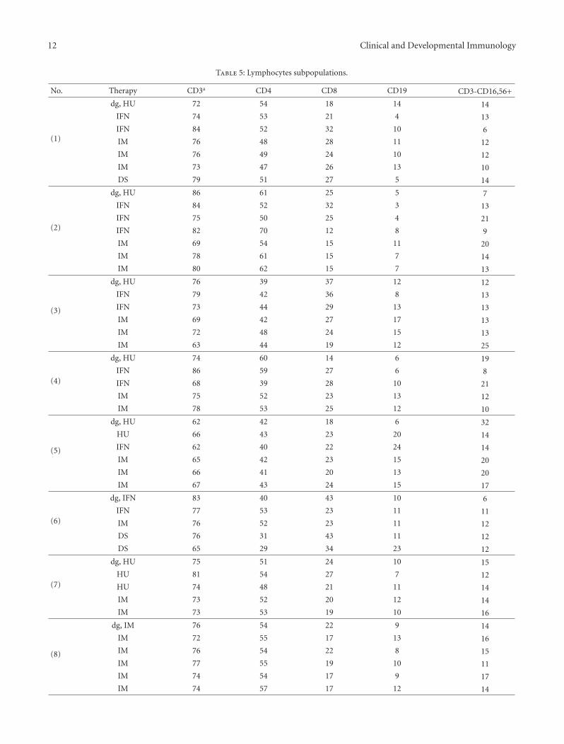
2.9. Subpopulations of Lymphocytes. Immunophenotypicanalysis of lymphocytes was performed using monoclonalantibodies directed against the following human surfaceantigens: CD3, CD4, CD8, CD19, CD16, and CD56 by flowcytometry as described previously [22]. The sum of cellsstained with CD4, CD8, CD19, and CD16-CD56 antibodieswas considered 100%. The standard laboratory referentialranges are: 59%–84% for CD3+, 25%–59% for CD4+, 19%–48% for CD8+, 6%–22% for CD19+, 6%–30% for CD16+,CD56+ cells.
2.10. Intracellular Cytokine Production by Stimulated CD3+Cells. Intracellular production of interleukin-2 (IL-2) inter-leukin-4 (IL-4), tumor necrosis alpha (TNFα), and inter-feron gamma (INFγ) in CD3+ cells stimulated by the mix-ture of brefeldinA and phorbol-12-myristate-13-acetate wasmonitored by flow cytometry as described previously [22].The figures shown in the Results section indicate thepercentages of CD3+ producing the individual cytokines.
2.11. Antibodies against Herpesviruses and Human Papillo-maviruses. Antibodies were determined against the follow-ing herpesviruses: the herpes simplex virus type 1 and type 2(HSV 1 and 2, IgG and IgM), the varicella-zoster virus (VZV,IgG and IgM), the human cytomegalovirus (CMV, IgG andIgM) and the EB virus (EBV, IgG and IgM against the viruscapsid antigen [VCA], IgG against the virus nuclear antigen[EBNA1] and against the early antigens [EA R+D]). Thefollowing commercial kits were used: ETI-HSVK-G-1/2 andETI-HSVK-M-1/2 (DiaSorin S.p.A., Italy) for the detectionof HSV 1/2 IgG and IgM antibody, respectively; ETI-CYTOK-G Plus and ETI-CYTOK-M REV Plus (DiaSorinS.p.A., Italy ) for the detection of CMV IgG and IgMantibody, respectively; VZV IgG and VZV IgM (Nova Tec,Immunodiagnostica, GmbH Germany) for the detection ofVZV IgG and IgM antibody, respectively; ETI-EBV VCA-Gand ETI-EBV-VCA-M-Rev (DiaSorin S.p.A., Italy) for thedetection of EBV VCA IgG and IgM antibody, respectively;ETI-EBNA-G (DiaSorin S.p.A., Italy) for the detection ofEBV EBNA IgG antibody; and ETI-EA-G (DiaSorin S.p.A.,Italy) for the detection of EBV EA IgG antibody. In addition,antibodies to six types of human papillomaviruses (HPV),namely, types 6, 11, 16, 18, 31, and 33 were determined

4 Clinical and Developmental Immunology
Table 1: Patients followed.
No. Gender Age1 Treatment Hematologic
Mo2 Drug Dosage/d WBC× 109/L Lympho× 109/L Hb (gr/L) Trc× 109/L HR3 CCR3
(1) F 55
0 dg, HU 2000 mg 302.6 4.60 89 413
19 No
8 IFN 5 MIU 16.2 1.45 130 188
16 IFN 5 MIU 69.0 3.43 131 442
22 IM 400 mg 10.5 2.86 124 399
27 IM 400 mg 7.4 1.93 120 505
39 IM 600 mg 4.0 1.11 120 175
58 DS 100 mg 2.1 0.89 119 139
(2) F 62
0 dg, HU 2500 mg 120.0 3.54 132 226
6 19
6 IFN 3 MIU 4.7 1.49 151 163
14 IFN 3 MIU 5.1 1.20 131 352
15 IFN 5 MIU 5.8 0.91 140 356
25 IM 400 mg 3.2 1.12 138 157
43 IM 400 mg 7.0 1.38 130 430
56 IM 400 mg 9.3 1.61 130 648
(3) M 40
0 dg, HU 3000 mg 21.9 2.07 168 327
8 26
7 IFN 3 MIU 8.1 1.08 163 262
15 IFN 5 MIU 3.1 0.85 160 125
26 IM 400 mg 4.2 1.10 146 246
43 IM 400 mg 4.5 1.37 147 209
58 IM 400 mg 3.9 1.01 158 210
(4) M 60
0 dg, HU 2000 mg 198.5 5.06 110 550
6 36
6 IFN 3 MIU 3.4 0.86 132 115
12 IFN 5 MIU 3.4 1.21 137 152
42 IM 400 mg 5.5 1.52 131 301
56 IM 400 mg 3.5 1.56 136 311
(5) M 33
0 dg, HU 2500 mg 99.7 5.91 142 169
2 40
6 HU 2000 mg 9.9 3.56 145 151
9 IFN 3 MIU 47.8 3.12 137 102
40 IM 400 mg 4.3 1.69 142 202
53 IM 400 mg 5.5 2.01 140 258
58 IM 400 mg 6.6 1.88 136 266
(6) F 35
0 dg, IFN 3 MIU 107.5 0.48 117 276
2 35
10 IFN 3 MIU 4.4 0.76 101 182
12 IM 400 mg 2.8 1.12 102 145
36 DS 100 mg 2.9 1.61 89 233
48 DS 100 mg 2.8 1.83 89 331
(7) M 54
0 dg, HU 2000 mg 16.4 4.86 136 595
11 38
7 HU 2000 mg 4.5 5.71 137 532
20 HU 1000 mg 5.5 3.21 134 209
34 IM 400 mg 6.9 1.23 134 206
47 IM 400 mg 6.8 2.19 132 236

Clinical and Developmental Immunology 5
Table 1: Continued.
No. Gender Age1 Treatment Hematologic
Mo2 Drug Dosage/d WBC× 109/L Lympho× 109/L Hb (gr/L) Trc× 109/L HR3 CCR3
(8) F 43
0 dg, IM 400 mg 26.9 4.05 144 464
2 17
2 IM 400 mg 8.3 1.66 129 228
9 IM 400 mg 6.0 1.76 123 185
17 IM 400 mg 7.5 1.26 126 207
31 IM 300 mg 7.6 1.73 120 239
45 IM 300 mg 8.3 1.45 125 315
(9) M 48
0 dg, IM 400 mg 439.2 6.16 81 184
7 No30 IM 400 mg 4.4 1.96 150 117
44 IM 400 mg 5.4 1.41 149 112
(10) F 38
0 dg, IM 400 mg 328.6 1.81 88 614
5 277 IM 400 mg 3.0 1.32 121 200
28 IM 400 mg 3.3 1.10 94 212
36 IM 400 mg 4.9 0.98 103 236
(11) F 61
0 dg,HU 2000 mg 16.8 5.31 109 1895
4 2529 DS 100 mg 5.8 2.02 100 519
52 DS 100 mg 5.4 2.75 104 2491Age at enrollment; 2Mo: month; WBC: white blood cell count; Hb: hemoglobin; and Trc, thrombocyte count; HU: hydroxyurea; IFN: interferon-alpha 2a;
IM: imatinib mesylate; DS: dasatinib; MIU, Millions International Units. 3The figure indicates month after diagnosis at which HR or CCR was first observed;“no” means that CCR was not achieved.Notes: Patient no. 1 developed vasculitis (erythema nodosum), neutropenia which was subsequently treated with growth factors and immunoglobulins.Patients 6 and 7 had laboratory tests positive for autoimmune thyroiditis without any clinical manifestations.Patient no. 11 decided to undergo homeopathic therapy and self-therapy with HU and was out of the evidence for some time. At the indicated interval, DStherapy was started because of pathological findings both in peripheral blood and bone marrow.
using ELISA using virus-like particles as antigen as describedpreviously [23]. Sera were diluted according to correspond-ing manufacturer recommendation, in case of anti-HPVantibodies 1 : 25. All samples originating from the samepatient were tested simultaneously on one microplate.
2.12. Statistical Methods. In individual patients, Spear-man’s correlation coefficient was used for quantifying themonotony of trend in time of the measured items. Thestatistical difference P for the deviation from zero wascalculated. When analyzing the hematologic findings for thewhole group of patients, the data transformed to logarithmswere analyzed by covariance analysis testing for differencesamong patients and the common linear regression onto thetime of treatment. The linear function was used as a basiccomponent of the experienced time trend. For comparison ofthe relative percentage distribution of different lymphocytepopulations in the first and last samples available, χ2 testwas used. For evaluation of the production of cytokines bystimulated CD3+ cells, the mean ratio of the last and firstsamples available was tested by One Sample Student t-testfor difference from 1. P-values have not been adjusted formultiple comparisons.
3. Results
3.1. Immunoglobulins Levels. The results of measuring theimmunoglobulins levels are summarized in Table 2. Nearly
generally, there was a tendency to a decrease of their levels.The decrease of total IgG for the whole group was just onthe brink of statistical significance (P = .05). In two patients(nos. 2 and 7) the IgG levels dropped below the norm.On the other hand, a marked increase of IgGs was observedin one patient (No. 6, treated gradually with IFNα, IMand DS) who had had the lowest level of total IgG andpathologically low levels of IgG1, IgG2, IgG3 and IgG4 inher pretreatment serum. The treatment resulted in theirrestoration up to the norm. It may be of interest that in thispatient the hemoglobin level dropped in the course of theobservation period (see Table 1). The most frequent changeswere in the IgM levels. Their slight or moderate decrease wasobserved in 9 patients and usually it was most marked aftertreatment with IM. The decrease of IgM levels for the wholegroup was significant (P = .011). Changes in IgA levels wereseen less frequently. Its levels dropped significantly with timein only one patient (no. 2) and increased in other three (nos.6, 9 and 11). The first one of these was the already mentionedpatient no. 6 with an increase in all subclasses of IgG. Itis noteworthy that throughout the observation period thechanges in this particular patient were not associated withany marked variation in the IgM level.
3.2. Presence of Autoantibodies. The presence of autoanti-bodies is shown in Table 3. Again, no consistent pattern isapparent. Autoantibodies, which had been present in thepretreatment sera of only three patients (twice against TPO

6 Clinical and Developmental Immunology
Table 2: Imunoglobulins.
No. Therapy IgG IgG1 IgG2 IgG3 IgG4 IgA IgM
(1)
dg, HU 10.60 6.73 4.29 0.686 0.526 2.16 1.65
IFN 11.10 6.14 3.74 0.767 0.364 1.95 1.50
IFN 9.30 5.97 3.22 0.530 0.250 1.93 1.56
IM 12.30 8.90 3.99 0.709 0.119 2.74 1.91
IM 9.40 6.82 3.34 0.575 0.157 2.17 1.61
IM 11.30 6.14 4.21 0.687 0.193 2.92 1.54
DS 11.40 6.70 3.81 0.907 0.253 2.73 1.29
(2)
dg, HU 10.00 6.72 3.34 0.418 0.460 2.31 0.76
IFN 8.58 5.00 3.34 0.353 0.339 2.12 0.72
IFN 8.53 5.64 3.00 0.358 0.397 1.68 0.45
IFN 9.39 4.50 3.13 0.320 0.247 1.50 0.40
IM 8.49 4.41 2.75 0.292 0.259 1.59 0.33
IM 6.78 3.98 2.18 0.312 0.179 1.35 0.20
IM 6.41∗∗ 3.91∗∗ 2.24∗∗ 2.92∗∗ 1.52∗∗ 1.23∗∗ 0.23∗∗
(3)
dg, HU 10.00 6.41 3.03 0.225 0.079 1.79 1.44
IFN 9.19 5.83 2.74 0.196 0.082 1.66 0.83
IFN 10.80 6.33 3.28 0.239 0.058 1.74 0.87
IM 9.08 5.71 3.23 0.190 0.091 1.66 0.38
IM 8.72 5.04 2.68 0.248 0.083 1.73 0.54
IM 9.41 5.18∗ 3.35 0.213 0.065 1.78 0.47
(4)
dg, HU 11.60 7.79 3.94 0.666 0.833 1.86 0.94
IFN 13.00 8.44 4.05 0.686 0.303 1.62 1.18
IFN 13.60 10.40 3.34 0.637 0.270 1.80 0.98
IM 10.90 6.51 3.10 0.541 0.231 1.62 0.58
IM 10.90 6.98 3.34 0.613 0.209∗∗ 1.84 0.66
(5)
dg, HU 10.40 6.29 3.04 0.248 0.133 1.59 1.76
HU 10.20 7.40 3.03 0.158 0.066 1.98 1.20
IFN 9.35 6.20 2.97 0.195 0.142 1.61 1.31
IM 9.21 5.01 2.82 0.236 0.097 1.83 1.13
IM 9.81 5.33 3.16 0.226 0.082 1.93 1.12
IM 9.11∗ 6.77 2.62 0.605 0.081 1.95 1.06∗∗
(6)
dg, IFN 8.05 5.63 1.27 0.135 0.073 1.17 1.10
IFN 11.30 n.t.a n.t. n.t. n.t. 1.39 1.29
IM 12.00 n.t. n.t. n.t. n.t. 1.58 1.41
DS 17.90 12.70 1.75 0.611 0.152 2.07 0.79
DS 13.6∗ 11.20 2.17∗∗ 0.416 0.188∗∗ 2.26∗∗ 1.18
(7)
dg, HU 8.92 4.60 4.01 0.388 0.610 1.22 0.95
HU 8.79 4.45 3.95 0.337 0.797 1.30 0.76
HU 8.76 3.81 4.11 0.266 0.702 1.34 0.51
IM 6.08 2.89 2.72 0.262 0.507 0.99 0.32
IM 6.63∗ 31.4∗ 2.90 0.303 0.425 0.93 0.36∗
(8)
dg, IM 12.60 9.51 3.53 0.080 n.t. 1.49 0.97
IM 11.60 8.81 2.73 0.083 0.320 1.75 0.79
IM 12.20 7.43 2.74 0.087 0.276 1.84 0.57
IM 12.60 7.64 3.21 0.087 0.188 1.67 0.63
IM 12.60 9.30 3.26 0.094 0.432 1.94 0.74
IM 11.70 7.41 2.95 0.072 0.273 1.93 0.72
Clinical and Developmental Immunology 7
Table 2: Continued.
No. Therapy IgG IgG1 IgG2 IgG3 IgG4 IgA IgM
(9)
dg, IM 10.60 7.58 3.41 0.523 0.697 1.31 1.11
IM 9.43 5.36 3.41 0.480 0.790 1.47 1.11
IM 9.53 5.90 3.95 0.479∗∗ 0.738 1.81∗∗ 1.32
(10)
dg, IM 13.50 8.46 4.93 0.231 2.180 1.04 1.61
IM 14.20 7.63 5.63 0.211 1.970 1.28 1.60
IM 12.10 7.49 4.49 0.262 1.200 1.04 1.39
IM 12.20 6.63∗∗ 4.51 0.232 1.340 1.15 1.41
(11)
dg, HU 12.00 6.21 5.66 0.231 0.146 0.88 0.67
DS 11.50 5.97 5.63 0.271 0.085 0.91 0.45
DS 12.40 8.12 3.50∗∗ 0.677∗∗ 0.080∗∗ 1.02∗∗ 0.32∗∗an.t.: not tested.∗P < .05, ∗∗P < .01P for trend with time.
and once against SM), were detected in the course of theobservation period in a total of eight patients. In most ofthem, their appearance was transitory. In two patients (nos.1 and 6, both of them were females), autoantibodies weredetected against three antigens: in two patients (nos. 4 and7) against two antigens and in four patients (nos. 2, 3,9 and 10) against one antigen only. The most frequentlydetected autoantibodies were reactive with TPO, TG andnucleus (in all instances in three patients). In two patientsantibodies reactive with SM were detected. In 1 of these(patient no. 1), they were present in the pretreatment serumsample, disappeared after starting the therapy with HU andwere not detected later on when HU was gradually replacedby IFNα and IM. A similar phenomenon was seen in patientno. 7, in whom the initial reactivity with TPO and TGdisappeared in the course of the therapy. Thus no consistentpattern was evident and, because of the multiplicity of drugsemployed for individual patients, no clear dependence onthe therapy used was observed. It may be of interest that theantibodies against mitochondria (AMAb) and endomysium(E-AAb and E-GAb) were never detected.
There seems to be some, but not quite a consistentcorrelation with the mode of therapy and the developmentof autoantibodies. Autoantibodies were detected in five ofsix patients treated with IFNα and in four out of fivepatients treated with HU (all of them had later on beentreated with IFNα) but in only two out of four patientsexclusively treated by TKI (either IM or DS). Furthermore,in four patients autoantibodies detected after HU and IFNαtreatment disappeared when these drugs were replaced byIM.
3.3. Complement C3 and C4 Components. In the pretreat-ment sera normal levels of C3 were observed in all but onepatient (no. 11), in whom the level was below the norm.As indicated in Table 4, little variation of C3 levels wasobserved in the course of the observation period; still, inthree patient’s a transitory decrease of its level below thestandard laboratory range was observed. In all instances(including patient no. 11), their levels returned to the norm
by the time hematological remission was achieved. In two ofthree patients with the increased levels of C4 in pre-treatmentsera (nos. 4, and 5), their levels dropped in the course oftreatment.
3.4. C-Reactive Protein and IL-6. The results are alsopresented in Table 4. Prior to the start of the therapy,increased levels of CRP were detected in six patients. Judgingby the levels detected in the last samples collected, thelevels dropped to the norm in all of them. However, it isnoteworthy that in two patients (nos. 3 and 8), in spite of thehematological remission having been achieved, their levelsincreased, and in four other patients (nos. 1, 2, 5 and 7)a transitory increase of CRP was detected in the course ofthe observation period. In patient no. 7 the increased levelof CRP corresponded with an acute infection of the upperrespiratory tract. In the other four patients (nos. 1, 3, 5 and7) no clinical complications at the time of CRP increase wereobserved or reported by the patients. As concerns IL-6 levels,there was a nearly general decrease in association with thehematological remission. The decrease for the whole groupwas highly significant (P = .001). Again, as in the previousstudy [22], no clear correlation between IL-6 and CRP levelswas apparent.
3.5. Lymphocyte Subpopulations. As indicated in Table 5,in spite of the drop of lymphocytes (shown in Table 1),there was little variation in the percentage of CD3+ cellsin the course of the observation period, including patientno. 6, in whom lymphopenia was detected in the pre-treatment sample (see Table 1), and patient no. 10 withCD3+ lymphocyte percentage slightly below the norm. Theonly exception was patient no. 11, initially treated with HUwho after a rather long interval without any treatment wastreated with DS. In this patient a marked drop of CD3+cells was detected. It is clear, however, that this decrease wasrelative, reflecting a substantial increase of NK cells. Thechanges in the percentages of CD4+ and CD8+ were slightor moderate in nearly all patients and no consistent pattern

8 Clinical and Developmental Immunology
Table 3: Autoantibodies.
No. Therapy TPOAb TGAb ANAb ANCAb AMAb ENDOAb A ENDOAb G SMAb DESMAb
(1)
dg, HU na n n n n n n pb n
IFN n n n p n n n n n
IFN n n n n n n n n p
IM n n n p n n n n p
IM n n n p n n n n p
IM n n n n n n n n p
DS n n n n n n n n p
(2)
dg, HU n n n n n n n n n
IFN n n n n n n n n n
IFN n n n n n n n n n
IFN n n p n n n n n n
IM n n n n n n n n n
IM n n n n n n n n n
IM n n n n n n n n n
(3)
dg, HU n n n n n n n n n
IFN n n n n n n n n n
IFN n n n n n n n p n
IM n n n n n n n n n
IM n n n n n n n p n
IM n n n n n n n n n
(4)
dg, HU n n n n n n n n n
IFN n n n n n n n n n
IFN n n n n n n n n p
IM p n n n n n n n n
IM n n n n n n n n n
(5)
dg, HU n n n n n n n n n
HU n n n n n n n n n
IFN n n n n n n n n n
IM n n n n n n n n n
IM n n n n n n n n n
IM n n n n n n n n n
(6)
dg, IFN p n n n n n n n n
IFN p p n n n n n n n
IM p n p n n n n n n
DS p n n n n n n n n
DS p n n n n n n n n
(7)
dg, HU p p n n n n n n n
HU p p n n n n n n n
HU n.t.c n.t. n n n n n n n
IM p n n n n n n n n
IM n n n n n n n n n
(8)
dg, IM n n n n n n n n n
IM n n n n n n n n n
IM n n n n n n n n n
IM n n n n n n n n n
IM n.t. n.t. n n n n n n n
IM n n n n n n n n n

Clinical and Developmental Immunology 9
Table 3: Continued.
No. Therapy TPOAb TGAb ANAb ANCAb AMAb ENDOAb A ENDOAb G SMAb DESMAb
(9)
dg, IM n n n n n n n n n
IM n n n n n n n n n
IM n n n p n n n n n
(10)
dg, IM n n n n n n n n n
IM n n n n n n n n n
IM n n n n n n n n n
IM n n p n n n n n n
(11)
dg, HU n n n n n n n n n
DS n n n n n n n n n
DS n n n n n n n n n
TPOAb: antibodies against thyroidal peroxidase; TGAb: antibodies against thyreoglobulin; ANAb: antinuclear antibodies; ANCAb: antibodies against thecytoplasm of neutrophils; AMAb: antimitochondrial antibodies; ENDOAb A, ENDOAb G: antibodies of the IgA and IgG classes against endomysium; SMAB:antibodies against smooth muscless; DESMAb: antidesmosomal antibodies.aNegative for the respective antibodies.bPositive for the respective antibody.cn.t.:not tested.
was evident. Still, in four of seven patients, in whose pre-treatment samples the percentage of CD8 cells was below thenorm, their percentages did not reach the lower limit of thereferential range. In four patients (nos. 4, 5, 6 and 10) theremission was associated with an increase in CD19+ cells.When the distribution of lymphocyte subpopulations in thefirst and last sample was compared, significant differences(P < .05 to< .001) were encountered in six patients (nos. 1, 3,5, 6, 9 and 11). However, no consistent pattern was apparentand thus the real significance of these findings is doubtful.
3.6. Intracellular Production of Cytokines. The results arepresented in Table 6. Two observations may be of interest.Possibly the most important one was an increase of the CD3+cells, which produce the cytokines, in most of the patients,including those, in whom the production of these cytokinesprior to the start of the therapy had been pathologically low(e.g., in the case of IL-2, patients nos. 3 and 8). Of the tenpatients, which could be evaluated, the percentage of IL-2-producing cells increased in seven, IL-4-producing cells alsoin seven, TNFα-producing cells in six and INFγ producingcells in seven. In some of the patients the level of cytokineproduction remained essentially unchanged (e.g., patient no.8, all four cytokines) and in one patient there was a dropof IL-2 and IL-4 production (patient no. 1) and in anotherpatient the drop of INFγ production (patient no. 2).
The other noteworthy observation, closely associatedwith the first one, was an increase in cells producing morethan one cytokine after stimulation, as reflected by anincrease of the percentage sum of reactive cells. The increasewas expressed in terms of the Cytokine Production Index(CPI) given by the ratio between the sum of cytokine-producing cells as detected in the last sample and the sumof cytokine producing cells in the first sample available (i.e.,in all but one patient before the start of therapy). A marked
increase in CPI was detected in seven (nos. 3, 4, 5, 6, 7, 9and 10) out of 10 patients which could be evaluated. Thedifference for the group was highly significant (P = .006).
3.7. Antibodies Against Herpesviruses and Papillomaviruses.To clarify whether treatment of the CML patients with HU,IFNα or TKI was associated with activation of latent and/orpersistent virus infections, we tested sera from the patientsfor presence of antibodies against four human herpesvirusesand six human papillomaviruses. The results are summarizedin Tables 7 and 8. For the sake of simplicity, only the resultsof testing sera taken prior to the start of any treatment andat the end of the observation period are presented. It isevident that there was only very little difference between thetwo sets of sera. In one patient (no. 11) treated with DS,cytomegalovirus infection was reactivated as revealed by avery marked increase in IgG antibody and the appearance ofIgM antibody (results not shown).
4. Discussion
In the preceding paper [22], we showed that CML patientsbefore treatment differed from matched healthy subjects ina number of immunological parameters. The major aimof the present investigation was to find out, whether theseaberrations persisted, decreased, or disappeared in the courseof treatment, in particular whether these changes correlatedwith the induction of hematological and/or cytogeneticremission, whether and how they were influenced by thedrugs used for treatment, and, in the long run, what was theirprognostic value, if any. Our efforts were seriously hamperedby two circumstances, that is, by the loss of 13 out of 24patients originally enrolled and, for ethical reasons, by theimpossibility to maintain the original treatment regimen

10 Clinical and Developmental Immunology
Table 4: Complement components, CRP and IL-6.
No. Therapy C3 C4 CRP IL-6
(1)
dg, HU 0.94 0.40 5.2 5.10
IFN 0.67 0.37 3.0 2.53
IFN 0.83 0.42 24.4 5.42
IM 0.71 0.30 3.5 1.83
IM 0.53 0.21 3.5 1.53
IM 0.72 0.30 3.3 1.67
DS 1.26 0.52 3.2 2.72
(2)
dg, HU 1.24 0.30 12.8 9.54
IFN 1.22 0.28 3.0 3.47
IFN 1.22 0.28 3.5 3.87
IFN 1.15 0.25 3.5 4.92
IM 1.19 0.26 3.2 3.21
IM 1.27 0.27 5.4 2.45
IM 1.20 0.24∗ 3.2 2.82∗
(3)
dg, HU 1.19 0.26 3.0 3.9
IFN 1.14 0.26 3.0 1.8
IFN 0.94 0.24 3.5 1.5
IM 1.15 0.27 3.3 2.16
IM 1.20 0.26 3.2 1.14
IM 1.40 0.30 32.9 3.03
(4)
dg, HU 1.35 0.38 11.6 4.40
IFN 0.90 0.21 3.0 1.54
IFN 1.11 0.23 3.5 1.97
IM 1.20 0.27 3.2 1.45
IM 1.06 0.25 3.2 1.50
(5)
dg, HU 1.03 1.23 3.0 3.30
HU 0.97 0.21 3.5 3.67
IFN 0.80 0.20 3.5 2.16
IM 0.91 0.19 3.2 2.49
IM 1.14 0.23 8.8 2.30
IM 1.00 0.25 3.4 1.99
(6)
dg, IFN 0.98 0.21 3.0 2.80
IFN 0.69 0.19 n.t.a 1.82
IM 0.78 0.17 3.5 1.87
DS 1.31 0.30 3.2 2.31
DS 1.14 0.24 3.2 0.00
(7)
dg, HU 0.91 0.16 3.5 5.08
HU 0.68 0.15 3.5 4.24
HU 0.91 0.18 3.3 5.29
IM 0.91 0.19 21.3 1.82
IM 0.98 0.17 3.2 3.00
(8)
dg, IM 0.93 0.19 3.5 3.80
IM 0.94 0.19 3.8 2.63
IM 0.89 0.17 3.2 2.31
IM 0.89 0.18 3.3 1.53
IM 1.07 0.26 9.4 0.27
IM 1.11 0.33 6.1 1.70∗
Clinical and Developmental Immunology 11
Table 4: Continued.
No. Therapy C3 C4 CRP IL-6
(9)
dg, IM 0.86 0.29 23.0 14.20
IM 0.91 0.24 3.2 1.21
IM 1.05∗∗ 0.27 3.2 1.00∗
(10)
dg, IM 0.79 0.30 5.9 16.71
IM 0.86 0.24 3.3 4.97
IM 0.81 0.17 3.2 n.t.
IM 0.76 0.17 3.2 1.00∗∗
(11)
dg, HU 0.65 0.36 6.8 5.20
DS 0.59 0.25 3.2 2.23
DS 0.80 0.35 3.4 3.50an.t.:not tested.∗P < .05, ∗∗P < .01P for trend with time.
with either HU or INFα. Because of the changes in the treat-ment modalities, the original set of patients split into twogroups. The first one consisted of eight patients originallytreated with HU and/or INFα and subsequently with TKI,while the remaining three patients treated exclusively withTKI constituted the second group.
Of these two shortcomings, the diminution of our setof patients from 24 to 11 was certainly the more impor-tant one. This reduction increased the difficulties alreadyinherent in the group studied, that is, its inhomogeneity(age, sex, different treatments), as regards evaluation of thedata obtained. Still, because in all patients hematologicalremission and in nearly all of them even CCR was achieved,this bringing an element of homogeneity into the studygroup, some conclusions can be drawn. The most importantone is the nearly general association of the remission witha normalization of the aberrant immunity parameters, bothhumoral a cellular. One can assume that this normalizationwas due to an alleviation of the CML-associated processes.The continuing followup of the patients should revealwhether any relapse of the disease would be associated withdeviations from the norm and with the reappearance of thesame or appearance of some other aberrations.
As regards the levels of immunoglobulins, there was atendency towards their decrease, predominantly IgM. Thegradual decrease of IgM levels was highly significant. Thesefindings correspond with the results reported by Steegmannet al. [24], who described considerable reduction of levels ofserum immunoglobulins, including IgM, in patients previ-ously exposed to IFNα and then treated with IM; accordingto their findings, the reduction of immunoglobulins wasespecially marked in patients expressing a pronounced cyto-genetic response. A marked selective effect of IM treatmenton serum IgM level in one patient has been reported byNagasawa and Mizutan [25]. We are unable to provide anyreasonable explanation for our observation. We can onlyspeculate that the decrease observed was due to qualitativealteration of B cells by the therapy employed. Otherwise,no consistent pattern was apparent. We only rarely detected
decrease in IgA levels, which we found significantly increasedin the pretreatment CML patients when compared with theirmatched healthy controls [22].
When monitoring the presence of autoantibodies, wedetected them in five out of six patients treated withINFα. These antibodies were directed against cytoplasm ofneutrophils (ANCAb), smooth muscles (ASMAb), nucleus(ANAb), thyreoglobulin (ATG), or thyroidal peroxidase(ATPO). This is not particularly surprising, since INFαexhibits allo- and autostimulation activity on antigen pre-senting cells. However, its activity in vivo may dependon the simultaneous regulation of network of immunecells rather than on the activation of individual popula-tions [10]. INFα, but not IM, was reported to cause anincreased transcription of proteinase 3 in CD14-positivemonocytes, this suggesting another possible mechanism,by which IFNα may promote self-antigen presentation [7].In our study the appearance of autoantibodies was in mostinstances of transitory character and that their disappear-ance quite frequently followed the replacement of INFαby IM. However, autoantibodies also developed in twoout of five patients untreated with INFα. It is thereforedifficult to claim, on the basis of the present results, thatINFα was markedly more active than TKI in inducingautoantibodies. It should be added that, in our patients, thedevelopment of antibodies did not manifest itself in anyclinically recognizable disease, with the possible exceptionof the simultaneous presence of ANCAb and vasculitis inpatient no. 1. This possible association and the subsequentcure of the vasculitis in parallel with the disappearance ofthe antibody after IM treatment seems to be in line witha recent report [26].
As concerns the C3 and C4 components of complementthere was little variation during the observation period.Again, there was a tendency to normalization. This seemsto be in agreement with other findings in patients withhematological malignancies including CML [27]; however,the changes we observed were rather small and their signif-icance is questionable. It seems clear that the follow-up of
12 Clinical and Developmental Immunology
Table 5: Lymphocytes subpopulations.
No. Therapy CD3a CD4 CD8 CD19 CD3-CD16,56+
(1)
dg, HU 72 54 18 14 14
IFN 74 53 21 4 13
IFN 84 52 32 10 6
IM 76 48 28 11 12
IM 76 49 24 10 12
IM 73 47 26 13 10
DS 79 51 27 5 14
(2)
dg, HU 86 61 25 5 7
IFN 84 52 32 3 13
IFN 75 50 25 4 21
IFN 82 70 12 8 9
IM 69 54 15 11 20
IM 78 61 15 7 14
IM 80 62 15 7 13
(3)
dg, HU 76 39 37 12 12
IFN 79 42 36 8 13
IFN 73 44 29 13 13
IM 69 42 27 17 13
IM 72 48 24 15 13
IM 63 44 19 12 25
(4)
dg, HU 74 60 14 6 19
IFN 86 59 27 6 8
IFN 68 39 28 10 21
IM 75 52 23 13 12
IM 78 53 25 12 10
(5)
dg, HU 62 42 18 6 32
HU 66 43 23 20 14
IFN 62 40 22 24 14
IM 65 42 23 15 20
IM 66 41 20 13 20
IM 67 43 24 15 17
(6)
dg, IFN 83 40 43 10 6
IFN 77 53 23 11 11
IM 76 52 23 11 12
DS 76 31 43 11 12
DS 65 29 34 23 12
(7)
dg, HU 75 51 24 10 15
HU 81 54 27 7 12
HU 74 48 21 11 14
IM 73 52 20 12 14
IM 73 53 19 10 16
(8)
dg, IM 76 54 22 9 14
IM 72 55 17 13 16
IM 76 54 22 8 15
IM 77 55 19 10 11
IM 74 54 17 9 17
IM 74 57 17 12 14

Clinical and Developmental Immunology 13
Table 5: Continued.
No. Therapy CD3a CD4 CD8 CD19 CD3-CD16,56+
(9)
dg, IM 85 29 53 3 12
IM 76 34 38 7 15
IM 74 37 34 9 15
(10)
dg, IM 67 52 14 14 16
IM 61 40 21 20 18
IM 55 37 16 18 25
IM 54 37 14 23 23
(11)
dg,HU 67 57 9 20 12
DS 59 49 9 16 23
DS 39 32 7 10 50aThe figures indicate the percentages of cells positive for the respective surface CDs.
C3 and C4 components of complement was not particularlyrewarding in this study.
The results obtained when testing the CRP and IL-6 levels are of greater interest. Both tended to decreasefollowing the therapy. The decrease was quite common inthe case of IL-6, where it was highly significant. Thesedata suggest that a reduction of IL-6 levels might be quitea reliable marker of successful therapy. Moreover, becauseof its biological effects, that is, inhibition of p53-inducedapoptosis [28] and suppression of phosphorylation of theretinoblastoma protein [29], its drop might contributeto the favorable course of the disease. Another favorableeffect of IL-6 decrease might be associated with its rolein STAT-3 activation. High levels of STAT-3 can preventdendritic cell maturation and subsequent presentation of theantigens [30]. Thus, increased levels of IL-6 might exhibitan immunosuppressive effect. In a way, the present datacorrespond with the earlier observations indicating that IL-6levels are raised in parallel with the progression of the diseaseinto blastic phase [31, 32]. An association of remission witha decrease of CRP levels was less consistent. Although CRPlevels dropped in all patients with increased levels detectedin their sera taken prior to the start of the therapy, in fourother patients a CRP increase was observed in the course ofthe observation period; in two of them it was only transitory.Because of the nature of this acute phase reactant [33–35],it is rather difficult to interpret the latter findings. In one ofthe patient the transitional rise of CRP was associated witha respiratory disease. In the other patients some undetectedmicroinflammation processes might have been involved.
Possibly the most important among our findings are thechanges of cell-mediated immunity parameters associatedwith the achievement of remission. They were representedby a markedly increased capability of stimulated CD3+ cellsto produce cytokines. Since at the time of remission, thesum of percentages of CD3+ cells producing any of thecytokines tested exceeded 100 in most of the patients, it ispossible to conclude that the changes observed were mainlydue to an increase in cells producing more than one cytokine.Thus, a restoration of the Tcell activities associated with the
suppression of the disease was observed. Similar results werereported by Reuben et al. [36]. and Guarini et al. [37]. inpatients, in whom hematologic remission was achieved byINFα treatment. The mechanisms responsible for the presentfindings are not quite clear. In this respect, two previous invitro studies may be of interest and provide a lead for furtherstudies. Pawelec et al. [38]. have reported the production ofthe immunosuppressive IL-10 cytokine, a potent inhibitor oftype 1 cytokines, by CML cells cultivated in vitro and havedemonstrated that its neutralization by monoclonal antibodyconsiderably enhanced the proliferation of lymphocytes inmixed lymphocyte/tumor cell cultures [38]. In anotherstudy, Kiani et al. [39]. Showed that CD4+ cells from CMLpatients, which had been separated from the leukemic cells,after stimulation produced type 1 cytokines in amountscomparable to those seen in normal subjects. Taken together,these two sets of data suggest that the products of CMLcells may themselves be responsible for the reduced immuno-competence of the CD3+ cells. One point deserves a specialcomment. All ten patients, in whom the respective data wereavailable, had been treated, at least for some time, withIM. This drug has been reported to suppress CD3+ cellactivation in vitro [40, 41] as well as other parameters of Tcellimmunity in vivo [42–44]. Our findings in the present studydo not seem to be in agreement with those observations. Onecan therefore hypothesize that the alleviation of the CML-associated processes, which was induced by the successfultherapy, was a more important factor than the puta-tive immunosuppressive effect of IM. Furthermore, it hasrecently been reported that IM is suppressing the activationand proliferation of CD4+CD25+ regulatory cells (Treg cells)which are producing strong immunosuppressive factors likeIL-10, transforming growth factor β (TGFβ) and granzymeB [45]. It is thus very well possible that in the patientsstudied the IM-induced suppression of Treg contributed tothe present observation. Unfortunately, we did not monitorthe levels of Treg cells in the present study. It should alsobe recalled that a significant increase of INFγ-producingTcells following IM treatment has been reported byAswald et al. [46].

14 Clinical and Developmental Immunology
Table 6: Intracellular cytokines production in activated CD3+ lymphocytes.
No. Therapy IL-2 TNFalfa INF-γ IL-4 CPI
(1)
dg, HU 56 44 12 4
IFN 42 49 18 3
IFN 31 35 23 5
IM 53 60 30 2
IM 61 48 36 3
IM n.t.a n.t. n.t. n.t.
DS 32 60 23∗ 1 1.000
(2)
dg, HU 31 53 62 2
IFN 10 23 29 5
IFN 69 66 30 4
IFN 76 65 32 5
IM 67 73 42 6
IM n.t. n.t. n.t. n.t.
IM 55 45 35 20∗ 1.047
(3)
dg, HU 1 54 41 6
IFN 24 46 36 3
IFN 48 66 50 3
IM 23 51 25 1
IM n.t. n.t. n.t. n.t.
IM 49 57 51 20 1.735
(4)
dg, HU 17 20 25 6
IFN 31 46 29 8
IFN 38 78 38 6
IM n.t. n.t. n.t. n.t.
IM 56∗∗ 65 44∗∗ 11 2.588
(5)
dg, HU 22 21 14 4
HU 50 55 25 4
IFN 39 50 30 2
IM 37 49 24 8
IM 52 58 30 2
IM 38 41 30 5 1.869
(6)
dg, IFN 11 17 22 2
IFN 36 51 32 3
IM 48 55 35 5
DS 20 59 45 3
DS 18 44 37∗ 19 2.269
(7)
dg, HU 51 46 34 3
HU 52 42 30 2
HU 52 68 55 5
IM n.t. n.t. n.t. n.t.
IM 52 53 34 25 1.223
(8)
dg, IM 42 44 23 2
IM 38 44 19 2
IM 16 25 13 5
IM n.t. n.t. n.t. n.t.
IM n.t. n.t. n.t. n.t.
IM 41 37 23 3 0.937
(9)dg, IM 2 34 45 1
IM n.t. n.t. n.t. n.t.
IM 31 74 57 13 2.134

Clinical and Developmental Immunology 15
Table 6: Continued.
No. Therapy IL-2 TNFalfa INF-γ IL-4 CPI
(10)
dg, IM n.t. n.t. n.t. n.t.
IM 27 32 14 1
IM 38 54 24 3
IM 50∗∗ 51 30∗∗ 6∗∗ 1.827
(11)dg, HU n.t. n.t. n.t. n.t.
DS 59 62 23 1
DS n.t. n.t. n.t. n.t. n.t.
CPI:Cytokine Production Index—a ratio between the sum of cytokine producing cells ls as detected in the last sample and the sum of cytokine producingcells in the first sample available.an.t.:not tested.∗P < .05, ∗∗P < .01. P for trend with time.
Table 7: Presence and geometric mean titres of antibodies against human herpesviruses in CML patients prior to the start of the therapyand after achieiving hematological and/or complete cytogenetic remission.
Antibody presence
Sample No. HSV1/2 CMV VZV EBV
IgG IgM IgG IgM IgG IgM VCAIgG VCAIgM EAIgG EBNA IgM
First 11 11 2 8 0 11 2 11 2 3 10
Last 11 11 0 8 1 11 2 11 2 3 10
GMT
First 11 8.12 0.65 1.83 0.47 2.83 0.74 7.41 0.33 0.57 4.28
Last 11 8.33 0.57 2.01 0.47 2.61 0.54 8.20 0.34 0.65 4.79
Table 8: Presence and geometric mean titres of antibodies against human papillomaviruses in CML patients prior to the start of the therapyand after achieving hematological and/or complete cytogenetic remission.
Antibody presence
Sample No. HPV6 HPV11 HPV16 HPV18 HPV31 HPV33
First 11 6 5 3 3 3 1
Last 11 5 3 3 3 2 1
GMT
First 11 1.27 1.01 0.68 1.00 0.79 0.50
Last 11 1.08 0.88 0.65 0.75 0.61 0.38
Our attempts to find a reflection of these changes in theantibody titers against herpesviruses and papillomaviruses,two virus families known to be activated under immunosup-pression, failed completely. As indicated in a previous paper[22], there were no significant differences in the prevalence ofantibodies against these viruses between the untreated CMLpatients and matched normal control subjects. In the presentstudy the antibody patterns in sera taken before the startof therapy and after achieving remission were comparable,this strongly suggesting that in the course of the observationperiod the activation of latent infections did not occur orcould not be revealed by the antibody tests used.
In spite of inconsistency of the treatment regimensdescribed above, some differences in the changes of theimmunological parameters studied, which might have beenassociated with the drugs used, were recorded. Two obser-vations are noteworthy. First, the autoantibodies were more
frequently seen in patients who had been treated with HUand INFα than in those treated exclusively with TKI, and theytended to disappear after substituting TKI for HU and INFα.Still, the lack of consistency and the small number of patientstested preclude any conclusions. Second, increase of NK cellswas detected more frequently in those exclusively treatedwith TKI than in patients who had undergone combinedtherapy.
5. Conclusions
The present results indicate that the altered parameters ofinnate immunity found in the CML patients prior to thestart of any therapy tended to normalize in the course oftherapy leading to remission. Other data suggested, but didnot prove, that some of the parameters monitored might beinfluenced by the treatment modality used.

16 Clinical and Developmental Immunology
Acknowledgments
Supported by grants from the Internal Granting Agencyof the Ministry of Health, Czech Republic (IGA MZCRNC/6957-3, and NR/9075-3) and by Research Project No.000 273 3601, Institute of Hematology and Blood Transfu-sion. The authors express their indebtedness to H. Tlaskalovafor critical reading of the manuscript.
References
[1] M. Suehiro, S. Kishimoto, T. Wakabayashi et al., “Hydroxyureadermopathy with a dermatomyositis-like eruption and a largeleg ulcer,” British Journal of Dermatology, vol. 139, no. 4, pp.748–749, 1998.
[2] M. J. Dacey and J. P. Callen, “Hydroxyurea-induced dermat-omyositis-like eruption,” Journal of the American Academy ofDermatology, vol. 48, no. 3, pp. 439–441, 2003.
[3] M. Talpaz, K. B. McCredie, G. M. Mavligit, and J. U. Gutter-man, “Leukocyte interferon-induced myeloid cytoreductionin chronic myelogenous leukemia,” Blood, vol. 62, no. 3, pp.689–692, 1983.
[4] M. Talpaz, K. McCredie, H. Kantarjian, J. Trujillo, M. Keating,and J. Gutterman, “Chronic myelogenous leukaemia: haema-tological remissions with alpha interferon,” British Journal ofHaematology, vol. 64, no. 1, pp. 87–95, 1986.
[5] F. Bonifazi, A. De Vivo, G. Rosti et al., “Chronic myeloidleukemia and interferon-α: a study of complete cytogeneticresponders,” Blood, vol. 98, no. 10, pp. 3074–3081, 2001.
[6] J. J. Molldrem, P. P. Lee, C. Wang et al., “Evidence that specificT lymphocytes may participate in the elimination of chronicmyelogenous leukemia,” Nature Medicine, vol. 6, no. 9, pp.1018–1023, 2000.
[7] A. Burchert, S. Wolfl, M. Schmidt et al., “Interferon-α, butnot the ABL-kinase inhibitor imatinib (STI571), inducesexpression of myeloblastin and a specific T-cell response inchronic myeloid leukemia,” Blood, vol. 101, no. 1, pp. 259–264,2003.
[8] C. J. Wu, X.-F. Yang, S. McLaughlin et al., “Detection of apotent humoral response associated with immune-inducedremission of chronic myelogenous leukemia,” Journal ofClinical Investigation, vol. 106, no. 5, pp. 705–714, 2000.
[9] D. Chakrabarti, B. Hultgren, and T. A. Stewart, “IFN-α inducesautoimmune T cells through the induction of intracellularadhesion molecule-1 and B7.2,” Journal of Immunology, vol.157, no. 2, pp. 522–528, 1996.
[10] D. Tosi, R. Valenti, A. Cova et al., “Role of cross-talk betweenIFN-α-induced monocyte-derived dendritic cells and NK cellsin priming CD8+ T cell responses against human tumorantigens,” Journal of Immunology, vol. 172, no. 9, pp. 5363–5370, 2004.
[11] S. Sacchi, H. Kantarjian, S. O’Brien, P. R. Cohen, S. Pierce,and M. Talpaz, “Immune-mediated and unusual complica-tions during interferon alfa therapy in chronic myelogenousleukemia,” Journal of Clinical Oncology, vol. 13, no. 9, pp.2401–2407, 1995.
[12] E. Tothova, A. Kafkova, N. Stecova, M. Fricova, T. Guman,and E. Svorcova, “Immune-mediated complications duringinterferon alpha therapy in chronic myelogenous leukemia,”Neoplasma, vol. 49, no. 2, pp. 91–94, 2002.
[13] M. W. N. Deininger, J. M. Goldman, and J. V. Melo, “Themolecular biology of chronic myeloid leukemia,” Blood, vol.96, no. 10, pp. 3343–3356, 2000.
[14] H. Kantarjian, C. Sawyers, A. Hochhaus et al., “Hematologicand cytogenetic responses to imatinib mesylate in chronicmyelogenous leukemia,” New England Journal of Medicine, vol.346, no. 9, pp. 645–652, 2002.
[15] B. J. Druker, M. Talpaz, D. J. Resta et al., “Efficacy and safety ofa specific inhibitor of the BCR-ABL tyrosine kinase in chronicmyeloid leukemia,” New England Journal of Medicine, vol. 344,no. 14, pp. 1031–1037, 2001.
[16] V. Nardi, M. Azam, and G. Q. Daley, “Mechanisms andimplications of imatinib resistance mutations in BCR-ABL,”Current Opinion in Hematology, vol. 11, no. 1, pp. 35–43, 2004.
[17] M. E. Gorre, M. Mohammed, K. Ellwood et al., “Clinicalresistance to STI-571 cancer therapy caused by BCR-ABL genemutation or amplification,” Science, vol. 293, no. 5531, pp.876–880, 2001.
[18] N. P. Shah, C. Tran, F. Y. Lee, P. Chen, D. Norris, and C. L.Sawyers, “Overriding imatinib resistance with a novel ABLkinase inhibitor,” Science, vol. 305, no. 5682, pp. 399–401,2004.
[19] E. Weisberg, P. Manley, J. Mestan, S. Cowan-Jacob, A. Ray,and J. D. Griffin, “AMN107 (nilotinib): a novel and selectiveinhibitor of BCR-ABL,” British Journal of Cancer, vol. 94, no.12, pp. 1765–1769, 2006.
[20] P. S. Kim, P. P. Lee, and D. Levy, “Dynamics and potentialimpact of the immune response to chronic myelogenousleukemia,” PLoS Computational Biology, vol. 4, no. 6, articleno. e1000095, 2008.
[21] V. Vonka, “Immunotherapy of chronic myeloid leukemia:present state and future prospects,” Immunotherapy, vol. 2, no.2, pp. 227–241, 2010.
[22] Z. Humlova, H. Klamova, I. Janatkova et al., “Immunologicprofiles of patients with chronic myeloid leukemia. I. Statebefore the start of treatment,” Folia Biologica (Praha), vol. 52,no. 3, pp. 47–58, 2006.
[23] E. Hamsıkova, V. Ludvıkova, M. Smahel, M. Sapp, and V.Vonka, “Prevalence of antibodies to human Papillomavirusesin the general population of the Czech Republic,” InternationalJournal of Cancer, vol. 77, no. 5, pp. 689–694, 1998.
[24] J. L. Steegmann, G. Moreno, C. Alaez et al., “Chronicmyeloid leukemia patients resistant to or intolerant ofinterferon α and subsequently treated with imatinib showreduced immunoglobulin levels and hypogammaglobuline-mia,” Haematologica, vol. 88, no. 7, pp. 762–768, 2003.
[25] M. Nagasawa and S. Mizutani, “Selective effect of imatinib onserum IgM in a patient with CML,” International Journal ofHematology, vol. 80, no. 4, pp. 381–382, 2004.
[26] A.-I. Kalsch, M. Soboletzki, W. H. Schmitt et al., “Imatinibmesylate, a new kid on the block for the treatment of anti-neutrophil cytoplasmic autoantibodies-associated vasculitis?”Clinical and Experimental Immunology, vol. 151, no. 3, pp.391–398, 2008.
[27] D. Q. Minh, E. Czink, A. Mod, G. Fust, and S. R. Hol-lan, “Serial complement measurements in patients withleukaemia,” Clinical and Laboratory Haematology, vol. 5, no.1, pp. 23–34, 1983.
[28] E. Yonish-Rouach, D. Resnitzky, J. Lotem, L. Sachs, A. Kimchi,and M. Oren, “Wild-type p53 induces apoptosis of myeloidleukaemic cells that is inhibited by interleukin-6,” Nature, vol.352, no. 6333, pp. 345–347, 1991.
[29] D. Resnitzky, N. Tiefenbrun, H. Berissi, and A. Kimchi,“Interferons and interleukin 6 suppress phosphorylation ofthe retinoblastoma protein in growth-sensitive hematopoietic

Clinical and Developmental Immunology 17
cells,” Proceedings of the National Academy of Sciences of theUnited States of America, vol. 89, no. 1, pp. 402–406, 1992.
[30] S.-J. Park, T. Nakagawa, H. Kitamura et al., “IL-6 regulates invivo dendritic cell differentiation through STAT3 activation,”Journal of Immunology, vol. 173, no. 6, pp. 3844–3854, 2004.
[31] M. Anand, S. K. Chodda, P. M. Parikh, and J. S. Nadkarni,“Abnormal levels of proinflammatory cytokines in serumand monocyte cultures from patients with chronic myeloidleukaemia in different stages, and their role in prognosis,”Haematological Oncology, vol. 16, no. 4, pp. 143–154, 1998.
[32] K. E. Panteli, E. C. Hatzimichael, P. K. Bouranta et al., “Seruminterleukin (IL)-1, IL-2, sIL-2Ra, IL-6 and thrombopoietinlevels in patients with chronic myeloproliferative diseases,”British Journal of Haematology, vol. 130, no. 5, pp. 709–715,2005.
[33] T. W. Du Clos and C. Mold, “C-reactive protein. An activatorof innate immunity and a modulator of adaptive immunity,”Immunologic Research, vol. 30, no. 3, pp. 261–277, 2004.
[34] A. J. Szalai, “C-reactive protein (CRP) and autoimmunedisease: facts and conjectures,” Clinical and DevelopmentalImmunology, vol. 11, no. 3-4, pp. 221–226, 2004.
[35] R. Kleemann, P. P. Gervois, L. Verschuren, B. Staels, H. M.G. Princen, and T. Kooistra, “Fibrates down-regulate IL-1-stimulated C-reactive protein gene expression in hepatocytesby reducing nuclear p50-NFκB-C/EBP-β complex formation,”Blood, vol. 101, no. 2, pp. 545–551, 2003.
[36] J. M. Reuben, B. N. Lee, H. Johnson, H. Fritsche, H. M.Kantarjian, and M. Talpaz, “Restoration of Th1 cytokinesynthesis by T cells of patients with chronic myelogenousleukaemia in cytogenetic and haematologic remission withinterferon-alpha,,” Clinical and Cancer Research, vol. 6, no. 5,pp. 1671–1677, 2000.
[37] A. Guarini, M. Breccia, E. Montefusco et al., “Phenotypic andfunctional characterization of the host immune compartmentof chronic myeloid leukaemia patients in complete haemato-logical remission,” British Journal of Haematology, vol. 113, no.1, pp. 136–142, 2001.
[38] G. Pawelec, A. Rehbein, E. Schlotz, and P. Da Silva, “Cel-lular immune responses to autologous chronic myelogenousleukaemia cells in vitro,” Cancer Immunology Immunotherapy,vol. 42, no. 3, pp. 193–199, 1996.
[39] A. Kiani, I. Habermann, K. Schakel, A. Neubauer, L. Rogge,and G. Ehninger, “Normal intrinsic Th1/Th2 balance inpatients with chronic phase chronic myeloid leukemia nottreated with interferon-α or imatinib,” Haematologica, vol. 88,no. 7, pp. 754–761, 2003.
[40] R. Seggewiss, K. Lore, E. Greiner et al., “Imatinib inhibits T-cell receptor-mediated T-cell proliferation and activation ina dose-dependent manner,” Blood, vol. 105, no. 6, pp. 2473–2479, 2005.
[41] A. B. Dietz, L. Souan, G. J. Knutson, P. A. Bulur, M. R.Litzow, and S. Vuk-Pavlovic, “Imatinib mesylate inhibits T-cell proliferation in vitro and delayed-type hypersensitivity invivo,” Blood, vol. 104, no. 4, pp. 1094–1099, 2004.
[42] J. J. Gu, N. Zhang, Z. W. He, A. J. Koleske, and A. M.Pendergast, “Defective T cell development and function in theabsence of Abelson kinases,” Journal of Immunology, vol. 179,no. 11, pp. 7334–7343, 2007.
[43] P. Sinai, R. E. Berg, J. M. Haynie, M. J. Egorin, R. L. IlariaJr., and J. Forman, “Imatinib mesylate inhibits antigen-specificmemory CD8 T cell responses in vivo,” Journal of Immunology,vol. 178, no. 4, pp. 2028–2037, 2007.
[44] S. Mumprecht, M. Matter, V. Pavelic, and A. F. Ochsenbein,“Imatinib mesylate selectively impairs expansion of memorycytotoxic T cells without affecting the control of primary viralinfections,” Blood, vol. 108, no. 10, pp. 3406–3413, 2006.
[45] J. Chen, A. Schmitt, K. Giannopoulos et al., “Imatinib impairsthe proliferation and function of CD4+CD25+ regulatory Tcells in a dose-dependent manner,” International Journal ofOncology, vol. 31, no. 5, pp. 1133–1139, 2007.
[46] J. M. Aswald, J. H. Lipton, S. Aswald, and H. A. Messner,“Increased IFN-γ synthesis by T cells from patients onimatinib therapy for chronic myeloid leukemia,” Cytokines,Cellular and Molecular Therapy, vol. 7, no. 4, pp. 143–149,2002.