The Recognition of N-Glycans by the Lectin ArtinM Mediates Cell Death of a Human Myeloid Leukemia...
10
The Recognition of N-Glycans by the Lectin ArtinM Mediates Cell Death of a Human Myeloid Leukemia Cell Line Fernanda Caroline Carvalho 1 , Sandro Gomes Soares 2 , Mirela Barros Tamarozzi 2 , Eduardo Magalha ˜es Rego 3 , Maria-Cristina Roque-Barreira 1 * 1 Departamento de Biologia Celular e Molecular e Bioagentes Patoge ˆ nicos, Faculdade de Medicina de Ribeira ˜o Preto, Universidade de Sa ˜o Paulo, Ribeira ˜o Preto, Sa ˜o Paulo, Brasil, 2 Invent Biotecnologia Ltda, Ribeira ˜o Preto, Sa ˜ o Paulo, Brasil, 3 Departamento de Clı ´nica Me ´ dica, Faculdade de Medicina de Ribeira ˜o Preto, Universidade de Sa ˜ o Paulo, Ribeira ˜o Preto, Sa ˜o Paulo, Brasil Abstract ArtinM, a D-mannose-binding lectin from Artocarpus heterophyllus (jackfruit), interacts with N-glycosylated receptors on the surface of several cells of hematopoietic origin, triggering cell migration, degranulation, and cytokine release. Because malignant transformation is often associated with altered expression of cell surface glycans, we evaluated the interaction of ArtinM with human myelocytic leukemia cells and investigated cellular responses to lectin binding. The intensity of ArtinM binding varied across 3 leukemia cell lines: NB4.K562.U937. The binding, which was directly related to cell growth suppression, was inhibited in the presence of Mana1-3(Mana1-6)Manb1, and was reverted in underglycosylated NB4 cells. ArtinM interaction with NB4 cells induced cell death (IC 50 = 10 mg/mL), as indicated by cell surface exposure of phosphatidylserine and disruption of mitochondrial membrane potential unassociated with caspase activation or DNA fragmentation. Moreover, ArtinM treatment of NB4 cells strongly induced reactive oxygen species generation and autophagy, as indicated by the detection of acidic vesicular organelles in the treated cells. NB4 cell death was attributed to ArtinM recognition of the trimannosyl core of N-glycans containing a ß1,6-GlcNAc branch linked to a1,6-mannose. This modification correlated with higher levels of N-acetylglucosaminyltransferase V transcripts in NB4 cells than in K562 or U937 cells. Our results provide new insights into the potential of N-glycans containing a b1,6-GlcNAc branch linked to a1,6- mannose as a novel target for anti-leukemia treatment. Citation: Carvalho FC, Soares SG, Tamarozzi MB, Rego EM, Roque-Barreira M-C (2011) The Recognition of N-Glycans by the Lectin ArtinM Mediates Cell Death of a Human Myeloid Leukemia Cell Line. PLoS ONE 6(11): e27892. doi:10.1371/journal.pone.0027892 Editor: Boris Zhivotovsky, Karolinska Institutet, Sweden Received June 4, 2011; Accepted October 27, 2011; Published November 23, 2011 Copyright: ß 2011 Carvalho et al. This is an open-access article distributed under the terms of the Creative Commons Attribution License, which permits unrestricted use, distribution, and reproduction in any medium, provided the original author and source are credited. Funding: This work was supported by Fundac ¸a ˜o de Amparo a Pesquisa do Estado de Sa ˜ o Paulo (FAPESP, fellowship 06/60642-2), and Conselho Nacional de Desenvolvimento Cientı ´fico e Tecnolo ´ gico (CNPq). FCC received a scholarship from Fundac ¸a ˜o de Amparo a Pesquisa do Estado de Sa ˜o Paulo (FAPESP, 09/3032-5). As a company, Invent Biotecnologia encourages its employees to submit manuscripts to journals. Besides this role, the company and the funders did not have a role in study design, data collection and analysis, decision to publish, or preparation of the manuscript. Competing Interests: The authors have read the journal’s policy and have the following conflicts: Sandro G. Soares and Mirela B. Tamarozzi are paid employees of Invent Biotecnologia. The employment of these authors in a commercial company does not alter the authors’ adherence to all the PLoS ONE policies on sharing data and materials. * E-mail: [email protected] Introduction Aberrant glycosylation of cell-surface glycoconjugates is a universal feature of cancer cells [1]. These alterations may be instrumental in the failure of intercellular contact and communi- cation [2] and in the invasive and infiltrative properties of cancerous cells. Several studies have evaluated lectin binding to malignant cells [3–6]. The recognition of altered glycosylation in cancer cells by specific lectins has aided the assessment of cancer disease status [7,8]. Lectins are carbohydrate-binding proteins or glycoproteins of non-immune origin that recognize and reversibly bind to glycans without altering their covalent structure. Plant lectins are important tools in cell biology and immunology, with potential for clinical application [8,9]. Lectins can identify glycan determinants that are markers of clinical interest and may possess anti-tumor and anticarcinogenic properties that could be useful in the development of cancer therapeutics. Several studies have suggested that lectins can induce apoptosis in several human cancer cell lines [10–12]. ArtinM (also known as KM+ and Artocarpin) [13], a lectin from Artocarpus heterophyllus, binds D-mannose and exhibits high specific- ity for the trimannoside Mana1-3[Mana1-6]Man, present in the core of N-glycans [14]. ArtinM possesses many relevant biological properties. It acts on neutrophils, inducing haptotactic migration and phenotypic and functional changes, which include intracel- lular tyrosine phosphorylation, shedding of L-selectin, release of inflammatory mediators, phagocytic and cell-killing activities, and increased expression of TLR2 [15,16]. Furthermore, an amplifi- cation loop for in vivo ArtinM inflammatory activity is provided by induction of mast cell degranulation [17]. ArtinM stimulates macrophage and dendritic cells to release IL-12, thereby establishing in vivo Th1 immunity and conferring protection against several intracellular pathogens [18–20]. ArtinM also accelerates wound healing and epithelial tissue regeneration [33] Pinto-da-Silva LL, Panunto-Castelo A, de Souza Goldman MH, PLoS ONE | www.plosone.org 1 November 2011 | Volume 6 | Issue 11 | e27892
-
Upload
independent -
Category
Documents
-
view
0 -
download
0
Transcript of The Recognition of N-Glycans by the Lectin ArtinM Mediates Cell Death of a Human Myeloid Leukemia...
The Recognition of N-Glycans by the Lectin ArtinMMediates Cell Death of a Human Myeloid Leukemia CellLineFernanda Caroline Carvalho1, Sandro Gomes Soares2, Mirela Barros Tamarozzi2, Eduardo Magalhaes
Rego3, Maria-Cristina Roque-Barreira1*
1 Departamento de Biologia Celular e Molecular e Bioagentes Patogenicos, Faculdade de Medicina de Ribeirao Preto, Universidade de Sao Paulo, Ribeirao Preto, Sao
Paulo, Brasil, 2 Invent Biotecnologia Ltda, Ribeirao Preto, Sao Paulo, Brasil, 3 Departamento de Clınica Medica, Faculdade de Medicina de Ribeirao Preto, Universidade de
Sao Paulo, Ribeirao Preto, Sao Paulo, Brasil
Abstract
ArtinM, a D-mannose-binding lectin from Artocarpus heterophyllus (jackfruit), interacts with N-glycosylated receptors on thesurface of several cells of hematopoietic origin, triggering cell migration, degranulation, and cytokine release. Becausemalignant transformation is often associated with altered expression of cell surface glycans, we evaluated the interaction ofArtinM with human myelocytic leukemia cells and investigated cellular responses to lectin binding. The intensity of ArtinMbinding varied across 3 leukemia cell lines: NB4.K562.U937. The binding, which was directly related to cell growthsuppression, was inhibited in the presence of Mana1-3(Mana1-6)Manb1, and was reverted in underglycosylated NB4 cells.ArtinM interaction with NB4 cells induced cell death (IC50 = 10 mg/mL), as indicated by cell surface exposure ofphosphatidylserine and disruption of mitochondrial membrane potential unassociated with caspase activation or DNAfragmentation. Moreover, ArtinM treatment of NB4 cells strongly induced reactive oxygen species generation andautophagy, as indicated by the detection of acidic vesicular organelles in the treated cells. NB4 cell death was attributed toArtinM recognition of the trimannosyl core of N-glycans containing a ß1,6-GlcNAc branch linked to a1,6-mannose. Thismodification correlated with higher levels of N-acetylglucosaminyltransferase V transcripts in NB4 cells than in K562 or U937cells. Our results provide new insights into the potential of N-glycans containing a b1,6-GlcNAc branch linked to a1,6-mannose as a novel target for anti-leukemia treatment.
Citation: Carvalho FC, Soares SG, Tamarozzi MB, Rego EM, Roque-Barreira M-C (2011) The Recognition of N-Glycans by the Lectin ArtinM Mediates Cell Death of aHuman Myeloid Leukemia Cell Line. PLoS ONE 6(11): e27892. doi:10.1371/journal.pone.0027892
Editor: Boris Zhivotovsky, Karolinska Institutet, Sweden
Received June 4, 2011; Accepted October 27, 2011; Published November 23, 2011
Copyright: � 2011 Carvalho et al. This is an open-access article distributed under the terms of the Creative Commons Attribution License, which permitsunrestricted use, distribution, and reproduction in any medium, provided the original author and source are credited.
Funding: This work was supported by Fundacao de Amparo a Pesquisa do Estado de Sao Paulo (FAPESP, fellowship 06/60642-2), and Conselho Nacional deDesenvolvimento Cientıfico e Tecnologico (CNPq). FCC received a scholarship from Fundacao de Amparo a Pesquisa do Estado de Sao Paulo (FAPESP, 09/3032-5).As a company, Invent Biotecnologia encourages its employees to submit manuscripts to journals. Besides this role, the company and the funders did not have arole in study design, data collection and analysis, decision to publish, or preparation of the manuscript.
Competing Interests: The authors have read the journal’s policy and have the following conflicts: Sandro G. Soares and Mirela B. Tamarozzi are paid employeesof Invent Biotecnologia. The employment of these authors in a commercial company does not alter the authors’ adherence to all the PLoS ONE policies on sharingdata and materials.
* E-mail: [email protected]
Introduction
Aberrant glycosylation of cell-surface glycoconjugates is a
universal feature of cancer cells [1]. These alterations may be
instrumental in the failure of intercellular contact and communi-
cation [2] and in the invasive and infiltrative properties of
cancerous cells. Several studies have evaluated lectin binding to
malignant cells [3–6]. The recognition of altered glycosylation in
cancer cells by specific lectins has aided the assessment of cancer
disease status [7,8].
Lectins are carbohydrate-binding proteins or glycoproteins of
non-immune origin that recognize and reversibly bind to glycans
without altering their covalent structure. Plant lectins are
important tools in cell biology and immunology, with potential
for clinical application [8,9]. Lectins can identify glycan
determinants that are markers of clinical interest and may possess
anti-tumor and anticarcinogenic properties that could be useful in
the development of cancer therapeutics. Several studies have
suggested that lectins can induce apoptosis in several human
cancer cell lines [10–12].
ArtinM (also known as KM+ and Artocarpin) [13], a lectin from
Artocarpus heterophyllus, binds D-mannose and exhibits high specific-
ity for the trimannoside Mana1-3[Mana1-6]Man, present in the
core of N-glycans [14]. ArtinM possesses many relevant biological
properties. It acts on neutrophils, inducing haptotactic migration
and phenotypic and functional changes, which include intracel-
lular tyrosine phosphorylation, shedding of L-selectin, release of
inflammatory mediators, phagocytic and cell-killing activities, and
increased expression of TLR2 [15,16]. Furthermore, an amplifi-
cation loop for in vivo ArtinM inflammatory activity is provided by
induction of mast cell degranulation [17]. ArtinM stimulates
macrophage and dendritic cells to release IL-12, thereby
establishing in vivo Th1 immunity and conferring protection
against several intracellular pathogens [18–20]. ArtinM also
accelerates wound healing and epithelial tissue regeneration [33]
Pinto-da-Silva LL, Panunto-Castelo A, de Souza Goldman MH,
PLoS ONE | www.plosone.org 1 November 2011 | Volume 6 | Issue 11 | e27892
Roque Barreira MC, de-Oliveira RS, Dias-Baruffi M, Blanco de
Molfetta Machado. J. WIPO, Patent WO2004100861; 2004. [21].
Previous data on ArtinM activity on cells of hematopoietic
origin led us to investigate the direct effect of ArtinM on leukemia
cells.
Results
ArtinM distinctly interacts with leukemia cells andinhibits growth of NB4 cells
Malignant transformation is accompanied by the modification
of surface glycans, which can become targets for lectin recognition
[1,8]. We used flow cytometry to evaluate ArtinM binding to 3
different leukemia cell lines. The level of cell staining indicative of
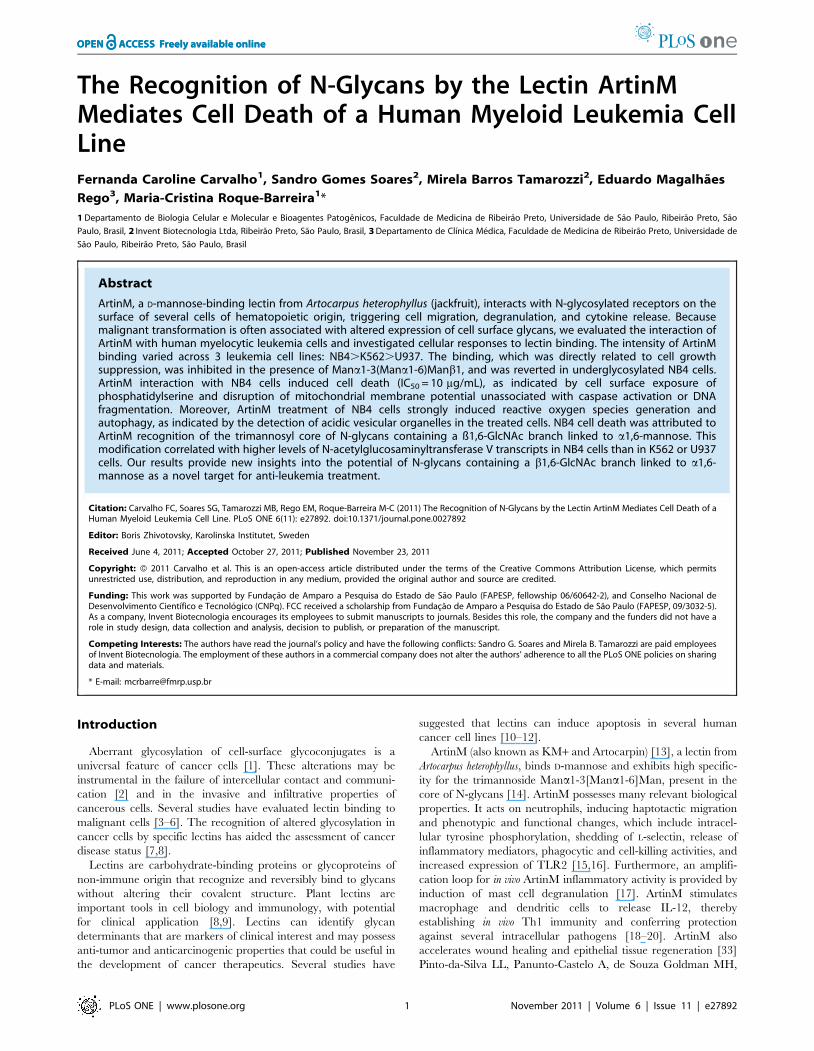
ArtinM binding to each cell line is shown in Figure 1A. The
fluorescence intensity in NB4 cells was at least 30% higher than in
K562 and U937 cells, despite the ability of ArtinM lectin to bind
more than 95% of cells in each cell line (data not shown).
Fluorescence microscopy confirmed ArtinM binding to NB4 cells
(Fig. 1C); this binding was completely inhibited by pre-incubation
with 10 mM Mana1-3[Mana1-6]Man (panel B), but not with
200 mM D-galactose (panel D), indicating that NB4 cell
recognition by ArtinM is mediated by its carbohydrate recognition
domain.
Considering that lectin interactions with tumor cells can trigger
biochemical responses [9], we investigated whether various levels
of ArtinM binding to the surface of leukemia cells could affect their
growth. We used MTT assays to determine cell viability and
generated growth inhibition curves for different ArtinM concen-
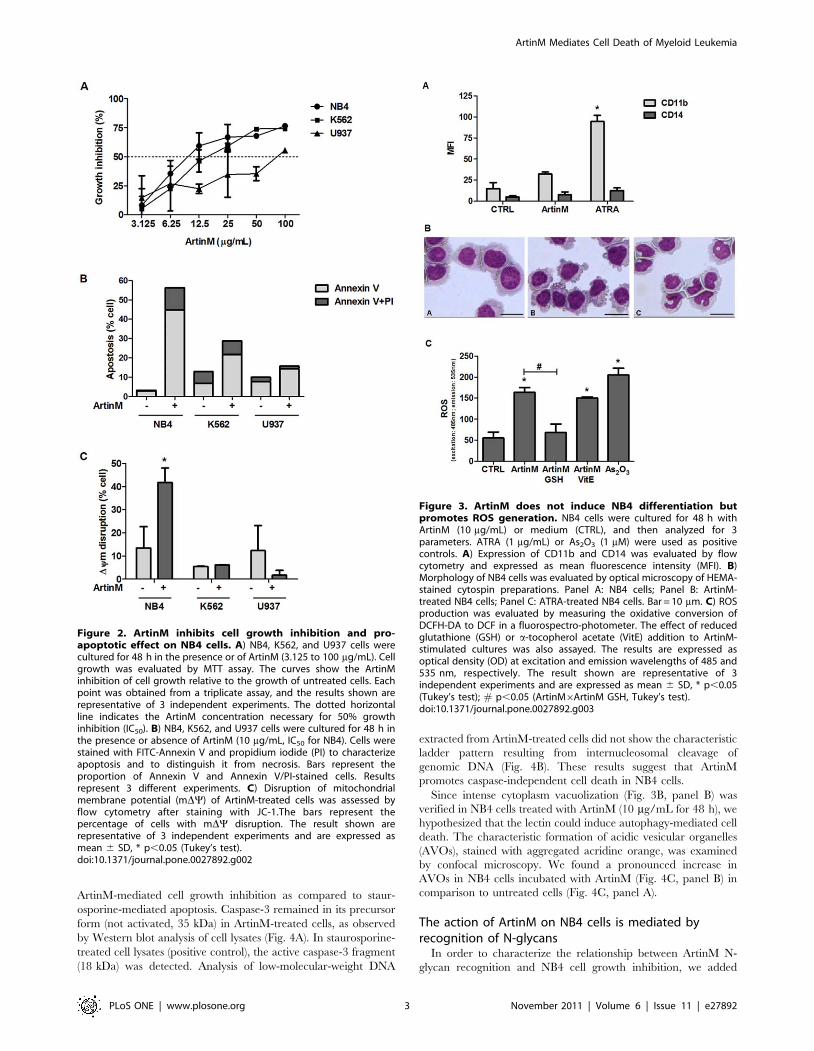
trations, as shown in Figure 2A. We thus determined the ArtinM
concentrations that inhibit 50% of cell growth (IC50). NB4 and
K562 cells were more sensitive to ArtinM inhibition, displaying
IC50 of 10 (61) and 14 (61) mg/mL, respectively, while U937 cells
exhibited an IC50 of 84 (61,5) mg/mL.
We performed a more detailed study to understand the effects of
ArtinM on leukemia cells. NB4, K562, and U937 leukemia cells
were cultured with ArtinM at 10 mg/mL, the IC50 for NB4, and
after 48 h, the cells were analyzed by flow cytometry for Annexin
V staining and PI incorporation. In NB4 cells, ArtinM induced
pronounced surface exposition of phosphatidylserine, as revealed
by Annexin V binding and minor PI incorporation, suggesting the
occurrence of apoptosis. In K562 and U937 cells, ArtinM
provoked lower levels of Annexin V binding and PI staining
(Fig. 2B).
The mitochondrial transmembrane electrical potential of
leukemia cells, following 48 h incubation with 10 mg/mL ArtinM,
was evaluated by JC-1 dye. ArtinM treatment promoted the
disruption of mitochondrial transmembrane electrical potential in
NB4 cells (Fig. 2C). The other tested cell lines (K562 and U937)
were resistant to ArtinM treatment (10 mg/mL).
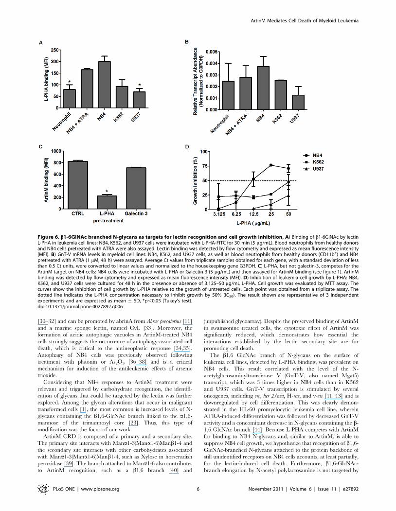
Non-differentiation and caspase-independent cell deathcould be related to autophagy under ArtinM treatment
Since augmented cell death rates could be caused by induction
of leukemia cell differentiation, as often happens following all-trans
retinoic acid (ATRA) therapeutic administration [22], we
investigated whether induction of NB4 cell differentiation could
account for the observed effect of ArtinM. As shown in Figure 3A,
ArtinM did not induce NB4 cell differentiation, as demonstrated
by levels of CD11b surface expression, which are 3 times lower
after ArtinM treatment than after ATRA treatment. ArtinM-
treated NB4 cells did not exhibit the multilobulated nucleus that
characterizes differentiated granulocytes, a feature that was
observed in ATRA-treated cells (Fig. 3B). Cytoplasmic vacuoliza-
tion was also observed in ArtinM-treated NB4 cells. Neither lower
doses of ArtinM nor increased length of exposure changed the
non-differentiated status of the cells (data not shown).
The absence of cell differentiation and the detection of
mitochondrial membrane depolarization led us to investigate the
occurrence of oxidative stress. ArtinM-stimulated NB4 cells
generate high levels of reactive oxygen species (ROS), similar to
those observed in the positive control (arsenic trioxide, As2O3)
(Fig. 3C). The augmented ROS production was inhibited to basal
levels in the presence of reduced glutathione (GSH). In contrast, a-
tocopherol acetate (vitamin E) had no effect on the cellular
response to ArtinM.
To assess whether the signaling event promoted by ArtinM led
to apoptosis, we investigated the involvement of caspase activity in
Figure 1. ArtinM interacts with leukemia cell lines. A) ArtinMbinding to NB4, K562, and U937 cells: cells were fixed and incubated for30 min with biotinyl-ArtinM/streptavidin-FITC (5 mg/mL). Lectin bindingto the cell surface was detected by flow cytometry and expressed asmean fluorescence intensity (MFI). B) NB4 cells adhered to Biobond-coated coverslips were incubated at 4uC for 60 min with biotinyl-ArtinM(5 mg/mL) (panel C) or PBS (panel A). For inhibition assays, biotinyl-ArtinM was pre-incubated at room temperature for 60 min with 10 mMMana1-3[Mana1-6]Man (panel B) or 200 mM D-galactose (panel D), andthen incubated with NB4 cells. After washing and incubation withstreptavidin-FITC, cells were fixed and examined by fluorescencemicroscopy. Magnification = 406. The result shown are representativeof 3 independent experiments and are expressed as mean 6 SD, *p,0.05 (Tukey’s test).doi:10.1371/journal.pone.0027892.g001
ArtinM Mediates Cell Death of Myeloid Leukemia
PLoS ONE | www.plosone.org 2 November 2011 | Volume 6 | Issue 11 | e27892
ArtinM-mediated cell growth inhibition as compared to staur-
osporine-mediated apoptosis. Caspase-3 remained in its precursor
form (not activated, 35 kDa) in ArtinM-treated cells, as observed
by Western blot analysis of cell lysates (Fig. 4A). In staurosporine-
treated cell lysates (positive control), the active caspase-3 fragment
(18 kDa) was detected. Analysis of low-molecular-weight DNA
extracted from ArtinM-treated cells did not show the characteristic
ladder pattern resulting from internucleosomal cleavage of
genomic DNA (Fig. 4B). These results suggest that ArtinM
promotes caspase-independent cell death in NB4 cells.
Since intense cytoplasm vacuolization (Fig. 3B, panel B) was
verified in NB4 cells treated with ArtinM (10 mg/mL for 48 h), we
hypothesized that the lectin could induce autophagy-mediated cell
death. The characteristic formation of acidic vesicular organelles
(AVOs), stained with aggregated acridine orange, was examined
by confocal microscopy. We found a pronounced increase in
AVOs in NB4 cells incubated with ArtinM (Fig. 4C, panel B) in
comparison to untreated cells (Fig. 4C, panel A).
The action of ArtinM on NB4 cells is mediated byrecognition of N-glycans
In order to characterize the relationship between ArtinM N-
glycan recognition and NB4 cell growth inhibition, we added
Figure 2. ArtinM inhibits cell growth inhibition and pro-apoptotic effect on NB4 cells. A) NB4, K562, and U937 cells werecultured for 48 h in the presence or of ArtinM (3.125 to 100 mg/mL). Cellgrowth was evaluated by MTT assay. The curves show the ArtinMinhibition of cell growth relative to the growth of untreated cells. Eachpoint was obtained from a triplicate assay, and the results shown arerepresentative of 3 independent experiments. The dotted horizontalline indicates the ArtinM concentration necessary for 50% growthinhibition (IC50). B) NB4, K562, and U937 cells were cultured for 48 h inthe presence or absence of ArtinM (10 mg/mL, IC50 for NB4). Cells werestained with FITC-Annexin V and propidium iodide (PI) to characterizeapoptosis and to distinguish it from necrosis. Bars represent theproportion of Annexin V and Annexin V/PI-stained cells. Resultsrepresent 3 different experiments. C) Disruption of mitochondrialmembrane potential (mDY) of ArtinM-treated cells was assessed byflow cytometry after staining with JC-1.The bars represent thepercentage of cells with mDY disruption. The result shown arerepresentative of 3 independent experiments and are expressed asmean 6 SD, * p,0.05 (Tukey’s test).doi:10.1371/journal.pone.0027892.g002
Figure 3. ArtinM does not induce NB4 differentiation butpromotes ROS generation. NB4 cells were cultured for 48 h withArtinM (10 mg/mL) or medium (CTRL), and then analyzed for 3parameters. ATRA (1 mg/mL) or As2O3 (1 mM) were used as positivecontrols. A) Expression of CD11b and CD14 was evaluated by flowcytometry and expressed as mean fluorescence intensity (MFI). B)Morphology of NB4 cells was evaluated by optical microscopy of HEMA-stained cytospin preparations. Panel A: NB4 cells; Panel B: ArtinM-treated NB4 cells; Panel C: ATRA-treated NB4 cells. Bar = 10 mm. C) ROSproduction was evaluated by measuring the oxidative conversion ofDCFH-DA to DCF in a fluorospectro-photometer. The effect of reducedglutathione (GSH) or a-tocopherol acetate (VitE) addition to ArtinM-stimulated cultures was also assayed. The results are expressed asoptical density (OD) at excitation and emission wavelengths of 485 and535 nm, respectively. The result shown are representative of 3independent experiments and are expressed as mean 6 SD, * p,0.05(Tukey’s test); # p,0.05 (ArtinM6ArtinM GSH, Tukey’s test).doi:10.1371/journal.pone.0027892.g003
ArtinM Mediates Cell Death of Myeloid Leukemia
PLoS ONE | www.plosone.org 3 November 2011 | Volume 6 | Issue 11 | e27892

tunicamycin (TM, 5 mg/mL) or swainsonine (SW, 5 mg/mL) to
NB4 cells that were subsequently stimulated with ArtinM after
24 h. TM and SW alone resulted in NB4 growth inhibition rates
of 30% and 10%, respectively, the viable cell was tested to ArtinM
binding and ArtinM growth inhibition. In comparison with fully
glycosylated cells (untreated cells), ArtinM binding of TM-treated
NB4 cells was strongly inhibited, whereas ArtinM binding of SW-
treated cells was preserved (Fig. 5A).
We next evaluated whether TM- or SW-treated NB4 cells were
responsive to ArtinM cell growth inhibition. Indeed, ArtinM
inhibition of cell growth reached 60% in TM-treated cells and
45% in SW-treated cells, as compared with fully glycosylated cells
(Fig. 5B). In contrast, the inhibition of NB4 cell growth by As2O3
(positive control) was not affected by the glycosylation status.
These results point to the importance of Mana1-6 elongation in
effective ArtinM response.
As ArtinM binding to the core of N-glycans is preserved when a
branch is added to Mana1-6 (unpublished glycoarray study
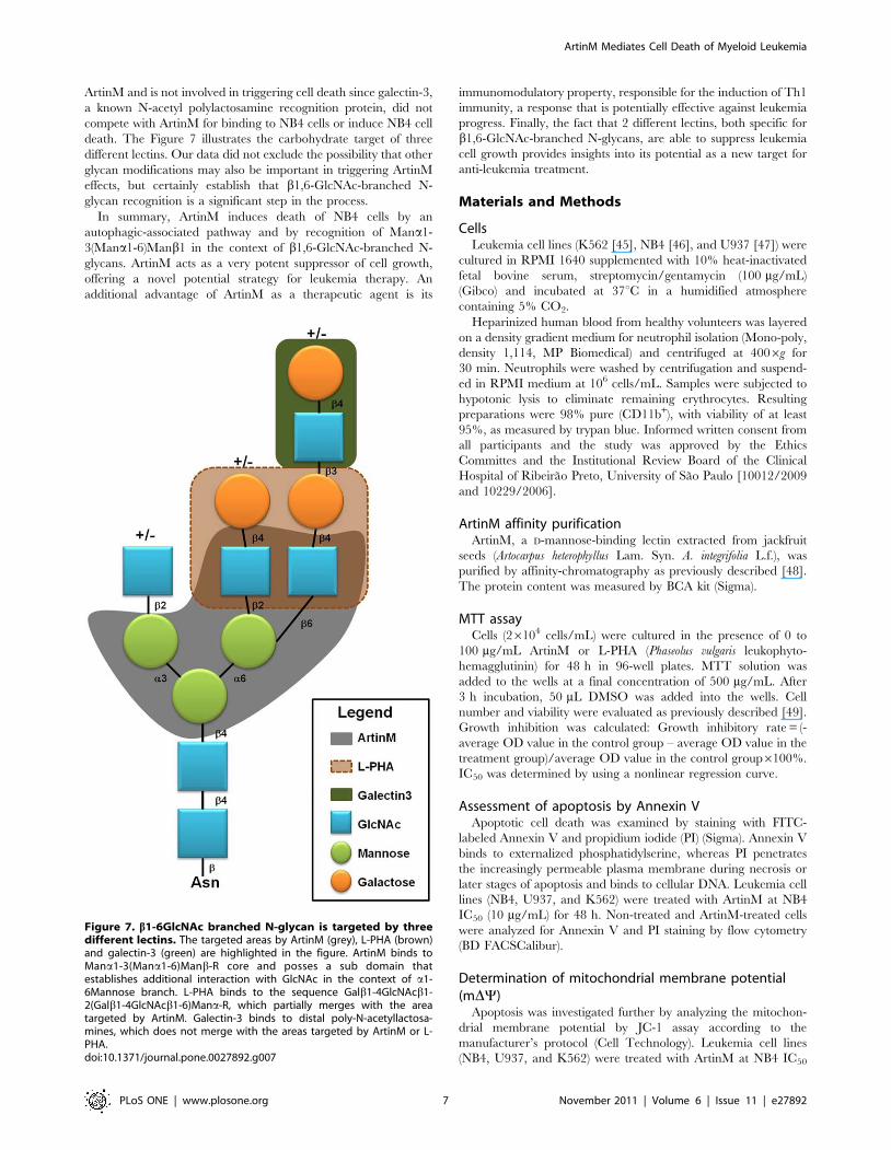
results), we tested whether b1,6-GlcNAc branching, which is
frequently present in cancer cells [23], was present in leukemia
cells. Its occurrence was disclosed by flow cytometry using
fluorescent Phaseolus vulgaris leukophyto-hemagglutinin (L-PHA).
The proportions of L-PHA-stained cells were 98%, 86%, and 69%
for NB4, K562, and U937 cells, respectively (data not shown). As
shown in Figure 6A, L-PHA staining was conspicuously strongest
in NB4 cells, especially in comparison with U937 cells, while
intermediate staining was detected in K562 cells. Weak L-PHA
staining was also detected in ATRA-differentiated NB4 cells and
neutrophils (CD11b+ cells) from healthy donors in comparison
with that of untreated NB4 cells, indicating a lower incidence of
b1,6-GlcNAc branched N-glycans in the non-malignant cells. As
expected, the detection of the b1,6-N-acetyl-glucosaminyltransfer-
ase (GnT-V or Mgat5) transcript in NB4, K562, U937,
neutrophils (CD11b+ cells), and ATRA-differentiated NB4 cells
was consistent with the incidence of b1,6-GlcNAc-branched N-
glycans (Fig. 6B). In addition, L-PHA binding to SW-treated cells
was inhibited 80% (data not shown) while ArtinM binding was
preserved.
The presence of b1,6-GlcNAc-branched N-glycans recognized
by L-PHA on NB4 cells led us to investigate whether this lectin
could compete with ArtinM for a related carbohydrate target. As
shown in Figure 6C, L-PHA inhibited ArtinM binding to NB4
cells by 75%, suggesting that ArtinM and L-PHA recognize targets
that are partially shared. Therefore, we evaluated whether L-PHA
Figure 4. Autophagy accounts for the caspase-independent mechanism of NB4 cell death induced by ArtinM. NB4 cells were culturedwith ArtinM (10 mg/mL) for 48 h or with Staurosporine (Stauro, 5 mM) for 4 h. A) Caspase-3 activation, manifested by cleavage of their precursorforms, was analyzed by Western blotting of RIPA cell lysates (100 mg protein) performed under reducing conditions. B) Fragmentation of genomicDNA from NB4 cells was evaluated by electrophoresis on 1% agarose gel followed by ethidium bromide visualization. St: standard markers. C) Acidicautophagic vacuoles in NB4 cells were detected through staining with 10 mg/mL acridine orange in serum-free medium. Fluorescent micrographsshow that the cytoplasm and nucleus of stained cells fluoresced bright green, whereas the acidic autophagic vacuoles fluoresced bright red.Bar = 10 mm.doi:10.1371/journal.pone.0027892.g004
ArtinM Mediates Cell Death of Myeloid Leukemia
PLoS ONE | www.plosone.org 4 November 2011 | Volume 6 | Issue 11 | e27892

inhibits leukemia cell growth in the same manner as ArtinM. As
shown in Figure 6D, L-PHA treatment yielded a growth inhibition
curve similar to that observed with ArtinM, particularly in NB4
cells, wherein the IC50 was 12 (61) mg/mL. Since galectin-3
recognizes N-acetyllactosamine-containing N-glycans, we also
performed a competition binding assay with ArtinM and observed
no competition, even after neuraminidase treatment (Fig. 6C).
Moreover, galectin-3 did not inhibit NB4 cell growth (data not
shown) as did ArtinM and L-PHA. We concluded that lactosamine
elongation of the b1,6-GlcNAc branch is irrelevant to ArtinM
binding and that lactosamine recognition distant from the N-
glycan core does not inhibit leukemia cell growth.
Discussion
In this study, we demonstrate that ArtinM interaction with NB4
myeloid leukemia cells suppresses cell proliferation. This effect was
attributed to the induction of cell death, apparent by the exposure
of phosphatidylserine on the cell surface and disruption of
mitochondrial membrane potential. Nonetheless, cell death was
not accompanied by cell differentiation, caspase-3 activation, or
DNA fragmentation. Moreover, augmented ROS production and
detection of acidic vesicular organelles in ArtinM-stimulated cells
strongly suggests the occurrence of autophagy-associated cell
death. The ArtinM carbohydrate recognition domain directly
triggers its activities on the cells, where N-glycans on the cell
surface glycoproteins are targets for recognition.
Among 3 different leukemia cell lines, NB4 was the most
sensitive to ArtinM-induced suppression of cell proliferation. The
NB4 cell line has a t(15;17)-positive karyotype and is considered an
appropriate model of acute promyelocytic leukemia (APL) for drug
evaluation. APL, a specific subtype of acute myelogenous
leukemia, is frequently associated with reciprocal translocations
between chromosomes 17 and 15 [t(15;17)], leading to fusion of
the retinoic acid receptor a (RARa) and promyelocytic leukemia
(PML) genes. The PML/RARa fusion product acts as a
transcription repressor and blocks the differentiation of APL blasts
at the promyelocyte stage [22,24,25]. The blockage can be
reverted by pharmacological doses of all-trans-retinoic acid
(ATRA), constituting the mainstay of APL therapy [25]. The
simultaneous administration of ATRA and anthracycline-based
chemotherapy is currently considered the standard treatment for
newly diagnosed APL patients, leading to high rates of remission.
APL relapses are associated with ATRA resistance. Alternative
therapy with As2O3 induces high rates of remission and is being
explored as induction treatment. Because it is not associated with
myelosuppression and other severe complications associated with
anthracycline administration, As2O3 is used to treat newly
diagnosed APL patients in whom chemotherapy is contraindicat-
ed. Other therapeutic strategies under development include
histone acetylase inhibitors, which revert the PML/RARatranscription repression and potentiate ATRA-induced granulo-
cytic differentiation [26] and granulocyte colony-stimulating factor
(G-CSF), which binds to the G-CSF receptor on acute myeloid
blasts and reduces their proliferation as a result of increased
commitment to terminal differentiation [27].
NB4 cells undergo differentiation by ATRA and apoptosis by
As2O3 treatment, responses that were confirmed in our in vitro
experiments. The distinct sensitivity of the assayed cell lines to
ArtinM was associated with the level of lectin binding to the cell
surface. The dependence on recognition of the trimannoside that
constitutes the common core structure of N-glycans was
demonstrated by (a) the inhibition of ArtinM binding to the
NB4 cell surface by Mana1-3(Mana1-6)Manb1 and (b) the cell
response to ArtinM in TM-treated NB4 cells. Indeed, our previous
work on the biological properties of ArtinM showed that its
binding to glycosylated receptors on the cell surface is responsible
for the triggered responses. This is true in (i) human neutrophils,
whose CXCR2 glycan recognition by ArtinM accounts for the
induction of haptotactic cell migration, increased mediator release,
and enhancement of effector functions, such as phagocytosis and
respiratory burst [28]; (ii) macrophages and dendritic cells, whose
TLR2 glycan recognition by ArtinM triggers IL-12 production,
which induces Th1-based immunity in vivo that confers protection
against intracellular pathogens [20]; and (iii) mast cell FceR glycan
recognition by ArtinM, which accounts for mast cell recruitment,
degranulation, and release of inflammatory mediators [17].
ArtinM-induced NB4 cell death, clearly demonstrated by
phosphatidylserine surface exposure and disruption of mitochon-
drial membrane potential, was not associated with cell maturation,
as provoked by other drugs, like ATRA. Indeed, NB4 cell death
was caspase-independent and was not accompanied by DNA
fragmentation. Moreover, ArtinM treatment induced ROS
generation, at levels as high as those provoked by As2O3.
Caspase-independent/ROS-dependent apoptosis can occur
through calcium-mediated mitochondrial membrane potential
depolarization, leading to the translocation of apoptotic factors
such as apoptosis-inducing factor from the mitochondria into the
nucleus, which could preserve an apoptotic phenotype in absence
of caspase activation [29]. The occurrence of caspase-independent
cell death was previously reported in leukemia and lymphoma cells
Figure 5. N-Glycan recognition by ArtinM accounts for NB4growth inhibition. A) ArtinM binding to NB4 cells pre-treated for24 h with tunicamycin (TM, 5 mg/mL) or swainsonine (SW, 5 mg/mL) wasanalyzed by flow cytometry. Fully glycosylated: untreated NB4 cells. B)Growth inhibition induced by ArtinM (10 mg/mL) in fully glycosylatedcells, and in TM- or SW-treated cells. Growth rate was measured by MTTassay. Bars represent cell growth inhibition by ArtinM relative to thegrowth of untreated cells. As2O3 (1 mM) was used as a positive controlfor cell death. The results shown are representative of 3 independentexperiments and are expressed as mean 6 SD, * p,0.05 (Tukey’s test)and # p,0.05 (Student’s t-test).doi:10.1371/journal.pone.0027892.g005
ArtinM Mediates Cell Death of Myeloid Leukemia
PLoS ONE | www.plosone.org 5 November 2011 | Volume 6 | Issue 11 | e27892
[30–32] and can be promoted by abrinA from Abrus precatorius [11]
and a marine sponge lectin, named CvL [33]. Moreover, the
formation of acidic autophagic vacuoles in ArtinM-treated NB4
cells strongly suggests the occurrence of autophagy-associated cell
death, which is critical to the antineoplastic response [34,35].
Autophagy of NB4 cells was previously observed following
treatment with platonin or As2O3 [36–38] and is a critical
mechanism for induction of the antileukemic effects of arsenic
trioxide.
Considering that NB4 responses to ArtinM treatment were
relevant and triggered by carbohydrate recognition, the identifi-
cation of glycans that could be targeted by the lectin was further
explored. Among the glycan alterations that occur in malignant
transformed cells [1], the most common is increased levels of N-
glycans containing the ß1,6-GlcNAc branch linked to the a1,6-
mannose of the trimannosyl core [23]. Thus, this type of
modification was the focus of our work.
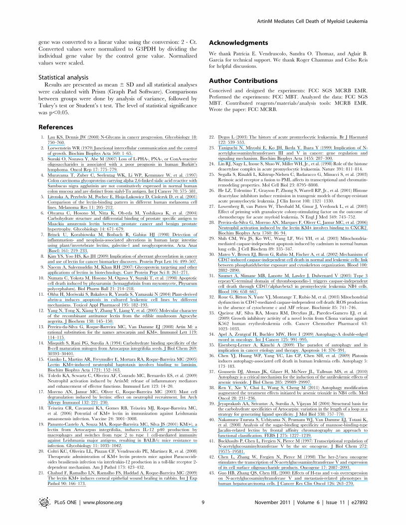
ArtinM CRD is composed of a primary and a secondary site.
The primary site interacts with Mana1-3(Mana1-6)Manb1-4 and
the secondary site interacts with other carbohydrates associated
with Mana1-3(Mana1-6)Manb1-4, such as Xylose in horseradish
peroxidase [39]. The branch attached to Mana1-6 also contributes
to ArtinM recognition, such as a b1,6 branch [40] and
(unpublished glycoarray). Despite the preserved binding of ArtinM
in swainsonine treated cells, the cytotoxic effect of ArtinM was
significantly reduced, which demonstrates how essential the
interactions established by the lectin secondary site are for
promoting cell death.
The b1,6 GlcNAc branch of N-glycans on the surface of
leukemia cell lines, detected by L-PHA binding, was prevalent on
NB4 cells. This result correlated with the level of the N-
acetylglucosaminyltransferase V (GnT-V, also named Mgat5)
transcript, which was 3 times higher in NB4 cells than in K562
and U937 cells. GnT-V transcription is stimulated by several
oncogenes, including src, her-2/neu, H-ras, and v-sis [41–43] and is
downregulated by cell differentiation. This was clearly demon-
strated in the HL-60 promyelocytic leukemia cell line, wherein
ATRA-induced differentiation was followed by decreased GnT-V
activity and a concomitant decrease in N-glycans containing the b-
1,6 GlcNAc branch [44]. Because L-PHA competes with ArtinM
for binding to NB4 N-glycans and, similar to ArtinM, is able to
suppress NB4 cell growth, we hypothesize that recognition of b1,6-
GlcNAc-branched N-glycans attached to the protein backbone of
still unidentified receptors on NB4 cells accounts, at least partially,
for the lectin-induced cell death. Furthermore, b1,6-GlcNAc-
branch elongation by N-acetyl polylactosamine is not targeted by
Figure 6. b1-6GlNAc branched N-glycans as targets for lectin recognition and cell growth inhibition. A) Binding of b1-6GlNAc by lectinL-PHA in leukemia cell lines: NB4, K562, and U937 cells were incubated with L-PHA-FITC for 30 min (5 mg/mL). Blood neutrophils from healthy donorsand NB4 cells pretreated with ATRA were also assayed. Lectin binding was detected by flow cytometry and expressed as mean fluorescence intensity(MFI). B) GnT-V mRNA levels in myeloid cell lines: NB4, K562, and U937 cells, as well as blood neutrophils from healthy donors (CD11b+) and NB4pretreated with ATRA (1 mM, 48 h) were assayed. Average Ct values from triplicate samples obtained for each gene, with a standard deviation of lessthan 0.5 Ct units, were converted to linear values and normalized to the housekeeping gene G3PDH. C) L-PHA, but not galectin-3, competes for theArtinM target on NB4 cells: NB4 cells were incubated with L-PHA or Galectin-3 (5 mg/mL) and then assayed for ArtinM binding (see figure 1). ArtinMbinding was detected by flow cytometry and expressed as mean fluorescence intensity (MFI). D) Inhibition of leukemia cell growth by L-PHA: NB4,K562, and U937 cells were cultured for 48 h in the presence or absence of 3.125–50 mg/mL L-PHA. Cell growth was evaluated by MTT assay. Thecurves show the inhibition of cell growth by L-PHA relative to the growth of untreated cells. Each point was obtained from a triplicate assay. Thedotted line indicates the L-PHA concentration necessary to inhibit growth by 50% (IC50). The result shown are representative of 3 independentexperiments and are expressed as mean 6 SD, *p,0.05 (Tukey’s test).doi:10.1371/journal.pone.0027892.g006
ArtinM Mediates Cell Death of Myeloid Leukemia
PLoS ONE | www.plosone.org 6 November 2011 | Volume 6 | Issue 11 | e27892
ArtinM and is not involved in triggering cell death since galectin-3,
a known N-acetyl polylactosamine recognition protein, did not
compete with ArtinM for binding to NB4 cells or induce NB4 cell
death. The Figure 7 illustrates the carbohydrate target of three
different lectins. Our data did not exclude the possibility that other
glycan modifications may also be important in triggering ArtinM
effects, but certainly establish that b1,6-GlcNAc-branched N-
glycan recognition is a significant step in the process.
In summary, ArtinM induces death of NB4 cells by an
autophagic-associated pathway and by recognition of Mana1-
3(Mana1-6)Manb1 in the context of b1,6-GlcNAc-branched N-
glycans. ArtinM acts as a very potent suppressor of cell growth,
offering a novel potential strategy for leukemia therapy. An
additional advantage of ArtinM as a therapeutic agent is its
immunomodulatory property, responsible for the induction of Th1
immunity, a response that is potentially effective against leukemia
progress. Finally, the fact that 2 different lectins, both specific for
b1,6-GlcNAc-branched N-glycans, are able to suppress leukemia
cell growth provides insights into its potential as a new target for
anti-leukemia treatment.
Materials and Methods
CellsLeukemia cell lines (K562 [45], NB4 [46], and U937 [47]) were
cultured in RPMI 1640 supplemented with 10% heat-inactivated
fetal bovine serum, streptomycin/gentamycin (100 mg/mL)
(Gibco) and incubated at 37uC in a humidified atmosphere
containing 5% CO2.
Heparinized human blood from healthy volunteers was layered
on a density gradient medium for neutrophil isolation (Mono-poly,
density 1,114, MP Biomedical) and centrifuged at 4006g for
30 min. Neutrophils were washed by centrifugation and suspend-
ed in RPMI medium at 106 cells/mL. Samples were subjected to
hypotonic lysis to eliminate remaining erythrocytes. Resulting
preparations were 98% pure (CD11b+), with viability of at least
95%, as measured by trypan blue. Informed written consent from
all participants and the study was approved by the Ethics
Committes and the Institutional Review Board of the Clinical
Hospital of Ribeirao Preto, University of Sao Paulo [10012/2009
and 10229/2006].
ArtinM affinity purificationArtinM, a D-mannose-binding lectin extracted from jackfruit
seeds (Artocarpus heterophyllus Lam. Syn. A. integrifolia L.f.), was
purified by affinity-chromatography as previously described [48].
The protein content was measured by BCA kit (Sigma).
MTT assayCells (26104 cells/mL) were cultured in the presence of 0 to
100 mg/mL ArtinM or L-PHA (Phaseolus vulgaris leukophyto-
hemagglutinin) for 48 h in 96-well plates. MTT solution was
added to the wells at a final concentration of 500 mg/mL. After
3 h incubation, 50 mL DMSO was added into the wells. Cell
number and viability were evaluated as previously described [49].
Growth inhibition was calculated: Growth inhibitory rate = (-
average OD value in the control group – average OD value in the
treatment group)/average OD value in the control group6100%.
IC50 was determined by using a nonlinear regression curve.
Assessment of apoptosis by Annexin VApoptotic cell death was examined by staining with FITC-
labeled Annexin V and propidium iodide (PI) (Sigma). Annexin V
binds to externalized phosphatidylserine, whereas PI penetrates
the increasingly permeable plasma membrane during necrosis or
later stages of apoptosis and binds to cellular DNA. Leukemia cell
lines (NB4, U937, and K562) were treated with ArtinM at NB4
IC50 (10 mg/mL) for 48 h. Non-treated and ArtinM-treated cells
were analyzed for Annexin V and PI staining by flow cytometry
(BD FACSCalibur).
Determination of mitochondrial membrane potential(mDY)
Apoptosis was investigated further by analyzing the mitochon-
drial membrane potential by JC-1 assay according to the
manufacturer’s protocol (Cell Technology). Leukemia cell lines
(NB4, U937, and K562) were treated with ArtinM at NB4 IC50
Figure 7. b1-6GlcNAc branched N-glycan is targeted by threedifferent lectins. The targeted areas by ArtinM (grey), L-PHA (brown)and galectin-3 (green) are highlighted in the figure. ArtinM binds toMana1-3(Mana1-6)Manb-R core and posses a sub domain thatestablishes additional interaction with GlcNAc in the context of a1-6Mannose branch. L-PHA binds to the sequence Galb1-4GlcNAcb1-2(Galb1-4GlcNAcb1-6)Mana-R, which partially merges with the areatargeted by ArtinM. Galectin-3 binds to distal poly-N-acetyllactosa-mines, which does not merge with the areas targeted by ArtinM or L-PHA.doi:10.1371/journal.pone.0027892.g007
ArtinM Mediates Cell Death of Myeloid Leukemia
PLoS ONE | www.plosone.org 7 November 2011 | Volume 6 | Issue 11 | e27892

(10 mg/mL) for 48 h prior to the addition of JC-1 for 30 min.
Non-treated and ArtinM-treated cells were analyzed by flow
cytometry (BD FACSCalibur). JC-1 dimers appear as red
fluorescence in stable mitochondria whereas monomers appear
as green fluorescence when the mitochondrial membrane potential
decreases.
Assessment of differentiation by flow cytometry andmorphology
The expression of differentiation markers CD14 and CD11b
was determined by flow cytometry. NB4 cells were harvested after
48 h incubation with ArtinM (10 mg/mL), washed twice with PBS,
and then incubated for 30 min at room temperature with mouse
anti-human PE-conjugated CD11b mAb (BD Bioscience) and
mouse anti-human FITC-conjugated CD14 mAb (BD Bioscience).
Mouse isotypes matching IgGs were used to set threshold
parameters for flow cytometry. In addition, cytospin preparations
stained with HEMA 3 (Biochemical Sciences) were used for
morphological evaluation. Cells treated with ATRA (1 mM) were
used as a positive control.
Accumulation of ROS (Reactive Oxygen Species)NB4 cells (26104 cells/mL) were cultured in the presence of
ArtinM (10 mg/mL) for 48 h in 96-well plates. ROS levels were
determined by measuring the oxidative conversion of cell-
permeable 29,79 dichlorofluorescein diacetate (DCFH-DA, Sigma),
after incubation for 30 min at 37uC, to fluorescent dichlorofluor-
escein (DCF) in a fluorospectro-photometer with excitation and
emission wavelengths of 485 and 535 nm, respectively. Cells
treated with As2O3 (1 mM) were used as a positive control. Cells
were also co-treated with ArtinM and antioxidants: a-tocopherol
acetate (10 mM, Sigma) or reduced glutathione (50 mM, Sigma).
Western blot analysis of caspase 3RIPA cell lysates (100 mg) were electrophoresed on a 12% SDS-
polyacrylamide gel (Bio-Rad) and electroblotted to a nitrocellulose
membrane (Millipore Corp). The membrane was incubated
overnight with anti-caspase 3 antibody (1 mg/mL, R&D Systems)
followed by a secondary horseradish peroxidase-conjugated anti-
mouse antibody (Amersham Biosciences). Detection was per-
formed with SuperSignalH chemiluminescence substrate (Pierce).
Blots were incubated overnight with murine monoclonal G3PDH
antibody (R&D, 1:2000 dilution) followed by a secondary
horseradish peroxidase-conjugated sheep anti-rabbit antibody
(Amersham Biosciences). Cells treated with staurosporine (5 mM,
4 h, Sigma) were used as a positive control.
Electrophoretic analysis of DNA fragmentationDNA in the lysates of NB4 cells cultured in the presence of
ArtinM was extracted with a DNA Purification Kit (Promega) and
fragmentation was visualized after electrophoresis on 1% agarose
gels containing 0.5 mg/mL ethidium bromide and photographed
with a Bio-Rad GD2000 (Bio-Rad). Cells treated with staurospor-
ine (5 mM, 4 h) were used as a positive control.
NB4 treatment with tunicamycin (TM) and swainsonine(SW)
NB4 cells (16106 cells/mL) were cultured in the presence of
5 mg/mL TM or SW(Sigma) for 24 h. Cells were washed twice
with PBS and incubated for 30 min at 4uC with FITC-conjugated
ConA or biothyl-ArtinM/streptavidin-FITC. Cells were tested for
positive staining using flow cytometry. TM- or SW-treated cells
were incubated in the presence of ArtinM for 48 h. Growth
inhibition was measured by MTT.
Detection of acidic vesicular organelles with acridineorange staining
To quantify the development of acidic vesicular organelles
(AVOs), ArtinM-treated cells were stained with acridine orange
(10 mg/mL) for 15 min and visualized by confocal laser scanning
microscopy (Leica SP5, Leica Microsystem, Wetziar, Germany).
In cells stained with acridine orange, the cytoplasm and nucleoli
emit green fluorescence while the acidic compartments emit red
fluorescence, the intensity of which is proportional to the acidity.
ArtinM and L-PHA binding and competition bindingassay
Cells were fixed with 2% paraformaldehyde at room temper-
ature for 20 min and incubated with biotinylated ArtinM/
streptavidin-FITC (5 mg/mL in PBS) or L-PHA-FITC (Phaseolus
vulgaris leukophyto-hemagglutinin) for 30 min. Lectin binding was
measured by flow cytometry. NB4 cells were fixed with 2%
paraformaldehyde at room temperature for 20 min and then
incubated with L-PHA (5 mg/mL) for 10 min. Finally, cells were
incubated with biotinylated ArtinM/streptavidin-FITC (5 mg/
mL). The competition binding analysis was performed by flow
cytometry.
Fluorescence microscopyNB4 cells were placed on coverslips coated with Biobond,
incubated with biotinylated ArtinM (5 mg/mL in PBS) or with
PBS alone, at 4uC for 60 min, and fixed with 2% paraformalde-
hyde at room temperature for 20 min. For some assays, ArtinM
was pre-incubated with 200 mM of D-Galactose or 10 mM of
Mana1-3[Mana1-6]Man, for 60 min at room temperature. Cells
were rinsed and then incubated with streptavidin-FITC for
30 min. Coverslips were mounted with Fluoromount-G and
examined by fluorescence microscopy (Axiophot, Carl Zeiss AG,
Germany).
Real Time PCR for Mgat5Total RNA isolation was performed using Trizol Reagent (Life
Technologies, Inc., Gaithersburg, MD, USA), as indicated by the
manufacturer. cDNA synthesis was performed in a final volume of
20 mL, using ImProm-II Reverse Transcriptase (Promega Corpo-
ration, Madison, WI, USA). The reaction mixture contained 4 mg
total RNA, 20 pmol oligo dT primer (Life Technologies), 40 U
RNasin, 500 mM dNTP mix, and 1 U reverse transcriptase in 16reverse transcriptase buffer. The cDNA was treated with 10 mg
RNase (Gibco) and immediately used or stored at 220uC. PCR
amplification and analysis were performed on an ABI Prism 7500
sequence detector (Applied Biosystems, Foster City, CA, USA). All
reactions were performed with SYBR Green Master Mix (Applied
Biosystems) in 25 mL reaction volumes containing 2 mL template
cDNA, 5 pmol of each primer, and 12.5 mL SYBR Green
(Applied Biosystems). The primers for PCR amplification were:
R: 59-TGAGTTCGCTGCTGGATGGT-39
F: 59-TCACTCCGTGGAAGTTGTCCT-39
Triplicate Ct values for each gene were averaged, and the
standard deviation was calculated. Samples that resulted in a
standard deviation of .0.5 Ct units were rerun until values with
standard deviations within an acceptable range were acquired.
The logarithmic average Ct value for each gene and the control
ArtinM Mediates Cell Death of Myeloid Leukemia
PLoS ONE | www.plosone.org 8 November 2011 | Volume 6 | Issue 11 | e27892
gene was converted to a linear value using the conversion: 2 - Ct.
Converted values were normalized to G3PDH by dividing the
individual gene value by the control gene value. Normalized
values were scaled.
Statistical analysisResults are presented as mean 6 SD and all statistical analyses
were calculated with Prism (Graph Pad Software). Comparisons
between groups were done by analysis of variance, followed by
Tukey’s test or Student’s t test. The level of statistical significance
was p,0.05.
Acknowledgments
We thank Patrıcia E. Vendruscolo, Sandra O. Thomaz, and Aglair B.
Garcia for technical support. We thank Roger Chammas and Celso Reis
for helpful discussions.
Author Contributions
Conceived and designed the experiments: FCC SGS MCRB EMR.
Performed the experiments: FCC MBT. Analyzed the data: FCC SGS
MBT. Contributed reagents/materials/analysis tools: MCRB EMR.
Wrote the paper: FCC MCRB.
References
1. Lau KS, Dennis JW (2008) N-Glycans in cancer progression. Glycobiology 18:
750–760.
2. Loewenstein WR (1979) Junctional intercellular communication and the control
of growth. Biochim Biophys Acta 560: 1–65.
3. Suzuki O, Nozawa Y, Abe M (2007) Loss of L-PHA-, PNA-, or ConA-reactive
oligosaccharides is associated with a poor prognosis in human Burkitt’s
lymphoma. Oncol Rep 17: 775–779.
4. Murayama T, Zuber C, Seelentag WK, Li WP, Kemmner W, et al. (1997)
Colon carcinoma glycoproteins carrying alpha 2,6-linked sialic acid reactive with
Sambucus nigra agglutinin are not constitutively expressed in normal human
colon mucosa and are distinct from sialyl-Tn antigen. Int J Cancer 70: 575–581.
5. Litynska A, Przybylo M, Pochec E, Hoja-Lukowicz D, Ciolczyk D, et al. (2001)
Comparison of the lectin-binding pattern in different human melanoma cell
lines. Melanoma Res 11: 205–212.
6. Ohyama C, Hosono M, Nitta K, Oh-eda M, Yoshikawa K, et al. (2004)
Carbohydrate structure and differential binding of prostate specific antigen to
Maackia amurensis lectin between prostate cancer and benign prostate
hypertrophy. Glycobiology 14: 671–679.
7. Brinck U, Korabiowska M, Bosbach R, Gabius HJ (1998) Detection of
inflammation- and neoplasia-associated alterations in human large intestine
using plant/invertebrate lectins, galectin-1 and neoglycoproteins. Acta Anat
(Basel) 161: 219–233.
8. Kim YS, Yoo HS, Ko JH (2009) Implication of aberrant glycosylation in cancer
and use of lectin for cancer biomarker discovery. Protein Pept Lett 16: 499–507.
9. Naeem A, Saleemuddin M, Khan RH (2007) Glycoprotein targeting and other
applications of lectins in biotechnology. Curr Protein Pept Sci 8: 261–271.
10. Numata C, Yokota M, Hosono M, Ogawa Y, Suzuki T, et al. (1998) Apoptotic
cell death induced by physarumin (hemagglutinin from myxomycete, Physarum
polycephalum). Biol Pharm Bull 21: 214–218.
11. Ohba H, Moriwaki S, Bakalova R, Yasuda S, Yamasaki N (2004) Plant-derived
abrin-a induces apoptosis in cultured leukemic cell lines by different
mechanisms. Toxicol Appl Pharmacol 195: 182–193.
12. Yang N, Tong X, Xiang Y, Zhang Y, Liang Y, et al. (2005) Molecular character
of the recombinant antitumor lectin from the edible mushroom Agrocybe
aegerita. J Biochem 138: 145–150.
13. Pereira-da-Silva G, Roque-Barreira MC, Van Damme EJ (2008) Artin M: a
rational substitution for the names artocarpin and KM+. Immunol Lett 119:
114–115.
14. Misquith S, Rani PG, Surolia A (1994) Carbohydrate binding specificity of the
B-cell maturation mitogen from Artocarpus integrifolia seeds. J Biol Chem 269:
30393–30401.
15. Ganiko L, Martins AR, Freymuller E, Mortara RA, Roque-Barreira MC (2005)
Lectin KM+-induced neutrophil haptotaxis involves binding to laminin.
Biochim Biophys Acta 1721: 152–163.
16. Toledo KA, Scwartz C, Oliveira AF, Conrado MC, Bernardes ES, et al. (2009)
Neutrophil activation induced by ArtinM: release of inflammatory mediators
and enhancement of effector functions. Immunol Lett 123: 14–20.
17. Moreno AN, Jamur MC, Oliver C, Roque-Barreira MC (2003) Mast cell
degranulation induced by lectins: effect on neutrophil recruitment. Int Arch
Allergy Immunol 132: 221–230.
18. Teixeira CR, Cavassani KA, Gomes RB, Teixeira MJ, Roque-Barreira MC,
et al. (2006) Potential of KM+ lectin in immunization against Leishmania
amazonensis infection. Vaccine 24: 3001–3008.
19. Panunto-Castelo A, Souza MA, Roque-Barreira MC, Silva JS (2001) KM(+), a
lectin from Artocarpus integrifolia, induces IL-12 p40 production by
macrophages and switches from type 2 to type 1 cell-mediated immunity
against Leishmania major antigens, resulting in BALB/c mice resistance to
infection. Glycobiology 11: 1035–1042.
20. Coltri KC, Oliveira LL, Pinzan CF, Vendruscolo PE, Martinez R, et al. (2008)
Therapeutic administration of KM+ lectin protects mice against Paracoccidi-
oides brasiliensis infection via interleukin-12 production in a toll-like receptor 2-
dependent mechanism. Am J Pathol 173: 423–432.
21. Chahud F, Ramalho LN, Ramalho FS, Haddad A, Roque-Barreira MC (2009)
The lectin KM+ induces corneal epithelial wound healing in rabbits. Int J Exp
Pathol 90: 166–173.
22. Degos L (2003) The history of acute promyelocytic leukaemia. Br J Haematol122: 539–553.
23. Taniguchi N, Miyoshi E, Ko JH, Ikeda Y, Ihara Y (1999) Implication of N-acetylglucosaminyltransferases III and V in cancer: gene regulation and
signaling mechanism. Biochim Biophys Acta 1455: 287–300.
24. Lin RJ, Nagy L, Inoue S, Shao W, Miller WH, Jr., et al. (1998) Role of the histone
deacetylase complex in acute promyelocytic leukaemia. Nature 391: 811–814.
25. Segalla S, Rinaldi L, Kilstrup-Nielsen C, Badaracco G, Minucci S, et al. (2003)
Retinoic acid receptor a fusion to PML affects its transcriptional and chromatin-remodeling properties. Mol Cell Biol 23: 8795–8808.
26. He LZ, Tolentino T, Grayson P, Zhong S, Warrell RP, Jr., et al. (2001) Histonedeacetylase inhibitors induce remission in transgenic models of therapy-resistant
acute promyelocytic leukemia. J Clin Invest 108: 1321–1330.
27. Lowenberg B, van Putten W, Theobald M, Gmur J, Verdonck L, et al. (2003)
Effect of priming with granulocyte colony-stimulating factor on the outcome of
chemotherapy for acute myeloid leukemia. N Engl J Med 349: 743–752.
28. Pereira-da-Silva G, Moreno AN, Marques F, Oliver C, Jamur MC, et al. (2006)
Neutrophil activation induced by the lectin KM+ involves binding to CXCR2.Biochim Biophys Acta 1760: 86–94.
29. Shih CM, Wu JS, Ko WC, Wang LF, Wei YH, et al. (2003) Mitochondria-mediated caspase-independent apoptosis induced by cadmium in normal human
lung cells. J Cell Biochem 89: 335–347.
30. Mateo V, Brown EJ, Biron G, Rubio M, Fischer A, et al. (2002) Mechanisms of
CD47-induced caspase-independent cell death in normal and leukemic cells: linkbetween phosphatidylserine exposure and cytoskeleton organization. Blood 100:
2882–2890.
31. Saumet A, Slimane MB, Lanotte M, Lawler J, Dubernard V (2005) Type 3
repeat/C-terminal domain of thrombospondin-1 triggers caspase-independent
cell death through CD47/alphavbeta3 in promyelocytic leukemia NB4 cells.Blood 106: 658–667.
32. Roue G, Bitton N, Yuste VJ, Montange T, Rubio M, et al. (2003) Mitochondrialdysfunction in CD47-mediated caspase-independent cell death: ROS production
in the absence of cytochrome c and AIF release. Biochimie 85: 741–746.
33. Queiroz AF, Silva RA, Moura RM, Dreyfuss JL, Paredes-Gamero EJ, et al.
(2009) Growth inhibitory activity of a novel lectin from Cliona varians againstK562 human erythroleukemia cells. Cancer Chemother Pharmacol 63:
1023–1033.
34. Apel A, Zentgraf H, Buchler MW, Herr I (2009) Autophagy-A double-edged
sword in oncology. Int J Cancer 125: 991–995.
35. Eisenberg-Lerner A, Kimchi A (2009) The paradox of autophagy and its
implication in cancer etiology and therapy. Apoptosis 14: 376–391.
36. Chen YJ, Huang WP, Yang YC, Lin CP, Chen SH, et al. (2009) Platonin
induces autophagy-associated cell death in human leukemia cells. Autophagy 5:
173–183.
37. Goussetis DJ, Altman JK, Glaser H, McNeer JL, Tallman MS, et al. (2010)
Autophagy is a critical mechanism for the induction of the antileukemic effects ofarsenic trioxide. J Biol Chem 285: 29989–29997.
38. Ren Y, Xie Y, Chai L, Wang S, Cheng M (2011) Autophagy modificationaugmented the treatment effects initiated by arsenic trioxide in NB4 cells. Med
Oncol 28: 231–236.
39. Jeyaprakash AA, Srivastav A, Surolia A, Vijayan M (2004) Structural basis for
the carbohydrate specificities of Artocarpin: variation in the length of a loop as astrategy for generating ligand specificity. J Mol Biol 338: 757–770.
40. Nakamura-Tsuruta S, Uchiyama N, Peumans WJ, Van Damme EJ, Totani K,et al. (2008) Analysis of the sugar-binding specificity of mannose-binding-type
Jacalin-related lectins by frontal affinity chromatography an approach to
functional classification. FEBS J 275: 1227–1239.
41. Buckhaults P, Chen L, Fregien N, Pierce M (1997) Transcriptional regulation of
N-acetylglucosaminyltransferase V by the src oncogene. J Biol Chem 272:19575–19581.
42. Chen L, Zhang W, Fregien N, Pierce M (1998) The her-2/neu oncogenestimulates the transcription of N-acetylglucosaminyltransferase V and expression
of its cell surface oligosaccharide products. Oncogene 17: 2087–2093.
43. Guo HB, Zhang QS, Chen HL (2000) Effects of H-ras and v-sis overexpression
on N-acetylglucosaminyltransferase V and metastasis-related phenotypes inhuman hepatocarcinoma cells. J Cancer Res Clin Oncol 126: 263–270.
ArtinM Mediates Cell Death of Myeloid Leukemia
PLoS ONE | www.plosone.org 9 November 2011 | Volume 6 | Issue 11 | e27892
44. Liu AH, Liu F, Li Z, Gu JX, Chen HL (1998) Alterations in glycosyltransferases
during myeloid and monocytoid differentiation of HL-60 cells. Cell Biol Int 22:545–550.
45. Lozzio CB, Lozzio BB (1975) Human chronic myelogenous leukemia cell-line
with positive Philadelphia chromosome. Blood 45: 321–334.46. Lanotte M, Martin-Thouvenin V, Najman S, Balerini P, Valensi F, et al. (1991)
NB4, a maturation inducible cell line with t(15;17) marker isolated from ahuman acute promyelocytic leukemia (M3). Blood 77: 1080–1086.
47. Sundstrom C, Nilsson K (1976) Establishment and characterization of a human
histiocytic lymphoma cell line (U-937). Int J Cancer 17: 565–577.48. Santos-de-Oliveira R, Dias-Baruffi M, Thomaz SM, Beltramini LM, Roque-
Barreira MC (1994) A neutrophil migration-inducing lectin from Artocarpus
integrifolia. J Immunol 153: 1798–1807.49. Liu JJ, Lin DJ, Liu PQ, Huang M, Li XD, et al. (2006) Induction of apoptosis
and inhibition of cell adhesive and invasive effects by tanshinone IIA in acutepromyelocytic leukemia cells in vitro. J Biomed Sci 13: 813–823.
ArtinM Mediates Cell Death of Myeloid Leukemia
PLoS ONE | www.plosone.org 10 November 2011 | Volume 6 | Issue 11 | e27892