N- and O-glycans of recombinant human C1 inhibitor expressed in the milk of transgenic rabbits
14
N- and O-glycans of recombinant human C1 inhibitor expressed in the milk of transgenic rabbits Kate Koles 1,4 , Patrick H. C. van Berkel 2,5 , Frank R. Pieper 5 , Jan H. Nuijens 5 , Maurice L.M. Mannesse 5 , Johannes F.G. Vliegenthart 4 , and Johannis P. Kamerling 3,4 4 Bijvoet Center for Biomolecular Research, Department of Bio-Organic Chemistry, Section of Glycoscience and Biocatalysis, Utrecht University, Padualaan 8, NL-3584 CH Utrecht, The Netherlands, and 5 Pharming Technologies BV, Archimedesweg 4, P.O. Box 451, NL-2300 AL Leiden, The Netherlands Received on July 22, 2003; revised on September 10, 2003; accepted on September 11, 2003 Human C1 inhibitor (hC1INH) is a therapeutic N, O-glycoprotein with a growing number of clinical applica- tions, but the current natural supplies are not likely to meet the clinical demands. Therefore, recombinant approaches are of interest, whereby specific attention has to be paid to the generated glycosylation patterns. Here, the N,O-glycoprotein was expressed in the mammary gland of transgenic rabbits and subjected to glycan analysis. After release of the N-glycans of recombinant-rabbit human C1 inhibitor (rhC1INH) by pep- tide-N 4 -(N-acetyl-b-glucosaminyl)asparagine amidase F, the oligosaccharides were separated from the O-glycoprotein by centrifugal filtration, then fractionated by a combination of anion-exchange, normal-phase, and high-pH anion-exchange liquid chromatography. The O-glycans, released from the O- glycoprotein by alkaline borohydride treatment, were fraction- ated by anion-exchange high-performance liquid chromato- graphy (HPLC). The structures of individual components were analysed by 500 MHz 1 H NMR spectroscopy, in most cases combined with MALDI-TOF MS. In contrast to the structural data reported for native serum hC1INH, rhC1INH contained a broad array of different N-glycans, made up of oligomannose-, hybrid-, and complex-type structures. In the case of complex-type N-glycans (partially) (a2-6)-sialylated (N-acetylneuraminic acid only), mono- and diantennary chains were found; part of the diantennary structures were (a1-6)-core-fucosylated or (a1-3)-fucosylated in the lower or upper antenna (Lewis x). The manno-oligosaccharide pattern of part of the hybrid- and oligomannose-type structures indi- cates that besides the usual N-glycan processing route, also the alternative endo-mannosidase pathway is followed. The small core 1-type O-glycans showed the usual (a2-3)- and (a2-6)-sialylation pattern of O-glycoproteins of nonmucinous origin. Key words: C1 inhibitor/glycosylation/transgenic rabbit milk Introduction Human C1 inhibitor (hC1INH), a plasma protease inhibitor belonging to the serine protease inhibitors super- family synthesized in the liver, is involved in the inhibition of components of the complement, coagulation, fibrinoly- tic, and kinin-releasing systems (Carreer, 1992). The single- chain polypeptide backbone of the plasma glycoprotein (M r ~71 kDa) consists of 478 amino acids and has six N-glycosylation sites at Asn-3, -47, -59, -216, -231, and -330, respectively, and seven O-glycosylation sites at Ser- 42 and Thr-26, -49, -61, -66, -70, and -74, respectively (Bock et al., 1986; Perkins, 1993). Its average carbohydrate con- tent is about 26% (Perkins et al., 1990). 1 H nuclear magnetic resonance (NMR) studies of chemically-released N- and O- glycans of native serum hC1INH revealed the presence of (a2-3)-disialylated and (a2-6)-disialylated complex-type diantennary N-glycans (molar ratio, 2:1), and the (a2-3)- monosialylated core 1-type O-glycan (Strecker et al., 1985). The N-glycans were (a1-6)-core-fucosylated for 30%, and the only sialic acid found was N-acetylneuraminic acid. Profiling studies using high-voltage paper electrophoresis followed by size-exclusion chromatography of chemically released N-glycans indicated, besides the presence of major amounts of sialylated diantennary structures, also lower amounts of sialylated tri- and tetraantennary struc- tures (Perkins et al., 1990). Lectin-binding assays confirmed the presence of both (a2-3)- and (a2-6)-linked sialic acid and core 1-type O-glycans and favored the presence of complex- and hybrid-type N-glycans; no indications for oligomannose-type structures were obtained (Schoenberger, 1992). Deficiency in the gene encoding hC1INH causes a condition called hereditary angioneurotic edema, which can be lethal when left untreated (Ebo and Stevens, 2000; Carugati et al., 2001). Furthermore, the lack of hC1INH leads to excessive complement activation, and this in turn to severe symptoms characterized by sudden local swellings of soft connective tissue (Ebo and Stevens, 2000). Current forms of treatment include hC1INH replacement therapy, which relies on hC1INH supplies prepared from donated human blood. However, there is a growing number of clin- ical conditions wherein hC1INH might be beneficial (Caliezi et al., 2000), hence current supplies of hC1INH are not likely to meet clinical demands. Recombinant glycoprotein production in mammary glands of transgenic animals with milk extraction is one 1 Present address: Department of Biochemistry and Biophysics, Texas A&M University, 2128 TAMU, College Station, TX 77843-2128 2 Present address: GenMab BV, Jenalaan 18d, NL-3584 CK Utrecht, The Netherlands 3 To whom correspondence should be addressed; e-mail: [email protected] Glycobiology vol. 14 no. 1 # Oxford University Press 2004; all rights reserved. 51 Glycobiology vol. 14 no. 1 pp. 51–64, 2004 DOI: 10.1093/glycob/cwh010 by guest on March 27, 2015 http://glycob.oxfordjournals.org/ Downloaded from
-
Upload
independent -
Category
Documents
-
view
0 -
download
0
Transcript of N- and O-glycans of recombinant human C1 inhibitor expressed in the milk of transgenic rabbits
N- and O-glycans of recombinant human C1 inhibitor expressed in the milk oftransgenic rabbits
Kate Koles1,4, Patrick H. C. van Berkel2,5, Frank R. Pieper5,Jan H. Nuijens5, Maurice L.M. Mannesse5, JohannesF.G. Vliegenthart4, and Johannis P. Kamerling3,4
4Bijvoet Center for Biomolecular Research, Department of Bio-OrganicChemistry, Section of Glycoscience and Biocatalysis, Utrecht University,Padualaan 8, NL-3584 CH Utrecht, The Netherlands, and 5PharmingTechnologies BV, Archimedesweg 4, P.O. Box 451, NL-2300 AL Leiden,The Netherlands
Received on July 22, 2003; revised on September 10, 2003; accepted onSeptember 11, 2003
Human C1 inhibitor (hC1INH) is a therapeutic N,O-glycoprotein with a growing number of clinical applica-tions, but the current natural supplies are not likely to meetthe clinical demands. Therefore, recombinant approaches areof interest, whereby specific attention has to be paid to thegenerated glycosylation patterns. Here, the N,O-glycoproteinwas expressed in the mammary gland of transgenic rabbits andsubjected to glycan analysis. After release of the N-glycans ofrecombinant-rabbit human C1 inhibitor (rhC1INH) by pep-tide-N4-(N-acetyl-b-glucosaminyl)asparagine amidase F, theoligosaccharides were separated from the O-glycoprotein bycentrifugal filtration, then fractionated by a combination ofanion-exchange, normal-phase, and high-pH anion-exchangeliquid chromatography. The O-glycans, released from the O-glycoprotein by alkaline borohydride treatment, were fraction-ated by anion-exchange high-performance liquid chromato-graphy (HPLC). The structures of individual componentswere analysed by 500 MHz 1H NMR spectroscopy, in mostcases combined with MALDI-TOF MS. In contrast to thestructural data reported for native serum hC1INH, rhC1INHcontained a broad array of different N-glycans, made up ofoligomannose-, hybrid-, and complex-type structures. In thecase of complex-type N-glycans (partially) (a2-6)-sialylated(N-acetylneuraminic acid only), mono- and diantennarychains were found; part of the diantennary structures were(a1-6)-core-fucosylated or (a1-3)-fucosylated in the lower orupper antenna (Lewis x). The manno-oligosaccharide patternof part of the hybrid- and oligomannose-type structures indi-cates that besides the usual N-glycan processing route, alsothe alternative endo-mannosidase pathway is followed. Thesmall core 1-type O-glycans showed the usual (a2-3)- and(a2-6)-sialylation pattern of O-glycoproteins of nonmucinousorigin.
Key words: C1 inhibitor/glycosylation/transgenic rabbitmilk
Introduction
Human C1 inhibitor (hC1INH), a plasma proteaseinhibitor belonging to the serine protease inhibitors super-family synthesized in the liver, is involved in the inhibitionof components of the complement, coagulation, fibrinoly-tic, and kinin-releasing systems (Carreer, 1992). The single-chain polypeptide backbone of the plasma glycoprotein(Mr ~71 kDa) consists of 478 amino acids and has sixN-glycosylation sites at Asn-3, -47, -59, -216, -231, and-330, respectively, and seven O-glycosylation sites at Ser-42 and Thr-26, -49, -61, -66, -70, and -74, respectively (Bocket al., 1986; Perkins, 1993). Its average carbohydrate con-tent is about 26% (Perkins et al., 1990). 1H nuclear magneticresonance (NMR) studies of chemically-released N- and O-glycans of native serum hC1INH revealed the presence of(a2-3)-disialylated and (a2-6)-disialylated complex-typediantennary N-glycans (molar ratio, 2:1), and the (a2-3)-monosialylated core 1-type O-glycan (Strecker et al., 1985).The N-glycans were (a1-6)-core-fucosylated for 30%, andthe only sialic acid found was N-acetylneuraminic acid.Profiling studies using high-voltage paper electrophoresisfollowed by size-exclusion chromatography of chemicallyreleased N-glycans indicated, besides the presence ofmajor amounts of sialylated diantennary structures, alsolower amounts of sialylated tri- and tetraantennary struc-tures (Perkins et al., 1990). Lectin-binding assays confirmedthe presence of both (a2-3)- and (a2-6)-linked sialic acidand core 1-type O-glycans and favored the presence ofcomplex- and hybrid-type N-glycans; no indications foroligomannose-type structures were obtained (Schoenberger,1992).
Deficiency in the gene encoding hC1INH causes acondition called hereditary angioneurotic edema, whichcan be lethal when left untreated (Ebo and Stevens, 2000;Carugati et al., 2001). Furthermore, the lack of hC1INHleads to excessive complement activation, and this in turn tosevere symptoms characterized by sudden local swellings ofsoft connective tissue (Ebo and Stevens, 2000). Currentforms of treatment include hC1INH replacement therapy,which relies on hC1INH supplies prepared from donatedhuman blood. However, there is a growing number of clin-ical conditions wherein hC1INH might be beneficial(Caliezi et al., 2000), hence current supplies of hC1INHare not likely to meet clinical demands.
Recombinant glycoprotein production in mammaryglands of transgenic animals with milk extraction is one
1Present address: Department of Biochemistry and Biophysics, TexasA&M University, 2128 TAMU, College Station, TX 77843-21282Present address: GenMab BV, Jenalaan 18d, NL-3584 CK Utrecht,The Netherlands3To whom correspondence should be addressed; e-mail:[email protected]
Glycobiology vol. 14 no. 1 # Oxford University Press 2004; all rights reserved. 51
Glycobiology vol. 14 no. 1 pp. 51±64, 2004DOI: 10.1093/glycob/cwh010
by guest on March 27, 2015
http://glycob.oxfordjournals.org/D
ownloaded from

of the most attractive approaches for the economic andlarge-scale production of therapeutic glycoproteins(Houdebine, 1995; Colman, 1996). In addition to providingeukaryotic posttranslational modifications often requiredfor biological activity, it is also the expression platformthat provides the highest recombinant protein productioncapacity available to date, exceeding the capacity of themore established mammalian cell lines. Typical examplesso far are human lactoferrin in mice (Nuijens et al., 1997)and cows (Van Berkel et al., 2002), human a-glucosidasein mice (Bijvoet et al., 1996) and rabbits (Visser et al.,2000), human granulocyte-macrophage colony stimulatingfactor in mice (Uusi-Oukari et al., 1997), human erythro-poietin in mice and rabbits (Korhonen et al., 1997),human antithrombin in goats (Edmunds et al., 1998),and human tissue-type plasminogen activator in goats(Denman et al., 1991).
The posttranslational glycosylation of proteins is aspecies-, tissue-, cell-type-, and protein-specific phenomenon.Thus recombinant proteins can have different glycosylationpatterns when compared with their native counterparts.Furthermore, in cell cultures, changes in growth mediacomposition and in growth conditions may lead to changesin glycosylation patterns. These findings are of special inter-est when focusing on human therapeutic recombinant glyco-proteins, because specific glycosylation patterns play impor-tant roles in the secretion, antigenicity, and clearance ofglycoproteins (Jenkins and Curling, 1994; Jenkins et al.,1996; Kamerling, 1996; Varki et al., 1999; Galet et al., 2001).
Nowadays, much knowledge is available with respect tothe glycosylation machineries of the two most popular mam-malian cell lines for glycoprotein expression, Chinese ham-ster ovary (CHO) and baby hamster kidney (BHK) cells(Kamerling, 1996). However, detailed glycan data and sys-tematic studies on the glycosylation capabilities of the mam-mary glands of the different species, including the effects ofstage of lactation, individual variation, and seasonal varia-tion, are scarce. For human lactoferrin from cow milk (VanBerkel et al., 2002) it was concluded that besides complex-type N-glycans, as in natural human lactoferrin, also oligo-mannose- and/or hybrid-type glycans do occur, whereas thepresence of N,N0-diacetyllactosediamine units has been sug-gested. It has been reported that natural bovine lactoferrincontains complex- (both partially [a2-6]-sialylated N-acetyllactosamine and N,N 0-diacetyllactosediamine units,and Gal[a1-3]Gal[b1-4]GlcNAc units) and oligomannose-type N-glycans (Montreuil et al., 1997). The occurrenceof N,N0-diacetyllactosediamine units has also beensuggested for human antithrombin produced in goat milk(Edmunds et al., 1998). In the latter case, in contrast toplasma human antithrombin, the transgenic form also con-tained oligomannose-type structures, and in addition toN-acetylneuraminic acid, N-glycolylneuraminic acid wasdetected.
Because the glycosylation machinery of the rabbitmammary glands is rather unexplored, it is unpredictablewhat kind of glycans would predominate on recombinantglycoproteins from transgenic rabbit milk. In this study, adetailed analysis of the N- and O-glycans of recombinanthuman C1 inhibitor expressed in the milk of transgenicrabbits (rhC1INH) is presented and discussed.
Results
General
Monosaccharide analysis (Kamerling and Vliegenthart,1989) of rhC1INH, having a total carbohydrate content of14% (w/w), revealed the presence of Fuc, Gal, GalNAc,GlcNAc, Man, and Neu5Ac in the molar ratio0.2:2.4:1.7:2.6:3.0:1.3.
Release and fractionation of N-glycans of rhC1INH
The N-glycans of rhC1INH were released by peptide-N4-(N-acetyl-b-glucosaminyl)asparagine amidase F (PNGaseF) digestion, separated as a pool from the remainingO-glycoprotein, and fractionated by anion-exchangechromatography on Resource Q. Three carbohydrate-containing fast protein liquid chromatography (FPLC)fractions, eluting at positions corresponding to neutral N0(25%), monosialylated N1 (67%), and disialylated N2 (8%)structures were obtained (Figure 1). Neutral FPLC fractionN0 was further separated into six high-performance liquidchromatography (HPLC) fractions, denoted N0.1±N0.6,by normal phase chromatography on LiChrospher-NH2
(Figure 2). The HPLC fractions N0.3 and N0.4 weresubfractionated by high pH anion-exhange chromato-graphy (HPAEC) on CarboPac PA-1 (Figure 3) after 2-aminobenzamide (2AB) labeling of the components present(to minimize high pH±induced epimerization of the redu-cing GlcNAc residues during HPAEC; Stroop et al., 2000),yielding four and five subfractions, respectively, denotedN0.3.12AB±N0.3.42AB and N0.4.12AB±N0.4.52AB. The mono-(N1) and dicharged (N2) FPLC fractions were subfractio-nated on CarboPac PA-1 (Figures 4 and 5), yieldingfractions N1.1±N1.8 and N2.1±N2.3, respectively.
Structural analysis of the various fractions was carriedout by 1H NMR spectroscopy in combination with
Fig. 1. Elution profile at 214 nm of the enzymically released N-glycanpool of rhC1INH on a Resource Q column. The column was first elutedwith 12 ml water, followed by a linear concentration gradient of 0±8%(v/v) 0.5 M NaCl over 5 min and by a steeper linear concentrationgradient of 8±50% (v/v) 0.5 M NaCl over 5 min at a flow rate of 4 ml/min.For subsequent runs, the column was washed and equilibrated to startingconditions in 30 min. The fraction marked by an asterisk and fractionsN3 and N4 did not contain carbohydrate material.
K. Koles et al.
52
by guest on March 27, 2015
http://glycob.oxfordjournals.org/D
ownloaded from

matrix-assisted laser desorption ionization time-of-flightmass spectrometry (MALDI-TOF-MS). For complex-typeN-glycans holds that an (a1-6)-fucosylated N,N0-diacetylchitobiose core element is recognized from theanomeric signals of aGlcNAc-1 at d 5.18, GlcNAc-2 at d4.66±4.67, and Fuc at d 4.89±4.91, together with the N-acetyl methyl signals of GlcNAc-1 at ~d 2.039 andGlcNAc-2 at d 2.09±2.10, as well as from the Fuc CH3
signals at d 1.21 (aGlcNAc-1) and d 1.22 (bGlcNAc-1),and the Fuc H-5 multiplets at d 4.10 (aGlcNAc-1) and d4.13 (bGlcNAc-1). The nonfucosylated N,N0-diacetylchito-biose unit is recognized from the anomeric signals ofaGlcNAc-1 at d 5.19 and GlcNAc-2 at d 4.60±4.61, togetherwith the N-acetyl methyl signals of GlcNAc-1 at ~d 2.039and GlcNAc-2 at ~d 2.081 (HaÊrd et al., 1992). In case ofhybrid- and oligomannose-type N-glycans, the N-acetylmethyl signals of GlcNAc-2 are generally observed at ~d2.064 (Stroop et al., 2000; Tseneklidou-Stoeter et al., 1995).
Fig. 3. Elution profiles using PAD of the neutral HPLC fractions N0.3(A) and N0.4 (B) on a CarboPac PA-1 column. The column was elutedwith a linear concentration gradient of 5±18% (v/v) solvent B (0.5 MNaOAc in 0.1 M NaOH) in solvent A (0.1 M NaOH) over 32 min, at aflow rate of 4 ml/min.
Fig. 5. Elution profile using PAD of the dicharged FPLC fraction N2 ona CarboPac PA-1 column. The column was eluted with a linearconcentration gradient of 20±25% (v/v) solvent B (0.5 M NaOAc in 0.1 MNaOH) in solvent A (0.1 M NaOH) over 9 min, followed by a linearconcentration gradient of 25±35% (v/v) solvent B in solvent A over 11 minat a flow rate of 4 ml/min.
Fig. 2. Elution profile at 206 nm of neutral FPLC fraction N0 on aLiChrospher-NH2 column. The column was eluted with a linear gradientof 18±42% (v/v) solvent B [10% (v/v) acetonitrile in water] in solvent A[10% (v/v) water in acetonitrile].
Fig. 4. Elution profile using PAD of the monocharged FPLC fractionN1 on a CarboPac PA-1 column. The column was eluted with a linearconcentration gradient of 15±28% (v/v) solvent B (0.5 M NaOAc in 0.1 MNaOH) in solvent A (0.1 M NaOH) over 40 min at a flow rate of 4 ml/min.
N- and O-glycans of rhC1INH
53
by guest on March 27, 2015
http://glycob.oxfordjournals.org/D
ownloaded from
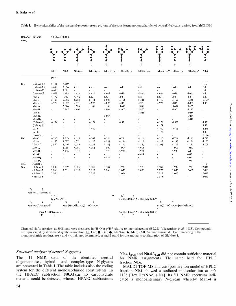
Structural analysis of neutral N-glycans
The 1H NMR data of the identified neutraloligomannose-, hybrid-, and complex-type N-glycansare presented in Table I. The table includes also the codingsystem for the different monosaccharide constitutents. Inthe HPAEC subfraction N0.3.12AB no carbohydratematerial could be detected, whereas HPAEC subfractions
N0.4.12AB and N0.4.32AB did not contain sufficient materialfor NMR assignments. The same held for HPLCfraction N0.6.
MALDI-TOF-MS analysis (positive-ion mode) of HPLCfraction N0.1 showed a sodiated molecular ion at m/z1136 [Hex3HexNAc3�Na]. Its 1H NMR spectrum indi-cated a monoantennary N-glycan whereby Man-4 is
Table I. 1H-chemical shifts of the structural-reporter-group protons of the constituent monosaccharides of neutral N-glycans, derived from rhC1INH
Chemical shifts are given at 300K and were measured in 2H2O at p2H7 relative to internal acetone (d 2.225; Vliegenthart et al., 1983). Compoundsare represented by short-hand symbolic notation: &, Fuc; &, Gal; *, GlcNAc; ^, Man; 2AB, 2-aminobenzamide. For numbering of themonosaccharide residues, see � and ��. n.d., not determined; a and b stand for the anomeric configuration of GlcNAc-1.
K. Koles et al.
54
by guest on March 27, 2015
http://glycob.oxfordjournals.org/D
ownloaded from
extended with GlcNAc-5, and Man-40 is terminal. Thestructural-reporter-group protons of Man-3, Man-4,Man-40, and GlcNAc-5 match the 1H NMR data of the(a1-6)-fucosylated analogue (compare with compound 5in Bendiak et al. [1988]).
HPLC fraction N0.2 was identified by 1H NMRspectroscopy as Man5GlcNAc2 (compare with compoundU8 in HaÊrd et al. [1991]). MALDI-TOF-MS analysis (posi-tive-ion mode) supported this finding with the sodiatedmolecular ion at m/z 1257 [Hex5HexNAc2 � Na]. Thisoligosaccharide was used as a model substance to evaluatethe impact of the 2AB labeling (! N0.22AB) on the struc-tural reporters of the nonlabeled counterpart. Typical up-or downfield shifts were found for Man-3 H-2 (d 4.253 !d2AB 4.219), Man-4 H-1 (d 5.096 ! d2AB 5.089), Man-4 H-2(d 4.077 ! d2AB 4.071), GlcNAc-1 NAc (d 2.039 ! d2AB
1.986), GlcNAc-2 H-1 (d 4.593 ! d2AB 4.624), andGlcNAc-2 NAc (d 2.065 ! d2AB 2.055).
MALDI-TOF-MS analysis (positive-ion mode) ofHPLC fraction N0.3 showed a major sodiated molecularion at m/z 1419 (Hex6HexNAc2 � Na), and minor onesat m/z 1257 (Hex5HexNAc2 � Na) and 1460 (Hex5Hex-NAc3 � Na).
The 1H NMR spectrum of HPAEC subfractionN0.3.22AB contained the typical signals for a Gal(b1-4)GlcNAc(b1-2)Man(a1-3) sequence attached to Man-3(Man-4, H-1/H-2, d 5.111/4.189; GlcNAc-5, H-1, d 4.574;Gal-6, H-1, d 4.466). These structural-reporter-groupsignals and those of terminal Man-A (H-1/H-2, d5.103/4.061) and Man-40 (H-1/H-2, d 4.892/4.133) occur inequimolar proportions. In the N-acetyl methyl regiononly three signals were present, assigned to GlcNAc-5(NAc, d 2.045), GlcNAc-2 (NAc, d 2.056), and GlcNAc-1(NAc, d 1.988). Such a combination of structural-reportergroups indicates the presence of a hybrid-type glycan, withMan-A extending Man-40 on the a1-6 branch, whereasMan-4 on the a1-3 branch carries an N-acetyllactosamineunit. 1H NMR data of a similar structure, but a(2-3)-sialylated and with GlcNAc-2 as reducing unit have
been reported (compare with compound EH-1 in Spellmanet al. [1991]).
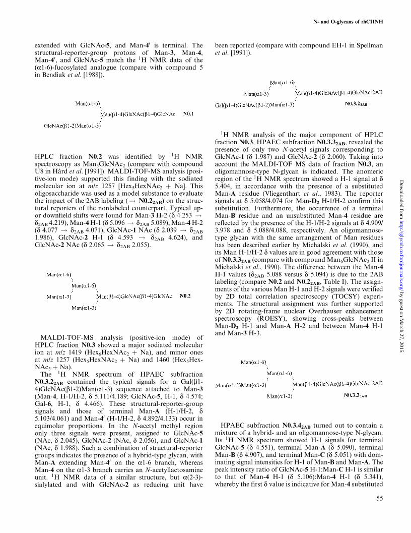
1H NMR analysis of the major component of HPLCfraction N0.3, HPAEC subfraction N0.3.32AB, revealed thepresence of only two N-acetyl signals corresponding toGlcNAc-1 (d 1.987) and GlcNAc-2 (d 2.060). Taking intoaccount the MALDI-TOF MS data of fraction N0.3, anoligomannose-type N-glycan is indicated. The anomericregion of the 1H NMR spectrum showed a H-1 signal at d5.404, in accordance with the presence of a substitutedMan-A residue (Vliegenthart et al., 1983). The reportersignals at d 5.058/4.074 for Man-D2 H-1/H-2 confirm thissubstitution. Furthermore, the occurrence of a terminalMan-B residue and an unsubstituted Man-4 residue arereflected by the presence of the H-1/H-2 signals at d 4.909/3.978 and d 5.088/4.088, respectively. An oligomannose-type glycan with the same arrangement of Man residueshas been described earlier by Michalski et al. (1990), andits Man H-1/H-2 d values are in good agreement with thoseof N0.3.32AB (compare with compound Man6GlcNAc2 II inMichalski et al., 1990). The difference between the Man-4H-1 values (d2AB 5.088 versus d 5.094) is due to the 2ABlabeling (compare N0.2 and N0.22AB, Table I). The assign-ments of the various Man H-1 and H-2 signals were verifiedby 2D total correlation spectroscopy (TOCSY) experi-ments. The structural assignment was further supportedby 2D rotating-frame nuclear Overhauser enhancementspectroscopy (ROESY), showing cross-peaks betweenMan-D2 H-1 and Man-A H-2 and between Man-4 H-1and Man-3 H-3.
HPAEC subfraction N0.3.42AB turned out to contain amixture of a hybrid- and an oligomannose-type N-glycan.Its 1H NMR spectrum showed H-1 signals for terminalGlcNAc-5 (d 4.551), terminal Man-A (d 5.090), terminalMan-B (d 4.907), and terminal Man-C (d 5.051) with dom-inating signal intensities for H-1 of Man-B and Man-A. Thepeak intensity ratio of GlcNAc-5 H-1:Man-C H-1 is similarto that of Man-4 H-1 (d 5.106):Man-4 H-1 (d 5.341),whereby the first d value is indicative for Man-4 substituted
N- and O-glycans of rhC1INH
55
by guest on March 27, 2015
http://glycob.oxfordjournals.org/D
ownloaded from
with GlcNAc-5 (N0.3.4A2AB) and the second one for Man-4substituted with Man-C (N0.3.4B2AB) (Vliegenthart et al.,1983). Therefore the structural reporters clearly indicatetwo structures with the same upper arm and variations inthe substitution of Man-4 in the lower arm (molar ratioA:B, 5:8). The downfield shifts of Man-4 H-1/H-2, causedby the attachment of GlcNAc-5, going from N0.22AB toN0.3.4A2AB, is comparable with such shifts reported in theliterature (Mulder et al., 1995). Compound N0.3.4B2AB isthe conventional Man6GlcNAc2 structure, and taking intoaccount the influence of the 2AB labeling, the structuralreporters are in agreement with those of the nonlabeledcompound (Tseneklidou-Stoeter et al., 1995).
MALDI-TOF-MS analysis (positive-ion mode) of HLPCfraction N0.4 revealed m/z values of 1580, 1619, 1660(major), and 1742, in accordance within 3 mass units withthe sodiated adducts of Hex7HexNAc2, Hex6HexNAc3,Hex5HexNAc4, and Hex8HexNAc2.
HPAEC subfraction N0.4.22AB contains a digalactosylateddiantennary complex-type N-glycan. The 1H NMR struc-tural-reporter groups are in accordance with earlier pub-lished data for such a glycan (compare with compound 11in Bendiak et al. [1988]), except for the values affected by the2AB labeling. Note the difference in d value betweenGlcNAc-2 NAc for complex-type (d 2.072) and hybrid-/oli-gomannose-type (d 2.055±2.062) 2-AB-labeled N-glycans(Table I).
The 1H NMR spectrum of HPAEC subfractionN0.4.42AB showed the occurrence of one major compoundin a mixture of hybrid and/or oligomannose-type N-glycans, a hybrid-type N-glycan (>85% as judged from theratio of the intensities of the N-acetyl methyl signals ofGlcNAc-5 and GlcNAc-1). The compound is an extension
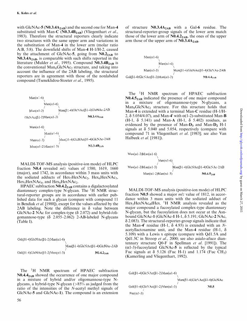
of structure N0.3.4A2AB with a Gal-6 residue. Thestructural-reporter-group signals of the lower arm matchthose of the lower arm of N0.4.22AB, the ones of the upperarm those of the upper arm of N0.3.4A2AB.
The 1H NMR spectrum of HPAEC subfractionN0.4.52AB indicated the presence of one major compoundin a mixture of oligomannose-type N-glycans, aMan8GlcNAc2 structure. For this structure holds thatMan-4 is extended with a terminal Man-C residue (H-1/H-2, d 5.054/4.07), and Man-40 with a(1-2)-substituted Man-B(H-1, d 5.141) and Man-A (H-1, d 5.402) residues, asevidenced by the presence of Man-D3 and Man-D2 H-1signals at d 5.040 and 5.054, respectively (compare withcompound 71 in Vliegenthart et al. [1983]; see also VanHalbeek et al. [1981]).
MALDI-TOF-MS analysis (positive-ion mode) of HLPCfraction N0.5 showed a major m/z value of 1812, in accor-dance within 3 mass units with the sodiated adduct ofHex5HexNAc4dHex. 1H NMR analysis revealed as themajor compound a fucosylated complex-type diantennaryN-glycan, but the fucosylation does not occur at the Asn-bound GlcNAc-1 (GlcNAc-1 H-1, d 5.191; GlcNAc-2 NAc,d 2.083). The structural-reporter-group signals indicate thatthe Man-40 residue (H-1, d 4.93) is extended with an N-acetyllactosamine unit, and the Man-4 residue (H-1, d5.109) with a Lewis x epitope (compare with Qd1.5A andQd1.5C in Stroop et al., 2000; see also asialo-afuco dian-tennary structure Q0-F in Spellman et al. [1991]). The(a1-3)-fucosylated GlcNAc-5 is reflected by the typicalFuc signals at d 5.126 (Fuc H-1) and 1.174 (Fuc CH3)(Kamerling and Vliegenthart, 1992).
K. Koles et al.
56
by guest on March 27, 2015
http://glycob.oxfordjournals.org/D
ownloaded from

Structural analysis of monosialylated N-glycans
The 1H NMR data of the identified monosialylatedhybrid- and complex-type N-glycans are presented inTable II. The table includes also the coding system for thedifferent monosaccharide constituents. HPAEC fractions
N1.7 and N1.8 did not contain sufficient material for NMRassignments. In all presented structures the lower, (a1-3)arm is Neu5Ac(a2-6)Gal (b1-4) GlcNAc(b1-2)Man(a1-3):Neu5Ac H-3e, d 2.667±2.671; Neu5Ac H-3a, d 1.717±1.718;Gal-6 H-1, d 4.444±4.446; GlcNAc-5 H-1, d 4.603±4.609;Man-4 H-1, d 5.132±5.137; Man-4 H-2, d 4.194±4.207
Table II. 1H-chemical shifts of the structural-reporter-group protons of the constituent monosaccharides of sialylated N-glycans,derived from rhC1INH
Chemical shifts are given at 300K and were measured in 2H2O at p2H7 relative to internal acetone (d 2.225; Vliegenthart et al., 1983).Compounds are represented by short-hand symbolic notation: *, Neu5Ac(a2-6); &, Fuc; &, Gal; *, GlcNAc; ^, Man. For numbering ofthe monosaccharide residues, see �. n.d., not determined; a and b stand for the anomeric configuration of GlcNAc-1.
N- and O-glycans of rhC1INH
57
by guest on March 27, 2015
http://glycob.oxfordjournals.org/D
ownloaded from
(slightly influenced by the type of upper arm) (compare withcompound HST in Damm et al. [1987]).
The 1H NMR spectrum of HPAEC fraction N1.1showed the presence of a diantennary N-glycan, (a2-6)-sialylated at the lower arm and an (a1-6)-fucosylatedGlcNAc-1 residue. The upper arm contains a Lewis xepitope as indicated by the typical structural reporters ofthe monosaccharide constituents (compare with com-pound Qd1.6A in Stroop et al., 2000). As indicated by1H NMR analysis, HPAEC fraction N1.2 contains thenoncore-fucosylated analog of N1.1. The 1H NMR spec-trum of HPAEC fraction N1.3 reflects the presence of aconvential (a1-6)-core-fucosylated diantennary N-glycan,(a2-6)-sialylated at the (a1-3) arm (compare with com-pound N1.5 in HaÊrd et al. [1992]). 1H NMR analysis ofHPAEC fraction N1.4 showed the presence of a mono-antennary N-glycan, (a2-6)-sialylated at the (a1-3) arm.The 1H NMR data of a similar structure, but missingGlcNAc-1 (compare with compound 526 in Van Peltet al. [1989]) as well as of the (a1-6)-fucosylated variant(compare compound 1-1 in De Waard et al. [1991]) havebeen reported.
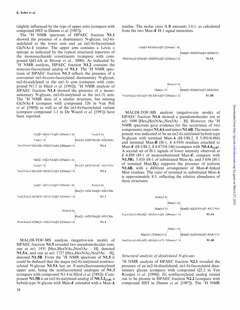
MALDI-TOF-MS analysis (negative-ion mode) ofHPAEC fraction N1.5 revealed two pseudomolecular ions,one at m/z 1931 [Hex5HexNAc4Neu5Ac ± H], denotedN1.5A, and one at m/z 1727 [Hex5HexNAc3Neu5Ac ± H],denoted N1.5B. From the 1H NMR spectrum of N1.5 itcould be deduced that the major (a2-6)-sialylated nonfuco-sylated N-glycan N1.5A has an N-acetyllactosaminylatedupper arm, being the nonfucosylated analogue of N1.3(compare with compound N1.4 in HaÊrd et al. [1992]). Com-ponent N1.5B is an (a2-6)-sialylated analog of N0.3.22AB, ahybrid-type N-glycan with Man-40 extended with a Man-A
residue. The molar ratio A:B amounts 3.6:1, as calculatedfrom the two Man-40 H-1 signal intensities.
MALDI-TOF-MS analysis (negative-ion mode) ofHPAEC fraction N1.6 showed a pseudomolecular ion atm/z 1890 [Hex6HexNAc3Neu5Ac ± H]. However, the 1HNMR spectrum gave evidence for the occurrence of twocomponents,majorN1.6AandminorN1.6B.Themajorcom-ponent was indicated to be an (a2-6)-sialylated hybrid-typeN-glycan with terminal Man-A (H-1/H-2, d 5.091/4.066)and terminal Man-B (H-1, d 4.910) residues attached toMan-40 (H-1/H-2, d 4.875/4.146) (compare with N0.4.42AB).A second set of H-1 signals of lower intensity observed atd 4.898 (H-1 of monosubstituted Man-40; compare withN1.5B), 5.410 (H-1 of substituted Man-A), and 5.056 (H-1of terminal Man-D2) supports the presence of isoformN1.6B, with a different arrangement of Man-40-linkedMan residues. The ratio of terminal to substituted Man-Ais approximately 8:3, reflecting the relative abundance ofthese structures.
Structural analysis of disialylated N-glycans1H NMR analysis of HPAEC fraction N2.1 revealed thepresence of an (a2-6)-disialylated, (a1-6)-fucosylated dian-tennary glycan (compare with compound Q2.2 in VanRooijen et al. [1998]). Its nonfucosylated analog turnedout to be present in HPAEC fraction N2.2 (compare withcompound HST in Damm et al. [1987]). The 1H NMR
K. Koles et al.
58
by guest on March 27, 2015
http://glycob.oxfordjournals.org/D
ownloaded from

data of both compounds are included in Table II. HPAECfraction N2.3 did not contain sufficient material for NMRassignments.
Release, fractionation, and structural analysis ofO-glycans of rhC1INH
Resource Q fractionation of the O-linked oligosaccharide-alditols, obtained after alkaline borohydride treatment ofthe N-deglycosylated rhC1INH, yielded three carbohydrate-containing fractions, denoted O.1 (45%), O.2 (33%), andO.3 (22%) (Figure 6). 1H NMR analysis of the neutralFPLC fraction O.1 showed the presence of the core 1 struc-ture Gal(b1-3)GalNAc-ol (compare with compound 2 inKamerling and Vliegenthart [1992]) (Table III). The mono-charged FPLC fraction O.2 contained a mixture of twomonosialylated alditols, Neu5Ac(a2-3)Gal(b1-3)GalNAc-ol(O.2A) (compare with compound 78 in Kamerling andVliegenthart [1992]) and Gal(b1-3)[Neu5Ac(a2-6)]GalNAc-ol(O.2B) (compare with compound 9 in Kamerling andVliegenthart [1992]) in an approximate molar ratio of 1:2,as judged from the intensities of the H-1 signals of substituted(d 4.546) and terminal (d 4.474) Gal. The dichargedFPLC fraction O.3 contained Neu5Ac(a2-3)Gal(b1-3)
[Neu5Ac(a2-6)]GalNAc-ol (compare with compound 85 inKamerling and Vliegenthart [1992]).
Discussion
In 1985 an 1H NMR analysis of the carbohydrate chains ofnative hC1INH, purified from normal serum, was reported(Strecker et al., 1985), and, following our coding system,compounds N2.1, N2.2, and O.2A, and the (a2-3)-disialylated isomers of N2.1 and N2.2 were shown tooccur. In view of the followed isolation protocol it is tempt-ing to assume that only major components were identified.
Fig. 6. Elution profile at 214 nm of the chemically released O-glycanpool from N-deglycosylated rhC1INH on a Resource Q column. Thecolumn was first eluted with 12 ml water, followed by a linearconcentration gradient of 0±100% (v/v) 0.5 M NaCl over 9 min at a flowrate of 4 ml/min. Fractions O.4, O.5, O.6, and O.7 did not containcarbohydrate material.
Table III. 1H-chemical shifts of the structural-reporter-group protons ofthe constituent monosaccharides of O-glycans, derived from rhC1INH
Chemical shifts are given at 300K and were measured in 2H2O at p2H7relative to internal acetone (d 2.225; Vliegenthart et al., 1983). Compoundsare represented by short-hand symbolic notation: ~, Neu5Ac(a2-3); *,Neu5Ac(a2-6); ^, GalNAc; &, Gal. n.d., not determined.
N- and O-glycans of rhC1INH
59
by guest on March 27, 2015
http://glycob.oxfordjournals.org/D
ownloaded from

Additional profiling studies (Perkins et al., 1990) indicatedthat besides sialylated complex-type diantennary, also lowamounts of tri- and tetraantennary N-glycans do occur.Lectin-based assays revealed the occurrence of (a2-6)/(a2-3)-sialylated complex- and hybrid-type N-glycans(Schoenberger,1992).Recently,biologicallyactiverhC1INHwith mainly oligomannose-type N-glycans has been exp-ressed in a baculovirus expression system (Wolff et al., 2001).
Here, we focus on the N,O-glycosylation pattern ofrhC1INH, the transgenic material expressed in the mam-mary gland of transgenic rabbits, excreted in the milk. Afirst inspection of the results learns that oligomannose-,hybrid-, and diantennary complex-type N-glycans dooccur. When sialylated, only Neu5Ac is present, and only(a2-6) linkages are found. Part of the complex-typeN-glycans contains the Lewis x epitope. The small core1-type O-glycans show the usual (a2-3)- and (a2-6)-sialylation pattern of O-glycoproteins of nonmucinousorigin. Neu5Gc, found in low amounts in CHO- and BHK-expressed recombinant glycoproteins (Hokke et al., 1990;Nimtz et al., 1993) and suggested to occur in transgenichuman antithrombin produced in goat milk (Edmundset al., 1998), was not observed on the rhC1INH glycans.This sialic acid is not expressed in normal adult human cells;higher levels of Neu5Gc may lead to immune reactions inhumans. In this context, it should be noted that N-glycansof CHO- and BHK-derived recombinant glycoproteinscontain (a2-3)-linked sialic acid only. Another well-documented immunogenic carbohydrate antigen, Gal(a1-3)Gal(b1-4)GlcNAc(b1- (Hamadeh et al., 1992), was also notdetected in rhC1INH.
In Table IV a survey is presented of the amounts in molarpercentages of the N- and O-glycans established. The neu-tral N-glycans in rhC1INH make up about 25% of the totalN-glycan pool. The majority of these glycans were of theoligomannose- and hybrid-type, with Man5GlcNAc2 beingthe most abundant structure, accounting for about 10% ofthe total N-glycan pool. About 75% of the N-glycans weresialylated. The monocharged N-glycans (about 67% of thetotal N-glycan pool) were of the hybrid- and mono- ordiantennary complex type. Here, the major structures werethe monosialylated, partially (a1-6)-fucosylated dianten-nary N-glycans (about 30% of the total N-glycan pool).The dicharged N-glycans (about 8% of the total N-glycanpool) comprised disialylated, partially (a1-6)-fucosylateddiantennary N-glycans. In this context it is of interest tomention that with increasing expression levels of rhC1INH,the ratio of oligomannose-type:hybrid-type N-glycansincreases and that a decrease in the extent of sialylationoccurs with the progress of lactation (Koles et al.,unpublished data).
An evaluation of the N-glycosylation pattern foundfor rhC1INH indicates that several of the oligomannose-and hybrid-type glycans contained a Man-D2 residue, whichis rather unusual for mature glycoproteins. Followingthe major pathway in the trimming of glycoprotein N-glycans from Glc3Man9GlcNAc2 to Man5GlcNAc2, thethree Glc residues are removed by the endoplasmic reticu-lum (ER)±resident glucosidases I and II and Man-D2 bya Golgi-localized a-mannosidase I, yielding a specificMan8GlcNAc2 isomer. Two other alternate early processing
Table IV. Survey of N- and O-glycans obtained fromrhC1INH, together with their relative amounts
Compounds are represented by shorthand symbolicnotation (see Tables I±III).
K. Koles et al.
60
by guest on March 27, 2015
http://glycob.oxfordjournals.org/D
ownloaded from

routes to Man8GlcNAc2 isomers comprise the removal ofMan-D2 by ER-localized a-mannosidase I or of Man-D3 byER-localized a-mannosidase II. In the Golgi (or partially inthe ER), specific a-mannosidases release the remainingMan-D residues and Man-C (Weng and Spiro, 1993, 1996;Verbert, 1995).
The finding of Man8GlcNAc2 isomer N0.4.5, containingMan-D2 and Man-D3 indicate that in the mammary glandof the rabbit the alternative endo-mannosidase pathway(More and Spiro, 1990) is competetively active. Here aGlc(a1-3)Man disaccharide is released by a Golgi-localizedendo-a-mannosidase yielding Man8GlcNAc2 missing Man-D1, followed by a sequential cleavage of Man-D3 andMan-C by Golgi-localized a-mannosidases (Verbert,1995), yielding the Man6GlcNAc2 isomer N0.3.3, the sec-ond most abundant oligomannose-type structure in thisstudy (a product that also could arise from the ER-a-man-nosidase II processing). Interestingly, the endo-mannosi-dase pathway seems to be an active pathway in themammary glands of cow, goat, sheep, and rhesus monkey,too, as lactoferrins from these animals contain, in additionto Man9GlcNAc2, the Man8GlcNAc2 structure N0.4.5 (VanHalbeek et al., 1981; Coddeville et al., 1992; Montreuil et al.,1997). Usually the endo-mannosidase pathway, a processingwith one exception so far only observed in vertebrates(Dairaku and Spiro, 1997), is followed for circumventinga-glucosidase blockades (Moore and Spiro, 1990;De Praeter et al., 2000) but can also be induced by inhibitingER/Golgi-localized a-mannosidases I, thereby blockingthe formation of Man8GlcNAc2 missing Man-D2 (Wengand Spiro, 1996). The reason why the mammary tissue isusing partly the endo-mannosidase pathway remains to beclarified.
Comparing the N-glycosylation patterns of native serumhC1INH and rhC1INH (Table IV), it can be concluded thatthe total processing in native serum hC1INH (only N2.1/N2.2 and the [a2-3]-sialylated isomers) is much more com-plete than in rhC1INH (N2.1/N2.2 make up about 10% ofthe total N-glycan pool). Comparing the O-glycosyla-tion patterns of native serum hC1INH and rhC1INH(Table IV), it is remarkable that the only structure (O.2A)present in native material is a minor one (about 10% of thetotal O-glycan pool) in the recombinant material. In rhC1-INH the (a2-6)-sialylated extensions of O.1 and O.2A arethe major products (about 60% of the total O-glycan pool).Taking together, when compared with native serum hC1-INH, in terms of sialylation the N-glycans of rhC1INH are(a2-6)-undersialylated [Gal(b1-4)GlcNAc a-2,6-sialyltrans-ferase/ST6Gal I], whereas the O-glycans are (a2-6)-over-sialylated (GalNAc a-2,6-sialyltransferase/ST6GalNAc II].
Biological activity studies of rhC1INH are in progress andwill be published elsewhere.
The detailed analysis of the N,O-glycosylation pattern ofrhC1INH, revealing remarkable differences in glycosyla-tion pattern between native serum hC1INH and rhC1INH,makes clear the importance of such studies for therapeuticglycoproteins when expressed in new biological systems.Earlier research has demonstrated the usefulness of study-ing the glycosylation machinery of CHO and BHK cells,and similar research has to be set up to explore the glyco-sylation machinery of the mammary gland of animals usedfor transgenic purposes.
Materials and methods
Release and isolation of N-glycans
Purified rhC1INH, isolated from pooled milk of line 2972p,was obtained from Pharming Technologies BV (Leiden,Netherlands). The N-glycans of rhC1INH were enzymicallyreleased with PNGase F (EC 3.5.1.52) (Roche MolecularBiochemicals, Indianapolis, IN), using incubation condi-tions optimized for rhC1INH. Briefly, 100 mg rhC1INH,dissolved in 5 ml 20 mM PBS pH 7.2 (Fluka, Buchs,Switzerland) containing 10 mM ethylenediamine tetra-acetic acid, was denatured in the presence of 10% sodiumdodecyl sulfate (w/v) and 10 mM 2-mercaptoethanol for 5min at 100�C, then digested with 5 U/mg PNGase F in thepresence of 6.4 mg/ml 3-[(3-cholamidodopropyl)dimethyl-ammonio]-1-propanesulfonate (Fluka) for 24 h at 37�C.Portions of the digest were filtered through 30 kDa cut-offcentrifugal concentrators and the filtrates, containing thereleased N-glycans, desalted on graphitized carbon columns(Alltech, Breda, the Netherlands) (Packer et al., 1998) afterremoval of detergents by Calbiosorb (Calbiochem, SanDiego, CA) beads. The concentrated N-deglycosylatedglycoprotein (~0.4 ml) was then extensively dialyzed andsubjected to de-O-glycosylation.
Release and isolation of O-glycans
N-deglycosylated rhC1INH was subjected to alkalineborohydride treatment according to Piller and Piller (1993).Briefly, the concentrated N-deglycosylated glycoproteinsolution (~0.4 ml) was mixed with 10 ml 0.1 M NaOHcontaining 1 M NaBH4, and kept for 16 h at 45�C, thencooled on ice and neutralized with 2 M aqueous HOAc.Boric acid was removed by repetitive coevaporation withMeOH, containing 1% (v/v) HOAc. Finally, the materialwas applied to a Dowex 50W-X8, H� column (100±200mesh, Pharmacia), and O-glycans (oligosaccharide-alditols)were eluted with water and lyophilized. When required,samples were further purified on graphitized carbon col-umns (Packer et al., 1998).
FPLC fractionation
The pools of N- and O-glycans were fractionated intoneutral and charged species on a Resource Q column(6 ml, Pharmacia, Uppsala, Sweden) using a PharmaciaFPLC system. Elutions were performed with water followedby a linear concentration gradient of NaCl in water at a
N- and O-glycans of rhC1INH
61
by guest on March 27, 2015
http://glycob.oxfordjournals.org/D
ownloaded from

flow rate of 4 ml/min; for gradient details, see relevantfigure captions. Fractionations were monitored by UVabsorbance at 214 nm and conductivity measurements.Individual fractions were lyophilized, desalted on 5 con-nected HiTrap columns (5 ml, Amersham, Little Chalfont,U.K.) using 5 mM NH4HCO3 as eluent, and lyophilizedagain.
HPLC fractionation
The FPLC fraction of neutral N-glycans was furtherfractionated on a LiChrospher-NH2 5 mm (250 mm� 4.6 mm,Alltech) column equipped with a LiChrospher Amino 5 mmguard column (7.5 � 4.6 mm), using a Waters 600 HPLCsystem. Elutions were carried out with a linear gradient ofwater in acetonitrile at a flow rate of 1 ml/min; for gradientdetails, see relevant figure captions. The fractionationwas monitored by UV absorbance at 206 nm. Individualfractions were lyophilized.
Labeling of N-glycans with 2AB
Neutral N-glycans from the HPLC fractionation weretreated with 0.35 M 2AB (Sigma)/1 M NaCNBH3 inMe2SO±HOAc (7:3, v/v) for 2 h at 65�C (Bigge et al.,1995; Stroop et al., 2000). The 2AB fluorescently labeledglycans were purified via paper chromatography on acid-pretreated QMA (Whatman) filter paper strips using acet-onitrile (three times) as a mobile phase. Glycans (remainingat the base line) were eluted from the dried paper strips withwater, concentrated, and subfractionated by HPAEC.
HPAEC fractionation
HPAEC was performed on a Dionex DX 500 workstationequipped with a pulsed amperometric detection (PAD) sys-tem. A series of N-glycan fractions were subfractionated ona CarboPac PA-1 (250 � 9 mm) column using linear gra-dients of 0.5 M NaOAc in 0.1 M NaOH at a flow rate of4 ml/min; for gradient details, see relevant figure captions.The following pulse potentials and durations were usedduring the triple-pulse amperometric detection with a goldelectrode at 300 mA: E1 0.05 V, 480 ms; E2 0.60 V, 120 ms;E3 ÿ0.60 V, 60 ms. Fractions were immediately neutralizedwith 0.1 M HCl, then lyophilized, desalted on five con-nected HiTrap columns (5 ml, Amersham Pharmacia)using 5 mM NH4HCO3 as eluent, and lyophilized again.
Quantification of oligosaccharides
The molar ratio of the FPLC fractions was calculated fromthe FPLC peak areas on the basis of the weighted averagenumber of C�O groups (responsible for UV absorption at214 nm) being known after structural identification anddetermination of the relative amounts of each individualcomponent. The molar ratio of the constituent oligosac-charides within each FPLC fraction was determined fromthe HPLC peak areas (corrected for the number of C�Ogroups) at 206 nm. When HPAEC/PAD was used forfurther fractionation of HPLC fractions, the PAD responsewas assumed to be equal for each oligosaccharide presentwithin one HPLC fraction. For mixtures, molar ratios weredetermined on the basis of the 1H NMR spectra.
1H NMR spectroscopy
Prior to 1H NMR spectroscopy, samples were lyophilizedtwice from 99.9% 2H2O (Cambridge Isotope Laboratories,Andover, MA), then dissolved in 99.96% 2H2O (CambridgeIsotope Laboratories). 1H NMR spectra were recorded at500 MHz on a Bruker AMX-500 spectrometer (BijvoetCenter, Department of NMR Spectroscopy, UtrechtUniversity) at a probe temperature of 300K and p2H 7.1D spectra of 5000 Hz spectral width were recorded in16K complex data sets using a water eliminated Fouriertransform pulse sequence as described by HaÊrd et al.(1992). Chemical shifts are expressed in ppm relative tointernal acetone (d 2.225 in 2H2O) or acetate (d 1.908 in2H2O) (Vliegenthart et al., 1983). 2D-TOCSY spectra at 500MHz were recorded using Bruker software with MLEV-17mixing sequence cycles between 20 and 100 ms. Datamatrices of 512 � 2048 points were collected, representinga spectral width of 4800 Hz in each dimension. The 2HO1Hsignal was suppressed by presaturation for 1 s during therelaxation delay. 2D-ROESY spectra were recorded with amixing time of 300 ms. Phase-sensitive handling of data wasperformed by the time-proportional phase incrementmethod implemented by the Bruker software. NMR datawere processed using a locally developed software package(J. A. van Kuik, Bijvoet Center, Department of Bio-OrganicChemistry, Utrecht University).
MS
For MALDI-TOF-MS in the positive-ion mode, samples(0.5±1 ml) were mixed in a 1:1 ratio with a mixture of 5mg/ml 2,5-dihydroxybenzoic acid and 0.25 mg/ml 5-methoxysalicylic acid in 1 ml ethanol/10 mM NaCl (1:1, v/v)as a matrix. For the negative-ion mode, 2 mg/ml 20,40,60-trihydroxyacetophenone monohydrate in acetonitrile/13.3mM ammonium citrate (1:3, v/v) was used as a matrix. Inthis case the sample matrix mixture was dried under reducedpressure (Papac et al., 1998). Measurements were performedon a PerSeptive Biosystems Voyager-DE MALDI-TOF massspectrometer with implemented delayed extraction techniqueusing an N2 laser (337 nm) with 3 ns pulse width. Spectra wererecorded in a linear mode at an accelerating voltage of 24.5 kVusing an extraction delay of 90 ns.
Acknowledgments
This study was financially supported by the TechnologyFoundation STW of the Netherlands Organization forScientific Research NWO and by Pharming TechnologiesBV (Leiden, Netherlands).
Abbreviations
2AB, 2-aminobenzamide; BHK, baby hamster kidney;CHO, Chinese hamster ovary; ER, endoplasmic reticulum;FPLC, fast protein liquid chromatography; hC1INH,human C1 inhibitor; HPAEC, high-pH anion-exchangechromatography; HPLC, high-performance liquid chro-matography; MALDI-TOF, matrix-assisted laser desorp-tion ionization time-of-flight; MS, mass spectrometry;
K. Koles et al.
62
by guest on March 27, 2015
http://glycob.oxfordjournals.org/D
ownloaded from

NMR, nuclear magnetic resonance; PAD, pulsed ampe-rometric detection; PNGase F, peptide-N4-(N-acetyl-b-glucosaminyl)asparagine amidase F; rhC1INH,recombinant-rabbit human C1 inhibitor; ROESY, rotating-frame nuclear Overhauser enhancement spectroscopy;TOCSY, total correlation spectroscopy.
References
Bendiak, B., Orr, J., Brockhausen, I., Vella, G., and Phoebe, C. (1988)Separation of neutral reducing oligosaccharides derived from glyco-proteins by HPLC on a hydroxylated polymeric support. Anal.Biochem., 175, 96±105.
Bigge, J.C., Patel, T.P., Bruce, J.A., Goulding, P.N., Charles, S.M., andParekh, R.B. (1995) Nonselective and efficient fluorescent labeling ofglycans using 2-aminobenzamide and anthranilic acid. Anal. Biochem.,230, 229±238.
Bijvoet, A.G.A., Kroos, M.A., Pieper, F.R., de Boer, H.A., Reuser, A.J.J.,van der Ploeg, A.T., and Verbeet, M.P. (1996) Expression of cDNA-encoded human acid a-glucosidase in milk of transgenic mice. Biochim.Biophys. Acta, 1308, 93±96.
Bock, S.C., Skriver, K., Nielsen, E., Thùgersen, H.-C., Wiman, B.,Donaldson, V.H., Eddy, R.L., Marrinan, J., Radziejewska, E., Huber,R., and others. (1986) Human C1 inhibitor: primary structure,cDNA cloning, and chromosomal localization. Biochemistry, 25,4292±4301.
Caliezi, C., Wuillemin, W.A., Zeerleder, S., Redondo, M., Eisele, B., andHack, C.E. (2000) C1-esterase inhibitor: an anti-inflammatory agentand its potential use in the treatment of diseases other than hereditaryangioedema. Pharmacol. Rev., 52, 91±112.
Carreer, F.M. (1992) The C1 inhibitor deficiency. A review. Eur. J. Clin.Chem. Clin. Biochem., 30, 793±807.
Carugati, A., Pappalardo, E., Zingale, L.C., and Cicardi, M. (2001)C1-inhibitor deficiency and angioedema. Mol. Immunol., 38,161±173.
Coddeville, B., Strecker, G., Wieruszeski, J.-M., Vliegenthart, J.F.G., vanHalbeek, H., Peter-Katalinic, J., Egge, H., and Spik, G. (1992)Heterogeneity of bovine lactotransferrin glycans. Characterizationof a-D-Galp-(1! 3)-b-D-Gal- and a-NeuAc-(2! 6)-b-D-GalpNAc-(1! 4)-b-D-GlcNAc-substituted N-linked glycans. Carbohydr. Res.,236, 145±164.
Colman, A. (1996) Production of proteins in the milk of transgeniclivestock: problems, solutions, and successes. Am. J. Clin. Nutr., 63,639S±645S.
Dairaku, K., and Spiro, R.G. (1997) Phylogenetic survey of endomanno-sidase indicates late evolutionary appearance of this N-linkedoligosaccharide processing enzyme. Glycobiology, 7, 579±586.
Damm, J.B.L., Kamerling, J.P., van Dedem, G.W.K., and Vliegenthart,J.F.G. (1987) A general strategy for the isolation of carbohydratechains from N-,O-glycoproteins and its application to humanchorionic gonadotropin. Glycoconj. J., 4, 129±144.
Denman, J., Hayes, M., O'Day, C., Edmunds, T., Bartlett, C., Hirani, S.,Ebert, K.M., Gordon, K., and McPherson, J.M. (1991) Transgenicexpression of a variant of human tissue-type plasminogen activator ingoat milk: purification and characterization of the recombinantenzyme. Bio/Technology, 9, 839±843.
De Praeter, C.M., Gerwig, G.J., Bause, E., Nuytinck, L.K., Vliegenthart,J.F.G., Breuer, W., Kamerling, J.P., Espeel, M.F., Martin, J.-J.R.,De Paepe, A.M., Shows, T.B., and Magnusson, S. (2000) A noveldisorder caused by defective biosynthesis of N-linked oligosaccharidesdue to glucosidase I deficiency. Am. J. Hum. Genet., 66, 1744±1756.
De Waard, P., Koorevaar, A., Kamerling, J.P., and Vliegenthart, J.F.G.(1991) Structure determination by 1H NMR spectroscopy of (sulfated)sialylated N-linked carbohydrate chains released from porcinethyroglobulin by peptide-N4-(N-acetyl-b-glucosaminyl)asparagineamidase-F. J. Biol. Chem., 266, 4237±4243.
Ebo, D.G. and Stevens, W.J. (2000) Hereditary angioneurotic edema:review of the literature. Acta Clin. Belg., 55, 22±29.
Edmunds, T., van Patten, S.M., Pollock, J., Hanson, E., Bernasconi, R.,Higgins, E., Manavalan, P., Ziomek, C., Meade, H., McPherson, J.M.,
and Cole, E.S. (1998) Transgenically produced human antithrombin:structural and functional comparison to human plasma-derivedantithrombin. Blood, 91, 4561±4571.
Galet, C., Menck Le Bourhis, C., Chopineau, M., Le Griec, G., Perrin, A.,Magallon, T., Attal, J., Viglietta, C., Houdebine, L.M., and Guillou, F.(2001) Expression of a single ba chain protein of equine LH/CG inmilk of transgenic rabbits and its biological activity. Mol. Cell.Endocrinol., 174, 31±40.
Hamadeh, R.M., Jarvis, G.A., Galili, U., Mandrell, R.E., Zhou, P., andGriffiss, J.M. (1992) Human natural anti-Gal IgG regulates alternativecomplement pathway activation on bacterial surfaces. J. Clin. Invest.,89, 1223±1235.
HaÊrd, K., Mekking, A., Kamerling, J.P., Dacremont, G.A.A., andVliegenthart, J.F.G. (1991) Different oligosaccharides accumulate inthe brain and urine of a cat with a-mannosidosis: structuredetermination of five brain-derived and seventeen urinary oligosac-charides. Glycoconj. J., 8, 17±28.
HaÊrd, K., van Zadelhoff, G., Moonen, P., Kamerling, J.P., andVliegenthart, J.F.G. (1992) The Asn-linked carbohydrate chains ofhuman Tamm-Horsfall glycoprotein of one male. Novel sulfated andnovel N-acetylgalactosamine-containing N-linked carbohydratechains. Eur. J. Biochem., 209, 895±915.
Hokke, C.H., Bergwerff, A.A., van Dedem, G.W.K., van Oostrum, J.,Kamerling, J.P., and Vliegenthart, J.F.G. (1990) Sialylated carbohy-drate chains of recombinant human glycoproteins expressed in Chinesehamster ovary cells contain traces of N-glycolylneuramic acid. FEBSLett., 275, 9±14.
Houdebine, L.M. (1995) The production of pharmaceutical proteins fromthe milk of transgenic animals. Reprod. Nutr. Dev., 35, 609±617.
Jenkins, N. and Curling, E.M.A. (1994) Glycosylation of recombinantproteins: problems and prospects. Enzyme Microbial Technol., 16,354±364.
Jenkins, N., Parekh, R.B., and James, D.C. (1996) Getting theglycosylation right: Implications for the biotechnology industry.Nature Biotechnol., 14, 975±981.
Kamerling, J.P. (1996) Carbohydrate features of recombinant humanglycoproteins. Biotecnologia Aplicada, 13, 167±180.
Kamerling, J.P. and Vliegenthart, J.F.G. (1989) Carbohydrates. InLawson, A.M. (Ed.) Clinical biochemistry; principles, methods,applications, vol. 1, Mass spectrometry. Walter de Gruyter, Berlin,pp. 175±263.
Kamerling, J.P. and Vliegenthart, J.F.G. (1992) High-resolution 1H-nuclear magnetic resonance spectroscopy of oligosaccharide-alditolsreleased from mucin-type O-glycoproteins. Biol. Magn. Reson., 10,1±194.
Korhonen, V.-P., Tolvanen, M., Hyttinen, J.-M., Uusi-Oukari, M.,Sinervirta, R., Alhonen, L., Jauhiainen, M., J�anne, O.A., and J�anne,J. (1997) Expression of bovine b-lactoglobulin/human erythropoietinfusion protein in the milk of transgenic mice and rabbits. Eur. J.Biochem., 245, 482±489.
Michalski, J.-C., Haeuw, J.-F., Wieruszeski, J.-M., Montreuil, J., andStrecker, G. (1990) In vitro hydrolysis of oligomannosyl oligosacchar-ides by the lysosomal a-D-mannosidases. Eur. J. Biochem., 189, 369±379.
Montreuil, J., Spik, G., and Mazurier, J. (1997) Transferrin superfamily.An outstanding model for studying biochemical evolution. InMontreuil, J., Vliegenthart, J.F.G., and Schachter, H. (Eds.),Glycoproteins II. Elsevier Science, Amsterdam, pp. 203±242.
Moore, S.E.H. and Spiro, R.G. (1990) Demonstration that Golgi endo-a-D-mannosidase provides a glucosidase-independent pathway for theformation of complex N-linked oligosaccharides of glycoproteins. J.Biol. Chem., 265, 13104±13112.
Mulder, H., Dideberg, F., Schachter, H., Spronk, B.A., de Jong-Brink,M., Kamerling, J.P., and Vliegenthart, J.F.G. (1995) In the biosynth-esis of N-glycans in connective tissue of the snail Lymnaea stagnalisincorporation of GlcNAc by b2GlcNAc-transferase I is an essentialprerequisite for the action of b2GlcNAc-transferase II and b2Xyl-transferase. Eur. J. Biochem., 232, 272±283.
Nimtz, M., Martin, W., Wray, V., Kl�oppel, K.-D., Augustin, J., andConradt, H.S. (1993) Structures of sialylated oligosaccharides ofhuman erythropoietin expressed in recombinant BHK-21 cells. Eur. J.Biochem., 213, 39±56.
N- and O-glycans of rhC1INH
63
by guest on March 27, 2015
http://glycob.oxfordjournals.org/D
ownloaded from

Nuijens, J.H., van Berkel, P.H.C., Geerts, M.E.J., Hartevelt, P.P., deBoer, H.A., van Veen, H.A., and Pieper, F.R. (1997) Characterizationof recombinant human lactoferrin secreted in milk of transgenic mice.J. Biol. Chem., 272, 8802±8807.
Packer, N.H., Lawson, M.A., Jardine, D.R., and Redmond, J.W. (1998) Ageneral approach to desalting oligosaccharides released from glyco-proteins. Glycoconj. J., 15, 737±747.
Papac, D.I., Briggs, J.B., Chin, E.T., and Jones, A.J.S. (1998) Ahigh-throughput microscale method to release N-linked oligo-saccharides from glycoproteins for matrix-assisted laser desorption/ionization time-of-flight mass spectrometric analysis. Glycobiology, 8,445±454.
Perkins, S.J. (1993) Three-dimensional structure and molecular modellingof C1 inhibitor. Behring Inst. Mitt., 93, 63±80.
Perkins, S.J., Smith, K.F., Amatayakul, S., Ashford, D.,Rademacher, T.W., Dwek, R.A., Lachmann, P.J., and Harrison,R.A. (1990) Two-domain structure of the native and reactive centrecleaved forms of C1 inhibitor of human complement by neutronscattering. J. Mol. Biol., 214, 751±763.
Piller, F. and Piller, V. (1993). Structural characterization of mucin-typeO-linked oligosaccharides. In Fukuda, M. and Kobata, A. (Eds.),Glycobiology, a practical approach. Oxford University Press, NewYork, pp. 291±328.
Schoenberger, O.L. (1992) Characterization of carbohydrate chains of C1-inhibitor and of desialylated C1-inhibitor. FEBS Lett., 314, 430±434.
Spellman, M.W., Leonard, C.K., Basa, L.J., Gelineo, I., and vanHalbeek, H. (1991) Carbohydrate structures of recombinant solublehuman CD4 expressed in Chinese hamster ovary cells. Biochemistry,30, 2395±2406.
Strecker, G., Ollier-Hartmann, M.-P., van Halbeek, H.,Vliegenthart, J.F.G., Montreuil, J., and Hartmann, L. (1985) Primarystructure elucidation of carbohydrate chains of normal C1-esteraseinhibitor (C1-INH) by 400-MHz 1H-NMR study. CR. Acad. Sci.Paris, 301, 571±576.
Stroop, C.J.M., Weber, W., Gerwig, G.J., Nimtz, M., Kamerling, J.P.,and Vliegenthart, J.F.G. (2000) Characterization of the carbohydratechains of the secreted form of the human epidermal growth factorreceptor. Glycobiology, 10, 901±917.
Tseneklidou-Stoeter, D., Gerwig, G.J., Kamerling, J.P., and Spindler, K.-D.(1995) Characterization of N-linked carbohydrate chains of the crayfish,Astacus leptodactylus hemocyanin. Biol. Chem. Hoppe-Seyler, 376,531±537.
Uusi-Oukari, M., Hyttinen, J.-M., Korhonen, V.-P., V�asti, A., Alhonen, L.,J�anne, O.A., and J�anne, J. (1997) Bovine aS1-casein gene sequencesdirect high level expression of human granulocyte-macrophage colony-stimulating factor in the milk of transgenic mice. Transgenic Res., 6,75±84.
Van Berkel, P.H.C., Welling, M.M., Geerts, M., van Veen, H.A.,Ravensbergen, B., Salaheddine, M., Pauwels, E.K.J., Pieper, F.,Nuijens, J.H., and Nibbering, P.H. (2002) Large scale production ofrecombinant human lactoferrin in the milk of transgenic cows. NatureBiotechnol., 20, 484±487.
Van Halbeek, H., Dorland, L., Vliegenthart, J.F.G., Spik, G., Cheron, A.,and Montreuil, J. (1981) Structure determination of two oligomanno-side-type glycopeptides obtained from bovine lactotransferrin, by 500MHz 1H-NMR spectroscopy. Biochim. Biophys. Acta, 675, 293±296.
Van Pelt, J., HaÊrd, K., Kamerling, J.P., Vliegenthart, J.F.G., Reuser, A.J.J.,and Galjaard, H. (1989) Isolation and structural characterization oftwenty-one sialyloligosaccharides from galactosialidosis urine. Anintact N,N0-diacetylchitobiose unit at the reducing end of a dianten-nary structure. Biol. Chem. Hoppe-Seyler, 370, 191±203.
Van Rooijen, J.J.M., Jeschke, U., Kamerling, J.P., and Vliegenthart, J.F.G.(1998) Expression of N-linked sialyl Lex determinants and O-glycansin the carbohydrate moiety of human amniotic fluid transferrin duringpregnancy. Glycobiology, 8, 1053±1064.
Varki, A., Cummings, R., Esko, J., Freeze, H., Hart, G., and Marth, J.(1999) Essentials of glycobiology. Cold Spring Harbor LaboratoryPress, Cold Spring Harbor, NY.
Verbert, A. (1995). Biosynthesis. 2b. From Glc3Man9GlcNAc2-protein toMan5GlcNAc2-protein: transfer `̀ en bloc'' and processing. InMontreuil, J., Vliegenthart, J.F.G., and Schachter, H. (Eds.),Glycoproteins. Elsevier Science, Amsterdam, pp. 145±152.
Visser, E.C., Dijkstra, R., Brakenhoff, J.P.J., van Corven, E.J.J.M., andvan Berkel, P.H.C. (2000) Analysis of the glycosylation of recombinanthuman acid a-glucosidase produced in the milk of transgenic rabbits.Glycobiology, 10, 1133.
Vliegenthart, J.F.G., Dorland, L., and van Halbeek, H. (1983) High-resolution, 1H-nuclear magnetic resonance spectroscopy as a tool inthe structural analysis of carbohydrates related to glycoproteins. Adv.Carbohydr. Chem. Biochem., 41, 209±374.
Weng, S. and Spiro, R.G. (1993) Demonstration that a kifunensine-resistant a-mannosidase with a unique processing action on N-linkedoligosaccharides occurs in rat liver endoplasmic reticulum and variouscultured cells. J. Biol. Chem., 268, 25656±25663.
Weng, S. and Spiro, R.G. (1996) Evaluation of the early processing routesof N-linked oligosaccharides of glycoproteins through the character-ization of Man8GlcNAc2 isomers: evidence that endomannosidasefunctions in vivo in the absence of a glucosidase blockade. Glycobiol-ogy, 6, 861±868.
Wolff, M.W., Zhang, F., Roberg, J.J., Caldwell, E.E.O., Kaul, P.R.,Serrahn, J.N., Murhammer, D.W., Linhardt, R.J., and Weiler, J.M.(2001) Expression of C1 esterase inhibitor by the baculovirusexpression vector system: preparation, purification, and characteriza-tion. Protein Expr. Purif., 22, 414±421.
K. Koles et al.
64
by guest on March 27, 2015
http://glycob.oxfordjournals.org/D
ownloaded from