Eosinophil and neutrophil extracellular DNA traps in human allergic asthmatic airways
Upload
independentCategory
view
1download
0
Structural characterisation of neutrophil glycans by ultrasensitive mass spectrometric glycomics methodology
Ponnusamy Babu & Simon J. North & Jihye Jang-Lee &
Sara Chalabi & Kathryn Mackerness & Sean R. Stowell &Richard D. Cummings & Sara Rankin & Anne Dell &Stuart M. Haslam
Received: 11 April 2008 /Revised: 13 May 2008 /Accepted: 15 May 2008# The Author(s) 2008
Abstract Neutrophils are the most abundant white bloodcells in humans and play a vital role in several aspects ofthe immune response. Numerous reports have implicatedneutrophil glycosylation as an important factor in mediatingthese interactions. We report here the application of highsensitivity glycomics methodologies, including matrixassisted laser desorption ionisation (MALDI-TOF) andMALDI-TOF/TOF analyses, to the structural analysis ofN- and O-linked carbohydrates released from two samplesof neutrophils, prepared by two separate and geographicallyremote laboratories. The data produced demonstrates thatthe cells display a diverse range of sialylated andfucosylated complex glycans, with a high level of similaritybetween the two preparations.
Keywords Mass spectrometry . Neutrophil . Glycomics .
Protein glycosylation
AbbreviationsPNGase F Peptide N-glycosidase FMALDI Matrix assisted laser desorption ionisation
CAD Collision activated decompositionHex HexoseHexNAc N-acetylhexosamineESL-1 E-selectin ligand-1FAB Fast atom bombardmentFuc FucoseGal GalactoseGalNAc N-acetylgalactosamineGlc GlucoseGlcNAc N-acetylglucosamineMan MannoseNeuAc N-acetylneuraminic acidMS Mass spectrometryMS/MS Tandem mass spectrometryPSGL-1 P-selectin glycoprotein ligand 1TOF Time of flightLex Lewisx
Lea Lewisa
sLex Sialyl Lewisx
sLea Sialyl Lewisa
Introduction
Neutrophils are the most abundant white blood cells inhumans. During an acute inflammatory response, circulatingneutrophils interact with the activated endothelium throughreceptor-mediated processes involving selectins and integrins.Neutrophils interact with P-selectin, E-selectin and otheradhesion molecules on activated endothelial cells or capturedplatelets to initiate leukocyte rolling and tethering [1–3]. Thispromotes leukocyte activation and integrin-mediated adhe-sion that allows activated neutrophils to migrate from thecirculation into the tissue space. Neutrophils are the first
Glycoconj JDOI 10.1007/s10719-008-9146-4
DO09146; No of Pages
P. Babu : S. J. North : J. Jang-Lee : S. Chalabi :A. Dell :S. M. Haslam (*)Division of Molecular Biosciences,Faculty of Natural Sciences, Imperial College London,London SW7 2AZ, UKe-mail: [email protected]
K. Mackerness : S. RankinDepartment of Leukocyte Biology, National Heart and LungInstitute, Faculty of Medicine, Imperial College London,London SW7 2AZ, UK
S. R. Stowell :R. D. CummingsSchool of Medicine, Biochemistry, Emory University,201 Dowman Drive,Atlanta, GA 30322, USA

immune cells to react to inflammation or infection viachemotaxis, internalising and killing microorganisms andingesting particles through the process of phagocytosis.Defects in phagocytosis can lead to immunodeficiencyrelated diseases in children [4]. Decreased neutrophiladherence and impaired chemotaxis have also beenassociated with congenital recurrence infections [5–7].The tethering of neutrophils is mediated by cell surfacecarbohydrate ligands and selectins present on the endo-thelial cells [8]. The structural characterization of cellsurface glycoconjugates from neutrophil granulocytes wasfirst addressed more than two decades ago using FastAtom Bombardment mass spectrometry (FAB-MS) com-plemented by linkage analyses and exoglycosidase digests[9]. This technology showed that the cell surface N-glycans were highly fucosylated and sialylated and manyof their antenna were comprised of poly-N-acetyllactosa-minyl backbones (-3Galβ1-4GlcNAcβ1-)n, often referredto as polyLacNAc. Also identified was the sialyl Lewisx
epitope (NeuAcα2–3Galβ1(Fucα1–3)4GlcNAc-R) which,several years later, was shown to play a critical role inselectin-mediated neutrophil trafficking [10–14].
FAB-MS technology was a powerful technique fordefining the structures of glycan determinants at the non-reducing ends of the N-glycan antennae and for giving anindication of the length of antennae. This information wasafforded by A-type fragment ions that were produced in thesource of the FAB mass spectrometer via cleavage at eachof the GlcNAc residues during the ionisation of permethy-lated glycans [15]. The weakness of the FAB-MS experi-ment was its poor sensitivity above m/z 3000 and the highchemical noise background throughout the observable massrange which made detection of minor components verydifficult. Thus, although the FAB-MS experiments of the1980s revealed vitally important aspects of neutrophilglycosylation, their characterisation of the neutrophilglycome was far from comprehensive.
Recent advancements in mass spectrometric techniqueshave had an enormous impact on the structural analysis ofcomplex glycan mixtures from cells and tissues and it istherefore timely to reassess neutrophil glycosylation [16].Probably the most significant mass spectrometric advancehas been the replacement of FAB-MS instrumentation byMALDI-TOF and MALDI-TOF/TOF-MS. This hasenabled very significant increases in levels of sensitivity,upper mass range and reduced levels of chemical noisebackground. Most significantly the tandem MS/MS capa-bility of MALDI-TOF/TOF instrumentation means thatindividual glycan molecular ions, even at high m/z values,can be fragmented to afford structurally informativefragment ion data [17]. However a critical step still remainsthe permethylation of glycans as this not only increases thesensitivity of the analysis but also facilitates the unambiguous
sequencing of individual carbohydrate structures in MS/MSexperiments.
In this paper we document the structural analysis of N-and O-glycans from resting human neutrophils using theabove described strategy. To establish sample-sampleconsistency, two batches of cells were prepared bygeographically remote groups, one based in the UK andone in the US. Permethylated N-glycans up to m/z 6500 inmass were detected in derivatised PNGase F releasedmaterial, the largest intact N-glycans thus far directlyobserved by MS in human samples. The sialyl Lex
containing carbohydrate cell surface antigens present onthe neutrophils were characterized by high-sensitivity MS/MS techniques. We found that sialyl Lex containingglycans constitute less than 0.05% of the total N-glycansobserved, while approximately 5% of the O-glycanstructures contain sialyl Lex as a terminal epitope. Inaddition, the robust and reproducible nature of theglycomic methodologies employed is highlighted by thefact that variation between Sample 1 (US) and Sample 2(UK) was minimal.
Materials and methods
Materials
All the reagents used in this study were of high purityobtained from Sigma-Aldrich except as noted.
Mixed granulocyte preparation and isolation of neutrophils
Human neutrophils sample 1 (USA) were isolated inaccordance with a protocol approved by the OUHSCInstitutional Review Board. Neutrophils were isolated bydrawing 30 ml whole blood into a 60 ml syringe containing100 U heparin, mixed with a 6% Dextran 70 in 0.9%Sodium Chloride injection USP (Braun Medical Inc.) andallowed to sediment for 30 min at RT. The leukocytefraction was then isolated, centrifuged and subjected tohypotonic lysis to remove contaminating RBCs. Leuko-cytes were then subjected to density gradient centrifugationusing Histopague-1077 (Sigma-Aldrich) followed by wash-ing cells twice in HBSS using the procedure of Zimmermanet al. 1985, [18]. Isolated cells were found to be >90%neutrophils by Wright–Giemsa staining.
The isolation of human neutrophils from Sample 2 (UK)was carried out according to a protocol approved by St.Mary’s Hospital (London, UK). Neutrophils were isolatedby drawing 30 ml whole blood into a 60 ml syringecontaining 4.4 ml of 3.8% trisodium citrate (Citric AcidSigma Cat. No. C8532). The blood was centrifuged at310 g for 20 min (no brake applied) and the top layer of
Glycoconj J
platelet-rich plasma was discarded. The remainingerythrocytes and buffy coat were mixed with a 6%Dextran® (GE Healthcare Cat. No. 17-0320-01) and0.9% sterile Sodium Chloride was added up to 50 mland allowed to sediment for 20 min at RT. Theleukocyte fraction was then subjected to density gradientcentrifugation using Histopague-1077 (Sigma-Aldrich).The remaining erythrocytes were lysed by hypotoniclysis. Neutrophils were positively isolated from themixed granulocyte preparation using anti-CD16 microbeads(50 μl of beads per 50×106 cells, Miltenyi Biotec Cat. No.130-045-701) and Miltenyi LS columns. This yieldedneutrophils of 99% purity [19].
Reduction and carboxymethylation
Approximately 2×10⁷ human neutrophil cells were sonicatedin extraction buffer (25 mM Tris, 150 mM NaCl, 5 mMEDTA and 1% CHAPS at pH 7.4) and then dialysed against4×4.5 l of 50 mM ammonium bicarbonate, pH 8.5, at 4°Cfor 48 h (as described previously [20]). After dialysis, thesample was lyophilized. The sample was then reduced in1 ml of 50 mM Tris–HCl buffer, pH 8.5, containing 2 mg/mldithiothreitol. Reduction was performed under a nitrogenatmosphere at 37°C for 1 h. Carboxymethylation wascarried out by the addition of iodoacetic acid (five-foldmolar excess over dithiothreitol), and the reaction wasallowed to proceed under a nitrogen atmosphere at roomtemperature in the dark for 2 h. Carboxymethylation wasterminated by dialysis against 4×4.5 l of 50 mM ammo-nium bicarbonate, pH 8.5, at 4°C for 48 h. After dialysis,the sample was lyophilized.
Tryptic digest
The reduced carboxymethylated proteins were digestedwith TPCK pre-treated bovine pancreas trypsin (EC3.4.21.4, Sigma), for 16 h at 37°C in 50 mM ammoniumbicarbonate buffer (pH 8.4). The products were purified byC18-Sep-Pak (Waters Corp.) as described [21].
Sep-Pak® separation of released glycans from peptides
The reverse-phase C18 Sep-Pak cartridge was primedsequentially with 5 ml methanol, 5 ml 5% acetic acid (v/v)and 5 ml propan-1-ol before being re-equilibrated with 10 ml5% acetic acid (v/v). The sample was then dissolved in aminimum volume of 5% acetic acid (v/v) and loaded directlyonto the Sep-Pak. Elution was achieved using 3 ml of 5%acetic acid (v/v), followed by 2 ml each of 20%, 40%, 60%and 100% propan-1-ol in 5% acetic acid (v/v). Each elutionstep was collected, reduced in volume on a Speed Vac andlyophilised [21].
PNGase F digestion of glycopeptides
PNGase F (EC3.5.1.52, Roche Molecular Biochemicals,Lewes, UK) digestion was carried out in 200 μl ammoniumbicarbonate (50 mM, pH 8.5) for 16 h at 37°C using 3 U ofenzyme. The reaction was terminated by lyophilization andthe released N-glycans were separated from peptides and O-glycopeptides by Sep-Pak C18 (Waters, Elstree, UK) asdescribed [22].
Sep-Pak® separation of permethylated glycans
The reverse-phase C18 Sep-Pak cartridge was primedsequentially with 5 ml methanol, 5 ml water and 5 mlacetonitrile before being re-equilibrated with 10 ml ofwater. The lyophilised permethylated oligosaccharide sam-ple was then dissolved in a minimum volume of methanoland loaded directly onto the Sep-Pak. Elution was achievedusing 3 ml of water followed by 2 ml each of 15%, 30%,50%, 75% and 100% acetonitrile in water (v/v). Eachelution step was collected, reduced in volume on a SpeedVac and lyophilised [22].
Reductive elimination
O-glycans were released by reductive elimination in400 μL of 0.1 M potassium borohydride (54 mg/ml ofpotassium hydroxide in water) solution at 45°C for 16 h.The reaction was terminated by dropwise addition of glacialacetic acid, followed by Dowex 50W-X8 (H) 50–100 mesh(Sigma) chromatography and borate removal.
Neuraminidase treatment
A portion of the underivatised N-glycan was dissolved in100 μl pH 5.5 50 mM ammonium acetate buffer and incubatedat 37°C with 50 U of Vibrio cholerae neuraminidase (EC No.3.2.1.18). After 18 h the sample was lyophilized and thenpermethylated before MALDI-TOF analysis.
Derivatisation for MALDI-TOF and tandem massspectrometry analysis
Permethylation was performed using the sodium hydroxideprocedure, as described previously [22]. 1 g of sodiumhydroxide pellets were crushed in a glass mortar with 3 mldistilled, anhydrous DMSO. 1 ml of the resulting slurry and200 μl of methyl iodide were added to the lyophilisedsample. The mixture was then shaken for 10 min before thereaction was quenched by dropwise addition of water. Thepermethylated sample was then extracted into 1 ml ofchloroform and washed with 4×1 ml of water. Thechloroform was then removed under a stream of nitrogen.
Glycoconj J
Mass spectrometric analysis
MALDI-TOF data were acquired on a Voyager-DE STR massspectrometer (Applied Biosystems, Foster City, CA) in thereflectron mode with delayed extraction. Permethylatedsamples were dissolved in 10 μl of methanol and 1 μl ofdissolved sample was premixed with 1 μl of matrix (20 mg/ml2,5-dihydroxybenzoic acid (DHB) in 70% (v/v) aqueousmethanol), spotted onto a target plate and dried under vacuum.
Peaks observed in the MS spectra were selected forfurther MS/MS. MS/MS data were acquired using a 4800MALDI TOF/TOF (Applied Biosystems) mass spectrometer.The potential difference between the source accelerationvoltage and the collision cell was set to 1 kV and argon wasused as collision gas. The 4700 Calibration Standard kit,calmix (Applied Biosystems), was used as the externalcalibrant for the MS mode and [Glu1] fibrinopeptide Bhuman (Sigma-Aldrich) was used as an external calibrant forthe MS/MS mode.
Automated MS and MS/MS analysis
Annotation of the MS and MS/MS data was achieved withassistance from the Cartoonist algorithm [23] and theGlycoWorkbench software suite [24].
Results
Employed strategy
In this communication we report the N- and O-glycanprofiles from human neutrophils using mass spectrometry.Cell preparations from the Cummings (Sample 1 (US)) andthe Rankin (Sample 2 (UK)) laboratories were sonicated,reduced/carboxymethylated and digested with trypsin. Thepreparation of tryptic glycopeptides facilitates the release ofN- and O-glycans by PNGase F and reductive elimination,respectively. Purified glycans were permethylated to enhancethe sensitivity of detection and to direct the subsequent MS/MS fragmentation.
MALDI-MS was employed to obtain a profile of themolecular ions giving singly charged sodiated molecularions [M +Na]+. Although not fully quantitative, recent studieshave demonstrated that relative quantitation based onsignal intensities of permethylated glycans analyzed byMALDI-TOF MS is a reliable method, especially whencomparing signals over a small mass range within the samespectrum [17]. Molecular ions observed in the MS spectrumwere subjected to MS/MS analysis, which affordedsequence informative fragment ions that provided vitalstructural information such as the non-reducing end se-quences i.e. antennae structures, branching patterns and
sometimes linkage positions. The assignments of neutrophilN-glycan spectra were carried out with the assistance ofCartoonist [23], a bespoke algorithm designed to mimicthe human approach to the analysis and assignment of N-glycan MALDI spectra. Cartoonist searches the raw MSdata for peak envelopes and uses knowledge of thebiosynthetic pathways in order to present the user withthe most likely permethylated carbohydrate structures foreach signal. MS/MS spectra were assigned with the supportof the GlycoWorkbench suite [24] of software tools, whichare designed to assist the experts during the annotation ofglycan fragment spectra. The graphical interface of Glyco-Workbench provides an environment in which structuremodels can be rapidly assembled, automatically matchedwith MSn data and compared to assess the best candidate.
MALDI-TOF analysis of released N-glycans from humanneutrophils
Samples of human neutrophil cells from the two geograph-ically remote sources (see “Materials and methods”) weresubjected to glycan profiling by MALDI-TOF MS analysis.The mass spectra of the PNGase F released glycans (Fig. 1and Table 1) were exceptionally rich in molecular ionsignals, corresponding to [M +Na]+ adducts up to m/z 6500.A relatively small amount of the sample (less than 5%) wasrepresented by the high mannose type structures (observedat m/z 1580.2, 1784.2, 1988.2, 2192.2 and 2396.1), with thevast majority of observed signals being consistent withcomplex type glycans, comprising of bi-, tri-, and tetra-antennary structures, capped with one, two, three or foursialic acid residues. There was a high degree of fucosylationamongst the complex glycans, with structures consistentwith both Lex/a and sialyl Lex/a antennae, as well asprevalent polyLacNAc extensions (m/z 2401.1–6528.1,Table 1). Previous detailed evidence from our neutrophilstudies categorically established that the antennae are Lex
and sialyl Lex rather than Lea and sialyl Lea [9].
MALDI-TOF/TOF analysis of released N-glycansfrom human neutrophils
Collision-activated decomposition (CAD) MALDI-TOF/TOF MS/MS experiments were carried out upon molecularions observed in the MALDI spectrum, yielding fragmentions that defined structural features including core fucosy-lation, antennal LacNAc extensions, Lex and sialyl Lex
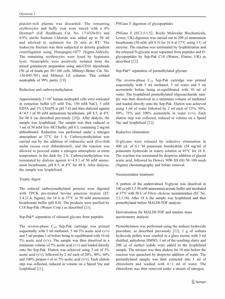
epitopes. These experiments were carried out on bothneutrophil samples, and returned highly consistent results.Data representative of these experiments is shown in Fig. 2(m/z 3141.1 and m/z 3766.6).
The most prominent peak in each sample is that of a bi-antennary, mono-sialylated, di-fucosylated structure of the
Glycoconj J

0
100
4500 4800 5100 5400 5700 6000Mass ( )m/z
%In
tens
ity
50
5111.7 5272.65125.7 5199.7 5212.6 5299.75285.8
5472.8
5373.7
5748.1
5459.8
5547.9 5560.9 5647.9 5660.9 5721.7 5734.0
5821.1 5834.5 5909.5 5922.9
5386.8
4763.5
4662.4
4576.4
4750.5
4836.6
5024.4
4850.5
4924.6
4937.6
5098.7
5111.7
5272.6
5125.75199.7
5212.6
5299.7
5285.8
5373.7
5459.85386.8
5472.8
5547.95560.9
5647.95660.9
5721.75734.0
5748.1
5821.15834.5
5909.55922.9
4763.54662.44576.4 4750.5 4836.6 5024.44850.5 4924.6 4937.6 5098.7
04500 4800 5100 5400 5700 6000Mass ( )m/z
%In
tens
ity
50
100
4761.4
4660.4
4574.4
4748.4
4834.3
5023.3
4848.3
4922.1
4935.35096.3
5109.3
5270.3
5122.35197.25210.3 5296.1
5283.2
5371.1
5458.25384.1
5471.1
5546.05559.1
5644.95657.9
5720.05733.0
5745.9
5819.95832.9
5906.95919.9
0
100
3000 3300 3600 3900 4200 4500Mass ( )m/z
%In
tens
ity
50
3315.8
3401.8
3414.8
3502.8
3588.8
3676.8
3775.7
3850.7
4037.6 4125.6
4211.5
4299.5
4400.4
4473.4
3951.6
x2x2x23040.9
3053.9
3139.8
3227.9
0
100
1500 1800 2100 2400 2700 3000Mass ( )m/z
%In
tens
ity
50
1580.2
1784.2
1836.2
1988.2 2192.2
2244.1
2396.1 2401.1 2418.1 2431.1
2592.1
2605.0
2693.1
2779.0
2867.0
2965.9
2156.1
0
100
1500 1800 2100 2400 2700 3000Mass ( )m/z
%In
tens
ity
50
1579.9
1784.0
1988.12192.2
2244.3
2396.3 2401.3 2418.4 2431.3
2590.1
2605.4
2693.5
2779.5
2867.6
2966.6
2156.2
0
100
3000 3300 3600 3900 4200 4500Mass ( )m/z
%In
tens
ity
50
3316.8
3402.8
3415.8
3503.9
3589.9 3678.0
3777.0
3852.1
4039.1 4127.2
4213.2
4301.3
4402.3
4475.4
3953.1
3041.7
3054.7
3140.7
3228.8
a
b
c
d
e
f
Fuc Man Gal Glc GalNAc GlcNAc NeuAcKey:
x1x1
x1x1x1
x1
x1x2
x1x1
x4x1
x3x2x1 x3x1
x3
x3x1x2
x2x1x1 x2x1
x2
x3x1
x2x2x1
x1x2
x1x1x2
x2x1
x2x2x1
x3x2x2
4761.44660.44574.4 4748.4 4834.3 5023.34848.3 4922.1 4935.3
5109.3 5270.35122.3 5197.2 5210.3 5296.15283.2
5096.3
5371.1 5458.25384.1
x4x1x1
x4x1x2
x4x2 x3x2x3
x3x3x1
x4x1x3
x4x2x1
x3x3x2
x4x1x4
x4x3x4x2x2
x5x1x2
x5x2 x4x1x5
x4x2x3
x4x3x1
x5x1x3
x5x2x2
x4x2x4
5471.1 5546.0 5559.1 5644.9 5657.9 5720.0 5733.0
5745.9 5819.9 5832.9 5906.9 5919.9
x4x3x2
x5x2x2
x4x3x3
x4x4x1
x5x1x5
x5x2x3
x5x1x4
x3x3x1
x2x3x4
x2x4x1
x3x2x4
x5x2x2
x3x2x2
x4x1x1
x4x1x2
x4x2 x3x2x3
x3x3x1
x4x1x3
x4x2x1
x3x3x2
x4x1x4
x4x3x4x2x2
x5x1x2
x5x2 x4x1x5
x4x2x3
x4x3x1
x5x1x3
x5x2x2
x4x2x4
x4x3x2
x5x2x2
x4x3x3
x4x4x1
x5x1x5
x5x2x3
x5x1x4
x3x3x1
x2x3x4
x2x4x1
x3x2x4
x5x2x2
x1x2
x1x1
x1x1x1
x1x2
x1x1x2
x2x1
x2x2x1
x2x1x1
x2x1x2
x3x1
x2x2x1
x3x2x1
x3x1x2
x2x2x2
x4x1
x3x1x3
x1 x1x1
Fig. 1 MALDI-TOF MS profiles of the permethylated N-linked glycansfrom human neutrophils. Major peaks are annotated with the relevantcarbohydrate structure shown in symbol form, according to the glycannomenclature adopted by the CFG (http://www.functionalglycomics.org/).
Neutrophil Sample 1 (US) is displayed in panels (a), (b) and (c).Neutrophil Sample 2 (UK) is displayed in panels (d), (e) and (f). Forcomplete annotation of the spectra see Table 1. All molecular ions arepresent in sodiated form ([M +Na]+)
Glycoconj J

empirical composition NeuAc1Fuc2Hex5HexNAc4 (m/z2779.0). Despite being a potential sialyl Lex structure,MSMS analysis demonstrates that all detectable isomersrepresented by the peak in fact carry the antennal fucose onthe non-sialylated arm (Fig. 2a). This is a theme thatpersists through the N- and O-glycan samples, highlightingthe apparent paucity of sialyl Lex amongst the neutrophilglycans. As exemplified by this component, whenever thereis an option to sialylate and fucosylate separate antennarather than place both substituents on a single antenna, the
Table 1 Compositional assignments of singly charged sodiatedmolecular ions, [M +Na]+, observed in MALDI-MS spectra ofpermethylated N-glycans from human neutrophils
Signal (m/z)Sample 1 (US)
Signal (m/z)Sample 2 (UK)
Molecular Assignments
1580.2 1579.9 Hex5HexNAc21591.2 1591.0 Fuc1Hex3HexNAc31765.2 1765.0 Fuc2Hex3HexNAc31784.2 1784.0 Hex6HexNAc21795.2 1795.0 Fuc1Hex4HexNAc31836.2 – Fuc1Hex3HexNAc41952.2 1952.1 NeuAc1Fuc1Hex3HexNAc31969.2 1969.1 Fuc2Hex4HexNAc31988.2 1988.1 Hex7HexNAc22040.1 2040.2 Fuc1Hex4HexNAc42082.1 2081.1 Fuc1Hex3HexNAc52156.1 2156.2 NeuAc1Fuc1Hex4HexNAc32192.2 2192.2 Hex8HexNAc22244.1 2244.3 Fuc1Hex5HexNAc42396.1 2396.3 Hex9HexNAc22401.1 2401.3 NeuAc1Fuc1Hex4HexNAc42418.1 2418.4 Fuc2Hex5HexNAc42431.1 2431.3 NeuAc1Hex5HexNAc42592.1 2592.4 Fuc3Hex5HexNAc42605.0 2605.4 NeuAc1Fuc1Hex5HexNAc42693.1 2693.5 Fuc1Hex6HexNAc52779.0 2779.5 NeuAc1Fuc2Hex5HexNAc42867.0 2867.6 Fuc2Hex6HexNAc52965.9 2966.6 NeuAc2Fuc1Hex5HexNAc43040.9 3041.7 Fuc3Hex6HexNAc53053.9 3054.7 NeuAc1Fuc1Hex6HexNAc53139.8 3140.7 NeuAc2Fuc2Hex5HexNAc43227.9 3228.8 NeuAc1Fuc2Hex6HexNAc53315.8 3316.8 Fuc2Hex7HexNAc63401.8 3402.8 NeuAc1Fuc3Hex6HexNAc53414.8 3415.8 NeuAc2Fuc1Hex6HexNAc53502.8 3503.9 NeuAc1Fuc1Hex7HexNAc63588.8 3589.9 NeuAc2Fuc2Hex6HexNAc53676.8 3678.0 NeuAc1Fuc2Hex7HexNAc63775.7 3777.0 NeuAc3Fuc1Hex6HexNAc53850.7 3852.1 NeuAc1Fuc3Hex7HexNAc63863.6 3865.0 NeuAc2Fuc1Hex7HexNAc63951.6 3953.1 NeuAc1Fuc1Hex8HexNAc74024.6 4026.2 NeuAc1Fuc4Hex7HexNAc64037.6 4039.1 NeuAc2Fuc2Hex7HexNAc64125.6 4127.2 NeuAc1Fuc2Hex8HexNAc74211.5 4213.2 NeuAc2Fuc3Hex7HexNAc64224.6 4226.2 NeuAc3Fuc1Hex7HexNAc64299.5 4301.3 NeuAc1Fuc3Hex8HexNAc74312.5 4314.3 NeuAc2Fuc1Hex8HexNAc74400.4 4402.3 NeuAc1Fuc1Hex9HexNAc84473.4 4475.4 NeuAc1Fuc4Hex8HexNAc74486.5 4488.3 NeuAc2Fuc2Hex8HexNAc74574.4 4576.4 NeuAc1Fuc2Hex9HexNAc84660.4 4662.4 NeuAc2Fuc3Hex8HexNAc74674.4 4675.4 NeuAc3Fuc1Hex8HexNAc74748.4 4750.5 NeuAc1Fuc3Hex9HexNAc84761.4 4763.5 NeuAc2Fuc1Hex9HexNAc8
Table 1 (continued)
Signal (m/z)Sample 1 (US)
Signal (m/z)Sample 2 (UK)
Molecular Assignments
4834.3 4836.6 NeuAc2Fuc4Hex8HexNAc74848.3 4850.5 NeuAc1Fuc1Hex10HexNAc94922.1 4924.6 NeuAc1Fuc4Hex9HexNAc84935.3 4937.6 NeuAc2Fuc2Hex9HexNAc85023.3 5024.6 NeuAc1Fuc2Hex10HexNAc95035.3 – NeuAc4Fuc1Hex8HexNAc75096.3 5098.7 NeuAc1Fuc5Hex9HexNAc85109.3 5111.7 NeuAc2Fuc3Hex9HexNAc85122.3 5125.7 NeuAc3Fuc1Hex9HexNAc85197.2 5199.7 NeuAc1Fuc3Hex10HexNAc95210.3 5212.6 NeuAc2Fuc1Hex10HexNAc95270.3 5272.6 NeuAc1Fuc6Hex9HexNAc85283.2 5285.8 NeuAc2Fuc4Hex9HexNAc85296.1 5299.7 NeuAc3Fuc2Hex9HexNAc85371.1 5373.7 NeuAc1Fuc4Hex10HexNAc95384.1 5386.8 NeuAc2Fuc2Hex10HexNAc95458.2 5459.8 NeuAc2Fuc5Hex9HexNAc85471.1 5472.8 NeuAc3Fuc3Hex9HexNAc85484.1 5486.8 NeuAc4Fuc1Hex9HexNAc85546.0 5547.9 NeuAc1Fuc5Hex10HexNAc95559.1 5560.9 NeuAc2Fuc3Hex10HexNAc95572.1 5573.8 NeuAc3Fuc1Hex10HexNAc95644.9 5647.9 NeuAc3Fuc4Hex9HexNAc85657.9 5660.9 NeuAc4Fuc2Hex9HexNAc85720.0 5721.7 NeuAc1Fuc6Hex10HexNAc95733.0 5734.0 NeuAc2Fuc4Hex10HexNAc95745.9 5748.1 NeuAc3Fuc2Hex10HexNAc95819.9 5821.1 NeuAc3Fuc5Hex9HexNAc85832.9 5834.5 NeuAc4Fuc3Hex9HexNAc85906.9 5909.5 NeuAc2Fuc5Hex10HexNAc95919.9 5922.9 NeuAc3Fuc3Hex10HexNAc95993.7 5996.0 NeuAc3Fuc6Hex9HexNAc86007.1 – NeuAc4Fuc4Hex9HexNAc86079.9 – NeuAc2Fuc6Hex10HexNAc96092.7 – NeuAc3Fuc4Hex10HexNAc96107.8 – NeuAc4Fuc2Hex10HexNAc96168.8 – NeuAc3Fuc7Hex9HexNAc86181.7 – NeuAc4Fuc5Hex9HexNAc86281.6 – NeuAc4Fuc3Hex10HexNAc96354.6 – NeuAc4Fuc6Hex9HexNAc86456.2 – NeuAc4Fuc4Hex10HexNAc96528.1 – NeuAc4Fuc7Hex9HexNAc8
Glycoconj J
former is observed. Thus, despite the composition Neu-Ac1Fuc2Hex5HexNAc4 being consistent with componentscarrying a sialyl Lex antennae, none were observed. Insteadthe NeuAc is located on an unsubstituted antenna as shownby fragment ions at m/z 1751.0, 1955.1 and 847.4, whilethe fucose residues are present on the chitobiose core (m/z474.1) and on Lex antennae (m/z 660.3, 2142.2 and1767.9).
The MS/MS analysis of the signals centred at m/z 3141.0(Fig. 2b), initially assigned as NeuAc2Fuc2Hex5HexNAc4,revealed an additional composition, namely Fuc1Hex7Hex-NAc6 which is only two mass units heavier than Neu-Ac2Fuc2Hex5HexNAc4 and therefore the isotopic clustersoverlap. The base peak of the spectrum at m/z 2766.8represents the loss of NeuAc from the sialylated compo-nent. The signals at m/z 474.2 (reducing end fucosylated
HexNAc) and m/z 1317.7 [FucHex3HexNAc2] are indica-tive of core fucosylation, while peaks at m/z 1021.5 and2141.2 establish the presence of a sialyl Lex antennae.Signals at m/z 847.4 and 2316.4 confirm that one of theantennae does not carry a fucose. Confirmation of the bi-and/or tri-antennary nature of the non-sialylated constitu-ents (as opposed to a tetra-antennary form of the samecomposition) comes from ions observed at m/z 935.4 and2230.5, representing loss of a single antenna consisting ofHex2HexNAc2. LacNAc extensions are also sequentiallylost, as demonstrated by signals at m/z 2680.3 and 2216.2.
PolyLacNAc containing N-glycans
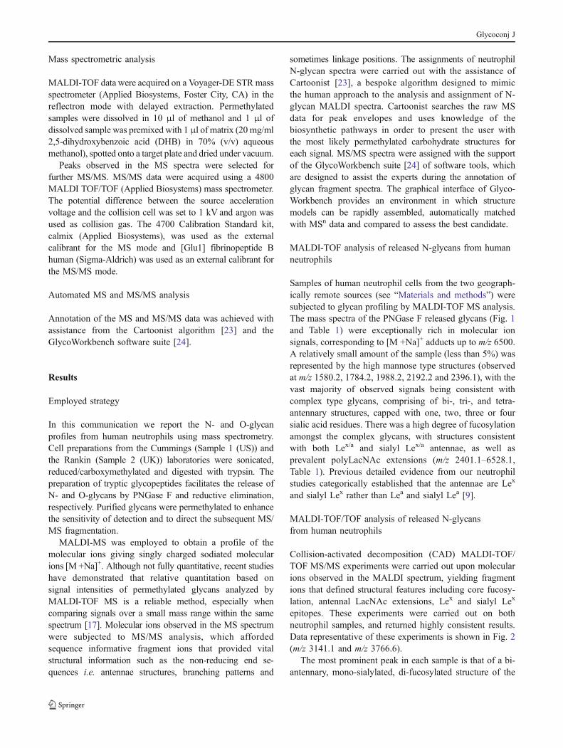
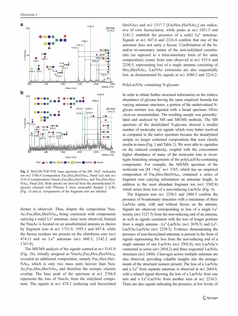
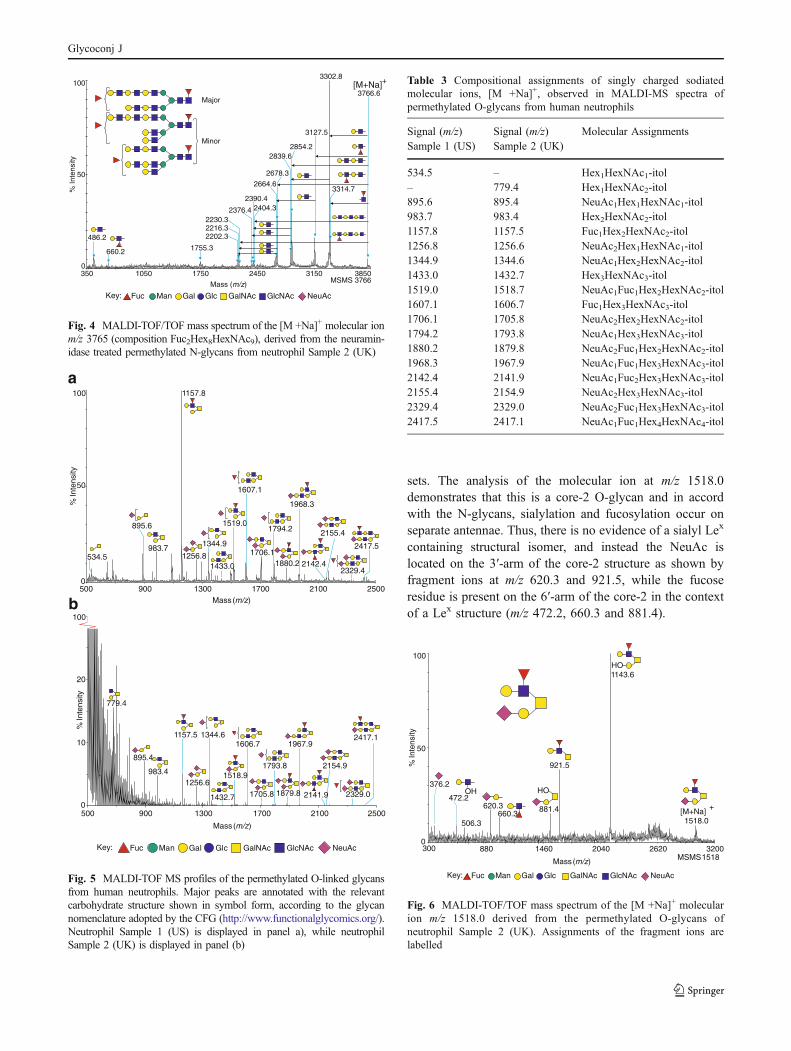
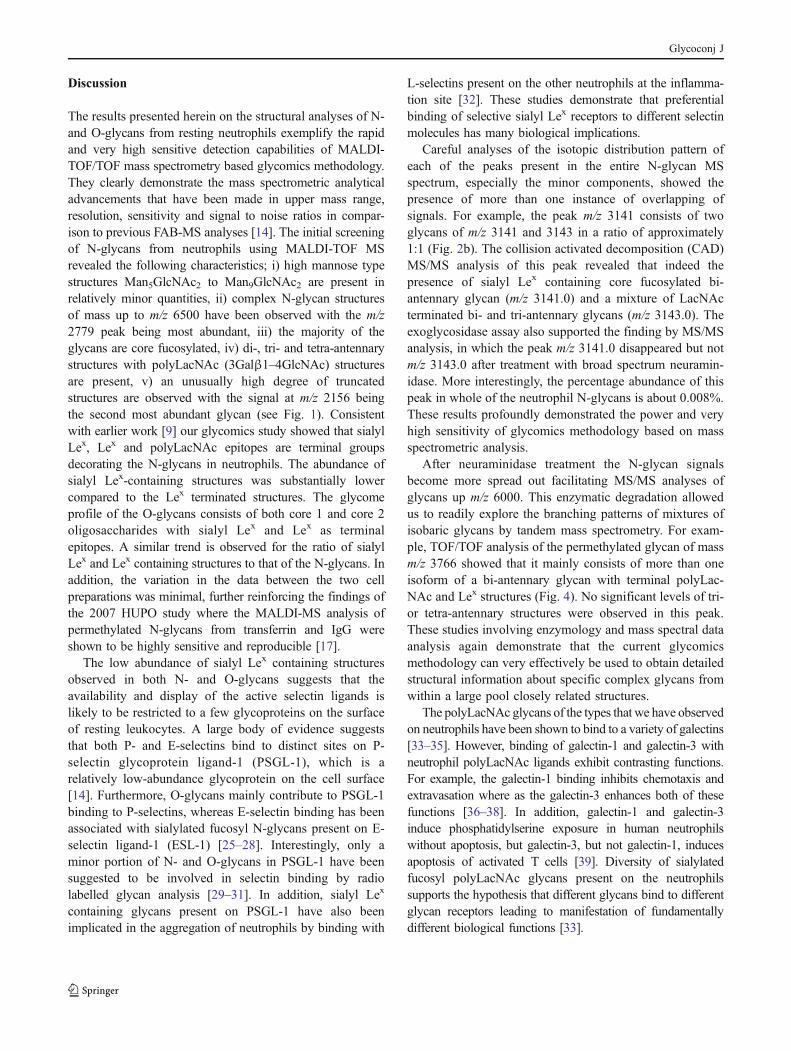
In order to obtain further structural information on the relativeabundance of glycans having the same empirical formula butvarying antennae structures, a portion of the underivatised N-glycan mixture was digested with a broad spectrum Vibriocholerae neuraminidase. The resulting sample was permethy-lated and analysed by MS and MS/MS methods. The MSspectrum of the desialylated N-glycans showed a smallernumber of molecular ion signals which were better resolvedas compared to the native spectrum because the desialylatedsample no longer contained compositions that were closelysimilar in mass (Fig. 3 and Table 2). We were able to capitaliseon the reduced complexity, coupled with the concomitanthigher abundance of many of the molecular ions to inves-tigate branching arrangements of the polyLacNAc-containingcomponents. For example, the MS/MS spectrum of themolecular ion [M +Na]+ m/z 3765, which has an empiricalcomposition of Fuc2Hex7HexNAc8, contained a series offragment ions carrying information on antennae length, inaddition to the most abundant fragment ion (m/z 3302.8)which arises from loss of a non-reducing LacNAc (Fig. 4).
The fragment ions m/z 2230.3 and 2404.3 confirm thepresence of bi-antennary structures with a maximum of threeLacNAc units, with and without fucose on the antenna.Signals are observed corresponding to loss of a single Lex
moiety (m/z 3127.5) from the non-reducing end of an antenna,as well as signals consistent with the loss of longer portionsfrom a single antenna—Lex-LacNAc (m/z 2678.3) and Lex-LacNAc-LacNAc (m/z 2230.3). Evidence demonstrating thepresence of non-fucosylated antennae is present in the form ofsignals representing the loss from the non-reducing end of asingle antenna of one LacNAc (m/z 3302.8), two LacNAc’sconnected in series (m/z 2854.2) and three sequential LacNAcstructures (m/z 2404). Cleavages across multiple antennae arealso observed, providing valuable insights into the arrange-ments of the structural isomers present. The loss of a LacNAcand a Lex from separate antennae is observed at m/z 2664.6,with a related signal showing the loss of a LacNAc from onearm and a Lex-LacNAc from another seen at m/z 2216.3.There are also signals indicating the presence, at low levels, of
0
100
300 880 1460 2040 2620 3200
Mass (m/z)
%In
tens
ity
50
MSMS 3142
847.4
1021.51317.7
486.1
646.1660.3
474.2
376.1
3141.0
2766.8
2680.3
2316.42142.2
2230.5
935.4
HO
HO
2216.2 [M+Na]+
Fuc Man Gal Glc GalNAc GlcNAc NeuAcKey:
a
bMass ( )m/z
MSMS 2779
0
100
200 730 1260 1790 2320 2850
%In
tens
ity
50
[M+Na]+
847.4 1317.7
660.3
474.2
376.2
2779.6
2574.1
2404.3
HO
HO
259.1
1113.4
HO
HO
2142.2
2328.8
1955.1
1751.01767.9
and
Fig. 2 MALDI-TOF/TOF mass spectrum of the [M +Na]+ molecularion m/z 3766.9 (composition Fuc2Hex8HexNAc9, Panel (a)) and m/z3141.0 (compositions NeuAc2Fuc2Hex5HexNAc4 and Fuc1Hex7Hex-NAc6, Panel (b)). Both spectra are derived from the permethylated N-glycans released with PNGase F from neutrophil Sample 2 (UK)(Fig. 1d and e). Assignments of the fragment ions are labelled
Glycoconj J

at least two tri-antennary isomers, with the loss of threeseparate non-reducing end LacNAc’s being observed at m/z2376 and the loss of two LacNAc antennae plus a Lex
antennal epitope seen at m/z 2202.3.
MALDI-TOF analysis of released O-glycans from humanneutrophils
O-glycans were chemically released by reductive elimina-tion and their permethyl derivatives were analysed byMALDI-TOFMS. The O-glycan profile (Fig. 5 and Table 3)demonstrates that the most abundant glycan species is afucosylated core-2 glycan (m/z 1157.8). There is extensivesialylation among the larger structures present, withdisialylated core 1 glycans being observed (m/z 1256.8)alongside the more prevalent mono- and di-sialylated core 2structures (m/z 1344.9 and 1706.1). The higher massregions contain fucosylated signals consistent with Lex
and sialyl Lex epitopes (m/z 2142.4) as well as polyLacNAcextensions (m/z 2329.4).
MALDI-TOF/TOF analysis of released O-glycansfrom human neutrophils
Exemplar data from these experiments are shown in Fig. 6.In similar fashion to the N-glycans, MSMS analysis wasconsistent across the two geographically remote sample
Table 2 Compositional assignments of singly charged sodiatedmolecular ions, [M+ Na]+, observed in MALDI-MS spectra ofpermethylated N-glycans after neuraminidase digestion
Signal m/z Molecular Assignments
1580.2 Hex5HexNAc21784.3 Hex6HexNAc21988.4 Hex7HexNAc22192.6 Hex8HexNAc22244.6 Fuc1Hex5HexNAc42396.7 Hex9HexNAc22418.7 Fuc2Hex5HexNAc42600.8 Hex10HexNAc22693.9 Fuc1Hex6HexNAc52868.0 Fuc2Hex6HexNAc53042.1 Fuc1Hex6HexNAc53143.2 Fuc1Hex7HexNAc63317.3 Fuc2Hex7HexNAc63491.4 Fuc3Hex7HexNAc63592.5 Fuc1Hex8HexNAc73665.5 Fuc4Hex7HexNAc63766.6 Fuc2Hex8HexNAc73940.7 Fuc3Hex8HexNAc74041.8 Fuc1Hex9HexNAc84114.8 Fuc4Hex8HexNAc74215.9 Fuc2Hex9HexNAc84288.9 Fuc5Hex8HexNAc74390.0 Fuc3Hex9HexNAc84491.0 Fuc1Hex10HexNAc94564.1 Fuc4Hex9HexNAc84665.1 Fuc2Hex10HexNAc94738.2 Fuc5Hex9HexNAc84840.2 Fuc3Hex10HexNAc94912.2 Fuc6Hex9HexNAc84940.2 Fuc1Hex11HexNAc105014.3 Fuc3Hex10HexNAc95115.3 Fuc2Hex11HexNAc105187.4 Fuc4Hex10HexNAc95288.7 Fuc3Hex11HexNAc105364.4 Fuc5Hex10HexNAc95465.2 Fuc4Hex11HexNAc105564.3 Fuc2Hex12HexNAc115636.5 Fuc5Hex11HexNAc105737.4 Fuc3Hex12HexNAc115914.6 Fuc4Hex12HexNAc116013.7 Fuc2Hex13HexNAc126085.8 Fuc4Hex12HexNAc116187.9 Fuc3Hex13HexNAc126361.3 Fuc4Hex13HexNAc126461.5 Fuc2Hex14HexNAc13
1580.1
1784.3
2600.9
2396.7
2192.6
1988.4
3143.2
3317.3
3592.5
3766.5
3491.4
3665.53042.1
3940.6
2244.6
2418.7
2693.9
2868.0
0
100
%In
tens
ity
50
1500 2000 2500 3000 3500 4000
Mass (m/z)
4912.2
4041.8
4564.1
6085.8
4215.8
4114.7
4390.0
4738.2
4840.2
5636.5
5737.4
6013.7
4665.1
4288.9
5564.3
5187.4
5465.2
5115.3
5364.4
5292.7
5914.65014.4
04000 4500 5000 5500 6000 6500
Mass ( )m/z
100
%In
tens
ity
50
6187.9
6361.3
6461.5
a
b
Fig. 3 MALDI-TOF profile of permethylated N-glycans afterneuraminidase digestion. Panel (a) shows the lower mass region andpanel (b) shows the higher mass region. All molecular ions are presentin sodiated form ([M +Na]+)
Glycoconj J
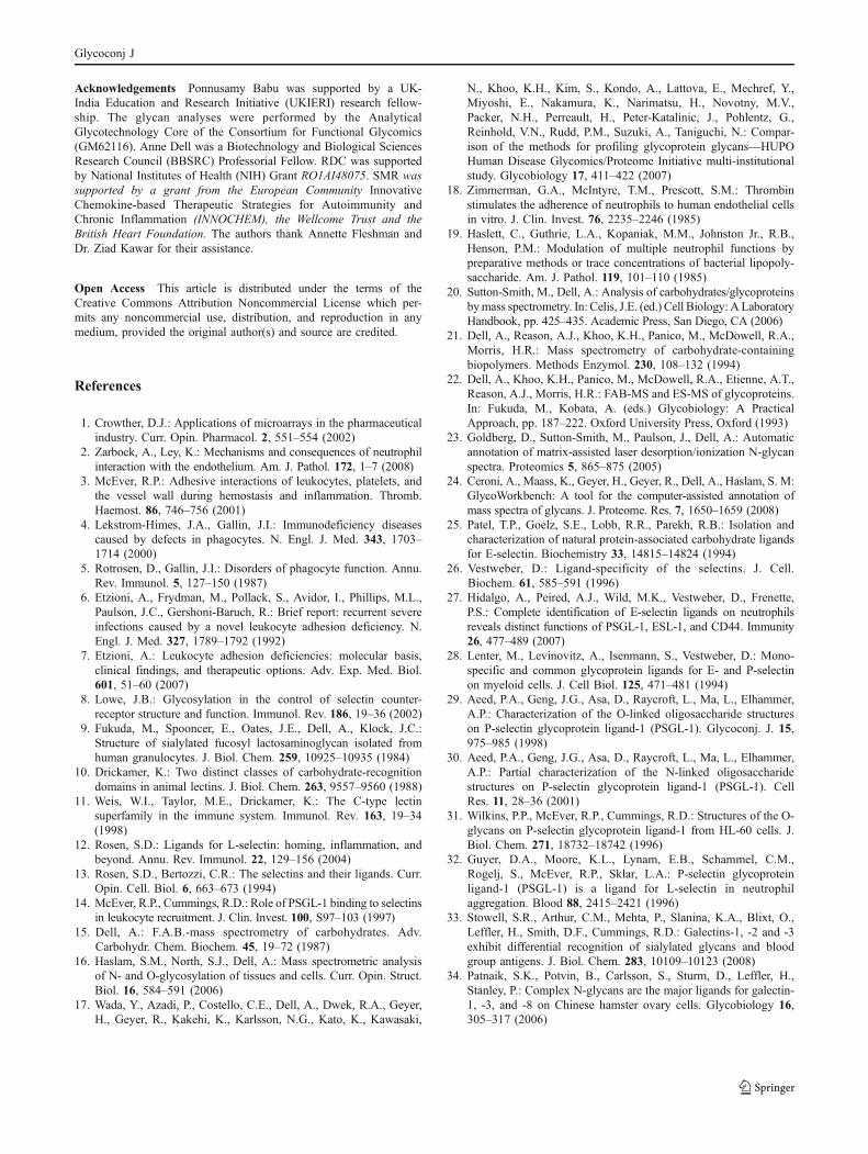
sets. The analysis of the molecular ion at m/z 1518.0demonstrates that this is a core-2 O-glycan and in accordwith the N-glycans, sialylation and fucosylation occur onseparate antennae. Thus, there is no evidence of a sialyl Lex
containing structural isomer, and instead the NeuAc islocated on the 3′-arm of the core-2 structure as shown byfragment ions at m/z 620.3 and 921.5, while the fucoseresidue is present on the 6′-arm of the core-2 in the contextof a Lex structure (m/z 472.2, 660.3 and 881.4).
a
b
0
100
%In
tens
ity
50
500 900 1300 1700 2100 2500Mass( )m/z
895.6
983.7
1157.8
1256.8
1344.9
1519.0
1706.1
1968.3
1433.0
1607.1
1880.2
2155.4
2329.4
2417.5
1794.2
2142.4534.5
0
%In
tens
ity
10
500 900 1300 1700 2100 2500
Mass( )m/z
20
100
779.4
895.4
1256.6
1344.6
983.4
1432.7
1518.9
1606.7
1705.8 1879.8
1967.9
2154.9
2329.0
2417.1
1793.8
2141.9
1157.5
Fuc Man Gal Glc GalNAc GlcNAc NeuAcKey:
Fig. 5 MALDI-TOF MS profiles of the permethylated O-linked glycansfrom human neutrophils. Major peaks are annotated with the relevantcarbohydrate structure shown in symbol form, according to the glycannomenclature adopted by the CFG (http://www.functionalglycomics.org/).Neutrophil Sample 1 (US) is displayed in panel a), while neutrophilSample 2 (UK) is displayed in panel (b)
Table 3 Compositional assignments of singly charged sodiatedmolecular ions, [M +Na]+, observed in MALDI-MS spectra ofpermethylated O-glycans from human neutrophils
Signal (m/z)Sample 1 (US)
Signal (m/z)Sample 2 (UK)
Molecular Assignments
534.5 – Hex1HexNAc1-itol– 779.4 Hex1HexNAc2-itol895.6 895.4 NeuAc1Hex1HexNAc1-itol983.7 983.4 Hex2HexNAc2-itol1157.8 1157.5 Fuc1Hex2HexNAc2-itol1256.8 1256.6 NeuAc2Hex1HexNAc1-itol1344.9 1344.6 NeuAc1Hex2HexNAc2-itol1433.0 1432.7 Hex3HexNAc3-itol1519.0 1518.7 NeuAc1Fuc1Hex2HexNAc2-itol1607.1 1606.7 Fuc1Hex3HexNAc3-itol1706.1 1705.8 NeuAc2Hex2HexNAc2-itol1794.2 1793.8 NeuAc1Hex3HexNAc3-itol1880.2 1879.8 NeuAc2Fuc1Hex2HexNAc2-itol1968.3 1967.9 NeuAc1Fuc1Hex3HexNAc3-itol2142.4 2141.9 NeuAc1Fuc2Hex3HexNAc3-itol2155.4 2154.9 NeuAc2Hex3HexNAc3-itol2329.4 2329.0 NeuAc2Fuc1Hex3HexNAc3-itol2417.5 2417.1 NeuAc1Fuc1Hex4HexNAc4-itol
0
100
300 880 1460 2040 2620 3200
Mass( )m/z
%In
tens
ity
50
MSMS1518
[M+Na] +660.3
620.3
376.2
506.3
472.2OH
881.4
921.5
1143.6
1518.0
HO
HO
Fuc Man Gal Glc GalNAc GlcNAc NeuAcKey:
Fig. 6 MALDI-TOF/TOF mass spectrum of the [M +Na]+ molecularion m/z 1518.0 derived from the permethylated O-glycans ofneutrophil Sample 2 (UK). Assignments of the fragment ions arelabelled
3766.6
0350 1050 1750 2450 3150 3850
Mass ( )m/z
100
%In
tens
ity
50
660.2
486.2
2664.6
2678.3
3302.8
2839.62854.2
3127.5
2216.3
1755.3
3314.7
2404.32376.4
2390.4
2230.3
2202.3
Major
Minor
[M+Na]+
MSMS 3766
Fuc Man Gal Glc GalNAc GlcNAc NeuAcKey:
Fig. 4 MALDI-TOF/TOF mass spectrum of the [M +Na]+ molecular ionm/z 3765 (composition Fuc2Hex8HexNAc9), derived from the neuramin-idase treated permethylated N-glycans from neutrophil Sample 2 (UK)
Glycoconj J
Discussion
The results presented herein on the structural analyses of N-and O-glycans from resting neutrophils exemplify the rapidand very high sensitive detection capabilities of MALDI-TOF/TOF mass spectrometry based glycomics methodology.They clearly demonstrate the mass spectrometric analyticaladvancements that have been made in upper mass range,resolution, sensitivity and signal to noise ratios in compar-ison to previous FAB-MS analyses [14]. The initial screeningof N-glycans from neutrophils using MALDI-TOF MSrevealed the following characteristics; i) high mannose typestructures Man5GlcNAc2 to Man9GlcNAc2 are present inrelatively minor quantities, ii) complex N-glycan structuresof mass up to m/z 6500 have been observed with the m/z2779 peak being most abundant, iii) the majority of theglycans are core fucosylated, iv) di-, tri- and tetra-antennarystructures with polyLacNAc (3Galβ1–4GlcNAc) structuresare present, v) an unusually high degree of truncatedstructures are observed with the signal at m/z 2156 beingthe second most abundant glycan (see Fig. 1). Consistentwith earlier work [9] our glycomics study showed that sialylLex, Lex and polyLacNAc epitopes are terminal groupsdecorating the N-glycans in neutrophils. The abundance ofsialyl Lex-containing structures was substantially lowercompared to the Lex terminated structures. The glycomeprofile of the O-glycans consists of both core 1 and core 2oligosaccharides with sialyl Lex and Lex as terminalepitopes. A similar trend is observed for the ratio of sialylLex and Lex containing structures to that of the N-glycans. Inaddition, the variation in the data between the two cellpreparations was minimal, further reinforcing the findings ofthe 2007 HUPO study where the MALDI-MS analysis ofpermethylated N-glycans from transferrin and IgG wereshown to be highly sensitive and reproducible [17].
The low abundance of sialyl Lex containing structuresobserved in both N- and O-glycans suggests that theavailability and display of the active selectin ligands islikely to be restricted to a few glycoproteins on the surfaceof resting leukocytes. A large body of evidence suggeststhat both P- and E-selectins bind to distinct sites on P-selectin glycoprotein ligand-1 (PSGL-1), which is arelatively low-abundance glycoprotein on the cell surface[14]. Furthermore, O-glycans mainly contribute to PSGL-1binding to P-selectins, whereas E-selectin binding has beenassociated with sialylated fucosyl N-glycans present on E-selectin ligand-1 (ESL-1) [25–28]. Interestingly, only aminor portion of N- and O-glycans in PSGL-1 have beensuggested to be involved in selectin binding by radiolabelled glycan analysis [29–31]. In addition, sialyl Lex
containing glycans present on PSGL-1 have also beenimplicated in the aggregation of neutrophils by binding with
L-selectins present on the other neutrophils at the inflamma-tion site [32]. These studies demonstrate that preferentialbinding of selective sialyl Lex receptors to different selectinmolecules has many biological implications.
Careful analyses of the isotopic distribution pattern ofeach of the peaks present in the entire N-glycan MSspectrum, especially the minor components, showed thepresence of more than one instance of overlapping ofsignals. For example, the peak m/z 3141 consists of twoglycans of m/z 3141 and 3143 in a ratio of approximately1:1 (Fig. 2b). The collision activated decomposition (CAD)MS/MS analysis of this peak revealed that indeed thepresence of sialyl Lex containing core fucosylated bi-antennary glycan (m/z 3141.0) and a mixture of LacNActerminated bi- and tri-antennary glycans (m/z 3143.0). Theexoglycosidase assay also supported the finding by MS/MSanalysis, in which the peak m/z 3141.0 disappeared but notm/z 3143.0 after treatment with broad spectrum neuramin-idase. More interestingly, the percentage abundance of thispeak in whole of the neutrophil N-glycans is about 0.008%.These results profoundly demonstrated the power and veryhigh sensitivity of glycomics methodology based on massspectrometric analysis.
After neuraminidase treatment the N-glycan signalsbecome more spread out facilitating MS/MS analyses ofglycans up m/z 6000. This enzymatic degradation allowedus to readily explore the branching patterns of mixtures ofisobaric glycans by tandem mass spectrometry. For exam-ple, TOF/TOF analysis of the permethylated glycan of massm/z 3766 showed that it mainly consists of more than oneisoform of a bi-antennary glycan with terminal polyLac-NAc and Lex structures (Fig. 4). No significant levels of tri-or tetra-antennary structures were observed in this peak.These studies involving enzymology and mass spectral dataanalysis again demonstrate that the current glycomicsmethodology can very effectively be used to obtain detailedstructural information about specific complex glycans fromwithin a large pool closely related structures.
The polyLacNAc glycans of the types that we have observedon neutrophils have been shown to bind to a variety of galectins[33–35]. However, binding of galectin-1 and galectin-3 withneutrophil polyLacNAc ligands exhibit contrasting functions.For example, the galectin-1 binding inhibits chemotaxis andextravasation where as the galectin-3 enhances both of thesefunctions [36–38]. In addition, galectin-1 and galectin-3induce phosphatidylserine exposure in human neutrophilswithout apoptosis, but galectin-3, but not galectin-1, inducesapoptosis of activated T cells [39]. Diversity of sialylatedfucosyl polyLacNAc glycans present on the neutrophilssupports the hypothesis that different glycans bind to differentglycan receptors leading to manifestation of fundamentallydifferent biological functions [33].
Glycoconj J
Acknowledgements Ponnusamy Babu was supported by a UK-India Education and Research Initiative (UKIERI) research fellow-ship. The glycan analyses were performed by the AnalyticalGlycotechnology Core of the Consortium for Functional Glycomics(GM62116). Anne Dell was a Biotechnology and Biological SciencesResearch Council (BBSRC) Professorial Fellow. RDC was supportedby National Institutes of Health (NIH) Grant RO1AI48075. SMR wassupported by a grant from the European Community InnovativeChemokine-based Therapeutic Strategies for Autoimmunity andChronic Inflammation (INNOCHEM), the Wellcome Trust and theBritish Heart Foundation. The authors thank Annette Fleshman andDr. Ziad Kawar for their assistance.
Open Access This article is distributed under the terms of theCreative Commons Attribution Noncommercial License which per-mits any noncommercial use, distribution, and reproduction in anymedium, provided the original author(s) and source are credited.
References
1. Crowther, D.J.: Applications of microarrays in the pharmaceuticalindustry. Curr. Opin. Pharmacol. 2, 551–554 (2002)
2. Zarbock, A., Ley, K.: Mechanisms and consequences of neutrophilinteraction with the endothelium. Am. J. Pathol. 172, 1–7 (2008)
3. McEver, R.P.: Adhesive interactions of leukocytes, platelets, andthe vessel wall during hemostasis and inflammation. Thromb.Haemost. 86, 746–756 (2001)
4. Lekstrom-Himes, J.A., Gallin, J.I.: Immunodeficiency diseasescaused by defects in phagocytes. N. Engl. J. Med. 343, 1703–1714 (2000)
5. Rotrosen, D., Gallin, J.I.: Disorders of phagocyte function. Annu.Rev. Immunol. 5, 127–150 (1987)
6. Etzioni, A., Frydman, M., Pollack, S., Avidor, I., Phillips, M.L.,Paulson, J.C., Gershoni-Baruch, R.: Brief report: recurrent severeinfections caused by a novel leukocyte adhesion deficiency. N.Engl. J. Med. 327, 1789–1792 (1992)
7. Etzioni, A.: Leukocyte adhesion deficiencies: molecular basis,clinical findings, and therapeutic options. Adv. Exp. Med. Biol.601, 51–60 (2007)
8. Lowe, J.B.: Glycosylation in the control of selectin counter-receptor structure and function. Immunol. Rev. 186, 19–36 (2002)
9. Fukuda, M., Spooncer, E., Oates, J.E., Dell, A., Klock, J.C.:Structure of sialylated fucosyl lactosaminoglycan isolated fromhuman granulocytes. J. Biol. Chem. 259, 10925–10935 (1984)
10. Drickamer, K.: Two distinct classes of carbohydrate-recognitiondomains in animal lectins. J. Biol. Chem. 263, 9557–9560 (1988)
11. Weis, W.I., Taylor, M.E., Drickamer, K.: The C-type lectinsuperfamily in the immune system. Immunol. Rev. 163, 19–34(1998)
12. Rosen, S.D.: Ligands for L-selectin: homing, inflammation, andbeyond. Annu. Rev. Immunol. 22, 129–156 (2004)
13. Rosen, S.D., Bertozzi, C.R.: The selectins and their ligands. Curr.Opin. Cell. Biol. 6, 663–673 (1994)
14. McEver, R.P., Cummings, R.D.: Role of PSGL-1 binding to selectinsin leukocyte recruitment. J. Clin. Invest. 100, S97–103 (1997)
15. Dell, A.: F.A.B.-mass spectrometry of carbohydrates. Adv.Carbohydr. Chem. Biochem. 45, 19–72 (1987)
16. Haslam, S.M., North, S.J., Dell, A.: Mass spectrometric analysisof N- and O-glycosylation of tissues and cells. Curr. Opin. Struct.Biol. 16, 584–591 (2006)
17. Wada, Y., Azadi, P., Costello, C.E., Dell, A., Dwek, R.A., Geyer,H., Geyer, R., Kakehi, K., Karlsson, N.G., Kato, K., Kawasaki,
N., Khoo, K.H., Kim, S., Kondo, A., Lattova, E., Mechref, Y.,Miyoshi, E., Nakamura, K., Narimatsu, H., Novotny, M.V.,Packer, N.H., Perreault, H., Peter-Katalinic, J., Pohlentz, G.,Reinhold, V.N., Rudd, P.M., Suzuki, A., Taniguchi, N.: Compar-ison of the methods for profiling glycoprotein glycans—HUPOHuman Disease Glycomics/Proteome Initiative multi-institutionalstudy. Glycobiology 17, 411–422 (2007)
18. Zimmerman, G.A., McIntyre, T.M., Prescott, S.M.: Thrombinstimulates the adherence of neutrophils to human endothelial cellsin vitro. J. Clin. Invest. 76, 2235–2246 (1985)
19. Haslett, C., Guthrie, L.A., Kopaniak, M.M., Johnston Jr., R.B.,Henson, P.M.: Modulation of multiple neutrophil functions bypreparative methods or trace concentrations of bacterial lipopoly-saccharide. Am. J. Pathol. 119, 101–110 (1985)
20. Sutton-Smith, M., Dell, A.: Analysis of carbohydrates/glycoproteinsbymass spectrometry. In: Celis, J.E. (ed.) Cell Biology: A LaboratoryHandbook, pp. 425–435. Academic Press, San Diego, CA (2006)
21. Dell, A., Reason, A.J., Khoo, K.H., Panico, M., McDowell, R.A.,Morris, H.R.: Mass spectrometry of carbohydrate-containingbiopolymers. Methods Enzymol. 230, 108–132 (1994)
22. Dell, A., Khoo, K.H., Panico, M., McDowell, R.A., Etienne, A.T.,Reason, A.J., Morris, H.R.: FAB-MS and ES-MS of glycoproteins.In: Fukuda, M., Kobata, A. (eds.) Glycobiology: A PracticalApproach, pp. 187–222. Oxford University Press, Oxford (1993)
23. Goldberg, D., Sutton-Smith, M., Paulson, J., Dell, A.: Automaticannotation of matrix-assisted laser desorption/ionization N-glycanspectra. Proteomics 5, 865–875 (2005)
24. Ceroni, A., Maass, K., Geyer, H., Geyer, R., Dell, A., Haslam, S. M:GlycoWorkbench: A tool for the computer-assisted annotation ofmass spectra of glycans. J. Proteome. Res. 7, 1650–1659 (2008)
25. Patel, T.P., Goelz, S.E., Lobb, R.R., Parekh, R.B.: Isolation andcharacterization of natural protein-associated carbohydrate ligandsfor E-selectin. Biochemistry 33, 14815–14824 (1994)
26. Vestweber, D.: Ligand-specificity of the selectins. J. Cell.Biochem. 61, 585–591 (1996)
27. Hidalgo, A., Peired, A.J., Wild, M.K., Vestweber, D., Frenette,P.S.: Complete identification of E-selectin ligands on neutrophilsreveals distinct functions of PSGL-1, ESL-1, and CD44. Immunity26, 477–489 (2007)
28. Lenter, M., Levinovitz, A., Isenmann, S., Vestweber, D.: Mono-specific and common glycoprotein ligands for E- and P-selectinon myeloid cells. J. Cell Biol. 125, 471–481 (1994)
29. Aeed, P.A., Geng, J.G., Asa, D., Raycroft, L., Ma, L., Elhammer,A.P.: Characterization of the O-linked oligosaccharide structureson P-selectin glycoprotein ligand-1 (PSGL-1). Glycoconj. J. 15,975–985 (1998)
30. Aeed, P.A., Geng, J.G., Asa, D., Raycroft, L., Ma, L., Elhammer,A.P.: Partial characterization of the N-linked oligosaccharidestructures on P-selectin glycoprotein ligand-1 (PSGL-1). CellRes. 11, 28–36 (2001)
31. Wilkins, P.P., McEver, R.P., Cummings, R.D.: Structures of the O-glycans on P-selectin glycoprotein ligand-1 from HL-60 cells. J.Biol. Chem. 271, 18732–18742 (1996)
32. Guyer, D.A., Moore, K.L., Lynam, E.B., Schammel, C.M.,Rogelj, S., McEver, R.P., Sklar, L.A.: P-selectin glycoproteinligand-1 (PSGL-1) is a ligand for L-selectin in neutrophilaggregation. Blood 88, 2415–2421 (1996)
33. Stowell, S.R., Arthur, C.M., Mehta, P., Slanina, K.A., Blixt, O.,Leffler, H., Smith, D.F., Cummings, R.D.: Galectins-1, -2 and -3exhibit differential recognition of sialylated glycans and bloodgroup antigens. J. Biol. Chem. 283, 10109–10123 (2008)
34. Patnaik, S.K., Potvin, B., Carlsson, S., Sturm, D., Leffler, H.,Stanley, P.: Complex N-glycans are the major ligands for galectin-1, -3, and -8 on Chinese hamster ovary cells. Glycobiology 16,305–317 (2006)
Glycoconj J
35. Leppanen, A., Stowell, S., Blixt, O., Cummings, R.D.: Dimericgalectin-1 binds with high affinity to alpha2,3-sialylated andnon-sialylated terminal N-acetyllactosamine units on surface-bound extended glycans. J. Biol. Chem. 280, 5549–5562 (2005)
36. La, M., Cao, T.V., Cerchiaro, G., Chilton, K., Hirabayashi, J.,Kasai, K., Oliani, S.M., Chernajovsky, Y., Perretti, M.: A novelbiological activity for galectin-1: inhibition of leukocyte-endothelialcell interactions in experimental inflammation. Am. J. Pathol. 163,1505–1515 (2003)
37. Sano, H., Hsu, D.K., Yu, L., Apgar, J.R., Kuwabara, I.,Yamanaka, T., Hirashima, M., Liu, F.T.: Human galectin-3 is a
novel chemoattractant for monocytes and macrophages. J.Immunol. 165, 2156–2164 (2000)
38. Hsu, D.K., Yang, R.Y., Pan, Z., Yu, L., Salomon, D.R.,Fung-Leung, W.P., Liu, F.T.: Targeted disruption of thegalectin-3 gene results in attenuated peritoneal inflammatoryresponses. Am. J. Pathol. 156, 1073–1083 (2000)
39. Stowell, S.R., Qian, Y., Karmakar, S., Koyama, N.S., Dias-Baruffi, M., Leffler, H., McEver, R.P., Cummings, R.D.: Differ-ential roles of galectin-1 and galectin-3 in regulating leukocyteviability and cytokine secretion. J. Immunol. 180, 3091–3102(2008)
Glycoconj J
Copyright © 2022 FDOKUMEN