Neutrophil Elastase Activity Imaging - HAL-CNRS
14
HAL Id: hal-02472427 https://hal-cnrs.archives-ouvertes.fr/hal-02472427 Submitted on 10 Feb 2020 HAL is a multi-disciplinary open access archive for the deposit and dissemination of sci- entific research documents, whether they are pub- lished or not. The documents may come from teaching and research institutions in France or abroad, or from public or private research centers. L’archive ouverte pluridisciplinaire HAL, est destinée au dépôt et à la diffusion de documents scientifiques de niveau recherche, publiés ou non, émanant des établissements d’enseignement et de recherche français ou étrangers, des laboratoires publics ou privés. Neutrophil Elastase Activity Imaging: Recent Approaches in the Design and Applications of Activity-Based Probes and Substrate-Based Probes Natacha Jugniot, Pierre Voisin, Abderrazzak Bentaher, Philippe Mellet To cite this version: Natacha Jugniot, Pierre Voisin, Abderrazzak Bentaher, Philippe Mellet. Neutrophil Elastase Ac- tivity Imaging: Recent Approaches in the Design and Applications of Activity-Based Probes and Substrate-Based Probes. Contrast Media and Molecular Imaging, Wiley, 2019, 2019, pp.7417192. 10.1155/2019/7417192. hal-02472427
-
Upload
khangminh22 -
Category
Documents
-
view
1 -
download
0
Transcript of Neutrophil Elastase Activity Imaging - HAL-CNRS
HAL Id: hal-02472427https://hal-cnrs.archives-ouvertes.fr/hal-02472427
Submitted on 10 Feb 2020
HAL is a multi-disciplinary open accessarchive for the deposit and dissemination of sci-entific research documents, whether they are pub-lished or not. The documents may come fromteaching and research institutions in France orabroad, or from public or private research centers.
L’archive ouverte pluridisciplinaire HAL, estdestinée au dépôt et à la diffusion de documentsscientifiques de niveau recherche, publiés ou non,émanant des établissements d’enseignement et derecherche français ou étrangers, des laboratoirespublics ou privés.
Neutrophil Elastase Activity Imaging: RecentApproaches in the Design and Applications of
Activity-Based Probes and Substrate-Based ProbesNatacha Jugniot, Pierre Voisin, Abderrazzak Bentaher, Philippe Mellet
To cite this version:Natacha Jugniot, Pierre Voisin, Abderrazzak Bentaher, Philippe Mellet. Neutrophil Elastase Ac-tivity Imaging: Recent Approaches in the Design and Applications of Activity-Based Probes andSubstrate-Based Probes. Contrast Media and Molecular Imaging, Wiley, 2019, 2019, pp.7417192.�10.1155/2019/7417192�. �hal-02472427�
Review ArticleNeutrophil Elastase Activity Imaging: Recent Approaches in theDesign and Applications of Activity-Based Probes andSubstrate-Based Probes
Natacha Jugniot,1 Pierre Voisin,1 Abderrazzak Bentaher,3 and Philippe Mellet 1,2
1Centre de Resonance Magnetique des Systemes Biologiques, UMR5536, CNRS, Universite de Bordeaux, 33076 Bordeaux, France2INSERM, 33076 Bordeaux Cedex, France3Equipe “In�ammation et Immunite de l’Epithelium Respiratoire”—EA7426 Faculte de Medecine Lyon Sud,69495 Pierre Benite, France
Correspondence should be addressed to Philippe Mellet; [email protected]
Received 28 March 2019; Accepted 19 May 2019; Published 12 June 2019
Guest Editor: Xiang-Guo Li
Copyright © 2019 Natacha Jugniot et al. �is is an open access article distributed under the Creative Commons AttributionLicense, which permits unrestricted use, distribution, and reproduction in any medium, provided the original work isproperly cited.
�e last few decades of protease research has con�rmed that a number of important biological processes are strictly dependent onproteolysis. Neutrophil elastase (NE) is a critical protease in immune response and host defense mechanisms in both physiologicaland disease-associated conditions. Particularly, NE has been identi�ed as a promising biomarker for early diagnosis of lungin�ammation. Recent studies have shown an increasing interest in developing methods for NE activity imaging both in vitro andin vivo. Unlike anatomical imaging modalities, functional molecular imaging, including enzymatic activities, enables diseasedetection at a very early stage and thus constitutes a much more accurate approach. When combined with advanced imagingtechnologies, opportunities arise for measuring imbalanced proteolytic activities with unprecedented details. Such technologiesconsist in building the highest resolved and sensitive instruments as well as the most speci�c probes based either on peptidesubstrates or on covalent inhibitors. �is review outlines strengths and weaknesses of these technologies and discuss theirapplications to investigate NE activity as biomarker of pulmonary in�ammatory diseases by imaging.
1. Introduction
Degradome analysis indicates that protease and proteaseinhibitor genes represent more than 2% of total genes inhuman genome [1]. Proteases regulate a variety of physio-logical processes critical for life [2] including regulation ofmaturation, localization, activity and recycling of manyproteins, modulation of protein-protein interactions, pro-cessing in signalization, transduction, and ampli�cation ofmolecular signals. Proteases in�uence DNA replication andtranscription, cell proliferation, migration, and di�erentia-tion, tissue remodeling, angiogenesis, wound repair, bloodcoagulation, digestion, ovulation, in�ammation, necrosis,and apoptosis and immune response as well as pathogenclearance [3]. Accordingly, proteases are a major focus ofattention for the pharmaceutical industry as potential drug
targets or as diagnostic and prognostic biomarkers [4]. Sincethe function of proteases is to cleave proteins and peptides inresponse to biological, chemical, or physical stimuli, theiraction must be carefully orchestrated and strictly controlledfor homeostasis purpose. �erefore, their actions are reg-ulated at multiple levels [5]. Proteases are traditionallysynthesized as inactive enzymes called zymogens that re-quire activation process. �ey consist in subunit multi-merization. For instance, dimerization of the HumanImmunode�ciency Virus type 1 (HIV-1) protease subunits isan essential process for the acquisition of proteolytic activity,which plays a critical role in the maturation and replicationof the virus [6]. Another way of regulation consists inproteolytic cleavage of precursors, as pancreatic serineproteases, activated for food digestion only when they reachthe duodenum [7]. Others mechanisms consist either in
HindawiContrast Media & Molecular ImagingVolume 2019, Article ID 7417192, 12 pageshttps://doi.org/10.1155/2019/7417192
blocking active proteases by endogenous inhibitors or intrapping them in a dedicated compartment such as mito-chondria, specific apical membranes, and lysosomes, inwhich acidic pH participates also to enzyme activity con-tainment. Particularly, mature protease neutrophil elastase(NE) is stored in specific neutrophilic granules in an activeform that should be inactivated by inhibitors upon its ex-tracellular release in the setting of inflammation or infection[8]. ,erefore, if not correctly regulated, NE activity has theability to damage host tissues leading to the development ofpathologies as pointed below. Accordingly, noninvasivemethods allowing direct and reliable monitoring of proteaseactivity in the context of complex biological samples or invivo especially in diseased situations are greatly needed for adiagnostic purpose. In this context, this review will focus onrecent advances in NE activity detection, quantification, andapplications both in vitro and in vivo.
1.1. Neutrophil Elastase: A Double-Edged Sword. NE (EC3.4.21.37) is a 29 kDa serine protease of chymotrypsin familystored in azurophilic granules of polymorphonuclear neu-trophils [9, 10] and released during neutrophil de-granulation [11]. NE plays a relatively important role inneutrophil-mediated bacterial killing, and a cidal mecha-nism of the enzyme against Gram-negative bacteria has beenelucidated [12, 13]. Furthermore, not only NE kills directlyinvading pathogens, but the enzyme fine tunes host in-flammatory response for better pathogen eradication and itsassociated inflammation [14]. Normal inflammatory re-sponse triggers recruitment and activation of neutrophilstoward the inflammation site. Neutrophil extracellular de-granulation occurs and an overwhelming concentration ofNE molecules over their endogenous inhibitors takes place.Protease activity is yet restricted in space and time by in-hibitors in the tissue. Paradoxically, persistent or excessiveinflammation as a result of prolonged exposure to stimulushas been found to be detrimental. NE with exacerbatedelastolytic activity was shown to be one of the main lungdestructive actors. Indeed, its large repertoire of substrates,particularly, elastin favorizes potent proteolysis [15]. NEactivity can result in extensive lung tissue damages poten-tially leading to organ failure and death making in-flammatory diseases a major health concern worldwide aswell as an important economic burden [16]. Clinical studieshave evidenced how an elevated concentration of NE cor-relates to acute lung injury [17], development of cystic fi-brosis (CF) symptoms [18] as well as Chronic obstructivepulmonary disorders (stable and/or exacerbated COPD)[19], and bronchiectasis [20]. Moreover, it has been pos-tulated that NE can contribute to the progress of lung cancer[21]. ,is correlation between NE activity level and diseaseprogress has prompted researchers to investigate the use ofNE as biomarker to diagnose and monitor pathologicalinflammations.
1.2. Pivotal Role of NE/Anti-NE Balance Status Imaging.A standard approach for looking at proteases in inflamedsituations is the analysis of their transcript levels. Because
of the posttranslational modifications, monitoring proteaseactivity directly is more reliable to translate its roles inbiological events. Another commonly used method for thequantification of NE is the immunodiagnostic from bi-ological fluid samples. Antibody-related techniques yieldinformation on total protease amount but lack the ability todifferentiate between active and inactive enzyme forms.Methods to overcome such limitations tried to combineclassical ELISA with the application of active-site in-hibitors. A colorimetric active site-specific immunoassay(CASSIA) was described [22] for several serine proteaseswith arginyl specificity. Preferred protocols consist in usingavidin capture of a biotinylated peptidyl arginyl chlor-omethyl ketone (CMK) and a specific antiprotease anti-body recognition revealed by an enzymatic amplificationstep. In the case of a chymotrypsin-like specificity, apeptidyl inhibitor with a hydrophobic P1 amino acid side-chain can be used. Based on the CASSIA technique, ca-thepsin G activity was successfully detected [23]. It is thuslargely conceivable that analog probes can be developed fora neutrophil-like activity detection. Nevertheless, a diffi-culty lies in the inescapable capture competition betweenprotease-coupled probes and the uncoupled probes. ,us,to make sure that the enzyme activity measurement is notunderestimated, a more complex assay is needed. It canconsist in a preliminary assay in which uncoupled probewould be removed or in an increase of coated avidinconcentration. Another method, termed ABRA-ELISA(Activity-Based probe RAtiometric-Enzyme Linked Im-munoSorbent Assay), was found particularly relevant onserine protease kallikreins for ovarian cancer diagnosis andcould be applied to NE [24]. ,is strategy consists incombining the high throughput and high sensitivity of anELISA-based detection with the advantage of an active-siteinhibitor labeling. ,e proportion of the active form rel-ative to the total concentration of the enzymatic biomarker(regular ELISA) would then be easily monitored in samplesand may therefore serves as a novel diagnostic tool toquantify the active form in pathophysiological settings.However, these assays have limitations as the requirementfor enzyme immobilization on a solid surface can result inits improper structural orientation or decreased reactivitywith the target. It also needs collection of biologicalsamples, requiring therefore an invasive intervention.,us,methods for in vivo monitoring and imaging of NE activityin real time are studied.
Clinical symptoms are often undetectable at early stageof a disease, making diagnosis approach even more chal-lenging. Nowadays, no reliable clinical methods exist toimage deleterious NE proteolytic activity, which is againconvincingly documented as a culprit in tissue destructivediseases. To address this concern, the field of functionalmolecular imaging represents an attractive and relevanttool [25]. ,e information quality provided is highly de-pendent on (a) the choice of the biomarker, (b) thephysical, biochemical and pharmacological characteristicsof the probe, and (c) the imaging modality [26] to char-acterize NE imbalance activity, especially during diseaseinitiation phase.
2 Contrast Media & Molecular Imaging
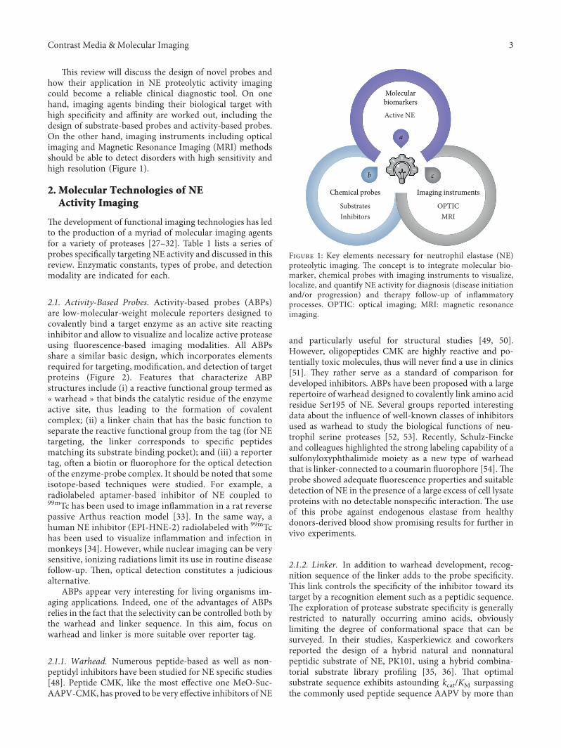
,is review will discuss the design of novel probes andhow their application in NE proteolytic activity imagingcould become a reliable clinical diagnostic tool. On onehand, imaging agents binding their biological target withhigh specificity and affinity are worked out, including thedesign of substrate-based probes and activity-based probes.On the other hand, imaging instruments including opticalimaging and Magnetic Resonance Imaging (MRI) methodsshould be able to detect disorders with high sensitivity andhigh resolution (Figure 1).
2. Molecular Technologies of NEActivity Imaging
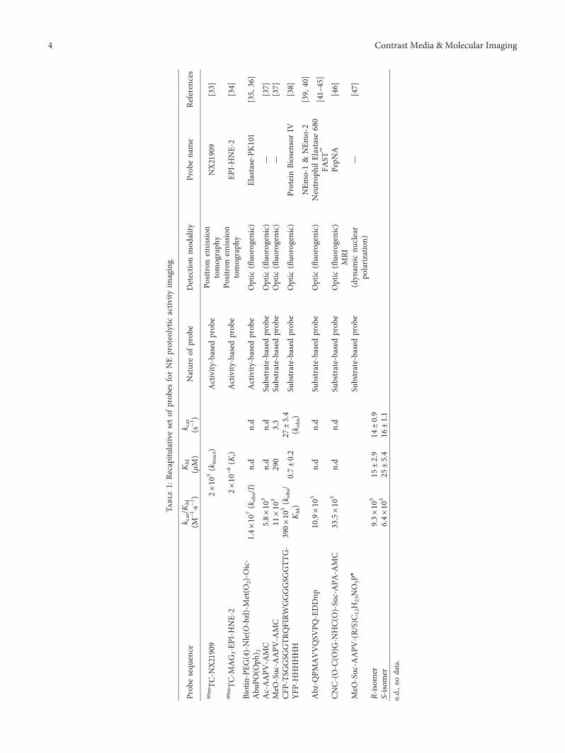
,e development of functional imaging technologies has ledto the production of a myriad of molecular imaging agentsfor a variety of proteases [27–32]. Table 1 lists a series ofprobes specifically targeting NE activity and discussed in thisreview. Enzymatic constants, types of probe, and detectionmodality are indicated for each.
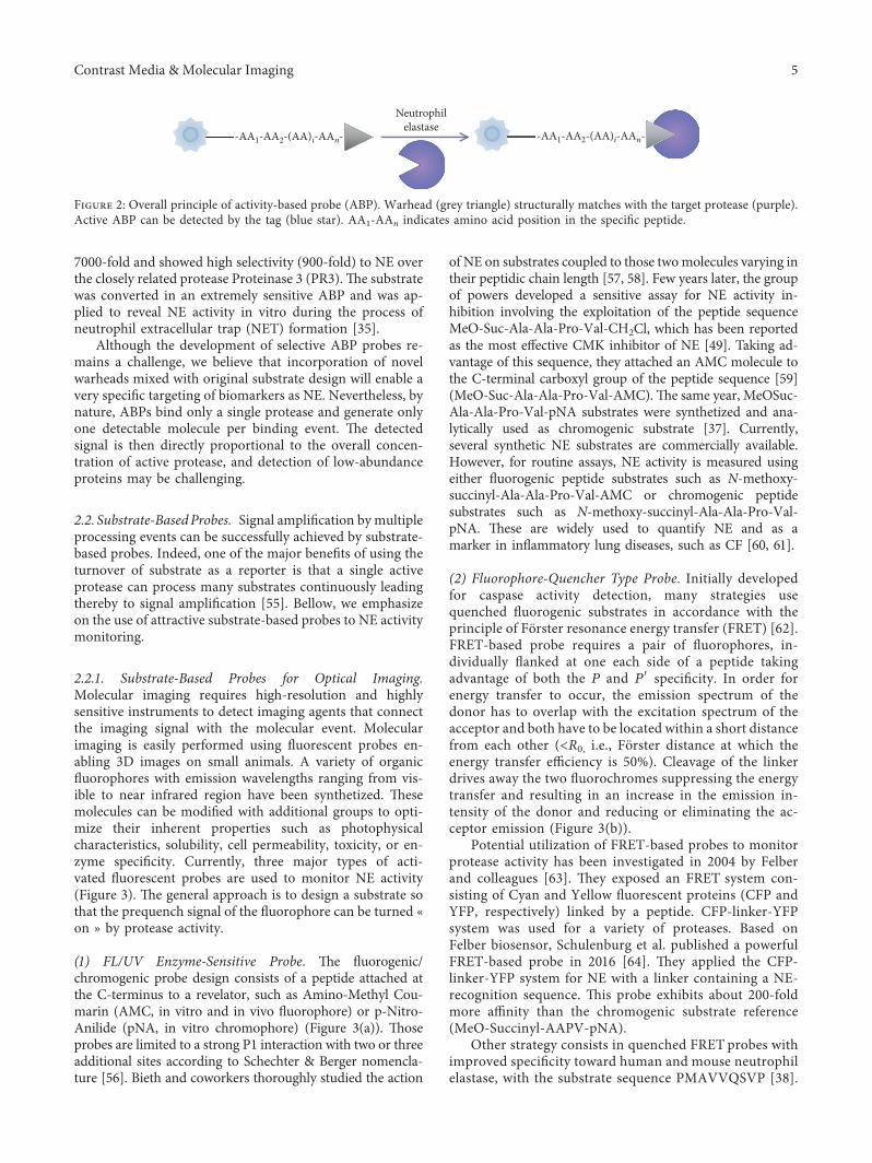
2.1. Activity-Based Probes. Activity-based probes (ABPs)are low-molecular-weight molecule reporters designed tocovalently bind a target enzyme as an active site reactinginhibitor and allow to visualize and localize active proteaseusing fluorescence-based imaging modalities. All ABPsshare a similar basic design, which incorporates elementsrequired for targeting, modification, and detection of targetproteins (Figure 2). Features that characterize ABPstructures include (i) a reactive functional group termed as« warhead » that binds the catalytic residue of the enzymeactive site, thus leading to the formation of covalentcomplex; (ii) a linker chain that has the basic function toseparate the reactive functional group from the tag (for NEtargeting, the linker corresponds to specific peptidesmatching its substrate binding pocket); and (iii) a reportertag, often a biotin or fluorophore for the optical detectionof the enzyme-probe complex. It should be noted that someisotope-based techniques were studied. For example, aradiolabeled aptamer-based inhibitor of NE coupled to99mTc has been used to image inflammation in a rat reversepassive Arthus reaction model [33]. In the same way, ahuman NE inhibitor (EPI-HNE-2) radiolabeled with 99mTchas been used to visualize inflammation and infection inmonkeys [34]. However, while nuclear imaging can be verysensitive, ionizing radiations limit its use in routine diseasefollow-up. ,en, optical detection constitutes a judiciousalternative.
ABPs appear very interesting for living organisms im-aging applications. Indeed, one of the advantages of ABPsrelies in the fact that the selectivity can be controlled both bythe warhead and linker sequence. In this aim, focus onwarhead and linker is more suitable over reporter tag.
2.1.1. Warhead. Numerous peptide-based as well as non-peptidyl inhibitors have been studied for NE specific studies[48]. Peptide CMK, like the most effective one MeO-Suc-AAPV-CMK, has proved to be very effective inhibitors of NE
and particularly useful for structural studies [49, 50].However, oligopeptides CMK are highly reactive and po-tentially toxic molecules, thus will never find a use in clinics[51]. ,ey rather serve as a standard of comparison fordeveloped inhibitors. ABPs have been proposed with a largerepertoire of warhead designed to covalently link amino acidresidue Ser195 of NE. Several groups reported interestingdata about the influence of well-known classes of inhibitorsused as warhead to study the biological functions of neu-trophil serine proteases [52, 53]. Recently, Schulz-Finckeand colleagues highlighted the strong labeling capability of asulfonyloxyphthalimide moiety as a new type of warheadthat is linker-connected to a coumarin fluorophore [54].,eprobe showed adequate fluorescence properties and suitabledetection of NE in the presence of a large excess of cell lysateproteins with no detectable nonspecific interaction. ,e useof this probe against endogenous elastase from healthydonors-derived blood show promising results for further invivo experiments.
2.1.2. Linker. In addition to warhead development, recog-nition sequence of the linker adds to the probe specificity.,is link controls the specificity of the inhibitor toward itstarget by a recognition element such as a peptidic sequence.,e exploration of protease substrate specificity is generallyrestricted to naturally occurring amino acids, obviouslylimiting the degree of conformational space that can besurveyed. In their studies, Kasperkiewicz and coworkersreported the design of a hybrid natural and nonnaturalpeptidic substrate of NE, PK101, using a hybrid combina-torial substrate library profiling [35, 36]. ,at optimalsubstrate sequence exhibits astounding kcat/KM surpassingthe commonly used peptide sequence AAPV by more than
Chemical probes
Molecularbiomarkers
cb
SubstratesInhibitors
OPTICMRI
Imaging instruments
a
Active NE
Figure 1: Key elements necessary for neutrophil elastase (NE)proteolytic imaging. ,e concept is to integrate molecular bio-marker, chemical probes with imaging instruments to visualize,localize, and quantify NE activity for diagnosis (disease initiationand/or progression) and therapy follow-up of inflammatoryprocesses. OPTIC: optical imaging; MRI: magnetic resonanceimaging.
Contrast Media & Molecular Imaging 3
Tabl
e1:
Recapitulativ
eseto
fprobesforNEproteolytic
activ
ityim
aging.
Prob
esequ
ence
k cat/K
M(M−1 ·s−
1 )KM
(μM)
k cat
(s−1 )
Natureof
prob
eDetectio
nmod
ality
Prob
ename
References
99mTC
-NX21909
2×10
5(k
inact)
Activity
-based
prob
ePo
sitronem
ission
tomograph
yNX21909
[33]
99mTC
-MAG3-EP
I-HNE-2
2×10⁻⁶(K
i)Activity
-based
prob
ePo
sitronem
ission
tomograph
yEP
I-HNE-2
[34]
Biotin-PEG
(4)-Nle(O
-bzl)-Met(O
2)-O
ic-
Abu
PO(O
ph) 2
1.4
×10
7(k
obs/I)
n.d
n.d
Activity
-based
prob
eOptic
(fluo
rogenic)
Elastase-PK101
[35,
36]
Ac-AAPV
-AMC
5.8
×10
3n.d
n.d
Substrate-basedprob
eOptic
(fluo
rogenic)
—[37]
MeO
-Suc-A
APV
-AMC
11×10
3290
3.3
Substrate-basedprob
eOptic
(fluo
rogenic)
—[37]
CFP
-TSG
GSG
GTR
QFIRW
GGGGSG
GTT
G-
YFP-HHHHHH
390
×10
5(k
obs/
KM)
0.7±0.2
27±5.4
(kob
s)Su
bstrate-basedprob
eOptic
(fluo
rogenic)
ProteinBiosensorIV
[38]
Abz-Q
PMAVVQSV
PQ-EDDnp
10.9
×10
5n.d
n.d
Substrate-basedprob
eOptic
(fluo
rogenic)
NEm
o-1&
NEm
o-2
[39,
40]
Neutrop
hilE
lastase680
FAST™
[41–
45]
CNC-(O-C
(O)G
-NHC(O
)-Su
c-APA
-AMC
33.5
×10
5n.d
n.d
Substrate-basedprob
eOptic
(fluo
rogenic)
PepN
A[46]
MeO
-Suc-A
APV
-(R/S)C12H
23NO5P▪
Substrate-basedprob
eMRI
(dyn
amic
nuclear
polarizatio
n)—
[47]
R-iso
mer
9.3
×10
515±2.9
14±0.9
S-iso
mer
6.4
×10
525±5.4
16±1.1
n.d.,n
odata.
4 Contrast Media & Molecular Imaging
7000-fold and showed high selectivity (900-fold) to NE overthe closely related protease Proteinase 3 (PR3).,e substratewas converted in an extremely sensitive ABP and was ap-plied to reveal NE activity in vitro during the process ofneutrophil extracellular trap (NET) formation [35].
Although the development of selective ABP probes re-mains a challenge, we believe that incorporation of novelwarheads mixed with original substrate design will enable avery specific targeting of biomarkers as NE. Nevertheless, bynature, ABPs bind only a single protease and generate onlyone detectable molecule per binding event. ,e detectedsignal is then directly proportional to the overall concen-tration of active protease, and detection of low-abundanceproteins may be challenging.
2.2. Substrate-BasedProbes. Signal amplification bymultipleprocessing events can be successfully achieved by substrate-based probes. Indeed, one of the major benefits of using theturnover of substrate as a reporter is that a single activeprotease can process many substrates continuously leadingthereby to signal amplification [55]. Bellow, we emphasizeon the use of attractive substrate-based probes to NE activitymonitoring.
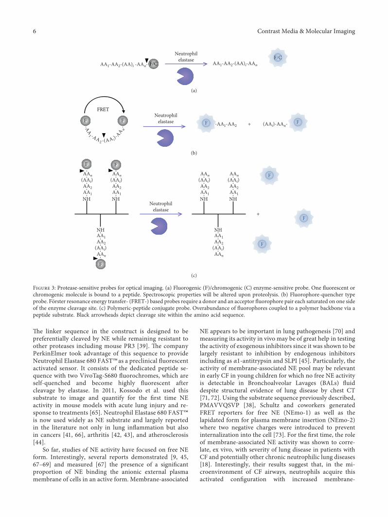
2.2.1. Substrate-Based Probes for Optical Imaging.Molecular imaging requires high-resolution and highlysensitive instruments to detect imaging agents that connectthe imaging signal with the molecular event. Molecularimaging is easily performed using fluorescent probes en-abling 3D images on small animals. A variety of organicfluorophores with emission wavelengths ranging from vis-ible to near infrared region have been synthetized. ,esemolecules can be modified with additional groups to opti-mize their inherent properties such as photophysicalcharacteristics, solubility, cell permeability, toxicity, or en-zyme specificity. Currently, three major types of acti-vated fluorescent probes are used to monitor NE activity(Figure 3). ,e general approach is to design a substrate sothat the prequench signal of the fluorophore can be turned «on » by protease activity.
(1) FL/UV Enzyme-Sensitive Probe. ,e fluorogenic/chromogenic probe design consists of a peptide attached atthe C-terminus to a revelator, such as Amino-Methyl Cou-marin (AMC, in vitro and in vivo fluorophore) or p-Nitro-Anilide (pNA, in vitro chromophore) (Figure 3(a)). ,oseprobes are limited to a strong P1 interaction with two or threeadditional sites according to Schechter & Berger nomencla-ture [56]. Bieth and coworkers thoroughly studied the action
of NE on substrates coupled to those twomolecules varying intheir peptidic chain length [57, 58]. Few years later, the groupof powers developed a sensitive assay for NE activity in-hibition involving the exploitation of the peptide sequenceMeO-Suc-Ala-Ala-Pro-Val-CH2Cl, which has been reportedas the most effective CMK inhibitor of NE [49]. Taking ad-vantage of this sequence, they attached an AMC molecule tothe C-terminal carboxyl group of the peptide sequence [59](MeO-Suc-Ala-Ala-Pro-Val-AMC).,e same year, MeOSuc-Ala-Ala-Pro-Val-pNA substrates were synthetized and ana-lytically used as chromogenic substrate [37]. Currently,several synthetic NE substrates are commercially available.However, for routine assays, NE activity is measured usingeither fluorogenic peptide substrates such as N-methoxy-succinyl-Ala-Ala-Pro-Val-AMC or chromogenic peptidesubstrates such as N-methoxy-succinyl-Ala-Ala-Pro-Val-pNA. ,ese are widely used to quantify NE and as amarker in inflammatory lung diseases, such as CF [60, 61].
(2) Fluorophore-Quencher Type Probe. Initially developedfor caspase activity detection, many strategies usequenched fluorogenic substrates in accordance with theprinciple of Forster resonance energy transfer (FRET) [62].FRET-based probe requires a pair of fluorophores, in-dividually flanked at one each side of a peptide takingadvantage of both the P and P′ specificity. In order forenergy transfer to occur, the emission spectrum of thedonor has to overlap with the excitation spectrum of theacceptor and both have to be located within a short distancefrom each other (<R0, i.e., Forster distance at which theenergy transfer efficiency is 50%). Cleavage of the linkerdrives away the two fluorochromes suppressing the energytransfer and resulting in an increase in the emission in-tensity of the donor and reducing or eliminating the ac-ceptor emission (Figure 3(b)).
Potential utilization of FRET-based probes to monitorprotease activity has been investigated in 2004 by Felberand colleagues [63]. ,ey exposed an FRET system con-sisting of Cyan and Yellow fluorescent proteins (CFP andYFP, respectively) linked by a peptide. CFP-linker-YFPsystem was used for a variety of proteases. Based onFelber biosensor, Schulenburg et al. published a powerfulFRET-based probe in 2016 [64]. ,ey applied the CFP-linker-YFP system for NE with a linker containing a NE-recognition sequence. ,is probe exhibits about 200-foldmore affinity than the chromogenic substrate reference(MeO-Succinyl-AAPV-pNA).
Other strategy consists in quenched FRET probes withimproved specificity toward human and mouse neutrophilelastase, with the substrate sequence PMAVVQSVP [38].
-AA1-AA2-(AA)i-AAn- -AA1-AA2-(AA)i-AAn-
Neutrophilelastase
Figure 2: Overall principle of activity-based probe (ABP). Warhead (grey triangle) structurally matches with the target protease (purple).Active ABP can be detected by the tag (blue star). AA1-AAn indicates amino acid position in the specific peptide.
Contrast Media & Molecular Imaging 5
,e linker sequence in the construct is designed to bepreferentially cleaved by NE while remaining resistant toother proteases including mouse PR3 [39]. ,e companyPerkinElmer took advantage of this sequence to provideNeutrophil Elastase 680 FAST™as a preclinical fluorescentactivated sensor. It consists of the dedicated peptide se-quence with two VivoTag-S680 fluorochromes, which areself-quenched and become highly fluorescent aftercleavage by elastase. In 2011, Kossodo et al. used thissubstrate to image and quantify for the first time NEactivity in mouse models with acute lung injury and re-sponse to treatments [65]. Neutrophil Elastase 680 FAST™is now used widely as NE substrate and largely reportedin the literature not only in lung inflammation but alsoin cancers [41, 66], arthritis [42, 43], and atherosclerosis[44].
So far, studies of NE activity have focused on free NEform. Interestingly, several reports demonstrated [9, 45,67–69] and measured [67] the presence of a significantproportion of NE binding the anionic external plasmamembrane of cells in an active form. Membrane-associated
NE appears to be important in lung pathogenesis [70] andmeasuring its activity in vivo may be of great help in testingthe activity of exogenous inhibitors since it was shown to belargely resistant to inhibition by endogenous inhibitorsincluding as α1-antitrypsin and SLPI [45]. Particularly, theactivity of membrane-associated NE pool may be relevantin early CF in young children for which no free NE activityis detectable in Bronchoalveolar Lavages (BALs) fluiddespite structural evidence of lung disease by chest CT[71, 72]. Using the substrate sequence previously described,PMAVVQSVP [38], Schultz and coworkers generatedFRET reporters for free NE (NEmo-1) as well as thelapidated form for plasma membrane insertion (NEmo-2)where two negative charges were introduced to preventinternalization into the cell [73]. For the first time, the roleof membrane-associated NE activity was shown to corre-late, ex vivo, with severity of lung disease in patients withCF and potentially other chronic neutrophilic lung diseases[18]. Interestingly, their results suggest that, in the mi-croenvironment of CF airways, neutrophils acquire thisactivated configuration with increased membrane-
F/CAA1-AA2-(AA)i -AAn- AA1-AA2-(AA)i-AAn
Neutrophilelastase F/C
(a)
F F
FRETNeutrophil
elastase -AA1-AA2 + (AAi)-AAn--AA1 -AA2 -(AA i)-
AA n-
F F
(b)
F F
--
--
-
F
+
AA1NH
AAn(AAi)AA2
--
--AA1
NH
AAn(AAi)AA2
--
--AA1
NH
AAn(AAi)AA2
--
--
-AA1NH
AAn(AAi)AA2
--
--
-
AA1
NH
AAn
(AAi)AA2
--
--
AA1
NH
AAn
(AAi)AA2
Neutrophilelastase
F
F
F
(c)
Figure 3: Protease-sensitive probes for optical imaging. (a) Fluorogenic (F)/chromogenic (C) enzyme-sensitive probe. One fluorescent orchromogenic molecule is bound to a peptide. Spectroscopic properties will be altered upon proteolysis. (b) Fluorophore-quencher typeprobe. Forster resonance energy transfer- (FRET-) based probes require a donor and an acceptor fluorophore pair each saturated on one sideof the enzyme cleavage site. (c) Polymeric-peptide conjugate probe. Overabundance of fluorophores coupled to a polymer backbone via apeptide substrate. Black arrowheads depict cleavage site within the amino acid sequence.
6 Contrast Media & Molecular Imaging
associated NE activity even at low levels of inflammation,when free NE activity is still contained by endogenousantiproteases. Longitudinal studies in larger patient co-horts will be required to determine the predictive value ofmembrane-associated NE activity as a biomarker of diseaseseverity and progression.
(3) Polymeric-Peptide Conjugate Probe. Alternatively, severalstudies used polymers modified with small organic fluo-rophores for proteolytic activity. Probes are developedutilizing a synthetic (i.e., dendrimer or polylysine) or protein(collagen, gelatin) [40] backbone to which a large number ofreporters are attached via peptide linkers in close proximityto each other. Overabundance of fluorochromes anchoredonto a polymer template through cleavable peptide substratesequences in close proximity causes self-quenching. Uponpeptide hydrolysis by proteases, fluorescence is restored andcan be measured (Figure 1(c)).
A widely used series of probes is based on poly-L-lysineas a scaffold for conjugation of near-infrared fluorophores.Originally, Weissleder and colleagues first developed in1999, a copolymer of poly-L-lysine and methoxypoly-ethylene glycol succinate (mPEGs) conjugated with cyaninedye (Cy5.5) for in vivo imaging [74]. Once internalized intocancer cells, 95% of the quenched fluorescence was re-covered, resulting in a 12-fold increase in the fluorescentsignal. Introduction of a selective peptide substrates betweenthe polylysine backbone and the fluorophore allowed im-aging of specific protease activities. By simply by replacingthe peptides, imaging probes were then developed fordetecting other disease-associated proteases such as ca-thepsins [75], MMPs [76, 77], caspases [78, 79], thrombin[80], or urokinase-type plasminogen activator [81, 82].
In 2005, Edwards and coworkers outlined an approachinvolving the use of cotton cellulose nanocrystal (CNC) fluo-rescent peptide conjugates as a support for a sensitive biosensorfor NE and porcine pancreatic elastase (CNC-(O-C(O)Gly-NHC(O))succinyl-Ala-Pro-Ala-AMC) [83]. Relative to thatshown by the tripeptide, peptide-CNC displayed 5-fold higherefficiency for NE as judged by a kcat/KM value of 33 500M−1·s−1.
,ere are however several drawbacks of using opticalimaging: (a) substrate fluorescence quenching is not completehence requiring long waiting times to eliminate nonspecific“blinding” light, (b) light tissue penetration is limited andprevents imaging of deeply seated tissues or skull, and (c)three-dimensional images are obtained by reconstruction.
2.2.2. Substrate-Based Probes for Magnetic Resonance Im-aging (MRI). MRI appears particularly well suited to deliverexquisite anatomical details. It has a superior true 3D codingalong with exceptionally good soft tissue contrast comparedto optical imaging. MRI offers high spatial resolution and anunlimited depth penetration. Completely noninvasive, it al-lows simultaneous acquisition of anatomical structure andphysiological function, particularly relevant for longitudinalfollow-up involving multiple acquisitions. Nevertheless, theuse of MRI is hampered by the limited sensitivity so farprevented clinical molecular imaging such as enzyme activity
imaging. ,us, it requires the design of smart contrast agentsand development of powerful signal amplification strategies.
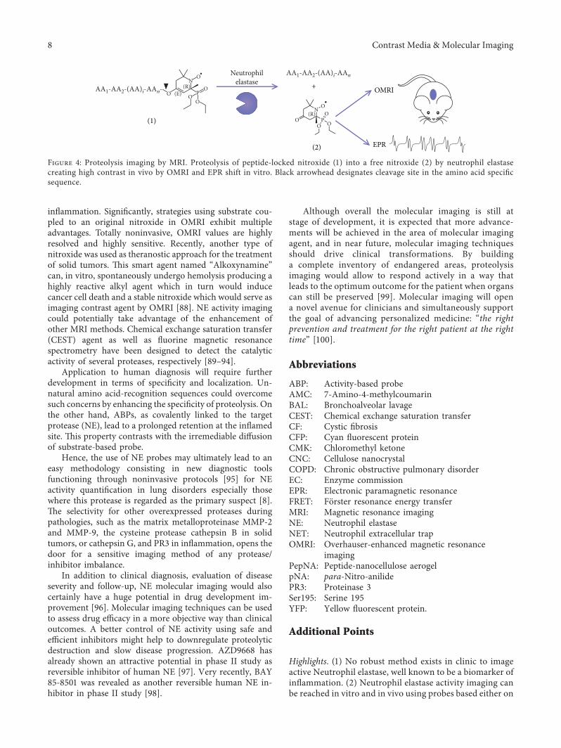
(1) Overhauser-Enhanced Magnetic Resonance Imaging(OMRI). To overcome this limitation, a particular imagingmethod based on Overhauser effect was developed to en-hance NMR sensitivity by increasing the signal/noise ratio[46]. Briefly, OMRI is a double-resonance experimenttransferring a part of the higher spin polarization of anunpaired electron to the environing water protons (throughelectron-proton Overhauser effect) which enhances the MRIsignal that appears brighter on the final image. ,is method,called Overhauser-Enhanced Magnetic Resonance Imaging(OMRI), was improved to localize and image molecularprocesses. NMR signal enhancement was observed in mouseglioma thanks to intravenous injection of an originalnonspecific spin probe design [84]. Probes with unpairedelectron such as nitroxide molecules are stable enough inphysiological conditions to be detected by OMRI, and theycan be turned into enzyme activity probes. In 2014, for thefirst time, 3D visualization of proteolytic activities happenedin vivo in mice using an on/off nitroxide-labeled with a30 kDa elastin substrate probe [85]. High Overhauser en-hancements of 10-fold were observed in the intestinal tractof mice after elastolytic activity on the probe.
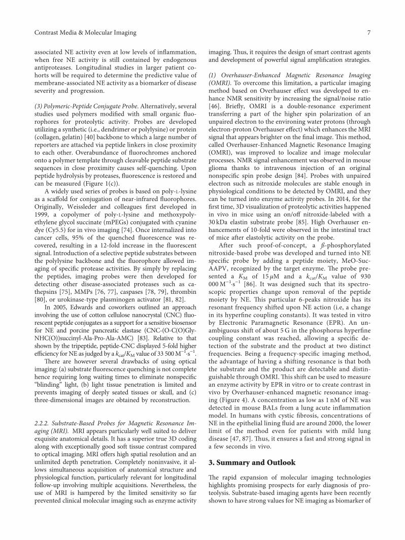
After such proof-of-concept, a ß-phosphorylatednitroxide-based probe was developed and turned into NEspecific probe by adding a peptide moiety, MeO-Suc-AAPV, recognized by the target enzyme. ,e probe pre-sented a KM of 15 μM and a kcat/KM value of 930000M−1·s−1 [86]. It was designed such that its spectro-scopic properties change upon removal of the peptidemoiety by NE. ,is particular 6-peaks nitroxide has itsresonant frequency shifted upon NE action (i.e, a changein its hyperfine coupling constants). It was tested in vitroby Electronic Paramagnetic Resonance (EPR). An un-ambiguous shift of about 5 G in the phosphorus hyperfinecoupling constant was reached, allowing a specific de-tection of the substrate and the product at two distinctfrequencies. Being a frequency-specific imaging method,the advantage of having a shifting resonance is that boththe substrate and the product are detectable and distin-guishable through OMRI.,is shift can be used to measurean enzyme activity by EPR in vitro or to create contrast invivo by Overhauser-enhanced magnetic resonance imag-ing (Figure 4). A concentration as low as 1 nM of NE wasdetected in mouse BALs from a lung acute inflammationmodel. In humans with cystic fibrosis, concentrations ofNE in the epithelial lining fluid are around 2000, the lowerlimit of the method even for patients with mild lungdisease [47, 87]. ,us, it ensures a fast and strong signal ina few seconds in vivo.
3. Summary and Outlook
,e rapid expansion of molecular imaging technologieshighlights promising prospects for early diagnosis of pro-teolysis. Substrate-based imaging agents have been recentlyshown to have strong values for NE imaging as biomarker of
Contrast Media & Molecular Imaging 7
inflammation. Significantly, strategies using substrate cou-pled to an original nitroxide in OMRI exhibit multipleadvantages. Totally noninvasive, OMRI values are highlyresolved and highly sensitive. Recently, another type ofnitroxide was used as theranostic approach for the treatmentof solid tumors. ,is smart agent named “Alkoxynamine”can, in vitro, spontaneously undergo hemolysis producing ahighly reactive alkyl agent which in turn would inducecancer cell death and a stable nitroxide which would serve asimaging contrast agent by OMRI [88]. NE activity imagingcould potentially take advantage of the enhancement ofother MRI methods. Chemical exchange saturation transfer(CEST) agent as well as fluorine magnetic resonancespectrometry have been designed to detect the catalyticactivity of several proteases, respectively [89–94].
Application to human diagnosis will require furtherdevelopment in terms of specificity and localization. Un-natural amino acid-recognition sequences could overcomesuch concerns by enhancing the specificity of proteolysis. Onthe other hand, ABPs, as covalently linked to the targetprotease (NE), lead to a prolonged retention at the inflamedsite. ,is property contrasts with the irremediable diffusionof substrate-based probe.
Hence, the use of NE probes may ultimately lead to aneasy methodology consisting in new diagnostic toolsfunctioning through noninvasive protocols [95] for NEactivity quantification in lung disorders especially thosewhere this protease is regarded as the primary suspect [8].,e selectivity for other overexpressed proteases duringpathologies, such as the matrix metalloproteinase MMP-2and MMP-9, the cysteine protease cathepsin B in solidtumors, or cathepsin G, and PR3 in inflammation, opens thedoor for a sensitive imaging method of any protease/inhibitor imbalance.
In addition to clinical diagnosis, evaluation of diseaseseverity and follow-up, NE molecular imaging would alsocertainly have a huge potential in drug development im-provement [96]. Molecular imaging techniques can be usedto assess drug efficacy in a more objective way than clinicaloutcomes. A better control of NE activity using safe andefficient inhibitors might help to downregulate proteolyticdestruction and slow disease progression. AZD9668 hasalready shown an attractive potential in phase II study asreversible inhibitor of human NE [97]. Very recently, BAY85-8501 was revealed as another reversible human NE in-hibitor in phase II study [98].
Although overall the molecular imaging is still atstage of development, it is expected that more advance-ments will be achieved in the area of molecular imagingagent, and in near future, molecular imaging techniquesshould drive clinical transformations. By buildinga complete inventory of endangered areas, proteolysisimaging would allow to respond actively in a way thatleads to the optimum outcome for the patient when organscan still be preserved [99]. Molecular imaging will opena novel avenue for clinicians and simultaneously supportthe goal of advancing personalized medicine: “the rightprevention and treatment for the right patient at the righttime” [100].
Abbreviations
ABP: Activity-based probeAMC: 7-Amino-4-methylcoumarinBAL: Bronchoalveolar lavageCEST: Chemical exchange saturation transferCF: Cystic fibrosisCFP: Cyan fluorescent proteinCMK: Chloromethyl ketoneCNC: Cellulose nanocrystalCOPD: Chronic obstructive pulmonary disorderEC: Enzyme commissionEPR: Electronic paramagnetic resonanceFRET: Forster resonance energy transferMRI: Magnetic resonance imagingNE: Neutrophil elastaseNET: Neutrophil extracellular trapOMRI: Overhauser-enhanced magnetic resonance
imagingPepNA: Peptide-nanocellulose aerogelpNA: para-Nitro-anilidePR3: Proteinase 3Ser195: Serine 195YFP: Yellow fluorescent protein.
Additional Points
Highlights. (1) No robust method exists in clinic to imageactive Neutrophil elastase, well known to be a biomarker ofinflammation. (2) Neutrophil elastase activity imaging canbe reached in vitro and in vivo using probes based either on
OMRI
EPR
+
(1)
(2)
AA1-AA2-(AA)i-AAn-O (E)
(R)N
O
O
O
OO
OO
N
P(R)
OOP
AA1-AA2-(AA)i-AAnNeutrophilelastase
Figure 4: Proteolysis imaging by MRI. Proteolysis of peptide-locked nitroxide (1) into a free nitroxide (2) by neutrophil elastasecreating high contrast in vivo by OMRI and EPR shift in vitro. Black arrowhead designates cleavage site in the amino acid specificsequence.
8 Contrast Media & Molecular Imaging
peptide substrates or on covalent inhibitors. (3),e variousprobes and instrumentations for Neutrophil elastase ac-tivity imaging have distinct advantages and could be bestused complementarily. (4) Improvements of probe struc-tures and imaging instrumentation should be appliedto further improve probes biodistribution and specificity.(5) Recent advances in molecular imaging open thedoor for a new clinical tool to image any protease/inhibitorimbalance.
Conflicts of Interest
,e authors declare that they have no conflicts of interest.
Acknowledgments
,is work was supported by the French ANR PULMO-ZYMAGE (ANR-15-CE18-0012-01).
References
[1] X. S. Puente, L. M. Sanchez, A. Gutierrez-Fernandez,G. Velasco, and C. Lopez-Otın, “A genomic view of thecomplexity of mammalian proteolytic systems,” BiochemicalSociety Transactions, vol. 33, no. 2, pp. 331–334, 2005.
[2] N. Rawlings and G. Salvesen, Handbook of ProteolyticEnzymes, Academic Press, Waltham, MA, USA, 3rd edition,2012.
[3] A.W. Segal, “How neutrophils kill microbes,”Annual Reviewof Immunology, vol. 23, no. 1, pp. 197–223, 2005.
[4] B. Turk, “Targeting proteases: successes, failures and futureprospects,” Nature Reviews Drug Discovery, vol. 5, no. 9,pp. 785–799, 2006.
[5] L. Hedstrom, “Serine protease mechanism and specificity,”Chemical Reviews, vol. 102, no. 12, pp. 4501–4524, 2002.
[6] A. Wlodawer, M. Miller, M. Jaskolski et al., “Conservedfolding in retroviral proteases: crystal structure of a syntheticHIV-1 protease,” Science, vol. 245, no. 4918, pp. 616–621,1989.
[7] H. Neurath and K. A. Walsh, “Role of proteolytic enzymesin biological regulation (a review),” Proceedings of theNational Academy of Sciences, vol. 73, no. 11, pp. 3825–3832, 1976.
[8] B. Korkmaz, M. S. Horwitz, D. E. Jenne, and F. Gauthier,“Neutrophil elastase, proteinase 3, and cathepsin G astherapeutic targets in human diseases,” PharmacologicalReviews, vol. 62, no. 4, pp. 726–759, 2010.
[9] C. T. N. Pham, “Neutrophil serine proteases: specific reg-ulators of inflammation,” Nature Reviews Immunology,vol. 6, no. 7, pp. 541–550, 2006.
[10] B. Korkmaz, T. Moreau, and F. Gauthier, “Neutrophilelastase, proteinase 3 and cathepsin G: physicochemicalproperties, activity and physiopathological functions,” Bio-chimie, vol. 90, no. 2, pp. 227–242, 2008.
[11] M. Faurschou and N. Borregaard, “Neutrophil granules andsecretory vesicles in inflammation,” Microbes and Infection,vol. 5, no. 14, pp. 1317–1327, 2003.
[12] A. Belaaouaj, R. McCarthy, M. Baumann et al., “Mice lackingneutrophil elastase reveal impaired host defense againstgram negative bacterial sepsis,” Nature Medicine, vol. 4,no. 5, pp. 615–618, 1998.
[13] A. A. Belaaouaj, “Degradation of outer membrane protein ain Escherichia coli killing by neutrophil elastase,” Science,vol. 289, no. 5482, pp. 1185–1187, 2000.
[14] Y. S. Lopez-Boado, M. Espinola, S. Bahr, and A. Belaaouaj,“Neutrophil serine proteinases cleave bacterial flagellin,abrogating its host response-inducing activity,” Journal ofImmunology, vol. 172, no. 1, pp. 509–515, 2004.
[15] N. Guyot, J. Wartelle, L. Malleret et al., “Unopposed ca-thepsin G, neutrophil elastase, and proteinase 3 cause severelung damage and emphysema,” American Journal of Pa-thology, vol. 184, no. 8, pp. 2197–2210, 2014.
[16] J. B. Soriano, A. A. Abajobir, K. H. Abate et al., “Global,regional, and national deaths, prevalence, disability-adjustedlife years, and years lived with disability for chronic ob-structive pulmonary disease and asthma, 1990–2015: a sys-tematic analysis for the Global Burden of Disease Study2015,” Ke Lancet Respiratory Medicine, vol. 5, no. 9,pp. 691–706, 2017.
[17] K. Kawabata, T. Hagio, and S. Matsuoka, “,e role ofneutrophil elastase in acute lung injury,” European Journal ofPharmacology, vol. 451, no. 1, pp. 1–10, 2002.
[18] A. S. Dittrich, I. Kuhbandner, S. Gehrig et al., “Elastaseactivity on sputum neutrophils correlates with severity oflung disease in cystic fibrosis,” European Respiratory Journal,vol. 51, no. 3, article 1701910, 2018.
[19] R. Pawar and S. Abhang, “Evaluation of serum level ofneutrophil elastase, superoxide dismutase and nitric oxide inCOPD patients and its correlation with lung function test,”International Journal of Biochemistry Research & Review,vol. 5, no. 2, pp. 153–161, 2015.
[20] J. D. Chalmers, K. L. Moffitt, G. Suarez-Cuartin et al.,“Neutrophil elastase activity is associated with exacerbationsand lung function decline in bronchiectasis,” AmericanJournal of Respiratory and Critical Care Medicine, vol. 195,no. 10, pp. 1384–1393, 2017.
[21] G. Moroy, A. J. P. Alix, J. Sapi, W. Hornebeck, andE. Bourguet, “Neutrophil elastase as a target in lung cancer,”Anti-Cancer Agents in Medicinal Chemistry, vol. 12, no. 6,pp. 565–579, 2012.
[22] K. G. Mann, E. B. Williams, S. Krishnaswamy, W. Church,A. Giles, and R. P. Tracy, “Active site-specific immunoas-says,” Blood, vol. 76, no. 4, pp. 755–766, 1990.
[23] F. Zou, M. Schmon, M. Sienczyk et al., “Application of a novelhighly sensitive activity-based probe for detection of cathepsinG,” Analytical Biochemistry, vol. 421, no. 2, pp. 667–672, 2012.
[24] K. Oikonomopoulou, K. K. Hansen, A. Baruch,M. D. Hollenberg, and E. P. Diamandis, “Immuno-fluorometric activity-based probe analysis of active KLK6 inbiological fluids,” Biological Chemistry, vol. 389, 2008.
[25] R. Yan and D. Ye, “Molecular imaging of enzyme activity invivo using activatable probes,” Science Bulletin, vol. 61,no. 21, pp. 1672–1679, 2016.
[26] M. L. James and S. S. Gambhir, “A molecular imaging primer:modalities, imaging agents, and applications,” PhysiologicalReviews, vol. 92, no. 2, pp. 897–965, 2012.
[27] W. Rut, M. Poreba, P. Kasperkiewicz, S. J. Snipas, andM. Drag, “Selective substrates and activity-based probes forimaging of the human constitutive 20S proteasome in cellsand blood samples,” Journal of Medicinal Chemistry, vol. 61,no. 12, pp. 5222–5234, 2018.
[28] M. Poreba, W. Rut, M. Vizovisek et al., “Selective imaging ofcathepsin L in breast cancer by fluorescent activity-basedprobes,” Chemical Science, vol. 9, no. 8, pp. 2113–2129, 2018.
Contrast Media & Molecular Imaging 9
[29] Y. Shaulov-Rotem, E. Merquiol, T. Weiss-Sadan et al., “Anovel quenched fluorescent activity-based probe revealscaspase-3 activity in the endoplasmic reticulum during ap-optosis,” Chemical Science, vol. 7, no. 2, pp. 1322–1337, 2016.
[30] I. Abd-Elrahman, H. Kosuge, T. W. Sada et al., “Cathepsinactivity-based probes and inhibitor for preclinical athero-sclerosis imaging and macrophage depletion,” PLos One,vol. 11, Article ID e0160522, 2016.
[31] B. F. Gilmore, D. J. Quinn, T. Duff, G. R. Cathcart, C. J. Scott,and B. Walker, “Expedited solid-phase synthesis of fluo-rescently labeled and biotinylated aminoalkane diphenylphosphonate affinity probes for chymotrypsin-and elastase-like serine proteases,” Bioconjugate Chemistry, vol. 20, no. 11,pp. 2098–2105, 2009.
[32] M. J. Page, A. L. Lourenço, T. David et al., “Non-invasiveimaging and cellular tracking of pulmonary emboli by near-infrared fluorescence and positron-emission tomography,”Nature Communications, vol. 11, no. 6, 2015.
[33] J. Charlton, J. Sennello, and D. Smith, “In vivo imaging ofinflammation using an aptamer inhibitor of human neu-trophil elastase,” Chemistry & Biology, vol. 4, no. 11,pp. 809–816, 1997.
[34] M. Rusckowski, T. Qu, J. Pullman et al., “Inflammation andinfection imaging with a 99m TC-neutrophil elastase in-hibitor in monkeys,” Journal of Nuclear Medicine, vol. 41,no. 2, pp. 363–374, 2000.
[35] P. Kasperkiewicz, M. Poreba, S. J. Snipas et al., “Design ofultrasensitive probes for human neutrophil elastase throughhybrid combinatorial substrate library profiling,” Pro-ceedings of the National Academy of Sciences, vol. 111, no. 7,pp. 2518–2523, 2014.
[36] B. C. Lechtenberg, P. Kasperkiewicz, H. Robinson, M. Drag,and S. J. Riedl, “,e elastase-PK101 structure: mechanism ofan ultrasensitive activity-based probe revealed,” ACSChemical Biology, vol. 10, no. 4, pp. 945–951, 2015.
[37] K. Nakajima, J. C. Powers, B. M. Ashe, and M. Zimmerman,“Sensitive mapping the extended substrate binding site ofcathepsin G and human leucocyte elastase,” Journal of Bi-ological Chemistry, vol. 254, no. 10, pp. 4027–4032, 1979.
[38] T. Kalupov, M. Brillard-Bourdet, S. Dade et al., “Structuralcharacterization of mouse neutrophil serine proteases andidentification of their substrate specificities,” Journal of Bi-ological Chemistry, vol. 284, no. 49, pp. 34084–34091, 2009.
[39] H. Crisford, E. Sapey, and R. A. Stockley, “Proteinase 3; apotential target in chronic obstructive pulmonary diseaseand other chronic inflammatory diseases,” Respiratory Re-search, vol. 19, no. 1, 2018.
[40] O. R. F. Mook, C. V. Overbeek, E. G. Ackema,F. V. Maldegem, andW.M. Frederiks, “In situ localization ofgelatinolytic activity in the extracellular matrix of metastasesof colon cancer in rat liver using quenched fluorogenic DQ-gelatin,” Journal of Histochemistry & Cytochemistry, vol. 51,no. 6, pp. 821–829, 2003.
[41] I. Lerman, M. D. L. L. Hernandez, J. Rangel-Moreno et al.,“Infiltrating myeloid cells exert protumorigenic actions vianeutrophil elastase,” Molecular Cancer Research, vol. 15,no. 9, pp. 1138–1152, 2017.
[42] H. E. Scales, M. Ierna, K. M. Smith et al., “Assessment ofmurine collagen-induced arthritis by longitudinal non-invasiveduplexed molecular optical imaging,” Rheumatology, vol. 55,no. 3, pp. 564–572, 2015.
[43] M. M. Muley, A. R. Reid, B. Botz, K. Bolcskei, Z. Helyes, andJ. J. McDougall, “Neutrophil elastase induces inflammationand pain in mouse knee joints via activation of proteinase-
activated receptor-2,” British Journal of Pharmacology,vol. 173, no. 4, pp. 766–777, 2016.
[44] A. Glinzer, X. Ma, J. Prakash et al., “Targeting elastase formolecular imaging of early atherosclerotic lesions,” Arte-riosclerosis, Krombosis, and Vascular Biology, vol. 37, no. 3,pp. 525–533, 2016.
[45] C. A. Owen, “Cell surface-bound elastase and cathepsin G onhuman neutrophils: a novel, non-oxidative mechanism bywhich neutrophils focus and preserve catalytic activity ofserine proteinases,” Journal of Cell Biology, vol. 131, no. 3,pp. 775–789, 1995.
[46] P. Mellet, P. Massot, G. Madelin et al., “New concepts inmolecular imaging: non-invasive MRI spotting of proteolysisusing an overhauser effect switch,” PLoS One, vol. 4, no. 4,Article ID e5244, 2009.
[47] S. I. Rennard, G. Basset, D. Lecossier et al., “Estimation ofvolume of epithelial lining fluid recovered by lavage usingurea as marker of dilution,” Journal of Applied Physiology,vol. 60, no. 2, pp. 532–538, 1986.
[48] P. D. Edwards, D. W. Andisik, A. M. Strimpler, B. Gomes,and P. A. Tuthill, “Nonpeptidic inhibitors of human neu-trophil elastase. 7. Design, synthesis, andin vitroactivity of aseries of pyridopyrimidine trifluoromethyl ketones,” Journalof Medicinal Chemistry, vol. 39, no. 5, pp. 1112–1124, 1996.
[49] J. C. Powers, B. F. Gupton, A. D. Harley, N. Nishino, andR. J. Whitley, “Specificity of porcine pancreatic elastase,human leukocyte elastase and cathepsin G Inhibition withpeptide chloromethyl ketones,” Biochimica et BiophysicaActa (BBA)—Enzymology, vol. 485, no. 1, pp. 156–166, 1977.
[50] P. Tuhy and J. C. Powers, “Inhibition of human leukocyteelastase by peptide chloromethyl ketones,” FEBS Letters,vol. 50, no. 3, pp. 359–361, 1975.
[51] V. Ranga, J. Kleinerman, M. P. C. Ip, J. Sorensen, andJ. C. Powers, “Effects of oligopeptide chloromelhyl ketoneadministered after elastase: renal toxicity and lack of ex-perimental emphysema,” American Review of RespiratoryDisease Returns, vol. 124, pp. 613–618, 1981.
[52] R. Grzywa, E. Burchacka, M. Łecka et al., “Synthesis of novelphosphonic-type activity-based probes for neutrophil serineproteases and their application in spleen lysates of differentorganisms,”ChemBioChem, vol.15, no.17, pp. 2605–2612, 2014.
[53] E. F. P. Ruivo, L. M. Gonçalves, L. A. R. Carvalho et al.,“Clickable 4-oxo-β-lactam-based selective probing for hu-man neutrophil elastase related proteomes,” Chem-MedChem, vol. 11, no. 18, pp. 2037–2042, 2016.
[54] A.-C. Schulz-Fincke, A. S. Tikhomirov, A. Braune et al.,“Design of an activity-based probe for human neutrophilelastase: implementation of the lossen rearrangement toinduce forster resonance energy transfers,” Biochemistry,vol. 57, no. 5, pp. 742–752, 2018.
[55] L. E. Edgington, M. Verdoes, and M. Bogyo, “Functionalimaging of proteases: recent advances in the design andapplication of substrate-based and activity-based probes,”Current Opinion in Chemical Biology, vol. 15, no. 6,pp. 798–805, 2011.
[56] I. Schechter and A. Berger, “On the size of the active site inproteases. I. Papain,” Biochemical and Biophysical ResearchCommunications, vol. 27, no. 2, pp. 157–162, 1967.
[57] J. Bieth and C. G. Wermuth, “,e action of elastase onp-nitroanilide substrates,” Biochemical and Biophysical Re-search Communications, vol. 53, no. 2, pp. 383–390, 1973.
[58] J. Bieth, B. Spiess, and C. G. Wermuth, “,e synthesis andanalytical use of a highly sensitive and convenient substrate
10 Contrast Media & Molecular Imaging
of elastase,” Biochemical Medicine, vol. 11, no. 4, pp. 350–357, 1974.
[59] M. J. Castillo, K. Nakajima, M. Zimmerman, andJ. C. Powers, “Sensitive substrates for human leukocyte andporcine pancreatic elastase: a study of the merits of variouschromophoric and fluorogenic leaving groups in assays forserine proteases,” Analytical Biochemistry, vol. 99, no. 1,pp. 53–64, 1979.
[60] L. P. A. McGarvey, K. Dunbar, S. L. Martin et al., “Cytokineconcentrations and neutrophil elastase activity in bron-choalveolar lavage and induced sputum from patients withcystic fibrosis, mild asthma and healthy volunteers,” Journalof Cystic Fibrosis, vol. 1, no. 4, pp. 269–275, 2002.
[61] D. S. Armstrong, S. M. Hook, K. M. Jamsen et al., “Lowerairway inflammation in infants with cystic fibrosis detectedby newborn screening,” Pediatric Pulmonology, vol. 40, no. 6,pp. 500–510, 2005.
[62] S. Mizukami, K. Kazuya, T. Higuchi et al., “Imaging ofcaspase-3 activation in HeLa cells stimulated with etoposideusing a novel fluorescent probe,” FEBS Letters, vol. 453, no. 3,pp. 356–360, 1999.
[63] L. M. Felber, S. M. Cloutier, C. Kundig et al., “Evaluation ofthe CFP-substrate-YFP system for protease studies: advan-tages and limitations,” Biotechniques, vol. 36, no. 5,pp. 878–885, 2004.
[64] C. Schulenburg, G. Faccio, D. Jankowska, K. Maniura-Weber, and M. Richter, “A FRET-based biosensor for thedetection of neutrophil elastase,”Ke Analyst, vol. 141, no. 5,pp. 1645–1648, 2016.
[65] S. Kossodo, J. Zhang, K. Groves et al., “Noninvasive in vivoquantification of neutrophil elastase activity in acute experi-mental mouse lung injury,” International Journal of MolecularImaging, vol. 2011, Article ID 581406, 11 pages, 2011.
[66] A.-S. Ho, C.-H. Chen, C.-C. Cheng et al., “Neutrophilelastase as a diagnostic marker and therapeutic target incolorectal cancers,” Oncotarget, vol. 5, no. 2, 2014.
[67] B. Korkmaz, S. Attucci, M. A. Juliano et al., “Measuringelastase, proteinase 3 and cathepsin G activities at the surfaceof human neutrophils with fluorescence resonance energytransfer substrates,” Nature Protocols, vol. 3, no. 6,pp. 991–1000, 2008.
[68] E. J. Campbell and C. A. Owen, “,e sulfate groups ofchondroitin sulfate-and heparan sulfate-containing pro-teoglycans in neutrophil plasmamembranes are novel bindingsites for human leukocyte elastase and cathepsin G,” Journal ofBiological Chemistry, vol. 282, no. 19, article 14645, 2007.
[69] A. J. O’Donoghue, Y. Jin, G. M. Knudsen et al., “Global sub-strate profiling of proteases in human neutrophil extracellulartraps reveals consensus motif predominantly contributed byelastase,” PLoS One, vol. 8, no. 9, Article ID e75141, 2013.
[70] E. Kelly, C. M. Greene, and N. G. McElvaney, “Targetingneutrophil elastase in cystic fibrosis,” Expert Opinion onKerapeutic Targets, vol. 12, no. 2, pp. 145–157, 2008.
[71] P. D. Sly, C. L. Gangell, L. Chen et al., “Risk factors forbronchiectasis in children with cystic fibrosis,” New EnglandJournal of Medicine, vol. 368, no. 21, pp. 1963–1970, 2013.
[72] P. D. Sly, S. Brennan, C. Gangell et al., “Lung disease atdiagnosis in infants with cystic fibrosis detected by newbornscreening,” American Journal of Respiratory and CriticalCare Medicine, vol. 180, no. 2, pp. 146–152, 2009.
[73] S. Gehrig, M. A. Mall, and C. Schultz, “Spatially resolvedmonitoring of neutrophil elastase activity with ratiometricfluorescent reporters,” Angewandte Chemie InternationalEdition, vol. 51, no. 25, pp. 6258–6261, 2012.
[74] R. Weissleder, C.-H. Tung, U. Mahmood, and A. BogdanovJr., “In vivo imaging of tumors with protease-activated near-infrared fluorescent probes,” Nature biotechnology, vol. 17,no. 4, pp. 375–378, 1999.
[75] F. A. Jaffer, D.-E. Kim, L. Quinti et al., “Optical visualizationof cathepsin K activity in atherosclerosis with a novel,protease-activatable fluorescence sensor,” Circulation,vol. 115, no. 17, pp. 2292–2298, 2007.
[76] R. A. Sheth, A. Kunin, L. Stangenberg et al., “In Vivo opticalmolecular imaging of matrix metalloproteinase activityfollowing celecoxib therapy for colorectal cancer,”Molecularimaging, vol. 11, no. 5, article 7290.2012.00003, 2012.
[77] C. Bremer, C.-H. Tung, and R. Weissleder, “In vivo mo-lecular target assessment of matrix metalloproteinase in-hibition,” Nature medicine, vol. 7, no. 6, pp. 743–748, 2001.
[78] M. Sameni, J. Dosescu, and F. Sloane, “Imaging proteolysisby living human glioma cells,” Biological Chemistry, vol. 382,no. 5, pp. 785–788, 2001.
[79] S. M. Messerli, S. Prabhakar, Y. Tang et al., “A novel methodfor imaging apoptosis using a caspase-1 near-infraredfluorescent probe,” Neoplasia, vol. 6, no. 2, pp. 95–105, 2004.
[80] F. A. Jaffer, C.-H. Tung, R. E. Gerszten, and R. Weissleder,“In vivo imaging of thrombin activity in experimentalthrombi with thrombin-sensitive near-infrared molecularprobe,” Arteriosclerosis, Krombosis, and Vascular Biology,vol. 22, no. 11, pp. 1929–1935, 2002.
[81] B. Law, A. Curino, T. H. Bugge, R. Weissleder, andC.-H. Tung, “Design, synthesis, and characterization ofurokinase plasminogen-activator-sensitive near-infraredreporter,” Chemistry & Biology, vol. 11, no. 1, pp. 99–106,2004.
[82] V. Fritz, D. Noel, C. Bouquet et al., “Antitumoral activity andosteogenic potential of mesenchymal stem cells expressingthe urokinase-type plasminogen antagonist amino-terminalfragment in a murine model of osteolytic tumor,” Stem Cells,vol. 26, no. 11, pp. 2981–2990, 2008.
[83] J. V. Edwards, N. T. Prevost, A. D. French, M. Concha, andB. D. Condon, “Kinetic and structural analysis of fluorescentpeptides on cotton cellulose nanocrystals as elastase sensors,”Carbohydrate Polymers, vol. 116, pp. 278–285, 2015.
[84] P. Massot, E. Parzy, L. Pourtau et al., “In vivo high-resolution3D overhauser-enhanced MRI in mice at 0.2 T,” ContrastMedia & Molecular Imaging, vol. 7, no. 1, pp. 45–50, 2012.
[85] N. Koonjoo, E. Parzy, P. Massot et al., “In vivo overhauser-enhanced MRI of proteolytic activity,” Contrast Media &Molecular Imaging, vol. 9, no. 5, pp. 363–371, 2014.
[86] N. Jugniot, I. Duttagupta, A. Rivot et al., “An elastase activityreporter for electronic paramagnetic resonance (EPR) andoverhauser-enhanced magnetic resonance imaging (OMRI)as a line-shifting nitroxide,” Free Radical Biology andMedicine, vol. 126, pp. 101–112, 2018.
[87] M. W. Crystal, K. A. Hilliard, T. M. Norvell, and M. Berger,“Bronchoalveolar lavage findings in cystic fibrosis patientswith stable, clinically mild lung disease suggest ongoinginfection and inflammation,” American Journal of Re-spiratory and Critical Care Medicine, vol. 150, no. 2,pp. 448–454, 1994.
[88] D. Moncelet, P. Voisin, N. Koonjoo et al., “Alkoxyamines:toward a new family of theranostic agents against cancer,”Molecular Pharmaceutics, vol. 11, no. 7, pp. 2412–2419, 2014.
[89] K. Akazawa, F. Sugihara, M. Minoshima, S. Mizukami, andK. Kikuchi, “Sensing caspase-1 activity using activatable 19FMRI nanoprobes with improved turn-on kinetics,” ChemicalCommunications, vol. 54, no. 83, pp. 11785–11788, 2018.
Contrast Media & Molecular Imaging 11
[90] X. Yue, Z. Wang, L. Zhu et al., “Novel 19F activatable probefor the detection of matrix metalloprotease-2 activity byMRI/MRS,” Molecular Pharmaceutics, vol. 11, no. 11,pp. 4208–4217, 2014.
[91] M. Suchy, R. Ta, A. X. Li et al., “A paramagnetic chemicalexchange-based MRI probe metabolized by cathepsin D:design, synthesis and cellular uptake studies,” Organic &Biomolecular Chemistry, vol. 8, no. 11, p. 2560, 2010.
[92] D. V. Hingorani, L. A. Montano, E. A. Randtke, Y. S. Lee,J. Cardenas-Rodrıguez, and M. D. Pagel, “A single dia-magnetic catalyCEST MRI contrast agent that detects ca-thepsin B enzyme activity by using a ratio of two CESTsignals,” Contrast Media & Molecular Imaging, vol. 11, no. 2,pp. 130–138, 2016.
[93] M. D. Pagel, B. Yoo, G. Liu, and R. Rosenblum, “CMR 2005:13.06: activatable MRI CEST agents that detect enzymeactivities,”Contrast Media &Molecular Imaging, vol. 1, no. 2,pp. 88-89, 2006.
[94] B. Yoo, V. R. Sheth, C. M. Howison et al., “Detection of invivo enzyme activity with catalyCEST MRI,” MagneticResonance in Medicine, vol. 71, no. 3, pp. 1221–1230, 2014.
[95] S. Chakraborti, T. Chakraborti, and N. S. Dhalla, Proteases inHuman Diseases, Springer, Berlin, Germany, 2017.
[96] Y. Wang and M. Deng, “Medical imaging in new drugclinical development,” Journal of Koracic Disease, vol. 2,pp. 245–252, 2010.
[97] R. Stockley, A. De Soyza, K. Gunawardena et al., “Phase IIstudy of a neutrophil elastase inhibitor (AZD9668) in pa-tients with bronchiectasis,” Respiratory Medicine, vol. 107,no. 4, pp. 524–533, 2013.
[98] H. Watz, J. Nagelschmitz, A. Kirsten et al., “Safety and ef-ficacy of the human neutrophil elastase inhibitor BAY 85-8501 for the treatment of non-cystic fibrosis bronchiectasis: arandomized controlled trial,” Pulmonary Pharmacology &Kerapeutics, vol. 56, pp. 86–93, 2019.
[99] J. Holland, “,e role of molecular imaging in personalisedhealthcare,” CHIMIA International Journal for Chemistry,vol. 70, no. 11, pp. 787–795, 2016.
[100] European Alliance for Personalised Medicine—Innovationand patient Access to personalised medicine, http://euapm.eu/pdf/EAPM_REPORT_on_Innovation_and_Patient_Access_to_Personalised_Medicine.pdf.
12 Contrast Media & Molecular Imaging

Stem Cells International
Hindawiwww.hindawi.com Volume 2018
Hindawiwww.hindawi.com Volume 2018
MEDIATORSINFLAMMATION
of
EndocrinologyInternational Journal of
Hindawiwww.hindawi.com Volume 2018
Hindawiwww.hindawi.com Volume 2018
Disease Markers
Hindawiwww.hindawi.com Volume 2018
BioMed Research International
OncologyJournal of
Hindawiwww.hindawi.com Volume 2013
Hindawiwww.hindawi.com Volume 2018
Oxidative Medicine and Cellular Longevity
Hindawiwww.hindawi.com Volume 2018
PPAR Research
Hindawi Publishing Corporation http://www.hindawi.com Volume 2013Hindawiwww.hindawi.com
The Scientific World Journal
Volume 2018
Immunology ResearchHindawiwww.hindawi.com Volume 2018
Journal of
ObesityJournal of
Hindawiwww.hindawi.com Volume 2018
Hindawiwww.hindawi.com Volume 2018
Computational and Mathematical Methods in Medicine
Hindawiwww.hindawi.com Volume 2018
Behavioural Neurology
OphthalmologyJournal of
Hindawiwww.hindawi.com Volume 2018
Diabetes ResearchJournal of
Hindawiwww.hindawi.com Volume 2018
Hindawiwww.hindawi.com Volume 2018
Research and TreatmentAIDS
Hindawiwww.hindawi.com Volume 2018
Gastroenterology Research and Practice
Hindawiwww.hindawi.com Volume 2018
Parkinson’s Disease
Evidence-Based Complementary andAlternative Medicine
Volume 2018Hindawiwww.hindawi.com
Submit your manuscripts atwww.hindawi.com