Invariants of Combinatorial Line Arrangements and Rybnikov's Example
Upload
independentCategory
view
1download
0
Eur. J. Biochem. 266, 403±412 (1999) q FEBS 1999
Selection of human elastase inhibitors from a conformationally constrainedcombinatorial peptide library
Jeffrey D. McBride1, Hamzah N. M. Freeman2 and Robin J. Leatherbarrow1
1Department of Chemistry, Imperial College of Science, Technology, and Medicine, London, UK; 2Protein Science Unit,
Glaxo Wellcome Research and Development Ltd, Stevenage, Herts, UK
A resin-bound cyclic peptide library was constructed based on the sequence of the reactive-site loop of
Bowman±Birk inhibitor, a proteinase inhibitor protein. The constrained loop sequence, which incorporates the
minimal proteinase-binding motif, was retained throughout the library, but selected residues known to be
important for inhibitor specificity were randomised. The approach was used to create a `one bead, one peptide'
library with 8000 variants resulting from randomization at three target locations in the sequence (P4, P1 and P20 ).
This library allows us to examine the degree to which variations in this proteinase-binding motif can redirect
activity, as well as providing information about the binding specificity of a proteinase target. Screening this
library for binding to human leucocyte elastase identified sequences with a strong consensus, and on resynthesis
all were found to act as inhibitors, with Ki values as low as 65 nm. Human leucocyte elastase is known to have a
substrate preference for small alkyl chains at the P1 locus, with valine being preferred. However, alanine and not
the expected valine was found in 21 out of 23 identified sequences. The remaining two sequences had threonine
at P1, a finding that would be hard to predict based on substrate specificity alone. Further analysis of
resynthesized peptides demonstrated that valine substitution results in an analogue that is hydrolysed far more
rapidly than ones having library-selected P1 residues. Testing of the human leucocyte elastase-selected sequences
as inhibitors of porcine pancreatic elastase demonstrates a significant difference in the specificity of the P4 locus
between these two proteinases.
Keywords: Bowman±Birk inhibitor; canonical loop; combinatorial chemistry; elastase; peptide library.
Elastases, which are serine proteinases capable of cleaving theconnective tissue elastin, are considered to play important rolesin the tissue destruction associated with pulmonary emphy-sema, rheumatoid arthritis, cystic fibrosis, adult respiratorydistress syndrome, chronic bronchitis, and pancreatitis [1,2].For some of these pathological conditions an imbalancebetween the amount of elastase and endogenous proteinaceousinhibitor(s) has been implicated. As a result, there has beenconsiderable interest in the design of inhibitors that may restorethis balance and/or clarify the role of these enzymes in theabove diseases. In addition to natural or engineered inhibitorproteins, which have high molecular masses, researchers havealso pursued both peptide-based and nonpeptide small inhibitorcompounds [2].
In many natural inhibitor proteins, the portion of the inhibitorprotein that interacts with the serine proteinase is an extendedor `canonical' loop. This is thought to have a structure similar
to that of a productively bound substrate [3,4]. Inhibitorsbehave as limited proteolysis substrates; residues interactingwith proteinase at the reactive site of the inhibitor determinespecificity and typically reflect the substrate specificity of thetarget proteinase. The identity of the P1 residue (nomenclatureof Schechter & Berger [5]) is considered to be the maindeterminant of specificity [3,6]. Qasim et al. [7] have describedcanonical loops as molecular vices, delivering the P1 side chainto the S1 cavity whether the interaction is favourable, or evenwhen it is neutral or locally deleterious. However, the identityof a given P1 residue alone does not always succeed in defininginhibitory activity. For example, chicken ovomucoid firstdomain with a P1 Lys residue is ineffective as an inhibitor ofbovine b-trypsin even though this is the preferred P1 of trypsinsubstrates [8]. Although many natural inhibitors are selectivefor the proteinases they target, some inhibitors are morepromiscuous and demonstrate activity against a range ofenzymes. For example, mucus proteinase inhibitor (P1 Leu,P10 Met) is active against human leucocyte esterase (HLE),
cathepsin G, chymotrypsin and trypsin [9]; turkey ovomucoid(TOM) (OMTKY3; P1 Leu, P1
0 Glu) is active against chymo-trypsin, porcine pancreatic elastase (PPE), Streptomyces griseusproteinases A and B, and subtilisins BPN 0 and Carlsberg [10].
Redirection and/or enhancement of elastase inhibitoryactivity resulting from alterations at the P1 locus have beendescribed following site-directed mutagenesis [11,12] andsemisynthesis or peptide synthesis [13,14]. In general, altera-tion of the P1 residue is based upon the known substratepreference of the target proteinase. For example, replacementof the P1 Met in a1-protease inhibitor by Val has been used to
Correspondence to R. Leatherbarrow, Department of Chemistry,
Imperial College of Science, Technology, and Medicine, South Kensington,
London SW7 2AY, UK.
Abbreviations: BBI, Bowman±Birk inhibitor; BPTI, bovine pancreatic
trypsin inhibitor; CMTI-III, Curcubita maxima trypsin inhibitor-III; FAB,
fast-atom bombardment; Fmoc, fluoren-9-ylmethoxycarbonyl; HLE, human
leucocyte elastase; OMGPP3, peacock pheasant ovomucoid third domain;
OMTKY3, turkey ovomucoid third domain; PPE, porcine pancreatic
elastase; TOM, turkey ovomucoid.
Enzymes: human leucocyte elastase (HLE; EC 3.4.21.37); porcine
pancreatic elastase (PPE; EC 3.4.21.35).
(Received 28 July 1999, accepted 14 September 1999)

404 J. D. McBride et al. (Eur. J. Biochem. 266) q FEBS 1999
overcome oxidation sensitivity, yet retain specificity [15]. Onoccasion, P1 alterations have resulted in unexpected activities.P1 point mutations to the squash seed proteinase inhibitor,Curcubita maxima trypsin inhibitor-III (CMTI-III) have beendescribed by McWherter et al. [16], who found that of thelimited range of variants tested, Phe and Gly were activeagainst HLE. The activity given by a P1 Gly is striking as thisresidue provides no side chain for interaction. Inhibitoryactivity of this variant must therefore rely on more extensiveinteractions distant to the S1 site. More recently, Rozycki et al.[17] have further reported reduction in activity when the P4±P5
residues were deleted at the N-terminus of CMTI-III (whichalso had the P1 residue substituted for valine in order toredirect activity towards HLE). In contrast, removal of thesetwo N-terminal residues from the native inhibitor (whichdisplays anti-tryptic activity) did not result in such decreasedactivity. Not only does this result further emphasize theimportance of subsites distant to P1 in contributing to bindingenergy with HLE, it suggests that these are correspondingly lessimportant in trypsin binding.
It seems reasonable that efficient redirection of proteinaseinhibitory activity would benefit from a more extensive rangeof modifications that is not limited to the P1 locus alone.Combinatorial peptide libraries have provided an attractiveapproach to screening for potential ligands to a given receptor,and have been used to screen for proteinase inhibitors. Phagedisplay libraries have been successfully used to selectredirected inhibitor activity, principally involving randomi-zation of the active sites from the Kunitz (or bovine pancreatictrypsin inhibitor, BPTI) family of proteinase inhibitors [18±20].We have recently used a synthetic combinatorial protocol tocreate a library of potential inhibitor structures [21], which hasthe potential advantage of allowing incorporation of nonproteinamino acids. This work is based on the ability of shortdisulfide-linked peptide sequences that reproduce the extendedloop region of Bowman±Birk inhibitor (BBI) proteins to retaininhibitory activity [13,22,23]. Our library contained 8000 cyclicpeptide variants obtained by randomization at the P2, P1 and P2
0residues [21], and used the `one bead, one peptide' approach[24,25] to combinatorial peptide synthesis. Screening thislibrary for redirected activity against chymotrypsin identified aconsensus-binding motif and allowed the selection of compe-titive inhibitors with Ki values in the nanomolar range. Usingthis same approach, we now describe use of a slightly differentcyclic peptide library to screen for inhibitors with activityredirected towards HLE. The active sequences that wereidentified were also tested as inhibitors of PPE to allow acomparison of the specificity of these elastases. These twoenzymes have similar activity, share 40% primary sequenceidentity and a number of HLE and PPE complexes havedemonstrated particular topological similarity in their active-site regions [26].
E X P E R I M E N T A L P R O C E D U R E S
Materials
Fluoren-9-ylmethoxycarbonyl (Fmoc)-protected amino acidsand derivatives were purchased from Novabiochem orAdvanced ChemTech with the following side-chain-protectinggroups: Ala, Arg (2,2,5,7,8-pentamethylchroman-6-sulfonyl),Asn (trityl), Asp (tertBu), Cys (trityl), Gln (trityl), Glu (anti-tryptic), Gly, His (trityl), Ile, Leu, Lys (tert-butyloxycarbonyl;tertBoc), Met, Nle, Phe, Pro, Ser (anti-tryptic), Thr (anti-tryptic), Trp (tertBoc), Tyr (anti-tryptic), Val. TentaGel-S-NH2
resin was from Rapp Polymere (Tubingen, Germany).Dimethylformamide and N-methylpyrrolidone were peptidesynthesis grade from Rathburn Chemicals (Walkerburn, UK)and 2-(1H-benzotriazole-1-yl)-1,1,3,3-tetramethyluronium hex-afluorophosphate and 1-hydroxybenzotriazole monohydratewere from SNPE (Croydon, Surrey. UK). 5-Bromo-4-chloro-3-indolyl phosphate, nitroblue tetrazolium, phenol, ethane-dithiol, thioanisole, dimethyl sulfoxide, monoclonal anti-rabbitIgG (g-chain specific) conjugated to alkaline phosphatase, andtrifluoroacetic acid were purchased from Sigma. Elastase fromhuman polymorphonuclear leucocytes (HLE, EC 3.4.21.37)was from Elastin Products Co. Inc., Owensville, MO, USA,PPE (EC 3.4.21.35) was from Sigma, and rabbit anti-HLE wasfrom Calbiochem.
Preparation of tethered cyclic peptide library
Synthesis of the library peptide, disulfide-cyclized NH2-XCTXSXPPQCYGGGGG-Resin, was performed on an ABI431A peptide synthesizer at 0.25 mmol scale using Fmocchemistry. The solid-phase resin was TentaGel-S-NH2, whichhas a noncleavable linker. 2-(1H-Benzotriazole-1-yl)-1,1,3,3-tetramethyluronium hexafluorophosphate/1-hydroxybenzotri-azole monohydrate [27] activation was used for amino acidcouplings at fivefold amino acid excess except for the positionsmarked X. At these positions, randomization was achievedusing preactivated pentafluorophenyl ester or 3-hydroxy-4-oxo-3,4-dihydro-1,2,3-benzotriazine ester amino acid esters at10-fold excess as described previously [21]. After side-chaindeprotection, cyclization was achieved using dimethyl sulf-oxide [28]. We have previously demonstrated that this methodprovides an efficient means of on-resin cyclization, with thegeneration of active resin-immobilized peptide [29]. Resin wasthen thoroughly washed and resuspended in NaCl/Pi with0.05% Tween 20 (NaCl/Pi/Tween) before use.
Preparation of defined sequence cyclic peptides
Peptide sequences identified from the screening procedure wereresynthesized by standard solid-phase Fmoc chemistry asdescribed previously using a Shimadzu PSSM-8 synthesizer.The crude peptides were cyclized using dimethylsulfoxide alsoas described previously [21].
The cyclic peptides were purified by HPLC using a Gilsoninstrument equipped with a Waters Radial Pak C18 column(25 mm � 10 cm, 6-mm particle size) reverse-phase column.Separations were carried out with 0.1% trifluoroacetic acid andacetonitrile as solvents. Peptides were recovered after semi-preparative chromatography by lyophilization. The homogene-ity of the synthesized peptides was assessed by TLC, analyticalHPLC and MS. Analytical HPLC was performed using a VydacC18 column (150 � 4.6 mm, 5 mm particle size) employing agradient between water and acetonitrile, each containing 0.1%trifluoroacetic acid (5% acetonitrile for 2.5 min, 5±80%acetonitrile over 22.5 min, 80% acetonitrile for a further2.5 min before returning to 5% for 2.5 min). Flow rate was1 mL´min21 with detection at 226 nm. TLC was conducted onprelayered silica-gel 60 F254 plates with fluorescent indicator(Merck), and two different solvent systems were used foranalysis. Solvent system A was methanol/chloroform/aceticacid (4 : 15 : 1, v/v), and system B was methanol/water/acetonitrile (5 : 5 : 3, v/v); detection was performed using UV(254 nm) and ninhydrin [0.2% (w/v) in acetone]. Mass spectrawere recorded on a VG Auto-spec Q instrument operating inthe fast-atom bombardment (FAB) mode. In all cases, peptides
q FEBS 1999 Human elastase inhibitor library (Eur. J. Biochem. 266) 405
were more than 85% pure by HPLC, displayed a single spot onTLC, and the correct mass was confirmed by FAB-MS.
Screening the BBI library against HLE
Screening was performed using a modification of the methoddescribed previously [21]. Briefly, HLE was incubated inNaCl/Pi/ Tween with sufficient resin to provide < 46 000beads. The resin was then washed thoroughly before incubatingwith rabbit anti-HLE in NaCl/Pi/ Tween. The washing step wasrepeated and beads were then incubated with monoclonal anti-rabbit IgG (g-chain specific) conjugated to alkaline phospha-tase. The washing step was again repeated with a final washof 100 mm Tris (pH 9.5)/0.5 mm MgCl2. Beads were thenincubated in the same buffer with 5-bromo-4-chloro-3-indolylphosphate and nitroblue tetrazolium as alkaline phosphatasesubstrate. Positively stained beads had a deep purple coloration,and were transferred manually to glass-fibre filters, washedwith 40 mL 50 mm HCl, and air-dried. N-Terminal sequencingwas performed on either an ABI 476A (PE Biosystems) oran HP G1005A protein sequencer (Hewlett-Packard) usingstandard sequencing protocols. The beads were prewashed withtrifluoroacetic acid and ethyl acetate before sequence analysis,as this was found to reduce the background signal in the firstcycle. On the ABI sequencer, the bead was sequenced on thetrifluoroacetic acid-washed glass-fibre filter, but on the HPsequencer the bead was trapped at the interface of the hydro-phobic and hydrophilic columns of an N-terminal sequencingcartridge.
Inhibition kinetics
Inhibition kinetics for the synthetic peptides were determinedusing competitive binding studies with MeO-succinyl-Ala-Ala-Pro-Val-p-nitroanilide (Sigma) as HLE substrate in 100 mmTris/500 mm NaCl, pH 7.5. For PPE, succinyl-Ala-Ala-Ala-p-nitroanilide (Sigma) was used as the substrate, in 0.1 m TrispH 8.0, both at 298 K. Substrate hydrolysis was monitored at
410 nm, and substrate concentration calculated from the finalabsorbance (: = 8800 m21´cm21 [30]). Enzyme concentrationswere determined from the initial rate of substrate hydrolysis inthe absence of inhibitor using values for Km and kcat of 0.14 mmand 17 s21, respectively, for HLE [31] and 1.15 mm and16.6 s21, respectively, for PPE [32]. Initial velocity data werefitted using the grafit software package [33,34]. Ki valueswere corrected for substrate competition using the formulaKi = Ki(app)(1 + [S]/Km), where Ki(app) is the apparent Ki for agiven substrate concentration.
Hydrolysis rates
Hydrolysis of peptides was performed using a 20-fold excess toHLE (4 mm HLE) in 100 mm Tris/500 mm NaCl, pH 7.5 at298 K. Degradation of the peak corresponding to the cyclicpeptide was followed by integration of the peak areas(monitored at 214 nm) after separation by a Micra NPS RP18reverse-phase column (4.6 � 33 mm, 1.5 mm particle size;Micra Scientific Ltd, Darien, IL, USA) using a Hewlett-PackardHP1100 binary pump system. Separation was performed usinga linear gradient between 0.3% trifluoroacetic acid and 0.3%trifluoroacetic acid/90% acetonitrile as solvents.
R E S U L T S A N D D I S C U S S I O N
Design of the library
In this study we have used a cyclic peptide library based on theanti-tryptic loop of BBI protein as the starting point forgeneration of inhibitors redirected against HLE. Variation wasintroduced into the library at selected positions in order togenerate diversity likely to modify specificity. Interactions withthe proteinase are expected to be exclusively via the `upper-face' of the loop, which spans the region P4±P2
0 [35]. Ser ishighly conserved at the P1
0 position in BBI proteins [36], andfor this reason this residue was not randomised. Similarly, Thrat P2 shows a high degree of conservation [36]. In our previous
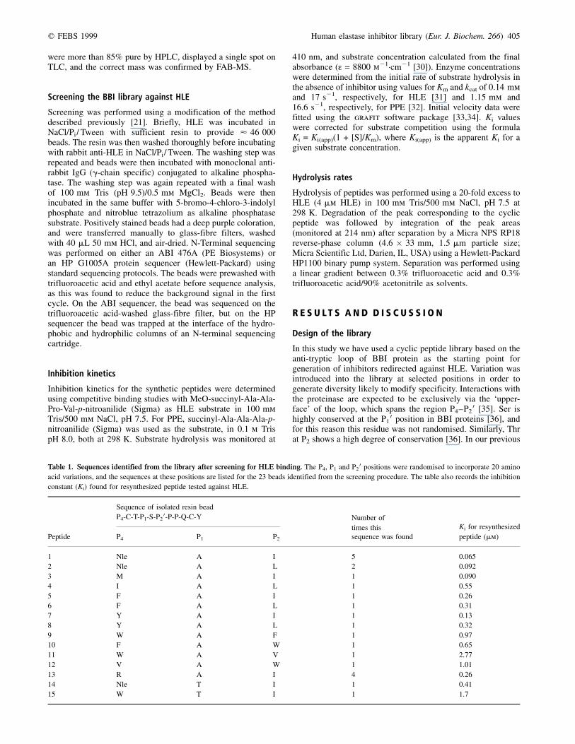
Table 1. Sequences identified from the library after screening for HLE binding. The P4, P1 and P20 positions were randomised to incorporate 20 amino
acid variations, and the sequences at these positions are listed for the 23 beads identified from the screening procedure. The table also records the inhibition
constant (Ki) found for resynthesized peptide tested against HLE.
Sequence of isolated resin bead
P4-C-T-P1-S-P20-P-P-Q-C-Y Number of
times this Ki for resynthesized
Peptide P4 P1 P20 sequence was found peptide (mm)
1 Nle A I 5 0�.065
2 Nle A L 2 0�.092
3 M A I 1 0�.090
4 I A L 1 0�.55
5 F A I 1 0�.26
6 F A L 1 0�.31
7 Y A I 1 0�.13
8 Y A L 1 0�.32
9 W A F 1 0�.97
10 F A W 1 0�.65
11 W A V 1 2�.77
12 V A W 1 1�.01
13 R A I 4 0�.26
14 Nle T I 1 0�.41
15 W T I 1 1�.7
406 J. D. McBride et al. (Eur. J. Biochem. 266) q FEBS 1999
BBI-based library, screening against chymotrypsin resulted inthe identification of only Thr at this locus for all activeinhibitors, although 20 variants were present at this positionwithin the library [21]. Further analysis of all included aminoacids at this locus has confirmed Thr as the optimal residue[36]. In the crystal structures of both BBI±trypsin [35] andOMTKY3±HLE complex [26] (where a Thr residue is alsofound at P2), the P2 Thr is found to make close contact withHis57. Further, in the structure of BBI complexed with trypsin[37], the hydroxyl function of this Thr is involved in hydrogenbonds within the loop structure. As the S2 subsite of HLE isconsidered to be similar to that of other mammalian Serproteinases including PPE [26], this locus was therefore notvaried. In the current communication, we have therefore electedto randomize at the remaining positions of the upper face,namely P1, P2
0 and P4. Each site had one of 20 different aminoacids, giving a library with 8000 different components. Thelibrary was synthesized on TentaGel resin, and included a five-residue polyGly spacer between the 11-residue inhibitortemplate and the resin. This spacer, which was also present inour previous library [21], was included to minimize possiblesteric hindrance between the proteinase and the resin.
Sequences discovered by screening the inhibitor library withHLE
Screening of the library was performed using an indirectenzyme-linked immunoassay. This was done using a relativelylow concentration of HLE with the aim of selecting only themost tightly binding species. Detection of bound HLE thenemployed further incubation with rabbit anti-HLE followed bymonoclonal anti-rabbit IgG (g-chain specific) conjugated toalkaline phosphatase. Visualization of positive beads used theenzyme activity of alkaline phosphatase to generate deeplystained beads [21,24]. The screening procedure identified atotal of 23 highly positive beads from a sample of < 46 000beads that were analysed. This means that < 1 in 2000 peptidesequences were active in the screening assay, a similarproportion to that found for chymotrypsin binding in ourearlier study [21]. The active beads were isolated using a pair ofdissecting tweezers and their peptide sequences determined bythe Edman method using an automated peptide sequencer.Unambiguous identification of the randomized positions waspossible in each case, and the sequences found are listed inTable 1. Each of the different 11-residue core sequences wasresynthesized, purified and analysed for inhibition of HLE insolution. In our earlier study we found that 10 of 13 identifiedsequences were active as inhibitors of chymotrypsin, and theremaining three presumably represented nonspecific binding orthe binding was an artefact [21]. However, in the present studywe found that all 23 peptide sequences inhibited HLE afterresynthesis, and their Ki values are also listed in Table 1. The Ki
values range from 0.065 to 2.77 mm, with 15 differentsequences being identified.
The peptide sequence found most frequently by the screeningprocedure (P4 Nle, P1 Ala, P2
0 Ile) was also found to have thelowest Ki value of those peptides tested, indicating that thescreening process is selective for tight binding. This sequencealso represents the overall consensus for all the positive beadsthat were analysed. It seems likely that the contributions tobinding from each of the three loci varied are additive, so thattheir combination gives the best available inhibitor, as has beendescribed in other proteinase inhibitor interactions [7].Sufficient resin was screened so that, on average, 5.75 copiesof each sequence would be present. The consensus sequence
itself was found on five separate beads, which means it ispossible that all of the beads having this sequence wereidentified by the screening protocol. At least one sequence(peptide 4) returned residues at the randomised positions thatcorrespond to a natural elastase inhibitor, elafin (P4 Ile, P1 Ala,P20 Leu).
The P1 position. The screening results against HLE indicate ahigh preference for alanine in the P1 position, with 21 out of 23of the sequences having Ala at this locus. This result is broadlyconsistent with the known substrate specificity of elastases forsmall alkyl side chains, although it is generally considered thatthe preferred P1 for HLE is valine [38±40], and this has beenthe most widely utilized residue at this locus in the design ofinhibitors [2]. It is therefore quite striking that no P1 sequencescontaining Val were recovered from the screening process.Natural macromolecular inhibitors with activity against HLEare known that have at the P1 position Met (a1-proteinaseinhibitor [41], guamerin [42]), Leu (eglin c [43], mucusproteinase inhibitor [9]) and Ala (elafin [44]). In addition,semisynthetic BPTI-derived inhibitors have been shown todisplay a preference for Val at P1 [45], and the most activeOMTKY3-derived inhibitors have P1 Ile [46]. Two of thesequences listed in Table 1 have a Thr residue at P1. Althoughthese sequences proved to be less potent inhibitors than thecorresponding Ala variants, they were still found to give Ki
values around the micromolar level. It is interesting that neitherthe available substrate specificity data nor the sequenceinformation for natural inhibitors with activity against HLEwould have led us to predict finding Thr at P1, which illustratesthe benefits of library-screening procedures. It should be notedthat Thr has been identified as the P1 residue by sequencealignment in peacock pheasant ovomucoid third domain(OMGPP3; TCTTEHRP, P4-P4
0) but to our knowledge, noactivity has been reported for this protein [47]. Although a P1
Thr is not normally considered to be a usual HLE substrate,hydrolysis by HLE of the Thr87-Thr88 sequence in apolypeptide linking the inhibitory Kunitz-1 and Kunitz-2domains of tissue factor pathway inhibitor has been described[48]. This confirms that HLE is able to interact with a P1 Thrresidue. Replacement of Thr226 in PPE by Asp226 in HLE hassuggested the design of inhibitors with positively charged P1
side chains [49]. However, no positively charged P1 side chainswere detected by the library screening. This is in agreementwith results obtained by Lu et al. [12] for P1 variations toOMTKY3 whereby charged residues and side chains withincreased steric bulk were found to decrease the Ka whencompared with the parent inhibitor (Leu at P1).
The P4 position. A preference for large apolar or aromaticgroups is observed in the P4 position consistent with thestructure of the S4 pocket [26], and NLe is found here with thegreatest frequency. Substrates with Lys at P4 have been found to
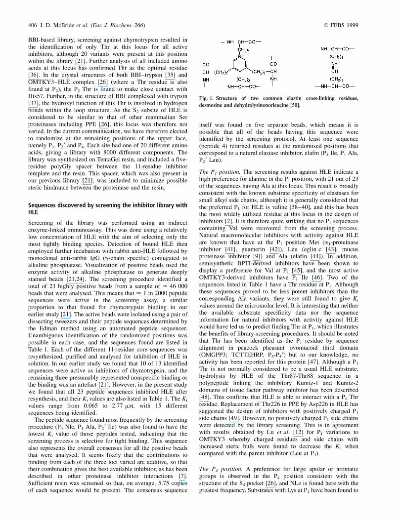
Fig. 1. Structure of two common elastin cross-linking residues,
desmosine and dehydrolysinonorleucine [50].

q FEBS 1999 Human elastase inhibitor library (Eur. J. Biochem. 266) 407
be less reactive than those with P4 Ala while those witharomatic or large hydrophobic groups at this subsite are morereactive [50]. An unexpected finding is Arg in this position for4/23 of the identified inhibitors as, in addition to hydrophobicresidues, Arg is also found in the S4±S5 region of both HLE(Arg217) and PPE. In the TOM complex with HLE, the sidechain of Ala (P4) runs parallel with Arg217 [51], and it has beensuggested that a negatively charged residue at the P4 (or P5)locus would facilitate a favourable electrostatic interaction [51].Indeed, negatively charged groups have been used in this regionof synthetic inhibitors in order to exploit electrostatic inter-actions at this locus [52]. However, the library results do notreflect this. In contrast, the crystal structure of methoxy-succinyl-Ala-Ala-Pro-Ala chloromethyl ketone in complexwith HLE shows that the Arg217 side chain is directed awayfrom the P4 residue [53]. The results are consistent with theobservation that ovomucoid third domains derived fromchestnut bellied scaled quail and Gambel's quail, which differonly at P4 (Asp and Ala, respectively), show a 160-foldreduction in Ka against PPE when an acidic group is present[54]. Arg is found as the P3 residue in both elafin [55] andguamerin [42], and the PPE±elafin structure [56] shows that theP3 Arg guanidino group forms intramolecular hydrogen bondswith carbonyl of P1
0 Met. In the BBI template, P3 is occupiedby one of the two cysteines constraining the peptide and so isnot available for interaction. Interestingly, for the reduction inactivity against HLE reported by Rozycki et al. [17] when theP4±P5 residues of CMTI-III were deleted, Val-Arg are therespective residues (which also had the P1 residue substitutedfor Val in order to redirect activity towards HLE).
Elastin is a highly cross-linked protein rich in Gly, Ala, Valand Pro residues [2,57]. Two common cross-linking residuesare desmosine and dehydrolysinonorleucine (Fig. 1). Both arecharacterized by multiple methylene groups and an iminogroup. The two most frequently found residues from ourscreening (Nle and Arg) have features in common withdehydrolysinonorleucine, which could explain their selection.This finding is consistent with the work of Yasutake & Powers[50], which studied the effect of protected Lys derivatives[Lys(Z), Lys(Bz), Lys(benzimidoyl) and Lys(picolinoyl)] at P2,P3 and P4 as surrogates for desmosine within short peptidesubstrates. In general, it was found that increased reactivitywas obtained at P4 and P3 by these analogues [optimalLys(picolinoyl) at P4]. It was also found that a positive chargeat P4 (Lys) or both a positive charge and aromatic ring[Lys(benzimidoyl)] were detrimental. Taken together, thesefindings suggested that HLE hydrolysis of elastin occurs nearthese cross-links rather than the Lys-rich regions of tropoelastinor those regions of elastin containing noncross-linked Lysresidues. In contrast, PPE was found to be much less reactive
and, in particular, would not accept the Lys and derivativeresidues at P4, which may indicate a more restricted S4 pocket.
The P20 position. The P2
0 position also shows a preference forlarge apolar side chains with Ile being found at this locus withgreatest frequency, and Ile or Leu together accounting for 83%of the sequences identified. Natural endogenous elastaseinhibitors have Ile (a1-proteinase inhibitor) or Leu (mucusproteinase inhibitor, elafin) at P2
0. On the leaving side of thescissile bond, Tyr at P2
0 in the TOM±HLE complex makes themost contact with HLE, principally interacting with Ile151 inthe S2
0 pocket [26]. Crystal structures of the complex withOMTKY3 or with substrate show that in PPE and HLE the S2
0pocket is lined by Leu, Leu and Leu, Ile, respectively [26].Thus the P2
0 specificity is likely to be similar between theseenzymes.
Phe, Trp and Val were also found at this position (Table 1).Phe is present at P2
0 in guamerin [42]. The ovomucoid thirddomains from Indian peafowl and OMTKY3 differ only at P2
0(His and Tyr, respectively). Comparison of their Ka values forPPE shows that the value for OMTKY3 is 44-fold higher [54],which may indicate some preference for hydrophobic residuesover basic residues at this locus.
Effect of P1 variation on activity
In order to compare the results of the library-selected sequenceswith the outcome of P1 alteration alone, the dissociationconstants of selected analogues were recorded, based on theoriginal template sequence (Table 2). The variants synthesizedwere selected on the basis of substrate specificity, andincorporated Ala, Thr, Val or Met at P1. All variants werefound to display considerably lower activity than the library-selected sequences, with P1 Ala having marginally betteractivity than the corresponding Val analogue. The Met variantdisplayed no measurable activity against HLE (or PPE). Inorder of Ki, we find Ala , Val , Thr , Met.
As each of these peptides had a P4 Ser residue (present in theoriginal trypsin inhibitor template) which was not identified inany library-selected sequences, we performed a furthercomparison using an optimal library-selected P4 residue.Table 1 shows that Nle was the most frequent P4 residue, andso we substituted the same P1 residues in the sequenceNleCTXSIPPQCY (Table 3). All variants now show measur-able activity. As Ile is present at P2
0 of both peptide series, asignificant contribution to binding must arise from the P4 Nle.In particular, it is noteworthy that the P1 Met variant only showsmeasurable activity in combination with P4 Nle. The Ki valuesremain in the order Ala , Val , Thr , Met, but are around
Table 2. Effect of P1 variation alone on HLE inhibition constants. The
effect of P1 variation alone was tested within the original nonoptimized
cyclic peptide template (S-C-T-P1-S-I-P-P-Q-C-Y). The inhibition constants
against HLE are tabulated. NI, no inhibition at the concentrations tested;
Ki . 1000 mm.
Peptide P1 Ki (mm)
16 A 3�.4
17 V 4�.6
18 M NI
19 T 15�.9
Table 3. Effect of P1 variation within an optimized sequence on HLE
inhibition constants. The effect of P1 variation was tested within a
sequence that contained an optimized residue at P4 (Nle) (Nle-C-T-P1-S-I-
P-P-Q-C-Y). The inhibition constants against HLE are tabulated.
Ki (mm)
Peptide P1 HLE PPE
20 V 0�.13 6�.7
21 M 11�.4 5�.8
1 A 0�.065 0�.7
14 T 0�.41 1�.5
408 J. D. McBride et al. (Eur. J. Biochem. 266) q FEBS 1999
50-fold lower than with a P4 Ser residue. It should be noted thatthe Val variant has a Ki lower than most of the library-selectedsequences, even though no P1 Val sequences were detected byscreening. The reasons for this are considered below.
Although in our system we find that P1 Ala gives a lower Ki
for HLE than does P1 Val, this is not the case for all systemsthat have been studied. As Val is generally considered to be thepreferred residue at the P1 locus, it is common for researchers toreplace this residue with Val when re-directing the specificity ofnatural inhibitors [58,59]. The Kazal inhibitor BPTI has oftenbeen studied because of its small size (58 residues) and potentinhibitory properties. A number of P1 variants of this proteinhave been prepared by semisynthesis [45]. Of the variantstested against HLE, Val was found to give the lowest Ki
(0.11 nm) and this was 22-fold lower than the Ala variant. Theorder of potency was Val . Nva (also referred to asApe) . aminobutyric acid . allo-isoleucine . Ile ¨ Ala ¨ Leu(the native inhibitor with P1 Lys has a Ki for HLE of 3.5 mm).Similarly, Lu et al. [12] have recently screened a wide range ofnatural and engineered P1 variants of OMTKY3. In this study,the order of potency in terms of Ka was Ile . aminobutyricacid . Val . Nva . Cys . Leu . Thr . Ala . Nle forHLE, and aminobutyric acid . Nva . Nle . Ala .Thr . Leu . Met . Val . Ile for PPE. McRae et al. [38]have used the reactive-site region of a1-proteinase inhibitor toevaluate substrates of HLE and found that the sequence derivedfrom a1-proteinase inhibitor was not optimal. Subtle dif-ferences, for example between Ser and Thr at P1
0, wereobserved primarily in terms of kcat. This result is consistent withour data, and once more demonstrates that optimal inhibitor andsubstrate sequences are not necessarily the same.
Our results clearly show that although the P1 site is importantfor activity, other P and P 0 sites must be optimized in order toobtain good inhibition. Similar conclusions have been drawn byother workers. In an attempt to improve further antielastaseactivity, additional alteration of K15V BPTI at the P1
0 and P20
positions (A16S, R17I) to match those of the a1-proteinaseinhibitor was found to result in a 40-fold decrease in Kd [59].The importance of the P4±S4 interaction in PPE has beendescribed by Thompson & Blout [60]. Using a limited range of
peptide amides, these authors were able to demonstrate P4
interactions that could either strengthen enzyme±substratebinding or those that could increase the rate of hydrolysis.
Effects on hydrolysis rates
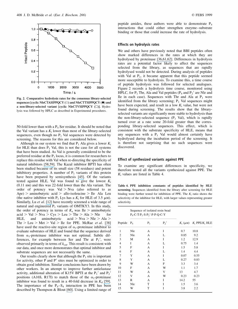
We and others have previously noted that BBI peptides oftenshow marked differences in the rates at which they arehydrolysed by proteinase [36,61,62]. Differences in hydrolysisrates are a potential factor likely to affect the sequencesselected from the library, as sequences that are rapidlyhydrolysed would not be detected. During analysis of peptideswith Val at P1, it became apparent that this peptide seemedmore susceptible to hydrolysis. To examine this, a time courseof peptide hydrolysis was followed for selected analogues.Figure 2 records a hydrolysis time course, monitored usingHPLC, for P1 Thr, Ala and Val peptides (P4 and P2
0 are Nle andIle in each case). Sequences with Thr and Ala at P1 wereidentified from the library screening; P1 Val sequences mighthave been expected, and result in a low Ki value, but were notfound during screening. The results show that the library-selected variants are significantly more stable to hydrolysis thanthe non-library-selected sequence (P1 Val), which is rapidlyturned over at a rate some 20-fold greater than the corres-ponding library-selected sequences. This effect, which isconsistent with the substrate specificity of HLE, means thatany sequences with a P1 Val would almost certainly havehydrolysed during the incubation period of the screening. Itis therefore not surprising that no such sequences werediscovered.
Effect of synthesized variants against PPE
To examine any significant differences in specificity, wetherefore tested all the variants synthesized against PPE. TheKi values are listed in Table 4.
Fig. 2. Comparative hydrolysis rates for the consensus library-selected
sequences [cyclic NleCTASIPPQCY (W) and NleCTTSIPPQCY (X) and
a non-library-selected variant [cyclic NleCTVSIPPQCY (A)]. Hydro-
lysis was followed by HPLC as described in Experimental procedures.
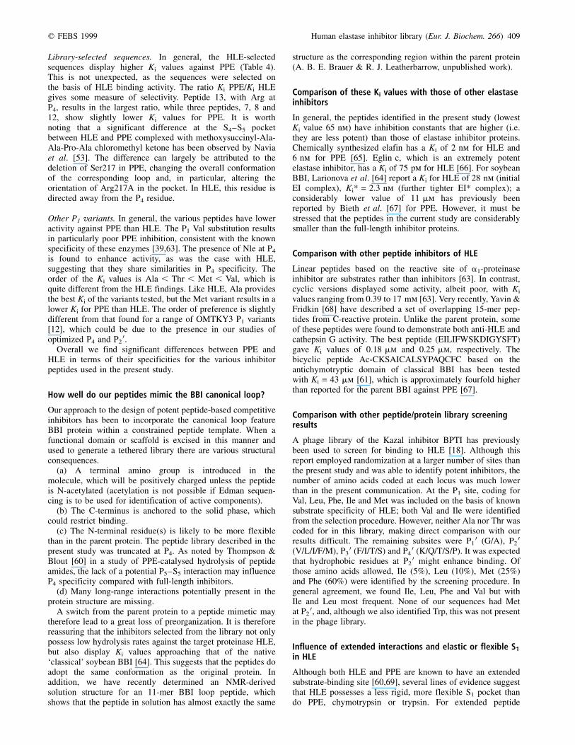
Table 4. PPE inhibition constants of peptides identified by HLE
screening. Sequences identified from the library after screening for HLE
binding were further tested for inhibition of PPE. The Ki ratio shows the
selectivity of the inhibitor for HLE, with larger values representing greater
selectivity.
Sequence of isolated resin bead
P4-C-T-P1-S-P20-P-P-Q-C-Y
Peptide P4 P1 P20 Ki (mm) Ki PPE/Ki HLE
1 Nle A I 0�.7 10�.8
2 Nle A L 0�.85 9�.2
3 M A I 1�.2 12�.9
4 I A L 0�.75 1�.4
5 F A I 1�.5 5�.8
6 F A L 1�.4 4�.4
7 Y A I 0�.07 0�.55
8 Y A L 0�.27 0�.83
9 W A F 3�.3 3�.4
10 F A W 1�.1 1�.7
11 W A V 13� 4�.7
12 V A W 0�.23 0�.23
13 R A I 14�.2 55�
14 Nle T I 1�.5 3�.6
15 W T I 3�.8 2�.2
q FEBS 1999 Human elastase inhibitor library (Eur. J. Biochem. 266) 409
Library-selected sequences. In general, the HLE-selectedsequences display higher Ki values against PPE (Table 4).This is not unexpected, as the sequences were selected onthe basis of HLE binding activity. The ratio Ki PPE/Ki HLEgives some measure of selectivity. Peptide 13, with Arg atP4, results in the largest ratio, while three peptides, 7, 8 and12, show slightly lower Ki values for PPE. It is worthnoting that a significant difference at the S4±S5 pocketbetween HLE and PPE complexed with methoxysuccinyl-Ala-Ala-Pro-Ala chloromethyl ketone has been observed by Naviaet al. [53]. The difference can largely be attributed to thedeletion of Ser217 in PPE, changing the overall conformationof the corresponding loop and, in particular, altering theorientation of Arg217A in the pocket. In HLE, this residue isdirected away from the P4 residue.
Other P1 variants. In general, the various peptides have loweractivity against PPE than HLE. The P1 Val substitution resultsin particularly poor PPE inhibition, consistent with the knownspecificity of these enzymes [39,63]. The presence of Nle at P4
is found to enhance activity, as was the case with HLE,suggesting that they share similarities in P4 specificity. Theorder of the Ki values is Ala , Thr , Met , Val, which isquite different from the HLE findings. Like HLE, Ala providesthe best Ki of the variants tested, but the Met variant results in alower Ki for PPE than HLE. The order of preference is slightlydifferent from that found for a range of OMTKY3 P1 variants[12], which could be due to the presence in our studies ofoptimized P4 and P2
0.Overall we find significant differences between PPE and
HLE in terms of their specificities for the various inhibitorpeptides used in the present study.
How well do our peptides mimic the BBI canonical loop?
Our approach to the design of potent peptide-based competitiveinhibitors has been to incorporate the canonical loop featureBBI protein within a constrained peptide template. When afunctional domain or scaffold is excised in this manner andused to generate a tethered library there are various structuralconsequences.
(a) A terminal amino group is introduced in themolecule, which will be positively charged unless the peptideis N-acetylated (acetylation is not possible if Edman sequen-cing is to be used for identification of active components).
(b) The C-terminus is anchored to the solid phase, whichcould restrict binding.
(c) The N-terminal residue(s) is likely to be more flexiblethan in the parent protein. The peptide library described in thepresent study was truncated at P4. As noted by Thompson &Blout [60] in a study of PPE-catalysed hydrolysis of peptideamides, the lack of a potential P5±S5 interaction may influenceP4 specificity compared with full-length inhibitors.
(d) Many long-range interactions potentially present in theprotein structure are missing.
A switch from the parent protein to a peptide mimetic maytherefore lead to a great loss of preorganization. It is thereforereassuring that the inhibitors selected from the library not onlypossess low hydrolysis rates against the target proteinase HLE,but also display Ki values approaching that of the native`classical' soybean BBI [64]. This suggests that the peptides doadopt the same conformation as the original protein. Inaddition, we have recently determined an NMR-derivedsolution structure for an 11-mer BBI loop peptide, whichshows that the peptide in solution has almost exactly the same
structure as the corresponding region within the parent protein(A. B. E. Brauer & R. J. Leatherbarrow, unpublished work).
Comparison of these Ki values with those of other elastaseinhibitors
In general, the peptides identified in the present study (lowestKi value 65 nm) have inhibition constants that are higher (i.e.they are less potent) than those of elastase inhibitor proteins.Chemically synthesized elafin has a Ki of 2 nm for HLE and6 nm for PPE [65]. Eglin c, which is an extremely potentelastase inhibitor, has a Ki of 75 pm for HLE [66]. For soybeanBBI, Larionova et al. [64] report a Ki for HLE of 28 nm (initialEI complex), Ki* = 2.3 nm (further tighter EI* complex); aconsiderably lower value of 11 mm has previously beenreported by Bieth et al. [67] for PPE. However, it must bestressed that the peptides in the current study are considerablysmaller than the full-length inhibitor proteins.
Comparison with other peptide inhibitors of HLE
Linear peptides based on the reactive site of a1-proteinaseinhibitor are substrates rather than inhibitors [63]. In contrast,cyclic versions displayed some activity, albeit poor, with Ki
values ranging from 0.39 to 17 mm [63]. Very recently, Yavin &Fridkin [68] have described a set of overlapping 15-mer pep-tides from C-reactive protein. Unlike the parent protein, someof these peptides were found to demonstrate both anti-HLE andcathepsin G activity. The best peptide (EILIFWSKDIGYSFT)gave Ki values of 0.18 mm and 0.25 mm, respectively. Thebicyclic peptide Ac-CKSAICALSYPAQCFC based on theantichymotryptic domain of classical BBI has been testedwith Ki = 43 mm [61], which is approximately fourfold higherthan reported for the parent BBI against PPE [67].
Comparison with other peptide/protein library screeningresults
A phage library of the Kazal inhibitor BPTI has previouslybeen used to screen for binding to HLE [18]. Although thisreport employed randomization at a larger number of sites thanthe present study and was able to identify potent inhibitors, thenumber of amino acids coded at each locus was much lowerthan in the present communication. At the P1 site, coding forVal, Leu, Phe, Ile and Met was included on the basis of knownsubstrate specificity of HLE; both Val and Ile were identifiedfrom the selection procedure. However, neither Ala nor Thr wascoded for in this library, making direct comparison with ourresults difficult. The remaining subsites were P1
0 (G/A), P20
(V/L/I/F/M), P30 (F/I/T/S) and P4
0 (K/Q/T/S/P). It was expectedthat hydrophobic residues at P2
0 might enhance binding. Ofthose amino acids allowed, Ile (5%), Leu (10%), Met (25%)and Phe (60%) were identified by the screening procedure. Ingeneral agreement, we found Ile, Leu, Phe and Val but withIle and Leu most frequent. None of our sequences had Metat P2
0, and, although we also identified Trp, this was not presentin the phage library.
Influence of extended interactions and elastic or flexible S1
in HLE
Although both HLE and PPE are known to have an extendedsubstrate-binding site [60,69], several lines of evidence suggestthat HLE possesses a less rigid, more flexible S1 pocket thando PPE, chymotrypsin or trypsin. For extended peptide
410 J. D. McBride et al. (Eur. J. Biochem. 266) q FEBS 1999
substrates, the P1 specificity may be more strict andmodulated by the remote subsites accommodating the extendedsubstrate residues.
Substrates. Increasing substrate length is known to improve thecatalytic efficiency of amide bond hydrolysis by Ser protei-nases, and this is typically reflected in increased kcat [70],which is equal to the rate-limiting acylation step. For HLE, thishas been attributed to the significant effect of subsites onacylation rate. In the absence of an extended chain andtherefore remote interactions, HLE, unlike PPE, displays abroad specificity at P1 [71]; for example, Tyr can be toleratedreasonably well. During hydrolysis of an extended chain, subtlechanges in the structure of HLE result in a narrowing of theS1 pocket [69,71]. Extended interactions appear to fulfilseveral functions: enhancing catalytic efficiency [72]; enlistingoperation of the charge-relay system [69]; regulating P1
specificity [71].
Inhibitors. Adaptation of the P1 binding is also demonstrated bythe structure of HLE in complex with methoxysuccinyl-Ala-Ala-Pro-Val-CH2Cl [73]. The b-branched side chain isaccommodated in P1 with slight tilting of the main chain.Simultaneously, the S1 pocket shrinks and adapts to the smallerside chain. Recent results of P1 variation of TOM also suggestthat in HLE the S1 site is more flexible than in trypsin,S. griseus proteinase A, S. griseus proteinase B, subtilisinCarlsberg or PPE [12]. In this study, b-branched P1 side chainswere found to be highly deleterious to all but HLE. Furtheranalysis of the Ka for the isomeric pair Hse/Thr, revealed thatThr was found to be deleterious for all other proteinases studiedbut advantageous for HLE and PPE. Other evidence suggestingan important role for subsites other than P1 comes from studiesof P1 variation in CMTI-III [16]. Inhibition was found with P1
Val, Ile, Gly, Leu, Ala, Phe and Met; inhibition by P1 Glymeans that there must be contribution from other subsites asthis residue has no side chain.
Conclusions
We have successfully used a tethered peptide library to discovernew HLE inhibitors. Using a library allows more efficientscreening than could be obtained by testing individualinhibitors, which is exemplified by the library-selectedsequences giving lower Ki values than could be obtained bysimple P1 substitutions alone. Our results show that thescreening procedure used identifies those sequences that offerthe best compromise between low Ki and low hydrolysis rate. Inthe context of finding the `best' inhibitor sequence, thisrepresents an obvious benefit. One surprising finding is that,contrary to expectations, sequences that correspond to theoptimal substrate specificity do not necessarily make the bestinhibitors (at least in the system studied). This is exemplifiedby comparison of sequences with P1 Val or Ala. The Val-containing sequences are better substrates, but Ala variants arebetter inhibitors in this particular system. Clearly, this hassignificant implications for proteinase inhibitor design, whichoften relies on substrate specificity data.
A C K N O W L E D G E M E N T S
This work was supported by the BBSRC and Glaxo-Wellcome Research.
We would like to thank the following people: John Barton (Department of
Chemistry, Imperial College) for performing the mass spectroscopy;
Norman Gray (Glaxo-Wellcome) for his assistance with the hydrolysis
assays; Robin Smith (Glaxo-Wellcome) for the gift of HLE. We are
indebted to Malcolm Weir of Glaxo-Wellcome for his continued support
and encouragement. Presented in part at the Fifth International Symposium
on Solid Phase Synthesis & Combinatorial Chemical Libraries, Imperial
College, University of London, UK, 2±6 September 1997.
R E F E R E N C E S
1. Geokas, M.C., Rinderknecht, H., Swanson, V. & Haverback, B.J.
(1968) The role of elastase in acute hemorrhagic pancreatitis in man.
Lab. Invest. 19, 235±239.
2. Edwards, P.D. & Bernstein, P.R. (1994) Synthetic inhibitors of elastase.
Med. Res. Rev. 14, 127±194.
3. Laskowski, M. Jr & Kato, I. (1980) Protein inhibitors of proteinases.
Annu. Rev. Biochem. 49, 593±626.
4. Bode, W. & Huber, R. (1992) Natural protein proteinase-inhibitors and
their interaction with proteinases. Eur. J. Biochem. 204, 433±451.
5. Schechter, I. & Berger, A. (1967) On the size of the active site in
proteases. I. Papain. Biochem. Biophys. Res. Commun. 27, 157±162.
6. Bode, W. & Huber, R. (1991) Ligand binding: proteinase±protein
inhibitor interactions. Curr. Opin. Struct. Biol. 1, 45±52.
7. Qasim, M.A., Ganz, P.J., Saunders, C.W., Bateman, K.S., James,
M.N.G. & Laskowski, M. (1997) Interscaffolding additivity.
Association of P-1 variants of eglin C and of turkey ovomucoid
third domain with serine proteinases. Biochemistry 36, 1598±1607.
8. Kato, I., Schrode, J., Kohr, W.J. & Laskowski, M. (1987) Chicken
ovomucoid: determination of its amino acid sequence, determination
of the trypsin reactive site, and preparation of all 3 of its domains.
Biochemistry 26, 193±201.
9. Boudier, C. & Bieth, J.G. (1994) Oxidized mucus proteinase inhibitor:
a fairly potent neutrophil elastase inhibitor. Biochem. J. 303, 61±68.
10. Ardelt, W. & Laskowski, M. (1985) Turkey ovomucoid 3rd domain
inhibits 8 different serine proteinases of varied specificity on the
same ¼Leu18-Glu19¼ reactive site. Biochemistry 24, 5313±5320.
11. Longstaff, C., Campbell, A.F. & Fersht, A.R. (1990) Recombinant
chymotrypsin inhibitor-2: expression, kinetic analysis of inhibition
with alpha-chymotrypsin and wild-type and mutant subtilisin Bpn 0,and protein engineering to investigate inhibitory specificity and
mechanism. Biochemistry 29, 7339±7347.
12. Lu, W.Y., Apostol, I., Qasim, M.A., Warne, N., Wynn, R., Zhang, W.L.,
Anderson, S., Chiang, Y.W., Ogin, E., Rothberg, I., Ryan, K. &
Laskowski, M. (1997) Binding of amino acid side-chains to S-1
cavities of serine proteinases. J. Mol. Biol. 266, 441±461.
13. Nishino, N., Aoyagi, H., Kato, T. & Izumiya, N. (1977) Studies
on the synthesis of proteinase inhibitors. I. Synthesis and
activity of nonapeptide fragments of soybean Bowman-Birk inhibitor.
J. Biochem. (Tokyo) 82, 901±909.
14. Favel, A., Lenguyen, D., Colettipreviero, M.A. & Castro, B. (1989)
Active-site chemical mutagenesis of ecballium-elaterium trypsin
inhibitor-II: new microproteins inhibiting elastase and chymotrypsin.
Biochem. Biophys. Res. Commun. 162, 79±82.
15. Carrell, R. (1984) Therapy by instant evolution. Nature (London) 312,
14.
16. McWherter, C.A., Walkenhorst, W.F., Campbell, E.J. & Glover, G.I.
(1989) Novel inhibitors of human-leukocyte elastase and cathe-
psin G: sequence variants of squash seed protease inhibitor with
altered protease selectivity. Biochemistry 28, 5708±5714.
17. Rozycki, J., Kupryszewski, G., Rolka, K., Ragnarsson, U., Zbyryt, T.,
Watorek, W. & Wilusz, T. (1997) The smallest peptide inhibitors of
human leukocyte elastase (HLE): structural requirements for
inhibition of HLE based on Cucurbita maxima trypsin inhibitor III
(CMTI-III) analogues. Pol. J. Chem. 71, 176±180.
18. Roberts, B.L., Markland, W., Ley, A.C., Kent, R.B., White, D.W.,
Guterman, S.K. & Ladner, R.C. (1992) Directed evolution of a
protein: selection of potent neutrophil elastase inhibitors displayed on
M13 fusion phage. Proc. Natl Acad. Sci. USA 89, 2429±2433.
19. Markland, W., Ley, A.C., Lee, S.W. & Ladner, R.C. (1996) Iterative
optimization of high-affinity protease inhibitors using phage display.
1. Plasmin. Biochemistry 35, 8045±8057.
q FEBS 1999 Human elastase inhibitor library (Eur. J. Biochem. 266) 411
20. Markland, W., Ley, A.C. & Ladner, R.C. (1996) Iterative optimization
of high-affinity protease inhibitors using phage display. 2. Plasma
kallikrein and thrombin. Biochemistry 35, 8058±8067.
21. McBride, J.D., Freeman, N., Domingo, G.J. & Leatherbarrow, R.J.
(1996) Selection of chymotrypsin inhibitors from a confor-
mationally-constrained combinatorial peptide library. J. Mol.
Biol. 259, 819±827.
22. Odani, S. & Ikenaka, T. (1973) Scission of soybean Bowman-Birk
proteinase inhibitor into two small fragments having either trypsin or
chymotrypsin inhibitory activity. J. Biochem. (Tokyo) 74, 857±860.
23. Terada, S., Sato, K., Kato, T. & Izumiya, N. (1978) Inhibitory
properties of nonapeptide loop structures related to reactive sites of
soybean Bowman-Birk inhibitor. FEBS Lett. 90, 89±92.
24. Lam, K.S., Salmon, S.E., Hersh, E.M., Hruby, V.J., Kazmierski, W.M.
& Knapp, R.J. (1991) A new type of synthetic peptide library for
identifying ligand-binding activity. Nature (London) 354, 82±84.
25. Furka, A., Sebestyen, F., Asgedom, M. & Dibo, G. (1991) General
method for rapid synthesis of multicomponent peptide mixtures. Int.
J. Pept. Protein Res. 37, 487±493.
26. Bode, W., Meyer, E. & Powers, J.C. (1989) Human leukocyte and
porcine pancreatic elastase: X-ray crystal structures, mechanism,
substrate-specificity, and mechanism-based inhibitors. Biochemistry
28, 1951±1963.
27. Knorr, R., Trzeciak, A., Bannwarth, W. & Gillessen, D. (1989)
New coupling reagents in peptide chemistry. Tetrahedron Lett. 30,
1927±1930.
28. Tam, J.P., Wu, C.R., Liu, W. & Zhang, J.W. (1991) Disulfide bond
formation in peptides by dimethyl sulfoxide: scope and applications.
J. Am. Chem. Soc. 113, 6657±6662.
29. McBride, J.D., Harbeck, B. & Leatherbarrow, R.J. (1996) Resin-
coupled cyclic peptides as proteinase inhibitors. Protein Peptide Lett.
3, 193±198.
30. Erlanger, B., Kokowski, N. & Cohen, W. (1961) The preparation and
properties of two new chromogenic substrates of trypsin. Arch.
Biochem. Biophys. 95, 271±278.
31. Nakajima, K., Powers, J.C., Ashe, B.M. & Zimmerman, M. (1979)
Mapping the extended substrate binding site of cathepsin G and
human leukocyte elastase. Studies with peptide substrates related
to the alpha 1-protease inhibitor reactive site. J. Biol. Chem. 254,
4027±4032.
32. Bieth, J., Spiess, B. & Wermuth, C.G. (1974) The synthesis and
analytical use of a highly sensitive and convenient substrate of
elastase. Biochem. Med. 11, 350±357.
33. Leatherbarrow, R.J. (1992) GraFit 3.0. Erithacus Software Ltd, Staines,
UK.
34. Leatherbarrow, R.J. (1990) Using linear and nonlinear-regression to fit
biochemical data. Trends Biochem. Sci. 15, 455±458.
35. Lin, G.D., Bode, W., Huber, R., Chi, C.W. & Engh, R.A. (1993) The
0.25-nm X-ray structure of the Bowman±Birk-type inhibitor from
mung bean in ternary complex with porcine trypsin. Eur. J. Biochem.
212, 549±555.
36. McBride, J.D., Brauer, A.B.E., Nievo, M. & Leatherbarrow, R.J. (1998)
The role of threonine in the P-2 position of Bowman±Birk proteinase
inhibitors: studies on P-2 variation in cyclic peptides encompassing
the reactive site loop. J. Mol. Biol. 282, 447±457.
37. Voss, R.H., Ermler, U., Essen, L.O., Wenzl, G., Kim, Y.M. & Flecker, P.
(1996) Crystal structure of the bifunctional soybean Bowman±Birk
inhibitor at 0.28-nm resolution: structural peculiarities in a folded
protein conformation. Eur. J. Biochem. 242, 122±131.
38. McRae, B., Nakajima, K., Travis, J. & Powers, J.C. (1980) Studies on
reactivity of human leukocyte elastase, cathepsin G, and porcine
pancreatic elastase toward peptides including sequences related to the
reactive site of alpha 1-protease inhibitor (alpha 1-antitrypsin).
Biochemistry 19, 3973±3978.
39. Harper, J.W., Cook, R.R., Roberts, C.J., McLaughlin, B.J. & Powers,
J.C. (1984) Active-site mapping of the serine proteases human-
leukocyte elastase, cathepsin g, porcine pancreatic elastase, rat
mast-cell protease-I and protease-Ii, bovine chymotrypsin-a and
Staphylococcus aureus protease V-8 using tripeptide thiobenzyl ester
substrates. Biochemistry 23, 2995±3002.
40. Stein, R.L., Trainer, D.A. & Wildonger, R.A. (1985) Neutrophil
elastase. Annu. Rep. Med. Chem. 20, 237±246.
41. Carrell, R., Jeppsson, J.O., Laurell, C.B., Brennan, S.O., Owen, M.C.,
Vaughan, L. & Boswell, D.R. (1982) Structure and variation of
human a1-antitrypsin. Nature (London) 298, 329±334.
42. Jung, H.I., Kim, S.I., Ha, K.S., Joe, C.O. & Kang, K.W. (1995)
Isolation and characterization of guamerin, a new human-
leukocyte elastase inhibitor from Hirudo±Nipponia. J. Biol.
Chem. 270, 13879±13884.
43. Heinz, D.W., Hyberts, S.G., Peng, J.W., Priestle, J.P., Wagner, G. &
Grutter, M.G. (1992) Changing the inhibitory specificity and function
of the proteinase inhibitor eglin-c by site-directed mutagenesis:
functional and structural investigation. Biochemistry 31, 8755±8766.
44. Ying, Q.L. & Simon, S.R. (1993) Kinetics of the inhibition of human-
leukocyte elastase by elafin, a 6-kilodalton elastase-specific inhibitor
from human skin. Biochemistry 32, 1866±1874.
45. Beckmann, J., Mehlich, A., Schroder, W., Wenzel, H.R. & Tschesche,
H. (1988) Preparation of chemically mutated aprotinin homologs
by semisynthesis: P1 substitutions change inhibitory specificity.
Eur. J. Biochem. 176, 675±682.
46. Bigler, T.L., Lu, W.Y., Park, S.J., Tashiro, M., Wieczorek, M., Wynn, R.
& Laskowski, M. (1993) Binding of amino-acid side-chains to
preformed cavities: interaction of serine proteinases with turkey
ovomucoid 3rd domains with coded and noncoded P1 residues.
Protein Sci. 2, 786±799.
47. Laskowski, M., Kato, I., Ardelt, W., Cook, J., Denton, A., Empie,
M.W., Kohr, W.J., Park, S.J., Parks, K., Schatzley, B.L.,
Schoenberger, O.L., Tashiro, M., Vichot, G., Whatley, H.E.,
Wieczorek, A. & Wieczorek, M. (1987) Ovomucoid 3rd domains
from 100 avian species: isolation, sequences, and hypervariability of
enzyme-inhibitor contact residues. Biochemistry 26, 202±221.
48. Petersen, L.C., Bjorn, S.E., Olsen, O.H., Nordfang, O., Norris, F. &
Norris, K. (1996) Inhibitory properties of separate recombinant
Kunitz-type-protease-inhibitor domains from tissue-factor-pathway
inhibitor. Eur. J. Biochem. 235, 310±316.
49. Sinha, S., Watorek, W., Karr, S., Giles, J., Bode, W. & Travis, J. (1987)
Primary structure of human neutrophil elastase. Proc. Natl Acad. Sci.
USA 84, 2228±2232.
50. Yasutake, A. & Powers, J.C. (1981) Reactivity of human-leukocyte
elastase and porcine pancreatic elastase toward peptide 4-nitroani-
lides containing model desmosine residues: evidence that human-
leukocyte elastase is selective for cross-linked regions of elastin.
Biochemistry 20, 3675±3679.
51. Bode, W., Wei, A.Z., Huber, R., Meyer, E., Travis, J. & Neumann, S.
(1986) X-ray crystal structure of the complex of human-leukocyte
elastase (PMN elastase) and the 3rd domain of the turkey ovomucoid
inhibitor. EMBO J. 5, 2453±2458.
52. Regan, J., McGarry, D., Bruno, J., Green, D., Newman, J., Hsu, C.Y.,
Kline, J., Barton, J., Travis, J., Choi, Y.M., Volz, F., Pauls, H.,
Harrison, R., Zilberstein, A., BenSasson, S.A. & Chang, M. (1997)
Anionic- and lipophilic-mediated surface binding inhibitors of human
leukocyte elastase. J. Med. Chem. 40, 3408±3422.
53. Navia, M.A., McKeever, B.M., Springer, J.P., Lin, T.Y., Williams, H.R.,
Fluder, E.M., Dorn, C.P. & Hoogsteen, K. (1989) Structure of human
neutrophil elastase in complex with a peptide chloromethyl ketone
inhibitor at 1.84-AÊ resolution. Proc. Natl Acad. Sci. USA 86, 7±11.
54. Empie, M.W. & Laskowski, M. (1982) Thermodynamics and kinetics
of single residue replacements in avian ovomucoid 3rd domains:
effect on inhibitor interactions with serine proteinases. Biochemistry
21, 2274±2284.
55. Wiedow, O., Schroder, J.M., Gregory, H., Young, J.A. & Christophers,
E. (1990) Elafin ± an elastase-specific inhibitor of human skin:
purification, characterization, and complete amino acid sequence.
J. Biol. Chem. 265, 14791±14795.
56. Tsunemi, M., Matsuura, Y., Sakakibara, S. & Katsube, Y. (1996)
Crystal structure of an elastase-specific inhibitor elafin complexed
412 J. D. McBride et al. (Eur. J. Biochem. 266) q FEBS 1999
with porcine pancreatic elastase determined at 1.9 angstrom
resolution. Biochemistry 35, 11570±11576.
57. Gowda, D.C., Luan, C.H., Furner, R.L., Peng, S.Q., Jing, N., Harris,
C.M., Parker, T.M. & Urry, D.W. (1995) Synthesis and characteri-
zation of the human elastin W4 sequence. Int. J. Pept. Protein Res.
46, 453±463.
58. Rosenberg, S., Barr, P.J., Najarian, R.C. & Hallewell, R.A. (1984)
Synthesis in yeast of a functional oxidation-resistant mutant of human
alpha-1-antitrypsin. Nature (London) 312, 77±80.
59. Wenzel, H.R. & Tschesche, H. (1995) Reversible inhibitors of serine
proteinases. In Peptides Synthesis, Structures, and Applications
(Gutte, B., ed.), pp. 321±362. Academic Press, San Diego, CA.
60. Thompson, R.C. & Blout, E.R. (1973) Dependence of the kinetic
parameters for elastase-catalyzed amide hydrolysis on the length of
peptide substrates. Biochemistry 12, 57±65.
61. Ando, S., Yasutake, A., Waki, M., Nishino, N., Kato, T. & Izumiya, N.
(1987) Anti-chymotrypsin and anti-elastase activities of a synthetic
bicyclic fragment containing a chymotrypsin-reactive site of soybean
Bowman-Birk inhibitor. Biochim. Biophys. Acta 916, 527±531.
62. Gariani, T. & Leatherbarrow, R.J. (1997) Stability of protease inhibitors
based on the Bowman±Birk reactive site loop to hydrolysis by
proteases. J. Pept. Res. 49, 467±475.
63. Powers, J.C., Yasutake, A., Nishino, N., Gupton, B.F. & Kam, C.M.
(1981) Synthetic elastase inhibitors and their role in the treatment of
disease. In Peptides: Synthesis, Structure, Function (Rich, D.H. &
Gross, E., eds), pp. 391±399. Pierce Chemical Co., Rockford, IL.
64. Larionova, N.I., Gladysheva, I.P. & Gladyshev, D.P. (1997) Human
leukocyte elastase inhibition by Bowman-Birk soybean inhibitor;
discrimination of the inhibition mechanisms. FEBS Lett. 404,
245±248.
65. Tsunemi, M., Kato, H., Nishiuchi, Y., Kumagaye, S. & Sakakibara, S.
(1992) Synthesis and structure-activity relationships of elafin, an
elastase-specific inhibitor. Biochem. Biophys. Res. Commun. 185,
967±973.
66. Braun, N.J., Bodmer, J.L., Virca, G.D., Metzvirca, G., Maschler, R.,
Bieth, J.G. & Schnebli, H.P. (1987) Kinetic studies on the interaction
of eglin-c with human-leukocyte elastase and cathepsin-G. Biol.
Chem. Hoppe Seyler 368, 299±308.
67. Bieth, J.G., Kandel, M.J., Zreika, M. & Pochon, F. (1983) Interaction of
protein substrate and inhibitors with alpha-2-macroglobulin-bound
proteinases. Ann. N.Y. Acad. Sci. 421, 209±217.
68. Yavin, E.J. & Fridkin, M. (1998) Peptides derived from human
C-reactive protein inhibit the enzymatic activities of human leukocyte
elastase and cathepsin G: use of overlapping peptide sequences to
identify a unique inhibitor. J. Pept. Res. 51, 282±289.
69. Stein, R.L. (1985) Catalysis by human-leukocyte elastase. 4. Role of
secondary-subsite interactions. J. Am. Chem. Soc. 107, 5767±5775.
70. Fersht, A.R. (1974) Catalysis, binding and enzyme-substrate comple-
mentarity. Proc. R. Soc. Lond. B. Biol. Sci. 187, 397±407.
71. Stein, R.L. (1985) Catalysis by human-leukocyte elastase. 3. Steady-
state kinetics for the hydrolysis of para-nitrophenyl esters. Arch.
Biochem. Biophys. 236, 677±680.
72. Kraut, J. (1977) Serine proteases: structure and mechanism of catalysis.
Annu. Rev. Biochem. 46, 331±358.
73. Wei, A.Z., Mayr, I. & Bode, W. (1988) The refined 2.3 AÊ crystal-
structure of human-leukocyte elastase in a complex with a valine
chloromethyl ketone inhibitor. FEBS Lett. 234, 367±373.
Copyright © 2022 FDOKUMEN