Inclusion of Sevoflurane in Cardioplegia Reduces Neutrophil Activity During Cardiopulmonary Bypass

10.1128/IAI.68.3.1125-1133.2000.
2000, 68(3):1125. DOI:Infect. Immun. RikihisaKiyotaka Yoshiie, Hyung-Yong Kim, Jason Mott and Yasuko Human Neutrophil ApoptosisGranulocytic Ehrlichiosis Agent Inhibits Intracellular Infection by the Human
http://iai.asm.org/content/68/3/1125Updated information and services can be found at:
These include:
REFERENCEShttp://iai.asm.org/content/68/3/1125#ref-list-1at:
This article cites 44 articles, 29 of which can be accessed free
CONTENT ALERTS more»articles cite this article),
Receive: RSS Feeds, eTOCs, free email alerts (when new
http://journals.asm.org/site/misc/reprints.xhtmlInformation about commercial reprint orders: http://journals.asm.org/site/subscriptions/To subscribe to to another ASM Journal go to:
on Decem
ber 5, 2013 by guesthttp://iai.asm
.org/D
ownloaded from
on D
ecember 5, 2013 by guest
http://iai.asm.org/
Dow
nloaded from

INFECTION AND IMMUNITY,0019-9567/00/$04.0010
Mar. 2000, p. 1125–1133 Vol. 68, No. 3
Copyright © 2000, American Society for Microbiology. All Rights Reserved.
Intracellular Infection by the Human Granulocytic EhrlichiosisAgent Inhibits Human Neutrophil Apoptosis
KIYOTAKA YOSHIIE,† HYUNG-YONG KIM, JASON MOTT, AND YASUKO RIKIHISA*
Department of Veterinary Biosciences, The Ohio State University, Columbus, Ohio 43210-1093
Received 13 August 1999/Returned for modification 7 October 1999/Accepted 23 November 1999
In patients with human granulocytic ehrlichiosis (HGE), the HGE agent has been seen only in the peripheralblood granulocytes, which have a life span too short for ehrlichial proliferation. To determine if the HGE agentdelays the apoptosis of human peripheral blood neutrophils for its advantage, peripheral blood granulocytesconsisting mostly of neutrophils were incubated with freshly freed host cell-free HGE agent in vitro. The HGEagent induced a significant delay in morphological apoptosis and the cytoplasmic appearance of histone-associated DNA fragments in the granulocytes. This antiapoptotic effect was dose dependent. Although muchweaker than the HGE agent freshly freed from the host cells, noninfectious purified HGE agent stored frozenand thawed also had antiapoptotic effect, which was lost with proteinase K treatment but not with periodatetreatment. Treatment of neutrophils with a transglutaminase inhibitor, monodansylcadaverine, blocked theantiapoptotic effect of the HGE agent. Addition of oxytetracycline, however, did not prevent or reverse theantiapoptotic effect of the HGE agent. These results suggest that binding of a protein component(s) of the HGEagent to neutrophils and subsequent cross-linking and/or internalization of the receptor and ehrlichiae arerequired for antiapoptotic signaling, but ehrlichial protein synthesis and/or proliferation is not required.MG-132, a proteasome inhibitor, and cycloheximide accelerated the apoptosis of neutrophils and overrode theantiapoptotic effect of the HGE agent. Studies with specific inhibitors suggest that protein kinase A, NF-kB,and interleukin 1b are not involved in the antiapoptotic mechanism of the HGE agent.
Human granulocytic ehrlichiosis (HGE) is a newly recog-nized tick-borne zoonosis in the United States and Europe (3,5, 6, 8, 9). HGE is an acute febrile systemic disease associatedwith hematologic abnormalities, such as thrombocytopenia andleukopenia, as well as increased serum aminotransferase activ-ity. HGE may be fatal when patients are immunocompromisedor antibiotic treatment is delayed. The etiologic agent, calledthe HGE agent, is a small gram-negative coccus closely relatedto previously known granulocytotropic Ehrlichia spp.—Ehrli-chia phagocytophila, the agent of tick-borne fever in sheep andgoats, and Ehrlichia equi, the agent of equine ehrlichiosis (9).These bacteria are incapable of extracellular survival and areseen to replicate in the cytoplasm of peripheral blood granu-locytes. A small number of individually dispersed ehrlichiaeare difficult to recognize under the light microscope. However,microcolonies, called morulae, that result from ehrlichial rep-lication stain dark blue to purple with Romanowsky dye; theyare large and have a characteristic morphology and thus can berecognized by the trained eye (34). All 12 patients initiallyreported in Minnesota and Wisconsin had detectable morulaein 1 to 41% of their peripheral blood granulocytes (5). Inanother study, intracytoplasmic morulae were seen in 3 of 12patients in New York, and the frequency of infected granulo-cytes ranged from 0.3 to 6% (3). The majority of infectedgranulocytes seen were neutrophils. Mature neutrophils un-dergo rapid apoptosis (programmed cell death), with a half-lifeof several hours in vivo (11). Considering the low growth rateof cytoplasmic ehrlichiae and the important roles neutrophilsplay in host defense and acute inflammation, the life span of
neutrophils is a critical determinant for ehrlichial survival andHGE pathogenesis. Recently, the HGE agent was reported toinduce apoptosis in the human promyelocytic leukemia cellline HL-60 (17). However, no information is available onwhether the HGE agent modulates apoptosis of human neu-trophils, its natural host. In the present study, we examinedwhether the HGE agent alters constitutive apoptosis of humanperipheral blood neutrophils in vitro. Furthermore, mecha-nisms by which apoptosis is delayed by the HGE agent wereexamined by using an HGE agent treated with various reagentsand host cells inhibited in intracellular signaling pathways. Thisstudy, therefore, is helpful in understanding not only the in-tracellular survival strategy of the HGE agent but also the roleof neutrophils in the pathogenesis of HGE.
MATERIALS AND METHODS
HGE agent and cell culture. HGE isolate HZ (37) was cultivated in HL-60cells in RPMI 1640 medium (GIBCO, Grand Island, N.Y.) supplemented with5% fetal bovine serum (Atlanta Biologicals, Norcross, Ga.), 2 mM L-glutamine(GIBCO), 0.1 mM minimal essential medium-nonessential amino acid mixture(GIBCO), and 1 mM minimal essential medium-sodium pyruvate (GIBCO).When .90% of the cells were infected, as determined by examining cells stainedwith Diff-Quik (Baxter Scientific Products, Obetz, Ohio), the infected cells weresonicated and centrifuged at 500 3 g for 5 min. The supernatant was centrifugedat 10,000 3 g for 10 min, and the pellet, containing the host cell-free, viable HGEagent, was immediately used to infect human peripheral blood neutrophils orperipheral blood leukocytes (PBL). Because the HGE agent is small and multi-plies as microcolonies, it is impractical to accurately count individual organisms.Therefore, the number of host cell-free ehrlichiae was estimated by using thefollowing formula: number of ehrlichial organisms 5 total infected cell num-ber 3 average number of morulae in an infected cell (typically five) 3 averagenumber of ehrlichial organisms in a morula (typically 19) 3 percentage ofehrlichiae recovered as host cell free (typically 50% as determined by usingmetabolically [35S]methionine-labeled ehrlichiae [36]).
Purified HGE agent was prepared by brief sonication, differential centrifuga-tion, and Sephacryl S-1000 chromatography and was kept frozen at 280°C untilit was used, as previously described (35). Periodate-treated ehrlichiae wereprepared by incubating purified HGE agent with 20 mM sodium periodate(Sigma Chemical Co., St. Louis, Mo.) in 50 mM sodium acetate buffer (pH 4.5)for 1 h at room temperature in the dark followed by incubation with 50 mMsodium borohydride (Sigma) in sterile phosphate-buffered saline (PBS; 2.7 mM
* Corresponding author. Mailing address: Department of Veteri-nary Biosciences, College of Veterinary Medicine, The Ohio StateUniversity, 1925 Coffey Rd., Columbus, OH 43210-1093. Phone: (614)292-5661. Fax: (614) 292-6473. E-mail: [email protected].
† Present address: Department of Bacteriology, Faculty of Medi-cine, Kagoshima University, Kagoshima 890-8520, Japan.
1125
on Decem
ber 5, 2013 by guesthttp://iai.asm
.org/D
ownloaded from

KCl–1.8 mM KH2PO4–137 mM NaCl–10 mM NaH2PO4, pH 7.4) for 30 min atroom temperature (21, 22). For proteinase K treatment, the purified HGE agentwas incubated in 1 mg of proteinase K (GIBCO)/ml in distilled water at 60°C for2 h. After incubation, 1 mM phenylmethylsulfonyl fluoride (Sigma) was added,the mixture was incubated for 10 min at 60°C, and then the ehrlichiae werewashed three times in RPMI 1640 medium (21, 22).
Neutrophils were isolated from buffy coats from healthy donors (Ohio RedCross, Columbus, Ohio). The buffy coat was overlaid on double layers of His-topaque 1077 and 1119 (Sigma) and centrifuged at 700 3 g for 15 min. Neutro-phils at the interface between the 1077 and 1119 were collected and washed twicewith PBS followed by hemolysis in 0.83% ammonium chloride (Fisher Scientific,Fair Lawn, N.J.) for 5 min at room temperature. Of the total cells, .98% wereneutrophils in morphology, as demonstrated by Diff-Quik staining, and .98%were viable by the trypan blue dye exclusion test. PBL were obtained from buffycoats by simply lysing erythrocytes as described above.
Treatment. After being washed twice, purified neutrophils or PBL were sus-pended at 106 cells/ml in RPMI 1640 medium. Uninfected HL-60 cells, preparedby brief sonication of the cell suspension at 106 cells/ml followed by differentialcentrifugation as previously described (21), and the purified HGE agent pre-pared as described above was added at a final concentration of 100 mg ofprotein/ml. Host cell-free HGE agent was added to purified neutrophils at amultiplicity of infection (MOI) of 100 immediately after the isolation of the HGEagent. Treated or infected neutrophil suspensions were seeded in 96-well flat-bottom plates (Becton Dickinson Co., Franklin Lakes, N.J.) at 200 ml/well intriplicate wells per assay point, and these samples were incubated at 37°C in a 5%CO2 atmosphere for #96 h. Cells were harvested every 8 h. To consider indi-vidual human variations, all experiments were independently repeated two orthree times on different days using neutrophils derived from different donors andthe freshly prepared host cell-free HGE agent each time. Donor cells were nevermixed, and each donor neutrophil assay included positive and negative controlsto ensure the quality of both neutrophil and HGE agent preparation.
For monodansylcadaverine (MDC) treatment, fresh human neutrophils weresuspended in RPMI medium with or without 250 mM MDC, plated in triplicateat 2 3 106 cells/well in a 24-well plate, and incubated for 30 min at 37°C in 95%air–5% CO2. The freshly prepared host cell-free HGE agent in RPMI mediumwas added to each well, and the mixture was incubated for 2 h at 37°C. The cellswere centrifuged at 500 3 g for 5 min, treated with 2 mg of pronase/ml in sterilePBS for 5 min at 37°C to remove extracellular ehrlichiae, washed two times withRPMI medium, and replated in medium without MDC. In another experiment,cells were incubated continuously without removing MDC and the extracellularHGE agent. Oxytetracycline (10-mg/ml final concentration) was added to eachwell at 0 or 8 h after the addition of freshly prepared host cell-free HGE agent.The cells were incubated at 37°C and cytocentrifuged to determine the percentapoptosis and percent infected neutrophils.
H-89 (1 or 10 mM; BIOMOL Research Laboratories, Plymouth Meeting, Pa.),50 mM genistein (Sigma), 100 mM MG-132 (BIOMOL), 100 mg of SN-50(BIOMOL)/ml, 2 mg of cycloheximide (Sigma)/ml, and 50 ng of blocking mono-clonal antibody to recombinant human interleukin 1b (IL-1b) (clone 8516.311; R& D Systems, Minneapolis, Minn.)/ml or 100 mM acetyl-Tyr-Val-Ala-Asp-chlo-romethyl ketone (YVAD-CMK) (Calbiochem, San Diego, Calif.) were added totriplicate wells at 0 h or 8 h (10 mM H-89 only) after the addition of HGE agent.
Microscopic determination of apoptosis. Cell suspensions (80 ml) were cen-trifuged on a glass slide at 30 3 g for 1 min with Cytospin 3 (Shandon Inc.,Pittsburgh, Pa.). The cells were stained with Diff-Quik and examined microscop-ically at 31,000. Apoptotic cells were determined based on their morphology,including densely condensed and homogeneous nuclei, loss of connection be-tween the lobules of nuclei, and eosinophilic cytoplasm (Fig. 1). A total of 500cells were scored from each well. The time required for 50% of the neutrophilsto show morphological apoptosis (T50) was determined by plotting the percent-age of apoptotic cells observed at each incubation time point.
Detection of histone-associated DNA fragments. To detect the fragmentationof DNA in apoptotic neutrophils, histone-associated DNA fragments were ex-
FIG. 1. Morphological apoptosis of human peripheral blood neutrophils incubated in vitro for 24 h. (A) Uninfected neutrophils. (B) Neutrophils incubated withfreshly prepared host cell-free HGE agent. (C) A morula (arrow) in a neutrophil incubated with freshly freed HGE agent for 24 h. (D) A morula (arrow) in aneosinophil incubated with freshly freed HGE agent for 24 h. Magnifications: A and B, 3630; C and D, 31350.
1126 YOSHIIE ET AL. INFECT. IMMUN.
on Decem
ber 5, 2013 by guesthttp://iai.asm
.org/D
ownloaded from

amined by using a Cell Death Detection ELISA Plus kit (Boehringer Mannheim,Indianapolis, Ind.). This assay is based on the quantitative sandwich enzymeimmunoassay principle, with mouse monoclonal antibodies directed againstDNA and histones. Cell suspensions (80 ml) incubated in a 96-well plate for 8, 12,and 16 h were harvested and centrifuged at 200 3 g for 10 min. The lysis buffer(200 ml) was added to the pellet and incubated at room temperature for 30 min.After centrifugation at 200 3 g for 1 min, a sample of the supernatant was diluted10-fold with lysis buffer, and 20 ml was applied in a well of the streptavidin-coatedmicrotiter plate. An immunoreagent mixture (80 ml) containing anti-histone-biotin (4 ml), anti-DNA-peroxidase (4 ml), and incubation buffer (72 ml) wasadded to each well and incubated for 2 h at room temperature. After threewashes with the incubation buffer, 100 ml of the substrate solution (2,29-azino-di[3-ethylbenzthiazolin-sulfonate]) was added. The absorbances of samples at405 nm and background at 490 nm were measured.
Statistical analysis. Statistical significance compared with the addition of themedium alone as a control was determined by Student’s t test with Sigmaplotversion 4.0. A P value of ,0.05 was considered significant.
RESULTS
Morphology. As a first approach, we investigated whetherthe HGE agent could interfere with physiological apoptosisthat occurs in mature neutrophils isolated from blood andcultured. The results of various treatment groups were com-pared using neutrophils prepared from a single donor for eachexperiment, and the experiments were repeated more thanthree times with neutrophils derived from different donors.Neutrophil apoptosis can be assessed by various parameters,including changes in cellular morphology. Apoptotic neutro-phils have condensed and homogenous nuclei whose lobuleshave lost their connection in condensed eosinophilic cytoplasm(Fig. 1A). The percentage of apoptotic neutrophils by thesecriteria rapidly increased in untreated neutrophils after 8 h ofincubation in vitro (Fig. 1A). Neutrophils treated with HL-60cell lysate as a negative control had a rate of apoptosis similarto that without HL-60 cell lysate, indicating that HL-60 celllysate (a fraction of which was present in the host cell-freeHGE agent preparation because the HGE agent had beencultivated in HL-60 cells) does not influence in vitro apoptosis(Fig. 2). Almost 100% of untreated as well as HL-60 celllysate-treated neutrophils were apoptotic after 24 h of incuba-tion in vitro (Fig. 1 and 2). In contrast, most neutrophils incu-bated with freshly prepared host cell-free HGE agent for 24 h
showed normal cytoplasm and lobulation of nuclei (Fig. 1Band 2). Of all neutrophils, .50% that were incubated withfreshly freed HGE agent were not apoptotic for up to 48 h, and10 to 20% of neutrophils were not apoptotic after 96 h ofincubation. In these nonapoptotic cells, formation of typicalehrlichial morulae was observed after 24 h of incubation, al-though the neutrophils having morulae were approximately5% of all neutrophils (Fig. 1C). Morulae were also observed ineosinophils present in the neutrophil preparation after 24 h ofincubation (Fig. 1D). Eosinophil numbers varied widely amongthe blood donors, but they survived longer even after the ap-optosis of a majority of neutrophils at 96 h, and morulaebecame larger in these cells.
The population of morphologically apoptotic cells increasedin a time-dependent manner (Fig. 2). The T50 of neutrophilsincubated with freshly freed HGE agent was 45.0 6 9.8 h (n 53), which was significantly longer than that of uninfected neu-trophils, 12.2 6 2.5 h (n 5 3). The T50 of neutrophils incubatedwith HL-60 lysate did not significantly differ from that of un-treated neutrophils (Fig. 2). The antigenic and molecular char-acteristics of the purified HGE agent preparation were de-scribed previously (20, 45, 46). Ehrlichiae lose their infectivitywithin 3 h after becoming extracellular (31). The purified HGEagent was noninfectious due to being host cell free for a longperiod of time and to the freezing and thawing procedure, butit consistently had a weak antiapoptotic effect (Fig. 2 to 5),indicating ehrlichial infection is not essential for the antiapop-totic effect. The T50 of neutrophils incubated with the purifiedHGE agent was 16.8 6 4.1 h (n 5 3). All figures show theresults of independent experiments performed on differentdays with different pairs of donor cells and freshly preparedhost cell-free HGE agent.
Detection of cytoplasmic histone-associated DNA frag-ments. Internucleosomal DNA fragmentation in apoptosis ap-pears as detectable histone-associated DNA fragments in thecytoplasm of apoptotic cells, after enrichment of mono- andoligonucleosomes in the cytoplasm of the apoptotic cells
FIG. 2. Time course of morphological apoptosis of peripheral blood neutro-phils incubated in vitro with freshly prepared host cell-free or purified HGEagent. Incubation with freshly freed or purified HGE agent in vitro significantly(p, P , 0.05; pp, P , 0.01) delayed apoptosis of neutrophils compared withmedium alone or HL-60 cell lysate control (n 5 3 assays using neutrophilsderived from a single donor). A representative experiment is shown from morethan three independent experiments performed, each with neutrophils derivedfrom different donors and freshly prepared host cell-free HGE agent.
FIG. 3. Enzyme-linked immunosorbent assay quantitation of histone-associ-ated DNA fragments in the cytoplasm of human neutrophils incubated in vitrowith freshly prepared host cell-free or purified HGE agent. Neutrophils incu-bated with freshly freed or purified HGE agent had significantly (p, P , 0.05; pp,P , 0.01) increased cytoplasmic histone-associated DNA fragments comparedwith neutrophils incubated with medium alone (n 5 3 assays using neutrophilsderived from a single donor). A representative experiment is shown from threeindependent experiments performed, each with neutrophils derived from differ-ent donors and freshly prepared host cell-free HGE agent.
VOL. 68, 2000 HGE AGENT INHIBITS APOPTOSIS IN HUMAN NEUTROPHILS 1127
on Decem
ber 5, 2013 by guesthttp://iai.asm
.org/D
ownloaded from

caused by DNA degradation occurring several hours beforeplasma membrane breakdown (15). Because morphologicalapoptotic changes in uninfected neutrophils were observed in.50% of untreated neutrophils after ;12 h of incubation, weexamined the numbers of these DNA fragments after 8 and16 h of incubation (Fig. 3). Untreated cells revealed rapidincreases in cytoplasmic histone-associated DNA fragmentsstarting at 12 h of incubation, which were paralleled by anincrease in morphologically apoptotic cells (Fig. 2). DNA frag-ments of neutrophils infected with freshly prepared host cell-free HGE agent did not increase up to a 16-h incubation
period. The purified HGE agent had a weak but significantinhibitory effect on apoptosis at 16 h postincubation (Fig. 3).Chromatin fragmentation by internucleosomal endonucleaseduring apoptosis was also examined by the conventional aga-rose gel electrophoresis of DNA extracted from neutrophils.Although the typical “ladder patterns” were detectable in con-trol neutrophils, but not in HGE agent-infected neutrophils, atmore than 24 h of incubation, the assay was not quantitativeand was less sensitive than the detection of cytoplasmic his-tone-associated DNA fragments (data not shown).
Dose response. The inhibitory effects on apoptosis of puri-fied and freshly prepared host cell-free HGE agents were dosedependent (Fig. 4). The purified HGE agent at 1 mg of pro-tein/ml significantly inhibited the morphological apoptosis ofneutrophils, but at ,0.1 mg/ml there was no effect. The level ofinhibitory effect with freshly freed HGE agent at an MOI of 10was comparable to that of 1 mg of protein/ml of the purifiedHGE agent.
Treatment of HGE agent with proteinase K or periodate. Toexamine the requirement for ehrlichial protein(s) or carbohy-drate(s) in the antiapoptotic effect, the purified HGE agentwas treated with 1 mg of proteinase K/ml or 20 mM sodiumperiodate. The HGE agent treated with proteinase K com-pletely lost its inhibitory effect, whereas periodate treatmenthad no effect (Fig. 5). The results indicates that ehrlichialproteins are required for antiapoptotic effect.
Effects of MDC and oxytetracycline. Previous results in ourlaboratory have shown that internalization and infection ofEhrlichia risticii in P388D1 cells and of Ehrlichia chaffeensis andthe HGE agent in THP-1 cells (7, 26, 31, 36) was inhibited bythe reversible transglutaminase inhibitor MDC. Transglutami-nase catalyzes the formation of ε-(g-glutamyl)-lysine betweenprotein molecules by coupling amines and diamines to theg-carboxyl residue of glutamine (23). Receptor-mediated en-docytosis of various ligands has been shown to be inhibited bytransglutaminase inhibitors (23), suggesting an involvement oftransglutaminase and receptor-mediated endocytosis in ehrli-chial uptake. Therefore, we utilized MDC to determinewhether internalization of the HGE agent is required to pre-vent apoptosis. MDC blocked the antiapoptotic effect of thehost cell-free HGE agent: the means and standard deviationsof the percentage of apoptotic neutrophils were 49.9 6 4.4 forthe medium control, 54.9 6 1.8 for the medium-plus-MDCcontrol, 25.5 6 0.8 with the host cell-free HGE agent, and45.1 6 3.4 with the host cell-free HGE agent plus MDC (n 53) when MDC and the extracellular HGE agent were removedafter 2 h of incubation and continuously incubated for 24 h.When MDC and the extracellular HGE agent were removed at2 h, the means and standard deviations of the percentage ofinfected cells at 24 h were 0.9 6 0.4 with MDC treatment and6.2 6 0.2 (n 5 3) without MDC, indicating that MDC blockedinternalization of the HGE agent. Similar results were ob-tained when MDC and the extracellular HGE agent were notremoved at 2 h and were kept throughout the incubation pe-riod. These results suggest that clustering and/or internaliza-tion of the ehrlichiae and their receptors is required for theinhibition of apoptosis.
To evaluate whether new protein synthesis or intracellularproliferation of ehrlichiae is required to inhibit neutrophilapoptosis, 10 mg of oxytetracycline/ml was added to the cellsuspension at 0 or 8 h after the addition of freshly preparedhost cell-free HGE agent. This concentration of oxytetracy-cline completely inhibited the proliferation of the HGE agentbut had no effect on apoptosis of neutrophils in either thepresence or absence of the HGE agent (Fig. 6 [data at 0 h afteraddition is shown]). Oxytetracycline (100 mg/ml) added at 8 h
FIG. 4. Morphological apoptosis of human neutrophils incubated in vitrowith different dosages of freshly prepared host cell-free or purified HGE agent.Freshly freed HGE agent added at an MOI of 100 or 10 or the purified HGEagent added at 1 mg of protein/ml significantly (p, P , 0.05; pp, P , 0.01) delayedapoptosis of neutrophils compared with medium alone (n 5 3 assays usingneutrophils derived from a single donor). A representative experiment is shownfrom three independent experiments performed, each with neutrophils derivedfrom different donors and freshly prepared host cell-free HGE agent.
FIG. 5. Morphological apoptosis of human neutrophils incubated in vitrowith proteinase K- or periodate-treated purified HGE agent. Treatment of pu-rified HGE agent with proteinase K completely eliminated the antiapoptoticeffect of the purified HGE agent. However, periodate treatment did not changethe antiapoptotic effect of the purified HGE agent. The results were significantly(p, P , 0.05; pp, P , 0.01) different from those with medium alone (n 5 3 assaysusing neutrophils derived from a single donor). A representative experiment isshown from three independent experiments performed, each with neutrophilsderived from different donors.
1128 YOSHIIE ET AL. INFECT. IMMUN.
on Decem
ber 5, 2013 by guesthttp://iai.asm
.org/D
ownloaded from

also did not have any influence on the inhibition of apoptosisby the HGE agent. Ehrlichial morulae were not seen in theseneutrophils treated with oxytetracycline after up to 48 h ofincubation. The result suggests that ehrlichial new protein syn-thesis and/or intracellular proliferation is not required for in-ducing or maintaining the inhibition of neutrophil apoptosis.
Effects of H-89. The cyclic AMP (cAMP) analog 8-CTP-cAMP was shown to delay the spontaneous and cycloheximide-or anti-FAS-induced apoptosis of human neutrophils in vitro(32). cAMP-elevating agents—prostaglandins and the phos-phodiesterase type IV inhibitor RO 20-1724—were shown toinhibit neutrophil apoptosis, and treatment of human periph-eral blood neutrophils with 1 mM H-89, a selective inhibitor ofprotein kinase A (10), prevented prostaglandin E2- and RO20-1724-induced inhibition of cell apoptosis (30). To investi-gate whether the host cell protein kinase A is involved in theinhibition of apoptosis of neutrophils by the HGE agent, H-89(1 mM at 0 h or 10 mM at 8 h postinfection) was added. Theapoptosis of neutrophils in the presence or absence of theviable HGE agent did not change with this concentration ofH-89 (Fig. 7). A concentration of H-89 of .10 mM present for.48 h was toxic to neutrophils. The result suggests that de-layed in vitro apoptosis of neutrophils by the HGE agent is notmediated by protein kinase A activation.
Effects of genistein, MG-132, and SN-50. Lipopolysaccha-ride (LPS)- or granulocyte-macrophage colony-stimulating fac-tor (GM-CSF)-induced delay in the spontaneous apoptosis ofhuman neutrophils was reported to be blocked by a tyrosinekinase inhibitor, herbimycin A, or by pyrrolidine dithiocarbam-ate (40). Both herbimycin A and pyrrolidine dithiocarbamateinhibit NF-kB activation in human granulocytes in response toLPS (40). To investigate whether NF-kB activation was in-volved in the delayed apoptosis of neutrophils by the HGEagent, effects of 50 mM genistein, a tyrosine kinase inhibitor(4); 100 mM MG-132, a cell-permeable peptide-aldehyde pro-tease inhibitor that blocks NF-kB activation via its effect on theproteasome (18); and 100 mg of SN-50/ml, a cell-permeableinhibitory peptide of nuclear translocation of NF-kB (24), wereexamined. Genistein had no effect on apoptosis of neutrophilsin the absence or presence of the HGE agent (Fig. 8A). MG-
132 accelerated the apoptosis of neutrophils regardless of thepresence of the HGE agent. Of both infected and uninfectedneutrophils, ;100% became morphologically apoptotic after16 h of incubation in vitro. This suggests that degradation ofsome host proteins by proteasomes is required for the inhibi-tion of the apoptotic process in both normal and HGE agent-infected neutrophils (Fig. 8B). On the other hand, 100 mg ofSN-50/ml slightly delayed apoptosis of uninfected neutrophils,but the percentage of morphologically apoptotic neutrophilsinfected with the HGE agent was the same whether SN-50 waspresent in the medium or not (Fig. 8C). These results suggestthat NF-kB is not involved in delaying apoptosis of neutrophilsby the HGE agent.
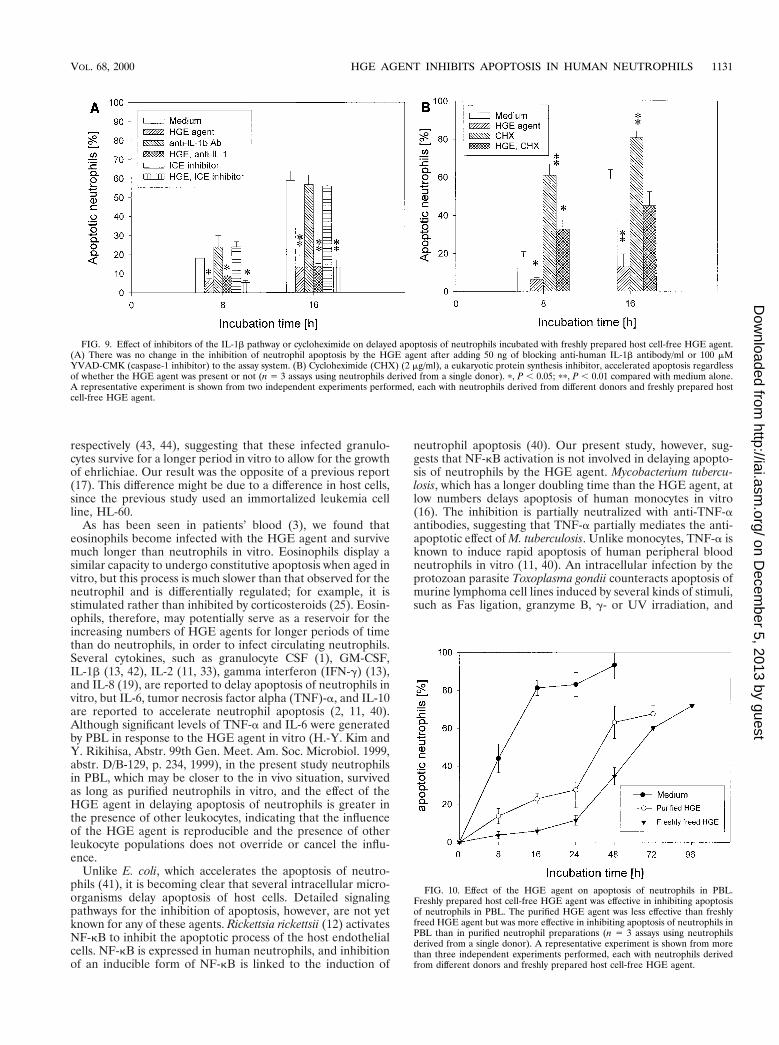
Effects of inhibitors of the IL-1b pathway. Our resultsshowed that the HGE agent induces IL-1b expression in PBLand neutrophils (H.-Y. Kim and Y. Rikihisa, Abstr. 99th Gen.Meet. Am. Soc. Microbiol., abstr. D/B-129, p. 234, 1999). Twodifferent proinflammatory stimuli, LPS and GM-CSF, upregu-late the expression of IL-1b-converting enzyme, also known ascaspase-1, and delay the apoptosis of neutrophils. The delay isblocked by blocking antibody to IL-1b or a caspase-1 inhibitor(42). Therefore, we examined whether endogenous IL-1b gen-erated by neutrophils in response to the HGE agent orcaspase-1 is involved in inhibition of neutrophil apoptosis bythe HGE agent by adding blocking anti-human IL-1b antibodyor an irreversible tetrapeptide inhibitor of caspase-1, YVAD-CMK (39), to the assay system. There was no change in theapoptosis of neutrophils regardless of the presence of the HGEagent (Fig. 9A).
Cycloheximide, a eukaryotic protein synthesis inhibitor, isknown to enhance the apoptosis of neutrophils in vitro (32).Cycloheximide has no effect on NF-kB activation but blocksthe antiapoptotic effect of LPS and GM-CSF by inhibitingIL-1b and caspase-1 upregulation (42). Cycloheximide treat-ment accelerated apoptosis regardless of the presence of theHGE agent (Fig. 9B).
Effect of HGE agent on apoptosis of neutrophils in PBL.Because other leukocytes, such as monocytes and lymphocytes,which coexist in the blood influence neutrophil apoptosis
FIG. 6. Effect of oxytetracycline on morphological apoptosis of human neu-trophils incubated with freshly prepared host cell-free HGE agent. Oxytetracy-cline (OTC) added at 0 h did not block the inhibition of apoptosis of neutrophilsby the HGE agent (n 5 3 assays using neutrophils derived from a single donor).A representative experiment is shown from three independent experiments per-formed, each with neutrophils derived from different donors and freshly pre-pared HGE agent.
FIG. 7. Effect of a protein kinase A inhibitor, H-89, on morphological apo-ptosis of human neutrophils incubated with freshly prepared host cell-free HGEagent. Treatment with 1 mM H-89 added at 0 h had no influence on morpho-logical apoptosis of neutrophils in the presence or absence of the freshly freedHGE agent (n 5 3 assays using neutrophils derived from a single donor). Arepresentative experiment is shown from three independent experiments per-formed, each with neutrophils derived from different donors and freshly pre-pared host cell-free HGE agent.
VOL. 68, 2000 HGE AGENT INHIBITS APOPTOSIS IN HUMAN NEUTROPHILS 1129
on Decem
ber 5, 2013 by guesthttp://iai.asm
.org/D
ownloaded from

through cytokines and cell-cell interactions (11), we examinedwhether the coexistence of other cell types in the blood influ-ences the inhibition of neutrophil apoptosis by the HGE agentin vitro. Freshly prepared host cell-free HGE agent was moreeffective in inhibiting apoptosis of neutrophils in PBL, whichconsisted of ;60% neutrophils, than in purified neutrophils(Fig. 10). The purified HGE agent was less effective than host
cell-free HGE agent but was more effective in inhibiting apo-ptosis of neutrophils in PBL than in purified neutrophils. Therate of apoptosis of uninfected neutrophils was, however, sim-ilar in PBL and purified neutrophil preparation.
DISCUSSION
The host of the HGE agent is the neutrophil, a suicidaleffector cell equipped with the most powerful antibacterialarmamentarium. Since ehrlichiae die if they remain extracel-lular, the HGE agent must enter neutrophils. Once it is intra-cellular, the HGE agent has solved the problem of lysosomaldestruction by inducing the formation of a unique membrane-bound niche, a parasitophorus vacuole which does not fusewith lysosomes (28). However, normal neutrophils survive fora limited time in the peripheral blood. Unless the neutrophil’slife span is extended, the intracellular HGE agent will dietogether with the host cell before having the chance to prolif-erate. The present study revealed that the HGE agent delaysthe apoptosis of human peripheral blood neutrophils suffi-ciently to allow intracellular proliferation of the HGE agent invitro, resulting in a significant morula formation in 24 to 48 hof incubation at a level comparable to those seen in patientswith HGE (3, 5). The actual percentage of infected cells maybe greater, since by using immunolabeling and flow cytometrywe previously found that almost 100% of P388D1 cells take upE. risticii at low levels at 3 h postincubation (26), although byDiff-Quik staining this low level of intracellular ehrlichiae isnot apparent (31).
Although small, condensed, elementary-body-like and large,light, reticular body-like ehrlichiae have been seen (34), achlamydia-like developmental cycle or eclipse stage has notbeen demonstrated in ehrlichiae. However, when we follow thetime course of in vitro ehrlichia infection in a leukemia cellline, during the first day of culture we can seldom see infectedcells (lag phase). After day 2 to 3 days of culture, logarithmicgrowth occurs, and 100% of the cells are infected with a largenumber of organisms by day 5 to 7 of culture, after which thecells are lysed (31). Since even with the delay the apoptosis ofinfected neutrophils takes place prior to complete ehrlichialproliferation and host cell lysis, the HGE agent in neutrophilsmust be horizontally transmitted to the next generation ofneutrophils prior to host cell apoptosis in order to survive. Thismay be one reason why heavily infected neutrophils are rarelyseen in patients. In the present study, nearly all neutrophilscould be prevented from undergoing rapid apoptosis whenstimulated with the host cell-free HGE agent at an MOI of100. Delaying apoptosis of all neutrophils is advantageous forthe HGE agent, because it gives more time for the HGE agentto survive and replicate inside neutrophils and to enhance thechance of its intercellular spreading. How the HGE agentspreads from infected to uninfected cells is unknown. Infectedneutrophils were rarely seen filled with the HGE agent and/orlysed. Spreading of monocytic ehrlichiae can occur withoutlysis of the infected host cells (36), probably by ehrlichial exo-cytosis from the infected cells followed by endocytosis of freedehrlichiae by other cells (36). The HGE agent may be trans-mitted by a similar mechanism from infected to uninfectedneutrophils after brief intracellular proliferation.
Previous observations with other granulocytotropic ehrli-chiae support our observation. In vitro incubation of periph-eral blood granulocytes from dogs experimentally infected withEhrlichia ewingii or heparinized whole blood from sheep ex-perimentally infected with E. phagocytophila results in an in-crease in the proportion of infected neutrophils and the num-ber of morulae in infected cells even after 2 to 4 days or 24 h,
FIG. 8. Effect of genistein, MG-132, and SN-50 on delayed apoptosis ofneutrophils incubated with freshly prepared host cell-free HGE agent. (A) Treat-ment of neutrophils with 50 mM genistein had no effect on the apoptosis ofneutrophils in the absence or presence of the HGE agent. (B) MG-132 (100 mM)significantly (p, P , 0.05; pp, P , 0.01 compared with medium alone) acceleratedthe apoptosis of neutrophils regardless of the presence of the HGE agent. (C)SN-50 (100 mg/ml) slightly (p, P , 0.05 compared to medium alone) delayedapoptosis of uninfected neutrophils but not that of infected neutrophils (n 5 3assays using neutrophils derived from a single donor). A representative experi-ment is shown from two independent experiments performed, each with neutro-phils derived from different donors and freshly prepared host cell-free HGEagent.
1130 YOSHIIE ET AL. INFECT. IMMUN.
on Decem
ber 5, 2013 by guesthttp://iai.asm
.org/D
ownloaded from
respectively (43, 44), suggesting that these infected granulo-cytes survive for a longer period in vitro to allow for the growthof ehrlichiae. Our result was the opposite of a previous report(17). This difference might be due to a difference in host cells,since the previous study used an immortalized leukemia cellline, HL-60.
As has been seen in patients’ blood (3), we found thateosinophils become infected with the HGE agent and survivemuch longer than neutrophils in vitro. Eosinophils display asimilar capacity to undergo constitutive apoptosis when aged invitro, but this process is much slower than that observed for theneutrophil and is differentially regulated; for example, it isstimulated rather than inhibited by corticosteroids (25). Eosin-ophils, therefore, may potentially serve as a reservoir for theincreasing numbers of HGE agents for longer periods of timethan do neutrophils, in order to infect circulating neutrophils.Several cytokines, such as granulocyte CSF (1), GM-CSF,IL-1b (13, 42), IL-2 (11, 33), gamma interferon (IFN-g) (13),and IL-8 (19), are reported to delay apoptosis of neutrophils invitro, but IL-6, tumor necrosis factor alpha (TNF)-a, and IL-10are reported to accelerate neutrophil apoptosis (2, 11, 40).Although significant levels of TNF-a and IL-6 were generatedby PBL in response to the HGE agent in vitro (H.-Y. Kim andY. Rikihisa, Abstr. 99th Gen. Meet. Am. Soc. Microbiol. 1999,abstr. D/B-129, p. 234, 1999), in the present study neutrophilsin PBL, which may be closer to the in vivo situation, survivedas long as purified neutrophils in vitro, and the effect of theHGE agent in delaying apoptosis of neutrophils is greater inthe presence of other leukocytes, indicating that the influenceof the HGE agent is reproducible and the presence of otherleukocyte populations does not override or cancel the influ-ence.
Unlike E. coli, which accelerates the apoptosis of neutro-phils (41), it is becoming clear that several intracellular micro-organisms delay apoptosis of host cells. Detailed signalingpathways for the inhibition of apoptosis, however, are not yetknown for any of these agents. Rickettsia rickettsii (12) activatesNF-kB to inhibit the apoptotic process of the host endothelialcells. NF-kB is expressed in human neutrophils, and inhibitionof an inducible form of NF-kB is linked to the induction of
neutrophil apoptosis (40). Our present study, however, sug-gests that NF-kB activation is not involved in delaying apopto-sis of neutrophils by the HGE agent. Mycobacterium tubercu-losis, which has a longer doubling time than the HGE agent, atlow numbers delays apoptosis of human monocytes in vitro(16). The inhibition is partially neutralized with anti-TNF-aantibodies, suggesting that TNF-a partially mediates the anti-apoptotic effect of M. tuberculosis. Unlike monocytes, TNF-a isknown to induce rapid apoptosis of human peripheral bloodneutrophils in vitro (11, 40). An intracellular infection by theprotozoan parasite Toxoplasma gondii counteracts apoptosis ofmurine lymphoma cell lines induced by several kinds of stimuli,such as Fas ligation, granzyme B, g- or UV irradiation, and
FIG. 9. Effect of inhibitors of the IL-1b pathway or cycloheximide on delayed apoptosis of neutrophils incubated with freshly prepared host cell-free HGE agent.(A) There was no change in the inhibition of neutrophil apoptosis by the HGE agent after adding 50 ng of blocking anti-human IL-1b antibody/ml or 100 mMYVAD-CMK (caspase-1 inhibitor) to the assay system. (B) Cycloheximide (CHX) (2 mg/ml), a eukaryotic protein synthesis inhibitor, accelerated apoptosis regardlessof whether the HGE agent was present or not (n 5 3 assays using neutrophils derived from a single donor). p, P , 0.05; pp, P , 0.01 compared with medium alone.A representative experiment is shown from two independent experiments performed, each with neutrophils derived from different donors and freshly prepared hostcell-free HGE agent.
FIG. 10. Effect of the HGE agent on apoptosis of neutrophils in PBL.Freshly prepared host cell-free HGE agent was effective in inhibiting apoptosisof neutrophils in PBL. The purified HGE agent was less effective than freshlyfreed HGE agent but was more effective in inhibiting apoptosis of neutrophils inPBL than in purified neutrophil preparations (n 5 3 assays using neutrophilsderived from a single donor). A representative experiment is shown from morethan three independent experiments performed, each with neutrophils derivedfrom different donors and freshly prepared host cell-free HGE agent.
VOL. 68, 2000 HGE AGENT INHIBITS APOPTOSIS IN HUMAN NEUTROPHILS 1131
on Decem
ber 5, 2013 by guesthttp://iai.asm
.org/D
ownloaded from

calcium ionophores (29), suggesting that a mechanism com-mon to many apoptotic pathways is involved. In contrast to theHGE agent, protection against apoptosis by T. gondii requiresthe continued presence of live organisms and ongoing proteinsynthesis (29). The intracellular protozoan parasite Leishma-nia donovani or treatment with lipophosphoglycan, the majorsurface molecule of the Leishmania promastigote, also inhibitsmouse bone marrow macrophage apoptosis induced by re-moval of macrophage CSF in vitro (27). Although exogenousTNF-a inhibits apoptosis in this assay system and Leishmaniainfection induces TNF-a secretion, the inhibition was not re-stored by anti-TNF-a-neutralizing antibodies. As far as weknow, the HGE agent is the first infectious agent known todelay apoptosis of neutrophils, and the antiapoptotic mecha-nism of the HGE agent appears to be different from the mech-anisms of other intracellular microorganisms. Therefore, theHGE agent may serve as a new tool for analysis of the apo-ptotic mechanism of neutrophils.
Because the noninfectious purified HGE agent had an an-tiapoptotic effect and because inhibition of ehrlichial proteinsynthesis or proliferation did not prevent or turn off the anti-apoptotic signal, infection per se is not essential for delayingapoptosis. However, the protein residue of the HGE agent,rather than carbohydrates, is required for the inhibition. Whenmonocytic ehrlichiae are treated with trypsin, a milder proteo-lytic enzyme than proteinase K, ehrlichial binding and subse-quent internalization in host cells is prevented (26). In addi-tion, our MDC study showed that ehrlichial internalizationand/or receptor cross-linking is required for apoptosis inhibi-tion. The inhibitory effect of the HGE agent on apoptosis wasdose dependent. These results suggest that initial occupationand cross-linking of host cell receptors by preformed proteinsof the HGE agent may be sufficient to trigger the antiapoptoticsignal. The reason the freshly prepared host cell-free HGEagent had stronger antiapoptotic activity than the purifiedHGE agent may be that the structural or conformational in-tegrity present in the freshly prepared host cell-free HGEagent, which is lost in the purified HGE agent, is required foreffective cross-linking of receptors or internalization.
E. chaffeensis, upon binding to THP-1 cells, increases proteinkinase A activity 25-fold within 30 min and inhibits tyrosinephosphorylation of Jak-1 and -2 and Stat1a in response toIFN-g. This inhibition does not require the internalization ofE. chaffeensis in THP-1 cells or the carbohydrate residue of theorganism, but binding of the protein of E. chaffeensis to thehost cells is required (22). It appears that these conditions aresimilar to those required for delaying apoptosis by the HGEagent. However, we did not find the involvement of proteinkinase A activation in apoptosis delay by the HGE agent.Similarly, inhibition of basal protein kinase A activity by 25 mMH-89 has no influence on (does not accelerate) spontaneous orcycloheximide- or anti-Fas-induced neutrophil apoptosis (32),although conditions that raise intracellular cAMP are shown todelay spontaneous neutrophil apoptosis and inhibit apoptosisinduced by cycloheximide or anti-Fas (30, 32). Additionally,because host cell interactions with E. chaffeensis and the HGEagent differ in several respects, such as the intracellular com-partments they occupy (28) and upregulation of host trans-ferrin receptor mRNA (7) and cytokine mRNA expression (21;Kim and Rikihisa, Abstr. 99th Gen. Meet. Am. Soc. Micro-biol), host cell receptors and intracellular signaling pathwaysmay be different. The antiapoptotic mechanism may share thesignaling pathway with dexamethasone-induced apoptosis, be-cause suppression of apoptosis by both dexamethasone and theHGE agent is abolished by cotreatment with cycloheximide(14).
The HGE agent might have LPS, since it belongs to thegram-negative bacteria. E. coli LPS is reported to delay apo-ptosis of neutrophils in vitro (38, 42). Inhibition of apoptosis ofneutrophils by bacterial LPS is mediated by induced pro-IL-1band caspase-1 through protein tyrosine phosphorylation-de-pendent activation of NF-kB (38, 42). IL-1b is known to delayapoptosis of neutrophils (42), and IL-1b was also induced inneutrophils exposed to the HGE agent (Kim and Rikihisa,Abstr. 99th Gen. Meet. Am. Soc. Microbiol.). However, IL-1band NF-kB do not appear to be involved in inhibition of apo-ptosis by the HGE agent. This suggests that the HGE agenteither lacks LPS or the structure and biological activity ofehrlichial LPS is distinct from those of E. coli LPS.
Delayed apoptosis of neutrophils may help ehrlichial prolif-eration and prolong proinflammatory cytokine generation,making patients more ill and thus prone to hospitalization. Inagreement with this speculation, Bakken et al. reported ahigher percentage of neutrophils in the PBL of hospitalizedthan nonhospitalized HGE patients (6). Elucidation of an ehr-lichial factor(s) and the signaling pathway in the neutrophilsthat inhibit apoptosis would be important in understanding thepathogenesis of HGE. The HGE agent or its protein compo-nents may also serve as a tool in analyzing the fundamentalapoptotic mechanisms of neutrophils.
ACKNOWLEDGMENTS
This research was supported by grant RO1AI30010 from the Na-tional Institutes of Health. K. Yoshiie was supported by a fellowshipfrom The Japan Health Sciences Foundation.
REFERENCES
1. Adachi, S., M. Kubota, Y. W. Lin, A. Okuda, K. Matsubara, Y. Wakazono, H.Hirota, K. Kuwakado, and Y. Akiyama. 1994. In vivo administration ofgranulocyte colony-stimulating factor promotes neutrophil survival in vitro.Eur. J. Haematol. 53:129–134.
2. Afford, S. C., J. Pongracz, R. A. Stockley, J. Crocker, and D. Burnett. 1992.The induction by human interleukin-6 of apoptosis in the promonocytic cellline U937 and human neutrophils. J. Biol. Chem. 267:21612–21616.
3. Aguero-Rosenfeld, M. E., H. W. Horowitz, G. P. Wormser, D. F. McKenna,J. Nowakowski, J. Munoz, and J. S. Dumler. 1996. Human granulocyticehrlichiosis: a case series from a medical center in New York State. Ann.Intern. Med. 125:904–908.
4. Akiyama, T., and H. Ogawara. 1991. Use and specificity of genistein asinhibitor of protein-tyrosine kinases. Methods Enzymol. 201:362–370.
5. Bakken, J. S., J. S. Dumler, S. M. Chen, M. R. Eckman, L. L. van Etta, andD. H. Walker. 1994. Human granulocytic ehrlichiosis in the upper MidwestUnited States. a new species emerging? JAMA 272:212–218.
6. Bakken, J. S., J. Krueth, C. Wilson-Nordskog, R. L. Tilden, K. Asanovich,and J. S. Dumler. 1996. Clinical and laboratory characteristics of humangranulocytic ehrlichiosis. JAMA 275:199–205.
7. Barnewall, R., N. Ohashi, and Y. Rikihisa. 1999. Ehrlichia chaffeensis and E.sennetsu, but not the Human granulocytic ehrlichiosis agent, colocalize withtransferrin receptor and up-regulate transferrin receptor mRNA by activat-ing iron-responsive protein 1. Infect Immun. 67:2258–2265.
8. Brouqui, P. 1999. Ehrlichiosis in Europe, p. 220–232. In D. Raoult and P.Brouqui (ed.), Rickettsiae and rickettsial diseases at the turn of the thirdmillennium. Elsevier, Paris, France.
9. Chen, S. M., J. S. Dumler, J. S. Bakken, and D. H. Walker. 1994. Identifi-cation of a granulocytotropic Ehrlichia species as the etiologic agent ofhuman disease. J. Clin. Microbiol. 32:589–595.
10. Chijiwa, T., A. Mishima, M. Hagiwara, M. Sano, K. Hayashi, T. Inoue, K.Naito, T. Toshioka, and H. Hidaka. 1990. Inhibition of forskolin-inducedneurite outgrowth and protein phosphorylation by a newly synthesized se-lective inhibitor of cyclic AMP-dependent protein kinase, N-[2-(p-bromocin-namylamino) ethyl] 5-isoquinolinesulfonamide (H-89), of PC12D pheochro-mocytoma cells. J. Biol. Chem. 265:5267–5272.
11. Christa, H. E., B. Homburg, and D. Roos. 1996. Apoptosis of neutrophils.Curr. Opin. Hematol. 3:94–99.
12. Clifton, D. R., R. A. Goss, S. K. Sahni, D. V. Antwerp, R. B. Baggs, V. J.Marder, D. J. Silverman, and L. A. Sporn. 1998. NF-kB-dependent inhibi-tion of apoptosis is essential for host cell survival during Rickettsia rickettsiiinfection. Proc. Natl. Acad. Sci. USA 95:4646–4651.
13. Colotta, F., F. Re, N. Polentarutti, S. Sozzani, and A. Mantovani. 1992.Modulation of granulocyte survival and programmed cell death by cytokines
1132 YOSHIIE ET AL. INFECT. IMMUN.
on Decem
ber 5, 2013 by guesthttp://iai.asm
.org/D
ownloaded from

and bacterial products. Blood 80:2012–2020.14. Cox, G., J. Gauldie, and M. Jordana. 1992. Dexamethasone-induced sup-
pression of apoptosis in human neutrophils requires continuous stimulationof new protein synthesis. J. Leukoc. Biol. 61:224–230.
15. Duke, R. C., and J. J. Cohen. 1986. IL-2 addiction: withdrawal of growthfactor activates a suicide program in dependent T cells. Lymphokine Res.5:289–299.
16. Durrbaum-Landmann, I., J. Gercken, H.-D. Flad, and M. Ernst. 1996. Effectof in vitro infection of human monocytes with low numbers of Mycobacteriumtuberculosis bacteria on monocyte apoptosis. Infect. Immun. 64:5384–5389.
17. Hsieh, T.-C., M. E. Aguero-Rosenfeld, J. M. Wu, C. Ng, N. A. Papanikolaou,S. A. Varde, I. Schwartz, J. G. Pizzolo, M. Melamed, H. W. Horowitz, R. B.Nadelman, and G. P. Wormser. 1997. Cellular changes and induction ofapoptosis in human promyelocytic HL-60 cells infected with the agent ofhuman granulocytic ehrlichiosis (HGE). Biochem. Biophys. Res. Commun.232:298–303.
18. Jensen, T. J., M. A. Loo, S. Pind, D. B. Williams, A. L. Goldberg, and J. R.Riordan. 1995. Multiple proteolytic systems, including the proteasome, con-tribute to CFTR processing. Cell 83:129–135.
19. Kettritz, R., M. L. Gaido, H. Haller, F. C. Luft, C. J. Jennette, and R. J. Falk.1998. Interleukin-8 delays spontaneous and tumor necrosis factor-a-medi-ated apoptosis of human neutrophils. Kidney Int. 53:84–91.
20. Kim, H.-Y., and Y. Rikihisa. 1998. Characterization of monoclonal antibod-ies against the major outer membrane protein of human granulocytic ehrli-chiosis agent. J. Clin. Microbiol. 36:3278–3284.
21. Lee, E. H., and Y. Rikihisa. 1996. Absence of tumor necrosis factor-a,interleukin-6 (IL-6), and granulocyte-macrophage colony-stimulating factorexpression but presence of IL-1b, IL-8, and IL-10 expression in humanmonocytes exposed to viable or killed Ehrlichia chaffeensis. Infect. Immun.64:4211–4219.
22. Lee, E. H., and Y. Rikihisa. 1998. Protein kinase A-mediated inhibition ofgamma interferon-induced tyrosine phosphorylation of Janus kinases andlatent cytoplasmic transcription factors in human monocytes by Ehrlichiachaffeensis. Infect. Immun. 66:2514–2520.
23. Levitzki, A., M. Willingham, and I. Pastan. 1980. Evidence for participationof transglutaminase in receptor-mediated endocytosis. Proc. Natl. Acad. Sci.USA 77:2706–2710.
24. Lin, Y., S. Yao, R. A. Veach, T. R. Torgerson, and J. Hawiger. 1995. Inhibi-tion of nuclear translocation of transcription factor NF-kB by a syntheticpeptide containing a cell membrane-permeable motif and nuclear localiza-tion sequence. J. Biol. Chem. 270:14255–14258.
25. Meagher, L. C., J. M. Cousin, J. R. Seckl, and C. Haslett. 1996. Opposingeffects of glucocorticoids on the rate of apoptosis in neutrophilic and eosin-ophilic granulocytes. J. Immunol. 156:4422–4428.
26. Messick, J. B., and Y. Rikihisa. 1993. Characterization of Ehrlichia risticiibinding, internalization, and growth in host cells by flow cytometry. Infect.Immun. 61:3803–3810.
27. Moore, K. J., and G. Matlashewski. 1994. Intracellular infection by Leish-mania donovani inhibits macrophage apoptosis. J. Immunol. 152:2930–2937.
28. Mott, J., R. Barnewall, and Y. Rikihisa. 1999. Human granulocytic ehrli-chiosis agent and Ehrlichia chaffeensis reside in different cytoplasmic com-partments in HL-60 cells. Infect. Immun. 67:1368–1378.
29. Nash, P. B., M. B. Purner, R. P. Leon, P. Clarke, R. C. Duke, and T. J.Curiel. 1998. Toxoplasma gondii-infected cells are resistant to multiple in-ducers of apoptosis. J. Immunol. 160:1824–1830.
30. Ottonello, L., R. Gonella, P. Dapino, C. Sacchetti, and F. Dallegri. 1998.Prostaglandin E2 inhibits apoptosis in human neutrophilic polymorphonu-
clear leukocytes: role of intracellular cyclic AMP levels. Exp. Hematol.26:895–902.
31. Park, J., and Y. Rikihisa. 1991. Inhibition of Ehrlichia risticii infection inmurine peritoneal macrophages by gamma interferon, a calcium ionophore,and concanavalin A. Infect. Immun. 59:3418–3423.
32. Parvathenani, L. K., S. Buescher, E. Chacon-Cruz, and S. J. Beebe. 1998.Type I cAMP-dependent protein kinase delays apoptosis in human neutro-phils at a site upstream of caspase-3. J. Biol. Chem. 273:6736–6743.
33. Pericle, F., J. H. Liu, J. I. Diaz, D. K. Blanchard, S. Wei, G. Forni, and J. Y.Djeu. 1994. Interleukin-2 prevention of apoptosis in human neutrophils. Eur.J. Immunol. 24:440–444.
34. Rikihisa, Y. 1991. The tribe Ehrlichieae and ehrlichial diseases. Clin. Micro-biol. Rev. 4:286–308.
35. Rikihisa, Y. 1991. Cross-reacting antigens between Neorickettsia helmintho-eca and Ehrlichia spp. shown by immunofluorescence and Western immu-noblotting. J. Clin. Microbiol. 29:2024–2029.
36. Rikihisa, Y., Y. Zhang, and J. Park. 1994. Inhibition of infection of macro-phages with Ehrlichia risticii by cytochalasins, monodansylcadaverine, andtaxol. Infect. Immun. 62:5126–5132.
37. Rikihisa, Y., N. Zhi, G. Wormser, B. Wen, H. W. Horowitz, and K. E.Hechemy. 1997. Direct isolation and cultivation of human granulocytic ehr-lichia from a human patient. J. Infect. Dis. 175:210–213.
38. Sweeney, J. F., P. K. Nguyen, G. M. Omann, and D. B. Hinshaw. 1998.Lipopolysaccharide protects polymorphonuclear leukocytes from apoptosisvia tyrosine phosphorylation-dependent signal transduction pathways.J. Surg. Res. 74:64–70.
39. Thornberry, N., H. G. Bull, J. R. Calaycay, K. T. Chapman, A. D. Howard,M. J. Kostural, D. K. Miller, S. M. Molineaux, J. R. Weidner, J. Aununs,K. O. Elliston, J. M. Ayala, F. J. Casano, J. Chin, G. J.-F. Ding, L. A. Egger,E. P. Gaffney, G. Limjuco, O. C. Palyha, S. M. Raju, A. M. Rolando, J. P.Sally, T.-T. Yamin, T. D. Lee, J. E. Shively, M. MacCross, R. A. Mumford,J. A. Schmidt, and M. J. Tocci. 1992. A novel heterodimeric cysteine pro-tease is required for interleukin-1b processing in monocytes. Nature 356:768–774.
40. Ward, C., E. R. Chilvers, M. F. Lawson, J. G. Pryde, S. Fujihara, S. N.Farrow, C. Haslett, and A. G. Rossi. 1999. NF-kB activation is a criticalregulator of human granulocyte apoptosis in vitro. J. Biol. Chem. 274:4309–4318.
41. Watson, R. W. G., H. P. Redmond, J. H. Wang, C. Condron, and D.Bouchier-Hays. 1996. Neutrophils undergo apoptosis following ingestion ofEscherichia coli. J. Immunol. 156:3986–3992.
42. William, R., G. Watson, O. D. Rotstein, J. Parodo, R. Bitar, and J. C.Marshall. 1998. The IL-1b-converting enzyme (caspase-1) inhibits apoptosisof inflammatory neutrophils through activation of IL-1b. J. Immunol. 161:957–962.
43. Winjum, N., and L. K. Riley. 1993. In vitro proliferation of a canine granu-locytic Ehrlichia. Vet. Microbiol. 34:355–362.
44. Woldehiwet, Z., and G. R. Scott. 1982. Stages in the development of Cytoectesphagocytophila, the causative agent of tick-borne fever. Comp. Pathol. 92:469–474.
45. Zhi, N., N. Ohashi, and Y. Rikihisa. 1999. Multiple p44 genes encodingmajor outer membrane proteins are expressed in the human granulocyticehrlichiosis agent. J. Biol. Chem. 274:17828–17836.
46. Zhi, N., Y. Rikihisa, H.-Y. Kim, G. P. Wormser, and H. W. Horowitz. 1997.Comparison of major antigenic proteins of six strains of human granulocyticehrlichiosis agents by Western immunoblot analysis. J. Clin. Microbiol. 35:2606–2611.
Editor: P. E. Orndorff
VOL. 68, 2000 HGE AGENT INHIBITS APOPTOSIS IN HUMAN NEUTROPHILS 1133
on Decem
ber 5, 2013 by guesthttp://iai.asm
.org/D
ownloaded from
Copyright © 2022 FDOKUMEN