Amelioration of Ischemic Acute Renal Injury by Neutrophil Gelatinase-Associated Lipocalin
10
Amelioration of Ischemic Acute Renal Injury by Neutrophil Gelatinase–Associated Lipocalin JAYA MISHRA,* KIYOSHI MORI, † QING MA,* CAITLIN KELLY,* JUN YANG, † MARK MITSNEFES,* JONATHAN BARASCH, † and PRASAD DEVARAJAN* *Department of Nephrology and Hypertension, Cincinnati Children’s Hospital Medical Center, University of Cincinnati, Cincinnati, Ohio; and † Department of Nephrology, College of Physicians and Surgeons, Columbia University, New York, New York Abstract. Acute renal failure secondary to ischemic injury remains a common problem, with limited and unsatisfactory therapeutic options. Neutrophil gelatinase–associated lipocalin (NGAL) was recently shown to be one of the maximally induced genes early in the postischemic kidney. In this study, the role of NGAL in ischemic renal injury was explored. Intravenous administration of purified recombinant NGAL in mice resulted in a rapid uptake of the protein predominantly by proximal tubule cells. In an established murine model of renal ischemia-reperfusion injury, intravenous NGAL administered before, during, or after ischemia resulted in marked ameliora- tion of the morphologic and functional consequences, as evi- denced by a significant decrease in the histopathologic damage to tubules and in serum creatinine measurements. NGAL- treated animals also displayed a reduction in the number of apoptotic tubule cells and an increase in proliferating proximal tubule cells after ischemic injury. The results indicate that NGAL may represent a novel therapeutic intervention in isch- emic acute renal failure, based at least in part on its ability to tilt the balance of tubule cell fate toward survival. Acute renal failure (ARF) secondary to ischemic injury re- mains a common and potentially devastating problem in clin- ical nephrology, with a persistently high rate of mortality despite significant advances in supportive care (1– 4). Pioneer- ing studies over several decades have illuminated the roles of persistent vasoconstriction, tubular obstruction, cellular struc- tural and metabolic alterations, and the inflammatory response in the pathogenesis of ARF (4 –7). Although these studies have paved the way for successful therapeutic approaches in animal models, translational research efforts in humans have yielded disappointing results (2– 4). The reasons for this may include the multifaceted response of the kidney to ischemia and a lack of early markers for ARF (4 – 8). Recent advances in cellular and molecular biology of ischemic renal injury have revealed that proximal tubule cells undergo a complex temporal se- quence of events. These include loss of cell polarity, cell death as a result of apoptosis and necrosis, dedifferentiation and proliferation of viable cells, and reestablishment of the epithe- lial phenotype (6,7). An improved understanding of the early cell injury and repair mechanisms is critical for innovative and effective therapy. Identification of interventions that may op- pose tubule cell death and/or enhance the recovery phase therefore is of considerable interest. Attempts to unravel the molecular basis of the myriad early renal responses have been facilitated by recent advances in functional genomics that have yielded new tools for genome- wide analysis of complex biologic processes such as ischemic ARF (8 –11). Using cDNA microarray techniques, we recently identified neutrophil gelatinase–associated lipocalin (NGAL) as one of the most dramatically induced transcripts in the kidney early after ischemic injury (11,12). Although previous studies have indicated that NGAL may represent a novel early urinary biomarker for ischemic renal injury (12), the role of NGAL in the kidney has remained puzzling. We showed previously that in the postischemic kidney, NGAL is upregu- lated in tubular epithelial cells that are undergoing proliferation (12). Other recent studies have suggested that NGAL can enhance the epithelial phenotype. We therefore tested the hy- pothesis that NGAL may play a renoprotective role in ischemic ARF. In this study, we examined the ability of intravenously administered recombinant NGAL to modify the structural and functional consequences of ischemic acute renal injury in an established murine model. Our results indicate that NGAL may represent a novel therapeutic intervention in ischemic ARF, based at least in part on its ability to ameliorate tubule cell apoptosis and enhance tubule cell proliferation. Materials and Methods Expression and Purification of Recombinant Murine NGAL Full-length mouse NGAL cDNA was cloned into the pGEX ex- pression vector, expressed as a fusion protein with glutathione S- transferase (GST) in Escherichia coli (XL1-Blue), and purified using Received March 11, 2004. Accepted August 24, 2004. Correspondence to Dr. Prasad Devarajan, Nephrology and Hypertension, MLC 7022, Cincinnati Children’s Hospital Medical Center, 3333 Burnet Avenue, Cincinnati, OH 45229-3039. Phone: 513-636-4531; Fax: 513-636-7407; E-mail: [email protected] 1046-6673/1512-3073 Journal of the American Society of Nephrology Copyright © 2004 by the American Society of Nephrology DOI: 10.1097/01.ASN.0000145013.44578.45 J Am Soc Nephrol 15: 3073–3082, 2004
-
Upload
independent -
Category
Documents
-
view
1 -
download
0
Transcript of Amelioration of Ischemic Acute Renal Injury by Neutrophil Gelatinase-Associated Lipocalin

Amelioration of Ischemic Acute Renal Injury by NeutrophilGelatinase–Associated Lipocalin
JAYA MISHRA,* KIYOSHI MORI,† QING MA,* CAITLIN KELLY,* JUN YANG,†
MARK MITSNEFES,* JONATHAN BARASCH,† and PRASAD DEVARAJAN**Department of Nephrology and Hypertension, Cincinnati Children’s Hospital Medical Center, University ofCincinnati, Cincinnati, Ohio; and †Department of Nephrology, College of Physicians and Surgeons, ColumbiaUniversity, New York, New York
Abstract. Acute renal failure secondary to ischemic injuryremains a common problem, with limited and unsatisfactorytherapeutic options. Neutrophil gelatinase–associated lipocalin(NGAL) was recently shown to be one of the maximallyinduced genes early in the postischemic kidney. In this study,the role of NGAL in ischemic renal injury was explored.Intravenous administration of purified recombinant NGAL inmice resulted in a rapid uptake of the protein predominantly byproximal tubule cells. In an established murine model of renalischemia-reperfusion injury, intravenous NGAL administered
before, during, or after ischemia resulted in marked ameliora-tion of the morphologic and functional consequences, as evi-denced by a significant decrease in the histopathologic damageto tubules and in serum creatinine measurements. NGAL-treated animals also displayed a reduction in the number ofapoptotic tubule cells and an increase in proliferating proximaltubule cells after ischemic injury. The results indicate thatNGAL may represent a novel therapeutic intervention in isch-emic acute renal failure, based at least in part on its ability totilt the balance of tubule cell fate toward survival.
Acute renal failure (ARF) secondary to ischemic injury re-mains a common and potentially devastating problem in clin-ical nephrology, with a persistently high rate of mortalitydespite significant advances in supportive care (1–4). Pioneer-ing studies over several decades have illuminated the roles ofpersistent vasoconstriction, tubular obstruction, cellular struc-tural and metabolic alterations, and the inflammatory responsein the pathogenesis of ARF (4–7). Although these studies havepaved the way for successful therapeutic approaches in animalmodels, translational research efforts in humans have yieldeddisappointing results (2–4). The reasons for this may includethe multifaceted response of the kidney to ischemia and a lackof early markers for ARF (4–8). Recent advances in cellularand molecular biology of ischemic renal injury have revealedthat proximal tubule cells undergo a complex temporal se-quence of events. These include loss of cell polarity, cell deathas a result of apoptosis and necrosis, dedifferentiation andproliferation of viable cells, and reestablishment of the epithe-lial phenotype (6,7). An improved understanding of the earlycell injury and repair mechanisms is critical for innovative andeffective therapy. Identification of interventions that may op-
pose tubule cell death and/or enhance the recovery phasetherefore is of considerable interest.
Attempts to unravel the molecular basis of the myriad earlyrenal responses have been facilitated by recent advances infunctional genomics that have yielded new tools for genome-wide analysis of complex biologic processes such as ischemicARF (8–11). Using cDNA microarray techniques, we recentlyidentified neutrophil gelatinase–associated lipocalin (NGAL)as one of the most dramatically induced transcripts in thekidney early after ischemic injury (11,12). Although previousstudies have indicated that NGAL may represent a novel earlyurinary biomarker for ischemic renal injury (12), the role ofNGAL in the kidney has remained puzzling. We showedpreviously that in the postischemic kidney, NGAL is upregu-lated in tubular epithelial cells that are undergoing proliferation(12). Other recent studies have suggested that NGAL canenhance the epithelial phenotype. We therefore tested the hy-pothesis that NGAL may play a renoprotective role in ischemicARF. In this study, we examined the ability of intravenouslyadministered recombinant NGAL to modify the structural andfunctional consequences of ischemic acute renal injury in anestablished murine model. Our results indicate that NGAL mayrepresent a novel therapeutic intervention in ischemic ARF,based at least in part on its ability to ameliorate tubule cellapoptosis and enhance tubule cell proliferation.
Materials and MethodsExpression and Purification of RecombinantMurine NGAL
Full-length mouse NGAL cDNA was cloned into the pGEX ex-pression vector, expressed as a fusion protein with glutathione S-transferase (GST) in Escherichia coli (XL1-Blue), and purified using
Received March 11, 2004. Accepted August 24, 2004.Correspondence to Dr. Prasad Devarajan, Nephrology and Hypertension, MLC7022, Cincinnati Children’s Hospital Medical Center, 3333 Burnet Avenue,Cincinnati, OH 45229-3039. Phone: 513-636-4531; Fax: 513-636-7407;E-mail: [email protected]
1046-6673/1512-3073Journal of the American Society of NephrologyCopyright © 2004 by the American Society of Nephrology
DOI: 10.1097/01.ASN.0000145013.44578.45
J Am Soc Nephrol 15: 3073–3082, 2004

glutathione-Sepharose columns (Amersham Biosciences) followed bythrombin cleavage as described previously (13–15). Purified NGALwas made endotoxin-free using the Detoxi-Gel endotoxin removingcolumn (Pierce) as recommended by the manufacturer. Because of theremote possibility of low levels of endotoxin contamination, an “ultra-pure” batch of NGAL was prepared using an additional gel filtrationcolumn (Superdex-75, SMART system; Amersham, ArlingtonHeights, IL). Proteins were analyzed by SDS-PAGE followed byCoomassie blue staining or by Western blotting with a polyclonalantibody to NGAL as described (12). Protein concentrations weredetermined using the Bradford assay.
Mouse Models of Renal Ischemia-Reperfusion InjuryWe used well-established murine models in which the structural
and functional consequences of brief periods of renal ischemia havebeen previously documented (11,12,15). Briefly, male Swiss-Webstermice (Taconic Farms, Germantown, NY) that weighed 25 to 30 gwere housed with 12:12-h light:dark cycle and were allowed freeaccess to food and water. The animals were anesthetized with sodiumpentobarbital (50 mg/kg intraperitoneally) and placed on a warmingtable to maintain a rectal temperature of 37°C. Both renal pedicleswere occluded with a nontraumatic vascular clamp for 30 min, duringwhich time the kidney was kept warm and moist. The clamps werethen removed, the kidney was observed for return of blood flow, andthe incision was sutured. The mice were allowed to recover in awarmed cage, and timed urine collections were obtained. After vari-ous reperfusion periods, the animals were re-anesthetized, the abdom-inal cavity was opened, and blood was obtained via puncture of theinferior vena cava for measurement of serum creatinine by quantita-tive colorimetric assay kit (Sigma, St. Louis, MO). The mice werekilled, the kidneys were perfusion fixed in situ with 4% paraformal-dehyde in PBS, and both kidneys were harvested. One half of eachkidney was snap-frozen in liquid nitrogen and stored at �70°C untilfurther processing; a sample was fixed in formalin, paraffin-embed-ded, and sectioned (4 �m). Paraffin sections were stained with hema-toxylin-eosin and examined histologically. The other half of eachkidney was embedded in OCT compound (Tissue-Tek), and frozensections (4 �m) were obtained for immunohistochemistry.
NGAL InjectionsPurified endotoxin-free NGAL was administered intravenously
into mice via tail-vein injections. In preliminary studies, animals weretreated with three different concentrations of NGAL (50, 100, or 250�g of a 250 �g/100 �l solution), subjected to 30 min of bilateral renalartery clamping 1 h later, and examined after 24 h of reflow. Whencompared with animals that were pretreated with an equal volume(100 �l) of saline, the group that was given 250 �g of NGALexhibited the best protection from the tubular damage and azotemia.In addition, when NGAL (250 �g) that was inactivated by boiling thepreparation for 10 min was infused, no renoprotective effect wasnoted. Also, no differences in renoprotection were encountered in therenal response to the NGAL batch rendered endotoxin-free usingDetoxi-gel in comparison with the “ultra-pure” batch of NGAL thatwas further purified by Superdex gel filtration. Furthermore, otherunrelated proteins prepared in this manner were devoid of a renopro-tective effect (data not shown), ruling out the remote possibility ofcontaminating endotoxins’ mediating the response observed to in-jected NGAL. All subsequent studies as reported here were carried outusing the 250-�g dose of endotoxin-free biologically active NGAL.
Comparisons were made between five different animal groups:nonischemic controls (n � 8), ischemic controls that were pretreated
with 100 �l of saline alone (n � 8), NGAL pretreated 1 h before renalartery clamping (n � 6), NGAL treated during renal artery clamping(n � 6), and NGAL treated 1 h after renal artery clamping (n � 6).
NGAL ImmunohistochemistryFor NGAL detection, frozen kidney sections were permeabilized
with 0.2% Triton X-100 in PBS for 10 min, blocked with goat serumfor 1 h, and incubated with primary antibody to NGAL (1:500dilution) for 1 h. Slides were then exposed for 30 min in the dark tosecondary antibodies conjugated with Cy5 (Amersham) and visual-ized with a fluorescence microscope (Zeiss Axiophot) equipped withrhodamine filters.
Histopathology ScoringKidney sections of 4 � were stained with hematoxylin-eosin and
scored for histopathologic damage to the tubules in a blinded manner,as described previously (16,17). Each parameter was assessed in fivehigh-power fields (�40) in the inner cortex and outer medullaryregions (where the tubular damage was most evident), and an averagewas determined for each section. The parameters included tubuledilation, tubule cast formation, and tubule cell necrosis. Each param-eter was scored on a scale of 0 to 4, ranging from none (0), mild (1),moderate (2), severe (3), to very severe/extensive (4).
Apoptosis AssaysFor the transferase-mediated dUTP nick-end labeling (TUNEL)
assay to detect apoptotic nuclei, we used the ApoAlert DNA Frag-mentation Assay Kit (Clontech). Paraffin sections were deparaffinizedthrough xylene and descending grades of ethanol, fixed with 4%formaldehyde/PBS for 30 min at 4°C, permeabilized with proteinaseK at room temperature for 15 min and 0.2% triton X-100/PBS for 15min at 4°C, and incubated with a mixture of nucleotides and TdTenzyme for 60 min at 37°C. The reaction was terminated with 2�SSC, and the sections were washed with PBS and mounted withCrystal/mount (Biomeda, Foster City, CA). TUNEL-positive apopto-tic nuclei were detected by visualization with a fluorescence micro-scope. Only cells that displayed the characteristic morphology ofapoptosis, including nuclear fragmentation, nuclear condensation, andintensely fluorescence nuclei by TUNEL assay, were counted asapoptotic. Merely TUNEL-positive cells, in the absence of morpho-logic criteria, were not considered apoptotic. Slides were examined ina blinded manner, and apoptosis was quantified by counting thenumber of TUNEL-positive nuclei per 100 cells counted in an averageof five high-power fields (�40) in each section.
Proliferation AssaysFor detection of proliferating cells, sections were incubated with an
mAb to proliferating cell nuclear antigen (PCNA; 1:500 dilution;Upstate Biotechnology), and detection was accomplished by immu-noperoxidase staining as recommended by the manufacturer (Immu-noCruz Staining System, Santa Cruz Biotechnology). Slides wereexamined in a blinded manner, and proliferation was quantified bycounting the number of PCNA-positive cells per 100 cells counted inan average of five high-power fields (�40) in each section.
Statistical AnalysesThe SPSS software (version 8.0) was used to generate univariate
statistics for each continuous variable, including means, SD, distribu-tions, range, and skewness. The data were examined for normality andequality of distribution. One-way ANOVA was used to compare
3074 Journal of the American Society of Nephrology J Am Soc Nephrol 15: 3073–3082, 2004

means � SD of continuous variables among different treatmentgroups. The Kruskal-Wallis ANOVA on ranks was used for nonnor-mally distributed data. To identify the group or groups that differedfrom the others, we used a multiple comparison procedure (Tukey testor Dunn’s method, depending on the normality of distribution). P �0.05 was considered statistically significant.
ResultsExpression and Purification of RecombinantMurine NGAL
To begin exploring the role of NGAL, the peptide wasexpressed as a GST fusion protein in Escherichia coli (XL1-Blue), purified using a bioaffinity column, and cleaved withthrombin to yield the recombinant protein as described previ-ously (13–15). Proteins were analyzed by Coomassie bluestaining and by Western blotting with a polyclonal antibody toNGAL as described (12). A single clean polypeptide of thepredicted size was detected by both techniques, as shown inFigure 1.
Intravenous NGAL Is Rapidly Taken up by TubuleEpithelial Cells In Vivo
It was next of importance to ascertain whether purifiedNGAL can be delivered to its putative site of action, namelythe tubular epithelial cells. Mice received intravenous NGAL(250 �g in 100 �l of saline) or an equal volume of saline alone,and the kidneys and urine were examined at various time
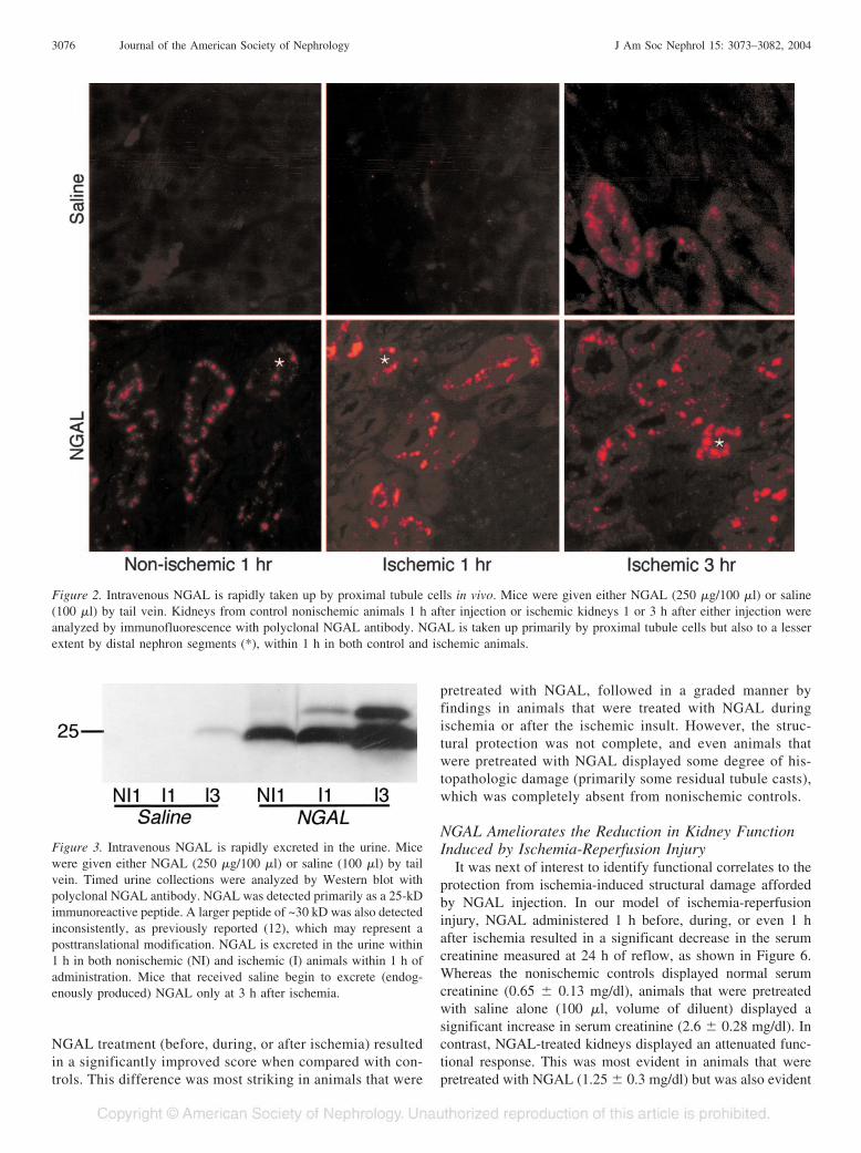
periods. Animals that received saline were devoid of kidney orurinary NGAL. In contrast, within 1 h of NGAL injection, itwas easily detected in a punctate cytoplasmic distributionpredominantly in the proximal tubules but also to a lesserextent in the distal tubules, as shown in Figure 2. Identificationof proximal versus distal tubules in these sections was based onlocation and morphology. In addition, NGAL was detected inthe urine within 1 h of injection, as shown in Figure 3. Theseresults confirm that exogenously administered NGAL is veryrapidly concentrated in the kidney and taken up by tubule cells.
Intravenous NGAL Is Rapidly Taken up by TubuleEpithelial Cells after Ischemic Injury
It was next of interest to determine whether purified NGALcan be delivered to the tubular epithelial cells after ischemicinjury. Mice received intravenous NGAL (250 �g in 100 �l ofsaline) or an equal volume of saline alone and were subjectedto ischemia-reperfusion injury, and the kidneys and urine wereexamined at various time periods. Animals that received salinewere devoid of kidney or urinary NGAL at the 1-h reflowperiod, and NGAL was just detectable at the 3-h reflow period,as shown in Figures 2 and 3, respectively. The 3-h datarepresent the endogenous response of kidney tubule cells toischemic injury, as previously reported (12). In contrast, inanimals that received an injection of NGAL and were simul-taneously subjected to ischemia-reperfusion injury, NGAL waseasily detected in the kidney and urine with 1 h of reflow, asshown in Figures 2 and 3. This represents the rapid uptake ofinjected NGAL after ischemic injury, because NGAL was notdetected at the 1-h reflow period in animals that receivedsaline.
NGAL Ameliorates the Histopathologic Damage toTubules Induced by Ischemia-Reperfusion Injury
Having established that exogenously administered NGALcan be delivered to the ischemic kidney, we next wished todetermine the structural consequences of this intervention.In an established murine model of renal ischemia-reperfu-sion injury, NGAL administered 1 h before, during, or even1 h after ischemia resulted in a significant decrease in thehistopathologic damage to tubules. Representative kidneysections obtained at 24 h of reflow and stained with hema-toxylin-eosin are shown in Figure 4. Whereas the nonisch-emic controls displayed normal histology, animals that werepretreated with saline alone (100 �l, volume of diluent)displayed extensive features of acute tubular necrosis asdescribed previously (11,12,15), including tubular dilation,tubular cast formation, and necrotic cells. In contrast,NGAL-treated kidneys displayed an attenuated histopatho-logic response. This was most evident in animals that werepretreated with NGAL but was also evident when the NGALwas administered during or even 1 h after the ischemicinjury. To quantify this response, we scored kidney sectionsfor histopathologic damage to the tubules in a blindedmanner, as described previously (16,17). The results areillustrated in Figure 5. In all three parameters examined(dilation, casts, and cell necrosis), all three modalities of
Figure 1. Expression and purification of recombinant murine neutro-phil gelatinase–associated lipocalin (NGAL). Coomassie Blue (CB)and enhanced chemiluminescence (ECL; with polyclonal NGAL an-tibody) analysis of defined quantities (as shown) of recombinantpurified NGAL. A single clean polypeptide of the predicted size wasdetected.
J Am Soc Nephrol 15: 3073–3082, 2004 NGAL in Ischemic Injury 3075
NGAL treatment (before, during, or after ischemia) resultedin a significantly improved score when compared with con-trols. This difference was most striking in animals that were
pretreated with NGAL, followed in a graded manner byfindings in animals that were treated with NGAL duringischemia or after the ischemic insult. However, the struc-tural protection was not complete, and even animals thatwere pretreated with NGAL displayed some degree of his-topathologic damage (primarily some residual tubule casts),which was completely absent from nonischemic controls.
NGAL Ameliorates the Reduction in Kidney FunctionInduced by Ischemia-Reperfusion Injury
It was next of interest to identify functional correlates to theprotection from ischemia-induced structural damage affordedby NGAL injection. In our model of ischemia-reperfusioninjury, NGAL administered 1 h before, during, or even 1 hafter ischemia resulted in a significant decrease in the serumcreatinine measured at 24 h of reflow, as shown in Figure 6.Whereas the nonischemic controls displayed normal serumcreatinine (0.65 � 0.13 mg/dl), animals that were pretreatedwith saline alone (100 �l, volume of diluent) displayed asignificant increase in serum creatinine (2.6 � 0.28 mg/dl). Incontrast, NGAL-treated kidneys displayed an attenuated func-tional response. This was most evident in animals that werepretreated with NGAL (1.25 � 0.3 mg/dl) but was also evident
Figure 2. Intravenous NGAL is rapidly taken up by proximal tubule cells in vivo. Mice were given either NGAL (250 �g/100 �l) or saline(100 �l) by tail vein. Kidneys from control nonischemic animals 1 h after injection or ischemic kidneys 1 or 3 h after either injection wereanalyzed by immunofluorescence with polyclonal NGAL antibody. NGAL is taken up primarily by proximal tubule cells but also to a lesserextent by distal nephron segments (*), within 1 h in both control and ischemic animals.
Figure 3. Intravenous NGAL is rapidly excreted in the urine. Micewere given either NGAL (250 �g/100 �l) or saline (100 �l) by tailvein. Timed urine collections were analyzed by Western blot withpolyclonal NGAL antibody. NGAL was detected primarily as a 25-kDimmunoreactive peptide. A larger peptide of ~30 kD was also detectedinconsistently, as previously reported (12), which may represent aposttranslational modification. NGAL is excreted in the urine within1 h in both nonischemic (NI) and ischemic (I) animals within 1 h ofadministration. Mice that received saline begin to excrete (endog-enously produced) NGAL only at 3 h after ischemia.
3076 Journal of the American Society of Nephrology J Am Soc Nephrol 15: 3073–3082, 2004

when the NGAL was administered during (1.5 � 0.2 mg/dl) oreven 1 h after (1.95 � 0.2 mg/dl) the ischemic injury. How-ever, the functional protection was not complete, and evenanimals that were pretreated with NGAL displayed a small butsignificant increase in serum creatinine when compared withnonischemic controls.
NGAL Ameliorates the Apoptotic Tubule Cell DeathInduced by Ischemia-Reperfusion Injury
Because apoptosis has been implicated in the tubule celldamage after ischemia-reperfusion injury (11,12,15), we nexttested the hypothesis that the structural and functional protec-tion observed with exogenous NGAL administration is a resultof decreased apoptosis. Representative kidney sections thatwere obtained at 24 h of reflow and subjected to TUNEL assayare shown in Figure 7. Whereas the nonischemic controlsdisplayed a minimal incidence of apoptosis (2.2 � 0.5 cells per100 cells examined), animals that were pretreated with salinealone (100 �l, volume of diluent) displayed a significantlygreater number of apoptotic tubule epithelial cells (12.6 �2.2%), as shown in Figure 8. Although apoptosis was moreprominent in the distal nephron, it was present in the proximaltubules as well, as described previously (11,12,15). In contrast,NGAL-treated kidneys displayed an attenuated apoptotic re-sponse. This was most evident in animals that were pretreatedwith NGAL (6.7 � 1.6%) but was also evident when theNGAL was administered during (7.6 � 0.8%) or even 1 h after(8.5 � 0.8%) the ischemic injury. However, the protectionfrom apoptotic cell death was not complete, and even animalsthat were pretreated with NGAL displayed a significantlygreater degree of apoptotic damage when compared with non-ischemic controls.
NGAL Enhances Tubule Cell Proliferation afterIschemic Injury
We next tested the hypothesis that the structural and func-tional protection observed with exogenous NGAL administra-tion is a result of enhanced tubule cell proliferation. Represen-tative kidney sections that were obtained at 24 h of reflow andstained with an antibody to PCNA are shown in Figure 7.Whereas the nonischemic controls displayed a minimal inci-dence of proliferating cells (1.9 � 0.4 cells per 100 cellsexamined), animals that were pretreated with saline alone (100�l, volume of diluent) displayed a small but significant in-crease in the number of PCNA-positive proximal tubule epi-thelial cells (4.4 � 1.2%), as shown in Figure 8. In contrast,NGAL-treated kidneys displayed a marked increase in prolif-erating proximal tubule cells. This was most evident in animalsthat were pretreated with NGAL (19.1 � 2.1%) but was alsoevident when the NGAL was administered during (14.9 �1.2%) or even 1 h after (14.5 � 1.2%) the ischemic injury.
NGAL Tilts the Balance of Proximal Tubule Cell FateToward Survival after Ischemic Injury
We next estimated the overall proximal tubule cell fate afterischemic injury using a one-way ANOVA to compare means �SD of proliferation and cell death among the various treatment
Figure 4. NGAL ameliorates the histopathologic damage to tubulesinduced by ischemia-reperfusion injury. Representative sections stainedwith hematoxylin-eosin of kidneys from control nonischemic mice, salinepretreated ischemic mice, or ischemic mice that were treated with NGAL1 h before, during, or 1 h after ischemia. The saline-pretreated ischemicmice displayed extensive features of acute tubular necrosis, includingtubular dilation, tubular cast formation, and necrotic cells. In contrast,NGAL-treated kidneys displayed an attenuated histopathologic response.Figure represents five animals in each group.
J Am Soc Nephrol 15: 3073–3082, 2004 NGAL in Ischemic Injury 3077

groups at 24 h of reflow. We restricted this analysis to theproximal tubule, because NGAL-induced proliferation was de-tected predominantly in this nephron segment. For quantifyingcell death, both necrosis and apoptosis were included. A pro-liferation/death ratio of unity may be assumed to indicate equalrates of cell survival and death, as would be expected in themature kidney at rest. The results are illustrated in Figure 9.Nonischemic control kidneys displayed a proximal tubule pro-liferation/death ratio of 0.9 � 0.2, close to the value of unity.As expected, animals that were pretreated with saline alone(100 �l, volume of diluent) displayed a significant decrease inthe proliferation/death ratio (0.34 � 0.2), indicating that celldeath is the predominant feature at the 24-h reflow time point.
In contrast, NGAL-treated kidneys displayed a marked in-crease in the ratio of proliferating versus apoptotic/necroticproximal tubule cells. This was most evident in animals thatwere pretreated with NGAL (3.4 � 0.5) but was also evidentwhen the NGAL was administered during (2.5 � 0.4) or even1 h after (1.7 � 0.4%) the ischemic injury. This analysisindicates that NGAL tilts the overall balance of proximaltubule cell fate toward cell survival after ischemic injury.
DiscussionHuman NGAL was originally identified as a 25-kD protein
covalently bound to gelatinase from human neutrophils (18)and was subsequently shown to be similar to the mouse 24p3gene first identified in primary cultures of mouse kidneys thatwere induced to proliferate (19). NGAL is expressed at verylow levels in several human tissues, including kidney, trachea,lungs, stomach, and colon (20). NGAL expression is markedlyinduced in stimulated epithelia. For example, NGAL concen-trations are elevated in the serum of patients with acute bac-terial infections, the sputum of patients with asthma or chronicobstructive pulmonary disease, and the bronchial fluid from theemphysematous lung (21). NGAL is also one of the maximallyinduced genes in the kidney after early ischemic injury (12). Inall of these instances, the role of NGAL remains unclear (22).In some cell types, NGAL has been shown to possess aproapoptotic property. For example, in the mouse pro-B lym-phocytic cell line, cytokine withdrawal resulted in a markedinduction of NGAL as well as onset of apoptosis (23,24).NGAL has also been linked to apoptosis in reproductive tis-sues. Epithelial cells of the involuting mammary gland anduterus express high levels of NGAL, temporally coincidingwith a period of maximal apoptosis (25). Thus, it is likely thata subset of epithelial cells may utilize this mechanism toregulate their own demise.
Accumulating evidence, however, suggests that NGAL canenhance the epithelial phenotype. During kidney development,NGAL is expressed by the penetrating ureteric bud and triggersnephrogenesis by stimulating the conversion of mesenchymalcells into kidney epithelia (14). In the postischemic maturekidney, NGAL is markedly upregulated predominantly inproximal tubules but also in distal nephron segments. In the
Figure 5. Histopathologic scoring. Sections of kidneys from ischemic mice that were pretreated with saline or treated with NGAL 1 h before,during, or 1 h after ischemia were analyzed for tubule dilation, tubule casts, and tubule cell necrosis using an arbitrary scale of 0 to 4. Valuesare means � SD of five animals in each treatment group. *P � 0.05 versus saline.
Figure 6. NGAL ameliorates the reduction in kidney function inducedby ischemia-reperfusion injury. Serum creatinine was measured innonischemic (Non Isch) control mice or 24 h after ischemia in micethat were pretreated with saline (Pre Sal) or treated with NGAL 1 hbefore (Pre NGAL), during (Dur NGAL), or 1 h after (Post NGAL)ischemic injury. Values are means � SD of six to eight animals (asshown within the bars) in each treatment group. *P � 0.05 versusnonischemic controls; #P � 0.05 versus saline. NGAL treatmentspartially but not completely prevented the rise in serum creatinineafter ischemic injury.
3078 Journal of the American Society of Nephrology J Am Soc Nephrol 15: 3073–3082, 2004

Figure 7. NGAL inhibits apoptosis and enhances proliferation induced by ischemia-reperfusion injury. Representative sections fromnonischemic control mice or 24 h after ischemia in mice that were pretreated with saline or treated with NGAL 1 h before, during, or 1 h afterischemic injury. Shown are results of transferase-mediated dUTP nick-end labeling staining at low and high power and PCNA staining at lowpower. Arrows point to the condensed, fragmented, intensely staining nuclei characteristic of apoptosis. Figure represents five animals in eachgroup.
J Am Soc Nephrol 15: 3073–3082, 2004 NGAL in Ischemic Injury 3079
proximal tubule, NGAL co-localizes at least in part with pro-liferating epithelial cells (12). These findings suggest thatNGAL may be expressed by the damaged tubule to inducere-epithelialization. In support of this hypothesis is the recentidentification of NGAL as an iron-transporting protein duringnephrogenesis (22). It is well known that the delivery of ironinto cells is crucial for cell growth and development, and thisis presumably also critical to renal regeneration after nephro-toxic injury. Because NGAL can be endocytosed by the prox-
imal tubule (22), the protein could potentially recycle iron intoviable cells, thereby stimulating regeneration of renal epithelialcells after ischemic injury. An alternative hypothesis is thatNGAL may serve as a reservoir for iron that is released fromtubule cells that are damaged by nephrotoxic injury. This mightremove iron, a reactive molecule, from the site of tissue injury,thereby limiting iron-mediated cytotoxicity. It is indeed possi-ble that both mechanisms are operative in the postischemickidney.
In this study, exogenous administration of NGAL strikinglyameliorated the structural damage inflicted by ischemia-reper-fusion injury. Both apoptosis and necrosis were significantlyblunted. The mechanism by which NGAL inhibits apoptosis inthis situation may be analogous to the well-documented anti-apoptotic effects of heme oxygenase 1 (HO-1). It is known thatHO-1 facilitates the extracellular transport of iron, therebylimiting iron-driven oxidant stress in the intracellular compart-ment (26). It is likely that NGAL may also facilitate theremoval of excess intracellular iron, thereby limiting oxidant-mediated apoptosis of renal tubule cell death after ischemia-reperfusion injury (27). With respect to necrosis, it was pro-posed recently that necrotic cell death after oxidant injuryoccurs by a two-stage process, namely initiation of apoptosisfollowed by a necrotic cell death (28). Our results suggest thata similar situation may pertain to the response of the kidneyafter ischemia-reperfusion injury and that apoptosis inhibitionby NGAL may be effective in also preventing this “secondary”necrosis.
The results of our study indicate that NGAL tilts the overallbalance of proximal tubule cell fate toward cell survival afterischemic injury. This is based on an analysis of the prolifera-tion/death ratio in proximal tubule cells, with the assumptionthat a ratio of unity indicates equal rates of cell survival anddeath, as would be expected in the mature kidney at rest.However, the specific assays that we used (PCNA for prolif-eration and TUNEL for apoptosis), although standard in thefield, provide only a snapshot measurement of each of thesedynamic processes (i.e., S-phase for proliferation and the ex-ecution phase of apoptosis). A more complete confirmation ofNGAL’s cellular homeostatic effects will require additionalmeasures of proliferation (e.g., Ki67 and cell cycle proteins)and apoptosis (e.g., death receptor activation, induction ofmitochondrial pathways, caspase cleavage).
The studies reported herein focused primarily on the abilityof NGAL to counter the morphologic response of renal tubularepithelial cells to ischemia. The potential effect of NGALinfusion on several other extracellular factors involved in thepathogenesis of ischemic ARF have not been explored. Forexample, an additional salutary effect of NGAL on the persis-tent vasoconstriction, tubular obstruction, or the inflammatoryresponse typical of ARF (4–7) cannot be ruled out.
Nevertheless, the findings reported here may have far-reach-ing clinical implications. ARF secondary to ischemic injuryremains a common problem, with limited and unsatisfactorytherapeutic options (1–4). Although previous studies suggestedtherapeutic approaches in animal models, translational researchefforts in humans have yielded disappointing results. The rea-
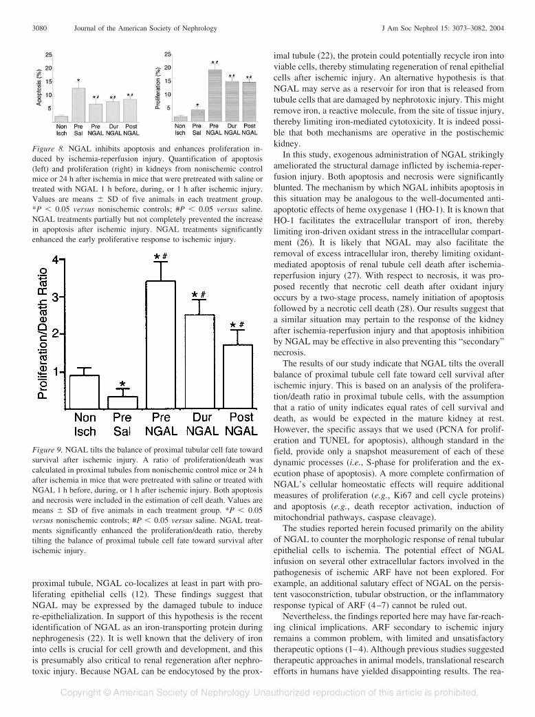
Figure 9. NGAL tilts the balance of proximal tubular cell fate towardsurvival after ischemic injury. A ratio of proliferation/death wascalculated in proximal tubules from nonischemic control mice or 24 hafter ischemia in mice that were pretreated with saline or treated withNGAL 1 h before, during, or 1 h after ischemic injury. Both apoptosisand necrosis were included in the estimation of cell death. Values aremeans � SD of five animals in each treatment group. *P � 0.05versus nonischemic controls; #P � 0.05 versus saline. NGAL treat-ments significantly enhanced the proliferation/death ratio, therebytilting the balance of proximal tubule cell fate toward survival afterischemic injury.
Figure 8. NGAL inhibits apoptosis and enhances proliferation in-duced by ischemia-reperfusion injury. Quantification of apoptosis(left) and proliferation (right) in kidneys from nonischemic controlmice or 24 h after ischemia in mice that were pretreated with saline ortreated with NGAL 1 h before, during, or 1 h after ischemic injury.Values are means � SD of five animals in each treatment group.*P � 0.05 versus nonischemic controls; #P � 0.05 versus saline.NGAL treatments partially but not completely prevented the increasein apoptosis after ischemic injury. NGAL treatments significantlyenhanced the early proliferative response to ischemic injury.
3080 Journal of the American Society of Nephrology J Am Soc Nephrol 15: 3073–3082, 2004

sons for this likely include a lack of early markers for ARF andthe multifactorial nature of the disease. Recent advances incellular and molecular biology of ischemic renal injury haverevealed that proximal tubule cells undergo a complex tempo-ral sequence of events. These include loss of cell polarity, celldeath as a result of apoptosis and necrosis, dedifferentiationand proliferation of viable cells, and re-establishment of theepithelial phenotype (6,7). Therefore, identification of factorsthat oppose tubule cell death and/or enhance the recoveryphase may provide critical clues toward novel therapeuticoptions. We propose NGAL as a potential candidate that pos-sesses both of these desirable properties. Exogenously admin-istered NGAL seems to limit the morphologic and functionalconsequences of ischemia-reperfusion injury in a mousemodel, by a combination of limiting tubule cell death andenhancing re-epithelialization. It will be important in futuretranslational research to examine these cytoprotective roles ofNGAL in human conditions that are known to predispose toischemic renal injury. One good example pertains to cadavericrenal transplantation, in which oxidant-mediated apoptosis isan important contributor to tubule cell death (29). In addition tothe usual complications of ARF, ischemia-reperfusion injury inthe transplanted kidney is known to result in delayed graftfunction (30), which significantly increases the risk of graftloss and acute rejection (31). It will be intriguing to explore themorphologic effects and clinical outcomes of adding NGAL tothe organ preservation solutions used during cold storage. It ishoped that such maneuvers will be successful in amelioratingthe delayed graft function characteristic of cadaveric kidneytransplantation. On the basis of our present findings thatNGAL is at least partially effective even when administeredafter the ischemic insult, it is also hoped that NGAL may offerpromising diagnostic and therapeutic possibilities in ischemicARF, a common clinical condition that is still associated witha dismal prognosis and for which novel therapies are desper-ately needed.
References1. Thadani R, Pascual M, Bonventre JV: Acute renal failure. N Engl
J Med 334: 1448–1460, 19962. Star RA: Treatment of acute renal failure. Kidney Int 54: 1817–
1831, 19983. Liaño F, Pascual J: Predictive factors and scoring. In: Acute
Renal Failure, edited by Molitoris BA, Finn WF, Philadelphia,WB Saunders, 2001, pp 507–518
4. Molitoris BA: Transitioning to therapy in ischemic acute renalfailure. J Am Soc Nephrol 14: 265–267, 2003
5. Brady HR, Brenner BM, Clarkson MR, Lieberthal W: Acuterenal failure. In: The Kidney, 6th Ed., edited by Brenner BM,Philadelphia, WB Saunders, 2000, pp 1201–1262
6. Sutton TA, Molitoris BA: Mechanisms of cellular injury inischemic acute renal failure. Semin Nephrol 18: 490–497, 1998
7. Sheridan AM, Bonventre JV: Cell biology and molecular mech-anisms of injury in ischemic acute renal failure. Curr OpinNephrol Hypertens 9: 327–334, 2000
8. Muramatsu Y, Tsujie M, Kohda Y, Pham B, Perantoni AO, ZhaoH, Jo S-K, Yuen PST, Craig L, Hu X, Star RA: Early detection
of cysteine rich protein 61 (CYR61, CCN1) in urine followingrenal ischemia reperfusion injury. Kidney Int 62: 1601–1610,2002
9. Kurella M, Hsiao L-L, Yishida T, Randall JD, Chow G,Sarang SS, Jensen RV, Gullans SR: DNA microarray analysisof complex biologic processes. J Am Soc Nephrol 12: 1072–1078, 2001
10. Yoshida T, Kurelia M, Beato F, Min H, Ingelfinger JR, StearsRL, Swinford RD, Gullans SR, Tang S-S: Monitoring changes ingene expression in renal ischemia-reperfusion in the rat. KidneyInt 61: 1646–1654, 2002
11. Supavekin S, Zhang W, Kucherlapati R, Kaskel FJ, Moore LC,Devarajan P: Differential gene expression following early renalischemia-reperfusion. Kidney Int 63: 1714–1724, 2003
12. Mishra J, Ma Q, Prada A, Mitsnefes M, Zahedi K, Yang J,Barasch J, Devarajan P: Identification of neutrophil gelati-nase-associated lipocalin as a novel early urinary biomarkerfor ischemic renal injury. J Am Soc Nephrol 14: 2534 –2543,2003
13. Bundgaard J, Sengelov H, Borregaard N, Kjeldsen L: Molecularcloning and expression of a cDNA encoding NGAL: A lipocalinexpressed in human neutrophils. Biochem Biophys Res Commun202: 1468–1475, 1994
14. Yang J, Goetz D, Li J-Y, Wand W, Mori K, Setlik D, Du T,Erdjument-Bromage H, Tempst P, Strong R, Barasch J: An irondelivery pathway mediated by a lipocalin. Mol Cell 10: 1045–1056, 2002
15. Del Rio M, Imam A, De Leon M, Gomez G, Mishra J, Ma Q,Parikh S, Devarajan P: The death domain of kidney ankyrininteracts with Fas and promotes Fas-mediated cell death in renalepithelia. J Am Soc Nephrol 15: 41–51, 2004
16. Deng J, Kohda Y, Chiao H, Wang Y, Hu X, Hewitt SM, MiyajiT, McLeroy P, Nibhanupudy B, Li S, Star RA: Interleukin-10inhibits ischemic and cisplatin-induced acute renal injury. KidneyInt 60: 2118–2128, 2001
17. Yokota N, Burne-Taney M, Racusen L, Rabb H: Contrastingroles for STAT4 and STAT6 signal transduction pathways inmurine renal ischemia-reperfusion injury. Am J Physiol RenalPhysiol 285: F319–F325, 2003
18. Kjeldsen L, Cowland JB, Borregaard N: Human neutrophil ge-latinase-associated lipocalin and homologous proteins in rat andmouse. Biochim Biophys Acta 1482: 272–283, 2000
19. Hraba-Renevey S, Turler H, Kress M, Salomon C, Weil R:SV40-induced expression of mouse gene 24p3 involves a post-transcriptional mechanism. Oncogene 4: 601–608, 1989
20. Cowland JB, Borregaard N: Molecular characterization and patternof tissue expression of the gene for neutrophil gelatinase-associatedlipocalin from humans. Genomics 45: 17–23, 1997
21. Xu S, Venge P: Lipocalins as biochemical markers of disease.Biochim Biophys Acta 1482: 298–307, 2000
22. Yang J, Mori K, Li JY, Barasch J: Iron, lipocalin, and kidneyepithelia. Am J Physiol Renal Physiol 285: F9–F18, 2003
23. Devireddy LR, Teodoro JG, Richard FA, Green MR: Induc-tion of apoptosis by a secreted lipocalin that is transcription-ally regulated by IL-3 deprivation. Science 293: 829 – 834,2001
24. Persengiev SP, Devireddy LR, Green MR: Inhibition of apopto-sis by ATFx: A novel role for a member of the ATF/CREBfamily of mammalian bZIP transcription factors. Genes Dev 16:1806–1814, 2002
J Am Soc Nephrol 15: 3073–3082, 2004 NGAL in Ischemic Injury 3081
25. Ryon J, Bendickson L, Nilsen-Hamilton M: High expression ininvoluting reproductive tissues of uterocalin/24p3, a lipocalinand acute phase protein. Biochem J 367: 271–277, 2002
26. Ferris CD, Jaffrey SR, Sawa A, Takahashi M, Brady SD, BarrowRK, Tysoe SA, Wolosker H, Baranano DE, Dore S, Poss KD,Snyder SH: Haem oxygenase-1 prevents cell death by regulatingcellular iron. Nat Cell Biol 3: 152–157, 1999
27. Kunduzova OR, Bianchi P, Pizzinat N, Escourrou G, SeguelasMH, Parini A, Cambon C: Regulation of JNK/ERK activation,cell apoptosis, and tissue regeneration by monoamine oxidasesafter renal ischemia-reperfusion. FASEB J 16: 1129–1131, 2002
28. Wang X, Ryter SW, Dai C, Tang Z-L, Watkins SC, Yin X-M,Song R, Choi AMK: Necrotic cell death in response to oxidantstress involves the activation of the apoptogenic caspase-8/Bidpathway. J Biol Chem 278: 29184–29191, 2003
29. Castaneda MP, Swiatecka-Urban A, Mitsnefes MM, FeuersteinD, Kaskel FJ, Tellis V, Devarajan P: Activation of mitochondrialapoptotic pathways in human renal allografts following ischemia.Transplantation 76: 50–54, 2003
30. Koning OH, Ploeg RJ, van Bockel JH, Groenewegen M, vander Woude FJ, Persijn GG, Hermans J: Risk factors fordelayed graft function in cadaveric kidney transplantation: Aprospective study of renal function and graft survival afterpreservation with University of Wisconsin solution in multi-organ donors. European Multicenter Study Group. Transplan-tation 63: 1620 –1628, 1997
31. Lu CY, Penfield JG, Kielar ML, Vasquez MA, Jeyarajah DR:Hypothesis: Is renal allograft rejection initiated by the responseto injury during the transplant process? Kidney Int 55: 2157–2168, 1999
3082 Journal of the American Society of Nephrology J Am Soc Nephrol 15: 3073–3082, 2004