AMELIORATION OF γ-RADIATION-INDUCED GENOTOXICITY BY NANOSILYMARIN: A COMPARATIVE STUDY INDICATES...
10
Trakia Journal of Sciences, Vol. 12, Suppl. 1, 2014 1 Trakia Journal of Sciences, Vol. 12, Suppl. 1, pp 1-10, 2014 Copyright © 2014 Trakia University Available online at: http://www.uni-sz.bg ISSN 1313-7050 (print) ISSN 1313-3551 (online) AMELIORATION OF γ-RADIATION-INDUCED GENOTOXICITY BY NANOSILYMARIN: A COMPARATIVE STUDY INDICATES POSSIBLE IMPLICATIONS FOR CHEMICAL BIOLOGICAL RADIOLOGICAL AND NUCLEAR (CBRN) DEFENCE R. Arora 1,2* , M. Adhikari 1 , P. Agarwal 1 , R. Chawla 1 , D. Gupta 1 , R. K. Sharma 1 , V. Ivanov 3 , Y. Karamalakova 3 , A. Zheleva 3 , V. Gadjeva 3 , S. Stoev 4 1 Division of Radiation Biosciences and CBRN Defence, Institute of Nuclear Medicine & Allied Sciences, Defense Research & Development Organization, Brig. S.K Mazumdar Marg, Timarpur, Delhi, India 2 Office of Director General Life Sciences, RDO Bhawan, Rajaji Marg, New Delhi, India 3 Department of Chemistry and Biochemistry, Medical Faculty, Trakia University, Stara Zagora, Bulgaria 4 Department of General and Clinical Pathology, Faculty of Veterinary Medicine, Trakia University, Stara Zagora, Bulgaria ABSTRACT Purpose: To check the comparative efficacy of DNA protection by silymarin and its nanoformulation as an effective radiation countermeasure agent in ameliorating γ-radiation-induced Genotoxicity. Methods: The study performed suggests the efficacy of silymarin and its nanoformulation specifically in ameliorating γ-radiation-induced genotoxic effects at cellular, plasmid DNA levels etc. Results: The retention of super-coiled DNA following treatment of DNA with various concentrations of silymarin (parent compound) was found to be maximum at 25µg/ml, whereas better retention was seen at 10µg/ml in case of silymarin nanoformulation. Micronuclei count also reduced maximally at 10µg/ml when treated with silymarin nanoformulation as compared to 25µg/ml using parent compound. Summary : Silymarin and its nanoformulation showed no toxic effects on DNA. The nanoformulation demonstrated better results in terms of protection of genetic material against -radiation due to increase in surface area and hence improved bioavailability. The nanoformulation can be of use in mitigating the deleterious effects of radiation and plausible biothreat agents. Key words: Silymarin, -radiation, nanoformulation, HEK cells, micronuclei, pUC19 INTRODUCTION Ionizing radiation inflicts substantial damage to living tissues through a cascade of molecular events (1). Radiation exposure of biological systems results in oxidative stress due to hydrolysis of water and generation of ROS (e.g., superoxide radicals [O 2 • ], hydrogen peroxide [H 2 O 2 ] and hydroxyl radicals [ • OH]) which initiates a plethora of chemical peroxidative processes (2). Interaction of ionizing radiation ____________________________ *Correspondence to: Dr. Rajesh Arora, Office of the Director General (Life Sciences), Room No. 339, DRDO Bhawan, Room No. 339, Rajaji Marg, New Delhi-110011, India. Tel.: +91-11-23014873; Fax: +91-11-23014259;E-mai: [email protected] directly with macromolecules results in breakage of covalent bonds (3). The damage to protein molecules has also been examined in great detail and has been found throughout polypeptides regardless of the site of the primary ionization (4). One of the most susceptible targets in a living cell is DNA. The radiation-induced DNA damage can be of various types e.g., single and double-strand breaks, base damage, damage to sugar moiety and cross-linkages of the intra and inter-strand types etc(5). These damages are a result of oxidative stress which can be induced by free radicals (6-8). Several compounds with antioxidant properties have the ability to alleviate deleterious effects of
Transcript of AMELIORATION OF γ-RADIATION-INDUCED GENOTOXICITY BY NANOSILYMARIN: A COMPARATIVE STUDY INDICATES...
Trakia Journal of Sciences, Vol. 12, Suppl. 1, 2014 1
Trakia Journal of Sciences, Vol. 12, Suppl. 1, pp 1-10, 2014
Copyright © 2014 Trakia University
Available online at:
http://www.uni-sz.bg
ISSN 1313-7050 (print)
ISSN 1313-3551 (online)
AMELIORATION OF γ-RADIATION-INDUCED GENOTOXICITY BY
NANOSILYMARIN: A COMPARATIVE STUDY INDICATES POSSIBLE
IMPLICATIONS FOR CHEMICAL BIOLOGICAL RADIOLOGICAL AND
NUCLEAR (CBRN) DEFENCE
R. Arora1,2*
, M. Adhikari1, P. Agarwal
1, R. Chawla
1, D. Gupta
1, R. K. Sharma
1, V. Ivanov
3,
Y. Karamalakova3, A. Zheleva
3, V. Gadjeva
3, S. Stoev
4
1Division of Radiation Biosciences and CBRN Defence, Institute of Nuclear Medicine & Allied Sciences,
Defense Research & Development Organization, Brig. S.K Mazumdar Marg, Timarpur, Delhi, India
2Office of Director General Life Sciences, RDO Bhawan, Rajaji Marg, New Delhi, India
3Department of Chemistry and Biochemistry, Medical Faculty, Trakia University, Stara Zagora, Bulgaria
4 Department of General and Clinical Pathology, Faculty of Veterinary Medicine, Trakia University,
Stara Zagora, Bulgaria
ABSTRACT
Purpose: To check the comparative efficacy of DNA protection by silymarin and its nanoformulation as
an effective radiation countermeasure agent in ameliorating γ-radiation-induced Genotoxicity.
Methods: The study performed suggests the efficacy of silymarin and its nanoformulation specifically in
ameliorating γ-radiation-induced genotoxic effects at cellular, plasmid DNA levels etc.
Results: The retention of super-coiled DNA following treatment of DNA with various concentrations of
silymarin (parent compound) was found to be maximum at 25µg/ml, whereas better retention was seen
at 10µg/ml in case of silymarin nanoformulation. Micronuclei count also reduced maximally at 10µg/ml
when treated with silymarin nanoformulation as compared to 25µg/ml using parent compound.
Summary : Silymarin and its nanoformulation showed no toxic effects on DNA. The nanoformulation
demonstrated better results in terms of protection of genetic material against -radiation due to
increase in surface area and hence improved bioavailability. The nanoformulation can be of use in
mitigating the deleterious effects of radiation and plausible biothreat agents.
Key words: Silymarin, -radiation, nanoformulation, HEK cells, micronuclei, pUC19
INTRODUCTION Ionizing radiation inflicts substantial damage to
living tissues through a cascade of molecular
events (1). Radiation exposure of biological
systems results in oxidative stress due to
hydrolysis of water and generation of ROS (e.g.,
superoxide radicals [O2•], hydrogen peroxide
[H2O2] and hydroxyl radicals [•OH]) which
initiates a plethora of chemical peroxidative
processes (2). Interaction of ionizing radiation
____________________________ *Correspondence to: Dr. Rajesh Arora, Office of the
Director General (Life Sciences), Room No. 339,
DRDO Bhawan, Room No. 339, Rajaji Marg, New
Delhi-110011, India. Tel.: +91-11-23014873; Fax:
+91-11-23014259;E-mai: [email protected]
directly with macromolecules results in breakage
of covalent bonds (3). The damage to protein
molecules has also been examined in great detail
and has been found throughout polypeptides
regardless of the site of the primary ionization
(4). One of the most susceptible targets in a
living cell is DNA. The radiation-induced DNA
damage can be of various types e.g., single and
double-strand breaks, base damage, damage to
sugar moiety and cross-linkages of the intra and
inter-strand types etc(5). These damages are a
result of oxidative stress which can be induced
by free radicals (6-8).
Several compounds with antioxidant properties
have the ability to alleviate deleterious effects of
ARORA R., et al.
2 Trakia Journal of Sciences, Vol. 12, Suppl. 1, 2014
ionizing radiation in living systems and bio-
molecules (9, 10). In view of this, efforts have
been made to improve the therapeutic effect of
radiotherapy by minimizing normal tissue injury
and by-stander damage to acceptable levels
using radioprotective compounds mainly
containing sulfhydryl groups like cysteine,
cysteamine and WR-2721 (11). The
radioprotective ability of these compounds has
been substantially attributed to their reactive
oxygen species (ROS) scavenging ability (12).
The search for effective and non-toxic radiation
countermeasures is a necessity and consequently
attention has been diverted towards the naturally
occurring antioxidants. Additionally, a plethora
of medicinal plants, endophytes and other
microbial products have also been investigated
for their radioprotective efficacy (13-21). In
view of the proven medicinal value of a number
of natural products in treating various ailments,
interest in medicinal and aromatic plants is
increasing worldwide (15). Radioprotection
being a multifaceted phenomenon, it is essential
to investigate and elucidate the mechanisms
involved in radioprotective action of the herbal
drugs.
A number of natural plant products
(polyphenols, flavonoids, vitamins and
carotenes) are known to exhibit antioxidant
properties (22). Silymarin is a non-toxic
bioactive flavonoid, an approved herbal
hepatoprotectant and is consumed worldwide as
a dietary antioxidant for prevention and
treatment of a number of diseases (23, 24). The
antioxidant, anti-inflammatory and anti-
carcinogenic properties of silymarin has been
demonstrated in various in vitro and in vivo
models against oxidative stress, inflammatory
responses and chemical carcinogen-induced
tumor promotion (25, 26). Silymarin has also
been reported to increase aspartate
aminotransferase (ALT) and -
glutamyltranspeptidase (GGT) in plasma (27). It
has also beenwidely used as a topical ointment
for the treatment of breast cancer (28). Silymarin
is known to modulate proinflammatory pathways
via downregulation of cyclooxygenase-2 (COX-
2) (29) and 5-lipooxygenase (LOX), thereby
inhibiting hepatic cytochrome P450
detoxification system involving hepatic
cytochrome P450 enzyme activities (30).
Silymarin is also known to be able to protect
hepatic tissues from radiation directly by
stabilizing membrane permeability and
preventing liver glutathione depletion (31). In
addition to its hepatoprotective properties,
silymarin also exhibits protective effects against
various drugs of nephrotoxic nature (32). The
compound silibinin has been reported to protect
hepatocytes by blocking the hepatotoxin
receptors present on the hepatocyte membrane,
thus preventing apoptosis and cell death (33). It
has been documented that silymarin protects
from hepatotoxins by reducing the levels of
oxidized Glutathione (GSH) in the liver and
intestine; stimulate the ribosomal RNA
polymerase and protein synthesis resulting in
enhanced regeneration of hepatocytes. Silymarin
has been shown to protect liver against
fumonisin B1 –induced damage (34) and sepsis-
induced acute lung and brain injury in mice. The
powerful hepatoprotective effects of silibinin
have been reported in cultured primary rat
hepatocytes against apoptosis and cytotoxicity
caused by Ochratoxin A (OTA) (35), not only in
OTA but hepatoprotective effects in Aflatoxin
B1 have also been studied in case of albino male
Wistar rats (36). It has been reported that
silymarin derived -phospholipid complex is
involved in reducing the toxic effects of
aflatoxin B1 (AFB1), as studied in broiler
chickens (37). Potential ameliorative effects of
Silymarin in combination with vitamin E against
(OTA)-induced immunotoxic effects in White
Leghorn cockerels have also been reported (38).
In addition, protective effects of silymarin on L-
arginine-induced genotoxicity in in vitro
lymphocyte culture have also been elucidated
(39).
The phytochemicals e.g., lignans and flavonoids
present in silymarin have been individually
reported to be of prominent efficacy against
various pathological symptoms and also possess
radioprotective properties (40)
Nanoparticles have been recently used as
efficient drug delivery systems in recent years to
protect against radiation injuries. Nanomedicines
are emerging as one of the new treatment
options, (41, 42) since they are novel in their
mode of action (43, 44). Silymarin is a known
natural lipophilic agent and despite its prominent
properties, it has the limitation of low
bioavailability in living organisms. A
nanotechnology based approach can lead to the
development of novel drug delivery systems to
increase the solubility and oral absorption of
various drugs for achieving better bioavailability
and therapeutic activity (45-48). Keeping this in
mind, a nanoformulation of silymarin was
ARORA R., et al.
Trakia Journal of Sciences, Vol. 12, Suppl. 1, 2014 3
prepared and utilized for experimentation in the
present study comparing it with the parental
compound silymarin.
Irradiation of purified DNA molecules has been
extensively used during the last decade for
studying the interaction of ionizing radiation
with DNA (49-53). Plasmid DNA is considered
a useful model for investigating interactions
between topologically constrained DNA and
radiation in addition to their role as vectors (54-
56). Micronuclei estimation can effectively serve
as a biological dosimeter to estimate in vitro
ionizing radiation exposure (57). Keeping these
key observations in view, this paper reports
comparative protection of DNA against gamma
radiation-induced damage using silymarin and its
nanoformulation. Protection to DNA under in
vitro conditions of irradiation was estimated in
plasmid DNA by plasmid relaxation assay and
micronuclei estimation in order to evaluate the
potential of silymarin and its nanoformulation. It
has been hypothesized that silymarin and the
nanoformulation interacts directly with plasmid
DNA to exhibit its shielding effect against
radiation exposure. Elucidation of the
mechanism of action of silymarin in radiation
protection can aid in forming strategies for the
development of an ideal radioprotector for
human use.
MATERIALS AND METHODS
Chemicals Low melting agarose, Tris-base, Ethidium
bromide, Tris-HCL, bromophenol blue, xylene
cyanol, glycerol, sucrose, high glucose Dulbecco
Modified Eagle Medium (HG-DMEM), Trypsin-
EDTA, Bovine serum albumin (BSA) and
Hoechst-33258 etc. were obtained from Sigma
Aldrich St. Louis MO, USA). pUC 19 plasmid
was obtained from Thermoscientific, PA, USA.
Methanol, Acetic acid, Tween-20 and citric acid
were purchased from Merck India Pvt. Ltd,
Mumbai, India.
Herbal extract
Silymarin was procured from Wuxi Gorunjie
Technology Co. Ltd, Jiangsu, China. The sample
was stored in a cool and dry place away from
strong light and heat as per the prescribed
information of the manufacturer.
Irradiation
A 60
Co gamma irradiator (Gamma cell 5000,
Board of Radiation and Isotope Technology,
Mumbai, India) with a dose rate of 1.12kGy/h
was used as irradiation source. HEK cells were
grown in 10% FBS supplemented with MEM at
37ºC in humidified 5% CO2: 95% Air
envrionment. Logarithmically growing cells
were exposed to -radiation using 60
Co gamma
chamber (Bhabhatron-II Telecobalt unit –
BARC, Mumbai, India; Dose rate of 1.4 Gy/min)
at room temperature.
DNA damage assay The effect of silymarin and its nanoformulation
on radiation-induced relaxation of plasmid
(pUC19) DNA was evaluated (53). 200ng of
plasmid DNA, in potassium phosphate buffer
(0.1mM, pH 7.4), was exposed to -radiation
(250Gy) in the presence of different
concentrations of silymarin and its nanoform
(10-500μg/ml). After irradiation, the plasmid and
silymarin reaction mixture was suspended in TE
buffer (Tris-EDTA, pH 8.0) and resolved on 1%
agarose (w/v) submarine gels in electrophoresis
buffer (TBE; pH 8.0) at 45V (3V/cm). The gel
was subsequently stained with 0.5μg/ml
ethidium bromide for 15-30min and
photographed under UV illumination. The
comparative amount of relaxed and supercoiled
DNA was determined by scanning the gel with
the Bio Rad GEL-DOC system (Bio Rad,
Hercules, California, USA).
Miconucleus assay
The micronucleus technique was used as a
method for measurement of radiation-induced
chromosomal damage. HEK cells were treated
with silymarin and nanoformulation 0.5 h prior
to -radiation (2Gy) and incubated for different
time intervals for evaluation of protection
against micronucleus formation. Upon
completion of incubation period, cells were
washed twice in phosphate buffered saline (PBS,
pH 7.2) and fixed in Carnoy‘s fixative
(Methanol: Acetic acid; 3:1) at 4C for 24h.
Fixed cells were spread on clean pre-chilled
microscopic slides. Following overnight air
drying, slides were stained with 10μg/ml
Hoechst-33258 in phosphate buffer
(Na2HPO4.2H2O, 0.5% tween-20, and 0.1M
citric acid) in the ratio 9:1, final pH 7.4 for
30min, in dark at room temperature (54). After
washing off excess stain with distilled water
followed by PBS, the slides were mounted in
PBS-glycerol (1:1) and observed under
fluorescence microscope (Olympus BX60,
Tokyo, Japan) using UV excitation filter. A total
of 500 cells in triplicates were scored per group.
ARORA R., et al.
4 Trakia Journal of Sciences, Vol. 12, Suppl. 1, 2014
The frequency of cells with micronuclei, called
the M-fraction (MF) was calculated as:
MF(%) = Nm/Nt X 100
where Nm is the number of cells with
micronuclei and Nt is the total number of cells
analyzed.
RESULTS
DNA Protective Effect of Silymarin and its
nanoformulation Radiation exposure causes
DNA strand breaks resulting in conformational
changes in terms of relaxation of plasmid DNA
from supercoiled form to open circular form.
The DNA protecting ability of silymarin was
investigated using the plasmid relaxation assay
which is a method of semi-quantitative
assessment of ionizing radiation-induced
oxidative damage to DNA (52). It was observed
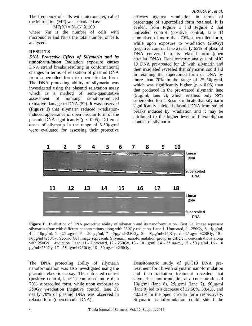
(Figure 1) that silymarin reduced -radiation-
induced appearance of open circular form of the
plasmid DNA significantly (p < 0.05). Different
doses of silymarin in the range of 5-50µg/ml
were evaluated for assessing their protective
efficacy against -radiation in terms of
percentage of supercoiled form retained. It is
evident from Figure 1 and Figure 2 that
untreated control (positive control, lane 1)
comprised of more than 70% supercoiled form,
while upon exposure to -radiation (250Gy)
(negative control, lane 2) nearly 65% of plasmid
DNA converted to its relaxed form (open
circular DNA). Densitometric analysis of pUC
19 DNA pre-treated for 1h with silymarin and
then irradiated revealed that silymarin could aid
in retaining the supercoiled form of DNA by
more than 70% in the range of 25–50µg/ml,
which was significantly higher (p < 0.05) than
that produced in the pre-treated silymarin lane
(5µg/ml, lane 7), which retained only 58%
supercoiled form. Results indicate that silymarin
significantly shielded plasmid DNA from strand
breaks induced by -radiation and it may be
attributed to the higher level of flavonolignan
content of silymarin.
Figure 1. Evaluation of DNA protective ability of silymarin and its nanoformulation. First Gel image represent
silymarin alone with different concentrations along with 250Gy-radiation. Lane 1- Untreated, 2 - 250Gy, 3 - 5µg/ml,
4 - 10µg/ml, 5 - 25 µg/ml, 6 - 50 µg/ml, 7 - 5µg/ml+250Gy, 8 - 10µg/ml+250Gy, 9 - 25µg/ml+250Gy, 10 -
50µg/ml+250Gy. Second Gel Image represents Silymarin nanoformulation group in different concentrations along
with 250Gy -radiation. Lane 11 - Untreated, 12 - 250Gy, 13 - 10 µg/ml, 14 - 25 µg/ml, 15 - 50 µg/ml, 16 - 10
µg/ml+250Gy, 17 - 25 µg/ml+250Gy, 18 - 50 µg/ml+250Gy.
The DNA protecting ability of silymarin
nanoformulation was also investigated using the
plasmid relaxation assay. The untreated control
(positive control, lane 1) comprised more than
70% supercoiled form, while upon exposure to
250Gy γ-radiation (negative control, lane 2),
nearly 70% of plasmid DNA was observed in
relaxed form (open circular DNA).
Densitometric study of pUC19 DNA pre-
treatment for 1h with silymarin nanoformulation
and then radiation treatment revealed that
silymarin nanoformulation at a concentration of
10μg/ml (lane 6), 25μg/ml (lane 7), 50μg/ml
(lane 8) led to a decrease of 32.58%, 38.43% and
40.51% in the open circular form respectively.
Silymarin nanoformulation could shield the
ARORA R., et al.
Trakia Journal of Sciences, Vol. 12, Suppl. 1, 2014 5
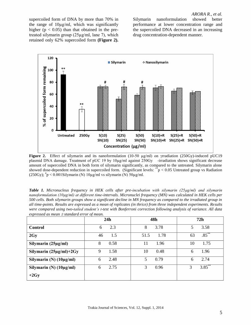
supercoiled form of DNA by more than 70% in
the range of 10µg/ml, which was significantly
higher (p < 0.05) than that obtained in the pre-
treated silymarin group (25µg/ml, lane 7), which
retained only 62% supercoiled form (Figure 2).
Silymarin nanoformulation showed better
performance at lower concentration range and
the supercoiled DNA decreased in an increasing
drug concentration-dependent manner.
Figure 2. Effect of silymarin and its nanoformulation (10-50 µg/ml) on rradiation (250Gy)-induced pUC19
plasmid DNA damage. Treatment of pUC 19 by 10µg/ml against 250Gy -irradiation shows significant decrease
amount of supercoiled DNA in both form of silymarin significantly, as compared to the untreated. Silymarin alone
showed dose-dependent reduction in supercoiled form. (Significant levels: **
p < 0.05 Untreated group vs Radiation
(250Gy); #p < 0.001Silymarin (N) 10µg/ml vs silymarin (N) 50µg/ml.
Table 1. Micronucleus frequency in HEK cells after pre-incubation with silymarin (25µg/ml) and silymarin
nanoformulation (10µg/ml) at different time-intervals. Micronuclei frequency (MN) was calculated in HEK cells per
500 cells. Both silymarin groups show a significant decline in MN frequency as compared to the irradiated group in
all time-points. Results are expressed as a mean of replicates (in thrice) from three independent experiments. Results
were compared using two-tailed student’s t-test with Bonferroni correction following analysis of variance. All data
expressed as mean standard error of mean.
24h 48h 72h
Control 6 2.3 8 3.78 5 3.58
2Gy 46 1.5 51.5 1.78 63 .85**
Silymarin (25µg/ml) 8 0.58 11 1.96 10 1.75
Silymarin (25µg/ml)+2Gy 9 1.58 10 0.48 6 1.96
Silymarin (N) (10µg/ml) 6 2.48 5 0.79 6 2.74
Silymarin (N) (10µg/ml)
+2Gy
6 2.75 3 0.96 3 3.85**
ARORA R., et al.
6 Trakia Journal of Sciences, Vol. 12, Suppl. 1, 2014
Effect of silymarin and its nanoformulation
pretreatment on -radiation-induced DNA
damage and micronuclei formation
rradiation possesses the potential to induce
micronuclei formation in HEK cells even at a
low dose. Radiation exposure (2Gy) significantly
induced MN formation by 8.0, 7.0 and 12.5-fold
at 24 h, 48 h and 72 h time intervals respectively
(p < 0.05) as compared to control (Table 1).
Pretreatment with silymarin followed by
irradiation of cells with -rays resulted in a
significant (p < 0.05) decrease in the percentage
of micro-nucleated cells and total MN in
comparison to the radiationalone group.
Silymarin alone (25µg/ml) decreased MN
frequency by nearly three-times as compared to
irradiated control, hence exhibiting its non-toxic
nature. Silymarin elicited maximum reduction
(63.85 %) in the radiation-induced MN
formation at 72 h time-point.
Silymarin nanoformulation when given alone
(10µg/ml), also caused a significant decrease (p
< 0.05) in MN numbers as compared to radiation
exposed group at all-time intervals and also
when compared to control, exhibiting its non-
toxic nature. At later time intervals, pre-treated
irradiated silymarin groups exhibited decreased
MN level much efficiently with maximum
reduction (61%).
DISCUSSION
Both ionizing-radiation and mycotoxins are
known to have mutagenic and carcinogenic
effects via generation of reactive oxygen species
(55-57). Various compounds possessing
antioxidant properties have been evaluated in
recent years to shield DNA against the harmful
effects of environmental genotoxins. Silymarin
has been shown to protect against ribavirin-
induced genotoxicity (58). The present study
provides direct evidence of protection against
genotoxic effects by silymarin and its
nanoformulation. Protecting cellular DNA from
radiation damage might aid in the prevention of
cancers/mutations induced by radiation. This
approach also has implications in reducing the
undesirable side effects caused by ionizing
radiation injury (64) In our previous report on
radioprotective studies, we have provided direct
evidence of free-radical scavenging ability of
silymarin (40). The present study shows that
under in vitro conditions, silymarin exhibits the
ability to shield DNA against exposure to -
radiation induced damage.
Most of the radiation damage to biomolecules is
mediated through ROS generation by radiolysis
of water. Therefore, the effect of silymarin on
DNA irradiated under aqueous condition, where
indirect effect becomes dominant, was evaluated.
It was observed that under acellular conditions of
irradiation, silymarin protected plasmid pUC19
DNA against - radiation-induced damage
significantly (Figure 1 and 2). It prevented the
occurrence of radiation-induced strand breakage
events in the plasmid DNA as is evident from
conservation of supercoiled form of DNA. This
fact can also be attributed to the physical
interaction of silymarin with plasmid DNA in
order to prevent free radicals from damaging the
DNA supercoiled conformations. Exposure of
pUC19 to 250Gy -radiation caused conversion
of supercoiled pUC19 DNA (fast mitigating),
into open circular form (slow mitigating) but
maximal retention of supercoiled form was
achieved at 25µg/ml range (lane 9). Silymarin
(25µg/ml) + 250Gy showed maximum
protection (< 72% supercoiled retention form) as
compared to radiation alone group (35%
supercoiled form) (Figure 1). Level of
supercoiled DNA retention in percent in
silymarin nanoformulation was also compared
and it showed enhanced supercoiled retention at
lower concentration range (10µg/ml) and it
decreased in a concentration-dependent manner.
This may be due to its DNA protective ability at
10µg/ml, which is lesser than parent silymarin
concentration, which protects pUC19 DNA at
25µg/ml (Figure 2).
The present study also provides evidence that
silymarin and its nanoformulation possess the
ability to inhibit radiationinduced micronuclei
formation and DNA damage in human
embryonic kidney cells (Figure 3). Studies on
radiation-induced DNA damage by Micronuclei
(MN) assay carried out in HEK cells as per the
method reported by Schmid, 1975, revealed that
irradiation (2Gy) significantly enhanced the
frequency of MN in HEK cells as compared to
control. Pre-irradiation (0.5h) treatment with
silymarin substantially countered this upsurge in
MN frequency clearly indicating its role in
reduction of radiation-induced DNA damage.
However, treatment with silymarin
nanoformulation 0.5h before irradiation showed
high reduction in micronuclei count in a
significant way in later time-intervals as
compared to radiation alone showing its
radioprotective nature (Figure 3b). Also, it was
found that the most effective concentration of
ARORA R., et al.
Trakia Journal of Sciences, Vol. 12, Suppl. 1, 2014 7
silymarin nanoformulation for minimizing
micronuclei count was lower than that of the
parent silymarin compound.
Our results have clearly shown that the
radioprotective efficacy of silymarin
nanoformulation is better than silymarin parent
compound. It may be due to the fact that
silymarin is orally absorbed but has very poor
bioavailability due to its poor water solubility
(65). The formulation work has been performed
to enhance its solubility so as to increase its
bioavailability and thus, its radioprotective
property as compared to its parent compound.
Under in vitro conditions of radiation exposure,
it was found that silymarin nanoformulation
significantly reduced DNA damage induced by
radiation as is evident from plasmid relaxation
assay and micronuclei count in HEK cells, which
is a sensitive technique to measure radiation
damage and can be a reliable method for
biodosimetry. Hence, in conclusion, it can be
stated that both silymarin and its
nanoformulation aid in preserving the structural
and functional integrity of DNA upon exposure
to ionizing radiations. However, the
nanoformulation is more efficient than its parent
compound in shielding the genetic material
against radiation-induced damage. Based on the
leads, and preliminary in vitro studies vis-à-vis
mycotoxins, it would be interesting to further
study the effects of silymarin, including
nanosilymarin formulation, in mitigating the
deleterious effects of mycotoxins particularly in
liver and kidneys of higher animal models. With
the preliminary results indicating promise, the
possible implications of silymarin and
nanosilymarin for CBRN defence is an area that
needs further exploration.
ACKNOWLEDGEMENTS This research has been supported in part through
the European Community under the Marie Curie
International Research Staff Exchange Scheme
(IRSES) project of FP7.
The authors are grateful to Director General
(Life Sciences), Defence Research and
Development Organization (DRDO)
Headquarters, New Delhi, India, Director,
INMAS, Delhi and Rector, Trakia University for
support in multifarious ways. Research support
received from DRDO is duly acknowledged.
REFERENCES 1. Chen Xi, Chunyan L, Chu Q, Zhou G, Lin X,
Li X, Lu H, Xu B, Yue Z. Dissecting the
Molecular Mechanism of Ionizing Radiation-
Induced Tissue Damage in the Feather
Follicle. Plos One. 9(2) (2012).
2. Azzam EI, Jay-Gerin J and Pain D. Ionizing
radiation-induced metabolic oxidative stress
and prolonged cell injury. Cancer Lett.
327(1-2):48-60, (2012).
3. Khodyreva SN and Lavrik OI. Affinity
modification in a proteomic study of DNA
repairs ensembles. BioorgKhim. 37(1):91-
107, (2011).
4. Kempner E.S. Effects of high-energy
electrons and gamma rays directly on protein
molecules. J. Pharm. Sci. 90: 1637–1646,
(2001).
5. Lomax ME, Folkes LK and O'Neill P.
Biological consequences of radiation-induced
DNA damage: relevance to radiotherapy.
ClinOncol (R CollRadiol).25(10): 578-585,
(2013).
6. Brierley DJ and Martin SA. Oxidative stress
and the DNA mismatch repair pathway.
Antioxid Redox Signal. 18(18): 2420-2428,
(2013).
7. Zitka O, Krizkova S, Skalickova S, Kopel P,
Babula P, Adam V andKizek R.
Electrochemical study of DNA damaged by
oxidation stress. Comb Chem High
Throughput Screen. 16(2):130-141, (2013).
8. Dizdaroglu M. Oxidatively induced DNA
damage: mechanisms, repair and disease.
Cancer Lett. 327(1-2): 26-47, (2012).
9. Hosseinimehr SJ. Trends in the development
of radioprotective agents. Drug Discov
Today.12(19-20): 794-805, (2007).
10. Okunieff P, Swarts S, Keng P, Sun W, Wang
W, Kim J, Yang S, Zhang H, Liu C, Williams
JP, Huser AK and Zhang L. Antioxidants
reduce consequences of radiation exposure.
AdvExp Med Biol.614: 165-78, (2008).
11. Suzuki K and Yamashita S.Radiation-Induced
Bystander Response: Mechanism and Clinical
Implications. Adv Wound Care (New
Rochelle).3(1): 16-24, (2014).
12. Kuntić VS, Stanković MB, Vujić ZB, Brborić
JS andUskoković-Marković SM.
Radioprotectors - the evergreen topic.
ChemBiodivers. 10(10):1791-803, (2013).
ARORA R., et al.
8 Trakia Journal of Sciences, Vol. 12, Suppl. 1, 2014
13. Kunwar A, Pritadarsini KI and Jain VK.
Organoselenium Compounds: A New
Generation of Radioprotectors. BARC
newletters.319: 1-7, (2011).
14. Kma L. Plant extracts and plant-derived
compounds: promising players in a
countermeasure strategy against radiological
exposure. Asian Pac J Cancer Prev.
15(6):2405-2425, (2014).
15. Arora R, Gupta D , Chawla R, Sagar R,
Sharma A, Kumar R, Prasad J, Singh S,
Samanta N and Sharma RK. Radioprotection
by plant products: Present status and future
prospects. Phytotherapy Research 19: 1– 22,
(2005).
16. Arora R, Singh S, Sagar RK, Chawla R,
Kumar R, Puri SC, Surender S , Prasad J ,
Gupta ML, Krishna B, et al.Radiomodulatory
and free-radical scavenging activity of the
fractionated aquo-alcoholic extract of the
adaptogenicnutraceutical (Rhodiolaimbricata)
– a comparative in vitro assessment with
ascorbate. Journal of Dietary Supplements 5:
147– 163, (2008).
17. Arora R, Chawla R, DhakerAS ,Adhikari M,
Sharma J, Singh S, Gupta D, Kumar R,
Sharma A , Sharma RK , et al. Podophyllum
hexandrum as a potential botanical
supplement for the medical management of
nuclear and radiological emergencies (NREs)
and free radical-mediated ailments: Leads
from in vitro/in vivo radioprotective efficacy
evaluation. Journal of Dietary Supplements 7:
31 – 50, (2010a).
18. Arora R, Chawla R, Dhaker AS , Adhikari M,
Sharma J , Jaiswal S , Gupta D , Amna T ,
Puri SC , Kumar R, et al. Pro-antioxidant
activities of fractions of a novel
camptothecin-producing endophytes
(Entrophosphora infrequens). Trakia Journal
of Sciences 8: 1 – 15, (2010b).
19. Arora R, Dhaker AS , Adhikari M, Sharma J ,
Chawla R, Gupta D, Zheleva A,
Karamalakova Y, Kumar R, Sharma R, et al.
Radical scavenging and
radiomodulatoryeffects of Psoraleacorylifolia
Linn. substantiated by in vitro assays and
EPR spectroscopy. Zeitschriftfür-
naturforschung C 66: 35– 46, (2011).
20. Puri SC, Nazir A, Chawla R, Arora R, Riyaz-
Ul-Hasan S, Amna T, Ahmed B, Verma V,
Singh S, Sagar R, et al. The endophytic
fungus Trameteshirsuta as a novel alternative
source of podophyllotoxin and related aryl
tetralinlignans. Journal of Biotechnology 122:
494 – 510, (2006).
21. Chawla R, Arora R, Singh S, Sagar RK,
Sharma RK, Kumar R, Sharma A, Gupta ML,
Prasad J, Khan HA, et al. Radioprotective and
antioxidant activity of fractionated extracts of
berries of Hippophae rhamnoides. Journal of
Medicinal Food 10: 101 – 109, (2007).
22. Vance TM, Su J, Fontham ET, Koo SI and
Chun OK. Dietary antioxidants and prostate
cancer: a review. Nutr Cancer. 65(6):793-
801, (2013).
23. Milić N, Milosević N, Suvajdzić L, Zarkov M
and Abenavoli L. New therapeutic potentials
of milk thistle (Silybum marianum). Nat Prod
Commun. 8(12):1801-1810, (2013).
24. El-Gazayerly ON, Makhlouf AI, Soelm AM
and Mohmoud MA. Antioxidant and
hepatoprotective effects of silymarin
phytosomes compared to milk thistle extract
in CCl4 induced hepatotoxicity in rats. J
Microencapsul. 31(1):23-30, (2014).
25. Adhikari M, Arora R, Chawla R, Sharma J,
Dhaker AS, Gupta D, Dubey N, Kumar R,
Ivanov V, Gadjeva V, Gevrenova R and
Sharma RK. Evaluation of silymarin as a
promising radioprotector. Z Naturforsch C.65
(5-6):337-46, (2010).
26. Sherif IO and Al-Gayyar MM. Antioxidant,
anti-inflammatory and hepatoprotective
effects of silymarin on hepatic dysfunction
induced by sodium nitrite. Eur Cytokine
Netw. 24(3):114-121, (2013).
27. KiruthigaPV, Shafreen RB, Pandian SK,
Arun S, Govindu S and Devi KP. Protective
effect of silymarin on erythrocyte
haemolysate against benzo(a)pyrene and
exogenous reactive oxygen species (H2O2)
induced oxidative stress. Chemosphere 68:
1511 – 1518, (2007).
28. Becker SM, Mengs U, Schaefer M, Bullita M
and Hoffmann W. Topical use of a silymarin-
based preparation to prevent radiodermatitis:
Results of a prospective study in breast
cancer patients. Strahlentherapie und
onkologie 187: 485– 491, (2011).
29. ŠkottováN ,Vecera R, Urbánek K, Vána P,
Walterová D and Cvak L. Effects of
polyphenolic fraction of silymarin on
lipoprotein profile in rats fed cholesterol-rich
diets. Pharmacological Research 47: 17 – 26,
(2003).
30. Ramadan LA, Roushdy HM, AbuSenna GM,
Amin NE and El-Deshw OA. Radioprotective
effect of silymarin against radiation induced
hepatotoxicity. Pharmacological Research
45: 447– 454, (2002).
ARORA R., et al.
Trakia Journal of Sciences, Vol. 12, Suppl. 1, 2014 9
31. Deep G, Singh RP, Agarwal C, Kroll DJ and
Agarwal R. Silymarin and silibinin cause G1
and G2-M cell cycle arrest via distinct
circuitries in human prostate cancer PC3
cells: A comparison of flavanonesilibinin
with flavanolignan mixture silymarin.
Oncogene 25: 1053 – 1069, (2005).
32. Shahbazi F, Dashti-KhavidakiS ,Khalili H
and Lessan-Pezeshki M. Potential
renoprotective effects of silymarin against
nephrotoxic drugs: A review of literature. J
Pharmacy and Pharmaceutical Sciences 1:
112– 123, (2012).
33. He Q, Kim J and Sharma RP. Silymarin
protects against liver damage in BALB/c
mice exposed to fumonisin B1despite
increasing accumulation of free sphiongoid
bases. Toxicological Sciences 80: 335-42,
(2004).
34. Kostek H, Szponar J, Tchórz M, Majewska
M, Lewandowska-Stanek H. Silibinin and its
hepatoprotective action from the perspective
of a toxicologist. Przegl Lek 69(8):541-3,
(2012).
35. Essid E(1), Dernawi Y, Petzinger E.
Apoptosis induction by OTA and TNF-α in
cultured primary rat hepatocytes and
prevention by silibinin. Toxins (Basel)
4(11):1139-56, (2012).
36. Shyamal S(1), Latha PG, Suja SR, Shine VJ,
Anuja GI, Sini S, Pradeep S, Shikha P,
Rajasekharan S. Hepatoprotective effect of
three herbal extracts on aflatoxin B1-
intoxicated rat liver. Singapore Med J.
51(4):326-31 (2010).
37. Tedesco D(1), Steidler S, Galletti S, Tameni
M, Sonzogni O, Ravarotto L. Efficacy of
silymarin-phospholipid complex in reducing
the toxicity of aflatoxin B1 in broiler chicks.
Poult Sci. 83(11):1839-43 (2004).Khatoon
A(1), Zargham Khan M, Khan A, Saleemi
MK, Javed I. Amelioration of Ochratoxin A-
induced immunotoxic effects by silymarin
and Vitamin E in White Leghorn cockerels. J
Immunotoxicol 10(1):25-31 (2013).
38. Yurtcu E, Kasapoglu E, Sahin FI. Protective
effects of beta-carotene and silymarin on
human lymphocytes. Turk J Biol. 36: 47-52,
(2012).
39. Adhikari M, Dhaker A, Adhikari J, Ivanov V,
Singh V, Chawla R, Kumar R, Sharma R,
Karamalakova Y, Gadjeva V andArora R. In
vitro studies on radioprotective efficacy of
silymarin against γ-irradiation. Int J Radiat
Biol. 89(3):200-11, (2013).
40. Lee SK, Kim GS, Wu Y et al. Nanowire
substrate-based laser scanning cytometry for
quantitation of circulating tumour cells. Nano
Lett. 12:2697-2704, (2012).
41. Dicheva BM, Ten Hagen TL, Li L et al.
Cationic thermosensitive liposomes: a novel
dual targeted heat-triggered drug delivery
approach for endothelial and tumour cells.
Nano Lett. 13: 2324-2331, (2013).
42. Kanapathipillai M, Mammoto A, Mammoto
T, et al. Inhibition of mammary tumour
growth using lysyl oxidase-targetting
nanoparticles to modify extracellular matrix.
Nano Lett. 12: 3213-3217, (2012).
43. Zhao G and Rodriguez BL. Molecular
targeting of liposomal nanoparticles to tumor
microenvironment. Int J Nanomedicine. 8:
61-71, (2013).
44. Tiwari SB and Amiji MM. Improved oral
delivery of paclitaxel following
administration in nanoformulations. J.
Nanosci. Nanotechnol. 6: 3215-3221, (2006).
45. Khandavilli S and Panchagnula R.
Nanoformulations as versatile formulations
for paclitaxel delivery: peroral and dermal
delivery studies in rats. J. Invest. Dermatol.
127: 154-162, (2007).
46. Vyas TK, Shahiwala A and Amiji MM.
Improved oral bioavailability and brain
transport of Saquinavir upon administration
in novel nanoformulation formulations. Int. J.
Pharm. 347: 93-101, (2008).
47. Javed S, Kohli K and Ali M. Patented
bioavailability enhancement techniques of
silymarin. Recent Pat. Drug Deliv. Formul 4:
145-152, (2010).
48. Milligan JR, Arnold AD and Ward JF. The
effect of superhelical density on the yield of
single-strand breaks in gamma-irradiated
plasmid DNA. Radiat. Res. 132: 69–73,
(1992).
49. Milligan JR, Aguilera JA and Ward JF.
Variation of single-strand break yield with
scavenger concentration for plasmid DNA
irradiated in aqueous solution. Radiat. Res.
133: 151–157, (1993).
50. Milligan JR, Aguilera JA, Wu CCL, Ng JYY
and Ward JF. The difference that linear
energy transfer makes to precursors of DNA
strand breaks. Radiat. Res. 145: 442–448,
(1996).
51. Jones GDD, Milligan JR, Ward JF, Calabro-
Jones PM and Aguilera JA. Yield of strand
breaks as a function of scavenger
concentration and LET for SV40 irradiated
ARORA R., et al.
10 Trakia Journal of Sciences, Vol. 12, Suppl. 1, 2014
with He ions. Radiat. Res. 136: 190–196,
(1993).
52. Jones GDD, Boswell TV, Lee J, Milligan JR,
Ward JF and Weinfeld M. A comparison of
DNA damage produced under conditions of
direct and indirect action of radiation. Int. J.
Radiat. Biol. 66(5): 441–445, (1994).
53. Swenberg CE and Speicher JM. Neutron and
Gamma-radiation sensitivity of plasmid
DNA by plasmid DNA of varying
superhelical density. Radiation Res. 144:
301–309, (1995).
54. Tigran H, Anna K, Galina H and Rouben A.
Combined genotoxic effects of aflatoxin b1,
ochratoxin a and zearalenone in rat bone
marrow and blood leukocytes. Korean J.
Environ. Biol. 31(3): 189–191 (2013).
55. Yokoya A, Watanabe R and Hara T. Single
and double-strand breaks in solid pBR322
plasmid DNA induced by ultrasoft X-rays at
photon energies of 388, 435, 573 ev. J.
Radiat. Res. 40: 145–158, (1999).
56. Vral A, Fenech M andThierens H. The
micronucleus assay as a biological dosimeter
of in vivo ionising radiation exposure.
Mutagenesis. 26(1):11-7, (2011).
57. Hussien N. Evaluation of the antioxidant
silymarin role in modulating the in vivo
genotoxicity of the antiviral drug ribavirin.
LAP Lambert Academic Publishing (2012),
pp.172.
58. Schmid W. The micronucleus test. Mutat.
Res. 31: 9–15, (1975).
59. Camenisch U andNaegeli H. Role of DNA
repair in the protection against genotoxic
stress. EXS. 99:111-50, (2009).
60. Waters RL, Newton GL, Olive PL and Fahey
RC. Radioprotection of human cell nuclear
DNA by polyamines: Radiosensitivity of
chromatin is influenced by tightly bound
spermine. Radiation Research 151: 354–362,
(1999).
61. Murray D. Aminothiols, Chapter 3: In
Radioprotectors: Chemical, Biological and
Clinical Perspectives ed. E. A. Bump and K.
Malakar, CRC press Inc., Boca Raton, Fla.
53–109, (1998).
62. Murray, D and McBride WH.
Radioprotective agents, In Kirk-
OthmerEnzyclopedia of Chemical
Technology, Fourth Edition, Vol 20, John
Wiley & Sons Inc., New York. 964–1006,
(1996).
63. Amin AR, Kucuk O, KhuriFR and ShinDM.
Perspectives for cancer prevention with
natural compounds.J. Clin. Oncol. 27: 2712–
2725, (2009).
64. Parveen R, Sanjula B, Javed A, Alka A,
Suruchi SV andSayeed A. Oil based
nanocarrier for improved oral delivery of
silymarin: In vitro and in vivo studies. Int J
Pharm 413: 245-253, (2011).