Localization of MMR proteins on meiotic chromosomes in mice indicates distinct functions during...
12
THE JOURNAL OF CELL BIOLOGY © The Rockefeller University Press $8.00 The Journal of Cell Biology, Vol. 171, No. 3, November 7, 2005 447–458 http://www.jcb.org/cgi/doi/10.1083/jcb.200506170 JCB: ARTICLE JCB 447 Localization of MMR proteins on meiotic chromosomes in mice indicates distinct functions during prophase I Nadine K. Kolas, 1 Anton Svetlanov, 1 Michelle L. Lenzi, 1 Frank P. Macaluso, 2 Steven M. Lipkin, 5 R. Michael Liskay, 6 John Greally, 1,3 Winfried Edelmann, 4 and Paula E. Cohen 1 1 Department of Molecular Genetics, 2 Department of Anatomy and Structural Biology, 3 Department of Medicine, and 4 Department of Cell Biology, Albert Einstein College of Medicine, Bronx, NY 10461 5 Department of Hematology, University of California, Irvine, Irvine, CA 92697 6 Molecular and Medical Genetics, Oregon Health Sciences University, Portland, OR 97239 ammalian MutL homologues function in DNA mismatch repair (MMR) after replication errors and in meiotic recombination. Both functions are initiated by a heterodimer of MutS homologues spe- cific to either MMR (MSH2–MSH3 or MSH2–MSH6) or crossing over (MSH4–MSH5). Mutations of three of the four MutL homologues ( Mlh1 , Mlh3 , and Pms2 ) result in meiotic defects. We show herein that two distinct com- plexes involving MLH3 are formed during murine meio- sis. The first is a stable association between MLH3 and MLH1 and is involved in promoting crossing over in con- M junction with MSH4–MSH5. The second complex in- volves MLH3 together with MSH2–MSH3 and localizes to repetitive sequences at centromeres and the Y chro- mosome. This complex is up-regulated in Pms2 / males, but not females, providing an explanation for the sexual dimorphism seen in Pms2 / mice. The associa- tion of MLH3 with repetitive DNA sequences is coinci- dent with MSH2–MSH3 and is decreased in Msh2 / and Msh3 / mice, suggesting a novel role for the MMR family in the maintenance of repeat unit integrity during mammalian meiosis. Introduction The eukaryotic DNA mismatch repair (MMR) family repairs a variety of mismatches and also participates in reciprocal re- combination events during meiosis (Kolodner, 1996; Modrich, 1997; Kirkpatrick, 1999). MMR activity is initiated by the de- tection of a substrate by a heterodimer of MutS homologues (MSHs), of which there are five in mammals, MSH2 through MSH6. They form three heterodimeric complexes: MSH2– MSH6 (MutS), MSH2–MSH3 (MutS), and MSH4–MSH5 (MutS). Of these, MutS initiates the repair of base–base and insertion–deletion mispairs, whereas MutS participates in the repair of only the latter (Nakagawa et al., 1999). The meiosis- specific MutS complex is crucial for reciprocal recombination in yeast (Ross-Macdonald and Roeder, 1994; Hollingsworth et al., 1995; Winand et al., 1998), nematodes (Zalevsky et al., 1999), and mice (de Vries et al., 1999; Edelmann et al., 1999; Kneitz et al., 2000), but has no MMR activity per se. After MutS association with the DNA, a heterodimeric complex of MutL homologues is recruited. Four mammalian MutL homologues exist, but the principal heterodimers that function in mitotic repair and meiotic reciprocal recombination are MLH1–PMS2 (postmeiotic segregation 2) and MLH1– MLH3, respectively (Kolodner and Marsischky, 1999; Flores- Rozas and Kolodner, 2000), whereas in yeast these heterodimers are represented by Mlh1–Pms1 and Mlh1–Mlh3 (Nakagawa et al., 1999; Wang et al., 1999; Hoffmann and Borts, 2004). The mammalian meiotic MMR complex, consisting of MSH4, MSH5, MLH1, and MLH3, has been implicated in the processing of DNA double strand breaks (DSB) through the double Holliday junction (dHJ) recombination intermediate repair pathway that results in reciprocal crossover events be- tween parental homologous chromosomes (Ross-Macdonald and Roeder, 1994; Hollingsworth et al., 1995; Novak et al., 2001; Borner et al., 2004; Hoffmann and Borts, 2004; Surtees et al., Nadine K. Kolas and Anton Svetlanov contributed equally to this work. Correspondence to P.E. Cohen: [email protected] P.E. Cohen’s present address is Dept. of Biomedical Sciences, Cornell University, Ithaca, NY 14853. Abbreviations used in this paper: DDB, double dense body; dHJ, double Holli- day junction; DSB, DNA double strand break; MLH1 and 3, MutL homologue 1 and 3; MMR, mismatch repair; MN, meiotic nodule; MSH2–6, MutS homo- logue; PAR, pseudoautosomal region; PMS2, postmeiotic segregation 2; TNR, trinucleotide repeat. The online version of this article includes supplemental material.
-
Upload
independent -
Category
Documents
-
view
0 -
download
0
Transcript of Localization of MMR proteins on meiotic chromosomes in mice indicates distinct functions during...
TH
EJ
OU
RN
AL
OF
CE
LL
BIO
LO
GY
©
The Rockefeller University Press $8.00The Journal of Cell Biology, Vol. 171, No. 3, November 7, 2005 447–458http://www.jcb.org/cgi/doi/10.1083/jcb.200506170
JCB: ARTICLE
JCB 447
Localization of MMR proteins on meiotic chromosomes in mice indicates distinct functions during prophase I
Nadine K. Kolas,
1
Anton Svetlanov,
1
Michelle L. Lenzi,
1
Frank P. Macaluso,
2
Steven M. Lipkin,
5
R. Michael Liskay,
6
John Greally,
1,3
Winfried Edelmann,
4
and Paula E. Cohen
1
1
Department of Molecular Genetics,
2
Department of Anatomy and Structural Biology,
3
Department of Medicine, and
4
Department of Cell Biology, Albert Einstein College of Medicine, Bronx, NY 10461
5
Department of Hematology, University of California, Irvine, Irvine, CA 92697
6
Molecular and Medical Genetics, Oregon Health Sciences University, Portland, OR 97239
ammalian MutL homologues function in DNAmismatch repair (MMR) after replication errorsand in meiotic recombination. Both functions
are initiated by a heterodimer of MutS homologues spe-cific to either MMR (MSH2–MSH3 or MSH2–MSH6) orcrossing over (MSH4–MSH5). Mutations of three of thefour MutL homologues (
Mlh1
,
Mlh3
, and
Pms2
) result inmeiotic defects. We show herein that two distinct com-plexes involving MLH3 are formed during murine meio-sis. The first is a stable association between MLH3 andMLH1 and is involved in promoting crossing over in con-
M
junction with MSH4–MSH5. The second complex in-volves MLH3 together with MSH2–MSH3 and localizesto repetitive sequences at centromeres and the Y chro-mosome. This complex is up-regulated in
Pms2
�
/
�
males, but not females, providing an explanation for thesexual dimorphism seen in
Pms2
�
/
�
mice. The associa-tion of MLH3 with repetitive DNA sequences is coinci-dent with MSH2–MSH3 and is decreased in
Msh2
�
/
�
and
Msh3
�
/
�
mice, suggesting a novel role for the MMRfamily in the maintenance of repeat unit integrity duringmammalian meiosis.
Introduction
The eukaryotic DNA mismatch repair (MMR) family repairs avariety of mismatches and also participates in reciprocal re-combination events during meiosis (Kolodner, 1996; Modrich,1997; Kirkpatrick, 1999). MMR activity is initiated by the de-tection of a substrate by a heterodimer of MutS homologues(MSHs), of which there are five in mammals, MSH2 throughMSH6. They form three heterodimeric complexes: MSH2–MSH6 (MutS
�
), MSH2–MSH3 (MutS
�
), and MSH4–MSH5(MutS
�
). Of these, MutS
�
initiates the repair of base–base andinsertion–deletion mispairs, whereas MutS
�
participates in therepair of only the latter (Nakagawa et al., 1999). The meiosis-specific MutS
�
complex is crucial for reciprocal recombination
in yeast (Ross-Macdonald and Roeder, 1994; Hollingsworth etal., 1995; Winand et al., 1998), nematodes (Zalevsky et al.,1999), and mice (de Vries et al., 1999; Edelmann et al., 1999;Kneitz et al., 2000), but has no MMR activity per se.
After MutS association with the DNA, a heterodimericcomplex of MutL homologues is recruited. Four mammalianMutL homologues exist, but the principal heterodimers thatfunction in mitotic repair and meiotic reciprocal recombinationare MLH1–PMS2 (postmeiotic segregation 2) and MLH1–MLH3, respectively (Kolodner and Marsischky, 1999; Flores-Rozas and Kolodner, 2000), whereas in yeast these heterodimersare represented by Mlh1–Pms1 and Mlh1–Mlh3 (Nakagawa etal., 1999; Wang et al., 1999; Hoffmann and Borts, 2004).
The mammalian meiotic MMR complex, consisting ofMSH4, MSH5, MLH1, and MLH3, has been implicated in theprocessing of DNA double strand breaks (DSB) through thedouble Holliday junction (dHJ) recombination intermediaterepair pathway that results in reciprocal crossover events be-tween parental homologous chromosomes (Ross-Macdonald andRoeder, 1994; Hollingsworth et al., 1995; Novak et al., 2001;Borner et al., 2004; Hoffmann and Borts, 2004; Surtees et al.,
Nadine K. Kolas and Anton Svetlanov contributed equally to this work.Correspondence to P.E. Cohen: [email protected]. Cohen’s present address is Dept. of Biomedical Sciences, Cornell University,Ithaca, NY 14853.Abbreviations used in this paper: DDB, double dense body; dHJ, double Holli-day junction; DSB, DNA double strand break; MLH1 and 3, MutL homologue 1and 3; MMR, mismatch repair; MN, meiotic nodule; MSH2–6, MutS homo-logue; PAR, pseudoautosomal region; PMS2, postmeiotic segregation 2; TNR,trinucleotide repeat.The online version of this article includes supplemental material.
JCB • VOLUME 171 • NUMBER 3 • 2005448
2004). Studies in mouse (Kneitz et al., 2000; Santucci-Darma-nin et al., 2000; Moens et al., 2002) and human germ cells(Lenzi et al., 2005) show that MSH4–MSH5 appear at recom-bination intermediates, as part of the multiprotein meiotic nod-ule (MN), early in prophase I and accumulate at frequenciesthat far exceed the eventual number of crossover sites (Kneitzet al., 2000). In vitro biochemical studies suggest that this het-erodimer forms a sliding clamp that associates with dHJ struc-tures at zygonema (Snowden et al., 2004) a subset of whichmay be stabilized by interactions with MLH1–MLH3 het-erodimers at pachynema. It is these sites that will eventuallybecome the crossovers that ensure accurate chromosomal seg-regation at the first meiotic division. The additional MSH4–MSH5 sites that are not stabilized by MLH1–MLH3 are thenrepaired through noncrossover mechanisms.
The temporal dynamics of MMR protein association withMNs raises the question of how MLH1 and MLH3 selects onlya subset of MSH4–MSH5 sites to become crossover events.Previous studies have demonstrated that MLH3-only foci areapparent during pachynema of prophase I, whereas MLH1 ap-pearance at crossovers is dependent on MLH3 (Lipkin et al.,2002). This suggests an MLH3-driven mechanism for dHJ selec-tion. Alternatively, the involvement of a third MutL homologue,PMS2, has been suggested, at least in males, by the observationthat
Pms2
�
/
�
males are sterile, whereas females remain fertile(Baker et al., 1995). By contrast, mutation of
Mlh1
or
Mlh3
re-sults in sterility of both males and females (Baker et al., 1996;Edelmann et al., 1996; Woods et al., 1999; Lipkin et al., 2002).Interestingly, however, although germline crossover rates aredecreased to
�
10% in male and female mice lacking
Mlh1
(Baker et al., 1996; Woods et al., 1999), they appear to be unaf-fected in male mice lacking
Pms2
(Qin et al., 2002). BecauseMLH1 is capable of heterodimerizing with both MLH3 andPMS2, this prompted us to explore the interactions betweenthese MutL homologues at the chromosomal level in spermato-cytes from wild-type mice, and from
Mlh1
-,
Mlh3
-, and
Pms2
-null animals.
Using a variety of cytological and molecular techniques,we present genetic evidence that MLH3 associates with com-plexes involving both MSH2–MSH3 and MSH4–MSH5 inmammalian meiocytes, and that only the latter is necessary forreciprocal recombination. A second complex, consisting ofMutS
�
(MSH2–MSH3) together with MLH3, associates withregions of the genome that are rich in repetitive sequences,such as the major and minor satellites of the centromere regionand the Y chromosome, suggesting a new role for MMR com-plexes in the mammalian germline.
Results
Temporal dynamics of MutL association with MN during recombination
The synaptonemal complex (SC) is the tripartite proteinstructure that forms a continuous filament structure along ho-mologous chromosomes and tethers them together duringprophase I (Page and Hawley, 2004). The SC also serves asthe docking site for key recombinational proteins of the MN.
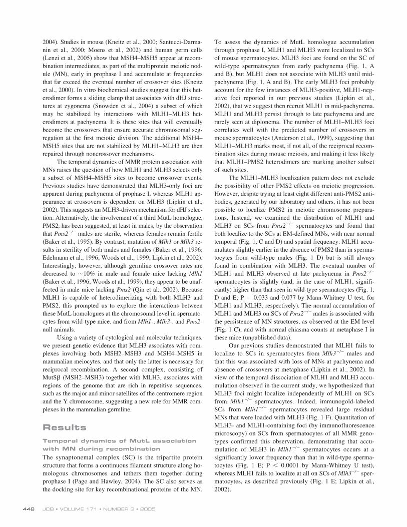
To assess the dynamics of MutL homologue accumulationthrough prophase I, MLH1 and MLH3 were localized to SCsof mouse spermatocytes. MLH3 foci are found on the SC ofwild-type spermatocytes from early pachynema (Fig. 1, Aand B), but MLH1 does not associate with MLH3 until mid-pachynema (Fig. 1, A and B). The early MLH3 foci probablyaccount for the few instances of MLH3-positive, MLH1-neg-ative foci reported in our previous studies (Lipkin et al.,2002), that we suggest then recruit MLH1 in mid-pachynema.MLH1 and MLH3 persist through to late pachynema and arerarely seen at diplonema. The number of MLH1–MLH3 focicorrelates well with the predicted number of crossovers inmouse spermatocytes (Anderson et al., 1999), suggesting thatMLH1–MLH3 marks most, if not all, of the reciprocal recom-bination sites during mouse meiosis, and making it less likelythat MLH1–PMS2 heterodimers are marking another subsetof such sites.
The MLH1–MLH3 localization pattern does not excludethe possibility of other PMS2 effects on meiotic progression.However, despite trying at least eight different anti-PMS2 anti-bodies, generated by our laboratory and others, it has not beenpossible to localize PMS2 in meiotic chromosome prepara-tions. Instead, we examined the distribution of MLH1 andMLH3 on SCs from
Pms2
�
/
�
spermatocytes and found thatboth localize to the SCs at EM-defined MNs, with near normaltemporal (Fig. 1, C and D) and spatial frequency. MLH1 accu-mulates slightly earlier in the absence of PMS2 than in sperma-tocytes from wild-type males (Fig. 1 D) but is still alwaysfound in combination with MLH3. The eventual number ofMLH1 and MLH3 observed at late pachynema in
Pms2
�
/
�
spermatocytes is slightly (and, in the case of MLH1, signifi-cantly) higher than that seen in wild-type spermatocytes (Fig. 1,D and E; P
�
0.033 and 0.077 by Mann-Whitney U test, forMLH1 and MLH3, respectively). The normal accumulation ofMLH1 and MLH3 on SCs of
Pms2
�
/
�
males is associated withthe persistence of MN structures, as observed at the EM level(Fig. 1 C), and with normal chiasma counts at metaphase I inthese mice (unpublished data).
Our previous studies demonstrated that MLH1 fails tolocalize to SCs in spermatocytes from
Mlh3
�
/
�
males andthat this was associated with loss of MNs at pachynema andabsence of crossovers at metaphase (Lipkin et al., 2002). Inview of the temporal dissociation of MLH1 and MLH3 accu-mulation observed in the current study, we hypothesized thatMLH3 foci might localize independently of MLH1 on SCsfrom
Mlh1
�
/
�
spermatocytes. Indeed, immunogold-labeledSCs from
Mlh1
�
/
�
spermatocytes revealed large residualMNs that were loaded with MLH3 (Fig. 1 F). Quantitation ofMLH3- and MLH1-containing foci (by immunofluorescencemicroscopy) on SCs from spermatocytes of all MMR geno-types confirmed this observation, demonstrating that accu-mulation of MLH3 in
Mlh1
�
/
�
spermatocytes occurs at asignificantly lower frequency than that in wild-type sperma-tocytes (Fig. 1 E; P
�
0.0001 by Mann-Whitney U test),whereas MLH1 fails to localize at all on SCs of
Mlh3
�
/
�
sper-matocytes, as described previously (Fig. 1 E; Lipkin et al.,2002).
MISMATCH REPAIR COMPLEXES IN MAMMALIAN MEIOSIS • KOLAS ET AL.
449
Localization of MLH3, and occasionally MLH1, with CREST-positive regions of wild-type and Pms2
�
/
�
male chromosomes reveals an unexpected centromere-associated MMR complex
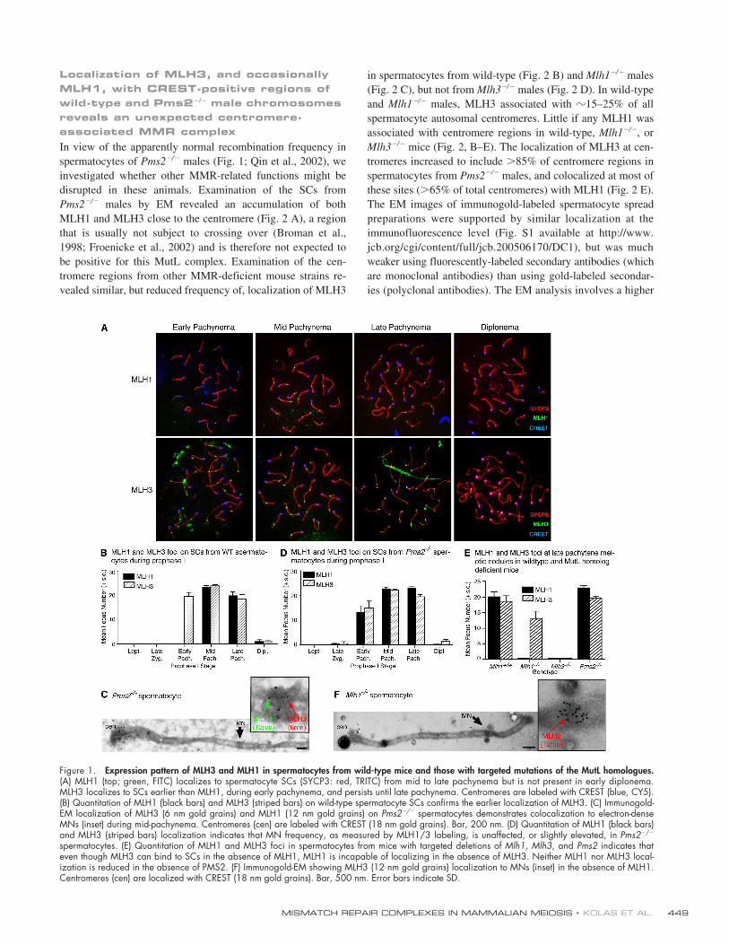
In view of the apparently normal recombination frequency inspermatocytes of
Pms2
�
/
�
males (Fig. 1; Qin et al., 2002), weinvestigated whether other MMR-related functions might bedisrupted in these animals. Examination of the SCs from
Pms2
�
/
�
males by EM revealed an accumulation of bothMLH1 and MLH3 close to the centromere (Fig. 2 A), a regionthat is usually not subject to crossing over (Broman et al.,1998; Froenicke et al., 2002) and is therefore not expected tobe positive for this MutL complex. Examination of the cen-tromere regions from other MMR-deficient mouse strains re-vealed similar, but reduced frequency of, localization of MLH3
in spermatocytes from wild-type (Fig. 2 B) and
Mlh1
�
/
�
males(Fig. 2 C), but not from
Mlh3
�
/
�
males (Fig. 2 D). In wild-typeand
Mlh1
�
/
�
males, MLH3 associated with
�
15–25% of allspermatocyte autosomal centromeres. Little if any MLH1 wasassociated with centromere regions in wild-type,
Mlh1
�
/
�
, or
Mlh3
�
/
�
mice (Fig. 2, B–E). The localization of MLH3 at cen-tromeres increased to include
�
85% of centromere regions inspermatocytes from
Pms2
�
/
�
males, and colocalized at most ofthese sites (
�
65% of total centromeres) with MLH1 (Fig. 2 E).The EM images of immunogold-labeled spermatocyte spreadpreparations were supported by similar localization at theimmunofluorescence level (Fig. S1 available at http://www.jcb.org/cgi/content/full/jcb.200506170/DC1), but was muchweaker using fluorescently-labeled secondary antibodies (whichare monoclonal antibodies) than using gold-labeled secondar-ies (polyclonal antibodies). The EM analysis involves a higher
Figure 1. Expression pattern of MLH3 and MLH1 in spermatocytes from wild-type mice and those with targeted mutations of the MutL homologues.(A) MLH1 (top; green, FITC) localizes to spermatocyte SCs (SYCP3: red, TRITC) from mid to late pachynema but is not present in early diplonema.MLH3 localizes to SCs earlier than MLH1, during early pachynema, and persists until late pachynema. Centromeres are labeled with CREST (blue, CY5).(B) Quantitation of MLH1 (black bars) and MLH3 (striped bars) on wild-type spermatocyte SCs confirms the earlier localization of MLH3. (C) Immunogold-EM localization of MLH3 (6 nm gold grains) and MLH1 (12 nm gold grains) on Pms2�/� spermatocytes demonstrates colocalization to electron-denseMNs (inset) during mid-pachynema. Centromeres (cen) are labeled with CREST (18 nm gold grains). Bar, 200 nm. (D) Quantitation of MLH1 (black bars)and MLH3 (striped bars) localization indicates that MN frequency, as measured by MLH1/3 labeling, is unaffected, or slightly elevated, in Pms2�/�
spermatocytes. (E) Quantitation of MLH1 and MLH3 foci in spermatocytes from mice with targeted deletions of Mlh1, Mlh3, and Pms2 indicates thateven though MLH3 can bind to SCs in the absence of MLH1, MLH1 is incapable of localizing in the absence of MLH3. Neither MLH1 nor MLH3 local-ization is reduced in the absence of PMS2. (F) Immunogold-EM showing MLH3 (12 nm gold grains) localization to MNs (inset) in the absence of MLH1.Centromeres (cen) are localized with CREST (18 nm gold grains). Bar, 500 nm. Error bars indicate SD.
JCB • VOLUME 171 • NUMBER 3 • 2005450
concentration of primary antibody, coupled with DNaseI diges-tion of heterochromatin to improve accessibility of the antibod-ies to the centromere sites, perhaps explaining the difference instaining intensity.
Considering mouse centromeres are telocentric, we ex-amined whether the accumulation of MLH1–MLH3 at thecentromere region in
Pms2
�
/
�
spermatocytes was due to thecentromere per se or the closely-adjacent telomere. We didnot observe elevated MLH1–MLH3 accumulation at the dis-tal telomere of
Pms2
�
/
�
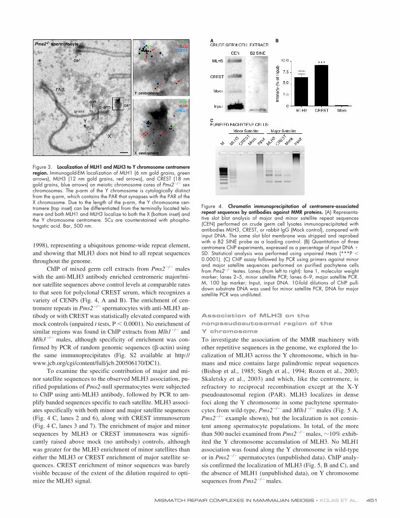
males (Fig. 2 F). Moreover, the Ychromosome, which has a larger expanse between the proxi-mal telomere and the centromere, showed specific accumula-tion to the centromeric region (Fig. 3), leading us to concludethat the accumulation of MLH3 at these regions was associ-ated specifically with centromere-associated sequences. Thepresence of MLH3 at centromere regions in wild-type and
Mlh1
�
/
�
spermatocytes indicates that MLH3 is able to local-ize to the chromosome independently of MLH1, as it does atearly MNs (Fig. 1, B and C).
Localization of MLH3 and MLH1 at centromeric regions is associated with major and minor satellite repeat sequences
The mammalian centromere is a highly specialized structureconsisting of protein-associated centromeric sequences and ad-
jacent minor satellite DNA flanked by pericentric major satel-lite DNA (Guenatri et al., 2004). Major/minor satellites are re-gions of highly repetitive heterochromatin-forming sequencesthat are important for centromere function associated withspindle attachment and sister chromatid cohesion. In mice, themajor satellite consists of tandem repeats of 234-bp units, ofwhich there are
�
700,000 copies per cell, reflecting an averageof 35,000 copies per chromosome (totaling 8 MB in length;Horz and Altenburger, 1981). The minor satellite binds histoneH3 and CENPA/B, is present in 50–100,000 copies, and ap-pears to share common ancestry with the major satellite (Silver,1995). In humans, similar repeats of
�
171 bp are present butare termed
�
satellites (Muro et al., 1992).The localization of MLH3 (and in some cases, MLH1) at
the centromere prompted us to investigate the specificity of thisassociation with centromere-associated repeats. We performedchromatin immunoprecipitation (ChIP) experiments using ei-ther crude germ cell extracts (consisting of mixed populationsof germ cells) or isolated pachytene spermatocytes (estimatedto be
�
95% pure). MLH3-containing complexes were immu-noprecipitated using an anti-MLH3 antibody, followed byamplification using degenerate oligonucleotide PCR (Tele-nius et al., 1992) and subsequent slot-blot analysis usingPCR-generated probes specific to mouse major and/or minorsatellite sequences (Garagna et al., 2002). A B2 SINE elementprobe was used as a loading control (Serdobova and Kramerov,
Figure 2. MLH3 immunolocalizes to a proportion of wild-type and Mlh1�/� spermatocyte centromeres, and both MLH3 and MLH1 localize to the centromeresof Pms2�/�spermatocytes but not Pms2�/� oocytes. (A–D) Immunogold-EM localization of MLH1 (12 nm gold grains, red arrows), MLH3 (6 nm goldgrains, green arrows), and CREST (18 nm gold grains, blue arrows) on meiotic chromosomes of Pms2�/� (A), wild-type (B), Mlh1�/� (C), and Mlh3�/� (D)spermatocytes. Bars: (A, B, and D) 250 nm; (C) C 500 nm. All SCs are counterstained with phosphotungstic acid. (E) Quantitation of MMR protein local-ization to centromeres from Mlh1�/�, Mlh1�/�, Mlh3�/�, Mlh3�/�, Pms2�/�, and Pms2�/� spermatocytes. Counts were obtained from immunogold-EMmicrographs exemplified in A–D and represent those centromeres showing more than four gold grains for each antibody staining. At least 100 cen-tromeres were counted for each genotype from three mice. Percentages represent pooled counts from all these mice. MLH3 (striped bars) immunolocalizesto 15–25% of wild-type and Mlh1�/�, but not Mlh3�/�, spermatocyte centromeres. MLH1 (black bars) does not localize in any appreciable amount to cen-tromeres in wild-type, Mlh1�/� or Mlh3�/� spermatocytes. CREST (white bars) localizes to 100% of the centromeres. Pms2�/� spermatocyte centromeresshow a statistically significant increase in localization of both MLH1 and MLH3 in comparison with the Pms2�/� and all other mouse strains observed. (F)Quantitation of distal (noncentromeric) telomere localization (more than four gold grains) indicates that the localization of MutL homologues is specific tothe centromere. (G) EM micrograph of MLH1 (6 nm gold grains) and MLH3 (12 nm gold grains) on SCs from Pms2�/� oocytes indicates that, whereasMLH3 and MLH1 localize normally to MNs (bottom inset), these MutL homologues fail to localize to the centromere (cen), labeled with CREST (18 nm goldgrains; top inset). Bar, 200 nm.
MISMATCH REPAIR COMPLEXES IN MAMMALIAN MEIOSIS • KOLAS ET AL. 451
1998), representing a ubiquitous genome-wide repeat element,and showing that MLH3 does not bind to all repeat sequencesthroughout the genome.
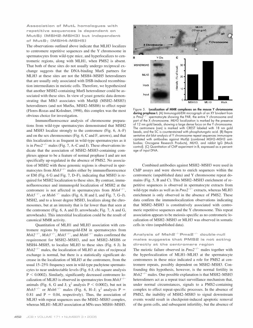
ChIP of mixed germ cell extracts from Pms2�/� maleswith the anti-MLH3 antibody enriched centromeric major/mi-nor satellite sequences above control levels at comparable ratesto that seen for polyclonal CREST serum, which recognizes avariety of CENPs (Fig. 4, A and B). The enrichment of cen-tromere repeats in Pms2�/� spermatocytes with anti-MLH3 an-tibody or with CREST was statistically elevated compared withmock controls (unpaired t tests, P � 0.0001). No enrichment ofsimilar regions was found in ChIP extracts from Mlh1�/� andMlh3�/� males, although specificity of enrichment was con-firmed by PCR of random genomic sequences (�-actin) usingthe same immunoprecipitates (Fig. S2 available at http://www.jcb.org/cgi/content/full/jcb.200506170/DC1).
To examine the specific contribution of major and mi-nor satellite sequences to the observed MLH3 association, pu-rified populations of Pms2-null spermatocytes were subjectedto ChIP using anti-MLH3 antibody, followed by PCR to am-plify banded sequences specific to each satellite. MLH3 associ-ates specifically with both minor and major satellite sequences(Fig. 4 C, lanes 2 and 6), along with CREST immunoserum(Fig. 4 C, lanes 3 and 7). The enrichment of major and minorsequences by MLH3 or CREST immunosera was signifi-cantly raised above mock (no antibody) controls, althoughwas greater for the MLH3 enrichment of minor satellites thaneither the MLH3 or CREST enrichment of major satellite se-quences. CREST enrichment of minor sequences was barelyvisible because of the extent of the dilution required to opti-mize the MLH3 signal.
Association of MLH3 on the nonpseudoautosomal region of the Y chromosomeTo investigate the association of the MMR machinery withother repetitive sequences in the genome, we explored the lo-calization of MLH3 across the Y chromosome, which in hu-mans and mice contains large palindromic repeat sequences(Bishop et al., 1985; Singh et al., 1994; Rozen et al., 2003;Skaletsky et al., 2003) and which, like the centromere, isrefractory to reciprocal recombination except at the X-Ypseudoautosomal region (PAR). MLH3 localizes in densefoci along the Y chromosome in some pachytene spermato-cytes from wild-type, Pms2�/� and Mlh1�/� males (Fig. 5 A,Pms2�/� example shown), but the localization is not consis-tent among spermatocyte populations. In total, of the morethan 500 nuclei examined from Pms2�/� males, �10% exhib-ited the Y chromosome accumulation of MLH3. No MLH1association was found along the Y chromosome in wild-typeor in Pms2�/� spermatocytes (unpublished data). ChIP analy-sis confirmed the localization of MLH3 (Fig. 5, B and C), andthe absence of MLH1 (unpublished data), on Y chromosomesequences from Pms2�/� males.
Figure 4. Chromatin immunoprecipitation of centromere-associatedrepeat sequences by antibodies against MMR proteins. (A) Representa-tive slot blot analysis of major and minor satellite repeat sequences(CEN) performed on crude germ cell lysates immunoprecipitated withantibodies MLH3, CREST, or rabbit IgG (Mock control), compared withinput DNA. The same slot blot membrane was stripped and reprobedwith a B2 SINE probe as a loading control. (B) Quantitation of threecentromere ChIP experiments, expressed as a percentage of input DNA �SD. Statistical analysis was performed using unpaired t-tests (***P �0.0001). (C) ChIP assay followed by PCR using primers against minorand major satellite sequences performed on purified pachytene cellsfrom Pms2�/� testes. Lanes (from left to right): lane 1, molecular weightmarker; lanes 2–5, minor satellite PCR; lanes 6–9, major satellite PCR.M, 100 bp marker; Input, input DNA. 10-fold dilutions of ChIP pull-down substrate DNA was used for minor satellite PCR, DNA for majorsatellite PCR was undiluted.
Figure 3. Localization of MLH1 and MLH3 to Y chromosome centromereregion. Immunogold-EM localization of MLH1 (6 nm gold grains, greenarrows), MLH3 (12 nm gold grains, red arrows), and CREST (18 nmgold grains, blue arrows) on meiotic chromosome cores of Pms2�/� sexchromosomes. The p-arm of the Y chromosome is cytologically distinctfrom the q-arm, which contains the PAR that synapses with the PAR of theX chromosome. Due to the length of the p-arm, the Y chromosome cen-tromere (top inset) can be differentiated from the terminally located telo-mere and both MLH1 and MLH3 localize to both the X (bottom inset) andthe Y chromosome centromere. SCs are counterstained with phospho-tungstic acid. Bar, 500 nm.
JCB • VOLUME 171 • NUMBER 3 • 2005452
Association of MutL homologues with repetitive sequences is dependent on MutS� (MSH2–MSH3) but independent of MutS� (MSH4–MSH5)The observations outlined above indicate that MLH3 localizesto centromere repetitive sequences and the Y chromosome inspermatocytes from wild-type mice, and hyperlocalizes to cen-tromeric regions, along with MLH1, when PMS2 is absent.That both of these sites do not usually undergo reciprocal ex-change suggests that the DNA-binding MutS partners forMLH3 at these sites are not the MSH4–MSH5 heterodimersthat are usually only associated with DSB-induced recombina-tion intermediates in meiotic cells. Therefore, we hypothesizedthat another MSH2-containing MutS heterodimer could be as-sociated with these sites. In view of yeast genetic data demon-strating that Mlh3 associates with MutS� (MSH2–MSH3)heterodimers (and not MutS�, MSH2–MSH6) to effect repair(Flores-Rozas and Kolodner, 1998), this complex was the mostobvious choice for investigation.
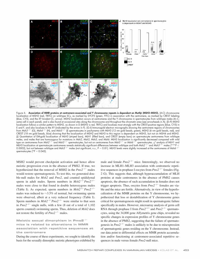
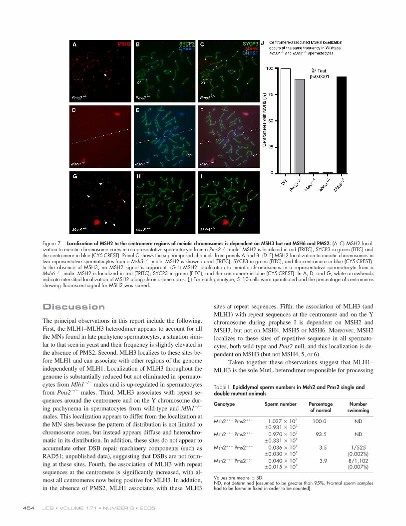
Immunofluorescence analysis of chromosome prepara-tions from wild-type spermatocytes demonstrated that MSH2and MSH3 localize strongly to the centromere (Fig. 6, A–F)and on the sex chromosomes (Fig. 6, C and F, arrows), and thatthis localization is as frequent in wild-type spermatocytes as itis in Pms2�/� males (Fig. 7, A–C and J). These observations in-dicate that the association of MSH2–MSH3-containing com-plexes appear to be a feature of normal prophase I and are notspecifically up-regulated in the absence of PMS2. No associa-tion of MSH2 with these genomic regions is observed in sper-matocytes from Msh3�/� males either by immunofluorescenceor EM (Fig. 6 G and Fig. 7, D–F), indicating that MSH3 is re-quired for MSH2 localization to these sites. By contrast, immu-nofluorescence and immunogold localization of MSH2 at thecentromere is not affected in spermatocytes from Msh4�/�,Msh5�/�, or Msh6�/� males (Fig. 6, H and I, and Fig. 7, G–J).MSH2, and to a lesser degree MSH3, localizes along the chro-mosomes, but at an intensity that is far lower than that seen atthe centromere (Fig. 6, A and D, arrowheads; Fig. 7, A and G,arrowheads). This interstitial localization could be the result ofcanonical MMR activity.
Quantitation of MLH1 and MLH3 association with cen-tromere regions by immunogold-EM in spermatocytes fromMsh2�/�, Msh3�/�, Msh5�/�, and Msh6�/� males confirmed therequirement for MSH2–MSH3, and not MSH2–MSH6 orMSH4–MSH5, to localize MLH3 to these sites (Fig. 6 J). InMsh2�/� males, the localization of MLH3 at sites of reciprocalexchange is normal, but there is a statistically significant de-crease in the localization of MLH3 at the centromere, from theusual 15–25% frequency seen in wild-type pachytene spermato-cytes to near undetectable levels (Fig. 6 J; chi-square analysisP � 0.0002). Similarly, significantly decreased centromere lo-calization of MLH3 is observed in spermatocytes from Msh3�/�
animals (Fig. 6, G and J; 2 analysis P � 0.0002), but not inMsh5�/� or Msh6�/� males (Fig. 6, H–J; 2 analysis P �
0.81 and P � 0.04, respectively). Thus, the association ofMLH3 with repeat sequences uses the MSH2–MSH3 complex,whereas MLH1–MLH3 association at MNs uses MSH4–MSH5.
Figure 5. Localization of MMR complexes on the mouse Y chromosomeduring prophase I. (A) Immunogold-EM micrograph of an XY bivalent froma Pms2�/� spermatocyte showing the PAR, the entire Y chromosome andpart of the X chromosome. MLH3 localization is marked by the presenceof 12 nm gold beads, showing a large dense focus on the Y chromosome.The centromere (cen) is marked with CREST labeled with 18 nm goldbeads, and the SC is counterstained with phosphotungstic acid. (B) Repre-sentative slot blot analysis of Y chromosome repeat sequences immunopre-cipitated with antibodies against MutS� (combined MSH2–MSH3 anti-bodies; Oncogene Research Products), MLH3, and rabbit IgG (Mockcontrol). (C) Quantitation of ChIP experiment in B, expressed as a percent-age of input DNA.
Combined antibodies against MSH2–MSH3 were used inChIP assays and were shown to enrich sequences within thecentromeric (unpublished data) and Y chromosome repeat do-mains (Fig. 5, B and C). This MSH2–MSH3 enrichment of re-petitive sequences is observed in spermatocyte extracts fromwild-type males as well as in Pms2�/� extracts, whereas MLH3enrichment is only observed in the absence of PMS2. Thesedata confirm the immunolocalization observations indicatingthat MSH2–MSH3 is constitutively associated with centro-meric repetitive sequences and the Y chromosome. This repeatassociation appears to be meiosis-specific as no centromeric lo-calization of MSH2–MSH3 or MLH3 was observed in somaticcells in vitro (unpublished data).
Analysis of Msh2�/�Pms2�/� double-null males suggests that PMS2 is not acting directly at the centromere regionThe meiotic failure observed in Pms2�/� males together withthe hyperlocalization of MLH1–MLH3 at the spermatocytecentromeres in these mice indicated a role for PMS2 at cen-tromere repeats, possibly dependent on MSH2–MSH3. Con-founding this hypothesis, however, is the normal fertility inMsh2�/� males. One possible explanation is that MSH2–MSH3heterodimers act as a repeat tract surveillance mechanism that,under normal circumstances, signals to a PMS2-containingcomplex to effect repeat-specific processes. In the absence ofPMS2, the inability of MSH2–MSH3 to signal downstreamevents would result in checkpoint-induced apoptotic removalof the germ cells, and subsequent infertility, but the absence of
MISMATCH REPAIR COMPLEXES IN MAMMALIAN MEIOSIS • KOLAS ET AL. 453
MSH2 would prevent checkpoint activation and hence allowmeiotic progression even in the absence of PMS2. If true, wehypothesized that the removal of MSH2 in the Pms2�/� maleswould restore spermatogenesis. To test this, we generated dou-ble-null males for Msh2 and Pms2, and counted epididymalsperm in adult males. Sperm numbers in Msh2�/�Pms2�/�
males were close to that found in double heterozygous males(Table I). As expected, sperm numbers in Msh2�/�Pms2�/�
males was reduced to �3.5% of normal, but swimming spermwere observed, albeit at a very reduced frequency (Table I).Sperm numbers in Msh2�/�Pms2�/� were similar to that seenin Pms2�/� single nulls, with a few (8 out of a total of 1,102sperm counted) swimming sperm. Thus, deletion of Msh2 doesnot restore the fertility of Pms2�/� males.
Meiotic sexual dimorphism in Pms2�/� mice is related to alterations in MMR association with repetitive sequences at the centromereDuring the course of these experiments, we sought to identify thebasis for the sexually dimorphic meiotic phenotypes exhibited by
male and female Pms2�/� mice. Interestingly, we observed noincrease in MLH1–MLH3 association with centromeric repeti-tive sequences in prophase I oocytes from Pms2�/� females (Fig.2 G). This suggests that, although hyperaccumulation of MLHproteins at male centromeres in the absence of PMS2 causesapoptosis, the absence of such accumulation in females does nottrigger apoptosis. Thus, oocytes from Pms2�/� females are via-ble and the mice are fertile. Alternatively, in view of the hyperlo-calization of the MMR proteins on the Y chromosome, we hy-pothesized that loss or destabilization of Y chromosome genescritical for spermatogenesis might result in spermatogenic failurespecifically in males. However, microarray analysis of germ cellRNA through prophase I from Pms2�/� and Pms2�/� spermato-cytes, using the 34,000 gene Affymetrix gene chips, revealed nospecific changes in expression profiles of Y chromosome genesin the absence of PMS2, suggesting that the failure of spermato-genesis in Pms2�/� males is unlikely to be due to misexpressionof spermatogenic genes residing on the Y chromosome. Instead,our data point to differential effects on MMR protein accumula-tion and/or functioning at centromere-associated repetitive se-quences in male versus female Pms2-null mice.
Figure 6. Association of MMR proteins at centromere-associated and Y chromosome repeats is dependent on MutS� (MSH2–MSH3). (A-C) chromosomelocalization of MSH2 (red, TRITC) on wild-type SCs, as marked by SYCP3 (green, FITC) in association with the centromere, as marked by CREST labeling(blue, CY5), and the XY bivalent (C, arrow). MSH2 localization occurs at centromeres and the Y chromosome in spermatocytes from wild-type males (A–C;same cell in each panel), and is also found at occasional sites along the chromosome and throughout the chromatin area (see arrowheads in A). (D–F) MSH3localization follows a similar pattern to MSH2, as shown in D (MSH3 is red, TRITC) and localizes most strongly with the CREST-positive regions (blue, CY5) inE and F, and also localizes to the XY (indicated by the arrow in F). (G–I) Immunogold electron micrographs showing the centromere regions of chromosomesfrom Msh3�/� (G), Msh4�/� (H), and Msh5�/� (I) spermatocytes in pachynema with MLH3 (12 nm gold beads, green), MSH2 (6 nm gold beads, red), andCREST (18 nm gold beads, blue) showing that the localization of MSH2 and MLH3 to this region is dependent on MSH3, but not on MSH4 and MSH5.(J) Quantitation of EM-gold localization of MLH3 (striped bars), MLH1 (filled bars), and CREST (empty bars) on spermatocyte centromeres from wild-typemales, and males that are homozygous for mutations in Msh2, Msh3, Msh5, and Msh6. MLH3 localization is significantly decreased compared with wildtype on centromeres from Msh2�/� and Msh3�/� spermatocytes, but not on centromeres from Msh5�/� or Msh6�/� spermatocytes. 2 analysis of MLH1 andMLH3 localization at spermatocyte centromeres reveals statistically significant differences between wild-type and both Msh2�/� and Msh3�/� males (***P �0.0002), but not between wild-type and Msh5�/� males (not significant, n.s., P � 0.81). MLH3 levels were slightly increased at the centromeres of Msh6�/�
spermatocytes (*P � 0.043).
JCB • VOLUME 171 • NUMBER 3 • 2005454
DiscussionThe principal observations in this report include the following.First, the MLH1–MLH3 heterodimer appears to account for allthe MNs found in late pachytene spermatocytes, a situation simi-lar to that seen in yeast and their frequency is slightly elevated inthe absence of PMS2. Second, MLH3 localizes to these sites be-fore MLH1 and can associate with other regions of the genomeindependently of MLH1. Localization of MLH3 throughout thegenome is substantially reduced but not eliminated in spermato-cytes from Mlh1�/� males and is up-regulated in spermatocytesfrom Pms2�/� males. Third, MLH3 associates with repeat se-quences around the centromere and on the Y chromosome dur-ing pachynema in spermatocytes from wild-type and Mlh1�/�
males. This localization appears to differ from the localization atthe MN sites because the pattern of distribution is not limited tochromosome cores, but instead appears diffuse and heterochro-matic in its distribution. In addition, these sites do not appear toaccumulate other DSB repair machinery components (such asRAD51; unpublished data), suggesting that DSBs are not form-ing at these sites. Fourth, the association of MLH3 with repeatsequences at the centromere is significantly increased, with al-most all centromeres now being positive for MLH3. In addition,in the absence of PMS2, MLH1 associates with these MLH3
sites at repeat sequences. Fifth, the association of MLH3 (andMLH1) with repeat sequences at the centromere and on the Ychromosome during prophase I is dependent on MSH2 andMSH3, but not on MSH4, MSH5 or MSH6. Moreover, MSH2localizes to these sites of repetitive sequence in all spermato-cytes, both wild-type and Pms2 null, and this localization is de-pendent on MSH3 (but not MSH4, 5, or 6).
Taken together these observations suggest that MLH1–MLH3 is the sole MutL heterodimer responsible for processing
Table I. Epididymal sperm numbers in Msh2 and Pms2 single and double mutant animals
Genotype Sperm number Percentage of normal
Number swimming
Msh2�/� Pms2�/� 1.037 107
�0.931 107100.0 ND
Msh2�/� Pms2�/� 0.970 107
�0.331 10793.5 ND
Msh2�/� Pms2�/� 0.036 107
�0.030 1073.5 1/525
(0.002%)Msh2�/� Pms2�/� 0.040 107
�0.015 1073.9 8/1,102
(0.007%)
Values are means � SD. ND, not determined (assumed to be greater than 95%. Normal sperm sampleshad to be formalin fixed in order to be counted).
Figure 7. Localization of MSH2 to the centromere regions of meiotic chromosomes is dependent on MSH3 but not MSH6 and PMS2. (A–C) MSH2 local-ization to meiotic chromosome cores in a representative spermatocyte from a Pms2�/� male. MSH2 is localized in red (TRITC), SYCP3 in green (FITC) andthe centromere in blue (CY5-CREST). Panel C shows the superimposed channels from panels A and B. (D–F) MSH2 localization to meiotic chromosomes intwo representative spermatocytes from a Msh3�/� male. MSH2 is shown in red (TRITC), SYCP3 in green (FITC), and the centromere in blue (CY5-CREST).In the absence of MSH3, no MSH2 signal is apparent. (G–I) MSH2 localization to meiotic chromosomes in a representative spermatocyte from aMsh6�/� male. MSH2 is localized in red (TRITC), SYCP3 in green (FITC), and the centromere in blue (CY5-CREST). In A, D, and G, white arrowheadsindicate interstitial localization of MSH2 along chromosome cores. (J) For each genotype, 5–10 cells were quantitated and the percentage of centromeresshowing fluorescent signal for MSH2 was scored.
MISMATCH REPAIR COMPLEXES IN MAMMALIAN MEIOSIS • KOLAS ET AL. 455
of meiotic crossovers and rule out a direct function for PMS2in crossing over, paralleling the situation in Saccharomycescerevisiae (Wang et al., 1999). In addition, these data demon-strate the existence of an additional meiotic MMR complexthat associates specifically with sites of repetitive DNA at sitesthat are presumed to be refractory to reciprocal recombinationevents. It was not surprising, therefore, to find an MMR com-plex driven by MSH2–MSH3 instead of MSH4–MSH5. Alsoof significance is the fact that MLH3 can associate with chro-mosome regions independently of MLH1, suggesting the exist-ence of a new MutL complex that is either stabilized by MLH1or that can function independently of other MutL homologues.The MLH3-only complex could be inactive in terms of MMRactivity, but could instead trigger alternative downstreamevents that would result in genome stabilization.
The up-regulation of centromere-associated MMR com-plexes in Pms2�/� males suggests that the absence of PMS2 fa-cilitates the accumulation of repeat-associated MMR complexes,but does not necessarily point to any direct role for PMS2 atthese sites. Msh2�/�•Pms2�/� males are also sterile whileMsh2�/� males are fertile, arguing against a role for PMS2 at thecentromere. Instead, our results show that the loss of PMS2 re-sults in an accumulation of the MSH2–MSH3–MLH1–MLH3complex at sites of repetitive DNA as a secondary effect. Wepropose that PMS2 is working at other sites during spermatogen-esis, in combination with MSH2, and that the loss of PMS2 atthese sites leads to a redistribution of MMR complexes throughthe genome, resulting in increased association of MLH1–MLH3with centromeric sequences (and, to a lesser extent, with sites ofDSB processing). Alternatively, or in addition, the removal ofPMS2 increases the need for MMR complexes at sites of repeti-tive DNA because of the general increase in genome instabilityin Pms2�/� mice. Moreover, we cannot rule out the possibilitythat the effect of Pms2 deletion occurs at an earlier point in sper-matogenesis, possibly during postreplicative repair at premeioticS-phase, and that the meiotic failure is secondary to these earlierevents. Indeed, Richardson et al. (2000) demonstrated high lev-els of mRNA and protein expression in mouse spermatogonia(Richardson et al., 2000), suggesting perhaps that PMS2 may befunctioning at this earlier time in spermatogenesis.
Previous studies have suggested that tandem repeats,such as those found in human � satellite centromeric DNA,are susceptible to mispairing and other genome destabilizingevents. They are conserved by gene conversion (Greig et al.,1993; Warburton et al., 1993; Schindelhauer and Schwarz,2002), as are palindromic repeat units on the Y (Rozen et al.,2003; Skaletsky et al., 2003). Given that localization of MMRproteins to these sites is determined by MSH2–MSH3, our datasupport a function in noncrossover and/or repair events at thesetandem repeat units, rather than a function in MSH4–MSH5-driven reciprocal recombination. In addition, the antirecombi-national role of MSH2–MSH3 (Lin et al., 2004) could be bene-ficial at sites such as the centromere to prevent deleteriouscrossover events during prophase I, and may explain the highdegree of conservation amongst centromere repeat regions.
The dynamics of MMR complex association with repeti-tive sequences at the centromere and on the Y chromosome dur-
ing meiosis is reminiscent of the dynamics of MMR involvementin the expansion of simple trinucleotide repeat (TNR) sequencesthat give rise to complex disorders such as Huntington disease(Kovtun et al., 2001; Kovtun and McMurray, 2001) and myo-tonic dystrophy (van den Broek et al., 2002; Nag, 2003). Thesecondary structures that arise as a result of TNR misalignmentsare substrates for MSH2 (Pearson et al., 1997), and mutations inMsh2 prevent CAG•CTG repeat expansion at the Huntingtondisease Hdh locus (Manley et al., 1999). In somatic cells, theseevents involve MSH3 because TNR expansion in myotonic dys-trophy knock-in mice is abolished on a Msh3-null background(van den Broek et al., 2002). These MutS�-dependent events,both in the germ line and in somatic cells, could involve canoni-cal MMR and/or recombination processes or, as has been sug-gested, could instead result from the stabilization and protectionfrom repair of CAG•CTG hairpin structures (Manley et al.,1999; Kovtun and McMurray, 2001) resulting from misalign-ment and looping of tandem triplet repeats (Moore et al., 1999).
Secondary looping structures could also play roles inMMR-driven events during meiosis. Heteroduplexes formedduring recombination in yeast can include insertions as large as5.6 kb in size (Kearney et al., 2001) and are targeted specifi-cally by Msh2–Msh3 (Kearney et al., 2001). These substratesmay induce very different biochemical events to the traditionalMMR pathway and may therefore initiate a different cascade ofdownstream events. Moreover, long repeat tracts, involvingtriplet CAG motifs or large palindromes, induce DSB forma-tion during yeast meiosis (Jankowski et al., 2000; Nasar et al.,2000; Nag, 2003) that could give rise to recombinational repairevents. This raises the possibility that the repeat tracts and pal-indromes in the centromere regions and on the Y chromosomecould present similar recombinational substrates during murinemeiosis, inducing DSBs in late pachynema through the actionof SPO11, which is still present during that time (Keeney et al.,1999; Romanienko and Camerini-Otero, 1999), that then needto be repaired by recombinational events, with a preference fornoncrossover mechanisms. However, this possibility is tem-pered by the failure to detect other DSB repair machinery com-ponents (RAD51) at these sites (unpublished data).
In conclusion, our results suggest a novel function for theMMR machinery during mammalian meiosis, whereby MSH2–MSH3 are present as a surveillance mechanism, to restore repeatfrequency, through stabilization/maintenance of hairpin struc-tures present within the context of tandem repeats, and/orthrough suppression of reciprocal recombination events resultingfrom secondary structure–induced DSB formation, that couldresult in chromosomal translocations, enlarged loops, and othercytogenetic errors. Should repeat units become misaligned orout of register, a resulting single stranded loop structure wouldbe detected by the surveillance complex and would activatedownstream (repair) events involving, at the very least, MLH3,and that would ultimately effect the restoration of unit numbers.
Materials and methodsAnimalsThe generation of mouse mutants for Pms2, Mlh1, Mlh3, Msh2, Msh3,Msh4, Msh5, and Msh6 have been described previously, along with
JCB • VOLUME 171 • NUMBER 3 • 2005456
genotyping strategies for each strain (Baker et al., 1995; Edelmann et al.,1996, 1999, 2000; Kneitz et al., 2000; Lipkin et al., 2002). All strainswere housed in the Albert Einstein College of Medicine Animal Institute onstandardized light, heat, and feeding schedules. All strains were main-tained on similar backgrounds by backcrossing heterozygote fathers withC57Bl/6J mothers.
Chromosome preparationsThe standard chromosome spreading protocol has been described previ-ously for our laboratory (Lenzi et al., 2005). The nuclear contents ofwhole-mount spermatocytes (or oocytes) were displayed by drying down acell suspension, in hypotonic buffer, from either testis, or ovary, in 1%paraformaldehyde containing 0.15% Triton X-100 (Peters et al., 1997).Whole testes or ovaries were incubated on ice for 60 min in hypotonic ex-traction buffer (HEB; 30 mM Tris, pH 8.2, 50 mM sucrose, 17 mM triso-dium citrate dihydrate, 5 mM EDTA, 0.5 mM DTT, and 0.5 mM PMSF). Ei-ther a one-inch length of tubule, or a whole ovary, were placed in a 20-�ldrop of 100 mM sucrose, pH 8.2, the tissue was macerated, and a sec-ond 20-�l drop of sucrose solution was added and the cell suspensionwas pipetted up and down several times. Remnant pieces of tubule wereremoved. Cleaned slides were dipped in the paraformaldehyde and TritonX-100 solution, and most liquid was drained off, such that only enough liq-uid remained to coat the slide. 20 �l of the cell suspension was added inone corner and the cells were slowly dispersed, first in a horizontal direc-tion and then vertical. The remaining 20 �l of cell suspension was used tomake a second slide and both were placed in a humid chamber to dryslowly at RT for 2 h. The slides were washed three times for 1 min in 0.4%Kodak Photo-Flo 200 and air dried for at least 15 min. For EM prepara-tions, to make the SCs accessible to immunogold grains, the slides wereDNaseI treated (1 �l/ml of DMEM) before being air dried (Moens et al.,2002). The slides were washed and blocked (three times for 10 min each)in PBS and incubated in primary antibodies overnight at RT in a humidchamber. Primary antibodies were used at varying concentrations, andgenerally a 10-fold higher concentration was used for EM than immuno-fluorescence. After washes, slides were incubated in secondary antibodies,conjugated to either fluorochrome or colloidal gold (Jackson ImmunoRe-search Laboratories), for 2 h at 37 C. After washes the slides weremounted with ProLong Antifade (Invitrogen) for fluorescence microscopy.Images were captured on a Olympus IX81 microscope attached to a 12-bit Cooke Sensicam CCD instrument and sent to IP Lab software.
For EM, slides were incubated in 4% alcoholic phosphotungsticacid for 15 min, followed by three 1-min washes in 95% ethanol, to en-hance visualization of MNs. Slides were air dried and then dipped in0.25% formvar (Electron Microscopy Sciences) and air dried under glass.The plastic was scored, treated with 25% hydrofluoric acid, and floatedoff in water with attached cells. Plastic was transferred to EM grids andused for transmission EM (JEOL 1200EX).
AntibodiesThe numerous antibodies used were obtained from a variety of sources:mouse anti–human MLH1 antibody (BD Biosciences, clone G168-15);mouse anti–human MSH2 (clone G219-1129; BD Biosciences); and mouseanti–human MSH3 (clone 52; BD Biosciences). In addition, the followingantibodies have been generated in our laboratory or by our collaborators:rabbit anti-MLH3 (Lipkin et al., 2002), rabbit anti–hamster COR1 to detectSYCP3 (Lenzi et al., 2005), and mouse anti–hamster COR1 (Lenzi et al.,2005).
Quantitation and statistical analysisImmunofluorescence chromosome counts and cell staging were per-formed by at least three independent observers, compiled using Excel,and then analyzed using the statistical software package, Prism 3.0(GraphPad Software). Staging of prophase I cells was performed usingwell-defined criteria for SC morphology, XY synapsis, and double densebody (DDB) and sex body status (Ashley and Plug, 1998; Moens et al.,2002). For such studies, we used previously published criteria (Dresser etal., 1987; Allen et al., 1988; Backer et al., 1988). Pachynema was di-vided into early, mid, and late stages on the basis of chromosome ap-pearance, degree of compaction, XY status, and DDB formation. In earlypachynema, the XY is synapsed along most of the length of the PAR andmuch of the X chromosome appears synapsed. Small regions of the auto-somes may not be synapsed at this point. By mid-pachynema, the auto-somes are completely synapsed and further compacted, whereas the XYis synapsed up to �20% of the length of the X chromosomes. The DDBappears at this time and the sex body becomes apparent. By latepachynema, only the tip of the PAR remains synapsed, the sex body is
much more distinct, and the DDB is still present. Note that the MLH3 (andMLH1) signal found at the centromere was not included in our MN focuscounts depicted in Fig. 1 and that these populations are, we believe, dis-tinct from one another.
Gold grain counts of centromere-associated proteins were countedat the EM level. For each staining preparation, counts were made fromspermatocytes of at least three animals, with no fewer than 200 cen-tromeres counted per animal, including up to 19 autosomes from a singlecell being counted. In some cases fewer autosomes were counted in cellswhose total chromosome count was not completely visible. Equivalenttelomere counts were performed for each preparation as matched controls.In all cases, the number of centromeres/telomeres having one, two orthree, or more than four gold grains were tabulated, but the threshold forspecificity was set at more than four gold grains.
Chromatin immunoprecipitationAdult testes were decapsulated, placed in a drop of cell culture media,minced, and seminiferous tubules squeezed to release germ cells. Alterna-tively, pure populations of pachytene spermatocytes were obtained bysedimentation velocity gradient (“STA-PUT”; Romrell et al., 1976). Germcells were taken up in 1 ml of cell culture media and cross-linking was per-formed at RT for 5 min by addition of formaldehyde to 1% concentration.Cross-linking was stopped by addition of glycine to 0.125 M. Cells werecollected by centrifugation, washed in PBS, and lysed in 1 ml of FA lysisbuffer (50 mM Hepes, 150 mM NaCl, 1 mM EDTA, 1% Triton X-100,0.1% sodium deoxycholate, 0.1% SDS, pH 7.5) with addition of proteaseinhibitors. Cell lysates were sonicated eight times for 20 s each at 20%output setting, and lysates were cleared of cell debris by centrifugation.For each immunoprecipitation 1/10 of the total testis lysate volume wasdiluted 10 times in FA buffer and precleared with 2 �g sheared salmonsperm DNA, 10 �l of preimmune serum and protein A/G-Sepharoseslurry for 1 h at 4 C. Immunoprecipitation was performed overnight at4 C with specific antibodies. After immunoprecipitation, protein A/G-Sepharose slurry was added and the incubation was continued for 1 h.Precipitates were washed sequentially for 5 min each in FA buffer, FAbuffer with 0.5 M NaCl, LiCl wash buffer (10 mM TrisCl, 0.25 M LiCl, 1mM EDTA, 0.5% Nonidet P-40, 0.5% sodium deoxycholate), and TEbuffer. The immune complexes were eluted from the beads by addition of50 mM Tris, 10 mM EDTA, 1% SDS. Cross-linking was reversed by incu-bation at 65 C for at least 6 h DNA fragments were purified by phenol-chloroform-isoamyl alcohol extraction and ethanol precipitation. Equal ali-quots from each immunoprecipitation were amplified using degenerateoligonucleotide PCR method of Telenius et al. (1992). Products of amplifi-cation were analyzed by slot-blot hybridization, using “North2South” hy-bridization kit (Pierce Chemical Co.). Probes for hybridization were gener-ated by PCR according to published sequences for the mouse minor andmajor satellite probe (Garagna et al., 2002), B2 SINE element probe (Ser-dobova and Kramerov, 1998), and mouse Y chromosome–specific LINE-1probe (DYzEms10 locus; Navin et al., 1996).
Online supplemental materialTwo figures are presented as supplemental data items. Fig. S1 showsMLH3 localization at the centromere of spermatocytes from Pms2�/�
males. Fig. S2 provides validation of ChIP experiments used in this study.Online supplemental materials are available at http://www.jcb.org/cgi/content/full/jcb.200506170/DC1.
The authors acknowledge, with gratitude, Dr. Stefan Scherer for guidance inlooking at somatic cell centromere behavior.
This work was funded by the National Institute of Child Health andDevelopment (RO1HD41012-01), The March of Dimes Birth Defects Founda-tion (MOD-FY2002-282), and by start-up funding from the Albert Einstein Col-lege of Medicine to P.E. Cohen and by the National Cancer Institute(R01CA76329) to W. Edelmann. At the time that this work was conducted,A. Svetlanov was supported by training grant sponsorship to the Albert Ein-stein College of Medicine from The National Institute of General MedicalSciences (T32 GM 07491).
Submitted: 27 June 2005Accepted: 4 October 2005
ReferencesAllen, J.W., P.A. Poorman, L.C. Backer, J.B. Gibson, B. Westbrook-Collins,
and M.J. Moses. 1988. Synaptonemal complex damage as a measure of
MISMATCH REPAIR COMPLEXES IN MAMMALIAN MEIOSIS • KOLAS ET AL. 457
genotoxicity at meiosis. Cell Biol. Toxicol. 4:487–494.
Anderson, L.K., A. Reeves, L.M. Webb, and T. Ashley. 1999. Distribution ofcrossing over on mouse synaptonemal complexes using immunofluores-cent localization of MLH1 protein. Genetics. 151:1569–1579.
Ashley, T., and A. Plug. 1998. Caught in the act: deducing meiotic functionfrom protein immunolocalization. Curr. Top. Dev. Biol. 37:201–239.
Backer, L.C., J.B. Gibson, M.J. Moses, and J.W. Allen. 1988. Synaptonemalcomplex damage in relation to meiotic chromosome aberrations after ex-posure of male mice to cyclophosphamide. Mutat. Res. 203:317–330.
Baker, S.M., C.E. Bronner, L. Zhang, A.K. Plug, M. Robatzek, G. Warren, E.A.Elliott, J. Yu, T. Ashley, N. Arnheim, et al. 1995. Male mice defective inthe DNA mismatch repair gene PMS2 exhibit abnormal chromosomesynapsis in meiosis. Cell. 82:309–319.
Baker, S.M., A.W. Plug, T.A. Prolla, C.E. Bronner, A.C. Harris, X. Yao, D.M.Christie, C. Monell, N. Arnheim, A. Bradley, et al. 1996. Involvement ofmouse Mlh1 in DNA mismatch repair and meiotic crossing over. Nat.Genet. 13:336–342.
Bishop, C.E., P. Boursot, B. Baron, F. Bonhomme, and D. Hatat. 1985. Mostclassical Mus musculus domesticus laboratory mouse strains carry a Musmusculus musculus Y chromosome. Nature. 315:70–72.
Borner, G.V., N. Kleckner, and N. Hunter. 2004. Crossover/noncrossover dif-ferentiation, synaptonemal complex formation, and regulatory surveil-lance at the leptotene/zygotene transition of meiosis. Cell. 117:29–45.
Broman, K.W., J.C. Murray, V.C. Sheffield, R.L. White, and J.L. Weber. 1998.Comprehensive human genetic maps: individual and sex-specific varia-tion in recombination. Am. J. Hum. Genet. 63:861–869.
de Vries, S.S., E.B. Baart, M. Dekker, A. Siezen, D.G. de Rooij, P. de Boer, and H.te Riele. 1999. Mouse MutS-like protein Msh5 is required for proper chro-mosome synapsis in male and female meiosis. Genes Dev. 13:523–531.
Dresser, M., D. Pisetsky, R. Warren, G. McCarty, and M. Moses. 1987. A newmethod for the cytological analysis of autoantibody specificities usingwhole-mount, surface-spread meiotic nuclei. J. Immunol. Methods.104:111–121.
Edelmann, W., P.E. Cohen, M. Kane, K. Lau, B. Morrow, S. Bennett, A. Umar,T. Kunkel, G. Cattoretti, R. Chaganti, et al. 1996. Meiotic pachytene ar-rest in MLH1-deficient mice. Cell. 85:1125–1134.
Edelmann, W., P.E. Cohen, B. Kneitz, N. Winand, M. Lia, J. Heyer, R. Kolodner,J.W. Pollard, and R. Kucherlapati. 1999. Mammalian MutS homologue 5is required for chromosome pairing in meiosis. Nat. Genet. 21:123–127.
Edelmann, W., A. Umar, K. Yang, J. Heyer, M. Kucherlapati, M. Lia, B. Kneitz,E. Avdievich, K. Fan, E. Wong, et al. 2000. The DNA mismatch repairgenes Msh3 and Msh6 cooperate in intestinal tumor suppression. CancerRes. 60:803–807.
Flores-Rozas, H., and R.D. Kolodner. 1998. The Saccharomyces cerevisiaeMLH3 gene functions in MSH3-dependent suppression of frameshiftmutations. Proc. Natl. Acad. Sci. USA. 95:12404–12409.
Flores-Rozas, H., and R.D. Kolodner. 2000. Links between replication, re-combination and genome instability in eukaryotes. Trends Biochem.Sci. 25:196–200.
Froenicke, L., L.K. Anderson, J. Wienberg, and T. Ashley. 2002. Male mouse re-combination maps for each autosome identified by chromosome painting.Am. J. Hum. Genet. 71:1353–1368.
Garagna, S., M. Zuccotti, E. Capanna, and C.A. Redi. 2002. High-resolution or-ganization of mouse telomeric and pericentromeric DNA. Cytogenet.Genome Res. 96:125–129.
Greig, G.M., P.E. Warburton, and H.F. Willard. 1993. Organization and evolu-tion of an alpha satellite DNA subset shared by human chromosomes 13and 21. J. Mol. Evol. 37:464–475.
Guenatri, M., D. Bailly, C. Maison, and G. Almouzni. 2004. Mouse centricand pericentric satellite repeats form distinct functional heterochromatin.J. Cell Biol. 166:493–505.
Hoffmann, E.R., and R.H. Borts. 2004. Meiotic recombination intermediatesand mismatch repair proteins. Cytogenet. Genome Res. 107:232–248.
Hollingsworth, N.M., L. Ponte, and C. Halsey. 1995. MSH5, a novel MutShomolog, facilitates meiotic reciprocal recombination between ho-mologs in Saccharomyces cerevisiae but not mismatch repair. GenesDev. 9:1728–1739.
Horz, W., and W. Altenburger. 1981. Nucleotide sequence of mouse satelliteDNA. Nucleic Acids Res. 9:683–696.
Jankowski, C., F. Nasar, and D.K. Nag. 2000. Meiotic instability of CAG repeattracts occurs by double-strand break repair in yeast. Proc. Natl. Acad.Sci. USA. 97:2134–2139.
Kearney, H.M., D.T. Kirkpatrick, J.L. Gerton, and T.D. Petes. 2001. Meiotic re-combination involving heterozygous large insertions in Saccharomycescerevisiae: formation and repair of large, unpaired DNA loops. Genetics.158:1457–1476.
Keeney, S., F. Baudat, M. Angeles, Z.H. Zhou, N.G. Copeland, N.A. Jenkins, K.Manova, and M. Jasin. 1999. A mouse homolog of the Saccharomycescerevisiae meiotic recombination DNA transesterase Spo11p. Genomics.61:170–182.
Kirkpatrick, D.T. 1999. Roles of the DNA mismatch repair and nucleotide exci-sion repair proteins during meiosis. Cell. Mol. Life Sci. 55:437–449.
Kneitz, B., P.E. Cohen, E. Avdievich, L. Zhu, M.F. Kane, H. Hou Jr., R.D.Kolodner, R. Kucherlapati, J.W. Pollard, and W. Edelmann. 2000.MutS homolog 4 localization to meiotic chromosomes is required forchromosome pairing during meiosis in male and female mice. GenesDev. 14:1085–1097.
Kolodner, R. 1996. Biochemistry and genetics of eukaryotic mismatch repair.Genes Dev. 10:1433–1442.
Kolodner, R.D., and G.T. Marsischky. 1999. Eukaryotic DNA mismatch repair.Curr. Opin. Genet. Dev. 9:89–96.
Kovtun, I.V., and C.T. McMurray. 2001. Trinucleotide expansion in haploidgerm cells by gap repair. Nat. Genet. 27:407–411.
Kovtun, I.V., G. Goellner, and C.T. McMurray. 2001. Structural features of tri-nucleotide repeats associated with DNA expansion. Biochem. Cell Biol.79:325–336.
Lenzi, M.L., J. Smith, T. Snowden, M. Kim, R. Fishel, B.K. Poulos, and P.E.Cohen. 2005. Extreme heterogeneity in the molecular events leading tothe establishment of chiasmata during meiosis i in human oocytes. Am. J.Hum. Genet. 76:112–127.
Lin, D.P., Y. Wang, S.J. Scherer, A.B. Clark, K. Yang, E. Avdievich, B. Jin, U.Werling, T. Parris, N. Kurihara, et al. 2004. An Msh2 point mutation un-couples DNA mismatch repair and apoptosis. Cancer Res. 64:517–522.
Lipkin, S.M., P.B. Moens, V. Wang, M. Lenzi, D. Shanmugarajah, A. Gilgeous,J. Thomas, J. Cheng, J.W. Touchman, E.D. Green, et al. 2002. Meioticarrest and aneuploidy in MLH3-deficient mice. Nat. Genet. 31:385–390.
Manley, K., T.L. Shirley, L. Flaherty, and A. Messer. 1999. Msh2 deficiencyprevents in vivo somatic instability of the CAG repeat in Huntington dis-ease transgenic mice. Nat. Genet. 23:471–473.
Modrich, P. 1997. Strand-specific mismatch repair in mammalian cells. J. Biol.Chem. 272:24727–24730.
Moens, P.B., N.K. Kolas, M. Tarsounas, E. Marcon, P.E. Cohen, and B. Spy-ropoulos. 2002. The time course and chromosomal localization of re-combination-related proteins at meiosis in the mouse are compatiblewith models that can resolve the early DNA-DNA interactions withoutreciprocal recombination. J. Cell Sci. 115:1611–1622.
Moore, H., P.W. Greenwell, C.P. Liu, N. Arnheim, and T.D. Petes. 1999. Tripletrepeats form secondary structures that escape DNA repair in yeast. Proc.Natl. Acad. Sci. USA. 96:1504–1509.
Muro, Y., H. Masumoto, K. Yoda, N. Nozaki, M. Ohashi, and T. Okazaki. 1992.Centromere protein B assembles human centromeric alpha-satellite DNAat the 17-bp sequence, CENP-B box. J. Cell Biol. 116:585–596.
Nag, D.K. 2003. Trinucleotide repeat expansions: timing is everything. TrendsMol. Med. 9:455–457.
Nakagawa, T., A. Datta, and R.D. Kolodner. 1999. Multiple functions ofMutS- and MutL-related heterocomplexes. Proc. Natl. Acad. Sci. USA.96:14186–14188.
Nasar, F., C. Jankowski, and D.K. Nag. 2000. Long palindromic sequencesinduce double-strand breaks during meiosis in yeast. Mol. Cell. Biol.20:3449–3458.
Navin, A., R. Prekeris, N.A. Lisitsyn, M.M. Sonti, D.A. Grieco, S. Narayan-swami, E.S. Lander, and E.M. Simpson. 1996. Mouse Y-specific repeatsisolated by whole chromosome representational difference analysis.Genomics. 36:349–353.
Novak, J.E., P.B. Ross-Macdonald, and G.S. Roeder. 2001. The budding yeastMsh4 protein functions in chromosome synapsis and the regulation ofcrossover distribution. Genetics. 158:1013–1025.
Page, S.L., and R.S. Hawley. 2004. The genetics and molecular biology of thesynaptonemal complex. Annu. Rev. Cell Dev. Biol. 20:525–558.
Pearson, C.E., A. Ewel, S. Acharya, R.A. Fishel, and R.R. Sinden. 1997. HumanMSH2 binds to trinucleotide repeat DNA structures associated with neu-rodegenerative diseases. Hum. Mol. Genet. 6:1117–1123.
Peters, A.H., A.W. Plug, M.J. van Vugt, and P. de Boer. 1997. A drying-downtechnique for the spreading of mammalian meiocytes from the male andfemale germline. Chromosome Res. 5:66–68.
Qin, J., S. Baker, H. Te Riele, R.M. Liskay, and N. Arnheim. 2002. Evidencefor the lack of mismatch-repair directed antirecombination during mousemeiosis. J. Hered. 93:201–205.
Richardson, L.L., C. Pedigo, and M.A. Handel. 2000. Expression of deoxyri-bonucleic acid repair enzymes during spermatogenesis in mice. Biol.Reprod. 62:789–796.
Romanienko, P.J., and R.D. Camerini-Otero. 1999. Cloning, characterization,
JCB • VOLUME 171 • NUMBER 3 • 2005458
and localization of mouse and human SPO11. Genomics. 61:156–169.
Romrell, L.J., A.R. Bellve, and D.W. Fawcett. 1976. Separation of mouse sper-matogenic cells by sedimentation velocity. A morphological character-ization. Dev. Biol. 49:119–131.
Ross-Macdonald, P., and G.S. Roeder. 1994. Mutation of a meiosis-specificMutS homolog decreases crossing over but not mismatch correction.Cell. 79:1069–1080.
Rozen, S., H. Skaletsky, J.D. Marszalek, P.J. Minx, H.S. Cordum, R.H. Water-ston, R.K. Wilson, and D.C. Page. 2003. Abundant gene conversion be-tween arms of palindromes in human and ape Y chromosomes. Nature.423:873–876.
Santucci-Darmanin, S., D. Walpita, F. Lespinasse, C. Desnuelle, T. Ashley, andV. Paquis-Flucklinger. 2000. MSH4 acts in conjunction with MLH1 dur-ing mammalian meiosis. FASEB J. 14:1539–1547.
Schindelhauer, D., and T. Schwarz. 2002. Evidence for a fast, intrachromosomalconversion mechanism from mapping of nucleotide variants within a ho-mogeneous alpha-satellite DNA array. Genome Res. 12:1815–1826.
Serdobova, I.M., and D.A. Kramerov. 1998. Short retroposons of the B2 su-perfamily: evolution and application for the study of rodent phylogeny.J. Mol. Evol. 46:202–214.
Silver, L.M. 1995. Mouse Genetics: Concepts and Applications. Oxford Univer-sity Press, New York. 362 pp.
Singh, L., S.G. Panicker, R. Nagaraj, and K.C. Majumdar. 1994. Banded kraitminor-satellite (Bkm)-associated Y chromosome-specific repetitive DNAin mouse. Nucleic Acids Res. 22:2289–2295.
Skaletsky, H., T. Kuroda-Kawaguchi, P.J. Minx, H.S. Cordum, L. Hillier, L.G.Brown, S. Repping, T. Pyntikova, J. Ali, T. Bieri, et al. 2003. The male-specific region of the human Y chromosome is a mosaic of discrete se-quence classes. Nature. 423:825–837.
Snowden, T., S. Acharya, C. Butz, M. Berardini, and R. Fishel. 2004.hMSH4-hMSH5 recognizes Holliday junctions and forms a meiosis-specific sliding clamp that embraces homologous chromosomes. Mol.Cell. 15:437–451.
Surtees, J.A., J.L. Argueso, and E. Alani. 2004. Mismatch repair proteins: key reg-ulators of genetic recombination. Cytogenet. Genome Res. 107:146–159.
Telenius, H., N.P. Carter, C.E. Bebb, M. Nordenskjold, B.A. Ponder, and A.Tunnacliffe. 1992. Degenerate oligonucleotide-primed PCR: generalamplification of target DNA by a single degenerate primer. Genomics.13:718–725.
van den Broek, W.J., M.R. Nelen, D.G. Wansink, M.M. Coerwinkel, H. te Ri-ele, P.J. Groenen, and B. Wieringa. 2002. Somatic expansion behaviourof the (CTG)n repeat in myotonic dystrophy knock-in mice is differen-tially affected by Msh3 and Msh6 mismatch-repair proteins. Hum. Mol.Genet. 11:191–198.
Wang, T.F., N. Kleckner, and N. Hunter. 1999. Functional specificity of MutLhomologs in yeast: evidence for three Mlh1-based heterocomplexes withdistinct roles during meiosis in recombination and mismatch correction.Proc. Natl. Acad. Sci. USA. 96:13914–13919 (see comments).
Warburton, P.E., J.S. Waye, and H.F. Willard. 1993. Nonrandom localization ofrecombination events in human alpha satellite repeat unit variants: impli-cations for higher-order structural characteristics within centromeric het-erochromatin. Mol. Cell. Biol. 13:6520–6529.
Winand, N.J., J.A. Panzer, and R.D. Kolodner. 1998. Cloning and characteriza-tion of the human and Caenorhabditis elegans homologs of the Saccha-romyces cerevisiae MSH5 gene. Genomics. 53:69–80.
Woods, L.M., C.A. Hodges, E. Baart, S.M. Baker, R.M. Liskay, and P.A. Hunt.1999. Chromosomal influence on meiotic spindle assembly: abnormalmeiosis I in female Mlh1 mutant mice. J. Cell Biol. 145:1395–1406.
Zalevsky, J., A.J. MacQueen, J.B. Duffy, K.J. Kemphues, and A.M. Villeneuve.1999. Crossing over during Caenorhabditis elegans meiosis requires aconserved MutS-based pathway that is partially dispensable in buddingyeast. Genetics. 153:1271–1283.