1 Microglobulin chromophores are located to three lysine residues semiburied in the lipocalin pocket...
10
a 1 -Microglobulin chromophores are located to three lysine residues semiburied in the lipocalin pocket and associated with a novel lipophilic compound TORD BERGGÅRD, 1,2 ARIEH COHEN, 3 PER PERSSON, 3 ANNIKA LINDQVIST, 1 TOMMY CEDERVALL, 1 MARIA SILOW, 4 IDA B. THØGERSEN, 2 JAN-ÅKE JÖNSSON, 3 JAN J. ENGHILD, 2 and BO ÅKERSTRÖM 1 1 Section for Molecular Signaling, Department of Cell and Molecular Biology, Lund University, P.O. Box 94, S-22100 Lund, Sweden 2 Department of Pathology, Duke University Medical Center, Durham, North Carolina 27710 3 Department of Analytical Chemistry, Lund University, P.O. Box 124, S-221 00 Lund, Sweden 4 Department of Biochemistry, Lund University, P.O. Box 124, S-221 00 Lund, Sweden ~Received March 15, 1999; Accepted September 24, 1999! Abstract a 1 -Microglobulin ~a 1 m! is an electrophoretically heterogeneous plasma protein. It belongs to the lipocalin superfamily, a group of proteins with a three-dimensional ~3D! structure that forms an internal hydrophobic ligand-binding pocket. a 1 m carries a covalently linked unidentified chromophore that gives the protein a characteristic brown color and extremely heterogeneous optical properties. Twenty-one different colored tryptic peptides corresponding to residues 88–94, 118–121, and 122–134 of human a 1 m were purified. In these peptides, the side chains of Lys92, Lys118, and Lys130 carried size heterogeneous, covalently attached, unidentified chromophores with molecular masses between 122 and 282 atomic mass units ~amu!. In addition, a previously unknown uncolored lipophilic 282 amu compound was found strongly, but noncovalently associated with the colored peptides. Uncolored tryptic peptides containing the same Lys residues were also purified. These peptides did not carry any additional mass ~i.e., chromophore! suggesting that only a fraction of the Lys92, Lys118, and Lys130 are modified. The results can explain the size, charge, and optical heterogeneity of a 1 m. A 3D model of a 1 m, based on the structure of rat epididymal retinoic acid-binding protein ~ ERABP!, suggests that Lys92, Lys118, and Lys130 are semiburied near the entrance of the lipocalin pocket. This was supported by the fluorescence spectra of a 1 m under native and denatured conditions, which indicated that the chro- mophores are buried, or semiburied, in the interior of the protein. In human plasma, approximately 50% of a 1 m is complex bound to IgA. Only the free a 1 m carried colored groups, whereas a 1 m linked to IgA was uncolored. Keywords: a 1 -microglobulin ~a 1 m!; chromophore; lipocalin; lysine modification; protein HC; structure a 1 -Microglobulin ~a 1 m!, a charge-heterogeneous glycoprotein with immunoregulatory properties ~ Ekström et al., 1975; Tejler & Grubb, 1976; Ekström & Berggård, 1977; Åkerström & Lögdberg, 1990!, is a member of the lipocalin protein superfamily. The lipocalins constitute a diverse family characterized by a three-dimensional ~3D! structure of two antiparallel b-sheets surrounding a hydro- phobic pocket. Despite the common lipocalin fold, only about 20% amino acid sequence identity exists between lipocalins with known structure. Many members of the family have been shown to act as transport-proteins transferring small hydrophobic molecules, for example, retinoids, arachidonic acid, and various steroids, between cells ~for a review, see Flower, 1996!. a 1 m is synthesized as a precursor-protein together with bikunin, a member of the pancreatic trypsin inhibitor ~ Kunitz! family ~for a review, see Salier, 1990!. The precursor-protein is cleaved in the latter part of the biosynthetic pathway and free a 1 m is secreted ~ Bratt et al., 1993; Thøgersen & Enghild, 1995!. The bikunin part is bound to other polypeptides, forming the mature plasma pro- teinase inhibitors inter-a-inhibitor, pre-a-inhibitor, and heavy chain 20 bikunin before secretion ~ Enghild et al., 1989!. The co-expression of a 1 m and bikunin has been conserved from fish to man ~ Hanley & Powell, 1994; Leaver et al., 1994!, suggesting that the two Reprint requests to: Bo Åkerström, Department of Cell and Molecular Biology, P.O. Box 94, Lund University, S-221 00 Lund, Sweden; e-mail: [email protected]. Abbreviations: a 1 m, a 1 -microglobulin; PBS, phosphate-buffered saline; m0z, mass 0charge ratio; amu, atomic mass unit; ES-MS, electrospray ion- ization mass spectrometry; TFA, trifluoroacetic acid; RP-HPLC, reversed- phase high-performance liquid chromatography. Protein Science ~1999!, 8:2611–2620. Cambridge University Press. Printed in the USA. Copyright © 1999 The Protein Society 2611
Transcript of 1 Microglobulin chromophores are located to three lysine residues semiburied in the lipocalin pocket...
a1-Microglobulin chromophores are located to threelysine residues semiburied in the lipocalin pocketand associated with a novel lipophilic compound
TORD BERGGÅRD,1,2 ARIEH COHEN,3 PER PERSSON,3 ANNIKA LINDQVIST, 1
TOMMY CEDERVALL,1 MARIA SILOW,4 IDA B. THØGERSEN,2
JAN-ÅKE JÖNSSON,3 JAN J. ENGHILD,2 and BO ÅKERSTRÖM1
1Section for Molecular Signaling, Department of Cell and Molecular Biology, Lund University,P.O. Box 94, S-22100 Lund, Sweden
2Department of Pathology, Duke University Medical Center, Durham, North Carolina 277103Department of Analytical Chemistry, Lund University, P.O. Box 124, S-221 00 Lund, Sweden4Department of Biochemistry, Lund University, P.O. Box 124, S-221 00 Lund, Sweden
~Received March 15, 1999;Accepted September 24, 1999!
Abstract
a1-Microglobulin ~a1m! is an electrophoretically heterogeneous plasma protein. It belongs to the lipocalin superfamily,a group of proteins with a three-dimensional~3D! structure that forms an internal hydrophobic ligand-binding pocket.a1m carries a covalently linked unidentified chromophore that gives the protein a characteristic brown color andextremely heterogeneous optical properties. Twenty-one different colored tryptic peptides corresponding to residues88–94, 118–121, and 122–134 of humana1m were purified. In these peptides, the side chains of Lys92, Lys118, andLys130 carried size heterogeneous, covalently attached, unidentified chromophores with molecular masses between 122and 282 atomic mass units~amu!. In addition, a previously unknown uncolored lipophilic 282 amu compound was foundstrongly, but noncovalently associated with the colored peptides. Uncolored tryptic peptides containing the same Lysresidues were also purified. These peptides did not carry any additional mass~i.e., chromophore! suggesting that onlya fraction of the Lys92, Lys118, and Lys130 are modified. The results can explain the size, charge, and opticalheterogeneity ofa1m. A 3D model ofa1m, based on the structure of rat epididymal retinoic acid-binding protein~ERABP!, suggests that Lys92, Lys118, and Lys130 are semiburied near the entrance of the lipocalin pocket. This wassupported by the fluorescence spectra ofa1m under native and denatured conditions, which indicated that the chro-mophores are buried, or semiburied, in the interior of the protein. In human plasma, approximately 50% ofa1m iscomplex bound to IgA. Only the freea1m carried colored groups, whereasa1m linked to IgA was uncolored.
Keywords: a1-microglobulin ~a1m!; chromophore; lipocalin; lysine modification; protein HC; structure
a1-Microglobulin~a1m!, a charge-heterogeneous glycoprotein withimmunoregulatory properties~Ekström et al., 1975; Tejler & Grubb,1976; Ekström & Berggård, 1977; Åkerström & Lögdberg, 1990!,is a member of the lipocalin protein superfamily. The lipocalinsconstitute a diverse family characterized by a three-dimensional~3D! structure of two antiparallelb-sheets surrounding a hydro-phobic pocket. Despite the common lipocalin fold, only about 20%
amino acid sequence identity exists between lipocalins with knownstructure. Many members of the family have been shown to act astransport-proteins transferring small hydrophobic molecules, forexample, retinoids, arachidonic acid, and various steroids, betweencells ~for a review, see Flower, 1996!.
a1m is synthesized as a precursor-protein together with bikunin,a member of the pancreatic trypsin inhibitor~Kunitz! family ~for areview, see Salier, 1990!. The precursor-protein is cleaved in thelatter part of the biosynthetic pathway and freea1m is secreted~Bratt et al., 1993; Thøgersen & Enghild, 1995!. The bikunin partis bound to other polypeptides, forming the mature plasma pro-teinase inhibitors inter-a-inhibitor, pre-a-inhibitor, and heavy chain20bikunin before secretion~Enghild et al., 1989!. The co-expressionof a1m and bikunin has been conserved from fish to man~Hanley& Powell, 1994; Leaver et al., 1994!, suggesting that the two
Reprint requests to: Bo Åkerström, Department of Cell and MolecularBiology, P.O. Box 94, Lund University, S-221 00 Lund, Sweden; e-mail:[email protected].
Abbreviations:a1m, a1-microglobulin; PBS, phosphate-buffered saline;m0z, mass0charge ratio; amu, atomic mass unit; ES-MS, electrospray ion-ization mass spectrometry; TFA, trifluoroacetic acid; RP-HPLC, reversed-phase high-performance liquid chromatography.
Protein Science~1999!, 8:2611–2620. Cambridge University Press. Printed in the USA.Copyright © 1999 The Protein Society
2611

proteins are involved in common processes. However, so far, nofunctional connection has been found between the two matureplasma proteins.
In plasma, more than 50% ofa1m is found as high molecularweight complexes with IgA, prothrombin, and albumin in man~Grubb et al., 1983; Tejler & Grubb, 1976; Berggård et al., 1997!anda1-inhibitor-3 and fibronectin in rat~Falkenberg et al., 1990,1994!. Neither the sites of formation nor the function of thea1mcomplexes are known.
Purifieda1m from various sources is yellow-brown. Insect cellstransfected with a virus carryinga1m-encoding DNA also expressyellow-brown a1m, indicating that the gene carries informationleading to the formation of the chromophore. Moreover, hepatomacells cultured in a completely synthetic medium secretes a coloreda1m protein, suggesting de novo synthesis of the chromophore.Apparently, the mechanism leading to the formation of the chro-mophore is present in both mammalian and insect cells, and it hasbeen speculated that this may be a basic post-translational mech-anism present in all cells~Åkerström et al., 1995!.
A previous report suggested that a chromophore is associated toCys34 ofa1m ~Escribano et al., 1991!. In this work, we show thatseveral heterogeneous yellow-brown chromophores are also boundto Lys92, Lys118, and Lys130 of humana1m. Purification of dif-ferent colored peptides, followed by mass analysis, showed thatchromophores with masses between 122 and 282 Da were cova-lently attached to the three lysine residues. A 3D model, supportedby fluorescence data, suggests that these residues are semiburied inthe lipocalin pocket of the protein. The results also indicate that anuncolored lipophilic, hydroxylated, and aminated substance isstrongly, but noncovalently associated with the chromophores.
Results
Purification and characterization of colored peptides
Thea1m chromophore can be identified by its absorbance at wave-lengths above 300 nm. Reduced, carboxymethylated, and trypsin-digesteda1m was subjected to reversed-phase high-performanceliquid chromatography~RP-HPLC! ~Fig. 1!. Peptides containing thechromophore were identified by absorbance at 350 nm and uncoloredpeptides by absorbance at 220 nm only. Several homogeneous peaksabsorbing at 220 nm were seen in the eluate. The peptides with lightabsorbance at 350 nm behaved heterogeneously, suggesting varia-tions in the structure. Six peaks with absorbance at 350 nm werecollected and yellow-brown peptides in each of the pools further pu-rified by repeated RP-HPLC~not shown!. Due to the heterogeneityof the colored peptides, as many as six different purification stepswere sometimes needed to obtain a homogeneous colored peptide.These were identified by Edman degradation as F88LYH ~K !SK,~K !118FSR, and H122HGPTITA~K !LYGR ~Fig. 1!. No amino acidcould be identified in the cycles corresponding to the Lys residuesin position 92, 118, and 130. These Lys residues are therefore shownwithin parentheses. Moreover, the peptides were not susceptible tocleavage by trypsin at these Lys residues, suggesting that they aremodified and thus not recognized by the enzyme. Three of the orig-inal peaks absorbing at 350 nm apparently contained cross-linkedor associated forms of the three colored peptides. Thus, one peakcontained the peptides~K !118FSR and F88LYH ~K !SK, a second con-tained H122HGPTITA~K !LYGR and ~K !118FSR, and a third con-tained H122HGPTITA~K !LYGR, F88LYH ~K !SK, and~K !118FSR~Fig. 1!. These peptides were re-purified on several RP-HPLC col-
Fig. 1. Analytical RP-HPLC of a tryptic digest ofa1m. Approximately 150mg of reduced and carboxymethylateda1m was digestedby trypsin and subjected to RP-HPLC. The absorbances at 220 nm~dotted line! and 350 nm~straight line! are shown. Fractions werecollected manually based on the absorbance at 350 nm and colored peptides were further purified by RP-HPLC and subjected to Edmandegradation. The amino acid sequences of the purified chromophore-carrying peptides are shown in the figure. The Lys residues withinbrackets were not recognized by Edman degradation. In the preparative runs, larger amounts~4 mg! of tryptic a1m peptides were usedfor purification of colored peptides~see Materials and methods!.
2612 T. Berggård et al.

umns, but could not be separated from each other. Attempts weremade to isolate colored peptides from a wide and undefined peak ofcolored material eluting near the end of the gradient, but no homo-geneous peptides could be purified from this peak.
To identify other potentially modified peptides, the tryptic digestof a1m was separated by size exclusion chromatography. Ten dif-ferent fractions were pooled from the size exclusion chromatog-raphy and then subjected to RP-HPLC~Fig. 2!. A large part of thechromophoric peptides were eluted in the first pool, e.g., as largemolecules. Calibration of the column showed that this extremelyheterogeneous material did not represent nondigesteda1m andcould therefore be aggregated colored peptides. Further purifica-tion of pools 1–4 by RP-HPLC gave no distinct peaks, and nohomogeneous peptides could be purified from this pool. The iden-tity of the colored material in pools 1–4 could therefore not bedetermined. Pools 5–11 contained several distinct peaks absorbingat 350 nm after RP-HPLC. These were further purified by repeatedRP-HPLC and identified by Edman degradation. Altogether, 21colored peptides were purified and sequenced~see Figs. 1, 2!, andall these consisted of F88LYH ~K !SK, H122HGPTITA~K !LYGR,~K !118FSR, or aggregated or cross-linked combinations of thesethree peptides.
Mass analysis of coloreda1m peptides
As shown above, coloreda1m peptides with the same sequenceeluted at several positions upon size exclusion chromatographyand upon RP-HPLC. An explanation for this may be that structur-ally different chromophores are attached to these peptides, result-ing in different adsorption effects and variations in shape of thepeptides. To investigate this, some of the purified peptides weresubjected to ES-MS~see Table 1!. The masses of two peptides withthe sequence F88LYH ~K !SK were determined to 1,205 and 1,045amu by ES-MS, respectively. Thus, the masses of the chromo-phores linked to these two identical peptides were different, 282and 122 amu, respectively. The masses of two other purified col-ored peptides H122HGPTITA~K !LYGR and ~K !118FSR were de-termined to 1,656 and 658 amu, respectively, by ES-MS. The datawere consistent with a delta increment of 206 amu of the peptidesequence HHGPTITAKLYGR and 122 amu of the peptide se-quence KFSR. The results thus suggest that the chromophoreslinked to Lys92, Lys118, and Lys130 are size heterogeneous.
Some of the colored peptides were further analyzed by MS0MS.A doubly charged ion atm0z 829 in the ES-MS spectrum ofH122HGPTITA~K !LYGR ~a mass increment of 206 amu of the
Fig. 2. Purification of colored tryptic peptides by size exclusion chromatography followed by RP-HPLC. Reduced, carboxymethyl-ated, and trypsin-digesteda1m was subjected to size exclusion chromatography and 11 different fractions were pooled~top figure!.Chromophore containing peptides were then purified from pools 5, 7, 8, 9, and 11 by RP-HPLC according to the absorbance at 350 nmand identified by Edman degradation~shown below!. The Lys residues within brackets were not recognized by Edman degradation.
a1m Chromophores located to lysine residues 2613

unmodified peptide! showed strong losses of 80 and 98 amu~Fig. 3!.The loss of 80 amu was the predominant cleavage and the remain-ing 126 amu mass increment was clearly more tightly bound to thepeptide. Evidence for this comes from they0 fragment ion atm0z1,304 and its corresponding b2 fragment ion atm0z 275. MS0MSof the 1,205 amu ion of F88LYH ~K !SK ~a mass increment of 282amu! yielded a loss of 44 to 1,161 amu, and MS0MS fragmenta-tion of the daughter ion at 1,161 amu resulted in masses of 1,046~dominant ion, loss of 115! and 1,072~loss of 89 amu!. Fragmen-tation of the 1,046 ion resulted in a very stable ion at 1,029 amu,which corresponds to a mass increment of 106 amu. The massincrement, 122, of~K !118FSR, finally, was tightly bound to thepeptide and could not be fragmented. No chemical structures of thechromophores could be suggested from these data. However, theresults confirm the heterogeneity of the chromophores.
All investigated colored peptides also showed a peak at 282amu. This peak was subjected to MS0MS analysis. Virtually iden-tical spectra were obtained from the various samples~Fig. 4!.Based on the fragmentation pattern, a structure of the 282 amu
compound could be suggested~Fig. 4, insert!. The two largestpeaks, 261 and 246, indicate a sequential loss of a primary amineand a hydroxyl group. A series of masses ranging from 81 to 205indicate a 12-carbon aliphatic chain. High resolution MS indicatedan elementary composition of C18H36NO, and the smallest frag-ment, 81, is then compatible with cyclohexene. The position of thedouble bond in the cyclohexene ring is arbitrary. The cyclohexene,amine-, and hydroxyl groups are bound to the aliphatic chain sep-arately. Although it is not possible to locate the positions of theamine- and hydroxyl groups on the aliphatic chain from the dataavailable, it is suggested that both groups are bound to the C2carbon due to the loss of 12 amu, as opposed to 14 amu, betweenmasses 95 and 107. Most likely, the proposed structure does notabsorb light above 300 nm and consequently is not a chromophore.
Mass spectrometry peptide mapping ofa1m
The purified colored tryptic peptides were obtained in a relativelylow yield based on the results of the Edman degradation. The low
Table 1. Analysis of tryptic peptides ofa1m by mass spectrometrya
Molecular mass~Da!
Residue Sequence Expectedb Measured Difference
Uncolored peptides21–24 IYGK 479.58 479.4 0.225–38 WYNLAIGSTCPWLK 1,709.99 1,710.2 0.240–43 IMDR 533.64 533.3 0.344–66 MTVSTLVLGEGATEAEISMTSTR 2,384.69 2,384.2 0.567–68 WR 360.42 360.4 070–81 GVCEETSGAYEK 1,330.4 1,329.8 0.682–87 TDTDGK 635.63 635.2 0.488–92 FLYHK 706.84 706.4 0.493–94 SK 233.27 233.4 0.1
119–121 FSR 408.46 408.4 0.1122–130 HHGPTITAK 961.09 960.4 0.7131–134 LYGR 507.59 507.4 0.2135–139 APQLR 583.69 583.4 0.3140–147 ETLLQDFR 1,021.14 1,020.4 0.7148–166 VVAQGVGIPEDSIFTMADR 2,005.27 2,005.4 0.1167–183 GECVPGEQEPEPILIPR 1,921.15 1,920.2 0.9
Undetected peptides1–20 GPVPTPPDNIQVQENFNISR c —
95–117 WNITMESYVVHTNYDEYAIFLTK c —39 K 146.19 d
69 K 146.19 d
118 K 146.19 d
Colored peptidese
88–94 FLYH~K !SK 922.7 1,204.8 28288–94 FLYH~K !SK 922.7 1,045 122
118–121 ~K !FSR 536.3 658.3 122118–121 ~K !FSR 536.3 658.1 122122–134 HHGPTITA~K !LYGR 1,450.4 1,656.4 206
aMass spectrometry peptide mapping ofa1m. Humana1m was digested with trypsin and the digest was subjected to ES-MS asdescribed in Materials and methods.
bCalculated masses were derived from the amino acid sequence of the peptides.cThe peptide is expected to be substituted with carbohydrates.dThe peptide was not in the scan range.eColored peptides were purified as described in Materials and methods. The modified Lys residues are displayed within parentheses.
2614 T. Berggård et al.

yield may be explained partly by the fact that each of the above-mentioned Lys residues carry size heterogeneous chromophores. Inaddition, the colored peptides seem to have a tendency to aggre-gate and form complexes as suggested above. Another factor thatcould potentially contribute is if only a fraction of Lys92, Lys118,Lys130 were modified. To investigate this possibility, we exam-ined all peptides in a tryptic digest ofa1m by ES-MS. All of theexpected peptides, including the cleaved peptides F88LYHK andS93K, K118, and F119SR, and H122HGPTITAK, and L131YGR, weredetected according to their expectedm0z ratio ~Table 1!. Thisshows that only a fraction of Lys92, Lys118, Lys130 are modified.The mass of the peptide W25YNLAIGSTCPWLK was 1,710 Da,consistent with the presence of carboxymethylated Cys34 in thepeptide, resulting from reduction and alkylation of the native pro-
tein. Cys34 was proposed to be the major binding site between thechromophore anda1m ~Escribano et al., 1991!. It is not likely thata chromophore bound to Cys34 have been lost during reductionand alkylation, since the bond between the chromophore anda1mhas proven to be reduction resistant. This indicates that at least partof the unpaired Cys34 residue is unmodified in the native protein.Two tryptic peptides corresponding to position 1–20 and 95–117 ofthea1m polypeptide were not identified, probably since these pep-tides contain carbohydrates bound to the Thr5, Asn17, and Asn96~Escribano et al., 1990!, and therefore had molecular masses out-side the scan range used in this study.
Glycation ofa1m
The possibility of glycation ofa1m was investigated.a1m wasincubated with@14C#-ribose,@14C#-fructose,@3H#-galactose, or@3H#-glucose for 0–5 days. The amounts of@14C# or @3H# incorporatedinto a1m was similar to those of orosomucoid and BSA, indicatinga similar glycation rate of the three proteins~not shown!. Theabsorbance spectrum ofa1m, which normally shows a heteroge-neous absorbance at 300–400 nm, was not significantly alteredafter incubation with 0.5 M glucose or fructose for 0–5 days,contra-indicating a fast and specific formation of chromophoreswith glucose or fructose as a precursor under these circumstances.Similar amounts~1–5%!, before or after trypsin digestion, ofa1mand human serum albumin was bound to a boronate affinity col-umn, whereas higher amounts of human serum albumin incubatedin 250 mmol glucose for 30 days was bound~40–60%!. Theresults indicate thata1m does not contain elevated amounts ofcis-diol groups, which is the binding specificity of the boronateaffinity gel. In conclusion, no indications of enhanced glycation orthe presence of glycation products were found, suggesting novelmechanisms for the formation of thea1m chromophore.
Three-dimensional model ofa1m
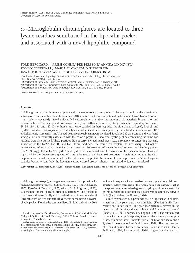
Based on primary and secondary structure homology,a1m is amember of the lipocalin family of proteins. The cup-shaped 3Dstructure of the lipocalins forms a pocket, which provides a bind-ing site for various small hydrophobic molecules. A 3D model ofa1m was obtained by alignment with the structure of the lipocalinepididymal retinoic acid-binding protein~ERABP!, which wasshown to be most similar toa1m among the lipocalins on the basisof primary structure and secondary structure predictions~Sansomet al., 1994!. Figure 5 shows the model derived using the sequenceof bovine a1m and the crystal structure of rat ERABP. Similarresults were obtained using the sequence of humana1m. The threechromophore-carrying Lys residues are highlighted. According tothis model, Lys118, Lys130, and Lys92 are semi-buried close tothe entrance of the pocket.
Fluorescence spectrum ofa1m
To verify the results from the molecular modeling ofa1m, weinvestigated the fluorescence spectra of the chromophores beforeand after reduction and denaturation ofa1m. The excitation wave-length, 360 nm, was chosen after obtaining excitation spectra atvarious emission wavelengths~not shown!. Figure 6 shows that thechromophore fluorophores have a red-shift in the maximum fluo-rescence from 464 to 482 nm after reduction and denaturation ofthe protein. The results indicate a change in the immediate envi-
Fig. 3. MS0MS spectrum of a doubly charged ion atm0z 829 from apurified colored petide with the sequence HHGPTITA~K !LYGR. A peptideabsorbing at 350 nm was purified as described in the caption to Figure 1,and the amino acid sequence was determined to HHGPTITA~K !LYGR byEdman degradation. The Lys residue within brackets was not recognized byEdman degradation. ES-MS of this peptide showed a doubly charged ion atm0z 829, consistent with a delta increment of 2060207 amu of the peptidesequence given. The MS-MS spectrum of the doubly charged ion atm0z829 is shown in the figure. The analysis was performed at M-Scan Inc.,West Chester, Pennsylvania.
Fig. 4. MS0MS spectrum of anm0z 282 ion found in the purified chro-mophore peptides. The sample was introduced via a nano spray interface ofa Bruker Esquire-LC ion trap mass spectrometer, operated in positive ion-isation mode. A proposed molecular structure based on the MS0MS spec-trum is inserted.
a1m Chromophores located to lysine residues 2615
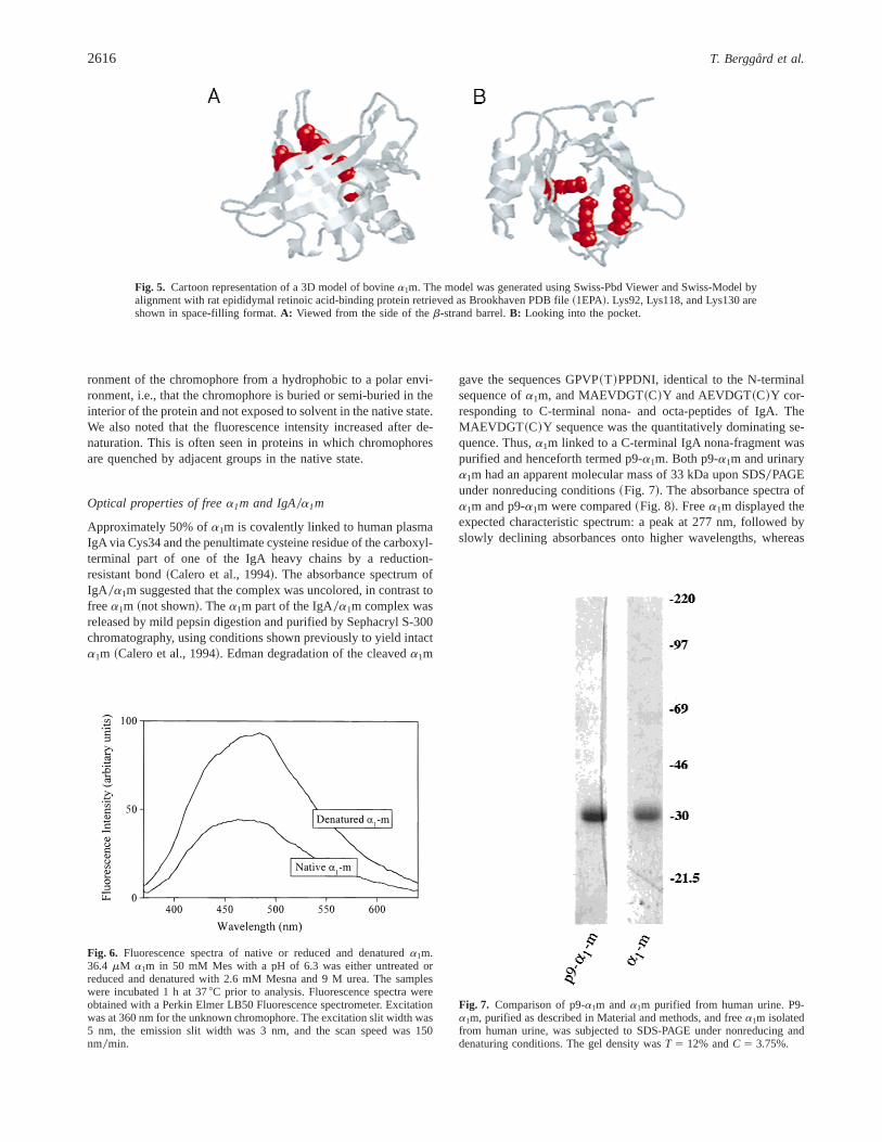
ronment of the chromophore from a hydrophobic to a polar envi-ronment, i.e., that the chromophore is buried or semi-buried in theinterior of the protein and not exposed to solvent in the native state.We also noted that the fluorescence intensity increased after de-naturation. This is often seen in proteins in which chromophoresare quenched by adjacent groups in the native state.
Optical properties of freea1m and IgA0a1m
Approximately 50% ofa1m is covalently linked to human plasmaIgA via Cys34 and the penultimate cysteine residue of the carboxyl-terminal part of one of the IgA heavy chains by a reduction-resistant bond~Calero et al., 1994!. The absorbance spectrum ofIgA 0a1m suggested that the complex was uncolored, in contrast tofreea1m ~not shown!. Thea1m part of the IgA0a1m complex wasreleased by mild pepsin digestion and purified by Sephacryl S-300chromatography, using conditions shown previously to yield intacta1m ~Calero et al., 1994!. Edman degradation of the cleaveda1m
gave the sequences GPVP~T!PPDNI, identical to the N-terminalsequence ofa1m, and MAEVDGT~C!Y and AEVDGT~C!Y cor-responding to C-terminal nona- and octa-peptides of IgA. TheMAEVDGT ~C!Y sequence was the quantitatively dominating se-quence. Thus,a1m linked to a C-terminal IgA nona-fragment waspurified and henceforth termed p9-a1m. Both p9-a1m and urinarya1m had an apparent molecular mass of 33 kDa upon SDS0PAGEunder nonreducing conditions~Fig. 7!. The absorbance spectra ofa1m and p9-a1m were compared~Fig. 8!. Freea1m displayed theexpected characteristic spectrum: a peak at 277 nm, followed byslowly declining absorbances onto higher wavelengths, whereas
Fig. 5. Cartoon representation of a 3D model of bovinea1m. The model was generated using Swiss-Pbd Viewer and Swiss-Model byalignment with rat epididymal retinoic acid-binding protein retrieved as Brookhaven PDB file~1EPA!. Lys92, Lys118, and Lys130 areshown in space-filling format.A: Viewed from the side of theb-strand barrel.B: Looking into the pocket.
Fig. 6. Fluorescence spectra of native or reduced and denatureda1m.36.4 mM a1m in 50 mM Mes with a pH of 6.3 was either untreated orreduced and denatured with 2.6 mM Mesna and 9 M urea. The sampleswere incubated 1 h at 378C prior to analysis. Fluorescence spectra wereobtained with a Perkin Elmer LB50 Fluorescence spectrometer. Excitationwas at 360 nm for the unknown chromophore. The excitation slit width was5 nm, the emission slit width was 3 nm, and the scan speed was 150nm0min.
Fig. 7. Comparison of p9-a1m anda1m purified from human urine. P9-a1m, purified as described in Material and methods, and freea1m isolatedfrom human urine, was subjected to SDS-PAGE under nonreducing anddenaturing conditions. The gel density wasT 5 12% andC 5 3.75%.
2616 T. Berggård et al.
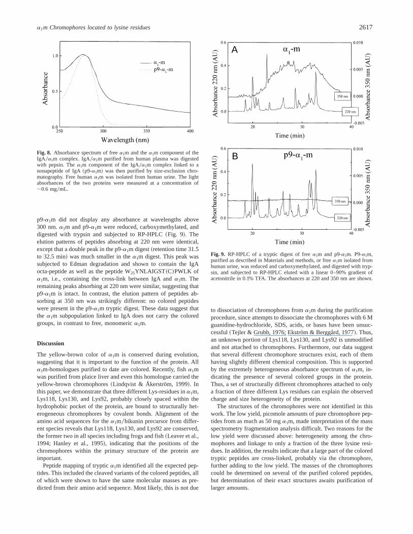
p9-a1m did not display any absorbance at wavelengths above300 nm.a1m and p9-a1m were reduced, carboxymethylated, anddigested with trypsin and subjected to RP-HPLC~Fig. 9!. Theelution patterns of peptides absorbing at 220 nm were identical,except that a double peak in the p9-a1m digest~retention time 31.5to 32.5 min! was much smaller in thea1m digest. This peak wassubjected to Edman degradation and shown to contain the IgAocta-peptide as well as the peptide W25YNLAIGST ~C!PWLK ofa1m, i.e., containing the cross-link between IgA anda1m. Theremaining peaks absorbing at 220 nm were similar, suggesting thatp9-a1m is intact. In contrast, the elution pattern of peptides ab-sorbing at 350 nm was strikingly different: no colored peptideswere present in the p9-a1m tryptic digest. These data suggest thatthe a1m subpopulation linked to IgA does not carry the coloredgroups, in contrast to free, monomerica1m.
Discussion
The yellow-brown color ofa1m is conserved during evolution,suggesting that it is important to the function of the protein. Alla1m-homologues purified to date are colored. Recently, fisha1mwas purified from plaice liver and even this homologue carried theyellow-brown chromophores~Lindqvist & Åkerström, 1999!. Inthis paper, we demonstrate that three different Lys-residues ina1m,Lys118, Lys130, and Lys92, probably closely spaced within thehydrophobic pocket of the protein, are bound to structurally het-erogeneous chromophores by covalent bonds. Alignment of theamino acid sequences for thea1m0bikunin precursor from differ-ent species reveals that Lys118, Lys130, and Lys92 are conserved,the former two in all species including frogs and fish~Leaver et al.,1994; Hanley et al., 1995!, indicating that the positions of thechromophores within the primary structure of the protein areimportant.
Peptide mapping of tryptica1m identified all the expected pep-tides. This included the cleaved variants of the colored peptides, allof which were shown to have the same molecular masses as pre-dicted from their amino acid sequence. Most likely, this is not due
to dissociation of chromophores froma1m during the purificationprocedure, since attempts to dissociate the chromophores with 6 Mguanidine-hydrochloride, SDS, acids, or bases have been unsuc-cessful~Tejler & Grubb, 1976; Ekström & Berggård, 1977!. Thus,an unknown portion of Lys118, Lys130, and Lys92 is unmodifiedand not attached to chromophores. Furthermore, our data suggestthat several different chromophore structures exist, each of themhaving slightly different chemical composition. This is supportedby the extremely heterogeneous absorbance spectrum ofa1m, in-dicating the presence of several colored groups in the protein.Thus, a set of structurally different chromophores attached to onlya fraction of three different Lys residues can explain the observedcharge and size heterogeneity of the protein.
The structures of the chromophores were not identified in thiswork. The low yield, picomole amounts of pure chromophore pep-tides from as much as 50 mga1m, made interpretation of the massspectrometry fragmentation analysis difficult. Two reasons for thelow yield were discussed above: heterogeneity among the chro-mophores and linkage to only a fraction of the three lysine resi-dues. In addition, the results indicate that a large part of the coloredtryptic peptides are cross-linked, probably via the chromophore,further adding to the low yield. The masses of the chromophorescould be determined on several of the purified colored peptides,but determination of their exact structures awaits purification oflarger amounts.
Fig. 8. Absorbance spectrum of freea1m and thea1m component of theIgA 0a1m complex. IgA0a1m purified from human plasma was digestedwith pepsin. Thea1m component of the IgA0a1m complex linked to anonapeptide of IgA~p9-a1m! was then purified by size-exclusion chro-matography. Free humana1m was isolated from human urine. The lightabsorbances of the two proteins were measured at a concentration of;0.6 mg0mL.
A
B
Fig. 9. RP-HPLC of a tryptic digest of freea1m and p9-a1m. P9-a1m,purified as described in Materials and methods, or freea1m isolated fromhuman urine, was reduced and carboxymethylated, and digested with tryp-sin, and subjected to RP-HPLC eluted with a linear 0–90% gradient ofacetonitrile in 0.1% TFA. The absorbances at 220 and 350 nm are shown.
a1m Chromophores located to lysine residues 2617
A lipophilic 282-amu substance was found associated with thecolored peptides. The MS0MS spectrum suggested a structure con-sisting of a 12-carbon aliphatic chain substituted with a cyclohex-ene, a hydroxyl, and an amine group. To our knowledge, thisstructure is previously unknown in a biological context. The sub-stance was seen as a separate peak after MS analysis of the purifiedpeptides, suggesting a noncovalent association. On the other hand,the association must be strong since the two components wereco-purified after as much as six cycles of HPLC chromatography.Such a strong, noncovalent interaction between the colored pep-tides and a 282-amu lipophilic substance could be a further expla-nation to the heterogeneous behavior of the chromophoric peptidesduring purification.
Lipocalins are characterized by highly conserved 3D structuresforming an internal pocket with the ability to bind a range of smallhydrophobic molecules. The fluorescence spectra of native anddenatureda1m indicate that the chromophores are buried or semi-buried in the hydrophobic core of the protein. According to amodel based on the sequence ofa1m in comparison with thelipocalin ERABP, the three chromophore-carrying Lys residues areclosely spaced and semiburied near the entrance of the predictedlipocalin pocket. This suggests that the 282 amu substance is alsolocated in the pocket or near its entrance. Thus, the substance couldbe the hitherto unknown lipocalin ligand ofa1m. A possible ex-planation for its presence in the lipocalin pocket is that it is sub-jected to transport bya1m. Another possibility is that the 282 amusubstance participates in the formation or turnover of the chromo-phores, either as a substrate, an intermediate, or a degradationproduct. Such a reaction could very well be mediated by the twofunctional groups, hydroxyl and amine, for example by formationof Schiff bases.
Other investigators have shown evidence that a chromophore isbound to Cys34~Escribano et al., 1991!. In this study, no coloredpeptides containing Cys34 were found. The purification protocolused in this study may be insufficient for purification of coloredpeptides containing Cys34. Alternatively, the chromophores boundto Cys34 may be of an even more heterogeneous nature than thechromophores linked to Lys118, Lys130, and Lys92, thus escapingdetection. The peptide W25YNLAIGSTCPWLK was identified byits expected molecular mass, suggesting that at least part of theCys34 is unmodified in the native protein. Future studies willhopefully clarify the relationship between the chromophores boundto Lys118, Lys130, Lys92, and Cys34.
A recent report describes the finding of unusually high levels ofglycation ina1m ~Bonay et al., 1997!. Glycation is a slow, non-enzymatic reaction between sugars and primary amino groups inproteins, i.e.,E-amino groups of Lys residues, leading to formationof brown-colored advanced glycation end products~AGE! ~Thorpe& Baynes, 1996; Chappey et al., 1997; Deyl & Miksik, 1997!. Theproperties of thea1m chromophores described here resemble thosedescribed for AGEs, but there are several differences which sug-gest that they are not glycation products:~1! a1m is by far morecolored than glycated proteins, suggesting a much more rapid for-mation of the chromophore ofa1m. Indeed, insect cells infectedwith a virus encodinga1m express a yellow-browna1m immedi-ately after infection~Åkerström et al., 1995; Wester et al., 1997!and intracellulara1m purified from place liver is yellow-brown~Lindqvist & Åkerström, 1999!. ~2! Incubation ofa1m with sugarsin vitro gave no evidence of a higher rate of glycation ofa1m thanof other plasma proteins.~3! No cis-diol hydroxyl groups, i.e.,early products of glycation, were found in the protein. Therefore,
the a1m chromophores may be similar to late glycation products,AGEs, but available data indicate that they are formed and at-tached to thea1m polypeptide by a different, but perhaps relatedmechanism.
a1m has a strong complex-forming ability. In man and in otherspecies, approximately 50% of the totala1m is found as covalentcomplexes with several different plasma proteins. The results inthis study show that thea1m subpopulation linked to IgA, thedominating complex-partner in human plasma, was almost com-pletely devoid of coloration. Similarly, the quantitatively domi-nating complex in rat plasma,a1-inhibitor-30a1m, seems to beuncolored~Falkenberg et al., 1990!. IgA was shown to be linkedvia Cys34 toa1m ~Calero et al., 1994!. Based on the above-mentioned 3D model, Cys34 is located in a large omega loop thatforms a lid, which may “close” the internal cavity of lipocalins~Flower, 1995, 1996!. It can thus be speculated that complex for-mation betweena1m and other plasma proteins alters the shape ofthe lid, thereby affecting the formation or turnover of the chromo-phores as well as the association of the 282 amu compound.
Materials and methods
Proteins and reagents
Human plasma was obtained from healthy blood donors. Mono-clonal mouse anti-a1m, BN 11.10, which binds to rat and humana1m, was prepared and purified as described~Babiker-Mohamedet al., 1991!. Modified porcine trypsin and pepsin were from Pro-mega~Madison, Wisconsin!. HPLC grade acetonitrile and waterwere obtained from Baker~Burdick and Jackson Division, Mus-kegon, Michigan!. Trifluoroacetic acid~TFA! was obtained fromPierce~Rockford, Illinois!. Formic acid was purchased from FisherScientific ~Fair Lawn, New Jersey!. Radiolabeled carbohydrateswere from NEN~Boston, Massachusetts! research products or fromBoehringer Mannheim~Mannheim, Germany!. a1m was purifiedfrom the urine of patients with tubular proteinuria as describedearlier ~Åkerström et al., 1995!.
Peptide purification
Lyophilized a1m ~1–10 mg! was dissolved in 7 mL of a buffer~100 mM NaCl, 50 mM Tris-HCl, pH 8.0! containing 6 M guani-dine and 5 mM dithioerythritol, and incubated for 1 h at 238C indarkness. After this, iodoacetamide was added to 10 mM, and thesolution incubated for another 30 min at 238C in darkness. Thesample was dialyzed exhaustively against 20 mM ammonium bi-carbonate, pH 7.4, and concentrated to;1 mL by vacuum centri-fugation. The reduced and S-carboxymethylateda1m was digestedwith modified porcine trypsin for 3 h at 378C using an enzyme0substrate ratio of 1:80, freeze-dried and dissolved in 1 mL 0.1%TFA. Colored peptides were purified by repeated RP-HPLC on aPharmacia~Uppsala, Sweden! Smart system equipped with a Brown-lee ~Aquapore RP-300, 2203 2.1 mm!, Brownlee ~Vydac C182003 2.1 mm!, Beckman~Ultrasphere 2503 2.0 mm!, or Phe-nomenex~Torrence, California! ~Nucleosil C18 2503 2.0 mm!column, eluting with a linear gradient of 0.1% TFA to 90%acetonitrile00.07% TFA at a flow rate of 200mL 0min. Some frac-tions were further purified by a Brownlee~Vydac C18 20032.1 mm! column followed by a Phenomenex~Nucleosil C18 25032.0 mm! column eluting with a linear gradient of 0.1% TFA to 75%
2618 T. Berggård et al.

2-propanol0 0.07% TFA at a flow rate of 125mL 0min. Fractionswere collected manually based on a 350 nm analogue signal fromthempeak detector~Pharmacia!. The purity of the colored peptideswas determined by Edman degradation. The colored peptides werepure when only one homogeneous, sharp peak of co-migratingabsorbance at both 350 and 220 nm were seen and the ratio of2200350 nm absorbance was lower than 2. The absorbances at 220and 350 nm were digitized and recorded on a computer for post-run analysis. In some experiments, the reduced, carboxymethyl-ated, and trypsin-digesteda1m was subjected to size exclusionchromatography on a Pharmacia Superdex HR 10030 column elut-ing with 0.1% TFA in H2O at a flow rate of 25mL 0min. Fractionswere collected according to the absorbance at 350 nm and coloredpeptides were further purified by RP-HPLC as described above.
Amino acid sequence analysis
Automated Edman degradation was carried out in an Applied Bio-systems 477 A sequencer with on-line detection of PTH aminoacids using an Applied Biosystems 120A HPLC.
Electrospray ionization mass spectrometry (ES-MS)analysis of chromophore peptides
The analysis was performed using a VG Biotech BIO-Q instru-ment with quadrupole analyzer. Sample aliquots of 50mL wereinjected into the instrument source. Elution was carried out usinga mixture of 50% aqueous acetonitrile containing 0.1% TFA, at aflow rate of 10mL 0min. Calibration was performed with cesiumiodide. Alternatively, analysis was performed using an Esquire-LCion-trap mass spectrometer~Bruker, Bremen, Germany! with aBruker nano spray interface, in positive ionization mode. The sam-ples were placed in a gold plated glass capillary needle and in-serted into the nano spray interface. The solvent was a mixture of50% aqueous methanol~Merck, Darmstadt, Germany! containing0.1% trichloroacetic acid~TCA!.
Mass spectrometry peptide mapping
Lyophilized a1m ~;50 mg! was dissolved in 50mL of 0.2 Mammonium bicarbonate, pH 8.0 and digested with trypsin using anenzyme0substrate ratio of 1:10, for 4 h at 378C. The pH was thenadjusted to 2 by the addition of TFA. An aliquot of the digest~5 mL! was subjected to RP-HPLC~Deltabond ODS 1503 1 mm,Keystone Scientific, Bellefonte, Pennsylvania! using an Isco~Lin-coln, Nebraska! microbore HPLC system. The digest was eluted at30 mL 0min using 0.025% TFA and 90% acetonitrile containing0.025% TFA. The effluent was split evenly into two streams. Onestream was monitored at 350 nm and the other was delivereddirectly to the source of the mass spectrometer. Mass spectra wererecorded on a Fisons Quattro-BQ triple quadrupole mass spectrom-eter equipped with a pneumatically assisted electrostatic ion sourceoperating at atmospheric pressure. The data were collected in thecontinuum mode from mass0charge~m0z! ratio 230–1,450 with ascan time of 5 s. Spectra were calibrated using polyethylene glycol.
Glycation analysis
One hundred twenty to 600mg of a1m, human serum albumin, ororosomucoid were glycated in vitro in PBS~phosphate-bufferedsaline: 8 mM sodium phosphate, 1.5 mM potassium phosphate,
pH 7.4, 0.12 M sodium chloride, 2.7 mM potassium chloride,pH 7.4! containing either 0.2 nM@3H#-glucose, 0.2 nM@3H#-galactose, 0.2 nM@14C#-ribose, or 0.2 nM@14C#-fructose at 378Cfor various amounts of time. The glycated protein was then sepa-rated from free sugar on a PD-10 column~Pharmacia! eluted withPBS. Fifty microliter fractions were collected and the radioactivitymeasured in ab-counter. Alternatively, 200mg of a1m, humanserum albumin, or orosomucoid were incubated at 378C with 0.5 Mglucose or 0.5 M fructose in PBS. The absorbance spectra of theproteins were measured at various time points in a Uvicon 930Spectrophotometer~Kontron Instruments, Neufahrm, Germany!.Human serum albumin was glycated by incubation with 250 mMglucose at 378C in PBS for 30 days and then separated from freesugar on a PD-10 column. Human urinarya1m ~200 mg! or gly-cated or nonglycated human serum albumin~200mg! was appliedto a 1 mL Glyco-gel II Boronate Affinity Gel column~Pierce,Rockford, Illinois!, and bound proteins eluted according to theinstructions supplied by the manufacturer. The amounts of proteinin the bound and nonbound fractions were quantified by radio-immunoassay, absorbance, and SDS-PAGE.
Construction of a 3D model ofa1m
Coordinates for the rat ERABP were retrieved as a BrookhavenDatabase Protein Data Bank~PDB! file, accession code 1EPA. Thefile was used together with the amino acid sequence of bovinea1m~Lindqvist & Åkerström, 1996! to generate a 3D model ofa1m,utilizing the Swiss-PdbViewer version 3.01 program linked to Swiss-Model, an automated homology modeling server~Guex & Peitsch,1997!.
Fluorescence spectra
Fluorescence spectra were obtained with a Perkin Elmer LB50Fluorescence spectrometer. Excitation was made at 360 nm for theunknown chromophore. The excitation slit width was 5 nm, theemission slit width was 3 nm, and the scan speed was 150 nm0min.Nonreduced and nondenatureda1m was measured at 36.4mM in50 mM Mes~2-morpholino ethane sulfonic acid0sodium salt! buffer,pH 6.3. The reduced and denatured samples were 36.4mM withregard toa1m, 2.6 mM with regard to Mesna~2-mercapto-ethane-sulfonic acid0sodium salt! and 9 M with regard to urea~ultra pure,Gibco BRL, Gaithersburg, Maryland0Life Technologies, Rock-ville, Missouri! in 50 mM Mes-buffer, pH 6.3. The samples wereincubated 1 h at 378C prior to analysis. All chemicals were fromSigma~St. Louis, Missouri!. Controls were measured in 50 mMMes buffer with or without 9 M urea and 2.6 mM Mesna.
Purification of IgA0a1m and preparationof thea1m component of the complex
Immunosorbent affinity chromatography of human plasma withmonoclonal mouse anti-a1m, BN11.10, immobilized to Affigel Hz~20 mg0mL! followed by Sephacryl S-300 size exclusion chroma-tography, was done as described~Berggård et al., 1997!. Fractionscontaining IgA0a1m, as determined by sodium dodecyl sulfate-polyacrylamide gel electrophoresis~SDS-PAGE, see below!, wereconcentrated and dialyzed. One-half milliliter of IgA0a1m ~3 mg0mL! in 50 mM sodium-acetate, pH 4.5, was partly digested withpepsin for 20 min at 208C using an enzyme0substrate ratio of 1:50.The reaction was stopped by addition of 2 M Tris-HCl buffer as
a1m Chromophores located to lysine residues 2619

described earlier~Calero et al., 1994!. The mixture was separatedby Sephacryl S-200 size exclusion chromatography. The two pro-tein fractions were analyzed by SDS-PAGE as described~Laem-mli, 1970!. Following electrophoresis, the proteins were eitherstained with Coomassie Brilliant Blue, or transferred to a poly-vinylidene difluoride membrane~“Immobilon,” Millipore! as de-scribed~Matsudaira, 1987! and subjected to Edman degradation.
Acknowledgments
The authors wish to thank Maria Allhorn and Eva Ohlson for excellenttechnical assistance. This work was supported by grants from the SwedishMedical Research Council~project no. 7144!, King Gustav V’s 80-yearFoundation, the Swedish Society for Medical Research, the Royal Physio-graphic Society in Lund, the Foundations of Greta and Johan Kock, andAlfred Österlund and National Institutes of Health~NIH ! HL49542~JJE!.
References
Åkerström B, Bratt T, Enghild JJ. 1995. Formation of thea1-microglobulinchromophore in mammalian and insect cells: A novel post-translationalmechanism?FEBS Lett 362:50–54.
Åkerström B, Lögdberg L. 1990. An intriguing member of the lipocalin proteinfamily: a1-microglobulin.Trends Biochem Sci 15:240–243.
Babiker-Mohamed H, Forsberg M, Olsson ML, Winquist O, Nilson BH, Lögd-berg L, Åkerström B. 1991. Characterization of monoclonal anti-a1-micro-globulin antibodies: Binding strength, binding sites, and inhibition oflymphocyte stimulation.Scand J Immunol 34:655–666.
Berggård T, Thelin N, Falkenberg C, Enghild JJ, Åkerström B. 1997. Prothrom-bin, albumin and immunoglobulin A form covalent complexes witha1-microglobulin in human plasma.Eur J Biochem 245:676–683.
Bonay P, Solis J, de la Calle H, Fresno M, Grubb A, Mendez E. 1997. Massiveglycation of protein HC, a low molecular weight lipocalin, in non-diabeticindividuals.FEBS Lett 416:276–280.
Bratt T, Olsson H, Sjöberg EM, Jergil B, Åkerström B. 1993. Cleavage of thea1-microglobulin-bikunin precursor is localized to the Golgi apparatus of ratliver cells.Biochim Biophys Acta 1157:147–154.
Calero M, Escribano J, Grubb A, Mendez E. 1994. Location of a novel type ofinterpolypeptide chain linkage in the human protein HC-IgA complex~HC-IgA! and identification of a heterogeneous chromophore associated with thecomplex.J Biol Chem 269:384–389.
Chappey O, Dosquet C, Wautier MP, Wautier JL. 1997. Advanced glycation endproducts, oxidant stress and vascular lesions.Eur J Clin Invest 27:97–108.
Deyl Z, Miksik I. 1997. Post-translational non-enzymatic modification of pro-teins. I. Chromatography of marker adducts with special emphasis to gly-cation reactions.J Chromatogr B Biomed Sci Appl 699:287–309.
Ekström B, Berggård I. 1977. Humana1-microglobulin. Purification procedure,chemical and physiochemical properties.J Biol Chem 252:8048–8057.
Ekström B, Peterson PA, Berggård I. 1975. A urinary and plasmaa1-glycoproteinof low molecular weight: Isolation and some properties.Biochem BiophysRes Commun 65:1427–1433.
Enghild JJ, Thøgersen IB, Pizzo SV, Salvesen G. 1989. Analysis of inter-a-trypsin inhibitor and a novel trypsin inhibitor, pre-a-trypsin inhibitor, fromhuman plasma. Polypeptide chain stoichiometry and assembly by glycan.J Biol Chem 264:15975–15981.
Escribano J, Grubb A, Calero M, Mendez E. 1991. The protein HC chromophore
is linked to the cysteine residue at position 34 of the polypeptide chain bya reduction-resistant bond and causes the charge heterogeneity of proteinHC. J Biol Chem 266:15758–15763.
Escribano J, Lopex-Otin C, Hjerpe A, Grubb A, Mendez E. 1990. Location andcharacterization of the three carbohydrate prosthetic groups of human pro-tein HC.FEBS Lett 266:167–170.
Falkenberg C, Enghild JJ, Thøgersen IB, Salvesen G, Åkerström B. 1994.Isolation and characterization of fibronectin-a1-microglobulin complex inrat plasma.Biochem J 301:745–751.
Falkenberg C, Grubb A, Åkerström B. 1990. Isolation of rat seruma1-micro-globulin. Identification of a complex witha1-inhibitor3, a rata2-macroglobulinhomologue.J Biol Chem 265:16150–16157.
Flower DR. 1995. Multiple molecular recognition properties of the lipocalinprotein family.J Mol Recognit 8:185–195.
Flower DR. 1996. The lipocalin protein family: structure and function.BiochemJ 318:1–14.
Grubb AO, Lopez C, Tejler L, Mendez E. 1983. Isolation of human complex-forming glycoprotein, heterogeneous in charge~protein HC!, and its IgAcomplex from plasma. Physiochemical and immunochemical properties,normal plasma concentration.J Biol Chem 258:14698–14707.
Guex N, Peitsch MC. 1997. SWISS-MODEL and the Swiss-PdbViewer: Anenvironment for comparative protein modeling.Electrophoresis 18:2714–2723.
Hanley S, McInerney JO, Powell R. 1995. Sequence analysis and evolutionaryaspects of piscinea1-microglobulin0bikunin mRNA transcripts.Mol MarBiol Biotechnol 4:105–109.
Hanley S, Powell R. 1994. Sequence of a cDNA clone encoding the Atlanticsalmona1-microglobulin0bikunin protein.Gene 147:297–298.
Laemmli UK. 1970. Cleavage of structural proteins during the assembly of thehead of bacteriophage T4.Nature 227:680–685.
Leaver MJ, Wright J, George SG. 1994. Conservation of the tandem arrange-ment ofa1-microglobulin0bikunin mRNA: Cloning of a cDNA from plaice~Pleuronectes platessa!. Comp Biochem Physiol Biochem Mol Biol 108:275–281.
Lindqvist A, Åkerström B. 1996. Bovinea1-microglobulin0bikunin. Isolationand characterization of liver cDNA and urinarya1-microglobulin.BiochimBiophys Acta 1306:98–106.
Lindqvist A, Åkerström B. 1999. Isolation of plaice~Pleuronectes platessa!a1-microglobulin: Conservation of structure and chromophore.Biochim Bio-phys Acta 1430:222–233.
Matsudaira P. 1987. Sequence from picomole quantities of proteins electroblot-ted onto polyvinylidene difluoride membranes.J Biol Chem 262:10035–10038.
Salier JP. 1990. Inter-a-trypsin inhibitor: Emergence of a family within theKunitz-type protease inhibitor superfamily.Trends Biochem Sci 15:435–439.
Sansom CE, North AC, Sawyer L. 1994. Structural analysis and classification oflipocalins and related proteins using a profile-search method.Biochim Bio-phys Acta 1208:247–255.
Tejler L, Grubb AO. 1976. A complex-forming glycoprotein heterogeneous incharge and present in human plasma, urine, and cerebrospinal fluid.BiochimBiophys Acta 439:82–94.
Thøgersen IB, Enghild JJ. 1995. Biosynthesis of bikunin proteins in the humancarcinoma cell line HepG2 and in primary human hepatocytes. Polypeptideassembly by glycosaminoglycan.J Biol Chem 270:18700–18709.
Thorpe SR, Baynes JW. 1996. Role of the Maillard reaction in diabetes mellitusand diseases of aging.Drugs Aging 9:69–77.
Wester L, Johansson MU, Åkerström B. 1997. Physicochemical and biochem-ical characterization of humana1-microglobulin expressed in baculovirus-infected insect cells.Protein Expr Purif 11:95–103.
2620 T. Berggård et al.