Cerebrospinal fluid data compilation and knowledge-based ...
Upload
independentCategory
view
1download
0
Eur J Pediatr (2006) 165: 636–645DOI 10.1007/s00431-006-0160-x
ORIGINAL PAPER
Ana Alarcon . Alfredo Garcia-Alix .Fernando Cabañas . Angel Hernanz .Dora Pascual-Salcedo . Ana Martin-Ancel .Marta Cabrera . Alfredo Tagarro . Jose Quero
Beta2-microglobulin concentrations in cerebrospinal fluidcorrelate with neuroimaging findings in newbornswith symptomatic congenital cytomegalovirus infectionReceived: 17 September 2005 / Revised: 5 April 2006 / Accepted: 7 April 2006 / Published online: 12 May 2006# Springer-Verlag 2006
Abstract Overview: In newborns with symptomaticcongenital cytomegalovirus (CMV) infection, neuroima-ging is the best available predictor of neurodevelopmentaloutcome. Cerebrospinal fluid (CSF) findings in congenitalCMV infection have seldom been described. Neonateswith central nervous system infections present high CSFBeta2-microglobulin (β2-m) levels. Objectives: Theobjectives of this study were: (1) to determine whetherCSF β2-m is increased in newborns with symptomaticcongenital CMV infection, and (2) to examine its corre-lation with neuroimaging findings. Materials andmethods: Fourteen newborns with symptomatic congen-ital CMV infection admitted to La Paz Hospital from 1990through 2004 underwent determination of CSF β2-m.Ninety-three newborns, constituting the comparisongroup, underwent CSF β2-m determination as part of asepsis or meningo/encephalitis work-up, and at dischargehad sterile cultures and normal neurological status.Neuroimaging findings were scored according to asemiquantitative system: (0) no abnormalities; (1) singlepunctate periventricular (PV) calcification and/or hyper-echogenic areas in the thalamus and basal ganglia; (2)multiple discrete PV calcifications and/or ventriculome-galy; and (3) extensive PV calcifications and/or brainatrophy. Discussion and conclusion: CSF β2-m wasincreased in newborns with CMV infection (median6.21 mg/L) compared with controls (1.68 mg/L)
(P<.001). β2-m showed a correlation with neuroimagingscores (rs=0.753, P=.002). β2-m was higher in patientswho scored 2–3 (12.83 mg/L) than in patients who scored0–1 (5.52 mg/L) (P=.028). CSF β2-m is increased innewborns with symptomatic congenital CMV infectionand correlates with neuroimaging abnormalities. β2-mappears to be an indicator of the severity of braininvolvement in congenital CMV infection.
Keywords Beta2-microglobulin . Cerebrospinal fluid .Cytomegalovirus . Neuroimaging . Newborn
Introduction
Cytomegalovirus (CMV) is the leading cause of con-genital infection and affects approximately 1% of alllive births [38]. The majority of these infants will havean asymptomatic infection, and only around 10% willbe symptomatic at birth [6, 38]. This group of childrenwith symptomatic congenital CMV infection is morelikely to experience sequelae, including sensorineuralhearing loss (SNHL), cognitive and motor deficits,seizures, and visual defects [9, 30, 38]. Predictors ofwhich infants will have an unfavourable long-termoutcome remain controversial [5, 6, 9, 28, 30, 34, 38].The best predictor of adverse neurodevelopmentalprognosis available today is the existence of cranialcomputed tomographic (CT) abnormalities detectedwithin the first month of life [5, 28, 38]. Furthermore,the categorization of the CT findings in the newbornperiod helps to determine the prognosis of children withsymptomatic congenital CMV [28].
Cerebrospinal fluid (CSF) in congenital CMV infectioncan show the findings of meningoencephalitis [2, 5, 6, 26,30, 49]. However, CSF findings in infants with symptom-atic congenital CMV infection have seldom been de-scribed, and the role of CSF as a marker of brain damageand predictor of neurological sequelae is unclear. Beta2-microglobulin (β2-m) is a low molecular weight (11800 D)
A. Garcia-Alix . F. Cabañas . A. Hernanz . D. Pascual-Salcedo .M. Cabrera . A. Tagarro . J. QueroDivision of Neonatology, Department of Pediatrics,Hospital La Paz, Universidad Autonoma de Madrid,Paseo de la Castellana, 261,28046 Madrid, Spaine-mail: [email protected]
A. Alarcon (*) . A. Martin-AncelDivision of Neonatology, Department of Pediatrics,Unitat Integrada Hospital Sant Joan de Deu-Hospital Clinic,Universitat de Barcelona,08950 Barcelona, Spaine-mail: [email protected]
protein that constitutes the light chain of HLA class Iantigens and is present on the surface of all nucleated cells[4, 10, 12, 17]. Its concentration in biological fluids isrelated to the rate of cell membrane renovation, and highlevels of this peptide reflect an increased cellular turnover.High β2-m concentrations in CSF have been described indifferent inflammatory or neoplastic central nervoussystem (CNS) disorders [16, 18, 20, 24, 25, 29, 33, 39,41, 43, 48]. In addition, we have previously observed highlevels of CSF β2-m in newborns with CNS infections,including bacterial or viral meningitis and TORCHinfections [14, 40], and CSF β2-m is used in our centreas an ancillary tool in neonates evaluated for CNSinfections.
We hypothesized that β2-m concentration in CSF couldbe an indicator of the severity of CNS involvement ininfants with symptomatic congenital CMV infection. Theobjectives of our study were: (1) to determine whether β2-m concentrations in CSF are increased in newborns withsymptomatic congenital CMV infection, and (2) toexamine the correlation between β2-m in CSF andneuroimaging findings in these patients.
Materials and methods
Patient population
All newborns with symptomatic congenital CMV infectionwho were admitted to La Paz Hospital, a UniversityTertiary Public Hospital, from 1990 through 2004, andunderwent determination of β2-m in CSF were included inthis study. Patients born between 1990 and 2000 wereretrospectively identified by a systematic review of theclinical records of the newborns admitted in the Division ofNeonatology during those years. Patients born between2001 and 2004 were enrolled prospectively. The study wasapproved by the Committee on Ethics in Clinical Researchof our institution, and written informed consent wasobtained from the parents of the study infants included inthe prospective cohort. During the study period, anothergroup of neonates underwent a spinal tap and determina-tion of CSF β2-m levels as part of a sepsis or meningo-encephalitis workup, and finally had sterile cultures andnormal neurological status at discharge. These newbornswithout CNS infection were used as the comparison group.Patients with a traumatic puncture were not excluded, sincewe have previously observed that the presence of blood inthe CSF does not alter β2-m concentration [14].
A patient was considered to have a congenital CMVinfection if specific CMV IgM was detected, CMV wasisolated from urine or blood and/or CMV antigen wasdetected in blood during the first 2 weeks of life [38]. Theinfection was considered symptomatic when, together withthe detection of CMV, at least one of the followingabnormalities was present at birth: (1) intrauterine growthrestriction (IUGR), defined as birth weight below an SDscore (SDS) of −2 for gestational age [46]; (2) petechiae;(3) hepatomegaly; (4) splenomegaly; (5) jaundice; (6)
microcephaly, defined as head circumference below anSDS of −2 for gestational age [46]; (7) thrombocytopenia(platelet count<75×103/μL); (8) laboratory evidence ofhepatitis (alanine aminotransferase level >100 U/L or directbilirubin level >3 mg/dL). Hearing was evaluated byauditory brainstem responses, and a hearing threshold≥20 dB was considered evidence of SNHL. Patients bornbetween 1990 and 2000 were regularly followed-up in theDivision of Pediatric Neurology. Neurodevelopmentaloutcome of patients born after year 2000 was assessed byone of our investigators at 6 month intervals by neu-rological examinations based on the method by Amiel-Tison and Grenier [1], and the Denver DevelopmentalScreening Test [13].
Demographic, clinical and follow-up data, as well asinformation regarding diagnostic studies were collected onstandardized data collection forms.
Neuroimaging studies
Neuroimaging studies included cranial ultrasound scans(US), cranial CT and/or magnetic resonance imaging(MRI) performed in the newborn period. Cranial USbefore 1998 were performed using a Toshiba SSH-140Acolor Doppler flow imaging (CDFI) system with a 5 MHzand 7.5 MHz probe. Since 1998, a Siemens Sonoline VersaPlus Power Doppler system (disk version 2.3.0) with asector and linear 5 and 7.5 MHz probe was used. CerebralUS were performed at several axes of the coronal, sagittal,and parasagittal planes, through the anterior fontanelle. Toevaluate the brain surface, the transducer was angled(tangential planes) towards the frontal and occipital brainsurface (coronal planes), and towards both hemisphericconvexities (sagittal planes). CDFI or Power Doppler wereused to examine the abnormalities of brain surface vascularpattern [32]. Images were stored as video and hard copies.
CT scans were obtained using a high resolutiontechnique on continuous 5 mm thick slices. MRI studieswere obtained with equipment operating at 0.5 or 1.5 Tesla,and included axial T1 and T2 spin echo sequences, with5 mm thick slices spaced every 2 mm.
The neuroimaging findings were evaluated blindly toCSF findings and graduated according to a modifiedscoring system that has shown a correlation with theneurodevelopmental outcome of infants with symptomaticcongenital CMV infection [28]: (0) no abnormalities orabnormalities not related to CMV; (1) single punctateperiventricular (PV) calcification and/or hyperechogenicareas in the thalamus and basal ganglia (HTBG) [8, 35, 42];(2) multiple discrete PV calcifications and/or moderate tosevere ventriculomegaly [23], without other signs of brainatrophy; (3) extensive PV calcifications and/or brainatrophy, defined as ventriculomegaly, widened subarach-noid space and prominent sulci. In addition, specialattention was paid to the presence of PV hyperechogenicity[31], germinolysis, PV cysts, dysgenesis of the corpuscallosum, neuronal migration disorders (NMDs) [32], andcerebellar hypoplasia.
637
CSF analysis
CSF evaluation included a standard citochemical analysis,with determination of protein and glucose concentrationsand white blood cell (WBC) count, as well as quantifica-tion of β2-m concentration. In addition, neuron specificenolase (NSE) and proinflammatory cytokines interleukin-6 (IL-6) and tumor necrosis factor-α (TNF-α) weremeasured in some patients. An elevated CSF WBC countwas considered when the number of cells was over 32/mm3
in a term infant and 29/mm3 in a preterm infant, while ahigh CSF protein concentration was defined as over170 mg/dl in a term infant and 150 mg/dl in a preterminfant [37]. β2-m and NSE determinations were performedby enzyme immunoassay, as previously described [14, 15].IL-6 was measured by “in house” enzyme immunoassay[47], and TNFα by Duoset-enzyme-linked immunosorbentassay (R&D Systems, Abigdon, UK). Samples wereassayed undiluted and at 2-fold serial dilutions. Detectionlimits were 1 pg/ml for IL-6 and 7 pg/ml for TNFα.
Statistical analysis
The numerical data are expressed as median and range,unless otherwise stated. The comparison between numer-ical variables was performed by the Mann-Whitney U test.The relationship between variables was evaluated by theSpearman’s rank correlation coefficient (rs). A P value less
than 0.05 was considered significant. The StatisticalPackage for the Social Sciences (SPSS, Chicago, IL)version 11.0 was used.
Results
Patient characteristics
A total number of 23 infants with symptomatic congenitalCMV infection were identified. Nine were excluded due tolack of determination of β2-m levels in CSF. Of theremaining 14 infants that comprised the study population,seven were born after year 2000 and were includedprospectively in the study. During the study period, β2-mCSF levels were measured in 93 newborns in whom CNSinfection was ruled out and who exhibited a normalneurological examination at discharge, thus constitutingthe control population.
The main clinical characteristics of the group of childrenwith symptomatic congenital CMV infection are shown inTable 1. All had Apgar scores over 6 at 5 min. Othercongenital infections were ruled out, and none showedmalformations or dismorphyc features suggesting trisomy.Patients 3,5–8,10–14 received treatment with ganciclovir.Two patients died at 1 and 7 months respectively, andadverse neurological outcome was seen in three of the teninfants who were at least 6 months old at their last recordedvisit.
Table 1 Clinical findings of 14 children with symptomatic congenital CMV infection
Pt.no.
Sex(M/F)
GA(wks)
BW(g)
HC(cm)
Clinical signs at birth Hearingloss (−/+)
Chorioretinitis(−/+)
Follow-up (mths lastevaluation)
1 F 38 1,950 26.5 IURG, Microcephaly NP + NP2 F 36 1,960 32 IURG NP NP Normal (3)3 M 38 2,550 33 Hepatosplenomegaly, Jaundice, Hepatitis - - Moderate psicomotor delay
(34)4 F 35 1,530 28.5 IURG, Microcephaly, Petechiae and ecchymoses,
Splenomegaly, Jaundice, Hepatitis- - Cerebral palsy (43)
5 M 36 1,730 29.8 IURG, Microcephaly, Petechiae, Jaundice,Thrombocytopenia, Hepatitis
+ NP Death (1)
6 F 39 1,750 28.3 IURG, Microcephaly, Petechiae,Hepatosplenomegaly, Thrombocytopenia
+ - Cerebral palsy, seizures,death (7)
7 F 36 1,650 29 IURG , Microcephaly - - Normal (14)8 M 36 1,900 31 IURG, Petechiae, Hepatosplenomegaly,
Thrombocytopenia, Hepatitis+ - Normal (18)
9 M 39 3,600 33.5 Hepatosplenomegaly - - NP10 M 40 2,210 31.5 IURG, Microcephaly, Petechiae + - Moderate psicomotor delay
(22)11 M 37 3,180 34.5 Petechiae - - Normal (6)12 F 39 2,150 30.5 IURG, Microcephaly + - Moderate motor delay,
seizures (30)13 F 41 2,990 33 Petechiae, Hepatomegaly, Jaundice, Hepatitis + - Normal (12)14 M 38 2,250 28.5 Microcephaly + - Severe psicomotor delay,
spasticity, seizures (10)
Abbreviations: Pt. No.= patient number; GA= gestational age; BW=birth weight; HC=head circumference; -=no; +=yes; NP=not performed
638
Neuroimaging findings
Cranial US was performed in all 14 patients, CT in 10(cases 2–6,8–11, and 14), and MRI in 4 (cases 1,12–14).The neuroimaging findings are summarized in Table 2.Median postmenstrual age at neuroimaging was over37 weeks for all techniques; 38 weeks (range 35–44 weeks) for US; 39 weeks (range 37–42 weeks) forCT; 40 weeks (range 38–45 weeks) for MRI.
Cranial US (Figs. 1 and 2) showed PV calcifications innine cases. HTBG observed in our patients were linear inthree patients (cases 3,11 and 12) and mixed (linear andpunctate) in eight (1,4–6,8,10,13,14). Seven cases (1,4–6,8,10 and 14) had subcortical and/or cortical calcifica-tions. Ventricular dilatation was present in seven infants,and PV hyperechogenicity was observed in eight patients.PV cysts were seen in two patients, while germynolisis waspresent in six. The corpus callosum was considered thin infour patients. Findings suggestive of NMDs, such asabnormal operculization or sulcation, were observed in sixpatients (3–6,12, and 14). The cerebellum was consideredsmall in one case.
Cranial CT (Fig. 1) confirmed the presence of PVcalcifications in seven of the ten patients on whom it wasperformed. Other calcifications observed were subcortical(patients 4–6), cortical (patients 5 and 6) and cerebellar(patient 3). Ventriculomegaly was present in six patients,while three (cases 2,5 and 6) had expanded subarachnoidspace. White matter hypodensity was seen in cases 5, 6, 8,10 and 11. Pachygyria and lissencephaly were apparent inpatients 5 and 14, respectively, while abnormal operculiza-
tion was observed in patients 4 and 6, and scarce sulcationin patient 6. As in US, patient 6 had a small cerebellum.
MRI (Fig. 2) confirmed the existence of PV calcifica-tions in patients 1, 12 and 14. Patient 13 had germynolisisof caudate nuclei. Patients 1, 12 and 14 had pachygiria andcase 14 also showed areas of polymicrogyria. Abnormalsignal intensity in the white matter was found in patient 12.
CSF findings
The median age at spinal tap was 6 days (range 1–30 days)for infants with symptomatic congenital CMV infectionand 5 days (range 1–21 days) for controls. All patientstreated with ganciclovir were tapped before treatment wasinitiated. In infants with congenital CMV, the median CSFprotein concentration was 110.10 mg/dl (range 55–634 mg/dl), and five patients had elevated protein concentrations.The median WBC count was 11/mm3 (range 0–240/mm3),and four patients had pleocytosis. A combination ofelevated protein concentration andWBC count was noticedin three patients, including two with a bloody CSF sample.In control infants, the median protein concentration was96 mg/dl (range 27–311 mg/dl), and the median WBCcount was 12/mm3 (range 0–160/mm3). Fifteen controlinfants showed an elevated protein concentration andWBCcount, all of them with traumatic taps.
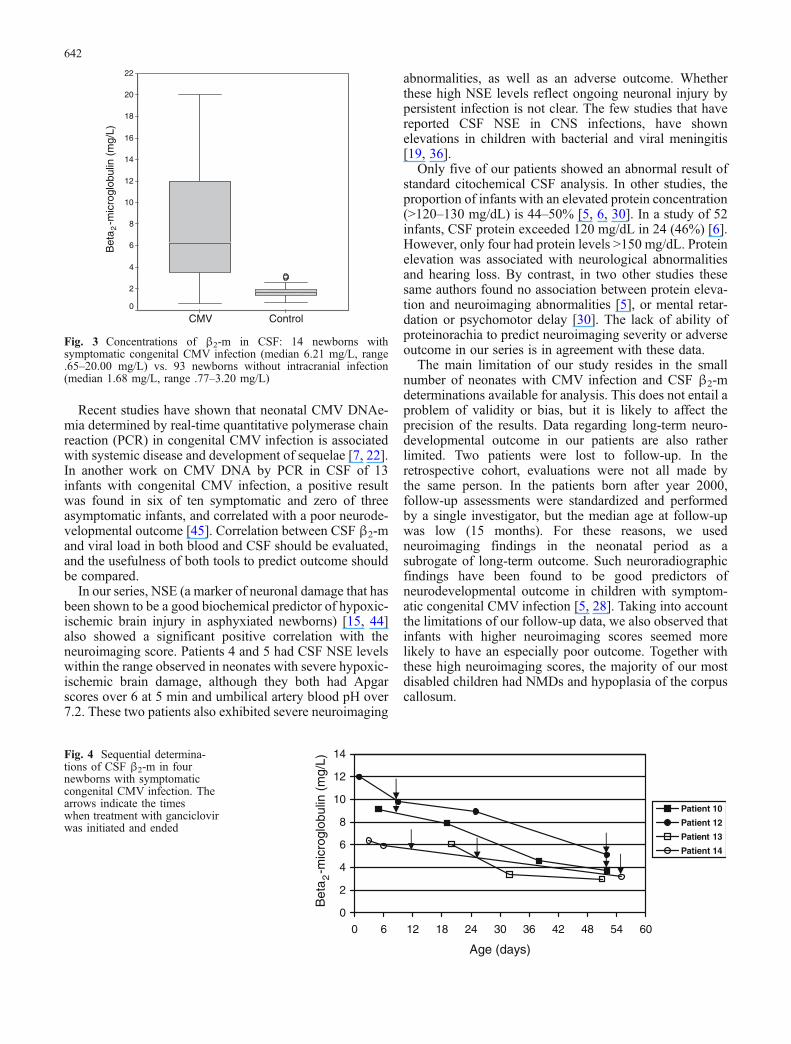
When β2-m concentrations were compared between thegroup of neonates with symptomatic congenital CMVinfection and the control group, patients with CMVinfection exhibited significantly higher β2-m in CSF(P<.001; Fig. 3). The number of patients with CSF β2-m
Table 2 Neuroimaging findings of 14 newborns with symptomatic congenital CMV infection
Pt.No.
PVcalcifications(−/+/++/+++)
HTBG(−/+)
Ventriculomegaly(−/+/++/+++)
PVhyperechogenicity(−/+/++/+++)
PVcysts(−/+)
Germynolisis(−/+)
Brainatrophy(−/+)
Corpuscallosumdysgenesis(−/+)
NMD (−/+) Cerebellarhypoplasia(−/+)
Score
1 ++ + +++ - - - + - +, pachygyria - 32 - - - - - - - + - - 03 - + + ++ - + - - + - 14 +++ + - ++ + - - - + - 35 +++ + ++ +++ + + + + +, pachygyria - 36 +++ + + - - - + + + + 37 - - - - - - - - - - 08 + + - ++ - - - - - - 19 - - - - - + - - - - 010 + + + ++ - + - - - - 111 + + + ++ - + - - - - 112 ++ + - ++ - - - - +, pachygyria - 213 - + - + - + - - - - 114 ++ + ++ - - - - + +, agyria-
pachygyria,polymicrogy-ria.
- 2
- no; + mild; ++ moderate; +++ severe
639

levels over the cut-off value of 2.25 mg/L [14] was 12 inthe CMV group and ten in the control group. β2-mconcentrations did not correlate with protein concentrationsor WBC count in either group. In four patients withsymptomatic congenital CMV infection, sequential CSFβ2-m levels were determined (Fig. 4). In all four, β2-mlevels declined in the second and successive determinations.
NSE determination was performed in all infants withCMV infection except patients 1 and 11, showing a medianconcentration of 12.45 ng/ml (range 2.90–98.40 ng/ml).Four patients had NSE concentrations over the cut-offvalue of 17 ng/ml [15]. A correlation was observedbetween β2-m and NSE levels in CSF (rs =.601, P=.039).IL-6 and TNF-α were measured in patients 11–14. Patients12 and 14 showed IL-6 levels of 2.0 pg/ml and 6.0 pg/ml,respectively. Except for these two figures slightly over thedetection limit, CSF proinflammatory cytokines wereundetectable.
Correlation between CSF and neuroimaging findings
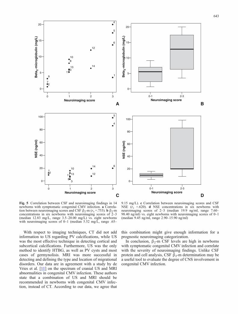
No correlation was observed between the neuroimagingscore and the protein concentration or the cellular count inCSF. In contrast, β2-m levels in CSF showed a significantpositive correlation with the neuroimaging score (P=.002;Fig. 5). In addition, infants with a neuroimaging score of 2–3 exhibited higher β2-m concentrations than those with aneuroimaging score of 0–1 (P=.028; Fig. 5). NSE alsoshowed a positive correlation with the neuroimaging score(P=.032, Fig. 5), and patients with neuroimaging scores of2–3 had higher NSE levels than those with scores of 0–1(P=.028, Fig. 5). Infants with higher CSF β2-m concentra-
tions and higher neuroimaging scores seemed more likelyto die or have an especially poor long-term outcome.
Discussion
β2-m levels in CSF have been found to be abnormally highin adults and children with bacterial or viral CNS infections[16, 18, 33, 39, 41, 43]. In addition, we have previouslyobserved that neonates with CNS infections, including onecongenital CMV, have high levels of CSF β2-m [14].Therefore, β2-m is used in our centre as an ancillary tool inneonates evaluated for CNS infections. In the presentstudy, we found that newborns with symptomatic congen-ital CMV infection show markedly higher CSF β2-mconcentrations than high risk neonates without intracranialinfection. β2-m levels did not correlate with proteinconcentrations in CSF. This finding is consistent withother studies [14, 39, 41, 43] and suggests that high levelsof β2-m in CSF do not seem to be explained by a blood-brain barrier disruption. Instead, these data suggest intra-thecal production of β2-m. High β2-m concentrations inCSF concurred with pleocytosis or proteinorachia in lessthan half of our cases. And, except for IL-6 levels slightlyover the detection limit in patients 12 and 14, CSFcytokines were undetectable in all the cases in which theywere determined. Besides, a correlation was observedbetween β2-m and NSE levels, and infants with radiolog-ical evidence of more destructive brain lesions had higherβ2-m CSF levels. These observations suggest that β2-mrelease might not be solely related to inflammation, but alsoto neuronal damage. Glial proliferation may also accountfor part of the increase in CSF β2-m, as β2-m is present on
Fig. 1 Neuroimaging findingsof a newborn with symptomaticcongenital CMV infection (case5). a,b Cranial ultrasound coro-nal scans performed at 1 day ofage showing periventricularcalcifications and ventriculome-galy. Note also the hyperecho-genicity and cystic regression ofcaudate nuclei. c,d CT scanperformed at 14 days of lifedemonstrating wide periventric-ular calcifications, ventriculo-megaly and white matterhypodensity, as well as corticaldensity abnormalities andpachygiria
640

the surface of glial cells, which are known to secrete β2-min vitro [27].
We observed an association between β2-m levels in CSFand the severity of the neuroimaging score. Moreover,infants with higher CSF β2-m concentrations showed aspecially poor long-term outcome. Although it hastraditionally been considered that symptomatic congenitalCMV infection implies a generally severe prognosis, morerecent studies suggest that the chances of a fair neurolog-ical outcome may be more favourable than formerlybelieved [9, 28]. In a study by Noyola et al. [28], 54% of 41children with symptomatic congenital CMV infection hadan intelligence quotient (IQ) >70, and 29% had an IQ >90.According to these authors, the advantages of accuratelydefining the grade of CNS involvement and predictinglong-term outcome in the newborn period include chancesfor appropriate counseling of parents and early interventionto maximize performance in children at high risk ofdisabilities. Also, categorization of patients at risk fordifferent outcomes may help in the evaluation of interven-
tions, such as antiviral therapy, aimed at improvingoutcome. According to our results, β2-m could be used,together with neuroimaging findings, as a predictor of CNSinvolvement in symptomatic congenital CMV infection.Further research on this marker could give more informa-tion about its ability to predict outcome in both symptom-atic and asymptomatic infections.
The results of the sequential determinations in fourpatients of our series are in agreement with findings ofother studies, which show decreasing CSF β2-m levels inrelation to treatment in bacterial meningitis [14, 41], in acase of perinatal CMV meningitis [3], and in an infant withcongenital neurosyphilis [40]. However, we are unaware ofthe prognostic implications of this fall in CSF β2-m withtreatment, given that ganciclovir appears to be successful inreducing viral load and excretion [26, 50], and may preventhearing deterioration [21], but is unlikely to alterneurodevelopmental outcome. A more long term courseof CSF β2-m levels with and without antiviral treatmentshould be a matter of further research.
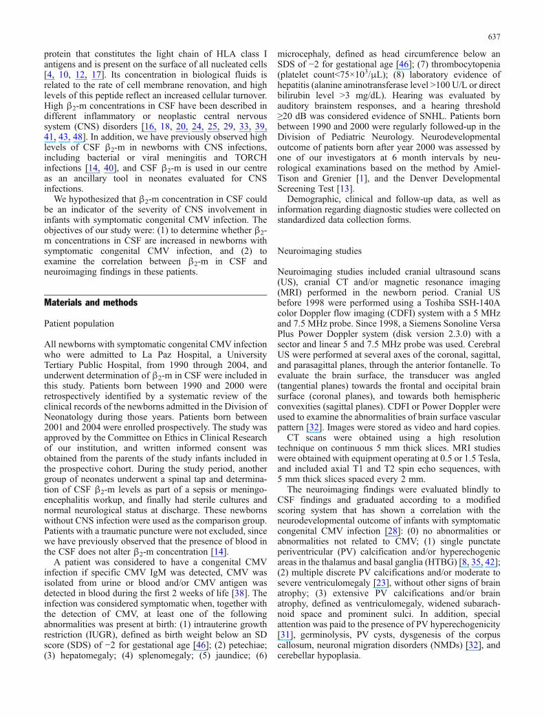
Fig. 2 Neuroimaging findingsof a newborn with symptomaticcongenital CMV infection (case14). a Coronal cranial ultra-sound scan performed at 4 daysof age showing ventriculome-galy, periventricular calcifica-tions and HTBG. b–d Sagittalscans showing dysgenesis(thinness) of the corpus callo-sum, mild ventriculomegaly,linear HTBG and poor sulca-tion. e T2-weighted, and f T1-weighted magnetic resonanceimages obtained at 14 days,demonstrating fronto-parietalareas of agyria-pachygyria andoccipital polymicrogyria, aswell as partial agenesis of thecorpus callosum
641
Recent studies have shown that neonatal CMV DNAe-mia determined by real-time quantitative polymerase chainreaction (PCR) in congenital CMV infection is associatedwith systemic disease and development of sequelae [7, 22].In another work on CMV DNA by PCR in CSF of 13infants with congenital CMV infection, a positive resultwas found in six of ten symptomatic and zero of threeasymptomatic infants, and correlated with a poor neurode-velopmental outcome [45]. Correlation between CSF β2-mand viral load in both blood and CSF should be evaluated,and the usefulness of both tools to predict outcome shouldbe compared.
In our series, NSE (a marker of neuronal damage that hasbeen shown to be a good biochemical predictor of hypoxic-ischemic brain injury in asphyxiated newborns) [15, 44]also showed a significant positive correlation with theneuroimaging score. Patients 4 and 5 had CSF NSE levelswithin the range observed in neonates with severe hypoxic-ischemic brain damage, although they both had Apgarscores over 6 at 5 min and umbilical artery blood pH over7.2. These two patients also exhibited severe neuroimaging
abnormalities, as well as an adverse outcome. Whetherthese high NSE levels reflect ongoing neuronal injury bypersistent infection is not clear. The few studies that havereported CSF NSE in CNS infections, have shownelevations in children with bacterial and viral meningitis[19, 36].
Only five of our patients showed an abnormal result ofstandard citochemical CSF analysis. In other studies, theproportion of infants with an elevated protein concentration(>120–130 mg/dL) is 44–50% [5, 6, 30]. In a study of 52infants, CSF protein exceeded 120 mg/dL in 24 (46%) [6].However, only four had protein levels >150 mg/dL. Proteinelevation was associated with neurological abnormalitiesand hearing loss. By contrast, in two other studies thesesame authors found no association between protein eleva-tion and neuroimaging abnormalities [5], or mental retar-dation or psychomotor delay [30]. The lack of ability ofproteinorachia to predict neuroimaging severity or adverseoutcome in our series is in agreement with these data.
The main limitation of our study resides in the smallnumber of neonates with CMV infection and CSF β2-mdeterminations available for analysis. This does not entail aproblem of validity or bias, but it is likely to affect theprecision of the results. Data regarding long-term neuro-developmental outcome in our patients are also ratherlimited. Two patients were lost to follow-up. In theretrospective cohort, evaluations were not all made bythe same person. In the patients born after year 2000,follow-up assessments were standardized and performedby a single investigator, but the median age at follow-upwas low (15 months). For these reasons, we usedneuroimaging findings in the neonatal period as asubrogate of long-term outcome. Such neuroradiographicfindings have been found to be good predictors ofneurodevelopmental outcome in children with symptom-atic congenital CMV infection [5, 28]. Taking into accountthe limitations of our follow-up data, we also observed thatinfants with higher neuroimaging scores seemed morelikely to have an especially poor outcome. Together withthese high neuroimaging scores, the majority of our mostdisabled children had NMDs and hypoplasia of the corpuscallosum.
ControlCMV
Bet
a -
mic
rogl
obul
in (
mg/
L)
22
20
18
16
14
12
10
8
6
4
2
0
2
Fig. 3 Concentrations of β2-m in CSF: 14 newborns withsymptomatic congenital CMV infection (median 6.21 mg/L, range.65–20.00 mg/L) vs. 93 newborns without intracranial infection(median 1.68 mg/L, range .77–3.20 mg/L)
0
2
4
6
8
10
12
14
0 6 12 18 24 30 36 42 48 54 60
Age (days)
Bet
a -
mic
rogl
obul
in (
mg/
L)
Patient 10
Patient 12
Patient 13
Patien t 14
2
Fig. 4 Sequential determina-tions of CSF β2-m in fournewborns with symptomaticcongenital CMV infection. Thearrows indicate the timeswhen treatment with ganciclovirwas initiated and ended
642
With respect to imaging techniques, CT did not addinformation to US regarding PV calcifications, while USwas the most effective technique in detecting cortical andsubcortical calcifications. Furthermore, US was the onlymethod to identify HTBG, as well as PV cysts and mostcases of germynolisis. MRI was more successful indetecting and defining the type and location of migrationaldisorders. Our data are in agreement with a study by deVries et al. [11] on the spectrum of cranial US and MRIabnormalities in congenital CMV infection. These authorsstate that a combination of US and MRI should berecommended in newborns with congenital CMV infec-tion, instead of CT. According to our data, we agree that
this combination might give enough information for aprognostic neuroimaging categorization.
In conclusion, β2-m CSF levels are high in newbornswith symptomatic congenital CMV infection and correlatewith the severity of neuroimaging findings. Unlike CSFprotein and cell analysis, CSF β2-m determination may bea useful tool to evaluate the degree of CNS involvement incongenital CMV infection.
0 1 2 3
Neuroimaging score
0
5
10
15
20
1
2
3
4
5
6
7
8
9
10
11
12
13 14
A
0-1 2-3Neuroimaging score
0
5
10
15
20
B
0 1 2 3
Neuroimaging score
0
20
40
60
80
100
NS
E (
ng
/ml)
2
3
4
5
6
7
8
10
11 1213
14
C
0-1 2-3Neuroimaging score
0
20
40
60
80
100
NS
E (
ng
/ml)
D
Bet
a -
mic
rog
lob
ulin
(m
g/L
)2
Bet
a -
mic
rog
lob
ulin
(m
g/L
)2
Fig. 5 Correlation between CSF and neuroimaging findings in 14newborns with symptomatic congenital CMV infection. a Correla-tion between neuroimaging scores and CSF β2-m (rs =.753). b β2-mconcentrations in six newborns with neuroimaging scores of 2–3(median 12.83 mg/L, range 3.5–20.00 mg/L) vs. eight newbornswith neuroimaging scores of 0–1 (median 5.52 mg/L, range .65–
9.15 mg/L). c Correlation between neuroimaging scores and CSFNSE (rs =.620). d NSE concentrations in six newborns withneuroimaging scores of 2–3 (median 19.9 ng/ml, range 7.60–98.40 ng/ml) vs. eight newborns with neuroimaging scores of 0–1(median 9.45 ng/ml, range 2.90–15.90 ng/ml)
643

Acknowledgements We are grateful to Dr. C. Roche, from theDivision of Pediatric Neurology, Dr. M. Martinez Biarge, and rest ofthe staff of the Division of Neonatology for their valuablecooperation. We thank all the children and families that participatedin this study.
References
1. Amiel-Tison C, Grenier A (1986) Neurological assessmentduring the first year of life. Oxford University Press, New York
2. Bale JF, Bray PF, Bell WE (1985) Neuroradiographicabnormalities in congenital cytomegalovirus infection. PediatrNeurol 1:42–77
3. Baquero-Artigao F, Mendez A, del Castillo F, Velazquez R(2004) Cerebrospinal fluid beta 2-microglobulin values inperinatally acquired cytomegalovirus infection. Pediatr InfectDis J 23:891–892
4. Bernier GM (1980) Beta 2-microglobulin: structure, functionand significance. Vox Sang 38:323–327
5. Boppana SB, Fowler KB, Vaid Y, Hedlund G, Stagno S, BrittWJ, Pass RF (1997) Neuroradiographic findings in the newbornperiod and long-term outcome in children with symptomaticcongenital cytomegalovirus infection. Pediatrics 99:409–414
6. Boppana SB, Pass RF, Britt WJ, Stagno S, Alford CA (1992)Symptomatic congenital cytomegalovirus infection: neonatalmorbidity and mortality. Pediatr Infect Dis J 11:93–99
7. Bradford RD, Cloud G, Lakeman AD, Boppana S, KimberlinDW, Jacobs R, Demmler G, Sanchez P, Britt W, Soong SJ,Whitley RJ (2005) Detection of cytomegalovirus (CMV) DNAby polymerase chain reaction is associated with hearing loss innewborns with symptomatic congenital CMV infection involv-ing the central nervous system. J Infect Dis 191:227–233
8. Cabañas F, Pellicer A, Morales C, Garcia-Alix A, Stiris TA,Quero J (1994) New pattern of hyperechogenicity in thalamusand basal ganglia studied by color Doppler flow imaging.Pediatr Neurol 10:109–116
9. Conboy TJ, Pass RF, Stagno S, Alford CA, Myers GJ, Britt WJ,McCollister FP, Summers MN, McFarland CE, Boll TJ (1987)Early clinical manifestations and intellectual outcome inchildren with symptomatic congenital cytomegalovirus infec-tion. J Pediatr 111:343–348
10. Cresswell P, Springer T, Strominger JL, Turner MJ, Grey HM,Kubo RT (1974) Immunological identity of the small subunit ofHL-A antigens and beta2-microglobulin and its turnover on thecell membrane. Proc Natl Acad Sci 71(5):2123–2127
11. de Vries LS, Gunardi H, Barth PG, Bok LA, Verboon-MaciolekMA, Groenendaal F (2004) The spectrum of cranial ultrasoundand magnetic resonance imaging abnormalities in congenitalcytomegalovirus infection. Neuropediatrics 35:113–119
12. Evrin PE, Pertoft H (1973) Beta2-microglobulin in humanblood cells. J Immunol 111:1147–1154
13. Frankenburg WK, Dodds JB (1967) The Denver developmentalscreening test. J Pediatr 71:181–191
14. Garcia- Alix A, Martin-Ancel A, Ramos MT, Salas S, PellicerA, Cabañas F, Quero J (1995) Cerebrospinal fluid beta 2-microglobulin in neonates with central nervous systeminfections. Eur J Pediatr 154:309–313
15. Garcia-Alix A, Cabañas F, Pellicer A, Hernanz A, Stiris TA,Quero J (1994) Neuron-specific enolase and myelin basicprotein: relationship of cerebrospinal fluid concentrations to theneurological condition of asphyxiated full-term infants. Pedi-atrics 93:234–240
16. Gekle D, Kult J, Roth R (1977) Lysozym und beta2-mikroglobulin in Liquor gesunder Kinder und bei Kinder mitErkrankungen des Zentralnervensystem. Klin Wochenschr55:189–191
17. Grey HM, Kubo RT, Colon SM, Poulik MD, Cresswell P,Springer T, Turner M, Strominger JL (1973) The small subunitof HL-A antigens is beta 2-microglobulin. J Exp Med138:1608–1612
18. Hallgren R, Terent A, Venge P (1982) Lactoferrin, lysozyme,and beta 2-microglobulin levels in cerebrospinal fluid: differ-ential indices of CNS inflammation. Inflammation 6:291–304
19. Inoue S, Takahashi H, Kaneko K (1994) The fluctuations ofneuron-specific enolase (NSE) levels of cerebrospinal fluidduring bacterial meningitis: the relationship between thefluctuations of NSE levels and neurological complications oroutcome. Acta Paediatr Jpn 36:485–488
20. Kawai M, Hirohata S (2000) Cerebrospinal fluid beta(2)-microglobulin in neuro-Behcet’s syndrome. J Neurol Sci179:132–139
21. Kimberlin DW, Lin CY, Sanchez PJ, Demmler GJ, Dankner W,Shelton M, Jacobs RF, Vaudry W, Pass RF, Kiell JM, Soong SJ,Whitley RJ (2003) Effect of ganciclovir therapy on hearing insymptomatic congenital cytomegalovirus disease involving thecentral nervous system: a randomized, controlled trial. J Pediatr143:16–25
22. Lanari M, Lazzarotto T, Venturi V, Papa I, Gabrielli L, Guerra B,Landini MP, Faldella G (2006) Neonatal cytomegalovirus bloodload and risk of sequelae in symptomatic and asymptomaticcongenitally infected newborns. Pediatrics 117:e76–e83
23. Levene MI (1981) Measurement of the growth of the lateralventricles in preterm infants with real-time ultrasound. ArchDis Child 56:900–904
24. Mavlight GM, Stuckey SE, Cabanillas FF, Keating MJ,Tourtellotte WW, Schold SC, Freireich EJ (1980) Diagnosisof leukemia or lymphoma in the central nervous system by beta2-microglobulin determination. N Eng J Med 303:718–722
25. McArthur JC, Nance-Sproson TE, Griffin DE, Hoover D,Selnes OA, Miller EN, Margolick JB, Cohen BA, FarzadeganH, Saah A (1992) The diagnostic utility of elevation incerebrospinal fluid beta 2-microglobulin in HIV-1 dementia.Multicenter AIDS Cohort Study. Neurology 42:1707–1712
26. Nigro G, Scholz H, Bartmann U (1994) Ganciclovir therapy forsymptomatic congenital cytomegalovirus infection in infants: atwo-regimen experience. J Pediatr 124:318–322
27. Nilsson K, Evrin PE, Welsh KI (1974) Production of beta 2-microglobulin by normal and malignant human cell lines andperipheral lymphocytes. Transplant Rev 21:53–58
28. Noyola DE, Demmler GJ, Nelson CT, Griesser C, WilliamsonWD, Atkins JT, Rozelle J, Turcich M, Llorente AM, Sellers-Vinson S, Reynolds A, Bale JF Jr, Gerson P, Yow MD (2001)Early predictors of neurodevelopmental outcome in symptomaticcongenital cytomegalovirus infection. J Pediatr 138:325–331
29. Oksanen V, Gronhagen-Riska C, Tikanoja S, Somer H,Fyhrquist F (1986) Cerebrospinal fluid lysozyme and beta 2-microglobulin in neurosarcoidosis. J Neurol Sci 73:79–87
30. Pass RF, Stagno S, Myers GJ, Alford CA (1980) Outcome ofsymptomatic congenital cytomegalovirus infection: results oflong-term longitudinal follow-up. Pediatrics 66:758–762
31. Pellicer A, Cabañas F, Garcia-Alix A, Perez Rodriguez J, QueroJ (1993) Natural history of ventricular dilatation in preterminfants: prognostic significance. Pediatr Neurol 9:108–114
32. Pellicer A, Cabañas F, Perez-Higueras A, Garcia-Alix A, QueroJ (1995) Neuronal migration disorders studied by cerebralultrasound and colour Doppler flow imaging. Arch Dis ChildFetal Neonatal Ed 73:F55–F61
33. Peterslund NA, Black FT, Geil JP, Mogensen CE (1989) Beta-2-microglobulin in the cerebrospinal fluid of patients withinfections of the central nervous system. Acta Neurol Scand80:579–583
34. Ramsay, ME, Miller E, Peckham CS (1991) Outcome ofconfirmed cytomegalovirus infection. Arch Dis Child66:1068–1069
35. Ries M, Deeg KH, Heininger U (1990) Demonstration ofperivascular echogenicities in congenital cytomegalovirusinfection by colour Doppler imaging. Eur J Pediatr 150:34–36
36. Rodriguez-Nuñez A, Cid E, Rodriguez-Garcia J, Camina F,Rodriguez-Segade S, Castro-Gago M (2003) Neuron-specificenolase, nucleotides, nucleosides, purine bases, oxypurines anduric acid concentrations in cerebrospinal fluid of children withmeningitis. Brain Dev 25:102–106
644

37. Sarff LD, Platt LH, McCracken GH Jr (1976) Cerebrospinalfluid elevation in neonates: comparison of high risk infants withand without meningitis. J Pediatr 88:473–477
38. Stagno S (2001) Cytomegalovirus. In: Remington JS, Klein JD(eds) Infectious diseases of the fetus and newborn infant, 5thedn. WB Saunders, Philadelphia, pp 389–424
39. Starmans JJ, Vos J, van der Helm HJ (1977) The beta 2-microglobulin content of the cerebrospinal fluid in neurologicaldisease. J Neurol Sci 33:45–49
40. Tagarro A, Garcia-Alix A, Alarcon A, Hernanz A, Quero J(2005) Congenital syphilis: β2-microglobulin in cerebrospinalfluid and diagnosis of neurosyphilis in an affected newborn.J Perinat Med 33:79–82
41. Takahashi S, Oki J, Miyamoto A, Moriyama T, Asano A,Inyaku F, Okuno A (1999) Beta-2-microglobulin and ferritin incerebrospinal fluid for evaluation of patients with meningitis ofdifferent etiologies. Brain Dev 21:192–199
42. Teele RL, Hernanz-Schulman M, Sotrel A (1988) Echogenicvasculature in the basal ganglia of neonates: a sonographic signof vasculopathy. Radiology 169:423–427
43. Tenhunen R, Iivanainen M, Kovanen J (1978) Cerebrospinalfluid beta 2-microglobulin in neurological disorders. ActaNeurol Scand 58:366–373
44. Thornberg E, Thiringer K, Hagberg H, Kjellmer I (1995)Neuron specific enolase in asphyxiated newborns: associationwith encephalopathy and cerebral function monitor trace. ArchDis Child Fetal Neonatal Ed 72:F39–F42
45. Troendle Atkins J, Demmler GJ, Williamson WD, McDonaldJM, Istas AS, Buffone GJ (1994) Polymerase chain reaction todetect cytomegalovirus DNA in the cerebrospinal fluid ofneonates with congenital infection. J Infect Dis 169:1334–1337
46. Usher R, McLean F (1969) Intrauterine growth of live-bornCaucasian infants at sea level: standards obtained frommeasurements in 7 dimensions of infants born between 25and 44 weeks of gestation. J Pediatr 74:901–910
47. Uson J, Balsa A, Pascual-Salcedo D, Cabezas JA, Gonzalez-Tarrio JM, Martin-Mola E, Fontan G (1997) Soluble inter-leukin 6 (IL-6) receptor and IL-6 levels in serum and synovialfluid of patients with different arthropathies. J Rheumatol24:2069–2075
48. Vicente V, Gonzalez M, Lopez Borrasca A (1982) Cerebrospi-nal fluid levels beta 2 microglobulin and ferritin in lympho-proliferative disorders. Acta Paediatr Scand 71:325–326
49. Volpe JJ (2001) Neurology of the newborn, 4th edn. WBSaunders, Philadelphia
50. Whitley RJ, Cloud G, Gruber W, Storch GA, Demmler GJ,Jacobs RF, Dankner W, Spector SA, Starr S, Pass RF, Stagno S,Britt WJ, Alford C Jr, Soong S, Zhou XJ, Sherrill L, FitzGeraldJM, Sommadossi JP (1997) Ganciclovir treatment of symp-tomatic congenital cytomegalovirus infection: results of a phaseII study. J Infect Dis 175:1080–1081
645
Copyright © 2022 FDOKUMEN