LYMPHOPROLIFERATIVE DISORDERS AFTER ORGAN TRANSPLANTATION IN CHILDREN
Upload
independentCategory
view
6download
0
10.1128/JVI.79.23.14668-14679.2005.
2005, 79(23):14668. DOI:J. Virol. Speck and Herbert W. Virgin IVMeagan A. Jacoby, Karen E. Weck, Jay L. Hess, Samuel H. Vera L. Tarakanova, Felipe Suarez, Scott A. Tibbetts, Microglobulin-Deficient Mice
2βDisease and Lymphoma in BALB Associated with Lymphoproliferative Murine Gammaherpesvirus 68 Infection Is
http://jvi.asm.org/content/79/23/14668Updated information and services can be found at:
These include:
REFERENCEShttp://jvi.asm.org/content/79/23/14668#ref-list-1at:
This article cites 71 articles, 41 of which can be accessed free
CONTENT ALERTS more»articles cite this article),
Receive: RSS Feeds, eTOCs, free email alerts (when new
http://journals.asm.org/site/misc/reprints.xhtmlInformation about commercial reprint orders: http://journals.asm.org/site/subscriptions/To subscribe to to another ASM Journal go to:
on Septem
ber 10, 2014 by guesthttp://jvi.asm
.org/D
ownloaded from
on S
eptember 10, 2014 by guest
http://jvi.asm.org/
Dow
nloaded from

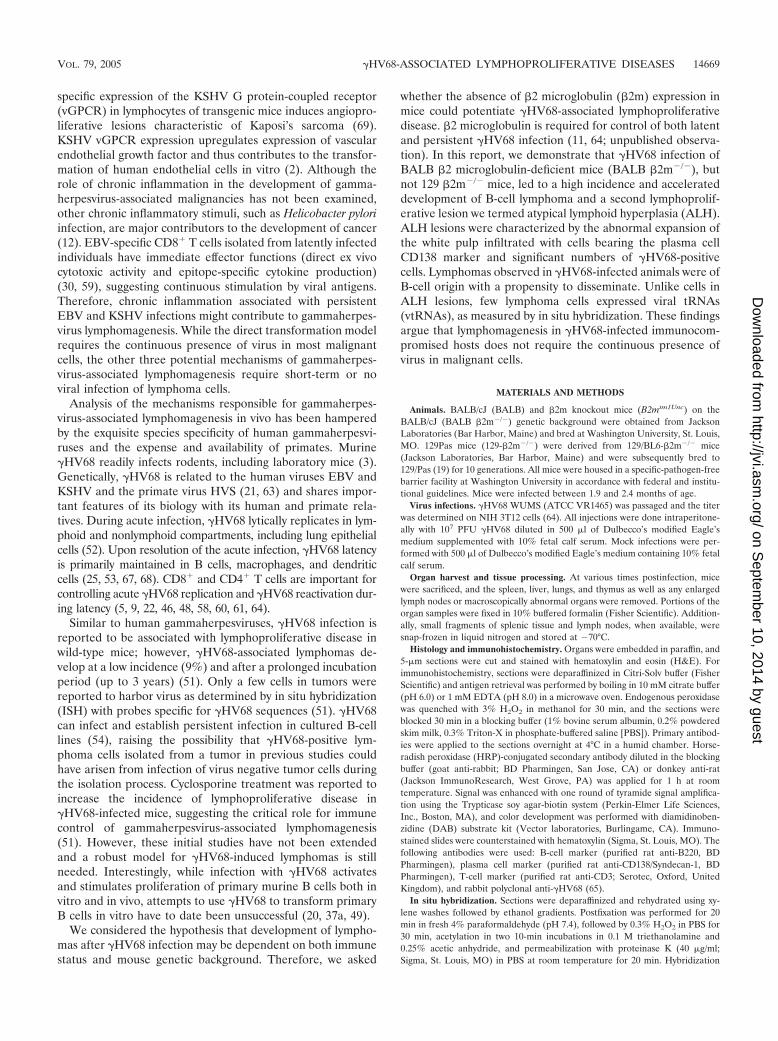
JOURNAL OF VIROLOGY, Dec. 2005, p. 14668–14679 Vol. 79, No. 230022-538X/05/$08.00�0 doi:10.1128/JVI.79.23.14668–14679.2005Copyright © 2005, American Society for Microbiology. All Rights Reserved.
Murine Gammaherpesvirus 68 Infection Is Associated withLymphoproliferative Disease and Lymphoma in BALB
�2 Microglobulin-Deficient MiceVera L. Tarakanova,1† Felipe Suarez,1† Scott A. Tibbetts,1 Meagan A. Jacoby,1Karen E. Weck,1 Jay L. Hess,2 Samuel H. Speck,3 and Herbert W. Virgin IV1*
Department of Pathology and Immunology and Department of Molecular Microbiology, Washington University School of Medicine,St. Louis, Missouri 631101; Department of Pathology and Laboratory Medicine, University of Pennsylvania School of Medicine,
Philadelphia, PA 191042; and Division of Microbiology and Immunology and The Center for Emerging Infectious Disease,Yerkes National Primate Research Center, Emory University School of Medicine, Atlanta, Georgia 303293
Received 14 July 2005/Accepted 12 September 2005
Human gammaherpesvirus infections are associated with development of lymphoproliferative disease. Un-derstanding of the mechanisms of gammaherpesvirus lymphomagenesis during chronic infection in a naturalhost has been limited by the exquisite species specificity of human gammaherpesviruses and the expense ofprimates. Murine gammaherpesvirus �HV68 is genetically and biologically related to human gammaherpes-viruses and herpesvirus saimiri and has been reported to be associated with lymphoproliferative disease inmice (N. P. Sunil-Chandra, J. Arno, J. Fazakerley, and A. A. Nash, Am. J. Pathol. 145:818–826, 1994). Wereport the development of an animal model of �HV68 lymphomagenesis in BALB/c �2 microglobulin-deficientmice (BALB �2m�/�). �HV68 infection induced two lymphoproliferative lesions: B-cell lymphoma and atypicallymphoid hyperplasia (ALH). ALH lesion histology resembled lesions of Epstein-Barr virus-associated post-transplant lymphoproliferative disease and was characterized by the abnormal infiltration of the white pulpwith cells expressing the plasma cell marker CD138. Lymphomas observed in �HV68-infected animals wereB220�/CD3� large-cell lymphomas. �HV68-infected cells were common in ALH lesions as measured by in situhybridization with a probe specific for viral tRNAs (vtRNAs), but they were scarce in �HV68-infected spleenswith normal histology. Unlike ALH lesions, �HV68 vtRNA-positive cells were rare in lymphomas. �HV68infection of BALB �2m�/� mice results in lymphoproliferation and lymphoma, providing a valuable tool foridentifying viral and host genes involved in gammaherpesvirus-associated malignancies. Our findings suggestthat �HV68 induces lymphomas via hit-and-run oncogenesis, paracrine effects, or stimulation of chronicinflammation.
Gammaherpesviruses establish lifelong latency in lymphoidand myeloid cells of the host. Importantly, many gammaher-pesviruses, including human Epstein-Barr virus (EBV), Kaposi’ssarcoma-associated herpesvirus (KSHV), and simian herpesvi-rus saimiri (HVS) are associated with a variety of lymphopro-liferative diseases (LPDs) (13, 31, 71). In addition to Kaposi’ssarcoma, a tumor of endothelial cell origin, KSHV has alsobeen associated with at least two types of lymphoproliferativeB-cell diseases, multicentric Castleman’s disease and primaryeffusion lymphoma (PEL) (6). EBV induces hyperproliferationof infected B cells that can evolve into lymphoma in an immu-nocompromised host (7, 14). Other lymphomas with a stronglink to EBV infection include Burkitt’s lymphoma, Hodgkin’slymphoma, and AIDS-associated lymphomas (40, 71). HVS isassociated with T-cell lymphomas in New World primates andin certain rabbits (31). Immunocompromised hosts have anincreased incidence of gammaherpesvirus-associated lympho-proliferative disease, suggesting a critical role of the immune
system in controlling lymphoproliferation and lymphomagen-esis (71).
The mechanism of gammaherpesvirus-induced lymphomagen-esis in animal models is not clear. The four major models ofvirus-associated oncogenesis include (i) direct transformation,(ii) “hit and run,” (iii) paracrine, and (iv) chronic inflamma-tion. Direct transformation is the only model that requires viralpresence in a majority of malignant cells (38). Such is the casein early onset posttransplant lymphomas that are uniformlyassociated with EBV (71). EBV genes expressed in infected Bcells of these lymphomas represent the latency III program ofEBV gene expression that provides the necessary signals foruncontrolled B-cell proliferation. In other cases, such as EBV-associated Hodgkin’s disease, virus is present in a minority ofcells found in the tumor (71). HVS-induced T-cell lymphomasuniformly retain viral genome (10). According to the “hit-and-run” model, the presence of the virus is only required to ini-tiate events leading to the transformation of infected cells (1).Once a fully transformed state is achieved, virus is lost (44). Insupport of the “hit-and-run” mechanism, cell lines derivedfrom the primary Kaposi’s sarcoma lesions lose viral episomeupon passage in vitro (24, 47). In vivo, EBV-associatedHodgkin’s lymphoma may no longer harbor virus upon relapse(15). The existence of the paracrine mechanism of lym-phomagenesis is supported by studies showing that cell-type-
* Corresponding author. Mailing address: Department of Pathologyand Immunology, Washington University, 660 S. Euclid Ave, Box 8118,St. Louis, MO, 63110. Phone: (314) 362-9223. Fax: (314) 362-0369.E-mail: [email protected].
† V.L.T. and F.S. contributed equally to this study.
14668
on Septem
ber 10, 2014 by guesthttp://jvi.asm
.org/D
ownloaded from
specific expression of the KSHV G protein-coupled receptor(vGPCR) in lymphocytes of transgenic mice induces angiopro-liferative lesions characteristic of Kaposi’s sarcoma (69).KSHV vGPCR expression upregulates expression of vascularendothelial growth factor and thus contributes to the transfor-mation of human endothelial cells in vitro (2). Although therole of chronic inflammation in the development of gamma-herpesvirus-associated malignancies has not been examined,other chronic inflammatory stimuli, such as Helicobacter pyloriinfection, are major contributors to the development of cancer(12). EBV-specific CD8� T cells isolated from latently infectedindividuals have immediate effector functions (direct ex vivocytotoxic activity and epitope-specific cytokine production)(30, 59), suggesting continuous stimulation by viral antigens.Therefore, chronic inflammation associated with persistentEBV and KSHV infections might contribute to gammaherpes-virus lymphomagenesis. While the direct transformation modelrequires the continuous presence of virus in most malignantcells, the other three potential mechanisms of gammaherpes-virus-associated lymphomagenesis require short-term or noviral infection of lymphoma cells.
Analysis of the mechanisms responsible for gammaherpes-virus-associated lymphomagenesis in vivo has been hamperedby the exquisite species specificity of human gammaherpesvi-ruses and the expense and availability of primates. Murine�HV68 readily infects rodents, including laboratory mice (3).Genetically, �HV68 is related to the human viruses EBV andKSHV and the primate virus HVS (21, 63) and shares impor-tant features of its biology with its human and primate rela-tives. During acute infection, �HV68 lytically replicates in lym-phoid and nonlymphoid compartments, including lung epithelialcells (52). Upon resolution of the acute infection, �HV68 latencyis primarily maintained in B cells, macrophages, and dendriticcells (25, 53, 67, 68). CD8� and CD4� T cells are important forcontrolling acute �HV68 replication and �HV68 reactivation dur-ing latency (5, 9, 22, 46, 48, 58, 60, 61, 64).
Similar to human gammaherpesviruses, �HV68 infection isreported to be associated with lymphoproliferative disease inwild-type mice; however, �HV68-associated lymphomas de-velop at a low incidence (9%) and after a prolonged incubationperiod (up to 3 years) (51). Only a few cells in tumors werereported to harbor virus as determined by in situ hybridization(ISH) with probes specific for �HV68 sequences (51). �HV68can infect and establish persistent infection in cultured B-celllines (54), raising the possibility that �HV68-positive lym-phoma cells isolated from a tumor in previous studies couldhave arisen from infection of virus negative tumor cells duringthe isolation process. Cyclosporine treatment was reported toincrease the incidence of lymphoproliferative disease in�HV68-infected mice, suggesting the critical role for immunecontrol of gammaherpesvirus-associated lymphomagenesis(51). However, these initial studies have not been extendedand a robust model for �HV68-induced lymphomas is stillneeded. Interestingly, while infection with �HV68 activatesand stimulates proliferation of primary murine B cells both invitro and in vivo, attempts to use �HV68 to transform primaryB cells in vitro have to date been unsuccessful (20, 37a, 49).
We considered the hypothesis that development of lympho-mas after �HV68 infection may be dependent on both immunestatus and mouse genetic background. Therefore, we asked
whether the absence of �2 microglobulin (�2m) expression inmice could potentiate �HV68-associated lymphoproliferativedisease. �2 microglobulin is required for control of both latentand persistent �HV68 infection (11, 64; unpublished observa-tion). In this report, we demonstrate that �HV68 infection ofBALB �2 microglobulin-deficient mice (BALB �2m�/�), butnot 129 �2m�/� mice, led to a high incidence and accelerateddevelopment of B-cell lymphoma and a second lymphoprolif-erative lesion we termed atypical lymphoid hyperplasia (ALH).ALH lesions were characterized by the abnormal expansion ofthe white pulp infiltrated with cells bearing the plasma cellCD138 marker and significant numbers of �HV68-positivecells. Lymphomas observed in �HV68-infected animals were ofB-cell origin with a propensity to disseminate. Unlike cells inALH lesions, few lymphoma cells expressed viral tRNAs(vtRNAs), as measured by in situ hybridization. These findingsargue that lymphomagenesis in �HV68-infected immunocom-promised hosts does not require the continuous presence ofvirus in malignant cells.
MATERIALS AND METHODS
Animals. BALB/cJ (BALB) and �2m knockout mice (B2mtm1Unc) on theBALB/cJ (BALB �2m�/�) genetic background were obtained from JacksonLaboratories (Bar Harbor, Maine) and bred at Washington University, St. Louis,MO. 129Pas mice (129-�2m�/�) were derived from 129/BL6-�2m�/� mice(Jackson Laboratories, Bar Harbor, Maine) and were subsequently bred to129/Pas (19) for 10 generations. All mice were housed in a specific-pathogen-freebarrier facility at Washington University in accordance with federal and institu-tional guidelines. Mice were infected between 1.9 and 2.4 months of age.
Virus infections. �HV68 WUMS (ATCC VR1465) was passaged and the titerwas determined on NIH 3T12 cells (64). All injections were done intraperitone-ally with 107 PFU �HV68 diluted in 500 �l of Dulbecco’s modified Eagle’smedium supplemented with 10% fetal calf serum. Mock infections were per-formed with 500 �l of Dulbecco’s modified Eagle’s medium containing 10% fetalcalf serum.
Organ harvest and tissue processing. At various times postinfection, micewere sacrificed, and the spleen, liver, lungs, and thymus as well as any enlargedlymph nodes or macroscopically abnormal organs were removed. Portions of theorgan samples were fixed in 10% buffered formalin (Fisher Scientific). Addition-ally, small fragments of splenic tissue and lymph nodes, when available, weresnap-frozen in liquid nitrogen and stored at �70°C.
Histology and immunohistochemistry. Organs were embedded in paraffin, and5-�m sections were cut and stained with hematoxylin and eosin (H&E). Forimmunohistochemistry, sections were deparaffinized in Citri-Solv buffer (FisherScientific) and antigen retrieval was performed by boiling in 10 mM citrate buffer(pH 6.0) or 1 mM EDTA (pH 8.0) in a microwave oven. Endogenous peroxidasewas quenched with 3% H2O2 in methanol for 30 min, and the sections wereblocked 30 min in a blocking buffer (1% bovine serum albumin, 0.2% powderedskim milk, 0.3% Triton-X in phosphate-buffered saline [PBS]). Primary antibod-ies were applied to the sections overnight at 4°C in a humid chamber. Horse-radish peroxidase (HRP)-conjugated secondary antibody diluted in the blockingbuffer (goat anti-rabbit; BD Pharmingen, San Jose, CA) or donkey anti-rat(Jackson ImmunoResearch, West Grove, PA) was applied for 1 h at roomtemperature. Signal was enhanced with one round of tyramide signal amplifica-tion using the Trypticase soy agar-biotin system (Perkin-Elmer Life Sciences,Inc., Boston, MA), and color development was performed with diamidinoben-zidine (DAB) substrate kit (Vector laboratories, Burlingame, CA). Immuno-stained slides were counterstained with hematoxylin (Sigma, St. Louis, MO). Thefollowing antibodies were used: B-cell marker (purified rat anti-B220, BDPharmingen), plasma cell marker (purified rat anti-CD138/Syndecan-1, BDPharmingen), T-cell marker (purified rat anti-CD3; Serotec, Oxford, UnitedKingdom), and rabbit polyclonal anti-�HV68 (65).
In situ hybridization. Sections were deparaffinized and rehydrated using xy-lene washes followed by ethanol gradients. Postfixation was performed for 20min in fresh 4% paraformaldehyde (pH 7.4), followed by 0.3% H2O2 in PBS for30 min, acetylation in two 10-min incubations in 0.1 M triethanolamine and0.25% acetic anhydride, and permeabilization with proteinase K (40 �g/ml;Sigma, St. Louis, MO) in PBS at room temperature for 20 min. Hybridization
VOL. 79, 2005 �HV68-ASSOCIATED LYMPHOPROLIFERATIVE DISEASES 14669
on Septem
ber 10, 2014 by guesthttp://jvi.asm
.org/D
ownloaded from

was conducted for 40 h at 37°C using a hybridization mix containing 4� SSC (1�SSC is 0.15 M NaCl plus 0.015M sodium citrate); 20% dextran sulfate; 50%formamide; 0.25 mg/ml each of poly(A), salmon sperm DNA, and tRNA; 5�Denhardt’s solution (all from Sigma); and 100 mM dithiothreitol (Fisher).GreenStar digoxigenin-labeled oligonucleotide probes (Genedetect, Auckland,New Zealand) specific for vtRNAs were used at a final concentration of 200ng/ml in hybridization mix. A cocktail of probes antisense to vtRNAs 1 to 4 wasutilized (vtRNA1, nucleotides [nt] 12 to 41; vtRNA2, nt 13 to 42; vtRNA3, nt 3to 32; and vtRNA4, nt 33 to 62). Stringent posthybridization washes were per-formed with 1� and 0.5� SSC at 55°C, and the hybridized probes were detectedusing a goat HRP-conjugated anti-digoxigenin Fab fragment (Dako) followed bya round of tyramide signal amplification and DAB substrate color development.
Molecular analysis of clonality. DNA was extracted from frozen spleen frag-ments using standard procedures. Analysis of the rearranged status of the im-munoglobulin heavy chain (IgH) locus was performed by PCR amplificationusing the family-specific degenerate forward primers Vh558, Vh7183, VhQ52,and DhL with the reverse primer Jh3 (35, 43). Amplified products were separatedby agarose gel electrophoresis. When a predominant fragment was observed, itwas cut, purifed, and reamplified in a second round using the appropriateprimers. The resulting product was cloned in pGEM-T and sequenced. Sequenceanalysis was performed by aligning the sequenced product using V-QUEST searchon the Immunogenetics database (http://imgt.cines.fr:8104/textes/vquest/).
Statistical analysis. The data were analyzed using GraphPad Instat software(San Diego, CA). All results were compared using unpaired Student’s t test orchi-square tests.
RESULTS
�HV68 infection is associated with an increased incidence oflymphoproliferative diseases in BALB-�2m-deficient mice.Gammaherpesvirus-associated lymphoproliferative disease isincreased in immunocompromised hosts. Given the impor-tance of CD8 T cells and �2 microglobulin in the control of�HV68 replication and latency (11, 22, 58, 61, 64), we assessedthe development of lymphoproliferative disease in mock-in-fected and �HV68-infected sex- and age-matched BALB/cmice lacking �2 microglobulin expression. Several defects areattributed to the �2 microglobulin deficiency, including lack ofclassical CD8 T cells, hepatic iron overload, and abnormal IgGhomeostasis due to the inefficient expression of neonatal Fcreceptor (28, 42, 70, 73). Mice were infected between 1.9 and2.4 months of age and sacrificed at the indicated times (Table 1).We then assessed lymphoid histology in spleens, lymph nodes,livers, lungs, and kidneys.
Two lymphoproliferative lesions were observed dispropor-tionately in infected compared to uninfected BALB �2m�/�
mice: atypical lymphoid hyperplasia with plasmacytosis (ALH),and large-cell lymphomas. The pathology of these lesions isdescribed below. Follicular hyperplasia, as defined by the pres-ence of a prominent germinal center reaction with otherwisenormal histology, was occasionally observed and was notscored as lymphoproliferative disease. Overall 37/55 (67%) ofthe infected BALB �2m�/� mice developed LPD (ALH or
lymphoma) compared to 11/50 (22%) of the mock-infectedcontrols (P � 0.0001) (Fig. 1A). There were significantly morelymphomas (29% versus 6%, P � 0.0022) and atypical lym-phoid hyperplasias (38% versus 16%, P � 0.0157) in the�HV68-infected group as compared to the mock-infected an-imals (Fig. 1A).
The development of �HV68-associated lymphoproliferativelesions was not detected in BALB (0% LPD, n � 23) or129/Pas �2m�/� animals (0% LPD, n � 20) (Table 1), dem-onstrating that both the �2m deficiency and the genetic back-ground of the mice are key factors in determining susceptibilityto �HV68-associated LPD.
Kinetics of lymphoproliferative disease development in�HV68-infected and control BALB-�2m�/� mice. We analyzedthe kinetics of lymphoproliferative lesions in �HV68-infectedor mock-infected BALB �2m�/� mice (Fig. 1B and C). Over-all, atypical lymphoid hyperplasia and lymphomas were de-tected at a higher incidence and at an earlier time in the�HV68-infected group as compared to the mock-infectedgroup. The mean age of mice with a diagnosis of lymphomawas 11.6 months in the �HV68-infected group compared to15.8 months in the mock-infected controls (P � 0.011). Atyp-ical lymphoid hyperplasia was observed earlier and at an in-creased incidence in the �HV68-infected group. At 6 to 7months postinfection, over 50% of the animals had atypicallymphoid hyperplasia lesions in the spleen, whereas none ofthe mock-infected animals had detectable lymphoproliferativedisease (compare Fig. 1B and C). In both mock- and �HV68-infected groups, atypical lymphoid hyperplasia was detected atan earlier time postinfection than lymphomas. Thus �HV68infection was associated with both an increased penetranceand an earlier onset of lymphoproliferative lesions.
Role of gender in development of lymphoproliferative dis-ease. An increased incidence of spontaneous lymphomas hasbeen previously observed in BALB female but not male ani-mals (23). Interestingly, we also observed a gender-dependentdifference in the incidence of lymphoproliferative disease inmock-infected BALB �2m�/� animals (Fig. 1A). Mock-in-fected male BALB �2m�/� animals had a low level of atypicallymphoid hyperplasia and no spontaneous lymphomas (Fig.1A, middle panel). The incidence of lymphoproliferativelesions was significantly increased in �HV68-infected males. Incontrast, while there was a trend towards an increase in lym-phoproliferative disease in �HV68-infected versus mock-in-fected females, the difference did not reach statistical signifi-cance. Mock-infected females had a higher incidence andseverity of lymphoproliferative lesions (6 of 27 females withatypical lymphoid hyperplasia, 3 of 27 with lymphomas). Themean time of development of LPD in �HV68-infected femaleswith lymphomas was shorter (13.2 months) than that observedin mock-infected females (15.8 months), suggesting that�HV68 accelerates the development of lymphomas in femaleBALB �2m�/� animals. A larger study will be required todetermine whether �HV68 induces lymphoproliferative dis-ease in female BALB �2m�/� mice.
Histopathology of atypical lymphoid hyperplasia. Four con-ditions were represented in spleens of the �HV68-infectedmice in this study. Normal spleen sections displayed well-de-lineated areas of white pulp containing tightly packed lympho-cytes with dark-staining nuclei (Fig. 2A and B). Follicular hy-
TABLE 1. Demographics of animals used in this study
Infection StrainNo. of animals Mean age (mo) at:
Total With LPD Infection Termination
Mock BALB 17 0 1.9 13.4BALB �2m 50 11 2.0 11.1129 �2m 7 0 1.9 17.7
�HV68 BALB 23 0 2.2 11.5BALB �2m 55 37 2.0 10.6129 �2m 20 0 2.4 19.6
14670 TARAKANOVA ET AL. J. VIROL.
on Septem
ber 10, 2014 by guesthttp://jvi.asm
.org/D
ownloaded from

perplasia was scored based on the presence of reactive B-cellfollicles (Fig. 2C and D, arrows) with otherwise normal splenichistology. Two examples of the atypical lymphoid hyperplasialesions described below are shown in Fig. 2E and F. Of note,atypical lymphoid hyperplasia lesions displayed a spectrum ofseverity ranging from only a mild effacement of a few whitepulp areas on a section (Fig. 2E, outlined) to a dramatic ex-pansion of most white pulp areas (Fig. 2F). Spleens with lym-phomas displayed a loss of normal splenic architecture andexpansion of large cells with moderate amounts of cytoplasmand vesicular nuclei (Fig. 2G and H).
Atypical lymphoid hyperplasia lesions presented with a par-tial disruption of the normal splenic architecture with an ir-regular expansion of the white pulp. In contrast to the lym-phomas, the distinction between red and white pulp remainedapparent. While disorganized in more severe lesions, the ex-pansion involved primarily the periarteriolar lymphoid sheathand sometimes the marginal zone (Fig. 3A and B). Atypicallymphoid hyperplasia lesions were also present in nonlym-phoid organs, mainly the lungs, where they appeared as nod-ular infiltrates in parenchyma (Fig. 3C). The expansion re-flected the presence of large lymphoid cells with moderateamounts of amphophilic cytoplasm and vesicular nuclei withclumped chromatin, features characteristic of plasmacytic dif-ferentiation (Fig. 3A to C, insets). Supporting the morpholog-ical findings, immunohistochemistry demonstrated an in-creased number of cells bearing the plasma cell CD138 marker
in the areas of ALH lesions (Fig. 3D). B220 staining wasconfined to the otherwise normal B-cell areas of the white pulp(Fig. 3E).
The presence of �HV68 in lymphoproliferative lesions. Todetermine whether �HV68 was present in lymphoproliferativelesions, we performed ISH for expression of viral tRNAs onspleen sections harvested from �HV68-infected BALB�2m�/� animals. Sections were hybridized to a digoxigenin-labeled probe antisense to the viral tRNA transcripts. �HV68tRNA genes are expressed during viral latency and are a robusttarget for detection by ISH (4, 45, 50). Spleen sections from�HV68-infected BALB mice (16 days postinfection) and naı̈veanimals were utilized as positive and negative controls. Nostaining was observed with vtRNA-specific probes on mock-infected spleen sections or with the control sense probe on�HV68-infected spleen sections (data not shown).
In order to test the sensitivity of detection of �HV68-in-fected cells by in situ hybridization, we calculated the fre-quency of vtRNA-positive cells on spleen sections obtainedfrom �HV68-infected BL6 mice at 16 days postinfection. Thenature of �HV68 latency in these mice is well established in theliterature. At 16 days postinfection, approximately 1 in 1,000splenocytes are �HV68 genome positive as determined by lim-ited dilution nested PCR (56). Out of over 12,500 cells countedin random fields of spleen sections stained with vtRNA-specificprobe, on average, 1 in 407 nucleated cells was vtRNA positive(range 1 in 186 cells to 1 in 1,256 cells). Therefore, the sensi-
FIG. 1. �HV68 infection increases incidence of lymphoproliferative disease in BALB �2 microglobulin-deficient mice. Age- and sex-matchedBALB �2m animals were mock infected or infected with �HV68 and monitored for the development of LPD. Total incidence of LPD (A) wassubdivided into incidence of each of the two primary lesions (ALH and lymphoma) in male and female animals. (B and C) Incidence of LPD atindicated times postinfection in �HV68 (B)- and mock (C)-infected groups. n, number of mice.
VOL. 79, 2005 �HV68-ASSOCIATED LYMPHOPROLIFERATIVE DISEASES 14671
on Septem
ber 10, 2014 by guesthttp://jvi.asm
.org/D
ownloaded from

FIG. 2. Splenic histology of �HV68-infected BALB �2m�/� mice. H&E staining of formalin-fixed paraffin-embedded tissues. (A and B)Normal (�20). (C and D) Follicular hyperplasia (�20). Arrows point to germinal centers abundant throughout the section. (E and F) Atypicallymphoid hyperplasia (�10). (E) A hyperplastic lesion is outlined. (G and H) Lymphoma (�4).
14672 TARAKANOVA ET AL. J. VIROL.
on Septem
ber 10, 2014 by guesthttp://jvi.asm
.org/D
ownloaded from

FIG. 3. Histopathology of atypical lymphoid hyperplasia. (A and B) ALH lesions in spleens (�4). The splenic architecture is partially disruptedwith expanded white pulp areas. (C) Lung lymphocytic infiltrates seen in �HV68-infected animals with ALH lesions (�4). (Insets in panels A toC) Prominent plasmacytic features of cells in ALH lesions (�100). (D) The majority of the ALH-associated cells stain with the plasma cell markerCD138 (�10). (E) B220 is detected in residual B-cell follicles (�10). Adjacent spleen sections obtained from �HV68-infected BALB �2m�/� micewere stained with hematoxylin and eosin (F and H) or hybridized with antisense digoxigenin-labeled vtRNA probe (G, I, and J) and counterstainedwith hematoxylin. Sections originated from spleens exhibiting follicular hyperplasia (F and G) or atypical lymphoid hyperplasia (H, I, and J).Magnification: F to I, �4; J, �40.
VOL. 79, 2005 �HV68-ASSOCIATED LYMPHOPROLIFERATIVE DISEASES 14673
on Septem
ber 10, 2014 by guesthttp://jvi.asm
.org/D
ownloaded from

tivity of detection of the �HV68-infected cell by in situ hybrid-ization using a vtRNA probe is comparable to the sensitivity ofestablished methods, such as limited dilution PCR. These datashow that in situ hybridization is not likely to significantly under-estimate the number of vtRNA-expressing cells in tumors.
When spleens from BALB �2m�/� mice were examined byin situ hybridization, no or a few small vtRNA-positive fociwere consistently detected in spleens from �HV68-infectedanimals in the absence of pathological lesions (defined as nor-mal or follicular hyperplasia). Consistent with a previous re-port (50), the positive cells, when present, colocalized withB-cell follicles on the adjacent section stained with H&E (Fig.3F and G, arrow).
Interestingly, spleen sections derived from �HV68-infectedanimals diagnosed with atypical lymphoid hyperplasia showeda greater abundance of vtRNA-positive foci. Based on theH&E-stained adjacent spleen section, the positive cells werelocalized to the B-cell follicles with some positive cells presentin the abnormally expanded T-cell zone (Fig. 3H and I, arrow).The intensity of vtRNA staining of cells in a positive focus wasvariable, ranging from intense to low (Fig. 3J). Based on thenuclear morphology, the positive staining was detected in lym-phocytes as well as cells with monocytoid-like nuclei.
Histopathology of splenic lymphomas found in �HV68-in-fected BALB �2m�/� mice. Lymphomas were characterized bynodular and/or diffuse proliferation of pleiomorphic largecells, showing a mixture of immunoblastic and centroblasticfeatures (Fig. 4A and B). The involved spleens had a largelydisrupted architecture with a massively expanded white pulpand a general loss of the distinction between B- and T-cellareas. The malignant proliferation involved mainly the whitepulp. The malignant cells had large nuclei with open chromatinand single prominent nucleoli (immunoblasts) or several smallbut conspicuous nucleoli (centroblasts). Aside from the largecells, some of the lymphomas showed various degrees of plas-macytosis. These lesions were histologically aggressive asjudged by the high mitotic index and the number of apoptoticbodies present.
Although lymphomas were primarily detected in the spleens,metastatic dissemination was also observed. Two of the ninelymphomas found in �HV68-infected males displayed metas-tases to the liver (periportal distribution, Fig. 4C); six out ofnine lymphomas metastasized to the lung (peribronchiovascu-lar distribution, Fig. 4D).
By immunohistochemistry, lymphoma cells expressed theB220 isoform of CD45, although the staining was often weakerthan that on the remaining normal B cells (Fig. 4E). Stainingwith a monoclonal antibody to CD138 specific for plasma cellsindicated that some of the atypical cells had detectable levelsof CD138 expression (Fig. 4F). CD3 immunostaining showedonly interspersed T cells but was consistently negative on thetumor cells (Fig. 4G).
In contrast to atypical lymphoid hyperplasia lesions, spleensderived from lymphoma-bearing �HV68-infected animals dis-played a very limited vtRNA staining. Overt vtRNA-positivefoci were not detected in the tumors analyzed. Instead, a smallnumber of individual cells in the tumors stained vtRNA posi-tive (Fig. 4H). The �HV68-harboring cells were scatteredthroughout the lesion, with most of the positive cells showinglow levels of vtRNA expression. Occasionally, small vtRNA-
positive lymphocytes were found outside the lesions in thetumor-bearing spleens (data not shown). It was not possible todetermine with certainty whether these virus-positive tumorcells were malignant.
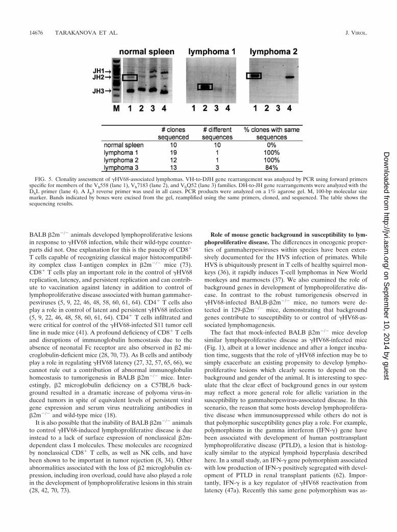
Clonality of splenic lymphomas. Because of the apparentB-cell nature of the �HV68-associated lymphomas in infectedanimals, clonality of lymphomas was assessed by PCR analysisof the IgH locus (43). Although this clonality assay is quiterobust in detection of specific rearrangements, it is not com-prehensive in detection of all possible heavy chain rearrange-ments. In this assay, rearranged, but not germ line, heavy chainsequences are amplified using forward primers homologous toconserved framework region 3 of three murine Vh gene fam-ilies (Fig. 5, lanes 1, 2, and 3 throughout) or primers homolo-gous to a majority of known murine D minigenes (lanes 4). Areverse primer specific for the Jh3 sequence is used in all cases.Based on the utilization of the specific J region during heavychain rearrangement (Jh1, Jh2, or Jh3), up to three major PCRproducts are obtained in each reaction from a heterogeneouspopulation of splenic B cells (Fig. 5, normal spleen). A varietyof sequences are expected to be present in any of the majorPCR products obtained from a nonclonal population of B cells(Fig. 5, normal spleen and summary in the table below). Theprimers selected for the PCR analysis of the IgH locus arecapable of amplifying only a select number of Vh families andare unable to detect heavy chain rearrangements utilizing Jh4.
Importantly, out of seven lymphomas analyzed, three tumorswere found to be clonal based on the limited number of se-quences present in the amplified major PCR product (Fig. 5).Specifically, two lymphomas were monoclonal; three se-quences were detected upon analysis of the third lymphomawith majority of the sequences (11/13) belonging to one clone.Detection of more than one sequence could be due to ampli-fication of the heavy chain of normal B cells present in thelymphoma-bearing spleen or due to the presence of severalclonal transformed populations. The clonality of the remainingfour lymphomas was indeterminate.
DISCUSSION
Gammaherpesvirus infection is associated with developmentof malignancies in immunocompromised hosts. To facilitateanalysis of mechanisms and pathogenesis of this process, wehave sought a murine model for gammaherpesvirus inductionof lymphoproliferative disease. In this report, we demonstratethat �HV68 infection induces two lymphoproliferative dis-eases, each with histologic similarities to human diseases asso-ciated with gammaherpesvirus infection. We found that lack ofthe immunologically important molecule �2 microglobulin, ge-netic background, and gender all contributed to the risk oflymphoproliferative disease. The role of genetic background isof particular interest since, while immunocompromise is aclear risk factor for gammaherpesvirus-associated tumors, thereason that these tumors arise in only a small proportion ofinfected hosts is obscure. Interestingly, �HV68 infection wasundetectable in most tumor cells, suggesting that direct trans-formation is not responsible for induction of malignancies inthis model.
�2 microglobulin deficiency of �HV68-infected BALB micecontributes to development of lymphoproliferative disease.
14674 TARAKANOVA ET AL. J. VIROL.
on Septem
ber 10, 2014 by guesthttp://jvi.asm
.org/D
ownloaded from

FIG. 4. Histopathology of splenic lymphomas. (A and B) Lymphoma-bearing spleens (�4). The general architecture of the spleen is disruptedby the massive expansion of the white pulp. (Insets in panels A and B) Large lymphoma cells with irregular nuclei with conspicuous nucleoli (�40).Frequent mitotic figures are present. (C) Metastasis to the liver (�10). Lymphoma cells are preferentially localized in the portal areas of the liversurrounding the bile ducts. (Inset) Tumor cells adjacent to a bile duct (�40). (D) Metastasis to the lung (�10). (E to G) HRP immunostainingwith hematoxylin counterstain. Magnification at �10 (insets at �40). (E) Expression of B-cell marker B220. (F) Expression of the plasma cellmarker CD138. (G) Expression of T-cell marker CD3. Peritumoral and tumor-infiltrating small lymphocytes but not lymphoma cells stain positive.(H) Lymphoma-bearing spleen section was hybridized with antisense digoxigenin-labeled vtRNA probe and counterstained with hematoxylin(�40).
14675
on Septem
ber 10, 2014 by guesthttp://jvi.asm
.org/D
ownloaded from
BALB �2m�/� animals developed lymphoproliferative lesionsin response to �HV68 infection, while their wild-type counter-parts did not. One explanation for this is the paucity of CD8�
T cells capable of recognizing classical major histocompatibil-ity complex class I-antigen complex in �2m�/� mice (73).CD8� T cells play an important role in the control of �HV68replication, latency, and persistent replication and can contrib-ute to vaccination against latency in addition to control oflymphoproliferative disease associated with human gammaher-pesviruses (5, 9, 22, 46, 48, 58, 60, 61, 64). CD4� T cells alsoplay a role in control of latent and persistent �HV68 infection(5, 9, 22, 46, 48, 58, 60, 61, 64). CD4� T cells infiltrated andwere critical for control of the �HV68-infected S11 tumor cellline in nude mice (41). A profound deficiency of CD8� T cellsand disruptions of immunoglobulin homeostasis due to theabsence of neonatal Fc receptor are also observed in �2 mi-croglobulin-deficient mice (28, 70, 73). As B cells and antibodyplay a role in regulating �HV68 latency (27, 32, 57, 65, 66), wecannot rule out a contribution of abnormal immunoglobulinhomeostasis to tumorigenesis in BALB �2m�/� mice. Inter-estingly, �2 microglobulin deficiency on a C57BL/6 back-ground resulted in a dramatic increase of polyoma virus-in-duced tumors in spite of equivalent levels of persistent viralgene expression and serum virus neutralizing antibodies in�2m�/� and wild-type mice (18).
It is also possible that the inability of BALB �2m�/� animalsto control �HV68-induced lymphoproliferative disease is dueinstead to a lack of surface expression of nonclassical �2m-dependent class I molecules. These molecules are recognizedby nonclassical CD8� T cells, as well as NK cells, and havebeen shown to be important in tumor rejection (8, 34). Otherabnormalities associated with the loss of �2 microglobulin ex-pression, including iron overload, could have also played a rolein the development of lymphoproliferative lesions in this strain(28, 42, 70, 73).
Role of mouse genetic background in susceptibility to lym-phoproliferative disease. The differences in oncogenic proper-ties of gammaherpesviruses within species have been exten-sively documented for the HVS infection of primates. WhileHVS is ubiquitously present in T cells of healthy squirrel mon-keys (36), it rapidly induces T-cell lymphomas in New Worldmonkeys and marmosets (37). We also examined the role ofbackground genes in development of lymphoproliferative dis-ease. In contrast to the robust tumorigenesis observed in�HV68-infected BALB-�2m�/� mice, no tumors were de-tected in 129-�2m�/� mice, demonstrating that backgroundgenes contribute to susceptibility to or control of �HV68-as-sociated lymphomagenesis.
The fact that mock-infected BALB �2m�/� mice developsimilar lymphoproliferative disease as �HV68-infected mice(Fig. 1), albeit at a lower incidence and after a longer incuba-tion time, suggests that the role of �HV68 infection may be tosimply exacerbate an existing propensity to develop lympho-proliferative lesions which clearly seems to depend on thebackground and gender of the animal. It is interesting to spec-ulate that the clear effect of background genes in our systemmay reflect a more general role for allelic variation in thesusceptibility to gammaherpesvirus-associated disease. In thisscenario, the reason that some hosts develop lymphoprolifera-tive disease when immunosuppressed while others do not isthat polymorphic susceptibility genes play a role. For example,polymorphisms in the gamma interferon (IFN-�) gene havebeen associated with development of human posttransplantlymphoproliferative disease (PTLD), a lesion that is histolog-ically similar to the atypical lymphoid hyperplasia describedhere. In a small study, an IFN-� gene polymorphism associatedwith low production of IFN-� positively segregated with devel-opment of PTLD in renal transplant patients (62). Impor-tantly, IFN-� is a key regulator of �HV68 reactivation fromlatency (47a). Recently this same gene polymorphism was as-
FIG. 5. Clonality assessment of �HV68-associated lymphomas. VH-to-DJH gene rearrangement was analyzed by PCR using forward primersspecific for members of the Vh558 (lane 1), Vh7183 (lane 2), and VhQ52 (lane 3) families. DH-to-JH gene rearrangements were analyzed with theDhL primer (lane 4). A Jh3 reverse primer was used in all cases. PCR products were analyzed on a 1% agarose gel. M, 100-bp molecular sizemarker. Bands indicated by boxes were excised from the gel, reamplified using the same primers, cloned, and sequenced. The table shows thesequencing results.
14676 TARAKANOVA ET AL. J. VIROL.
on Septem
ber 10, 2014 by guesthttp://jvi.asm
.org/D
ownloaded from

sociated with the development of EBV� lymphoproliferativedisease in the human peripheral blood lymphocyte-SCIDmouse model (16).
The BALB/c background is associated with a functionalpolymorphism of the coding region of DNA dependent proteinkinase catalytic subunit involved in DNA repair following ion-izing radiation exposure (72). The BALB variant of this geneproduces a kinase with decreased activity and, therefore, in-creases susceptibility of BALB mice to ionizing radiation-in-duced genomic instability (72). Another functional codingpolymorphism unique to BALB/c results in decreased activityof p16INK4a, a cyclin-dependent kinase inhibitor (17). Cross-ing of BALB/c, but not other strains, to p53-null mice wasimportant to establish the mouse model reflecting the criticalrole of p53 heterozygosity in breast cancer development (33).In our study, the presence of atypical lymphoid hyperplasia inuninfected BALB �2m�/� mice revealed that �2m deficiencycombined with the BALB background predisposes to lympho-proliferative disease. Although �HV68-dependent lymphopro-liferative disease has been reported in BALB/c mice (51), theshorter duration of this study, differences in mouse strains/suppliers, and animal housing conditions could have contrib-uted to the absence of tumors in BALB/c �HV68-infectedanimals in our study.
Relationship between �HV68-associated atypical lymphoidhyperplasia and lymphoma. �HV68 infection accelerated de-velopment and increased incidence of two lymphoproliferativelesions in BALB �2m�/� mice: lymphoma and atypical lym-phoid hyperplasia. Compared to lymphomas, atypical lym-phoid hyperplasia lesions appeared at a higher frequency andat an earlier time postinfection in animals infected with�HV68. Expression of CD138, which we observed in atypicallymphoid hyperplasia lesions, has also been reported to bepresent at high frequency in KSHV-associated primary effu-sion lymphoma (26).
Atypical lymphoid hyperplasia lesions have significant simi-larities to lesions of human PTLD. We have selected the termALH rather than PTLD to emphasize that there is no currentproof that the mechanisms underlying �HV68-associated ALHare shared with human PTLD. Importantly, atypical lymphoidhyperplasia lacked features typical for malignant lesions. ALHlesions were spatially restricted within the disrupted white pulparea and lacked mitotic figures and apoptotic bodies. Becauseof the earlier appearance of ALH as compared to lymphomasin �HV68-infected animals and because of the retention ofsome CD138� cells in the �HV68-associated lymphomas, it istempting to speculate that ALH is a precursor to malignancy.However, a causal relationship between the two lymphoprolif-erative diseases cannot be established by these morphologicalstudies.
Role of �HV68 infection in the development of atypicallymphoid hyperplasia. Clusters of vtRNA-positive cells wereobserved in atypical lymphoid hyperplasia lesions, but not inspleens with normal histology taken from �HV68-infected an-imals. It is not clear at this point whether these cells representexpansion of latent or lytically infected cells. Interestingly,EBV-triggered PTLD in humans is characterized by increasednumbers of EBV-positive cells and increased viral replicationand leads to malignancies if not controlled (55). Perhaps lym-phoma cells arise from the vtRNA-positive cells associated
with atypical lymphoid hyperplasia. In the future, it will beimportant to determine the type of cell expressing vtRNAs inALH lesions.
Role of �HV68 infection in the development of lymphoma.The increased incidence and accelerated development of lym-phoproliferative lesions in �HV68-infected BALB �2m�/�
mice (Fig. 1) strongly argues for the role of �HV68 infection inthe genesis of lymphomas. However, unlike certain EBV-asso-ciated tumors, only a few tumor cells in �HV68-infected ani-mals were positive for vtRNA expression. Our results are inagreement with previously published studies of �HV68-associ-ated lymphomagenesis in wild-type mice, where only a smallproportion of malignant cells were reported to harbor �HV68in infected animals (39, 51). Thus, the direct transformationmodel that operates in certain EBV-associated tumors isclearly not applicable to �HV68-associated lymphomagenesisin BALB �2m�/� mice.
The role of �HV68 vtRNA-positive cells in lymphomas isnot clear. They may be incidental to the malignancy process.However, it is also possible that these cells are driving themalignancy process. For example, although only a small por-tion of cells in Hodgkin’s lymphoma are EBV positive, it isthought that these cells are the malignant cells surrounded bynonmalignant inflammatory cell infiltrate. Interestingly, it wasreported that EBV was lost from a malignant Hodgkin’s tumorupon relapse of the same malignancy, raising an intriguingpossibility that EBV presence may not be required to maintaina malignant phenotype of tumor in vivo (15). Unfortunately, atrue case of hit-and-run oncogenesis may be difficult to provefor human gammaherpesviruses, especially, in the case of a“clean exit” where the viral genome is completely lost (1).
Although the direct transformation model with continuouspresence of viral genome in malignant cells is not applicable toour tumor system, several other mechanisms of virus-associ-ated lymphomagenesis may contribute to the �HV68-inducedlymphoproliferative disease. �HV68-infected cells might stim-ulate proliferation and transformation of uninfected malignantB cells through release of growth factors (paracrine model). Asimilar situation may occur in Kaposi’s sarcomas where releaseof vascular endothelial growth factor by KSHV-infected cellsexpressing viral G protein-coupled receptor can stimulate angio-genesis and proliferation of uninfected cells in the lesion (6).
Alternatively, chronic inflammation associated with �HV68infection may drive proliferation of antigen-specific and by-stander B cells that could subsequently acquire deleteriousmutations leading to transformation. The monoclonal natureof some �HV68-associated tumors suggests that a single trans-formed B cell gave rise to the observed malignancies. Interest-ingly, in humans, B-cell receptors of chronic lymphocytic lym-phoma cells are strikingly similar among several independentcases and resemble monoclonal antibodies reactive with car-bohydrates found on pathogens (29), suggesting that chronicB-cell stimulation may play a role in human lymphomagenesis.If chronic inflammation is indeed the primary driving forcebehind �HV68-associated lymphomagenesis in our tumormodel, then other mouse pathogens that cause chronic infec-tions (i.e., mouse cytomegalovirus) may also trigger lympho-proliferative disease in BALB �2m�/� mice. Therefore, mon-itoring of lymphoproliferative disease in BALB �2m�/� mice
VOL. 79, 2005 �HV68-ASSOCIATED LYMPHOPROLIFERATIVE DISEASES 14677
on Septem
ber 10, 2014 by guesthttp://jvi.asm
.org/D
ownloaded from

chronically infected with other viruses is an important priorityfor future studies.
In summary, �2m deficiency in BALB/c mice greatly in-creases susceptibility to �HV68-associated lymphomagenesis.�HV68 infection of these animals leads to increased incidenceand accelerated development of atypical lymphoid hyperplasiaand B-cell lymphoma and represents a valuable tool to studyhost and viral factors involved in gammaherpesvirus-associatedpathogenesis in a natural host. Because �HV68 is geneticallyand biologically related to the human gammaherpesviruses,EBV and KSHV, identification of mechanisms of lymphomagen-esis associated with �HV68 infection in vivo might contribute tothe understanding of lymphomagenesis associated with humangammaherpesviruses. The availability of a model in which onecan study potential mechanisms of tumorigenesis such as hitand run, paracrine, and chronic inflammation should enhancechances of understanding fundamental mechanisms responsi-ble for virus-associated tumors.
ACKNOWLEDGMENTS
H.W.V. and S.H.S. were supported by National Institutes of Health(NIH) grant CA74730. V.L.T. was supported by an NIH training grantT32 CA 09547 and NIH/NRSA fellowship F32 AI065079-01. F.S. wassupported by and American Cancer Society postdoctoral fellowship.S.A.T. is a Leukemia and Lymphoma Society special fellow. We ac-knowledge the assistance of core histopathology facilities funded byDRTC and NIH grant P30 DK52514.
We thank Darren Kreamalmayer for his outstanding expertise in thebreeding of knockout animals and all members of the Virgin lab forhelpful discussions. A special acknowledgment goes to Stacy Efsta-thiou, who selflessly provided reagents and helpful suggestions foranalysis of tumors by in situ hybridization.
REFERENCES
1. Ambinder, R. F. 2000. Gammaherpesviruses and “Hit-and-Run” oncogene-sis. Am. J. Pathol. 156:1–3.
2. Bais, C., B. Santomasso, O. Coso, L. Arvanitakis, E. G. Raaka, J. S. Gutkind,A. S. Asch, E. Cesarman, M. C. Gershengorn, E. A. Mesri, and M. C.Gerhengorn. 1998. G-protein-coupled receptor of Kaposi’s sarcoma-associ-ated herpesvirus is a viral oncogene and angiogenesis activator Nature 391:86–89. (Erratum, 392:6672.)
3. Blaskovic, D., M. Stancekova, J. Svobodova, and J. Mistrikova. 1980. Isola-tion of five strains of herpesviruses from two species of free living smallrodents. Acta Virol. 24:468.
4. Bowden, R. J., J. P. Simas, A. J. Davis, and S. Efstathiou. 1997. Murinegammaherpesvirus 68 encodes tRNA-like sequences which are expressedduring latency. J. Gen. Virol. 78:1675–1687.
5. Cardin, R. D., J. W. Brooks, S. R. Sarawar, and P. C. Doherty. 1996.Progressive loss of CD8� T cell-mediated control of a gamma-herpesvirus inthe absence of CD4� T cells. J. Exp. Med. 184:863–871.
6. Cathomas, G. 2003. Kaposi’s sarcoma-associated herpesvirus (KSHV)/hu-man herpesvirus 8 (HHV-8) as a tumor virus. Herpes 10:72–77.
7. Chadburn, A., J. M. Chen, D. T. Hsu, G. Frizzera, E. Cesarman, T. J.Garrett, J. G. Mears, S. D. Zangwill, L. J. Addonizio, R. E. Michler, andD. M. Knowles. 1998. The morphologic and molecular genetic categories ofposttransplantation lymphoproliferative disorders are clinically relevant.Cancer 82:1978–1987.
8. Chiang, E. Y., and I. Stroynowski. 2004. A nonclassical MHC class I mole-cule restricts CTL-mediated rejection of a syngeneic melanoma tumor. J. Im-munol. 173:4394–4401.
9. Christensen, J. P., R. D. Cardin, K. C. Branum, and P. C. Doherty. 1999.CD4(�) T cell-mediated control of a gamma-herpesvirus in B cell-deficientmice is mediated by IFN-gamma. Proc. Natl. Acad. Sci. USA 96:5135–5140.
10. Collins, C. M., and P. G. Medveczky. 2001. Oncogenic transformation of Tcells by Herpesvirus saimiri, p. 140–158. In L. J. Rosenthal (ed.), Mechanismsof DNA tumor virus transformation. Karger, Basel, Switzerland.
11. Coppola, M. A., E. Flano, P. Nguyen, C. L. Hardy, R. D. Cardin, N. Shastri,D. L. Woodland, and M. A. Blackman. 1999. Apparent MHC-independentstimulation of CD8� T cells in vivo during latent murine gammaherpesvirusinfection. J. Immunol. 163:1481–1489.
12. Crowe, S. E. 2005. Helicobacter infection, chronic inflammation, and thedevelopment of malignancy. Curr. Opin. Gastroenterol. 21:32–38.
13. Damania, B. 2004. Oncogenic gammaherpesviruses: comparison of viralproteins involved in tumorigenesis. Nat. Rev. Microbiol. 2:656–668.
14. Darenkov, I. A., M. A. Marcarelli, G. P. Basadonna, A. L. Friedman, K. M.Lorber, J. G. Howe, J. Crouch, M. J. Bia, A. S. Kliger, and M. I. Lorber.1997. Reduced incidence of Epstein-Barr virus-associated posttransplantlymphoproliferative disorder using preemptive antiviral therapy. Transplan-tation 64:848–852.
15. Delecluse, H. J., T. Marafioti, M. Hummel, F. Dallenbach, I. Anagnostopoulos,and H. Stein. 1997. Disappearance of the Epstein-Barr virus in a relapse ofHodgkin’s disease. J. Pathol. 182:475–479.
16. Dierksheide, J. E., R. A. Baiocchi, A. K. Ferketich, S. Roychowdhury, R. P.Pelletier, C. F. Eisenbeis, M. A. Caligiuri, and A. M. VanBuskirk. 2005.IFN-gamma gene polymorphisms associate with development of EBV� lym-phoproliferative disease in hu PBL-SCID mice. Blood 105:1558–1565.
17. Dragani, T. A. 2003. 10 years of mouse cancer modifier loci: human rele-vance. Cancer Res. 63:3011–3018.
18. Drake, D. R., III, and A. E. Lukacher. 1998. Beta 2-microglobulin knockoutmice are highly susceptible to polyoma virus tumorigenesis. Virology 252:275–284.
19. Dunn, G. P., A. T. Bruce, K. C. F. Sheehan, V. Shankaran, R. Uppaluri, J. D.Bui, M. S. Diamond, C. M. Koebel, C. Arthur, J. M. White, and R. D.Schreiber. 2005. A critical function for type I interferons in cancer immu-noediting. [Erratum.] Nat. Immunol. 8:852.
20. Dutia, B. M., J. P. Stewart, R. A. Clayton, H. Dyson, and A. A. Nash. 1999.Kinetic and phenotypic changes in murine lymphocytes infected with murinegammaherpesvirus-68 in vitro. J. Gen. Virol. 80:2729–2736.
21. Efstathiou, S., Y. M. Ho, S. Hall, C. J. Styles, S. D. Scott, and U. A. Gompels.1990. Murine herpesvirus 68 is genetically related to the gammaherpesvi-ruses Epstein-Barr virus and herpesvirus saimiri. J. Gen. Virol. 71:1365–1372.
22. Ehtisham, S., N. P. Sunil-Chandra, and A. A. Nash. 1993. Pathogenesis ofmurine gammaherpesvirus infection in mice deficient in CD4 and CD8 Tcells. J. Virol. 67:5247–5252.
23. Flaks, A. 1968. The susceptibility of various strains of neonatal mice to thecarcinogenic effects of 9,10-dimethyl-1,2-benzanthracene. Eur. J. Cancer4:579–585.
24. Flamand, L., R. A. Zeman, J. L. Bryant, Y. Lunardi-Iskander, and R. C.Gallo. 1996. Absence of human herpesvirus 8 DNA sequences in neoplasticKaposi’s sarcoma cell lines. J. Acquir. Immune Defic. Syndr. Hum. Retro-virol. 13:194–197.
25. Flano, E., S. M. Husain, J. T. Sample, D. L. Woodland, and M. A. Blackman.2000. Latent murine gamma-herpesvirus infection is established in activatedB cells, dendritic cells, and macrophages. J. Immunol. 165:1074–1081.
26. Gaidano, G., A. Gloghini, V. Gattei, M. F. Rossi, A. M. Cilia, C. Godeas, M.Degan, T. Perin, V. Canzonieri, D. Aldinucci, G. Saglio, A. Carbone, and A.Pinto. 1997. Association of Kaposi’s sarcoma-associated herpesvirus-positiveprimary effusion lymphoma with expression of the CD138/syndecan-1 anti-gen. Blood 90:4894–4900.
27. Gangappa, S., S. B. Kapadia, S. H. Speck, and H. W. Virgin IV. 2002.Antibody to a lytic cycle viral protein decreases gammaherpesvirus latency inB-cell-deficient mice. J. Virol. 76:11460–11468.
28. Ghetie, V., J. G. Hubbard, J. K. Kim, M. F. Tsen, Y. Lee, and E. S. Ward.1996. Abnormally short serum half-lives of IgG in beta 2-microglobulin-deficient mice. Eur. J. Immunol. 26:690–696.
29. Ghiotto, F., F. Fais, A. Valetto, E. Albesiano, S. Hashimoto, M. Dono, H.Ikematsu, S. L. Allen, J. Kolitz, K. R. Rai, M. Nardini, A. Tramontano, M.Ferrarini, and N. Chiorazzi. 2004. Remarkably similar antigen receptorsamong a subset of patients with chronic lymphocytic leukemia. J. Clin.Investig. 113:1008–1016.
30. Hislop, A. D., N. H. Gudgeon, M. Callan, C. Fazou, H. Hasegawa, M.Salmon, and A. Rickinson. 2001. EBV-specific CD8� T cell memory: rela-tionships between epitope specificity, cell phenotype, and immediate effectorfunction. J. Immunol. 167:2019–2029.
31. Jung, J. U., J. K. Choi, A. Ensser, and B. Biesinger. 1999. Herpesvirus saimirias a model for gammaherpesvirus oncogenesis. Semin. Cancer Biol. 9:231–239.
32. Kim, I. J., E. Flano, D. L. Woodland, and M. A. Blackman. 2002. Antibody-mediated control of persistent �-herpesvirus infection. J. Immunol. 168:3958–3964.
33. Kuperwasser, C., G. D. Hurlbut, F. S. Kittrell, E. S. Dickinson, R. Laucirica,D. Medina, S. P. Naber, and D. J. Jerry. 2000. Development of spontaneousmammary tumors in BALB/c p53 heterozygous mice. A model for Li-Frau-menni syndrome. Am. J. Pathol. 157:2151–2159.
34. Lopez-Botet, M., M. Llano, F. Navarro, and T. Bellon. 2000. NK cell recog-nition of non-classical HLA class I molecules. Semin. Immunol. 12:109–119.
35. Malek, S. N., D. I. Dordai, J. Reim, H. Dintzis, and S. Desiderio. 1998.Malignant transformation of early lymphoid progenitors in mice expressingan activated Blk tyrosine kinase. Proc. Natl. Acad. Sci. USA 95:7351–7356.
36. Melendez, L. V., M. D. Daniel, R. D. Hunt, and F. G. Garcia. 1968. Anapparently new herpesvirus from primary kidney cultures of the squirrelmonkey (Saimiri Sciureus). Lab. Anim. Care 18:374–381.
37. Melendez, L. V., R. D. Hunt, M. D. Daniel, F. G. Garcia, and C. E. Fraser.
14678 TARAKANOVA ET AL. J. VIROL.
on Septem
ber 10, 2014 by guesthttp://jvi.asm
.org/D
ownloaded from
1969. Herpesvirus saimiri. II. Experimentally induced malignant lymphoma inprimates. Lab. Anim. Care 19:378–386.
37a.Moser, J. M., J. W. Upton, R. D. Allen III, C. B. Wilson, and S. H. Speck.2005. Role of B-cell proliferation in the establishment of gammaherpesviruslatency. J. Virol. 79:9480–9491.
38. Nevins, J. R. 2001. Cell transformation by viruses, p. 245–283. In D. M.Knipe and P. M. Howley (ed.), Fields virology, 4th ed., vol. 1. LippincottWilliams& Wilkins, Philadelphia, Pa.
39. Pappova, M., M. Stancekova, I. Spissakova, V. Durmanova, and J.Mistrikova. 2004. Pathogenetical characterization of isolate MHV-60 ofmouse herpesvirus strain 68. Acta Virol. 48:91–96.
40. Rickinson, A. B., and E. Kieff. 1996. Epstein-Barr virus, p. 2397–2446.In B. N. Fields, D. M. Knipe, and P. M. Howley (ed.), Virology, 3rd ed.,vol. 2. Lippincott-Raven, Philadelphia, Pa.
41. Robertson, K. A., E. J. Usherwood, and A. A. Nash. 2001. Regression of amurine gammaherpesvirus 68-positive B-cell lymphoma mediated by CD4T lymphocytes. J. Virol. 75:3480–3482.
42. Santos, M., M. W. Schilham, L. H. Rademakers, J. J. Marx, M. de Sousa,and H. Clevers. 1996. Defective iron homeostasis in beta 2-microglobulinknockout mice recapitulates hereditary hemochromatosis in man. J. Exp.Med. 184:1975–1985.
43. Schlissel, M. S., L. M. Corcoran, and D. Baltimore. 1991. Virus-transformedpre-B cells show ordered activation but not inactivation of immunoglobulingene rearrangement and transcription. J. Exp. Med. 173:711–720.
44. Shen, Y., H. Zhu, and T. Shenk. 1997. Human cytomegalovirus IE1 and IE2proteins are mutagenic and mediate “hit-and-run” oncogenic transformationin cooperation with the adenovirus E1A proteins. Proc. Natl. Acad. Sci. USA94:3341–3345.
45. Simas, J. P., D. Swann, R. Bowden, and S. Efstathiou. 1999. Analysis ofmurine gammaherpesvirus-68 transcription during lytic and latent infection.J. Gen. Virol. 80:75–82.
46. Sparks-Thissen, R. L., D. C. Braaten, S. Kreher, S. H. Speck, and H. W.Virgin IV. 2004. An optimized CD4 T-cell response can control productiveand latent gammaherpesvirus infection. J. Virol. 78:6827–6835.
47. Srinivas, S. K., J. T. Sample, and J. W. Sixbey. 1998. Spontaneous loss ofviral episomes accompanying Epstein-Barr virus reactivation in a Burkitt’slymphoma cell line. J. Infect. Dis. 177:1705–1709.
47a.Steed, A., E. S. Barton, S. A. Tibbetts, D. L. Popkin, M. L. Lutzke, R.Rochford, and H. W. Virgin IV. Gamma interferon blocks gammaherpesvirusreactivation from latency. J. Virol., in press.
48. Stevenson, P. G., R. D. Cardin, J. P. Christensen, and P. C. Doherty. 1999.Immunological control of a murine gammaherpesvirus independent ofCD8� T cells. J. Gen. Virol. 80:477–483.
49. Stevenson, P. G., and P. C. Doherty. 1999. Non-antigen-specific B-cell acti-vation following murine gammaherpesvirus infection is CD4 independent invitro but CD4 dependent in vivo. J. Virol. 73:1075–1079.
50. Stewart, J. P., E. J. Usherwood, A. Ross, H. Dyson, and T. Nash. 1998. Lungepithelial cells are a major site of murine gammaherpesvirus persistence.J. Exp. Med. 187:1941–1951.
51. Sunil-Chandra, N. P., J. Arno, J. Fazakerley, and A. A. Nash. 1994. Lym-phoproliferative disease in mice infected with murine gammaherpesvirus 68.Am. J. Pathol. 145:818–826.
52. Sunil-Chandra, N. P., S. Efstathiou, J. Arno, and A. A. Nash. 1992. Viro-logical and pathological features of mice infected with murine gammaher-pesvirus 68. J. Gen. Virol. 73:2347–2356.
53. Sunil-Chandra, N. P., S. Efstathiou, and A. A. Nash. 1992. Murine gamma-herpesvirus 68 establishes a latent infection in mouse B lymphocytes in vivo.J. Gen. Virol. 73:3275–3279.
54. Sunil-Chandra, N. P., S. Efstathiou, and A. A. Nash. 1993. Interactions ofmurine gammaherpesvirus 68 with B and T cell lines. Virology 193:825–833.
55. Thorley-Lawson, D. A., and A. Gross. 2004. Persistence of the Epstein-Barrvirus and the origins of associated lymphomas. N. Engl. J. Med. 350:1328–1337.
56. Tibbetts, S. A., J. Loh, V. van Berkel, J. S. McClellan, M. A. Jacoby, S. B.Kapadia, S. H. Speck, and H. W. Virgin IV. 2003. Establishment and main-tenance of gammaherpesvirus latency are independent of infective dose androute of infection. J. Virol. 77:7696–7701.
57. Tibbetts, S. A., J. S. McClellan, S. Gangappa, S. H. Speck, and H. W. VirginIV. 2003. Effective vaccination against long-term gammaherpesvirus latency.J. Virol. 77:2522–2529.
58. Tibbetts, S. A., L. Van Dyk, S. H. Speck, and H. W. Virgin IV. 2002. Immunecontrol of the number and reactivation phenotype of cells latently infectedwith a gammaherpesvirus. J. Virol. 76:7125–7132.
59. Turcanova, V., and P. Hollsberg. 2004. Sustained CD8� T-cell immuneresponse to a novel immunodominant HLA-B*0702-associated epitope de-rived from an Epstein-Barr virus helicase-primase-associated protein.J. Med. Virol. 72:635–645.
60. Usherwood, E. J., A. J. Ross, D. J. Allen, and A. A. Nash. 1996. Murinegammaherpesvirus-induced splenomegaly: a critical role for CD4 T cells.J. Gen. Virol. 77:627–630.
61. Usherwood, E. J., D. J. Roy, K. Ward, S. L. Surman, B. M. Dutia, M. A.Blackman, J. P. Stewart, and D. L. Woodland. 2000. Control of gammaher-pesvirus latency by latent antigen-specific CD8(�) T cells. J. Exp. Med.192:943–952.
62. VanBuskirk, A. M., V. Malik, D. Xia, and R. P. Pelletier. 2001. A genepolymorphism associated with posttransplant lymphoproliferative disorder.Transplant. Proc. 33:1834.
63. Virgin, H. W., IV, P. Latreille, P. Wamsley, K. Hallsworth, K. E. Weck, A. J.Dal Canto, and S. H. Speck. 1997. Complete sequence and genomic analysisof murine gammaherpesvirus 68. J. Virol. 71:5894–5904.
64. Weck, K. E., M. L. Barkon, L. I. Yoo, S. H. Speck, and H. W. Virgin IV. 1996.Mature B cells are required for acute splenic infection, but not for estab-lishment of latency, by murine gammaherpesvirus 68. J. Virol. 70:6775–6780.
65. Weck, K. E., A. J. Dal Canto, J. D. Gould, A. K. O’Guin, K. A. Roth, J. E.Saffitz, S. H. Speck, and H. W. Virgin. 1997. Murine gammaherpesvirus 68causes severe large vessel arteritis in mice lacking interferon-gamma respon-siveness: a new model for virus induced vascular disease. Nat. Med. 3:1346–1353.
66. Weck, K. E., S. S. Kim, H. W. Virgin IV, and S. H. Speck. 1999. B cellsregulate murine gammaherpesvirus 68 latency. J. Virol. 73:4651–4661.
67. Weck, K. E., S. S. Kim, H. W. Virgin IV, and S. H. Speck. 1999. Macrophagesare the major reservoir of latent murine gammaherpesvirus 68 in peritonealcells. J. Virol. 73:3273–3283.
68. Willer, D. O., and S. H. Speck. 2003. Long-term latent murine gammaher-pesvirus 68 infection is preferentially found within the surface immunoglob-ulin D-negative subset of splenic B cells in vivo. J. Virol. 77:8310–8321.
69. Yang, T. Y., S. C. Chen, M. W. Leach, D. Manfra, B. Homey, M. Wiekowski,L. Sullivan, C. H. Jenh, S. K. Narula, S. W. Chensue, and S. A. Lira. 2000.Transgenic expression of the chemokine receptor encoded by human her-pesvirus 8 induces an angioproliferative disease resembling Kaposi’s sar-coma. J. Exp. Med. 191:445–454.
70. Yoshida, M., S. M. Claypool, J. S. Wagner, E. Mizoguchi, A. Mizoguchi,D. C. Roopenian, W. I. Lencer, and R. S. Blumberg. 2004. Human neonatalFc receptor mediates transport of IgG into luminal secretions for delivery ofantigens to mucosal dendritic cells. Immunity 20:769–783.
71. Young, L. S., and A. Rickinson. 2004. Epstein-Barr virus: 40 years on. Nat.Rev. Immunol. 4:757–768.
72. Yu, Y., R. Okayasu, M. M. Weil, A. Silver, M. McCarthy, R. Zabriskie, S.Long, R. Cox, and R. L. Ullrich. 2001. Elevated breast cancer risk in irradi-ated BALB/c mice associates with unique functional polymorphism of thePrkdc (DNA-dependent protein kinase catalytic subunit) gene. Cancer Res.61:1820–1824.
73. Zijlstra, M., M. Bix, N. E. Simister, J. M. Loring, D. H. Raulet, and R.Jaenisch. 1990. Beta 2-microglobulin deficient mice lack CD4-8� cytolyticT cells. Nature 344:742–746.
VOL. 79, 2005 �HV68-ASSOCIATED LYMPHOPROLIFERATIVE DISEASES 14679
on Septem
ber 10, 2014 by guesthttp://jvi.asm
.org/D
ownloaded from
Copyright © 2022 FDOKUMEN