A case–control study of pre-operative levels of serum neutrophil gelatinase-associated lipocalin...
12
RESEARCH ARTICLE Open Access A case–control study of pre-operative levels of serum neutrophil gelatinase-associated lipocalin and other potential inflammatory markers in colorectal cancer Laurence Duvillard 1,2,10* , Pablo Ortega-Deballon 1,3 , Abderrahmane Bourredjem 4,5 , Marie-Lorraine Scherrer 6 , Georges Mantion 7 , Jean-Baptiste Delhorme 8 , Sophie Deguelte-Lardière 9 , Jean-Michel Petit 1 , Claire Bonithon-Kopp 1,4,5 and for the AGARIC study group Abstract Background: Chronic inflammation is a key feature of colorectal cancer (CRC), meaning that inflammatory biomarkers may be useful for its diagnosis. In particular, high neutrophil gelatinase-associated lipocalin (NGAL) expression has been reported in CRC. Thus, we investigated whether serum NGAL and NGAL/MMP-9 could be potential biomarkers for the early detection of CRC. Concurrently, we studied other inflammatory biomarkers such as soluble tumor necrosis factor receptor 1 and 2 (sTNFR-1, sTNFR-2), and C reactive protein (CRP). Methods: The AGARIC multicenter case–control study was performed in eastern France and included patients admitted for elective surgery either for a priori non-metastatic incident CRC (n = 224) or for benign causes (n = 252). Pre-operative serum levels of NGAL, NGAL/MMP-9, sTNFR-1, sTNFR-2 and CRP were measured. Results: Median values of serum NGAL, NGAL/MMP-9, sTNFR-1, sTNFR-2 and CRP were significantly higher in CRC patients than in controls. Receiver Operating Characteristic analysis provided relatively poor values of area under the curve, ranging from 0.65 to 0.58. Except for NGAL/MMP-9, all biological parameters were strongly correlated in CRC cases and, less strongly in controls. Multivariate odds ratio (OR) of CRC comparing the extreme tertiles of serum NGAL was 2.76 (95% confidence interval (CI): 1.59-4.78; p < 0.001),. Lower but significant multivariate associations were observed for sTNFR-1, and sTNFR-2: OR = 2.44 (95% CI : 1.34-4.45, p = 0.015) and 1.93 (95% : CI 1.12-3.31), respectively. No independent association was found between case–control status and NGAL/MMP-9. Among CRC cases, maximal tumor size was an independent determinant of serum NGAL (p = 0.028) but this association was reduced after adjustment for CRP (p = 0.11). Conclusion: Despite a significant increase in serum NGAL and other inflammatory markers among CRC patients, our findings suggest that they may not be suitable biomarkers for the diagnosis and especially early detection of CRC. Keywords: Colorectal cancer, Case–control study, CRP, Inflammation, NGAL, NGAL/MMP-9, sTNFR-1, sTNFR-2 * Correspondence: [email protected] 1 Inserm UMR 866, Faculté de médecine de Dijon, Dijon 21079-F, France 2 Biochemistry department, University hospital of Dijon, Dijon 21070-F, France Full list of author information is available at the end of the article © 2014 Duvillard et al.; licensee BioMed Central Ltd. This is an Open Access article distributed under the terms of the Creative Commons Attribution License (http://creativecommons.org/licenses/by/4.0), which permits unrestricted use, distribution, and reproduction in any medium, provided the original work is properly credited. The Creative Commons Public Domain Dedication waiver (http://creativecommons.org/publicdomain/zero/1.0/) applies to the data made available in this article, unless otherwise stated. Duvillard et al. BMC Cancer 2014, 14:912 http://www.biomedcentral.com/1471-2407/14/912
-
Upload
independent -
Category
Documents
-
view
1 -
download
0
Transcript of A case–control study of pre-operative levels of serum neutrophil gelatinase-associated lipocalin...
Duvillard et al. BMC Cancer 2014, 14:912http://www.biomedcentral.com/1471-2407/14/912
RESEARCH ARTICLE Open Access
A case–control study of pre-operative levels ofserum neutrophil gelatinase-associated lipocalinand other potential inflammatory markers incolorectal cancerLaurence Duvillard1,2,10*, Pablo Ortega-Deballon1,3, Abderrahmane Bourredjem4,5, Marie-Lorraine Scherrer6,Georges Mantion7, Jean-Baptiste Delhorme8, Sophie Deguelte-Lardière9, Jean-Michel Petit1,Claire Bonithon-Kopp1,4,5 and for the AGARIC study group
Abstract
Background: Chronic inflammation is a key feature of colorectal cancer (CRC), meaning that inflammatorybiomarkers may be useful for its diagnosis. In particular, high neutrophil gelatinase-associated lipocalin (NGAL)expression has been reported in CRC. Thus, we investigated whether serum NGAL and NGAL/MMP-9 could bepotential biomarkers for the early detection of CRC. Concurrently, we studied other inflammatory biomarkers suchas soluble tumor necrosis factor receptor 1 and 2 (sTNFR-1, sTNFR-2), and C reactive protein (CRP).
Methods: The AGARIC multicenter case–control study was performed in eastern France and included patientsadmitted for elective surgery either for a priori non-metastatic incident CRC (n = 224) or for benign causes (n = 252).Pre-operative serum levels of NGAL, NGAL/MMP-9, sTNFR-1, sTNFR-2 and CRP were measured.
Results: Median values of serum NGAL, NGAL/MMP-9, sTNFR-1, sTNFR-2 and CRP were significantly higher in CRCpatients than in controls. Receiver Operating Characteristic analysis provided relatively poor values of area under thecurve, ranging from 0.65 to 0.58. Except for NGAL/MMP-9, all biological parameters were strongly correlated in CRCcases and, less strongly in controls. Multivariate odds ratio (OR) of CRC comparing the extreme tertiles of serumNGAL was 2.76 (95% confidence interval (CI): 1.59-4.78; p < 0.001),. Lower but significant multivariate associationswere observed for sTNFR-1, and sTNFR-2: OR = 2.44 (95% CI : 1.34-4.45, p = 0.015) and 1.93 (95% : CI 1.12-3.31),respectively. No independent association was found between case–control status and NGAL/MMP-9. Among CRCcases, maximal tumor size was an independent determinant of serum NGAL (p = 0.028) but this association wasreduced after adjustment for CRP (p = 0.11).
Conclusion: Despite a significant increase in serum NGAL and other inflammatory markers among CRC patients,our findings suggest that they may not be suitable biomarkers for the diagnosis and especially early detectionof CRC.
Keywords: Colorectal cancer, Case–control study, CRP, Inflammation, NGAL, NGAL/MMP-9, sTNFR-1, sTNFR-2
* Correspondence: [email protected] UMR 866, Faculté de médecine de Dijon, Dijon 21079-F, France2Biochemistry department, University hospital of Dijon, Dijon 21070-F, FranceFull list of author information is available at the end of the article
© 2014 Duvillard et al.; licensee BioMed Central Ltd. This is an Open Access article distributed under the terms of the CreativeCommons Attribution License (http://creativecommons.org/licenses/by/4.0), which permits unrestricted use, distribution, andreproduction in any medium, provided the original work is properly credited. The Creative Commons Public DomainDedication waiver (http://creativecommons.org/publicdomain/zero/1.0/) applies to the data made available in this article,unless otherwise stated.
Duvillard et al. BMC Cancer 2014, 14:912 Page 2 of 12http://www.biomedcentral.com/1471-2407/14/912
BackgroundColorectal cancer (CRC) is the second and third mostcommon cancer in women and men, respectively, andthe fourth leading cause of cancer-related death world-wide [1,2]. Detection of CRC at an early stage is criticalsince the 5-year survival rate ranges from 96% for patientswith stage I CRC to 5% for patients with stage IV CRC [3].About half of CRCs are detected at advanced stages. Thus,the search for new markers is of major interest for earlydetection of CRC and identification of new potentialtherapeutic targets.During recent years, Neutrophil Gelatinase-Associated
Lipocalin (NGAL) gave rise to great interest in the oncol-ogy field. NGAL is a 198 amino acid glycoprotein first iso-lated in human neutrophils and expressed in many tissues[4]. In normal tissues, NGAL provides protection againstbacterial infection and oxidative stress [5-8]. High NGALexpression was reported in many inflammatory benigndiseases [9-16], as a consequence of a positive transcrip-tional regulation by inflammatory cytokines.The diagnostic or prognostic potential of NGAL
appears very variable according to the type of cancer. Intissues such as thyroid, breast, endometrium, pancreas,NGAL expression was shown to be null or weak in non-neoplastic tissues and to increase in the presence of dyspla-sia or neoplasia [17-23]. Moreover, some studies suggestedthat NGAL could be a predictor of disease-free survival inbreast cancer [21,22,24]. Conversely, another report founda positive association between NGAL expression andthe degree of differentiation of carcinoma cells in ovar-ian cancer [25].Regarding serum NGAL concentration, several studies
have reported higher levels in cancer patients than inhealthy controls, for ovarian, gastric, pancreatic and kidneycancers [17,18,25,26]. With respect to CRC, relativelyfew studies have investigated the role of NGAL in the de-velopment and progression of the neoplastic process. InCRC, immunohistochemical staining experiments demon-strated that NGAL expression in normal tissue was nullor weak in 98% of cases, whereas it was moderate or in-tense in 74% of carcinoma. Moreover, a higher proportionof stages III and IV CRC expressed NGAL intensivelycompared to stages I and II (57% versus 42%) [27]. Small-sized studies even suggested that NGAL tissue expressioncould be a marker for poor prognosis in stage I CRC[28,29]. Previous studies having examined serum NGALlevels provided discordant results possibly due to theirsmall sample size and their inability to take into accountpotential confounders [27,30]. Sun et al., did not show anysignificant increase in serum NGAL concentration in 39CRC patients compared to matched controls, nor anysignificant association with cancer stage [27]. Fung et al.reported moderately higher serum NGAL concentrationwithout any correlation with Duke’s or T stage [31]. At the
opposite, Marti et al. evidenced a 145- and 185-fold in-crease in serum NGAL in non-metastatic and metastaticCRC respectively, compared to controls [30].NGAL is able to complex with MMP-9, protecting
MMP-9 from its autodegradation and consequently result-ing in a higher gelatinolytic action of MMP-9 on extracellu-lar matrix. By this way, MMP-9 may promote cancerdevelopment [32,33]. Thus, serum NGAL/MMP-9 complexcould also be a marker of CRC diagnosis and/or severity.The mechanisms underlying the increase in NGAL
expression in CRC are not fully understood, but thisincrease could be linked to inflammation. Thus, otherinflammatory serum proteins may also be potentialmarkers for the diagnosis of CRC. While serum C-reactive protein (CRP) concentration has been shown tobe higher in CRC patients than in controls in previousstudies [34-36], markers such as soluble Tumor NecrosisFactor-α Receptor-1 (sTNFR-1) and sTNFR-2 have neverbeen studied.Thus, the main aim of the present study was to assess,
in a large set of patients, the clinical value of serumlevels of NGAL, NGAL/MMP-9, CRP, sTNFR-1 andsTNFR-2, for early diagnosis of CRC. Secondary aimswere to explore the inter-relationships between these po-tential biomarkers and to examine their associationswith tumoral characteristics. This analysis was based onthe clinical and biological data collected in the AGARIC(Acides Gras polyinsaturés, métabolisme du tissue Adi-peux et RIsque de cancer Colorectal) case–control studywhich was primarily designed to assess the role of thefatty acid composition of adipose tissue and erythrocytemembranes in CRC occurrence.
MethodsStudy populationThe AGARIC case–control study was conducted in di-gestive surgery departments from five University hospi-tals localized in north-eastern France (Besançon, Dijon,Nancy, Reims and Strasbourg) between June 2008 andJune 2011. Cases were patients aged 45 years or overconsecutively admitted for elective surgery with curativeintent for a newly diagnosed primary CRC. CRC patientswere excluded if distant synchronous metastases wereknown before surgery (except for a priori resectable livermetastases), or if they had known familial adenomatouspolyposis or hereditary non polyposis CRC. However,patients with distant metastases discovered during sur-gery or the immediate postoperative period were kept inthe analysis. Patients were also excluded if they hadundergone pre-operative radiotherapy or chemotherapyin order to avoid any treatment impact on biomarkers.Control patients were aged 45 years or over, admitted inthe department for elective abdominal surgery for be-nign diseases. They had to be free of any history of CRC

Duvillard et al. BMC Cancer 2014, 14:912 Page 3 of 12http://www.biomedcentral.com/1471-2407/14/912
or polyp resection. Cases and control patients with a his-tory of inflammatory bowel disease, or with another ma-lignancy in progression were excluded. In the frame ofthe primary purpose of the AGARIC study in the field ofnutrition, cases and control patients who displayed sig-nificant changes in their dietary habits within the lastthree months were not considered eligible. Furthermore,patients who had a serious concomitant organic or psy-chic disorder that would prevent the understanding ofstudy protocol were also excluded. A total of 224 patientswith CRC and 252 controls (105 patients with hiatus oringuinal hernia, 75 patients with incisional hernia, 45 pa-tients with diverticulitis and 27 patients with other benignillnesses) fulfilled inclusion criteria.The study was carried out in accordance with the prin-
ciples of the Declaration of Helsinki. The protocol wasapproved by the local Ethics Committee (CPP Est 1,Dijon, France) of the coordinating centre (date of ap-proval: February 21st 2008) and the National Commissionfor Data Processing and Liberties (CNIL; date of approval:May 5th 2008). According to French law, no approval bylocal ethics Committee of other study centres is required.All patients signed written informed consent before theirinclusion in the study. The study was registered on Clini-calTrials.gov (study NCT01966081; date of registration:October 16th 2013.
Collection of clinical dataIn each study center, clinical data were collected by themedical staff with the help of research assistants. Infor-mation about tumor characteristics was recorded fromsurgical and pathological reports and included the cancersite, invasion depth (pathologic T factor of the AmericanJoint Committee on cancer (AJCC) TNM classification),maximal tumor size, lymph node metastasis and distantmetastasis. Pre-surgical treatment (transfusion, anticoagu-lant drugs, antiplatelet drugs) was also recorded. Measure-ments in the pre-operative period included systolic anddiastolic blood pressure, weight, waist and hip circumfer-ences assessed in standing patients breathing normally.Height and weight history was reported by patients. Bodymass index (BMI) was defined as weight (kg) divided byheight squared (m2). In each center, cases and controlswere interviewed by the same research assistant abouttheir personal medical history, family history of CRCamong first-degree relatives, marital status, educationlevel, smoking and alcohol consumption. Medications anddietary supplements in the month preceding the inclusionwere obtained from anaesthetic reports and physician pre-scriptions, completed by the patient’s interview. Type 2diabetes was defined as self-reported physician diagnosisof diabetes or fasting plasma glucose ≥ 126 mg/dL orcurrent treatment for diabetes. Leisure time physicalactivity (walking, biking, sports activities, do-it-yourself
activities, gardening…) was defined as high if patientshad an intensive physical activity (leading to sweatingand/or breathlessness) > 2 hours per week, as low if patientshad no intensive physical activity and a moderate activity(not leading to sweating nor breathlessness) <1 hour perweek, and as medium in other situations. Alcohol intakewas classified in three categories according to the approxi-mate median in drinkers: no alcohol intake, < 5 drinks perweek (reference category) and ≥ 5 drinks per week.
Biological measurementsPre-operative blood samples (15 mL) were collectedafter overnight fasting. Blood samples were immediatelystored at +4°C, processed in each study center and fro-zen at −80°C within a maximal 4 hours. They were latertransported to the coordinating center in Dijon (France)and then sent to the central biological laboratories(Inserm UMR 866, Dijon, France) or stored at −80°C ac-cording to French rules (cryopreservation in the Centerof Biological Resources Ferdinand Cabanne of the DijonUniversity Hospital, France).Glycemia and albumin were measured on aVista Dimen-
sion analyzer (Siemens Healthcare Diagnostics, DeerfieldUSA) with the dedicated reagents (hexokinase and bro-mocresol purple method, respectively). UltrasensitiveCRP, albumin and prealbumin were quantified by immu-nonephelemetry using the same analyzer. Insulin wasquantified by a chemiluminesent method on an Immuliteanalyzer (Siemens). NGAL, NGAL/MMP-9, sTNFR-1,sTNFR-2 were quantified by enzyme immunoassay kits(Quantikine R&D, Systems, Minneapolis, Minn). For these3 parameters, intra and inter assay coefficients of variationwere below 5 and 7%, respectively. Controls and caseswere mixed on each plate and all analyses were blinded.The homeostatic model assessment of insulin resistance
(HOMA-IR) was calculated as fasting glucose (mmol/L) ×fasting insulin (mU/L)/22.5.
Statistical methodsDescriptive characteristics were expressed as percentagesfor categorical variables or medians with interquartilerange (IQR) for continuous variables. Univariate compari-sons between groups (cases and controls) were performedusing chi-square tests or Fisher exact tests, when appropri-ate, for qualitative variables, and using the Kruskal-Wallistest for quantitative variables. Spearman correlation coeffi-cient was used to assess correlations between quantitativevariables. The ability of serum NGAL, NGAL/MMP-9 andother biological parameters (sTNFR-1, sTNFR-2, CRP) todiscriminate cancer and control patients was evaluatedusing Receiver Operating Characteristic (ROC) curve ana-lysis. The area under the curve (AUC) and 95% confidenceinterval were used to assess the discriminatory power ofeach biological parameter with an AUC of 1 considered
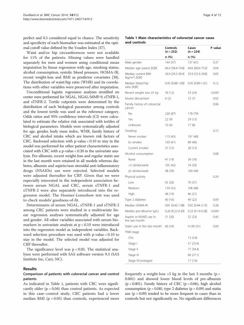
Table 1 Main characteristics of colorectal cancer casesand controls
Controls(n = 252)
Cases(n = 224)
P value
n (%) n (%)
Male gender 144 (57) 137 (61) 0.37
Median age (years) [IQR] 66.4 [58.4-74.8] 69.6 [60.6-75.9] 0.04
Median current BMI(kg/m2) [IQR]
26.9 [24.2-30.4] 25.9 [23.3-29.8] 0.05
Median Waist/Hipratio [IQR]
0.93 [0.88-1.00] 0.95 [0.89-1.01] 0.12
Recent weight loss ≥5 kg 30 (12) 53 (24) <0.001
Severe denutrition 6 (2) 15 (7) 0.02
Family history of colorectalcancer
0.06
No 220 (87) 178 (79)
Yes 22 (9) 29 (13)
Unknown 10 (4) 17 (8)
Smoking 0.72
Never smoker 113 (45) 107 (48)
Ex smoker 103 (41) 89 (40)
Current smoker 37 (15) 28 (13)
Alcohol consumption 0.08
None 47 (19) 39 (18)
<5 drinks/week 105 (42) 74 (33)
≥5 drinks/week 98 (39) 109 (49)
Physical activity 0.29
Low 65 (26) 70 (31)
Medium 139 (55) 108 (48)
High 48 (19) 46 (21)
Type 2 diabetes 40 (16) 49 (22) 0.09
Median HOMA-IR 0.81 [0.42-1.98] 0.92 [0.44-2.15] 0.28
Median pre-albumin (g/L) 0.26 [0.22-0.30] 0.23 [0.19-0.28] <0.001
Aspirin or NSAID use inthe last month
51 (20) 52 (23) 0.43
Statin use in the last month 60 (24) m 69 (31) 0.09
TNM stage
(Tis) 13 (5.8)
Stage I 57 (25.4)
Stage II 77 (34.4)
Stage III 60 (27.1)
Stage IV/unstaged 17 (7.6)
Duvillard et al. BMC Cancer 2014, 14:912 Page 4 of 12http://www.biomedcentral.com/1471-2407/14/912
perfect and 0.5 considered equal to chance. The sensitivityand specificity of each biomarker was estimated at the opti-mal cutoff value defined by the Youden Index [37].Waist and/or hip circumferences were not available
for 11% of the patients. Missing values were handledseparately for men and women using conditional meanimputation by linear regression with age, smoking status,alcohol consumption, systolic blood pressure, HOMA-IR,recent weight-loss and BMI as predictor covariates [38].The distribution of waist/hip ratio (WHR) and its correla-tions with other variables were preserved after imputation.Unconditional logistic regression analyses stratified on
center were performed for NGAL, NGAL/MMP-9, sTNFR-1,and sTNFR-2. Tertile cutpoints were determined by thedistribution of each biological parameter among controlsand the lowest tertile was used as the reference category.Odds ratios and 95% confidence intervals (CI) were calcu-lated to estimate the relative risk associated with tertiles ofbiological parameters. Models were systematically adjustedfor age, gender, body mass index, WHR, family history ofCRC and alcohol intake which are known risk factors ofCRC. Backward selection with p-value < 0.10 to stay in themodel was performed for other patient characteristics asso-ciated with CRC with a p-value < 0.20 in the univariate ana-lysis. Pre-albumin, recent weight-loss and regular statin usein the last month were retained in all models whereas dia-betes, albumin and aspirin/non steroidal anti-inflammatorydrugs (NSAIDs) use were rejected. Selected modelswere adjusted thereafter for CRP. Given that we wereespecially interested in the independent association be-tween serum NGAL and CRC, serum sTNFR-1 andsTNFR-2 were also separately introduced into the re-gression model. The Hosmer-Lemeshow test was usedto check models’ goodness-of-fit.Determinants of serum NGAL, sTNFR-1 and sTNFR-2
among CRC patients were studied in a multivariate lin-ear regression analyses systematically adjusted for ageand gender. All other variables associated with serum bio-markers in univariate analysis at p < 0.10 were introducedinto the regression model as independent variables. Back-ward selection procedure was used with p-value < 0.10 tostay in the model. The selected model was adjusted forCRP thereafter.The significance level was p < 0.05. The statistical ana-
lyses were performed with SAS software version 9.3 (SASInstitute Inc, Cary, NC).
ResultsComparison of patients with colorectal cancer and controlpatientsAs indicated in Table 1, patients with CRC were signifi-cantly older (p = 0.04) than control patients. As expectedin this case–control study, CRC patients had a lowermedian BMI (p = 0.05) than controls, experienced more
frequently a weight-loss >5 kg in the last 3 months (p <0.001) and showed lower blood levels of pre-albumin(p < 0.001). Family history of CRC (p = 0.06), high alcoholconsumption (p = 0.08), type 2 diabetes (p = 0.09) and statinuse (p = 0.09) tended to be more frequent in cases than incontrols but not significantly so. No significant differences
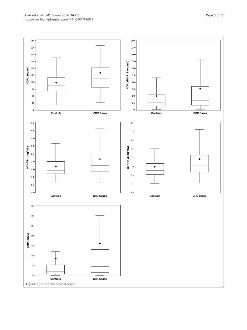
Figure 1 (See legend on next page.)
Duvillard et al. BMC Cancer 2014, 14:912 Page 5 of 12http://www.biomedcentral.com/1471-2407/14/912

(See figure on previous page.)Figure 1 Serum levels of NGAL, NGAL/MMP-9, sTNFR-1, sTNFR-2 and CRP in colorectal cancer cases and controls. The boundary of thebox closest to zero indicates the 25th percentile, a line within the box marks the median, and the boundary of the box farthest from zeroindicates the 75th percentile. The lozenge symbol indicates the mean. Error bars above the boxes indicate the maximum observation above thethird quintile (Q3) plus 1.5 multiplied by the difference between the values of the third and the first quintile (Q3-Q1). Errors bars below the boxesindicate the minimum observation below Q1 minus 1.5× (Q3-Q1). All p values for differences between CRC cases and controls were <0.001 usingKruskal-Wallis test.
Table 2 Spearman correlation coefficients between NGALand other inflammatory markers in colorectal cancercases and controls
CRP NGAL NGAL/MMP-9 sTNFR-1 sTNFR-2
CRP 1 0.14a 0.19b 0.43c 0.35c
NGAL 0.41c 1 0.41c 0.43c 0.44c
NGAL/MMP-9 0.15a 0.46c 1 0.15a 0.06
sTNFR-1 0.51c 0.65c 0.15a 1 0.78c
sTNFR-2 0.42c 0.59c 0.04 0.85b 1ap < 0.05, bp < 0.01, cp < 0.001.Correlation coefficients are indicated in bold characters for cases (n = 219) andin italics for controls (n = 250).
Duvillard et al. BMC Cancer 2014, 14:912 Page 6 of 12http://www.biomedcentral.com/1471-2407/14/912
in gender distribution, WHR, HOMA-IR, smoking habits,physical activity and use of aspirin/NSAIDS were observedbetween cases and controls.In univariate analyses, higher serum concentrations
of NGAL, NGAL/MMP-9, CRP, sTNFR-1, sTNFR-2 (allp values <0.005) were observed in patients with CRCcompared to controls (Figure 1). Among cases and con-trols, median levels (interquartile range: IQR) were re-spectively 115 ng/mL (82–153) versus 89.5 (68–117) forNGAL, 35.0 ng/mL (17.1-85-5) versus 26.3 (15.0-57.1)for NGAL/MMP-9, 4.54 mg/L (1.49-13.2) versus 1.96(0.56-5.39) for CRP, 1.76 ng/mL (1.38-2.50) versus 1.45(1.22-2.01) for sTNFR-1 and 3.07 ng/mL (2.35-4.32)versus 2.56 (2.11-3.36) for sTNFR-2. These results werevirtually unchanged after exclusion of control patientswith diverticulitis. The ROC analysis revealed that thediscriminative power of serum NGAL between CRC pa-tients and controls was moderate as assessed by an areaunder the curve (AUC) of 0.65 (95% CI:0.60-0.70) andpoor for NGAL/MMP-9 with an AUC of 0.58 (95%CI:0.52-0.63). The discriminative power of other bio-logical parameters was not better with an AUC of 0.64(95% CI:0.59-0.69) for sTNFR-1, 0.63 (95% CI:0.58-0.68)for sTNFR-2, and 0.63 (95% CI:0.58-0.68) for CRP. Forthe optimum cutoff value of NGAL (>106 ng/mL) andNGAL/MMP-9 (>71.7 ng/mL), sensitivites and specific-ities were respectively 57 and 69% for NGAL and 34 and81% for NGAL/MMP-9. The optimum cutoff values ofsTNFR-1 (>1.56 ng/mL), sTNFR-2 (>3.58 ng/mL) andCRP (>4.45 mg/L) provide sensitivities and specificities of66 and 58%, 41 and 79%, 52 and 71%, for each biomarkerrespectively. For all biological parameters, the exclusion ofcontrol patients with diverticulitis had only a marginal im-pact on AUC values.Among cases, there were strong inter-relationships be-
tween NGAL, sTNFR-1, sTNFR-2, and CRP (Table 2).Spearman correlation coefficients were especially highbetween serum NGAL and both sTNFR-1 and sTNFR-2(p < 0.001); they were lower with CRP. Although the as-sociation between NGAL and NGAL/MMP-9 was rela-tively high (p < 0.001), correlation coefficients betweenthe complex and other serum markers were very low.Among controls, associations between NGAL and otherserum markers were attenuated for sTNFR-1, and sTNFR-2, although being highly significant (p < 0.001). In controls,NGAL/MMP-9 remained highly correlated to NGAL (p <
0.001) and weakly associated with other serum markers(CRP, sTNFR-1, sTNFR-2).Multiple logistic regression models were used to test
the associations between serum NGAL, NGAL/MMP-9,sTNFR-1, sTNFR-2, and the risk of CRC. After adjust-ment for known risk factors of CRC (age, gender, BMI,WHR, family history of CRC, alcohol intake) and for sta-tin use, recent weight loss and serum pre-albumin, weobserved a significant association between serum NGALand the risk of CRC (3rd versus 1st tertile, OR: 2.76; 95%CI:1.59-4.78) (Table 3). Significant associations with therisk of CRC were also found for sTNFR-1 and sTNFR-2but not with NGAL/MMP-9. Further adjustment forCRP did not affect these associations (Table 3). WhensTNFR-1 and sTNFR-2 were each concurrently consid-ered with serum NGAL in the regression model, none ofthem could enter the model with p values of 0.14 and0.52 respectively.
Serum levels of NGAL, sTNFR-1 and sTNFR-2 according totumor characteristicsTumors were located in the rectum for 48 patients(21%), left colon for 85 (38%), right colon for 83 (37%)and 8 patients presented multiple locations (4%). Therewere no significant differences in biological parametersby location. The distribution of TNM stages given inTable 1 showed that only 34% of the patients had ad-vanced CRC (stages III and IV/unstaged). As indicatedin Figure 2, serum NGAL levels significantly increasedwith the invasion depth (p = 0.028) with median values(IQR) ranging from 91 ng/mL (71–165) for Tis tumors, to100 ng/mL (80–148) for pT1, 96 ng/mL (76–128) for pT2,120 ng/ml (86–156) for pT3 and 146 ng/mL (106–214)
Table 3 Multivariate associations between serum levels of NGAL, sTNFR-1, sTNFR-2 and risk of colorectal cancer
Multivariate logistic modela Multivariate logistic model adjusted for CRPb
Serum marker (ng/ml) Number of case/controls OR 95% CI P valuec OR 95% CI P valuec
NGAL <0.001 <0.001
< 75 40/82 1 1
75- 104 52/85 1.42 0.81-2.50 1.43 0.81-2.50
≥ 104 127/83 2.76 1.59-4.78 2.81 1.61-4.89
NGAL/MMP-9 0.75 0.74
< 18.5 60/83 1 1
18.5-42.3 61/84 0.95 0.56-1.62 0.95 0.56-1.62
≥ 42.3 98/84 1.16 0.65-2.06 1.17 0.66-2.09
sTNFR-1 0.015 0.013
< 1.30 38/83 1 1
1.30-1.77 72/84 2.00 1.14-3.50 2.03 1.16-3.55
≥ 1.77 109/83 2.44 1.34-4.45 2.51 1.36-4.61
sTNFR-2 0.016 0.015
< 2.23 47/83 1 1
2.23-3.09 63/85 am 1.34 0.79-2.28 am 1.34 0.79-2.29
≥ 3.09 109/82 1.93 1.12-3.31 1.95 1.13-3.37aModels were stratified on centre and adjusted for age, gender, body mass index, waist/hip ratio, family history of colorectal cancer, alcohol intake, pre-albumin,recent weight-loss and regular statin intake in the last month.bC reactive protein (CRP) was forced in the multivariate models (p values for association with the risk of colorectal cancer were respectively of 0.58, 0.87, 0.60, 0.69in the NGAL, NGAL/MMP-9, sTNFR-1 and sTNFR-2 models).cp value for trend.
Duvillard et al. BMC Cancer 2014, 14:912 Page 7 of 12http://www.biomedcentral.com/1471-2407/14/912
for pT4. Serum NGAL was also significantly associatedwith maximal tumor size (p < 0.001) with median values(IQR) ranging from 99 ng/ml (79–137) for tumors ≤3 cm,to 110 ng/ml (86–158) for tumors between 3 and 4.5 cm,113 ng/mL (81–147) for tumors between 4.5 and 6 cmand 159 ng/mL (116–214) for tumors > 6 cm. We did notobserve any significant association between NGAL levelsand lymph node involvement (p = 0.65), distant metastasis(p = 0.17) and TNM stage (p = 0.32). Serum levels ofsTNFR-1 tended to be associated with maximal tumorsize (p = 0.064) and lymph node involvement (p = 0.056)whereas serum sTNFR-2 levels were only associatedwith maximal tumor size (p = 0.046). No associationswere found between serum NGAL/MMP-9 and tumorcharacteristics or stage (all p values >0.30).
Independent determinants of serum levels of NGAL,sTNFR-1 and sTNFR-2Multivariate linear regression analysis showed that, amongCRC patients, serum levels of NGAL significantly de-creased with serum albumin (p < 0.001) and were positivelyassociated with WHR (p = 0.005), recent weight loss >5 kg(p = 0.026) and maximal tumor size (p = 0.028) (Table 4).Past and current smoking, age and male gender were notindependently associated with serum levels of NGAL. Theintroduction of serum CRP in the regression modelshowed that CRP was an important significant determinant
of serum NGAL (p = 0.004) which reduced the effects ofother variables. Especially, the effect of tumor size onserum NGAL was reduced by 26% and no longer reachedthe significance level (p = 0.11). None of the tumoral char-acteristics were independently associated with serum levelsof sTNFR-1 and sTNFR-2. Both inflammatory markerswere independently related to increasing age (p = 0.007and p = 0.021 respectively), waist/hip ratio (p = 0.004 andp = 0.019 respectively) and to decreasing albumin levels(p = 0.037 and p = 0.019, respectively). Serum CRP wasalso a strong determinant of sTNFR-1 levels (p < 0.001),but less strongly related to sTNFR-2 levels (p = 0.058).Last, high physical activity was only associated with de-creasing sTNFR-2 levels (p = 0.012). The sole independentdeterminants of NGAL/MMP-9 were CRP (p = 0.03) andcurrent smoking (p < 0.001).Among control patients, the sole significant determi-
nants of serum NGAL were age (p < 0.001), male gender(p = 0.021) and serum CRP (p < 0.001). The sole signifi-cant determinants of sTNFR-1 and sTNFR-2 levels wereage (p < 0.001 for both markers), body mass index (p =0.014 and p = 0.006, respectively) and CRP levels (p < 0.001and p = 0.007, respectively).
Discussion and conclusionsOur case–control study demonstrated a significant in-crease in serum NGAL levels in CRC patients compared
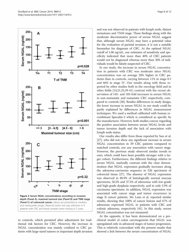
Figure 2 Serum NGAL concentrations according to invasiondepth (Panel A), maximal tumoral size (Panel B) and TNM stage(Panel C) of colorectal cancer. Values are presented as mediansand interquartile ranges. Maximal tumor size was unknown in 9patients with CRC and blood samples were missing in 5 cases.
Duvillard et al. BMC Cancer 2014, 14:912 Page 8 of 12http://www.biomedcentral.com/1471-2407/14/912
to controls, which persisted after adjustment for trad-itional risk factors for CRC. However, the increase inNGAL concentration was mainly confined to CRC pa-tients with large-sized tumors or important depth invasion
and was not observed in patients with lymph node, distantmetastases and TNM stage. These findings along with themoderate discriminative power of serum NGAL suggestthat, although serum NGAL may have a potential valuefor the evaluation of parietal invasion, it is not a suitablebiomarker for diagnosis of CRC. At the optimal NGALcutoff of 1.06 ng/mL, our estimates of sensitivity and spe-cificity indicated that more than 40% of CRC patientswould not be diagnosed whereas more than 30% of indi-viduals would be falsely suspected of CRC.In our study, the increase in serum NGAL concentra-
tion in patients with CRC was moderate since NGALconcentration was on average 28% higher in CRC pa-tients than in controls, varying between 11% in stage 0-Iand 66% in stage IV. Our results along with those re-ported by other studies both in the oncology field and inother fields [14,21,25,39-41] contrast with the recent ob-servation of 145- and 185-fold increases in serum NGALin non-metastatic and metastatic CRC respectively, com-pared to controls [30]. Besides differences in study design,the lower increase in serum NGAL in our study could bepartly explained by differences in NGAL measurementtechniques. We used a method calibrated with human re-combinant lipocalin-2 which is considered as specific bythe manufacturer. However, both studies concur regardingthe positive association between serum NGAL levels andtumor invasion depth and the lack of association withlymph node status.Our results also differ from those reported by Sun et al.
[27], who did not show any significant increase in serumNGAL concentration in 39 CRC patients compared tomatched controls, nor any association with cancer stage.However, the previous study observed similar trends toours, which could have been possibly stronger with a lar-ger cohort. Furthermore, the different findings relative toserum NGAL markedly contrast with the clear demon-stration that NGAL expression gradually increases alongthe adenoma-carcinoma sequence in 526 specimens ofcolorectal tissue [27]. The absence of NGAL expressionwas observed in 80.9% of histologically normal mucosaspecimens, 56.2% and 37.5% of adenomas with low-gradeand high-grade dysplasia respectively, and in only 5.9% ofcarcinoma specimens. In addition, NGAL expression wasassociated with cancer stage and tumor recurrence instage II cancer patients. Mc Lean et al. reported similarresults, showing that 100% of cancer lesions and 67% ofadenomas expressed NGAL in patients with CRC andcolonic adenoma, respectively [41]. In this study, serumNGAL concentration was not measured.At the opposite, it has been demonstrated on a pre-
clinical model of colon carcinogenesis that NGAL wasupregulated only in advanced stages of tumor progression.This is relatively concordant with the present results thatshowed a link between the serum concentration of NGAL
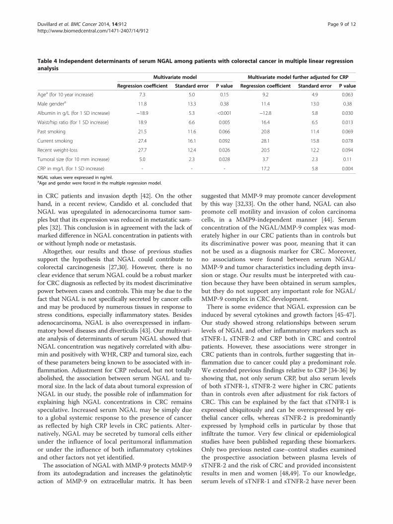
Table 4 Independent determinants of serum NGAL among patients with colorectal cancer in multiple linear regressionanalysis
Multivariate model Multivariate model further adjusted for CRP
Regression coefficient Standard error P value Regression coefficient Standard error P value
Agea (for 10 year increase) 7.3 5.0 0.15 9.2 4.9 0.063
Male gendera 11.8 13.3 0.38 11.4 13.0 0.38
Albumin in g/L (for 1 SD increase) −18.9 5.3 <0.001 −12.8 5.8 0.030
Waist/hip ratio (for 1 SD increase) 18.9 6.6 0.005 16.4 6.5 0.013
Past smoking 21.5 11.6 0.066 20.8 11.4 0.069
Current smoking 27.4 16.1 0.092 28.1 15.8 0.078
Recent weight-loss 27.7 12.4 0.026 20.5 12.2 0.094
Tumoral size (for 10 mm increase) 5.0 2.3 0.028 3.7 2.3 0.11
CRP in mg/L (for 1 SD increase) - - - 17.2 5.8 0.004
NGAL values were expressed in ng/ml.aAge and gender were forced in the multiple regression model.
Duvillard et al. BMC Cancer 2014, 14:912 Page 9 of 12http://www.biomedcentral.com/1471-2407/14/912
in CRC patients and invasion depth [42]. On the otherhand, in a recent review, Candido et al. concluded thatNGAL was upregulated in adenocarcinoma tumor sam-ples but that its expression was reduced in metastatic sam-ples [32]. This conclusion is in agreement with the lack ofmarked difference in NGAL concentration in patients withor without lymph node or metastasis.Altogether, our results and those of previous studies
support the hypothesis that NGAL could contribute tocolorectal carcinogenesis [27,30]. However, there is noclear evidence that serum NGAL could be a robust markerfor CRC diagnosis as reflected by its modest discriminativepower between cases and controls. This may be due to thefact that NGAL is not specifically secreted by cancer cellsand may be produced by numerous tissues in response tostress conditions, especially inflammatory states. Besidesadenocarcinoma, NGAL is also overexpressed in inflam-matory bowel diseases and diverticulis [43]. Our multivari-ate analysis of determinants of serum NGAL showed thatNGAL concentration was negatively correlated with albu-min and positively with WHR, CRP and tumoral size, eachof these parameters being known to be associated with in-flammation. Adjustment for CRP reduced, but not totallyabolished, the association between serum NGAL and tu-moral size. In the lack of data about tumoral expression ofNGAL in our study, the possible role of inflammation forexplaining high NGAL concentrations in CRC remainsspeculative. Increased serum NGAL may be simply dueto a global systemic response to the presence of canceras reflected by high CRP levels in CRC patients. Alter-natively, NGAL may be secreted by tumoral cells eitherunder the influence of local peritumoral inflammationor under the influence of both inflammatory cytokinesand other factors not yet identified.The association of NGAL with MMP-9 protects MMP-9
from its autodegradation and increases the gelatinolyticaction of MMP-9 on extracellular matrix. It has been
suggested that MMP-9 may promote cancer developmentby this way [32,33]. On the other hand, NGAL can alsopromote cell motility and invasion of colon carcinomacells, in a MMP9-independent manner [44]. Serumconcentration of the NGAL/MMP-9 complex was mod-erately higher in our CRC patients than in controls butits discriminative power was poor, meaning that it cannot be used as a diagnosis marker for CRC. Moreover,no associations were found between serum NGAL/MMP-9 and tumor characteristics including depth inva-sion or stage. Our results must be interpreted with cau-tion because they have been obtained in serum samples,but they do not support any important role for NGAL/MMP-9 complex in CRC development.There is some evidence that NGAL expression can be
induced by several cytokines and growth factors [45-47].Our study showed strong relationships between serumlevels of NGAL and other inflammatory markers such assTNFR-1, sTNFR-2 and CRP both in CRC and controlpatients. However, these associations were stronger inCRC patients than in controls, further suggesting that in-flammation due to cancer could play a predominant role.We extended previous findings relative to CRP [34-36] byshowing that, not only serum CRP, but also serum levelsof both sTNFR-1, sTNFR-2 were higher in CRC patientsthan in controls even after adjustment for risk factors ofCRC. This can be explained by the fact that sTNFR-1 isexpressed ubiquitously and can be overexpressed by epi-thelial cancer cells, whereas sTNFR-2 is predominantlyexpressed by lymphoid cells in particular by those thatinfiltrate the tumor. Very few clinical or epidemiologicalstudies have been published regarding these biomarkers.Only two previous nested case–control studies examinedthe prospective association between plasma levels ofsTNFR-2 and the risk of CRC and provided inconsistentresults in men and women [48,49]. To our knowledge,serum levels of sTNFR-1 and sTNFR-2 have never been
Duvillard et al. BMC Cancer 2014, 14:912 Page 10 of 12http://www.biomedcentral.com/1471-2407/14/912
studied as potential diagnostic biomarkers of CRC.Despite the significant increase in serum sTNFR-1 andsTNFR-2 in CRC patients, the intensity of their associ-ation with CRC was lower than that observed withserum NGAL. Furthermore, both markers had a poorerdiscriminative power than serum NGAL, and only pre-sented marginal associations with tumoral size and lymphnode involvement. These observations do not support theidea that serum sTNFR-1 and sTNFR-2 could be of inter-est for helping diagnosis of CRC.Our study presented some limitations. First, the cross-
sectional design precludes any temporal inference beingmade and any causal conclusion being drawn from ourresults. Second, this study may have been subject to se-lection biases. CRC patients recruited in university hos-pitals may not be representative of all CRC and, becausepatients with pre-operative treatment were discarded forother study purposes, relatively few patients with rectalcancer were included. However, our study did not provideany evidence that serum biomarkers depended on primarycancer location. The recruitment of control patients op-erated for benign diseases with possible inflammatorybackground could have reduced the effect of potentialbiomarkers. However, our results were not altered afterexclusion of such controls. On the other hand, our studyhad the advantage of including large samples of wellcharacterized cases and controls which allowed poten-tial confounders to be taken into account, and of relyingon centralized and blinded analysis of blood samplesstored in optimal conditions. Finally, we did not com-pare NGAL and sTNFRs with usual tumoral markerssuch as CEA, CA19-9. Such a comparison could havebeen interesting if the markers we quantified had dem-onstrated their potential usefulness in routine but has alesser interest in front of the results we observed.In conclusion, this case–control study showed that
NGAL, NGAL/MMP9, sTNFR-1 and sTNFR-2 serum con-centrations are higher in patients with CRC, and thatNGAL levels are especially elevated in patients with largetumors. However, our findings do not suggest that theseserum parameters may be clinically relevant markers forthe detection of CRC and especially for early detection.
AbbreviationsAUC: Area under the curve; BMI: Body mass index; CI: Confidence interval;CRC: Colorectal cancer; CRP: C reactive protein; HOMA-IR: Homeostatic modelassessment of insulin resistance; IQR: Interquartile range; MMP9: Matrixmetallopeptidase 9; NGAL: Neutrophil gelatinase-associated lipocalin;NSAID: Non steroidal anti-inflammatory drugs; OR: Odds ratio; ROC: Receiveroperating characteristic; sTNFR: Soluble tumor necrosis factor receptor 1 and 2.
Competing interestsThe authors declare that they have no competing interests.
Authors’ contributionsLD carried out laboratory measurements and wrote the paper. POD, MLS,GM, JBD, SDL coordinated the study in the different surgical departmentsand recruited patients. JMP participated in the design of the study and in
the interpretation of data. AB performed statistical analysis. CBK coordinated thestudy and helped to draft the manuscript. All authors read and approved thefinal version of the manuscript.
AcknowledgmentsWe thank the staff of the Biological Resource Center Ferdinand CabanneBB-0033-00044 (University hospital of Dijon, F) for their assistance in handlingand storing biological samples. We thank the members of the AGARIC StudyGroup for their contribution to the inclusion of patients, data collection andbiological analyses.
Members of the AGARIC study groupCoordinating center; Pr C Bonithon-Kopp (study coordinator), Dr V Cottet(epidemiologist), A Bourredjem (statistician), S Vinault (data manager), A Felinand E Galizzi (logistic coordination and data quality control) Clinicalinvestigators: Pr P Ortega-Deballon and Dr O Facy (Dijon-F); Pr G Mantion,Pr B Heyd, Dr S Demaret, Dr O Idelcadi, Dr J Lubrano, Dr P Mathieu, Dr GLandecy and Dr P Morati (Besançon-F); Pr A Ayav, Pr L Bresler, Pr L Brunaud,Dr M Fau, Dr D V Frentiu, Dr A Rouers, and Dr M-L Scherrer (Nancy-F); Pr JFDelattre, Pr R Kianmanesh, Dr S Coussinet-Poulizac, Dr S Deguelte-Lardièreand Dr A Goya (Reims-F); Pr S Rohr, Dr C Brigand, Dr S Dragomir, Dr JBDelhorme, and Dr J-C Ollier (Strasbourg-F) . Laboratory analyses: Pr JM Petit,Pr L Duvillard, E Niot et L Loïodice (Dijon-F), Drs N Combe and C Vaysse(ITERG, Bordeaux- F). Local research assistant staff: M-L Asensio, (Inserm CIE1, Dijon-F); D Da Costa-Souihel (Inserm CBT 506, Besançon), N Valentin(Inserm CIE 6, Nancy-F), and F Hardy (CHU, Reims-F), N Derridj-Ait Younes, GLarderet, E Richer (CHU, Strasbourg-F).
Funding sourcesPr Claire Bonithon-Kopp obtained financial grants from the National Instituteof Cancer (INCa), Ligue contre le cancer de Bourgogne-Franche-Comté,Fondation de France, Regional Council of Burgundy, and the UniversityHospital of Dijon (France). The study was also supported by a FrenchGovernment grant managed by the French National Research Agency underthe program “Investissements d’Avenir”, reference ANR-11-LABX-0021.
Author details1Inserm UMR 866, Faculté de médecine de Dijon, Dijon 21079-F, France.2Biochemistry department, University hospital of Dijon, Dijon 21070-F, France.3Department of digestive surgical oncology, University hospital of Dijon,Dijon 21000-F, France. 4Inserm CIC 1432, Faculté de médecine de Dijon,Dijon 21079-F, France. 5Clinical investigation center (team clinicalepidemiology), University hospital of Dijon, Dijon 21000-F, France.6Department of general and digestive surgery, Hôpital Brabois, Universityhospital of Nancy, Vandoeuvre-les-Nancy, Nancy 54511-F, France.7Department of general, digestive and oncologic surgery, University hospitalof Besançon, Besançon 25030-F, France. 8Department of general anddigestive surgery, Hôpital de Hautepierre, University hospital of Strasbourg,Strasbourg 67098-F, France. 9Department of general, digestive and endocrinesurgery, University hospital of Reims, Reims 51100-F, France. 10BiochimieMédicale, Plateau Technique de Biologie, 2, rue Angélique Ducoudray, BP37013, 21070 Dijon Cedex, France.
Received: 4 April 2014 Accepted: 20 November 2014Published: 3 December 2014
References1. Ferlay J, Shin HR, Bray F, Forman D, Mathers C, Parkin DM: Estimates of
worldwide burden of cancer in 2008: GLOBOCAN 2008. Int J Cancer 2010,2010(127):2893–2917.
2. Jemal A, Bray F, Center MM, Ferlay J, Ward E, Forman D: Global cancerstatistics. CA Cancer J Clin 2011, 61(2):69–90.
3. Sanjoaquin MA, Choodari-Oskooei B, Dolbear C, Putcha V, Sehgal A, Key TJ,Møller H: Colorectal cancer incidence, mortality and survival in South-eastEngland between 1972 and 2001. Eur J Cancer Prev 2007, 16(1):10–16.
4. Kjeldsen L, Johnsen AH, Sengeløv H, Borregaard N: Isolation and primarystructure of NGAL, a novel protein associated with human neutrophilgelatinase. J Biol Chem 1993, 268:10425–10432.
5. Roudkenar MH, Halabian R, Ghasemipour Z, Roushandeh AM, Rouhbakhsh M,Nekogoftar M, Kuwahara Y, Fukumoto M, Shokrgozar MA: Neutrophil
Duvillard et al. BMC Cancer 2014, 14:912 Page 11 of 12http://www.biomedcentral.com/1471-2407/14/912
gelatinase-associated lipocalin acts as a protective factor against H(2)O(2)toxicity. Arch Med Res 2008, 39(6):560–566.
6. Bahmani P, Halabian R, Rouhbakhsh M, Roushandeh AM, Masroori N,Ebrahimi M, Samadikuchaksaraei A, Shokrgozar MA, Roudkenar MH:Neutrophil gelatinase-associated lipocalin induces the expression ofheme oxygenase-1 and superoxide dismutase 1, 2. Cell Stress Chaperones2010, 15(4):395–403.
7. Aujla SJ, Chan YR, Zheng M, Fei M, Askew DJ, Pociask DA, Reinhart TA,McAllister F, Edeal J, Gaus K, Husain S, Kreindler JL, Dubin PJ, Pilewski JM,Myerburg MM, Mason CA, Iwakura Y, Kolls JK: IL-22 mediates mucosal hostdefense against Gram-negative bacterial pneumonia. Nat Med 2008,14(3):275–281.
8. Nairz M, Theurl I, Schroll A, Theurl M, Fritsche G, Lindner E, Seifert M, CrouchML, Hantke K, Akira S, Fang FC, Weiss G: Absence of functional Hfeprotects mice from invasive Salmonella enterica serovar Typhimuriuminfection via induction of lipocalin-2. Blood 2009, 114(17):3642–3651.
9. Nomura I, Gao B, Boguniewicz M, Darst MA, Travers JB, Leung DY: Distinctpatterns of gene expression in the skin lesions of atopic dermatitis andpsoriasis: a gene microarray analysis. J Allergy Clin Immunol 2003,112:1195–1202.
10. Van Dyke TE, Schweinebraten M, Cianciola LJ, Offenbacher S, Genco RJ:Neutrophil chemotaxis in families with localized juvenile periodontitis.J Periodontal Res 1985, 20:503–514.
11. Ding L, Hanawa H, Ota Y, Hasegawa G, Hao K, Asami F, Watanabe R,Yoshida T, Toba K, Yoshida K, Ogura M, Kodama M, Aizawa Y: Lipocalin-2/neutrophil gelatinase-B associated lipocalin is strongly induced in heartsof rats with autoimmune myocarditis and in human myocarditis. CircJ 2010, 74(3):523–530.
12. Yndestad A, Landrø L, Ueland T, Dahl CP, Flo TH, Vinge LE, Espevik T,Frøland SS, Husberg C, Christensen G, Dickstein K, Kjekshus J, Øie E,Gullestad L, Aukrust P: Increased systemic and myocardial expression ofneutrophil gelatinase-associated lipocalin in clinical and experimentalheart failure. Eur Heart J 2009, 30(10):1229–1236.
13. Galamb O, Györffy B, Sipos F, Spisák S, Németh AM, Miheller P, Tulassay Z,Dinya E, Molnár B: Inflammation, adenoma and cancer: objectiveclassification of colon biopsy specimens with gene expression signature.Dis Markers 2008, 25(1):1–16.
14. Oikonomou KA, Kapsoritakis AN, Theodoridou C, Karangelis D, Germenis A,Stefanidis I, Potamianos SP: Neutrophil gelatinase-associated lipocalin(NGAL) in inflammatory bowel disease: association with pathophysiologyof inflammation, established markers, and disease activity. J Gastroenterol2012, 47(5):519–530.
15. Sahinarslan A, Kocaman SA, Bas D, Akyel A, Ercin U, Zengin O, Timurkaynak T:Plasma neutrophil gelatinase-associated lipocalin levels in acute myocardialinfarction and stable coronary artery disease. Coron Artery Dis 2011,22(5):333–338.
16. Bu DX, Hemdahl AL, Gabrielsen A, Fuxe J, Zhu C, Eriksson P, Yan ZQ:Induction of neutrophil gelatinase-associated lipocalin in vascularinjury via activation of nuclear factor-kappaB. Am J Pathol 2006,169(6):2245–2253.
17. El-Mesallamy HO, Hamdy NM, Zaghloul AS, Sallam AM: Clinical value ofcirculating lipocalins and insulin-like growth factor axis in pancreaticcancer diagnosis. Pancreas 2013, 42(1):149–154.
18. Wang HJ, He XJ, Ma YY, Jiang XT, Xia YJ, Ye ZY, Zhao ZS, Tao HQ:Expressions of neutrophil gelatinase-associated lipocalin in gastriccancer: a potential biomarker for prognosis and an ancillary diagnostictest. Anat Rec (Hoboken) 2010, 293(11):1855–1863.
19. Iannetti A, Pacifico F, Acquaviva R, Lavorgna A, Crescenzi E, Vascotto C,Tell G, Salzano AM, Scaloni A, Vuttariello E, Chiappetta G, Formisano S,Leonardi A: The neutrophil gelatinase-associated lipocalin (NGAL), aNF-kappaB-regulated gene, is a survival factor for thyroid neoplasticcells. Proc Natl Acad Sci U S A 2008, 05(37):14058–14063.
20. Miyamoto T, Kashima H, Suzuki A, Kikuchi N, Konishi I, Seki N, Shiozawa T:Laser-captured microdissection-microarray analysis of the genesinvolved in endometrial carcinogenesis: stepwise up-regulation oflipocalin2 expression in normal and neoplastic endometria and itsfunctional relevance. Hum Pathol 2011, 42(9):1265–1274.
21. Sung H, Choi JY, Lee SA, Lee KM, Han S, Jeon S, Song M, Lee Y, Park SK,Yoo KY, Noh DY, Ahn SH, Kang D: The association between thepreoperative serum levels of lipocalin-2 and matrix metalloproteinase-9(MMP-9) and prognosis of breast cancer. BMC Cancer 2012, 12:193.
22. Wenners AS, Mehta K, Loibl S, Park H, Mueller B, Arnold N, Hamann S,Weimer J, Ataseven B, Darb-Esfahani S, Schem C, Mundhenke C, Khandan F,Thomssen C, Jonat W, Holzhausen HJ, von Minckwitz G, Denkert C, Bauer M:Neutrophil gelatinase-associated lipocalin (NGAL) predicts response toneoadjuvant chemotherapy and clinical outcome in primary humanbreast cancer. PLoS One 2012, 7(10):e45826.
23. Yang J, Bielenberg DR, Rodig SJ, Doiron R, Clifton MC, Kung AL, Strong RK,Zurakowski D, Moses MA: Lipocalin 2 promotes breast cancer progression.Proc Natl Acad Sci U S A 2009, 106(10):3913–3918.
24. Bauer M, Eickhoff JC, Gould MN, Mundhenke C, Maass N, Friedl A:Neutrophil gelatinase-associated lipocalin (NGAL) is a predictor of poorprognosis in human primary breast cancer. Breast Cancer Res Treat 2008,108(3):389–397.
25. Cho H, Kim JH: Lipocalin2 expressions correlate significantly with tumordifferentiation in epithelial ovarian cancer. J Histochem Cytochem 2009,57(5):513–521.
26. Porta C, Paglino C, De Amici M, Quaglini S, Sacchi L, Imarisio I, Canipari C:Predictive value of baseline serum vascular endothelial growth factorand neutrophil gelatinase-associated lipocalin in advanced kidneycancer patients receiving sunitinib. Kidney Int 2010, 77(9):809–815.
27. Sun Y, Yokoi K, Li H, Gao J, Hu L, Liu B, Chen K, Hamilton SR, Fan D, Sun B,Zhang W: NGAL expression is elevated in both colorectal adenoma-carcinoma sequence and cancer progression and enhances tumorigenesisin xenograft mouse models. Clin Cancer Res 2011, 17(13):4331–4340.
28. Barresi V, Reggiani-Bonetti L, Di Gregorio C, Vitarelli E, Ponz De Leon M,Barresi G: Neutrophil gelatinase-associated lipocalin (NGAL) and matrixmetalloproteinase-9 (MMP-9) prognostic value in stage I colorectalcarcinoma. Pathol Res Pract 2011, 207(8):479–486.
29. Barresi V, Di Gregorio C, Reggiani-Bonetti L, Ieni A, Ponz-De Leon M, Barresi G:Neutrophil gelatinase-associated lipocalin: a new prognostic marker instage I colorectal carcinoma? Hum Pathol 2011, 42(11):1720–1726.
30. Marti J, Fuster J: Prognostic value of serum neutrophil gelatinase-associated lipocalin in metastatic and non-metastatic colorectal cancer:reply. World J Surg 2013, 37(11):2729.
31. Fung KY, Priebe I, Purins L, Tabor B, Brierley GV, Lockett T, Nice E, Gibbs P,Tie J, McMurrick P, Moore J, Ruszkiewicz A, Burgess A, Cosgrove LJ:Performance of serum lipocalin 2 as a diagnostic marker for colorectalcancer. Cancer Biomark 2013, 13(2):75–79.
32. Candido S, Maestro R, Polesel J, Catania A, Maira F, Signorelli SS, McCubrey JA,Libra M: Roles of neutrophil gelatinase-associated lipocalin (NGAL) inhuman cancer. Oncotarget 2014, 5(6):1576–1594.
33. Yan L, Borregaard N, Kjeldsen L, Moses MA MA: The high molecular weighturinary matrix metalloproteinase (MMP) activity is a complex ofgelatinase B/MMP-9 and neutrophil gelatinase-associated lipocalin(NGAL). Modulation of MMP-9 activity by NGAL. J Biol Chem 2001,276(40):37258–37265.
34. Toriola AT, Cheng TY, Neuhouser ML, Wener MH, Zheng Y, Brown E, Miller JW,Song X, Beresford SA, Gunter MJ, Caudill MA, Ulrich CM: Biomarkers ofinflammation are associated with colorectal cancer risk in women but arenot suitable as early detection markers. Int J Cancer 2013, 132(11):2648–2658.
35. Bünger S, Haug U, Kelly M, Posorski N, Klempt-Giessing K, Cartwright A,Fitzgerald SP, Toner V, McAleer D, Gemoll T, Laubert T, Büning J, Fellermann K,Bruch HP, Roblick UJ, Brenner H, von Eggeling F, Habermann JK: A novelmultiplex-protein array for serum diagnostics of colon cancer: acase–control study. BMC Cancer 2012, 12:393.
36. Aleksandrova K, Jenab M, Boeing H, Jansen E, Bueno-de-Mesquita HB,Rinaldi S, Riboli E, Overvad K, Dahm CC, Olsen A, Tjønneland A, Boutron-Ruault MC, Clavel-Chapelon F, Morois S, Palli D, Krogh V, Tumino R, Vineis P,Panico S, Kaaks R, Rohrmann S, Trichopoulou A, Lagiou P, Trichopoulos D,van Duijnhoven FJ, Leufkens AM, Peeters PH, Rodríguez L, Bonet C,Sánchez MJ, et al: Circulating C-reactive protein concentrations and risksof colon and rectal cancer: a nested case–control study within theEuropean Prospective Investigation into Cancer and Nutrition. Am JEpidemiol 2010, 172(4):407–418.
37. Youden WJ: Index for rating diagnostic tests. Cancer 1950, 3(1):32–35.38. Little RJA, Rubin DB: Statistical Analysis with Missing Data. 2nd edition. New
York 20: John Wiley & Sons; 2002.39. Haase-Fielitz A, Bellomo R, Devarajan P, Story D, Matalanis G, Dragun D,
Haase M: Novel and conventional serum biomarkers predicting acutekidney injury in adult cardiac surgery–a prospective cohort study.Crit Care Med 2009, 37(2):553–560.
Duvillard et al. BMC Cancer 2014, 14:912 Page 12 of 12http://www.biomedcentral.com/1471-2407/14/912
40. Constantin JM, Futier E, Perbet S, Roszyk L, Lautrette A, Gillart T, Guerin R,Jabaudon M, Souweine B, Bazin JE, Sapin V: Plasma neutrophil gelatinase-associated lipocalin is an early marker of acute kidney injury in adultcritically ill patients: a prospective study. J Crit Care 2010, 25(1):176. e1-6.
41. McLean MH, Thomson AJ, Murray GI, Fyfe N, Hold GL, El-Omar EM: Expressionof neutrophil gelatinase-associated lipocalin in colorectal neoplasticprogression a marker of malignant potential? Br J Cancer 2013,108(12):2537–2541.
42. Bousserouel S, Kauntz H, Gossé F, Bouhadjar M, Soler L, Marescaux J, Raul F:Identification of gene expression profiles correlated to tumorprogression in a preclinical model of colon carcinogenesis. Int J Oncol2010, 36(6):1485–1490.
43. Nielsen BS, Borregaard N, Bundgaard JR, Timshel S, Sehested M: Kjeldsen :Induction of NGAL synthesis in epithelial cells of human colorectalneoplasia and inflammatory bowel diseases. Gut 1996, 38(3):414–420.
44. Hu L, Hittelman W, Lu T, Ji P, Arlinghaus R, Shmulevich I, Hamilton SR,Zhang W: NGAL decreases E-cadherin-mediated cell-cell adhesion andincreases cell motility and invasion through Rac1 in colon carcinomacells. Lab Invest 2009, 89:531–548.
45. Karlsen JR, Borregaard N, Cowland JB: Induction of neutrophil gelatinase-associated lipocalin expression by co-stimulation with interleukin-17 andtumor necrosis factor-alpha is controlled by IkappaB-zeta but neither byC/EBP-beta nor C/EBP-delta. J Biol Chem 2010, 285:14088–14100.
46. Landrø L, Damås JK, Flo TH, Heggelund L, Ueland T, Tjønnfjord GE, Espevik T,Aukrust P, Frøland SS: Decreased serum lipocalin-2 levels in humanimmunodeficiency virus-infected patients: increase during highly activeanti-retroviral therapy. Clin Exp Immunol 2008, 152(1):57–63.
47. Bando M, Hiroshima Y, Kataoka M, Shinohara Y, Herzberg MC, Ross KF,Nagata T, Kido J: Interleukin-1alpha regulates antimicrobial peptideexpression in human keratinocytes. Immunol Cell Biol 2007, 85(7):532–537.
48. Chan AT, Ogino S, Giovannucci EL, Fuchs CS: Inflammatory markers areassociated with risk of colorectal cancer and chemopreventive responseto anti-inflammatory drugs. Gastroenterology 2011, 140(3):799–808.
49. Song M, Wu K, Ogino S, Fuchs CS, Giovannucci EL, Chan AT: A prospectivestudy of plasma inflammatory markers and risk of colorectal cancer inmen. Br J Cancer 2013, 108(9):1891–1898.
doi:10.1186/1471-2407-14-912Cite this article as: Duvillard et al.: A case–control study of pre-operativelevels of serum neutrophil gelatinase-associated lipocalin and otherpotential inflammatory markers in colorectal cancer. BMC Cancer2014 14:912.
Submit your next manuscript to BioMed Centraland take full advantage of:
• Convenient online submission
• Thorough peer review
• No space constraints or color figure charges
• Immediate publication on acceptance
• Inclusion in PubMed, CAS, Scopus and Google Scholar
• Research which is freely available for redistribution
Submit your manuscript at www.biomedcentral.com/submit