Positron Emission Tomography scanning in Anti-Neutrophil Cytoplasmic Antibodies-Associated...
9
Positron Emission Tomography scanning in Anti-Neutrophil Cytoplasmic Antibodies-Associated Vasculitis Michael J. Kemna, BSc, Fre´de´ric Vandergheynst, MD, Stefan Vo¨o¨, MD, PhD, Didier Blocklet, MD, Thomas Nguyen, MD, Sjoerd A.M.E.G. Timmermans, BSc, Pieter van Paassen, MD, PhD, Elie Cogan, MD, PhD, Marinus J.P.G. van Kroonenburgh, MD, PhD, and Jan Willem Cohen Tervaert, MD, PhD Abstract: Tools for evaluation of disease activity in patients with anti- neutrophil cytoplasmic antibodies (ANCA)-associated vasculitis (AAV) include scoring clinical manifestations, determination of biochemical parameters of inflammation, and obtaining tissue biopsies. These tools, however, are sometimes inconclusive. 2-deoxy-2-[ 18 F]-fluoro-D-glu- cose (FDG) positron emission tomography (PET) scans are commonly used to detect inflammatory or malignant lesions. Our objective is to explore the ability of PET scanning to assess the extent of disease activity in patients with AAV. Consecutive PET scans made between December 2006 and March 2014 in Maastricht (MUMC) and between July 2008 and June 2013 in Brussels (EUH) to assess disease activity in patients with AAV were retrospectively included. Scans were re-examined and quantitatively scored using maximum standard uptake values (SUVmax). PET find- ings were compared with C-reactive protein (CRP) and ANCA posi- tivity at the time of scanning. Forty-four scans were performed in 33 patients during a period of suspected active disease. All but 2 scans showed PET-positive sites, most commonly the nasopharynx (n ¼ 22) and the lung (n ¼ 22). Forty- one clinically occult lesions were found, including the thyroid gland (n ¼ 4 patients), aorta (n ¼ 8), and bone marrow (n ¼ 7). The amount of hotspots, but not the highest observed SUVmax value, was higher if CRP levels were elevated. Seventeen follow-up scans were made in 13 patients and showed decreased SUVmax values. FDG PET scans in AAV patients with active disease show positive findings in multiple sites of the body even when biochemical parameters are inconclusive, including sites clinically unsuspected and difficult to assess otherwise. (Medicine 94(20):e747) Abbreviations: AAV = ANCA-associated vasculitis, ANCA = anti-neutrophil cytoplasmic antibodies, FDG = 2-deoxy-2-[ 18 F]- fluoro-D-glucose, PET = positron emission tomography, SUVmax = maximum standard uptake value. INTRODUCTION G ranulomatosis with polyangiitis (GPA; Wegener’s) is an inflammatory disease entity affecting small to medium vessels. It is, together with microscopic polyangiitis (MPA) and eosinophilic granulomatosis with polyangiitis (EGPA; Churg Strauss Syndrome), characterized by the presence of anti-neutro- phil cytoplasmic antibodies (ANCA) and they are frequently grouped together under the term ANCA-associated vasculitis (AAV). 1 Early diagnosis and assessment of the extent of disease activity are important for adequate therapeutic decisions. 1 Multiple tools may be helpful, such as biochemical parameters of inflammation, imaging techniques, and tissue biopsies. Even though these tools suffice to diagnose active disease in most episodes, the results can sometimes be inconclusive. In particu- lar, it is sometimes problematic to determine whether symptoms are due to active disease, vasculitic damage, and/or treatment- related side-effects. 2-deoxy-2-[ 18 F]-fluoro-D-glucose (FDG) positron emission tomography (PET) scanning is used for detecting high glucose metabolism in malignancies, infectious, and auto-immune dis- eases. 2–4 Co-registration with computed tomography (CT) allows the increased FDG uptake to be localized to the underlying anatomy. PET scanning has been proven to be a useful diagnostic tool in large vessel vasculitis. 5–8 PET scanning can visualize glucose-consuming inflamed vessels, provided that their diameter is >4 mm. The limited spatial resolution was previously thought to be insufficient to detect the involvement of small- and medium-size vessels. 6,7 Recent studies, however, have shown that PET scans show abnormalities in patients with ANCA- associated vasculitis. 9–11 This novel imaging technique may therefore be a useful tool for diagnosing active disease and, in addition, to assess the severity and the extent of the disease. The latter may be relevant to detect occult diagnostic biopsy sites as previously demonstrated in sarcoidosis. 12 The objective of our study is to explore the ability of PET scanning to assess the extent of disease activity in patients with AAV. METHODS Study Population Consecutive PET scans were performed in patients with AAV at Maastricht University Medical Center (MUMC) Editor: Michael Masoomi. Received: December 26, 2014; revised: March 12, 2015; accepted: March 14, 2015. From the Clinical and Experimental Immunology, Maastricht University Medical Center (MJK, SAMEGT, PP); Cardiovascular Research Institute Maastricht (CARIM), Maastricht University, Maastricht, The Netherlands (MJK, SAMEGT, JWCT); Department of Internal Medicine, Erasme University Hospital, Universite ´ Libre de Bruxelles, Brussels, Belgium (FV, TN, EC); Department of Nuclear Medicine, Maastricht University Medical Centre, Maastricht, The Netherlands (SV, MJPGVK); Department of Nuclear Medicine, Erasme University Hospital, Universite ´ Libre de Bruxelles, Brussels, Belgium (DB); Clinical and Experimental Immunol- ogy, Maastricht University, Maastricht (JWCT); and Noordoever Academy, Sint Franciscus Gasthuis, Rotterdam, The Netherlands (JWCT). Correspondence: Prof. Dr. Jan Willem Cohen Tervaert, Universiteitsingel 40, 6229ER Maastricht, The Netherlands (e-mail: jw.cohentervaert@ maastrichtuniversity.nl). MJK and FV have equally contributed to the work. The authors report no conflicts of interest. Copyright # 2015 Wolters Kluwer Health, Inc. All rights reserved. This is an open access article distributed under the terms of the Creative Commons Attribution-NonCommercial-ShareAlike 4.0 License, which allows others to remix, tweak, and build upon the work non-commercially, as long as the author is credited and the new creations are licensed under the identical terms. ISSN: 0025-7974 DOI: 10.1097/MD.0000000000000747 Medicine Volume 94, Number 20, May 2015 www.md-journal.com | 1
-
Upload
independent -
Category
Documents
-
view
1 -
download
0
Transcript of Positron Emission Tomography scanning in Anti-Neutrophil Cytoplasmic Antibodies-Associated...

Positron Emission Tomography scanning in Anti-NeutrophilCytoplasmic Antibodies-Associated Vasculitis
MD, Stefan Voo, MD Blocklet, MD,, B
Michael J. Kemna, BSc, Frederic Vandergheynst,Thomas Nguyen, MD, Sjoerd A.M.E.G. Timmermans
Ph
findings in multiple sites of the body even when biochemical parameters
are inconclusive, including sites clinically unsuspected and difficult to
assess otherwise.
Study PopulationConsecutive PET
AAV at Maastricht
Editor: Michael Masoomi.Received: December 26, 2014; revised: March 12, 2015; accepted: March14, 2015.From the Clinical and Experimental Immunology, Maastricht UniversityMedical Center (MJK, SAMEGT, PP); Cardiovascular Research InstituteMaastricht (CARIM), Maastricht University, Maastricht, The Netherlands(MJK, SAMEGT, JWCT); Department of Internal Medicine, ErasmeUniversity Hospital, Universite Libre de Bruxelles, Brussels, Belgium (FV,TN, EC); Department of Nuclear Medicine, Maastricht University MedicalCentre, Maastricht, The Netherlands (SV, MJPGVK); Department ofNuclear Medicine, Erasme University Hospital, Universite Libre deBruxelles, Brussels, Belgium (DB); Clinical and Experimental Immunol-ogy, Maastricht University, Maastricht (JWCT); and Noordoever Academy,Sint Franciscus Gasthuis, Rotterdam, The Netherlands (JWCT).Correspondence: Prof. Dr. Jan Willem Cohen Tervaert, Universiteitsingel
40, 6229ER Maastricht, The Netherlands (e-mail: [email protected]).
MJK and FV have equally contributed to the work.The authors report no conflicts of interest.Copyright # 2015 Wolters Kluwer Health, Inc. All rights reserved.This is an open access article distributed under the terms of the CreativeCommons Attribution-NonCommercial-ShareAlike 4.0 License, whichallows others to remix, tweak, and build upon the work non-commercially,as long as the author is credited and the new creations are licensed underthe identical terms.ISSN: 0025-7974DOI: 10.1097/MD.0000000000000747
Medicine � Volume 94, Number 20, May 2015
, PhD, Didiersen, MD, PhD
PhD, Marinus J.P.G. van Kroonenburgh, MD,
Abstract: Tools for evaluation of disease activity in patients with anti-
neutrophil cytoplasmic antibodies (ANCA)-associated vasculitis (AAV)
include scoring clinical manifestations, determination of biochemical
parameters of inflammation, and obtaining tissue biopsies. These tools,
however, are sometimes inconclusive. 2-deoxy-2-[18F]-fluoro-D-glu-
cose (FDG) positron emission tomography (PET) scans are commonly
used to detect inflammatory or malignant lesions. Our objective is to
explore the ability of PET scanning to assess the extent of disease
activity in patients with AAV.
Consecutive PET scans made between December 2006 and March
2014 in Maastricht (MUMC) and between July 2008 and June 2013 in
Brussels (EUH) to assess disease activity in patients with AAV were
retrospectively included. Scans were re-examined and quantitatively
scored using maximum standard uptake values (SUVmax). PET find-
ings were compared with C-reactive protein (CRP) and ANCA posi-
tivity at the time of scanning.
Forty-four scans were performed in 33 patients during a period of
suspected active disease. All but 2 scans showed PET-positive sites,
most commonly the nasopharynx (n¼ 22) and the lung (n¼ 22). Forty-
one clinically occult lesions were found, including the thyroid gland
(n¼ 4 patients), aorta (n¼ 8), and bone marrow (n¼ 7). The amount of
hotspots, but not the highest observed SUVmax value, was higher if
CRP levels were elevated. Seventeen follow-up scans were made in 13
patients and showed decreased SUVmax values.
FDG PET scans in AAV patients with active disease show positive
Sc, Pieter van Paas , Elie Cogan, MD,D, and Jan Willem Cohen Tervaert, MD, PhD
(Medicine 94(20):e747)
Abbreviations: AAV = ANCA-associated vasculitis, ANCA =
anti-neutrophil cytoplasmic antibodies, FDG = 2-deoxy-2-[18F]-
fluoro-D-glucose, PET = positron emission tomography, SUVmax
= maximum standard uptake value.
INTRODUCTION
G ranulomatosis with polyangiitis (GPA; Wegener’s) is aninflammatory disease entity affecting small to medium
vessels. It is, together with microscopic polyangiitis (MPA)and eosinophilic granulomatosis with polyangiitis (EGPA; ChurgStrauss Syndrome), characterized by the presence of anti-neutro-phil cytoplasmic antibodies (ANCA) and they are frequentlygrouped together under the term ANCA-associated vasculitis(AAV).1
Early diagnosis and assessment of the extent of diseaseactivity are important for adequate therapeutic decisions.1
Multiple tools may be helpful, such as biochemical parametersof inflammation, imaging techniques, and tissue biopsies. Eventhough these tools suffice to diagnose active disease in mostepisodes, the results can sometimes be inconclusive. In particu-lar, it is sometimes problematic to determine whether symptomsare due to active disease, vasculitic damage, and/or treatment-related side-effects.
2-deoxy-2-[18F]-fluoro-D-glucose (FDG) positron emissiontomography (PET) scanning is used for detecting high glucosemetabolism in malignancies, infectious, and auto-immune dis-eases.2–4 Co-registration with computed tomography (CT)allows the increased FDG uptake to be localized to the underlyinganatomy. PET scanning has been proven to be a useful diagnostictool in large vessel vasculitis.5–8 PET scanning can visualizeglucose-consuming inflamed vessels, provided that theirdiameter is>4 mm. The limited spatial resolution was previouslythought to be insufficient to detect the involvement of small- andmedium-size vessels.6,7 Recent studies, however, have shownthat PET scans show abnormalities in patients with ANCA-associated vasculitis.9–11 This novel imaging technique maytherefore be a useful tool for diagnosing active disease and, inaddition, to assess the severity and the extent of the disease. Thelatter may be relevant to detect occult diagnostic biopsy sites aspreviously demonstrated in sarcoidosis.12
The objective of our study is to explore the ability of PETscanning to assess the extent of disease activity in patientswith AAV.
METHODS
scans were performed in patients withUniversity Medical Center (MUMC)
www.md-journal.com | 1

between December 2006 and March 2014 and at ErasmeUniversity Hospital (EUH) in Brussels between July 2008and June 2013 and were retrospectively included. All patientsfulfilled a diagnosis of GPA according to the 2012 revisedInternational Chapel Hill Consensus Conference Nomencla-ture.13 Patients were previously treated according to the recom-mendations of the European League Against Rheumatism(EULAR).14 Disease states were defined according to theEULAR recommendations.15 A PET scan was performed inpatients with clinically suspected disease activity (diagnosis orrelapse), whereas other tools for evaluation of activity wereinconclusive. The possibility of an active bacterial or viralinfection was excluded by culture, serology, and persistenceof symptoms despite empirical antibiotic treatment. This studywas carried out in compliance with the Helsinki Declaration.
Diagnostic ParametersAn extensive diagnostic work-up was done in all cases,
including analysis of clinical features, laboratory assessment,imaging techniques, and, if appropriate, a biopsy. Laboratoryassessment included high-sensitivity C-reactive protein (CRP,cutoff value �10 ng/mL) levels, ANCA levels, and urineanalysis at the time of scanning. Hematuria was defined as�10 erythrocytes in a urinary sediment, combined with dys-morphic erythrocytes and/or red blood cell casts. In Maastricht,ANCA levels were determined using the Fluorescent-EnzymeImmuno-Assay (FEIA) method.16 FEIA detection for bothproteinase-3 (PR3) and myeloperoxidase (MPO) antibodieswere fully automated as performed in a UniCAP 100 (Pharma-cia Diagnostics). Values �10 AU were considered positive.
In Brussels, ANCA levels were determined using anenzyme-linked immunosorbent assay (ELISA) method (Euro-immun, Lubeck, Germany) until September 4, 2011. Values>20 U/mL were considered positive. Thereafter, MPO- andPR3-ANCA were detected using a FEIA method in a fullyautomated Unicap 250 (ThermoFisher Scientific, Waltham,MA). Values of MPO- and PR3-ANCAwere considered positiveif >5 and >3 IU/mL, respectively.
[18F]-FDG-PET/CTA whole-body [18F]-FDG-PET/CT scan was performed in
both centers. In Maastricht, a Gemini_ PET-CT (PhilipsMedical Systems) scanner with time-of-flight (TOF) capabilitywas used, together with a 64-slice Brilliance CT scanner. Thisscanner has a transverse and axial Field of View (FOV) of 57.6and 18 cm, respectively. The spatial resolution is around 5 mm.In Brussels, a Gemini_ PET-CT (Philips Medical Systems)scanner was used without TOF capability, but with the samePET FOV and spatial resolution, together with a 16-sliceBrilliance CT scanner.
Patients were fasting for at least 6 hours before the exam-ination. In all patients, blood glucose was measured to ensurethat the blood glucose was <10 mmol/L. The injected totalactivity of FDG depended on the weight of the patient. Meaninjected dose was: 200 MBq. After a resting period of 45 min inMaastricht and 90 min in Brussels (time needed for uptake ofFDG), PET and CT images were acquired from the head to thefeet. A low-dose CT scan was performed without intravenouscontrast agent. The PET images were acquired in 5-min bedpositions and were reconstructed using the standard BLOB-OS-
Kemna et al.
TF reconstruction algorithm. PET images were corrected forattenuation, scatter, randoms, dead-time, radioactive decay, andanalyzed on a dedicated workstation using a dedicated fusion
2 | www.md-journal.com
software (Syntegra, Philips Healthcare). Quantification of FDGuptake was performed by assessing the standardized uptakevalue (SUV; measured activity concentration [Bq/ml]� bodyweight [g]/injected activity [Bq]).
Retrospectively, all PET scans were re-examined byexperienced nuclear physicians who were blinded to the clinicalsymptoms (in Maastricht MvK, SV; in Brussels DB). Findingswere scored visually as either positive or negative, in which apositive finding showed non-physiological increased FDGuptake (‘‘hotspot’’). Positive PET findings were classifiedaccording to their localization. The maximal SUV (SUVmax)was measured at the various PET-positive localizations. PET-positive lesions that were unsuspected based on the clinicalevaluation are referred to as ‘‘occult lesions.’’ The amount ofhotspots and the highest observed SUVmax value were com-pared during episodes that were ANCA- or CRP-negative versusepisodes that were ANCA- or CRP-positive. In these analyses,findings that were related to other causes were excluded.
Statistical ProcedureNumerical variables were expressed as mean (standard
deviation) or as median (interquartile range) and categoricalvariables as numbers (percentages). Gaussian normality wastested with the D’Agostino and Pearson omnibus normality test.Two unpaired variables were compared with the Studentunpaired t test for parametric data or with the Mann–Whitneytest if otherwise. All statistical analyses were performed usingGraphPad Prism version 6.04 for Windows (GraphPad Soft-ware, La Jolla, CA).
RESULTS
Patient CharacteristicsThirty-three patients were included; an overview of the
patient characteristics is shown in Table 1. Twenty patientswere positive for PR3-ANCA at diagnosis, 9 patients for MPO-ANCA, and 4 patients were ANCA-negative.
Forty-four PET scans were made during an episode ofsuspected disease activity (Table 2). Eleven scans were per-formed at diagnosis and 33 scans at a suspected relapse. Thesuspected relapses occurred after a median of 68 (30–113)months since diagnosis. In 5 patients, �2 consecutive episodesoccurred during which a PET scan was performed. Thesepatients were in remission between episodes.
Results of PET Scans During Suspected DiseaseActivity
All PET scans during an episode of suspected diseaseactivity except 2 revealed enhanced non-physiological FDGuptake. Table 3 shows the anatomic location of the positive sitesand the corresponding median SUVmax values. The majority ofthese sites disclosed a SUVmax value between >2.5 and <6.Examples of PET/CT images of patients with AAV are shown inFigures 1 and 2.
Occult LesionsForty-one occult lesions were found on the PET scans that
were unsuspected based on the clinical evaluations. These
Medicine � Volume 94, Number 20, May 2015
lesions were localized in the lung (4 patients), mediastinum(2), nasopharynx (2), parotis (1), orbita (1), skin (3), spleen (2),axillary lymph node (1), cervical lymph node (1), muscles (2),
Copyright # 2015 Wolters Kluwer Health, Inc. All rights reserved.

TABLE 1. Patient Characteristics
PatientCharacteristics All MUMC EUH
Patients 33 22 11PET scans 61 29 32
During suspectedactivity
44 23 21
During remission 17 6 11Male 14 (42.4%) 7 (31.8%) 7 (63.6%)Age 57.1 (15.8) 58.3 (16.5) 54.7 (14.7)
ANCA serotypePR3 20 (60.6%) 14 (63.6%) 6 (54.5%)MPO 9 (27.3%) 5 (22.7%) 4 (36.4%)Negative 4 (20%) 3 (13.6%) 1 (9.1%)
Medicine � Volume 94, Number 20, May 2015
bone marrow (7), aorta (8), thyroid (3), spinal canal (1), kidney(1), skull (1), and pineal gland (1).
Three biopsies were performed after the finding of an
ANCA¼ anti-neutrophil cytoplasmic antibodies, MPO¼myelopero-xidase, PET¼ positron emission tomography, PR3¼ proteinase 3.
occult lesion. In 1 patient, a fine-needle aspiration of the thyroidwas performed showing an inflammatory process with giantcells, suggestive of active GPA. A muscle biopsy in another
TABLE 2. Clinical Characteristics, Biochemical Parameters,and Treatment at PET Scans Made During an Episode ofClinically Suspected Disease Activity
PET ScanCharacteristics All MUMC EUH
PET scans 44 23 21Suspected organ involvement
Constitutional 21 (47.7%) 9 (39.1%) 12 (57.1%)Ear, nose, throat 27 (61.4%) 16 (69.6%) 11 (52.4%)Orbita 5 (11.4%) 5 (21.7%) 0 (0%)Cutis 6 (13.6%) 4 (17.4%) 2 (9.5%)Lung 19 (43.2%) 13 (56.5%) 6 (28.6%)Cardiovascular 1 (2.3%) 1 (4.3%) 0 (0%)Gastrointestinal 0 (0%) 0 (0%) 1 (4.8%)Kidney 7 (15.9%) 5 (21.7%) 2 (9.5%)Nervous system 6 (13.6%) 3 (13.1%) 3 (14.3%)
Laboratorium resultsCRP-positive 21 (47.7%) 12 (52.2%) 9 (42.9%)ANCA-positive 19 (43.2%) 12 (52.2%) 7 (33.3%)CRP- and ANCA-
positive11 (25%) 8 (34.8%) 3 (14.3%)
Treatment at the time of scanningPrednisone 22 (50%) 16 (69.6%) 12 (57.1%)Prednisone dosage 15 (6–30) 15 (5–30) 14 (8–28)Cyclophosphamide 7 (15.9%) 4 (17.4%) 3 (14.3%)Rituximab 2 (4.5%) 1 (4.3%) 1 (4.8%)Mofetil mycophenolate 1 (2.3%) 1 (4.3%) 0 (0%)Methotrexate 9 (20.5%) 1 (4.3%) 8 (38.1%)Azathioprine 2 (4.5%) 4 (17.4%) 2 (9.5%)
ANCA¼ anti-neutrophil cytoplasmic antibodies, CRP¼C-reactiveprotein, PET¼ positron emission tomography.
Copyright # 2015 Wolters Kluwer Health, Inc. All rights reserved.
patient revealed a cryptococcal myositis. Lastly, a biopsy of theparotis was suggestive of a Warthin tumor.
PET Findings in Relation to CRP and ANCAPositivity
An average of 2.4 (1.5) PET-positive findings wereobserved during episodes of suspected disease activity(Figure 3). More hotspots were observed during episodes inwhich the CRP level was elevated (P¼ 0.003). A similaramount of hotspots were observed during episodes in whicha positive ANCA was found compared with episodes in whichthe ANCA level was negative (P¼ 0.125).
The highest observed SUVmax value during the episodeswas a median of 5.4 (3.9–7.1). No differences in the highestSUVmax value were observed between episodes with elevatedCRP levels and episodes in which CRP levels were notincreased (P¼ 0.148). The highest observed SUVmax valuetended to be higher during episodes in which the ANCA levelwas negative, but this did not reach significance (P¼ 0.051,Figure 4).
Follow-UpThree patients developed additional symptoms after scan-
ning. In two patients, hematuria occurred after the PET scan wasmade. A kidney biopsy disclosed pauci-immune necrotizingcrescentic glomerulonephritis in both. One patient developednasal mucosal inflammation with bloody discharge, crusts,nasal obstruction, and epistaxis 2 weeks after scanning; thescan did not reveal enhanced uptake in the nasopharynx.
Immunosuppressive treatment was initiated or increased in37 of 42 (88.1%) episodes with positive findings on the PETscan. In 1 patient, antifungal treatment was initiated after thefinding of a fungal infection. In 4 patients, the relapse wastreated with cotrimoxazol monotherapy because of a clinicallysuspected loco-regional relapse with PET-positive findingslimited to the respiratory tract.
In 1 patient with a negative PET scan, prednisone (30 mg)was added to the therapy because of persistent symptoms in thenasopharynx. The other patient with a negative PET scan hadconstitutional symptoms, low-grade fever, arthralgia, anddeveloped symptoms in the nasopharynx 2 weeks after scan-ning, after which immunosuppressive treatment was sub-sequently started.
Patients entered remission in 43 of 44 (97.8%) episodesafter initiation of treatment. One patient with severe diseasedeveloped a partial remission during therapy and died after 4month due to respiratory insufficiency; at autopsy, a fungalinfection was found, possibly secondary to the prolongedimmunosuppressive therapy.
A follow-up PET scan was made in 13 episodes after a meanfollow-up time of 7 (2.5) months. In 2 patients, 3 follow-up scanswere made. The results of the follow-up PET scans are shown inTable 4 (for an example, see Figure 2). On all follow-up scans,SUVmax values normalized to physiological uptake or declined.New localization of enhanced FDG uptake was seen in 3 patients:in 1 patient in the thyroid after 7 months (SUVmax 7.3), in 1patient in the skull after 9 months (SUVmax 4.7), and in 1 patientin a mediastinal lymph node (SUVmax 16.0).
PET Scanning in AAV
DISCUSSIONIn our study, PET scans in AAV patients revealed positive
findings in multiple sites of the body, including sites not
www.md-journal.com | 3

TABLE 3. PET-positive Findings on the PET Scans Made During 36 Episodes of Suspected Disease Activity in 25 Patients WithANCA-associated Vasculitis
Total MUMC EUH
Anatomic site Pt Scans Pt Scans SUVmax Pt Scans SUVmax
Thoracic 19 25 14 14 5 11Mediastinum 12 12 9 9 3.9 (2.8–5.8) 3 3 3.0 (2.6–11.0)Lung 16 22 11 11 4.7 (3.9–6.3) 5 11 3.9 (1.8–5.2)
Extra thoracic 28 37 18 19 10 18Spleen 2 2 2 2 1.5; 2.4 0 0Nasopharynx 18 22 13 14 4.1 (3.4–5.4) 5 8 7.1 (4.8–7.2)Subglottis 2 2 2 2 3.9; 4.7 0 0Orbita 3 3 3 3 6.4 (3.9–7.6) 0 0Cutaneous 4 4 4 4 4.4 (4.0–5.3) 0 0Muscles 2 2 1 1 8.4 1 1 2.7Axillary lymph node 1 1 1 1 1.6 0 0Cervical lymph node 1 1 1 1 4.2 0 0Bone marrow 7 8 6 6 2.9 (2.6–3.5) 1 2 2.7; 3.4Aorta 8 11 4 4 2.9 (1.9–3.4) 4 7 4.7 (3.8–7.6)Joints 10 11 7 7 2.2 (1.7–2.6) 3 4 2.4 (1.7–3.2)Thyroid 3 4 3 4 3.1 (2.5–4.7) 0 0Pineal gland 1 1 1 1 4.9 0 0Kidney 1 2 0 0 1 2 18.4; 25.3Spinal canal 1 3 0 0 1 3 5.9 (4.7–7.9)Parotis 1 1 1 1 1.6 0 0
issi
Kemna et al. Medicine � Volume 94, Number 20, May 2015
clinically suspected and difficult to assess otherwise. PET scansmay show FDG-positive findings during episodes in whichother tools for evaluation of disease activity are inconclusive.
Similar to our findings using Gallium-67 [67Ga] scintigra-phy17 in patients with GPA, PET scans seem to be a sensitivetool to assess disease activity. In our current study, all but 2scans showed non-physiological FDG uptake during an episodeof clinically suspected disease activity. Compared with galliumscanning, however, PET scanning offers additional information.First, Gallium scintigraphy suffers from practical limitations,such as the required interval between time of injection of theradiopharmaceuticals and time of scanning (48–72 hours) andthe high radiation exposure. Second, the spatial resolution ishigher in PET scans. Third, a low-dose CT scan may be usedconcomitantly to correlate the FDG uptake with the preciseanatomical location. In sarcoidosis, PET scans are of value indetecting occult diagnostic biopsy sites.12 In our cohort, 41clinically occult sites were found on the PET scan, and in 1patient this resulted in a diagnostic biopsy.9
Whether hotspots on the PET scan can be attributed toactivity of vasculitis is sometimes difficult to assess. A biopsyof PET-positive lesions would result in a definitive diagnosis.However, such a strategy is not realistic, as it does not corre-spond to routine clinical practice and was not performed in thecurrent study. As we observed a favorable outcome afterintensifying immunosuppressive treatment, we hypothesize thatthese patients indeed had active disease at the time of scanning.It is important to note that PET scans do not differentiate activevasculitis from infections, as observed in 2 of our patients with
ANCA¼ anti-neutrophil cytoplasmic antibodies, PET¼ positron emvalues.
PET-positive findings due to an underlying fungal infection. Inone of these patients, a biopsy of a clinically occult lesion led tothe discovery of cryptococcal myositis and masquerading
4 | www.md-journal.com
vasculitis.18 The differentiation between infections andANCA-mediated disease activity remains an area of uncer-tainty, especially because there is strong evidence that infec-tions may be an important trigger in the multifactorial etiologyof ANCA-associated vasculitis.19 In the future, more sensitivediagnostic modalities, such as the combination of PET scanningwith magnetic resonance imaging (PET/MRI), may identify theinfectious foci, which started the cascade leading to the (re)ac-tivation of vasculitis.
Most importantly, PET scans revealed abnormalitiesduring episodes of active disease in which ANCA were some-times not detected and CRP levels not increased. However,more hotspots were observed if the CRP levels were elevated. Incontrast, the highest observed SUVmax values were not relatedto CRP levels. These findings suggest that the disease may bemore extensive, but not more severe, if biochemical parametersof inflammation are increased.
Remarkably, multiple hotspots were found in organs thatwere not clinically suspected, such as the thyroid gland (5 scansin 4 patients), the aorta (11 scans in 8 patients), and bonemarrow (8 scans in 7 patients). Stone et al20 previously reporteda high prevalence of thyroid dysfunction in patients with severeAAV. Interestingly, in 4 patients, enhanced uptake was found inthe thyroid gland, whereas only 1 patient presented with clini-cally evident hyperthyroidism.9 In a case–control study, it wasshown that thyroid disease was associated with AAV indepen-dently from the use of anti-thyroid agents.21 Enhanced uptake inthe thyroid was seen in 4 scans made during suspected diseaseactivity and in 1 scan during remission. Importantly, none of our
on tomography, Pt¼ patients, SUVmax¼maximum standard uptake
current patients suffered from antithyroid drug-associatedAAV. The significantly enhanced uptake in the aorta observedin 8 of our patients strongly suggests inflammation of the vessel
Copyright # 2015 Wolters Kluwer Health, Inc. All rights reserved.

FIGURE 1. Examples of fusion PET/CT images of AAV patients during a period of suspected active disease. (A) CT scan, (B) PET scan, and(C) fusion PET/CT of a patient with suspected nasal involvement. (D) CT scan, (e) PET scan, and (F) fusion PET/CT of a patient withsuspected ocular involvement. (G) CT scan, (H) PET scan, and (I) fusion PET/CT of a patient with enhanced uptake in the aorta and bone/bone marrow (occult lesions). (J) CTscan, (K) PETscan, and (L) fusion PET/CTof a patient with aortitis and cervical pachymeningitis (occultlesions). AAV¼anti-neutrophil cytoplasmic antibodies-associated vasculitis, CT¼computed tomography, PET¼positron emission tomo-graphy.
Medicine � Volume 94, Number 20, May 2015 PET Scanning in AAV
Copyright # 2015 Wolters Kluwer Health, Inc. All rights reserved. www.md-journal.com | 5

FIGURE 1. (Continued)
FIGURE 2. Examples of fusion PET/CT images of AAV patients during follow-up. (A) CT scan, (B) PET scan, and (C) fusion PET/CT of apatient with suspected pulmonary disease activity. (d) CT scan, (E) PETscan, and (F) fusion PET/CTof the same patient 5 months later afterintensified immunosuppressive treatment. AAV¼anti-neutrophil cytoplasmic antibodies-associated vasculitis, CT¼computed tomogra-phy, PET¼positron emission tomography.
Kemna et al. Medicine � Volume 94, Number 20, May 2015
6 | www.md-journal.com Copyright # 2015 Wolters Kluwer Health, Inc. All rights reserved.

FIGURE 3. Differences in the amount of PET-positive findings observed during episodes that are CRP or ANCA negative compared to CRP(cutoff value�10 ng/mL) or ANCA positive episodes. Midline and brackets represent the mean and SD.
�p<0.01. ANCA¼ anti-neutrophil
cytoplasmic antibodies, CRP¼C-reactive protein, PET¼positron emission tomography, SD¼ standard deviation.
FIGURE 4. Differences in the highest SUVmax value observed during episodes that are CRP- or ANCA-negative compared with CRP (cutoffvalue�10 ng/mL) or ANCA-positive episodes. Midline and brackets represent the median and interquartile range. ANCA¼ anti-neutrophilcytoplasmic antibodies, CRP¼C-reactive protein, SUVmax¼maximum standard uptake values.
Medicine � Volume 94, Number 20, May 2015 PET Scanning in AAV
Copyright # 2015 Wolters Kluwer Health, Inc. All rights reserved. www.md-journal.com | 7
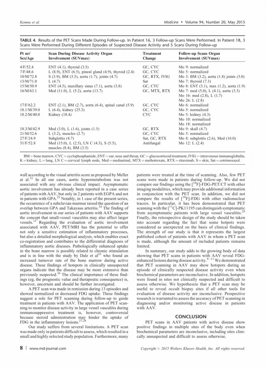
TABLE 4. Results of the PET Scans Made During Follow-up. In Patient 16, 3 Follow-up Scans Were Performed. In Patient 18, 3Scans Were Performed During Different Episodes of Suspected Disease Activity and 5 Scans During Follow-up
Pt nr/Sex/Age
Scan During Disease Activity OrganInvolvement (SUVmax)
TreatmentChange
Follow-up Scans OrganInvolvement (SUVmax)
4/F/52.4 ENT (4.1), thyroid (3.5) GC, CYC Mo 9: normalized7/F/48.6 L (8.9), ENT (6.5), pineal gland (4.9), thyroid (2.4) GC, CYC Mo 5: normalized10/M/72.8 S (3.9), BM (3.5), aorta (1.7), joints (4.7) GC, RTX, IVIG Mo 3: BM (3.2), aorta (1.8) joints (3.0)13/M/71.0 L (4.7) Sat Mo 7: thyroid (7.3)15/M/50.9 ENT (4.5), maxillary sinus (7.1), aorta (3.8) GC, CYC Mo 8: ENT (3.1), max (1.2), aorta (1.9)16/M/63.1 Med (11.0), L (5.2), aorta (13.7) GC, MTX, RTX Mo 7: med (5.0), L (4.1), aorta (3.3)
Mo 16: med (2.8), L (1.7)Mo 26: L (2.8)
17/F/62.2 ENT (2.1), BM (2.7), aorta (6.4), spinal canal (5.9) GC, CYC Mo 6: normalized18.1/M/39.0 L (6.4), kidney (25.3) GC, CYC Mo 5: normalized18.2/M/40.0 Kidney (18.4) CYC Mo 5: kidney (4.3)
Mo 10: normalizedMo 18: normalized
18.3/M/42.9 Med (3.0), L (1.6), joints (1.5) GC, RTX Mo 9: skull (4.7)21/M/52.6 L (3.2), muscles (2.7) GC, CYC Mo 5: normalized27/F/24.9 Subglottis (4.7) Sat, GC, RTX Mo 8: subglottis (2.6), Med (16.0)31/F/52.8 Med (15.0), L (2.5), LN C (4.3), S (5.5),
muscles (8.4), BM (3.5)Antifungal Mo 12: L (2.4)
hro, M
Kemna et al. Medicine � Volume 94, Number 20, May 2015
wall according to the visual arteritis score as proposed by Mellerat al.22 In all our cases, aortic hypermetabolism was notassociated with any obvious clinical impact. Asymptomaticaortic involvement has already been reported in a case seriesof patients with AAV, but only in 2 patients with EGPA and notin patients with GPA.23 Notably, in 1 case of the present series,the occurrence of a subclavian murmur raised the question of anoverlap between GPA and Takayasu arteritis.24 The finding ofaortic involvement in our series of patients with AAV supportsthe concept that small-vessel vasculitis may also affect largervessels.13 Regarding the evaluation of aortic involvementassociated with AAV, PET/MRI has the potential to offernot only a sensitive estimation of inflammatory processes,but also a detailed morphological analysis, which enables exactco-registration and contributes to the differential diagnosis ofinflammatory aortic diseases. Pathologically enhanced uptakein the bone marrow is possibly related to chronic stimulationand is in line with the study by Dale et al25 who found anincreased turnover rate of the bone marrow during activedisease. These findings of hotspots in clinically unsuspectedorgans indicate that the disease may be more extensive thanpreviously suspected.26 The clinical importance of these find-ings (eg, the prognostic value and therapeutic consequence) is,however, uncertain and should be further investigated.
A PET scan was made in remission during 13 episodes andshowed normalized or decreased FDG uptake. These findingssuggest a role for PET scanning during follow-up to guidetreatment in patients with AAV. The application of PET scan-ning to monitor disease activity in large vessel vasculitis duringimmunosuppressive treatment is, however, controversialbecause steroid administration may hinder the uptake ofFDG in the inflammatory lesions.27,28
BM¼ bone marrow, CYC¼ cyclophosphamide, ENT¼ ear, nose and tK¼ kidney, L¼ lung, LN C¼ cervical lymph node, Med¼mediastinal
Our study suffers from several limitations. A PET scanwas made only in patients difficult to assess, which resulted in asmall and highly selected study population. Furthermore, many
8 | www.md-journal.com
patients were treated at the time of scanning. Also, few PETscans were made in patients during follow-up. We did notcompare our findings using the [18F]-FDG-PET/CT with otherimaging modalities, which may provide additional informationin conjunction with the PET scan. In addition, we did notcompare the results of [18F]-FDG with other radionucleartracers. In particular, it has been demonstrated that PETscanning with the [11C]-PK11195 can distinguish symptomaticfrom asymptomatic patients with large vessel vasculitis.29
Finally, the retrospective design of the study should be takeninto account regarding the fact that some hotpots wereconsidered as unexpected on the basis of clinical findings.The strength of our study is that it represents the largestpublished cohort of patients with AAV in whom a PET scanis made, although the amount of included patients remainslimited.
In summary, our study adds to the growing body of datashowing that PET scans in patients with AAV reveal FDG-enhanced lesions during disease activity.9–11 We demonstratedthat PET scanning in AAV may show hotspots during anepisode of clinically suspected disease activity even whenbiochemical parameters are inconclusive. In addition, hotspotswere found in sites not clinically suspected and difficult toassess otherwise. We hypothesize that a PET scan may beuseful to reveal occult biopsy sites if all other tools forevaluation of disease activity are inconclusive. Prospectiveresearch is warranted to assess the accuracy of PET scanning indiagnosing and/or monitoring active disease in patientswith AAV.
CONCLUSIONPET scans in AAV patients with active disease show
at, GC¼ glucocorticoid treatment, IVIG¼ intravenous immunoglobulin,TX¼methotrexate, RTX¼ rituximab, S¼ skin, Sat¼ cotrimoxazol.
positive findings in multiple sites of the body even whenbiochemical parameters are inconclusive, including sites clini-cally unsuspected and difficult to assess otherwise.
Copyright # 2015 Wolters Kluwer Health, Inc. All rights reserved.

ACKNOWLEDGMENTS
We would like to thank Mr Salim Anthony Joly for hisvaluable contribution in the collection of data for the manu-script.
REFERENCES
1. Wilde B, van Paassen P, Witzke O, et al. New pathophysiological
insights and treatment of ANCA-associated vasculitis. Kidney Int.
2011;79:599–612.
2. Gotthardt M, Bleeker-Rovers CP, Boerman OC, et al. Imaging of
Inflammation by PET, conventional scintigraphy, and other imaging
techniques. J Nucl Med. 2010;51:1937–1949.
3. Mostard RLM, Voo S, van Kroonenburgh MJPG, et al. Inflammatory
activity assessment by F18 FDG-PET/CT in persistent symptomatic
sarcoidosis. Respir Med. 2011;105:1917–1924.
4. Fletcher JW, Djulbegovic B, Soares HP, et al. Recommendations on
the use of 18F-FDG PET in oncology, Journal of nuclear medicine:
official publication. Soc Nucl Med. 2008;49:480–508.
5. Fuchs M, Briel M, Daikeler T, et al. The impact of 18F-FDG PET
on the management of patients with suspected large vessel vasculitis.
Eur J Nucl Med Mol Imag. 2012;39:344–353.
6. Zerizer I, Tan K, Khan S, et al. Role of FDG-PET and PET/CT in
the diagnosis and management of vasculitis. Eur J Radiol.
2010;73:504–509.
7. Treglia G, Mattoli M, Leccisotti L, et al. Usefulness of whole-body
fluorine-18-fluorodeoxyglucose positron emission tomography in
patients with large-vessel vasculitis: a systematic review. Clin
Rheumatol. 2011;30:1265–1275.
8. Blockmans D, Ceuninck Ld, Vanderschueren S, et al. Repetitive
18F-fluorodeoxyglucose positron emission tomography in giant cell
arteritis: A prospective study of 35 patients. Arthritis Care Res.
2006;55:131–137.
9. Durme C, Kisters J, Paassen P, et al. Multiple endocrine abnormal-
ities. Lancet. 2011;378:540.
10. Ito K, Minamimoto R, Yamashita H, et al. Evaluation of Wegener’s
granulomatosis using 18F-fluorodeoxyglucose positron emission
tomography/computed tomography. Ann Nucl Med. 2013;27:209–
216.
11. Soussan M, Abisror N, Abad S, et al. FDG-PET/CT in patients with
ANCA-associated vasculitis: case-series and literature review. Auto-
immun Rev. 2014;13:125–131.
12. Teirstein A, Machac J, Almeida O, et al. Results of 188 whole-body
fluorodeoxyglucose positron emission tomography scans in 137
patients with sarcoidosis. Chest. 2007;132:1949–1953.
Medicine � Volume 94, Number 20, May 2015
Chapel Hill Consensus Conference Nomenclature of Vasculitides.
Arthritis Rheum. 2013;65:1–11.
Copyright # 2015 Wolters Kluwer Health, Inc. All rights reserved.
14. Mukhtyar C, Guillevin L, Cid MC, et al. EULAR recommendations
for the management of primary small and medium vessel vasculitis.
Ann Rheum Dis. 2009;68:310–317.
15. Hellmich B, Flossmann O, Gross WL, et al. EULAR recommenda-
tions for conducting clinical studies and/or clinical trials in systemic
vasculitis: focus on anti-neutrophil cytoplasm antibody-associated
vasculitis. Ann Rheum Dis. 2007;66:605–617.
16. Damoiseaux JGMC, Slot MC, Vaessen M, et al. Evaluation of a new
fluorescent-enzyme immuno-assay for diagnosis and follow-up of
ANCA-associated vasculitis. J Clin Immunol. 2005;25:202–208.
17. Jager R, Poot L, Piers D, et al. Clinical value of gallium-67
scintigraphy in assessment of disease activity in Wegener’s granulo-
matosis. Ann Rheum Dis. 2003;62:659–662.
18. Gave AA, Torres R, Kaplan L. Cryptococcal myositis and vasculitis:
an unusual necrotizing soft tissue infection. Surg Infect. 2004;5:309–
313.
19. Konstantinov KN, Ulff-Møller CJ, Tzamaloukas AH. Infections and
antineutrophil cytoplasmic antibodies: triggering mechanisms. Auto-
immun Rev. 2015;14:201–203.
20. Stone JH. Limited versus severe Wegener’s granulomatosis: baseline
data on patients in the Wegener’s granulomatosis etanercept trial.
Arthritis Rheum. 2003;48:2299–2309.
21. Lionaki S, Hogan SL, Falk RJ, et al. Association between thyroid
disease and its treatment with ANCA small-vessel vasculitis: a case-
control study. Nephrol Dial Transplant. 2007;22:3508–3515.
22. Meller J, Strutz F, Siefker U, et al. Early diagnosis and follow-up of
aortitis with [(18)F]FDG PET and MRI. Eur J Nucl Med Mol Imag.
2003;30:730–736.
23. Soussan M, Abad S, Mekinian A, et al. Detection of asymptomatic
aortic involvement in ANCA-associated vasculitis using FDG PET/
CT. Clin Exp Rheumatol. 2013;31:S56–S58.
24. Vandergheynst F, Goldman S, Cogan E. Wegener’s granulomatosis
overlapping with Takayasu’s arteritis revealed by FDG-PET scan.
Eur J Intern Med. 2007;18:148–149.
25. Dale DC, Fauci AS, Wolff SM. The effect of cyclophosphamide on
leukocyte kinetics and susceptibility to infection in patients with
wegener’s granulomatosis. Arthritis Rheum. 1973;16:657–664.
26. Kemna MJ, Tervaert JWC. Does one size fit all? J Rheumatol.
2013;40:1781–1784.
27. Blockmans D. Diagnosis and extension of giant cell arteritis.
Contribution of imaging techniques. La Presse Medicale.
2012;41:948–954.
28. Both M, Ahmadi-Simab K, Reuter M, et al. MRI and FDG-PET in
the assessment of inflammatory aortic arch syndrome in complicated
courses of giant cell arteritis. Ann Rheum Dis. 2008;67:1030–1033.
PET Scanning in AAV
29. Pugliese F, Gaemperli O, Kinderlerer AR, et al. Imaging of vascular
inflammation with [11C]-PK11195 and positron emission tomogra-
13. Jennette JC, Falk RJ, Bacon PA, et al. 2012 revised Internationalphy/computed tomography angiography. J Am Coll Cardiol.
2010;56:653–661.
www.md-journal.com | 9