Thucydides' Other "Traps": The United States, China, and the ...
Upload
khangminh22Category
view
1download
0
THE ROLE OF NEUTROPHIL EXTRACELLULAR TRAPS IN
THE PATHOGENESIS OF PERIODONTAL DISEASES
by
Phillipa Claire White
A thesis submitted to
The University of Birmingham
for the degree of
DOCTOR OF PHILOSOPHY
Department of Periodontology
School of Dentistry
College of Medical & Dental Sciences
The University of Birmingham
September 2015

University of Birmingham Research Archive
e-theses repository This unpublished thesis/dissertation is copyright of the author and/or third parties. The intellectual property rights of the author or third parties in respect of this work are as defined by The Copyright Designs and Patents Act 1988 or as modified by any successor legislation. Any use made of information contained in this thesis/dissertation must be in accordance with that legislation and must be properly acknowledged. Further distribution or reproduction in any format is prohibited without the permission of the copyright holder.

ABSTRACT
This thesis investigated neutrophil extracellular traps (NETs) in the pathogenesis of
periodontal diseases, including chronic periodontitis, experimental gingivitis and Papillon
Lefèvre syndrome (PLS). In vitro assays investigated the interactions between periodontal
bacteria and peripheral neutrophils isolated by discontinuous Percoll gradients, and
demonstrated differential NET release in response to bacteria. Interestingly, NETs entrapped
all periodontal bacteria assayed to some extent; however bacterial growth and survival were
not impeded. A longitudinal intervention clinical study of chronic periodontitis patients and
matched healthy controls revealed no differences in peripheral NET production; however
NET production by patients decreased following non-surgical treatment. Furthermore, a
subset of patients displayed impeded NET degradation by plasma that was restored following
disease treatment; this may be the result of increased circulating immunoglobulins and free
light chains (FLCs) pre-treatment. Peripheral NET production did not change throughout the
experimental gingivitis model study; however NET release was impeded in PLS patients
relative to healthy controls. Additional in vitro studies demonstrated that cigarette smoking
had an inhibitory effect on NET release. Collectively, this thesis indicates that NETs
contribute to innate immunity, however, given that periodontitis pathogenesis is characterised
by aberrant neutrophil responses, NETs may also be involved in the progression of the
disease.

ACKNOWLEDMENTS
I would like to begin by thanking my PhD supervisors Professor Iain Chapple, Professor Paul
Cooper and Dr Mike Milward for their continued guidance and encouragement. I feel
extremely lucky to have supervisors who care so much about my work and I am grateful for
their support in response to my (endless) questions throughout my PhD. I want to express my
gratitude to Dr Helen Wright, Dr Melissa Grant and Dr Naomi Hubber, all of whom have
helped a great deal in the last few years. I would also like to thank Dr Adam Usher and Dr
Iru Dias, who helped with the Dextran neutrophil isolation and flow cytometry, respectively.
I would like to acknowledge the University of Birmingham Dental School, the Medical
Research Council, the British Society of Periodontology and the Oral & Dental Research
Trust for funding during my PhD. I am extremely grateful for the additional funding I
benefitted from, which made it possible to go to several fascinating international conferences.
I wish to thank all of the postgraduate students at the Dental School, in particular those in the
7th
floor post-graduate office, all of whom kept their sense of humour even on days when I
had lost mine! I would also like to say a special thank you to Helen Roberts, who as well as
being a great desk neighbour, will be a dear friend for life. In addition, I am very
appreciative to all of those who repeatedly rolled up their sleeves to donate their blood in the
name of science.
Most importantly, I would like to thank my family for their endless support throughout my
PhD. In particular my husband, David White, who could no doubt write his own version of
my thesis considering how much I’ve talked about my PhD.

TABLE OF CONTENTS
CHAPTER 1: INTRODUCTION ........................................................................................... 1
1.1 Neutrophilic polymorphonuclear leukocytes (PMNLs) ................................................. 2
1.1.1 Neutrophil origin, maturation and longevity ........................................................ 3
1.1.2 The neutrophil extravasation cascade ................................................................... 4
1.1.3 Chemotaxis ........................................................................................................... 6
1.1.4 Neutrophil killing .................................................................................................. 7
1.1.4.1 Neutrophil cytoplasmic granules ........................................................... 7
1.1.4.2 Neutrophil oxidative killing ................................................................... 8
1.1.4.3 Intracellular killing: phagocytosis........................................................ 10
1.1.4.4 Extracellular killing: degranulation ..................................................... 11
1.1.4.5 Extracellular killing: Neutrophil extracellular traps (NETs) ............... 11
1.2 The process of NET release ............................................................................................. 14
1.2.1 Requirements for NET release ............................................................................ 15
1.2.2 Stimuli that induce NET release ......................................................................... 17
1.2.2.1 Ex vivo stimuli...................................................................................... 19
1.2.2.2 Bacteria-derived stimuli ....................................................................... 19
1.2.2.3 Protozoan/parasitic and fungal-derived stimuli ................................... 20
1.2.2.4 Host-derived NET stimuli .................................................................... 21
1.3 Microbial virulence factors used to bypass NET effects .............................................. 22
1.3.1 Formation of a bacterial capsule ......................................................................... 22
1.3.2 Bacterial surface charge modifications ............................................................... 23
1.3.3 Deoxyribonucleases (DNases) ............................................................................ 24
1.4 The role of NETs in the immune response .................................................................... 25
1.5 NET degradation and removal ....................................................................................... 28
1.5.1 Host-derived DNase ............................................................................................ 28
1.6 NETs and autoimmune processes ................................................................................... 30
1.6.1 Rheumatoid arthritis (RA) .................................................................................. 30
1.6.2 Systemic lupus erythematosus (SLE) ................................................................. 31
1.6.3 Cystic fibrosis (CF) ............................................................................................. 32
1.7 Periodontitis...................................................................................................................... 33
1.7.1 Role of the microbial biofilm in periodontitis .................................................... 35

1.7.2 The contribution of the immune response to host periodontal tissue damage .... 38
1.7.3 Risk factors for periodontitis .............................................................................. 43
1.8 The role of NETs in periodontitis ................................................................................... 46
1.9 Neutrophil isolation and ex vivo handling ..................................................................... 50
1.9.1 Neutrophil isolation: density gradient materials ................................................. 51
1.10 Aims of this study ........................................................................................................... 52
CHAPTER 2: MATERIALS AND METHODS ................................................................. 54
2.1 MATERIALS ................................................................................................................... 55
2.1.1 GENERAL BUFFERS........................................................................................ 55
2.1.1.1 Phosphate Buffered Saline (PBS) ........................................................ 55
2.1.1.2 PBS supplemented with glucose and cations (gPBS) .......................... 55
2.1.1.3 Blocking buffer: 1% bovine serum albumin (BSA) ............................ 55
2.1.1.4 4-(2-Hydroxyethyl) piperazine-1-ethanesulfonic acid, N-(2-
Hydroxyethyl) piperazine-N′-(2-ethanesulfonic acid) (HEPES) buffer .......... 55
2.1.1.5 0.9% saline solution ............................................................................. 56
2.1.2 NEUTROPHIL PREPARATION AND ASSAYS ............................................. 56
2.1.2.1.1 Preparation of Percoll gradients ............................................ 56
2.1.2.1.2 Lysis Buffer .......................................................................... 56
2.1.2.1.3 Preparation of Dextran and Percoll gradients ....................... 56
2.1.2.1.4 2% Dextran ........................................................................... 57
2.1.2.1.5 90% isotonic stock Percoll .................................................... 57
2.1.2.1.6 Luminol ................................................................................. 57
2.1.2.1.7 Isoluminol ............................................................................. 57
2.1.2.1.8 Lucigenin .............................................................................. 58
2.1.2.2 Micrococcal nuclease (MNase)............................................................ 58
2.1.2.3 Sytox green .......................................................................................... 58
2.1.2.4 Fluorescein-5-isothiocyanate (FITC) ................................................... 58
2.1.2.5 Cytochalasin B ..................................................................................... 59
2.1.3 BACTERIAL CULTURE ................................................................................... 59
2.1.3.1 Bacterial stimuli ................................................................................... 59
2.1.3.2 Blood agar plates.................................................................................. 59
2.1.3.3 Fastidious plates ................................................................................... 59
2.1.3.4 Tryptone soya agar (TSA) plates ......................................................... 60

2.1.3.5 Brain heart infusion (BHI) broth.......................................................... 60
2.1.3.6 Fastidious anaerobe broth .................................................................... 60
2.1.3.7 Typtone soya broth (TSB) ................................................................... 60
2.1.3.8 Crystal violet ........................................................................................ 61
2.1.3.9 Carbon fuchsin ..................................................................................... 61
2.1.3.10 Glutaraldehyde in sodium cacodylate buffer ..................................... 61
2.1.4 CIGARETTE SMOKING REAGENTS ............................................................. 61
2.1.4.1 Nicotine ................................................................................................ 61
2.1.4.2 Cotinine ................................................................................................ 62
2.1.4.3 Thiocyanate (SCN-) ............................................................................. 62
2.1.5 PCR STUDIES.................................................................................................... 62
2.1.5.1 The synthesis of complementary DNA (cDNA) .................................. 62
2.1.5.2 Design and validation of PCR primers ................................................ 63
2.1.6 CELL CULTURE ............................................................................................... 65
2.1.6.1 Culture of an oral epithelium cell line ................................................. 65
2.1.6.2 Supplemented Dulbecco’s Modified Eagle’s Medium:nutrient mixture
F-12 (DMEM/F-12) ......................................................................................... 65
2.1.6.3 Trypsin Ethylenediaminetetraacetic Acid (T-EDTA) .......................... 65
2.2 METHODS ....................................................................................................................... 66
2.2.1 ISOLATION OF NEUTROPHILS FROM PERIPHERAL BLOOD ................ 66
2.2.1.1 Percoll components .............................................................................. 66
2.2.1.1.1 Preparation of Percoll gradients ............................................ 66
2.2.1.1.2 Neutrophil isolation with Percoll .......................................... 66
2.2.1.1.3 Isolation of neutrophils by Histopaque gradients ................. 67
2.2.1.1.4 Isolation of neutrophils using Dextran .................................. 67
2.2.1.1.5 Neutrophil cell counting ....................................................... 68
2.2.1.2 NEUTROPHIL VIABILITY AND METABOLIC ACTIVITY ......... 68
2.2.1.2.1 Confirmation of neutrophil isolation by cytospin ................. 68
2.2.1.2.2 Confirmation of neutrophil isolation by flow cytometry ...... 69
2.2.1.2.3 Trypan blue exclusion ........................................................... 69
2.2.1.2.4 Quantification of neutrophil caspase-3 and 7 activity .......... 69
2.2.1.2.5 Quantification of neutrophil adenosine triphosphate (ATP)
activity.................................................................................................. 70

2.2.2 NEUTROPHIL ACTIVATION .......................................................................... 70
2.2.2.1 Stimuli employed to activate neutrophils ............................................ 70
2.2.2.2 Determination of the concentration of HOCl ....................................... 71
2.2.2.2.1 Culture of periodontal bacteria ............................................. 71
2.2.2.2.2 Determination of bacterial growth ........................................ 71
2.2.2.2.3 Heat-killing of bacteria ......................................................... 72
2.2.2.2.4 Comparison of live and heat-killed bacteria ......................... 72
2.2.2.2.5 Opsonisation of S. aureus ..................................................... 72
2.2.2.2.6 Gram-stain protocol .............................................................. 73
2.2.2.3 Modulators of neutrophil activation..................................................... 76
2.2.3 NEUTROPHIL ASSAYS ................................................................................... 77
2.2.3.1 Treatment of neutrophil assay plastic-ware ......................................... 77
2.2.3.2 Chemiluminescence protocol for ROS assay ....................................... 77
2.2.3.3 Quantification of NET-DNA ............................................................... 77
2.2.3.4 Quantification of NET-bound components .......................................... 78
2.2.3.4.1 Neutrophil elastase (NE) ....................................................... 79
2.2.3.4.2 Myeloperoxidase (MPO) ...................................................... 79
2.2.3.4.3 Cathepsin G (CG) ................................................................. 79
2.2.3.5 NET entrapment of bacteria ................................................................. 80
2.2.3.6 NET killing of bacteria ........................................................................ 81
2.2.3.7 NET fluorescence visualisation ........................................................... 82
2.2.3.8 Scanning electron microscopy (SEM) of NETs ................................... 82
2.2.4 CIGARETTE SMOKING AND NEUTROPHIL RESPONSES ........................ 83
2.2.4.1 Extraction and preparation of cigarette smoke extract (CSE) ............. 83
2.2.4.1.1 Estimation of CSE concentration .......................................... 84
2.2.4.2 Cigarette components........................................................................... 85
2.2.4.2.1 Impact of CSE and CSE components on NET production ... 85
2.2.4.2.2 NET visualisation of CSE and CSE component-treated
neutrophils............................................................................................ 85
2.2.4.2.3 Neutrophil Chemotaxis assay ............................................... 85
2.2.4.2.4 Neutrophil chemotaxis analysis ............................................ 87
2.2.4.3 Gene expression of CSE and SCN- treated neutrophils....................... 88
2.2.4.3.1 Storage of neutrophils for subsequent RNA isolation .......... 88
2.2.4.3.2 Isolation of RNA ................................................................... 88

2.2.4.3.3 Quantification of RNA .......................................................... 89
2.2.4.3.4 The synthesis of complementary DNA (cDNA) ................... 89
2.2.4.3.5 Quantitative real time PCR analysis ..................................... 90
2.2.4.3.6 Validation of PCR primers and assays.................................. 90
2.2.4.3.7 Analysis of gene expression levels ....................................... 91
2.2.5 NET DEGRADATION AND THE EFFECT OF DELAYED NET REMOVAL
...................................................................................................................................... 94
2.2.5.1 NET degradation with plasma ............................................................. 94
2.2.5.2 Quantification of immunoglobulin G (IgG), free light chains (FLCs),
and cystatin C ................................................................................................... 94
2.2.5.3 Collection of NET supernatants ........................................................... 95
2.2.5.4 NET supernatant concentration ............................................................ 95
2.2.5.5 NET supernatants effects on oral epithelial cells (OECs) ................... 96
2.2.5.5.1 H400 oral epithelial cell storage and recovery ..................... 96
2.2.5.5.2 Cell passage .......................................................................... 97
2.2.5.5.3 Determination of cell concentrations and viability ............... 97
2.2.5.6 Culture of H400 oral epithelial cells in a 96-well format .................... 98
2.2.5.7 Determination of H400 oral epithelial cell lactate dehydrogenase
release .............................................................................................................. 98
2.2.6 NEUTROPHIL RESPONSES IN PERIODONTAL HEALTH AND DISEASE
...................................................................................................................................... 98
2.2.6.1 NEUTROPHIL RESPONSES IN GINGIVITIS ................................. 98
2.2.6.2 21-day gingivitis model ....................................................................... 98
2.2.6.2.1 Clinical measures of gingival inflammation ......................... 99
2.2.6.2.2 Plaque sampling .................................................................. 100
2.2.6.2.3 Collection of plasma ........................................................... 100
2.2.6.2.4 Plaque as a neutrophil stimulus........................................... 101
2.2.6.2.5 Characterisation of plaque samples .................................... 102
2.2.6.2.5.1 Quantification of endotoxin ................................. 102
2.2.6.2.5.2 Quantification of DNA ........................................ 102
2.2.6.2.5.3 Quantification of protein ...................................... 103
2.2.6.3 NEUTROPHIL RESPONSES IN CHRONIC PERIODONTITIS .... 103
2.2.6.3.1 Chronic periodontitis clinical study volunteers .................. 103
2.2.6.3.2 Clinical measures of gingival inflammation ....................... 104

2.2.6.4 NEUTROPHIL RESPONSES IN PAPILLON LEFÈVRE
SYNDROME (PLS)....................................................................................... 105
2.2.6.4.1 PLS study volunteers .......................................................... 105
2.2.6.4.2 Quantification of NE and LL-37 in plasma samples .......... 105
2.2.6.4.2.1 Quantification of NE ............................................ 106
2.2.6.4.2.2 Quantification of LL-37 ....................................... 106
2.2.7 STATISTICAL ANALYSIS ............................................................................ 107
CHAPTER 3: NEUTROPHIL ISOLATION TECHNIQUES AND THEIR EFFECTS
UPON EX VIVO NEUTROPHIL BEHAVIOUR.............................................................. 108
3.1 Introduction .................................................................................................................... 109
3.2 Comparisons between isolation techniques ................................................................. 110
3.2.1 Cell yield: Percoll vs Histopaque ...................................................................... 110
3.2.2 Cell yield: Percoll vs Dextran ........................................................................... 110
3.2.3 Total peak ROS production: Percoll vs Histopaque isolation procedures ........ 112
3.2.4 Total peak ROS production: Percoll vs Dextran isolation procedures ............. 112
3.2.5 Total ROS production over stimulation period: Percoll vs Histopaque isolation
procedures .................................................................................................................. 114
3.2.6 Total ROS production over stimulation period: Percoll vs Dextran isolation
procedures .................................................................................................................. 114
3.2.7 NET production: Percoll vs Histopaque isolation procedures .......................... 117
3.2.8 NET production: Percoll vs Dextran isolation procedures ............................... 117
3.2.9 Metabolic activity: Percoll vs Histopaque ........................................................ 119
3.2.10 Metabolic activity: Percoll vs Dextran ........................................................... 119
3.2.11 Caspase activity: Percoll vs Histopaque ......................................................... 121
3.2.12 Caspase activity: Percoll vs Dextran ............................................................... 121
3.3 Efficiency of neutrophil preparation using Percoll..................................................... 123
3.3.1 Validation of chosen neutrophil preparation technique using Percoll gradients
.................................................................................................................................... 123
3.3.1.1 Cytospin of Percoll-isolated neutrophils ............................................ 123
3.3.1.2 CD66 staining of neutrophil preparations .......................................... 123
3.3.1.3 Flow cytometric identification of neutrophils.................................... 125
3.4 Discussion........................................................................................................................ 127

CHAPTER 4: INTERACTIONS BETWEEN PERIODONTAL BACTERIA AND
PERIPHERAL BLOOD NEUTROPHILS ........................................................................ 133
4.1 Introduction .................................................................................................................... 134
4.2 Neutrophil ROS release in response to periodontal bacteria..................................... 134
4.2.1 Time-course of neutrophil ROS production in response to periodontal bacteria
.................................................................................................................................... 138
4.3 Quantification of NET production in response to periodontal bacteria exposure... 140
4.4 NET entrapment of bacteria ......................................................................................... 144
4.5 Scanning electron microscopy of NET-bacteria interactions .................................... 150
4.6 Quantification of NET-mediated killing of periodontal bacteria .............................. 153
4.7 Effect of NADPH-oxidase pathway modulating agents on ROS and NET production .
................................................................................................................................. 156
4.8 Effect of TLR inhibition on ROS and NET production ............................................. 161
4.9 Discussion........................................................................................................................ 166
CHAPTER 5: NET PRODUCTION BY PERIPHERAL BLOOD NEUTROPHILS IN
CHRONIC PERIODONTITIS: A LONGITUDINAL INTERVENTION STUDY ...... 175
5.1 Introduction .................................................................................................................... 176
5.2 Volunteer information ................................................................................................... 176
5.2.1 Clinical measures of periodontitis .................................................................... 178
5.2.2 Clinical attachment loss .................................................................................... 178
5.2.3 Periodontal Pocket depth .................................................................................. 178
5.2.4 Bleeding on probing .......................................................................................... 179
5.2.5 Cumulative gingival index ................................................................................ 179
5.2.6 Plaque index ...................................................................................................... 179
5.3 Comparison of NET production by periodontitis patients and healthy matched
controls ................................................................................................................................. 185
5.3.1 NET production by periodontitis patients and healthy matched controls
following successful treatment .................................................................................. 187
5.3.2 Fluorescence microscopy of NETs from patient and controls pre-treatment ... 190
5.3.3 Fluorescence microscopy of NETs from patient and controls post-treatment .. 190
5.4 Effect of age on NET production .................................................................................. 193
5.5 Analysis of the association between severity of periodontitis and NET production
pre- and post-treatment ....................................................................................................... 195
5.6 Discussion........................................................................................................................ 198

CHAPTER 6: NET DEGRADATION AND ITS POTENTIAL EFFECTS ON
NEUTROPHIL RESPONSES AND THE ORAL EPITHELIUM .................................. 205
6.1 Introduction .................................................................................................................... 206
6.2 NET degradation over 24 hours ................................................................................... 207
6.2.1 NET degradation with plasma .......................................................................... 207
6.2.2 NET degradation with plasma over 24 hours ................................................... 207
6.3 NET degradation by plasma from periodontitis patient’s pre- and post-periodontal
treatment ............................................................................................................................... 210
6.3.1 Variability in NET degradation with patient age .............................................. 212
6.3.2 MNase-treated patient plasma ........................................................................... 212
6.3.3 Plasma IgG concentrations in periodontitis and controls ................................. 214
6.3.3.1 Plasma IgG subclasses concentration plotted against NET degradation .
...................................................................................................................... 216
6.3.4 Plasma FLC concentrations in periodontitis and controls ................................ 217
6.3.4.1 Plasma FLC concentration plotted against NET degradation ............ 219
6.3.5 Plasma-derived cystatin C detection in periodontitis ........................................ 221
6.4 Determination of the DNA-content in NET supernatants.......................................... 223
6.4.1 NET supernatants as a stimulus for ROS production ....................................... 224
6.4.2 NET supernatants as a stimulus for NET production ....................................... 227
6.4.3 The ability of NETs to induce neutrophil chemotaxis ...................................... 229
6.4.3.1 Neutrophil speed in response to NET supernatants ........................... 231
6.4.3.2 Neutrophil velocity in response to NET supernatants ....................... 233
6.4.3.3 Directional accuracy of neutrophil movement (resultant vector length) .
...................................................................................................................... 234
6.4.3.4 Neutrophil resultant vector length ..................................................... 236
6.5 The effect of NETs on H400 oral epithelial cell responses ......................................... 238
6.5.1 The effect of NETs on H400 oral epithelial cell apoptosis ............................... 238
6.5.2 The effect of NETs on H400 oral epithelial cell metabolic activity ................. 239
6.5.3 The effect of NETs on H400 oral epithelial cell lactate dehydrogenase release
.................................................................................................................................... 241
6.6 Discussion........................................................................................................................ 243
CHAPTER 7: EFFECT OF CIGARETTE SMOKE EXTRACT ON NEUTROPHIL
RESPONSES ........................................................................................................................ 250

7.1 Introduction .................................................................................................................... 251
7.2 Effect of cigarette smoke extract on NET release ....................................................... 252
7.2.1 Fluorescence visualisation of NETs following treatment with CSE ................ 252
7.3 Effect of CSE components on NET release.................................................................. 255
7.3.1 Fluorescence visualisation of NETs following nicotine, cotinine and thiocyanate
treatment .................................................................................................................... 260
7.3.2 Cell viability following treatment with CSE and CSE components ................. 262
7.4 Neutrophil chemotactic accuracy ................................................................................. 263
7.4.1 Analysis of migration of CSE-treated neutrophils in response to fMLP using
spider plots ................................................................................................................. 263
7.4.1.1 Speed of CSE treated neutrophils ...................................................... 266
7.4.1.2 Velocity of CSE treated neutrophils .................................................. 268
7.4.1.3 Directional accuracy of neutrophil movement (resultant vector length) .
...................................................................................................................... 270
7.4.1.4 Resultant vector length of CSE-treated neutrophil migration............ 273
7.5 Gene expression following CSE and SCN- treatment ................................................ 275
7.5.1 Impact of CSE on neutrophil gene expression .................................................. 275
7.5.2 Impact of thiocyanate on neutrophil gene expression ....................................... 275
7.6 Discussion........................................................................................................................ 278
CHAPTER 8: NET PRODUCTION IN OTHER INFLAMMATORY PERIODONTAL
DISORDERS: GINGIVITIS & PAPILLON LEFÈVRE SYNDROME ........................ 285
8.1 Introduction .................................................................................................................... 286
8.2 Experimental gingivitis .................................................................................................. 286
8.2.1 Clinical measures of gingivitis.......................................................................... 287
8.2.1.1 Plaque index ....................................................................................... 287
8.2.1.2 Gingival index .................................................................................... 287
8.2.1.3 Gingival crevicular fluid .................................................................... 287
8.2.2 NET production during the 21-day experimental gingivitis model .................. 291
8.2.3 Visualisation of NET production during the 21-day experimental gingivitis
model.......................................................................................................................... 291
8.2.4 NET degradation by volunteer plasma during the 21-day experimental gingivitis
model.......................................................................................................................... 294
8.2.4.1 Plasma IgG concentrations during the 21-day experimental gingivitis
model.............................................................................................................. 296
8.2.4.2 Plasma FLC concentration during the 21-day experimental gingivitis
model.............................................................................................................. 298
8.2.5 Neutrophil ROS and NET production in response to plaque stimulation ......... 302
8.3 Papillon Lefèvre syndrome ........................................................................................... 306
8.3.1 Comparison of NET production by PLS patients and healthy controls ............ 308
8.3.1.1 Quantification of NET-bound components in PLS patients and healthy
controls ........................................................................................................... 308
8.3.2 Fluorescence visualisation of NET production in PLS patients ....................... 312
8.3.3 Quantification of plasma NE in PLS ................................................................ 314
8.3.4 Quantification of plasma LL-37 in PLS............................................................ 314
8.4 Discussion........................................................................................................................ 316
CHAPTER 9: CONCLUDING REMARKS ..................................................................... 322
9.1 Summary of main findings ............................................................................................ 323
9.2 Overall conclusion and recommendations for future research ................................. 328
REFERENCES…………………………………………………………………………….331
Appendix I: ROS and NET production in response to live and dead bacteria……………..359
Appendix II: NET killing preliminary assays……………………………………………...361
Appendix III: Validation of the quantification of NET-bound components………………364
Appendix IV: Gram stains of bacteria……………………………………………………..365
Appendix V: Comparisons between free light chain detection in plasma and serum
samples……………………………………………………………………………………...367
Appendix VI: Collection and storage of NET supernatants……………………………….369
Appendix VII: H400 cell counting………………………………………………………...371
Appendix VIII: Effect of cigarette smoke extract and cigarette smoke extract components
on the fluorometric quantification of calf thymus DNA…………………………………....373
Appendix IX: Isolation of neutrophil RNA……………………………………………….375
Appendix X: Determination of stable housekeeping genes………………………………..377
Appendix XI: RNA expression of CSE treated neutrophils relative to other reference
genes………………………………………………………………………………………...379
Appendix XII: RNA expression of thiocyanate treated neutrophils relative to other reference
genes………………………………………………………………………………………...381

LIST OF FIGURES
CHAPTER 1: INTRODUCTION
1.1 Microanatomy of the human neutrophil……………………………………… 3
1.2 Neutrophil extravasation cascade…………………………………………….. 5
1.3 NADPH-oxidase pathway for the generation of ROS ……………………….. 10
1.4 Photomicrograph illustrating fluorescence microscopy of NETs…………….. 12
1.5 Confocal immunofluorescensce images of NET release……………………... 14
1.6 Schematic representation of the steps involved in nuclear NET release……... 16
1.7 NETs associated with bacteria………………………………………………... 25
1.8 NET degradation by DNase 1……………………………………………….. 29
1.9 Clinical symptoms of chronic periodontitis………………………………….. 34
1.10 Sequential bacterial colonisation which results in the formation of a plaque
biofilm………………………………………………………………….......... 37
1.11 Periodontal bacteria assigned to the Socransky colour complexes…………… 38
1.12 The pathogenic model of periodontitis……………………………………….. 41
1.13 Clinical signs and symptoms in a PLS case study……………………………. 43
1.14 NET entrapment of periodontal bacteria……………………………………. 48
1.15 NETs in human gingival tissues……………………………………………… 49
CHAPTER 2: MATERIALS AND METHODS
2.1 Diagrammatic representation of the quantification of NET-bound NE………. 80
2.2 Schematic representation showing the collection of CSE………………… 84
2.3 Photograph of Insall chemotaxis chamber……………………………………. 87
2.4 PCR cycling protocol…………………………………………………………. 92
2.5 Amplification curve, efficiency standard curve and melt curve for the
YWHAZ assay………………………………………………………………... 93
CHAPTER 3: NEUTROPHIL ISOLATION TECHNIQUES AND THEIR
EFFECTS UPON EX VIVO NEUTROPHIL BEHAVIOUR
3.1 The effect of neutrophil preparation techniques on cell numbers…………….. 111
3.2 The effect of neutrophil preparation technique on peak total ROS
production…………………………………………………………………….. 113
3.3 Neutrophil isolation time-course analysis of ROS production……………….. 115
3.4 Neutrophil isolation time-course analysis of ROS production……………….. 116
3.5 The effect of neutrophil preparation techniques on subsequent NET
production…………………………………………………………………….. 118
3.6 The effect of the neutrophil preparation technique on neutrophil metabolic
activity………………………………………………………………………… 120
3.7 The effect of the neutrophil preparation technique on caspase activity……… 122
3.8 Cytospin of Percoll-isolated neutrophils……………………………………… 124
3.9 Fluorescence microscopy of surface marker CD66…………………………... 124
3.10 Flow cytometric characterisation of neutrophils……………………………… 126
CHAPTER 4:
4.1 Neutrophil ROS production in response to periodontal bacteria…………….. 137
4.2 Time-course of neutrophil ROS production in response to periodontal
bacteria………………………………………………………………………. 139

4.3
Quantification of NET production in response to periodontal bacteria
exposure……………………………………………………………………….
143
4.4 NET entrapment of periodontal bacteria……………………………………… 149
4.5 Scanning electron microscopy of resting neutrophils………………………… 151
4.6 Scanning electron microscopy of neutrophils stimulated with periodontal
bacteria……………………………………………………………………….. 152
4.7 Quantification of NET-mediated killing of periodontal bacteria…………….. 155
4.8 Effect of NADPH-oxidase pathway modulating agents on ROS and NET
production…………………………………………………………………….. 160
4.9 Effect of TLR inhibition on ROS and NET production………………………. 165
CHAPTER 5: NET PRODUCTION BY PERIPHERAL BLOOD
NEUTROPHILS IN CHRONIC PERIODONTITIS: A LONGITUDINAL
INTERVENTION STUDY
5.1 NET production in periodontitis patients (pre-treatment) and healthy
controls………………………………………………………………………... 186
5.2 NET production in post-treatment periodontitis patients and healthy
controls………………………………………………………………………... 188
5.3 Pre- and post-treatment NET production by periodontitis patients…………... 189
5.4 Fluorescence microscopy of NETs from patient and controls pre-treatment… 191
5.5 Fluorescence microscopy of NETs from patient and controls post-
treatment……………………………………………………………………… 192
5.6 The effect of age on pre- and post-treatment NET production in patients and
healthy controls………………………………………………………………. 194
5.7 Analysis of the association between severity of periodontitis and NET
production pre- and post-treatment…………………………………………… 196
5.8 Analysis of the association between periodontitis disease severity and NET
production pre- and post-treatment…………………………………………… 197
CHAPTER 6: NET DEGRADATION AND ITS POTENTIAL EFFECTS ON
NEUTROPHIL RESPONSES AND THE ORAL EPITHELIUM
6.1 NET degradation over 24 hours………………………………………………. 208
6.2 NET degradation with plasma………………………………………………... 209
6.3 NET degradation by plasma over 24 hours…………………………………… 209
6.4 NET degradation by plasma from periodontitis patients pre- and post-
periodontal treatment…………………………………………………………. 211
6.5 NET degradation plotted against patient age…………………………………. 213
6.6 MNase-treated patient plasma………………………………………………… 213
6.7 Plasma IgG concentrations in periodontitis and controls…………………….. 215
6.8 Plasma IgG subclasses concentration plotted against NET degradation……... 216
6.9 Plasma FLC concentrations in periodontitis and controls……………………. 218
6.10 Plasma FLC concentration plotted against NET degradation………………… 220
6.11 Plasma-derived cystatin C detection in periodontitis…………………………. 222
6.12 NET supernatants as a stimulus for ROS production………………………… 226
6.13 NET supernatants as a stimulus for NET production………………………… 228
6.14 Spider plots representing neutrophil migration in response to NET
supernatants…………………………………………………………………… 230
6.15 Neutrophil speed in response to NET supernatants…………………………... 232
6.16 Neutrophil velocity in response to NET supernatants………………………... 233

6.17 Directional accuracy of neutrophil movement (resultant vector length)……... 235
6.18 Neutrophil resultant vector length……………………………………………. 237
6.19 The effect of NETs on H400 oral epithelial cell caspase activity…………….. 239
6.20 The effect of NETs on H400 oral epithelial cell metabolic activity………….. 240
6.21 The effect of NETs on H400 oral epithelial cell lactate dehydrogenase
release………………………………………………………………………… 242
CHAPTER 7: EFFECT OF CIGARETTE SMOKE EXTRACT ON
NEUTROPHIL RESPONSES
7.1 Effect of cigarette smoke extract on NET production………………………... 253
7.2 Fluorescence visualisation of NETs in response to cigarette smoke extract…. 254
7.3 a) Effect of nicotine on NET production……………………………………...
b) Effect of cotinine on NET production……………………………………..
c) Effect of thiocyanate (SCN-) on NET production………………………….
257
258
259
7.4 Fluorescence visualisation of NETs in response to CSE and its key
components…………………………………………………………………… 261
7.5 Cell viability following treatment with CSE and CSE components………….. 262
7.6 Analysis of migration of CSE-treated neutrophils in response to fMLP using
spider plots……………………………………………………………………. 264
7.7 Analysis of migration of CSE-treated neutrophils in response to IL-8 using
spider plots……………………………………………………………………. 265
7.8 Speed of CSE treated neutrophils…………………………………………….. 267
7.9 Velocity of CSE treated neutrophils………………………………………….. 269
7.10 Directional accuracy of neutrophil movement (resultant vector length)……... 271
7.11 Directional accuracy of neutrophil movement (resultant vector length)……... 272
7.12 Resultant vector length of CSE-treated neutrophil migration………………… 274
7.13 Gene expression following cigarette smoke extract or thiocyanate treatment.. 277
CHAPTER 8: NET PRODUCTION IN OTHER INFLAMMATORY
PERIODONTAL DISORDERS: GINGIVITIS & PLS
8.1 NET production in 21-day gingivitis model………………………………….. 292
8.2 Visualisation of NET production during the 21-day experimental gingivitis
model…………………………………………………………………………. 293
8.3 NET degradation by volunteer plasma during the 21-day experimental
gingivitis model ……………………………………………………………… 295
8.4 Plasma IgG concentrations in gingivitis……………………………………… 297
8.5 Plasma FLC concentration in gingivitis………………………………………. 299
8.6 The ability of a plaque biofilm to stimulate neutrophil responses……………. 301
8.7 ROS production in response to plaque stimulation…………………………… 303
8.8 NET production in response to plaque stimulation………………………… 305
8.9 Quantification of NET-bound components in PLS………………………… 311
8.10 Fluorescence visualisation of NET production in PLS patients……………… 313
8.11 Quantification of plasma derived neutrophil elastase and LL-37 in PLS…….. 315

LIST OF TABLES
CHAPTER 1: INTRODUCTION
1.1 All previously reported NET stimuli…………………………………… 18
1.2 Effect of smoking status on periodontal health………………………… 46
CHAPTER 2: MATERIALS AND METHODS
2.1 Preparation of Percoll density gradients………………………………... 56
2.2 Preparation of Percoll density gradients following Dextran
sedimentation…………………………………………………………… 57
2.3 Components and quantities used for cDNA synthesis…………………. 63
2.4 Primers employed to quantify gene expression in CSE and SCN-
treated neutrophils using real time PCR……………………………... 64
2.5 Characterisation of periodontal bacterial species………………………. 74
2.6 Culture conditions of periodontal bacterial species…………………… 75
2.7 Modulators of neutrophil activation…………………………………. 76
2.8 Neutrophil chemotaxis measurements………………………………….. 88
2.9 PCR cycling protocol…………………………………………………... 92
2.10 Outline of the 21-day gingivitis model study………………………… 101
CHAPTER 5: NET PRODUCTION BY PERIPHERAL BLOOD
NEUTROPHILS IN CHRONIC PERIODONTITIS: A LONGITUDINAL
INTERVENTION STUDY
5.1 Age and gender of volunteers in the study……………………………... 177
5.2 Clinical attachment loss in periodontitis patients………………………. 180
5.3 Comparison of probing pocket depth in periodontitis patients and
healthy controls………………………………………………………… 181
5.4 The number of sites that bled from the pocket base upon periodontal
probing in periodontitis patients and healthy controls…………………. 182
5.5 Gingival indices in periodontitis patients and healthy controls………... 183
5.6 Plaque indices in periodontitis patients and healthy controls…………... 184
CHAPTER 6: NET DEGRADATION AND ITS POTENTIAL EFFECTS
ON NEUTROPHIL RESPONSES AND THE ORAL EPITHELIUM
6.1 Determination of the DNA-content in NET supernatants……………… 224
CHAPTER 8: NET PRODUCTION IN OTHER INFLAMMATORY
PERIODONTAL DISORDERS: GINGIVITIS & PLS
8.1 21-day gingivitis model plaque index………………………………….. 288
8.2 21-day gingivitis model gingival index………………………………… 289
8.3 21-day gingivitis model gingival crevicular fluid……………………… 290
8.4 Clinical measures of PLS………………………………………………. 307

ABBREVIATIONS
ACPA Anti-citrullinated protein antibody
AFU Arbitrary fluorescence units
AMP Antimicrobial peptide
ANCA Anti-neutrophil cytoplasmic antibodies
ANOVA Analysis of variance
ATCC American Type Culture Collection
ATP Adenosine triphosphate
BAPTA 1,2-bis(o-aminophenoxy)ethane-N,N,N',N'-tetraacetic acid
BHI Brain heart infusion
BOP Bleeding on probing
BPI Bactericidal/permeability-increasing
BSA Bovine serum albumin
C Complement
Ca2+
Calcium ions
CAM Cell adhesion molecule
cDNA Complementary deoxyribonucleic acid
CF Cystic fibrosis
CFTR Cystic fibrosis transmembrane conductance regulator
CG Cathepsin G
CGD Chronic granulomatous disease
Cl- Chloride ions
CP Crossing point
CRP C-reactive protein
CSE Cigarette smoke extract
DAG Diacylglycerol
DEPC Diethylpyrocarbonate
DMEM/F-12 Dulbecco’s modified Eagle’s medium:nutrient mixture F-12
DMSO Dimethyl sulfoxide
DNA Deoxyribonucleic acid
DNase Deoxyribonuclease
DPI Diphenylene iodonium
dsDNA Double stranded DNA
dTNPmix Deoxynucleotide mix
EGTA Ethylene glycol tetraacetic acid
ELISA Enzyme linked immunosorbent assay
FCγR Fc-gamma receptor
Fiji Fiji is just Image J
FLC Free light chain
fMLP N-formyl-methionyl-leucyl-phenylalanine

FS Forward scatter
GAPDH Glyceraldehyde-3-phosphate dehydrogenase
GAS Group A streptococcal
GCF Gingival crevicular fluid
GI Gingival index
GM-CSF Granulocyte-macrophage colony-stimulating factors
GO Glucose oxidase
GPCR G-protein-coupled receptor
GSH Glutathione
GSSG Oxidised glutathione
GTP Guanosine triphosphate
H2O2 Hydrogen peroxide
HEPES 4-(2-Hydroxyethyl) piperazine-1-ethanesulfonic acid, N-(2-Hydroxyethyl)
piperazine-N′-(2-ethanesulfonic acid)
HIV Human immunodeficiency virus
HKG Housekeeping gene
HMDS Hexamethyldisilazane
HOCl Hypochlorous acid
HOSCN Hypothiocyanous acid
HPRT1 Hypoxanthine phosphoribosyltransferase 1
HRP Horseradish peroxidase
HSP Heat shock protein
HUVEC Human umbilical vein endothelial cells
ICAM Intercellular adhesion molecule
IgG Immunoglobulin G
IL- Interleukin-
K+ Potassium ions
KO Knock out
L Litre
LAL Limulus Amebocyte Lysate
LDH Lactase dehydrogenase
LPS Lipopolysaccharide
M Molar
Mins Minutes
MIP-1 alpha Macrophage inflammatory protein-1 alpha
mm Millimetre
MMP Matrix metalloproteinase
MNase Micrococcal nuclease
MOI Multiplicity of infection
MPO Myeloperoxidase
NAC N-acetyl-cysteine

NADPH Nicotinamide adenine dinucleotide phosphate
NE Neutrophil elastase
NEi Neutrophil elastase inhibitor
NETs Neutrophil extracellular traps
NFĸB Nuclear factor kappa B
Nm Nanometres
NO Nitric oxide
NS Not significant
O2- Superoxide
OCl- Hypochlorite ions
OD600nm Optical density at 600 nanometres
OEC Oral epithelium cell line
Oligo-dT Oligonucleotide deoxy-thymine
OxPAPC Oxidation of 1-palmitoyl-2-arachidonyl-sn- glycero-3-phosphorylcholine
PAD Peptidyl arginine deiminase
PAF Platelet activating factor
PAF Platelet activating factor
PAMP Pathogen-associated molecular patterns
PAR2 Protease-activated receptor-2
PBN Peripheral blood neutrophil
PBS Phosphate Buffered Saline
PCR Polymerase chain reaction
PI Plaque index
PKC Protein kinase C
PLS Papillon Lefèvre syndrome
PMA Phorbol 12-myristate 13-acetate
PMNL Polymorphonuclear leukocytes
PPAD P. gingivalis peptidyl arginine deiminase
PPD Probing pocket depth
PR3 Proteinase 3
PRM Pattern recognition molecule
PRR Pattern recognition receptor
PVP Polyvinylpyrrolidone
RA Rheumatoid arthritis
rcf Relative centrifugal force
RF Rheumatoid factor
rhDNase Recombinant deoxyribonuclease
RLU Relative light units
RNA Ribonucleic acid
RNase Ribonuclease
ROS Reactive oxygen species

RPL13 Ribosomal protein L13
RPMI Roswell Park Memorial Institute
RT Reverse transcriptase
SAP Serum amyloid P
SCN- Thiocyanate
SD Standard deviation
Secs Seconds
SEM Scanning electron microscopy
SEM Standard error of the mean
SIRS Systemic inflammatory response syndrome
SLE Systemic lupus erythematosus
SLPI Serum leukocyte protease inhibitor
SOD Superoxide dismutase
SS Side scatter
T-EDTA Trypsin ethylenediaminetetraacetic acid
TIMP Tissue inhibitors of metalloproteinases
TLR Toll like receptor
TMB 3,3’,5,5’-Tetramethylbenzidine
TNF Tumour necrosis factor
TSA Tryptone soya agar
TSB Tryptone soya broth
UC Ulcerative colitis
WT Wild type
YWHAZ Tyrosine 3/tryptophan, 5-monooxygenase activation protein, zeta polypeptide

1
INTRODUCTION CHAPTER 1:

2
1.1 Neutrophilic polymorphonuclear leukocytes (PMNLs)
Neutrophilic polymorphonuclear leukocytes (PMNLs), otherwise known as neutrophils, are
recognised for playing a critical role in both the innate and acquired (humoral) immune
response. The name PMNLs derives from the cells’ multi-lobed nucleus (Figure 1.1), which
distinguishes the neutrophil from other granulocytes, such as eosinophils, basophils and mast
cells (Kobayashi et al., 2009). The innate immune system is characterised by a rapid, though
poorly specific response to infection, which does not require previous exposure to
microorganisms to be effective. This process is facilitated by pattern recognition receptors
(PRR) on the PMNL membrane that recognise highly conserved pathogen-associated
molecular patterns (PAMPs) from foreign species or “non-self”. In contrast, the acquired
(humoral) immune system specifically targets previously encountered pathogens and thus
results in a delayed, but highly targeted and effective response (Kumar & Sharma 2010). In
addition to the neutrophil’s direct effector functions, such as phagocytosis and NETosis (see
1.1.4), they are able to modulate the activity of acquired immune cells by releasing signalling
molecules, such as cytokines and by expressing humoral pattern recognition molecules
(PRMs). Collectively, the diverse functions of neutrophils make them indispensable to both
the cellular and humoral arms of the acquired immune response (Jaillon et al., 2013).

3
Figure 1.1: Microanatomy of the human neutrophil
(a) Schematic of neutrophil activation and the subsequent sequencing cascade in response to
a microbe. (b) Transmission electron microscopy image of a neutrophil that has phagocytosed
a microbe (red arrows indicate the microbe, phagosome, granules and nuclei) (Kobayashi et
al., 2009).
Neutrophil origin, maturation and longevity 1.1.1
Neutrophil production initiates in the bone marrow, with an estimated two thirds of
haematopoiesis being assigned to the formation of myeloblasts (cells which are committed to
becoming granulocytes). In a healthy individual, neutrophil production is maintained at a
consistent level, with an estimated 1-2 x 10¹¹
neutrophils being generated per day (Savil et
al., 1989). However, neutrophil release significantly increases up to 10 fold (Furze & Rankin
2008) during periods of infection and inflammation, and the transient increase in circulating
neutrophils accommodates the heightened demands placed upon the immune system (Metcalf
et al., 1991). Whilst neutrophils were previously considered to be short-lived cells equipped
with an estimated half-life of 8 hours in humans, a recent study demonstrated that neutrophils
have a much longer circulatory lifespan of approximately 5.4 days (Pillay et al., 2010).
Precise adjustments of neutrophil levels are regulated by apoptosis pathways within the
tissues, whereby increased neutrophil apoptosis signals a reduction in the number of stem

4
cells assigned to myeloblast formation within bone marrow (Michlewska et al., 2009).
Apoptotic neutrophils, along with the particles they have engulfed, are efficiently cleared by
macrophages and dendritic cells, which recognise death signals on the neutrophil surface in
the form of phosphatidylserine groups (Michlewska et al., 2009, Hochreiter-
Hufford & Ravichandran 2013).
The neutrophil extravasation cascade 1.1.2
Once released into the circulatory system, neutrophils resolutely survey for invading
microorganisms. If pathogens are encountered the neutrophil transitions from a circulating
non-activated cell, crosses the vascular endothelium and traverses the tissues for in situ
eradication of the invading pathogen (Figure 1.2). This occurs as a result of the infection
triggering the release of bacterial virulence factors, for example lipopolysaccharide (LPS) and
N-formyl-methionyl-leucyl-phenylalanine (fMLP), as well as host derived inflammatory
mediators such as interleukin-8 (IL-8) and macrophage inflammatory protein-1 alpha (MIP-1
alpha). The release of vascular peptides, such as histamine and complement (e.g. C5a and
C3a) cause vasodilation within the capillary beds; this reduces flow rate and releases
neutrophils from mid-stream flow, enabling neutrophil contact with the vascular endothelium
(Muller et al., 2013). The concomitant activation of the endothelium causes endothelial cells
to up-regulate their expression of cell adhesion molecules (CAMs) on their luminal surfaces.
Heightened expression of selectins (e.g. P- and E-selecins), a family of glycoproteins with an
extracellular lectin-like domain, facilitates the adhesion between the endothelium and
circulating leukocytes. Selectins bind to their corresponding ligand, causing the neutrophils
to intermittently adhere to the vascular endothelium, which represents the initial stage of the
cells’ migration into the tissues. This transient adhesion causes the neutrophil to roll along
the endothelium in the same direction as the vessel’s blood flow, a process also referred to as

5
“leukocyte tethering”. This further slows the cell and the contact with the endothelium
induces neutrophil activation. Activation is postulated to be a synergistic process, instigated
by a combination of several pro-inflammatory cytokines, (e.g. tumour necrosis factors-α
[TNFα] and IL-1) and contact with the activated endothelial cells. The expression of
immunoglobulin-superfamily cell adhesion molecules, such as platelet endothelial cell
adhesion molecule-1 (PECAM-1), are also thought to facilitate transmigration (Christofidou-
Solomidou et al., 1997). Neutrophil diapedesis through the endothelium is believed to occur
either between endothelial cells (paracellularly) or via a single endothelial cell
(transcellularly) (Wagner et al., 2000).
Figure 1.2: Neutrophil extravasation cascade
Tissue infection triggers the release of cytokines and bacterial virulence factors that activate
the vascular endothelium. This causes the expression of cell adhesion molecules (CAMs),
such as selectins and integrins, which bind to their corresponding ligand on the neutrophil
and cause the cells to roll along the endothelium. Interaction with the endothelium induces
neutrophil activation and subsequent adhesion, which results in neutrophil transmigration.
Once the endothelium and basement membrane is traversed, neutrophils follow a chemotactic
gradient, e.g. complement or bacterial components that emanate from the infected tissue.
(Image produced by P. White).

6
Chemotaxis 1.1.3
Chemotaxis describes the directional movement of neutrophils in response to a chemotactic
gradient, enabling the cells to migrate through the infected tissues. Once leaving the
bloodstream and entering the tissue, neutrophils no longer rely on chemokines produced by
the endothelium, but follow a new chemotactic gradient emanating from the site of infection.
It is hypothesised that this occurs as a result of a hierarchy of chemotactic molecules that
overrides the endothelial chemoattractants (Kim & Haynes 2012). Chemoattactants can be
categorised into endogenous factors, such as platelet activating factor (PAF), complement
components (e.g. C5a) and cytokines/chemokines (e.g. IL-8); or exogenous factors, which
include bacterial components such as fMLP and endotoxins (e.g. LPS). Chemoattractants
provide a chemical hierarchy due to differences in concentrations and potencies, which
orchestrate the recruitment of neutrophils towards the site of infection. Typically neutrophils
are the first immune cells to arrive at the site of infection and are present in abundance
(Swirski et al., 2013). Neutrophil activation triggers a downstream signalling cascade
leading to the activation of their cytoskeleton to facilitate cell movement (Samanta et al.,
1990). The highly sequential migration of cells occurs by polymerisation of F-actin, which
brings about cytoskeletal changes and the protrusion of pseudopods in the direction of the
gradient enabling the motile behaviour seen in neutrophil chemotaxis (Andrew & Insall
2007). The chemotactic gradient also activates neutrophils via the complementary neutrophil
receptors of the transmembrane G-protein-coupled receptor (GPCR) family.

7
Neutrophil killing 1.1.4
In order to fulfil their primary role of eliminating invading pathogens, neutrophils rely on the
release of granule proteins and the generation of ROS. Research suggests that a combination
of both granule proteins and oxidase activity are required for the most efficient microbial
killing. This is evidenced in knockout (KO) mice deficient in either ROS or neutrophil
proteases (e.g. neutrophil elastase [NE]), in which both strains of mice were susceptible to
infection compared with wild type (WT) mice (Papayannopoulos et al., 2010).
1.1.4.1 Neutrophil cytoplasmic granules
One of the neutrophils’ defining features is their granules, which are organelles assigned to
housing a variety of antimicrobial molecules and deploying them into either the phagosome
or extracellularly via secretory vesicles. Dependent upon their contents and the time at which
they are produced during haematopoeisis, granules are subdivided into 3 principal types (as
described below); however there is significant overlap between granule content (Faurschou &
Borregaard 2003).
Peroxide positive granules (azurophilic) were named on the basis of their uptake of the dye
“azure A”; they are the largest of the three granules and the first to be produced in the cell.
Azurophilic granules contain multiple proteins and peptides that function to aid the neutrophil
in eliminating microbes. Myeloperoxidase (MPO), NE, cathepsin G (CG) and proteinase 3
(PR3) are all contained in azurophilic granules (Faurschou & Borregaard 2003). Azurophilic
granules also provide a rich source of defensins, which are cationic peptides that possess the
ability to kill a wide variety of microbes (Ganz et al., 1985).
Peroxide-negative granules can be categorised into “specific” and “gelatinase” granules
(Kjeldsen et al., 1992). There are significant differences between the granule content;
8
whereas the specific granules are a reservoir of proteins targeted to microbial killing,
gelatinase granules are involved in other neutrophilic processes.
Specific granules provide a rich source of lactoferrin, which upon release, has the ability to
sequester bacterial iron and bind to the bacterial membranes. This enables lactoferrin to elicit
antimicrobial activity against a range of both Gram-positive and Gram-negative bacteria by
causing membrane disruption and subsequent cell death (Yamauchi et al., 1993).
Gelatinase-containing granules facilitate neutrophil extravasation, providing the enzymes
necessary to digest the extracellular matrix. Typically, gelatinase granules are assigned based
on these granules lacking lactoferrin, whereas many lactoferrin-containing specific granules
will also contain gelatinase. Both specific and gelatinase granules contain flavocytochrome
b558, which is a heterodimer membrane component of nicotinamide adenine dinucleotide
phosphate (NADPH)-oxidase and composed of cytochrome P-22phox
and GP-91phox
subunits
(Yu et al., 1999, Borregaard & Cowland 1997).
1.1.4.2 Neutrophil oxidative killing
The generation of ROS via the NADPH-oxidase pathway is critical to the neutrophil’s ability
to kill invading pathogens. The significance of this process is exemplified in chronic
granulomatous disease (CGD) patients, in whom a mutation in any of the four genes
encoding the NADPH-oxidase complex proteins causes impaired ROS generation and thus
defective microbial killing (Bianchi et al., 2011). Oxidative killing relies on the assembly of
the NADPH-oxidase, resulting in the non-mitochondrial generation of ROS and increased
oxygen uptake via the “respiratory burst”. Flavocytochrome b558 plays a pivotal role in
sequentially transporting electrons from NADPH-oxidase, residing in the neutrophil
cytoplasm, across the phagosome membrane. In order for electron transfer to occur,

9
NADPH-oxidase needs to bind with an assembled form of flavocytochrome b558. Following
neutrophil stimulation, second messengers such as protein kinase C (PKC), activate the
assembly of the cytosolic oxidase components, P-47phox
, P-67phox
and P40phox
, which
translocate to join flavocytochrome b558 on the phagosome membrane and activate the
subsequent electron transfer within the completely assembled oxidase complex. Electron
transfer results in molecular oxygen being reduced to form superoxide (O2 ̇-), which is
pumped into the vacuole where it is believed to produce active forms of pro-active enzymes.
The O2 ̇- is then converted to hydrogen peroxide (H2O2), either spontaneously or following
superoxide dismutase-2 (SOD-2) enzyme activity. Finally, catalysed by MPO, H2O2
combines with chloride ions (Cl-) to form HOCl (Robinson 2008, Chapple 1996) (Figure 1.3).
HOCl plays a vital role in bacterial killing; despite not being well characterised, HOCl has
been suggested to elicit its bactericidal activity by chlorinating bacteria (Chapman et al.,
2002). In addition to killing bacteria directly, the generation of ROS via the NADPH-oxidase
cascade also activates granule proteins. This involves an elevation in pH and potassium ions
(K+) to compensate for the charge difference created by the movement of electrons into the
vacuole. Increased K+
causes the cationic contents of the granules to lose their charge
interaction with negatively charged proteoglycans and triggers the release of granule proteins
(Reeves et al., 2002).

10
Figure 1.3: NADPH-oxidase pathway for the generation of ROS
Neutrophil activation stimulates the assembly of the NADPH-oxidase complex. This results
in molecular oxygen being reduced to superoxide (O2 ̇-), which is subsequently converted to
hydrogen peroxide (H2O2), either spontaneously or catalysed by superoxide dismutase (SOD-
2). Finally, catalysed by myeloperoxidase (MPO), H2O2 combines with chloride ions (Cl-) to
produce hypochlorous acid (HOCl) (Chapple 1996).
1.1.4.3 Intracellular killing: phagocytosis
Phagocytosis is the intracellular microbicidal mechanism of neutrophils thought to represent
“safe” killing through containment of cytotoxic molecules within a membrane bound
phagosome. Neutrophils are equipped to recognise unopsonised pathogens, as well as
opsonised pathogens. Phagocytosis of opsonised pathogens, either with immunoglobulins or
complement components, relies primarily on Fc-gamma receptors (FcγR) or integrins,
respectively (Nordenfelt & Tapper 2011). Neutrophils are also capable of recognising
pathogens directly by their expression of Toll-like receptors (TLR), which are type-1
transmembrane PRRs that have the capacity to recognise invading microbes. For example,

11
TLR2 allows the neutrophil to detect peptidoglycans of Gram-positive bacteria and TLR4
detects LPS present from Gram-negative bacteria (Takeuchi et al., 1999). Following
recognition of invading pathogens by neutrophils, the microorganism is internalised into the
neutrophil phagocytic vacuole, and the maturation of the phagosome follows. This is
characterised by the activation of NADPH-oxidase and the fusion of granules within the
phagosome, thus delivering proteins derived from azurophilic granules into the phagosome,
which destroy the engulfed pathogen (Nunes et al., 2013). Following bacterial phagocytosis,
neutrophils undergo apoptosis to prevent the release of cytotoxic substances derived from
necrosing phagocytic neutrophils, such as oxygen radicals, which are reported to cause
collateral tissue host damage and further propagate the immune response into a chronic state
(Henson & Johnston 1987).
1.1.4.4 Extracellular killing: degranulation
In addition to the granule contents being released into the phagosome, neutrophil activation
can also trigger the extracellular expulsion of granule proteins. The initial step in exocytosis
is the acquisition of granules from the neutrophil cytoplasm to the plasma membrane.
Following translocation, the granules tether along the cell membrane surface, which promotes
granule fusion with the target membrane and the release of the granule contents. The process
relies on elevations of intracellular calcium ions (Ca2+
) and the hydrolysis of adenosine
triphosphate (ATP) and guanosine triphosphate (GTP) (Lacy et al., 2006).
1.1.4.5 Extracellular killing: Neutrophil extracellular traps (NETs)
A relatively novel neutrophil-mediated defence mechanism, termed neutrophil extracellular
traps (NETs), was recently reported (Brinkmann et al., 2004). NETs are highly conserved
extracellular mesh-like structures of decondensed nuclear chromatin (Figure 1.4). The DNA
backbone is associated with antimicrobial peptides (AMPs) derived from the azurophilic,

12
specific and gelatinase granules. The DNA component is essential for extracellular fibres to
maintain their structure. Indeed analysis of the nuclear and granular NET components
revealed that deoxyribonuclease (DNase) treatment, which degrades DNA, resulted in NET
digestion, whilst protease treatment did not (Brinkmann et al., 2004). NETs are believed to
function by containing and subsequently destroying pathogenic microbes following their
immobilisation within the “DNA” traps, thus preventing bacterial dissemination into
underlying tissues.
Figure 1.4: Photomicrograph illustrating fluorescence microscopy of NETs
Image captured by fluorescence microscopy (x10 magnification) using a Sytox green stain for
DNA on 50nM phorbol 12-myristate 13-acetate (PMA)-stimulated neutrophils (105) after 4
hours. Red arrows indicate neutrophils and white arrows show NET strands, scale bar
represents 100µm. (Image produced by P. White).
NETs are only released by mature neutrophils, endowed with the molecular equipment to
trigger receptor signalling and the subsequent enzyme activation necessary for NET extrusion
(Martinelli et al., 2004). Live image microscopy by Fuchs et al., (2007) demonstrated that

13
NETs are produced during a unique and active form of programmed cell death (termed
NETosis), which is distinct from apoptosis and necrosis. A range of stains were employed to
unravel the sequential events of NET formation and data indicated that neutrophils treated
with phorbol 12-myristate 13-acetate (PMA), a commonly utilised stimulus for in vitro NET
production, exhibited nuclear DNA decondensation and subsequent release into the
cytoplasm following plasma membrane rupture, leading to chromatin and granular protein
expulsion (Figure 1.5). This process was in agreement with previous findings by Brinkmann
et al., (2004), who demonstrated that NET formation was indeed distinct from necrosis as
lactate dehydrogenase (a marker of cell necrosis) was not simultaneously released with
NETs. NETs have also been shown to be released by viable neutrophils in response to
priming with granulocyte-macrophage colony-stimulating factors (GM-CSF) and subsequent
stimulation with LPS or complement (C5a), a process referred to as vital NET release. This
requires a much shorter stimulation period of as little as 15 mins. Interestingly, polymerase-
chain reaction (PCR) analysis demonstrated that the extracellular DNA sequences in vital
NET release were derived from mitochondrial DNA, rather than nuclear DNA.
Mitochondrial NETs are distinct from nuclear DNA derived NETs, as they lack nuclear
proteins, such as lamin B and nuclear matrix protein-45 (Yousefi et al., 2009). It is therefore
possible that nuclear and mitochondrial NETs have different extracellular effects.

14
Figure 1.5: Confocal immunofluorescensce images of NET release
(a) 180 min incubation period without stimulation: neutrophil chromatin and granules are
distinctly separate. (b) 60 min incubation period with 20nM phorbol 12-myristate 13-acetate
(PMA) stimulation: neutrophils flatten to show clear nuclei and granules. (c) 120 min
incubation period with 20nM PMA: co-localisation of nuclei and granules (arrows show
intense yellow staining at nuclear borders). (d) 180 min incubation period with 20nM PMA:
some cells are releasing NETs. Immunofluorescence stains: Red (histone-DNA), green
(neutrophil elastase), yellow (co-localisation of chromatin and NE). Scale bar represents
10μm (Fuchs et al., 2007).
1.2 The process of NET release
The initial step in the production of NETs by activated and NETosing neutrophils is the
generation of ROS via the NADPH-oxidase complex. As previously described, this results in
the production of H2O2, which is catalysed by MPO to produce HOCl. Subsequent to the
production of ROS, arginine and methylarginine residues are citrullined by peptidyl arginine
deiminase 4 (PAD4), causing the nuclear chromatin to decondense (Wang et al., 2009).
Chromatin decondensation promotes the formation of vesicles between the inner and outer
nuclear membranes, and neutrophil granule proteins are released and co-localise with the
chromatin (Figure 1.6). Nuclear membrane integrity is lost and this enables the DNA to
combine with AMPs. Indeed, proteins such as elastase and MPO have all been found to
associate with the NET complex. Subsequently, the decondensed DNA, now decorated with
AMPs, occupies the intracellular cytoplasmic space before rupturing the neutrophil cell

15
membrane. Active expulsion of the NET web-like structure requires the activation of the
neutrophil actin cytoskeleton and microtubular systems (Neeli et al., 2009). Finally, the
DNA/histone complex is released into the extracellular space.
Figure 1.6: Schematic representation of the steps involved in nuclear NET release (Adapted from Brinkmann & Zychlinsky 2007).
Requirements for NET release 1.2.1
The role of the NADPH-oxidase was confirmed by Fuchs et al., (2007), who demonstrated
that neutrophils subjected to PMA, alongside diphenylene iodonium (DPI) treatment (an
NADPH-oxidase inhibitor), produced fewer NETs. Subsequent findings from patients with
CGD showed these individuals also lacked the ability to produce NETs. However the
phagocytosis of glucose oxidase (GO) coated latex spherules, which circumvents the
mutation by producing a H2O2 generating system (Baehner et al., 1970), restored NET
production. Recent findings by Bianchi et al., (2011) have confirmed the importance of
NETs in the immune response in CGD patients suffering from pulmonary aspergillosis,
where the restoration of NET formation using gene therapy was correlated with the
16
eradication of the Aspergillus infection. These results lend further support for the importance
of NADPH-oxidase activity in NET production and immune defence. NET release is also
reported to require PAD4, an enzyme which converts arginine and methylarginine residues to
citrulline. Hypercitrullination of condensed nuclear chromatin causes chromatin
decondensation/unfolding, which is crucial for extracellular NET formation and subsequent
NET extrusion (Wang et al., 2009). Mice deficient in PAD4 do not citrullinate histones,
which subsequently results in no unfolding of the nuclear chromatin and an inability to
produce NETs. Abrogated NET production in these mice has been associated with a
susceptibility to bacterial infections (Li et al., 2010). It has also been reported that azurophil-
derived NE necessitates chromatin decondensation prior to NET extrusion. Papayannopoulos
et al., (2010) demonstrated that NE inhibitors caused attenuated NET release, as a result of
inhibited nuclear decondensation. NE has been proposed to play a key role in determining
whether neutrophils are assigned to phagocytosis or NETosis in response to invading
pathogens. It has been suggested that the acquisition of NE to the phagosome concomitantly
sequesters NE from the nucleus, which inhibits chromatin decondensation and NET release
(Branzk et al., 2014). Use of additional specific inhibitors revealed that MPO and other
serine proteases, including CG and PR3, were not essential for NET release. Active
expulsion of the NET web-like structure also relies on the actions of the neutrophil actin
cytoskeleton and microtubular systems. Inhibition of either of these mechanisms, for
example with cytochalasin D and nocodazole, considerably reduced NET formation in
response to LPS from 30% to 5% (Neeli et al., 2009). Whilst it is believed that the actin
cytoskeleton is essential for signal transmission involved in histone deamination (Neeli et al.,
2009), microtubules have been suggested to transport other components that are vital for NET
release, such as NE and PAD4 (Cooper et al., 2013). Furthermore, intracellular calcium has
been found to play a role in NET release. In response to PMA, neutrophils pre-treated with

17
2-bis(o-aminophenoxy)ethane-N,N,N',N'-tetraacetic acid (BAPTA; which chelates
intracellular calcium) exhibited significantly reduced NET production. Notably, NET
production was not inhibited to the same extent in response to bacterial stimulation of NETs,
thus the exact contribution of calcium to this process is difficult to delineate (Parker et al.,
2012). These requirements for NET release further support the contention that their
expulsion involves a highly sequential and orchestrated series of events.
Stimuli that induce NET release 1.2.2
Neutrophils release NETs in response to multiple stimuli including bacteria and their
components, viruses, fungi and protozoa/parasites (Table 1.1). Interestingly, host derived
components, such as cytokines and activated platelets, can also potentiate NET production.

18
Table 1.1: All previously reported NET stimuli
Bacterial Parasites/protozoa and fungi
Streptococcus pneumonia1 Candida albicans
18
Staphyloccus aureus2 Aspergillus funigatus
19
Shigella flexneri3 Cryptococcus gattii
20
Escherichia coli4
Mycobacterium tuberculosis5
Leishmania amazonensis21
, chagasi21
, donovani
15,
major15
Listeria monocytogenes6 Eimeria bovis
22
Klebsiella pneumonia7 Plasmodium falciparum
23
Mannheimia haemolytica8
Serratia marcescens9
Viruses Yersinia pseudotuberculosis
10
Porphyromonas gingivalis11
Influenza A24
HIV-125
Feline leukaemia virus26
Bacteria-derived components Host/Inflammatory mediators
Glucose oxidase12
Platelets & platelet activating factor (PAF)27
Lipopolysaccharide (LPS)13
Nitric oxide28
fMLP14
Antibodies29
Lipophosphoglycan15
TNF-α30
Leukotoxin GH (S. aureus)16
IL-1β30
, 830
Panton-Valentine leukocidin (S. aureus)16
Streptococcal surface enolase17
GM-CSF+C5a, LPS31
LL-3732
Adhesive protein invasin (Y. enterocolitica)10
Non-physiological
Phorbol 12-myristate 13-acetate (PMA)13
Hypochlorous acid (HOCl)33
Statins34
References: (1) Beiter et al., 2006. (2) Pilsczek et al., 2010. (3) Brinkmann et al., 2004. (4)
Grinberg et al., 2008. (5) Ramos-Kichik et al., 2009. (6) Munafo et al., 2009. (7)
Papayannopoulos et al., 2010. (8) Aulik et al., 2010. (9) González-Juarbe et al., 2015. (10)
Gillenius & Urban 2015. (11) Delbosc et al., 2011. (12) Yost et al., 2009. (13) Fuchs et al.,
2007. (14) Neeli et al., 2009. (15) Gabriel et al., 2010). (16) Malachowa et al., 2013. (17)
Mori et al., 2012. (18) Byrd et al., 2013. (19) Urban et al., 2006. (20) Springer et al., 2010.
(21) Guimarães-Costa et al., 2009. (22) Behrendt et al., 2010. (23) Baker et al., 2008. (24)
Narasaraju et al., 2011. (25) Saitoh et al., 2012. (26) Wardini et al., 2010. (27) Clark et al.,
2007. (28) Patel et al., 2010. (29) Short et al., 2014. (30) Keshari et al., 2012. (31) Yousefi et
al., 2009. (32) Neumann et al., 2014. (33) Palmer et al., 2012. (34) Chow et al., 2010.

19
1.2.2.1 Ex vivo stimuli
PMA is structurally and functionally analogous to diacylglycerol (DAG), an intracellular
second-messenger lipid and an agonist for PKC. PMA exposure triggers NET production as
it bypasses neutrophil surface receptors and the activation of phagocytosis. Fuchs et al.,
(2007) reported that in vitro stimulation neutrophils with PMA caused one third of cells to
release NETs. HOCl is also known to be required for NET release in vivo and using relevant
concentrations of sodium hypochlorite as a stimulus can replicate this in ex vivo assays.
HOCl is usually produced by activated neutrophils following NADPH-oxidase assembly.
This is followed by the production of H2O2, which is converted to HOCl by the enzymatic
activity of MPO. HOCl as an ex vivo stimulant negates the requirement for receptor-ligand
binding and the activation of the NADPH-oxidase, and has been reported to produce NETs in
as little as 30 mins (Palmer et al., 2012).
1.2.2.2 Bacteria-derived stimuli
Live bacteria, such as Staphylococcus aureus (S. aureus) (Fuchs et al., 2007) and Shigella
flexneri (S. flexneri) (Brinkmann et al., 2004), which are Gram-positive and Gram-negative
bacteria, respectively, have been found to stimulate NET production. As previously
mentioned (section 1.2.1), bacteria-induced NET production is dependent upon the activation
of NADPH-oxidase as DPI treatment (an NADPH-oxidase inhibitor) attenuates NET
production. Intriguingly, it has been reported that NETs can be produced in an oxidant-
independent manner in as little as 10 mins in response to S. aureus. Unlike the typical 4 hour
release that results in nuclear DNA NETs, this rapid mechanism is thought to also involve the
expulsion of intact secretory vesicles without nuclear and plasma membrane fusion. The
authors hypothesise that this rapid NET release may be critical to contain bacterial infections

20
and the mechanism is dependent on vesicular exocytosis, rather than activation of NADPH-
oxidase (Pilsczek et al., 2010).
LPS is a key component of Gram-negative bacterial cell walls, where it maintains the
structural integrity, stability and negative charge of the bacteria. LPS does not have the
capacity to directly induce neutrophils to release NETs, however, there is growing evidence
that LPS can activate platelets, which subsequently initiates NET release. TLR4 has recently
been reported to be present on platelets, which is indicative of platelets having the capacity to
recognise and respond to LPS from Gram-negative bacteria (Andonegui et al., 2005). TLR4
positive platelets have also been found to sequester in infectious tissues as a result of
bacterial LPS release. Activation of platelet TLR4 receptors by LPS causes binding of
platelets to neutrophils, which has been found to trigger NET release and is believed to
enable the ensnaring of invading microbes (Clark et al., 2007).
Streptococcus pneumoniae (S. pneumoniae) is reported to associate with plasmin(ogen), an
enzyme that may facilitate bacterial dissemination via increased proteolytic activity. S.
pyogenes, and other group A streptococcal species (GAS), possess a plasmin(ogen) receptor,
known as streptococcal surface enolase (Bergmann et al., 2001). Streptococcal surface
enolase expressed by S. pneumoniae was found to stimulate NET release by human
peripheral blood neutrophils (PBN). However, the mechanisms pertaining to the production
of surface enolase-mediated NET release remains to be elucidated (Mori et al., 2012).
1.2.2.3 Protozoan/parasitic and fungal-derived stimuli
Neutrophils also produce NETs in response to a range of protozoan parasites, including
Apicomplexan and Trypanosomatid species (Behrendt et al., 2010, Guimarães-Costa et al.,
2009, respectively). These parasites are intracellular, however the putative function of NET

21
release in this context is to prevent the dissemination and invasion of protozoa into other
cells. Fungi also possess the requisite signals to trigger NET release, including Aspergillus
fumigatus (A. fumigatus) (Urban et al., 2006) and Candida albicans (C. albicans) (Byrd et
al., 2013). C. albicans can be found in both cellular and hyphal forms. Similar to protozoan
parasites, NET activation by the cellular form is limited to when C. albicans is emerging
from cells to invade other cells. Hyphal forms are extracellular and are likely to be too large
to phagocytose; thus NETs may represent a critical extracellular defence mechanism (Branzk
et al., 2014). Interestingly, C. albicans’ β-glucan, a pathogen associated molecule pattern
(PAMP), can also stimulate for NET release. β-glucan is present in the cell walls of fungi
and enables neutrophils to recognise the invading fungus. In the presence of fibronectin, an
extracellular matrix glycoprotein, β-glucan reportedly stimulated neutrophils to release NETs
in as little as 30 mins (Byrd et al., 2013). The authors postulate that neutrophil activation and
downstream NET release is likely due to fibronectin binding with integrins, which causes an
oxidant-independent mechanism via homotypic aggregration.
1.2.2.4 Host-derived NET stimuli
Host-derived inflammatory mediators involved in neutrophil recruitment and activation have
recently been reported to induce NET production. Systemic inflammatory response
syndrome (SIRS) is a disease characterised by a dysregulated inflammatory response to non-
infectious insults, such as surgery and trauma (Drifte et al., 2013, Nystrom et al., 1998).
SIRS patients are known to have high circulating levels of cytokines, including IL-8, IL-1β
and TNFα and reported to release elevated levels of NETs. This knowledge prompted the
investigation into the contribution of circulating cytokines to NET release, which revealed
that the addition of SIRS patients’ plasma to healthy neutrophils resulted in NET production.
The neutralisation of patient plasma cytokines prior to incubation with neutrophils

22
normalised NET release, confirming the contribution of cytokines to NET production
(Keshari et al., 2012).
Another known stimulus for NETs is LL-37, a member of the cathelicidin family.
Cathelicidins are AMPs that bind to cholesterol deficient membranes to elicit antibacterial
(Gennaro & Zanetti 2000), antiviral (Takiguchi et al., 2014) and antifungal (Lopez-Garcia et
al., 2005) activity. Neutrophil production of LL-37 is considerably increased when the cell
is challenged with an infectious agent. Ex vivo experiments with PBNs have shown that
physiologically relevant concentrations of LL-37 can cause the production of NETs. It was
found that LL-37 could trigger NET release directly, but also confer increased NET
production in response to S. aureus. Subsequent immunostaining studies suggested that LL-
37 translocates to the nucleus, which then ruptures the nuclear membrane and facilitates the
extrusion of nuclear chromatin-derived NETs (Neumann et al., 2014).
1.3 Microbial virulence factors used to bypass NET effects
Bacteria are known to possess virulence factors that enable them to evade host-defence
mechanisms, thereby increasing the likelihood of bacterial survival and dissemination. Some
bacterial evasion strategies are believed to target the NET response with the aim of escaping
this form of extracellular entrapment and killing.
Formation of a bacterial capsule 1.3.1
The expression of a bacterial capsule comprising a layer of polysaccharides can mimic the
host’s sialic acids. This allows bacteria to conceal their antigenic molecules, which would
otherwise be recognised by the host’s immune cells and trigger innate and acquired immune
responses (Moxon & Kroll 1990). Capsule expression has recently been shown to allow the
fungal pathogen, Cryptococcus neoformans (C. neoformans), to evade NET production. It

23
was discovered that compared with a mutant strain, the WT capsular fungus inhibited
neutrophil ROS production in PMA-stimulated neutrophils. Considering NET production is
largely ROS-dependent, dampened ROS responses may be responsible for the attenuated
NET production observed (Rocha et al., 2015). In addition, Wartha et al., (2007) observed
that S. pneumonia trapped in NET structures were resistant to killing compared with the non-
capsulated serotype of the same organism. The evasion mechanism appears to be the result
of a polysaccharide capsule and the incorporation of a positive surface charge, which repels
the positively charged AMPs within the NETs. Conversely, some bacteria do not attempt to
prevent recognition by immune cells, but rely on exaggerated responses to bring about pro-
inflammatory activity. Lauth et al., (2009) demonstrated that GAS express a crucial
virulence factor, M1 protein, which causes the production of excessive NETs relative to M1
KOs. Subsequent to increased NET production, M1 also promoted GAS survival by
conferring a resistance to AMPs and thus inhibiting NET-mediated killing.
Bacterial surface charge modifications 1.3.2
Bacterial surface charge modifications can also facilitate bacterial survival in the host. By
altering their usual anionic (negative) surface charge, bacteria are thought to bypass specific
innate responses (Ernst et al., 1999). AMPs harbour positive cations, which allow them to
electrostatically bind to bacterial surfaces. NETs provide a high local concentration of AMPs
in the extracellular milieu, however if the bacterial surface charge is altered, the AMPs can be
repelled rendering NETs ineffective. Surface modifications employed by the Gram-negative
bacteria, S. flexneri, allow it to escape neutrophil killing. S. flexneri were found to utilise their
negatively charged surface to bind AMPs derived from neutrophil degranulation. This
enhanced bacterial adherence resulted in hyper-invasion into epithelial cells and the authors
postulated that S. flexneri rely on its adherence to AMPs to disseminate into epithelial cells
24
and escape neutrophil killing. However this appears to be counter-intuitive, as whilst the
bacteria may be conferred with enhanced invasion capabilities allowing dissemination into
epithelial tissues, they would also surely be susceptible to AMP killing (Eilers et al., 2010).
Deoxyribonucleases (DNases) 1.3.3
One virulence strategy that appears to be specifically directed at NET structures involves the
release of bacterial DNases. DNases target the DNA backbone of NETs, thus disassembling
the network of AMPs. DNase production by Gram-negative bacteria, and to a lesser extent,
Gram-positive bacteria, has been recognised for many years (Porschen & Sandra 1974), but
has lacked a logical explanation. However, the discovery of NETs provides a rationale for
bacterial DNase release and it seems logical to hypothesise that invading bacteria may
promote their survival by degrading NETs. A recent study in mice revealed that treatment
with human recombinant deoxyribonuclease (rhDNase) results in decreased NET-derived
antimicrobial activity. Notably, rhDNase treatment during polymicrobial sepsis facilitates
sepsis progression, increased murine mortality and perturbed neutrophil function. Increased
neutrophil infiltration was also observed in the lungs and liver of mice treated with rhDNase,
and was associated with severe tissue damage. It is possible that the increased neutrophil
recruitment is a compensation strategy for attenuated NET bactericidal activity. These
findings suggest that NETs play a pivotal role in the elimination of bacteria in vivo (Meng et
al., 2012). Similar findings have been observed in mono-microbial infections with S. aureus.
Berends et al., (2010) investigated the ability of NETs to kill a WT S. aureus strain and a
nuclease deficient mutant strain. The nuclease deficient strain of S. aureus was susceptible to
entrapment and killing by NETs, while the WT organism exhibited resistance.

25
1.4 The role of NETs in the immune response
NETs may be fundamental to containing sizeable pathogens in a plaque biofilm, which may
be too large to remove by phagocytosis, such as fungal hyphae (Branzk et al., 2014). The
nuclear chromatin backbone is negatively charged and it is likely that the charge interactions
between NETs and the pathogens’ cell surface cause microbial immobilisation (Figure 1.7).
Indeed, NETs have been found to trap a significant majority of microorganisms, including
Gram-positive and Gram-negative bacteria (Brinkmann et al., 2004).
Figure 1.7: NETs associated with bacteria
Scanning electron micrographs illustrating (a) Staphylococcus aureus (Gram-positive), (b)
Salmonella typhimurium (Gram-negative) and (c) Shigella flexneri (Gram-negative)
interaction with NET structures. Neutrophils were primed with 100ng of IL-8 for 40 mins
prior to infection. (Scale bar represents 500nm) (Adapted from Brinkmann et al., 2004).
NET-associated proteases, such as NE, can elicit bactericidal activity against Gram-positive
and Gram-negative bacteria by degrading bacterial virulence factors (Brinkman et al., 2004).
MPO is also reported to be involved in NET-mediated microbial killing. In a recent study
where S. aureus were incubated with NETs, the growth of colonies following incubation
indicated that NETs did not affect bacterial viability. However the addition of H2O2, which

26
functions as a substrate for MPO in the generation of HOCl, led to the killing of the bacteria.
It is possible that in inflamed tissues, there is a continued recruitment of neutrophils, which
provides a source of H2O2 to enable the production of HOCl by NET-bound MPO, resulting
in a lethal environment in the extracellular space (Parker et al., 2012).
As well as acting as a trigger for NET release, the presence of NET-bound LL-37 has been
strongly correlated with NET antimicrobial activity (Kaplan & Radic 2012). This fortuitous
association inferred that a high local concentration of LL-37 in the extracellular milieu may
have bactericidal properties. However, earlier studies by Weiner et al., (2003) showed that
the interaction between LL-37 with DNA resulted in LL-37 losing its antimicrobial activity
against Pseudomonas spp bacteria. This considered; a recent publication by Neumann et al.,
(2014) sought to characterise the exact contribution of NET-bound LL-37 to bacterial killing.
The authors found that LL-37 is a cationic molecule that can bind to extracellular DNA and
prevent the action of bacteria-derived nucleases. Virulent nucleases released by invading
bacteria, such as S. aureus and S. pneumoniae, allow them to degrade NETs and thus evade
this immune killing mechanism. Subsequently the association of LL-37 with NET-DNA is
postulated to promote NET longevity during their encounter with nuclease-producing
bacterial species (Neumann et al., 2014).
Histones are also proposed to elicit antimicrobial properties against Gram-positive and Gram-
negative bacteria, including S. flexneri and S. aureus respectively (Brinkmann et al., 2004).
Antibody neutralisation of histones, both NET-bound and purified histones, causes increased
bacterial viability, indicating that histones do participate in NET-mediated killing. However,
it is likely that histone antimicrobial activity is pathogen specific, as C. albicans, in both
fungal and hyphal forms, appears to demonstrate resistance to histone treatment and remains
viable (Urban et al., 2006).

27
In addition to fighting bacterial pathogens, NETs are equipped to defend against viral
challenge. The role of NETs in viral infections is less well understood than in bacterial
infections, however recent findings by Jenne et al., (2013) demonstrated that NETs are
produced following the systemic administration of poxvirus in a mouse model. This is
believed to be the result of the concurrent recruitment of neutrophils and platelets, as already
alluded to, platelets are capable of activating NET release. The importance of extracellular
DNA was evident following DNase degradation, which led to increased viral survival and
dissemination. Supporting the contribution of NETs to the host’s response to viral infections,
NET release is also believed to be important in human immunodeficiency virus (HIV)-1
infection. The interactions of the NET-bound components, MPO and α-defensin, have been
reported to inactivate HIV-1 (Saitoh et al., 2012).
A further consideration is the participation of NETs in facilitating the acquired immune
response. NET production may provide a link between the innate and acquired immune
responses. NETs can directly prime helper T-cells (CD4+) and cytotoxic T-cells (CD8
+),
which has been shown to elevate T-cell responses by decreasing their activation threshold.
NET-mediated activation of T-cells resulted in increased cytokine release, proliferation and
surface marker expression. Collectively, the presence of NETs can bridge the gap between
the innate and acquired immune systems, providing for a more efficient activation of specific
immune responses (Tillack et al., 2012). NETs can also interact with the classical pathway of
the host’s complement system. The classical pathway plays a key role in the humoral branch
of the immune response, and involves opsonising foreign microorganisms to facilitate the
lysis of the target pathogen. Binding of C1q to NET DNA initiates the activation of the
classical pathway (Leffler et al., 2012).

28
1.5 NET degradation and removal
Following the discovery of NETs in 2004, investigations into NET removal have only been
performed recently. Whilst it is appreciated that the appropriate removal of NETs is essential
for tissue homeostasis, the time scale and processes involved in this are not yet fully
understood. In 2010 it was reported NETs produced in vitro are stable for over 90 hours.
However NET treatment with human sera results in the degradation of extracellular NET-
DNA. The authors deduced that it was the acquisition of serum-derived DNase 1 that was
responsible for deconstructing the NETs, as specifically chelating calcium impeded NET
degradation (Hakkim et al., 2010) (Figure 1.8). NET degradation by DNase 1 is believed to
represent the initial stage in NET clearance, which facilitates their subsequent removal by
macrophages. Following disassembly, NET histones and DNA are opsonised by complement
component C1q, which causes phagocytosis of NET debris by macrophages. It is postulated
that NETs are then degraded in the lysosomal compartment of the macrophage; however the
specific nucleases involved in this process remain elusive. Key to this process is that NET
clearance resembles a mechanism similar to that of apoptosis, whereby macrophages appear
non-phlogistic and do not release pro-inflammatory cytokines (Farrera & Fadeel 2013).
Host-derived DNase 1.5.1
DNases may also be necessary to maintain a normal tissue homeostasis, such as in
fertilisation, where NETs are produced by “sperm-activated neutrophils”, which may entrap
and immobilise spermatozoa. Artificial insemination of spermatozoa alongside seminal
plasma was found to increase the chance of fertilisation, which is believed to be due to the
presence of DNase in the seminal plasma preventing NET entrapment and facilitating
motility of the spermatozoa (Alghamdi et al., 2005). The importance of host DNase
production is also evident in cystic fibrosis (CF) (see section 1.6.3), where it has been
proposed that not only are NETs detrimental to the host, evidenced by the fact that NET

29
production is positively correlated with impaired obstructive lung function (Marcos et al.,
2010), but DNase may have utility in CF treatment. The administration of DNase as an
adjuvant therapy alongside current anti-proteases facilitates NET degradation and thus the
removal of potentially immunogenic structures (Papayannopoulos et al., 2011).
Figure 1.8: NET degradation by DNase 1
(a) NETs were incubated with healthy DNase-containing serum (10%) for 6 hours in the
presence of G-actin. NET degradation was inhibited in a concentration-dependent matter
following treatment with G-actin, a specific DNase 1 inhibitor. (b) Neutrophils were
stimulated to produce NETs and were subsequently incubated with 10% serum for 0, 3 or 6
hours. The cells were fixed and immunostained for histones (red), DNA (blue) and MPO
(green). There were considerably fewer NETs following incubation with serum. Scale bar
represents 10µm (Hakkim et al., 2010).

30
1.6 NETs and autoimmune processes
In contrast to the proposed beneficial anti-infectious role of NETs, excess NET formation has
been associated with deleterious effects in a range of pathologies. Whilst the extracellular
DNA and high localised concentrations of AMPs function to remove invading pathogens,
they concomitantly provide a source of cytotoxicity; therefore perturbed NET release or
delayed NET clearance may initiate undesirable immune processes.
Rheumatoid arthritis (RA) 1.6.1
Rheumatoid arthritis (RA) is a chronic systemic inflammatory disease that causes
inflammation of joints, causing pain, swelling and restricted movement. Neutrophils play an
active role in RA, by processes such as the release of cytokines and interacting with other
immune cells. RA patient neutrophils exhibit hyperactivity in terms of ROS generation and
have elevated concentrations of granule proteins in the synovial fluid, which have been linked
to collateral tissue damage (Wright et al., 2014). RA patients are known to release
autoantibodies, namely rheumatoid factor (RF) and anti-citrullinated protein antibodies
(ACPA). RA patients also exhibit higher levels of circulating pro-inflammatory cytokines,
including TNFα (Wright et al., 2014). Neutrophils from RA patients reportedly have a
greater propensity to produce NETs compared with healthy controls. Notably, patient
derived RF, ACPAs and higher levels of pro-inflammatory cytokines reportedly stimulate
healthy neutrophils to release NETs. RA patients are therefore susceptible to excessive NET
production in both circulating and RA synovial fluid neutrophils (Khandpur et al., 2013). As
previously discussed (section 1.2.1), NETs are dependent on the enzymatic actions of PAD4
(Wang et al., 2009), which is essential for NET release, as NETs have been found to harbour
citrullinated proteins in their extracellular structures. Interestingly, ACPAs react with
citrullinated histone H4, indicating that NETs may provide a source of autoantigenic histones

31
that enhance ACPA formation (Pratesi et al., 2014). This may bring about a vicious cycle
characterised by increased NETs and subsequent increased ACPA levels. The authors also
demonstrated that elevated levels of NETs were associated with more severe clinical
endpoints, such as increased joint destruction. Moreover, there is significant interest in the
association between RA and periodontitis, with NET production in periodontal tissues
serving as a plausible mechanism for ACPA generation following a break in immune
tolerance to citrullinated proteins released by neutrophils (dePablo et al., 2009). In addition
the periodontal pathogen, P. gingivalis, possesses a unique PAD enzyme that functions
similarly to that of the mammalian isoform (Wegner et al., 2010). P. gingivalis PAD (PPAD)
may citrullinate P. gingivalis proteins in a form of molecular mimicry or may citrullinate host
proteins, thus generating ACPAs.
Systemic lupus erythematosus (SLE) 1.6.2
Systemic lupus erythematosus (SLE) is an autoimmune disease caused by a loss of tolerance
to self-antigens that results in autoantibody production against nuclear antigens including
double-stranded DNA (dsDNA) and histones. Neutrophils are understood to play a
predominant role in disease mechanisms that contribute to the pathogenesis of SLE (Yu & Su
2013). In addition to exaggerated neutrophil activation, SLE neutrophils reportedly display
abnormal phagocytosis, increased aggregation and elevated apoptosis (Abramson et al., 1983;
Courtney et al., 1999). Anti-neutrophil cytoplasmic antibodies (ANCAs) are present in 25%-
56% of SLE patients (Fauzi et al., 2004) and NETs may therefore perpetuate SLE, as they
contain multiple neutrophil-derived granular proteins that act as immunogens. Notably, it has
recently been reported that NET clearance by sera is impeded in a subset of SLE patients. To
establish the mechanisms preventing NET disassembly by DNase 1, Hakkim et al., (2010)
identified two processes that resulted in reduced NET removal. In one group of “poor

32
degraders”, DNase 1 inhibitors were believed to be responsible. However, some individuals
exhibited elevated levels of anti-NET antibodies, which were thought to provide a physical
barrier that blocked enzymatic NET degradation by host DNases. Abrogated NET clearance
in some SLE patients was also confirmed by Leffler et al., (2012) in a different patient
cohort. However Leffler et al., (2012) found that DNase 1 treatment could augment NET
degradation in all “poorly degrading” patients, indicating the absence of DNase inhibitors.
Conversely, it was found that NETs could bind C1q, a component of the classical
complement pathway. Whilst the deposition of C1q is believed to facilitate NET
phagocytosis by macrophages (Farrera & Fadeel 2013), Leffler et al., (2012) proposed that
C1q may concomitantly provide a physical barrier preventing enzymatic disassembly by
DNase 1. The delayed removal of NETs may provide a source of citrullinated autoantigens
in the extracellular space, such as nuclear chromatin and AMPs, which may indeed promote
anti-dsDNA titres and therefore contribute to SLE pathogenesis (Khandpur et al., 2013).
Cystic fibrosis (CF) 1.6.3
Cystic fibrosis (CF) is an autosomal recessive disease caused by a mutation in the cystic
fibrosis transmembrane conductance regulator (CFTR) gene, which is associated with
mucocilary dysfunction characterised by excess amounts of abnormal mucus. Despite an
excessive inflammatory response, patients are unable to effectively eradicate bacteria inhaled
into the lungs, such as Pseudomonas aeruginosa (Gifford & Chalmers et al., 2014, Lyczak et
al., 2002). The neutrophil is the predominant immune cell in CF; however whilst their
recruitment facilitates defence against the bacterial challenge, neutrophil accumulation and
their compromised clearance concomitantly fuels the inflammatory response associated with
disease progression. In addition, CF neutrophils are believed to be less responsive to anti-
inflammatory signals released by IL-10, which perpetuates the inflammatory response (Petit-
33
Bertron et al., 2008). Following this, the excessive production and accumulation of NETs in
CF patients is reported to exacerbate disease development. Recent findings have identified a
specific chemokine receptor, CXCR2, believed to facilitate NADPH-oxidase independent
NET release in CF patients. This was confirmed by CXCR2 inhibitors that negated NET
production and may represent a therapeutic target in CF patients, as interestingly, inhibited
NET production was not reported to affect antimicrobial defence (Marcos et al., 2010). It is
likely that exaggerated NET production in CF produces NET-bacteria complexes, which if
not removed efficiently, provide a source of autoantigens that trigger further inflammatory
episodes. As previously discussed (1.5.1), DNase inhalation therapy has been found to
improve lung function in CF by digesting NET structures within the mucous (Saffarzadeh et
al., 2013).
1.7 Periodontitis
Periodontitis is the most prevalent chronic inflammatory disease of humans, and severe
periodontitis represents the sixth most common disease worldwide (Kassebaum et al., 2014).
In the UK, 45% of all adults have periodontitis, defined by periodontal pockets measuring
≥4mm (Adult Dental Health Survey 2009, White et al., 2012). Periodontitis is believed to be
the leading cause of tooth loss worldwide, but also impacts upon speech, function, nutrition
and quality of life. Moreover, periodontitis has far reaching consequences for general health
contributing to elevations in the systemic inflammatory burden, which is associated with
increased risk of age-related conditions including cerebrovascular accident (CVA) (Grau et
al., 2004), atherosclerosis (Pinho et al., 2013) and RA (Smit et al., 2012). It is well
recognised that periodontitis is the result of a bacterially evoked inflammatory response in
susceptible individuals, which leads to tissue destruction and disease progression (Figure
1.9). This results in periodontal pocket formation, gingival bleeding and alveolar bone
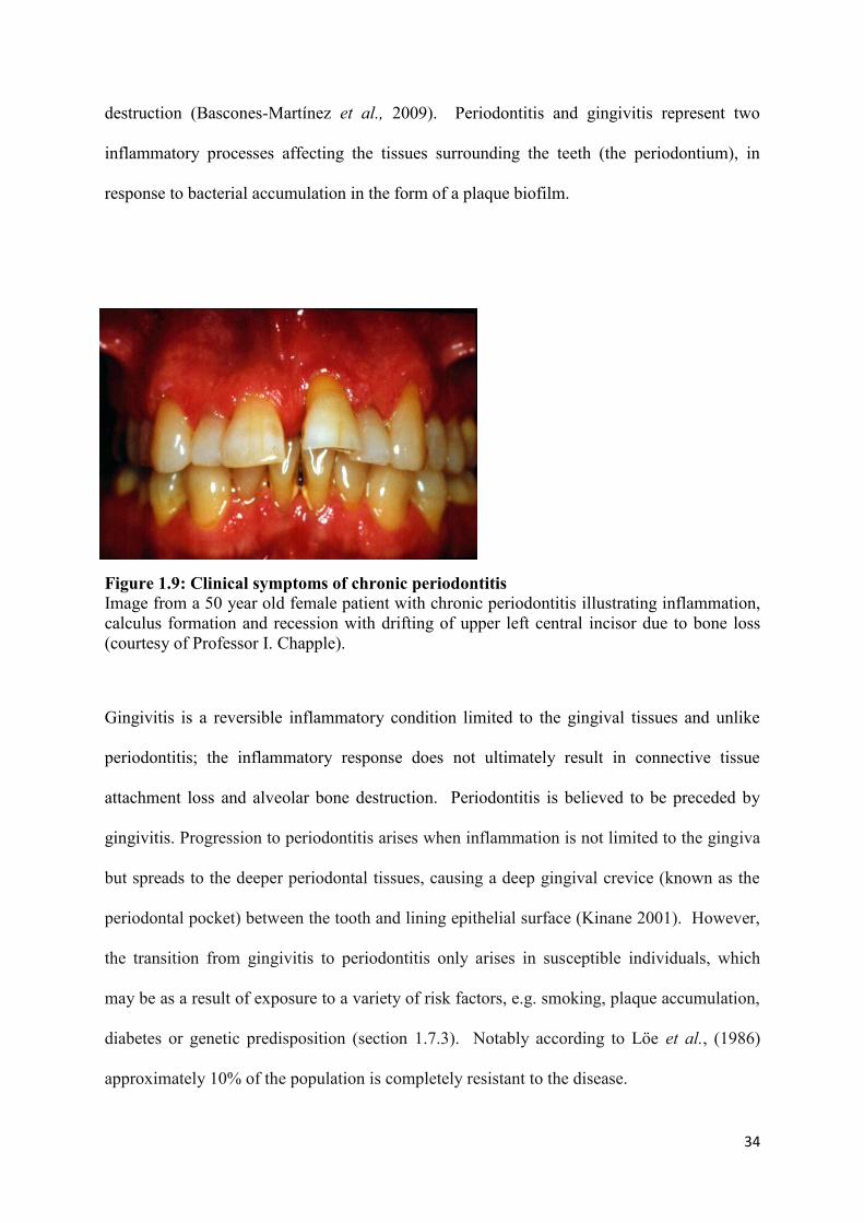
34
destruction (Bascones-Martínez et al., 2009). Periodontitis and gingivitis represent two
inflammatory processes affecting the tissues surrounding the teeth (the periodontium), in
response to bacterial accumulation in the form of a plaque biofilm.
Figure 1.9: Clinical symptoms of chronic periodontitis
Image from a 50 year old female patient with chronic periodontitis illustrating inflammation,
calculus formation and recession with drifting of upper left central incisor due to bone loss
(courtesy of Professor I. Chapple).
Gingivitis is a reversible inflammatory condition limited to the gingival tissues and unlike
periodontitis; the inflammatory response does not ultimately result in connective tissue
attachment loss and alveolar bone destruction. Periodontitis is believed to be preceded by
gingivitis. Progression to periodontitis arises when inflammation is not limited to the gingiva
but spreads to the deeper periodontal tissues, causing a deep gingival crevice (known as the
periodontal pocket) between the tooth and lining epithelial surface (Kinane 2001). However,
the transition from gingivitis to periodontitis only arises in susceptible individuals, which
may be as a result of exposure to a variety of risk factors, e.g. smoking, plaque accumulation,
diabetes or genetic predisposition (section 1.7.3). Notably according to Lӧe et al., (1986)
approximately 10% of the population is completely resistant to the disease.

35
Role of the microbial biofilm in periodontitis 1.7.1
Periodontitis is initiated by the accumulation of bacterial plaque at, and below, the gingival
margin (Figure 1.10). Indeed it has been estimated that ~500 oral bacterial species and ~1200
predominant phylotypes exist (Paster & Dewhirst 2009, Dewhirst et al., 2010). Of these
bacterial species, 5 major bacterial complexes (red, orange, yellow, green and purple) have
been identified by Socransky et al., (1988). The clustering and ordination analysis allowed
Socransky to assign microbial species to a colour complex dependent upon the strength of
association with each other and the clinical staging of periodontitis (Figure 1.11). For
example, bacteria assigned to orange and red complexes denote late stage colonisers
associated with the subgingival plaque. Bacteria have also been categorised according to
their location in relation to the gingival margin: supragingival or subgingival. Whereas the
supragingival plaque contains facultative species (such as Streptococcus species), the
subgingival plaque is dominated by a Gram-negative anaerobic motile microbiota (Loesche et
al., 1982). The role of bacteria in periodontitis has historically been encapsulated in two
different hypotheses, describing how microflora may contribute to the host response and
disease pathogenesis. The “non-specific hypothesis” (Loesche 1976) proposes that the entire
microbial flora contribute to the disease and once a threshold quantity is reached,
periodontitis will occur. Alternatively, the “specific hypothesis” (Theilade 1986) proposes
that the presence of particular pathogenic bacterial species is necessary for the diseased state
(Loesche & Grossman 2001). A third model was introduced by Marsh (1994), the
“ecological hypothesis”, which encompasses both previous hypotheses. This model proposes
that an inflammatory response is initiated as a direct result of the accumulation of a
pathogenic subgingival microbiota, which triggers a shift away from microbial homeostasis,
encouraging the growth of proteolytic and anaerobic bacterial species. The colonisation of
multiple bacteria in the generation of a mature plaque biofilm is a highly organised sequential

36
process. The biofilm is orchestrated to maximise adherence, communication, survival and
the accumulation of other bacterial species. Oxygen levels, environmental exposures and
competing commensal bacteria will all determine microbial growth and govern the locality of
where each bacterial species will thrive (Feng & Weinberg 2006). The biofilm encloses the
bacterial community and protects it from mechanical and chemical disruption; however a
synergistic and antagonistic relationship exists between bacteria (Koll-Klais et al., 2005).
Development of the plaque biofilm is initiated by deposition of salivary components, such as
glycoproteins (termed the acquired pellicle), onto the tooth surface prior to deposition of
early colonising bacteria. The acquired pellicle provides receptors on the tooth surface for
microorganisms to adhere to; routinely Streptococci species are the first to adhere,
representing 60-90% of the initial colonisers (Nyvad & Kilian 1987). Subsequent to initial
colonisation, secondary colonisers adhere via co-adhesion and co-aggregation, increasing the
biofilm size and complexity. Particular bacterial species appear to play a more dynamic role.
For example Fusobacterium nucleatum (F. nucleatum), a Gram-negative anaerobe that is
present in periodontally diseased sites, is not generally classified as an early or late coloniser,
but is a quorum sensing organism believed to facilitate biofilm formation (Kolenbrander et
al., 2009, 2010). A health promoting biofilm is essential for periodontal health, as bacteria
exist symbiotically with each other and the host. However, if the biofilm is allowed to
mature and is not frequently disrupted, then pathogenic species emerge. Dysbiosis results in
susceptible individuals and the host-microbial equilibrium is lost, which can result in the
onset of disease (Hajishengallis 2014).

37
Figure 1.10: Sequential bacterial colonisation which results in the formation of a plaque
biofilm
The acquired pellicle (yellow), containing receptors (green), is deposited onto the tooth
surface. Early colonisers (blue) bind to their complementary receptors (black lines), followed
by the adherence of secondary colonisers. Sequential binding enables co-aggregation
between different species, which bridges the gap between bacteria. F. nucleatum (pink)
bridges early and late colonisers, allowing interaction between the two groups. Co-
aggregation is represented by complementary shapes, for example the top left depicts
adhesion (red and white, with stem) and it’s receptor (red, no stem) (Kolenbrander et al.,
2002).

38
Figure 1.11: Periodontal bacteria assigned to the Socransky colour complexes
Diagrammatic representation indicates the relationships between specific bacterial species
within the colour complexes. The complexes also provide a possible sequence of
colonisation in the biofilm and the associations with the clinical parameters of periodontitis.
The bacteria listed above represent the panel of bacterial species employed in this thesis
(adapted from Socransky et al., 1998).
The contribution of the immune response to host periodontal tissue damage 1.7.2
Periodontitis occurs as a result of dysbiosis, which involves an increase in harmful
periodontal bacteria that become established in the host (Hajishengallis 2014) (Figure 1.12).
However, the microbial biofilm thrives by exploiting the host response, in particular the
neutrophil-mediated inflammatory response in susceptible individuals. This fuels a vicious
cycle of bacterial accumulation, inflammation and subsequent tissue destruction. The acute
inflammatory response is initially protective, providing the recruitment of neutrophils, the
activation of innate neutrophil-derived defence mechanisms and the activation of the acquired
cellular and humoral immune response. However in periodontitis, the neutrophil response is
reputed to unintentionally contribute to the progression of periodontitis causing collateral

39
tissue damage, which perpetuates the inflammation leading to chronicity and providing
nutrients for pathogenic bacteria like P. gingivalis to survive and proliferate (Scott & Krauss
2012). A common hypothesis for the involvement of the host in periodontitis pathogenesis is
that neutrophils exhibit hyperactivity and hyper-reactivity in response to the biofilm. This
exaggerated immune response is exemplified in the systemic neutrophils derived from
chronic periodontitis patients. PBNs are reportedly hyper-reactive in response to a microbial
challenge, but also hyperactive in the absence of an exogenous stimulus (Matthews et al.,
2007a, 2007b). In addition, excessive proteolytic activity has been observed in periodontitis
patients, which normalised following successful treatment. This is thought to be partly
attributed to the increased intensity of protease-activated receptor-2 (PAR2) expression in
chronic periodontitis. PAR2 is a receptor present on multiple cells, including neutrophils, and
is involved in the initiation of an inflammatory response via interactions with bacterial
proteases (Lourbakos et al., 1998). Increased PAR2 expression can promote the production
of cytokines and proteolytic enzymes, such as IL-6, IL-8 and matrix metalloproteinase
(MMP)-9 (Holzhausen et al., 2006). MMPs are a family of proteolytic enzymes that function
to degrade extracellular matrix molecules during physiological and pathophysiological tissue
remodeling. However these proteolytic agents are also thought to be key proteinases
involved in periodontal tissue destruction. Periodontal bacteria, including F. nucleatum and
P. gingivalis, trigger the extracellular release of MMPs. MMP release is regulated by
endogenous inhibitors, such as the tissue inhibitors of metalloproteinases (TIMPs), however
neutrophils do not produce this inhibitor de novo (Masure et al., 1991) and it is thought this
inhibitory mechanism is overwhelmed in periodontitis (Romanelli et al., 1999). The elevated
release of MMPs facilitates tissue degradation, which is likely to be exacerbated by the
heightened recruitment of neutrophils in periodontitis. Other neutrophil serine proteases also
associated with tissue degradation include NE and CG. NE can contribute to tissue

40
destruction by degrading molecules such as collagen and fibronectin and has been positively
correlated with periodontal attachment loss (Armitage et al., 1996). Interestingly, CG can
activate MMP-8 (Ding et al., 2005) and has been found to be higher in periodontitis patients
(Tervahartiala et al., 1996). It has been proposed that the elevated release of NE is the result
of a genetic polymorphism that leads to neutrophils with an intrinsically elevated function.
For example neutrophils from individuals with a (131 H/H) polymorphism of the Fcγ-
receptor are associated with more severe periodontitis. These individuals also overexpress
degranulation markers, CD63 and CD66b, resulting in increased NE release when challenged
with an infectious agent (Nicu et al., 2007).
Periodontitis is also associated with increased levels of cytokines, as a result of plaque
bacteria initiating the recruitment of neutrophils. As already discussed, chemokines
orchestrate the directional movement of neutrophils to the infected periodontal tissues (see
section 1.1.3). Chemokines have been suggested to be involved in bone resorption, as they
play a role in maintaining the equilibrium between osteoblastogenesis (bone formation) and
osteoclastogenesis (bone resorption) (Silva et al., 2007), a loss of which may associate with
alveolar bone resorption in periodontitis. Chemokines have been reported to be involved in
multiple steps of the signalling cascade resulting in osteoclastogenesis. For example CXCR4
expressed on osteoclast precursor cells is involved in their chemotaxis and differentiation into
osteoclasts (Wright et al., 2005). Furthermore, chemokines can indirectly modulate
osteoclastogenesis by possessing ligands that can increase MMP-9 activity, which in turn
facilitates tissue destruction and bone resorption (Grassi et al., 2004). Cytokines are also
deemed partly responsible for promoting bone resorption in periodontitis. This is evident in
periodontitis patients, in whom IL-1β was found to be elevated at sites of recent bone and
attachment loss (Lee et al., 1995).

41
Figure 1.12: The pathogenic model of periodontitis In susceptible individuals, the equilibrium between commensal and periodontal bacteria is
lost; giving rise to polymicrobial dysbiosis. This triggers activation of immune cells, for
example by toll-like receptors (TLRs), pattern recognition receptors (PRRs) and complement.
This triggers an inflammatory response, in which neutrophils are the predominant immune
cell. Whilst aiming to contain the bacteria attempting to colonise the epithelium, neutrophil
immune responses concurrently cause tissue damage. This provides nutrients which bacteria
utilise to proliferate, which promotes further dysbiosis (adapted from Hajishengallis 2014).
However, it is noteworthy that whilst neutrophil hyperactivity likely exacerbates periodontal
tissue destruction, impaired neutrophil activity can also increase disease severity. Multiple
diseases characterised by a deficient immune response are also correlated with severe
periodontitis. For example Papillon Lefèvre syndrome (PLS) is an autosomal recessive
disease in which a mutation in the cathepsin C gene (CTSC) results in a failure to encode
lysosomal cysteine protease, cathepsin C. Disease is characterised by palmoplantar
hyperkeratosis (abnormal thickening of the skin on the palms of the hands and soles of the
feet), significant pre-pubertal periodontal attachment loss, alveolar bone destruction, and the

42
subsequent loss of primary and permanent dentition (Singh et al., 2011). CTSC is essential
for post-translational trimming of serine proteases, e.g. granule-associated neutrophil serine
proteases (NSP) including NE, CG, PR3 and the recently described neutrophil serine protease
4 (Perera et al., 2013). CTSC removes N-terminal amino acids that block the active site of the
proteases and is thus necessary for activation of these proteases. These proteases are stored
as active enzymes in neutrophil granules, and localise to primary granules (azurophilic)
(Figure 1.13). NET production has recently been found to be impeded in a female PLS
patient (homozygous missense mutation in the CTSC gene) and the authors report that this is
due to decreased NE, which necessitates NET release (Sorensen et al., 2014). As ROS is not
inhibited in PLS patients, it is unlikely that reduced NET production is due to aberrant
NADPH-oxidase production (as in CGD). Whilst CGD patients also fail to generate NETs,
there are contradictory findings as to whether these individuals suffer from rapidly
progressing periodontitis (Budeneli et al., 2001, Cohen et al., 1985). Notably, the mild
immunodeficiency in PLS patients indicates that NET formation is not a major antimicrobial
defence mechanism, however the exact process that leads to such aggressive periodontitis
with clinically severe inflammation in PLS remains elusive. Furthermore, patients with
cyclic neutropenia who have considerably fewer neutrophil numbers are susceptible to severe
periodontitis (Matarasso et al., 2009).

43
Figure 1.13: Clinical signs and symptoms in a PLS case study
Intraoral periapical radiograph (a) and clinical photograph (b) from a 4 year old male with
PLS. The images show generalised gingival recession and severe periodontal destruction,
indicated by the yellow arrows (courtesy of Professor I. Chapple).
The importance of the host response is also evident in KO mice deficient in the chemokine,
CXCR2. Upon infection with P. gingivalis, CXCR2 KO mice had elevated periodontal bone
destruction compared with WT mice (Jeffrey et al., 2007). Interestingly, as previously
discussed (1.6.3) CXCR2 is also postulated to be required for NET release in an NADPH-
oxidase independent mechanism (Marcos et al., 2010). Collectively these results highlight
that both a hyperactive and deficient neutrophil response are associated with increased
disease. It is therefore likely that a homeostatic imbalance in the immune response
contributes to the initiation and development of disease in susceptible individuals.
Risk factors for periodontitis 1.7.3
Substantial epidemiological data exists which have identified the risk factors contributing to
the initiation and progression of periodontal disease. These include poor oral hygiene
(Kakudate et al., 2008), smoking (Bergstrom et al., 2000), obesity (Gocke et al., 2014),
a b
44
poorly controlled diabetes (Demmer et al., 2012), genetic predisposition (Divaris et al.,
2013), stress (MousaviJazi et al., 2013) and osteoporosis (Esfahanian et al., 2012). The
understanding that periodontitis only affects a proportion of the population who exhibit
atypical susceptibility has fuelled further research to understand the importance of specific
risk factors. Cigarette smoking is considered to be a major risk factor in the development of
periodontitis (Grossi et al., 1994, 1995). Tobacco smoking causes the inhalation of over
4000 toxins and is associated with many diseases, including cardiovascular disease (Burns
1991, Ambrose et al., 2004). Ragghianti et al., (2004) reported an association between
smoking and the clinical manifestations of periodontitis (Table 1.2). Worthy of note is that
numerous studies have shown that smoking is positively correlated with poor oral hygiene;
however even after adjustment for oral hygiene as a cofactor, smoking still appears to be a
primary independent risk factor (Tonetti et al., 1998). There is also a body of evidence
suggesting that smoking adversely affects treatment responses, indeed considerably more
smokers require additional treatment after the initial periodontal therapy (Delima et al.,
2010). These findings are supported by individuals who showed improved treatment
outcomes following smoking cessation. A possible explanation for the increased prevalence
of periodontitis amongst smokers is that smoking was found to select for specific periodontal
bacteria, including P. gingivalis, Treponema denticola (T. denticola) and Tannerella
forsythia (T. forsythia) (Zambon et al., 1996). These are all bacterial species assigned to the
red Sokransky complexes and are associated with severe disease. Smoking has also been
found to adversely affect neutrophil function, indeed nicotine has been shown to promote
neutrophil degranulation, which can cause an exaggerated response to the invading bacteria
and induce local tissue damage (Soder et al., 1999).
It has been reported that smoking can affect neutrophil total, extracellular and superoxide
ROS production (Matthews et al., 2011, 2012). High concentrations of cigarette smoke

45
extract (CSE) resulted in increased ROS production; however pre-treatment of neutrophils
with lower concentrations of CSE had an inhibitory effect on subsequent ROS production in
response to FcγR and TLR stimulation. In periodontitis, reduced ROS production in smokers
may impede the elimination of periodontal bacteria; however in heavy smokers increased
ROS production may facilitate oxidative stress and tissue damage. Intriguingly, neither
nicotine nor cotinine were deemed responsible for the observed effects, as these did not
directly alter ROS production. Notably, CSE was not found to be cytotoxic towards
neutrophils, but did have an inhibitory effect on apoptosis. It is possible that transient
exposure to cigarette smoke components may perturb the neutrophil response to bacterial
infection if ROS species are not being generated accordingly. However in the absence of an
exogenous stimulus, such as a bacterial biofilm, smokers may elicit excessive background
ROS production, which could associate with collateral tissue damage. Abnormal neutrophil
responses may therefore represent a crucial link between smoking and periodontitis.
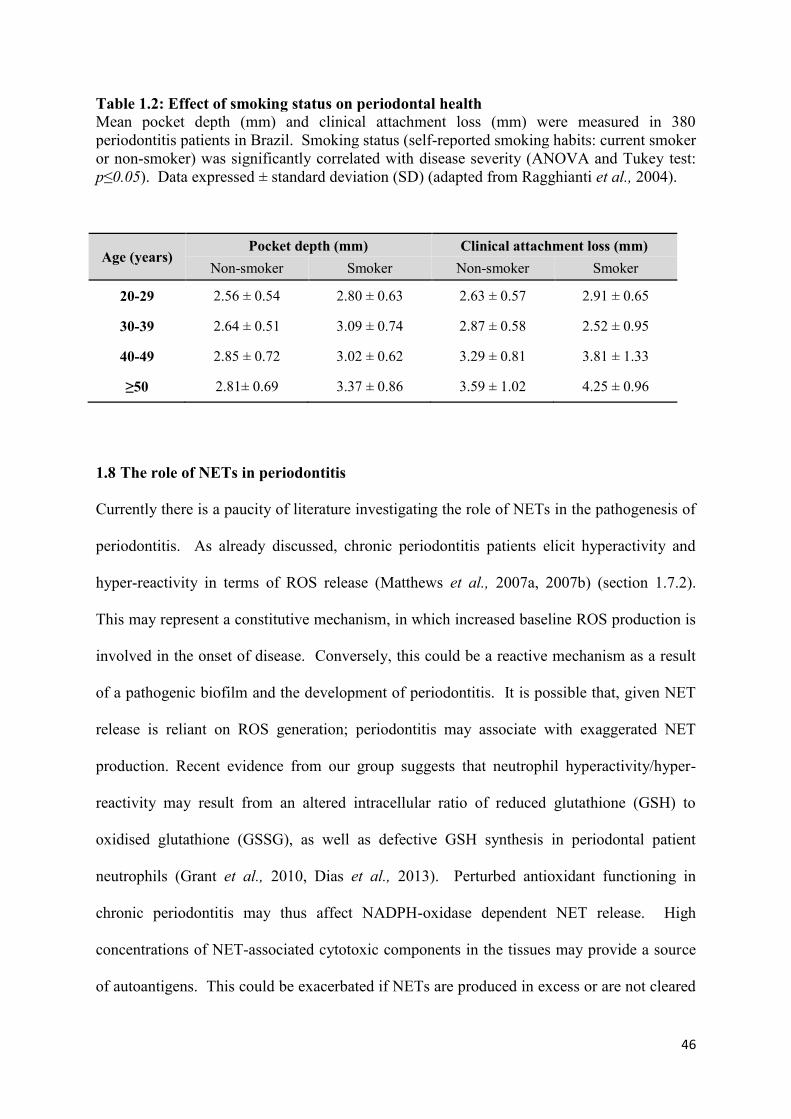
46
Table 1.2: Effect of smoking status on periodontal health
Mean pocket depth (mm) and clinical attachment loss (mm) were measured in 380
periodontitis patients in Brazil. Smoking status (self-reported smoking habits: current smoker
or non-smoker) was significantly correlated with disease severity (ANOVA and Tukey test:
p≤0.05). Data expressed ± standard deviation (SD) (adapted from Ragghianti et al., 2004).
Age (years) Pocket depth (mm) Clinical attachment loss (mm)
Non-smoker Smoker Non-smoker Smoker
20-29 2.56 ± 0.54 2.80 ± 0.63 2.63 ± 0.57 2.91 ± 0.65
30-39 2.64 ± 0.51 3.09 ± 0.74 2.87 ± 0.58 2.52 ± 0.95
40-49 2.85 ± 0.72 3.02 ± 0.62 3.29 ± 0.81 3.81 ± 1.33
≥50 2.81± 0.69 3.37 ± 0.86 3.59 ± 1.02 4.25 ± 0.96
1.8 The role of NETs in periodontitis
Currently there is a paucity of literature investigating the role of NETs in the pathogenesis of
periodontitis. As already discussed, chronic periodontitis patients elicit hyperactivity and
hyper-reactivity in terms of ROS release (Matthews et al., 2007a, 2007b) (section 1.7.2).
This may represent a constitutive mechanism, in which increased baseline ROS production is
involved in the onset of disease. Conversely, this could be a reactive mechanism as a result
of a pathogenic biofilm and the development of periodontitis. It is possible that, given NET
release is reliant on ROS generation; periodontitis may associate with exaggerated NET
production. Recent evidence from our group suggests that neutrophil hyperactivity/hyper-
reactivity may result from an altered intracellular ratio of reduced glutathione (GSH) to
oxidised glutathione (GSSG), as well as defective GSH synthesis in periodontal patient
neutrophils (Grant et al., 2010, Dias et al., 2013). Perturbed antioxidant functioning in
chronic periodontitis may thus affect NADPH-oxidase dependent NET release. High
concentrations of NET-associated cytotoxic components in the tissues may provide a source
of autoantigens. This could be exacerbated if NETs are produced in excess or are not cleared

47
efficiently, which may result in further periodontal tissue damage. Vitkov et al., (2009)
employed microscopy to visualise NETs, which appeared abundant in gingival pocket
epithelium, the gingival exudate and in the gingival crevicular fluid (GCF) of chronic
periodontitis patients (Vitkov et al., 2010) (Figure 1.14). The authors postulated that during
the bacteria’s attempt to migrate and adhere to the gingival surface, they encounter NETs
which shield the epithelium and prevent adhesion. Interestingly, the bacterial counts in the
purulent GCF exudate were relatively inconsistent, which led to the theory that if
phagocytosis is overwhelmed, for example due to large numbers of colonising bacteria, NETs
may compensate for the increased demand on the immune system by providing an additional
neutrophil-defence mechanism. Further observations, such as the lack of neutrophils
containing engulfed bacteria and the large quantities of NETs in the GCF suggest NETosis is
the predominant defence mechanism employed by neutrophils in the GCF at periodontally
diseased sites (Vitkov et al., 2010). Worthy of note is that this research did not include
healthy matched controls and that NETs were not quantified in the periodontal tissues.
Indeed it would be interesting to compare GCF and periodontal tissue samples from
periodontitis patients and matched controls to determine if any disparity exists in NET
formation and accumulation. Preliminary findings presented in Cooper et al., (2013)
employed confocal microscopy and immunofluorescence staining to identify increased levels
of NET-like structures in inflamed gingival tissues, compared with healthy control tissues
(Figure 1.15). Supporting the theory of increased NETosis in periodontitis is that a range of
molecules that are elevated in periodontitis, such as nitric oxide and interferon-α (IFNα), can
also stimulate for NET release (Reher et al., 2007, Wright et al., 2008).

48
Figure 1.14: NET entrapment of periodontal bacteria
Scanning electron micrographs illustrating: (a1) Crevicular exudate containing bacteria
(white arrowheads), contrast with (a), which has received DNase treatment (degrading NETs)
and shows no entrapped bacteria. (b) Crevicular exudate with multiple large NETs entrapping
bacteria (white arrowheads). (c) Pocket epithelium with entrapped bacteria, (e) denotes
epithelium surface. (d) enlarged version of (c), showing NET fibres (small white
arrowheads). Scale bar represents 5μm (a1, b, c), 1μm (a, d) (Vitkov et al., 2009).
A1

49
Figure 1.15: NETs in human gingival tissues Confocal microscopy where DNA is stained red and MPO is stained green in gingival tissues
in health and disease. Considerably more NET-like structures were observed in gingivitis
compared with health (Cooper et al., 2013, courtesy of Dr Yousefi University of Berne,
Switzerland).
The timely removal of NETs may represent an important mechanism in periodontitis. As
previously mentioned, the removal of NETs in SLE patients is impeded, compared with
healthy controls (see section 1.6.2) and the delayed removal of NETs provides a high
concentration of NET-associated molecules that may drive the autoimmune response. Indeed,
de Pablo et al., 2013) reported an increased prevalence of ACPAs and raised ACPA titres
within the serum of non-RA patients with periodontitis, relative to non-periodontitis controls
(see section 1.6.1).
Conversely, decreased levels of NETs in periodontitis may enhance patient susceptibility to
increased periodontal bacterial colonisation. DNases released by bacteria can degrade the
DNA backbone, which disassembles the NET structure (see 1.3.3). This is thought to enable
invading bacteria to circumvent NET defences and allow bacterial dissemination into the
surrounding tissues (Berends et al., 2010). Notably, several periodontal bacteria, including P.
50
gingivalis and F. nucleatum, have been found to express membrane-bound and secreted
DNases (Palmer et al., 2012). DNase release will enable bacterial infiltration and their
establishment within the biofilm. NET production has also been reported to be reduced in
older (over 50 years) periodontitis patients compared with younger patients (under 50) in
response to HOCl. This implies the neutrophil responses in periodontitis may deteriorate
with age, which may be associated with periodontitis being an age related disease (Grossi et
al., 1995), however in this cohort (n=19 pairs) no differences in NET production were
observed between periodontitis patients and their healthy controls (Hazeldine et al., 2014).
Reduced levels of NETs have been associated with increased susceptibility to infections, for
example in neonates (Yost et al., 2009) and CGD (Bianchi et al., 2011). It is possible that
decreased NETs in periodontitis would impair the elimination of pathogenic species;
facilitating persistent dysbiosis. This would likely result in further neutrophil recruitment and
activation, such as extracellular degranulation, which may result in further tissue damage.
1.9 Neutrophil isolation and ex vivo handling
To characterise neutrophils in periodontitis, as well as other inflammatory diseases, cells are
often isolated from the peripheral blood of volunteers and investigated ex vivo. Indeed there
are neutrophil-like cells, such as the HL60 human promyelocytic cell line, which can be
employed within the laboratory to circumvent problems of inter-individual variations
between neutrophil donors (Martin et al., 1990). Nevertheless, these immortalised cell lines
do not fully reflect primary neutrophils in their behaviour or in their intricate biological
functions (Hallett 1990). In addition, despite 99% of murine genes having an equivalent
human counterpart, murine disease models also do not fully replicate the human situation.
Exemplifying the limitations of such murine models is the fact that while human neutrophils
represent 50-70% of circulating PBNs, mouse neutrophils make up only 10-25% of cells
51
(Doeing et al., 2003). Isolation from human blood requires an efficient, aseptic and
reproducible method to obtain non-activated and viable cells. A further consideration is the
effect of the isolation and purification method on neutrophil behaviour; in particular
neutrophils may become activated as a result of ex vivo manipulation (Oh et al., 2008).
Therefore, research investigating neutrophil function needs to consider whether ex vivo
purification techniques influence neutrophil activation status. Methods of neutrophil
isolation from whole blood can differ between research groups, creating potential phenotypic
differences in neutrophils even if retrieved from the same volunteer.
Neutrophil isolation: density gradient materials 1.9.1
There are currently several materials employed for the isolation and purification of
neutrophils from whole blood; including Percoll™ (GE Healthcare), Dextran (Sigma) and
Histopaque™ (Sigma). Neutrophil isolation aims to provide an appropriate separation range
at a high purity from other blood cells, whilst at the same time, not stimulating or activating
the cells.
1. Percoll is comprised of colloidal silica and coated with non-dialyzable
polyvinylpyrrolidone (PVP), and is used in conjunction with NaCl and water to
produce a gradient to isolate neutrophils by differences in density.
2. An alternative density gradient material is Histopaque, which is a polysucrose and
sodium diatrizoate solution that also enables the isolation of neutrophils at a density
gradient interface.
3. A third method involves layering the blood on Dextran, which results in the
sedimentation of a neutrophil layer above the aggregated erythrocytes. The neutrophil
layer is then collected by aspiration and centrifuged on layered Percoll gradients to
isolate the neutrophils further.

52
1.10 Aims of this study
Background: The pathogenic biofilm that initiates periodontitis has previously been
characterised with respect to the sequence of microbial colonisation and associations with
disease manifestations (Socransky et al., 1998). However, the interactions between
periodontal bacteria and NET release are less well understood. Specifically, whether
bacterial species stimulate for increased or decreased NET release, and whether NETs have
the capacity to entrap and kill periodontal microbes remains to be elucidated.
Aim 1: To investigate the interactions between NETs and a range of periodontal bacteria.
Background: NET release represents a novel neutrophilic defence mechanism; however the
exact role of NETs in the pathogenesis of periodontitis remains unclear. Periodontitis is
driven by a dysfunctional inflammatory response towards a pathogenic biofilm, in which the
neutrophil is a key effector cell and patients’ neutrophils are hyperactive and hyper-reactive
in terms of ROS production (Matthews et al., 2007a, 2007b). It is likely that NETs are also
employed by the neutrophil in periodontitis pathogenesis, and given that NETs are largely
dependent on ROS generation (Fuchs et al., 2007), it is possible that NET release is also
perturbed in periodontitis.
Aim 2: To investigate NET production by PBNs from periodontitis patients and in patients
with PLS in comparison with healthy control groups.
Background: The importance of NET clearance has recently received substantial attention in
studies of disease pathogenesis, such as in CF and SLE. As with periodontitis, both CF and
SLE are characterised by a dysfunctional host immune response. NET clearance is reportedly
impeded in SLE patients (Hakkim et al., 2010); however the clearance of NETs in
periodontitis has not been studied. Therefore, the ability of periodontitis patients to degrade

53
NETs and the nature of the mechanisms involved is of interest. An accumulation of NETs in
certain tissues has been associated with disease severity via autoimmune mechanisms, for
example in RA (Khandpur et al., 2013). Indeed our own group have reported significantly
elevated levels of autoantibodies to NET-related antigens in the serum of periodontitis
patients (dePablo et al., 2013).
Aim 3: To investigate NET degradation by periodontitis patients’ plasma and the effect of
NETs on neutrophil function.
Background: Cigarette smoking is a major risk factor in the development of periodontitis
(Grossi et al., 1995), and it has previously been reported that smoking has a range of effects
upon neutrophil ROS production (Matthews et al,. 2011, 2012).
Aim 4: To determine the impact of CSE upon NET production, neutrophil chemotaxis and
the expression of key genes involved in neutrophil activation.

54
MATERIALS AND METHODS CHAPTER 2:

55
2.1 MATERIALS
GENERAL BUFFERS 2.1.1
2.1.1.1 Phosphate Buffered Saline (PBS)
A 1L bottle of PBS was prepared by adding 7.75g NaCl (Sigma S9625), 0.2g KH2PO4
(Sigma P5379) and 1.5g K2HPO4 (Sigma P8281) to 1L of sterile distilled water. This was
autoclaved (121°C for 15 mins) and stored at 4°C for up to 1 month prior to use.
2.1.1.2 PBS supplemented with glucose and cations (gPBS)
A 1L bottle of gPBS was prepared by adding 1.8g glucose (Sigma G8270), 0.15g CaCl2
(BDH 10070), 1.5ml 1M MgCl2 (BDH 22093) to 1L of PBS. This solution was made up in
order with each component mixed before adding the next. This was stored at 4°C for up to 1
month prior to use.
2.1.1.3 Blocking buffer: 1% bovine serum albumin (BSA)
A 1L bottle of BSA was produced by adding 10g BSA (Sigma A2153) to 1L of PBS. The
resulting solution was filter-sterilised (pore size 0.22 µm), added to 25ml universal containers
and frozen at -20°C until needed.
2.1.1.4 4-(2-Hydroxyethyl) piperazine-1-ethanesulfonic acid, N-(2-Hydroxyethyl)
piperazine-N′-(2-ethanesulfonic acid) (HEPES) buffer
HEPES (0.1M) (Sigma H3375) was prepared by adding 2.38g HEPES to 100ml of distilled
water. This was adjusted to a pH of 7.5 (Beckman 40 pH meter, pH adjusted with HCl or
NaOH) and stored at 4°C prior to use.

56
2.1.1.5 0.9% saline solution
A 1L bottle of 0.9% saline solution was prepared by adding 9g of NaCl (Sigma S9625) to 1L
of distilled water and autoclaving (121°C for 15 mins).
NEUTROPHIL PREPARATION AND ASSAYS 2.1.2
2.1.2.1.1 Preparation of Percoll gradients
Percoll densities composed of 1.13g/ml Percoll, distilled water and 1.5M NaCl (Sigma
S9625) were prepared according to Table 2.1.
Table 2.1: Preparation of Percoll density gradients
Density 1.079 1.098
Percoll 19.708ml 24.823ml
Water 11.792ml 6.677ml
NaCl (1.5M) 3.5ml 3.5ml
2.1.2.1.2 Lysis Buffer
A 1L bottle of lysis buffer was prepared by adding 8.3g NH4Cl (A9434 Sigma), 1g KHCO3
(Sigma P9144), 0.04g Na2 EDTA 2H2O (E5134 Sigma) and 2.5g BSA (Sigma A2153) to 1L
of sterile distilled water. This was stored at 4°C for up to 1 month prior to use.
2.1.2.1.3 Preparation of Dextran and Percoll gradients
Dextran-isolation of neutrophils involves layering the blood onto Dextran (see 2.2.1.1.4),
which results in the sedimentation of a neutrophil layer above the aggregated erythrocytes.
The neutrophil layer is then collected with a Pasteur pipette and centrifuged on layered
Percoll gradients to separate the neutrophils further.

57
2.1.2.1.4 2% Dextran
A 2% Dextran solution was prepared by adding 1g Dextran (Sigma 31392) to 50ml of 0.9%
NaCl (saline). This was mixed by vortexing prior to being syringe-filter sterilised (pore size
0.22µm).
2.1.2.1.5 90% isotonic stock Percoll
Percoll for use in the preparation of 80% and 56% gradients (see Table 2.2) was prepared by
combining 5ml of 9% NaCl with 45ml of Percoll (17-0891-01 GE Healthcare).
Table 2.2: Preparation of Percoll density gradients following Dextran sedimentation
Density 80% 56%
90% Percoll 40ml 28ml
0.9% NaCl (saline) 10ml 22ml
2.1.2.1.6 Luminol
A 30mM stock solution was prepared by dissolving 0.5g luminol (Sigma A8511) in 94.05ml
of 1mM NaOH. The stock solution was foil-wrapped and stored for up to 6 months at 4°C
prior to use. The working solution was prepared on the day by diluting 1ml of stock solution
with 9ml of PBS and the pH adjusted to 7.3 (Beckman 40 pH meter, pH adjusted with HCl or
NaOH).
2.1.2.1.7 Isoluminol
A 30mM stock solution was prepared by dissolving 0.5g isoluminol (Sigma A8264) in
94.05ml of 1mM NaOH. The stock solution was foil-wrapped and stored for up to 6 months
at 4°C. The working solution was prepared on the day by diluting 1ml of stock solution with

58
9ml PBS and the pH adjusted to 7.3 (Beckman 40 pH meter, pH adjusted with HCl or
NaOH).
2.1.2.1.8 Lucigenin
A 1mg/ml stock solution of Lucigenin was prepared by dissolving 0.005g lucigenin (Sigma
B49203) in 5ml of PBS. The stock solution was foil-wrapped and stored for up to 6 months
at 4°C prior to use. The working solution was made up on the day prior to use by diluting 1:3
with PBS.
2.1.2.2 Micrococcal nuclease (MNase)
MNase (Worthington 4789) was diluted to a 13.3U/ml working solution and stored at -20°C
in 1ml aliquots. MNase was used at a final concentration of 1U/ml in 15µl aliquots unless
otherwise stated.
2.1.2.3 Sytox green
Sytox green (Invitrogen S7020) was diluted 1:500 in RPMI (Roswell Park Memorial
Institute)-1640 (Sigma R7509) to obtain a 10µM working solution. This solution was foil
wrapped and stored at -20°C in 1ml aliquots.
2.1.2.4 Fluorescein-5-isothiocyanate (FITC)
FITC (isomer I, Invitrogen F-1906) was diluted to a working concentration of 6mg/ml in
1.5ml Eppendorfs, which were foil wrapped and stored at -20°C prior to use. When needed,
FITC was defrosted at room temperature and used at a final concentration of 0.3mg/ml by
adding 50µl to 1ml of bacterial suspension in a 1.5ml Eppendorf.

59
2.1.2.5 Cytochalasin B
Cytochalasin B (Sigma C6762), which blocks phagocytosis (Densen & Mandell 1978), was
used at a final concentration of 10µg/ml by adding 166.5µl cytochalasin B (2mg/ml stock in
DMSO, Sigma D8418) to 4.833ml of PBS. This was stored at -20°C.
BACTERIAL CULTURE 2.1.3
2.1.3.1 Bacterial stimuli
A panel of 19 bacteria (see Tables 2.5 & 2.6), plus opsonised S. aureus, was employed to
stimulate neutrophils via TLR and FcγR, respectively. Bacterial stocks were provided by Dr.
Mike Milward, originally obtained from the Forsyth Institute (Boston, US) or purchased from
the American Type Culture Collection (ATCC).
2.1.3.2 Blood agar plates
Pre-prepared blood agar plates (Base no. 2 with 7% horse blood) were purchased from Oxoid
(PB0114). Plates were stored at 4C and allowed to equilibrate to room temperature for an
hour prior to use. All plates were used before the manufacturer’s expiry date.
2.1.3.3 Fastidious plates
P. gingivalis (strain W83) was cultured on pre-prepared anaerobic 20% blood agar plates
(Wilkins Chalgren) purchased from Oxoid (PB0112). This is a medium enriched with
neomycin that is specific for the growth of fastidious anaerobes. Plates were stored at 4C
and allowed to equilibrate to room temperature for an hour prior to use. All plates were used
before the manufacturer’s expiry date.

60
2.1.3.4 Tryptone soya agar (TSA) plates
S. aureus was cultured on tryptone soya agar (TSA) plates. Plates were made according to
the manufacturer’s instructions by adding 40g TSA (Oxoid CM0131) to 1L of distilled water.
This was autoclaved (121°C for 15 mins) and cooled to 50°C, prior to pouring the agar into
Petri dishes (Sterilin 101VR20). Plates were inverted and incubated at room temperature to
solidify. Plates were subsequently stored at 4C until needed, at which point they were
equilibrated to room temperature for an hour prior to use.
2.1.3.5 Brain heart infusion (BHI) broth
BHI broth was made according to the manufacturer’s instructions (Oxoid CM1135); 37g
dehydrated culture medium were dissolved in 1L of distilled water. The BHI broth was
mixed, autoclaved (121°C for 15 mins) and stored at 4°C until needed.
2.1.3.6 Fastidious anaerobe broth
Fastidious anaerobe broth was used to culture P. gingivalis. This dehydrated medium
enriched with vitamin K, haemin and L-cysteine was purchased from Lab M (LAB071).
Fastidious anaerobe broth was made according to the manufacturer’s instructions by adding
29.7g to 1L of distilled water. This mixture was autoclaved (121°C for 15 mins) and stored
at 4°C until needed.
2.1.3.7 Typtone soya broth (TSB)
TSB (Oxoid CM0876) was made by adding 30g of dehydrated culture medium to 1L of
distilled water. This was mixed, autoclaved (121°C for 15 mins) and stored at 4°C until
needed.

61
2.1.3.8 Crystal violet
Crystal violet solution was prepared by adding 2g crystal violet (Sigma C0775) to 20ml of
95% ethanol (Sigma E7023). 0.8g ammonium oxalate (Sigma A8545) was dissolved in 80ml
of distilled water to produce 1% ammonium oxalate. The crystal violet solution was mixed
with 1% ammonium oxalate in a 1:1 dilution and allowed to stand at room temperature for
several days prior to use.
2.1.3.9 Carbon fuchsin
A 1ml stock of carbol fuchsin solution (BDH 351874U) was mixed with 9ml of distilled
water and kept at room temperature.
2.1.3.10 Glutaraldehyde in sodium cacodylate buffer
A 100ml of 0.1M sodium cacodylate buffer were prepared by adding 4.28g sodium
cacodylate trihydrate (Sigma CO250) to 100ml of distilled water, and adjusted to pH 7.4
(Beckman 40 pH meter, pH adjusted with HCl or NaOH). 2.5% glutaraldehyde was made by
combining 7.5ml of sodium cacodylate buffer, 6ml of distilled water and 1.5ml of
glutaraldehyde solution (Sigma G5882).
CIGARETTE SMOKING REAGENTS 2.1.4
2.1.4.1 Nicotine
Liquid nicotine was purchased (Sigma N3876) and diluted to final concentrations of 1µg/ml,
5µg/ml and 10µg/ml in PBS (section 2.2.4.1.1) and stored at -20°C.

62
2.1.4.2 Cotinine
Cotinine was purchased from Sigma (C5923) and used at the same final concentrations as
nicotine (see 2.2.4.1.1); 1µg/ml, 5µg/ml and 10µg/ml in PBS and stored at -20°C. Cotinine is
the principal stable metabolite of nicotine and it has been demonstrated that 70-80% of
absorbed nicotine is subsequently metabolised to produce cotinine (Yuki et al., 2013,
Hukkanen et al., 2005). Concentrations in the saliva of smokers are reportedly 0.23 ± 0.17
µg/ml, ranging from 0.003µg/ml to 0.733µg/ml (Etter & Perneger 2001).
2.1.4.3 Thiocyanate (SCN-)
Thiocyanate (SCN-) levels are significantly elevated in the plasma of smokers, as it in an end
product of detoxification of hydrogen cyanide, which is inhaled in cigarette smoke. SCN- is
present in the plasma of smokers at levels of 131.4 30.8µM (Morgan et al., 2011). SCN-
functions as a substrate for neutrophil derived MPO in the generation of hypothiocyanous
acid (HOSCN-). Sodium thiocyanate was purchased from Sigma (251410) and diluted to
final concentrations of 50µM, 100µM and 150µM in PBS and stored at -20°C.
PCR STUDIES 2.1.5
2.1.5.1 The synthesis of complementary DNA (cDNA)
Ribonucleic acid (RNA) was reverse transcribed to produce cDNA from pooled RNA using a
cDNA synthesis kit (Tetro Bioline 65042) (Table 2.3).

63
Table 2.3: Components and quantities used for cDNA synthesis
All components were provided in the cDNA synthesis kit (Tetro Bioline 65042).
Abbreviations: Oligo-dT (oligonucleotide deoxy-thymine) dNTP mix (deoxynucleotide mix),
RT (reverse transcriptase), RNase (ribonuclease), DEPC (diethylpyrocarbonate).
cDNA Kit components 1 x Eppendorf
Pooled RNA (0.5-0.6µg) 8µl
Oligo-dT primer mix 1µl
dNTP mix 10mM total 1µl
5x RT Buffer 4µl
RNase inhibitor (10u/µl) 1µl
Reverse transcriptase (200u/µl) 1µl
DEPC-treated water 4µl
2.1.5.2 Design and validation of PCR primers
Housekeeping and target gene primer sequences were selected from the literature (Table 2.4)
and purchased from Invitrogen, UK.

64
Table 2.4: Primers employed to quantify gene expression in CSE and SCN- treated
neutrophils using real time PCR
Abbreviations: YWHAZ (tyrosine 3/tryptophan, 5-monooxygenase activation protein, zeta
polypeptide); GAPDH (glyceraldehyde-3-phosphate dehydrogenase); RPL13 (ribosomal
protein L13); HPRT1 (hypoxanthine phosphoribosyltransferase 1); NFĸB epsilon (nuclear
factor kappa B); HSP40 (heat shock protein 40); IL-8 (interleukin-8). Efficiency refers to the
amplification of the targer molecule, where an efficiency of 2.0 is considered 100% efficient
and means the target molecule doubles every PCR cycle (LightCycler®).
Gene Primer sequence (5’-3’) Amplicon
size (bp) Efficiency
Housekeeping genes
YWHAZ
(f) ACTTTTGGTACATTGTGGCTTCAA
(r) CCGCCAGGACAAACCAGTAT
94 1.93
GAPDH
(f) CTCCTGTTCGACAGTCAG
(r) GCCCAATACGACCAAATC
111 1.83
RPL13
(f) CCTGGAGGAGAAGAGGAAAGAGA
(r) TTGAGGACCTCTGTGTATTTGTCAA
126 1.86
HPRT1
(f) GACCAGTCAACAGGGGACAT
(r) AACACTTCGTGGGGTCCTTTTC
195 1.97
Target genes
NFĸB
epsilon
(f) GTGAAGCCTGTTTGCCTCTC
(r) AGGGTCCTCAACAGCAAGAA
172 2.07
HSP40
(f) TACAGGAGCACTGTGGAAACG
(r) AGGTCTGAGCACTGGACTGG
192 1.83
IL-8
(f) TAGCAAAATTGAGGCCAAGG
(r) GGACTTGTGGATCCTGGCTA
204 1.75
P-47phox
(f) ACCCAGCCAGCACTATGTGT
(r) AGTAGCCTGTGACGTCGTCT
767 1.89
P-67phox
(f) CGAGGGAACCAGCTGATAGA
(r) CATGGGAACACTGAGCTTCA
726 1.78
GP-91phox
(f) GCTGTTCAATGCTTGTGGCT
(r) TCTCCTCATCATGGTGCACA
403 1.93

65
CELL CULTURE 2.1.6
2.1.6.1 Culture of an oral epithelium cell line
A human oral epithelium cell line (OEC), H400, was kindly donated by Dr S. Prime from
University of Bristol, UK. The cell line was derived from a squamous cell carcimona of the
alveolar process from a 55 year old female. The carcinoma was at stage 2, node negative,
moderately differentiated and measured 20-40mm in size. Prime et al., (1990) demonstrated
close similarities to gingival epithelium and therefore H400s represent a suitable cell line for
use as a model system.
2.1.6.2 Supplemented Dulbecco’s Modified Eagle’s Medium:nutrient mixture F-12
(DMEM/F-12)
DMEM/F-12 (Invitrogen 21331-020 500ml) is a commonly used medium for the growth of
human endothelial cells, which is a 1:1 solution of DMEM and Ham’s F-12. The media was
supplemented with heat-inactivated foetal calf serum (FCS, 10% v/v), L-glutamine (2% v/v,
Sigma G7513) and 10mg/ml hydrocortisone (0.005% v/v, Sigma H0888). Supplemented
growth media was stored at 4°C prior to use.
2.1.6.3 Trypsin Ethylenediaminetetraacetic Acid (T-EDTA)
T-EDTA (Invitrogen R-001-100), comprising 0.0025% trypsin and 0.01% EDTA, was used
to release adherent cells to produce a cell suspension. This solution was divided and frozen
at -20°C in 4ml aliquots until needed.

66
2.2 METHODS
ISOLATION OF NEUTROPHILS FROM PERIPHERAL BLOOD 2.2.1
2.2.1.1 Percoll components
2.2.1.1.1 Preparation of Percoll gradients
PMNLs were isolated from whole blood using discontinuous Percoll gradients (17-0891-01
GE Healthcare). The discontinuous gradients were generated by carefully layering 8ml of
1.079 density gradient over 8ml of 1.098 (see 2.1.2.1.1) in a 25ml centrifuge tube (Fisher
Scientific).
2.2.1.1.2 Neutrophil isolation with Percoll
Blood was collected in 6ml lithium heparin vacutainers (Greiner Bio-One) and inverted to
ensure the mixing of the anticoagulant. Post-collection, 6ml of blood were layered over the
Percoll density gradients and centrifuged (Hettich Universal 320R) for 8 mins at 150 relative
centrifugal force (rcf), followed by 10 mins at 1200rcf (both with a ramp and brake of 1 and
at 4°C). Plasma, monocytes, lymphocytes and density gradient material were aspirated off
and discarded, allowing for the transfer of the neutrophil layer to 30ml of lysis buffer (see
2.1.2.1.2) in a 50ml centrifuge tube (Fisher Scientific), which was then made up to a total
volume of 50ml with lysis buffer. Following gentle inversion, the centrifuge tube was
incubated for 15 mins at room temperature to lyse any remaining erythrocytes. Subsequently,
the tube was centrifuged for 6 mins at 500rcf (4°C) to pellet the neutrophils. The supernatant
was aspirated off, whilst the pellet was re-suspended in 2ml of lysis buffer and incubated for
5 mins at room temperature, prior to centrifugation for 6 mins at 500rcf (4°C). The
supernatant was again aspirated off and the neutrophil pellet washed in 2ml of PBS, followed
by re-centrifugation (6 mins at 500rcf, 4°C). The supernatant was discarded, cells re-

67
suspended in 2ml of PBS (see 2.1.1.1) and counted by light microscopy (Leitz Laborlux s)
(2.2.1.1.5).
2.2.1.1.3 Isolation of neutrophils by Histopaque gradients
H1077 (Sigma) density Histopaque (3ml) were layered over H1119 (Sigma) (3ml) in a 15ml
tube (Fisher Scientific). 6ml of blood were carefully layered over the gradients using a
Pasteur pipette, prior to centrifugation (Hettich Universal 320R) for 50 mins at 800rcf (at
4°C). Plasma, monocytes, lymphocytes and density gradient material were aspirated and
discarded, allowing for the retrieval of the neutrophil layer from the H1077/H1119 interface.
Neutrophils were transferred to 5ml of Dulbecco’s PBS (Sigma D8537) in a fresh 15ml
centrifuge tube and centrifuged for 6 mins at 400rcf, followed by manual aspiration of the
supernatant. The neutrophil pellet was re-suspended in 10ml of Dulbecco’s PBS, prior to re-
centrifugation for 6 mins at 400rcf (4°C). The supernatant was again aspirated off and cells
re-suspended in 1ml of Dulbecco’s PBS prior to erythrocyte lysis, where 5ml of distilled ice
cold water were added, followed by the immediate addition of 2ml 4x PBS after 30 secs.
Neutrophils were then centrifuged for 8 mins at 450rcf (4°C), finally re-suspended in 2ml
Dulbecco’s PBS and cells were counted by light microscopy (Leitz Laborlux s) (2.2.1.1.5).
2.2.1.1.4 Isolation of neutrophils using Dextran
Blood (approximately 6ml) was added to a 50ml tube containing 1ml of 2% Dextran (see
2.1.2.1.3); this was incubated for 40 mins at room temperature to sediment erythrocytes.
Post-incubation, the neutrophil “buffy coat” was collected from the blood and carefully
layered over the Percoll density gradients using a Pasteur pipette (2.1.2.1.4). This was
centrifuged (Hettich Universal 320R) for 20 mins at 220rcf (4°C) and post-incubation the
neutrophils were added to a falcon tube (Fisher Scientific) containing 5ml PBS. This was re-
centrifuged (10 mins at 250rcf, 4°C) and the supernatant discarded. The cells were then re-

68
suspended in 5ml of RPMI-1640 (Sigma R7509) and counted by light microscopy (Leitz
Laborlux s) (2.2.1.1.5).
2.2.1.1.5 Neutrophil cell counting
Cells (10µl of isolated cell suspension) were counted using a haemocytometer (Neubauer,
Reichert) on a light microscope at x20 magnification (Leitz Laborlux s). The central area of
the haemocytometer (1mm2) is sub-divided into 25 smaller squares (0.04mm
2). Cells were
counted in 9 of the 0.04mm2
squares and calculations were subsequently performed according
to the equation below, where 25 refers to the number of squares, 1x10⁴
denotes the
haemocytometer dimensions and 9 is the number of squares counted.
(Cell count x 25 x 1x104) = number of cells/ml stock cell suspension
9
2.2.1.2 NEUTROPHIL VIABILITY AND METABOLIC ACTIVITY
2.2.1.2.1 Confirmation of neutrophil isolation by cytospin
Neutrophils were re-suspended in PBS (see 2.1.1.1) at 2x10⁶ cells/ml, added to a cytospin
funnel and centrifuged for 5 mins at 300rcf (Shandon cytospin). The resultant smears on
glass slides were allowed to air dry for 20 mins. Neutrophils were stained with Diff-Quick,
which is a Romanowski stain, in which slides are submerged in a methanol fixative, followed by
staining with eosin and methylene blue. Slides were then rinsed in distilled water and allowed to
air dry, this was followed by visualisation and differential cell counts using light microscopy
(x20 magnification) (Leitz Laborlux s). The nuclei of neutrophils stain a dark blue, whilst the
cytoplasms appear pink.

69
2.2.1.2.2 Confirmation of neutrophil isolation by flow cytometry
Flow cytometry was employed to determine the isolation and purity of neutrophils. Percoll-
isolated cells were diluted to 1x10⁶
in 100µl of PBS (see 2.1.1.1) and combined with 1µl of
CD66b with an anti-human phycoerythrin tag (eBioscience, clone G10F5) and incubated for
30 mins on ice. Erythrocytes were lysed by adding 50µl of whole blood to 450µl OptiLyse C
(Beckman Coulter A11895) and 500µl of PBS, and incubated for 30 mins on ice. To serve as
a negative control, whole blood was also assayed. Post-incubation, whole blood or isolated
cells were transferred to a flow cytometry tube and analysed (300 secs or 50,000 parts) on a
Cytomics FC 500 flow cytometer (Beckman Coulter, CXP software, FL3 wavelength).
2.2.1.2.3 Trypan blue exclusion
Cell viability was confirmed by trypan blue dye (Sigma T8154) exclusion staining using a 1:1
dilution of cells with trypan blue. Cells were visualised with a haemocytometer (Neubauer,
Reichert) and light microscopy (x20 magnification) (Leitz Laborlux s). Tyrpan blue is not
absorbed by viable cells, however if the neutrophil cell membrane is damaged, the dye will
be absorbed and stain the cell blue. A calculation was employed to determine the percentage
of viable cells: number of viable cells/ total number of cells x 100.
2.2.1.2.4 Quantification of neutrophil caspase-3 and 7 activity
Neutrophil apoptosis was measured by quantifying caspase-3 and 7 activities using the
Caspase-Glo® 3/7 assay (Promega G8091). Following isolation, 1x10⁵
neutrophils were re-
suspended in 100µl of gPBS (see 2.1.1.2). 50µl of Caspase-Glo® 3/7 substrate, containing a
tetrapeptide sequence (DEVD), were added to each well. Caspase activity results in the
cleavage of the caspase-substrate to release aminoluciferin generating light, which was
measured by chemiluminescence (Berthold Tristar2 LB942, MikroWin2000) for 10 mins. The
light production is directly proportional to apoptotic activity.

70
2.2.1.2.5 Quantification of neutrophil adenosine triphosphate (ATP) activity
The amount of ATP released by neutrophils is indicative of the metabolic activity and
therefore proportional to the number of viable cells (Crouch et al., 1993). ATP was measured
using CellTiter-Glo® (Promega G7570), which utilises the luminescence produced by
luciferase in the presence of ATP. Following isolation, 1x10⁵
neutrophils were re-suspended
in 100µl of gPBS (see 2.1.1.2). 50µl of CellTiter-Glo® were added to each well. ATP
production is proportional to light production, which was measured by chemiluminescence
(Berthold Tristar2 LB942, MikroWin2000) for 10 mins.
NEUTROPHIL ACTIVATION 2.2.2
2.2.2.1 Stimuli employed to activate neutrophils
Neutrophils were stimulated using a range of stimuli. Phorbol 12-myristate 13-acetate
(PMA) targets NET production via the activation of PKC. Previous findings (Palmer 2010),
demonstrated that double the concentration of PMA is required for NET release (50nM)
compared with what is needed to stimulate ROS production (25nM) by neutrophils. HOCl is
usually produced from H2O2, in a reaction catalysed by MPO. HOCl stimulation therefore
avoids the requirement for receptor-ligand binding and the activation of NADPH-oxidase,
and thus avoids the upstream signalling of the NET production pathway (Palmer et al., 2012).
Gram-positive and -negative bacteria activate neutrophils via TLR-2 and -4 and were
employed as a stimulus for ROS (2.2.3.2) and NET (2.2.3.3) release. In addition, ROS and
NETs were produced in response to stimulation with opsonised S. aureus (2.2.2.2.5), which
activates neutrophils via FcγR.

71
2.2.2.2 Determination of the concentration of HOCl
Sodium hypochlorite was purchased from Sigma (425044), which contains 10-15% available
chlorine. The concentration of hypochlorite ions (OCl-) was determined previously (Palmer
et al., 2012) by measuring the optical density of solutions at pH 12 (OD290nm, extinction
coefficient of 350M/cm). HOCl used in experimentation had approximately the same pH and
acid dissociation constant (pKa) of 7.5. Therefore it was assumed that 50% of the solution
existed as HOCl and 50% as OCl-. HOCl was used at a final concentration of 0.75mM for all
neutrophil assays.
2.2.2.2.1 Culture of periodontal bacteria
Bacterial suspensions (2.1.3.1) in cryotubes containing tryptone soya broth (TSB, Oxoid
CM0876) and 10% dimethyl sulfoxide (DMSO, Sigma D8418) were stored at -80°C. When
required, they were resuscitated by defrosting at room temperature. Inocula were established
by pipetting 100µl of bacterial suspension onto agar plates (sections 2.1.3.2, 2.1.3.3 &
2.1.3.4) and spread with disposable pre-sterilised loops. Plates were loosely wrapped in
Clingfilm to prevent desiccation of agar following prolonged incubation, inverted and
incubated in the appropriate chamber (Tables 2.5 & 2.6) for at least 3 days. Cultures were
grown planktonically by inoculating a single representative colony into the appropriate
growth broth (sections 2.1.3.5, 2.1.3.6 & 2.1.3.7).
2.2.2.2.2 Determination of bacterial growth
Bacterial cell suspensions were measured spectrophotometrically (Jenway 6300) at an optical
density of 600nm (OD600nm). Non-inoculated media was used to calibrate the
spectrophotometer. The OD600nm values used to estimate bacterial numbers were calculated
by the Forsyth Institute (Boston) and have been previously reported by Roberts et al., (2005).

72
2.2.2.2.3 Heat-killing of bacteria
Following planktonic growth in broth, the bacterial suspension was centrifuged (Harrier
18/80, 15 mins, 1800rcf, 4°C). The supernatant was discarded and the bacterial pellet re-
suspended in sterile PBS (see 2.1.1.1), this step was repeated a total of 3 times to wash the
bacteria. The bacterial concentration was determined by spectrophotometry (section
2.2.2.2.2) and then diluted to the desired bacterial concentration in PBS (2.1.1.1). The
bacteria were heat-killed by incubating at 80°C for 30 mins (microbiology oven, LTE OP30).
2.2.2.2.4 Comparison of live and heat-killed bacteria
Experiments were conducted to compare live and heat-killed bacterial stimuli. A panel of 4
bacteria was cultured on agar plates (2.1.3.2, 2.1.3.4), followed by inoculation of broth
(2.1.3.5, 2.1.3.7), and incubation for a further 48 hours. Once turbid due to bacterial growth,
the bacteria were subjected to 3 PBS (2.1.1.1) wash steps as described above. Each bacterial
species suspension was divided into 2 tubes and bacterial concentrations determined by
spectrophotometry (section 2.2.2.2.2). All bacteria were diluted to 5x10⁷ (multiplicity of
infection [MOI] of 500) and whilst 1 tube of each species was placed in an oven
(microbiology oven, LTE OP30) at 80°C for heat-killing, the second tube was maintained at
4°C. Neutrophil ROS and NET production was determined following stimulation with live
or dead bacteria in parallel.
2.2.2.2.5 Opsonisation of S. aureus
S. aureus (ATCC 9144) was grown on TSA plates (2.1.3.4) prior to planktonic growth in
TSB (2.1.3.7). Following a 48 hour growth in the aerobic chamber (Swallow), the bacteria
were pelleted by centrifugation (Harrier 18/80, 15 mins, 1800rcf, 4°C). The supernatant was
discarded and the pellet re-suspended in sterile PBS (see 2.1.1.1); this was repeated a total of
3 times to remove any remaining broth. Bacterial concentration was determined by

73
spectrophotometry and diluted as necessary for subsequent assays (2.2.2.2.2). Bacteria were
heat-killed by incubating at 80°C for 30 mins (2.2.2.2.3) prior to opsonisation with Vigam
liquid (5mg/ml IgG, Bio Products Laboratory), which was used at 33µl per ml of bacterial
suspension (Bergstrom & Åsman 1993). This mixture was agitated (Luckham R11/TW,
speed 2) overnight at room temperature to enable thorough opsonisation of the bacteria.
Following this, the bacteria were subjected to 2 wash steps in PBS (2.1.1.1) and stored at -
20°C until needed.
2.2.2.2.6 Gram-stain protocol
To identify bacterial species, colonies were visualised microscopically to determine their
colour, size and morphology. Gram-stains were also utilised to determine whether the
bacteria were Gram-positive or Gram-negative. Gram-staining differentiates positive and
negative bacteria by detecting peptidoglycan, which whilst present as a thick outer layer in
Gram-positive cell walls, is thinner and surrounded by an outer lipid membrane in Gram-
negative species. These characteristic cell wall properties enable species distinction between
Gram-positive bacteria (appear purple or blue) and Gram-negative bacteria (appear red or
pink) (McClelland 2001). A drop of saline was deposited onto a microscope slide, prior to
the addition of a bacterial colony sample, forming an emulsion, which was heat fixed using a
Bunsen burner. Crystal violet (see 2.1.3.8) was added to the slide for 30 secs, rinsed with
water, followed by the addition of Lugol’s iodine (Sigma L6146) for a further 30 secs prior to
rinsing with distilled water. Acetone was then added and immediately rinsed with water;
finally the slide was flooded with carbol fuchsin (see 2.1.3.9) for 30 secs and rinsed with
distilled water. The slide was blotted dry and visualised under oil immersion microscopy at
x100 magnification (Leitz Dialux 22) and digital images captured (Nikon Coolpix 1990).
Images were subsequently analysed in Fiji Is Just ImageJ (Fiji) (http://fiji.sc/Fiji).

74
Table 2.5: Characterisation of periodontal bacterial species
The Socransky complex indicates the colour complex the bacteria are assigned to (Socransky
et al., 1998). Abbreviations: ATCC (American type culture collection)
Bacteria Strain ATCC number Socransky
complex Gram-stain
Actinomyces viscosus (naeslundii
genospecies 2) 43146 Blue Positive
Aggregatibacter
actinomycetemcomitans serotype b 43718 White Negative
Propionybacterium acnes 11827 White Positive
Selenomonas noxia 43541 White Negative
Veillonella parvula 10790 Purple Negative
Streptococcus sanguinis 10556 Yellow Positive
Streptococcus oralis 35037 Yellow Positive
Streptococcus intermedius 27335 Yellow Positive
Streptococcus anginosus 33397 Yellow Positive
Streptococcus gordonii 10558 Yellow Positive
Capnocytophagia gingivalis 33624(27) Green Negative
Aggregatibacter
actinomycetemcomitans serotype a 29523 Green Negative
Capnocytophaga sputigena 33612(4) Green Negative
Streptococcus constellatus 27823(M32b) Orange Positive
Camylobacter rectus 33238(371) Orange Negative
Campylobacter showae 51146 Orange Negative
Fusobacterium nucleatum sp.
Nucleatum 25586 Orange Negative
Fusobacterium nucleatum sp.
Polymorphum 10953 Orange Negative
Porphyromonas gingivalis W83 Red Negative
Staphyloccus aureus (opsonised) 9144 N/A Positive

75
Table 2.6: Culture conditions of periodontal bacterial species
Abbreviations: BHI (brain heart infusion), TSA (tryptone soya agar), TSB (tryptone soya
broth). Bacteria were incubated at 37°C in an aerobic incubator (Swallow), a 5% CO2
incubator (Napco 6500) or under anaerobic conditions (Don Whitley Scientific, Modular
Atmosphere Controlled System, CAL-3200).
Bacteria Strain Culture medium Growing
conditions
Bacteria per ml
if OD600nm =1
Actinomyces viscosus (naeslundii
genospecies 2)
Plate: Blood
Broth: BHI Anaerobic 8.3x10
8
Aggregatibacter
actinomycetemcomitans serotype b
Plate: Blood
Broth: BHI Anaerobic 6.8x10
9
Propionybacterium acnes Plate: Blood
Broth: BHI Anaerobic 1.69x10
9
Selenomonas noxia Plate: Blood
Broth: BHI Anaerobic 1.69x10
9
Veillonella parvula Plate: Blood
Broth: BHI Anaerobic 6.8x10
9
Streptococcus sanguinis Plate: Blood
Broth: BHI 5% CO2 1.69x10
9
Streptococcus oralis Plate: Blood
Broth: BHI 5% CO2 1.69x10
9
Streptococcus intermedius Plate: Blood
Broth: BHI 5% CO2 1.69x10
9
Streptococcus anginosus Plate: Blood
Broth: BHI 5% CO2 1.69x10
9
Streptococcus gordonii Plate: Blood
Broth: BHI 5% CO2 1.69x10
9
Capnocytophagia gingivalis Plate: Blood
Broth: BHI Anaerobic 1.62x10
9
Aggregatibacter
actinomycetemcomitans serotype a
Plate: Blood
Broth: BHI Anaerobic 6.8x10
9
Capnocytophaga sputigena Plate: Blood
Broth: BHI Anaerobic 1.62x10
9
Streptococcus constellatus Plate: Blood
Broth: BHI 5% CO2 1.69x10
9
Camylobacter rectus Plate: Blood
Broth: BHI Anaerobic 6.8x10
9
Campylobacter showae Plate: Blood
Broth: BHI Anaerobic 6.8x10
9
Fusobacterium nucleatum sp.
Nucleatum
Plate: Blood
Broth: BHI Anaerobic 1.62x10
9
Fusobacterium nucleatum sp.
Polymorphum
Plate: Blood
Broth: BHI Anaerobic 1.62x10
9
Porphyromonas gingivalis
Plate: Fastidious blood
with neomycin
Broth: Fastidious
Anaerobic 1.69x10⁹
Staphyloccus aureus (opsonised) Plate: TSA
Broth: TSB Aerobic 1.69x10
9

76
2.2.2.3 Modulators of neutrophil activation
To further characterise the activation of neutrophils, specific components of the neutrophil
activation cascade were targeted (Table 2.7) to determine their effect on ROS (2.2.3.2) and
NET (2.2.3.3) release in response to a range of stimuli. All modulating agents were added to
wells in 50µl aliquots prior to the addition of 1x10⁵ neutrophils (2.2.1.1.2), incubated for 30
mins (Galaxy S, 37°C, 5% CO2) and subsequently stimulated. Neutrophil total ROS and
NET production with and without the addition of modulating agents was quantified in
response to a panel of bacterial species (1x10⁸, MOI of 1000) and PMA (50nM).
Table 2.7: Modulators of neutrophil activation
Abbreviations: TLR (toll like receptors), NADPH-oxidase (nicotinamide adenine
dinucleotide phosphate-oxidase), H2O2 (hydrogen peroxide), HOCl (hypochlorous acid).
Modulating agent Concentration Neutrophil activation cascade target
OxPAPC (Invivogen tlrl-
oxp1)
Working:
120µg/ml
Final: 30µg/ml
Interacts with bacterial lipids, which
prevents bacterial signalling and blocks
TLR2 and TLR4 activation.
Chloroquine (Invivogen
tlrl-chq)
Working: 400µM
Final: 100µM
Inhibits endosomal TLRs 3, 7 and 9 by
preventing endosomal acidification.
Diphenyleneiodonium
chloride (DPI) (Sigma
D2926)
Working: 100µM
Final: 25µM
Inhibitor of flavoenzymes such as
neutrophil NADPH-oxidase.
N-acetyl-cysteine (NAC)
(Sigma A7250)
Working: 40mM
Final: 10mM
Glutathione peroxidase precursor
substrate, causing the removal of H2O2.
Taurine (Sigma T0625) Working: 400mM
Final: 100mM
Scavenges HOCl by reacting with it to
produce taurine chloramine.

77
NEUTROPHIL ASSAYS 2.2.3
2.2.3.1 Treatment of neutrophil assay plastic-ware
All plastic-ware was coated with filter-sterilised 1% BSA (2.1.1.3) and incubated at 4°C
overnight, as the addition of BSA “blocks” assay plates to prevent neutrophil adhesion and
inadvertent activation (Palmer 2010, Steinckwich et al., 2007). Following incubation, un-
adhered BSA was removed by washing 5 times in PBS (2.1.1.1) with a plate washer (assay
plates, BioTek ELx50) or using a Pasteur pipette (coverslips).
2.2.3.2 Chemiluminescence protocol for ROS assay
Neutrophils (1x10⁵
in 100μl gPBS, 2.1.1.2) were added to a white flat bottom non-treated 96-
well plate (Costar 3912) previously coated with 200μl filter-sterilised 1% BSA (2.2.3.1). To
measure total ROS, 30µl luminol (2.1.2.1.6) was added, and to identify extracellular radical
generation 60µl of isoluminol (2.1.2.1.7) and 15µl of 1.5 units of horseradish peroxidase
(HRP, Sigma P8415) were added. To selectively measure superoxide production, 30µl of
lucigenin (2.1.2.1.8) was added. The plate was placed in the luminometer (Berthold Tristar2
LB942) and after a 30 mins, selected wells were stimulated with PMA (25nM), bacteria
(1x10⁸, MOI of 1000, see section 2.2.2) or opsonised S. aureus (5 x 10
7, MOI of 500, see
section 2.2.2.2.5). Light output was read for a further 120 mins. All readings were expressed
as relative light units (RLUs) and read at 37°C in MikroWin2000.
2.2.3.3 Quantification of NET-DNA
Neutrophils (1x10⁵ in 175µl RPMI-1640, Sigma R7509) were added to a white flat bottom
non-treated 96-well plate (Costar 3912) previously coated with 200μl filter-sterilised 1%
BSA (2.2.3.1). After a 30 min incubation period (Galaxy S, 37°C, 5% CO2), selected wells
were stimulated with either PMA (50nM), heat-killed bacteria (1x10⁸, MOI of 1000) or

78
opsonised S. aureus (5x10⁷, MOI of 500) (see 2.2.2), and incubated for 4 hours (Galaxy S,
37°C, 5% CO2). Post-incubation, 15μl of 1U/ml micrococcal nuclease (MNase, Worthington,
2.1.2.2) were added to each well and incubated at room temperature for 15 mins, prior to
centrifugation (Hettich Universal 320R, 10 mins at 1800rcf). Subsequently, 150μl of the
supernatant were transferred to a black flat bottom non-treated 96-well plate (Costar 3915),
with the addition of 15μl of 10μM Sytox green (Invitrogen S7020, 2.1.2.3) to quantify free
DNA within the supernatant. Fluorescence was read in arbitrary fluorescence units (AFU)
using a fluorometer (Twinkle LB970, Berthold Technologies; MikroWin2000, excitation
filter F485, emission filter F535, lamp energy 2000) 10 times at 37°C. Bacteria-derived
DNA was also quantified in a neutrophil-free 96-well plate and these values were subtracted
from the final NET quantification readings.
2.2.3.4 Quantification of NET-bound components
Quantifying neutrophil granule components associated with NETs is a DNA-independent
measure of NET production. Neutrophils (1x10⁶ in 800µl RPMI-1640, Sigma R7509) were
added to a clear non-treated 24-well plate (Costar 3526) previously coated with 200μl filter-
sterilised 1% BSA (2.2.3.1). After a 30 min incubation period (Galaxy S, 37°C, 5% CO2),
selected wells were stimulated with PMA (50nM), heat-killed bacteria (1x10⁸, MOI of 1000)
or opsonised S. aureus (5x10⁷, MOI of 500) (see 2.2.2). The plate was incubated for 4 hours
(Galaxy S, 37°C, 5% CO2). Post incubation, the supernatant was gently aspirated and the
NETs were subjected to 2 x 1ml wash steps with RPMI-1640. This approach removes non
NET-bound components concurrently released during neutrophil activation. 75µl of 1U/ml
MNase (2.1.2.2) were added to the well and incubated at room temperature for 15 mins,
followed by centrifugation (Hettich Universal 320R, 10 mins at 1800rcf). The supernatants
were then transferred to fresh cryotubes and stored at -20°C. To quantify NE, MPO or CG,

79
the samples were defrosted at room temperature and centrifuged (Geneflow micro, 10 mins at
1800rcf).
2.2.3.4.1 Neutrophil elastase (NE)
NET-bound NE was quantified by adding 100µl of sample to a clear non-treated 96-well
plate (Costar 3370) in duplicate. 100µl of 0.5mM N-Methoxysuccinyl-Ala-Ala-Pro-Val p-
nitroanilide (Sigma M4765) was added to each well and the plate was covered and incubated
for 2 hours (Galaxy S, 37°C, 5% CO2). Post incubation, the optical density was determined
at 405nm (OD405nm) (ELx800, Bio-Tek Instruments Inc. Gen 5 1.11) (Figure 2.1). A standard
curve was generated by serially diluting a human NE standard solution (Caymann chemical
601014).
2.2.3.4.2 Myeloperoxidase (MPO)
NET-bound MPO was quantified by adding 50µl of sample to a clear non-treated 96-well
plate (Costar 3370) in duplicate. 50µl of 3,3’,5,5’-Tetramethylbenzidine (TMB) substrate
(Cayman chemical 400074) was added to each well and the plate covered and incubated for
20 mins at room temperature. Post-incubation, 50µl of 1M sodium phosphate was added to
stop the reaction. The optical density was immediately read at 450nm (OD450nm) (ELx800,
Bio-Tek Instruments Inc. Gen 5 1.11). A standard curve was generated by serially diluting
the MPO standard derived from human leukocytes (Sigma M6908).
2.2.3.4.3 Cathepsin G (CG)
NET-bound CG was quantified by adding 50µl of sample to a clear non-treated 96-well plate
(Costar 3370) in duplicate. 50µl of 1mM N-Succinyl-Ala-Ala-Pro-Phe p-nitroanilide (Sigma
S7388) in 0.1M HEPES (see 2.1.1.4) was added to each well and the plate covered and
incubated for 2 hours (Galaxy S, 37°C, 5% CO2). Post incubation, the absorbance was read

80
at 405nm (OD405nm) (ELx800, Bio-Tek Instruments Inc. Gen 5 1.11). A standard curve was
generated by serially diluting CG from human leukocytes (Sigma 4428).
Figure 2.1: Diagrammatic representation of the quantification of NET-bound NE
PMA-activated neutrophils are incubated for 4 hours (37°C, 5% CO2) to produce NETs
(green strands) and allow for NE release (purple triangles). Following multiple wash steps
and freeze storage, NET-bound NE is quantified in the supernatants by the addition of 0.5mM
N-Methoxysuccinyl-Ala-Ala-Pro-Val p-nitroanilide substrate (Sigma M4765, orange
chevrons) and incubated for 2 hours. The optical density is read at 405nm (OD405nm).
Schematic based on Cayman Chemical kit (601010). Abbreviations: NE (neutrophil
elastase); PMA (phorbol 12-myristate 13-acetate); MNase (micrococcal nuclease).
2.2.3.5 NET entrapment of bacteria
Neutrophils (1x10⁵ in 175µl RPMI-1640, Sigma R7509) were added to a black flat bottom
non-treated 96-well plate (Costar 3915) previously coated with 200µl filter-sterilised 1%
BSA (2.2.3.1). After a 30 min incubation period (Galaxy S, 37°C, 5% CO2), selected wells
were stimulated with 25μl of 0.75mM HOCl (Palmer et al., 2012) and incubated for 3 hours

81
(Galaxy S, 37°C, 5% CO2). Bacteria were measured by spectroscopy (section 2.2.2.2.2) and
diluted to 1x10⁷
per 50µl (MOI of 100) prior to incubation with 0.3mg/ml fluorescein-5-
isothiocyanate (FITC isomer I, Invitrogen F-1906) (see 0) with continuous agitation
(Luckham R11/TW, speed 2) for 30 mins on ice. The bacteria were centrifuged (Geneflow
micro, 10 mins at 5000rpm) to precipitate FITC-stained live bacteria, which were then re-
suspended in 1ml PBS (see 2.1.1.1) and added to the NETs suspensions in 50µl aliquots and
incubated for 1 hour (Napco 6500, 37°C, 5% CO2). Post-incubation, the supernatant was
carefully aspirated and replaced with 200µl fresh, pre-warmed (20°C) PBS (2.1.1.1). This
washing procedure was repeated to ensure bacteria not actively bound within NETs were
removed. Fluorescence was read in AFU on a fluorometer (2.2.3.3).
2.2.3.6 NET killing of bacteria
Neutrophils (1x10⁵ in 440µl RPMI-1640, Sigma R7509) were added to a clear non-treated
24-well plate (Costar 3526) previously coated with 500µl filter-sterilised 1% BSA (2.2.3.1).
After a 30 min incubation period (Galaxy S, 37°C, 5% CO2), selected wells were stimulated
with 60μl of 0.75mM HOCl (section 2.2.2.2, Palmer et al., 2012) and incubated for 3 hours
(Galaxy S, 37°C, 5% CO2). Post-incubation, the supernatant was carefully removed from
each well and replaced with fresh 500µl RPMI-1640 with or without 10µg/ml cytochalasin B
(to prevent phagocytosis, see 2.1.2.5) or 100U/ml MNase (2.1.2.2) and incubated for 15 mins
at room temperature. Live bacteria were added to each well at 1x10⁷ in 50µl aliquots (MOI
of 100), determined by OD measurements (2.2.2.2.2). The plate was centrifuged (10 mins at
700rcf) and incubated for 1 hour (Napco 6500, 37°C, 5% CO2). Post-incubation, selected
wells were subjected to MNase digestion (100U/ml, 2.1.2.2) for 15 mins at room temperature,
to degrade NETs. The well contents were aliquoted, diluted in broth and 50µl of this

82
suspension, sub-cultured onto blood agar plates and incubated for 24 hours in the appropriate
atmospheric incubator for subsequent colony counting.
2.2.3.7 NET fluorescence visualisation
Neutrophils (1x10⁵ in 260μl RPMI-1640, Sigma R7509) were added to a clear non-treated 24-
well plate (Costar 3526) previously coated with 300μl filter-sterilised 1% BSA (2.2.3.1).
After a 30 min incubation period (Galaxy S, 37°C, 5% CO2), selected wells were stimulated
with PMA (50nM) and the plate covered and incubated for 4 hours (Galaxy S, 37°C, 5%
CO2). Post-incubation, 30μl of 10μM Sytox green (Invitrogen S7020, 2.1.2.3) were added to
each well. NETs were visualised with an epi-fluorescence microscope (Nikon Eclipse TE300)
with excitation and emission fluorescence filters of 472nm and 520nm, respectively. Images
were captured with a QImaging camera (Retiga 2000R) in Micro-Manager and analysed in
Fiji.
2.2.3.8 Scanning electron microscopy (SEM) of NETs
Round 11mm glass coverslips were sterilised in 0.2M HCl, followed by 2 wash steps in
distilled water. After which, coverslips were coated in 100µl filter-sterilised 1% BSA and
stored at 4°C overnight (2.2.3.1). Neutrophils (1x10⁵
in 100µl) were seeded onto the
coverslips, and after a 30 min period (Napco 6500, 37°C, 5% CO2) stimulated with either
50nM PMA or 1x10⁷
previously cultured live bacteria (MOI of 100) (section 2.2.2.2.1). After
4 hours of incubation (Napco 6500, 37°C, 5% CO2), samples were fixed in 2.5%
glutaraldehyde in 0.1M sodium cacodylate buffer (pH 7.3) (see 2.1.3.10) for 30 mins at room
temperature. The samples were dehydrated through a graded ethanol (Sigma E7023) series
diluted in distilled water (20%, 30%, 40%, 50%, 60%, 70%, 90%, 100% x2 for 10 mins
each). Samples were then dried with hexamethyldisilazane (HMDS, Sigma 440191), which

83
was allowed to evaporate in a fume hood overnight. Dried samples were mounted onto
25mm aluminium stubs (G3024 Agar Scientific) with carbon conductive tabs and coated in
gold for 90 secs (Denton Vacuum Desk II). The samples were analysed using a Zeiss EVO
MA10 scanning electron microscope at an accelerating voltage of 5 kilovolts (kV). Images
were captured in SmartSEM software and analysed in Fiji.
CIGARETTE SMOKING AND NEUTROPHIL RESPONSES 2.2.4
2.2.4.1 Extraction and preparation of cigarette smoke extract (CSE)
To mimic the in vivo inhalation of cigarette constituents, cigarettes were attached to
automatic smoking apparatus and lit (Matthews et al., 2011, Figure 2.2). The cigarette smoke
extract (CSE) was collected by drawing the smoke through a cigarette filter and bubbled
through 30ml of PBS (see 2.1.1.1) (pH 7.4, 37°C) via a peristaltic pump. The cigarettes
employed (Superkings Black) were commercially available filter cigarettes (10mg tar, 0.9mg
nicotine, 10mg CO) and measured 100mm in length, including a 30mm filter. The CSE was
collected over a 5 min period and stopped after 65mm of the cigarette had burnt. Three
cigarettes were consumed for every 30ml PBS, and this process was repeated 10 times to
produce 300ml of pooled CSE. This was divided into 1ml aliquots and stored under liquid
nitrogen until needed; the resultant extract represented 100% CSE. For use in neutrophil
assays, CSE was defrosted at room temperature and used immediately (Matthews et al., 2011,
2012).

84
Figure 2.2: Schematic representation showing the collection of CSE Cigarettes were burnt until 65mm were consumed (5 mins) and the smoke was drawn through
PBS by a peristaltic pump to collect 100% CSE (image produced by P. White).
2.2.4.1.1 Estimation of CSE concentration
The concentration of CSE was determined based on the assumption that all available nicotine
was collected during 65mm burning to produce the 100% CSE solution (Matthews et al.,
2011). The stored CSE aliquots would thus constitute an estimated 83.6µg/ml of nicotine
(Matthews et al., 2011). Reported levels of nicotine in saliva and GCF after smoking one
cigarette are 1.82 ± 0.61μg/ml and 5.96 ± 0.77µg/ml respectively (mean ± SE) (Ryder et al.,
1998). 100% CSE was diluted with PBS to final concentrations of 1%, 5% and 10%, which
is equivalent to 0.84µg/ml, 4.2µg/ml and 8.4µg/ml of nicotine (2.1.4.1), respectively.

85
2.2.4.2 Cigarette components
In addition to looking at the impact of CSE on neutrophil activity, specific cigarette
constituents were also assayed, including nicotine (section 2.1.4.1), cotinine (section 2.1.4.2)
and thiocyanate (SCN-) (section 2.1.4.3).
2.2.4.2.1 Impact of CSE and CSE components on NET production
The impact of CSE and CSE components on NET production was determined by quantifying
NET-DNA (section 2.2.3.3). In brief, neutrophils were exposed to CSE (1%, 5% or 10%),
nicotine (1µg/ml, 5µg/ml or 10µg/ml), cotinine (1µg/ml, 5µg/ml or 10µg/ml) or SCN-
(50µM, 100µM or 150µM) priming, followed by subsequent stimulation with PMA (50nM)
or HOCl (0.75mM). Alternatively, neutrophils did not receive priming, but were stimulated
with CSE or CSE components. Both primed and stimulated neutrophils were incubated for 4
hours prior to quantification of NET-DNA with Sytox green (2.2.3.3).
2.2.4.2.2 NET visualisation of CSE and CSE component-treated neutrophils
NETs were also visualised in response to CSE (1%, 5% or 10%), nicotine (1µg/ml, 5µg/ml or
10µg/ml), cotinine (1µg/ml, 5µg/ml or 10µg/ml) or SCN- (50µM, 100µM or 150µM). NET-
DNA was visualised by fluorescence microscopy by staining extracellular DNA with Sytox
green 4 hours post-stimulation (see 2.2.3.7).
2.2.4.2.3 Neutrophil Chemotaxis assay
To determine the effect of CSE on neutrophil chemotaxis, cells were incubated with PBS
(2.1.1.1) or CSE (1%, 5% or 10%) (2.2.4.1.1) for 30 mins, prior to washing by centrifugation
(6 mins at 500rcf) and re-suspending in RPMI-1640 (Sigma R7509). To visualise and
quantify neutrophil chemotaxis (speed, velocity, and resultant vector length) in response to
chemoattractants, an assay was developed in house (Roberts et al., 2015) using the Insall

86
chamber (Muinonen-Martin et al., 2010, Figure 2.3). Isolated neutrophils were diluted to
1x10⁶/ml in RPMI-1640 (Sigma R7509), plus 1.5µl of 7.5% BSA (Sigma A2153, 2.1.1.3) per
ml of cell suspension. Coverslips (square 22ml, VWR international) were washed by
submerging in 0.2M HCl, followed by 2 wash steps in distilled water. After air drying
(approximately 30 mins), 400µl of 7.5% filter-sterilised BSA (2.1.1.3) was added to each
coverslip and this was incubated at room temperature for 20 mins to facilitate neutrophil
adhesion. During this time, the Insall chamber was washed 3 times in RPMI-1640. Post-
incubation, the coverslip was inverted onto the washed chamber, ensuring the
chemoattractant loading bays were still exposed. Excess fluid was aspirated from the
chamber with filter paper to create a seal between the coverslip and chamber. 80µl of the
chemoattractant was pipetted into the loading bay, which included RPMI-1640 (negative
control), 10nM N-Formyl-Met-Leu-Phe (fMLP, Sigma F3506) or 200ng/ml IL-8 (Sigma,
SRP3098). Neutrophil migration was visualised by microscopy (x20 magnification) (Zeiss
Primovert, Carl Zeiss Imaging Thornwood) and time-lapse images captured every 30 secs for
40 frames over 20 mins (Q Imaging Retiga 2000R).

87
Figure 2.3: Photograph of Insall chemotaxis chamber
To visualise neutrophil chemotaxis, neutrophils on a coverslip were placed over the Insall
chamber, as shown by the white dotted square in Figure (a). The yellow arrows indicate the
addition of the chemoattractant via the loading bays. Figure (b) shows a magnified image of
the area visualised by time-lapse microscopy, indicated by the blue square. Neutrophil
chemotaxis in response to the chemoattractant is recorded every 30 secs for 40 frames (20
mins), typical migration towards the chemoattractant is illustrated by the green arrows. Scale
bar represents 1mm (image produced by P. White).
2.2.4.2.4 Neutrophil chemotaxis analysis
The 40 time-lapse images produced for each condition were processed with Q pro-imaging
software, followed by analysis in Fiji. The movement of 15 mobile cells (selection of cells
validated by Helen Roberts) was tracked through each frame with a manual tracking plug-in
(MtrackJ). The quantitative data generated was used to calculate neutrophil speed, velocity
and chemotactic index, see Table 2.8 for definitions.
a b

88
Table 2.8: Neutrophil chemotaxis measurements
Chemotaxis
measure Description
Speed Average speed of neutrophil movement in any direction over 40 frames (20
mins).
Velocity Average speed of neutrophil directional movement over 40 frames.
Chemotactic
index
Measure of directional accuracy of neutrophil chemotaxis calculated by the
angle of cell movement along the Y-axis (Andrew & Insall 2007).
2.2.4.3 Gene expression of CSE and SCN- treated neutrophils
Quantitative real time polymerase chain reaction (RT-PCR) was employed to compare the
expression profile of neutrophil gene transcripts in response to CSE and SCN- treatment.
2.2.4.3.1 Storage of neutrophils for subsequent RNA isolation
Following isolation (2.2.1.1.2), neutrophils were re-suspended at 1x10⁶
in 1ml of RPMI-1640
(Sigma R7509) in a 1.5ml Eppendorf. Cells were incubated (Galaxy S, 37°C, 5% CO2) with
CSE (1%, 5% or 10%, see 2.2.4.1) or SCN- (50µM, 100µM or 150µM, see 2.1.4.3) for 4
hours, then centrifuged for 6 mins at 500rcf, after which the supernatant was carefully
removed and discarded. 1ml of Tri-reagent (Sigma T9424) was added to each Eppendorf, the
pellet re-suspended by pipetting and the sample frozen at -20°C until needed.
2.2.4.3.2 Isolation of RNA
RNA was isolated from neutrophils using a Qiagen RNeasy Minikit (74104) according to the
manufacturer’s instructions. Neutrophils previously stored in tri-reagent were defrosted and
200µl of chloroform (Sigma 34854) added to each Eppendorf prior to mixing and
centrifugation (Eppendorf 5415D) for 15 mins at 800rcf. Following centrifugation, 450µl
(clear section) from each tube were transferred to a fresh 1.5ml Eppendorf, followed by the

89
addition of 450µl 70% ethanol (Sigma E7023). The mixture was gently combined by
pipetting, before being transferred to a spin column assembly and centrifuged (30 secs at
3500rcf). The collection tube was emptied to discard the flow through fluid. 700µl of wash
buffer were added to the spin column to wash the membrane, this was then centrifuged (30
secs at 3500rcf). The collection tube contents were discarded and 500µl of RPE buffer
(provided in kit and prepared by adding 44ml 100% ethanol prior to use) were added to the
spin column. This was centrifuged for 30 secs at 3500rcf, this step was repeated, however
with a centrifugation step of 2 mins at 3500rcf to dry the membrane and to ensure that all the
ethanol was removed. The flow through fluid and collection tube were discarded and the spin
column membrane was placed in a fresh Eppendorf. 50µl RNase free water was added and
this was centrifuged for 1 min at 3500rcf. This results in the RNA being eluted from the
membrane into the Eppendorf collection tube. The spin column was discarded and the
samples were stored at -80°C until needed.
2.2.4.3.3 Quantification of RNA
The amount and purity of neutrophil RNA isolated was determined by measuring the optical
density and the ratio of absorbance at 260/280nm (OD260/280nm). Pure RNA has a 260/280nm
absorbance of 2.1, thus a value between 1.8-2.0nm is considered to be pure (Sambrook et al.,
1989; Manchester 1996). RNA samples were diluted in RNase free water (2µl sample plus
68µl diluent). Based on the amount of RNA in each sample, samples were equally pooled
across the treatment conditions (n=10 biological repeats).
2.2.4.3.4 The synthesis of complementary DNA (cDNA)
RNA was reverse transcribed to produce complementary DNA (cDNA) from pooled RNA
with a cDNA synthesis kit (Tetro Bioline 65042). Pooled RNA samples were defrosted,
gently mixed and kept on ice until needed. The primer mix was made up in an RNase free

90
reaction tube as described in section 2.1.5.1. Samples were heated to 45°C for 40 mins and
the reaction terminated by incubating the samples for 5 mins at 85°C (Eppendorf
Mastercycler Gradient). Samples were made up to a final volume of 500µl with RNase free
water and the sample was transferred to a Microcon® tube (Millipore 42410). This was
centrifuged for 2 mins at 100rcf, prior to inverting the column into a new tube to collect
approximately 50µl of clean cDNA. This was centrifuged for 1 min at 3500rcf and the
cDNA samples were stored at -20°C until needed for real time PCR.
2.2.4.3.5 Quantitative real time PCR analysis
A white 96-well multi-well plate (LightCycler® 480, 04729692001) was prepared by
combining 2µl of cDNA (2.2.4.3.4), 10µl LightCycler® 480 SYBR green PCR mix
(04707516001), 0.5µM of forward and reverse primers and RNase-free water to give a final
volume of 20µl per well. The plate was covered and centrifuged (3 mins at 1500rcf) and
PCR was performed by placing the plate in the Roche LightCycler® 480 PCR system. All
samples were amplified in duplicate and 2 “no-template” controls per primer pair were
included in every run (Table 2.9 and Figure 2.4). Gene expression levels were measured by
following the manufacturer’s instructions from crossing point (Cp) values by fit points
methods programmed on the LightCycler® 480 software (Roche diagnostics version 1.5).
2.2.4.3.6 Validation of PCR primers and assays
Preliminary experiments were used to determine the PCR amplification and efficiency for
each primer pair (see 2.1.5.2) for reference genes and target genes), to indicate the amount of
amplicon being amplified each cycle. An efficiency of 2.0 is considered 100% efficient, and
means the target molecule doubles every PCR cycle (LightCycler®). Pooled cDNA was
serially diluted (1, 1:1, 1:10, 1:100, 1:1000) with RNase free water using the LightCycler®

91
480 software. Melt curves were also produced for each primer to ensure primers were only
amplifying a single amplicon, which is the housekeeper or target gene (Figure 2.5).
2.2.4.3.7 Analysis of gene expression levels
Target gene expression was determined using LightCycler® 480 software by calculating
fold-changes in gene expression as a ratio of a housekeeping gene. The most appropriate
housekeeper was determined by analysis with the statistical algorithm “BestKeeper” (Pfaffl et
al., 2004). The BestKeeper program index was created using the geometric mean of each
candidate gene’s Cp values. The variation of the housekeeping gene is calculated according
to the Cp values, standard deviation (SD) and coefficient of variance. Reference genes with a
SD of >1 are considered inconsistent, whilst the lowest SD is considered the most stable
(Piehler et al., 2010, Radonic et al., 2004). Based on these results, the YWHAZ housekeeper
was considered the most consistent and subsequently used as a reference to quantify the
expression of the genes of interest.

92
Temperature Time Cycles
Pre-incubation 98°C 5 mins 1
Denaturation 98°C 20 secs
60 Annealing 60°C 20 secs
Extension 72°C 30 secs
Melting 98°C - 1
Cooling 4°C Hold 1
+9
Table 2.9 and Figure 2.4: PCR cycling protocol
Amplification of the neutrophil housekeepering and target genes was performed according to
the temperatures and times outlined in the graph and table. Graph shows the representative
cycling conditions for reference gene, YWHAZ.
Denaturation of DNA at 98°C Primers anneal at 60°C and extend
at 72°C. Green indicates the point
at which the product is quantified.
Product melts at 98°C

93
Figure 2.5: Amplification curve, efficiency standard curve and melt curve for the
YWHAZ assay
(a) Representative amplification curve of serially diluted reference gene YWHAZ, including
2 no-template controls. This enabled the generation of a YWHAZ standard efficiency curve
by the LightCycler® 480 software, (b), which is indicative of the amount of amplicon being
amplified each cycle. An efficiency of 2.0 is considered 100% efficient. (c) YWHAZ melt
curve was assessed to ensure primers are only amplifying a single amplicon (i.e. the product
of interest), as shown by the single peak. Amplification curves, standard efficiency curves
and melt curves were produced for all references and targets genes.
(a)
(b)
(c)
Error: 0.0158, Efficiency: 1.93, Slope: -3.5

94
NET DEGRADATION AND THE EFFECT OF DELAYED NET REMOVAL 2.2.5
2.2.5.1 NET degradation with plasma
Neutrophils were isolated (2.2.1.1.2) and stimulated for NET production in a 96-well plate
(Corning) (section 2.2.3.3). Following NET release, previously isolated plasma samples
(section 2.2.6.2.3) were defrosted and diluted to 10% in PBS (2.1.1.1). Plasma was added to
wells in 50µl aliquots and incubated for 3 hours (Galaxy S, 37°C, 5% CO2). Selected wells
containing NETs were treated with 1U/ml MNase (2.1.2.2) for 15 mins to act as a positive
control for NET degradation, and were considered “100% NET degradation” (Hakkim et al.,
2010). After 3 hours incubation, the plate was centrifuged (Hettich Universal 320R, 10 mins
at 1800rcf). Next, 150μl of the supernatant were transferred to a a black flat bottom non-
treated 96-well plate (Costar 3915), with the addition of 15μl of 10μM Sytox green
(Invitrogen S7020) to quantify any free DNA within the supernatant. Controls included the
addition of 1U/ml Mnase to the plasma sample prior to incubation with NETs. Fluorescence
was read in AFU on a fluorometer (2.2.3.3).
2.2.5.2 Quantification of immunoglobulin G (IgG), free light chains (FLCs), and cystatin
C
Plasma samples (500µl aliquots, 2.2.6.2.3) from chronic periodontitis patients and healthy
controls were processed for the quantification of IgG subclasses (1-4), FLCs and cystatin C.
All plasma samples were analysed by The Binding Site Group Ltd (Birmingham, UK). IgG
subclasses were quantified using the SPAPLUS® analyser and the reference ranges used for
IgG subclasses were: IgG1: 3.824-9.286g/L, IgG2: 2.418-7.003g/L, IgG3: 0.218-1.761g/L,
IgG4 0.039-0.864 (Schauer et al., 2003). Samples were analysed for FLC kappa and FLC
lambda on the SPAPLUS® turbidimeter (Freelite, The Binding Site Group Ltd, Bradwell et
al., 2001). The reference ranges employed for FLC analyses were: FLC kappa: 3.3-
19.4mg/L, FLC lambda: 5.71-26.3mg/L, FLC kappa/lambda ratio: 0.26-1.65, summated FLC:

95
9.01-45.7 (Katzmann et al., 2002). Plasma cystatin C levels were quantified using the
SPAPLUS® assay, the reference range was 0.51-0.98 mg/L (Finney et al., 2000).
2.2.5.3 Collection of NET supernatants
Isolated neutrophils (1x10⁵, 2.2.1.1.2) were stimulated to produce NETs using 0.75mM HOCl
in a clear non-treated 24-well plate (Costar 3526) (section 2.2.3.3). Following a 4-hour
incubation (Galaxy S, 37°C, 5% CO2), the supernatant was gently aspirated and the NETs
washed by adding 1ml of pre-warmed (20°C) RPMI-1640 (Sigma R7509). This process
removes components that are not NET bound and also any remaining HOCl (Appendix III).
NETs were digested by adding 75µl MNase (1U/ml, 2.1.2.2) and the plate was incubated for
15 mins at room temperature. Post incubation, the plate was centrifuged (Hettich Universal
320R, 10 mins at 1800rcf). The supernatants were then transferred to fresh 2mL cryotubes
and frozen under liquid nitrogen prior to storage at -80°C. Preliminary experiments revealed
that snap freezing produced NET supernatants most representative of fresh NETs, compared
with -20°C, -80°C and slow freezing (1°/min, Mr FrostyTM
) (Appendix VI).
2.2.5.4 NET supernatant concentration
To determine the concentration of NET supernatants, the amount of DNA in each pooled
sample (from 5 volunteers) were quantified fluorometrically with Sytox green (2.2.3.3). 50µl
of 25%, 50% or 100% of NET supernatants were added to a black flat bottom non-treated 96-
well plate (Costar 3915), 125µl of RPMI-1640 (Sigma R7509) added, plus 15µl of 10μM
Sytox green. A standard curve was produced by serially diluting calf thymus DNA (Sigma
D4522). Fluorescence was read in AFU using a fluorometer (2.2.3.3). To determine whether
NET supernatants stimulate neutrophil responses, pooled samples were employed as total
ROS stimuli (2.2.3.2), NET stimuli (2.2.3.3) and chemoattractants (2.2.4.2.3).

96
2.2.5.5 NET supernatants effects on oral epithelial cells (OECs)
To determine whether NET supernatants affect the growth of OECs, H400s were cultured
(see section 2.1.6) in a 96-well plate and incubated with NET supernatants (25%, 50%,
100%, 2.2.5.3). At 4 and 24 hours, the impact of NET supernatants on H400 cell apoptosis,
metabolic activity and necrosis was determined by quantifying caspase activity (see
2.2.1.2.4), ATP activity (see section 2.2.1.2.5) and lactase dehydrogenase (LDH) release (see
2.2.5.7).
2.2.5.5.1 H400 oral epithelial cell storage and recovery
Preservation of H400 oral epithelial cells for future use was achieved by storing cells at -
80°C. Cells in supplemented Dulbecco’s Modified Eagle’s Medium:nutrient mixture F-12
(DMEM/F-12) were divided into 1ml cryotubes containing 1x10⁶ cells in 20% (v/v) heat
inactivated FCS (Labtech) and 10% (v/v) DMSO (Sigma D8418) (see 2.1.6.2). When
required, cell suspensions were rapidly defrosted in a water bath (37°C) and transferred to
9ml of pre-warmed (37oC) DMEM/F-12 and gently mixed. This mix was centrifuged (10
mins at 150rcf) and the supernatant removed prior to the pellet being re-suspended in 10ml of
fresh warmed (37oC) DMEM/F-12. This was then transferred to a sterile cell 75cm
2 culture
flask (Thermo Scientific) and incubated for 3-4 days (Galaxy S, 37°C, 5% CO2). Cell growth
was observed microscopically (Leitz Laborlux s), after which the media was removed and
replaced with 10ml of fresh pre-warmed (37oC) DMEM/F-12. The flask was re-incubated
(Galaxy S, 37°C, 5% CO2) for an additional 2-3 days and cells were passaged when the
monolayer was nearing confluence with a neutral pH. This was indicated by the phenol red
present in DMEM/F-12, where if the cells are left too long and the nutrients in the media are
exhausted, the media solution will turn yellow due to increased acidity (The Merck Index
1996).

97
2.2.5.5.2 Cell passage
H400 oral epithelial cells were passaged when they reached 90-100% confluence. This
process dissociates the monolayer of adherent cells to produce a cell suspension for re-
seeding into a new flask. Growth media was removed and the adherent cells were washed
with pre-warmed (37oC) Dulbecco’s PBS (Invitrogen 14190). This was then removed and
4ml of T-EDTA added (see 2.1.6.3), and the flask then incubated for 10 mins (Galaxy S,
37°C, 5% CO2). Microscopy was employed to confirm cells were released from the
monolayer. The resultant single cell suspension was transferred to a universal container with
4ml of pre-warmed (37oC) DMEM/F-12, in order to terminate the action of T-EDTA. The
cells were centrifuged (IEC Centra CL2, 10 mins at 150rcf) and the supernatant removed.
Pelleted cells were re-suspended in 5ml of pre-warmed (37oC) DMEM/F-12, counted with a
haemocytometer (Neubauer, Reichert) (2.2.5.5.3) and seeded accordingly in a sterile flask
(75cm2, Thermo Scientific). All experiments were undertaken between 4 and 10 passages.
2.2.5.5.3 Determination of cell concentrations and viability
Trypsinised cells were counted under a light microscope at x20 magnification (Leitz
Laborlux s) on each of the 4 quadrants of a haemocytometer (Neubauer, Reichert). This
count was divided by 4 to yield a cell count of 1x10⁴
per ml of cell suspension. This stock of
cells was diluted appropriately in DMEM/F-12 (2.1.6.2). Cell viability was confirmed by
trypan blue exclusion (Sigma T8154, 2.2.1.2.3). Tyrpan blue is not absorbed by viable cells,
however if the neutrophil cell membrane is damaged, the dye will be absorbed and stain the
cell blue. A calculation was employed to determine the percentage of viable cells: number of
viable cells/ total number of cells x 100. Preliminary experiments compared trypsinised cells
and in situ cell counts (Appendix VII), and based on these results all subsequent experiments
relied on trypsinised cell counts.

98
2.2.5.6 Culture of H400 oral epithelial cells in a 96-well format
To determine the effect of NETs on H400 oral epithelial cell growth, cultures were
maintained in a white walled tissue culture grade 96-well plate with clear bottoms (Costar
3610). Following cell passage, 1x10⁴
cells in 100µl of DMEM/F-12 (2.1.6.2) were seeded
into each well of a 96-well plate. Cells were cultured until 90% confluent prior to addition of
NET supernatants (25%, 50% and 100%) in 50µl aliquots (2.2.5.3). Treatment with 0.1%
triton (Sigma T8787) was also employed to act as a positive control for cell death.
2.2.5.7 Determination of H400 oral epithelial cell lactate dehydrogenase release
The CytoTox-ONETM
(Promega G7890) homogenous membrane integrity assay was
employed to determine H400 cell viability, by fluorometrically quantifying lactate
dehydrogenase (LDH), which is released when cell membrane integrity is lost. Cell culture
supernatants (50µl) were transferred to a black flat bottom non-treated 96-well plate (Costar
3915) and an equal volume of CytoTOx-ONE reagent was added. The plate was covered and
incubated at room temperature for 10 mins. 25µl of stop solution (kit reagent) was added to
each well and the fluorescence was read at an excitation of 560nm and an emission of 590nm
(Berthold Tristar2 LB942, ICE).
NEUTROPHIL RESPONSES IN PERIODONTAL HEALTH AND DISEASE 2.2.6
2.2.6.1 NEUTROPHIL RESPONSES IN GINGIVITIS
2.2.6.2 21-day gingivitis model
An experimental model of gingivitis in 10 systemically and periodontally healthy
undergraduate dental students from the University of Birmingham was employed (Loe et al.,
1965). Ethical approval was obtained from South Birmingham Research Ethics Committee

99
(2004/074) and all volunteers gave informed consent. A split mouth model was used, in
which volunteers were required to wear a soft vinyl mouth guard that covers the maxillary
left 4-6 teeth (the right side was used in left handed individuals) during brushing for 21 days
(Table 2.10). This prevents mechanical or chemical cleaning of these teeth, allowing for the
accumulation of plaque and the development of inflammatory responses characteristic of
gingivitis. The maxillary right 4-6 teeth were used as control teeth, as these teeth were
subjected to normal oral hygiene, comprised of brushing twice a day. All volunteers were
non-smokers (for at least 5 years), had not been prescribed antibiotics or anti-inflammatories
in the past 3-months and were not diabetic or pregnant. Volunteers also refrained from using
mouth wash and chewing gum for the entirety of the study period.
2.2.6.2.1 Clinical measures of gingival inflammation
Plaque accumulation was determined using the Quigley & Hein, Turesky & Lobene modified
(1982) plaque index (PI; Quigley & Hein 1962, Turesky et al., 1970, Lobene et al., 1982).
Gingival inflammation was measured using the Löe (1967) gingival index (GI; Löe 1967)
and the volume of GCF was also measured using a pre-calibrated Periotron 8000TM
(Chapple
et al., 1999). GCF was collected over a 30 sec period by inserting Periopaper strips
(OraFlow) into the mesio-buccal gingival crevices of test and control sites. Clinical
parameters were measured immediately prior to blood collection for neutrophil assays. All
volunteers were assessed and indices performed at days 0, 7, 14 and 21 of plaque
accumulation (Table 2.10). At day 21 all plaque was removed by prophylaxis and volunteers
recommenced normal brushing prior to final assessment at day 35 (designated as return to
health). All clinical parameters were measured immediately prior to blood collection for
neutrophil assays by a single experienced examiner.

100
2.2.6.2.2 Plaque sampling
Plaque samples were collected from the palatal test and control teeth at each time point
outlined in Table 2.10, using a size 30 finger spreader (25mm, Claudius Ash). Plaque was re-
suspended in 200µl PBS (2.1.1.1) and vortexed until the plaque was homogenised and
samples were stored at -20°C within 1 hour of collection. Collection of plaque at each time
point was from different teeth to ensure plaque accumulation was assessed after 0, 7, 14 and
21 days of build-up, and final samples were obtained at day 35 (return to health).
2.2.6.2.3 Collection of plasma
Plasma was isolated from blood collected into 6ml lithium heparin anticoagulant tubes by
centrifuging for 30 mins at 1000rcf (4°C). Following centrifugation, plasma was transferred
to cryotubes in 500µl aliquots and stored at -80°C.

101
Table 2.10: Outline of the 21-day gingivitis model study
Pink background denotes the 21-day study period and ticks indicate the time points at which
clinical indices and bloods were collected. Abbreviations: GCF (gingival crevicular fluid).
Time
(Days) Gingivitis model study design
Measurement
of gingivitis
and plaque &
GCF collection
Blood collection
for
plasma/neutrophil
assays
-14
Volunteers were introduced to the
study and those who consented
were assessed, their teeth
professionally cleaned and
impressions taken for the
construction of an occlusal guard.
0
Volunteers were fitted with a guard
to commence 21 days of no
brushing of the maxillary left 4-6
teeth.
7 Volunteers were examined and
clinical indices measured.
14 Volunteers were examined and
clinical indices measured.
21
Volunteers no longer have to wear
the mouth guard and their teeth are
professionally cleaned.
35
Volunteers were examined to
ensure they have returned to
gingival health.
2.2.6.2.4 Plaque as a neutrophil stimulus
The ability of plaque to stimulate neutrophils for ROS (2.2.3.2) and NET production (2.2.3.3)
was assayed. Preliminary experiments sought to determine the concentrations of plaque
required to evoke ROS and NET production; plaque was initially derived from a healthy
volunteer who had not brushed for 24 hours. Preliminary experiments were conducted to
determine the concentration of plaque required to stimulate neutrophils, and based on these
resutls all further assays employed plaque as a neutrophil stimulus at 25% in 25µl aliquots.

102
Plaque was pooled across the 10 volunteers at each time point, providing 1 control and 1 test
plaque suspension at days 0, 7, 14, 21 and 35.
2.2.6.2.5 Characterisation of plaque samples
Pooled plaque samples were characterised in order to normalise ROS and NET production in
response to a plaque stimulus. Endotoxin, DNA and protein content were quantified for
pooled plaque sample at each time-point.
2.2.6.2.5.1 Quantification of endotoxin
Gram-negative bacterial endotoxins present within plaque were characterised using the
Pierce® Kit Limulus Amebocyte Lysate (LAL) Chromogenic Endotoxin Quantitation Kit
(88282). A clear non-treated 24-well plate (Costar 3526) was equilibrated to 37°C for 10
mins, followed by the addition of 25µl of 50% plaque sample. A standard curve was
produced by serially diluting an Escherichia coli endotoxin standard (011:B4, provided in
kit). The plate was covered and incubated for 5 mins (Galaxy S, 37°C, 5% CO2). 50µl LAL
reagent were added to each well, the plate was again covered and incubated for 10 mins
(Galaxy S, 37°C, 5% CO2). Post-incubation, 100µl of substrate solution was added to each
well, this was then incubated for a further 6 mins (Galaxy S, 37°C, 5% CO2). Finally, 50µl
of stop reagent (25% acetic acid) were added to each well. The optical density was read on a
plate reader at 405nm (OD405nm) (ELx800, Bio-Tek Instruments Inc. Gen 5 1.11). Plaque
samples and standards were assayed in duplicate.
2.2.6.2.5.2 Quantification of DNA
The amount of plaque DNA within plaque samples was quantified fluorometrically with
Sytox green. 25µl of 50% plaque samples was added to a black flat bottom non-treated 96-
well plate (Costar 3915). 125µl of RPMI-1640 (Sigma R7509) was added, plus 15µl of

103
10μM Sytox green (2.1.2.3). A standard curve was produced by serially diluting calf thymus
DNA (D4522). Fluorescence was read in AFU on a fluorometer (2.2.3.3).
2.2.6.2.5.3 Quantification of protein
Plaque protein content was measured using a Pierce™ bicinchoninic acid (BCA) Protein
Assay Kit (23227), which enables colorimetric detection of total protein content. 25µl of a
50% plaque sample were added to a clear non-treated 96-well plate (Costar 3370), followed
by the addition of 200µl working reagent. A standard curve was produced by serially
diluting BSA (Sigma A2153, 2.1.1.3). The plate was covered and incubated for 30 mins
(Galaxy S, 37°C, 5% CO2). Post-incubation the plate was cooled at room temperature for 5
mins and the optical density read at 562nm (OD562nm) on a plate reader (ELx800, Bio-Tek
Instruments Inc. Gen 5 1.11)
2.2.6.3 NEUTROPHIL RESPONSES IN CHRONIC PERIODONTITIS
2.2.6.3.1 Chronic periodontitis clinical study volunteers
NET production (2.2.3.3) by neutrophils derived from the peripheral blood of chronic
periodontitis patients pre- and post-treatment was investigated. All volunteers provided
written informed consent, and ethical approval for the study was obtained from the West
Midlands Research Ethics Committee (10/H1208/48). Chronic periodontitis was defined by a
minimum of 2 non-adjacent sites per quadrant with probing pocket depth >4mm, which bled
upon probing and displayed radiographic bone loss >30% of the root length, as defined by the
European Federation of Periodontology (Tonetti & Claffey 2005). All patients were non-
smokers (for at least 5 years), had not been prescribed antibiotics or anti-inflammatories in
the past 3-months and were not diabetic or pregnant. The study group age ranged from 33 to
63, however age was accounted for by recruiting age and gender-matched healthy controls.

104
The patients received tailored oral hygiene instruction and non-surgical periodontal treatment
that involved scaling and root surface debridement of periodontal pockets >4mm. The
periodontally healthy control group showed no interproximal attachment loss, no probing
pocket depths >3mm and whole mouth bleeding scores were <10%. These volunteers were
also subjected to the same exclusion criteria as the chronic periodontitis patients. All NET
assays were carried out on patients and their healthy counterpart in parallel on the same day.
Plasma (2.2.6.2.3) and neutrophils (2.2.1.1.2) were isolated from peripheral venous blood
pre-treatment and 3 months post-therapy.
2.2.6.3.2 Clinical measures of gingival inflammation
To determine the severity of periodontitis and the response to treatment, several parameters
were measured. These included clinical attachment loss (CAL) probing pocket depth (PDD)
and bleeding on probing (BOP). CAL was calculated by summation of periodontal pocket
depth and gingival recession measurements in accordance with Haffajee & Socransky (1986).
In addition, Plaque accumulation was determined using the Quigley & Hein, Turesky &
Lobene modified (1982) PI (Quigley & Hein 1962, Turesky et al., 1970, Lobene et al., 1982).
Gingival inflammation was measured using the Löe (1967) GI (Löe 1967) and the volume of
GCF was also measured using a pre-calibrated Periotron 8000TM
(Chapple et al., 1999). All
clinical parameters were measured immediately prior to blood collection for neutrophil
assays by a single experienced examiner, who also confirmed periodontal health in the
healthy controls.
105
2.2.6.4 NEUTROPHIL RESPONSES IN PAPILLON LEFÈVRE SYNDROME (PLS)
2.2.6.4.1 PLS study volunteers
PLS patients ranging from 9-14 years old (n=3 males, n=2 females) were enrolled into the
study by the University of Birmingham Dental School following a specialised clinic at the
Birmingham Children’s Hospital. All volunteers provided written informed consent, and
ethical approval for the study was obtained from the West Midlands Research Ethics
Committee (14/WM/1175). Prior to the study, all volunteers had been genotyped, assessed
for comorbidities and their dermatological and periodontal health was determined by
Birmingham Children’s Hospital. Plasma (2.2.6.2.3) and neutrophils (2.2.1.1.2) from the
peripheral blood of patients and healthy gender-matched controls were isolated in parallel for
subsequent NET assays (2.2.3.3). Healthy volunteers were periodontally and systemically
healthy, non-smokers (for at least 5 years), had not been prescribed antibiotics or anti-
inflammatories in the past 3-months and were not diabetic or pregnant.
2.2.6.4.2 Quantification of NE and LL-37 in plasma samples
Commercially available sandwich ELISA kits were employed to quantify NE (eBioscience
BMS269) and LL-37 (Hycult biotech HK321) in PLS patient and control plasma samples
(2.2.6.2.3). To analyse the previously stored (-80°C) patient and control plasma samples,
samples were defrosted, centrifuged (6 mins at 500rcf to pellet any cell debris), and the
supernatants transferred to a fresh cryotube. All reagents were provided in the kit were
equilibrated to room temperature and diluted according to the manufacturer’s instructions
prior to use.

106
2.2.6.4.2.1 Quantification of NE
Samples were diluted 1:100 with sample diluent (provided in kit) and 100µl added to each
well (samples assayed in duplicate). The standard (human NE) was serially diluted from
10ng/ml to achieve a suitable standard dilution range and also added to the plate in 100µl
aliquots. The plate was covered and incubated at room temperature for 1 hour on a shaker
(Luckham R11/TW, speed 2). Post incubation, wells were washed 4 times by emptying
wells, adding 400µl wash buffer (incubate for 15 secs at room temperature) followed by
aspiration. 150µl of HRP was added to each well and the plate covered and incubated at
room temperature for 1 hour on a shaker (Luckham R11/TW, speed 2). The wells were again
washed 4 times (as before with 400µl wash buffer), followed by the addition of 200µl TMB
substrate and incubation at room temperature in the dark for 20 mins. The reaction was
subsequently stopped by the addition of 50µl of stop solution (2M HCl) and the absorbance
read at 450nm (OD450nm) (ELx800, Bio-Tek Instruments Inc. Gen 5 1.11). The standard
curve was used to determine sample concentrations and expressed in ng/ml.
2.2.6.4.2.2 Quantification of LL-37
Samples were diluted 1:20 with sample diluent (provided in kit) and 100µl added to each well
(samples assayed in duplicate). The standard (human LL-37) was serially diluted from
100ng/ml to achieve a suitable standard dilution range and also added to the plate in 100µl
aliquots. The plate was covered and incubated at room temperature for 1 hour on a shaker
(Luckham R11/TW, speed 2). Post incubation, wells were washed 4 times by emptying
wells, adding 400µl wash buffer (incubate for 15 secs at room temperature) followed by
aspiration. 100µl of biotinylated tracer antibody was added to each well and the plate
covered and incubated at room temperature for 1 hour on a shaker (Luckham R11/TW, speed
2). The wells were again washed 4 times (as before with 400µl wash buffer), followed by the

107
addition of 100µl of streptavidin peroxidase and incubation at room temperature for 1 hour
on a shaker (Luckham R11/TW, speed 2). The 4 wash steps were repeated, prior to the
addition of 100µl of TMB solution and incubation at room temperature in the dark for 30
mins. The reaction was subsequently stopped by the addition of 100µl of stop solution (2%
oxalic acid) and the absorbance read at 450nm (OD450nm) (ELx800, Bio-Tek Instruments Inc.
Gen 5 1.11). The standard curve was used to determine sample concentrations and expressed
in ng/ml.
STATISTICAL ANALYSIS 2.2.7
Unless otherwise stated, all statistical analyses were performed in GraphPad Prism 5 software
package. The distribution of data, and thus whether data was considered parametric or non-
parametric, was determined by Kolmogorov-Smirnov tests. Statistical tests employed for the
purpose of this study were at a significance of 0.05. The level of significance is indicated as
follows: *, **, *** and **** denotes <0.05, <0.01, <0.001 and <0.0001 respectively, ns means
the results are >0.05 and therefore not significant.

108
NEUTROPHIL ISOLATION TECHNIQUES AND THEIR CHAPTER 3:
EFFECTS UPON EX VIVO NEUTROPHIL BEHAVIOUR

109
3.1 Introduction
This chapter reports on the preparation technique of neutrophils upon ex vivo neutrophil
behaviour using a variety of assays to determine neutrophil function and viability. Isolation
aims to separate neutrophils from other blood cells, whilst at the same time not causing
inadvertent cell activation, which may affect subsequent ex vivo neutrophil analysis (1.9).
There are a number of neutrophil isolation methods reported in the literature, including use of
different density gradient materials, such as Percoll, Histopaque and Dextran sedimentation
followed by Percoll centrifugation (2.2.1). This work aims to determine whether the method
of isolation affects ex vivo neutrophil activation levels and to identify the most appropriate
technique prior to application of a range of ex vivo neutrophil assays used in this thesis.
Initially, neutrophils were isolated from healthy volunteers (University of Birmingham Ethics
Reference ERN_13-0325) using Percoll or Histopaque density centrifugation. Subsequent
experiments compared cell isolation by Percoll density centrifugation and Dextran
sedimentation (Dextran neutrophils were kindly isolated by Dr Adam Usher, University of
Birmingham). Comparisons between isolation techniques were performed in parallel from
the same individual.
110
3.2 Comparisons between isolation techniques
It is worthy of note that Percoll-Histopaque comparisons were undertaken at a different time
and in a different research laboratory to the Percoll-Dextran comparisons. Therefore
differences in blood donors, laboratory equipment and the neutrophil stimuli employed only
allow for these direct comparisons, and not comparisons between Dextran and Histopaque
isolation procedures.
Cell yield: Percoll vs Histopaque 3.2.1
Following neutrophil isolation from whole blood, cells were counted to ensure the
appropriate numbers of re-suspended cells were employed in subsequent assays (2.2.1.1.5),
and also to provide relative yields following Percoll, Histopaque or Dextran isolation
procedures (Figure 3.1). Comparisons between Percoll and Histopaque demonstrated that
across 10 paired isolations neutrophil yield was significantly higher for the Percoll gradient
isolation procedure (Mann-Whitney **p=0.0014, n=10) (Figure 3.1a).
Cell yield: Percoll vs Dextran 3.2.2
Across 5 paired isolations with Percoll or Dextran isolation gradients, there was no
significant difference in the cell yield isolated per ml of blood. Percoll and Dextran isolation
approaches resulted in a mean cell yield of 8.9x10⁶
and 9.7x10⁶, respectively (Mann-Whitney
p=0.467, n=5) (Figure 3.1b).

111
Ce
lls iso
alte
d p
er
ml o
f b
loo
d
Percoll Histopaque0
500000
1000000
1.510 0 6 **
a) Cell yield: Percoll vs Histopaque
Ce
lls iso
alte
d p
er
ml o
f b
loo
d
Percoll Dextran0
500000
1000000
1.510 0 6 ns
b) Cell yield: Percoll vs Dextran
Figure 3.1: The effect of neutrophil preparation techniques on cell numbers
Cell yield (per ml of blood) was compared between (a) Percoll- and Histopaque-isolated
neutrophils (n=10 in triplicate), and (b) Percoll- and Dextran- isolated neutrophils (n=5 in
triplicate). Statistical significance calculated using Mann-Whitney tests (**p<0.01, ns=not
significant) n=10 in triplicate. Data expressed as mean ± SEM (n=5 in triplicate).

112
Total peak ROS production: Percoll vs Histopaque isolation procedures 3.2.3
Subsequent to isolation, Percoll and Histopaque separated neutrophils were assessed for ROS
generation (total ROS was measured with luminol, section 2.2.3.2) (Figure 3.2). Histopaque-
isolated neutrophils demonstrated higher peak ROS production in response to PBS
(unstimulated control); however this did not reach statistical significance. Percoll-isolated
cells stimulated with opsonised S. aureus (5x10⁷
and MOI of 500) produced significantly
more ROS compared with Histopaque-isolated neutrophils (2 way ANOVA and Bonferroni
post-hoc tests ***p<0.001, n=10), however there was no significant difference in total ROS
production between F. nucleatum poly-stimulated (1x10⁸
and MOI of 1000) Histopaque- and
Percoll-isolated neutrophils (Figure 3.2a).
Total peak ROS production: Percoll vs Dextran isolation procedures 3.2.4
Following isolation, both Percoll and Dextran separated neutrophils were assessed for ROS
generation (total ROS measured with luminol) in response to PBS (unstimulated control), F.
nucleatum poly (1x10⁸
and MOI of 1000) or PMA (25nM). There was no significant
difference in ROS generation by untreated cells (PBS) or cells stimulated with F. nucleatum
poly or PMA between Percoll and Dextran isolated neutrophils (2way ANOVA p=0.0762,
n=5) (Figure 3.2b).

113
RL
U
PBS
F. nuc
leat
um
Ops
onised
S.a
ureu
s
0
250
500
10000
20000
30000
40000
50000Percoll
Histopaque
***
a) Total peak ROS production: Percoll vs Histopaque
RL
U
PBS
F.nuc
leat
umPM
A
0
2000
4000
6000Percoll
Dextran
ns
b) Total peak ROS production: Percoll vs Dextran
Figure 3.2: The effect of neutrophil preparation technique on peak total ROS
production
Total ROS production (quantified with luminol) was compared between (a) Percoll- and
Histopaque-isolated neutrophils in response to F. nucleatum poly (1x10⁸, MOI of 1000) and
opsonised S. aureus (5x10⁷, MOI of 500) (n=10 in triplicate), or (b) Percoll- and Dextran-
isolated neutrophils in response to F. nucleatum poly (1x10⁸, MOI of 1000) and PMA (25nM)
(n=5 in triplicate). Statistical significance calculated using 2way ANOVA and Bonferroni
post-tests (***p<0.001, ns=not significant). Data expressed as the peak RLU (relative light
units) and mean ± SEM.
114
Total ROS production over stimulation period: Percoll vs Histopaque isolation 3.2.5
procedures
Consistent with the total peak ROS results, ROS production expressed as “area under the
curve” over a 175 min time-course revealed that Histopaque-isolated cells produced
marginally higher levels of ROS in response to PBS (unstimulated control), compared with
Percoll, implying greater background activation of the isolated neutrophils (Figure 3.3a). No
differences were observed between Percoll and Histopaque cells in response to F. nucleatum
poly (1x10⁸
and MOI of 1000) (Figure 3.3b), however higher levels of total ROS were
produced following opsonised S. aureus stimulation (5x10⁷
and MOI of 500) in Percoll-
isolated cells (Figure 3.3c). Following stimulation at 30 mins, levels of total ROS
considerably increased in both F. nucleatum poly and opsonised S. aureus-stimulated cells at
approximately 60 mins, and peaked at approximately 80 mins.
Total ROS production over stimulation period: Percoll vs Dextran isolation 3.2.6
procedures
Consistent with the total peak ROS results, time-course ROS production expressed as “area
under the curve” over 250 mins revealed no observable differences in ROS production
between Percoll and Dextran cells in response to PBS (unstimulated control) (Figure 3.4a), F.
nucleatum poly (1x10⁸
and MOI of 1000) (Figure 3.4b) and PMA (25nM) (Figure 3.4c).
Following stimulation at 30 mins, ROS production in response to F. nucleatum poly and
PMA steadily increased at approximately 125 mins.

115
Time/min
RL
U
0 25 50 75 100 125 150 1750
50
100
150
200
250
30020000
40000Percoll
Histopaque
a) ROS time-course in response to PBS
Time/min
RL
U
0 25 50 75 100 125 150 1750
5000
10000
15000
2000020000
40000Percoll
Histopaque
b) ROS time-course in response to F. nucleatum
Time/min
RL
U
0 25 50 75 100 125 150 1750
10000
20000
30000
40000Percoll
Histopaque
c) ROS time-course in response to opsonised S. aureus
Figure 3.3: Neutrophil isolation time-course analysis of ROS production
Time-course total ROS production over 175 mins by cells isolated with Percoll (green) or
Histopaque (purple). (a) ROS production by cells treated with PBS (unstimulated), (b) F.
nucleatum poly (1x10⁸, MOI of 1000) or (c) opsonised S. aureus (5x10
7, MOI of 500). Data
presented as mean RLU (relative light units) (n=10 in triplicate).

116
Time/min
RL
U
0 50 100 150 200 2500
100
200
300
400
5002000
3000Percoll
Dextran
a) ROS time-course in response to PBS
Time/min
RL
U
0 50 100 150 200 2500
1000
2000
3000Percoll
Dextran
b) ROS time-course in response to F. nucleatum
Time/min
RL
U
0 50 100 150 200 2500
500
1000
15002000
3000Percoll
Dextran
c) ROS time-course in response to PMA
Figure 3.4: Neutrophil isolation time-course analysis of ROS production
Time-course total ROS production over 250 mins by cells isolated with Percoll (green) or
Dextran (blue). (a) ROS production by cells treated with PBS (unstimulated), (b) F.
nucleatum poly (1x10⁸, MOI of 1000) or (c) PMA (25nM). Data presented as mean RLU
(relative light units) (n=5 in triplicate).

117
NET production: Percoll vs Histopaque isolation procedures 3.2.7
NET production (NET-DNA quantified with Sytox green, section 2.2.3.3) was considerably
higher in Percoll-isolated unstimulated cells (PBS); however this did not reach statistical
significance. Neutrophils stimulated with F. nucleatum poly (1x10⁸
and MOI of 1000) or
opsonised S. aureus (5x10⁷
and MOI of 500) also exhibited increased NET release when
isolated with Percoll (2way ANOVA and Bonferroni post-tests ***p<0.001 and *p<0.05 for
F. nucleatum poly and opsonised S. aureus, respectively, n=10) (Figure 3.5a).
NET production: Percoll vs Dextran isolation procedures 3.2.8
Dextran-prepared neutrophils produced significantly fewer NETs in response to stimulation
with PMA (50nM) and HOCl (0.75mM) relative to cells isolated with Percoll gradients
(2way ANOVA and Bonferroni post-tests **p<0.01 and ***p<0.001, for PMA and HOCl,
respectively, n=5). Higher NET release in unstimulated Percoll-isolated cells was also
observed, however this difference was not statistically significant (Figure 3.5b).

118
AF
U
PBS
F.nuc
leat
um
Ops
onised
S.a
ureu
s
0
1000
2000
3000
4000Percoll
Histopaque
***
*
a) NET production: Percoll vs Histopaque
AF
U
PBS
PMA
HOCl
0
20000
40000
60000
80000Percoll
Dextran
**
***
b) NET production: Percoll vs Dextran
Figure 3.5: The effect of neutrophil preparation techniques on subsequent NET
production
NET production (quantified with Sytox green) was compared between (a) Percoll- and
Histopaque-isolated neutrophils in response to F. nucleatum poly (1x10⁸, MOI of 1000) and
opsonised S. aureus (5x10⁷, MOI of 500) (n=10 in triplicate), or (b) Percoll- and Dextran-
isolated neutrophils in response to PMA (50nM) and HOCl (0.75mM) (n=5 in triplicate).
Statistical significance calculated using 2way ANOVA and Bonferroni post-tests (*p<0.05,
**p<0.01, ***p<0.001). Data expressed as AFU (arbitrary fluorescence units) and mean ±
SEM.

119
Metabolic activity: Percoll vs Histopaque 3.2.9
To investigate neutrophil metabolic activity, ATP production by isolated neutrophils was
measured at 30 min intervals over a 60 min period (section 2.2.1.2.5). ATP generation was
initially higher in Histopaque-isolated neutrophils compared with Percoll-isolated neutrophils
(2way ANOVA and Bonferroni post-tests *p<0.05, n=10). However over the 60 min period,
ATP production by Histopaque-isolated neutrophils decreased. The metabolic activity of
Percoll-isolated neutrophils was lower at the onset of the ROS assay and despite also
decreasing with time, the reduction in ATP activity did not reach the level of that of
Histopaque-isolated neutrophils. Therefore the difference between Percoll- and Histopaque-
isolated cell activity at 30 and 60 mins was not statistically significantly different (Figure
3.6a).
Metabolic activity: Percoll vs Dextran 3.2.10
ATP production by isolated neutrophils was measured at 30 min intervals over a 60 min
period. ATP generation was significantly higher in Dextran-isolated neutrophils compared
with Percoll-isolated cells at all 3 time-points assayed (2way ANOVA and Bonferroni post-
tests ***p<0.001, ***p<0.001 and **p<0.01 at time-points 0, 30 and 60 mins, respectively,
n=5). The metabolic activity of both Percoll and Dextran-isolated cells remained relatively
consistent over the 60 min period (Figure 3.6b).

120
Time/min
RL
U
0 30 600
200000
400000
600000
800000Percoll
Histopaque
*
a) Metabolic activity: Percoll vs Histopaque
Time/min
RL
U
0 30 600
200000
400000
600000
800000Percoll
Dextran
*** ***
**
b) Metabolic activity: Percoll vs Dextran
Figure 3.6: The effect of the neutrophil preparation technique on neutrophil metabolic
activity
ATP production (indicative of metabolic activity) was compared between (a) Percoll- and
Histopaque-isolated neutrophils (n=10 in triplicate) and (b) Percoll- and Dextran- isolated
neutrophils (n=5 in triplicate) following isolation. Statistical significance calculated using
2way ANOVA and Bonferroni post-tests (*p<0.05, **p<0.01, ***p<0.001). Data expressed
as RLU (relative light units) and mean ± SEM.

121
Caspase activity: Percoll vs Histopaque 3.2.11
To investigate the effect of neutrophil preparation methods upon apoptosis, caspase-3 and -7
activity was assayed in isolated neutrophils at 30 min intervals over a 60 min period (section
2.2.1.2.4). Caspase activity was initially higher in Histopaque-isolated neutrophils compared
with Percoll-isolated neutrophils (0mins, 2way ANOVA and Bonferroni post-tests *p<0.05,
n=10). Over the 60 min period, caspase activity in Histopaque-isolated neutrophils gradually
decreased. The apoptotic activity of Percoll-isolated neutrophils remained consistent over the
60 min measurement period. There was no significant difference in caspase-3 and -7 activity
between Percoll and Histopaque isolated cells at 30 or 60 mins (Figure 3.7a).
Caspase activity: Percoll vs Dextran 3.2.12
Caspase activity in Percoll and Dextran isolated neutrophils was measured at 30 min intervals
over a 60 min period. Caspase-3 and -7 production was marginally higher at each time-point
(0, 30 and 60 mins) in cells isolated by Dextran sedimentation, however this difference was
not statistically significant (2way ANOVA p=0.0855, n=5) (Figure 3.7b).

122
Time/min
RL
U
0 30 600
100000
200000
300000
400000
500000Percoll
Histopaque*
a) Caspase activity: Percoll vs Histopaque
Time/min
RL
U
0 30 600
50000
100000
150000
200000
250000Percoll
Dextran
ns
b) Caspase activity: Percoll vs Dextran
Figure 3.7: The effect of the neutrophil preparation technique on caspase activity
Caspase-3 and -7 activity (indicative of apoptotic activity) was compared between (a)
Percoll- and Histopaque-isolated neutrophils (n=10 in triplicate) and (b) Percoll- and
Dextran- isolated neutrophils (n=5 in triplicate) following isolation. Statistical significance
calculated using 2way ANOVA and Bonferroni post-tests (*p<0.05, ns=not significant).
Data expressed as RLU (relative light units) and mean ± SEM.

123
3.3 Efficiency of neutrophil preparation using Percoll
Validation of chosen neutrophil preparation technique using Percoll gradients 3.3.1
The isolation and purity of neutrophils obtained from peripheral venous blood by
discontinuous Percoll gradients was determined by nuclear morphological analysis using a
cytospin technique, and the expression of the CD66 surface marker (Carlos et al., 1994) by
fluorescence microscopy and flow cytometry.
3.3.1.1 Cytospin of Percoll-isolated neutrophils
Representative cytospins from 3 separate experiments demonstrated that Percoll-isolated
neutrophils display the characteristic multi-lobed nuclei of neutrophils stained blue (indicated
by yellow arrows), whilst the cytoplasm stained pink, following staining with Diff-Quick
(Figure 3.8).
3.3.1.2 CD66 staining of neutrophil preparations
Neutrophil isolation using Percoll gradients was confirmed by staining for surface marker
activation antigen, CD66, which is specific to granulocytes (Carlos et al., 1994).
Fluorescence microscopy demonstrated that the cells isolated by Percoll density gradient
centrifugation expressed the surface marker CD66 (Figure 3.9).

124
Figure 3.8: Cytospin of Percoll-isolated neutrophils
Following neutrophil isolation using a Percoll gradient, cells were stained with Diff-Quick
and visualised using light microscopy (x10 magnification). Multi-lobed nuclei characteristic
of neutrophils stained dark blue and the cytoplasms stained pink. Representative image from
3 separate experiments is shown. Scale bar represents 100µm.
Figure 3.9: Fluorescence microscopy of surface marker CD66
Following neutrophil isolation, cells were stained for CD66 expression and visualised using
fluorescence microscopy (x10 magnification). The expression of CD66 on neutrophils was
indicated by the red staining. Representative image from 3 separate experiments is shown.
Scale bar represents 100µm.

125
3.3.1.3 Flow cytometric identification of neutrophils.
Flow cytometry was employed to determine the isolation efficiency and purity of neutrophils
in Percoll-derived cell suspensions (section 2.2.1.2.2). Neutrophils were quantified in whole
peripheral blood prior to isolation by measuring their auto-fluorescence in the blood sample,
which indicated that neutrophils represent 40.21% of the cells (representative results from 2
separate isolations) (Figure 3.10a). Following isolation with Percoll gradients, levels of the
surface marker CD66b was quantified. Flow cytometry data indicated that Percoll gradients
resulted in a cell suspension comprising 97.3% neutrophils (Figure 3.10b). Representative
results from 2 separate isolations.
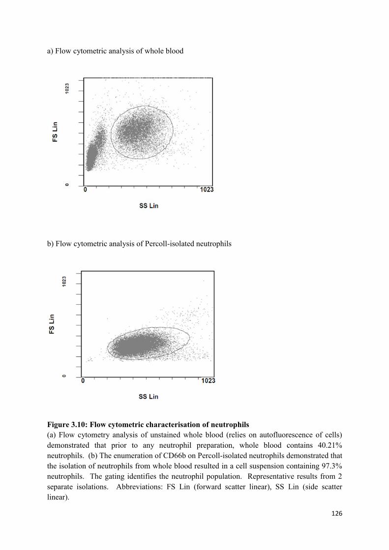
126
a) Flow cytometric analysis of whole blood
b) Flow cytometric analysis of Percoll-isolated neutrophils
Figure 3.10: Flow cytometric characterisation of neutrophils
(a) Flow cytometry analysis of unstained whole blood (relies on autofluorescence of cells)
demonstrated that prior to any neutrophil preparation, whole blood contains 40.21%
neutrophils. (b) The enumeration of CD66b on Percoll-isolated neutrophils demonstrated that
the isolation of neutrophils from whole blood resulted in a cell suspension containing 97.3%
neutrophils. The gating identifies the neutrophil population. Representative results from 2
separate isolations. Abbreviations: FS Lin (forward scatter linear), SS Lin (side scatter
linear).

127
3.4 Discussion
The aim of this work was to determine the most suitable method for peripheral blood
neutrophil isolation by comparing Percoll isolation with two additional neutrophil isolation
approaches with respect to ex vivo neutrophil responses. Parameters investigated included
cell yield, ROS release (luminol chemiluminescence), NET production (quantification of
NET-DNA with Sytox green), apoptotic activity (caspase-3 and -7 production) and metabolic
status (ATP production). Overall, results revealed that there were indeed differences in
cellular responses generated following the various neutrophil isolation techniques.
The numbers of neutrophils obtained from the same individual were significantly higher
when a Percoll gradient was used for purification (section 3.2.1, 3.2.2). This is consistent
with the findings of Grisham (1985), who reported that the use of two discontinuous Percoll
gradients produced a higher cell yield than Histopaque gradients. However no difference in
cell yield was observed between Percoll and Dextran gradient isolated neutrophils. Whilst
recent in vivo analyses have demonstrated neutrophil longevity within the blood stream of 5.4
days (Pillay et al., 2010), neutrophils are terminally differentiated cells, meaning they cannot
be passaged in tissue culture without undergoing apoptosis (Gottlieb et al., 1995). Therefore,
despite the fact that this difference in yield is corrected for in subsequent neutrophil assays,
yield is an important variable as the number of cells attainable from a limited amount of
venous blood governs the number of subsequent experiments that can be conducted.
To determine whether the isolation method activated neutrophils, baseline ROS production
over a 30 min period was measured (section 3.2.3, 3.2.4). At this point, cells were
unstimulated and the method of neutrophil isolation represented the only variable between
these cells. Although no statistical difference in ROS production was measured between
Percoll and Histopaque-isolated cells, there was a general trend towards higher ROS
128
production in neutrophils isolated with Histopaque. It is possible that the Histopaque
isolation method activates the neutrophils, resulting in inadvertent ROS production
immediately after isolation. ROS release is known to be a causative agent in neutrophil
death, as co-incubation of neutrophils with glutathione, which scavenges H2O2 and other
ROS, has been found to decrease neutrophil death (Yamamoto et al., 2002). These findings
infer that upon exposure to a threshold level of ROS, as a result of activation, neutrophils will
enter into apoptosis (Scheel-Toellner et al., 2004). Conversely, high levels of ROS can also
inhibit caspase activity, suggesting that caspase-independent pathways may be involved in
ROS-dependent cell death (Fadeel et al., 1998). However it is noteworthy that apoptosing
neutrophils are still capable of producing substantial amounts of ROS in response to
exogenous stimulation (Xu et al., 2009), thus baseline ROS may be derived from a
combination of activated and/or apoptosing neutrophils.
Caspase activity was determined by quantifying effector caspases-3 and -7 over a 60 min
period post-isolation (section 3.2.11, 3.2.12). Caspase activity was higher in Histopaque-
isolated cells immediately following isolation; however less caspase activity was evident over
time. In response to stimulation, Percoll-isolated cells produced higher levels of ROS than
Histopaque-isolated cells. This could be due to the Histopaque-prepared cells being less
responsive to exogenous stimulation, or the possibility that initially apoptotic neutrophils are
no longer viable and thus less ROS is produced in response to stimulation. No differences
were observed between Percoll and Dextran isolation with respect to baseline total ROS
release, as well as in response to stimulation. Furthermore, no significant differences were
found between Percoll and Dextran isolation techniques with regard to neutrophil apoptosis,
which lends further support for the association of caspase activity with ROS production.

129
Percoll, Histopaque and Dextan-purified neutrophils were also compared in terms of their
ability to produce NETs (3.2.7, 3.2.8). Percoll-isolated neutrophils were found to produce
consistently more NETs following stimulation with F. nucleatum poly and opsonised S.
aureus. This was also observed following Dextran isolation, with Percoll-isolated cells
producing significantly higher levels of NETs in response to PMA and HOCl. Collectively,
these findings suggest that Percoll isolated neutrophils are more sensitive to NET production
following stimulation. The differences between Percoll and Histopaque cell NET production
may be explained by the total ROS results being lower in Histopaque isolated cells following
stimulation, as ROS is known to be vital for NET release (Kirchner et al., 2011, Nishinaka et
al., 2011); in support of this is treatment of neutrophils with NADPH-oxidase inhibitor (DPI),
which abolishes NET release. Whilst it is recognised that NADPH-oxidase independent
NETs may occur, and could therefore provide an explanation for the reduced NET release in
Dextran cells (as decreased ROS was not observed in these cells), this seems unlikely as
NADPH-oxidase independent NET release is reported in response to specific stimuli such as
uric acid (Arai et al., 2014) and soluble immune complexes (Chen et al., 2012). It is also a
possibility that a higher proportion of Histopaque- and Dextran-isolated neutrophils are no
longer viable 3 hours following isolation, which is the time at which NETs are quantified and
may explain the lower number of NETs quantified.
Consistent with the hypothesis that Histopaque isolation causes activation of neutrophils,
ATP quantification (indicative of metabolic activity) revealed that Histopaque isolated cells
were metabolically active initially, however less ATP was produced over time, indicating
either that negative internal feedback mechanisms switch neutrophils to a less active state, or
potentially that apoptosis was indeed being induced (Scheel-Toellner et al 2004) (section
3.2.9). Metabolic activity was significantly higher in Dextran-isolated cells at each time
points, relative to Percoll cells (section 3.2.10). Physiologically relevant concentrations of

130
ATP are correlated with a concentration dependent delay in neutrophil apoptosis (Vaughan et
al., 2007). Although NETosis is distinct from apoptosis (Fuchs et al., 2007); it is possible
that increases in ATP may also be associated with delayed or decreased NET release, which
was observed in both Histopaque- and Dextran-prepared cells. To the best of my knowledge,
the effect of neutrophil isolation technique upon NET responses has not previously been
investigated.
A further consideration is that neutrophils isolated with Percoll were subjected to multiple
centrifugation and wash steps, whereas isolation with Histopaque and Dextran required less
handling and transfers between tubes; suggesting Histopaque and Dextran neutrophils should
be less activated. All three gradient protocols take approximately 90 mins, and following
several reports demonstrating that delays prior to isolation can trigger neutrophil activation
(Ferrante & Thong 1980, Maqbool et al., 2011), isolation was started immediately following
blood collection. Thus with regard to neutrophils’ relatively short life span, no gradient is
superior in terms of isolation time. Whilst erythrocyte lysis produces a cleaner cell
suspension, it has been reported that repeated or extended lysis can damage neutrophils
(Nauseef et al., 2007). Dextran isolation did not employ a lysis step, however the Histopaque
protocol relied on a 30 sec erythrocyte lysis with distilled water, as initial experiments
demonstrated erythrocyte contamination. This is distinct from Percoll-isolated neutrophils,
which are subjected to an ammonium chloride (NH4Cl) lysis. Although it has been suggested
that distilled water-induced erythrocyte lysis causes less neutrophil activation, the ROS
results in this particular study indicate this may not be the case under these particular assay
conditions. This is also supported by Vuorte et al., (2001), who reported that neither
hypotonic shock with distilled water or NH4Cl lysis influenced neutrophil integrity or
activation. In addition the choice of anticoagulant blood collection tube may also influence
neutrophil isolation and effector functions. All blood for the current study was collected in
131
lithium heparin tubes, acting as an anticoagulant by activating anti-thrombin. However,
another commonly used collection tube is ethylenediamine tetraacetic acid (EDTA), which
chelates free calcium ions. Calcium ions play a vital role in the activation of the neutrophil
respiratory burst, and are the likely explanation for reports demonstrating reduced
Histopaque-isolated neutrophil ROS release from blood collected into EDTA tubes,
compared with heparin (Freitas et al., 2008).
Visualisation of Percoll-isolated neutrophils by cytospin identified neutrophils in the cell
suspension, evidenced by the characteristic multi-lobed nuclei observed that stained dark blue
(section 3.3.1.1). Fluorescence microscopy of the neutrophil surface marker CD66 also
confirmed the isolation of neutrophils (section 3.3.1.2). To determine the purity of Percoll-
isolated cells, neutrophil marker CD66b was quantified by flow cytometry; this demonstrated
that the cell suspension contained 97.3% neutrophils (section 3.3.1.3), which is in line with
other reports that obtain >95% purity following the isolation of human neutrophils (Kirchner
et al., 2011, Maqbool et al., 2011). The remaining 2.7% of cells are likely to include
eosinophils and monoculear cells (Dorward et al., 2013). In addition, neutrophils were
quantified in lysed whole peripheral blood prior to Percoll-isolation by measuring the auto-
fluorescence in the blood sample, which demonstrated that neutrophils comprised 40.21% of
whole blood prior to isolation. Optimisation of the Percoll-isolation protocol and meticulous
technique can reduce the number of contaminating mononuclear cells in the neutrophil cell
suspension. However it is noteworthy that density gradient centrifugation techniques cannot
prevent the complete exclusion of contaminating cells, for example, it is reported that in
blood derived from individuals with atopic conditions, such as asthma, eosinophils may
represent 5-10% of the granulocyte population isolated (Dorward et al., 2013).

132
In conclusion, when analysing the impact of isolation technique on neutrophil cell yield,
function and activation, Percoll appears to be the most appropriate method, evidenced by the
lower metabolic activation and higher NET release by Percoll-isolated cells. Percoll was
subsequently selected as the isolation technique for all ex vivo neutrophil assays in this thesis.

133
INTERACTIONS BETWEEN PERIODONTAL CHAPTER 4:
BACTERIA AND PERIPHERAL BLOOD NEUTROPHILS

134
4.1 Introduction
Given that NET release is reported to be predominantly ROS dependent (Fuchs et al., 2007),
various neutrophil responses to heat-killed periodontal bacteria were investigated
(Appendices I, IV), including total, extracellular and superoxide ROS production (section
2.2.3.2). NET production was determined by quantifying the amount of DNA released
(2.2.3.3) alongside the NET-bound components, neutrophil elastase (NE), myeloperoxidase
(MPO) and cathepsin G (CG) (2.2.3.4, Appendix III). The ability of NETs to entrap bacteria
was also qualitatively determined by visualising interactions using SEM (2.2.3.8), and by
quantifying the number of viable bacteria entrapped within NET structures (2.2.3.5, 2.2.3.6,
Appendix II). All neutrophils were from periodontally and systemically healthy volunteers
(University of Birmingham Ethics Reference ERN_13-0325) and neutrophil assays employed
a panel of bacteria associated with periodontitis. For clinical relevance, data are presented by
grouping periodontal bacteria according to the Socransky complexes (Socransky et al., 1998).
The ROS-dependent activation and signalling cascades for NET release in response to heat-
killed periodontal bacteria were also investigated by determining the effects of modulators of
neutrophil activation (2.2.2.3).
4.2 Neutrophil ROS release in response to periodontal bacteria
ROS production in response to a panel of 19 heat-killed periodontal bacteria (1x10⁸ and MOI
of 1000) was determined using enhanced chemiluminescence (2.2.3.2). ROS release
following exposure to PBS (unstimulated negative control), PMA (25nM, positive control)
and opsonised S. aureus (5x10⁷ and MOI of 500, positive control) was also quantified.
Neutrophils were stimulated after being equilibrate for a 30 min baseline period and then
ROS measured over the subsequent 100 mins. Total neutrophil ROS release was determined
using luminol chemiluminescence. Opsonised S.aureus induced significantly higher ROS

135
production than the PBS control (Kruskal-Wallis and Dunn’s multiple comparison post-tests
***p<0.001, n=5). Certain periodontal bacteria elicited higher total ROS production, which
was statistically significant for P. acnes, S. anginosus and C. rectus (Kruskal-Wallis and
Dunn’s multiple comparison post-tests **p<0.01, **p<0.01, *p<0.05, respectively, n=5)
(Figure 4.1a).
Neutrophil extracellular ROS production was subsequently analysed by isoluminol
chemiluminescence. PMA induced significantly higher ROS production than PBS treatment
(Kruskal-Wallis and Dunn’s multiple comparison post-tests **p<0.01, n=5). S. gordonii also
elicited significantly higher total ROS production (Kruskal-Wallis and Dunn’s multiple
comparison post-tests *p<0.05, n=5) (Figure 4.1b).
Neutrophil superoxide production was measured with lucigenin. PMA and opsonised S.
aureus did not induce significantly higher superoxide production relative to the PBS control.
However, some periodontal bacterial species elicited higher superoxide production, which
was statistically significant for S. anginosus, C. suptigena, and F. nucleatum nuc (Kruskal-
Wallis and Dunn’s multiple comparison post-tests **p<0.01, *p<0.05, *p<0.05, respectively,
n=5) (Figure 4.1c).

136
a) Neutrophil total ROS production R
LU
PBS
PM
A
Ops
onised
S. a
ureu
s
A. v
isco
sus
A. a
ctinom
ycet
emco
mita
ns b
P. a
cnes
Sel. n
oxia
V. p
arvu
la
S. s
angu
inis
S. o
ralis
S. int
erm
edius
S. a
nginos
us
S. g
ordo
nii
C. g
ingiva
lis
A. a
ctinom
ycet
emco
mita
ns a
C. s
putig
ena
S. c
onstellatu
s
C. r
ectu
s
C. s
howae
F. nuc
leat
um n
uc
F. nuc
leat
um p
oly
P. g
ingiva
lis
0
100000
200000
300000
400000
*****
**
*
b) Neutrophil extracellular ROS production
RL
U
PBS
PM
A
Ops
onised
S. a
ureu
s
A. v
isco
sus
A. a
ctinom
ycet
emco
mita
ns b
P. a
cnes
Sel. n
oxia
V. p
arvu
la
S. s
angu
inis
S. o
ralis
S. int
erm
edius
S. a
nginos
us
S. g
ordo
nii
C. g
ingiva
lis
A. a
ctinom
ycet
emco
mita
ns a
C. s
putig
ena
S. c
onstellatu
s
C. r
ectu
s
C. s
howae
F. nuc
leat
um n
uc
F. nuc
leat
um p
oly
P. g
ingiva
lis
0
5000
10000
15000
20000
25000 **
*

137
c) Neutrophil superoxide production R
LU
PBS
PM
A
Ops
onised
S. a
ureu
s
A. v
isco
sus
A. a
ctinom
ycet
emco
mita
ns b
P. a
cnes
Sel. n
oxia
V. p
arvu
la
S. s
angu
inis
S. o
ralis
S. int
erm
edius
S. a
nginos
us
S. g
ordo
nii
C. g
ingiva
lis
A. a
ctinom
ycet
emco
mita
ns a
C. s
putig
ena
S. c
onstellatu
s
C. r
ectu
s
C. s
howae
F. nuc
leat
um n
uc
F. nuc
leat
um p
oly
P. g
ingiva
lis
0
5000
10000
15000
20000
25000
**
*
*
Figure 4.1: Neutrophil ROS production in response to periodontal bacteria
Neutrophil (a) total ROS, (b) extracellular ROS and (c) superoxide production were
quantified in response to a panel of periodontal bacteria (1x10⁸ and MOI of 1000) using
luminol, isoluminol and lucigenin enhanced chemiluminescence, respectively. ROS release
in response to PBS (unstimulated negative control), PMA (25nM, positive control) and
opsonised S. aureus (5x10⁷ and MOI of 500, positive control) was also quantified. Statistical
significance calculated using Kruskal-Wallis and Dunn’s multiple comparison post-tests
(*p<0.05, **p<0.01, ***p<0.001). Data presented as RLU (relative light units) and expressed
as mean ± SEM (n=5 in triplicate).
138
Time-course of neutrophil ROS production in response to periodontal bacteria 4.2.1
The time-course of ROS production was analysed following stimulation with heat-killed
periodontal bacteria, as previously described. Consistent with the data expressed as total
peak ROS production, time-course ROS production expressed as “area under the curve”
demonstrated that total ROS production (luminol) was highest in response to opsonised S.
aureus (5x10⁷ and MOI of 500), followed by S. anginosus (1x10
8 and MOI of 1000).
Notably, the increase in total ROS following stimulation with opsonised S. aureus was more
rapid than following bacterial stimulation, as illustrated by the sharp increase in ROS levels
following stimulation. Total ROS release following opsonised S. aureus-stimulation peaked
at approximately 20 mins post stimulation, which was followed by a steady decrease in total
ROS production (Figure 4.2a). Extracellular ROS production measured using isoluminol
demonstrated that PMA (25nM) stimulation induced the highest ROS signal. The steep time-
course curve in response to PMA suggests a rapid neutrophil response to PMA. PMA-
induced extracellular ROS release peaked approximately 20 mins post stimulation, followed
by a steady decrease in total ROS. S. gordonii (1x10⁸ and MOI of 1000) also produced
elevated levels of extracellular ROS in comparison with other periodontal bacteria (Figure
4.2b). The quantification of superoxide production (lucigenin) in response to periodontal
bacteria demonstrated that F. nucleatum nuc (1x10⁸ and MOI of 1000) generated the highest
levels of superoxide, relative to other periodontal bacteria. F. nucleatum nuc-induced
superoxide release peaked approximately 30 mins post-stimulation, followed by a steady
decrease in total ROS. PMA and opsonised S. aureus did not generate detectable elevations
in superoxide production (Figure 4.2c).

139
Time/min
RL
U
0 26 52 78 104 1300
100000
200000
300000
400000
a) Neutrophil total ROS production
Time/min
RL
U
0 26 52 78 104 1300
10000
20000
30000
40000
50000
b) Neutrophil extracellular ROS production
Time/min
RL
U
0 26 52 78 104 1300
5000
10000
15000
20000
25000
PBS
PMA
F. nucleatum poly
V. parvula
A. viscosus
S. sanguinis
S. constellatus
A. actinomycetemcomitans a
C. gingivalis
S. gordonii
S. intermedius
A. actinomycetemcomitans b
C. rectus
F. nucleatum nuc
Opsonised S. aureus
C. sputigena
S. anginosus
P. acnes
Sel. noxia
C. showae
S. oralisP. gingivalis
c) Neutrophil superoxide production
Figure 4.2: Time-course of neutrophil ROS production in response to periodontal
bacteria
Time-course for ROS production assayed over 130 mins by neutrophils stimulated with PMA
(25nM), opsonised S. aureus (5x10⁷ and MOI of 500) or periodontal bacteria (1x10
8 and MOI
of 1000). (a) Total ROS production (luminol), (b) extracellular ROS production (isoluminol),
(c) superoxide production (lucigenin). Data presented as mean RLU (relative light units) (n=5
in triplicate).

140
4.3 Quantification of NET production in response to periodontal bacteria exposure
NET production in response to a panel of 19 heat-killed periodontal bacteria (1x10⁸ and MOI
of 1000) was determined by quantifying NET-DNA (2.2.3.3) and NET bound components
(2.2.3.4). NET release following exposure to PBS (unstimulated negative control), PMA
(50nM, positive control) and opsonised S. aureus (5x10⁷ and MOI of 500, positive control)
was also quantified. Neutrophils were stimulated after being equilibrate for a 30 min baseline
period and then NET production measured after a 4 hour incubation period. NET-DNA
measured with Sytox green demonstrated that PMA stimulated significantly higher NET
production compared with PBS (Kruskal-Wallis and Dunn’s multiple comparison post-tests
*p<0.05, n=5). Certain periodontal bacteria elicited higher NET-DNA production and this
was statistically significant compared with PBS for P. acnes, V. parvula, and S. gordonii
(Kruskal-Wallis and Dunn’s multiple comparison post-tests *p<0.05, *p<0.05, *p<0.05,
respectively, n=5) (Figure 4.3a).
NET-bound NE was quantified colorimetrically (2.2.3.4.1) and demonstrated that certain
periodontal bacteria elicited higher NET-bound NE production relative to PBS, however this
was not statistically significant. Similarly, stimulation with PMA and opsonised S. aureus
(positive controls) stimulation did not reach statistical significance (Kruskal-Wallis p>0.05,
n=3) (Figure 4.3b).
NET-bound MPO was quantified colorimetrically (2.2.3.4.2) and demonstrated that PMA
stimulated significantly higher NET-bound MPO production (Kruskal-Wallis and Dunn’s
multiple comparison post-tests *p<0.05, n=3). Certain periodontal bacterial species elicited
higher NET-bound MPO production relative to PBS; however this did not reach statistical
significance (Kruskal-Wallis and Dunn’s multiple comparison post-tests p>0.05, n=3)
(Figure 4.3c).

141
NET-bound CG was quantified colorimetrically (2.2.3.4.3) and demonstrated that opsonised
S. aureus stimulated for significantly higher NET-bound CG production (Kruskal-Wallis and
Dunn’s multiple comparison post-tests *p<0.05, n=3). Certain periodontal bacterial species
elicited higher NET-bound CG production, which was statistically significant for A. viscosus
and C. gingivalis (Kruskal-Wallis and Dunn’s multiple comparison post-tests *p<0.05 for
both, n=3) (Figure 4.3d).
a) NET-DNA
AF
U
PBS
PM
A
Ops
onised
S. a
ureu
s
A. v
isco
sus
A. a
ctinom
ycet
emco
mita
ns b
P. a
cnes
Sel. n
oxia
V. p
arvu
la
S. s
angu
inis
S. o
ralis
S. int
erm
edius
S. a
nginos
us
S. g
ordo
nii
C. g
ingiva
lis
A. a
ctinom
ycet
emco
mita
ns a
C. s
putig
ena
S. c
onstellatu
s
C. r
ectu
s
C. s
howae
F. nuc
leat
um n
uc
F. nuc
leat
um p
oly
P. g
ingiva
lis
0
2000
4000
6000
8000
10000
**
*
*

142
b) NET-bound neutrophil elastase N
E m
U/m
l
PBS
PM
A
opso
nise
d S. a
ureu
s
A. v
isco
sus
A. a
ctinom
ycet
emco
mita
ns b
P. a
cnes
Sel. n
oxia
V. p
arvu
la
S. s
angu
inis
S. o
ralis
S. int
erm
edius
S. a
nginos
us
S. g
ordo
nii
C.g
ingiva
lis
A. a
ctinom
ycet
emco
mita
ns a
C. s
putig
ena
S. c
onstellatu
s
C. r
ectu
s
C. s
howae
F. nuc
leat
um n
uc
F. nuc
leat
um p
ol
P. g
ingiva
lis
0
20
40
60
80 ns
c) NET-bound myeloperoxidase
MP
O U
/ml
PBS
PM
A
opso
nise
d S. a
ureu
s
A. v
isco
sus
A. a
ctinom
ycet
emco
mita
ns b
P. a
cnes
Sel. n
oxia
V. p
arvu
la
S. s
angu
inis
S. o
ralis
S. int
erm
edius
S. a
nginos
us
S. g
ordo
nii
C. g
ingiva
lis
A. a
ctinom
ycet
emco
mita
ns a
C. s
putig
ena
S. c
onstellatu
s
C. r
ectu
s
C. s
howae
F. nuc
leat
um n
uc
F. nuc
leat
um p
ol
P. g
ingiva
lis
0.00
0.25
0.50
0.75
1.00
1.25*

143
d) NET-bound cathepsin G
C
G m
U/m
l
PBS
PM
A
opso
nise
d S. a
ureu
s
A. v
isco
sus
A. a
ctinom
ycet
emco
mita
ns b
P. a
cnes
Sel. n
oxia
V. p
arvu
la
S. s
angu
inis
S. o
ralis
S. int
erm
edius
S. a
nginos
us
S. g
ordo
nii
C. g
ingiva
lis
A. a
ctinom
ycet
emco
mita
ns a
C. s
putig
ena
S. c
onstellatu
s
C. r
ectu
s
C. s
howae
F. nuc
leat
um n
uc
F. nuc
leat
um p
ol
P. g
ingiva
lis
0
1
2
3
4
5*
*
*
Figure 4.3: Quantification of NET production in response to periodontal bacteria
exposure
NET production was quantified in response to a panel of periodontal bacteria (1x10⁸ and
MOI of 1000). NET release in response to PBS (unstimulated negative control), PMA
(50nM, positive control) and opsonised S. aureus (5x10⁷ and MOI of 500, positive control)
was also quantified. (a) NET-DNA was quantified with Sytox green and NET-bound (b)
neutrophil elastase, (c) myeloperoxidase and (d) cathepsin G were quantified
colorimetrically. Statistical significance was calculated using Kruskal-Wallis and Dunn’s
multiple comparison post-tests (*p<0.05, ns=not significant). Data is presented as (a) AFU
(arbitrary fluorescence units), (c) U/ml or (d, e) mU/ml and expressed as mean ± SEM (n=5
in triplicate).

144
4.4 NET entrapment of bacteria
To assess the ability of NETs to immobilise periodontal bacteria, FITC-stained live bacteria
(1x10⁷ and MOI of 100) were incubated for 1 hour with either unstimulated neutrophils,
intact NETs (produced by 0.75mM HOCl stimulation) or NET structures following
degradation with MNase (section 2.2.3.5). Following multiple wash steps to remove any
unbound bacteria the amount of bacteria entrapped was fluorometrically quantified and
normalised to FITC-stained bacteria incubated with PBS (cell-free control). For clinical
relevance, data are presented by grouping periodontal bacteria according to the Socransky
complexes (Socransky et al., 1998).
NET entrapment of bacteria not assigned to a Socransky complex
Sel. noxia appeared to be entrapped within NET structures relative to bacteria entrapped
within neutrophils or degraded NETs (negative controls) (1way ANOVA *p=0.0111, n=5).
However, neither A. actinomycetemcomitans (serotype b) nor P. acnes were significantly
entrapped within NETs (1way ANOVA p>0.05, n=5) (Figure 4.5a).
NET entrapment of blue complex bacteria
A. viscosus appeared to evade entrapment within NET, compared with bacteria entrapped
within neutrophils or degraded NETs (negative controls) (1way ANOVA p=0.1169, n=5)
(Figure 4.5b).
NET entrapment of purple complex bacteria
V. parvula was found to be significantly entrapped within NETs compared with bacteria
incubated with unstimulated neutrophils or degraded NET structures (1way ANOVA
*p=0.0129, n=5) (Figure 4.5c).

145
NET entrapment of yellow complex bacteria
S. anginosus and S. gordonii were significantly entrapped within NETs, relative to bacteria
entrapped within neutrophils or degraded NETs (1way ANOVA *p=0.0216 and *p=0.0305,
respectively, n=5). However the other yellow complex bacteria assayed, S. sanguinis, S.
oralis and S. intermedius, appeared to evade entrapment within NET structures in this assay
system (1way ANOVA p>0.05, n=5) (Figure 4.5d).
NET entrapment of green complex bacteria
No green complex bacteria assayed appeared significantly entrapped within NETs relative to
bacteria entrapped within neutrophils or degraded NETs (negative controls) according to the
assay conditions employed (1way ANOVA p=0.1135, n=5) (Figure 4.5e).
NET entrapment of orange complex bacteria
C. rectus, C. showae and F. nucleatum poly were significantly entrapped within NETs
relative to bacteria entrapped within neutrophils or degraded NETs (negative controls) (1way
ANOVA ****p=0.0004, ****p=0.0002, **p=0.0079, respectively, n=5). However the other
orange complex bacteria assayed, S. constellatus and F. nucleatum nuc, were not significantly
entrapped within NET structures (1way ANOVA p>0.05, n=5) (Figure 4.5f).
NET entrapment of red complex bacteria
P. gingivalis was significantly entrapped within NET structures compared with bacteria
incubated with unstimulated neutrophils or degraded NET structures (1way ANOVA
*p=0.027, n=5) (Figure 4.5g).

146
a) Bacteria not assigned to a Socransky complex A
FU
A. a
ctinom
yces
b
P. a
cnes
Sel. n
oxia
0
1
2
3
4
*Neutrophils
NETs
Degraded-NETs
b) Blue complex bacteria
AF
U
A. v
isco
sus
0
1
2
3
4Neutrophils
NETs
Degraded-NETs
ns
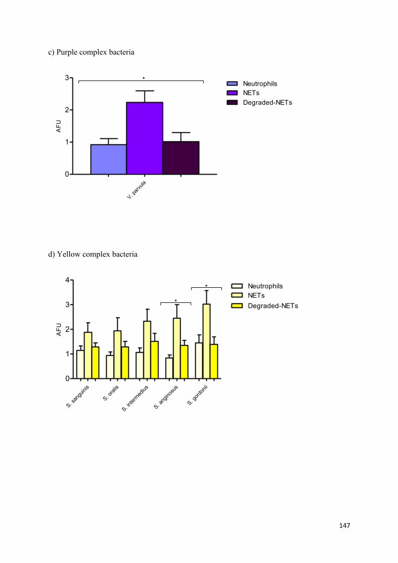
147
c) Purple complex bacteria A
FU
V. p
arvu
la
0
1
2
3Neutrophils
NETs
Degraded-NETs
*
d) Yellow complex bacteria
AF
U
S. s
angu
inis
S. o
ralis
S. int
erm
ediu
s
S. a
nginos
us
S. g
ordo
nii
0
1
2
3
4*
*
Neutrophils
NETs
Degraded-NETs

148
(e) Green complex bacteria
AF
U
C. g
ingi
valis
A. a
ctinom
yces
a
C. s
putig
ena
0
1
2
3
4Neutrophils
NETs
Degraded-NETs
ns
f) Orange complex bacteria
AF
U
S. c
onstellatu
s
C. r
ectu
s
C. s
howae
F. nuc
leat
um n
uc
F. nuc
leat
um p
oly
0
1
2
3
**
****
**** Neutrophils
NETs
Degraded-NETs

149
g) Red complex bacteria A
FU
P. g
ingiva
lis
0
1
2
3
4Neutrophils
NETs
Degraded-NETs
*
Figure 4.4: NET entrapment of periodontal bacteria
NET entrapment of (a) bacteria not assigned to a Socransky complex, (b) blue complex
bacteria, (c) purple complex bacteria, (d) yellow complex bacteria, (e) green complex
bacteria, (f) orange complex bacteria and (g) red complex bacteria was determined by
quantifying FITC-stained bacteria immobilised in NET structures. Results are normalised to
FITC-stained bacteria entrapped in PBS. Statistical significance (1way ANOVA) of NET-
derived bacterial entrapment is relative to bacteria entrapped within neutrophils and degraded
NETs (*p<0.05, **p<0.01, ***p<0.001, ****p<0.0001, ns=not significant). Data presented as
AFU (arbitrary fluorescence units) and expressed as mean ± SEM (n=5 in triplicate).

150
4.5 Scanning electron microscopy of NET-bacteria interactions
To visualise the interactions between NETs and bacteria, NET structures were visualised by
SEM following incubation with PBS (negative control) and selected live periodontal bacteria
(1x10⁷ MOI of 100) (2.2.3.8). As expected, SEM images of unstimulated neutrophils
demonstrated spherical cells with no NET structures evident (Figure 4.5). The visualisation
of neutrophils following incubation with bacteria; A. actinomycetemcomitans serotype a
(Figure 4.6a), V. parvula (Figure 4.6b) and A. viscosus (Figure 4.6c), revealed the release of
NET structures from neutrophils. The strand-like filaments between the neutrophils appeared
to associate with bacteria, for example A. actinomycetemcomitans (serotype a) appear to
cluster along the length of the NET structure (Figure 4.6a). SEM analysis also enabled
detailed visualisation of bacteria at a high magnification, which consistent with Gram stains
(Appendix IV), confirmed bacterial morphology.

151
Figure 4.5: Scanning electron microscopy of resting neutrophils
Resting neutrophils (1x10⁵) were visualised by SEM. Representative image of 3
experiments, scale bar represents 10µm.
a) A. actinomycetemcomitans serotype a
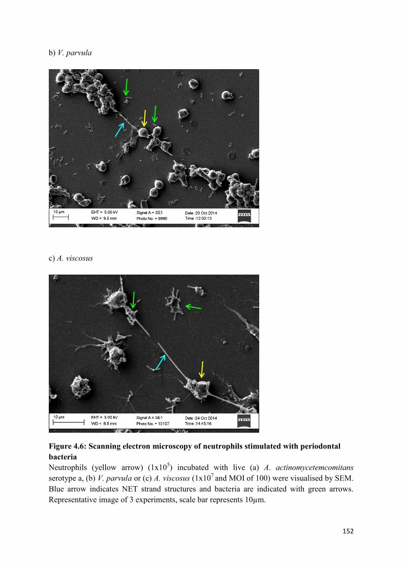
152
b) V. parvula
c) A. viscosus
Figure 4.6: Scanning electron microscopy of neutrophils stimulated with periodontal
bacteria
Neutrophils (yellow arrow) (1x10⁵) incubated with live (a) A. actinomycetemcomitans
serotype a, (b) V. parvula or (c) A. viscosus (1x10⁷
and MOI of 100) were visualised by SEM.
Blue arrow indicates NET strand structures and bacteria are indicated with green arrows.
Representative image of 3 experiments, scale bar represents 10µm.

153
4.6 Quantification of NET-mediated killing of periodontal bacteria
To determine whether NETs are capable of killing entrapped bacteria, periodontal bacteria
(1x10⁷
and MOI of 100) were incubated with PBS (negative control), unstimulated
neutrophils, intact NETs (produced by 0.75mM HOCl), degraded NETs (by MNase
digestion), unseparated NETs (NETs not degraded with MNase prior to bacteria subculture)
or with cytochalasin B-treated NETs (to block neutrophil phagocytosis) (section 2.2.3.6,
Appendix II). Following 1 hour incubation bacteria were released from NETs by MNase
digestion, diluted and inoculated onto agar plates and cultured for 24 hours prior to
performing colony counts. Of the 6 periodontal bacteria assayed, no significant NET-
mediated killing of periodontal bacteria was observed (all analysed by 2way ANOVA, n=5)
F. nucleatum poly (p=0.5873), S. intermedius (p=0.6007), S. sanguinis (p=0.8977), A.
viscosus (p=0.9573), V. parvula (p=0.9239) and C. gingivalis (p=0.9787) (Figures 4.7a-f).

154
Co
lon
y c
ou
nts
PBS
Neu
troph
ils
NETS
Deg
arde
d NETs
Not
sep
arat
ed
Cytoc
halasin
B
0
20
40
60
a) F. nucleatum polymorphum
ns
Co
lon
y c
ou
nts
PBS
Neu
troph
ils
NETS
Deg
arde
d NETs
Not
sep
arat
ed
Cytoc
halasin
B
0
20
40
60
80
100
b) S. intermedius
ns
Co
lon
y c
ou
nts
PBS
Neu
troph
ils
NETS
Deg
arde
d NETs
Not
sep
arat
ed
Cytoc
halasin
B
0
20
40
60
80
c) S. sanguinis
ns

155
Co
lon
y c
ou
nts
PBS
Neu
troph
ils
NETS
Deg
arde
d NETs
Not
sep
arat
ed
Cytoc
halasin
B
0
50
100
150
200
d) A. viscosus
ns
Co
lon
y c
ou
nts
PBS
Neu
troph
ils
NETS
Deg
arde
d NETs
Not
sep
arat
ed
Cytoc
halasin
B
0
50
100
150
200
250
e) V. parvula
ns
Co
lon
y c
ou
nts
PBS
Neu
troph
ils
NETS
Deg
arde
d NETs
Not
sep
arat
ed
Cytoc
halasin
B
0
50
100
150
f) C. gingivalis
ns
Figure 4.7: Quantification of NET-mediated killing of periodontal bacteria
Bacterial colonies were counted following incubation with PBS (negative control),
neutrophils, intact NETs (0.75mM HOCl), degraded NETs (100U/ml MNase), unseparated
NETs (NETs not degraded with MNase prior to bacteria subculture) or with cytochalasin B-
treated NETs (to block neutrophil phagocytosis). No significant NET-mediated killing of
periodontal bacteria was observed (1way ANOVA p>0.05). Data presented as total colony
counts and expressed as mean ± SEM (n=5).

156
4.7 Effect of NADPH-oxidase pathway modulating agents on ROS and NET production
To further understand the importance of NADPH-oxidase and downstream products in
bacteria-induced ROS and NET production, specific components of the NADPH-oxidase
signalling pathway were targeted (section 2.2.2.3). Isolated neutrophils were incubated with
diphenyleneiodonium (DPI) (25µM), an inhibitor of NADPH-oxidase, N-acetyl-cysteine
(NAC) (10mM), a synthetic glutathione precursor that scavenges H2O2, or taurine (100mM),
which scavenges HOCl to produce taurine chloramine. Neutrophil total ROS and NET
production were measured following pre-incubation with the modulating agent for 30 mins
prior to stimulation with PMA (50nM), opsonised S. aureus (5x10⁷ and MOI of 500) and a
panel of 9 selected heat-killed periodontal bacteria (1x10⁸ and MOI of 1000).
DPI pre-treatment resulted in a reduction in total ROS release in response to all stimuli,
relative to non-DPI treated cells. This was statistically significant for PMA (*p<0.05), as well
as following stimulation with opsonised S. aureus (****p<0.0001), S. gordonii
(****p<0.0001), F. nucleatum poly (*p<0.05) and Sel. noxia (*p<0.05) (2way ANOVA and
Bonferroni post-tests, n=3) (Figure 4.8a). NET-DNA was quantified with Sytox green
following enzymatic degradation of NET structures with MNase. DPI pre-treatment also
significantly reduced NET release in response to all stimuli relative to cells that did not
receive DPI pre-treatment. This was statistically significant in response to PMA
(****p<0.0001), but not for S. aureus-induced NET production or the periodontal bacteria
assayed (2way ANOVA and Bonferroni post-tests, n=3) (Figure 4.8b).
NAC treatment significantly reduced total ROS release in response to all stimuli relative to
cells that did not receive NAC pre-treatment. This was statistically significant for PMA
(*p<0.05) and opsonised S. aureus (****p<0.0001), as well as following bacterial stimulation
with S. gordonii (****p<0.0001) and Sel. noxia (*p<0.05) (2way ANOVA and Bonferroni
post-tests, n=3) (Figure 4.8c). NET-DNA was quantified with Sytox green following

157
enzymatic degradation of NET structures with MNase. NAC treatment reduced NET release
in response to all stimuli relative to cells that did not receive NAC pre-treatment. This was
statistically significant for to PMA (****p<0.0001) and Sel. noxia (*p<0.05), but not for
opsonised S. aureus or the other periodontal bacteria assayed (analysed by 2way ANOVA
and Bonferroni post-tests, n=3) (Figure 4.8d).
Taurine treatment significantly reduced total ROS release in response to all stimuli relative to
cells that did not receive taurine; however this was to a lesser extent than that observed with
DPI and NAC. Decreased ROS release following taurine pre-treatment was significant for
opsonised S. aureus (****p<0.0001), S. gordonii (***p<0.001) and Sel. noxia (*p<0.05)
(2way ANOVA and Bonferroni post-tests, n=3) (Figure 4.8e). Taurine pre-treatment had a
differential effect on neutrophil NET production, relative to untreated cells. Taurine
significantly reduced NET production following stimulation with PMA (2way ANOVA and
Bonferroni post-test ****p<0.0001, n=3), but did not significantly reduce NET production in
response to opsonised S. aureus or bacterial stimulation (Figure 4.8f).
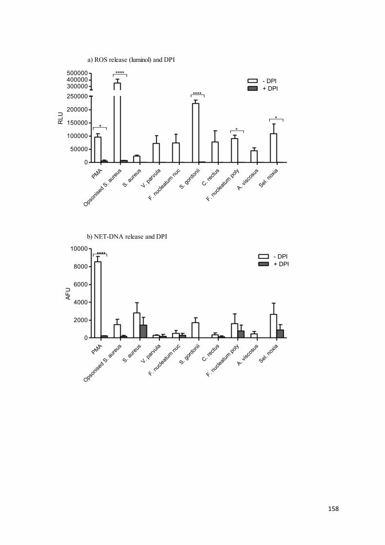
158
RL
U
PM
A
Ops
onised
S. a
ureu
s
S. a
ureu
s
V. p
arvu
la
F. nuc
leat
um n
uc
S. g
ordo
nii
C. r
ectu
s
F. nuc
leat
um p
oly
A. v
isco
sus
Sel. n
oxia
0
50000
100000
150000
200000
250000
300000400000500000
- DPI
+ DPI
a) ROS release (luminol) and DPI
**
*
****
****
AF
U
PM
A
Ops
onised
S. a
ureu
s
S. a
ureu
s
V. p
arvu
la
F. nuc
leat
um n
uc
S. g
ordo
nii
C. r
ectu
s
F. nuc
leat
um p
oly
A. v
isco
sus
Sel. n
oxia
0
2000
4000
6000
8000
10000
- DPI
+ DPI
********
b) NET-DNA release and DPI

159
RL
U
PMA
Ops
onised
S. a
ureu
s
S. aur
eus
V. par
vula
F. nuc
leat
um n
uc
S. gor
donii
C. r
ectu
s
F. nuc
leat
um p
oly
A. visco
sus
Sel. n
oxia
0
50000
100000
150000
200000
250000
300000400000500000
- NAC
+ NAC
c) ROS release (luminol) and NAC
*
*
****
****
AF
U
PMA
Ops
onised
S. a
ureu
s
S. aur
eus
V. par
vula
F. nuc
leat
um n
uc
S. gor
donii
C. r
ectu
s
F. nuc
leat
um p
oly
A. visco
sus
Sel. n
oxia
0
2000
4000
6000
8000
10000- NAC
+ NAC
****
*
d) NET-DNA release and NAC

160
RL
U
PM
A
Ops
onised
S. a
ureu
s
S. a
ureu
s
V. p
arvu
la
F. nuc
leat
um n
uc
S. g
ordo
nii
C. r
ectu
s
F. nuc
leat
um p
oly
A. v
isco
sus
Sel. n
oxia
0
50000
100000
150000
200000
250000
300000400000500000
- Taurine
+ Taurine
e) ROS release (luminol) and taurine
****
***
*
AF
U
PM
A
Ops
onised
S. a
ureu
s
S. a
ureu
s
V. p
arvu
la
F. nuc
leat
um n
uc
S. g
ordo
nii
C. r
ectu
s
F. nuc
leat
um p
oly
A. v
isco
sus
Sel. n
oxia
0
2000
4000
6000
8000
10000
- Taurine
+ Taurine
****
f) NET-DNA release and taurine
Figure 4.8: Effect of NADPH-oxidase pathway modulating agents on ROS and NET
production
Total ROS production and NET production were quantified in response to a panel of
periodontal bacteria (1x10⁸ and MOI of 1000), as well as PMA (50nM) and opsonised S.
aureus (5x10⁷
and MOI of 500) following pre-incubation (30 mins) with (a, b) DPI (25µM),
(c, d) NAC (10mM) or (e, f) taurine (100mM). Statistical significance was calculated using
2way ANOVA and Bonferroni post-tests (*p<0.05, **p<0.01, ****p<0.0001, ns=not
significant). Data presented as (a, c, e) RLU (relative light units) and (b, d, f) AFU (arbitrary
fluorescence units). Data expressed as mean ± SEM (n=3 in duplicate).

161
4.8 Effect of TLR inhibition on ROS and NET production
To attempt to dissect the signalling involved in ROS NET activation, the effect of TLR
inhibitors was investigated (section 2.2.2.3). Isolated neutrophils were incubated with
chloroquine (100µM), which inhibits TLRs 3, 7 and 9, or OxPAPC (30µg/ml), which inhibits
TLRs 2 and 4, or both TLR inhibitors. Neutrophil total ROS and NET production were
measured following pre-incubation with the inhibitor for 30 mins prior to stimulation with
PMA (50nM), opsonised S. aureus (5x10⁷ and MOI of 500) and a panel of 9 selected heat-
killed periodontal bacteria (1x10⁸ and MOI of 1000).
Chloroquine pre-treatment had a differential effect on neutrophil total ROS production.
Whilst chloroquine significantly reduced ROS production following stimulation with
opsonised S. aureus (**p<0.01), V. parvula (*p<0.05) and S. gordonii (****p<0.0001),
chloroquine pre-treatment caused a significant increase in total ROS in PMA-stimulated
neutrophils (*p<0.05) (2way ANOVA and Bonferroni post-tests, n=3) (Figure 4.9a).
Chloroquine pre-treatment resulted in a moderate reduction in NET production in response to
bacterial stimuli, but treatment prior to stimulation with PMA and opsonised S. aureus caused
a non-significant increase in NET release (2way ANOVA p>0.05) (Figure 4.9b).
OxPAPC pre-treatment was not found to have an inhibitory effect on ROS production, but
was found to increase total ROS release relative to cells that did not receive OxPAPC
treatment. This was statistically significant for stimulation with opsonised S. aureus, which
produced significantly higher levels of total ROS following OxPAPC pre-incubation (2way
ANOVA and Bonferroni post-test ***p<0.001, n=3) (Figure 4.9c). OxPAPC pre-treatment
resulted in a non-significant reduction in NET release in response to opsonised S. aureus and
bacterial stimulation; however OxPAPC treatment prior to PMA stimulation caused a non-
significant increase in NET release (2way ANOVA p>0.05) (Figure 4.9d).

162
Chloroquine and OxPAPC pre-treatment produced a differential effect on neutrophil total
ROS production relative to cells that did not receive pre-treatment. Chloroquine and
OxPAPC pre-treatment caused a significant increase in ROS by PMA-stimulated neutrophils
(2way ANOVA and Bonferroni post-test **p=0.01, n=3) and a significant decrease in ROS
by opsonised S. aureus-stimulated neutrophils (2way ANOVA and Bonferroni post-test
*p=0.05, n=3) (Figure 4.9e). Chloroquine and OxPAPC pre-treatment resulted in a non-
significant reduction in NET production in response to bacterial stimuli. However, treatment
prior to stimulation with PMA and opsonised S. aureus caused a non-significant increase in
NET release (2way ANOVA p>0.05) (Figure 4.9f).

163
RL
U
PM
A
Ops
onised
S. a
ureu
s
S. a
ureu
s
V. p
arvu
la
F. nuc
leat
um n
uc
S. g
ordo
nii
C. r
ectu
s
F. nuc
leat
um p
oly
A. v
isco
sus
Sel. n
oxia
0
50000
100000
150000 -Chlroroquine
+Chloroquine
a) ROS release (luminol) and chloroquine
*
*
**
****
AF
U
PM
A
Ops
onised
S. a
ureu
s
S. a
ureu
s
V. p
arvu
la
F. nuc
leat
um n
uc
S. g
ordo
nii
C. r
ectu
s
F. nuc
leat
um p
oly
A. v
isco
sus
Sel. n
oxia
0
3000
6000
9000
12000
-Chloroquine
+Chloroquine
b) NET-DNA release and chloroquine
ns

164
RL
U
PMA
Ops
onised
S. a
ureu
s
S. aur
eus
V. par
vula
F. nuc
leat
um n
uc
S. gor
donii
C. r
ectu
s
F. nuc
leat
um p
oly
A. visco
sus
Sel. n
oxia
0
100000
200000
300000
400000-Oxpapc
+Oxpapc
c) ROS release (luminol) and OxPAPC
***
AF
U
PMA
Ops
onised
S. a
ureu
s
S. aur
eus
V. par
vula
F. nuc
leat
um n
uc
S. gor
donii
C. r
ectu
s
F. nuc
leat
um p
oly
A. visco
sus
Sel. n
oxia
0
3000
6000
9000
12000
-Oxpapc
+Oxpapc
d) NET-DNA release and OxPAPC
ns

165
RL
U
PM
A
Ops
onised
S. a
ureu
s
S. a
ureu
s
V. p
arvu
la
F. nuc
leat
um n
uc
S. g
ordo
nii
C. r
ectu
s
F. nuc
leat
um p
oly
A. v
isco
sus
Sel. n
oxia
0
50000
100000
150000
200000
250000
-Chloroquine/OxPAPC
+Chloroquine/OxPAPC
e) ROS release (luminol) and chloroquine + OxPAPC
**
*
AF
U
PM
A
Ops
onised
S. a
ureu
s
S. a
ureu
s
V. p
arvu
la
F. nuc
leat
um n
uc
S. g
ordo
nii
C. r
ectu
s
F. nuc
leat
um p
oly
A. v
isco
sus
Sel. n
oxia
0
3000
6000
9000
12000
-Chloroquine/Oxpapc
+Chloroquine/Oxpapc
f) NET-DNA release and chloroquine + OxPAPC
ns
Figure 4.9: Effect of TLR inhibition on ROS and NET production
Total ROS production and NET production were quantified in response to a panel of
periodontal bacteria (1x10⁸ and MOI of 1000), as well as PMA (50nM) and opsonised S.
aureus (5x10⁷
and MOI of 500) following pre-incubation (30 mins) with (a, b) Chloroquine
(100µM), (c, d) OxPAPC (30µg/ml) or (e, f) Chloroquine and OxPAPC. Statistical
significance was calculated using 2way ANOVA and Bonferroni post-tests (*p<0.05,
**p<0.01, ****p<0.0001, ns=not significant). Data presented as (a, c, e) RLU (relative light
units) and (b, d, f) AFU (arbitrary fluorescence units). Data expressed as mean ± SEM (n=3
in duplicate).
166
4.9 Discussion
Neutrophil ROS production is a vital component of the innate immune response which
enables the killing and clearance of pathogens. Pathogenic bacteria are known to stimulate
ROS production via NADPH-oxidase, whereby molecular oxygen is reduced by electrons to
form O2 ̇-. The O2̇- is initially converted to H2O2, and finally, H2O2 is converted to HOCl in a
reaction catalysed by MPO (Chapple 1996, Robinson 2008). Neutrophils are the
predominant immune cell in periodontitis, and the results presented here support findings that
stimulation of peripheral blood neutrophils with periodontal bacteria promotes extracellular,
intracellular and superoxide ROS release; however data indicates that this may be species
specific (section 4.2). Indeed, certain Streptococcal and F. nucleatum species consistently
elicited higher neutrophil ROS production while, under these experimental conditions, other
bacteria such as P. gingivalis, were not found to significantly promote ROS release.
Interestingly, P. gingivalis is reportedly resistant to neutrophil-derived ROS production,
which is attributed to a non-heme iron protein, rubrerythrin, which shields the bacteria from
oxidative stress (Mydel et al., 2006). It is possible that the low levels of P. gingivalis-
induced ROS may function to afford protection to other biofilm organisms that are not
resistant to ROS.
Whilst it is recognised that ROS facilitate microbial killing, the progression of periodontitis
appears host-mediated via an exaggerated inflammatory response to microbial plaque
(Hajishengallis 2014). ROS release does not discriminate between pathogens and host
tissues, and therefore tissue injury can arise from plaque-induced extracellular ROS
production. ROS is reported to contribute to periodontitis progression by direct mechanisms,
such as tissue degradation (Sharma & Sharma 2011), lipid peroxidation (Dix & Aikens 1993)
and DNA strand breaks (Dix et al. 1996). In addition, ROS are believed to promote disease
progression indirectly , by for example increasing osteoclast differentiation (Waddington et

167
al., 2000) and initiating a self-perpetuating cycle that activates immune cells to generate
further ROS (Dahiya et al., 2013). Notably, Matthews et al., (2007a, 2007b) demonstrated
increased ROS production in chronic periodontitis, and in light of reports indicating that
patients also have elevated levels of circulating neutrophils (Al-Rasheed et al., 2012); it is
possible that periodontitis patients are more susceptible to ROS-induced tissue damage. A
mechanism by which patients may be more susceptible to the deleterious effects of ROS is
due to the discordance between oxidant/antioxidant levels may exist. This is supported by
Chapple et al., (2007), who reported that total antioxidant activity is lower in the saliva of
periodontitis patients. Neutrophil chemotaxis is also reported to be compromised in chronic
periodontitis, which is likely to increase neutrophil tissue transit times and thereby potentially
exacerbating ROS-mediated collateral tissue damage (Roberts et al., 2015).
Quantification of NET-DNA and NET-bound components demonstrated differential NET
production in response to a panel of periodontal bacteria (section 4.3). DNA is released in
other forms of neutrophil cell death processes and the quantification of NET-bound
components (NE, MPO and CG) provides a DNA-independent measure of NETs. Other
published methods to quantify NET production include microscopy (Brinkmann et al., 2004),
automated microscopy (Hakkim et al., 2011) and immunostaining (Brinkman et al., 2004,
2010). It is noteworthy that inter-individual differences between volunteers, such as
neutrophil responsiveness to stimuli, can also affect NET quantification results regardless of
the analytical method employed (Barrientos et al., 2013, Halverson et al., 2015). In the
current work, assays were not conducted to determine whether bacteria-induced NETs
contained nuclear or mitochondria DNA (Yousefi et al., 2009). However, reportedly
mitochondrial DNA is 100,000 times less abundant in granulocyte NET structures (Pilsczek
et al., 2010); thus it is likely that nuclear NET-DNA represents a larger proportion of the
NET structures quantified. NET production in response to individual periodontal bacteria

168
suggests these events may occur in vivo, and thus differences in NET induction between
species may be compensated for when these species co-exist within a biofilm structure.
NETs have previously been shown to exist in purulent exudate from periodontal pockets,
where they are postulated to entrap invading microbes and prevent their dissemination
(Vitkov et al., 2009, 2010). This is corroborated by recent findings by Hirchfeld et al.,
(2015), who demonstrated that following the recruitment of neutrophils to the oral biofilm,
they can infiltrate the dental plaque and subsequently release NET structures. Further support
for the presence of NET production in periodontitis is that periodontal pockets reportedly
provide an appropriate environment for ROS production, by the provision of sufficient
oxygen tension and pH (Mettraux et al., 1984, Eggert et al., 1991). This may suggest that the
periodontal pocket environment is likely to enable ROS-dependent NET release. Recent
findings have also suggested that some periodontal bacteria reduce complement-induced NET
production, which may also afford protection to other biofilm bacteria by evading NET
entrapment (Palmer et al., 2015). Collectively, these findings highlight that whilst
periodontal bacteria can stimulate for NET release, prospective studies are needed to fully
elucidate the role of NETs in periodontitis, in particular within the local diseased tissue
environment.
HOCl-induced NETs were found to entrap all periodontal bacteria to some extent (section
4.4), which is consistent with previous findings suggesting NETs can ensnare a significant
majority of microorganisms, including Gram-positive and Gram-negative bacteria
(Brinkmann et al., 2004). It is noteworthy that reports by Brinkmann et al., (2012) observed
that in vivo, and space permitting, NETs may occupy and therefore immobilise bacteria in
areas 10-15 times larger than the cell they originated from. Interactions between bacteria and
NETs are believed to be the result of charge differences between the negatively charged
chromatin NET backbone and the pathogens’ cell surface (Urban et al 2009). Furthermore,
169
many periodontal bacteria are reported to release DNases, which in addition to regulating
biofilm formation (Whitchurch et al., 2002), may have the ability to disassemble NET
structures and contribute to biofilm pathogenicity and NET evasion (Palmer et al., 2012). In
support of this hypothesis, S. aureus nuclease production reportedly confers resistance to
NET-mediated entrapment and killing. Indeed bacterial DNase expression may explain why
some periodontal species showed less entrapment, such as S. constellatus, which reportedly
releases large quantities of DNase (Palmer et al., 2012). However, further work is required to
elucidate whether DNase release by specific pathogens allows only these species to
disseminate, or if DNase release provides a mutually beneficial environment in which non-
DNase producing bacteria present in the biofilm are also afforded protection from NET
entrapment.
Following bacterial entrapment, the high local concentration of AMPs associated with NETs
is postulated to kill pathogens (Brinkmann et al., 2012). In support of this, NET-bound NE is
believed to cleave bacterial virulence factors (Brinkmann et al., 2004), and in addition MPO
and CG are reported to be vital for the clearance of invading bacteria (Parker et al., 2012,
Lappann et al., 2013). The nuclear DNA backbone is also believed to exhibit direct
bactericidal properties by disrupting bacterial membrane integrity and lysing bacterial cells
(Halverson et al., 2015). The authors postulate that given the low abundance of AMPs on
NETs (<1-6%), and that a key virulence factor is the release of DNases, it is possible that
nuclear DNA is the predominant antibacterial component of NETs (Halverson et al., 2015).
Under the experimental conditions applied here, the incubation of NETs with periodontal
bacteria did not appear to impede bacterial growth or survival (section 4.6). However, it is
difficult to ascertain whether the ongoing growth was a result of NETs not being capable of
killing these bacteria, or a result of these specific experimental conditions. Interestingly,
Menegazzi et al., (2012) also questioned the robustness of NET killing assays and suggested

170
that previous studies where bacteria were not released from NET structures post-incubation
did not prove that the microbes trapped within NETs were in fact dead. Indeed, subsequent
experiments by Menegazzi et al., (2012) addressing this revealed that NETs do not have the
capacity to kill S. aureus or C. albicans, which is consistent with the protocol applied and the
results presented here. Alternatively, an apparent inability of NETs to kill bacteria could be
due to various stages of the assay that may compromise NET antibacterial capacity, or not
correctly identifying numbers of viable bacteria; examples of this include MNase digestion of
NETs prior to co-incubation, incubation conditions, measuring bacterial killing by the
enumeration of colonies or the bacteria and NET concentrations (MOI). Future experiments
to quantify NET-mediated killing may benefit from application of multiple assays performed
in parallel, such as enumeration of colonies, turbidity measurements (Harris 2012), flow
cytometry and microscopy of SYTO9/propidium iodide stained bacteria (Halverson et al.,
2015). Whilst the importance of NETs is evident in individuals with reduced NET release,
such as neonates (Yost et al., 2009), CGD patients (Bianchi et al., 2009) and PLS patients
(Sorensen et al., 2014), this could be due to increased bacterial dissemination rather than the
direct antimicrobial properties of NET structures (Menegazzi et al., 2012). Conversely, if
NETs are interacting with, but not killing, periodontal pathogens, it is possible that NET-
bacteria complexes may provide a source of autoantigens and trigger further inflammatory
responses, as observed in cystic fibrosis (Marcos et al., 2010). This also raises questions
regarding NET production within the microbial biofilm (Hirschfeld et al., 2015), as it is not
known whether NETs prevent bacteria dissemination, or in fact promote biofilm stability and
growth.
Treatment of neutrophils with DPI (NADPH-oxidase inhibitor) abrogated total ROS release
in response to PMA and bacterial stimulation, as expected (section 4.7). ROS inhibition also
translated to decreased NET release following stimulation with PMA (as previously reported

171
by Fuchs et al., 2007 & Palmer et al., 2012), as well as following bacterial stimulation.
These findings confirm that NAPDH-oxidase, at least in part, necessitates NET release; this
is also evidenced in CGD patients who harbour an NADPH-oxidase mutation and
consequently do not produce NETs in response to infection (Bianchi et al., 2009). Notably,
DPI had a relatively mild effect on NET production in response to S. aureus, which is
consistent with findings by Pilsczek et al., (2010), who hypothesised that S. aureus may
trigger the release of NADPH-oxidase independent NET release 1-2 hours after stimulation,
followed by oxidase dependent NET release at 3-4 hours. In the current studies, NET
quantification at 4 hours does not discriminate between oxidase-dependent and -independent
NET release, which suggests that oxidase-independent NETs are also present, and hence, DPI
does not significantly reduce the quantification of NETs. Noteworthy is a report that
explored the role of autophagy, a homeostatic process facilitating intracellular degradation,
which has also been suggested to play a role in cell death. The authors demonstrated that
both NADPH activity and autophagy are required for NET release, and without autophagy,
NADPH is insufficient to cause the decondensation of chromatin and mediate NET release
(Remijsen et al., 2011).
To establish the contribution of H2O2 to NET release, the effect of increased removal of H2O2
was determined by the addition of a glutathione peroxidase precursor substrate (NAC), which
promotes the removal of H2O2 (section 4.7). NAC pre-treatment of neutrophils demonstrated
reduced total ROS and NET release, notably the smallest decrease in NET release following
NAC pre-treatment was again quantified in response to S. aureus. Reduced NET production
following NAC treatment is consistent with findings by Palmer et al., (2012) and confirms
the role of H2O2 in bacterial-induced NET release.
Subsequently, further work to ascertain whether other H2O2 metabolites are required for NET
release was undertaken (section 4.7). One potential pathway involved utilises the conversion

172
of H2O2 to HOCl in a reaction catalysed by MPO, which produces further reactive oxidants to
facilitate the removal of invading pathogens. The importance of HOCl in NET production
was determined by treating cells with taurine, which scavenges HOCl. The addition of
taurine resulted in a reduction in total ROS release, however a differential effect on NET
production was observed. Whilst PMA-induced NET release decreased, which is consistent
with findings by Palmer et al., (2012), taurine caused increased NET release in response to
certain bacterial stimuli. These findings emphasise the differences between stimuli, as PMA
may induce NETs dissimilar to those evoked by physiologically relevant bacteria.
Furthermore, given that taurine has been suggested to reduce NET release by rescuing cells
from cell death (Palmer et al., 2012); the current results further highlight the need for a
greater understanding of NETosis and whether bacteria-induced NET release is indeed a form
of programmed cell death.
Further experiments aimed to establish the role of TLR activation in NET production (section
4.8). Whilst pre-treatment of neutrophils with Chloroquine (intracellular TLR3, 7 and 9
inhibitor) associated with a moderate decrease in total ROS release in response to opsonised
S. aureus and bacterial stimuli, PMA-induced ROS production increased. In addition,
Chloroquine treatment resulted in moderately reduced bacteria-induced NET production;
however NET release in response to PMA and opsonised S. aureus increased. Notably,
neutrophils are reportedly not responsive to TLR 3 and 7 ligands (Janke et al., 2009), which
suggests that these observations are attributed to decreased TLR9 signalling. TLR9 is central
to the activation of neutrophils by bacterial DNA, which promotes phagocytosis, pro-
inflammatory cytokine release and ROS production (El Kebir et al., 2009). The importance
of TLR9 signalling is particularly interesting in light of recent reports suggesting that NET-
DNA can further activate TLR9 on neutrophils and dendritic cells, which may aid the

173
activation of the immune response (e.g. further NET release), or conversely initiate
autoimmune responses (Itagaki et al., 2015, Lande et al., 2011).
Treatment of cells with OxPAPC (cell surface TLR2 and 4 inhibitor) exhibited increased
ROS production and a mild decrease in bacteria-induced NET release (section 4.8).
Similarly, very little differences were observed in NET production in cells pre-treated with
Chloroquine and OxPAPC. Previous findings have suggested that ROS release is TLR2 and
4 dependent (Sabroe et al., 2005); however Gould et al., (2014) demonstrated that TLR2 and
4 blocking antibodies did not abolish NET release. Further experiments are required to
determine whether these discrepancies are a result of differences in experimental procedures
(such as the stimuli employed), or whether alternative activation pathways are being utilised
(such as TLR co-receptors or ROS independent NET release) to compensate for the TLR
inhibition (Perera et al., 2001).
In summary, these data demonstrated variability between periodontal bacteria in their ability
to stimulate neutrophil ROS production and NET responses. This variability may contribute
to the pathogenesis of periodontitis by mechanisms such as bacterial avoidance of NET
stimulation and thus persistence of infection, or excess NET release with associated
autoimmunity. The ability of NETs to entrap bacteria may facilitate pathogen containment
and prevent their dissemination within tissues and disease progression; however further work
is needed to elucidate how NETs interact with a bacterial biofilm and whether bacterial
DNases allow colonising bacteria to subvert NET defences. Under the experimental
conditions applied here NETs appeared unable to kill bacteria; if NETs are interacting with,
but not killing, periodontal pathogens, it is possible that NET-bacteria complexes may
provide a source of autoantigens. Furthermore, treatment of neutrophils with activation

174
modulating agents demonstrated that NADPH-oxidase, H2O2, HOCl and intracellular TLRs
modulate bacteria-induced NET production.

175
NET PRODUCTION BY PERIPHERAL BLOOD CHAPTER 5:
NEUTROPHILS IN CHRONIC PERIODONTITIS: A
LONGITUDINAL INTERVENTION STUDY

176
5.1 Introduction
The results presented in this chapter are based on clinical work in a study coordinated by Dr
Martin Ling in 2013 and preliminary data from this study concerning NET production have
previously been reported in Ling (2015); all NET assays and analyses were however
performed as part of this thesis. Chronic periodontitis patients, with age and gender-matched
periodontally healthy controls, were recruited to the study (2.2.6.3). Neutrophils were
isolated from the peripheral blood of all volunteers to determine the effect of the periodontal
inflammatory process on peripheral blood neutrophil NET responses (West Midlands
Research Ethics Committee 10/H1208/48). The patients received tailored oral hygiene
instruction and non-surgical periodontal treatment that involved scaling and root surface
debridement of periodontal pockets >4mm. Patients were reviewed 3 months post-therapy, at
which point blood was also collected to determine the impact of reducing local periodontal
inflammation via treatment upon peripheral NET production. At both time points
periodontitis patients were assayed in parallel with their matched healthy controls. The
periodontal status of patients was recorded at both time points, and healthy controls were
assessed upon recruitment to the study (2.2.6.3.2). NET-DNA was quantified by quantifying
NET-DNA with Sytox green (2.2.3.3) and the effect of volunteer age and disease severity
upon NET production was also analysed.
5.2 Volunteer information
Patient and control details are illustrated in Table 5.1. For inclusion and exclusion criteria
please see section 2.2.6.3.1. There was no significant difference in age between patients and
controls (unpaired t-test p=0.501). One volunteer patient (#13) decided they no longer
wished to receive periodontal treatment and withdrew from the study; resulting in n=20 pairs
pre-treatment and n=19 pairs post-treatment.

177
Table 5.1: Age and gender of volunteers in the study
Age and gender of patients and healthy age- and gender-matched controls (n=20 pairs).
There was no significant difference in age between patients and controls (12 males and 8
females, unpaired t-test p=0.501).
Volunteer number Patient
Gender-matched
control
Gender Age Age
1 Male 42 44
2 Male 45 42
3 Male 40 45
4 Female 56 63
5 Male 42 41
6 Male 46 47
7 Female 47 51
8 Male 39 33
9 Male 38 38
10 Female 38 43
11 Male 59 55
12 Male 37 45
13 Female 48 43
14 Female 61 59
15 Male 51 58
16 Female 42 39
17 Female 37 35
18 Female 52 52
19 Male 57 48
20 Male 48 48
Mean 46 46
SD 8 8

178
Clinical measures of periodontitis 5.2.1
To determine the severity of periodontitis and the response to treatment, several parameters
were measured (section 2.2.6.3.2). Clinical attachment loss (CAL), periodontal probing
pocket depths (PPD), bleeding on probing (BOP) from the pocket base, gingival index (GI)
and plaque index (PI) were recorded pre- and post-treatment in periodontitis patients.
Chronic periodontitis was defined by a minimum of 2 non-adjacent sites per quadrant with
probing pocket depth >4mm, which bled upon probing and displayed radiographic bone loss
>30% of the root length, as defined by the European Federation of Periodontology (Tonetti &
Claffey 2005). All clinical parameters were measured immediately prior to blood collection
for neutrophil assays by a single experienced examiner, who also confirmed periodontal
health in the healthy controls.
Clinical attachment loss 5.2.2
Clinical attachment loss (CAL) was calculated pre-treatment by summation of periodontal
pocket depth and gingival recession measurements in accordance with Haffajee & Socransky
(1986). Mean CAL and percentage sites with 1-2, 3-4 and >4mm CAL are shown in Table
5.2 and corresponds to mild, moderate and severe disease, respectively. From the 20 patients
recruited to the study, n=12 (60%) presented with generalised moderate and localised severe
chronic periodontitis, n=3 (15%) presented with generalised mild and localised severe
chronic periodontitis, and n=5 (25%) presented with generalised severe chronic periodontitis.
Periodontal Pocket depth 5.2.3
The number of sites measuring over 4mm was significantly higher in patients, compared with
healthy controls (unpaired t-test ****p<0.0001, n=20 pairs). This difference between patients
and controls was reduced although still evident following treatment (unpaired t-test
***p=0.0017, n=19 pairs). However, there was a significant difference in the number of sites

179
over 4mm present in periodontitis patients pre- and post-treatment, demonstrating that non-
surgical therapy significantly reduced pocket depths (unpaired t-test ***p=0.0023, n=19
pairs) in patients, but not to control levels (Table 5.3).
Bleeding on probing 5.2.4
The percentage of sites that displayed bleeding on probing (BOP) pre-treatment was
significantly higher in patients compared with healthy controls (unpaired t-test ****p<0.0001,
n=20 pairs). This difference between patients and controls reduced but remained significant
post-treatment (unpaired t-test *p=0.0478, n=19 pairs). However, there was a significant
difference in the percentage of sites that bled upon probing in periodontitis patients pre- and
post-treatment (unpaired t-test ****p<0.0001, n=19 pairs) (Table 5.4).
Cumulative gingival index 5.2.5
Cumulative gingival index (GI) was significantly higher in patients compared with healthy
controls pre-treatment (unpaired t-test ****p<0.0001, n=20 pairs). This difference between
patients and controls was no longer significant as a result of treatment (unpaired t-test
p=0.101, n=19 pairs) and GI was significantly different in periodontitis patients pre- and
post-treatment (unpaired t-test ****p<0.0001, n=19 pairs) (Table 5.5).
Plaque index 5.2.6
Cumulative plaque index (PI) was significantly higher in patients, compared with the healthy
controls pre-treatment (unpaired t-test ****p<0.0001, n=20 pairs). The difference between
patients and controls was corrected as a result of treatment (unpaired t-test p=0.679, n=19
pairs) and PI was significantly different in periodontitis patients pre- and post-treatment
(unpaired t-test ****p<0.0001, n=19 pairs) (Table 5.6).

180
Table 5.2: Clinical attachment loss in periodontitis patients
Measurements from patients pre-treatment (n=20 pairs). Data is presented as percentage sites
with 1-2, 3-4 and >4mm clinical attachment loss (mean ± SD and range).
Clinical attachment loss (% sites)
Volunteer
Number Mean + SD (mm)
% sites
1-2mm
% sites
3-4mm
% sites
>4mm
1 4.4+3.2 39.1 21.8 39.1
2 2.4+1.9 66.7 26.3 7.0
3 3.7+2.1 36.5 35.3 28.2
4 3.5+1.8 35.9 41.0 23.1
5 2.8+1.6 59.0 24.4 16.6
6 3.7+2.1 37.0 31.2 31.8
7 3.4+2.4 55.6 21.6 22.8
8 2.4+0.9 67.9 28.6 3.5
9 3.4+1.6 36.9 45.8 16.3
10 2.8+1.3 44.4 48.3 7.2
11 2.2+1.8 85.7 5.4 8.9
12 2.9+2.6 66.1 13.3 20.6
13 6.0+2.7 5.8 33.3 60.8
14 3.3+2.0 46.4 34.5 19.0
15 3.9+1.6 20.1 50.3 29.6
16 3.2+2.2 55.7 25.9 18.4
17 5.9+3.3 25.6 11.9 62.5
18 2.2+1.5 78.0 16.7 5.3
19 4.1+2.5 33.3 33.3 33.3
20 1.9+1.4 79.0 15.6 5.4
Mean 3.4 48.7 28.2 23.0
SD 1.1 21.1 12.4 16.8
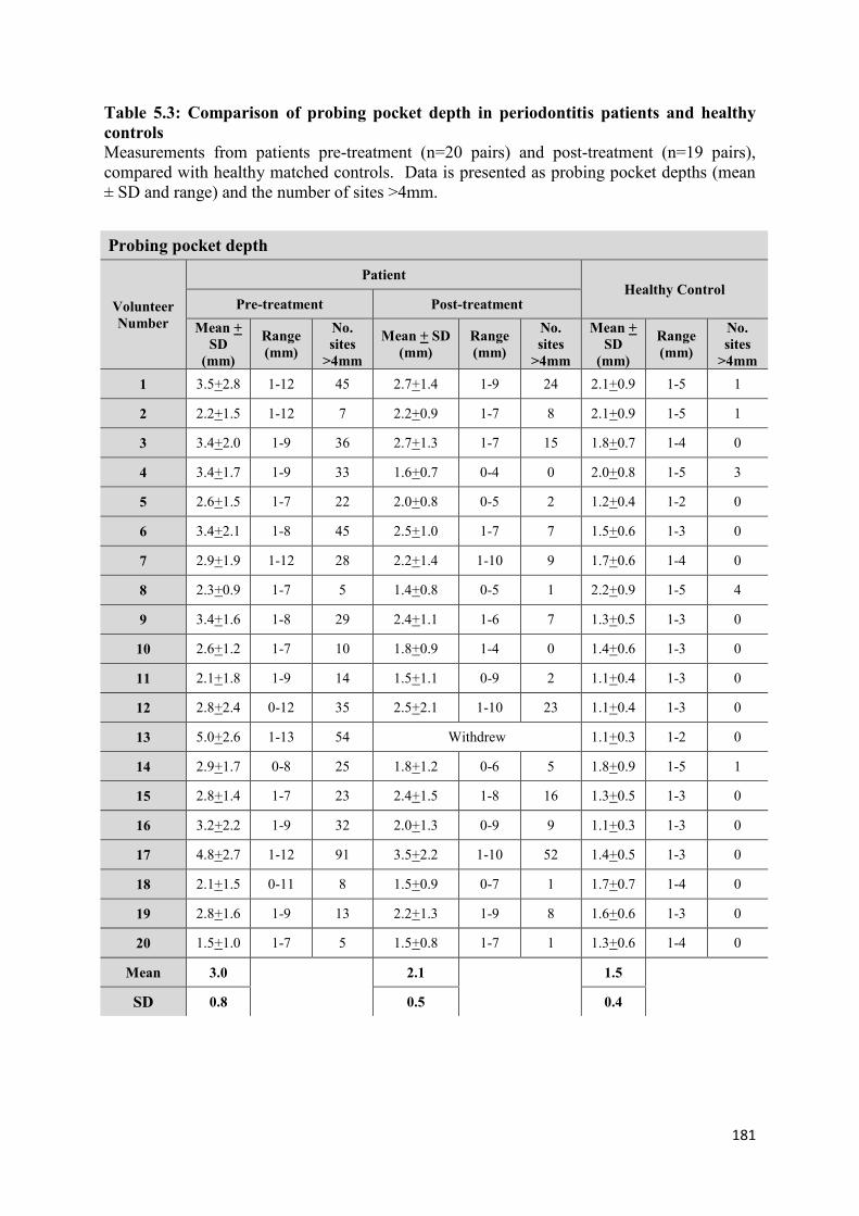
181
Table 5.3: Comparison of probing pocket depth in periodontitis patients and healthy
controls
Measurements from patients pre-treatment (n=20 pairs) and post-treatment (n=19 pairs),
compared with healthy matched controls. Data is presented as probing pocket depths (mean
± SD and range) and the number of sites >4mm.
Probing pocket depth
Volunteer
Number
Patient Healthy Control
Pre-treatment Post-treatment
Mean +
SD
(mm)
Range
(mm)
No.
sites
>4mm
Mean + SD
(mm)
Range
(mm)
No.
sites
>4mm
Mean +
SD
(mm)
Range
(mm)
No.
sites
>4mm
1 3.5+2.8 1-12 45 2.7+1.4 1-9 24 2.1+0.9 1-5 1
2 2.2+1.5 1-12 7 2.2+0.9 1-7 8 2.1+0.9 1-5 1
3 3.4+2.0 1-9 36 2.7+1.3 1-7 15 1.8+0.7 1-4 0
4 3.4+1.7 1-9 33 1.6+0.7 0-4 0 2.0+0.8 1-5 3
5 2.6+1.5 1-7 22 2.0+0.8 0-5 2 1.2+0.4 1-2 0
6 3.4+2.1 1-8 45 2.5+1.0 1-7 7 1.5+0.6 1-3 0
7 2.9+1.9 1-12 28 2.2+1.4 1-10 9 1.7+0.6 1-4 0
8 2.3+0.9 1-7 5 1.4+0.8 0-5 1 2.2+0.9 1-5 4
9 3.4+1.6 1-8 29 2.4+1.1 1-6 7 1.3+0.5 1-3 0
10 2.6+1.2 1-7 10 1.8+0.9 1-4 0 1.4+0.6 1-3 0
11 2.1+1.8 1-9 14 1.5+1.1 0-9 2 1.1+0.4 1-3 0
12 2.8+2.4 0-12 35 2.5+2.1 1-10 23 1.1+0.4 1-3 0
13 5.0+2.6 1-13 54 Withdrew 1.1+0.3 1-2 0
14 2.9+1.7 0-8 25 1.8+1.2 0-6 5 1.8+0.9 1-5 1
15 2.8+1.4 1-7 23 2.4+1.5 1-8 16 1.3+0.5 1-3 0
16 3.2+2.2 1-9 32 2.0+1.3 0-9 9 1.1+0.3 1-3 0
17 4.8+2.7 1-12 91 3.5+2.2 1-10 52 1.4+0.5 1-3 0
18 2.1+1.5 0-11 8 1.5+0.9 0-7 1 1.7+0.7 1-4 0
19 2.8+1.6 1-9 13 2.2+1.3 1-9 8 1.6+0.6 1-3 0
20 1.5+1.0 1-7 5 1.5+0.8 1-7 1 1.3+0.6 1-4 0
Mean 3.0 2.1 1.5
SD 0.8 0.5 0.4
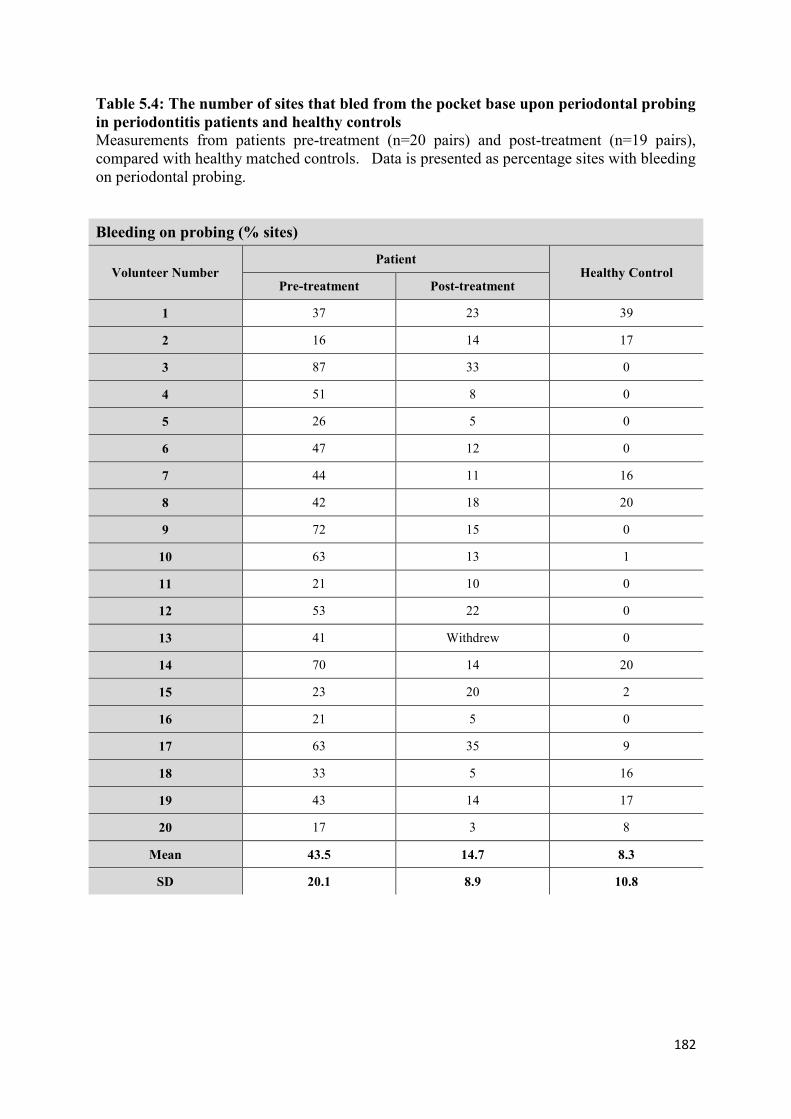
182
Table 5.4: The number of sites that bled from the pocket base upon periodontal probing
in periodontitis patients and healthy controls
Measurements from patients pre-treatment (n=20 pairs) and post-treatment (n=19 pairs),
compared with healthy matched controls. Data is presented as percentage sites with bleeding
on periodontal probing.
Bleeding on probing (% sites)
Volunteer Number Patient
Healthy Control Pre-treatment Post-treatment
1 37 23 39
2 16 14 17
3 87 33 0
4 51 8 0
5 26 5 0
6 47 12 0
7 44 11 16
8 42 18 20
9 72 15 0
10 63 13 1
11 21 10 0
12 53 22 0
13 41 Withdrew 0
14 70 14 20
15 23 20 2
16 21 5 0
17 63 35 9
18 33 5 16
19 43 14 17
20 17 3 8
Mean 43.5 14.7 8.3
SD 20.1 8.9 10.8
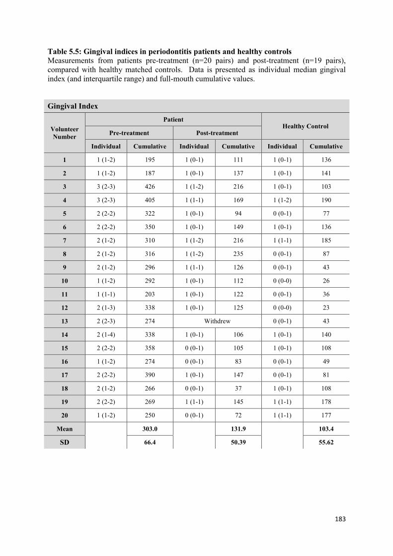
183
Table 5.5: Gingival indices in periodontitis patients and healthy controls
Measurements from patients pre-treatment (n=20 pairs) and post-treatment (n=19 pairs),
compared with healthy matched controls. Data is presented as individual median gingival
index (and interquartile range) and full-mouth cumulative values.
Gingival Index
Volunteer
Number
Patient Healthy Control
Pre-treatment Post-treatment
Individual Cumulative Individual Cumulative Individual Cumulative
1 1 (1-2) 195 1 (0-1) 111 1 (0-1) 136
2 1 (1-2) 187 1 (0-1) 137 1 (0-1) 141
3 3 (2-3) 426 1 (1-2) 216 1 (0-1) 103
4 3 (2-3) 405 1 (1-1) 169 1 (1-2) 190
5 2 (2-2) 322 1 (0-1) 94 0 (0-1) 77
6 2 (2-2) 350 1 (0-1) 149 1 (0-1) 136
7 2 (1-2) 310 1 (1-2) 216 1 (1-1) 185
8 2 (1-2) 316 1 (1-2) 235 0 (0-1) 87
9 2 (1-2) 296 1 (1-1) 126 0 (0-1) 43
10 1 (1-2) 292 1 (0-1) 112 0 (0-0) 26
11 1 (1-1) 203 1 (0-1) 122 0 (0-1) 36
12 2 (1-3) 338 1 (0-1) 125 0 (0-0) 23
13 2 (2-3) 274 Withdrew 0 (0-1) 43
14 2 (1-4) 338 1 (0-1) 106 1 (0-1) 140
15 2 (2-2) 358 0 (0-1) 105 1 (0-1) 108
16 1 (1-2) 274 0 (0-1) 83 0 (0-1) 49
17 2 (2-2) 390 1 (0-1) 147 0 (0-1) 81
18 2 (1-2) 266 0 (0-1) 37 1 (0-1) 108
19 2 (2-2) 269 1 (1-1) 145 1 (1-1) 178
20 1 (1-2) 250 0 (0-1) 72 1 (1-1) 177
Mean 303.0 131.9 103.4
SD 66.4 50.39 55.62

184
Table 5.6: Plaque indices in periodontitis patients and healthy controls
Measurements from patients pre-treatment (n=20 pairs) and post-treatment (n=19 pairs),
compared with healthy matched controls. Data is presented as median plaque index (and
interquartile range) and full-mouth cumulative values.
Plaque Index
Volunteer
Number
Patient Healthy Control
Pre-treatment Post-treatment
Individual Cumulative Individual Cumulative Individual Cumulative
1 1 (1-2) 210 1 (1-2) 185 1 (1-1) 168
2 1 (1-1) 137 1 (1-2) 197 1 (0-1) 125
3 3 (2-3) 429 2 (1-2) 244 1 (1-1) 182
4 2 (2-3) 382 1 (1-1) 150 2 (1-2) 249
5 2 (2-3) 367 1 (0-1) 141 1 (1-2) 186
6 2 (2-2) 311 1 (1-2) 212 1 (1-2) 253
7 2 (1-3) 351 1 (1-2) 218 1 (1-2) 212
8 2 (1-2) 314 2 (1-2) 265 1 (0-1) 145
9 2 (1-2) 256 1 (1-1) 150 0 (0-1) 60
10 1 (1-2) 228 1 (0-1) 100 1 (1-1) 157
11 1 (1-2) 250 0 (0-1) 73 1 (1-1) 119
12 2 (1-2) 267 1 (0-1) 117 0 (0-1) 61
13 3 (3-4) 345 Withdrew 1 (0-1) 124
14 2 (2-3) 339 1 (1-2) 135 1 (1-2) 156
15 2 (2-3) 353 1 (1-1) 116 1 (0-1) 128
16 2 (1-2) 272 0 (0-1) 51 1 (0-1) 125
17 2 (1-3) 324 1 (0-1) 106 1 (0-1) 143
18 2 (1-3) 279 0 (0-1) 57 1 (1-1) 150
19 2 (2-3) 271 1 (1-2) 147 1 (1-1) 175
20 2 (2-3) 366 1 (0-1) 122 1 (1-2) 162
Mean 302.6 146.6 154.0
SD 68.6 60.5 49.7

185
5.3 Comparison of NET production by periodontitis patients and healthy matched
controls
Neutrophils isolated from the peripheral blood of periodontitis patients and healthy matched
controls were stimulated for NET production with PMA (50nM) and HOCl (0.75mM)
(section 2.2.3.3). Baseline NET production in response to PBS (negative unstimulated
control) was also quantified. Patient and healthy matched paired samples were assayed in
parallel and analysed using fluorometric quantification. There were no significant differences
between patient and control NET production assessed fluorometrically when neutrophils were
treated with PBS (unstimulated negative control), PMA or HOCl (Wilcoxon matched pairs
p=0.78, p=0.30, p=0.69, respectively, n=20 pairs) (Figure 5.1).

186
AF
U
Patient Control0
10000
20000
30000ns
AF
U
Patient Control0
10000
20000
30000ns
AF
U
Patient Control0
10000
20000
30000ns
a) NET production in response to PBS
b) NET production in response to PMA
c) NET production in response to HOCl
Figure 5.1: NET production in periodontitis patients (pre-treatment) and healthy
controls
Pre-treatment NET production was quantified fluometrically following a 3 hour incubation
period after neutrophil stimulation. (a) PBS (unstimulated negative control), (b) PMA
(50nM) or (c) HOCl (0.75mM). Statistical significance calculated using Wilcoxon matched
pairs tests (ns=not significant). Data are presented as AFU (arbitrary fluorescence units) and
expressed as mean ± SEM (n=20 pairs in quadruplicate).

187
NET production by periodontitis patients and healthy matched controls following 5.3.1
successful treatment
Three months post-treatment, patients were reviewed and neutrophils from the peripheral
blood of periodontitis patients and healthy matched controls were isolated and stimulated for
NET production with PMA (50nM) and HOCl (0.75mM) (section 2.2.3.3). Baseline NET
production in response to PBS (negative unstimulated control) was also quantified. 19
matched pairs were assayed in parallel and analysed by fluorometric quantification. There
were significant differences detected between patients and controls in response to PBS
exposure (unstimulated negative control) (Figure 5.2a) and PMA (Figure 5.2b) (Wilcoxon
matched pairs *p=0.041, *p=0.021, respectively, n=19 pairs). However, there were no
significant differences detected between comparators when neutrophils were stimulated with
HOCl (Figure 5.2c) (Wilcoxon matched pairs p=0.62, n=19 pairs). To control for day-to-day
variations in neutrophil responsiveness, differences in NET production between pre-treatment
and post-treatment were expressed as a ratio of patient matched healthy controls. NET
production by patient neutrophils significantly decreased following treatment in response to
PBS (unstimulated), PMA (50nM) and HOCl (0.75mM) (Wilcoxon matched pairs
**p=0.007, **p=0.004, *p=0.012, respectively, n=19 pairs) (Figure 5.3).

188
AF
U
Patient Control0
10000
20000
30000 *
AF
U
Patient Control0
10000
20000
30000 *
AF
U
Patient Control0
10000
20000
30000 ns
a) NET production in response to PBS
b) NET production in response to PMA
c) NET production in response to HOCl
Figure 5.2: NET production in post-treatment periodontitis patients and healthy
controls
Post-treatment NET production was fluorometrically quantified following successful
treatment (MNase digestion and Sytox green staining). (a) PBS (unstimulated negative
control), (b) PMA (50nM), (c) HOCl (0.75mM). Statistical significance calculated using
Wilcoxon matched pairs tests (*p<0.05, ns=not significant). Data are presented as AFU
(arbitrary fluorescence units) and expressed as mean ± SEM (n=19 pairs in quadruplicate).
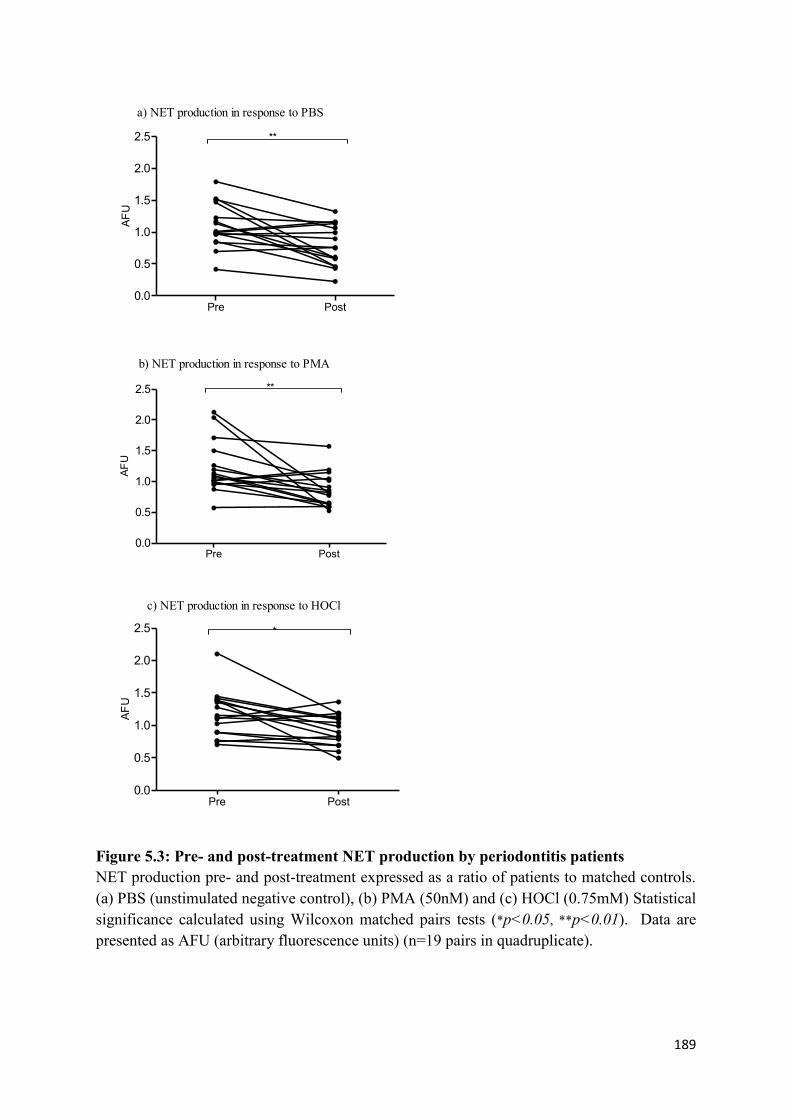
189
AF
U
Pre Post0.0
0.5
1.0
1.5
2.0
2.5 **
AF
U
Pre Post0.0
0.5
1.0
1.5
2.0
2.5 **
AF
U
Pre Post0.0
0.5
1.0
1.5
2.0
2.5 *
a) NET production in response to PBS
b) NET production in response to PMA
c) NET production in response to HOCl
Figure 5.3: Pre- and post-treatment NET production by periodontitis patients
NET production pre- and post-treatment expressed as a ratio of patients to matched controls.
(a) PBS (unstimulated negative control), (b) PMA (50nM) and (c) HOCl (0.75mM) Statistical
significance calculated using Wilcoxon matched pairs tests (*p<0.05, **p<0.01). Data are
presented as AFU (arbitrary fluorescence units) (n=19 pairs in quadruplicate).

190
Fluorescence microscopy of NETs from patient and controls pre-treatment 5.3.2
There was no apparent qualitative difference observed between patient and control NET
production visualised following fluorometric staining with Sytox green (section 2.2.3.7).
Both PMA (50nM) and HOCl (0.75mM) stimulated NETs, as shown by the extracellular
web-like structures that stained positive with the DNA-stain, Sytox green (Figure 5.4). PBS-
treated neutrophils (negative unstimulated control) also stained positive for DNA, which is
likely to be due to cell death during the incubation period that allowed for Sytox green to
permeate the compromised cell membrane.
Fluorescence microscopy of NETs from patient and controls post-treatment 5.3.3
There were no qualitative differences observed between patient and control NETs visualised
by fluorescence microscopy (Figure 5.5). Consistent with pre-treatment observations, both
PMA (50nM) and HOCl (0.75mM) produced extracellular web-like structures.

191
Figure 5.4: Fluorescence microscopy of NETs from patient and controls pre-treatment
NETs were visualised by fluorescence microscopy (x20 magnification) with Sytox green
staining in response to PBS (unstimulated negative control), PMA (50nM) and HOCl
(0.75mM) exposure for 3 hours. Images are representative of 2 experiments performed in
triplicate. Scale bars are 100µm.
Patient Control
PBS
PMA
HOCl

192
Figure 5.5: Fluorescence microscopy of NETs from patient and controls post-treatment NETs were visualised following successful treatment by fluorescence microscopy (x20
magnification) with Sytox green staining in response to PBS (unstimulated negative control),
PMA (50nM) and HOCl (0.75mM) stimulation for 3 hours. Images are representative of 2
experiments in triplicate. Scale bar is 100µm.
Patient Control
PBS
PMA
HOCl

193
5.4 Effect of age on NET production
NET production levels were analysed following stratification for age (<50 years vs >50 years
old) in pre-treatment patients and controls (published in Hazeldine et al., 2014). Data was
available for 13 patients and 13 matched controls who were under 50 years (mean age
42 ± 4.9 years), and 6 patients and controls who were over 50 years of age (mean
57 ± 3.4 years). NET production by older periodontally healthy volunteers was significantly
reduced compared with healthy volunteers under 50 years old (Mann-Whitney PBS
*p=0.045; PMA; *p=0.045; HOCl *p=0.026, respectively, n=20 pairs). NET production was
also reduced in periodontitis patients over 50 years of age in response to HOCl (0.75mM)
(Mann-Whitney *p=0.026). There was a similar non-significant reduction in NET
production in periodontitis patients in response to PBS and PMA (Figure 5.6a).
NET production was also analysed by age (<50 years vs >50 years old) in post-treatment
patients and controls. Following the discontinuation of the study by volunteer 13, there were
data from 12 patients and age matched controls under 50 years and 6 patients and controls
over 50 years of age which could be used for analysis. No statistically significant differences
in NET production between volunteers under 50 years and those over 50 years were observed
in response to PBS (unstimulated), PMA (50nM) or HOCl (0.75mM) in patients or controls
(Mann-Whitney p=0.089) (Figure 5.6b).

194
a) Pre-treatment A
FU
<50
Pat
ient
<50
Hea
lthy
>50
Pat
ient
>50
Hea
lthy
<50
Pat
ient
<50
Hea
lthy
>50
Pat
ient
>50
Hea
lthy
<50
Pat
ient
<50
Hea
lthy
>50
Pat
ient
>50
Hea
lthy
0
5000
10000
15000
20000
25000
PMA
HOCl
PBS
*
*
*
*
b) Post-treatment
AF
U
<50
Pat
ient
<50
Hea
lthy
>50
Pat
ient
>50
Hea
lthy
<50
Pat
ient
<50
Hea
lthy
>50
Pat
ient
>50
Hea
lthy
<50
Pat
ient
<50
Hea
lthy
>50
Pat
ient
>50
Hea
lthy
0
5000
10000
15000
20000
PMA
HOCl
PBS
ns
Figure 5.6: The effect of age on pre- and post-treatment NET production in patients and
healthy controls
NET production in volunteers <50 (n=13 patients and controls) and >50 (n=6 patients and
controls) following no stimulation (PBS) and in response to PMA (50nM) and HOCl
(0.75mM) (a) pre-treatment and (b) post-treatment. Statistical significance calculated using
Mann-Whitney tests (*p<0.05, ns=not significant). Data are presented as AFU (arbitrary
fluorescence units) and expressed as mean ± SEM.

195
5.5 Analysis of the association between severity of periodontitis and NET production
pre- and post-treatment
No significant correlation was observed between pocket depth and NET production pre-
treatment (linear regression p=0.92, p=0.94 and p=0.92 for PBS, PMA and HOCl,
respectively, n=20 pairs) or post-treatment (linear regression p=0.28, p=0.74 and p=0.84 for
PBS, PMA and HOCl, respectively, n=19 pairs) (Figure 5.7a&b). No significant correlation
was observed between bleeding on probing and NET production pre-treatment (linear
regression p=0.19, p=0.52 and p=062 for PBS, PMA and HOCl, respectively, n=20 pairs) or
post-treatment (linear regression p=0.72, p=0.53 and p=0.61 for PBS, PMA and HOCl,
respectively, n=19 pairs) (Figure 5.7c&d). Furthermore, no significant correlation was
observed between cumulative gingival index and NET production pre-treatment (linear
regression p=0.55, p=0.91 and p=0.77 for PBS, PMA and HOCl, respectively, n=20 pairs) or
post-treatment (linear regression p=0.93, p=0.11 and p=0.41 for PBS, PMA and HOCl,
respectively, n=19 pairs) (Figure 5.8a&b). There was also no significant correlation between
cumulative plaque index and NET production pre-treatment (linear regression p=0.44,
p=0.17 and p=067 for PBS, PMA and HOCl, respectively, n=20 pairs). However post-
treatment results revealed a significant negative correlation between plaque index and NET
production in response to PMA (linear regression *p=0.022, n=19 pairs) (Figure 5.8c&d).

196
Pocket depth/no. sites >4mm
AF
U
0 20 40 60 80 1000
10000
20000
30000PBS
PMA
HOCl
R2=0.0030; p=0.82
R2=0.0031; p=0.94
R2=0.00055; p=0.92
a) Pre-treatment pocket depth (number of sites over 4mm)
plotted against NET release
Pocket depth/no. sites >4mm
AF
U
0 10 20 300
10000
20000
30000PBS
PMA
HOClR2=0.0027; p=0.84
R2=0.0079; p=0.74
R2=0.082; p=0.28
b) Post-treatment pocket depth (number of sites over 4mm)
plotted against NET release
BOP %
AF
U
0 20 40 60 80 1000
10000
20000
30000PBS
PMA
HOClR2=0.014; p=0.62
R2=0.025; p=0.52
R2=0.099; p=0.19
c) Pre-treatment % bleeding on probingplotted against NET release
BOP %
AF
U
0 10 20 30 400
10000
20000
30000PBS
PMA
HOCl
R2=0.018; p=0.61
R2=0.028; p=0.53
R2=0.0093; p=0.72
d) Post-treatment % bleeding on probingplotted against NET release
Figure 5.7: Analysis of the association between severity of periodontitis and NET production pre- and post-treatment
Disease severity was plotted against NET production in response to PBS (unstimulated negative control), PMA (50nM) and HOCl (0.75mM).
Clinical measures included pocket depth and percentage bleeding on probing (BOP %). There was no significant correlation between pocket depth
(number of sites over 4mm) or bleeding on probing and NET production either pre- or post-treatment (linear regression p>0.05). Data are presented
as AFU (arbitrary fluorescence units) (n=19 in quadruplicate).

197
Figure 5.8: Analysis of the association between periodontitis disease severity and NET production pre- and post-treatment
Disease severity was plotted against NET production in response to PBS (unstimulated negative control), PMA (50nM) and HOCl (0.75mM). There
was no significant correlation between gingival index and NET production pre- or post-treatment (linear regression p>0.05). No significant
correlation was observed for plaque index pre-treatment, however a negative correlation was observed between post-treatment PMA-induced NET
production and plaque index (linear regression *p=0.022). Data are presented as AFU (arbitrary fluorescence units) (n=19 in quadruplicate).
Gingival index
AF
U
0 100 200 300 400 5000
10000
20000
30000PBS
PMA
HOCl
R2=0.0050; p=0.77
R2=0.00074; p=0.91
R2=0.021; p=0.55
a) Pre-treatment gingival indexplotted against NET release
Gingival index
AF
U
0 50 100 150 200 2500
10000
20000
30000PBS
PMA
HOCl
R2=0.046; p=0.41
R2=0.18; p=0.11
R2=0.00059; p=0.93
b) Post-treatment gingival indexplotted against NET release
Plaque index
AF
U
0 100 200 300 400 5000
10000
20000
30000PBS
PMA
HOCl
R2=0.011; p=0.67
R2=0.11; p=0.17
R2=0.036; p=0.44
c) Pre-treatment plaque indexplotted against NET release
Plaque index
AF
U0 100 200 300
0
10000
20000
30000PBS
PMA
HOCl
R2=0.051; p=0.38
R2=0.32; p=0.022
R2=0.017; p=0.27
d) Post-treatment plaque index
plotted against NET release

198
5.6 Discussion
Neutrophils play a critical role within the periodontal lesion in response to colonisation by
subgingival bacteria in chronic periodontitis patients (Miyasaki 1991, Schenkein 2000).
Chronic activation of accumulating neutrophils in the periodontal tissues is thought to induce
further tissue damage by releasing their cytotoxic arsenal into the extracellular space
(Kantarci et al., 2003). There is also evidence to suggest that peripheral neutrophils in
periodontitis patients exhibit hyperactivity (in the absence of an exogenous stimulus) and
hyper-reactivity (in response to periodontal bacteria) with regard to ROS production
(Matthews et al., 2007a, 2007b). This has been partly attributed to elevated levels of plasma-
derived pro-inflammatory cytokines measured in periodontitis patients, such as IL-8 and GM-
CSF, which may prime circulating neutrophils (Dias et al., 2011). There is also evidence for
constitutively defective redox signalling pathways, which may also contribute to neutrophil
hyper-responsiveness in periodontitis (Dias et al., 2013). There is however currently no data
exploring whether these abnormalities extend to aberrant NET production in chronic
periodontitis patients. In the present study, to account for interpersonal and intrapersonal
variability in neutrophilic responses previously described (Harris 2012), neutrophils were
isolated from chronic periodontitis patients in parallel with age and gender matched healthy
controls. To circumvent the potential observer bias that can arise with NET quantification by
microscopy, NET production was quantified by fluorometric detection of a DNA label.
Notably qualitative visual characterisation of NET images has also been reported to be
susceptible to experimental artefacts and misinterpretation. Indeed Krautgartner et al., (2010)
observed that SEM images of NETs were not able to distinguish NET structures from fibrin.
This is the first time NET release has been investigated in peripheral blood neutrophils from
periodontitis patients. No differences were found between patients and controls (5.3), a
finding consistent with a parallel study performed by collaborators in a Greek cohort (Dimitra
199
Sakellari, Aristotle University of Thessaloniki - personal communication, unpublished data).
The data presented here indicate that the elevated ROS release previously reported in
periodontitis patients (Matthews et al., 2007) may not translate to increased NET production,
suggesting that NET release may not be solely ROS-dependent and mechanisms that lead to
ROS-independent NETosis may exist. This is consistent with reports by Pilsczek et al.,
(2010), who found that NETs were rapidly produced in response to S. aureus in an NADPH-
oxidase independent manner. In this study the authors postulated that NADPH-oxidase
independent NET production may also arise from vesicular exocytosis, resulting in nuclear
envelope destruction and the release of nuclear DNA into the cytoplasm prior to release into
the extracellular space. However this has, to date, only been demonstrated for S. aureus
stimulation and other Gram-positive and -negative bacteria previously studied, as well as
stimulation with PMA, did not elicit such responses.
Increases in intracellular calcium are also required for NET release. Parker et al., (2012)
reported that whilst NADPH-oxidase assembly was essential for PMA-activated NET release,
it was not necessary for the production of NETs by the calcium ionophore, ionomycin.
Ionomycin stimulation activates NADPH-oxidase; however inhibition of the oxidative burst
with DPI did not attenuate NET release. Furthermore, a recently reported self-protective
mechanism within glutathione deficient periodontitis neutrophils involves re-siting of the
NADPH-oxidase to membrane-associated lipid rafts. Neutrophils are believed to be endowed
with the molecular equipment to direct excess ROS extracellularly, and thus ROS cannot act
intracellularly to facilitate elevated NET production (Dias et al., 2013). This could also
explain the discordance in ROS and NET production in periodontitis patients reported here.
Supporting this premise is that the ROS hyperactivity observed in chronic periodontitis
patients’ neutrophils is specific to extracellular release, rather than the intracellular ROS that
would necessitate NET production (Matthews et al., 2007).

200
A further explanation for elevated ROS production with no significant increase in NET
release may be that if ROS are a prerequisite for NET production, the elevated ROS
production associated with neutrophil hyperactivity in chronic periodontitis patients may not
be sufficient to trigger the same increases in NET release, or may be antagonised by the
intracellular antioxidant repertoire. Findings by Palmer et al., (2012) lend support to this
thesis, who reported that concentrations of over 0.5mM of HOCl were needed to trigger NET
production. Indeed, Al-Rasheed et al., (2012) reported that levels of peripheral white blood
cells (including neutrophils) are significantly higher in periodontitis patients (7.22 ± 1.42 x
109 cells/L) compared with healthy controls (5.64 ± 1.56x10
9 cells/L). The current in vitro
assays utilised neutrophils derived from patients and controls re-suspended at the same
concentration; however in vivo it is possible that whilst patients may not exhibit hyperactivity
or reactivity in terms of NET production, NET responses may be greater due to a
leukocytosis.
Data presented here indicate that NET production following successful non-surgical
treatment deceased in periodontitis patients (5.3.1). This was more pronounced when the
results were expressed as a ratio of patients to control pre- and post-treatment, an analysis
designed to control for the day-to-day variability of neutrophil assays. While there are
currently no published longitudinal studies looking at the effect of periodontal treatment on
peripheral NET production there have, however, been studies quantifying neutrophil ROS
production following successful periodontal therapy (Matthews et al., 2007). These studies
reported the normalisation of hyper-reactive total ROS production in response to FcγR
stimulation post-treatment, however the hyperactive extracellular ROS release measured pre-
treatment persisted following treatment. Ling (2015) also demonstrated that hyperactivity
and –reactivity with regard to O2-
production following bacterial stimulation normalised to
control levels as a result of successful treatment. Notably, these findings were reported in the

201
same patients and matched controls employed for the current NET assays, and all assays were
conducted on the same day. Whilst there is evidence to suggest NADPH-oxidase
independent NETosis can occur, significant decreases in O2- production following
periodontal treatment may associate with reduced NET release. Interestingly, Remijsen et
al., (2011) have demonstrated that superoxide production and autophagy are both required for
PMA-induced NET production, but function independently of each other. Superoxide is
believed to be the primary ROS capable of stimulating autophagy and it has been suggested
that autophagy may provide one of the links to ROS dependence in the decondensation of
chromatin during NET formation.
Keshari et al., (2012) has previously demonstrated that cytokines, such as IL-1β and TNFα,
can induce NETs. Work by Ling (2015) demonstrated that chronic periodontitis neutrophils
are hyper-reactive to stimulation by F. nucleatum with regard to the release of IL-1β and
TNFα, as well as IL-6 and IL-8. No significant difference in TNFα release was measured
between patients and controls following successful treatment. This is consistent with the
premise that periodontitis associates with a perturbed neutrophil phenotype, which successful
periodontal therapy can partially normalise. Considering that cytokines can impact upon
other neutrophilic responses, such as NET release, it is possible that reduced TNFα
concomitantly attenuates NET release in chronic periodontitis patients. Notably the other
cytokines assayed by Ling (2015), IL-1β IL-6 and IL-8, were not restored to the same level as
the controls, but remained hyper-reactive post-treatment. This may be explained by systemic
inflammation in periodontitis patients remaining higher than in unaffected controls, or may
also relate to a constitutive component to neutrophil hyper-reactivity in periodontitis
neutrophils.

202
In terms of age, pre-treatment results demonstrated NET production was lower in those over
50 years of age (published in Hazeldine et al., 2014) (5.4). This was most pronounced in
healthy individuals, with NET production being significantly lower in response to stimulation
by PBS, PMA and HOCl. By contrast, patients over 50 years of age only produced
significantly fewer NETs in response to HOCl. It has previously been reported that ROS
production by TNFα primed neutrophils in response to IL-8 and LPS stimulation significantly
decreases with age, which may cause a decline in NADPH-oxidase dependent NET release
and provide a mechanistic explanation for the results observed (Hazeldine et al., 2014, Fulop
et al., 2004). It is possible that these results occur in those over 50 years due to an aberrant
response to TNFα priming or IL-8 and LPS stimulation. However, the activation and
phosphorylation of a signalling molecule downstream of TNFα, p38 mitogen-activated
protein kinases (MAPK), did not exhibit age-related changes (Hazeldine et al., 2014,
Tortorella et al., 2004). The expression of the IL-8 receptors (CXCR1 and CXCR2) and LPS
receptors (TLR4) has also been reported to be comparable between old and young individuals
(Fulop et al., 2004). Thus it is postulated that age-related reductions in NET production may
be associated with receptor signalling, rather than receptor expression.
Differences in age-associated NET production were not observed in patients or controls post-
treatment (5.4). It is possible that the declines in NET production in periodontitis patients
post-treatment affected the data when analysed in terms of age, however this would not
explain the loss of age-related differences in NET production by healthy controls. Whilst the
study aimed to control for as many variables as possible, such as blood being collected
immediately prior to isolation to minimise the inadvertent activation of neutrophils, it is
likely that day-to-day variation in neutrophilic responses arise as a result of other variables.
For example, food consumed prior to donating blood may affect NADPH-oxidase dependent
NET release as dietary glucose has been suggested to cause exaggerated NADPH oxidase

203
activity (Chapple 2009). A further limitation of this analysis is that stratification for age
reduced volunteer numbers and may have resulted in an under powering of the study for sub
analysis.
NET production by patients was not strongly correlated with the clinical parameters of
periodontitis, namely pocket depth, bleeding on probing, gingival index and plaque index
(5.5). This is not entirely unexpected given that chronic periodontitis was not found to
associate with aberrant NET release. However, following successful treatment, a decreased
plaque index was significantly correlated with reduced NET release in response to PMA;
however no correlations were observed between the other more physiologically relevant
stimuli and clinical measures. Plaque index decreased in patients as a result of treatment and
it is therefore possible that plaque dysbiosis plays a key role in altering peripheral neutrophil
function, as colonising bacteria and their virulence factors can disseminate into the systemic
circulation (Li et al., 2000). Wahaidi et al., (2009) demonstrated that plaque accumulation is
associated with neutrophil total ROS hyper-reactivity in response to fMLP. Significant
reductions in plaque following treatment may therefore represent a possible mechanism that
contributes to abrogated NADPH-oxidase dependent NET production in patients; however a
study with a larger patient cohort and multiple time points would be required to establish this.
In conclusion, this is the first study to quantify peripheral NET release in periodontitis, and
no differences were observed in NET production between chronic periodontitis patients and
healthy controls; a finding consistent with a parallel study in a Greek cohort. However, as a
result of successful treatment, NET production significantly decreased in patients, which may
be a result of decreases in O2- production following periodontal treatment. Age was found to
associate with decreased NET release; however this was independent of periodontal health
status suggesting that decreased peripheral NET production does not predispose individuals
204
to periodontitis. Furthermore, whilst post-treatment plaque deposits were negatively
correlated with peripheral NET release; no other clinical measures of periodontitis were
associated with alterations in NET production. Overall, the data presented here indicates that
the neutrophil hyperactivity and hyper-reactivity previously reported in periodontitis patients
may not translate to peripheral NET production; however it would be interesting to quantify
peripheral and localised NET release in response to periodontally relevant bacteria in chronic
periodontitis patients.

205
NET DEGRADATION AND ITS POTENTIAL EFFECTS CHAPTER 6:
ON NEUTROPHIL RESPONSES AND THE ORAL EPITHELIUM

206
6.1 Introduction
This chapter outlines preliminary experiments aimed at investigating the production and
degradation of NET structures in vitro, followed by the quantification of NET degradation in
chronic periodontitis patients and healthy matched controls (2.2.5.1). Plasma levels of
cystatin C, IgG (1-4 subclasses) and FLCs were also determined in patients and orally healthy
controls as possible contributing factors for altered NET degradation in chronic periodontitis
(undertaken by The Binding Site Group Ltd, Birmingham, UK, section 2.2.5.2, Appendix V).
The potential effects of delayed or ineffective NET degradation was investigated by
evaluating the impact of NET supernatants (2.2.5.3) on neutrophil responses, including ROS
production (2.2.3.2), further NET production (2.2.3.3) and the ability of NETs to function as
chemoattractants (2.2.4.2.3). The effect of NETs on H400 oral epithelial cell growth was
also determined by incubating NET supernatants with H400 oral epithelial cells and
quantifying apoptotic activity, metabolic activity and cell membrane integrity.

207
6.2 NET degradation over 24 hours
To determine the duration of NET stability in vitro, NETs were quantified over a 24-hour
period with and without MNase degradation. At each time point there was a significant
difference between the number of NETs quantified following MNase digestion and the
addition of PBS (negative control) (2way ANOVA and Bonferroni post-tests ****p<0.0001 at
1, 2 and 3 hours, n=3). However this difference between MNase digestion and the negative
PBS control was reduced at 12 and 24 hours (2way ANOVA and Bonferroni post-tests
***p<0.001, n=3), due to a noticeable increase in the number of NETs quantified following
PBS treatment (negative control), suggesting NETs are less stable after 12 hours of
incubation (Figure 6.1).
NET degradation with plasma 6.2.1
NETs were incubated with different concentrations of plasma (from a periodontally and
sytemically healthy individual) for 3 hours. Percentage NET degradation was calculated
based on a 1U/ml Mnase digestion for 15 mins which represented 100% degradation
(Hakkim et al., 2010). Minimal difference in NET degradation was observed following the
addition of between 50% and 5% plasma, however below 5% plasma supplementation there
was a noticeable concentration dependent decrease in NET degradation (Figure 6.2).
NET degradation with plasma over 24 hours 6.2.2
To assess the incubation period necessary for plasma (from a healthy individual) to degrade
NETs, NETs were incubated with 10% plasma (Hakkim et al., 2010) or PBS (negative
control) and quantified at selected time points over 24 hours. NET degradation increased up
to 12 hours, and subsequently levels plateaued. Similarly to MNase experiments (section
6.2), there was a significant difference between the number of NETs quantified following
plasma digestion and the addition of PBS (negative control). This was statistically significant

208
at 2, 3, 12 and 24 hours (2way ANOVA and Bonferroni post-tests (*p<0.05, ***p<0.001,
****p<0.0001, n=3). However once again the number of NETs quantified following a PBS
incubation increased at the 12 and 24-hours time-points (Figure 6.3).
AF
U
1 ho
ur
2 ho
ur
3 ho
ur
12 h
our
24 h
our
0
5000
10000
15000
20000
25000MNase degradation
PBS
**** **** ****
*** ***
Figure 6.1: NET degradation over 24 hours
Neutrophils were stimulated to produce NETs (0.75mM HOCl) and NET-DNA quantified at
different time points (Sytox green) following an MNase digestion (1U/ml) or following a no
digestion control (PBS). A significantly higher number of NETs were quantified following
MNase digestion relative to PBS (2way ANOVA ***p<0.001, ****p<0.0001). Data is
presented as AFU (arbitrary fluorescence units) and expressed as mean ± SEM (n=3 in
triplicate).

209
50%
40%
30%
20%
10% 5%
2.5%
1.0%
0.5%
0
0
50
100
150%
NE
T d
eg
rad
atio
n
Figure 6.2: NET degradation with plasma
Neutrophils were stimulated to produce NETs (0.75mM HOCl) and NETs were subsequently
incubated with different percentage concentrations of plasma for 3 hours and quantified
(Sytox green). % NET degradation was calculated based on a 1U/ml MNase digest
representing 100%. Plasma samples were derived from periodontally healthy individuals.
Data expressed as mean ± SEM (n=3 in triplicate).
% N
ET
de
gra
da
tio
n
1 ho
ur
2 ho
ur
3 ho
ur
12 h
our
24 h
our
0
50
100
15010% plasma
PBS
*
******* ***
Figure 6.3: NET degradation by plasma over 24 hours
Neutrophils were stimulated to produce NETs (0.75mM HOCl) and subsequently incubated
with 10% plasma or PBS (negative control) over 24 hours prior to fluorometric analysis with
Sytox green. Statistical significance calculated using 2way ANOVA and Bonferroni post-
tests (*p<0.05, ***p<0.001, ****p<0.0001). Data expressed as mean ± SEM (n=3 in
triplicate).
210
6.3 NET degradation by plasma from periodontitis patient’s pre- and post-periodontal
treatment
Neutrophils were stimulated for NET production using HOCl (0.75mM) and NETs produced
were then subjected to a 3 hour 10% plasma incubation. NETs were incubated with plasma
derived from periodontitis patients or healthy age/gender matched controls. Following
incubation, the numbers of degraded NETs were quantified fluorometrically using Sytox
green. The percentage of NETs degraded was calculated in relation to a 15 min MNase
digestion, which was used to represent the 100% digestion standard (Hakkim et al., 2010,
section 2.2.5.1). In the 19 matched pairs analysed there was a significant difference in NET
degradation by patient and control plasma prior to periodontal treatment (unpaired t-test
****p=0.0001, n=19), with periodontitis patients degrading significantly fewer NET
structures. Patient results also exhibited a larger variance with a range of 77.1 % NET
degradation, compared with the control range of 52.5 % NET degradation (Figure 6.4a).
Following successful periodontal treatment NET degradation by patients and healthy matched
controls was repeated. NET degradation by post-treatment periodontitis patients’ plasma was
comparable to that of the controls (unpaired t-test p=0.322, n=19). However a larger % NET
degradation variance was still observed in patients, with a range of 72.0, compared with the
control range which was 47.02 (Figure 6.4b).
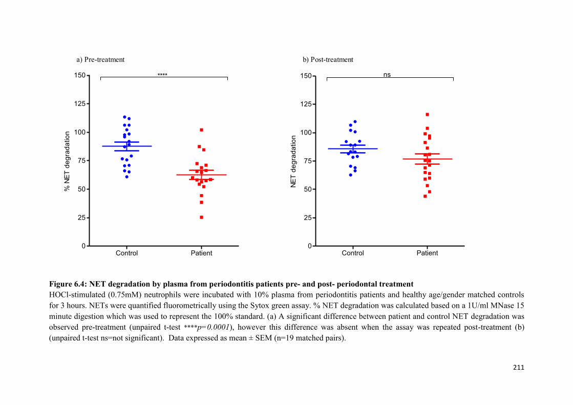
211
% N
ET
de
gra
da
tio
n
Control Patient0
25
50
75
100
125
150 ****
NE
T d
eg
rad
atio
n
Control Patient0
25
50
75
100
125
150 ns
a) Pre-treatment b) Post-treatment
Figure 6.4: NET degradation by plasma from periodontitis patients pre- and post- periodontal treatment
HOCl-stimulated (0.75mM) neutrophils were incubated with 10% plasma from periodontitis patients and healthy age/gender matched controls
for 3 hours. NETs were quantified fluorometrically using the Sytox green assay. % NET degradation was calculated based on a 1U/ml MNase 15
minute digestion which was used to represent the 100% standard. (a) A significant difference between patient and control NET degradation was
observed pre-treatment (unpaired t-test ****p=0.0001), however this difference was absent when the assay was repeated post-treatment (b)
(unpaired t-test ns=not significant). Data expressed as mean ± SEM (n=19 matched pairs).

212
Variability in NET degradation with patient age 6.3.1
Plasma NET degradation from periodontitis patients was analysed to determine whether an
age-associated link exists. Percentage NET degradation was calculated based on a 1U/ml
MNase degradation representing 100% (Hakkim et al., 2010). NET degradation results for
19 patients were plotted against their corresponding age. No significant correlation was
found between NET degradation and age (linear regression p=0.52, n=19) (Figure 6.5).
MNase-treated patient plasma 6.3.2
To determine whether the addition of MNase to plasma samples could restore NET
degradation in periodontitis patients, 1U/ml MNase-spiked 10% plasma was incubated with
NETs for 3 hours. % NET degradation was calculated based on a 1U/ml MNase digest
representing 100% degradation. A significant difference was observed between periodontitis
plasma and MNase-spiked plasma (unpaired t-test ****p=0.0001, n=10). Of the 10 plasma
samples treated with 1U/ml MNase, the significant increase in NET degradation was
observed in 9 patients. One sample however did not show any considerable increase in NET
degradation in the presence of MNase supplementation (Figure 6.6).

213
Age/years
% N
ET
de
gra
da
tio
n
30 40 50 60 700
50
100
150
R2=0.02; p=0.52
Figure 6.5: NET degradation plotted against patient age
Percentage NET degradation by periodontitis patient plasma was plotted against patient age.
% NET degradation was calculated based on a 1U/ml MNase digest which was used to
represent the 100% standard. No significant difference was observed between the amount of
NET degradation and age. Data expressed ± SEM (n=19 in triplicate).
% N
ET
de
gra
da
tio
n
Plasma MNase/plasma0
50
100
150 ****
Figure 6.6: MNase-treated patient plasma
NETs incubated with 1U/ml MNase-spiked periodontitis plasma for 3 hours were quantified
fluorometrically (Sytox green). % NET degradation was calculated based on a 1U/ml MNase
digest, which was used to represent the 100% digestion standard. A significant difference was
observed between periodontitis plasma and MNase-spiked plasma (unpaired t-test
****p=0.0001) (n=10 plasma samples in triplicate).

214
Reduced NET degradation in periodontitis patients may be due to elevated circulating
antibodies or immunoglobulin free light chains (FLCs), which may provide a physical barrier
and prevent NET digestion by DNase (Hakkim et al., 2010). To evaluate this hypothesis,
plasma IgG subclasses 1-4 and FLC concentrations were measured in periodontitis patients
and age- and gender-matched controls.
Plasma IgG concentrations in periodontitis and controls 6.3.3
IgG subclass 1-4 concentrations were measured in plasma samples from periodontitis patients
and healthy matched controls pre- and post- periodontal treatment. Protein turbidimetric
analysis demonstrated that patient IgG1 detection was marginally higher than that of the
controls pre- and post-treatment, however this was not statistically significant (unpaired t-test
p=0.50, p=0.43 pre- and post-treatment, respectively) (Figure 6.7a). Plasma IgG subclass 2
concentrations in patients were significantly higher than controls (unpaired t-test *p=0.038).
However no significant difference was observed between patient and control plasma IgG2
levels post-treatment (p=0.11) (Figure 6.7b). IgG3 plasma levels were significantly higher in
patients pre- and post-treatment compared with their matched controls (unpaired t-test
***p=0.0007, **p=0.0042 pre and post-treatment, respectively) (Figure 6.7c). The detection
of plasma IgG4 levels in patients and controls demonstrated higher levels in patients,
however this was only significant post-treatment (unpaired t-test *p=0.042) (Figure 6.7d). A
small number of sample readings fell outside of the IgG reference ranges (IgG1: 3.824-
9.286g/litre, IgG2: 2.418-7.003g/litre, IgG3: 0.218-1.761g/litre, IgG4 0.039-0.864) (Schauer
et al., 2003). This appeared to be dependent upon disease status, as whilst 8 control readings
were below the reference ranges (3 pre-treatment and 5 post-treatment), this only occurred in
3 periodontitis patient samples (1 pre-treatment and 2 post-treatment). Conversely, no
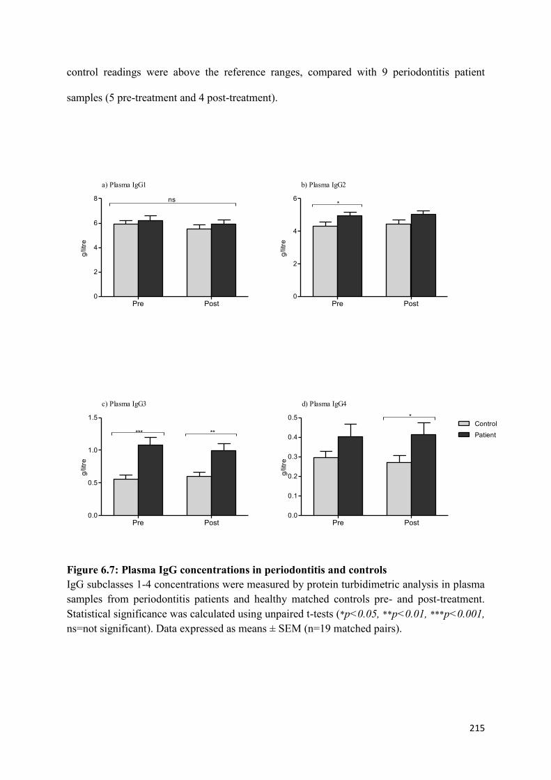
215
control readings were above the reference ranges, compared with 9 periodontitis patient
samples (5 pre-treatment and 4 post-treatment).
g/litre
Pre Post0
2
4
6
8 ns
a) Plasma IgG1
g/litre
Pre Post0
2
4
6*
b) Plasma IgG2
g/litre
Pre Post0.0
0.5
1.0
1.5
*** **
c) Plasma IgG3
g/litre
Pre Post0.0
0.1
0.2
0.3
0.4
0.5Control
Patient
*
d) Plasma IgG4
Figure 6.7: Plasma IgG concentrations in periodontitis and controls
IgG subclasses 1-4 concentrations were measured by protein turbidimetric analysis in plasma
samples from periodontitis patients and healthy matched controls pre- and post-treatment.
Statistical significance was calculated using unpaired t-tests (*p<0.05, **p<0.01, ***p<0.001,
ns=not significant). Data expressed as means ± SEM (n=19 matched pairs).

216
6.3.3.1 Plasma IgG subclasses concentration plotted against NET degradation
Plasma-derived IgG subclass 1-4 measurements (g/litre) were plotted against pre-treatment %
NET degradation results for each patient and control (n=19 pairs). No significant correlation
by linear regression was observed between IgG subclasses 1-3 and NET degradation (p=0.52,
p=0.60, p=0.37 for IgG1, IgG2 and IgG3, respectively). Notably, a significant negative
correlation was observed between IgG4 and NET degradation (*p=0.023) (Figure 6.8).
% NET degradation
IgG
g/litre
0 40 80 1200
2
4
6
8
10
IgG1
IgG2
IgG3
IgG4
R2=0.011; p=0.52
R2=0.0077; p=0.60
R2=0.022; p=0.37
R2=0.013; p=0.023
Figure 6.8: Plasma IgG subclasses concentration plotted against NET degradation
Plasma IgG subclasses 1-4 measurements (g/litre) were plotted against pre-treatment % NET
degradation results for each patient and matched control. A significant negative correlation
was observed between IgG4 concentration and NET degradation (linear regression *p=0.023)
(n=19 matched pairs).

217
Plasma FLC concentrations in periodontitis and controls 6.3.4
Free kappa and lambda light chains were assayed in plasma samples from periodontitis
patients and healthy matched controls pre- and post-treatment. Protein turbidimetric analysis
demonstrated that free kappa light chain concentrations were significantly higher in patients
than controls, both pre- and post-treatment (unpaired t-test *p=0.049, *p=0.048 pre- and
post-treatment, respectively) (Figure 6.9a). Free lambda light chain concentrations in patient
plasma were higher than controls pre- and post-treatment, however this did not reach
statistical significance (unpaired t-test p=0.22, p=0.23 pre- and post-treatment, respectively)
(Figure 6.9b). The kappa/lambda ratio demonstrated higher levels in patients pre-treatment
(unpaired t-test *p=0.047), but not post-treatment (p=0.66) (Figure 6.9c). Similar results
were observed for summated FLC values, in which patients exhibited significantly higher
levels pre-treatment (unpaired t-test *p=0.032), however this was not statistically significant
post-treatment (p=0.085) (Figure 6.9d). A small number of sample readings fell outside of
the reference ranges (free kappa light chains: 3.3-19.4mg/litre, free lambda light chains: 5.71-
26.3mg/litre, kappa/lambda ratio: 0.26-1.65, summated FLC: 9.01-45.7) (Katzmann et al.,
2002). Deviation from the reference range appeared to be dependent on disease state, as
whilst 4 control readings were below the reference ranges (1 pre-treatment and 3 post-
treatment), this did not occur in any patients. Conversely, 5 control IgG readings were above
the reference ranges (2 pre-treatment and 3 post-treatment), compared with 13 patients (6 pre-
treatment and 7 post-treatment).

218
mg
/litre
Pre Post0
5
10
15
20* *
a) Plasma kappa
Pre Post0
5
10
15
20ns
b) Plasma lambda
mg
/litre
Pre Post0.0
0.5
1.0
1.5*
c) Plasma kappa/lambda ratio
Ka
pp
a/L
am
bd
a r
atio
Su
mm
ate
d
Pre Post0
10
20
30
40Control
Patient
d) Plasma summated FLC
*
Figure 6.9: Plasma FLC concentrations in periodontitis and controls
Free kappa light chains, free lambda light chains, the kappa/lambda ratio and summated
FLCs were measured by protein turbidimetric analysis in plasma samples from periodontitis
patients and healthy matched controls pre- and post-treatment. Statistical significance was
calculated using unpaired t-tests (*p<0.05, ns=not significant). Data expressed as means ±
SEM (n=19 matched pairs).

219
6.3.4.1 Plasma FLC concentration plotted against NET degradation
Plasma-derived free kappa light chains (mg/litre), free lambda light chains (mg/litre),
kappa/lambda ratio and summated FLC measurements were plotted against pre-treatment
NET degradation data (n=19 pairs). No significant correlation was observed between FLC
detection and NET degradation (linear regression p=0.96, p=0.078, p=087, p=0.57 for free
kappa light chains, free lambda light chains, kappa/lambda ratio and summated FLC,
respectively) (Figure 6.10).

220
% NET degradation
FL
C m
g/litre
0 40 80 1200
20
40
60
Kappa
Lambda
Ratio
Summated
R2=0.00077; p=0.87
R2=0.0001; p=0.96
R2=0.0023; p=0.078
R2=0.0091; p=0.57
Figure 6.10: Plasma FLC concentration plotted against NET degradation
Plasma-derived free kappa light chains (mg/litre), free lambda light chains (mg/litre), the
kappa/lambda ratio and summated FLC measurements were plotted against pre-treatment %
NET degradation data for each patient and matched control. No significant correlation was
observed between FLCs and NET degradation (linear regression p>0.05) (n=19 matched
pairs).

221
Elevated levels of FLC may be the result of excess production by plasma cells, or reduced
clearance of FLCs by the kidneys (Randers et al., 2000). To determine whether the increased
FLC concentrations measured in periodontitis patients were due to impeded FLC clearance,
cystatin C concentrations were measured in plasma samples from periodontitis patients and
healthy matched controls as an independent measure of kidney function. Cystatin C is an
inhibitor of cysteine proteinases and considered a marker of renal function, where elevated
levels of cystatin C are positively correlated with decreased renal function (Randers et al.,
2000).
Plasma-derived cystatin C detection in periodontitis 6.3.5
Protein turbidimetric analysis demonstrated there was no significant difference in cystatin C
levels (mg/litre) between control and patient plasma samples pre- and post-treatment
(unpaired t-test p=0.74, p=0.82 pre- and post-treatment, respectively, n=19 matched pairs).
A small minority of samples were higher than the cystatin C reference range (0.56-
0.99mg/litre); however as this encompassed both patients and controls, this did not appear
related to periodontal disease status (Figure 6.11).

222
mg
/litre
Pre Post0.0
0.2
0.4
0.6
0.8
1.0Control
Patient
ns ns
Figure 6.11: Plasma-derived cystatin C detection in periodontitis
Cystatin C detection by protein turbidimetric analysis revealed no significant difference
between control and patient plasma samples pre- and post-treatment (unpaired t-test ns=not
significant). Data expressed as means ± SEM (n=19 matched pairs).

223
To evaluate the effect of reduced NET degradation on neutrophil responses and the
epithelium, neutrophils from healthy individuals were stimulated to produce NETs (0.75mM
HOCl) and the washed NET supernatants collected by MNase digestion (section 2.2.5.3) and
stored at -80°C by snap freezing (Appendix VI) until required. NET supernatants were
subsequently pooled to determine the effect of NET exposure on the neutrophil responses of
ROS and NET production, chemotaxis and their effect on H400 oral epithelial cells.
6.4 Determination of the DNA-content in NET supernatants
NET supernatants were used at concentrations of 25%, 50% and 100% (section 2.2.5.4). To
ensure outcomes were as a result of exposure to NET structures and not the HOCl stimulus
initially employed to generate the NETs, a HOCl cell-free supernatant was also collected for
future assay controls. To determine the concentration of NET supernatants, NET-DNA was
quantified in pooled samples (n=10) with Sytox green and compared with a standard curve
generated using calf thymus DNA (n=5) to provide interpolated values (Palmer 2010). NET
supernatants were applied in a volume of 50µl to investigate whether NETs could stimulate
neutrophils to release ROS, release NETs and the effect of NET supernatants on H400
epithelial cells growth. For chemotaxis assays, NET supernatants were used at a volume of
80µl. The concentration of DNA present in the HOCl cell-free control and 25%, 50% and
100% NET supernatant test sample (50µl and 80µl) is presented in table 6.1.
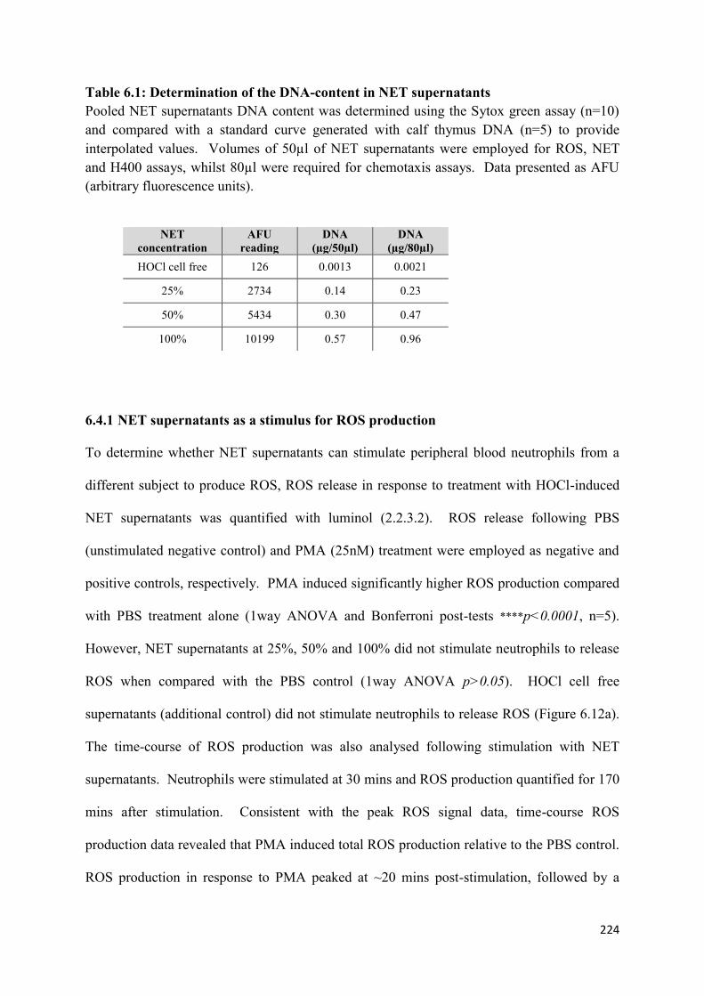
224
Table 6.1: Determination of the DNA-content in NET supernatants
Pooled NET supernatants DNA content was determined using the Sytox green assay (n=10)
and compared with a standard curve generated with calf thymus DNA (n=5) to provide
interpolated values. Volumes of 50µl of NET supernatants were employed for ROS, NET
and H400 assays, whilst 80µl were required for chemotaxis assays. Data presented as AFU
(arbitrary fluorescence units).
NET
concentration
AFU
reading
DNA
(µg/50µl)
DNA
(µg/80µl)
HOCl cell free 126 0.0013 0.0021
25% 2734 0.14 0.23
50% 5434 0.30 0.47
100% 10199 0.57 0.96
NET supernatants as a stimulus for ROS production 6.4.1
To determine whether NET supernatants can stimulate peripheral blood neutrophils from a
different subject to produce ROS, ROS release in response to treatment with HOCl-induced
NET supernatants was quantified with luminol (2.2.3.2). ROS release following PBS
(unstimulated negative control) and PMA (25nM) treatment were employed as negative and
positive controls, respectively. PMA induced significantly higher ROS production compared
with PBS treatment alone (1way ANOVA and Bonferroni post-tests ****p<0.0001, n=5).
However, NET supernatants at 25%, 50% and 100% did not stimulate neutrophils to release
ROS when compared with the PBS control (1way ANOVA p>0.05). HOCl cell free
supernatants (additional control) did not stimulate neutrophils to release ROS (Figure 6.12a).
The time-course of ROS production was also analysed following stimulation with NET
supernatants. Neutrophils were stimulated at 30 mins and ROS production quantified for 170
mins after stimulation. Consistent with the peak ROS signal data, time-course ROS
production data revealed that PMA induced total ROS production relative to the PBS control.
ROS production in response to PMA peaked at ~20 mins post-stimulation, followed by a

225
steep decline in ROS release. NET supernatants (25%, 50% and 100%) and the HOCl-cell
free supernatant control did not stimulate ROS release (Figure 6.12b).

226
RL
U
PBS
PM
A
HOCl c
ell fre
e
NETs
25%
NETs
50%
NETs
100%
0
5000
10000
15000
20000
25000****
ns
ns
ns
ns
a) Peak ROS release
Time/min
RL
U
0 50 100 150 2000
5000
10000
15000
20000
25000PBS
PMA
HOCl cell free
NETs 25%
NETs 50%
NETs 100%
b) Time-course ROS release
Figure 6.12: NET supernatants as a stimulus for ROS production
(a) Total ROS production was quantified with luminol in response to PBS (unstimulated),
PMA (25nM), HOCl cell free supernatant or NET supernatants (25%, 50% and 100%).
Statistical significance calculated using 1way ANOVA and Bonferroni post-tests
(****p<0.0001, ns=not significant). Data is expressed as mean ± SEM. (b) Time course total
ROS production over 200 mins by cells stimulated with PBS (unstimulated), PMA (50nM),
HOCl cell free supernatant or NET supernatants (25%, 50% and 100%). Data is presented as
RLU (relative light units) (n=5 in triplicate).

227
NET supernatants as a stimulus for NET production 6.4.2
To determine whether NET supernatants can stimulate neutrophils to produce NETs, NET
production in response to treatment (4 hours) with HOCl-induced NET supernatants was
quantified using the Sytox green assay (2.2.3.3). NET release following PBS (unstimulated)
and PMA (50nM) treatment were employed as negative and positive controls, respectively.
PMA induced significantly higher NET release compared with PBS treatment (1way
ANOVA and Bonferroni post-tests ****p<0.0001, n=5). However NET supernatants at 25%,
50% and 100% did not stimulate neutrophils to release NETs, evidenced by no significant
increase in NETs relative to the PBS control treatment (1way ANOVA p>0.05). HOCl cell
free supernatants (additional control) did not stimulate neutrophils to release NETs (Figure
6.13).

228
AF
U
PBS
PM
A
HOCl c
ell fre
e
NETs
25%
NETs
50%
NETs
100%
0
5000
10000
15000
****
ns
ns
ns
ns
Figure 6.13: NET supernatants as a stimulus for NET production
NET-DNA production was quantified with Sytox green in response to PBS (unstimulated),
PMA (50nM), HOCl cell free supernatant or NET supernatants (25%, 50% and 100%).
Statistical significance calculated using 1way ANOVA and Bonferroni post-tests
(****p<0.0001, ns=not significant). Data presented as AFU (arbitrary fluorescence units) and
expressed as mean ± SEM (n=5 in triplicate).

229
The ability of NETs to induce neutrophil chemotaxis 6.4.3
To determine whether NETs serve as chemoattractants and induce the directional migration
of neutrophils towards them, NET supernatants were employed as chemoattractants and
neutrophil chemotaxis was analysed using the Insall Chamber (2.2.4.2.3). The movement of
15 randomly chosen cells was tracked by video microscopy and the quantitative data
generated used to produce spider plots and to calculate neutrophil speed, velocity and
resultant vector length (Roberts et al., 2015).
Spider plots enable visualisation of individual cell movement (µm) from a central point,
where the top of the Y-axis represents the source of the chemoattractant. Cells displayed
directional movement towards the known chemoattractant, fMLP. Neutrophil migration was
observed in response to 25% NET supernatants, and to greater extent with 50% and 100%
NET supernatants; however this movement did not appear to be towards the chemoattractant
source, and lacked directionality (Figure 6.14).

230
fMLP 25% NETs
50% NETs 100% NETs
Figure 6.14: Spider plots representing neutrophil migration in response to NET
supernatants
Spider plots show the tracking (µm) of individual neutrophils (n=15 cells from n=5
experiments) from a central reference point (0, 0) towards the chemoattractant. The top of
the Y-axis represents the source of the chemoattractant. Neutrophil chemotaxis in response
to a known chemoattractant, fMLP (100nM), was employed as a positive control. Cell
migration was tracked in response to 25%, 50% and 100% NET supernatants.
-150
-100
-50
0
50
100
150
-150 -100 -50 0 50 100 150µm
-150
-100
-50
0
50
100
150
-150 -100 -50 0 50 100 150µm
-150
-100
-50
0
50
100
150
-150 -100 -50 0 50 100 150µm
-150
-100
-50
0
50
100
150
-150 -100 -50 0 50 100 150µm

231
Whilst spider diagrams represent a relatively novel way to visualise cell migration in
response to a chemoattractant, they cannot be used objectively or quantitatively to measure
neutrophil movement. Thus, the data generated from tracking 15 individual cells was used to
quantify the speed, velocity and directional accuracy of neutrophil migration.
6.4.3.1 Neutrophil speed in response to NET supernatants
Neutrophil speed (µm/min) in response to fMLP (100nM, positive control) was significantly
greater than for RPMI (negative control) (1way ANOVA and Bonferroni post-test
****p<0.0001, n=5). The speed of neutrophil migration in response to NET supernatants
(25%, 50% and 100%) was also significantly higher than RPMI (1way ANOVA and
Bonferroni post-test ****p<0.0001 for 25%, 50% and 100%, n=5). However, increased
neutrophil speed in response to NET supernatants was significantly lower than for fMLP-
induced migration (positive control) (1way ANOVA and Bonferroni post-test ****p<0.0001
for 25%, 50% and 100%, n=5, statistics not shown on graph). Notably, HOCl cell free
supernatants (additional control) did not induce an increase in neutrophil speed (1way
ANOVA p>0.05) (Figure 6.15).

232
RPM
I
fMLP
HO
Cl c
ell fre
e
NETs
25%
NETs
50%
NETs
100%
0
2
4
6
m
pe
r m
in
****
ns****
****
****
Figure 6.15: Neutrophil speed in response to NET supernatants
Neutrophil chemotactic speed (µm/min) in response to fMLP (100nM, positive control),
RPMI (negative control) and different concentrations of NET supernatants was measured.
Statistical significance calculated using 1way ANOVA and Bonferroni post-test
(****p<0.0001, ns=not significant). Data calculated from the tracking of 15 cells and is
expressed as mean ± SEM (n=5).

233
6.4.3.2 Neutrophil velocity in response to NET supernatants
Velocity is the speed of movement in a specific direction towards a chemoattractant.
Neutrophil velocity in response to fMLP (100nM, positive control) was significantly higher
than for RPMI (negative control) (1way ANOVA and Bonferroni post-test ****p<0.0001,
n=5). However NET supernatant-induced velocity was not significantly different to (1way
ANOVA p>0.05) (Figure 6.16).
RPM
I
fMLP
HOCl c
ell fre
e
NETs
25%
NETs
50%
NETs
100%
-2
0
2
4
6
m
pe
r m
in
nsns
ns
ns
****
Figure 6.16: Neutrophil velocity in response to NET supernatants
Neutrophil velocity (µm/min) in response to fMLP (100nM, positive control), RPMI
(negative control) and different concentrations of NET supernatants was measured.
Statistical significance calculated using 1way ANOVA and Bonferroni post-test
(****p<0.0001, ns=not significant). Data calculated from the tracking of 15 cells and is
expressed as mean ± SEM (n=5).

234
6.4.3.3 Directional accuracy of neutrophil movement (resultant vector length)
Representative angular histograms and resultant vector plots of neutrophil directional
chemotaxis in response to fMLP (100nM) or 100% NET supernatants were plotted. The
angular histograms measure cell directionality and strength of this movement, which is
determined by the histogram bar angle and width, respectively. Angular histograms and
resultant vector plots of neutrophil migration in response to fMLP (100nM, positive control)
show strong directional accuracy of movement towards the chemoattractant. However
neutrophil chemotaxis in response to 100% NET supernatants exhibited movement that was
not directionally targeted towards the source of the chemoattractant (90°), evidenced by the
cell movement endpoint (blue circles on circle circumference) being in every direction and
the shortness of the red line (Figure 6.17).

235
fMLP NETs 100%
Figure 6.17: Directional accuracy of neutrophil movement (resultant vector length)
Representative angular histograms and resultant vector plots of neutrophils treated with
fMLP (100nM) or 100% NET supernatants. The angle and width of the histogram bars
indicates the direction and the proportion of cells migrating in that direction, respectively.
The resultant vector plots indicate the strength and directionality of the cells in response to
the chemoattractant. The red line denotes the mean resultant vector and the dashed line
shows the variation (95% confidence intervals) within the entire cohort of cells. The length
of the red line is indicative of the strength of cell directionality. The blue circles along the
circumference of the plot represent individual neutrophil migration end-points. For both
plots, 90° at the top of both plots represents the source of the chemoattractant.
Histograms
Resultant
vector
plots

236
6.4.3.4 Neutrophil resultant vector length
The resultant vector length measures the directional accuracy of neutrophil chemotaxis
towards the chemoattractant. Resultant vector length was calculated from the resultant vector
plots, in which the red line denotes the mean resultant vector and the dashed line shows the
variation (95% confidence intervals) within the entire cohort of cells. The length of the red
line is indicative of the strength of cell directionality. Resultant vector length in response to
fMLP (100nM, positive control) was found to be significantly higher than neutrophil
migration when RPMI (negative control) was employed as a chemoattractant (1way ANOVA
and Bonferroni post-test ***p<0.001, n=5). However NET supernatant-induced chemotaxis
did not cause a significant increase in neutrophil directional movement, compared with the
RPMI control (1way ANOVA p>0.05) (Figure 6.18).

237
Re
su
lta
nt
ve
cto
r le
ng
th
RPM
I
fMLP
cell fre
e25
%50
%
100%
0.0
0.2
0.4
0.6
0.8
1.0ns
ns
ns
ns
***
Figure 6.18: Neutrophil resultant vector length
Resultant vector length of neutrophil chemotaxis in response to fMLP (100nM, positive
control), RPMI (negative control) and different concentrations of NET supernatants was
measured. Statistical significance calculated using 1way ANOVA and Bonferroni post-test
(***p<0.001, ns=not significant). Data calculated from the tracking of 15 cells and is
expressed as mean ± SEM (n=5).

238
6.5 The effect of NETs on H400 oral epithelial cell responses
To evaluate the effect of reduced NET degradation on the activity and viability of oral
epithelial cells, H400s were cultured in a 96-well plate (until 90% confluent) and incubated
with NET supernatants (25%, 50% and 100%). At 4 and 24 hours, the impact of NET
supernatants on H400 oral epithelial cell growth was determined by quantifying caspase
activity (apoptosis), ATP activity (metabolic activity) and LDH release (cytotoxicity). HOCl
cell free supernatants were employed as a control, RPMI and 0.1% triton were used as
negative and positive treatment controls, respectively.
The effect of NETs on H400 oral epithelial cell apoptosis 6.5.1
To investigate whether NET supernatants induce apoptosis in H400 oral epithelial cells,
caspase-3 and 7 production by H400s (90% confluent) was measured at 4 and 24 hours
(2.2.1.2.4). Treatment with triton (0.1%, positive control) caused a decrease in caspase
activity at 4 and 24 hours, relative to the RPMI negative control, however this was not
statistically significant (1way ANOVA p>0.05, n=5). Caspase activity in response to RPMI
(negative control), HOCl cell free supernatants and NET supernatants (25%, 50% and 100%)
was higher at 24 hours than 4 hours. A concentration dependent decrease in caspase-3 and 7
production was observed at 24 hours following treatment with 25%, 50% and 100% NET
supernatants, however this was not statistically significant (1way ANOVA p>0.05, n=5)
(Figure 6.19).

239
RL
U
- con
trol
+ co
ntro
l
HOCl c
ell fre
e
NETs
25%
NETs
50%
NETs
100%
0
500000
1000000
1.5100 6
4 hours
24 hours
ns
Figure 6.19: The effect of NETs on H400 oral epithelial cell caspase activity
Apoptotic activity in H400 oral epithelial cells was determined by measuring caspase-3 and -
7 activity at 4 and 24 hours in response to RPMI (negative control), 0.1% triton (positive
control), HOCl cell free supernatants (additional control) and NET supernatants (25%, 50%
and 100%). Statistical significance calculated using 1way ANOVA (ns=not significant).
Data expressed as RLU (relative light units) and mean ± SEM (n=5 in triplicate).
The effect of NETs on H400 oral epithelial cell metabolic activity 6.5.2
To investigate the metabolic activity of H400 oral epithelial cells in response to NET
supernatants, ATP production by H400s (90% confluent) was measured at 4 and 24 hours
(2.2.1.2.5). Treatment with triton (0.1%, positive control) caused a significant decrease in
metabolic activity at 4 and 24 hours, relative to the RPMI negative control (1way ANOVA
and Bonferroni post-tests **p<0.01, ***p<0.001 at 4 and 24 hours, respectively, n=5).
Metabolic activity in response to RPMI (negative control), HOCl cell free supernatants and
NET supernatants (25%, 50% and 100%) was higher at 24 hours, relative to 4 hours. A
concentration dependent decrease in ATP production was observed at 24 hours following
treatment with 25%, 50% and 100% NET supernatants, however this was not statistically
significant (1way ANOVA p>0.05, n=5) (Figure 6.20).

240
RLU
- con
trol
+ co
ntro
l
HOCl c
ell fre
e
NETs
25%
NETs
50%
NETs
100%
0
5.0106
1.0107
1.5107
4 hours
24 hours
*****
Figure 6.20: The effect of NETs on H400 oral epithelial cell metabolic activity
Metabolic activity of H400 oral epithelial cells was determined by measuring ATP release at
4 and 24 hours in response to RPMI (negative control), 0.1% triton (positive control), HOCl
cell free supernatants (additional control) and NET supernatants (25%, 50% and 100%).
Statistical significance calculated using 1way ANOVA and Bonferroni post-test (**p<0.01,
***p<0.001). Data expressed as RLU (relative light units) and mean ± SEM (n=5 in
triplicate).

241
The effect of NETs on H400 oral epithelial cell lactate dehydrogenase release 6.5.3
To investigate whether NETs induce cytotoxicity in H400 oral epithelial cells, lactate
dehydrogenase (LDH) was measured at 4 and 24 hours (2.2.5.7). LDH is released by
damaged membranes, therefore an increase in LDH, and thus an increase in AFU, is
associated with reduced membrane integrity. Treatment with triton (0.1%, positive control)
caused a significant increase in LDH release at 4 and 24 hours, relative to the RPMI negative
control (1way ANOVA and Bonferroni post-tests ****p<0.0001 at 4 and 24 hours, n=5).
LDH release in response to RPMI (negative control), HOCl cell free supernatants and NET
supernatants (25%, 50% and 100%) was higher at 24 hours, relative to 4 hours. Despite a
noticeable increase in LDH release at 24 hours following treatment with 100% NET
supernatants, this was not statistically significant. There was no significant increase in LDH
release as a result of treatment with HOCl free supernatants or NET supernatants (25% and
50%) (1way ANOVA p>0.05, n=5) (Figure 6.21).

242
AF
U
- con
trol
+ co
ntro
l
HOCl c
ell fre
e
NETs
25%
NETs
50%
NETs
100%
0
20000
40000
60000200000
3000004 hours
24 hours
********
Figure 6.21: The effect of NETs on H400 oral epithelial cell lactate dehydrogenase
release
LDH released into the media was quantified at 4 and 24 hours in response to RPMI (negative
control), 0.1% triton (positive control), HOCl cell free supernatants (additional control) and
NET supernatants (25%, 50% and 100%). Statistical significance calculated using 1way
ANOVA and Bonferroni post-test (****p<0.0001). Data expressed as AFU (arbitrary
fluorescence units) and mean ± SEM (n=5 in triplicate).

243
6.6 Discussion
Plasma-derived NET degradation was significantly lower in chronic periodontitis patients
pre-treatment relative to periodontally healthy matched controls (section 6.3). This is the first
report evaluating NET degradation in periodontitis, however the removal of NETs has been
well characterised in other diseases such as systemic lupus erythematosus (SLE) patients, in
which NET clearance by sera was reportedly impeded in a subset of patients (Hakkim et al.,
2010, Leffler et al., 2012). The mechanisms involved in the removal of NETs remain to be
thoroughly elucidated; however reports indicate that degradation of the DNA backbone by
DNase 1 initiates their removal, followed by the engulfment of degraded structures by
macrophages and further degradation in the lysosome. Key to this process is NET clearance
resembles a mechanism similar to that of apoptosis, whereby macrophages behave non-
phlogistic, do not release pro-inflammatory cytokines and thus NET disassembly is regarded
as an “immunologically silent” process (Farrera & Fadeel 2013). Possible reported
explanations for attenuated NET degradation in SLE patients include the presence of DNase1
inhibitors preventing the enzymatic removal of the NETs (Yeh et al., 2003), or alternatively
higher antibody titers in patients’ plasma, which may provide a physical barrier to NET
removal by binding to NETs and blocking the DNase cleavage sites (Hakkim et al., 2010,
Leffler et al., 2012). Notably contradictory findings are reported regarding the effect of
supplementing patient sera with DNase to restore NET degradation in all “low-degrading”
patients (Hakkim et al., 2010, Leffler et al., 2012). In the periodontitis cohort studied here, 9
out of 10 patients displayed restored NET degradation following the addition of MNase
(section 6.3.2), suggesting that if enzymatic inhibitors are present, they may be DNase
specific. Following non-surgical treatment, NET degradation in periodontitis patients was
restored and no significant difference was detected between patients and controls. This is
consistent with reports by Leffler et al., (2012), who demonstrated that NET degradation was

244
improved when SLE patients were in remission, relative to during a disease flare. Notably, as
with the SLE cohort, reduced NET degradation was a variable phenomenon and only
observed in a subset of periodontitis patients; this could be linked to disease susceptibility,
disease severity, disease activity status or bacterial load and virulence factors (such as
bacteria-derived DNases).
To establish whether a physical barrier, such as circulating antibodies or immunoglobulin
free light chains (FLC), may be preventing the enzymatic removal of NETs, IgG subclasses
were quantified in patient and matched control plasma samples. In addition to analysing
immunoglobulins, levels of FLCs were measured. Immunoglobulins comprise 2 identical
heavy and 2 identical light chains (either kappa or lambda), however during their normal
synthesis by B cells, there is an excess of light chain production which is released into
circulation and known as FLCs (Solomon 1985). However, FLC overproduction has been
reported in several chronic diseases, such as SLE (Aggarwal et al., 2011) and rheumatoid
arthritis (RA) (Gottenberg et al., 2007), which may be the result of chronic immune
stimulation (Brebner & Stockley 2013). Periodontitis has been linked to these pathologies
(Fabbri et al., 2014, Smit et al., 2012), and furthermore, periodontitis is a disease exacerbated
by a chronic inflammatory-immune host response and notably overactive B cells are deemed
partly responsible (Reinhardt et al., 1988). Accordingly, the results reported here
demonstrate increased plasma-derived FLCs and IgG 1-4 subclasses in periodontitis patients
(sections 6.3.3, 6.3.4). Increased IgG levels are consistent with the findings of Graswinckel
et al., (2004), who reported higher levels of IgG1 and IgG2 in periodontitis patients relative
to controls. In the present study, cystatin C levels were also measured to establish whether
excessive plasma FLCs in periodontitis patients resulted from insufficient renal FLC
clearance; notably no differences in renal clearance were observed between patients and
controls. There is a paucity of data pertaining to FLCs and periodontitis; however the results

245
here suggest that in this patient cohort, periodontitis is associated with elevated FLC
production. Further experiments are therefore warranted to determine whether increasing
circulating levels of antibodies and FLCs prevents the enzymatic degradation of NETs in
periodontitis.
Attenuated NET removal may constitute an antimicrobial host response during periods of
infection, as NETs can prevent microbial dissemination by entrapping various periodontal
bacteria (section 4.4). In support of the contribution of NETs to the host response in SLE,
impeded NET production in NADPH-oxidase KO mice exhibited exacerbated lupus-like
symptoms, which led the authors to conclude that NET release does not contribute to SLE in
vivo (Campbell et al., 2012). The host’s ability to adjust the extracellular “life-span” of
NETs in response to the microbial biofilm may also explain why no differences in peripheral
NET release were observed between periodontitis patients and controls (see Chapter 4).
Notably, it has been reported that mice immunised with NETs produced in vitro with H2O2
did not demonstrate a break in immune tolerance as a result of NET-derived immunogens. In
support of the premise that NETs are non-immunogenic, the work in this chapter
demonstrated that under the reported experimental conditions, treatment of peripheral
neutrophils with HOCl-stimulated NET supernatants did not prompt ROS or NET production.
In addition, no NET-mediated cytotoxicity was identified in H400 cells following their
incubation with NET supernatants. This is contradictory to findings by Saffarazadeh et al.,
(2012), who demonstrated that NETs induced cell death in epithelial cells. These differences
may be a result of different cell lines (Saffarazadeh employed alveolar epithelial cells), or due
to differences in the NET supernatants, for example Saffarazadeh employed PMA-induced
NETs whereas HOCl was the NET stimulus of choice in the work outlined here.

246
NET supernatants employed as chemoattractants did however increase neutrophil
chemotaxis; albeit non-directional movement, suggesting that whilst increased motility was
observed, neutrophils were not being recruited to NET structures (section 6.4.3). Increased
neutrophil speed in the absence of directionality may be detrimental to the host, as the
random movement of cells may cause ROS-mediated collateral tissue damage during
neutrophil tissue transit (Roberts et al., 2015). It is difficult to ascertain whether these
observations reflect in vivo chemotaxis, where bacteria and host derived chemoattractants
will also be present and may provide a chemical hierarchy (Kim & Haynes 2012) that
overrides the chemotactic signals produced by NETs. Interestingly, NETs have been
suggested to provide danger signals that activate DNA receptors (such as TLR9), which
subsequently alert the immune system (Brinkmann et al., 2012). Activation of DNA
receptors was not investigated here; however the work outlined in this chapter suggests that
under the experimental conditions reported, NETs do not activate neutrophil effector
functions.
Evaluating pro-inflammatory responses to NETs produced in vitro is subject to various
limitations and may not be representative of the localised NET production in periodontitis.
For example, in vivo NETs are produced in response to a diverse microbial biofilm, the host
response is chronic, and inflammatory mediators may prime neutrophils prior to NET
production (Matthews et al., 2007a, 2007b). Notably, NET-evoked pro-inflammatory
responses have been suggested to act synergistically with the presence of microbes, whereby
immune response proteins in the extracellular milieu are required in addition to NET
structures (Farrera & Fadeel 2013). In accordance with this, several studies have suggested
that NETs complexed with bacteria and anti-NET antibodies may be resistant to degradation
and may exacerbate diseases such as cystic fibrosis (Dwyer et al., 2014) and small vessel
vasculitis (SVV) (Kessenbrock et al., 2009). Several of the peptides associated with the

247
NET-DNA backbone are reportedly autoantigens, such as MPO and PR3, which in SVV,
trigger the release of anti-neutrophil cytoplasmic antibodies (ANCAs), and notably the
production of ANCAs subsequently induce the production of NETs (Kessenbrock et al.,
2009). Findings by Gomes et al., (2009) suggest this may also be of relevance in
periodontitis, as the induction of experimental periodontitis in rats resulted in significantly
elevated levels of MPO in the gingival tissues, however whether this was NET-bound MPO
was not determined. Furthermore, in the light of our findings that circulating IgG levels are
higher in periodontitis patients (6.3.3), it’s noteworthy that IgG reportedly interacts with
chromatin, and these IgG-chromatin complexes potentiate autoantibody production
(Leadbetter et al., 2002). Thus in chronic periodontitis, increased IgG levels, a bacterial
biofilm and delayed NET removal may break immune tolerance, resulting in autoantibody
production and further impeded NET removal which generates a self-perpetuating disease
cycle.
Histones represent 70% of NET-associated proteins (Urban et al., 2009) and are proposed to
be key autoantigens in autoimmune diseases (Van Bavel et al., 2011). Histones undergo
post-translational modifications during the formation of NETs, whereby arginine residues are
converted to citrulline by the enzymatic activity of PAD4. Notably, autoantibodies produced
in Felty’s syndrome, a form of rheumatoid arthritis, preferentially react with citrullinated
histones; this suggests that NET release and the protein modifications required for this
mechanism to occur are implicated in autoimmunity (Dwivedi et al., 2012, 2014). Notably
recent findings investigating the association between RA and periodontitis demonstrated
higher levels of anti-citrullinated protein antibody (ACPAs) against citrullinated and
uncitrullinated peptides in periodontitis patients. It is possible that NET production in
periodontal tissues may serve as a plausible mechanism for ACPA production following a
break in immune tolerance to NET-derived citrullinated peptides released by neutrophils (de

248
Pablo et al., 2009), and this process may be exacerbated if NETs are not removed in a timely
manner.
The recent discovery that P. gingivalis possesses a unique PAD enzyme (PPAD) capable of
citrullinating bacterial and host proteins might also contribute to the generation of ACPAs
(Wegner et al., 2010). It has been suggested that PPADs can likely disseminate from the
bacterial biofilm, thus it is possible that NETs and PPADs interact during colonisation by P.
gingivalis. This raises questions as to whether PPADs have the ability to citrullinate proteins
associated with NETs that would ordinarily be citrullinated by host PADs. For example, the
NET-bound peptide, LL-37, is citrullinated by host-derived PAD2 and PAD4, and certain
citrullinated LL-37 peptides are reportedly pro-inflammatory relative to the uncitrullinated
peptide (Kilsgard et al., 2012). It is interesting to speculate that if P. gingivalis is also able to
citrullinate NET-derived peptides that promote pro-inflammatory responses, it may utilise
this mechanism to bring about non-resolving inflammation that is characteristic of
periodontitis. Furthermore, in SLE, LL-37 reportedly promotes the production of
autoantibodies and the production of immune complexes that are capable of stimulating
plasmacytoid dendritic cells to release type 1 IFN (Lande et al., 2011). This may be of
relevance in periodontitis, where the reduced NET degradation reported here may provide a
possible explanation for the increased levels of type 1 IFN observed in chronic periodontitis
patients (Wright et al., 2008).
In summary, a subset of periodontitis patients displayed impeded NET degradation; however
NET degradation was restored following successful non-surgical treatment. This may be the
result of increased circulating IgGs and FLCs in patients, which may provide a physical
barrier preventing enzymatic removal of NETs; further experiments are however warranted to
confirm this. Subsequent assays to determine the effect of delayed NET removal
demonstrated that NETs did not stimulate for neutrophil ROS production, further NET

249
release, or induce NET-mediated cytotoxicity in H400 cells. Notably, NETs employed as
neutrophil chemoattractants caused increased neutrophil migration speed, however this
movement did not exhibit directionality. Whilst no NET-induced pro-inflammatory
responses were observed here, NETs are implicated in disease progression in several
pathologies (Cooper et al., 2013), thus it is possible that in vitro assays do not fully mimic the
in vivo environment in periodontitis, comprising a microbial biofilm, complex pro-
inflammatory mediators and chronic immune activation.

250
EFFECT OF CIGARETTE SMOKE EXTRACT ON CHAPTER 7:
NEUTROPHIL RESPONSES

251
7.1 Introduction
Smoking is a significant risk factor for periodontitis, and cigarette smoke extract (CSE) has
previously been shown to impact upon neutrophil ROS production (section 1.7.3, Matthews
et al., 2011, 2012). However, there is no published data on the effects of CSE upon NET
formation; therefore to further understand the impact of smoking on neutrophilic responses,
healthy neutrophils were treated with CSE previously prepared (section 2.2.4.1, Matthews et
al., 2011). In addition to CSE, the effect of specific cigarette constituents on neutrophil
responses was also assayed, including pre-incubation with nicotine, cotinine and thiocyanate
(SCN-) (University of Birmingham Ethics Reference ERN_13-0325) (section 2.2.4.2.1).
Neutrophil directional chemotaxis in response to the known chemoattractants, fMLP and IL-
8, was also measured in cells primed with increasing concentrations of CSE. The speed,
velocity and directional chemotactic accuracy were measured using the Insall chamber and
real-time video microscopy (sections 2.2.4.2.3 & 2.2.4.2.4, Roberts et al., 2015). Finally, the
impact of CSE and SCN- treatment on neutrophil inflammatory and redox related gene
expression was determined by real time PCR (2.2.4.3).

252
7.2 Effect of cigarette smoke extract on NET release
NET production was determined as previously described by quantifying NET-DNA in the
Sytox green assay (2.2.4.2.1, Appendix VIII) Matthews et al., 2011, 2012). The effect of
CSE (at concentrations of 1%, 5% or 10%) on NET release did not differ significantly from
PBS-treated cells (unstimulated negative control) (1way ANOVA p=0.99, n=10) (Figure
7.1a). By contrast, CSE priming and subsequent stimulation with PMA (50nM) was
associated with significantly decreased NET production at concentrations of 5% and 10%
CSE (1way ANOVA and Dunnett’s post-tests *p<0.05, ****p<0.0001 for 5% and 10% CSE,
respectively, n=10) (Figure 7.1b). However, when cells were primed with CSE and
stimulated with HOCl (0.75mM), no differences in NET production due to CSE priming were
observed (1way ANOVA p=0.90, n=10) (Figure 7.1c).
Fluorescence visualisation of NETs following treatment with CSE 7.2.1
NET production in response to PMA (50nM) stimulation (Figure 7.2a) was qualitatively
compared visually between neutrophils primed for 30 mins with increasing concentrations of
CSE prior to stimulation (2.2.4.2.2). NETs were visualised by fluorescence microscopy after
staining with Sytox green, which targets DNA. NET-like structures appeared aggregated
when primed with 1% CSE prior to PMA stimulation (Figure 7.2b). In contrast, cells that
were primed with 5% or 10% CSE and subsequently stimulated with PMA did not form
visible web-like structures, however aggregates of DNA were observed (Figures 7.2c&d).

253
AF
U
PBS 1% 5% 10%0
500
1000
1500
CSE concentration
ns
a) NET production in response to CSE stimulationA
FU
PBS 1% 5% 10%0
5000
10000
15000
CSE concentration
ns
*****
b) NET production in response to CSE priming and PMA stimulation
AF
U
PBS 1% 5% 10%0
5000
10000
15000
20000
CSE concentration
ns
c) NET production in response to CSE priming and HOCl stimulation
Figure 7.1: Effect of cigarette smoke extract on NET production
NET-DNA was quantified fluorometrically in response to stimulation with 1, 5 or 10% CSE
and compared with PBS-treated cells (unstimulated negative control). (a) 4 hour stimulation
of neutrophils with CSE, (b) 30 min CSE priming prior to stimulation with PMA (50nM), (c)
30 min CSE priming prior to stimulation with HOCl (0.75mM). Statistical significance was
calculated using 1way ANOVA and Dunnett’s post-tests (*p<0.05, ****p<0.0001, ns=not
significant). Data are presented as AFU (arbitrary fluorescence units) and are expressed as
mean ± SEM (n=10 in triplicate).

254
Figure 7.2: Fluorescence visualisation of NETs in response to cigarette smoke extract
NETs were visualised with Sytox green using fluorescence microscopy (x20 magnification),
following stimulation with (a) PMA (50nM), or primed for 30 mins with (b) 1% CSE, (c) 5%
CSE or (d) 10% CSE, prior to PMA stimulation. Images are representative of 2 experiments
in duplicate. Scale bar represents 100µm.
c d
a b

255
7.3 Effect of CSE components on NET release
To attempt to determine whether specific CSE components of CSE were contributing to the
attenuated NET release observed, the effect of specific cigarette constituents on NET release
were investigated, including nicotine, cotinine and SCN-.
Effect of nicotine on NET production
Nicotine stimulation (applied at concentrations of 1µg/ml, 5µg/ml or 10µg/ml) (2.1.4.1,
2.2.4.2.1) did not alter NET production compared with unstimulated control cells (1way
ANOVA p=0.23, n=10). Nicotine priming for 30 mins and subsequent stimulation with
PMA (50nM) and HOCl (0.75mM) was also found to have no apparent effect on NET
release, compared with cells primed with PBS (1way ANOVA p=0.98, p=0.75 for PMA and
HOCl respectively, n=10) (Figure 7.3a).
Effect of cotinine on NET production
Cotinine stimulation (applied at concentrations of 1µg/ml, 5µg/ml or 10µg/ml) (2.1.4.2,
2.2.4.2.1) did not alter NET production compared with PBS (negative control cells) (1way
ANOVA p=0.85, n=10). Cotinine priming for 30 mins and subsequent stimulation with
PMA (50nM) and HOCl (0.75mM) was also found to have no effect on NET release (1way
ANOVA p=0.96, p=0.99 for PMA and HOCl respectively, n=10) (Figure 7.3b).
Effect of thiocyanate on NET production
SCN- stimulation at lower concentrations (applied at concentrations of 50µM, 100µM)
(2.1.4.3, 2.2.4.2.1) did not alter NET production relative to unstimulated control cells.
However stimulation with 150µM SCN-
resulted in a significant increase in NET release
(1way ANOVA and Dunnett’s post-tests *p<0.05). SCN- priming for 30 mins prior to PMA
(50nM) stimulation inhibited NET release compared with un-primed neutrophils; this was

256
observed at all concentrations of SCN- (1way ANOVA and Dunnett’s post-tests ***p<0.001
for each concentration). SCN- priming for 30 mins prior to HOCl (0.75mM) stimulation also
inhibited NET release compared with un-primed neutrophils; this was observed in all
concentrations of SCN-
(1way ANOVA and Dunnett’s post-tests ***p<0.001 for each
concentration) (Figure 7.3c).

257
AF
U
PBS 1g/ml 5g/ml 10g/ml0
200
400
600
800 ns
Nicotine concentration
i) NET production in response to nicotine stimulationA
FU
PBS 1g/ml 5g/ml 10g/ml0
5000
10000
15000 ns
Nicotine concentration
ii) NET production in response to nicotine priming and PMA stimulation
AF
U
PBS 1g/ml 5g/ml 10g/ml0
5000
10000
15000
20000 ns
Nicotine concentration
iii) NET production in response to nicotine priming and HOCl stimulation
Figure 7.3a: Effect of nicotine on NET production
NET-DNA production was quantified fluorometrically in response to stimulation with
1µg/ml, 5µg/ml or 10µg/ml nicotine and PBS-treated cells (unstimulated negative control).
(i) 4 hour stimulation of neutrophils with nicotine, (ii) 30 min nicotine priming prior to
stimulation with PMA (50nM), (iii) 30 min nicotine priming prior to stimulation with HOCl
(0.75mM). Statistical significance was calculated using 1way ANOVA and Dunnett’s post-
tests (ns=not significant). Data are presented as AFU (arbitrary fluorescence units) and are
expressed as mean ± SEM (n=5 in triplicate).

258
AF
U
PBS 1g/ml 5mg/ml 10g/ml0
200
400
600
800 ns
Cotinine concentration
i) NET production in response to cotinine stimulationA
FU
PBS 1g/ml 5mg/ml 10g/ml0
5000
10000
15000 ns
Cotinine concentration
ii) NET production in response to cotinine priming and PMA stimulation
AF
U
PBS 1g/ml 5mg/ml 10g/ml0
5000
10000
15000
20000 ns
Cotinine concentration
iii) NET production in response to cotinine priming and HOCl stimulation
Figure 7.3b: Effect of cotinine on NET production
NET-DNA production was quantified fluorometrically in response to stimulation with
1µg/ml, 5µg/ml or 10µg/ml cotinine with PBS-treated cells (unstimulated negative control).
(i) 4 hour stimulation of neutrophils with cotinine, (ii) 30 min cotinine priming prior to
stimulation with PMA (50nM), (iii) 30 min cotinine priming prior to stimulation with HOCl
(0.75mM). Statistical significance was calculated using 1way ANOVA and Dunnett’s post-
tests (ns=not significant). Data are presented as AFU (arbitrary fluorescence units) and are
expressed as mean ± SEM (n=5 in triplicate).

259
AF
U
PBS 50M 100M 150M0
1000
2000
3000
4000
ns
ns
*
SCN- concentration
i) NET production in response to SCN- stimulationA
FU
PBS 50M 100M 150M0
2000
4000
6000
8000
10000***
***
***
SCN- concentration
ii) NET production in response to SCN-priming and PMA stimulation
AF
U
PBS 50M 100M 150M0
5000
10000
15000
20000
***
***
***
SCN- concentration
iii) NET production in response to SCN- priming and HOCl stimulation
Figure 7.3c: Effect of thiocyanate (SCN-) on NET production
NET-DNA production was quantified fluorometrically in response to stimulation with 50µM,
100µM or 150µM SCN- and PBS-treated cells (unstimulated negative control). (i) 4 hour
stimulation of neutrophils with SCN-, (ii) 30 min SCN- priming prior to stimulation with
PMA (50nM), (iii) 30 min SCN- priming prior to stimulation with HOCl (0.75mM).
Statistical significance was calculated using 1way ANOVA and Dunnett’s post-tests
(*p=0.05, ***p<0.001, ns=not significant). Data are presented as AFU (arbitrary
fluorescence units) and are expressed as mean ± SEM (n=5 in triplicate).

260
Fluorescence visualisation of NETs following nicotine, cotinine and thiocyanate 7.3.1
treatment
NET production was visualised following 30 mins priming with increasing concentrations of
nicotine, cotinine and SCN, and followed by stimulation with PMA (50nM) (2.2.4.2.2).
Fluorescence microscopy of Sytox green stained DNA was employed. Nicotine (1µg/ml,
5µg/ml or 10µg/ml) and cotinine (1µg/ml, 5µg/ml or 10µg/ml) priming prior to PMA
stimulation resulted in NET production, as shown by the extracellular web-like structures that
stained positive with Sytox green. In contrast, cells that were primed with SCN- (50µM,
100µM or 150µM) prior to PMA stimulation formed no apparent NET-like structures,
however aggregates of DNA were observed (Figure 7.4). Sytox green can also stain non-
viable cells, as it is able to permeate the compromised cell membrane; however Sytox green
staining does not differentiate between DNA derived from NETs and DNA released by other
cell processes, such as necrosis. It is therefore possible that SCN- treatment led to cell
necrosis rather than NETosis (investigated in section 7.3.2).

261
Figure 7.4: Fluorescence visualisation of NETs in response to CSE and its key
components
NETs were visualised following Sytox green staining using fluorescence microscopy (x20
magnification). NETs visualised in response to nicotine (a) 1µg/ml, (b) 5µg/ml or (c)
10µg/ml; cotinine (a) 1µg/ml, (b) 5µg/ml or (c) 10µg/ml; or SCN- (a) 50µM, (b) 100µM or
(c) 150µM. Images are representative of 2 experiments in duplicate. Scale bar represents
100µm.
[Type a quote from the document or
the summary of an interesting point.
You can position the text box
anywhere in the document. Use the
Drawing Tools tab to change the
formatting of the pull quote text
box.]
Nicotine Cotinine SCN-
a
b
c

262
Cell viability following treatment with CSE and CSE components 7.3.2
To determine whether reduced NET production may be due to CSE- or CSE components-
induced neutrophil cell death, the effect of CSE and CSE components (nicotine, cotinine and
SCN-) on neutrophil viability was compared with unstimulated cells (PBS). Following 4
hours of incubation, trypan blue exclusion revealed that CSE and CSE components did not
significantly affect neutrophil viability (1way ANOVA p=0.23). Data are presented as the
percentage number of viable cells (Figure 7.5).
% C
ell
via
bili
ty
PBS
1% 5% 10%
g/m
l
1g/
ml
5g/
ml
10
g/m
l
1g/
ml
5g/
ml
10
M50
M10
0M
150
0
20
40
60
80
100
120
PBS
CSE
Nicotine
Cotinine
SCN-
ns
Figure 7.5: Cell viability following treatment with CSE and CSE components
Percentage cell viability was determined by differential counts of cells stained with trypan
blue (1:1 dilution). Viability of neutrophils treated with CSE, nicotine, cotinine and SCN-
was not significantly different from PBS-treated cells (unstimulated negative control).
Statistical significance calculated using a 1way ANOVA (ns=not significant). Data are
presented as the percentage of viable cells and are expressed as mean ± SEM (n=3 in
duplicate).

263
7.4 Neutrophil chemotactic accuracy
Neutrophil directional chemotaxis following 30 mins incubation with PBS (negative control)
or CSE (1%, 5% or 10%) was determined by real-time video microscopy. Neutrophil
migration was measured in response to known chemoattractants, fMLP (100nM) and IL-8
(10nM), as well as RPMI (negative control). The movement of 15 randomly chosen cells
(validated by H. Roberts) was tracked and the quantitative data generated used to generate
spider plots and calculate neutrophil speed, velocity and resultant vector length.
Analysis of migration of CSE-treated neutrophils in response to fMLP using 7.4.1
spider plots
Spider plots enable visualisation of individual cell movement (µm) from a central point,
where the top of the Y-axis represents the source of the chemoattractant (100nM fMLP).
Cells incubated with PBS displayed directional movement towards the source of the
chemotactic signals. However this directionality appeared reduced in cells incubated with
increasing concentrations of CSE, demonstrated by the non-directional cell movement
following 1% and, to a greater extent, 5% CSE treatment. Cells incubated with 10% CSE
showed minimal migrational movement in any direction (Figure 7.6).
The movement of individual cells from a central point in response to IL-8 (10nM) was also
visualised using spider plots. Movement of cells incubated with PBS displayed directionality
of movement towards the source of the chemotactic signals; however it was noted that IL-8
did not behave as strongly as fMLP as a chemoattractant, evidenced by the reduced
directionality in cell movement. There was greater variation in cell movement following
treatment with 1% and 5% CSE, with less directional movement towards the chemoattractant.
Consistent with the observations for fMLP, neutrophils treated with 10% CSE exhibited very
little movement in any direction (Figure 7.7).

264
fMLP: PBS control fMLP: 1% CSE
fMLP: 5% CSE fMLP: 10% CSE
Figure 7.6: Analysis of migration of CSE-treated neutrophils in response to fMLP using
spider plots
Spider plots of individual neutrophils (n=15 cells from n=5 experiments) from a central
reference point (0, 0) in response to fMLP (100nM). Cells were incubated with PBS
(control) or CSE (1%, 5% or 10%) prior to the addition of fMLP. The top of the Y-axis
represents the source of the chemoattractant (fMLP). Whilst PBS-treated neutrophils
generally moved towards the source of the chemoattractant signal, CSE-treated neutrophils
exhibited less directional movement.
-150
-100
-50
0
50
100
150
-150 -100 -50 0 50 100 150µm
-150
-100
-50
0
50
100
150
-150 -100 -50 0 50 100 150µm
-150
-100
-50
0
50
100
150
-150 -100 -50 0 50 100 150µm
-150
-100
-50
0
50
100
150
-150 -100 -50 0 50 100 150µm

265
IL-8: PBS control IL-8: 1% CSE
IL-8: 5% CSE IL-8: 10% CSE
Figure 7.7: Analysis of migration of CSE-treated neutrophils in response to IL-8 using
spider plots
Spider plots show the tracking (µm) of individual neutrophils (n=15 cells from n=5
experiments) from a centre reference point (0, 0) in response to IL-8 (10nM). Cells were
incubated with PBS (control) or CSE (1%, 5% or 10%) prior to the addition of IL-8. The top
of the Y-axis represents the source of the chemoattractant (fMLP).
Whilst PBS-treated neutrophils generally moved towards the source of the chemoattractant
signal, CSE-treated neutrophils exhibited less directional movement.
-150
-100
-50
0
50
100
150
-150 -100 -50 0 50 100 150µm
-150
-100
-50
0
50
100
150
-150 -100 -50 0 50 100 150µm
-150
-100
-50
0
50
100
150
-150 -100 -50 0 50 100 150µm
-150
-100
-50
0
50
100
150
-150 -100 -50 0 50 100 150µm

266
Whilst spider diagrams represent a novel way to visualise cell migration in response to a
chemoattractant, they cannot be used to quantify neutrophil movement. Thus, the data
generated from tracking 15 individual cells was used to quantify the speed, velocity and
directional accuracy of neutrophil migration.
7.4.1.1 Speed of CSE treated neutrophils
The speed (µm/minute) of neutrophil chemotaxis in response to fMLP (100nM) significantly
increased following incubation with 5% CSE, compared with cells incubated with PBS (1way
ANOVA and Bonferroni post-test ***p<0.001, n=5). However, treatment with 10% CSE
resulted in significantly decreased speed of neutrophil migration in response to fMLP (1way
ANOVA and Bonferroni post-test ***p<0.001, n=5). Notably the speed of 10% CSE-treated
neutrophils in response to fMLP was not significantly different from cells moving in response
to the negative control chemoattractant, RPMI. Neutrophil speed in response to IL-8 (10nM)
following 10% CSE treatment was significantly lower than that for PBS controls (1way
ANOVA and Bonferroni post-test ***p<0.001, n=5) (Figure 7.8).

267
CSE concentration
m
pe
r m
in
RPMI 0 1% 5% 10%0
2
4
6
ns
ns
***
***
a) Neutrophil speed in response to fMLP
CSE concentration
m
pe
r m
in
RPMI 0 1% 5% 10%0
1
2
3
4
5
ns
***
nsns
b) Neutrophil speed in response to IL-8
Figure 7.8: Speed of CSE treated neutrophils
Neutrophil speed (µm/min) following incubation with CSE (0 [PBS], 1%, 5% or 10%) in
response to (a) fMLP (100nM), (b) IL-8 (10nM) and RPMI (negative control, shown on each
graph). Statistical significance was calculated using 1way ANOVA and Bonferroni post-tests
(*p<0.05, ***p<0.001, ns=not significant). Data calculated from the tracking of 15 cells and
data is expressed as mean ± SEM (n=5).

268
7.4.1.2 Velocity of CSE treated neutrophils
Velocity is the speed of movement in a specific direction towards a chemoattractant.
Neutrophil velocity was significantly decreased in response to fMLP (100nM) following
treatment with 1%, 5% and 10% CSE, compared with those treated with PBS (1way ANOVA
and Bonferroni post-test *p<0.05, **p<0.01 and ****p<0.0001 for 1%, 5% and 10% for CSE
treatment, respectively, n=5). The velocity of 10% CSE-treated neutrophils in response to
fMLP was not significantly different from cells moving in response to the negative control
chemoattractant, RPMI (p>0.05). Neutrophil velocity in response to IL-8 (10nM) was also
significantly lower in CSE treated cells compared to cells incubated with PBS (1way
ANOVA Bonferroni post-test ****p<0.0001 for 1%, 5% and 10% CSE treatment, n=5). The
velocity of 5% and 10% CSE-treated neutrophils in response to IL-8 was not significantly
different from cells moving in response to the negative control chemoattractant, RPMI
(p>0.05, n=5) (Figure 7.9).

269
CSE concentration
m
pe
r m
in
RPMI 0 1% 5% 10%-1
0
1
2
3
4
ns
****
***
a) Neutrophil velocity in response to fMLP
CSE concentration
m
pe
r m
in
RPMI 0 1% 5% 10%-1
0
1
2
3
ns
ns
****
********
b) Neutrophil velocity in response to IL-8
Figure 7.9: Velocity of CSE treated neutrophils
Neutrophil velocity (µm/minute) following incubation with CSE (0 [PBS], 1%, 5% or 10%)
in response to (a) fMLP (100nM), (b) IL-8 (10nM) and RPMI (negative control, shown on
each graph). Statistical significance was calculated using 1way ANOVA and Bonferroni
post-tests (*p<0.05, **p<0.01, ****p<0.0001, ns=not significant). Data calculated from the
tracking of 15 cells and data is expressed as mean ± SEM (n=5).

270
7.4.1.3 Directional accuracy of neutrophil movement (resultant vector length)
Representative angular histograms and resultant vector plots of neutrophils treated with PBS
(control) or 10% CSE in response to fMLP (100nM) were produced. The angular histograms
measure cell directionality and strength of this directional movement, which is determined by
the histogram bar angle and width, respectively.
Angular histograms and resultant vector plots in response to fMLP
Whilst there was a relatively strong movement towards the chemoattractant in cells treated
with PBS (negative control), 10% CSE-treated neutrophils demonstrated very little
directionality towards fMLP (100nM). These plots are consistent with the histograms, where
10% CSE-treated cells exhibited a lack of directional movement (Figure 7.10).
Angular histograms and resultant vector plots in response to IL-8
Representative angular histograms and resultant vector plots of neutrophils treated with PBS
(control) in response to IL-8 (10nM) also demonstrated migration towards the source of the
chemotactic signals. However, whilst 10% CSE-treated cells were found to have reduced
directional movement to IL-8, this was to a lesser extent than for fMLP (Figure 7.11).

271
fMLP: PBS control fMLP: 10% CSE
Figure 7.10: Directional accuracy of neutrophil movement (resultant vector length)
Representative angular histograms and resultant vector plots of neutrophils treated with PBS
(control) or 10% CSE in response to fMLP (100nM). The angle and width of the histogram
bars indicates the direction and the proportion of cells migrating in that direction,
respectively. The resultant vector plots indicate the strength and directionality of the cells in
response to the chemoattractant. The red line denotes the mean resultant vector and the
dashed line shows the variation (95% confidence intervals) within the entire cohort of cells.
The length of the red line is indicative of the strength of cell directionality. The blue circles
along the circumference of the plot represent individual neutrophil migration end-points. For
both plots, 90° at the top of both plots represents the source of the chemoattractant (fMLP).
The above plots show neutrophil directional chemotaxis is reduced in 10% CSE-treated
neutrophils compared with PBS-treated control cells, evidenced by the distribution and
strength of cell directionality.
Histograms
Resultant
vector
plots

272
IL-8: PBS control IL-8: 10% CSE
Figure 7.11: Directional accuracy of neutrophil movement (resultant vector length)
Representative angular histograms and resultant vector plots of neutrophils treated with PBS
(control) or 10% CSE in response to IL-8 (10nM). The angle and width of the histogram bars
indicates the migrational direction and the proportion of cells migrating in that direction,
respectively. The resultant vector plots indicate the strength and directionality of the cells in
response to the chemoattractant. The red line denotes the mean resultant vector and the
dashed line shows the variation (95% confidence intervals) within the entire cohort of cells.
The length of the red line is indicative of the strength of cell directionality. The blue circles
along the circumference of the plot represent individual neutrophil migration end-points. For
both plots, 90° at the top of both plots represents the source of the chemoattractant (IL-8).
The above plots show neutrophil directional chemotaxis is reduced in 10% CSE-treated
neutrophils compared with PBS-treated control cells, evidenced by the distribution and
strength of cell directionality.
Histograms
Resultant
vector
plots

273
7.4.1.4 Resultant vector length of CSE-treated neutrophil migration
The resultant vector length measures the directional accuracy of neutrophil chemotaxis
towards the chemoattractant. The resultant vector lengths were calculated from the resultant
vector plots, in which the red line denotes the mean resultant vector and the dashed line
shows the variation (95% confidence intervals) within the entire cohort of cells. The length
of the red line is indicative of the strength of cell directionality. Incubation of neutrophils
with 5% and 10% CSE caused significant decreases in resultant vector length in response to
fMLP (100nM), compared with cells incubated with PBS (negative control) (1way ANOVA
and Bonferroni post-test *p<0.05, **p<0.001 for 5% and 10% CSE, respectively, n=5). The
resultant vector lengths of 5% and 10% CSE-treated neutrophils in response to fMLP was not
significantly different from cells moving in response to the negative control chemoattractant,
RPMI (p>0.05) (Figure 7.12a). Neutrophil resultant vector lengths in response to IL-8
(10nM) were also significantly lower in 5% and 10% CSE-treated cells compared with cells
incubated with PBS (1way ANOVA and Bonferroni post-test **p<0.01, *p<0.05 for 5% and
10% CSE treatment, respectively, n=5). The resultant vector length of 5% and 10% CSE-
treated neutrophils in response to IL-8 was not significantly different from cells moving in
response to the negative control chemoattractant, RPMI (p>0.05) (Figure 7.12b).

274
CSE concentration
RPMI 0 1% 5% 10%0.0
0.2
0.4
0.6
0.8
1.0 ns*
***
a) Neutrophil resultant vector in response to fMLPR
esu
lta
nt
ve
cto
r le
ng
th
ns
ns
CSE concentration
RPMI 0 1% 5% 10%0.0
0.2
0.4
0.6
0.8
1.0 ns**
*
b) Neutrophil resultant vector in response to IL-8
Re
su
lta
nt
ve
cto
r le
ng
th
ns
ns
Figure 7.12: Resultant vector length of CSE-treated neutrophil migration
Resultant vector length following incubation with CSE (0 [PBS], 1%, 5% or 10%) was
determined in response to (a) fMLP (100nM), (b) IL-8 (10nM) and RPMI (negative control
shown on each graph). Statistical significance was calculated using 1way ANOVA and
Bonferroni post-tests (*p<0.05, **p<0.01, ***p<0.001, ns=not significant). Data calculated
from the tracking of 15 cells and data is expressed as mean ± SEM (n=5).

275
7.5 Gene expression following CSE and SCN- treatment
Quantitative real-time PCR analysis was employed to compare neutrophil inflammatory and
redox related gene expression profiles in response to CSE and SCN- treatment (Please also
see Appendices IX, X, XI, XII).
Impact of CSE on neutrophil gene expression 7.5.1
Neutrophil gene expression of NF-ĸB, HSP40, IL-8, P-47phox
, P-67phox
and GP-91phox
was
quantified by real-time PCR following CSE treatment (1%, 5% or 10%). Data are expressed
as a ratio of the reference gene, YWHAZ, and RNA expression of cells treated with PBS
(negative control) was normalised to 1. In response to CSE treatment, HSP40 expression
increased, and was statistically significant at concentrations of 5% and 10% CSE (2way
ANOVA and Bonferroni post-tests ****p=0.0001 for 5% and 10%, n=3 technical repeats).
Other trends included a CSE-concentration dependent decrease in NF-ĸB and GP-91phox
expression. Reduced expression of P-47phox
and P-67phox
was also observed, however these
differences were not statistically significant (Figure 7.13a).
Impact of thiocyanate on neutrophil gene expression 7.5.2
Neutrophil gene expression of NF-ĸB, HSP40, IL-8, P-47phox
, P-67phox
, GP-91phox
were
quantified by real-time PCR following SCN- treatment (50µM, 100µM or 150µM). Data are
expressed as a ratio of the reference gene, YWHAZ, and RNA expression of cells treated
with PBS (negative control) was normalised to 1. In response to all concentrations of SCN-
treatment, HSP40 expression significantly increased (2way ANOVA and Bonferroni multiple
comparisons tests ****p=0.0001 for 50µM, 100µM or 150µM, n=3 technical repeats). The
expression of P-47phox
increased, and was statistically significant following 150µM SCN-
treatment (2way ANOVA and Bonferroni multiple comparisons tests ****p=0.0001, n=3
technical repeats). P-67phox
expression was also elevated following treatment with 100µM

276
and 150µM SCN- (2way ANOVA and Bonferroni multiple comparisons tests ****p<0.0001
for both concentrations, n=3 technical repeats). SCN- treatment at all concentrations was
found to have an inhibitory effect on the expression of GP-91phox;
however this was not
statistically significant (Figure 7.13b).

277
NF-B HSP40 IL-8 P-47phox P-67phox Gp-91phox0.0
0.5
1.0
1.5
2.0
2.5
3.0
203040
PBS
1%
5%
10%
Ra
tio
to
ho
use
ke
ep
er
****
****
a) Gene expression following CSE treatment
NF-B HSP40 IL-8 P-47phox P-67phox Gp-91 phox0.0
0.5
1.0
1.5
2.0
5
10
15 PBS
50M
100M
150M
Ra
tio
to
ho
use
ke
ep
er ****
****
****
****
****
****
b) Gene expression following SCN- treatment
Figure 7.13: Gene expression following cigarette smoke extract or thiocyanate
treatment
Neutrophil gene expression following treatment with (a) CSE or (b) SCN- is expressed as a
ratio to the reference gene, YWHAZ, and RNA expression in cells treated with PBS
(unstimulated negative control) is normalised to 1. Statistical significance was calculated
using 1way ANOVA and Bonferroni post-tests (****p<0.0001). Data is expressed as mean ±
SEM (n=10 pooled biological repeats and n=3 technical repeats in triplicate).

278
7.6 Discussion
The data reported in this chapter demonstrated that CSE has a differential effect upon ex vivo
NET release, with CSE priming having an inhibitory effect upon PMA-induced NET
production, but no effect on HOCl-induced NET formation. However, direct stimulation of
neutrophils with CSE did not trigger NET release (section 7.2). This is in line with findings
by Buls et al., (2015), who also demonstrated attenuated PMA-induced NET release
following CSE treatment. Notably the authors highlight commonalities between the effects
of the anti-inflammatory drug Mesalazine and CSE, suggesting that smoking has a beneficial
role in ulcerative colitis (UC) due to its ability to reduce NET release, and therefore reduce
the aberrant inflammatory response. Whilst this may be true in UC, an inflammatory
condition characterised by an exaggerated neutrophil response in the absence of a microbial
infection, disease severity in pathologies such as periodontitis is positively correlated with
smoking (Grossi et al., 1994, 1995). This may be due to the presence of a pathogenic biofilm
in periodontitis, whereby reduced NET production as a result of smoking may indirectly
associate with reduced bacterial clearance and thus disease chronicity.
There is a paucity of literature investigating smoking and NET release, however there are
several reports demonstrating the inhibitory effect of CSE on PMA-induced neutrophil ROS
production (Ryder et al., 1998, Nguyen et al., 2001). PMA is a potent agonist for PKC,
which triggers neutrophil NADPH-oxidase activation, and subsequent ROS production and
ROS-dependent NET release. Whilst it is recognised that smoke exposure can alter PKC
signalling and therefore influence cell inflammatory responses (Wyatt et al., 1999, Whang et
al., 2013), the exact mechanisms involved in the cigarette-associated inhibition of neutrophil
responses are not fully understood. Reduced neutrophil total and extracellular ROS and
superoxide following CSE pre-treatment (Matthews et al., 2011, 2012) have been observed in
response to the periodontal pathogen, F. nucleatum. This suggests that smoking may subvert

279
the elimination of periodontal bacteria, as neutrophils are unable to effectively kill the
colonising pathogens. No observable differences in HOCl-induced NET production
following CSE priming supports a mechanism involving perturbed PKC signalling, as unlike
PMA, HOCl activates neutrophils downstream of PKC (Palmer et al., 2012). Notably the
current data demonstrates that the inhibitory effects of CSE on NET release are not due to
cytotoxic effects, as neutrophil viability was shown by trypan blue dye exclusion.
No differences were observed in NET release following pre-treatment with nicotine or
cotinine, which is consistent with previous reports showing no differences in ROS production
as a result of nicotine or cotinine exposure (Matthews et al., 2011, 2012). On the other hand,
SCN- stimulation caused a concentration dependent increase in NET release, whilst SCN-
priming reduced PMA- and HOCl-induced NET production (section 7.3). SCN- is a product
of hydrogen cyanide and the preferred substrate of MPO, which results in an increase in the
production of hypothiocyanous acid (HOSCN) and a decrease in HOCl. HOSCN levels are
therefore of particular interest in smokers, as SCN- is markedly increased in the plasma of
these individuals (Morgan et al., 2011). HOSCN selectively reacts with thiols (such as
glutathione) (Nagy et al., 2009), and due to elevations in HOSCN in smokers, causes
increased depletion of thiols in these individuals. Interestingly increases in HOSCN,
following a switch from HOCl production as a result of increased SCN-, is believed to afford
protection against non-thiol targets and HOCl-mediated tissue damage (Talib et al., 2012),
yet HOSCN is still toxic towards pathogenic bacteria (Chandler et al., 2013). Notably in
order to benefit from increased levels of HOSCN, it is believed that adequate thiol repair
systems are required, which if compromised, for example in smokers who reportedly have
decreased antioxidant capacity (Reilly et al., 1996), increased levels of HOSCN may actually
exacerbate tissue damage. Thus Talib et al., (2012) concluded that equilibrium between

280
HOSCN and HOCl production is required, and whether this is deleterious or beneficial may
depend upon each individual’s antioxidant defences.
With that in mind, salivary SCN- is reportedly implicated in the delayed wound healing
observed in smokers (Mosely et al., 1977). Conversely, a study investigating the effects of
SCN- on gingival health demonstrated that toothpaste containing SCN- and H2O2 can inhibit
plaque accumulation and gingivitis in periodontally healthy non-smokers (Rosin et al., 2001).
The authors hypothesise that this is the result of a synergistic process comprising the
antimicrobial effects of SCN- and the ability of SCN- to enhance tissue regeneration (Kramer
& Böhland 1996). A further consideration, which may be of particular relevance in
periodontitis, where neutrophils are the predominant immune cell, is that HOSCN has been
shown to up-regulate adhesion molecules such as E-selectin and intracellular adhesion
molecule-1 (ICAM-1) in in-vitro experiments with human umbilical vein endothelial cells
(HUVEC). Notably HOSCN-treated HUVEC monolayers bound 8-fold more neutrophils
compared with untreated monolayers, which suggests HOSCN may promote neutrophil
extravasation (Jian-Guo et al., 2006). These findings may extend to periodontitis, whereby
increased neutrophil accumulation in the gingival tissues of smokers, as a result of increased
SCN-, may contribute to the self-perpetuating inflammation characteristic of the disease.
Chemotaxis is the movement of cells along a chemical signal gradient towards an infectious
stimulus, which is orchestrated by various chemoattactants. During this process, neutrophils
form pseudopods in the direction of the gradient following the ligation of GPCRs in response
to chemoattactants (Andrew & Insall 2007). Neutrophils pre-treated with CSE exhibited
reduced speed, velocity and directionality (resultant vector length) relative to untreated
neutrophils, in response to fMLP and IL-8 (7.4). Notably, neutrophils were washed
following pre-treatment with CSE, suggesting that the continued presence of CSE is not

281
required to exert its inhibitory effects on chemotaxis and thus transient cigarette exposure
may have long-standing effects. Neutrophil chemotaxis is believed to be modulated by ROS,
as chemotaxis in CGD patients and healthy DPI-treated (DPI is an NADPH-oxidase inhibitor)
neutrophils exhibit reduced directionality (Hattori et al., 2010). The authors postulate that
neutrophils produce multiple pseudopods and NADPH-derived ROS are involved in
modulating neutrophil directionality by destroying pseudopods that are not in the direction of
the chemoattractant. This may be of interest in CSE-treated neutrophils, as CSE treatment
has been found to have a significant inhibitory effect on neutrophil ROS release (Matthews et
al., 2011, 2012); therefore reduced ROS as a result of CSE treatment may contribute to the
reduced chemotactic directionality observed in these studies. As previously discussed (1.7.3)
smoking is a major risk factor for the development of periodontitis (Grossi et al., 1994,
1995), and indeed recent findings by Roberts et al., (2015) have demonstrated that neutrophil
chemotaxis is dysfunctional in chronic periodontitis patients.
Conversely, whilst smoking may associate with reduced chemotaxis, the severity of
periodontitis (determined by pocket depth) is positively correlated with neutrophil counts in
dental plaque, saliva and GCF (Bhadbhade et al., 2012). This suggests that whilst
chemotactic accuracy may be compromised in periodontitis, neutrophils still reach the
infected tissues, albeit with longer tissue transit times that may associate with increased
neutrophil-mediated tissue damage. Worthy of note is the role of the matrikine, proline-
glycine-proline (PGP), an extracellular matrix-derived peptide implicated in many cellular
processes, including neutrophil chemotaxis, which is regulated by various MMPs, and
increased levels have been reported to cause dose dependent neutrophil chemotaxis (van
Houweligen et al., 2008). Cigarette smoking is reported to increase MMP and PGP
production, as well as inactivating leukotriene A4 hydrolase, which normally functions to
reduce PGP-mediated neutrophil chemotaxis (Wells et al., 2014). Collectively, these results

282
suggest that smoking promotes elevations in PGP-mediated neutrophil chemotaxis and
recruitment; however whilst the contribution of smoking and PGP-mediated chemotaxis is
described in inflammatory diseases such as COPD (Snelgrove et al., 2010), their role in
periodontitis remains to be elucidated.
Relative to the reference gene, YWHAZ, CSE and SCN- treatment caused a concentration
dependent increase in neutrophil HSP40 expression (section 7.5.1). HSPs are highly
conserved molecular chaperones that are involved in the protection of other proteins by
orchestrating processes such as protein translation and folding (Benjamin & McMillan 1998).
HSP expression is elevated in response to cell stressors, such as heat shock (Edwards &
Hansen 1996) and wound healing (Laplante et al., 1998), which affords protection and
facilitates cell recovery. HSP40 is also believed to interact with and regulate many processes
performed by HSP70 (Fan et al., 2003). Guzik et al., (2011) demonstrated that HSP70
significantly increases immediately following 100% CSE treatment, and notably HSP70
reportedly confers cell protection by inhibiting caspase dependent and independent apoptosis.
This is consistent with findings by Matthews et al., (2011), who also reported reduced
neutrophil caspase-3 and -7 activity following CSE treatment. Elevations in HSP40 in
response to CSE and SCN- treatment shown in the current work may therefore suppress
apoptosis by interactions with HSP70. In the periodontal tissues of smokers, this may
associate with neutrophil longevity and necrosis, which may contribute to exacerbated
inflammation tissue damage and adverse treatment responses (Delima et al., 2010).
Following treatment with SCN-, P-47phox
and P-67phox
gene expression levels were
significantly increased. Both P-47phox
and P-67phox
are cytosolic subunits that play a critical
role in the activation of NADPH-oxidase (section 7.5.2). The current findings investigating
the ability of SCN- to stimulate neutrophils suggests that SCN- results in a concentration

283
dependent increase in NET release, which was supported by the gene expression data
whereby SCN- activates NADPH-oxidase. It would be interesting to characterise this
mechanism further by analysing gene expression in response to priming with SCN- prior to
stimulation with periodontal bacteria, however due to time constraints this was not
investigated in the present work. Differences in P-47phox
and P-67phox
expression were not
observed following CSE treatment, highlighting that despite SCN- being a major component
of cigarette smoke, these substances alter neutrophil function by distinct mechanisms.
Notably the expression of P-47phox
and P-67phox
does not differ between non-smoker chronic
periodontitis patients and healthy controls (Matthews et al., 2007), suggesting that other
mechanisms are involved in the exaggerated neutrophil responses observed in periodontitis.
Furthermore, despite not reaching statistical significance, GP-91phox
expression was
considerably lower in response to all concentrations of CSE and SCN- treatment. Findings
by Matthews et al., (2007) demonstrated that GP-91phox
expression was down-regulated in
both healthy and periodontitis patient-derived neutrophils following stimulation with
periodontal bacteria. In SCN- treated cells, this may well be a protective mechanism to
compensate for the increased expression of P-47phox
and P-67phox
observed following SCN-
treatment. Notably, abolished GP-91phox
expression is characteristic of X-linked CGD, which
is associated with reduced ROS production and consequently recurrent bacterial infections
(Williams et al., 1995). As previously discussed, decreased ROS production is reported in
CSE-treated neutrophils (Matthews et al., 2011, 2012), thus it is possible that reduced GP-
91phox
gene expression also contributes to attenuated ROS release and the subsequent
reductions in NADPH-oxidase dependent NET production observed in CSE and SCN-
primed neutrophils.
In conclusion, the presented data demonstrates that CSE and SCN- priming have an
inhibitory effect on NET release, albeit these responses appear stimulus specific, and CSE

284
treated neutrophils exhibit reduced chemotaxis in response to known chemoattractants. CSE
and SCN- both promoted HSP40 expression; however differential Phox gene expression was
observed in response to CSE and SCN- treatment. Collectively these data suggest that
cigarette smoking alters neutrophil activation and SCN- is one component of cigarettes that
may contribute to the perturbed innate immune responses observed in vivo.

285
NET PRODUCTION IN OTHER INFLAMMATORY CHAPTER 8:
PERIODONTAL DISORDERS: GINGIVITIS & PAPILLON
LEFÈVRE SYNDROME

286
8.1 Introduction
To enhance our understanding of the temporality of NET production following plaque
accumulation and their contribution to periodontal tissue damage, studies of neutrophil
function during the transition from health to gingivitis were performed (section 8.2). In
addition, NET production in patients with Papillon Lefèvre syndrome (PLS) was
systematically evaluated in order to shed light upon key stages in the process, which may be
dependent upon the cathepsin C and neutrophil serine proteases (NSPs) reported to be
deficient in PLS patients (section 8.3).
8.2 Experimental gingivitis
To quantify peripheral neutrophil responses in experimental gingivitis, a split mouth 21-day
experimental gingivitis model was investigated in 10 healthy volunteers (Loe et al., 1965).
(2.2.6.2). Volunteers wore a mouth guard that covered the maxillary left 4-6 teeth (test)
during brushing for 21 days (3 weeks) (South Birmingham Research Ethics Committee
2004/074). At day 21 of plaque accumulation all plaque was removed by prophylaxis and
volunteers recommenced normal brushing prior to final assessment at day 35 (designated as
return to health). The maxillary right 4-6 teeth (control) were used as a control, as these teeth
were subjected to normal oral hygiene procedures throughout the 21-day period of non-
brushing at test sites. Neutrophils were isolated from the peripheral blood of volunteers to
study NET production in response to known stimuli during transition from gingival health to
gingivitis (2.2.3.3, 2.2.3.4). Plasma-derived NET degradation and plasma levels of IgG (1-4
subclasses) and FLCs were also determined (undertaken by The Binding Site Group Ltd,
Birmingham, UK, sections 2.2.5.1, 2.2.5.2). In addition, the effect of plaque on neutrophil
ROS and NET responses were also investigated.

287
Clinical measures of gingivitis 8.2.1
To confirm the development of gingival inflammation and subsequent resolution of gingivitis
at 35 days, several clinical parameters were measured (section 2.2.6.3.2). Plaque index (PI)
and gingival index (GI) were recorded at days 0, 7, 14, 21 and 35 from test and control sites.
The volume of gingival crevicular fluid (GCF) was also measured using a calibrated
Periotron 8000TM
(Chapple et al., 1999). GCF was collected over a 30 sec period by inserting
Periopaper strips (OraFlow) into the mesio-buccal gingival crevices of test and control sites.
All clinical parameters were measured immediately prior to blood collection for neutrophil
assays by a single experienced examiner.
8.2.1.1 Plaque index
Mean plaque indices measured across 3 sites from volunteers at days 0, 7, 14, 21 and 35 were
significantly higher at the test site compared with the control site at days 7, 14 and 21 (2way
ANOVA and Bonferonni post-test **p<0.01, ****p<0.0001, ****p<0.0001 for days 7, 14 and
21, respectively, n=10) (Table 8.1).
8.2.1.2 Gingival index
Mean gingival indices measured across 3 sites from volunteers at days 0, 7, 14, 21 and 35
were significantly higher at the test site compared with the control site at days 14 and 21
(2way ANOVA and Bonferonni post-test **p<0.01, *p<0.05 for days 14 and 21, respectively,
n=10) (Table 8.2).
8.2.1.3 Gingival crevicular fluid
Mean gingival crevicular fluid (GCF) calibrated volumes recorded from 3 test and control
sites at days 0, 7, 14, 21 and 35 were higher at test sites at days 7, 14 and 21; however this did
not reach statistical significance (n=10) (Table 8.3).
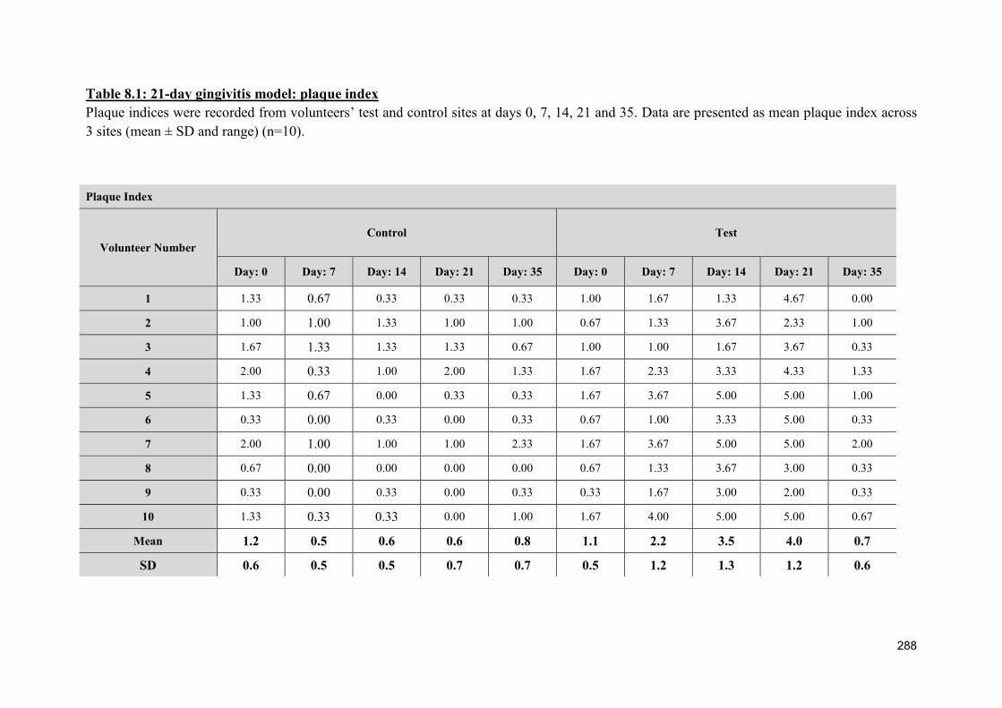
288
Table 8.1: 21-day gingivitis model: plaque index
Plaque indices were recorded from volunteers’ test and control sites at days 0, 7, 14, 21 and 35. Data are presented as mean plaque index across
3 sites (mean ± SD and range) (n=10).
Plaque Index
Volunteer Number
Control Test
Day: 0 Day: 7 Day: 14 Day: 21 Day: 35 Day: 0 Day: 7 Day: 14 Day: 21 Day: 35
1 1.33 0.67 0.33 0.33 0.33 1.00 1.67 1.33 4.67 0.00
2 1.00 1.00 1.33 1.00 1.00 0.67 1.33 3.67 2.33 1.00
3 1.67 1.33 1.33 1.33 0.67 1.00 1.00 1.67 3.67 0.33
4 2.00 0.33 1.00 2.00 1.33 1.67 2.33 3.33 4.33 1.33
5 1.33 0.67 0.00 0.33 0.33 1.67 3.67 5.00 5.00 1.00
6 0.33 0.00 0.33 0.00 0.33 0.67 1.00 3.33 5.00 0.33
7 2.00 1.00 1.00 1.00 2.33 1.67 3.67 5.00 5.00 2.00
8 0.67 0.00 0.00 0.00 0.00 0.67 1.33 3.67 3.00 0.33
9 0.33 0.00 0.33 0.00 0.33 0.33 1.67 3.00 2.00 0.33
10 1.33 0.33 0.33 0.00 1.00 1.67 4.00 5.00 5.00 0.67
Mean 1.2 0.5 0.6 0.6 0.8 1.1 2.2 3.5 4.0 0.7
SD 0.6 0.5 0.5 0.7 0.7 0.5 1.2 1.3 1.2 0.6

289
Table 8.2: 21-day gingivitis model: gingival index
Gingival index measurements were recorded from volunteers’ test and control sites at days 0, 7, 14, 21 and 35. Data presented as mean gingival
index across 3 sites (mean ± SD and range) (n=10).
Gingival Index
Volunteer Number
Control Test
Day: 0 Day: 7 Day: 14 Day: 21 Day: 35 Day: 0 Day: 7 Day: 14 Day: 21 Day: 35
1 0.00 0.00 0.33 0.33 0.33 1.67 0.33 1.00 0.33 0.33
2 0.00 0.33 0.33 0.00 0.67 2.00 1.67 1.33 1.33 0.00
3 0.00 0.00 0.00 0.67 0.00 0.67 0.33 0.33 0.33 0.00
4 0.00 0.00 0.33 0.00 0.00 0.67 1.00 1.33 1.33 0.67
5 0.33 0.67 1.33 0.67 0.00 0.33 1.00 1.67 1.67 0.00
6 0.67 0.00 0.00 1.00 0.33 0.67 2.33 2.00 1.67 0.33
7 0.67 0.00 0.33 0.00 0.00 1.00 1.00 2.33 2.00 1.33
8 0.33 0.33 0.00 0.67 0.33 1.00 0.67 1.00 1.00 0.00
9 0.33 0.00 0.00 0.00 0.00 0.00 0.67 0.33 0.33 0.00
10 0.33 0.00 0.00 0.00 0.00 0.67 1.00 1.00 1.33 0.67
Mean 0.3 0.1 0.3 0.3 0.2 0.9 1.0 1.2 1.1 0.3
SD 0.3 0.2 0.4 0.4 0.2 0.6 0.6 0.6 0.6 0.4

290
Table 8.3: 21-day gingivitis model: gingival crevicular fluid volume
Gingival crevicular fluid (GCF) volumes were recorded from volunteers’ test and control sites at days 0, 7, 14, 21 and 35. Data presented as
mean volumes (µl) across 3 sites (mean ± SD and range) (n=10).
Gingival crevicular fluid calibrated volumes (µl)
Volunteer Number
Control Test
Day: 0 Day: 7 Day: 14 Day: 21 Day: 35 Day: 0 Day: 7 Day: 14 Day: 21 Day: 35
1 0.06 0.17 0.11 0.08 0.08 0.12 0.21 0.18 0.36 0.14
2 0.28 0.11 0.14 0.22 0.11 0.51 0.63 0.34 0.53 0.59
3 0.05 0.03 0.06 0.39 0.06 0.06 0.23 0.23 0.79 0.52
4 0.13 0.40 0.50 0.11 0.20 0.08 0.59 1.11 2.50 1.16
5 0.07 0.32 0.25 0.49 1.38 0.15 1.08 0.56 0.39 2.64
6 0.04 0.17 0.08 0.05 0.14 0.09 0.13 0.27 0.14 0.20
7 0.54 1.12 0.33 1.29 0.24 0.68 3.75 5.12 4.92 0.33
8 0.15 0.05 0.05 0.05 0.12 0.16 0.25 0.17 0.32 0.30
9 0.06 0.15 0.07 0.07 0.10 0.07 0.14 0.08 0.09 0.06
10 0.09 0.06 0.08 0.05 0.09 0.17 0.47 0.79 0.44 0.18
Mean 0.1 0.3 0.2 0.3 0.3 0.2 0.7 0.9 1.0 0.6
SD 0.2 0.3 0.1 0.4 0.4 0.2 1.1 1.5 1.5 0.8

291
NET production during the 21-day experimental gingivitis model 8.2.2
Blood was collected from study volunteers at days 0, 21 and 35 to assay NET-DNA
production in response to PMA (50nM) and HOCl (0.75mM) (2.2.3.3). Baseline NET
production in response to PBS (negative unstimulated control) was also quantified. There
was no significant difference in NET production between each time-point in any treatment
condition (1way ANOVA p=0.1621, p=0.1092, p=0.0546 in response to PBS (unstimulated),
PMA or HOCl, respectively, n=10) (Figure 8.1).
Visualisation of NET production during the 21-day experimental gingivitis model 8.2.3
There was no qualitative difference observed in NET production at days 0, 21 and 35
(2.2.3.7). Both PMA (50nM) and HOCl (0.75mM) produced NETs, and some PBS-treated
neutrophils also stained positive for DNA, which is likely due to necrosis during the
incubation period, which enables Sytox green to permeate the compromised cell membrane
(Figure 8.2).
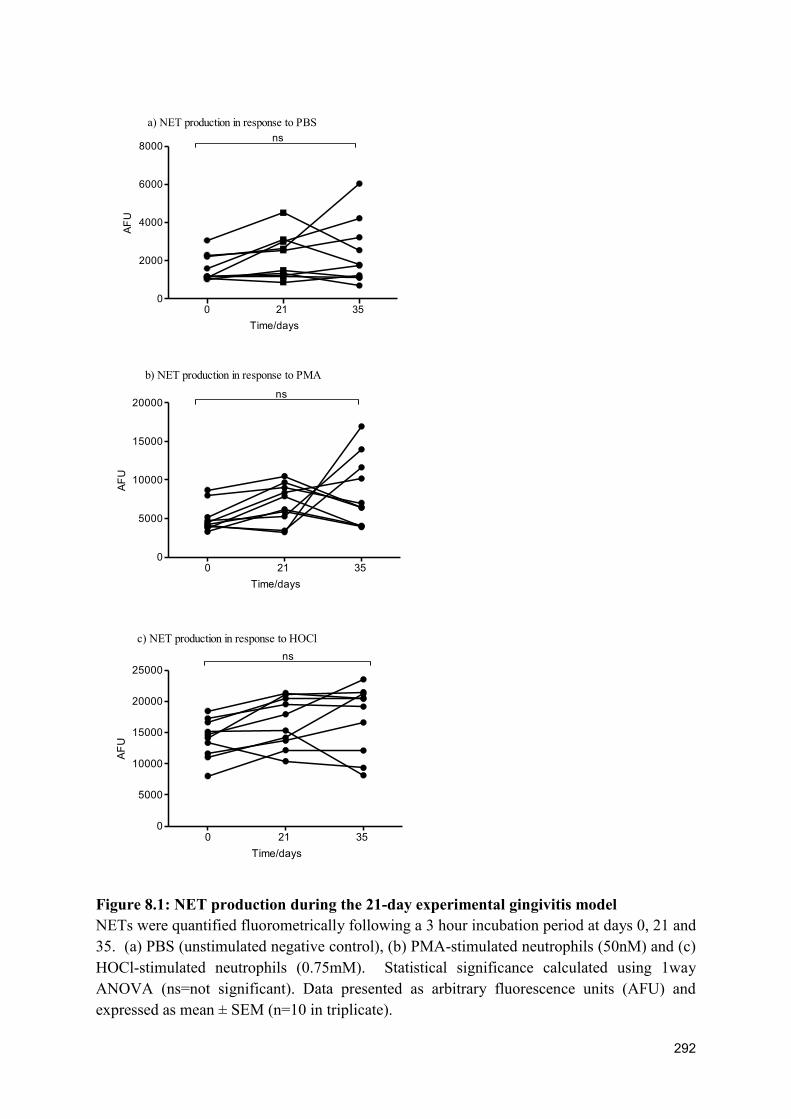
292
AF
U
0 21 35 0
2000
4000
6000
8000ns
a) NET production in response to PBS
Time/days
AF
U
0 21 35 0
5000
10000
15000
20000ns
b) NET production in response to PMA
Time/days
AF
U
0 21 35 0
5000
10000
15000
20000
25000
ns
c) NET production in response to HOCl
Time/days
Figure 8.1: NET production during the 21-day experimental gingivitis model
NETs were quantified fluorometrically following a 3 hour incubation period at days 0, 21 and
35. (a) PBS (unstimulated negative control), (b) PMA-stimulated neutrophils (50nM) and (c)
HOCl-stimulated neutrophils (0.75mM). Statistical significance calculated using 1way
ANOVA (ns=not significant). Data presented as arbitrary fluorescence units (AFU) and
expressed as mean ± SEM (n=10 in triplicate).

293
Figure 8.2: Visualisation of NET production during the 21-day experimental gingivitis model
NETs were visualised by fluorescence microscopy (x20 magnification) with Sytox green staining in response to PBS (negative unstimulated
control), PMA (50nM) and HOCl (0.75mM) at days 0, 21 and 35. Images are representative of 10 volunteers in duplicate. Scale bar is 100µm.
PBS PMA HOCl
0 days
21 days
35 days

294
NET degradation by volunteer plasma during the 21-day experimental gingivitis 8.2.4
model
To investigate the effect of gingivitis upon NET degradation by plasma, neutrophils were
stimulated for NET production with HOCl (0.75mM) and NETs were subsequently subjected
to 3 hours incubation with 10% plasma (2.2.5.1). NETs from periodontally and systemically
healthy volunteers were incubated with plasma derived from experimental gingivitis
volunteers at 0, 21 and 35 days. Following incubation, the number of degraded NETs was
quantified fluorometrically using the Sytox green assay. The percentage of NETs degraded
was calculated in relation to a 15 min MNase digestion, which was used to represent the
100% digestion standard (Hakkim et al., 2010). In the 10 volunteers analysed there was no
significant difference in NET degradation between the different time-points (1way ANOVA
p=0.3828, n=10). It was also noted that the lowest NET-degrading plasma (27%, 47% and
26% at time-points 0, 21 and 35 days respectively) were from the same individual (Figure
8.3).

295
Time/days
% N
ET
de
gra
da
tio
n
0 21 350
50
100
150ns
Figure 8.3: NET degradation by volunteer plasma during the 21-day experimental
gingivitis model
HOCl-stimulated (0.75mM) neutrophils were incubated with 10% plasma from volunteers at
days 0, 21 and 35. NETs were quantified fluorometrically using the Sytox green assay. %
NET degradation was calculated based on a 1Unit/ml MNase digest, which was used to
represent the 100% standard. Statistical significance calculated using 1way ANOVA (ns=not
significant). Data presented as % NET degradation and expressed as mean ± SEM (n=10 in
triplicate).

296
Whilst NET degradation appeared unimpeded in experimental gingivitis relative to health,
chronic periodontitis patients exhibited reduced NET degradation by plasma, which may be
the result of increased levels of IgGs and free light chains (FLCs) (Chapter 6). To determine
whether volunteers with experimental gingivitis also had elevated levels of circulating IgGs
and FLCs, IgG subclass 1-4 and FLC concentrations were measured in plasma collected at
days 0, 21 and 35 (2.2.5.2, Appendix V).
8.2.4.1 Plasma IgG concentrations during the 21-day experimental gingivitis model
IgG subclasses 1-4 concentrations were measured in plasma samples from study volunteers at
days 0, 21 and 35. Protein turbidimetric analysis demonstrated no significant differences
between the detection of IgG1, IgG2, IgG3 or IgG4 at the different time-points analysed (1
way ANOVA p=0.903, p=0.996, p=0.924, p=0.952 for IgG1, IgG2, IgG3 and IgG4,
respectively, n=10). A small number of samples were outside of the IgG reference ranges
(IgG1: 3.824-9.286g/L, IgG2: 2.418-7.003g/L, IgG3: 0.218-1.761g/L, IgG4 0.039-0.864)
(Schauer et al., 2003), however, this was not associated with specific time-points in the
gingivitis model (Figure 8.4).

297
Time/days
g/litre
0 21 350
2
4
6
8 ns
a) IgG1
Time/days
g/litre
0 21 350
1
2
3
4
5 ns
b) IgG2
Time/days
g/litre
0 21 350.0
0.5
1.0
1.5 ns
c) IgG3
Time/days
g/litre
0 21 350.0
0.2
0.4
0.6 ns
d) IgG4
Figure 8.4: Plasma IgG concentrations during the 21-day experimental gingivitis model
IgG subclasses 1-4 concentrations were measured in plasma samples from study volunteers at
days 0, 21 and 35. Protein turbidimetric analysis demonstrated no significant difference
between the detection of IgG1, IgG2, IgG3 or IgG4 at the different time points (1 way
ANOVA, ns=not significant). Data expressed as means ± SEM (n=10).

298
8.2.4.2 Plasma FLC concentration during the 21-day experimental gingivitis model
Free kappa and lambda light chains were measured in plasma samples from study volunteers
at days 0, 21 and 35. Protein turbidimetric analysis demonstrated no significant differences
between the detection of free kappa light chain, free lambda light chains, the kappa/lambda
ratio or summated FLC values (1 way ANOVA p=0.972, p=0.741, p=0.969, p=0.833 for
free kappa light chain, free lambda light chains, kappa/lambda ratio or summated FLC values,
respectively, n=10). A small number of samples were outside of the reference ranges (free
kappa light chains: 3.3-19.4mg/litre, free lambda light chains: 5.71-26.3mg/litre,
kappa/lambda ratio: 0.26-1.65, summated FLC: 9.01-45.7) (Katzmann et al., 2002), however,
this was not associated with specific time-points in the gingivitis model (Figure 8.5).

299
Time/days
mg
/litre
0 21 350
5
10
15 ns
a) Kappa
Time/days
mg
/litre
0 21 350
5
10
15
20 ns
b) Lambda
Time/days
0 21 350.0
0.5
1.0
1.5
Ka
pp
a/L
am
bd
a r
atio
ns
c) Kappa/Lambda ratio
Time/days
Su
mm
ate
d
0 21 350
10
20
30
40 ns
d) Summated FLC
Figure 8.5: Plasma FLC concentration during the 21-day experimental gingivitis model
Free kappa and free lambda light chains, the kappa/lambda ratio and summated FLCs were
measured in plasma samples from study volunteers at days 0, 21 and 35. Protein
turbidimetric analysis demonstrated no significant difference between the detection of free
kappa light chain, free lambda light chains, the kappa/lambda ratio or summated FLC values
(1 way ANOVA, ns=not significant). Data expressed as means ± SEM (n-10).

300
The ability of a plaque biofilm to stimulate neutrophil responses
Increasing concentrations of plaque were employed as a stimulus for ROS and NETs in
neutrophils derived from a healthy volunteer to investigate whether plaque activates
neutrophil effector functions (2.2.6.2.4). Neutrophil NET and ROS production were
quantified in response to stimulation with plaque at increasing percentage concentrations (0,
12.5, 25, 50 75 and 100%), compared with PMA (50nM) stimulation (Figure 8.6). There was
a gradual increase in NET and ROS production in response to neutrophil stimulation by
plaque (n=2). Based on these results, subsequent experiments assaying NET and ROS
production in response to plaque derived from the experimental gingivitis model were diluted
in PBS and used at 25%.

301
Plaque concentration
RL
U
PMA 0 12.5% 25% 50% 75% 100%0
40000
80000
120000
a) Total ROS production in response to plaque
Plaque concentration
AF
U
PMA 0 12.5% 25% 50% 75% 100%0
5000
10000
15000
b) NET production in response to plaque
Figure 8.6: The ability of a plaque biofilm to stimulate neutrophil responses
The effect of increasing concentrations of plaque (0, 12.5, 25, 50, 75 and 100%) and PMA
(50nM) on neutrophils (a) ROS and (b) NET production. Data expressed as mean ± SEM.
Data is presented as (a) RLU (relative light units) and (b) AFU (arbitrary fluorescence units)
(n=2 in triplicate).
302
Neutrophil ROS and NET production in response to plaque stimulation 8.2.5
Plaque was collected from test and control sites from 10 volunteers at days 0, 7, 14, 21 and
35. Plaque samples (n=10) were homogenised, pooled and diluted (to 25% of the original
concentration) for subsequent assays in which plaque was employed as a stimulus for NET
and ROS production in healthy volunteers (n=5) (2.2.6.2.4). To characterise the plaque
samples collected, the amount of protein (2.2.6.2.5.3), LPS (2.2.6.2.5.1) and DNA
(2.2.6.2.5.2) in the pooled plaque samples were measured to enable the ROS and NET
quantification results to be normalised for the plaque content.
ROS production normalised for protein content
Plaque collected from test sites at 7, 14 and 21 days induced significantly higher ROS
production after normalising for plaque protein content (2way ANOVA and Bonferroni post-
tests **p<0.01, ****p<0.0001 and **p=0.01 at days 7, 14 and 21, respectively, n=10 samples
and n=5 technical repeats) (Figure 8.7a)
ROS production normalised for LPS content
Plaque collected from test sites at 7, 14 and 21 days induced significantly higher ROS
production after normalising for plaque LPS content (2way ANOVA and Bonferroni post-
tests *p<0.05, ****p<0.0001, **p<0.01 at days 7, 14 and 21, respectively, n=10 samples and
n=5 technical repeats) (Figure 8.7b)
ROS production normalised for DNA content
Plaque collected from test sites at 7, 14 and 21 days induced significantly higher ROS
production after normalising for plaque DNA content (2way ANOVA and Bonferroni post-
tests ****p<0.0001, ****p<0.0001, *p<0.05 at days 7, 14 and 21, respectively, n=10 samples
and n=5 technical repeats) (Figure 8.7c).

303
Time/days
RO
S p
rod
uctio
n/r
atio
of
pro
tein
pe
r m
l
0 7 14 21 350
500
1000
1500
2000
2500
Control
Test
****
**
**
a) ROS results normalised for plaque proetin content
Time/days
RO
S p
rod
uctio
n/r
atio
of
E.c
oli
pe
r m
l
0 7 14 21 350
2000
4000
6000
8000 Control
Test
*
****
**
b) ROS results normalised for plaque LPS content
c) ROS results normalised for plaque DNA content
Time/days
RO
S p
rod
uctio
n/r
atio
of
DN
A p
er
ml
0 7 14 21 350
20000
40000
60000
80000Control
Test****
****
*
Figure 8.7: ROS production in response to plaque stimulation
ROS production was quantified in response to plaque stimulation (25%) collected at 0, 7, 14,
21 and 35 days from test and control sites and employed as a stimulus on healthy volunteer
neutrophils. ROS production was normalised for (a) plaque protein content, (b) plaque LPS
content, (c) plaque DNA content. Statistical significance was calculated using 2way
ANOVA and Bonferroni post-tests (*p<0.05, ****p<0.0001). Data presented as RLU
(relative light units) and expressed as mean ± SEM (n=10 samples and n=5 technical repeats).

304
NET production normalised for protein content
Plaque collected from test sites at 7, 14 and 21 days induced significantly higher NET
production after normalising for plaque protein content (2way ANOVA and Bonferroni post-
tests p<0.01, p<0.0001, p=0.01 for days 7, 14 and 21, respectively, n=10 samples and n=5
technical repeats) (Figure 8.8a).
NET production normalised for LPS content
Plaque collected from test sites at 7, 14 and 21 days induced significantly higher NET
production after normalising for plaque LPS content (2way ANOVA and Bonferroni post-
tests p<0.01, p<0.0001, p<0.01 at days 7, 14 and 21, respectively, n=10 samples and n=5
technical repeats) (Figure 8.8b).
NET production normalised for DNA content
Plaque collected from test sites at 7 and 14 days induced significantly higher NET production
after normalising for plaque DNA content (2way ANOVA and Bonferroni post-tests
p<0.0001for days 7 and 14, n=10 samples and n=5 technical repeats) (Figure 8.8c).

305
Time/days
NE
t p
rod
uctio
n/r
atio
of
pro
tein
pe
r m
l
0 7 14 21 350
500
1000
1500
2000
2500 Control
Test
**
**
****
a) NET production normalised for plaque protein content
b) NET production normalised for plaque LPS content
Time/days
NE
t p
rod
uctio
n/r
atio
of E
. co
li p
er
ml
0 7 14 21 350
2000
4000
6000
8000 Control
Test
*
****
**
c) NETs production normalised for plaque DNA content
Time/days
NE
t p
rod
uctio
n/r
atio
of
DN
A
0 7 14 21 350
5000
10000
15000
20000
25000 Control
Test***
****
Figure 8.8: NET production in response to plaque stimulation
NETs were quantified in response to plaque stimulation (25%) collected at 0, 7, 14, 21 and 35
days from test and control sites and employed as a stimulus on healthy volunteer neutrophils.
NET production was normalised for (a) plaque protein content, (b) plaque LPS content, (c)
plaque DNA content. Statistical significance was calculated using 2way ANOVA and
Bonferroni post-tests (*p<0.05, **p<0.01, ****p<0.0001). Data presented as AFU (arbitrary
fluorescence units) and expressed as mean ± SEM (n=10 samples and n=5 technical repeats).

306
8.3 Papillon Lefèvre syndrome
The release of NETs represents a multi-stage process that is reliant on the neutrophil serine
protease (NSP), neutrophil elastase (NE) (Papayannopoulos et al., 2010). NE is activated by
cathepsin C, which is the deficient cysteine protease in PLS patients. Therefore, PLS patients
provide an interesting clinical model for the study of NET biology in relation to periodontitis.
NET responses in PLS patients with severe pre-pubertal periodontitis were investigated
alongside healthy gender-matched controls (2.2.6.4). All PLS patients were genotyped,
assessed for co-morbidities and their dermatological and periodontal health determined
(2.2.6.4.1). Neutrophils were isolated from patients and controls to investigate what effect
cathepsin C deficiency in PLS had upon peripheral NET production. NETs were quantified
by the release of NET-DNA (Sytox green) and also NET-bound proteases (neutrophil elastase
[NE], myeloperoxidase [MPO] and cathepsin G [CG]) (2.2.3.3, 2.2.3.4). Plasma samples
were also collected from patients and controls and subsequent experiments analysed plasma
samples for the expression of NE and LL-37 (2.2.6.4.2). Whilst the production of NET-DNA
was quantified in all patients, due to low patient blood neutrophil counts, subsequent NET
assays did not include patient 1 or 2.

307
Table 8.4: Clinical measures of PLS
PLS patient volunteer information, including age, sex, ethnicity, PLS type, mutation, oral health, dermatological health, co-morbidities and
current medication. Abbreviations: BMI, (body mass index), PPK (palmar plantar hyperkeratosis)
Volunteer
no. Age Sex Ethnicity BMI PLS type Mutation Oral health
Dermatological
health Co-morbidities Medication
1 12 Male Caucasian 18.93 Heterozygous 1/2 G139R; 1/2
N427T
No significant
pocketing No skin problems None recorded None recorded
2 14 Female Indian 19.90 Heterozygous Unidentified
Tooth pain &
discomfort, teeth
very mobile,
bleeding on
brushing.
Moderate PPK
β-thalassemia
carrier (from
mother)
None recorded
3 14 Female Pakistani 20.04 Homozygous 2/2 R272P Dentures
PPK - quite bad
on feet, deep
fissures; hands ok
None recorded
Acitretin once
daily. Vitamin
D replacement
therapy
4 9 Male Pakistani 22.83 Not done 2/2 R272P Teeth OK and good
oral hygiene.
Lips prone to
cracking
Vitamin D
deficiency.
Previous cerebral
intracranial
abscesses.
Acitretin once
daily
5 14 Male Pakistani 18.15 Homozygous 2/2 R272P
Dentures with pain
on replacing and
denture stomatitis.
Mild PPK - hands
very good
History of recurrent
infections since
birth.
Acitretin once
daily

308
Comparison of NET production by PLS patients and healthy controls 8.3.1
Venous blood was collected from PLS patients and controls to quantify their NET-DNA
production in response to a 4 hour stimulation with PMA (50nM), HOCl (0.75mM),
opsonised S. aureus (5x10⁷
and MOI of 500), S. gordonii (1x10⁸
and MOI of 1000) or F.
nucleatum poly (1x10⁸
and MOI of 1000) (2.2.3.3). Baseline NET production in response to
PBS (unstimulated negative control) was also quantified. There was no significant difference
in NET-DNA production between patients and controls in response to PBS (2way ANOVA
and Bonferroni post-tests p>0.05). Systemically and periodontally healthy controls exhibited
higher NET release in response to stimulation, which was statistically significant for PMA,
HOCl and F. nucleatum poly (2way ANOVA and Bonferroni post-tests p<0.0001, p<0.0001,
p<0.001, respectively) (Figure 8.9a).
8.3.1.1 Quantification of NET-bound components in PLS patients and healthy controls
In addition to quantifying NET-DNA, NET release from PLS patients and controls was
determined by quantifying NET-bound proteins (2.2.3.4). NE, MPO and CG were
colorimetrically quantified in response to 4 hours stimulation with PMA (50nM), HOCl
(0.75mM), A. actinomycetemcomitans (serotype b) or V. parvula (all bacteria at 1x10⁸
and
MOI of 1000) and PBS (unstimulated negative control).
Quantification of NET-bound neutrophil elastase
Healthy controls produced significantly more NETs, as determined by NET-bound neutrophil
elastase (NE), compared with PLS patients in response to PMA (50nM), HOCl (0.75mM), A.
actinomycetemcomitans (serotype b) and V. parvula (all bacteria at 1x10⁸
and MOI of 1000)
(2way ANOVA and Bonferroni post-tests ****p<0.0001, ****p<0.0001, ***p<0.001,
****p<0.0001, respectively). There was no difference between control and patient NET

309
production in response to PBS (unstimulated negative control) (2way ANOVA and
Bonferroni post-tests p>0.05) (Figure 8.9b).
Quantification of NET-bound myeloperoxidase
Healthy controls produced significantly more NETs, as determined by NET-bound
myeloperoxidase (MPO), compared with PLS patients in response to PMA (50nM), HOCl
(0.75mM), A. actinomycetemcomitans (serotype b) and V. parvula (all bacteria at 1x10⁸
and
MOI of 1000) (2way ANOVA and Bonferroni post-tests *p<0.05, ***p<0.001, *p<0.05,
*p<0.05, respectively). There was no difference between control and patient NET production
in response to PBS (unstimulated negative control) (2way ANOVA and Bonferroni post-tests
p>0.05) (Figure 8.9c).
Quantification of NET-bound cathepsin G
Healthy controls produced significantly more NETs, as determined by NET-bound cathepsin
G (CG), compared with PLS patients following stimulation with HOCl, A.
actinomycetemcomitans (serotype b) or V. parvula (2way ANOVA and Bonferroni post-tests
****p<0.0001, ***p<0.001, ****p<0.0001, respectively). There was no difference between
control and patients NET production in response to PMA or PBS (unstimulated negative
control) (Figure 8.9d).

310
a) NET-DNA A
FU
PBS
PM
A
HOCl
Ops
onised
S.a
ureu
s
S. g
ordo
nii
F. nuc
leat
um p
oly
0
5000
10000
15000
20000Control
Patient
****
***
****
b) NET-bound neutrophil elastase
NE
mU
/ml
PBS
PM
A
HOCl
A. a
ctinom
yces
b
V. p
arvu
la
0
10
20
30
40
50Control
Patient****
****
*******

311
c) NET-bound myeloperoxidase M
PO
U/m
l
PBS
PM
A
HOCl
A. a
ctinom
yces
b
V. p
arvu
la
0.0
0.2
0.4
0.6
0.8
1.0
1.2Control
Patient***
*
* *
d) NET-bound cathepsin G
Ca
the
psin
G m
U/m
l
PBS
PM
A
HOCl
A. a
ctinom
yces
b
V. p
arvu
la
0
2
4
6
8 Control
Patient
****
***
****
Figure 8.9: Comparison of NET production by PLS patients and healthy controls
NET bound (a) NET-DNA (n=5 patients, n=4 controls) (b) NET-bound NE, (c) NET-bound
MPO and (d) NET-bound CG were quantified in PLS patients and healthy controls
fluorometrically in response to 4 hours of stimulation with PMA (50nM), HOCl (0.75mM),
opsonised S. aureus (5x10⁷
and MOI of 500), or heat-killed bacteria (1x10⁸
and MOI of
1000). Statistical significance was calculated using 2way ANOVA and Bonferroni post-tests
(*p<0.05, ***p<0.001, ****p<0.0001). Data is presented as (a) AFU (arbitrary fluorescence
units), (c) U/ml or (d, e) mU/ml and expressed as mean ± SEM (quantification of NET-bound
components n=3 pairs).

312
Fluorescence visualisation of NET production in PLS patients 8.3.2
A qualitative difference was observed in NET production between PLS patients and healthy
controls, with patients producing fewer PMA-induced (50nM) extracellular web-like
structures that stained positive with the DNA-stain, Sytox green (2.2.3.7). No difference in
NET production was observed between patients and controls in PBS-treated neutrophils,
however some cells stained positive for DNA, which is likely to be cells that have necrosed
during the incubation period and enabled Sytox green to permeate the compromised cell
membrane (Figure 8.10).

313
Figure 8.10: Fluorescence visualisation of NET production in PLS patients
NETs were visualised (x10 magnification) with Sytox green in response to treatment with
PBS (negative control) or PMA (50nM) for 3 hours. Images are representative of 5
experiments performed in duplicate. Scale bar represents 100µm.
Control PLS
PBS
PMA

314
Quantification of plasma NE in PLS 8.3.3
Plasma was isolated from the peripheral blood of PLS patients and controls (2.2.6.2.3).
Plasma-derived NE was quantified by ELISA and demonstrated significantly less NE in PLS
patients (Mann-Whitney *p=0.0294) (2.2.6.4.2.1). A NE standard curve enabled absorbance
readings to be converted into ng/ml of NE (Figure 8.11a).
Quantification of plasma LL-37 in PLS 8.3.4
Plasma was isolated from the peripheral blood of PLS patients and controls (2.2.6.2.3).
Plasma-derived LL-37 was quantified by ELISA and demonstrated significantly less LL-37
in PLS patients (Mann-Whitney *p=0.0297) (2.2.6.4.2.2). An LL-37 standard curve enabled
absorbance readings to be converted into ng/ml of LL-37 (Figure 8.11b).

315
a) Neutrophil elastase N
E n
g/m
l
Patient Control0.0
0.5
1.0
1.5 *
b) LL-37
LL
-37
ng
/ml
Patient Control0
20
40
60
80 *
Figure 8.11: Quantification of plasma derived neutrophil elastase and LL-37 in PLS
Plasma derived (a) NE and (b) LL-37 were quantified in plasma from PLS patients and
healthy controls by ELISA. Statistical significance was calculated using Mann-Whitney tests
*p=0.0294, *p=0.0297 for NE and LL-37, respectively). Data presented as ng/ml and
expressed as mean ± SEM (n=4 pairs).

316
8.4 Discussion
Gingivitis is an inflammatory response to bacterial plaque that precedes periodontitis in
susceptible individuals (Hajishengallis 2014). Gingivitis, whilst normally chronic in nature,
can be rapidly induced and reversed, and therefore the 21-day gingivitis model developed by
Loe et al., (1965) constitutes a useful model to investigate the role of a supragingival plaque
biofilm in the pathogenesis of periodontal disease and systemic diseases with periodontal
manifestations. In the current study, no significant differences were observed in peripheral
blood NET production during the 21-day gingivitis model (section 8.2.2). This is not
unexpected, as peripheral NET release by chronic periodontitis patients was no different to
healthy controls (Chapter 5). Furthermore, there was no significant difference in plasma-
derived NET degradation or plasma levels of IgG and FLCs throughout the gingivitis study
period. Clinical measures of inflammation increased up to 21 days (plaque accumulation) on
the test side, followed by a decrease at 35 days (return to health), which confirms volunteer
compliance and the validity of the study for both the induction and resolution of
inflammation (section 8.2.1). Consistent with our findings, Wahaidi et al., (2009)
demonstrated no systemic oxidative response to the accumulation of plaque in a 21-day
gingivitis model. Conversely, it has been reported that plaque accumulation during the 21-
day experimental gingivitis model results in significant increases in peripheral blood
neutrophil counts (Kowolik et al., 2001).
Plaque was collected from test and control sites at 0, 7, 14, 21 and 35 days and pooled.
Plaque was used as a stimulus for neutrophil ROS and NET production, and results
normalised to plaque DNA, LPS and protein content. Neutrophil ROS and NET production
increased in response to homogenised plaque collected from test sites at 7, 14 and 21 days
(plaque accumulation), and there was no difference at 0 (baseline) or 35 days (return to
health) (section 8.2.5). Given that all results were normalised to the plaque content (DNA,

317
LPS, protein content), this data suggests that increased neutrophil responses may be due to
changes in plaque composition, rather than the quantify. However, microbial characterisation
of the biofilm was not performed as part of this study, however this would be interesting to
explore. Indeed, a study by Eberhard et al., (2013) which identified bacterial species in
plaque produced from a 21-day experimental gingivitis model included species such as P.
gingivalis, F. nucleatum and V. parvula, and notably, experiments in Chapter 4 revealed that
these bacterial species were capable of stimulating healthy neutrophils to produce ROS and
NETs. Consistent with this, Hirschfeld et al., (2015) demonstrated that the 21-day plaque
biofilm comprises neutrophils and NET structures. The authors hypothesised that neutrophils
are recruited to supragingival plaque, where they exert bactericidal activity, such as
phagocytosis, degranulation and NET release. However, as discussed previously (Chapter 4),
further work is needed to elucidate whether NETs prevent bacterial dissemination, or whether
the infiltration of neutrophils and the release of NETs in supragingival plaque actually
promotes biofilm stability and growth (Hirschfeld et al., 2015).
The study population may be a limitation of the current study, as whilst there were both male
and female volunteers, all volunteers were aged between 19 and 21. Furthermore it has been
suggested that the 21-day model of experimental gingivitis may not fully reflect persistent
chronic gingivitis, as a study by Loesche & Grossman (2001) comparing experimental and
chronic gingivitis revealed that volunteers enrolled in the 21-day model demonstrated higher
levels of plaque accumulation. This discordance is believed to be a limitation of the
experimental model, for example, individuals with persistent gingivitis do not neglect all
forms of oral hygiene (as is the case with the vinyl mouth guard), and gingivitis is a chronic
state that persists for longer than 21 days.

318
PLS is an autosomal recessive disease causes by a mutation in the CTSC gene that encodes
the lysosomal cysteine protease, cathepsin C. Cathepsin C plays a vital role in the post-
translational processing of neutrophil serine proteases (NSPs), where it removes N-terminal
amino acids that block the active site of NSPs. Thus, a lack of cathepsin C results in the
production of NSPs that do not exhibit enzymatic activity. NSPs, such as NE, CG and PR3,
are synthesised as inactive pro-enzymes in the bone marrow and prior to storage in neutrophil
azurophilic granules as zymogens, they undergo cathepsin-C mediated processing. In
response to neutrophil activation, these lysosomal proteins are released into the
phagolysosome or undergo limited exocytosis to facilitate bacterial clearance. The loss of
cathepsin C activity in PLS patients results in the cessation of NSP function and stability,
which is associated with severe periodontal destruction (Sorensen et al., 2014).
This study demonstrated impeded NET release in PLS patients (NET-DNA and NET-bound
components) (sections 8.3.1, 8.3.1.1), which is consistent with recent findings by Sorensen et
al., (2014), who demonstrated abrogated NET release in a single adult female PLS patient.
It is postulated that the reduced NET production is a result of dysfunctional NE in PLS
patients (Sorensen et al., 2014), as Papayannopoulos et al., (2010) demonstrated that NE is
necessary for NET release. Following neutrophil activation, NE is reportedly released from
azurophilic granules and translocates to the nucleus where it promotes chromatin
decondensation prior to NET expulsion (Papayannopoulos et al., 2010). The importance of
NE in NET production is also exemplified in ELA2-neutropenia, in which deficient NE
associates with diminished NET production; however this is not observed when NE is fully
functional in HAX1-neutropenia (Happle et al., 2011). In the absence of post-translational
processing in PLS patients, NSP proforms have been suggested to either be constitutively
released following their synthesis in the bone marrow (Garwicz et al., 2005), or degraded
(Pham & Lay 1999). Indeed Sorensen et al., (2014) demonstrated that NSP production and

319
allocation to granules appeared normal in PLS immature neutrophils, however the proteins
were absent from mature neutrophils; suggesting NSPs are degraded prior to their release
from the bone marrow. Accordingly, in the current study NE levels were significantly lower
in patient-derived plasma samples, relative to the controls (section 8.3.3).
In healthy individuals PR3 cleaves cathelicidin (hCAP-18) extracellularly to produce the
bactericidal peptide LL-37. In PLS patients, deficient LL-37 results from a failure to encode
fully functioning cathepsin C, and consequently compromised PR3 activity. Confirming this,
LL-37 was significantly lower in PLS plasma samples relative to healthy controls (section
8.3.4). This is consistent with the findings of de Haar et al., (2006), who reported that in
addition to LL-37 deficiency, neutrophils derived from PLS patients demonstrate a reduced
capacity to kill A. actinomycetemcomitans compared with healthy controls. This may,
however, be a direct result of inhibited LL-37 antimicrobial activity, as LL-37 is reportedly
involved in the clearance of A. actinomycetemcomitans (Ji et al., 2007). Notably patients
with Morbus Kostmann syndrome, a severe congenital neutropenia that is associated with
LL-37 deficiency, are also susceptible to a high prevalence of A. actinomycetemcomitans
infection and severe periodontitis in adulthood (Putsep et al., 2002, Gonzales & Frydman
2014); supporting the hypothesis that LL-37 plays a vital role in bacterial clearance. It has
also been suggested that the overgrowth of A. actinomycetemcomitans in PLS patients may
provide favourable conditions for the proliferation of other periodontal bacteria, such as P.
gingivalis (Eick et al., 2014).
It is noteworthy that despite the loss of function of NSPs, the associated immunodeficiency is
modest, with 15-20% of patients reporting recurrent infections, such as tonsillitis and liver
abscesses (Sorensen et al., 2014, Almuneef et al., 2002). Accordingly, the physical
examination of one patient revealed that IgGs, complement, as well as T- and B-cell

320
responses were within the normal reference ranges (Sorensen et al., 2014). The fact that
patients do not succumb to major illnesses, and also reports from in vitro studies
demonstrating that PLS neutrophils elicit normal bactericidal activity against infection (Pham
et al., 2004), suggests that NSPs do not constitute the only antimicrobial defence mechanism.
This also indicates that NE-dependent NETs may not play a critical role in host defence, or
that alternative compensatory defence mechanisms have evolved in PLS. It is also unlikely
that deficient NET release accounts for the severe periodontitis observed in PLS, as CGD
patients, who also lack the ability to produce NETs, do not present with severe periodontal
destruction (Bianchi et al., 2009). Thus, severe periodontitis may be the result of other
neutrophil abnormalities, such as perturbed chemotaxis, which is reportedly defective in PLS
patients. However, normal neutrophil levels are reported in the GCF of PLS patients,
suggesting that whilst the neutrophils are recruited normally, they may exhibit longer tissue
transit times and contribute to collateral periodontal tissue destruction (Firatli et al., 1996). A
possible explanation for the mild systemic immunodeficiency observed in PLS are recent
findings by Glenthoj et al., (2015), who demonstrated that NSPs are not required for the
processing and activity of neutrophil α-defensins, which are a family of peptides that possess
antimicrobial activity. Notably, PLS patients are able to synthesise α-defensins, which
constitute 50% of azurophil granules (Rice et al., 1987), and it has been proposed that the
presence of these microbicidal peptides may afford protection and compensate for absent
NSPs.
In conclusion, peripheral NET production did not change throughout the 21-day gingivitis
model study; however neutrophil ROS and NET production significantly increased in
response to stimulation with homogenised plaque samples collected from test sites. These
findings suggest that whilst the plaque biofilm evokes ROS and NET release in situ, biofilm
accumulation and the subsequent localised inflammation in gingivitis do not impact upon

321
systemic neutrophil responses. NET production was impeded in PLS patients, which is
hypothesised to be the result of absent NE. Furthermore, LL-37, which reportedly plays an
important role in the clearance of A. actinomycetemcomitans, was lower in PLS patient
plasma samples compared with controls. However, despite the severe periodontal
destruction, the majority of PLS patients are not susceptible to recurrent infections, which
suggests that NSPs and NET release is not the only process involved in host defence.
Further work is thus required to elucidate the mechanisms that result in dysfunctional NSPs
associated with severe periodontitis in PLS.

322
CONCLUDING REMARKS CHAPTER 9:

323
9.1 Summary of main findings
The aim of this thesis was to further explore the role of NETs in periodontal diseases,
including experimental gingivitis, chronic periodontitis and Papillon Lefèvre syndrome (PLS)
patients. Specific aims were to investigate:
1. The interactions between NETs and a range of periodontal bacteria.
2. NET production by peripheral blood neutrophils from healthy volunteers developing
experimental gingivitis, chronic periodontitis patients and PLS patients.
3. NET degradation by periodontitis patients’ plasma and the potential effects of NETs
on neutrophil responses.
4. The impact of CSE upon NET production, neutrophil chemotaxis and the expression
of key genes involved in neutrophil activation.
In order to study NET release, neutrophils were isolated from the peripheral blood of
volunteers using discontinuous Percoll gradients. Preliminary experiments (Chapter 3)
investigated neutrophil preparation techniques and Percoll appeared to be the most
appropriate isolation method, evidenced by the metabolic activation and higher NET release
by Percoll-isolated cells. Percoll was subsequently selected as the isolation technique for all
ex vivo neutrophil assays in this thesis.
Neutrophil stimulation with a panel of periodontal bacteria demonstrated differential ROS
and NET release between organisms (Chapter 4). This variability may contribute to the
pathogenesis of periodontal diseases by mechanisms such as bacterial avoidance of host
responses, which may promote bacteria colonisation and persistence of infection. Whilst
neutrophil responses may limit dysbiosis and the emergence of pathogenic bacteria, excessive
NET and ROS release may conversely cause periodontal tissue damage and indirectly support
the proliferation of pathogenic bacteria by providing a source of nutrients.

324
NETs were found to entrap all periodontal bacteria tested to some extent, consistent with
previous findings suggesting NETs can entrap a significant majority of microorganisms
(Brinkmann et al., 2004). Notably, many periodontal bacteria are reported to release DNases,
which may have the ability to disassemble NET structures and contribute to biofilm
pathogenicity (Palmer et al., 2011). However, further work is required to elucidate whether
DNase release by certain bacterial pathogens specifically allows these species to disseminate,
or whether DNase release provides a mutually beneficial environment in which all biofilm
bacteria are also afforded protection. Recent findings by Hirschfeld et al., (2015)
demonstrated that following the recruitment of neutrophils to the oral biofilm, they can
infiltrate the dental plaque and subsequently release NET structures. Whilst the ex vivo NET
assays presented in this thesis further our understanding of the interactions that occur
between NETs and specific periodontal bacteria, these data are not representative of an in
vivo bacterial biofilm. It is interesting to speculate that differences in NET induction between
periodontal species may be compensated for when these species co-exist within a biofilm
structure. Under the experimental conditions employed, the incubation of NETs with
periodontal bacteria did not appear to impede bacterial growth or survival. It is difficult to
ascertain whether this uninhibited growth was the result of NETs being unable to kill these
bacteria, or a result of the experimental conditions, as the robustness of NET killing assays
previously published has been questioned (Menegazzi et al., 2011).
To investigate NET release by peripheral blood neutrophils in chronic periodontitis, a
longitudinal intervention study was undertaken (Chapter 5). This is the first study to quantify
peripheral NET release in periodontitis, and no differences were observed in NET production
between chronic periodontitis patients and healthy controls; a finding consistent with a
collaborative parallel study in a Greek cohort. Given that NET production is ROS-
dependent, and it is well established that chronic periodontitis patient neutrophils are

325
hyperactive and hyper-reactive with respect to ROS release (Matthews et al., 2007a, 2007b),
no differences in NET release between patients and controls suggests that the hyper-
responsive phenotype does not extend to NET release. Notably, NET production following
successful non-surgical periodontal treatment decreased in periodontitis patients. In terms of
age, pre-treatment results demonstrated NET production was lower in healthy controls, and to
lesser extent, patients, over 50 years of age (published in Hazeldine et al., 2014). It is
postulated that age-related reductions in NET production may be associated with reduced
receptor signalling in response to stimulation (Hazeldine et al., 2014); however, further work
is required to understand the age-associated changes in NET production, which appear to be
independent of periodontal inflammatory status.
NET degradation by chronic periodontitis patients’ plasma was measured, for the first time,
and demonstrated that NET degradation was significantly lower in a subset of chronic
periodontitis patients pre-treatment, relative to periodontally healthy matched controls
(Chapter 6). Following non-surgical treatment, NET degradation in periodontitis patients
was comparable to controls. Attenuated NET degradation has been reported in SLE, and has
been suggested to be the result of DNase 1 inhibitors or the presence of a physical barrier that
prevents DNase 1 access to cleavage sites on the NET backbone (Hakkim et al., 2010, Leffler
et al., 2012). Consistent with findings in SLE, plasma IgG subclass concentrations were
significantly higher in periodontitis patients, relative to controls. In addition, plasma
immunoglobulin FLC concentrations were higher in patients, and notably plasma cystatin C
levels (an independent measure of renal clearance) demonstrated that elevated FLC in
patients is not a result of insufficient renal FLC clearance, and thus likely to be due to FLC
overproduction by B cells. Increased circulating IgG and FLCs in periodontitis patients may
provide a physical barrier and prevent the enzymatic degradation of NETs by DNase 1,
however further experiments are required to confirm this hypothesis in periodontitis.

326
Attenuated NET removal may constitute an antimicrobial host response during periods of
infection, as NETs can prevent microbial dissemination by entrapping various periodontal
bacteria (Chapter 4). Indeed, under the reported experimental conditions, treatment of
peripheral neutrophils with NET supernatants did not prompt ROS or further NET
production, or induce NET-mediated cytotoxicity in H400 cells; suggesting NETs are non-
immunogenic (Chapter 6). Conversely, it is possible that NET production in periodontal
tissues may serve as a plausible mechanism for anti-citrullinated protein antibodies (ACPAs)
production following a break in immune tolerance, and this process may be exacerbated if
NETs are not removed efficiently (dePablo et al., 2009). Whilst no NET-induced pro-
inflammatory responses were observed in this thesis, it is possible that ex vivo assays do not
fully mimic the in vivo environment in periodontitis, comprising a microbial biofilm, other
pro-inflammatory mediators and chronic activation of immune defence systems.
Pre-treatment of peripheral neutrophils with cigarette smoke extract (CSE) had an inhibitory
effect on NET release, which may be due to alterations in PKC signalling and subsequently
attenuated neutrophil ROS production (Matthews et al., 2011, 2012) (Chapter 7). Cigarette
smoking may therefore subvert the elimination of periodontal bacteria in vivo, as neutrophils
are unable to effectively kill the colonising pathogens, which may be one mechanism to
explain why smoking is a major risk factor for the development of periodontitis (Grossi et al.,
1994, 1995). Notably, no differences were observed in NET release following pre-treatment
with nicotine or cotinine. However, neutrophil priming with thiocyanate (SCN-), the
preferred substrate for MPO causing the production of hypothiocyanous acid (HOSCN),
caused a reduction in NET release. Increased HOSCN production concomitantly decreases
HOCl production, a process that may afford protection of non-thiol targets against HOCl-
mediated tissue damage (Talib et al., 2012).

327
Neutrophils pre-treated with CSE exhibited reduced chemotactic speed, velocity and
directionality (resultant vector length), which may be the result of attenuated ROS production
(Hattori et al., 2010) (Chapter 7). However, given that the severity of periodontitis is
positively correlated with neutrophil counts in dental plaque, saliva and GCF (Bhadbhade et
al., 2012), whilst chemotactic accuracy may be compromised in smokers, neutrophils still
reach the infected tissues, albeit with longer tissue transit times, which may be associated
with increased neutrophil-mediated tissue damage (Roberts et al., 2015). Furthermore,
relative to the reference gene YWHAZ, CSE and SCN- treatment caused a concentration
dependent increase in neutrophil HSP40 expression. SCN- treatment was also associated
with a significant increase in P-47phox
and P-67phox
expression; however this was not observed
following CSE treatment of neutrophils, highlighting that despite SCN- being a major
component of cigarette smoke, these substances alter neutrophil activation and function by
distinct mechanisms.
NET production by peripheral blood neutrophils did not alter during the 21-day experimental
gingivitis study period (Chapter 8), consistent with chronic periodontitis patients (Chapter 5).
Neutrophil ROS and NET production significantly increased in response to stimulation with
homogenised plaque samples collected from volunteer test sites. These findings suggest that
whilst the local periodontal inflammation does not impact upon systemic neutrophil
responses, the plaque biofilm evokes ROS and NET release in situ. Indeed, findings by
Hirschfeld et al., (2015) demonstrated that the plaque biofilm that results from the 21-day
gingivitis model comprises neutrophils and NET structures. However, whether ex vivo
assays (Chapter 4) are reflective of the in vivo interactions between the biofilm bacteria and
NETs remains to be determined.

328
Peripheral NET release was impeded in PLS patients (Chapter 8) relative to healthy controls,
which is consistent with recent findings by Sorensen et al., (2014), who hypothesised that
decreased NET production results from absent NSPs from mature neutrophils, and
subsequently absent NE. Accordingly, in the current study NE concentrations were
significantly lower in patient-derived plasma samples, relative to the non-PLS controls.
Additionally, LL-37, which is reportedly involved in the clearance of A.
actinomycetemcomitans (Tanaka et al., 2000), was significantly lower in PLS plasma samples
compared with control samples. However, despite the severe prepubertal periodontal
destruction seen in PLS patients, the majority of PLS patients are not susceptible to recurrent
infections, which suggests that NSPs and NET release do not play a predominant role in host
periodontal defence or that compensatory mechanisms exist which are less effective in the
periodontium. It is therefore likely that the periodontal destruction seen in PLS arises due to
a constant stimulation of neutrophil recruitment and activation by the plaque biofilm and
absent NSPs impacts upon multiple pathways that result in periodontal tissue destruction.
9.2 Overall conclusion and recommendations for future research
This thesis contributes to our understanding of NET release and function in the pathogenesis
of periodontal diseases. The data presented in this thesis indicate that NETs contribute to
innate immunity, as they are released in response to a homogenised plaque biofilm and to
specific bacteria that are associated with periodontitis. However, given that periodontitis
pathogenesis is characterised by exaggerated neutrophil responses, NETs may also facilitate
the progression of the disease, as if NET degradation and removal is inefficient they may
provide a source of autoantigens. It is hoped that a greater understanding of NET responses
in periodontal diseases in the future may contribute to improvements in prevention,
diagnostic and treatment strategies.

329
Additional questions and further areas of research identified from the results presented in this
thesis remain. Therefore recommendations for future research include:
Quantification of peripheral NET production in response to a bacterial biofilm. Whilst
NETs were quantified in response to individual bacterial species associated with
periodontitis (Chapter 4), it is possible that peripheral NET responses by neutrophils
exposed to biofilm of varying complexity. In addition to studying peripheral NET
hyperactivity/hyper-reactivity, it would be interesting to quantify in situ NET release in
the gingival tissues of chronic periodontitis patients.
NET degradation was significantly impeded in periodontitis patients relative to healthy
controls. It is hypothesised that this is the result of increased circulating antibodies and
FLCs that provide a physical barrier and prevent enzymatic degradation of NET
structures (Chapter 6). Indeed antibody and FLC concentrations were higher in the
plasma of periodontitis patients, however subsequent experiments are required to confirm
that this is resulting in attenuated NET removal. Experiments involving the removal of
IgGs and FLCs from “low-degrading” plasma samples prior to quantifying NET
degradation would address the current hypothesis.
The effect of CSE and SCN pre-treatment on NET production in response to stimulation
with periodontally-relevant bacteria. CSE and SCN- were found to have an inhibitory
effect on PMA-induced NET production (Chapter 7). Whilst PMA is a known NET
stimulus, it is possible that cigarette components will have a different effect on bacteria-
induced NET production, which would be of relevance in chronic periodontitis patients.

330
REFERENCES
Abramson SB, Given WP, Edelson HS, Weissmann G (1983). Neutrophil aggregation
induced by sera from patients with active systemic lupus erythematosus. Arthritis &
Rheumatism 26(5):630-636.
Aggarwal R, Sequeira W, Kokebie R, Mikolaitis RA, Fogg L, Finnegan A et al. (2011).
Serum free light chains as biomarkers for systemic lupus erythematosus disease activity.
Arthritis care & research 63(6):891-898.
Alghamdi AS, Foster DN (2005). Seminal DNase frees spermatozoa entangled in neutrophil
extracellular traps. Biology of reproduction 73(6):1174-1181.
Almuneef M, Al Khenaizan S, Al Ajaji S, Al-Anazi A (2003). Pyogenic liver abscess and
Papillon-Lefevre syndrome: not a rare association. Pediatrics 111(1):e85-e88.
Ambrose JA, Barua RS (2004). The pathophysiology of cigarette smoking and cardiovascular
disease: an update. Journal of the American College of Cardiology 43(10):1731-1737.
Amulic B, Cazalet C, Hayes GL, Metzler KD, Zychlinsky A (2012). Neutrophil function:
from mechanisms to disease. Annual review of immunology 30(459-489.
Andonegui G, Kerfoot SM, McNagny K, Ebbert KV, Patel KD, Kubes P (2005). Platelets
express functional Toll-like receptor-4. Blood 106(7):2417-2423.
Andrew N, Insall RH (2007). Chemotaxis in shallow gradients is mediated independently of
PtdIns 3-kinase by biased choices between random protrusions. Nature cell biology 9(2):193-
200.
Arai Y, Nishinaka Y, Arai T, Morita M, Mizugishi K, Adachi S et al. (2014). Uric acid
induces NADPH oxidase-independent neutrophil extracellular trap formation. Biochemical
and biophysical research communications 443(2):556-561.
Ariane N, Evelien T, Andreas N, Richard LG, Timo M, Victor N et al. (2014). The
antimicrobial peptide LL-37 facilitates the formation of neutrophil extracellular traps.
Biochemical Journal 464(1):3-11.
Armitage GC (1996). Periodontal diseases: diagnosis. Annals of periodontology/the American
Academy of Periodontology 1(1):37.

331
Aulik NA, Hellenbrand KM, Klos H, Czuprynski CJ (2010). Mannheimia haemolytica and its
leukotoxin cause neutrophil extracellular trap formation by bovine neutrophils. Infection and
immunity 78(11):4454-4466.
Bach JN, Levan NE (1968). Papillon-Lefevre syndrome. Archives of dermatology 97(2):154-
158.
Baker VS, Imade GE, Molta NB, Tawde P, Pam SD, Obadofin MO et al. (2008). Cytokine-
associated neutrophil extracellular traps and antinuclear antibodies in Plasmodium falciparum
infected children under six years of age. Malar J 7(41).
Barrientos L, Marin-Esteban V, de Chaisemartin L, Le-Moal VL, Sandré C, Bianchini E et al.
(2013). An improved strategy to recover large fragments of functional human neutrophil
extracellular traps. Frontiers in immunology 4.
Bascones-Martínez A, Muñoz-Corcuera M, Noronha S, Mota P, Bascones-Ilundain C,
Campo-Trapero J (2009). Host defence mechanisms against bacterial aggression in
periodontal disease: Basic mechanisms. Med Oral Patol Oral Cir Bucal 14(12):e680-685.
Behrendt JH, Ruiz A, Zahner H, Taubert A, Hermosilla C (2010). Neutrophil extracellular
trap formation as innate immune reactions against the apicomplexan parasite Eimeria bovis.
Veterinary immunology and immunopathology 133(1):1-8.
Beiter K, Wartha F, Albiger B, Normark S, Zychlinsky A, Henriques-Normark B (2006). An
endonuclease allows Streptococcus pneumoniae to escape from neutrophil extracellular traps.
Current Biology 16(4):401-407.
Benjamin IJ, McMillan DR (1998). Stress (heat shock) proteins molecular chaperones in
cardiovascular biology and disease. Circulation research 83(2):117-132.
Berends ET, Horswill AR, Haste NM, Monestier M, Nizet V, von Köckritz-Blickwede M
(2010). Nuclease expression by Staphylococcus aureus facilitates escape from neutrophil
extracellular traps. Journal of innate immunity 2(6):576.
Bergmann S, Rohde M, Chhatwal GS, Hammerschmidt S (2001). α‐Enolase of Streptococcus
pneumoniae is a plasmin (ogen)‐binding protein displayed on the bacterial cell surface.
Molecular microbiology 40(6):1273-1287.
Bergström J, Eliasson S, Dock J (2000). A 10-year prospective study of tobacco smoking and
periodontal health. Journal of periodontology 71(8):1338-1347.

332
Bhadbhade SJ, Acharya AB, Thakur S (2012). Correlation between probing pocket depth and
neutrophil counts in dental plaque, saliva, and gingival crevicular fluid. Quintessence
international (Berlin, Germany: 1985) 43(2):111-117.
Bianchi M, Hakkim A, Brinkmann V, Siler U, Seger RA, Zychlinsky A et al. (2009).
Restoration of NET formation by gene therapy in CGD controls aspergillosis. Blood
114(13):2619-2622.
Bianchi M, Niemiec MJ, Siler U, Urban CF, Reichenbach J (2011). Restoration of anti-
Aspergillus defense by neutrophil extracellular traps in human chronic granulomatous disease
after gene therapy is calprotectin-dependent. Journal of Allergy and Clinical Immunology
127(5):1243-1252. e1247.
Borregaard N, Cowland JB (1997). Granules of the human neutrophilic polymorphonuclear
leukocyte. Blood 89(10):3503-3521.
Branzk N, Lubojemska A, Hardison SE, Wang Q, Gutierrez MG, Brown GD et al. (2014).
Neutrophils sense microbe size and selectively release neutrophil extracellular traps in
response to large pathogens. Nature immunology.
Brebner JA, Stockley RA (2013). Polyclonal free light chains: a biomarker of inflammatory
disease or treatment target? F1000 medicine reports 5(
Brill A, Fuchs T, Savchenko A, Thomas G, Martinod K, De Meyer S et al. (2012).
Neutrophil extracellular traps promote deep vein thrombosis in mice. Journal of Thrombosis
and Haemostasis 10(1):136-144.
Brinkmann V, Reichard U, Goosmann C, Fauler B, Uhlemann Y, Weiss DS et al. (2004).
Neutrophil extracellular traps kill bacteria. science 303(5663):1532-1535.
Brinkmann V, Zychlinsky A (2007). Beneficial suicide: why neutrophils die to make NETs.
Nature Reviews Microbiology 5(8):577-582.
Brinkmann V, Laube B, Abed UA, Goosmann C, Zychlinsky A (2010). Neutrophil
extracellular traps: how to generate and visualize them. Journal of visualized experiments:
JoVE 36).
Brinkmann V, Goosmann C, Kühn LI, Zychlinsky A (2012). Automatic quantification of in
vitro NET formation. Frontiers in immunology 3.
333
Buis M, Sinnema N, Blokzijl T, Faber K-N, Westra J, Dijkstra G Mesalazine and cigarette
smoke inhibit neutrophil extracellular trap formation in vitro. Journal of crohns & colitis
2015: Oxford Univ Press Great Clarendon St, Oxford, England.
Burns D (1991). Cigarettes and cigarette smoking. Clinics in chest medicine 12(4):631.
Byrd AS, O’Brien XM, Johnson CM, Lavigne LM, Reichner JS (2013). An extracellular
matrix–based mechanism of rapid neutrophil extracellular trap formation in response to
Candida albicans. The Journal of Immunology 190(8):4136-4148.
Campbell AM, Kashgarian M, Shlomchik MJ (2012). NADPH oxidase inhibits the
pathogenesis of systemic lupus erythematosus. Science translational medicine
4(157):157ra141-157ra141.
Carlin AF, Uchiyama S, Chang Y-C, Lewis AL, Nizet V, Varki A (2009). Molecular mimicry
of host sialylated glycans allows a bacterial pathogen to engage neutrophil Siglec-9 and
dampen the innate immune response. Blood 113(14):3333-3336.
Carlos TM, Harlan JM (1994). Leukocyte-endothelial adhesion molecules. Blood 84(7):2068-
2101.
Chandler JD, Nichols DP, Nick JA, Hondal RJ, Day BJ (2013). Selective metabolism of
hypothiocyanous acid by mammalian thioredoxin reductase promotes lung innate immunity
and antioxidant defense. Journal of Biological Chemistry 288(25):18421-18428.
Chapple I, Landini G, Griffiths G, Patel N, Ward R (1999). Calibration of the Periotron
8000® and 6000® by polynomial regression. Journal of periodontal research 34(2):79-86.
Chapple I, Brock G, Milward M, Ling N, Matthews J (2007). Compromised GCF total
antioxidant capacity in periodontitis: cause or effect? Journal of clinical periodontology
34(2):103-110.
Chapple IL (1996). Role of free radicals and antioxidants in the pathogenesis of the
inflammatory periodontal diseases. Clinical Molecular Pathology 49(5):M247.
Chapple IL (2009). Potential mechanisms underpinning the nutritional modulation of
periodontal inflammation. The Journal of the American Dental Association 140(2):178-184.
Chen K, Nishi H, Travers R, Tsuboi N, Martinod K, Wagner DD et al. (2012). Endocytosis
of soluble immune complexes leads to their clearance by FcγRIIIB but induces neutrophil
extracellular traps via FcγRIIA in vivo. Blood 120(22):4421-4431.

334
Chow OA, von Köckritz-Blickwede M, Bright AT, Hensler ME, Zinkernagel AS, Cogen AL
et al. (2010). Statins enhance formation of phagocyte extracellular traps. Cell host & microbe
8(5):445-454.
Christofidou-Solomidou M, Nakada MT, Williams J, Muller WA, DeLisser HM (1997).
Neutrophil platelet endothelial cell adhesion molecule-1 participates in neutrophil recruitment
at inflammatory sites and is down-regulated after leukocyte extravasation. The Journal of
Immunology 158(10):4872-4878.
Clark SR, Ma AC, Tavener SA, McDonald B, Goodarzi Z, Kelly MM et al. (2007). Platelet
TLR4 activates neutrophil extracellular traps to ensnare bacteria in septic blood. Nature
medicine 13(4):463-469.
Cooper PR, Palmer LJ, Chapple IL (2013). Neutrophil extracellular traps as a new paradigm
in innate immunity: friend or foe? Periodontology 2000 63(1):165-197.
Courtney P, Crockard A, Williamson K, Irvine A, Kennedy R, Bell A (1999). Increased
apoptotic peripheral blood neutrophils in systemic lupus erythematosus: relations with
disease activity, antibodies to double stranded DNA, and neutropenia. Annals of the
rheumatic diseases 58(5):309-314.
Dahiya P, Kamal R, Gupta R, Bhardwaj R, Chaudhary K, Kaur S (2013). Reactive oxygen
species in periodontitis. Journal of Indian Society of Periodontology 17(4):411.
Darveau RP, Tanner A, Page RC (1997). The microbial challenge in periodontitis.
Periodontology 2000 14(1):12-32.
de Haar SF, Hiemstra PS, van Steenbergen MT, Everts V, Beertsen W (2006). Role of
polymorphonuclear leukocyte-derived serine proteinases in defense against Actinobacillus
actinomycetemcomitans. Infection and immunity 74(9):5284-5291.
de Pablo P, Chapple IL, Buckley CD, Dietrich T (2009). Periodontitis in systemic rheumatic
diseases. Nature Reviews Rheumatology 5(4):218-224.
de Pablo P, Dietrich T, Chapple IL, Milward M, Chowdhury M, Charles PJ et al. (2013). The
autoantibody repertoire in periodontitis: a role in the induction of autoimmunity to
citrullinated proteins in rheumatoid arthritis? Annals of the rheumatic diseases:annrheumdis-
2012-202701.
Delbosc S, Alsac J-M, Journe C, Louedec L, Castier Y, Bonnaure-Mallet M et al. (2011).
Porphyromonas gingivalis participates in pathogenesis of human abdominal aortic aneurysm
by neutrophil activation. Proof of concept in rats. PLoS One 6(4):e18679.
335
Delima A, Oates T, Assuma R, Schwartz Z, Cochran D, Amar S et al. (2001). Soluble
antagonists to interleukin‐1 (IL‐1) and tumor necrosis factor (TNF) inhibits loss of tissue
attachment in experimental periodontitis. Journal of clinical periodontology 28(3):233-240.
Delima SL, McBride RK, Preshaw PM, Heasman PA, Kumar PS (2010). Response of
subgingival bacteria to smoking cessation. Journal of clinical microbiology 48(7):2344-2349.
Demmer RT, Holtfreter B, Desvarieux M, Jacobs DR, Kerner W, Nauck M et al. (2012). The
Influence of Type 1 and Type 2 Diabetes on Periodontal Disease Progression Prospective
results from the Study of Health in Pomerania (SHIP). Diabetes Care 35(10):2036-2042.
Dewhirst FE, Chen T, Izard J, Paster BJ, Tanner AC, Yu W-H et al. (2010). The human oral
microbiome. Journal of bacteriology 192(19):5002-5017.
Dias I, Chapple I, Milward M, Grant MM, Hill E, Brown J et al. (2013). Sulforaphane
restores cellular glutathione levels and reduces chronic periodontitis neutrophil hyperactivity
in vitro. PLoS One 8(6):e66407.
Dias IH, Matthews JB, Chapple IL, Wright HJ, Dunston CR, Griffiths HR (2011). Activation
of the neutrophil respiratory burst by plasma from periodontitis patients is mediated by pro‐inflammatory cytokines. Journal of clinical periodontology 38(1):1-7.
Ding Y, Haapasalo M, Kerosuo E, Lounatmaa K, Kotiranta A, Sorsa T (1997). Release and
activation of human neutrophil matrix metallo‐and serine proteinases during phagocytosis of
Fusobacterium nucleatum, Porphyromonas gingivalis and Treponema denticola. Journal of
clinical periodontology 24(4):237-248.
Divaris K, Monda KL, North KE, Olshan AF, Reynolds LM, Hsueh W-C et al. (2013).
Exploring the genetic basis of chronic periodontitis: a genome-wide association study.
Human molecular genetics 22(11):2312-2324.
Dix TA, Aikens J (1993). Mechanisms and biological relevance of lipid peroxidation
initiation. Chemical research in toxicology 6(1):2-18.
Dix TA, Hess KM, Medina MA, Sullivan RW, Tilly SL, Webb TLL (1996). Mechanism of
site-selective DNA nicking by the hydrodioxyl (perhydroxyl) radical. Biochemistry
35(14):4578-4583.
Doeing DC, Borowicz JL, Crockett ET (2003). Gender dimorphism in differential peripheral
blood leukocyte counts in mice using cardiac, tail, foot, and saphenous vein puncture
methods. BMC clinical pathology 3(1):3.

336
Dorward DA, Lucas CD, Alessandri AL, Marwick JA, Rossi F, Dransfield I et al. (2013).
Technical advance: autofluorescence-based sorting: rapid and nonperturbing isolation of
ultrapure neutrophils to determine cytokine production. Journal of leukocyte biology
94(1):193-202.
Drifte G, Dunn-Siegrist I, Tissières P, Pugin J (2013). Innate Immune Functions of Immature
Neutrophils in Patients With Sepsis and Severe Systemic Inflammatory Response Syndrome.
Critical care medicine 41(3):820-832.
Dwivedi N, Upadhyay J, Neeli I, Khan S, Pattanaik D, Myers L et al. (2012). Felty's
syndrome autoantibodies bind to deiminated histones and neutrophil extracellular chromatin
traps. Arthritis & Rheumatism 64(4):982-992.
Dwivedi N, Radic M (2014). Citrullination of autoantigens implicates NETosis in the
induction of autoimmunity. Annals of the rheumatic diseases 73(3):483-491.
Dwyer M, Shan Q, D'Ortona S, Maurer R, Mitchell R, Olesen H et al. (2014). Cystic fibrosis
sputum DNA has NETosis characteristics and neutrophil extracellular trap release is
regulated by macrophage migration-inhibitory factor. Journal of innate immunity 6(6):765-
779.
Eberhard J, Grote K, Luchtefeld M, Heuer W, Schuett H, Divchev D et al. (2013).
Experimental gingivitis induces systemic inflammatory markers in young healthy individuals:
a single-subject interventional study. PLoS One 8(2):e55265.
Edwards JL, Hansen P (1996). Elevated temperature increases heat shock protein 70
synthesis in bovine two-cell embryos and compromises function of maturing oocytes. Biology
of reproduction 55(2):341-346.
Eggert F, Drewell L, Bigelow J, Speck J, Goldner M (1991). The pH of gingival crevices and
periodontal pockets in children, teenagers and adults. Archives of oral biology 36(3):233-238.
Eggleton P, Wang L, Penhallow J, Crawford N, Brown KA (1995). Differences in oxidative
response of subpopulations of neutrophils from healthy subjects and patients with rheumatoid
arthritis. Annals of the rheumatic diseases 54(11):916-923.
Eilers B, Mayer‐Scholl A, Walker T, Tang C, Weinrauch Y, Zychlinsky A (2010). Neutrophil
antimicrobial proteins enhance Shigella flexneri adhesion and invasion. Cellular
microbiology 12(8):1134-1143.
El Kebir D, József L, Pan W, Wang L, Filep JG (2009). Bacterial DNA activates endothelial
cells and promotes neutrophil adherence through TLR9 signaling. The Journal of
Immunology 182(7):4386-4394.

337
Ernst RK, Guina T, Miller SI (1999). How intracellular bacteria survive: surface
modifications that promote resistance to host innate immune responses. Journal of Infectious
Diseases 179(Supplement 2):S326-S330.
Esfahanian V, Shamami MS, Shamami MS (2012). Relationship between osteoporosis and
periodontal disease: review of the literature. Journal of dentistry (Tehran, Iran) 9(4):256.
Fabbri C, Fuller R, Bonfá E, Guedes LK, D’Alleva PSR, Borba EF (2014). Periodontitis
treatment improves systemic lupus erythematosus response to immunosuppressive therapy.
Clinical rheumatology 33(4):505-509.
Fadeel B, Åhlin A, Henter J-I, Orrenius S, Hampton MB (1998). Involvement of caspases in
neutrophil apoptosis: regulation by reactive oxygen species. Blood 92(12):4808-4818.
Fan C-Y, Lee S, Cyr DM (2003). Mechanisms for regulation of Hsp70 function by Hsp40.
Cell stress & chaperones 8(4):309.
Farrera C, Fadeel B (2013). Macrophage clearance of neutrophil extracellular traps is a silent
process. The Journal of Immunology 191(5):2647-2656.
Fauzi A, Kong N, Chua M, Jeyabalan V, Idris M, Azizah R (2004). Antibodies in systemic
lupus antineutrophil cytoplasmic erythematosus: prevalence, disease activity correlations and
organ system associations. Med J Malaysia 59(3):372-377.
Feng Z, Weinberg A (2006). Role of bacteria in health and disease of periodontal tissues.
Periodontology 2000 40(1):50-76.
Ferrante A, Thong Y (1980). Optimal conditions for simultaneous purification of
mononuclear and polymorphonuclear leucocytes from human blood by the Hypaque-Ficoll
method. Journal of immunological methods 36(2):109-117.
Firatli E, Tüzün B, Efeoğlu A (1996). Papillon-Lefevre syndrome. Analysis of neutrophil
chemotaxis. Journal of periodontology 67(6):617-620.
Freitas M, Porto G, Lima JL, Fernandes E (2008). Isolation and activation of human
neutrophils in vitro. The importance of the anticoagulant used during blood collection.
Clinical biochemistry 41(7):570-575.
Fuchs TA, Abed U, Goosmann C, Hurwitz R, Schulze I, Wahn V et al. (2007). Novel cell
death program leads to neutrophil extracellular traps. The Journal of cell biology 176(2):231-
241.
338
Fulop T, Larbi A, Douziech N, Fortin C, Guérard KP, Lesur O et al. (2004). Signal
transduction and functional changes in neutrophils with aging. Aging cell 3(4):217-226.
Furze RC, Rankin SM (2008). Neutrophil mobilization and clearance in the bone marrow.
Immunology 125(3):281-288.
Gabriel C, McMaster WR, Girard D, Descoteaux A (2010). Leishmania donovani
promastigotes evade the antimicrobial activity of neutrophil extracellular traps. The Journal
of Immunology 185(7):4319-4327.
Ganz T, Selsted ME, Szklarek D, Harwig S, Daher K, Bainton DF et al. (1985). Defensins.
Natural peptide antibiotics of human neutrophils. Journal of Clinical Investigation
76(4):1427.
Garwicz D, Lennartsson A, Jacobsen S, Gullberg U, Lindmark A (2005). Biosynthetic
profiles of neutrophil serine proteases in a human bone marrow-derived cellular myeloid
differentiation model. haematologica 90(1):38-44.
Gennaro R, Zanetti M (2000). Structural features and biological activities of the cathelicidin‐derived antimicrobial peptides. Peptide Science 55(1):31-49.
Gifford AM, Chalmers JD (2014). The role of neutrophils in cystic fibrosis. Current opinion
in hematology 21(1):16-22.
Gillenius E, Urban CF (2015). The adhesive protein invasin of Yersinia pseudotuberculosis
induces neutrophil extracellular traps via β1 integrins. Microbes and Infection.
Glenthoj A, Nickles K, Cowland JB, Borregaard N (2014). Processing of Neutrophil α-
Defensins Does Not Rely on Serine Proteases in Vivo. Blood 124(21):1400-1400.
Gocke C, Holtfreter B, Meisel P, Grotevendt A, Jablonowski L, Nauck M et al. (2014).
Abdominal obesity modifies long-term associations between periodontitis and markers of
systemic inflammation. Atherosclerosis 235(2):351-357.
Gomes DA, Pires JR, Zuza EP, Muscara MN, Herrera BS, Spolidorio LC et al. (2009).
Myeloperoxidase as inflammatory marker of periodontal disease: experimental study in rats.
Immunological investigations 38(2):117-122.
González-Juarbe N, Mares CA, Hinojosa CA, Medina JL, Cantwell A, Dube PH et al. (2015).
Requirement for Serratia marcescens cytolysin in a murine model of hemorrhagic pneumonia.
Infection and immunity 83(2):614-624.

339
Gonzalez S, Frydman A (2014). The non-surgical management of a patient with Kostmann
syndrome-associated periodontitis: a case report. Journal of oral science 56(4):315-318.
Gottenberg J-E, Aucouturier F, Goetz J, Sordet C, Jahn I, Busson M et al. (2007). Serum
immunoglobulin free light chain assessment in rheumatoid arthritis and primary Sjögren’s
syndrome. Annals of the rheumatic diseases 66(1):23-27.
Gottlieb R, Giesing H, Engler R, Babior B (1995). The acid deoxyribonuclease of
neutrophils: a possible participant in apoptosis-associated genome destruction. Blood
86(6):2414-2418.
Gould TJ, Vu TT, Swystun LL, Dwivedi DJ, Mai SH, Weitz JI et al. (2014). Neutrophil
extracellular traps promote thrombin generation through platelet-dependent and platelet-
independent mechanisms. Arteriosclerosis, thrombosis, and vascular biology 34(9):1977-
1984.
Grant MM, Brock GR, Matthews JB, Chapple IL (2010). Crevicular fluid glutathione levels
in periodontitis and the effect of non‐surgical therapy. Journal of clinical periodontology
37(1):17-23.
Grassi F, Cristino S, Toneguzzi S, Piacentini A, Facchini A, Lisignoli G (2004). CXCL12
chemokine up‐regulates bone resorption and MMP‐9 release by human osteoclasts: CXCL12
levels are increased in synovial and bone tissue of rheumatoid arthritis patients. Journal of
cellular physiology 199(2):244-251.
Graswinckel J, Van Der Velden U, Van Winkelhoff A, Hoek F, Loos B (2004). Plasma
antibody levels in periodontitis patients and controls. Journal of clinical periodontology
31(7):562-568.
Grau AJ, Becher H, Ziegler CM, Lichy C, Buggle F, Kaiser C et al. (2004). Periodontal
disease as a risk factor for ischemic stroke. Stroke 35(2):496-501.
Grinberg N, Elazar S, Rosenshine I, Shpigel NY (2008). β-Hydroxybutyrate abrogates
formation of bovine neutrophil extracellular traps and bactericidal activity against mammary
pathogenic Escherichia coli. Infection and immunity 76(6):2802-2807.
Grisham MB, Engerson TD, McCord JM, Jones HP (1985). A comparative study of
neutrophil purification and function. Journal of immunological methods 82(2):315-320.
Grossi S, Genco R, Machtet E, Ho A, Koch G, Dunford R et al. (1995). Assessment of Risk
for Periodontal Disease. II. Risk Indicators for Alveolar Bone Loss. Journal of
periodontology 66(1):23-29.

340
Grossi SG, Zambon JJ, Ho AW, Koch G, Dunford RG, Machtei EE et al. (1994). Assessment
of risk for periodontal disease. I. Risk indicators for attachment loss. Journal of
periodontology 65(3):260-267.
Guimarães-Costa AB, Nascimento MT, Froment GS, Soares RP, Morgado FN, Conceição-
Silva F et al. (2009). Leishmania amazonensis promastigotes induce and are killed by
neutrophil extracellular traps. Proceedings of the National Academy of Sciences
106(16):6748-6753.
Guzik K, Skret J, Smagur J, Bzowska M, Gajkowska B, Scott D et al. (2011). Cigarette
smoke-exposed neutrophils die unconventionally but are rapidly phagocytosed by
macrophages. Cell death & disease 2(3):e131.
Haffajee AD, Socransky SS (1986). Attachment level changes in destructive periodontal
diseases. Journal of clinical periodontology 13(5):461-472.
Hajishengallis G (2014). Immunomicrobial pathogenesis of periodontitis: keystones,
pathobionts, and host response. Trends in immunology 35(1):3-11.
Hakkim A, Fürnrohr BG, Amann K, Laube B, Abed UA, Brinkmann V et al. (2010).
Impairment of neutrophil extracellular trap degradation is associated with lupus nephritis.
Proceedings of the National Academy of Sciences 107(21):9813-9818.
Halverson TW, Wilton M, Poon KK, Petri B, Lewenza S (2015). DNA is an antimicrobial
component of neutrophil extracellular traps. PLoS pathogens 11(1).
Happle C, Germeshausen M, Zeidler C, Welte K, Skokowa J Neutrophil Extracellular Traps
(NETs) in Patients with Congenital Neutropenia. Blood2011: AMER SOC Hematology 1900
M Street. NW suite 200, Washington, USA.
Harper L, Cockwell P, Adu D, Savage CO (2001). Neutrophil priming and apoptosis in anti-
neutrophil cytoplasmic autoantibody-associated vasculitis1. Kidney international 59(5):1729-
1738.
Harris PC (2012). Effect of density gradient material upon ex-vivo neutrophil behaviour, and
Effect of neutrophil extracellular traps upon the growth and survival of periodontopathogenic
bacteria, University of Birmingham.
Hattori H, Subramanian KK, Sakai J, Luo HR (2010). Reactive oxygen species as signaling
molecules in neutrophil chemotaxis. Communicative & integrative biology 3(3):278-281.

341
Hazeldine J, Harris P, Chapple IL, Grant M, Greenwood H, Livesey A et al. (2014). Impaired
neutrophil extracellular trap formation: a novel defect in the innate immune system of aged
individuals. Aging cell 13(4):690-698.
Hirschfeld J, Dommisch H, Skora P, Horvath G, Latz E, Hoerauf A et al. (2015). Neutrophil
extracellular trap formation in supragingival biofilms. International Journal of Medical
Microbiology.
Hochreiter-Hufford A, Ravichandran KS (2013). Clearing the dead: apoptotic cell sensing,
recognition, engulfment, and digestion. Cold Spring Harbor perspectives in biology
5(1):a008748.
Holzhausen M, Spolidorio LC, Ellen RP, Jobin M-C, Steinhoff M, Andrade-Gordon P et al.
(2006). Protease-activated receptor-2 activation: a major role in the pathogenesis of
Porphyromonas gingivalis infection. The American journal of pathology 168(4):1189-1199.
Index M (1996). An encyclopedia of Chemicals, Drugs and Biologicals 12th edition., Merck
and Co. INC, USA:8697.
Itagaki K, Kaczmarek E, Lee YT, Tang IT, Isal B, Adibnia Y et al. (2015). Mitochondrial
DNA Released by Trauma Induces Neutrophil Extracellular Traps. PLoS One
10(3):e0120549.
Jaillon S, Galdiero MR, Del Prete D, Cassatella MA, Garlanda C, Mantovani A Neutrophils
in innate and adaptive immunity. Seminars in immunopathology2013: Springer.
Janke M, Poth J, Wimmenauer V, Giese T, Coch C, Barchet W et al. (2009). Selective and
direct activation of human neutrophils but not eosinophils by Toll-like receptor 8. Journal of
Allergy and Clinical Immunology 123(5):1026-1033.
Jeffrey JY, Ruddy MJ, Wong GC, Sfintescu C, Baker PJ, Smith JB et al. (2007). An essential
role for IL-17 in preventing pathogen-initiated bone destruction: recruitment of neutrophils to
inflamed bone requires IL-17 receptor–dependent signals. Blood 109(9):3794-3802.
Jenne CN, Wong CH, Zemp FJ, McDonald B, Rahman MM, Forsyth PA et al. (2013).
Neutrophils recruited to sites of infection protect from virus challenge by releasing neutrophil
extracellular traps. Cell host & microbe 13(2):169-180.
Ji S, Hyun J, Park E, Lee BL, Kim KK, Choi Y (2007). Susceptibility of various oral bacteria
to antimicrobial peptides and to phagocytosis by neutrophils. Journal of periodontal research
42(5):410-419.

342
Kakudate N, Morita M, Kawanami M (2008). Oral health care-specific self-efficacy
assessment predicts patient completion of periodontal treatment: a pilot cohort study. Journal
of periodontology 79(6):1041-1047.
Kantarci A, Oyaizu K, Dyke TEV (2003). Neutrophil-mediated tissue injury in periodontal
disease pathogenesis: findings from localized aggressive periodontitis. Journal of
periodontology 74(1):66-75.
Kaplan MJ, Radic M (2012). Neutrophil extracellular traps: double-edged swords of innate
immunity. The Journal of Immunology 189(6):2689-2695.
Karlsson A, Nixon JB, McPhail LC (2000). Phorbol myristate acetate induces neutrophil
NADPH-oxidase activity by two separate signal transduction pathways: dependent or
independent of phosphatidylinositol 3-kinase. Journal of leukocyte biology 67(3):396-404.
Kassebaum N, Bernabé E, Dahiya M, Bhandari B, Murray C, Marcenes W (2014). Global
Burden of Severe Periodontitis in 1990-2010 A Systematic Review and Meta-regression.
Journal of dental research:0022034514552491.
Katzmann JA, Clark RJ, Abraham RS, Bryant S, Lymp JF, Bradwell AR et al. (2002). Serum
reference intervals and diagnostic ranges for free κ and free λ immunoglobulin light chains:
relative sensitivity for detection of monoclonal light chains. Clinical Chemistry 48(9):1437-
1444.
Keshari RS, Jyoti A, Dubey M, Kothari N, Kohli M, Bogra J et al. (2012). Cytokines induced
neutrophil extracellular traps formation: implication for the inflammatory disease condition.
PLoS One 7(10):e48111.
Kessenbrock K, Krumbholz M, Schönermarck U, Back W, Gross WL, Werb Z et al. (2009).
Netting neutrophils in autoimmune small-vessel vasculitis. Nature medicine 15(6):623-625.
Khandpur R, Carmona-Rivera C, Vivekanandan-Giri A, Gizinski A, Yalavarthi S, Knight JS
et al. (2013). NETs are a source of citrullinated autoantigens and stimulate inflammatory
responses in rheumatoid arthritis. Science translational medicine 5(178):178ra140-178ra140.
Kilsgård O, Andersson P, Malmsten M, Nordin SL, Linge HM, Eliasson M et al. (2012).
Peptidylarginine deiminases present in the airways during tobacco smoking and inflammation
can citrullinate the host defense peptide LL-37, resulting in altered activities. American
journal of respiratory cell and molecular biology 46(2):240-248.
Kim D, Haynes CL (2012). Neutrophil chemotaxis within a competing gradient of
chemoattractants. Analytical chemistry 84(14):6070-6078.

343
Kinane DF (2001). Causation and pathogenesis of periodontal disease. Periodontology 2000
25(1):8-20.
Kirchner T, Möller S, Klinger M, Solbach W, Laskay T, Behnen M (2012). The impact of
various reactive oxygen species on the formation of neutrophil extracellular traps. Mediators
of inflammation 2012.
Kjeldsen L, Bjerrum O, Askaa J, Borregaard N (1992). Subcellular localization and release of
human neutrophil gelatinase, confirming the existence of separate gelatinase-containing
granules. Biochem J 287(603-610.
Kobayashi SD, DeLeo FR (2009). Role of neutrophils in innate immunity: a systems biology‐level approach. Wiley Interdisciplinary Reviews: Systems Biology and Medicine 1(3):309-
333.
Kolenbrander PE, Jakubovics NS, Chalmers NI, Palmer Jr RJ, Staffan Kjelleberg K, Givskov
M et al. (2009). Human oral multi-species biofilms: bacterial communities in health and
disease. The Biofilm Mode of Life: Mechanisms and Adaptations:175-194.
Kolenbrander PE, Palmer RJ, Periasamy S, Jakubovics NS (2010). Oral multispecies biofilm
development and the key role of cell–cell distance. Nature Reviews Microbiology 8(7):471-
480.
Kowolik MJ, Dowsett SA, Rodriguez J, De La Rosa R M, Eckert GJ (2001). Systemic
neutrophil response resulting from dental plaque accumulation. Journal of periodontology
72(2):146-151.
Krautgartner WD, Klappacher M, Hannig M, Obermayer A, Hartl D, Marcos V et al. (2010).
Fibrin mimics neutrophil extracellular traps in SEM. Ultrastructural pathology 34(4):226-
231.
Krysko DV, D’Herde K, Vandenabeele P (2006). Clearance of apoptotic and necrotic cells
and its immunological consequences. Apoptosis 11(10):1709-1726.
Kumar V, Sharma A (2010). Neutrophils: Cinderella of innate immune system. International
immunopharmacology 10(11):1325-1334.
Lacy P (2006). Mechanisms of degranulation in neutrophils. Allergy Asthma Clin Immunol
2(3):98-108.
Lakshman R, Finn A (2001). Neutrophil disorders and their management. Journal of clinical
pathology 54(1):7-19.

344
Lande R, Ganguly D, Facchinetti V, Frasca L, Conrad C, Gregorio J et al. (2011).
Neutrophils activate plasmacytoid dendritic cells by releasing self-DNA–peptide complexes
in systemic lupus erythematosus. Science translational medicine 3(73):73ra19-73ra19.
Laplante AF, Moulin V, Auger FA, Landry J, Li H, Morrow G et al. (1998). Expression of
heat shock proteins in mouse skin during wound healing. Journal of Histochemistry &
Cytochemistry 46(11):1291-1301.
Lappann M, Danhof S, Guenther F, Olivares‐Florez S, Mordhorst IL, Vogel U (2013). In
vitro resistance mechanisms of Neisseria meningitidis against neutrophil extracellular traps.
Molecular microbiology 89(3):433-449.
Lauth X, von Köckritz-Blickwede M, McNamara CW, Myskowski S, Zinkernagel AS, Beall
B et al. (2009). M1 protein allows Group A streptococcal survival in phagocyte extracellular
traps through cathelicidin inhibition. Journal of innate immunity 1(3):202.
Leadbetter EA, Rifkin IR, Hohlbaum AM, Beaudette BC, Shlomchik MJ, Marshak-Rothstein
A (2002). Chromatin–IgG complexes activate B cells by dual engagement of IgM and Toll-
like receptors. Nature 416(6881):603-607.
Lee HJ, Kang IK, Chung CP, Choi SM (1995). The subgingival microflora and gingival
crevicular fluid cytokines in refractory periodontitis. Journal of clinical periodontology
22(11):885-890.
Leffler J, Martin M, Gullstrand B, Tydén H, Lood C, Truedsson L et al. (2012). Neutrophil
extracellular traps that are not degraded in systemic lupus erythematosus activate
complement exacerbating the disease. The Journal of Immunology 188(7):3522-3531.
Ley K (2003). The role of selectins in inflammation and disease. Trends in molecular
medicine 9(6):263-268.
Li P, Li M, Lindberg MR, Kennett MJ, Xiong N, Wang Y (2010). PAD4 is essential for
antibacterial innate immunity mediated by neutrophil extracellular traps. The Journal of
experimental medicine 207(9):1853-1862.
Li X, Kolltveit KM, Tronstad L, Olsen I (2000). Systemic diseases caused by oral infection.
Clinical microbiology reviews 13(4):547-558.
Ling MR (2015). Neutrophil function in chronic periodontitis, University of Birmingham.
Lobene R, Soparkar P, Newman M (1982). Use of dental floss. Effect on plaque and
gingivitis. Clinical preventive dentistry 4(1):5.

345
Löe H, Theilade E, Jensen SB (1965). Experimental gingivitis in man. Journal of
periodontology 36(3):177-187.
Löe H (1967). The gingival index, the plaque index and the retention index systems. Journal
of periodontology 38(6 Part II):610-616.
Löe H, Anerud A, Boysen H, Morrison E (1986). Natural history of periodontal disease in
man. Journal of clinical periodontology 13(5):431-440.
Loesche W (1976). Chemotherapy of dental plaque infections. Oral sciences reviews 9(65-
107.
Loesche W, Gusberti F, Mettraux G, Higgins T, Syed S (1982). Relationship between oxygen
tension and subgingival bacterial flora in untreated human periodontal pockets. Infection and
immunity 42(2):659-667.
Loesche WJ, Grossman NS (2001). Periodontal disease as a specific, albeit chronic, infection:
diagnosis and treatment. Clinical microbiology reviews 14(4):727-752.
Lourbakos A, Chinni C, Thompson P, Potempa J, Travis J, Mackie EJ et al. (1998). Cleavage
and activation of proteinase-activated receptor-2 on human neutrophils by gingipain-R from
Porphyromonas gingivalis. FEBS letters 435(1):45-48.
Malachowa N, Kobayashi SD, Freedman B, Dorward DW, DeLeo FR (2013).
Staphylococcus aureus leukotoxin GH promotes formation of neutrophil extracellular traps.
The Journal of Immunology 191(12):6022-6029.
Maqbool M, Vidyadaran S, George E, Ramasamy R (2011). Optimisation of laboratory
procedures for isolating human peripheral blood derived neutrophils. Med J Malaysia
66(4):297.
Marcos V, Zhou Z, Yildirim AÖ, Bohla A, Hector A, Vitkov L et al. (2010). CXCR2
mediates NADPH oxidase-independent neutrophil extracellular trap formation in cystic
fibrosis airway inflammation. Nature medicine 16(9):1018-1023.
Marsh P (1994). Microbial ecology of dental plaque and its significance in health and disease.
Advances in dental research 8(2):263-271.
Martin S, Bradley J, Cotter T (1990). HL‐60 cells induced to differentiate towards neutrophils
subsequently die via apoptosis. Clinical & Experimental Immunology 79(3):448-453.

346
Masure S, Proost P, Damme J, Opdenakker G (1991). Purification and identification of 91‐kDa neutrophil gelatinase. European Journal of Biochemistry 198(2):391-398.
Matarasso S, Daniele V, Iorio Siciliano V, Mignogna MD, Andreuccetti G, Cafiero C (2009).
The effect of recombinant granulocyte colony-stimulating factor on oral and periodontal
manifestations in a patient with cyclic neutropenia: a case report. International journal of
dentistry 2009(
Matthews J, Wright H, Roberts A, Cooper P, Chapple I (2007a). Hyperactivity and reactivity
of peripheral blood neutrophils in chronic periodontitis. Clinical & Experimental
Immunology 147(2):255-264.
Matthews J, Wright H, Roberts A, Ling-Mountford N, Cooper P, Chapple I (2007b).
Neutrophil hyper-responsiveness in periodontitis. Journal of dental research 86(8):718-722.
Matthews JB, Chen FM, Milward MR, Wright HJ, Carter K, McDonagh A et al. (2011).
Effect of nicotine, cotinine and cigarette smoke extract on the neutrophil respiratory burst.
Journal of clinical periodontology 38(3):208-218.
Matthews JB, Chen FM, Milward MR, Ling MR, Chapple IL (2012). Neutrophil superoxide
production in the presence of cigarette smoke extract, nicotine and cotinine. Journal of
clinical periodontology 39(7):626-634.
Menegazzi R, Decleva E, Dri P (2012). Killing by neutrophil extracellular traps: fact or
folklore? Blood 119(5):1214-1216.
Meng W, Paunel-Gorgulu A, Flohé S, Hoffmann A, Witte I, MacKenzie C et al. (2012).
Depletion of neutrophil extracellular traps in vivo results in hypersusceptibility to
polymicrobial sepsis in mice. Crit Care 16(4):R137.
Metcalf D (1991). Control of granulocytes and macrophages: molecular, cellular, and clinical
aspects. science 254(5031):529-533.
Mettraux G, Gusberti F, Graf H (1984). Oxygen Tension (pO2) in Untreated Human
Periodontal Pockets. Journal of periodontology 55(9):516-521.
Michlewska S, Dransfield I, Megson IL, Rossi AG (2009). Macrophage phagocytosis of
apoptotic neutrophils is critically regulated by the opposing actions of pro-inflammatory and
anti-inflammatory agents: key role for TNF-α. The FASEB Journal 23(3):844-854.
Miyasaki KT (1991). The neutrophil: mechanisms of controlling periodontal bacteria.
Journal of periodontology 62(12):761-774.

347
Morgan PE, Pattison DI, Talib J, Summers FA, Harmer JA, Celermajer DS et al. (2011).
High plasma thiocyanate levels in smokers are a key determinant of thiol oxidation induced
by myeloperoxidase. Free Radical Biology and Medicine 51(9):1815-1822.
Mori Y, Yamaguchi M, Terao Y, Hamada S, Ooshima T, Kawabata S (2012). α-Enolase of
Streptococcus pneumoniae induces formation of neutrophil extracellular traps. Journal of
Biological Chemistry 287(13):10472-10481.
Mosely LH, Finseth F (1977). Cigarette smoking: impairment of digital blood flow and
wound healing in the hand. The Hand 2):97-101.
MousaviJazi M, Naderan A, Ebrahimpoor M, Sadeghipoor M (2013). Association between
psychological stress and stimulation of inflammatory responses in periodontal disease.
Journal of dentistry (Tehran, Iran) 10(1):103.
Moxon E, Kroll J (1990). The role of bacterial polysaccharide capsules as virulence factors.
In: Bacterial capsules: Springer, pp. 65-85.
Munafo DB, Johnson JL, Brzezinska AA, Ellis BA, Wood MR, Catz SD (2009). DNase I
inhibits a late phase of reactive oxygen species production in neutrophils. Journal of innate
immunity 1(6):527.
Mydel P, Takahashi Y, Yumoto H, Sztukowska M, Kubica M, Gibson 3rd F et al. (2006).
Roles of the host oxidative immune response and bacterial antioxidant rubrerythrin during
Porphyromonas gingivalis infection. PLoS Pathog 2(7):e76-e76.
Nagy P, Jameson GN, Winterbourn CC (2009). Kinetics and mechanisms of the reaction of
hypothiocyanous acid with 5-thio-2-nitrobenzoic acid and reduced glutathione. Chemical
research in toxicology 22(11):1833-1840.
Narasaraju T, Yang E, Samy RP, Ng HH, Poh WP, Liew A-A et al. (2011). Excessive
neutrophils and neutrophil extracellular traps contribute to acute lung injury of influenza
pneumonitis. The American journal of pathology 179(1):199-210.
Nauseef WM (2007). Isolation of human neutrophils from venous blood. In: Neutrophil
Methods and Protocols: Springer, pp. 15-20.
Neumann A, Völlger L, Berends ET, Molhoek EM, Stapels DA, Midon M et al. (2014).
Novel Role of the Antimicrobial Peptide LL-37 in the Protection of Neutrophil Extracellular
Traps against Degradation by Bacterial Nucleases. Journal of innate immunity 6(6):860-868.

348
Nguyen H, Finkelstein E, Reznick A, Cross C, van der Vliet A (2001). Cigarette smoke
impairs neutrophil respiratory burst activation by aldehyde-induced thiol modifications.
Toxicology 160(1):207-217.
Nicu E, Van der Velden U, Everts V, Van Winkelhoff A, Roos D, Loos B (2007). Hyper‐reactive PMNs in FcγRIIa 131 H/H genotype periodontitis patients. Journal of clinical
periodontology 34(11):938-945.
Nishinaka Y, Arai T, Adachi S, Takaori-Kondo A, Yamashita K (2011). Singlet oxygen is
essential for neutrophil extracellular trap formation. Biochemical and biophysical research
communications 413(1):75-79.
Nordenfelt P, Tapper H (2011). Phagosome dynamics during phagocytosis by neutrophils.
Journal of leukocyte biology 90(2):271-284.
Nunes P, Demaurex N, Dinauer MC (2013). Regulation of the NADPH oxidase and
associated ion fluxes during phagocytosis. Traffic 14(11):1118-1131.
Nyström P-O (1998). The systemic inflammatory response syndrome: definitions and
aetiology. Journal of Antimicrobial Chemotherapy 41(suppl 1):1-7.
Nyvad B, Kilian M (1987). Microbiology of the early colonization of human enamel and root
surfaces in vivo. European Journal of Oral Sciences 95(5):369-380.
Oh H, Siano B, Diamond S (2008). Neutrophil Isolation Protocol. JoVE. 17.
Ooi CE, Weiss J, Elsbach P, Frangione B, Mannion B (1987). A 25-kDa NH2-terminal
fragment carries all the antibacterial activities of the human neutrophil 60-kDa
bactericidal/permeability-increasing protein. Journal of Biological Chemistry 262(31):14891-
14894.
Palmer L, Chapple I, Wright H, Roberts A, Cooper P (2012a). Extracellular
deoxyribonuclease production by periodontal bacteria. Journal of periodontal research
47(4):439-445.
Palmer L, Cooper P, Ling M, Wright H, Huissoon A, Chapple I (2012b). Hypochlorous acid
regulates neutrophil extracellular trap release in humans. Clinical & Experimental
Immunology 167(2):261-268.
Palmer LJ, Damgaard C, Holmstrup P, Nielsen CH (2015). Influence of complement on
neutrophil extracellular trap release induced by bacteria. Journal of periodontal research.

349
Papayannopoulos V, Metzler KD, Hakkim A, Zychlinsky A (2010). Neutrophil elastase and
myeloperoxidase regulate the formation of neutrophil extracellular traps. The Journal of cell
biology 191(3):677-691.
Papayannopoulos V, Staab D, Zychlinsky A (2011). Neutrophil elastase enhances sputum
solubilization in cystic fibrosis patients receiving DNase therapy. PLoS One 6(12):e28526.
Parker H, Dragunow M, Hampton MB, Kettle AJ, Winterbourn CC (2012). Requirements for
NADPH oxidase and myeloperoxidase in neutrophil extracellular trap formation differ
depending on the stimulus. Journal of leukocyte biology 92(4):841-849.
Paster BJ, Dewhirst FE (2009). Molecular microbial diagnosis. Periodontology 2000
51(1):38-44.
Patel S, Kumar S, Jyoti A, Srinag BS, Keshari RS, Saluja R et al. (2010). Nitric oxide donors
release extracellular traps from human neutrophils by augmenting free radical generation.
Nitric Oxide 22(3):226-234.
Perera NC, Wiesmüller K-H, Larsen MT, Schacher B, Eickholz P, Borregaard N et al.
(2013). NSP4 is stored in azurophil granules and released by activated neutrophils as active
endoprotease with restricted specificity. The Journal of Immunology 191(5):2700-2707.
Perera P-Y, Mayadas TN, Takeuchi O, Akira S, Zaks-Zilberman M, Goyert SM et al. (2001).
CD11b/CD18 acts in concert with CD14 and Toll-like receptor (TLR) 4 to elicit full
lipopolysaccharide and taxol-inducible gene expression. The Journal of Immunology
166(1):574-581.
Petit-Bertron A-F, Tabary O, Corvol H, Jacquot J, Clément A, Cavaillon J-M et al. (2008).
Circulating and airway neutrophils in cystic fibrosis display different TLR expression and
responsiveness to interleukin-10. Cytokine 41(1):54-60.
Pham CT, Ley TJ (1999). Dipeptidyl peptidase I is required for the processing and activation
of granzymes A and B in vivo. Proceedings of the National Academy of Sciences
96(15):8627-8632.
Pham CT, Ivanovich JL, Raptis SZ, Zehnbauer B, Ley TJ (2004). Papillon-Lefevre
syndrome: correlating the molecular, cellular, and clinical consequences of cathepsin
C/dipeptidyl peptidase I deficiency in humans. The Journal of Immunology 173(12):7277-
7281.
Pillay J, den Braber I, Vrisekoop N, Kwast LM, de Boer RJ, Borghans JA et al. (2010). In
vivo labeling with 2H2O reveals a human neutrophil lifespan of 5.4 days. Blood 116(4):625-
627.

350
Pilsczek FH, Salina D, Poon KK, Fahey C, Yipp BG, Sibley CD et al. (2010). A novel
mechanism of rapid nuclear neutrophil extracellular trap formation in response to
Staphylococcus aureus. The Journal of Immunology 185(12):7413-7425.
Pinho MM, Faria‐Almeida R, Azevedo E, Conceição Manso M, Martins L (2013).
Periodontitis and atherosclerosis: an observational study. Journal of periodontal research
48(4):452-457.
Porschen RK, Sonntag S (1974). Extracellular deoxyribonuclease production by anaerobic
bacteria. Applied microbiology 27(6):1031-1033.
Pütsep K, Carlsson G, Boman HG, Andersson M (2002). Deficiency of antibacterial peptides
in patients with morbus Kostmann: an observation study. The Lancet 360(9340):1144-1149.
Quigley GA, Hein JW (1962). Comparative cleansing efficiency of manual and power
brushing. The Journal of the American Dental Association 65(1):26-29.
Ragghianti MS, Greghi SLA, Lauris JRP, Sant'Ana ACP, Passanezi E (2004). Influence of
age, sex, plaque and smoking on periodontal conditions in a population from Bauru, Brazil.
Journal of Applied Oral Science 12(4):273-279.
Ramos-Kichik V, Mondragón-Flores R, Mondragón-Castelán M, Gonzalez-Pozos S, Muñiz-
Hernandez S, Rojas-Espinosa O et al. (2009). Neutrophil extracellular traps are induced by
Mycobacterium tuberculosis. Tuberculosis 89(1):29-37.
Ramsay RG, Gonda TJ (2008). MYB function in normal and cancer cells. Nature Reviews
Cancer 8(7):523-534.
Randers E, Erlandsen E, Pedersen O, Hasling C, Danielsen H (2000). Serum cystatin C as an
endogenous parameter of the renal function in patients with normal to moderately impaired
kidney function. Clinical nephrology 54(3):203-209.
Reeves EP, Lu H, Jacobs HL, Messina CG, Bolsover S, Gabella G et al. (2002). Killing
activity of neutrophils is mediated through activation of proteases by K+ flux. Nature
416(6878):291-297.
Reher VG, Zenóbio EG, Costa FO, Reher P, Soares RV (2007). Nitric oxide levels in saliva
increase with severity of chronic periodontitis. Journal of oral science 49(4):271-276.
Reilly M, Delanty N, Lawson JA, FitzGerald GA (1996). Modulation of oxidant stress in
vivo in chronic cigarette smokers. Circulation 94(1):19-25.

351
Reinhardt RA, Bolton RW, McDonald TL, DuBois LM, Kaidahl WB (1988). In Situ
Lymphocyte Subpopulations from Active versus Stable Periodontal Sites. Journal of
periodontology 59(10):656-670.
Remijsen Q, Berghe TV, Wirawan E, Asselbergh B, Parthoens E, De Rycke R et al. (2011).
Neutrophil extracellular trap cell death requires both autophagy and superoxide generation.
Cell research 21(2):290-304.
Rice W, Ganz T, Kinkade JJ, Selsted M, Lehrer R, Parmley R (1987). Defensin-rich dense
granules of human neutrophils. Blood 70(3):757-765.
Roberts HM, Ling MR, Insall R, Kalna G, Spengler J, Grant MM et al. (2015). Impaired
neutrophil directional chemotactic accuracy in chronic periodontitis patients. Journal of
clinical periodontology.
Roberts A, Matthews J, Socransky S, Freestone P, Williams P, Chapple I (2005). Stress and
the periodontal diseases: growth responses of periodontal bacteria to Escherichia coli stress‐associated autoinducer and exogenous Fe. Oral microbiology and immunology 20(3):147-
153.
Robinson JM (2008). Reactive oxygen species in phagocytic leukocytes. Histochemistry and
cell biology 130(2):281-297.
Rocha JD, Nascimento MT, Decote-Ricardo D, Côrte-Real S, Morrot A, Heise N et al.
(2015). Capsular polysaccharides from Cryptococcus neoformans modulate production of
neutrophil extracellular traps (NETs) by human neutrophils. Scientific reports 5.
Romanelli R, Mancini S, Laschinger C, Overall CM, Sodek J, McCulloch CA (1999).
Activation of neutrophil collagenase in periodontitis. Infection and immunity 67(5):2319-
2326.
Rosin M, Kramer A, Bradtke D, Richter G, Kocher T (2002). The effect of a SCN–/H2O2
toothpaste compared to a commercially available triclosan‐containing toothpaste on oral
hygiene and gingival health–a 6‐month home‐use study. Journal of clinical periodontology
29(12):1086-1091.
Roxo-Junior P, Simão H (2014). Chronic granulomatous disease: why an inflammatory
disease? Brazilian Journal of Medical and Biological Research 47(11):924-928.
Ryder MI, Fujitaki R, Johnson G, Hyun W (1998). Alterations of neutrophil oxidative burst
by in vitro smoke exposure: implications for oral and systemic diseases. Annals of
Periodontology 3(1):76-87.

352
Sabroe I, Dower SK, Whyte MK (2005). The role of Toll-like receptors in the regulation of
neutrophil migration, activation, and apoptosis. Clinical Infectious Diseases 41(Supplement
7):S421-S426.
Saffarzadeh M, Juenemann C, Queisser MA, Lochnit G, Barreto G, Galuska SP et al. (2012).
Neutrophil extracellular traps directly induce epithelial and endothelial cell death: a
predominant role of histones. PLoS One 7(2):e32366-e32366.
Saffarzadeh M, Preissner KT (2013). Fighting against the dark side of neutrophil extracellular
traps in disease: manoeuvres for host protection. Current opinion in hematology 20(1):3-9.
Saitoh T, Komano J, Saitoh Y, Misawa T, Takahama M, Kozaki T et al. (2012). Neutrophil
extracellular traps mediate a host defense response to human immunodeficiency virus-1. Cell
host & microbe 12(1):109-116.
Sakai J, Li J, Subramanian KK, Mondal S, Bajrami B, Hattori H et al. (2012). Reactive
oxygen species-induced actin glutathionylation controls actin dynamics in neutrophils.
Immunity 37(6):1037-1049.
Samanta A, Oppenheim JJ, Matsushima K (1990). Interleukin 8 (monocyte-derived
neutrophil chemotactic factor) dynamically regulates its own receptor expression on human
neutrophils. Journal of Biological Chemistry 265(1):183-189.
Savill J, Wyllie A, Henson J, Walport M, Henson P, Haslett C (1989). Macrophage
phagocytosis of aging neutrophils in inflammation. Programmed cell death in the neutrophil
leads to its recognition by macrophages. Journal of Clinical Investigation 83(3):865.
Schauer U, Stemberg F, Rieger CH, Borte M, Schubert S, Riedel F et al. (2003). IgG subclass
concentrations in certified reference material 470 and reference values for children and adults
determined with the binding site reagents. Clinical Chemistry 49(11):1924-1929.
Scheel-Toellner D, Wang K, Craddock R, Webb PR, McGettrick HM, Assi LK et al. (2004).
Reactive oxygen species limit neutrophil life span by activating death receptor signaling.
Blood 104(8):2557-2564.
Schenkein HA (2006). Host responses in maintaining periodontal health and determining
periodontal disease. Periodontology 2000 40(1):77-93.
Scott DA, Krauss JL (2012). Neutrophils in periodontal inflammation. Frontiers of oral
biology 15(56.

353
Sengeløv H, Kjeldsen L, Borregaard N (1993). Control of exocytosis in early neutrophil
activation. The Journal of Immunology 150(4):1535-1543.
Sharma A, Sharma S (2011). Reactive oxygen species and antioxidants in periodontics: a
review. International Journal of Dental Clinics 3(2).
Short KR, von Köckritz-Blickwede M, Langereis JD, Chew KY, Job ER, Armitage CW et al.
(2014). Antibodies mediate formation of neutrophil extracellular traps in the middle ear and
facilitate secondary pneumococcal otitis media. Infection and immunity 82(1):364-370.
Silva T, Garlet G, Fukada S, Silva J, Cunha F (2007). Chemokines in oral inflammatory
diseases: apical periodontitis and periodontal disease. Journal of dental research 86(4):306-
319.
Smit M, Westra J, Vissink A, Doornbos-van der Meer B, Brouwer E, van Winkelhoff AJ
(2012). Periodontitis in established rheumatoid arthritis patients: a cross-sectional clinical,
microbiological and serological study. Arthritis Res Ther 14(5):R222.
Snelgrove RJ, Jackson PL, Hardison MT, Noerager BD, Kinloch A, Gaggar A et al. (2010).
A critical role for LTA4H in limiting chronic pulmonary neutrophilic inflammation. science
330(6000):90-94.
Socransky S, Haffajee A, Cugini M, Smith C, Kent R (1998). Microbial complexes in
subgingival plaque. Journal of clinical periodontology 25(2):134-144.
Söder B, Nedlich U, Jin LJ (1999). Longitudinal effect of non-surgical treatment and
systemic metronidazole for 1 week in smokers and non-smokers with refractory periodontitis:
a 5-year study. Journal of periodontology 70(7):761-771.
Solomon A (1985). Light chains of human immunoglobulins. Methods in enzymology
116(101).
Sørensen OE, Clemmensen SN, Dahl SL, Østergaard O, Heegaard NH, Glenthøj A et al.
(2014). Papillon-Lefèvre syndrome patient reveals species-dependent requirements for
neutrophil defenses. The Journal of clinical investigation 124(10):4539.
Springer DJ, Ren P, Raina R, Dong Y, Behr MJ, McEwen BF et al. (2010). Extracellular
fibrils of pathogenic yeast Cryptococcus gattii are important for ecological niche, murine
virulence and human neutrophil interactions. PLoS One 5(6):e10978.
Takeuchi O, Hoshino K, Kawai T, Sanjo H, Takada H, Ogawa T et al. (1999). Differential
roles of TLR2 and TLR4 in recognition of gram-negative and gram-positive bacterial cell
wall components. Immunity 11(4):443-451.

354
Takiguchi T, Morizane S, Yamamoto T, Kajita A, Ikeda K, Iwatsuki K (2014). Cathelicidin
antimicrobial peptide LL‐37 augments interferon‐β expression and antiviral activity induced
by double‐stranded RNA in keratinocytes. British Journal of Dermatology 171(3):492-498.
Talib J, Pattison DI, Harmer JA, Celermajer DS, Davies MJ (2012). High plasma thiocyanate
levels modulate protein damage induced by myeloperoxidase and perturb measurement of 3-
chlorotyrosine. Free Radical Biology and Medicine 53(1):20-29.
Tervahartiala T, Konttinen YT, Ingman T, Häyrinen‐Immonen R, Ding Y, Sorsa T (1996).
Cathepsin G in gingival tissue and crevicular fluid in adult periodontitis. Journal of clinical
periodontology 23(2):68-75.
Theilade E (1986). The non‐specific theory in microbial etiology of inflammatory periodontal
diseases. Journal of clinical periodontology 13(10):905-911.
Tillack K, Breiden P, Martin R, Sospedra M (2012). T lymphocyte priming by neutrophil
extracellular traps links innate and adaptive immune responses. The Journal of Immunology
188(7):3150-3159.
Tonetti M, Claffey N (2005). Advances in the progression of periodontitis and proposal of
definitions of a periodontitis case and disease progression for use in risk factor research.
Journal of clinical periodontology 32(s6):210-213.
Tonetti MS (1998). Cigarette smoking and periodontal diseases: etiology and management of
disease. Annals of Periodontology 3(1):88-101.
Tortorella C, Stella I, Piazzolla G, Simone O, Cappiello V, Antonaci S (2004). Role of
defective ERK phosphorylation in the impaired GM-CSF-induced oxidative response of
neutrophils in elderly humans. Mechanisms of ageing and development 125(8):539-546.
Turesky S, Gilmore ND, Glickman I (1970). Reduced plaque formation by the chloromethyl
analogue of victamine C. Journal of periodontology 41(1):41.
Urban CF, Reichard U, Brinkmann V, Zychlinsky A (2006). Neutrophil extracellular traps
capture and kill Candida albicans yeast and hyphal forms. Cellular microbiology 8(4):668-
676.
Urban CF, Ermert D, Schmid M, Abu-Abed U, Goosmann C, Nacken W et al. (2009).
Neutrophil extracellular traps contain calprotectin, a cytosolic protein complex involved in
host defense against Candida albicans. PLoS Pathog 5(10):e1000639.
355
Van Bavel CC, Dieker JW, Kroeze Y, Tamboer WP, Voll R, Muller S et al. (2011).
Apoptosis-induced histone H3 methylation is targeted by autoantibodies in systemic lupus
erythematosus. Annals of the rheumatic diseases 70(1):201-207.
van Houwelingen AH, Weathington NM, Verweij V, Blalock JE, Nijkamp FP, Folkerts G
(2008). Induction of lung emphysema is prevented by L-arginine-threonine-arginine. The
FASEB Journal 22(9):3403-3408.
Vaughan KR, Stokes L, Prince LR, Marriott HM, Meis S, Kassack MU et al. (2007).
Inhibition of neutrophil apoptosis by ATP is mediated by the P2Y11 receptor. The Journal of
Immunology 179(12):8544-8553.
Vitkov L, Klappacher M, Hannig M, Krautgartner W (2009). Extracellular neutrophil traps in
periodontitis. Journal of periodontal research 44(5):664-672.
Vitkov L, Klappacher M, Hannig M, Krautgartner WD (2010). Neutrophil fate in gingival
crevicular fluid. Ultrastructural pathology 34(1):25-30.
Vuorte J, Jansson SE, Repo H (2001). Evaluation of red blood cell lysing solutions in the
study of neutrophil oxidative burst by the DCFH assay. Cytometry 43(4):290-296.
Waddington R, Moseley R, Embery G (2000). Reactive oxygen species: a potential role in
the pathogenesis of periodontal diseases. Oral diseases 6(3):138-151.
Wagner JG, Roth RA (2000). Neutrophil migration mechanisms, with an emphasis on the
pulmonary vasculature. Pharmacological reviews 52(3):349-374.
Wahaidi V, Dowsett S, Eckert G, Kowolik M (2009). Neutrophil response to dental plaque by
gender and race. Journal of dental research 88(8):709-714.
Wahaidi VY, Kowolik MJ, Eckert GJ, Galli DM (2011). Endotoxemia and the host systemic
response during experimental gingivitis. Journal of clinical periodontology 38(5):412-417.
Wang J-G, Mahmud SA, Nguyen J, Slungaard A (2006). Thiocyanate-dependent induction of
endothelial cell adhesion molecule expression by phagocyte peroxidases: a novel HOSCN-
specific oxidant mechanism to amplify inflammation. The Journal of Immunology
177(12):8714-8722.
Wang Y, Li M, Stadler S, Correll S, Li P, Wang D et al. (2009). Histone hypercitrullination
mediates chromatin decondensation and neutrophil extracellular trap formation. The Journal
of cell biology 184(2):205-213.

356
Wardini AB, Guimarães-Costa AB, Nascimento MT, Nadaes NR, Danelli MG, Mazur C et
al. (2010). Characterization of neutrophil extracellular traps in cats naturally infected with
feline leukemia virus. Journal of general virology 91(1):259-264.
Wartha F, Beiter K, Albiger B, Fernebro J, Zychlinsky A, Normark S et al. (2007). Capsule
and d‐alanylated lipoteichoic acids protect Streptococcus pneumoniae against neutrophil
extracellular traps. Cellular microbiology 9(5):1162-1171.
Wegner N, Wait R, Sroka A, Eick S, Nguyen KA, Lundberg K et al. (2010). Peptidylarginine
deiminase from Porphyromonas gingivalis citrullinates human fibrinogen and α‐enolase:
Implications for autoimmunity in rheumatoid arthritis. Arthritis & Rheumatism 62(9):2662-
2672.
Weiner DJ, Bucki R, Janmey PA (2003). The antimicrobial activity of the cathelicidin LL37
is inhibited by F-actin bundles and restored by gelsolin. American journal of respiratory cell
and molecular biology 28(6):738-745.
Wells JM, Gaggar A, Blalock JE (2015). MMP generated matrikines. Matrix Biology.
Whang YM, Jo U, Sung JS, Ju HJ, Kim HK, Park KH et al. (2013). Wnt5a is associated with
cigarette smoke-related lung carcinogenesis via protein kinase C. PLoS One 8(1):e53012.
Whitchurch CB, Tolker-Nielsen T, Ragas PC, Mattick JS (2002). Extracellular DNA required
for bacterial biofilm formation. science 295(5559):1487-1487.
White D, Tsakos G, Pitts N, Fuller E, Douglas G, Murray J et al. (2012). Adult Dental Health
Survey 2009: common oral health conditions and their impact on the population. British
dental journal 213(11):567-572.
Wright HJ, Matthews JB, Chapple IL, Ling-Mountford N, Cooper PR (2008). Periodontitis
associates with a type 1 IFN signature in peripheral blood neutrophils. The Journal of
Immunology 181(8):5775-5784.
Wright HL, Moots RJ, Edwards SW (2014). The multifactorial role of neutrophils in
rheumatoid arthritis. Nature Reviews Rheumatology 10(10):593-601.
Wright LM, Maloney W, Yu X, Kindle L, Collin-Osdoby P, Osdoby P (2005). Stromal cell-
derived factor-1 binding to its chemokine receptor CXCR4 on precursor cells promotes the
chemotactic recruitment, development and survival of human osteoclasts. Bone 36(5):840-
853.

357
Wyatt T, Heires A, Sanderson S, Floreani A (1999). Protein kinase C activation is required
for cigarette smoke–enhanced C5a-mediated release of interleukin-8 in human bronchial
epithelial cells. American journal of respiratory cell and molecular biology 21(2):283-288.
Xu Y, Loison F, Luo HR (2010). Neutrophil spontaneous death is mediated by down-
regulation of autocrine signaling through GPCR, PI3Kγ, ROS, and actin. Proceedings of the
National Academy of Sciences 107(7):2950-2955.
Yamamoto A, Taniuchi S, Tsuji S, KOBAYASHI MH, Kobayashi Y (2002). Role of reactive
oxygen species in neutrophil apoptosis following ingestion of heat‐killed Staphylococcus
aureus. Clinical & Experimental Immunology 129(3):479-484.
Yamauchi K, Tomita M, Giehl T, Ellison Rr (1993). Antibacterial activity of lactoferrin and a
pepsin-derived lactoferrin peptide fragment. Infection and immunity 61(2):719-728.
Yeh T-M, Chang H-C, Liang C-C, Wu J-J, Liu M-F (2003). Deoxyribonuclease-inhibitory
antibodies in systemic lupus erythematosus. Journal of biomedical science 10(5):544-551.
Yost CC, Cody MJ, Harris ES, Thornton NL, McInturff AM, Martinez ML et al. (2009).
Impaired neutrophil extracellular trap (NET) formation: a novel innate immune deficiency of
human neonates. Blood 113(25):6419-6427.
Yousefi S, Mihalache C, Kozlowski E, Schmid I, Simon H (2009). Viable neutrophils release
mitochondrial DNA to form neutrophil extracellular traps. Cell Death & Differentiation
16(11):1438-1444.
Yu L, DeLeo FR, Biberstine-Kinkade KJ, Renee J, Nauseef WM, Dinauer MC (1999).
Biosynthesis of Flavocytochrome b 558 gp91 phox IS SYNTHESIZED AS A 65-kDa
PRECURSOR (p65) IN THE ENDOPLASMIC RETICULUM. Journal of Biological
Chemistry 274(7):4364-4369.
Yu Y, Su K (2013). Neutrophil extracellular traps and systemic lupus erythematosus. Journal
of clinical & cellular immunology 4(139).
Zambon J, Grossi S, Machtei E, Ho A, Dunford R, Genco R (1996). Cigarette Smoking
Increases the Risk for Subgingival Infection With Periodontal Pathogens. Journal of
periodontology 67(10s):1050-1054.

358
APPENDIX

359
I. ROS and NET production in response to live and dead bacteria
To determine whether neutrophil activation differs between live and heat-killed bacterial
stimuli, ROS and NET production were quantified in response to stimulation with 4 bacterial
species. Live and heat-killed bacteria were assayed in parallel at the same concentration
(1x10⁸ and MOI of 1000). Total ROS production quantified by enhanced luminol
chemiluminescence was significantly higher in cells stimulated with heat-killed bacteria.
This was statistically significant following stimulation with C. rectus, F. nucleatum poly and
S. aureus compared with live bacteria (2way ANOVA and Bonferroni post-tests *p<0.05,
****p<0.0001, *p<0.05, respectively, n=3) (Figure Ia). Quantification of NET-DNA with
Sytox green demonstrated that live bacteria induced elevated NET release compared with
dead bacteria after 4 hours, however this was not statistically significant (2way ANOVA
p>0.05, n=3) (Figure Ib).

360
RL
U
C. r
ectu
s
F. nuc
leat
um p
oly
S. a
ureu
s
V. p
arvu
la
0
20000
40000
60000100000
150000Live
Dead
*
****
*
a) ROS (luminol)A
FU
C. r
ectu
s
F. nuc
leat
um p
oly
S. a
ureu
s
V. p
arvu
la
0
2000
4000
6000
8000Live
Dead
ns
b) NET-DNA
Figure I: ROS and NET production in response to live and dead bacteria
Live and heat-killed bacteria (both at 1x10⁸ and MOI of 1000) were employed as neutrophil
stimuli. (a) Total ROS production measured by luminol enhanced chemiluminescence. Data
is presented as RLU (relative light units). (b) Quantification of NET-DNA with Sytox green.
Data is presented as AFU (arbitrary fluorescence units). Statistical significance calculated
using 2way ANOVA and Bonferroni post-tests (*p<0.05, ****p<0.0001 ns=not significant).
Data expressed as mean ± SEM (n=3 in triplicate).

361
II. NET killing preliminary assays
NET localisation to plastic-ware
To determine the location of NETs post stimulation, NETs were fluorometrically quantified
by digestion assay Sytox green staining under three different conditions:
1. The supernatant was carefully aspirated off and the NETs in the supernatant were
quantified with MNase digestion and Sytox green staining in a fresh plate.
2. The addition of fresh RPMI to selected wells, which was mixed by pipetting and
quantified in the same plate.
3. NETs were quantified in the same well without the addition of fresh media but
subjected to mixing by pipetting.
These preliminary experiments indicated that NETs adhere to the culture well surface, as
there was no significant difference between the number of NETs quantified in the well with
or without the addition of fresh media (1way ANOVA p>0.05, n=2). However there were
significantly fewer NETs present in the well supernatant if removed from the well prior to the
addition of MNase (1way ANOVA, ****p<0.0001, n=2). These findings would allow for the
media to be aspirated off prior to the addition of live bacteria to the well in future killing
assays (Figure IIa). This data was also corroborated by neutrophil and NET visualisation
analysis, whereby 50nM PMA-stimulated neutrophils present in a 24-well plate were
incubated for 3 hours, after which the supernatant was removed and the well contents
visualised under bright field microscopy or by fluorometric analysis following Sytox green
straining (Figure IIb).

362
Bacteria incubated with penicillin-streptomycin
Treatment of bacteria with increasing concentrations of penicillin-streptomycin (pen-strep)
was employed as a positive control for bacterial growth inhibition. S. sanguinis
(representative graph) was incubated with PBS (negative control) or pen-strep
(concentrations ranging from 0.125-8.0µg/ml) for 1 hour prior to diluting the bacterial
suspension and inoculating agar plates for the enumeration of colonies 24 hours later. A
concentration dependent decrease in bacterial growth was observed as a result of pen-strep
treatment, relative to the PBS control. This was statistically significant at 8.0µg/ml (Kruskal-
Wallis and Dunn’s post-tests *p<0.05, n=3) (Figure IIc).
AF
U
Sup
erna
tant
Fresh
med
ia m
ixed
Med
ia m
ixed
0
5000
10000
15000control
stimulated
***
***
i ii
a)
b)

363
Penstrep µg/ml
Co
lon
y c
ou
nt
PBS
0.12
50.
25 0.5
1.0
2.0
4.0
8.0
0
10
20
30
40
50
c) Pen-strep
*
Figure II: NET killing preliminary experiments
(a) Neutrophils were stimulated for NET production (0.75mM HOCl for 3 hours) in a 24-well
plate and the NETs fluorometrically quantified (Sytox green) under different experimental
conditions. NETs were quantified in: 1) the supernatant carefully removed from the well; 2)
in the well following the careful removal of the supernatant and the addition of fresh media
which was mixed by pipetting; and 3) in the well where the supernatant had been mixed by
pipetting. Statistical significance was calculated using 1way ANOVA (***p<0.001). Data
presented as AFU (arbitrary fluorescence units) and expressed as mean ± SEM (n=2 in
triplicate). (b) Microscopic analysis by stimulating neutrophils to produce NETs (50nM
PMA for 3 hours) and removing the supernatant. Adherent neutrophils were visualised under
(i) brightfield and (ii) following the addition of Sytox green stain. Representative images of 2
experiments. (c) Bacteria (representative graph showing S. sanguinis) incubated with PBS
(negative control) or pen-strep (concentrations ranging from 0.125-8.0µg/ml) for 1 hour prior
to inoculating agar plates to determine the effect on colony counts 24 hours later. Statistical
significance calculated using Kruskal-Wallis and Dunn’s post-tests (*p<0.05). Data presented
as colony counts and expressed as mean ± SEM (n=3).

364
III. Validation of the quantification of NET-bound components
NET-bound components (NE, MPO and CG) were quantified following stimulation with
PMA (50nM) to serve as a DNA-independent measure of NET release and to distinguish
DNA release by necrosis from NETosis. NETs were washed in a 24-well plate (2x in PBS)
to ensure the quantification of neutrophil granule components associated with NET
structures, and not components concurrently released during neutrophil activation. To
validate the assumption that wash steps enable only the quantification of NET bound
components, preliminary experiments compared the quantification of commercially available
MPO (0.5U/ml) in the supernatants of unwashed and washed wells. MPO was significantly
higher (OD540nm) in unwashed wells, compared with washed (Mann-Whitney *p=0.028, n=3).
Similar observations were observed for the quantification of NE and CG (data not shown).
These preliminary results confirm that 2 wash steps are sufficient to remove components in
the supernatants and quantify only components associated with NETs (Figure III).
Unwashed Washed0.0
0.1
0.2
0.3
0.4
Ab
so
rba
nce
OD
54
0n
m
*
Figure III: Validation of the quantification of NET-bound components
Commercially available MPO (0.5U/ml) was quantified in the supernatants of unwashed and
washed wells (2x PBS). MPO was significantly higher (OD540nm) in unwashed wells (2x PBS
wash steps), compared with washed (Mann-Whitney *p=0.028). Data presented as OD540nm
and expressed as mean ± SEM (n=3 in duplicate).
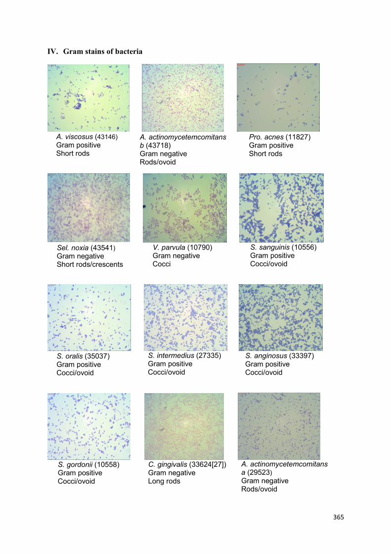
365
A. viscosus (43146) Gram positive Short rods
A. actinomycetemcomitans b (43718) Gram negative Rods/ovoid
Pro. acnes (11827) Gram positive Short rods
Sel. noxia (43541) Gram negative Short rods/crescents
V. parvula (10790) Gram negative Cocci
S. sanguinis (10556) Gram positive Cocci/ovoid
S. oralis (35037) Gram positive Cocci/ovoid
S. intermedius (27335) Gram positive Cocci/ovoid
S. anginosus (33397) Gram positive Cocci/ovoid
S. gordonii (10558) Gram positive Cocci/ovoid
C. gingivalis (33624[27]) Gram negative Long rods
A. actinomycetemcomitans a (29523) Gram negative Rods/ovoid
IV. Gram stains of bacteria

366
C. sput (33612[4]) Gram negative Ovoid/rods
S. constellatus (27823[M32b]) Gram positive Cocci/ovoid
C. rectus (33238[371]) Gram negative Ovoid/rods
C. showae (51146) Gram negative Ovoid/rods
F. nucleatum nuc (25586) Gram negative Rods
F. nucleatum poly (10953) Gram negative Rods
P. gingivalis (W83) Gram negative Rods
S. aureus (9144) Gram positive Cocci
Figure IV: Gram stains of bacterial cultures
Bacterial species were confirmed by Gram staining and analysis of bacteria morphology.
Gram-negative bacteria stained-pink and Gram-positive bacteria stained blue. Cell
morphologies included cocci (spherical), ovoid or rods (bacilli). Bacteria ATCC numbers are
provided.

367
V. Comparisons between free light chain detection in plasma and serum samples
The assay employed for the detection of FLCs carried out by The Binding Site is validated in
serum samples (Katzmann et al., 2002). To validate this technique in plasma samples for
subsequent analysis of chronic periodontitis plasma samples, kappa and lambda FLCs were
quantified in plasma and serum samples from the same individuals in parallel (n=30). No
significant difference was observed in the quantification of kappa (unpaired t-test p=0.58) or
lambda (unpaired t-test p=0.28) FLCs in plasma and serum samples. Based on these results,
FLCs were quantified in plasma samples using the same technique currently used by The
Binding Site for the quantification of FLC in serum samples (Figure V).

368
mg
/litre
Plasma Serum0
5
10
15
20
25
a) Kappa
ns
mg
/litre
Plasma Serum0
5
10
15
20
25
b) Lambda
ns
Figure V: Comparison between free light detection in plasma and serum samples
(a) Kappa and (b) lambda FLCs were quantified in plasma and serum from the same
individuals (n=30). No significant difference was observed in the quantification of kappa
(p=0.58) or lambda (p=0.28) FLCs in plasma and serum samples (analysed by unpaired t-
test). Data presented as mg/l and expressed as mean ± SEM (n=30).

369
VI. Collection and storage of NET supernatants
To store NET supernatants for subsequent assays investigating the effect of NETs on
neutrophil responses and H400 epithelial cell growth, NETs were collected and stored at
different temperatures. Storage conditions included 20°C, 37°C, 4°C, -20°C and -80°C (slow
freezing by 1°C/minute or snap freezing with liquid nitrogen). The effectiveness of the
storage method was determined by comparing the number of NETs (NET-DNA) quantified in
the supernatant with Sytox green before and after storage for 24 hours. Results are presented
as raw AFU data, as well as a ratio of the number of NETs quantified prior to storage. No
significant differences were observed in the number of NETs quantified between the storage
conditions compared with NET quantification results prior to storage (2way ANOVA
p>0.05, n=5) (Figure VIa). Data expressed as a ratio of NET quantification results prior to
storage demonstrated that the number of NETs quantified in the supernatant decreased as a
result of storage at some temperatures, however this did not reach statistical significance
(2way ANOVA p>0.05, n=5). Quantification of NET-DNA in supernatants stored at -80°C
by snap freezing in liquid nitrogen was the most representative of the NETs prior to storage,
evidenced by the smallest ratio change. Based on these findings, all NET supernatants were
stored at -80°C by snap freezing prior to subsequent assays (Figure VIb).

370
Storage condition
AF
U
Day 1 20C 37C 4C -20C -80C -80C0
5000
10000
15000
20000
25000PBS
PMA
HOCl
slow snap
ns
a) NET quantification
Storage condition
AF
U
20C 37C 4C -20C -80C -80C-1.0
-0.5
0.0
0.5
1.0PBS
PMA
HOCl
slow snap
ns
b) NET quantification expressed as a ratio
Figure VI: NET supernatant storage
NETs were stored at different temperatures (20°C, 37°C, 4°C, -20°C and -80°C [slow
freezing by 1°C/minute or snap freezing with liquid nitrogen]). The effectiveness of the
storage method was determined by comparing the number of NETs before and after storage
for 24 hours. (a) NETs quantified by comparing AFU (arbitrary fluorescence units) values.
(b) NET quantification expressed as a ratio of the number of NETs quantified prior to.
Statistical significance calculated using 2way ANOVA (ns=not significant). Data expressed
as mean ± SEM (n=5 in triplicate).

371
VII. H400 cell counting
Preliminary experiments compared in situ and trypsinised cell counts of H400 epithelial cells.
In situ cell counts involved counting the cells comprising the monolayer adhered to the
bottom of the flask. Trypsinised counts were performed by discarding the supernatants from
the flask and washing in PBS, followed by the addition of T-EDTA for 10 mins at room
temperature. The cell suspension was transferred to a fresh universal tube, centrifuged and
re-suspended in cell culture media for subsequent cell counts. Cell counts were compared at
2, 3, 4 and 5 days after seeding flasks at 2x10⁵. Results demonstrated that higher cell counts
were achieved when cells were counted in situ compared with trypsinised cells. However,
both methods demonstrated comparable shaped growth curves, with similar standard
deviations. In situ cell counts may be less accurate, as the monolayer confluency may not be
consistent across the entire flask. Furthermore, to seed cells in a 96-well plate for subsequent
NET supernatant assays, the cells will require trypsinising as they will not remain in the
flask. Therefore, all future counts for assays involving H400s and NET supernatants relied
on trypsinised counts (Figure VII).

372
Days after seeding
Ce
ll co
un
ts (
x10
5)
0 1 2 3 4 50
20
40
60
a) In situ cell counts
Days after seeding
Ce
ll co
un
ts (
x10
5)
0 1 2 3 4 50
10
20
30
40
b) Trypsinised cell counts
Figure VII: H400 cell counting
Preliminary experiments compared (a) in situ and (b) trypsinised cell counts of H400
epithelial cells. Cell counts were compared at 2, 3, 4 and 5 days after seeding flasks at 2x10⁵.
Results are representative of a single experiment, data expressed as the mean ± Std Dev.
373
VIII. Effect of cigarette smoke extract and cigarette smoke extract components on the
fluorometric quantification of calf thymus DNA
As it has previously been found that CSE inhibits luminol and isoluminol enhanced-
chemiluminescence (Matthews et al., 2011), preliminary studies were undertaken to establish
whether CSE interferes with the NET quantification assay with Sytox green. CSE (1%, 5%,
10%) incubated with calf thymus DNA for 4 hours did not significantly affect the
fluorometric quantification of calf thymus DNA (1way ANOVA p=0.98, n=5). The same
preliminary experiments were employed to determine whether CSE components affect the
quantification of NET-DNA. Nicotine (1µg/ml, 5µg/ml, 10µg/ml), cotinine (1µg/ml, 5µg/ml,
10µg/ml) and SCN- (50µM, 100µM, 150µM) also had no inhibitory effect on NET
quantification (1way ANOVA p=0.99, p=0.98, p=0.99 for nicotine, cotinine and SCN-,
respectively, n=5). Based on these results, neutrophils were not washed following a 30 min
pre-treatment with CSE or CSE components in subsequent assays (Figure VIII).

374
DNA g
Lo
g1
0 A
FU
0.0 0.2 0.4 0.6 0.8 1.0100
1000
10000
100000PBS
CSE 1%
CSE 5%
CSE 10%
ns
a) CSE
DNA g
Lo
g1
0 A
FU
0.0 0.2 0.4 0.6 0.8 1.0100
1000
10000
100000PBS
Nicotine 1g/ml
Nicotine 5g/ml
Nicotine 10g/ml
ns
b) Nicotine
DNA g
Lo
g1
0 A
FU
0.0 0.2 0.4 0.6 0.8 1.0100
1000
10000
100000PBS
Cotinine 1g/ml
Cotinine 5g/ml
Cotinine 10g/ml
ns
c) Cotinine
DNA g
Lo
g1
0 A
FU
0.0 0.2 0.4 0.6 0.8 1.0100
1000
10000
100000PBS
SCN- 50M
SCN- 100M
SCN- 150M
ns
d) SCN-
Figure VIII: Effect of cigarette smoke extract and cigarette smoke extract components on the fluorometric quantification of calf thymus
DNA
Calf thymus DNA at increasing concentrations (µg) was incubated with PBS (negative control), CSE, nicotine, cotinine or SCN- for 4 hours.
Post-incubation DNA was quantified fluorometrically with Sytox green. Statistical significance was calculated using 1way ANOVA (ns=not
significant). Data are presented as Log10 AFU (arbitrary fluorescence units) and are expressed as mean ± SEM (n=5 in triplicate).

375
IX. Isolation of neutrophil RNA
The amount and purity of RNA isolated from treated neutrophils was determined by
measuring the absorbance and the ratio of absorbance at 260/280nm. Pure RNA has a
260/280nm absorbance of 2.1, thus a value between 1.8-2.0nm is considered to be pure
(Sambrook et al., 1989; Manchester 1996). Adequate amounts (µg/ml) of RNA were
measured for each sample, and based on the amount of RNA in each sample, samples were
equally pooled across the treatment conditions (n=10 biological repeats per treatment
condition) (Table IX a&b).

376
Table IX: Quantity and purity of RNA
The amount and purity of isolated neutrophil RNA was determined by measuring the
absorbance and the ratio of absorbance at 260/280nm.
Sample
No.
PBS CSE 1% CSE 5% CSE 10%
RNA
µg/ml
260nm/2
80nm
ratio
RNA
µg/ml
260nm/2
80nm
ratio
RNA
µg/ml
260nm/2
80nm
ratio
RNA
µg/ml
260nm/2
80nm
ratio
1 28.6 1.43 42.30 1.33 31.6 1.57 29.3 1.69
2 31.9 1.99 54.90 1.40 32.5 1.61 28.2 1.7
3 36.6 1.55 32.00 1.75 45.7 1.50 59.3 1.80
4 55.0 1.60 31.08 1.90 29.6 1.15 31.3 1.70
5 31.1 1.55 29.30 1.46 50.4 1.33 51.6 120
6 33.1 1.29 33.50 1.30 54.5 1.19 43.1 1.25
7 31.9 1.84 34.70 1.51 46.2 1.41 38.1 1.65
8 33.5 1.62 49.80 1.12 33.9 1.18 54.5 1.23
9 31.9 1.84 47.30 1.62 25.6 1.67 34.6 1.51
10 45.1 1.21 33.70 1.89 57.0 1.24 32.4 1.58
Sample
No.
PBS SCN- 50µM SCN
- 100µM SCN
- 150µM
RNA
µg/ml
260nm/2
80nm
ratio
RNA
µg/ml
260nm/2
80nm
ratio
RNA
µg/ml
260nm/2
80nm
ratio
RNA
µg/ml
260nm/2
80nm
ratio
1 38.1 1.66 54.1 1.50 57.5 1.33 47.2 1.69
2 32.8 1.73 56.9 1.65 59.0 1.62 49.5 1.72
3 42.9 1.71 54.6 1.66 56.5 1.59 41.9 1.69
4 66.3 1.56 55.7 1.48 36.8 1.41 20.8 1.87
5 42.6 1.85 55.6 1.51 43.8 1.59 51.1 1.76
6 52.0 1.82 42.4 1.94 36.9 1.55 48.0 1.80
7 32.0 1.49 49.0 1.65 28.0 1.76 31.0 1.71
8 29.5 1.71 45.8 1.39 38.4 1.70 24.6 1.70
9 41.7 1.45 39.6 1.30 59.7 1.80 35.8 1.81
10 47.6 1.47 29.3 1.58 26.2 1.74 36.5 1.69
a)
b)

377
X. Determination of stable housekeeping genes
Preliminary assays determined the melt curves, amplification and efficiency of reference
genes and the genes of interest. Target gene expression was determined by calculating fold
changes in gene expression as a ratio of a reference housekeeping gene. The most
appropriate housekeeper gene was determined by analysis of crossing point (CP) data with
statistical algorithm BestKeeper (Pfaffl et al., 2004). CP data indicates the number at which
the fluorescence generation crosses the threshold and thus a sufficient number of amplicons
have been produced (Rasmussen 2001). BestKeeper expression indicates that all candidate
housekeeping genes had standard deviations lower than 1, suggesting they were all viable
housekeeping genes for the study of these cells. However, YWHAZ had the lowest standard
deviation (0.45) and was therefore considered the most stable of the reference genes tests.
Based on these results, the YWHAZ housekeeping gene was considered the most consistent
and therefore used as the reference against which to quantify the expression of genes of
interest (Table X).

378
Table X: Comparison of housekeeping genes.
Analysis of 4 candidate housekeeping genes (HKG) used crossing point (CP) data.
Abbreviations: SD (standard deviation), coefficient of variance % of CP (the coefficient of
variance expressed as a percentage on the CP level), minimum/maximum [x-fold] (the
extreme values of expression levels expressed as an absolute x-fold over- or under-regulation
coefficient). Pfaffl et al., 2004, Rasmussen 2001.
CP data of housekeeping Genes
GAPDH YWHAZ HPRT1 RPL13
HKG 1 HKG 2 HKG 3 HKG 4
Number of samples 4 4 4 4
Geometric mean of CP 29.50 27.36 32.40 25.58
Arithmetic mean of CP 29.52 27.36 32.41 25.59
Minimum CP 27.99 26.96 31.73 24.94
Maximum CP 30.87 28.27 33.72 26.46
SD of CP 0.79 0.45 0.65 0.56
coefficient of variance (%
of CP) 2.66 1.66 2.02 2.20
minimum [x-fold] -2.48 -1.31 -1.58 -1.49
maximum [x-fold] 2.28 1.85 2.45 1.73
Std Dev of absolute
regulation coefficients 1.60 1.31 1.48 1.40

379
XI. RNA expression of cigarette smoke extract treated neutrophils relative to other
reference genes
Neutrophil RNA expression of NF-ĸB, HSP40, IL-8, P-47phox
, P-67phox
and GP-91phox
was
quantified by RT-PCR following CSE treatment (1%, 5%, 10%). Data are expressed as a
ratio of the reference genes, GADPH, HPTY1 and RPL13, and RNA expression of cells
treated with PBS (negative control) were normalised to 1.
GADPH
In response to CSE treatment, HSP40 expression increased and this was statistically
significant at concentrations of 5% and 10% CSE (2way ANOVA and Bonferroni post-tests
****p<0.0001 for 5% and 10%, n=10 biological repeats, n=3 technical repeats) (Figure XIa).
HPTY1
In response to CSE treatment, HSP40 expression increased and this was statistically
significant at concentrations of 5% and 10% CSE (2way ANOVA and Bonferroni post-tests
****p<0.0001 for 5% and 10%, n=10 biological repeats, n=3 technical repeats). Other non-
significant trends included decreases in P-67phox
and GP-91phox
expression (Figure XIb).
RPL13
In response to CSE treatment, HSP40 expression increased and this was statistically
significant at concentrations of 5% and 10% CSE (2way ANOVA and Bonferroni post-tests
**p<0.01, ****p<0.0001 for 5% and 10%, respectively, n=10 biological repeats, n=3
technical repeats). Other non-significant trends included a CSE-concentration dependent
decrease in NF-ĸB and decreased GP-91phox
expression (Figure XIc).

380
NFkB HSP40 IL-8 P47 P67 Gp910
2
4
6
8
40
800
1%
5%
10%
(a) GAPDHR
atio
to
ho
use
ke
ep
er
****
****
NFkB HSP40 IL-8 P47 P67 Gp910.0
0.5
1.0
1.5
2.0
102030
0
1%
5%
10%
Ra
tio
to
ho
use
ke
ep
er
(b) HPTY1
****
****
NFkB HSP40 IL-8 P47 P67 Gp910.0
0.5
1.0
1.5
102030
0
1%
5%
10%
Ra
tio
to
ho
use
ke
ep
er
(c) RPL13
****
**
Figure XI: RNA expression of CSE treated neutrophils relative to other reference genes
Data are expressed as a ratio of the reference gene and RNA expression of cells treated with
PBS (negative control) was normalised to 1. RNA expression as a ratio of reference gene (a)
GAPDH, (b) HPTY1, (c) RPL13. Statistical significance calculated using 2way ANOVA
and Bonferroni post-tests (**p<0.01, ****p<0.0001). Data is expressed as mean ± SEM
(n=10 pooled biological repeats and n=3 technical repeats).

381
XII. RNA expression of thiocyanate treated neutrophils relative to other reference
genes
Neutrophil RNA expression of NF-ĸB, HSP40, IL-8, P-47phox
, P-67phox
and GP-91phox
was
quantified by real time PCR following SCN- treatment (1%, 5% or 10%). Data are expressed
as a ratio of the reference genes, GADPH, HPTY1 and RPL13, and RNA expression of cells
treated with PBS (negative control) were normalised to 1.
GADPH
In response to all concentrations of SCN- treatment, HSP40 expression significantly
increased (2way ANOVA and Bonferroni multiple comparisons tests ****p<0.0001 for 50µM
and 100µM, p<0.05 for 150µM, n=3 technical repeats). The expression of P-67phox
increased
following 50µM SCN- treatment (2way ANOVA and Bonferroni multiple comparisons tests
*p<0.05, n=10 biological repeats, n=3 technical repeats). SCN- treatment at all
concentrations was found to have an inhibitory effect on the expression of GP-91phox
;
however this was not statistically significant (Figure XIIa).
HPTY1
In response to all concentrations of SCN- treatment, HSP40 expression increased and this
was statistically significant at concentrations of 50µM and 150µM SCN- (2way ANOVA and
Bonferroni post-tests ****p<0.0001 for 50µM and 150µM, n=3 technical repeats). The
expression of P-47phox
increased following treatment with 150µM SCN- (2way ANOVA and
Bonferroni multiple comparisons tests ****p<0.0001, n=10 biological repeats, n=3 technical
repeats). SCN- treatment at all concentrations was found to increase P-67phox
expression and
was statistically significant at 100µM and 150µM SCN- (2way ANOVA and Bonferroni
multiple comparisons tests **p<0.01, ****p<0.0001 for 100µM and 150µM SCN-,
respectively, n=10 biological repeats, n=3 technical repeats) (Figure XIIb).
382
RPL13
In response to SCN- treatment, HSP40 expression significantly increased and this was
statistically significant at concentrations of 50µM and 100µM SCN- (2way ANOVA and
Bonferroni post-tests **p<0.01, ***p<0.001 for 50µM and 100µM SCN-, respectively, n=10
biological repeats, n=3 technical repeats). P-67phox
expression increased following treatment
with 100µM SCN- (2way ANOVA and Bonferroni post-tests ****p<0.0001, n=10 biological
repeats, n=3 technical repeats). Decreased GP-91phox
expression was also observed in
response to all concentrations of SCN-, however this was not statistically significant (Figure
XIIc).

383
NFkB HSP40 IL-8 P47 P67 Gp910.0
0.5
1.0
1.5
2.0
2.5
3.0
51015
0
50M
100M
150M
Ra
tio
to
ho
use
ke
ep
er
(a) GAPDH
*****
****
*
NFkB HSP40 IL-8 P47 P67 Gp910.0
0.5
1.0
1.5
2.0
20
400
50M
100M
150M
Ra
tio
to
ho
use
ke
ep
er
(b) HPTY1
****
**** ****
****
**
NFkB HSP40 IL-8 P47 P67 Gp910.0
0.5
1.0
1.5
2.0
2.5
5
10
150
50M
100M
150M
Ra
tio
to
ho
use
ke
ep
er
(c) RPL13
*****
****
Figure XII: RNA expression of thiocyanate treated neutrophils relative to other
reference genes
Data are expressed as a ratio of the reference gene and RNA expression of cells treated with
PBS (negative control) was normalised to 1. RNA expression as a ratio of reference gene (a)
GAPDH, (b) HPTY1, (c) RPL13. Statistical significance calculated using 2way ANOVA
and Bonferroni post-tests (*p<0.05, **p<0.01, ****p<0.0001). Data is expressed as mean ±
SEM (n=10 pooled biological repeats and n=3 technical repeats).
Copyright © 2022 FDOKUMEN