Amidinoquinoxaline N-oxides as novel spin traps
9
RSC Advances RSCPublishing PAPER This journal is © The Royal Society of Chemistry 2013 J. Name ., 2013, 00, 1-3 | 1 Cite this: DOI: 10.1039/x0xx00000x Received 00th January 2012, Accepted 00th January 2012 DOI: 10.1039/x0xx00000x www.rsc.org/ Amidinoquinoxaline N-oxides as novel spin traps Nadia Gruber, a,b,c Lidia L. Piehl, b Emilio Rubin de Celis, b Jimena E. Díaz, a María B. García, a Pierluigi Stipa,* c and Liliana R. Orelli* a A novel type of spin traps 1 derived from the pyrimidoquinoxaline N-oxide heterocyclic core is reported. EPR technique was used to evaluate their ability to trap methyl radicals generated in a Fenton reaction in the presence of DMSO. All the synthesized nitrones showed spin trapping properties and the corresponding nitroxides 2 were characterized by EPR. The novel spin traps showed remarkably persistent signals, as evidenced in a competition experiment with DMPO. The addition rate constants leading to the spin adducts (k add ) were determined, and very good correlations were found with steric and electronic parameters of the parent nitrones. The spin adducts decomposition rate constants (k dec ) and the corresponding half-life times (t 1/2 ) were also determined. DFT and MP2 calculations were used in order to rationalize the adducts hfcc and the structural factors influencing their addition and decomposition rates. Introduction Electron paramagnetic resonance (EPR) spin trapping represents one of the most specific and reliable techniques for detecting and identifying transient free radicals, such as those produced in chemical and biological processes, whose lifetime is too short in the EPR spectroscopic time scale. This technique, widely used since its introduction about 40 years ago, 1 is based upon the fast reaction between a suitable diamagnetic molecule (a spin trap) and short-lived free radicals with formation of relatively long lived radicals (spin adducts), whose EPR signals are persistent enough to be recorded and analyzed. Spectral parameters such as hyperfine coupling constants (hfcc) and g- factors are generally characteristic of the type of radical initially trapped. This technique has been successfully used in biological systems, with many applications in a series of human diseases like ischemia-reperfusion syndrome, Friedreich’s ataxia, atherosclerosis, 2 neurodegenerative diseases 3 and cellular aging. 4 Nitrones (N-oxides) are very efficient spin traps, 5 being able to undergo fast radical additions by C- and O-centered radicals, to yield aminoxyls (nitroxides) as spin adducts, these species being among the most persistent organic free radicals in liquid solution. Among the commercially available nitrones, N- tert-butylbenzylideneamine N-oxide (PBN), 5,5-dimethyl-3,4- dihydro-2H-pyrrole N-oxide (DMPO) 6 are the most popular, but their use is not without limitations. For example, PBN and its analogues give spin adducts with similar EPR spectra generally consisting of a triplet of doublets with a relatively small variation in the doublet splitting depending on the radical trapped, and this may be a source of misinterpretations in spin trapping experiments. 7 On the other hand, the use of DMPO is limited by its sensitivity to nucleophilic attack by water and the relatively low stability of its superoxide spin adduct, which decomposes rapidly. 8 A continuous effort has been devoted to the synthesis of PBN and DMPO analogues 9 and of other nitrones 10 to be used in spin trapping experiments without the above mentioned drawbacks. To this end, we synthesized a series of 5-aryl-2,3-dihydro1H-pyrimido[1,2-a]quinoxaline 6- oxides 1a-h and a fused pyridyl analogue 1i and evaluated their spin trapping ability by EPR spectroscopy. 2,3-Dihydro-1H- pyrimido[1,2-a]quinoxaline N-oxides represent a heterocyclic core of interest due to its pharmacological properties. Some suitably substituted derivatives possess antineoplastic activity, 11 especially against hypoxic tumours. Other pyrimidoquinoxaline 6-oxides have been employed as antiamoebic 12 and antianaerobic agents 13 . As part of our research on nitrogen heterocycles, we reported a novel methodology for their synthesis, 14 and investigated some of their chemical, 15 spectroscopic 16 and pharmacological 17 properties. Two structural features of these compounds which could both have a stabilizing effect on the corresponding spin adducts prompted us to test these nitrones as potential spin traps: the absence of hydrogens - to the N-oxide function and the presence of a conjugated amidine moiety. In fact, it is known that aminoxyls bearing -hydrogens are unstable and may disproportionate to the corresponding nitrone and a hydroxylamine, 18 while it has been observed that electron- withdrawing substituents close to the N-O function could stabilize spin adducts. 9e,19 Taking advantage of recent developments in the field, 20 in the present paper we also present DFT 21 and MP2 calculations which were able to describe the EPR features of the novel spin
-
Upload
independent -
Category
Documents
-
view
3 -
download
0
Transcript of Amidinoquinoxaline N-oxides as novel spin traps
RSC Advances RSCPublishing
PAPER
This journal is © The Royal Society of Chemistry 2013 J. Name., 2013, 00, 1-3 | 1
Cite this: DOI: 10.1039/x0xx00000x
Received 00th January 2012,
Accepted 00th January 2012
DOI: 10.1039/x0xx00000x
www.rsc.org/
Amidinoquinoxaline N-oxides as novel spin traps
Nadia Gruber,a,b,c
Lidia L. Piehl,b Emilio Rubin de Celis,
b Jimena E. Díaz,
a María B.
García,a Pierluigi Stipa,*
c and Liliana R. Orelli*
a
A novel type of spin traps 1 derived from the pyrimidoquinoxaline N-oxide heterocyclic core is
reported. EPR technique was used to evaluate their ability to trap methyl radicals generated in
a Fenton reaction in the presence of DMSO. All the synthesized nitrones showed spin trapping
properties and the corresponding nitroxides 2 were characterized by EPR. The novel spin traps
showed remarkably persistent signals, as evidenced in a competition experiment with DMPO.
The addition rate constants leading to the spin adducts (kadd) were determined, and very good
correlations were found with steric and electronic parameters of the parent nitrones. The spin
adducts decomposition rate constants (kdec) and the corresponding half-life times (t1/2) were
also determined. DFT and MP2 calculations were used in order to rationalize the adducts hfcc
and the structural factors influencing their addition and decomposition rates.
Introduction
Electron paramagnetic resonance (EPR) spin trapping
represents one of the most specific and reliable techniques for
detecting and identifying transient free radicals, such as those
produced in chemical and biological processes, whose lifetime
is too short in the EPR spectroscopic time scale. This technique,
widely used since its introduction about 40 years ago,1 is based
upon the fast reaction between a suitable diamagnetic molecule
(a spin trap) and short-lived free radicals with formation of
relatively long lived radicals (spin adducts), whose EPR signals
are persistent enough to be recorded and analyzed. Spectral
parameters such as hyperfine coupling constants (hfcc) and g-
factors are generally characteristic of the type of radical initially
trapped. This technique has been successfully used in biological
systems, with many applications in a series of human diseases
like ischemia-reperfusion syndrome, Friedreich’s ataxia,
atherosclerosis,2 neurodegenerative diseases3 and cellular
aging.4 Nitrones (N-oxides) are very efficient spin traps,5 being
able to undergo fast radical additions by C- and O-centered
radicals, to yield aminoxyls (nitroxides) as spin adducts, these
species being among the most persistent organic free radicals in
liquid solution. Among the commercially available nitrones, N-
tert-butylbenzylideneamine N-oxide (PBN), 5,5-dimethyl-3,4-
dihydro-2H-pyrrole N-oxide (DMPO)6 are the most popular,
but their use is not without limitations. For example, PBN and
its analogues give spin adducts with similar EPR spectra
generally consisting of a triplet of doublets with a relatively
small variation in the doublet splitting depending on the radical
trapped, and this may be a source of misinterpretations in spin
trapping experiments.7 On the other hand, the use of DMPO is
limited by its sensitivity to nucleophilic attack by water and the
relatively low stability of its superoxide spin adduct, which
decomposes rapidly.8 A continuous effort has been devoted to
the synthesis of PBN and DMPO analogues9 and of other
nitrones10 to be used in spin trapping experiments without the
above mentioned drawbacks. To this end, we synthesized a
series of 5-aryl-2,3-dihydro1H-pyrimido[1,2-a]quinoxaline 6-
oxides 1a-h and a fused pyridyl analogue 1i and evaluated their
spin trapping ability by EPR spectroscopy. 2,3-Dihydro-1H-
pyrimido[1,2-a]quinoxaline N-oxides represent a heterocyclic
core of interest due to its pharmacological properties. Some
suitably substituted derivatives possess antineoplastic activity,11
especially against hypoxic tumours. Other pyrimidoquinoxaline
6-oxides have been employed as antiamoebic12 and
antianaerobic agents13. As part of our research on nitrogen
heterocycles, we reported a novel methodology for their
synthesis,14 and investigated some of their chemical,15
spectroscopic16 and pharmacological17 properties.
Two structural features of these compounds which could both
have a stabilizing effect on the corresponding spin adducts
prompted us to test these nitrones as potential spin traps: the
absence of hydrogens - to the N-oxide function and the
presence of a conjugated amidine moiety. In fact, it is known
that aminoxyls bearing -hydrogens are unstable and may
disproportionate to the corresponding nitrone and a
hydroxylamine,18 while it has been observed that electron-
withdrawing substituents close to the N-O function could
stabilize spin adducts.9e,19
Taking advantage of recent developments in the field,20 in the
present paper we also present DFT21 and MP2 calculations
which were able to describe the EPR features of the novel spin

ARTICLE Journal Name
2 | J. Name., 2012, 00, 1-3 This journal is © The Royal Society of Chemistry 2012
adducts as well as to rationalise some trends concerning their
formation and decomposition rates.
Results and discussion
The compounds under study are shown in Table 1, and were
chosen so as to include 5-aryl groups with variable
stereoelectronic features or the presence of a strongly electron
withdrawing group in the fused ring, which might in turn
stabilize the resulting spin adduct.
Table 1. Compounds under study
Compound 1 Ar X
a C6H5 CH
b 4-OCH3C6H4 CH
c 4-ClC6H4 CH
d 4-NO2C6H4 CH
e 2-OCH3C6H4 CH
f 2-ClC6H4 CH
g 2-IC6H4 CH
h 3-C4H3S CH
i C6H5 N
We had previously described compounds 1a-d,f.14,16 The
remaining compounds were prepared by a modification of our
previously reported method.14
All compounds showed spin trap properties in a Fenton reaction
system in the presence of DMSO, where methyl radicals are
formed,22 in agreement with Scheme 1. This system, involving
the presence of oxygen and aqueous medium, was chosen as a
first approximation to biocompatible conditions.
Scheme 1. Spin-trapping reaction
For the sake of comparison, DMPO adducts were also obtained
with the same trapping Fenton reaction system (Figure 1a). The
simulated spectrum of DMPO adduct (Figure 1b) was
compatible with the DMPO/·CH3 adduct (aN: 16.1 Gauss, aH:
22.8 Gauss, percentage > 95).
Figure 1. (a) Experimental and (b) Simulated EPR spectrum of
DMPO (30 mM final concentration) adduct obtained by
addition of the spin trap to methyl radicals produced by a
Fenton reaction. (1.33 mM FeSO4 plus 3.33 mM H2O2, final
concentrations) in PBS pH 7.6 containing 33 % DMSO.
Spin adducts 2 displayed similar EPR spectra and were characterized
by simulation, showing a typical EPR trace with 9 resolved peaks.
Figure 2 shows the EPR spectrum of 2a as a representative example.
Figure 2. (a) Experimental and (b) simulated EPR spectrum of the
spin adduct of 1a (30 mM final concentration) obtained by addition
of methyl radicals produced by a Fenton reaction. (1.33 mM FeSO4
plus 3.33 mM H2O2, final concentrations) in PBS pH 7.6 containing
33% DMSO.
In general, the spin density in aminoxyls is mainly located on the N-
O group, with typical N-hfcc values of 14-15G for alkyl aminoxyls23
and of 10-11G for aryl aminoxyls.24 Consistently, the N-hfcc for the
nitroxides under study also display values of 10-11G, which can be
attributed to the extensive electron delocalization in this system,
mainly in the fused aromatic ring. A plot of spin density in nitroxide
2a (Figure 3) illustrates this feature.

Journal Name ARTICLE
This journal is © The Royal Society of Chemistry 2012 J. Name., 2012, 00, 1-3 | 3
Figure 3: spin density distribution () of nitroxide 2a
computed at the B3LYP/EPR-III level (positive values in blue
and negative in green). C-24 has been arrowed (see text).
This is in accordance with previous findings in other
heteroaromatic analogues, such as indolinonic25 and
benzoxazinic nitroxides.26 In line with this, the EPR spectra of
nitroxides 2 were interpreted on the basis of hfcc typical of this
kind of radicals, in which the nitrogen three lines are mainly
split by two different couples of aromatic hydrogens of the
heteroaromatic ring moiety (Figure 4), H(13) and H(15) with a
larger hfcc (ca. 3 Gauss) and H(12) and H(14) with a smaller
one (ca. 1 Gauss). In addition, the splitting of the amidine ring
nitrogen N-21 was considered. The assignments, confirmed by
means of appropriate DFT calculations previously described,25
are reported in Table 2. However, because the experimental
conditions followed in our study (aqueous medium in the
presence of molecular oxygen) did not allow us to achieve a
better spectral resolution than that shown in Figure 2, the hfcc
values given in the last two columns of Table 2 are tentative
and mainly based on DFT computational predictions.
Figure 4: Ball and stick representation of the optimized geometry of
nitroxide 2a showing the arbitrary atom numbering and the
corresponding hfcc computed at the B3LYP/EPR-III level.
Table 2. Experimental hyperfine coupling constants (in Gauss)
of nitroxides 2 confirmed by computer simulation.
Nitroxide 2 N-19 H-13, 15 H-12, 14 N-21
a 10.77 3.13 0.88 0.29
b 10.84 3.12 0.89 0.23
c 10.76 3.08 0.88 0.31
d 10.64 3.07 0.91 0.41
e 10.62 3.09 0.98 0.57
f 10.53 3.08 0.87 0.47
g 10.55 3.04 0.83 0.67
h 10.90 3.15 0.80 0.24
i 10.45 3.48 0.89;
0.82 (N)
0.34
The isotropic hfcc (aX) arises from the Fermi contact interaction
between the unpaired electron and the nucleus (X), and is
correlated to the corresponding spin density (X) by:
aX = 2/3 0 ge B gX X
where 0 is the vacuum permeability, B the Bohr magneton, ge
and gX the electron and nuclear g-factor respectively.27 The N-
19 hfcc of spin adducts 2 (Table 2) decrease in the sequence: h
> b > a > c > d > e > g > f > i, which can be ascribed to a
decrease of the corresponding spin density in the same order.
This is possibly due to the presence of an electron withdrawing
group at C-24, the ortho position of the 5-aryl group, where a
positive spin density has been found, as shown in Figure 3 for
2a. At the same time, the fact that no significant spin densities
have been found in any other position of such group explains
why this effect upon N-19 is present only in aminoxyls 2e-g. It
should also be noted that derivatives 2e-g may be subject to
stereoelectronic effects due to the presence of the substituents
in the ortho-position of the aromatic ring linked to the -
carbon. In 2i, on the other hand, the decrease in the N-hhfc
would be related to the electron withdrawing effect exerted by
the presence of a conjugated nitrogen in position 8, also
responsible of the large value found for the H-13,15 hfcc.
In order to evaluate the stability of the novel spin adducts, their
EPR signals were followed by recording the corresponding
spectra at different times until disappearance. Figure 5 shows
the time-dependent EPR signal intensity of one of the new spin
traps (compound 1d) adduct.
ARTICLE Journal Name
4 | J. Name., 2012, 00, 1-3 This journal is © The Royal Society of Chemistry 2012
Figure 5. Time-dependent EPR spin adduct spectra of 1d (10
mM final concentration) upon reaction with methyl radicals
produced by a Fenton reaction. (1.33 mM FeSO4 plus 3.33 mM
H2O2, final concentrations) in PBS pH 7.6 containing 33%
DMSO.
Comparatively, the DMPO/·CH3 adduct was less stable as a
function of time, as can be seen in Figure 6.
Figure 6. Time-dependent EPR spectra of DMPO/·CH3 spin
adduct. DMPO (10 mM final concentration) was tested in a
Fenton reaction. (1.33 mM FeSO4 plus 3.33 mM H2O2, final
concentrations) in PBS pH 7.6 containing 33% DMSO.
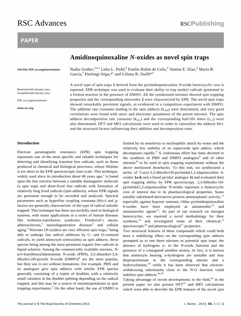
To further investigate this difference, a competition experiment
between DMPO and 1a was performed, and EPR spectra are
shown in Figure 7.
Figure 7. (a) Experimental and (b) simulated EPR spectra of the
competition assay between DMPO and 1a adducts. FeSO4 (1.33
mM, final concentration) and H2O2 (3.33 mM, final
concentration) were added to a solution containing both DMPO
and compound 1a in 33 % DMSO-PBS pH 7.6. Spectra a1, a2
and a3 are the experimental EPR signals obtained at 1, 15 and
30 minutes and b1, b2 and b3 are their respective simulated EPR
spectra.
This experiment reveals the persistence of the signal of the
novel adducts after DMPO adduct signal disappearance. Hence,
even if the newly synthesized nitrones react more slowly with
methyl radicals than DMPO, the corresponding adducts resulted
much more persistent. The enhanced stability of adducts 2
would result from the absence of a hydrogen to the N-O·
group, which is the main issue concerning the stability of
DMPO spin adducts.18
The kinetics of formation and decomposition of the spin
adducts generated by methyl radicals addition to the new
nitrones 1 were then studied. The experimental conditions were
modified to prevent precipitation of some derivatives and to
enhance signal intensity, using a higher proportion of DMSO
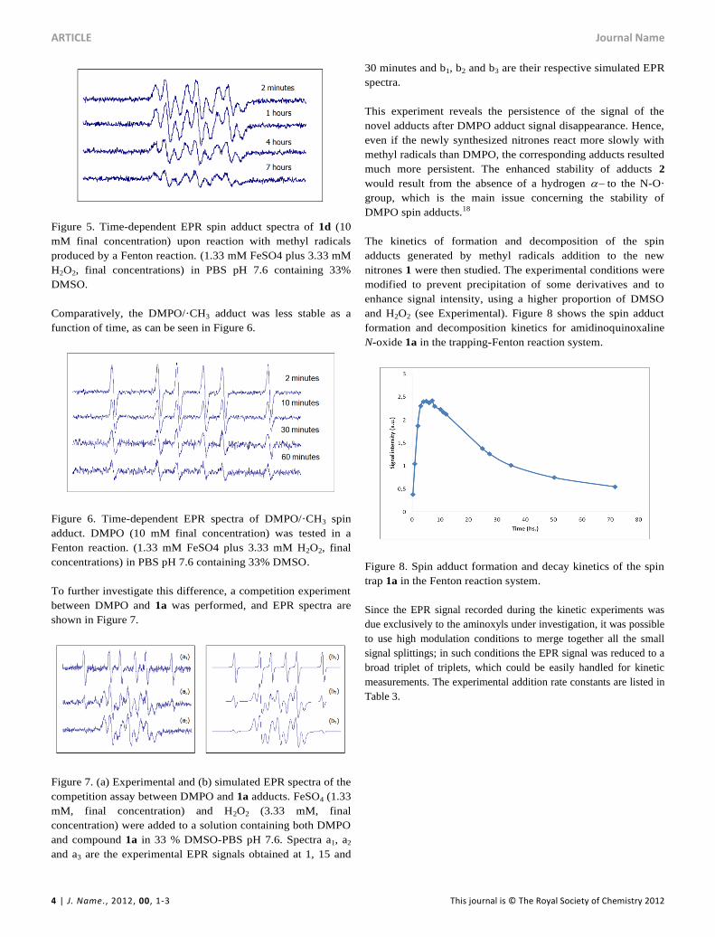
and H2O2 (see Experimental). Figure 8 shows the spin adduct
formation and decomposition kinetics for amidinoquinoxaline
N-oxide 1a in the trapping-Fenton reaction system.
Figure 8. Spin adduct formation and decay kinetics of the spin
trap 1a in the Fenton reaction system.
Since the EPR signal recorded during the kinetic experiments was
due exclusively to the aminoxyls under investigation, it was possible
to use high modulation conditions to merge together all the small
signal splittings; in such conditions the EPR signal was reduced to a
broad triplet of triplets, which could be easily handled for kinetic
measurements. The experimental addition rate constants are listed in
Table 3.
Journal Name ARTICLE
This journal is © The Royal Society of Chemistry 2012 J. Name., 2012, 00, 1-3 | 5
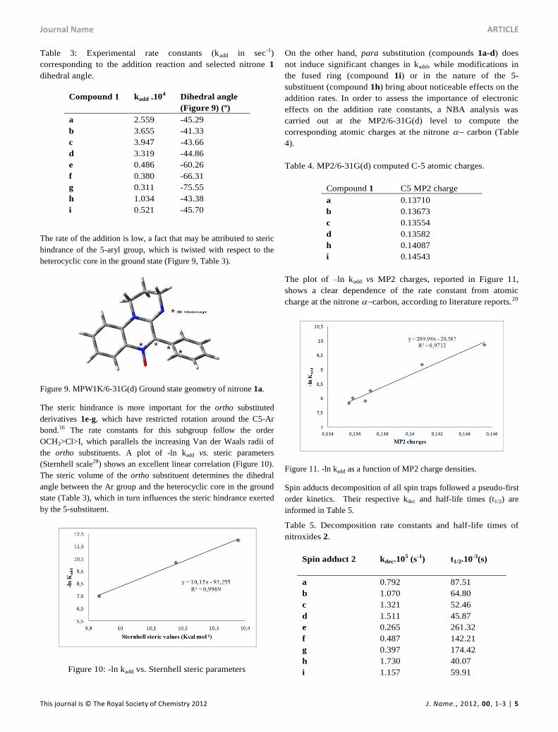
Table 3: Experimental rate constants (kadd in sec-1)
corresponding to the addition reaction and selected nitrone 1
dihedral angle.
Compound 1 kadd .104 Dihedral angle
(Figure 9) (º)
a 2.559 -45.29
b 3.655 -41.33
c 3.947 -43.66
d 3.319 -44.86
e 0.486 -60.26
f 0.380 -66.31
g 0.311 -75.55
h 1.034 -43.38
i 0.521 -45.70
The rate of the addition is low, a fact that may be attributed to steric
hindrance of the 5-aryl group, which is twisted with respect to the
heterocyclic core in the ground state (Figure 9, Table 3).
Figure 9. MPW1K/6-31G(d) Ground state geometry of nitrone 1a.
The steric hindrance is more important for the ortho substituted
derivatives 1e-g, which have restricted rotation around the C5-Ar
bond.16 The rate constants for this subgroup follow the order
OCH3>Cl>I, which parallels the increasing Van der Waals radii of
the ortho substituents. A plot of -ln kadd vs. steric parameters
(Sternhell scale28) shows an excellent linear correlation (Figure 10).
The steric volume of the ortho substituent determines the dihedral
angle between the Ar group and the heterocyclic core in the ground
state (Table 3), which in turn influences the steric hindrance exerted
by the 5-substituent.
Figure 10: -ln kadd vs. Sternhell steric parameters
On the other hand, para substitution (compounds 1a-d) does
not induce significant changes in kadd, while modifications in
the fused ring (compound 1i) or in the nature of the 5-
substituent (compound 1h) bring about noticeable effects on the
addition rates. In order to assess the importance of electronic
effects on the addition rate constants, a NBA analysis was
carried out at the MP2/6-31G(d) level to compute the
corresponding atomic charges at the nitrone carbon (Table
4).
Table 4. MP2/6-31G(d) computed C-5 atomic charges.
Compound 1 C5 MP2 charge
a 0.13710
b 0.13673
c 0.13554
d 0.13582
h 0.14087
i 0.14543
The plot of –ln kadd vs MP2 charges, reported in Figure 11,
shows a clear dependence of the rate constant from atomic
charge at the nitrone carbon, according to literature reports.29
Figure 11. -ln kadd as a function of MP2 charge densities.
Spin adducts decomposition of all spin traps followed a pseudo-first
order kinetics. Their respective kdec and half-life times (t1/2) are
informed in Table 5.
Table 5. Decomposition rate constants and half-life times of
nitroxides 2.
Spin adduct 2 kdec.105 (s-1) t1/2.10-3(s)
a 0.792 87.51
b 1.070 64.80
c 1.321 52.46
d 1.511 45.87
e 0.265 261.32
f 0.487 142.21
g 0.397 174.42
h 1.730 40.07
i 1.157 59.91
ARTICLE Journal Name
6 | J. Name., 2012, 00, 1-3 This journal is © The Royal Society of Chemistry 2012
The overall spin adducts decomposition could be simplified as in the
following scheme:
Scheme 2. Reactions involved in the spin adduct decomposition.
Assuming the reversibility of the first reaction, the C-centered
radicals formed in this step can undergo self-coupling and/or react
with the nitroxide in excess present in the reaction medium. Both
these two last processes are extremely fast and largely shift the
equilibrium of the first step to the right, also allowing to
approximate the observed EPR signal decay to a pseudo first order
process. The t1/2 values reported in Table 5 have been determined
following this assumption.
In spite of the previous considerations, other chemical reactions
cannot be ruled out in the oxidative aqueous reaction medium.
Therefore, no clear relationship between structure and
decomposition rates of the spin adducts comprising all the
compounds under study could be established. In spite of this,
analysis of the results reported in Table 5 clearly show that ortho
substitution stabilizes the spin adducts, as can be seen on comparing
the decomposition rate constants of compounds 2e and 2f with the
corresponding para isomers (2b and 2c, respectively). An attempt to
correlate the nitroxide decomposition rate constant with the
corresponding dihedral angle has been reported in Figure 12:
Figure 12. Decomposition rate constants (kdec in sec-1) of nitroxides
2 vs. the corresponding dihedral angle N19-C5-C23-C25 (in degrees)
(see Figure 4).
Upon this basis, it could be hypothesized that the more “twisted” the
nitrone, the more persistent is the corresponding spin adduct. This
could be likely due to the fact that in nitroxides 2e-g it becomes
more difficult to reach the planarity necessary for the formation of
the nitrone N=C double bond required by the adduct decomposition
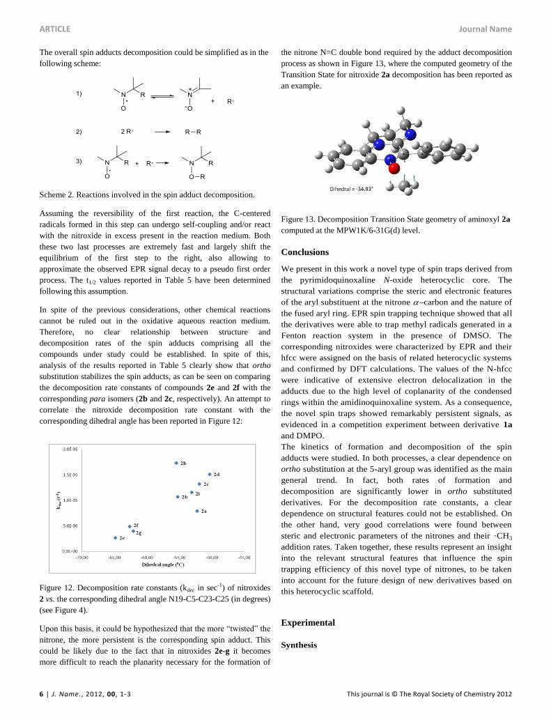
process as shown in Figure 13, where the computed geometry of the
Transition State for nitroxide 2a decomposition has been reported as
an example.
Figure 13. Decomposition Transition State geometry of aminoxyl 2a
computed at the MPW1K/6-31G(d) level.
Conclusions
We present in this work a novel type of spin traps derived from
the pyrimidoquinoxaline N-oxide heterocyclic core. The
structural variations comprise the steric and electronic features
of the aryl substituent at the nitrone carbon and the nature of
the fused aryl ring. EPR spin trapping technique showed that all
the derivatives were able to trap methyl radicals generated in a
Fenton reaction system in the presence of DMSO. The
corresponding nitroxides were characterized by EPR and their
hfcc were assigned on the basis of related heterocyclic systems
and confirmed by DFT calculations. The values of the N-hfcc
were indicative of extensive electron delocalization in the
adducts due to the high level of coplanarity of the condensed
rings within the amidinoquinoxaline system. As a consequence,
the novel spin traps showed remarkably persistent signals, as
evidenced in a competition experiment between derivative 1a
and DMPO.
The kinetics of formation and decomposition of the spin
adducts were studied. In both processes, a clear dependence on
ortho substitution at the 5-aryl group was identified as the main
general trend. In fact, both rates of formation and
decomposition are significantly lower in ortho substituted
derivatives. For the decomposition rate constants, a clear
dependence on structural features could not be established. On
the other hand, very good correlations were found between
steric and electronic parameters of the nitrones and their ·CH3
addition rates. Taken together, these results represent an insight
into the relevant structural features that influence the spin
trapping efficiency of this novel type of nitrones, to be taken
into account for the future design of new derivatives based on
this heterocyclic scaffold.
Experimental
Synthesis

Journal Name ARTICLE
This journal is © The Royal Society of Chemistry 2012 J. Name., 2012, 00, 1-3 | 7
General
Melting points were determined with a Büchi capillary
apparatus and are uncorrected. 1H and 13C NMR spectra were
recorded on a Bruker Avance II 500 MHz spectrometer, using
deuteriochloroform as the solvent. Chemical shifts are reported
in parts per million () relative to TMS as an internal standard.
Coupling constants are reported in Hz. D2O was employed to
confirm exchangeable protons (ex). Splitting multiplicities are
reported as singlet (s), broad signal (bs), doublet (d), double
doublet (dd), doublet of double doublets (ddd), triplet (t), triple
doublet (td), pentet (p), and multiplet (m). Elemental analyses
were determined using an Exeter CE 440 elemental analyzer.
Reagents, solvents, and starting materials were purchased from
standard sources and purified according to literature
procedures.
Synthesis of 5-aryl-2,3-dihydro-1H-pyrimido[1,2-
a]quinoxaline 6-oxides 1. General procedure.
A mixture of the corresponding aminoamide (1 mmol) and
ethyl polyphosphate (PPE, 1 mL/0.05 g) was refluxed for 5 h.
After reaching room temperature, the resulting solution was
extracted with water (5 x 6 mL). The aqueous phases were
pooled, filtered and made alkaline with 10% aqueous NaOH.
The mixture was extracted with chloroform (3 x 15 mL). The
organic phases were washed with water, dried over sodium
sulphate and filtered. The chloroformic solution was left at r.t.
until no further conversion to compounds 1 was evidenced by
TLC (silica gel, chloroform:methanol 9:1). The solvent was
then removed in vacuo and the crude product was purified by
column chromatography (silica gel, chloroform:methanol 10:0-
9:1). Compounds 1a-d,14 f16 were described in the literature.
Yields and analytical data of compounds 1e,g-i are as follows.
5-(2-Methoxyphenyl)-2,3-dihydro-1H-pyrimido[1,2-a]quinoxa-
line 6-oxide (1e).
This compound was obtained as a yellow solid, (0,141 g, 46%),
mp 191-193ºC (from hexane/ethyl acetate). 1H NMR (500
MHz, CDCl3, 25 ºC, TMS): = 8.38 (1H, dd, J=8.2, 1.4), 7.51-
7.54 (1H, m), 7.43 (1H, ddd, J=8,5, 7,5, 1,7), 7.30 (1H, dd,
J=7.5, 1.7), 7.19 (1H, ddd, J=8.2, 7.3, 1.1), 7.12-7.14 (1H, m),
7.08 (1H, td, J=7.5, 0,9), 7.03 (1H, dd, J=8.5, 0.9), 3.92 (2H, t,
J=6.2), 3.82 (3H, s), 3.56-3.69 (2H, m, bs), 3.41 (2H, s), 2.02-
2.09 (2H, m). 13C NMR (125 MHz, CDCl3, 25 ºC): = 157.2,
145.4, 139.2, 135.0, 131.6, 131.0, 130.6, 130.4, 121.9, 121.2,
120.8, 119.1, 111.5, 111.2, 55.9, 44.0, 43.8, 19.5. Anal. Calcd
for C18H17N3O2: C, 70.3; H, 5.6; N, 13.7. Found: C, 70.1; H,
5.8; N, 13.6%.
5-(2-Iodophenyl)-2,3-dihydro-1H-pyrimido[1,2-a]quinoxaline
6-oxide (1g).
This compound was obtained as a yellow solid, (0.206 g, 51%),
mp 153-155 °C (from hexane/ethyl acetate). 1H NMR (500
MHz, CDCl3, 25 ºC, TMS): = 8.40 (1H, dd, J= 8.2, 1.4), 7.95
(1H, dd, J= 8.0, 0.9), 7.56 (1H, dd, J= 8.6, 7.1), 7.50-7.53 (1H,
m), 7.32 (1H, dd, J= 7.8, 1.4), 7.20 (1H, ddd, J= 8.2, 7.1, 1.0),
7.16-7.18 (1H, m), 7.13 (1H, dd. J= 8.6, 1.0), 3.93-3.98 (1H,
m), 3.86-3.91 (1H, m), 3.62-3.68 (1H, m), 3.52-3.57 (1H, m),
2.05-2.11 (2H, m). 13C NMR (125 MHz, CDCl3, 25 ºC): =
144.43, 142.61, 139.01, 136.48, 135.36, 131.91, 130.53,
130.33, 130.01, 128.41, 121.69, 121.28, 110.99, 97.44, 44.49,
43.64, 19.58. Anal. Calcd for C17H14IN3O: C, 50.6; H, 3.5; N,
10.4. Found: C, 50.6; H, 3.7; N, 10.3%.
5-(3-Thienyl)-2,3-dihydro-1H-pyrimido[1,2-a]quinoxaline 6-
oxide (1h).
This compound was obtained as a yellow solid (0.153 g, 54%),
mp 160-161ºC. (from hexane/chloroform). 1H NMR (500
MHz, CDCl3, 25 ºC, TMS): = 8.61 (1H, dd, J=3.1, 1.0), 8.40
(1H, dd, J=8.4, 1.3), 7.89 (1H, dd, J= 5.1, 1.0), 7.45-7.49 (1H,
m), 7.30 (1H, dd, J= 5.1, 3.1), 7.15-7.18 (1H, m), 7.03 (1H, d,
J= 8.5), 3.87 (2H, t, J= 6.2), 3.66 (2H, t, J= 5.5), 2.08 (2H, m). 13C NMR (125 MHz, CDCl3, 25 ºC): = 144.5, 136.2, 134.4,
132.6, 131.2, 130.2, 130.1, 128.6, 122.4, 121.7, 121.1, 110.5,
44.4, 43.9, 19.9. Anal. Calcd for C15H13N3OS: C, 63.6; H, 4.6;
N, 14.8. Found: C, 63.4; H, 4.7; N, 14.7%.
8H-Pyrido[3´,2´:5,6]pyrazino[1,2-a]pyrimidine-9,10-dihydro-6-
phenyl 5-oxide (1i).
This compound was obtained as a yellow solid (0.142 g, 51%),
mp 215-217ºC. (from hexane/chloroform). 1H NMR (500
MHz, CDCl3, 25 ºC, TMS): = 8.55 (1H, dd, J= 8.0, 1.7), 8.42
(1H, dd, J= 4.8, 1.7), 7.59-7.62 (2H, m), 7.47-7.51 (2H, m),
7.42-7.46 (1H, m), 7.10 (1H, dd, J= 8.0, 4.8), 4.22 (2H, t, J=
6.2), 3.66 (2H, t, J= 5.6), 1.99-2.03 (2H, m). 13C NMR (125
MHz, CDCl3, 25 ºC): = 149.9, 145.3, 144.8, 141.2, 130.1,
129.6, 129.3, 129.1, 128.0, 126.2, 117.3, 45.7, 41.3, 19.6. Anal.
Calcd for C16H14N4O: C, 69.05; H, 5.1; N, 20.1. Found: C,
70.1; H, 5.2; N, 20.0%.
Spin adducts characterization and competition experiments
EPR spectra were recorded at 20°C using an X-band EPR
Spectrometer Bruker ECS 106. General EPR spectrometer
settings: center field 3480 Gauss; sweep width 80 Gauss; time
constant 2.56 ms; conversion time 2.56 ms; modulation
amplitude 0.1 Gauss; modulation frequency 50 kHz; receiver
gain 2 104; microwave power 10 mW. EPR spectra simulations
were carried out by means of the Winsim program, freely
available from NIEHS.30
Kinetic experiments
Each nitrone (5mM final concentration) was tested in a Fenton
reaction (0.72 mM FeSO4 plus 147 mM H2O2, final
concentrations) in DMSO with 33% PBS (phosphate buffered
saline) pH 7.6. Nitroxide radical signal was measured as the
peak-to-peak intensity variation. Kinetic experiments were
performed at least three times and rate constants were
determined as averaged values from the independent runs.
EPR spectra were recorded at 20ºC using an X-band EPR
Spectrometer Bruker EMX Plus. General EPR spectrometer
settings: center field 3512 Gauss; sweep width: 100 Gauss; time

ARTICLE Journal Name
8 | J. Name., 2012, 00, 1-3 This journal is © The Royal Society of Chemistry 2012
constant 5.12 ms; conversion time 5.12 ms; modulation
amplitude: 0.75 G; modulation frequency 50 kHz, receiver gain
1 105; microwave power 10 mW. The spectra are the result of
the accumulation of a variable number of scans in order to
improve signal-to-noise ratio.
Computational Details
Density Functional Theory calculations21 were carried out using
the GAUSSIAN 09 suite of programs31 on an EURORA
EUROTECH Cluster at Cineca Supercomputing Center.32 All
aminoxyls geometries were optimized at the B3-LYP/6-31G(d)
level of theory and were carried out with the unrestricted
formalism, giving <S2>=0.7501 ± 0.0003 for spin
contamination (after annihilation). Aminoxyls conformations
were systematically screened by means of appropriate relaxed
(i.e., with optimization at each point) Potential Energy Surface
Scans to ensure that species were global minimum energy
structures. In addition, in frequency calculations, imaginary
(negative) values were never found, confirming that the
computed geometries were always referred to a minimum. EPR
parameters calculations were performed following the multistep
procedure previously described.25 Transition State
optimizations were performed employing the MPW1K
functional33 in conjunction with the 6-31G(d) basis set for
optimizations and 6-31+G(d,p) for frequency calculations; in
these last runs, all optimized stationary points were found to
have the appropriate number of imaginary frequencies, and the
imaginary modes (negative sign) corresponded to the correct
reaction coordinates, also confirmed by their visualization with
appropriate programs.
Acknowledgements
This work was supported by the University of Buenos Aires
(20020100100935) and by CONICET (PIP 286). MIUR
(Ministero dell’Università e della Ricerca Scientifica e
Tecnologica) is kindly acknowledged for financial support
(2010PFLRJR - project PROxi) (P.S.) and Cineca
Supercomputing Center for computational resource allocation
(ISCRA grant ALKOXY-B, code: HP10CYGED5).
Notes and references
a Departamento de Química Orgánica. Facultad de Farmacia y Bioquímica. Universidad de Buenos Aires. CONICET. Junín 956, (1113)
Buenos Aires, Argentina. b Cátedra de Física. Departamento de Fisicomatemática. Facultad de Farmacia y Bioquímica. Universidad de Buenos Aires. Junín 956, (1113)
Buenos Aires, Argentina. c S.I.M.A.U. Department – Chemistry Division, Università Politecnica delle Marche, via Brecce Bianche 12, I-60131 Ancona, Italy.
* Corresponding author: Tel./fax +5411 49648252 (L. R. O.). Email: [email protected] (L. R. O.), [email protected] (P. S.).
Electronic Supplementary Information (ESI) available: Experimental
details and spectral data for all the new compounds. See
DOI: 00.0000/b000000x/
1 (a) A. Mackor, Th. A. J. W. Wajer and Th. J. de Boer, Tetrahedron
Lett. 1966, 7, 2115. (b) M. Iwamura and N. Inamoto, Bull. Chem.
Soc. Jpn., 1967, 40, 702. (c) G. R. Chalfont, M. J. Perkins and A.
Horsfield, J. Am. Chem. Soc., 1968, 90, 7141. (d) E. G. Janzen and B.
J. Blackburn, J. Am. Chem. Soc., 1968, 90, 5909. (e) C. Lagerscrantz
and S. Forshult, Nature, 1968, 218, 1247.
2 J. A. Berliner and J. W. Heinecke, Free Radical Biol. Med., 1996, 20,
707.
3 (a) R. T. Dean, S. Fu, R. Stocker and M. J. Davies, Biochem. J.,
1997, 324, 1; (b) S. Fu, M. J. Davies, R. Stocker, R. T. Dean,
Biochem. J., 1998, 333, 519.
4 (a) J. Moskovitz, M. B. Yim and P. B. Chock, Arch. Biochem.
Biophys., 2002, 397, 354; (b) R. A. Floyd and K. Hensley, Neurobiol.
Aging, 2002, 23, 795.
5 (a) E. G. Janzen, Acc. Chem. Res., 1971, 4, 31. (b) C. A. Evans,
Aldrichimica Acta, 1979, 12, 23. (c) C. Mottley and R. P. Mason, in
Biological Magnetic Resonance 8, ed. L. J. Berliner, J. Reuben,
Plenum Publishers, New York, 1989, p 489. (d) P. Tordo, Electron
Paramagn. Reson. 1998, 16, 116. (e) G. R. Buettner, Free Radical
Biol. Med., 1987, 3, 259. (f) E. G. Janzen and J. I.-P. Liu, J. Magn.
Reson., 1973, 9, 510. (g) E. G. Janzen, C. A. Evans and J. I.-P. Liu, J.
Magn. Reson., 1973, 9, 513. (h) E. G. Janzen, in Free Radicals in
Biology, ed. W. A. Prior, Academic Press, New York, 1980, p 115.
6 C. Fréjaville, H. Karoui, B. Tuccio, F. Le Moigne, M. Culcasi, S.
Pietri, R. Lauricella and P. Tordo, J. Med. Chem., 1995, 38, 258.
7 Y. Kotake and E. G. Janzen, J. Am. Chem. Soc., 1991, 113, 9503.
8 (a) K. Makino, T. Hagiwara, H. Imaishi, M. Nishi, S. Fuji, H. Ohya
and A. Murakami, Free Radical Res. Commun., 1990, 9, 233. (b) E.
Finkelstein, G. M. Rosen and E. J. Rauckman, Mol. Pharmacol.,
1982, 21, 262.
9 (a) R. D. Hinton and E. G. Janzen, J. Org. Chem., 1992, 57, 2646.
(b) A. Zeghdaoui, B. Tuccio, J.-P. Finet, V. Cerri and P. Tordo, J.
Chem. Soc., Perkin Trans. 1, 1995, 2087. (c) E. G. Janzen, Y.-K
Zhang and D. L. Haire, Magn. Reson. Chem., 1994, 32, 711. (d) C.
Fréjaville, H. Karoui, B. Tuccio, F. Le Moigne, M. Culcasi, S. Pietri,
R. Lauricella and P. Tordo, J. Chem. Soc., Chem. Commun., 1994,
1793. (e) C. Fréjaville, H. Karoui, B. Tuccio, F. Le Moigne, M.
Culcasi, S. Pietri, R. Lauricella and P. Tordo, J. Med. Chem., 1995,
38, 258. (f) K. Stolze, N. Udilova and H. Nohl, Biol. Chem., 2002,
383, 813. (g) K. Stolze, N. Udilova, T. Rosenau, A. Hofinger and H.
Nohl, Biol. Chem., 2003, 384, 493. (h) H. Zhao, J. Joseph, H. Zhang,
H. Karoui and B. Kalyanaraman, Free Radical Biol. Med., 2001, 31,
599. (i) G. Olive, A. Mercier, F. Le Moigne, A. Rockenbauer and P.
Tordo, Free Radical Biol. Med., 2000, 28, 403.
10 (a) P. Tsai, S. Pou, R. Straus and G. M. Rosen, J. Chem. Soc., Perkin
Trans. 2, 1999, 1759. (b) G. M. Rosen, P. Tsai, E. D. Barth, G.
Dorey, P. Casara, M. Spedding and H. J. Halpern, J. Org. Chem.,
2000, 65, 4460; P. Astolfi, M. Marini and P. Stipa, J. Org. Chem.
2007, 72, 8677.
11 G. E. Adams, E. M. Fielden, M. A. Naylor and I. J. Stratford, UK
Pat. Appl. GB 2257360, 1993.
Journal Name ARTICLE
This journal is © The Royal Society of Chemistry 2012 J. Name., 2012, 00, 1-3 | 9
12 P. C. Parthasarathy, B. S. Joshi, M. R. Chaphekar, D. H. Gawad, L.
Anandan, M. A. Likhate, M. Hendi, S. Mudaliar, S. Iyer, D. K. Ray
and V. B. Srivastava, Indian J. Chem. Sect. B, 1983, 22, 1250.
13 G. J. Ellames, K. R. Lawson, A. A. Jaxa-Chamiec and R. M. Upton,
EP 0256545, 1988.
14 M. B. García, L. R. Orelli, M. L. Magri and I. A. Perillo, Synthesis,
2002, 2687.
15 M. B. García, L. R. Orelli and I. A. Perillo, J. Heterocycl. Chem.,
2006, 43, 1703.
16 J. E. Díaz, M. B. García and L. R. Orelli, J. Mol. Struct., 2010, 982,
50.
17 M. L. Lavaggi, G. Aguirre, L. Boiani, L. Orelli, B. García, H.
Cerecetto and M. González, Eur. J. Med. Chem., 2008, 43, 1737.
18 (a) R.-M. Dupeyre and A. Rassat, J. Am. Chem. Soc., 1966, 88, 3180.
(b) K. Adamic, D. F. Bowman, T. Gillan and K. U. Ingold, J. Am.
Chem. Soc., 1971, 93, 902.
19 (a) H. Karoui, C. Nsanzumuhire, F. Le Moigne and P. Tordo, J. Org.
Chem., 1999, 64, 1471. (b) A. Allouch, V. Roubaud, R. Lauricella, J.-
C. Bouteiller and B. Tuccio, Org. Biomol. Chem., 2005, 3, 2458.
20 For a review see R. Improta and V. Barone, Chem. Rev., 2004, 104,
1231.
21 (a) R. G. Parr and W. Yang, in Density Functional Theory of Atoms
and Molecules, Oxford University Press, New York, NY, 1998; (b)
W. Koch and M. C. A. Holthausen, in Chemist’s Guide to Density
Functional Theory, Wiley-VCH, Weinheim, Germany, 2000.
22 M. K. Eberhardt and R. Colina, J. Org. Chem. 1988, 53, 1071.
23 (a) A. Di Matteo, C. Adamo, M. Cossi, V. Barone and P. Rey, Chem.
Phys. Lett., 1999, 310, 159. (b) A. Di Matteo, C. Adamo, V. Barone
and P. Rey, J. Phys. Chem., 1999, 103, 3481. (c) A. Di Matteo, A.
Bencini, M. Cossi, V. Barone, M. Mattesini and F. Totti, J. Am.
Chem. Soc., 1998, 120, 7069. (d) V. Barone, A. Grand, D. Luneau, P.
Rey, C. Minichino and R. Subra, New J. Chem., 1993, 17, 545. (e) J.
Cirujeda, J. Vidal-Gancedo, O. Jürgens, F. Mota, J. J. Novoa, C.
Rovira and J. Veciana, J. Am. Chem. Soc., 2000, 122, 11393. (f) A.
Zheludev, V. Barone, M. Bonnet, B. Delley, A. Grand, E. Ressouche,
P. Rey, R. Subra and J. Schweizer, J. Am. Chem. Soc., 1994, 116,
2019. (g) S. M. Mattar and A. L. Stephens, Chem. Phys. Lett., 2000,
319, 601.
24 A. Alberti, in Nitroxide Radicals and Nitroxide Based High-Spin
Systems, Landolt-Börnstein – Group II Molecules and Radicals,
2005, 26D, pp. 1-6.
25 P. Stipa, Chem. Phys., 2006, 323, 501.
26 P. Astolfi and P. Stipa, J. Org. Chem., 2011, 76, 9253.
27 W. Weltner, in Magnetic Atoms and Molecules, Dover, New York,
1989.
28 G. Bott, L. D. Field and S. Sternhell, J. Am. Chem. Soc., 1980, 102,
5618.
29 F. A. Villamena, C. M. Hadad and J. L. Zweier, J. Am. Chem. Soc.,
2004, 126, 1816.
30 D. Duling, PEST Winsim, version 0.96; National Institute of
Environmental Health Sciences: Triangle Park, NC, 1996.
31 M. J. Frisch, et al., Gaussian 09, Revision D.01.
32 Cineca Supercomputing Center, via Magnanelli 6/3, I-40033
Casalecchio di Reno, Bologna, Italy;
http://www.cineca.it/HPSystems.
33 B. J. Lynch, P. L. Fast, M. Harris and D. G. Truhlar, J. Phys. Chem.
A, 2000, 104, 481.